tumoral pulmonary hypertension · 2019-02-05 · tumoral pulmonary hypertension laura c. price1,...
TRANSCRIPT
Tumoral pulmonary hypertension
Laura C. Price1, Michael J. Seckl2, Peter Dorfmüller3,4 and S. John Wort1
Number 2 in the Series “Group 5 Pulmonary Hypertension”Edited by Yochai Adir and Laurent Savale
Affiliations: 1National Pulmonary Hypertension Service, Royal Brompton Hospital, Imperial College London,London, UK. 2Charing Cross Gestational Trophoblastic Disease Centre, Molecular Oncology, CR-UKLaboratories, Hammersmith Hospital Campus of Imperial College London, London, UK. 3Pulmonary VascularPathology, University Hospital Giessen, Marburg, Germany. 4Justus-Liebig-University, Giessen, Germany.
Correspondence: Laura C. Price, Pulmonary Hypertension Service, Royal Brompton Hospital, Sydney Street,London, SW3 6NP, UK. E-mail: [email protected]
@ERSpublicationsTumoral PH includes pulmonary tumour micro-embolism and pulmonary tumour thromboticmicroangiopathy. Diagnosis is difficult and often delayed, with high mortality. Improved survival isreported with some cancer therapies so early recognition is imperative. http://ow.ly/slAn30mLDW7
Cite this article as: Price LC, Seckl MJ, Dorfmüller P, et al. Tumoral pulmonary hypertension. Eur RespirRev 2019; 28: 180065 [https://doi.org/10.1183/16000617.0065-2018].
ABSTRACT Tumoral pulmonary hypertension (PH) comprises a variety of subtypes in patients with acurrent or previous malignancy. Tumoral PH principally includes the tumour-related pulmonarymicrovascular conditions pulmonary tumour microembolism and pulmonary tumour thromboticmicroangiopathy. These inter-related conditions are frequently found in post mortem specimens but arenotoriously difficult to diagnose ante mortem. The outlook for patients remains extremely poor althoughthere is some emerging evidence that pulmonary vasodilators and anti-inflammatory approaches mayimprove survival. Tumoral PH also includes pulmonary macroembolism and tumours that involve theproximal pulmonary vasculature, such as angiosarcoma; both may mimic pulmonary embolism andchronic thromboembolic PH. Finally, tumoral PH may develop in response to treatments of an underlyingmalignancy. There is increasing interest in pulmonary arterial hypertension induced by tyrosine kinaseinhibitors, such as dasatanib. In addition, radiotherapy and chemotherapeutic agents such as mitomycin-Ccan cause pulmonary veno-occlusive disease. Tumoral PH should be considered in any patient presentingwith unexplained PH, especially if it is poorly responsive to standard approaches or there is a history ofmalignancy. This article will describe subtypes of tumoral PH, their pathophysiology, investigation andmanagement options in turn.
IntroductionTumoral pulmonary hypertension (PH) comprises a variety of subtypes in patients with a current orprevious malignancy. Pulmonary tumour “microvascular disease” includes both pulmonary tumourmicroembolism (PTE) and pulmonary tumour thrombotic microangiopathy (PTTM). These twoconditions are likely to be a disease spectrum. Tumour emboli are frequently reported in autopsyspecimens, and may even be asymptomatic. When PH does develop, progressive and fatal right ventricularfailure ensues. Lymphangitic carcinomatosis often coexists but does not usually cause significant PH inisolation. Tumoral PH can also result from tumour “macroembolism”, i.e. the onset of a proximal
Copyright ©ERS 2019. ERR articles are open access and distributed under the terms of the Creative CommonsAttribution Non-Commercial Licence 4.0.
Previous articles in this series: No. 1: Jutant E-M, Girerd B, Jaïs X, et al. Pulmonary hypertension associated withneurofibromatosis type 1. Eur Respir Rev 2018; 27: 180053.
Provenance: Commissioned article, peer reviewed.
Received: July 26 2018 | Accepted after revision: Nov 23 2018
https://doi.org/10.1183/16000617.0065-2018 Eur Respir Rev 2019; 28: 180065
SERIESPULMONARY HYPERTENSION

pulmonary embolism related to the tumour-induced prothrombotic environment, with resulting acuteright heart failure. The complications of cancer treatments including chemotherapy and radiotherapy ascontributors to PH are increasingly recognised, including cancer treatment-related pulmonaryveno-occlusive disease (PVOD). Tumoral PH is currently classified within group 5 of the clinicalclassification of PH, reflecting the multifaceted aetiology. This article will describe subtypes of tumoral PH,their pathophysiology, investigation and management options.
Pulmonary microvascular diseaseIn the context of tumoral PH, pulmonary microvascular disease comprises PTE and PTTM, which canproduce a similar clinical picture to chronic thromboembolic disease. PTE was described in early studies,and defines the occlusion of the pulmonary microvasculature by tumour cells and associated thrombi.PTTM, which was described much later (from 1990) yields a similar clinical picture, and is characterisedby more extensive remodelling of pulmonary vessels in association with nests of tumour emboli. Theseconditions are more commonly a post mortem than ante mortem diagnosis, although PTTM has beenincreasingly reported ante mortem in recent reports. It is likely that PTE and PTTM represent a diseasespectrum related to the degree and vessel location of pulmonary vascular remodelling.
We review the definition, epidemiology and pathophysiology separately; then the clinical presentation,investigations and management of PTE and PTTM together, including recently described cases of PTTMwhere ante mortem diagnosis provides hope for this devastating condition.
Pulmonary tumour embolismDefinition of PTEPTE describes the occlusion of small pulmonary arteries by cohesive tumour cells [1], without causingpulmonary metastases [2]. Early descriptions include PTE with “subacute cor pulmonale” in 1937 by Brilland Robertson [1] and, in 1959, BAGSHAWE and BROOKS [3] described PTE in a choriocarcinoma patienttreated with chemotherapy.
Incidence/epidemiology of PTEPost mortem studies suggest that the diagnosis of PTE is greatly underestimated, with autopsy studiesshowing that 3–26% of patients with solid tumours had evidence for neoplastic emboli [2, 4, 5]. Therefore,it is likely that a degree of PTE would be an asymptomatic finding in some patients. In the series ofmainly carcinoma patients, where WINTERBAUER et al. [5] confirmed PTE in 26%, 6% were deemed severewith >30% pulmonary arterioles obstructed. In a 1951–1990 retrospective autopsy series of 20 patientswith neoplastic emboli, 14 patients were female with a mean (range) age of 49 (18–82) years. Tumour wasclinically occult in three patients, and in only one case was PTE and PH clinically considered. The intervalbetween diagnosis of malignancy and development of respiratory symptoms was 14 months, but the meaninterval between respiratory symptoms and death was only 1 month [4].
Most reported cases of PTE occur in association with adenocarcinomas, including liver [6, 7], renal, breast[8, 9], gastric [5, 7], bladder [1] and choriocarcinoma [3, 10]. In a handful of cases of PTE, estimated at5% [11], the primary cancer is unknown.
Pulmonary tumour thrombotic microangiopathyPTTM was first described in 1990 by VON HERBAY et al. [12], in patients with carcinoma. They documentedPTTM as a condition that appeared to progress from PTE, from microscopic (not necessarily occlusive)tumour cell emboli to occlusive fibrocellular intimal proliferation, mainly within small pre-capillarypulmonary vessels [12]. Like PTE, PTTM may present as PH of unknown origin, may be difficult todiagnose, and may only be distinguishable from PTE by histological examination. Until recently, PTTMwas a post mortem finding with a dismal prognosis [13, 14]. Recent advances include ante mortemdiagnosis and some improvements in short-term outcomes. PTTM has also recently been suggested to bea paraneoplastic syndrome [15]. More recent reports of PTTM, with earlier ante mortem diagnosis, suggesta longer interval between diagnosis and death, but the outlook for these conditions remains dismal.
Definition of PTTMPTTM is characterised by the presence of pulmonary vascular tumour microembolic “nests” with evidencefor activation of coagulation, obliterative intimal proliferation and ultimately PH [2, 12]. PTE is a relatedcondition with cohesive tumour cells but without such marked changes in pulmonary vessel architecture [1].By definition, pulmonary metastases and larger pulmonary emboli are excluded in these conditions of thepulmonary microcirculation.
https://doi.org/10.1183/16000617.0065-2018 2
PULMONARY HYPERTENSION | L.C. PRICE ET AL.
Incidence/epidemiology of PTTMLike PTE, PTTM typically relates to a carcinoma, usually an adenocarcinoma, including gastric cancer andalso breast [16], lung, bladder [17], ovarian clear cell [18], hepatocellular [19], gallbladder carcinoma [20]and choriocarcinoma [21]. A common feature is the presence of additional metastatic disease, often withlymphangitic spread [18, 22]. An autopsy series of carcinomas suggest that the reported prevalence of PTTMis 1–3% [22, 23]. Most reported cases of PTTM have been described in Japan, likely reflecting their highprevalence of gastric adenocarcinoma. PTTM occurs in 16–27% of cases of gastric carcinoma [14, 22, 24],especially the mucinous, signet ring and poorly differentiated subtypes [13, 22, 24].
Pathogenesis of PTE/PTTMAs previously stated, small vessel PTE are observed in autopsy specimens in much higher numbers thanare clinically apparent [2, 4, 5]. In terms of how and why tumour cells lodge in the pulmonary vessels, it isfirst important to consider that all tumours metastasise haematogeneously to the pulmonary circulation,especially lung, thyroid, renal and liver cancers. This relates to direct venous extension of tumour cells orfragments, as can also occur via the subhepatic veins in hepatic metastases from any primary [6].
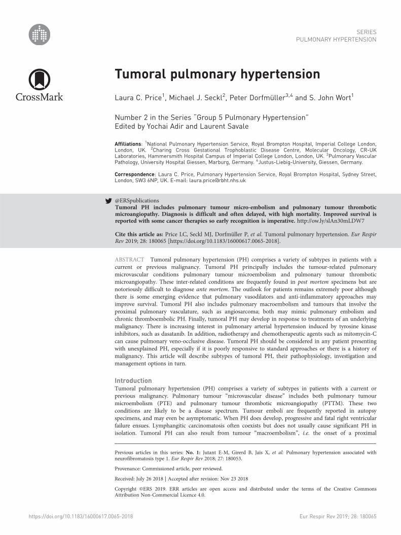
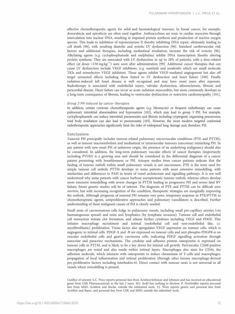
In the 1951–1990 autopsy series of 20 patients with neoplastic emboli mentioned above, SHIELDS andEDWARDS [4] identified three subgroups based on microscopic and gross features: 1) six patients withpredominantly neoplastic microemboli (PTE) (figure 1a); 2) 10 patients with mixed neoplastic andthrombotic microemboli (later termed PTTM) (figure 1b and c); and 3) four cases with both neoplasticmicroemboli and large, fatal tumour macroemboli (primary tumours in kidney, femur, cervix andthyroid). They noted changes in the morphology of resistance vessels of the lung (small arteries andarterioles), including pulmonary arterial hypertension (PAH)-like medial and intimal hypertrophy, intimalfibrosis and fibrinoid necrosis of the vessel wall, as well as noticing that plexiform lesions were notablyabsent. Medial hypertrophy was grade 1 (11–15% remodelling of the smooth muscle layer) in 12 cases,grade 2 (16–25%) in two cases and normal grade 0 (<10%) in six cases. This is indicative of the subacutenature of the disease, although no cases described severe grade 3 (>25%) remodelling [4].
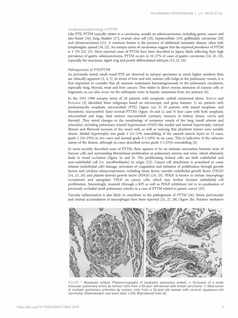
In more recently described cases of PTTM, there appears to be an intimate association between nests oftumour cells and surrounding fibrointimal proliferation in pulmonary arteries and veins, which ultimatelyleads to vessel occlusion (figure 2a and b). The proliferating intimal cells are both endothelial andnon-endothelial cell (i.e. myofibroblastic) in origin [22]. Cancer cell attachment is postulated to causeintimal (endothelial cell) damage, activation of coagulation and initiation of proliferation through growthfactors and cytokine release/expression, including tissue factor, vascular endothelial growth factor (VEGF)[14, 23, 24] and platelet-derived growth factor (PDGF) [24, 25]. PDGF is known to initiate macrophagerecruitment and upregulate VEGF on cancer cells, which may further increase endothelial cellproliferation. Interestingly, imatinib (through c-KIT as well as PDGF inhibition) led to re-canalisation ofpreviously occluded small pulmonary arteries in a case of PTTM related to gastric cancer [25].
Vascular inflammation is also likely to contribute to the pathogenesis of PTTM [26]. Dense perivascularand intimal accumulation of macrophages have been reported [21, 27, 28] (figure 2b). Putative mediators
a) b)
FIGURE 1 Neoplastic emboli. Photomicrographs of neoplastic pulmonary emboli. a) Occlusion of a smallmuscular pulmonary artery by tumour cells from a 55-year-old woman with breast carcinoma. b) Obstructionof multiple pulmonary arterioles by tumour cells from a 36-year-old woman with cervical squamous-cellcarcinoma. (Haematoxylin and eosin stain ×120). Reproduced from [4].
https://doi.org/10.1183/16000617.0065-2018 3
PULMONARY HYPERTENSION | L.C. PRICE ET AL.

include osteopontin, a cytokine and adhesive protein that is implicated in tumoral thrombosis andneointima formation [28], as well as promotion of progression and metastasis of cancer [29]. In PTTM,macrophages stain for osteopontin and CD44 (an osteopontin receptor) [28], with tumour cells andproliferating fibrointimal cells also overexpressing both CD44 and osteopontin, as well as PDGF andVEGF [27, 28]. Therefore, crosstalk between tumour cells, macrophages and intimal cells may occurthrough the osteopontin-CD44 axis to drive both tumorigenesis and ongoing macrophage recruitment.Macrophage-derived factors, such as interleukin-6, as implicated in PAH [30], may also contribute to thefibrointimal proliferation in PTTM.
Tumour cell nests are also evident in pulmonary veins and lymphatics [16, 21, 24], with evidence forlymphatic tumour invasion (figure 2c). Fibrointimal proliferation in pulmonary veins in PTTM lesions hasbeen reported in several recent reports [21, 31], and may be analogous to the remodelling of pulmonaryveins seen in distal chronic thromboembolic PH (CTEPH) [32]. Putative interactions between tumourcells, macrophages and vascular cells are summarised in figure 3.
Development of PHThere are two hypotheses for the development of PH in PTE/PTTM. First, inadequate clearance ofocclusive tumour emboli may increase pulmonary vascular resistance through mechanical obstruction. InPTE, a median 30% of vessels were occluded in symptomatic patients [2]. PH will develop in PTTM asprogressively more vessels become stenotic and occluded by the fibrointimal layer [13, 22]. Analysis ofautopsy specimens from patients with PTTM showed variable luminal occlusion but that widespreadsevere luminal narrowing was only a feature in severe PH [24].
Secondly, vascular occlusion is also likely to relate to PAH-like pulmonary vascular remodelling. There aremany unanswered questions in terms of signalling, including the potential role of genetic influences in
a) b)
c)
FIGURE 2 Lung histology from a case of pulmonary tumour thrombotic microangiopathy related to severepulmonary hypertension. a) Post mortem section showing occlusion of a medium-sized pulmonary arteriallumen by fibrointimal proliferation of fibroblasts and collagen (white arrow) and tumour emboli (black arrow)(Haematoxylin and eosin stain ×4.3). Scale bar=500 μm. b) Post mortem section showing a medium-sizedpulmonary artery with two elastic layers, with a normal-sized smooth muscle layer. There is exaggeratedluminal occlusion by fibrointimal thickening (white arrow) surrounding nests of tumour emboli (black arrow).The adventitia contains lymphatic tumoral thrombi. Increased alveolar macrophages are seen surrounding thelung (Elastica van Gieson stain ×4.6). Scale bar=500 μm. c) Evidence for fibrointimal proliferation within thelumen of small pulmonary veins (black arrow) and tumour involvement of accompanying lymphatics (whitearrow). Inset: veins close to the centrilobular bronchovascular bundles show eccentric fibrointimal remodelling(Elastica van Gieson ×28.4). Scale bar=80 μm. Reproduced from [26].
https://doi.org/10.1183/16000617.0065-2018 4
PULMONARY HYPERTENSION | L.C. PRICE ET AL.

terms of why a large number of patients have asymptomatic tumour emboli but a very small numberprogress to PTE or PTTM.
Haematological aspectsThe onset of venous thromboembolism in patients with cancer is of course well described. For example inpatients with acute pulmonary embolism, where an underlying malignancy should be excluded, and alsoin patients with CTEPH. Carcinomas promote coagulation by several mechanisms [33], such as the release ofmicrovesicles containing tissue factor [24] and through interaction (by mucinous carcinomas) with selectin onplatelets [34]. Binding of tissue factor with factor VII and calcium ultimately leads to fibrin deposition andplatelet activation to form clots. Progressive disseminated intravascular coagulation consumes fibrin, withrelease of fibrinogen degradation products or d-dimers. Usually, disseminated intravascular coagulation is rapidand associated with bleeding, whereas in malignancy “compensated disseminated intravascular coagulation” canoccur, where the rate of consumption of coagulation factors and platelets is slow; thrombotic manifestationsexceed bleeding events [35]. Evidence for raised d-dimer or fibrinogen degradation products has been auniform finding when reported in cases of PTTM [23, 35], with features of microangiopathic haemolyticanaemia (MAHA) or disseminated intravascular coagulation in half of cases at presentation [23, 35–37].
PH classification of PTE/PTTMPTE was first classified in 2004 within group 4 of the World Health Organization (WHO) classification systemof PH [38], and in the most recent update “other intravascular tumours” remains within group 4.2.2 [39].
Pulmonary
hypertension
Macrophage
recruitment
Clot propagation
VEGF
PDGFOsteopontin
Carcinoma cell MacrophagePlatelet
Tissue
factor
Vascular
occlusion
Fibrointimal
proliferation
Osteopontin
IL-6, others?
T
fa
V
PDG
Clot prop
hage
Osteopontin
IL-6, others?
ntin
orclusi
Endothelial/intimal cell
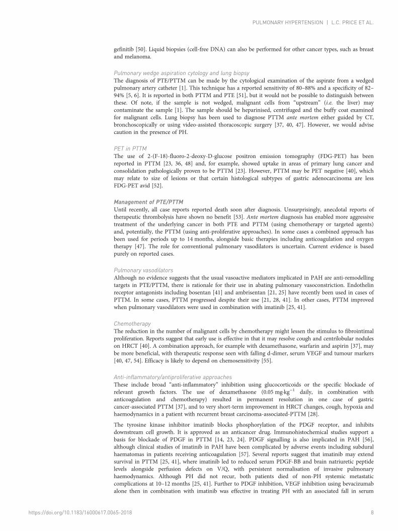
FIGURE 3 Proposed mechanisms for fibrointimal proliferation in pulmonary tumour thrombotic microangiopathy(PTTM). Small nests of carcinomatous cells lodge in the pulmonary vessels, including small pre-capillaryarteries (via haematogenous spread) and on the post-capillary side of the pulmonary circulation to pulmonaryveins and lymphatics (by lymphatic invasion). Tumour cell and endothelial cell interaction initiates clotformation, and releases further cytokines including vascular endothelial growth factor (VEGF) andplatelet-derived growth factor (PDGF). This initiates macrophage recruitment and intimal (endothelial cell andnon-endothelial cell like, i.e. myofibroblastic) proliferation. Tissue factor also upregulates VEGF expression ontumour cells, which is angiogenic to intimal cells. PDGF-A and -B are expressed on tumour cells, andanti-phospho-PDGFR-α is expressed on vascular endothelial cell and gastric carcinoma cells. This indicatesthat PDGF signalling activation of tumour cell growth is present through both autocrine and paracrinemechanisms. The cytokine and adhesive protein osteopontin is expressed on tumour cells in PTTM, and is likelyto be a key driver for intimal cell growth. Perivascular CD68-positive macrophages are noted and also residewithin intimal layers. Macrophages also stain for CD44, the adhesion molecule which interacts with osteopontinto induce chemotaxis of T-cells and macrophages, propagation of local inflammation and intimal proliferation(through other known macrophage-derived pro-proliferative factors including interleukin (IL)-6). Direct contactwith tumour nests is not universal in all vessels where remodelling is present.
https://doi.org/10.1183/16000617.0065-2018 5
PULMONARY HYPERTENSION | L.C. PRICE ET AL.

PTTM, with increasing recognition and understanding of its multifaceted mechanisms, now lies withingroup 5, i.e. that described to reflect multifactorial and/or unclear mechanisms. Tumoral obstruction,therefore, sits within subgroup 5.4 of the current classification of PH [39]. However, this classification doesnot necessarily guide management.
Clinical presentation of PTE/PTTMPatients with PTTM usually present with progressive exertional breathlessness, hypoxia and progressionright ventricular (RV) dysfunction [23]. Onset is usually 3 weeks to 6 months prior to presentation [14, 22, 40].Cough may predate dyspnoea. Haemoptysis, chest and abdominal pain, which may reflect liver metastasesor venous congestion, have also been reported [37]. The progression of symptoms correlates withhigh-resolution computed tomography (HRCT) findings prior to the onset of PH, which may be a fewweeks to months later [40]. Fatigue is also reported [41]. SOARES et al. [6] compared symptoms in a groupof pathologically confirmed tumour emboli patients with lymphangitic patients. The tumour embolismgroup was more likely to have dyspnoea (58%) than those with lymphatic disease (46%), howeverdiagnostic utility of this symptom was low [6].
Depending on the timing of presentation, symptoms and signs, PH may or may not be present. Theseinclude a prominent pulmonary second heart sound, raised jugular venous pressure, a right parasternalheave and further signs of RV decompensation. However these signs may often be absent even withconfirmed PH [42].
Investigations for PTE/PTTMDiagnosis of PTTM can be challenging and many investigative findings are nonspecific. ECG may showsigns of RV strain in the presence of PH including an S1Q3T3 appearance. Chest radiography is oftennormal. Bloods demonstrate raised d-dimer or fibrinogen degradation products [23] and, in some cases,MAHA. Patients desaturate markedly on 6-min walk test assessment. Pulmonary function testing wouldshow pulmonary vascular limitation, characterised by relative preservation of lung volumes but very low gastransfer values (transfer factor of the lung for carbon monoxide 25–30%), even lower than in PAH [43].
Radionucleotide ventilation/perfusion (V/Q) scanning may show multiple small peripheral sub-segmentalperfusion defects not evident on computed tomography pulmonary angiography, with normal ventilation(figure 4a). However, V/Q scanning abnormalities do not reliably distinguish PVOD from idiopathic PAH[4, 44]. Serial V/Q may show resolution of perfusion defects following treatment [25].
The confirmation of PH, once suspected clinically and suggested by a raised brain natriuretic peptide withechocardiographic findings, is by right heart catheterisation. Pre-capillary PH is defined by a meanpulmonary artery pressure ⩾25 mmHg and pulmonary capillary wedge pressure <15 mmHg [39]. Theassessment of haemodynamic severity would be consistent with all other causes of PH, with signs of rightheart dysfunction and low cardiac index being key prognostic factors [39]. At the time of right heartcatheterisation, pulmonary wedge aspiration cytology should be performed to detect tumour cells (seebelow) to attempt to confirm the diagnosis of PTTM or PTE.
Radiological findings in PTE/PTTMPlain chest radiography is often normal or shows diffuse reticulonodular opacities, and less often Kerley Blines and pleural effusions [45]. Parenchymal computed tomography (CT) abnormalities (figure 4b) arenonspecific and include centrilobular nodules, ground-glass attenuation, interlobular septal thickening andconsolidation [23].
CT signs of PH may be present including enlargement of the central pulmonary artery, right heartchamber enlargement and flattening of the intraventricular septum [21, 45]. Other pulmonarymanifestations of malignancy may be present, such as discrete metastatic deposits.
Main HRCT findingsCompared to PTE, where HRCT appearances are more often unremarkable, HRCT in PTTM may showcentrilobular nodularity, ground-glass opacities and interlobular septal thickening (figure 4b). These CTappearances will be discussed in turn.
Centrilobular nodularity on HRCT describes a central small nodule within the secondary pulmonarylobule, and is often reported in PTTM [21, 40, 41]. The nodularity is usually of an ultrafine granularappearance and is likely to represent the peripheral pulmonary arterial lesions. These opacities are usuallywell defined as they reflect peripheral blood vessels rather than the respiratory bronchioles when thenodule border is usually more blurred, for example in bronchiolitis. Centrilobular nodularity on HRCT isan early sign in PTTM, and may disappear following chemotherapy [40], in keeping with resolution of the
https://doi.org/10.1183/16000617.0065-2018 6
PULMONARY HYPERTENSION | L.C. PRICE ET AL.

small vessel lesions. Quite frequently the centrilobular nodules exhibit a tree in bud nodular pattern, afeature usually associated with plugging of respiratory bronchioles rather than vascular disease [46].
Ground-glass opacities are reported in PTTM (often patchy or wedged shaped) [21, 41, 47], for which theradiological differential diagnosis remains broad. In the setting of PTTM and PH, especially with post-capillary involvement, ground-glass opacities may be consistent with interstitial and airspace oedema [21],or interstitial inflammation, as suggested by resolution 1 month after high-dose steroids [28].
Interlobular septal thickening on HRCT (the corollary to septal lines on chest radiography) is described inPTTM, and is again a nonspecific finding. It simply indicates disease of the connective tissue, lymphaticsand/or pulmonary veins. The nature and distribution of septal thickening may provide clues to aetiology.In PTTM it appears smooth and peripheral in distribution [21, 41, 48], usually without pleural effusions.Lymphangitis carcinomatosa tends to cause more irregular, nodular interlobular septal thickening,resulting in prominence of the secondary pulmonary nodules, as well as thickening of the bronchovascularinterstitium, subpleural nodules, pleural effusions and often hilar or mediastinal node enlargement.Distinguishing PTTM from lymphangitis carcinomatosa may be difficult and of course these conditionsmay coexist.
Further assessment for malignancyInvestigations in a patient with new-onset PH and suspected PTE/PTTM should include screening forundiagnosed malignancy. This may involve a full clinical assessment similar to the assessment ofunprovoked pulmonary embolism [49]: mammography, a contrast-enhanced CT abdomen/pelvis andconsideration of further targeted tests [25]. This has formed part of our usual practice given the increasedawareness of PTTM, although it is not specified in current PH guidelines [39]. Raised tumour markers suchas CEA, CA19–9, CA15-3 and SCC may provide clues but are not diagnostic of an underlying cancer [40],except for human chorionic gonadotrophin and alpha feto-protein. The former in the absence ofpregnancy or false-positive readings is diagnostic of gestational and non-gestational cancers and the latteris highly indicative of germ cell and hepatocellular cancers. If the patient is too ill for a biopsy of asuspicious lung cancer then venous blood sampling to request mutant epidermal growth factor receptor(EGFR) testing on extracted circulating free DNA can diagnose an EGFR driven lung adenocarcinoma.This enables commencement of life-saving therapy with EGFR targeted drugs, such as erlotinib or
FIGURE 4 Radiology in pulmonarytumour thrombotic microangiopathy(PTTM). a) Ventilation/perfusiondemonstrating sub-segmental defectsin lung perfusion in a patient withPTTM. Computed tomographypulmonary angiography did notdemonstrate these peripheral lesions(not shown). b) High-resolutioncomputed tomography scanning inPTTM showing widespread ground-glass opacification, micronodules,interlobular septal thickening andsmall bilateral pleural effusions.
a)
b)
VENTILATION
PERFUSION
https://doi.org/10.1183/16000617.0065-2018 7
PULMONARY HYPERTENSION | L.C. PRICE ET AL.
gefinitib [50]. Liquid biopsies (cell-free DNA) can also be performed for other cancer types, such as breastand melanoma.
Pulmonary wedge aspiration cytology and lung biopsyThe diagnosis of PTE/PTTM can be made by the cytological examination of the aspirate from a wedgedpulmonary artery catheter [1]. This technique has a reported sensitivity of 80–88% and a specificity of 82–94% [5, 6]. It is reported in both PTTM and PTE [51], but it would not be possible to distinguish betweenthese. Of note, if the sample is not wedged, malignant cells from “upstream” (i.e. the liver) maycontaminate the sample [1]. The sample should be heparinised, centrifuged and the buffy coat examinedfor malignant cells. Lung biopsy has been used to diagnose PTTM ante mortem either guided by CT,bronchoscopically or using video-assisted thoracoscopic surgery [37, 40, 47]. However, we would advisecaution in the presence of PH.
PET in PTTMThe use of 2-(F-18)-fluoro-2-deoxy-D-glucose positron emission tomography (FDG-PET) has beenreported in PTTM [23, 36, 48] and, for example, showed uptake in areas of primary lung cancer andconsolidation pathologically proven to be PTTM [23]. However, PTTM may be PET negative [40], whichmay relate to size of lesions or that certain histological subtypes of gastric adenocarcinoma are lessFDG-PET avid [52].
Management of PTE/PTTMUntil recently, all case reports reported death soon after diagnosis. Unsurprisingly, anecdotal reports oftherapeutic thrombolysis have shown no benefit [53]. Ante mortem diagnosis has enabled more aggressivetreatment of the underlying cancer in both PTE and PTTM (using chemotherapy or targeted agents)and, potentially, the PTTM (using anti-proliferative approaches). In some cases a combined approach hasbeen used for periods up to 14 months, alongside basic therapies including anticoagulation and oxygentherapy [47]. The role for conventional pulmonary vasodilators is uncertain. Current evidence is basedpurely on reported cases.
Pulmonary vasodilatorsAlthough no evidence suggests that the usual vasoactive mediators implicated in PAH are anti-remodellingtargets in PTE/PTTM, there is rationale for their use in abating pulmonary vasoconstriction. Endothelinreceptor antagonists including bosentan [41] and ambrisentan [21, 25] have recently been used in cases ofPTTM. In some cases, PTTM progressed despite their use [21, 28, 41]. In other cases, PTTM improvedwhen pulmonary vasodilators were used in combination with imatinib [25, 41].
ChemotherapyThe reduction in the number of malignant cells by chemotherapy might lessen the stimulus to fibrointimalproliferation. Reports suggest that early use is effective in that it may resolve cough and centrilobular noduleson HRCT [40]. A combination approach, for example with dexamethasone, warfarin and aspirin [37], maybe more beneficial, with therapeutic response seen with falling d-dimer, serum VEGF and tumour markers[40, 47, 54]. Efficacy is likely to depend on chemosensitivity [55].
Anti-inflammatory/antiproliferative approachesThese include broad “anti-inflammatory” inhibition using glucocorticoids or the specific blockade ofrelevant growth factors. The use of dexamethasone (0.05 mg·kg−1 daily, in combination withanticoagulation and chemotherapy) resulted in permanent resolution in one case of gastriccancer-associated PTTM [37], and to very short-term improvement in HRCT changes, cough, hypoxia andhaemodynamics in a patient with recurrent breast carcinoma-associated-PTTM [28].
The tyrosine kinase inhibitor imatinib blocks phosphorylation of the PDGF receptor, and inhibitsdownstream cell growth. It is approved as an anticancer drug. Immunohistochemical studies support abasis for blockade of PDGF in PTTM [14, 23, 24]. PDGF signalling is also implicated in PAH [56],although clinical studies of imatinib in PAH have been complicated by adverse events including subduralhaematomas in patients receiving anticoagulation [57]. Several reports suggest that imatinib may extendsurvival in PTTM [25, 41], where imatinib led to reduced serum PDGF-BB and brain natriuretic peptidelevels alongside perfusion defects on V/Q, with persistent normalisation of invasive pulmonaryhaemodynamics. Although PH did not recur, both patients died of non-PH systemic metastaticcomplications at 10–12 months [25, 41]. Further to PDGF inhibition, VEGF inhibition using bevacizumabalone then in combination with imatinib was effective in treating PH with an associated fall in serum
https://doi.org/10.1183/16000617.0065-2018 8
PULMONARY HYPERTENSION | L.C. PRICE ET AL.
VEGF levels. Again, the patient died of a non-PH cause at 12 months [58]. These cases targeting specificgrowth factors are encouraging and require systematic assessment.
Tumour macroembolismRarely, a large tumour metastasis can mimic a proximal pulmonary embolism. Clinical features includingrapid onset of symptoms with PH and acute right heart failure would be indistinguishable. Tumourmacroembolism is reported in cases of breast cancer [59], hepatocellular carcinoma [60], choriocarcinoma[10, 55, 61, 62] and, less often, subdiaphragmatic cancers including cervical carcinoma [63]. PTE/PTTMmay of course coexist [4, 60]. These reflect the tumours that more commonly spread to the heart and greatvessels (renal, lung, breast, oesophagus, malignant lymphoma, leukaemia and malignant melanoma),through the vena cava (including invasive renal tumours that may fragment), arterial circulation,retrograde lymph node spread, or directly from adjacent viscera [63]. Overall metastatic cardiac cancerrates are low at 1.2% [64], and thought to relate to cardiac movement thereby rapid blood and lymphaticflow away from the heart [63]. In addition, some tumours may compress the proximal pulmonary arteries,with or without additional lymph nodal compression.
Finally, it should be remembered that venous thromboembolic disease including pulmonary embolism ismore common in patients with an underlying malignancy (see section below), hence careful history taking iskey in any initial venous thromboembolic assessment. Malignancy is also a clear risk factor for CTEPH [65].
Other causes of tumoral PHAlthough rare, there are additional groups of cancer-related PH, or apparent PH, that are important toinclude within a section on tumoral PH. First, pulmonary vascular tumours may lie proximally within themain pulmonary artery vessels. For example, pulmonary artery angiosarcomas form an important althoughdevastating differential diagnosis for patients presenting with apparent proximal venous thromboembolismor CTEPH [66], and are discussed in more detail below. Recognition even by experienced teams relies onadvanced imaging including magnetic resonance and PET. Secondly, extrinsic compression by lymphnodes or mediastinal tumours, for example a metastatic thymic carcinoid tumour, can cause extrinsiccompression mimicking stenosis of the pulmonary valve and apparent PH [67].
Pulmonary artery angiosarcomaSarcomas are rare tumours of mesenchymal origin that originate in bones or in soft tissues. Of these,angiosarcomas arise from endothelial cells and comprise 2% of all sarcomas [68]. Pulmonary arteryangiosarcomas typically arise from the pulmonary trunk [69] and can metastasise to the lung andmediastinal lymph nodes, as well as through haematogenous dissemination. They are important albeit raremimics of pulmonary thromboembolic disease.
Recognised associations with angiosarcoma include exposure to radiotherapy, genetic mutations includingBRCA, neurofibromatosis type 1 and Kippel Trelawney syndrome, and patients with immunosuppressionincluding HIV [68].
The clinical presentation of pulmonary artery angiosarcomas is nonspecific. The most common symptomsare dyspnoea, pleuritic chest pain, cough and haemoptysis [70].
CT findings include low attenuation filling defects, enhancement of the mass in the lumen of the arteryand extravascular spread of the lesion [71]. Gadolinium-enhanced magnetic resonance imaging may helpdifferentiate between thrombotic masses and vascular tumours: unlike thromboemboli, angiosarcomasexhibit heterogeneous enhancement with gadolinium [69]. The use of FDG-PET/CT can help differentiatemalignant from benign pulmonary artery lesions [71].
Surgical treatment is preferred for patients with primary pulmonary angiosarcoma, and is only an optionin the 50% presenting without metastatic disease [4]. Radical resection offers a median survival of37±20 months, compared to 11±3 months following subtotal resection or debulking [72]. Without surgery,the mean survival is 1.5 months. Chemotherapy may improve survival, but evidence remains limited [72].Inhibitors of angiogenesis may be more promising future therapies [73].
Cancer treatment-related PHSeveral oncological treatments may promote the development of numerous subtypes of PH in patientswith active or previous cancer. These aetiologies can be considered according to the PH classification, andof course may overlap with other causes of tumoral PH mentioned above.
PAH induced by cancer therapiesOf cancer therapies that can potentially induce PAH, the tyrosine kinase inhibitors (TKIs) are highlighted.TKIs include dasatanib [74], often used in chronic myeloid leukaemia therapy, and ponatinib, bosutinib and
https://doi.org/10.1183/16000617.0065-2018 9
PULMONARY HYPERTENSION | L.C. PRICE ET AL.

lapatinib, which are all used to treat several oncological conditions. Despite inhibiting relevant pathwaysincluding PDGF that might be expected to reverse PAH, these agents can cause direct pulmonary arteryendothelial cell toxicity through the production of mitochondrial reactive oxygen species [75]. The PAHthat arises usually resolves after discontinuation of the TKI, but persists in over a third of patients [76], andcan be fatal in this setting, with irreversible remodelling reported requiring lung transplantation [77].GUIGNABERT et al. [78] demonstrated a putative mechanism involving dasatinib-induced endothelialdysfunction and reactive oxygen species. TKI-induced PAH can unfortunately recur when other TKIs areused. Treatment with PAH-specific therapy is recommended for patients with right heart failure orpersistent PAH after discontinuation of the TKI [75].
PVOD induced by cancer therapiesSeveral cancer treatment-related mechanisms may precipitate the onset of PVOD, with the commonmechanism likely to relate to venous endothelial injury from cytotoxic chemotherapy or irradiation [79, 80].PVOD is a rare, devastating form of PAH, classified as group 1’ PH, characterised by widespread fibrousintimal proliferation of septal veins and preseptal venules also often with pulmonary capillary dilatation andproliferation [81] and substantial remodelling of the small pulmonary arteries [82]. Clinical features includemarked oxygen desaturation and very low gas transfer, with a radiographic triad of interlobular septalthickening, pleural effusions and lymphadenopathy. PVOD has been described in patients receivingchemotherapy including gemcitabine [83] and mitomycin-C [79, 84–86], and also in several reportsfollowing bone marrow transplantation [80, 86–88]. Radiotherapy has been reported to induce PVOD, witha potential delay in onset for several years, for example following mantle irradiation for Hodgkin’s lymphoma.Again, the mechanism is thought to relate to radiotherapy-induced venous endothelial damage [89]. It hasbeen suggested that therapy-induced PVOD may be under-recognised by a recent series of patients who diedwith pulmonary graft versus host disease more than 1 year after allogeneic haematopoietic stem celltransplantation, where 34% had histological evidence for PVOD as well as 29% having evidence forobliterative bronchiolitis; in both cases far more than had been clinically recognised [90].
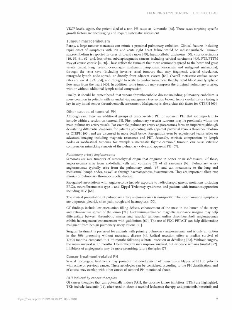
Recent advances have been made in the understanding of chemotherapy-induced PVOD. In 2002,GAGNADOUX et al. [85] described severe PVOD following surgery and perioperative mitomycin-C (MMC)chemotherapy for nonsmall cell lung carcinoma. More recently, PVOD cases from the French registry wereidentified whose anal carcinoma was treated with MMC, 5-flurouracil and radiotherapy 4 months prior toPVOD onset [2–12]. Baseline right heart catheterisation showed mean pulmonary artery pressure of41 mmHg and PVR of 11.2 (5.5–13.6) Wood units [36–63], with all patients in WHO functional class IIIor IV at presentation. Where tested, no patients had BMPR2 or EIF2AK4 mutations. Death followed infour out of the seven patients due to RV failure (n=2), local tumour progression (n=1) and severepneumonia (n=1). The same team developed a rat model of MMC-induced PVOD where intraperitonealMMC led to severe PAH at day 21–35, major remodelling of small pulmonary veins with endothelial cellproliferation in the capillary bed. Interestingly, a perivenular accumulation of eosinophils was seen in thisrat model [84]. In terms of signalling, a dose-dependent depletion of pulmonary GCN2 content anddecreased smad1/5/8 signalling was measured, despite normal tissue BMPR2 levels. GCN2 is the proteincoded by the recently described EIF2A2K gene whose function is depleted in heritable cases of PVOD,with the function of GCN2 likely to relate to antioxidant and anti-inflammatory properties [91]. Theseimportant observations by PERROS et al. [84] have been highlighted by SAVALE et al. [92] in a case of severePVOD in a 52-year-old woman with MMC, 5-flurouracil and local radiotherapy for anal cancer 6 monthspreviously. She underwent stabilisation with intravenous diuretics and dobutamine, and initiation of anendothelin receptor antagonist. She was given intravenous methylprednisolone, given that perivenularinflammation was present in the rat MMC-induced PVOD model [84], with resulting radiographicimprovement at 2 weeks (figure 5a) and clinical improvement that lasted a year.
However, she deteriorated further at that point requiring addition of a phosphodiesterase type-5 inhibitor,more steroids and ultimately successful lung transplantation. Pathologic assessment of explanted lungsconfirmed the diagnosis of PVOD with arterial remodelling without plexiform lesions, fibrous venousocclusion and capillary congestion (figure 5), although no perivenular inflammation was noted [92].
Preventative adjunctsFinally, in terms of chemotherapy- or radiotherapy-induced PAH/PVOD, there is an urgent clinical needto identify preventative adjuncts. Given the likely initial endothelial injury, cytoprotective approachesincluding amifostine, as well as steroids [92, 93], may be effective. Amifostine targets the endothelium, andis one of several cytoprotective agents developed to protect normal tissues from radiation andchemotherapy-induced damage. As an inactive prodrug, amifostine is dephosphorylated to an active thiolby alkaline phosphatase in normal endothelium, but not in acidic neoplastic tissues. This results incytoprotective selectivity involving free radical scavenging, DNA protection and repair acceleration, and
https://doi.org/10.1183/16000617.0065-2018 10
PULMONARY HYPERTENSION | L.C. PRICE ET AL.

induction of cellular hypoxia [94]. PERROS et al. [84] showed that in the rat MMC-induced PVOD model,2 weeks of amifostine co-treatment partially prevented the onset of MMC-induced PVOD, with improvedhaemodynamics and remodelling. Given the continuing clinical need for oncological therapies, althoughthe incidence of therapy-induced PVOD and PAH is not yet known, over 30% of patients had PVOD atautopsy following bone marrow transplantation [90]. The future use of cytoprotective therapies is,therefore, likely to be important.
Group 2 PH induced by cancer therapiesChemotherapy and radiation can also cause left ventricular (LV) dysfunction [95], with the potential onsetof group 2 PH. This leads to a form of cardiomyopathy, characterised by fall in LV ejection fraction,resulting in changes in biomarkers (troponin and BNP) and imaging, depending on the therapeutic agentand the stage of presentation. The fall in LV function may be irreversible or reversible depending on theagent, with the latter scenario highlighting the case for early recognition and monitoring [96]. Potentialaetiologies include anthracyclines (doxorubicin, daunorubicin, epirubicin and idarubicin) [97], which are
a) b)
c) d)
e) f)
FIGURE 5 High-resolution computed tomography (HRCT) of the chest and lung histology (haematoxylin andeosin–saffron staining) from explanted lungs. a) HRCT at diagnosis showed septal lines and centrilobularground-glass opacities suggestive of pulmonary veno-occlusive disease. b) Improvement of radiologicalabnormalities after 2 weeks of treatment including diuretics, dobutamine, endothelin receptor antagonist andhigh-dose corticosteroids. c) Overview of lung parenchyma displaying emphysematous changes andvasculopathy. Note the pulmonary vessels with narrowed lumina (arrows). d) Septal vein with pronouncedintimal fibrosis and heavily narrowed lumen. e) Focal capillary congestion in the vicinity of a remodelled vein(bottom). f ) Pulmonary artery (centre) with adjacent airway (top left) displaying medial hypertrophy andconcentric intimal fibrosis. c) Scale bar=1000 μm. d–f ) Scale bar=200 μm. Reproduced from [92].
https://doi.org/10.1183/16000617.0065-2018 11
PULMONARY HYPERTENSION | L.C. PRICE ET AL.
effective chemotherapeutic agents for solid and haematological tumours. In breast cancer, for example,doxorubicin and epirubicin are often used together. Anthracyclines are toxic to cardiac myocytes throughintercalation into nuclear DNA, resulting in impaired protein synthesis and production of reactive oxygenspecies. This leads to inhibition of topoisomerase II thereby inhibiting DNA repair, ultimately leading tocell death [98], with resulting diastolic and systolic LV dysfunction [96]. Standard cardiovascular riskfactors and additional therapies, including mediastinal irradiation, increase the risk of toxicity [96].Alkylating agents (e.g. cyclophosphamide and melphalan) inhibit DNA transcription thereby alteringprotein synthesis. They are associated with LV dysfunction in up to 28% of patients, with a dose-relatedeffect (at doses >150 mg·kg−1) seen soon after administration [99]. Additional cancer therapies that cancause LV dysfunction include VEGF inhibitors, (e.g. sunitinib and sorafenib) which are small moleculeTKIs and nonselective VEGF inhibitors. These agents inhibit VEGF-mediated angiogenesis but also offtarget unwanted effects including those linked to LV dysfunction and heart failure [100]. Finallyradiation-induced left heart disease is well recognised and may have onset years after exposure.Radiotherapy is associated with endothelial injury, valvular dysfunction, atherosclerosis, fibrosis andpericardial disease. Heart failure can occur as acute radiation myocarditis, but more commonly develops asa long-term consequence of fibrosis, leading to ventricular dysfunction or restrictive cardiomyopathy [101].
Group 3 PH induced by cancer therapiesIn addition, certain cytotoxic chemotherapeutic agents (e.g. bleomycin) or frequent radiotherapy can causepulmonary interstitial abnormalities and hypoxaemia [102], which may lead to group 3 PH. For example,cyclophosphamide can induce interstitial pneumonitis and fibrosis including cryptogenic organising pneumonia;total body irradiation can also lead to pneumonitis [103]. However, the more modern targeted conformalradiotherapeutic approaches significantly limit the risks of widespread lung damage and, therefore, PH.
ConclusionsTumoral PH principally includes tumour-related pulmonary microvascular conditions (PTE and PTTM),as well as tumour macroembolism and mediastinal or intravascular tumours (sarcomas) mimicking PH. Inany patient with new-onset PH of unknown origin, the presence of an underlying malignancy should alsobe considered. In addition, the long-term pulmonary vascular effects of cancer therapies (importantlyincluding PVOD) is a growing area and should be considered in the differential diagnosis of a cancerpatient presenting with breathlessness or PH. Autopsy studies from cancer patients indicate that thefinding of tumour emboli within small pulmonary vessels is not uncommon. PTE is the term used forsimple tumour cell emboli; PTTM develops in some patients with more extensive remodelling, withsimilarities and differences to PAH in terms of vessel architecture and signalling pathways. It is not wellunderstood why some patients with cancer harbour asymptomatic tumour emboli, whereas others developmore extensive remodelling with severe changes in PTTM leading to progressive PH and severe fatal RVfailure; future genetic studies will be of interest. The diagnosis of PTE and PTTM can be difficult antemortem, but with increasing recognition of the condition, therapeutic strategies are marginally improvingthe outlook. Although prognosis of tumoral PH remains very poor, temporary response in PTE/PTTM tochemotherapeutic agents, antiproliferative approaches and pulmonary vasodilators is described. Furtherunderstanding of these malignant causes of PH is clearly needed.
Small nests of carcinomatous cells lodge in pulmonary vessels, including small pre-capillary arteries (viahaematogenous spread) and veins and lymphatics (by lymphatic invasion). Tumour cell and endothelialcell interaction initiate clot formation, and release further cytokines including VEGF and PDGF. Thisinitiates macrophage recruitment and intimal (endothelial cell and non-endothelial like, i.e.myofibroblastic) proliferation. Tissue factor also upregulates VEGF expression on tumour cells, which isangiogenic to intimal cells. PDGF-A and -B are expressed on tumour cells and anti-phospho-PDGFR-α onvascular endothelial cells and gastric carcinoma cells, indicating PDGF signalling activation throughautocrine and paracrine mechanisms. The cytokine and adhesive protein osteopontin is expressed ontumour cells in PTTM, and is likely to be a key driver for intimal cell growth. Perivascular CD68-positivemacrophages are noted and also reside within intimal layers. Macrophages also stain for CD44, theadhesion molecule, which interacts with osteopontin to induce chemotaxis of T-cells and macrophages,propagation of local inflammation and intimal proliferation (through other known macrophage-derivedpro-proliferative factors including interleukin-6). Direct contact with tumour nests is not universal in allvessels where remodelling is present.
Conflict of interest: L.C. Price reports personal fees from Actelion/Johnson and Johnson and has received an educationalgrant from GSK Pharmaceuticals in the last 2 years. M.J. Seckl has nothing to disclose. P. Dorfmüller reports personalfees from MSD, Actelion and Roche, outside the submitted work. S.J. Wort reports grants and personal fees fromActelion and Bayer, and personal fees from GSK and MSD, outside the submitted work.
https://doi.org/10.1183/16000617.0065-2018 12
PULMONARY HYPERTENSION | L.C. PRICE ET AL.
References1 Roberts KE, Hamele-Bena D, Saqi A, et al. Pulmonary tumor embolism: a review of the literature. Am J Med
2003; 115: 228–232.2 Kane RD, Hawkins HK, Miller JA, et al. Microscopic pulmonary tumor emboli associated with dyspnea. Cancer
1975; 36: 1473–1482.3 Bagshawe KD, Brooks WD. Subacute pulmonary hypertension due to chorionepithelioma. Lancet 1959; 1:
653–658.4 Shields DJ, Edwards WD. Pulmonary hypertension attributable to neoplastic emboli: an autopsy study of 20
cases and a review of literature. Cardiovasc Pathol 1992; 1: 279–287.5 Winterbauer RH, Elfenbein IB, Ball WC Jr. Incidence and clinical significance of tumor embolization to the
lungs. Am J Med 1968; 45: 271–290.6 Soares FA, Pinto AP, Landell GA, et al. Pulmonary tumor embolism to arterial vessels and carcinomatous
lymphangitis. A comparative clinicopathological study. Arch Pathol Lab Med 1993; 117: 827–831.7 Wilson K, Guardino J, Shapira O. Pulmonary tumor embolism as a presenting feature of cavoatrial hepatocellular
carcinoma. Chest 2001; 119: 657–658.8 Schriner RW, Ryu JH, Edwards WD. Microscopic pulmonary tumor embolism causing subacute cor pulmonale:
a difficult antemortem diagnosis. Mayo Clin Proc 1991; 66: 143–148.9 Yutani C, Imakita M, Ishibashi-Ueda H, et al. Pulmonary hypertension due to tumor emboli: a report of three
autopsy cases with morphological correlations to radiological findings. Acta Pathol Jpn 1993; 43: 135–141.10 Seckl MJ, Rustin GJ, Newlands ES, et al. Pulmonary embolism, pulmonary hypertension, and choriocarcinoma.
Lancet 1991; 338: 1313–1315.11 Goldhaber SZ, Dricker E, Buring JE, et al. Clinical suspicion of autopsy-proven thrombotic and tumor
pulmonary embolism in cancer patients. Am Heart J 1987; 114: 1432–1435.12 von Herbay A, Illes A, Waldherr R, et al. Pulmonary tumor thrombotic microangiopathy with pulmonary
hypertension. Cancer 1990; 66: 587–592.13 Pinckard JK, Wick MR. Tumor-related thrombotic pulmonary microangiopathy: review of pathologic findings
and pathophysiologic mechanisms. Ann Diagn Pathol 2000; 4: 154–157.14 Chinen K, Tokuda Y, Fujiwara M, et al. Pulmonary tumor thrombotic microangiopathy in patients with gastric
carcinoma: an analysis of 6 autopsy cases and review of the literature. Pathol Res Pract 2010; 206: 682–689.15 Carter CA, Scicinski JJ, Lybeck HE, et al. Pulmonary tumor thrombotic microangiopathy: a new paraneoplastic
syndrome? Case Rep Oncol 2016; 9: 246–248.16 Uga S, Ikeda S, Matsukage S, et al. An autopsy case of acute cor pulmonale and paradoxical systemic embolism
due to tumour cell microemboli in a patient with breast cancer. BMJ Case Rep 2012; 2012: bcr2012006682.17 Buser M, Felizeter-Kessler M, Lenggenhager D, et al. Rapidly progressive pulmonary hypertension in a patient
with pulmonary tumor thrombotic microangiopathy. Am J Respir Crit Care Med 2015; 191: 711–712.18 Gru AA, Pai RK, Roma AA. Pulmonary tumor thrombotic microangiopathy in patients with low-grade ovarian
serous neoplasm: a clinicopathologic review of 2 cases of a previously unknown association. Int J Gynecol Pathol2012; 31: 438–442.
19 Tanaka K, Nakasya A, Miyazaki M, et al. [A case of hepatocellular carcinoma with respiratory failure caused bywidespread tumor microemboli]. Fukuoka Igaku Zasshi 2011; 102: 298–302.
20 Malani AK, Gupta C, Kutty AV, et al. Pulmonary tumor thrombotic microangiopathy from metastaticgallbladder carcinoma: an unusual cause of severe pulmonary hypertension. Dig Dis Sci 2007; 52: 555–557.
21 Kumar N, Price LC, Montero MA, et al. Pulmonary tumour thrombotic microangiopathy: unclassifiablepulmonary hypertension? Eur Respir J 2015; 46: 1214–1217.
22 von Herbay A, Maiwald M, Ditton HJ, et al. Histology of intestinal Whipple’s disease revisited. A study of 48patients. Virchows Arch 1996; 429: 335–343.
23 Uruga H, Fujii T, Kurosaki A, et al. Pulmonary tumor thrombotic microangiopathy: a clinical analysis of 30autopsy cases. Intern Med 2013; 52: 1317–1323.
24 Okubo Y, Wakayama M, Kitahara K, et al. Pulmonary tumor thrombotic microangiopathy induced bygastric carcinoma: morphometric and immunohistochemical analysis of six autopsy cases. Diagn Pathol2011; 6: 27.
25 Minatsuki S, Miura I, Yao A, et al. Platelet-derived growth factor receptor-tyrosine kinase inhibitor, imatinib, iseffective for treating pulmonary hypertension induced by pulmonary tumor thrombotic microangiopathy.Int Heart J 2015; 56: 245–248.
26 Price LC, Wells AU, Wort SJ. Pulmonary tumour thrombotic microangiopathy. Curr Opin Pulm Med 2016; 22:421–428.
27 Takahashi F, Kumasaka T, Nagaoka T, et al. Osteopontin expression in pulmonary tumor thromboticmicroangiopathy caused by gastric carcinoma. Pathol Int 2009; 59: 752–756.
28 Higashi A, Dohi Y, Uraoka N, et al. The Potential role of inflammation associated with interaction betweenosteopontin and CD44 in a case of pulmonary tumor thrombotic microangiopathy caused by breast cancer.Intern Med 2015; 54: 2877–2880.
29 Khan SA, Cook AC, Kappil M, et al. Enhanced cell surface CD44 variant (v6, v9) expression by osteopontin inbreast cancer epithelial cells facilitates tumor cell migration: novel post-transcriptional, post-translationalregulation. Clin Exp Metastasis 2005; 22: 663–673.
30 Hashimoto-Kataoka T, Hosen N, Sonobe T, et al. Interleukin-6/interleukin-21 signaling axis is critical in thepathogenesis of pulmonary arterial hypertension. Proc Natl Acad Sci USA 2015; 112: E2677–E2686.
31 Godbole R, Saggar R, Zider A, et al. Insights on pulmonary tumor thrombotic microangiopathy: a seven-patientcase series. Pulm Circ 2017; 7: 813–820.
32 Dorfmuller P, Gunther S, Ghigna MR, et al. Microvascular disease in chronic thromboembolic pulmonaryhypertension: a role for pulmonary veins and systemic vasculature. Eur Respir J 2014; 44: 1275–1288.
33 Caine GJ, Stonelake PS, Lip GY, et al. The hypercoagulable state of malignancy: pathogenesis and current debate.Neoplasia 2002; 4: 465–473.
34 Wahrenbrock M, Borsig L, Le D, et al. Selectin-mucin interactions as a probable molecular explanation for theassociation of Trousseau syndrome with mucinous adenocarcinomas. J Clin Invest 2003; 112: 853–862.
https://doi.org/10.1183/16000617.0065-2018 13
PULMONARY HYPERTENSION | L.C. PRICE ET AL.

35 Gainza E, Fernandez S, Martinez D, et al. Pulmonary tumor thrombotic microangiopathy: report of 3 cases andreview of the literature. Medicine (Baltimore) 2014; 93: 359–363.
36 Ho AL, Szulakowski P, Mohamid WH. The diagnostic challenge of pulmonary tumour thromboticmicroangiopathy as a presentation for metastatic gastric cancer: a case report and review of the literature. BMCCancer 2015; 15: 450.
37 Miyano S, Izumi S, Takeda Y, et al. Pulmonary tumor thrombotic microangiopathy. J Clin Oncol 2007; 25: 597–599.38 Simonneau G, Galie N, Rubin LJ, et al. Clinical classification of pulmonary hypertension. J Am Coll Cardiol
2004; 43: 12 Suppl S, 5S–12S.39 Galie N, Humbert M, Vachiery J-L, et al. 2015 ESC/ERS Guidelines for the diagnosis and treatment of
pulmonary hypertension. The Joint Task Force for the Diagnosis and Treatment of Pulmonary Hypertension ofthe European Society of Cardiology (ESC) and the European Respiratory Society (ERS). Eur Respir J 2015; 46:903–975.
40 Kayatani H, Matsuo K, Ueda Y, et al. Pulmonary tumor thrombotic microangiopathy diagnosed antemortem andtreated with combination chemotherapy. Intern Med 2012; 51: 2767–2770.
41 Ogawa A, Yamadori I, Matsubara O, et al. Pulmonary tumor thrombotic microangiopathy with circulatoryfailure treated with imatinib. Intern Med 2013; 52: 1927–1930.
42 Veinot JP, Ford SE, Price RG. Subacute cor pulmonale due to tumor embolization. Arch Pathol Lab Med 1992;116: 131–134.
43 Montani D, Achouh L, Dorfmuller P, et al. Pulmonary veno-occlusive disease: clinical, functional, radiologic, andhemodynamic characteristics and outcome of 24 cases confirmed by histology. Medicine (Baltimore) 2008; 87:220–233.
44 Seferian A, Helal B, Jais X, et al. Ventilation/perfusion lung scan in pulmonary veno-occlusive disease. Eur Respir J2012; 40: 75–83.
45 Godbole R, Ghatol A, Betancourt J, et al. Pulmonary tumor thrombotic microangiopathy: clinical, radiologic, andhistologic correlation. J Clin Imaging Sci 2015; 5: 44.
46 Franquet T, Gimenez A, Prats R, et al. Thrombotic microangiopathy of pulmonary tumors: a vascular cause oftree-in-bud pattern on CT. AJR Am J Roentgenol 2002; 179: 897–899.
47 Kitamura A, Nishimura N, Jinta T, et al. A case of pulmonary tumor thrombotic microangiopathy diagnosed bytransbronchial lung biopsy and treated with chemotherapy and long-term oxygen and anticoagulation therapies.Case Rep Pulmonol 2013; 2013: 259080.
48 Tashima Y, Abe K, Matsuo Y, et al. Pulmonary tumor thrombotic microangiopathy: FDG-PET/CT findings. ClinNucl Med 2009; 34: 175–177.
49 Carrier M, Lazo-Langner A, Shivakumar S, et al. Screening for occult cancer in unprovoked venousthromboembolism. N Engl J Med 2015; 373: 697–704.
50 Hutchinson JC, Fulcher JW, Hanna J, et al. Pulmonary tumor thrombotic microangiopathy: case report andreview of literature. Am J Forensic Med Pathol 2018; 39: 56–60.
51 Bhuvaneswaran JS, Venkitachalam CG, Sandhyamani S. Pulmonary wedge aspiration cytology in the diagnosis ofrecurrent tumour embolism causing pulmonary arterial hypertension. Int J Cardiol 1993; 39: 209–212.
52 Stahl A, Ott K, Weber WA, et al. FDG PET imaging of locally advanced gastric carcinomas: correlation withendoscopic and histopathological findings. Eur J Nucl Med Mol Imaging 2003; 30: 288–295.
53 Keenan NG, Nicholson AG, Oldershaw PJ. Fatal acute pulmonary hypertension caused by pulmonary tumourthrombotic microangiopathy. Int J Cardiol 2008; 124: e11–e13.
54 Simonneau G, Robbins IM, Beghetti M, et al. Updated clinical classification of pulmonary hypertension. J AmColl Cardiol 2009; 54: 1 Suppl, S43–S54.
55 Savage P, Roddie M, Seckl MJ. A 28-year-old woman with a pulmonary embolus. Lancet 1998; 352: 30.56 Ghofrani HA, Morrell NW, Hoeper MM, et al. Imatinib in pulmonary arterial hypertension patients with
inadequate response to established therapy. Am J Respir Crit Care Med 2010; 182: 1171–1177.57 Hoeper MM, Barst RJ, Bourge RC, et al. Imatinib mesylate as add-on therapy for pulmonary arterial
hypertension: results of the randomized IMPRES study. Circulation 2013; 127: 1128–1138.58 Higo K, Kubota K, Takeda A, et al. Successful antemortem diagnosis and treatment of pulmonary tumor
thrombotic microangiopathy. Intern Med 2014; 53: 2595–2599.59 Geschwind JF, Dagli MS, Vogel-Claussen J, et al. Metastatic breast carcinoma presenting as a large pulmonary
embolus: case report and review of the literature. Am J Clin Oncol 2003; 26: 89–91.60 Jakel J, Ramaswamy A, Kohler U, et al. Massive pulmonary tumor microembolism from a hepatocellular
carcinoma. Pathol Res Pract 2006; 202: 395–399.61 Perroni D, Grecchi GL, La Ciura P, et al. Right ventricular metastasis from choriocarcinoma: report of a rare case
and review of the literature. Eur J Surg Oncol 1993; 19: 378–381.62 Bozaci EA, Taskin S, Gurkan O, et al. Intracavitary cardiac metastasis and pulmonary tumor emboli of
choriocarcinoma: the first case diagnosed and treated without surgical intervention. Gynecol Oncol 2005; 99:753–756.
63 Han GH, Kwon DY, Ulak R, et al. Right ventricular metastatic tumor from a primary carcinoma of uterinecervix: a cause of pulmonary embolism. Obstet Gynecol Sci 2017; 60: 129–132.
64 Jann H, Wertenbruch T, Pape U, et al. A matter of the heart: myocardial metastases in neuroendocrine tumors.Horm Metab Res 2010; 42: 967–976.
65 Kim NH, Lang IM. Risk factors for chronic thromboembolic pulmonary hypertension. Eur Respir Rev 2012; 21:27–31.
66 Govender D, Pillay SV. Right pulmonary artery sarcoma. Pathology 2001; 33: 243–245.67 Lynch M, Blevins LS, Martin RP. Acquired supravalvular pulmonary stenosis due to extrinsic compression by a
metastatic thymic carcinoid tumor. Int J Card Imaging 1996; 12: 61–63.68 Young RJ, Brown NJ, Reed MW, et al. Angiosarcoma. Lancet Oncol 2010; 11: 983–991.69 Huo L, Lai S, Gladish G, et al. Pulmonary artery angiosarcoma: a clinicopathologic and radiological correlation.
Ann Diagn Pathol 2005; 9: 209–214.70 Desmarais P, Laskine M, Caporuscio C. Primary pulmonary artery angiosarcoma mimicking pulmonary
embolism in a 66-year-old man with dyspnea. CMAJ 2016; 188: E509–E512.
https://doi.org/10.1183/16000617.0065-2018 14
PULMONARY HYPERTENSION | L.C. PRICE ET AL.

71 Lee EJ, Moon SH, Choi JY, et al. Usefulness of fluorodeoxyglucose positron emission tomography in malignancyof pulmonary artery mimicking pulmonary embolism. ANZ J Surg 2013; 83: 342–347.
72 Blackmon SH, Rice DC, Correa AM, et al. Management of primary pulmonary artery sarcomas. Ann ThoracSurg 2009; 87: 977–984.
73 Azzariti A, Porcelli L, Mangia A, et al. Irradiation-induced angiosarcoma and anti-angiogenic therapy:a therapeutic hope? Exp Cell Res 2014; 321: 240–247.
74 Montani D, Bergot E, Gunther S, et al. Pulmonary arterial hypertension in patients treated by dasatinib.Circulation 2012; 125: 2128–2137.
75 Weatherald J, Chaumais MC, Montani D. Pulmonary arterial hypertension induced by tyrosine kinase inhibitors.Curr Opin Pulm Med 2017; 23: 392–397.
76 Weatherald J, Chaumais MC, Savale L, et al. Long-term outcomes of dasatinib-induced pulmonary arterialhypertension: a population-based study. Eur Respir J 2017; 50: 1700217.
77 Daccord C, Letovanec I, Yerly P, et al. First histopathological evidence of irreversible pulmonary vascular diseasein dasatinib-induced pulmonary arterial hypertension. Eur Respir J 2018; 51: 1701694.
78 Guignabert C, Phan C, Seferian A, et al. Dasatinib induces lung vascular toxicity and predisposes to pulmonaryhypertension. J Clin Invest 2016; 126: 3207–3218.
79 Joselson R, Warnock M. Pulmonary veno-occlusive disease after chemotherapy. Hum Pathol 1983; 14: 88–91.80 Bunte MC, Patnaik MM, Pritzker MR, et al. Pulmonary veno-occlusive disease following hematopoietic stem cell
transplantation: a rare model of endothelial dysfunction. Bone Marrow Transplant 2008; 41: 677–686.81 Montani D, Price LC, Dorfmuller P, et al. Pulmonary veno-occlusive disease. Eur Respir J 2009; 33: 189–200.82 Nossent EJ, Antigny F, Montani D, et al. Pulmonary vascular remodeling patterns and expression of general
control nonderepressible 2 (GCN2) in pulmonary veno-occlusive disease. J Heart Lung Transplant 2018; 37:647–655.
83 Turco C, Jary M, Kim S, et al. Gemcitabine-induced pulmonary toxicity: a case report of pulmonaryveno-occlusive disease. Clin Med Insights Oncol 2015; 9: 75–79.
84 Perros F, Gunther S, Ranchoux B, et al. Mitomycin-induced pulmonary veno-occlusive disease: evidence fromhuman disease and animal models. Circulation 2015; 132: 834–847.
85 Gagnadoux F, Capron F, Lebeau B. Pulmonary veno-occlusive disease after neoadjuvant mitomycinchemotherapy and surgery for lung carcinoma. Lung Cancer 2002; 36: 213–215.
86 Seguchi M, Hirabayashi N, Fujii Y, et al. Pulmonary hypertension associated with pulmonary occlusivevasculopathy after allogeneic bone marrow transplantation. Transplantation 2000; 69: 177–179.
87 Troussard X, Bernaudin JF, Cordonnier C, et al. Pulmonary veno-occlusive disease after bone marrowtransplantation. Thorax 1984; 39: 956–957.
88 Williams LM, Fussell S, Veith RW, et al. Pulmonary veno-occlusive disease in an adult following bone marrowtransplantation. Case report and review of the literature. Chest 1996; 109: 1388–1391.
89 Kramer MR, Estenne M, Berkman N, et al. Radiation-induced pulmonary veno-occlusive disease. Chest 1993;104: 1282–1284.
90 Gazourian L, Spring L, Meserve E, et al. Pulmonary clinicopathological correlation after allogeneic hematopoieticstem cell transplantation: an autopsy series. Biol Blood Marrow Transplant 2017; 23: 1767–1772.
91 Wilson GJ, Bunpo P, Cundiff JK, et al. The eukaryotic initiation factor 2 kinase GCN2 protects againsthepatotoxicity during asparaginase treatment. Am J Physiol Endocrinol Metab 2013; 305: E1124–E1133.
92 Savale L, Chaumais MC, Dorfmuller P, et al. Lung transplantation for mitomycin-induced pulmonaryveno-occlusive disease. Presse Med 2017; 46: 1223–1225.
93 Hosokawa K, Yamazaki H, Nishitsuji M, et al. Pulmonary veno-occlusive disease following reduced-intensityallogeneic bone marrow transplantation for acute myeloid leukemia. Intern Med 2012; 51: 195–198.
94 Kouvaris JR, Kouloulias VE, Vlahos LJ. Amifostine: the first selective-target and broad-spectrum radioprotector.Oncologist 2007; 12: 738–747.
95 Lipshultz SE, Cochran TR, Franco VI, et al. Treatment-related cardiotoxicity in survivors of childhood cancer.Nat Rev Clin Oncol 2013; 10: 697–710.
96 Bloom MW, Hamo CE, Cardinale D, et al. Cancer therapy-related cardiac dysfunction and heart failure: part 1:definitions, pathophysiology, risk factors, and imaging. Circ Heart Fail 2016; 9: e002661.
97 Shan K, Lincoff AM, Young JB. Anthracycline-induced cardiotoxicity. Ann Intern Med 1996; 125: 47–58.98 Lyu YL, Kerrigan JE, Lin CP, et al. Topoisomerase IIbeta mediated DNA double-strand breaks: implications in
doxorubicin cardiotoxicity and prevention by dexrazoxane. Cancer Res 2007; 67: 8839–8846.99 Braverman AC, Antin JH, Plappert MT, et al. Cyclophosphamide cardiotoxicity in bone marrow transplantation:
a prospective evaluation of new dosing regimens. J Clin Oncol 1991; 9: 1215–1223.100 Groarke JD, Choueiri TK, Slosky D, et al. Recognizing and managing left ventricular dysfunction associated with
therapeutic inhibition of the vascular endothelial growth factor signaling pathway. Curr Treat Options CardiovascMed 2014; 16: 335.
101 Prosnitz RG, Chen YH, Marks LB. Cardiac toxicity following thoracic radiation. Semin Oncol 2005; 32: Suppl 3,S71–S80.
102 Abid SH, Malhotra V, Perry MC. Radiation-induced and chemotherapy-induced pulmonary injury. Curr OpinOncol 2001; 13: 242–248.
103 Shahab N, Haider S, Doll DC. Vascular toxicity of antineoplastic agents. Semin Oncol 2006; 33: 121–138.
https://doi.org/10.1183/16000617.0065-2018 15
PULMONARY HYPERTENSION | L.C. PRICE ET AL.