tumors of nasal paranasal - pdfs.semanticscholar.org · tumors of the nasal and paranasal cavities...
TRANSCRIPT

TUMORS OF THE NASAL AND PARANASAL CAVITIES
CHARLES F. GESCHICRTER, M.D.'
(From the dur.qirnl Pnlhologiral Lnbornlory, Depurlniaiit of Surgery, Jokm Hapk'ins Aoapilnl and University)
EM BRY O ~ ~ O O Y A N n CLASSIFICATION
The lining membrane of the nasal cnvity and sinuses is formed from the invaginating ectoderm of the olftictory plates at the end of the fourth week of embryonic life. T t is continuous with the mucous mem- brane of the nasopharynx, which is entodermal in origin but of similar histologic structure. At first these olfactory plates are in contact with the cerebral vessels, but later the meninges and cthmoid bone separate them from the brain. Outgrowths of the nasal lining in the third month form the middle meatus, and further extensions a t the posterior end form the pockets for the maxillary sinus. From the anterior portion four furrows develop, two representing buds for the frontal sinuses and the remainder the air cells of the ethmoid. The lining membrane differ- entiates to form ciliated columnar epithelium resembling the respira- tory epithelium of the pulmonary tract. From the undifferentiated cells of tho bastil layers the mucous glands of Bowman are derived. The submucous tissue of this region is particularly rich in mucou8 glands and in blood vessels. Portions of the olfactory cctoderm in the upper ethmoidal region arc ncuro-cpithcliul in character and form con- nections with the olfactory bulb. Otiiigliomas may rarely form from these cells.
The sinuses tire rudimentary at birth, but reach adult size between adolescence and the twenty-fifth year. The majority of carcinomas of the nose arise in the region of the middle turbinate at the embryonic site of the outpouching of the sinuses, and are epidermal in type. In the nasopharynx, and more rarely in the nose aiid antrum, malignant epidermal cells from the mucous memhmne are interspersed with lymphoid tissue, producing ti variety of lymphodermal cancer usually referred to as lympho-epithelioma. The post-embryonic exteiisions of the epithelial membrane within the maxillary sinuses provide a source for epidermal carcinomas of the antrum. From the undifferentiated epithelium of the basal layers ilest ined to form glandular appendages cystic basal-cell cancer aiid adenocarcinoma arise. It is usually im- possible at the time of their clinical recognition to determine whether malignant epithelial tumors of this region have had their primary site within the nose or antrum. Intraiiasal carcinomas extend to the maxil- lary sinus, and antral tumors invade the nasal cavity relatively early. The largecrt number of cancers arc both antral and intranasal at the
a Aided by a grant from The Anna Fuller Fund. 637
638 CHARLES F. GESCHICETER
time of clinical observation, and may be referred to as maxillo- ethmoidal tumors. Such carcinomas comprise from 1 to 2 per cent of cancer throughout the body. These tumors obstruct respiration, erode the surrounding skeletal structures, and usually produce death by ex- tension. Clinical recognition is usually late, and prior to the introduc- tion of radium therapy permanent cures were exceedingly rare.
Probably because slowly growing tumors of this region remain asymptomatic, epithelial tumors of benign character are seldom re- ported. So-called hard papillomas, adenomas and cystadenomas, and rarely aberrant salivary tumors are among the benign epithelial growths of the nasal and paranasal cavities. Osteomas, angiomas, plasmocytomas and a variety of benign and malignant connective-tissue tumors occur, but with less frequency than epithelial tumors.
The classification and incidence of the 211 tumors in the present series, selected by microscopic study from over 2,000 specimens re- moved surgically in the Department of Laryngology and Otology of the Johns Hopkins Hospital, are as follows :
Benign Epithelial Tumors ............................................. 1'3 cases Epidermal papilloma ................................. 10 cases
Aberrant salivary tumors ........................... 3 cases Appendage-cell tumors
Cysttidenoma ...................................... 6 cases Malignant Epithelial Tumors ........................................... 139 caties
Epidermal cancer Keratinizing and non-keratiriieing squamouci-cell cancer 73 cases
.......... 36 cases Lymphodermal cancer (Igmpho-epitheliomu)
Cystic basal-cell cancer .............................. 15 cases
Angioma ............................................ 25 cases Plasmocytoma ....................................... 6 cases Fibroma ............................................ 6 cases
Lymphosarcoma ...................................... 7 eases
Myeloma ............................................ 3 cases
Appendage-cell carcinoma
Adenocarcinoma (including Srhneiderinn cancer) ....... 15 cases Benign Conncctiae-Ziasue Tumors (czclwwire of 76 oateomus) ................ 37 cases
Surcomas ............................................................. 16 caws
Melanosarcoma ....................................... 2 cases Rhabdomyosnrcoma ................................... 2 oases
Fibrosarcomu ........................................ 3 cases
Zicflaimat ury Chwths or Nasal Polyps : Among the non-neoplastic conditions in this series, the majority were nasal polyps. These aro sometimes classed as fibro-epithelial tumors or as soft papillomas of the nose.
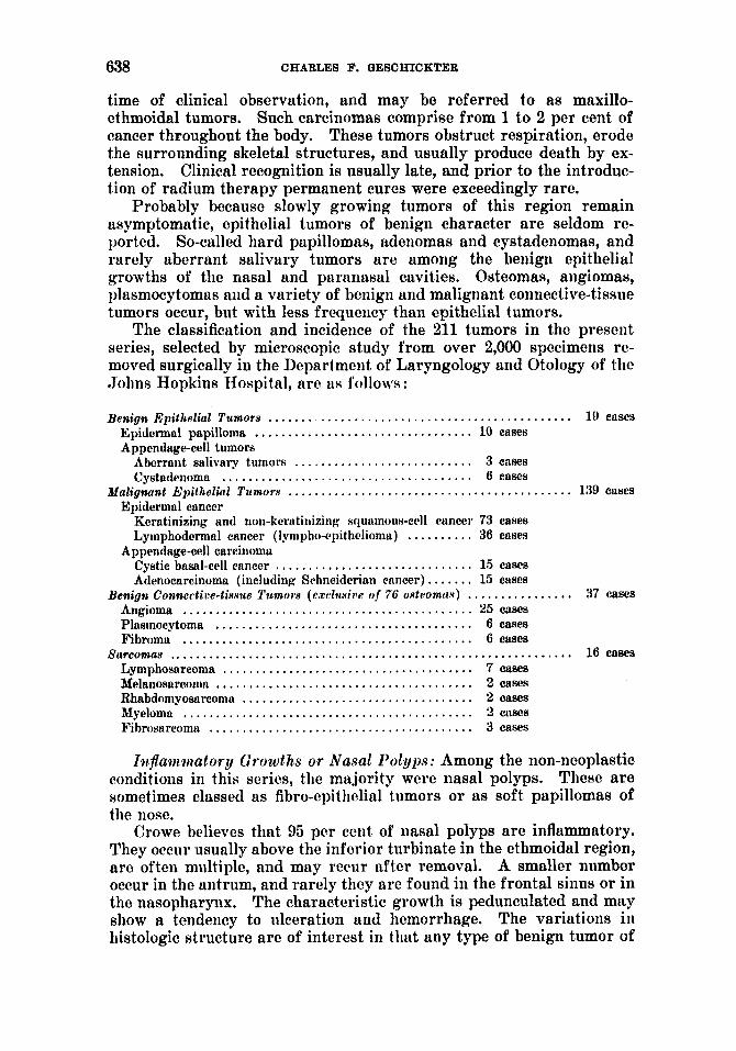
Crowe believes that 95 per cent of nasal polyps are inflammatory. They occur usually above the inferior turbinate in the ethmoidal region, are often multiple, and may recur after removal. A smaller number occur in the antrum, and rarely they arc found in the frontal sinus or in the nasopharynx. The characteristic growth is pedunculated and may show a tendency to ulceration and hemorrhage. The variations in histologic structure are of interest in that any type of benign tumor of
TUMORS OF T H E NASAL A N D PARANASAL CAVITIEB 639
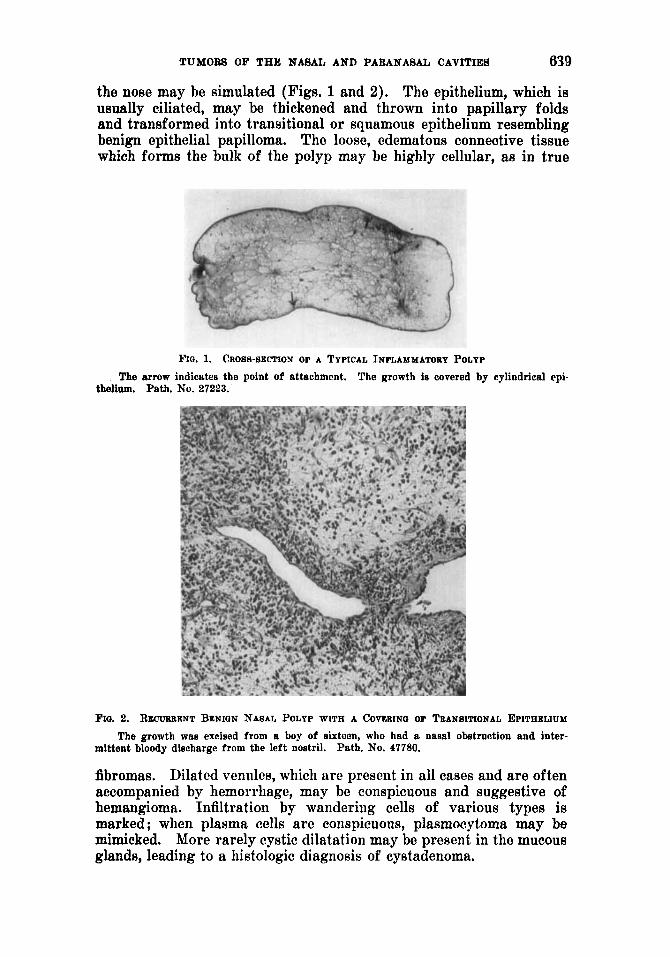
the nose may be simulated (Figs. 1 and 2). The epithelium, which is usually ciliated, may be thickened and thrown into papillary folds and transformed into transitional or squamous epithelium resembling benign epithelial papilloma. The loose, edematous connective tissue which forms the bulk of the polyp may be highly cellular, as in true
Wo. 1. CROSS-SECTION OF A TYPICAL INFLAMMATORY POLYP The arrow indicates the point of attachment. The growth is covered by cylindrical epi-
tbelinm. Path. No. 27223.
FIG. 2. R E C U ~ ~ E N T BENIGN NASAL POLYP WITH A COvGaINo OF TEANBITIONAL EPITHELIUM The growth was excised from a boy of sixteen, who had a nasal obstruction and inter-
mittent bloody diecharge from the left nostril. Path. No. 47780.
fibromas. Dilated venules, which are present in all cases and are often accompanied by hemorrhage, may be conspicuous and suggestive of hemangioma. Infiltration by wandering cells of various types is marked ; when plasma cells arc conspicuous, plasmocytoma may be mimicked. More rarely cystic dilatation may be present in the mucous glands, leading to a histologic diagnosis of cystadenoma.
640 CHARLES F. QESCHICKTER
The more moderate increases in the epithelial covering, in the sub- mucous fibrous tissue, in vessels, and in the plasma-cell infiltration are not inconsistent with the diagnosis of benign inflammatory nasal polyp. It is doubtful if such lesions undergo neoplastic or malignant change. More often these polypoid conditions are the result rather than the source of malignancy. They occur as secondary manifestations in car- cinoma of the nose or sinuses, the vascular obstruction caused by the malignant new growth giving rise to polypoid masges.
Impairment of the venous circulation in the mucous membrane, re- sulting from an osteitis in the ethmoid bone through which the vessels pass, is apparently the most commoii etiologic factor in the formation of these growths. The resultant congestion leads to a localized disten- tion which eventuates in a pcdiinculated tumor. Local removal suffices
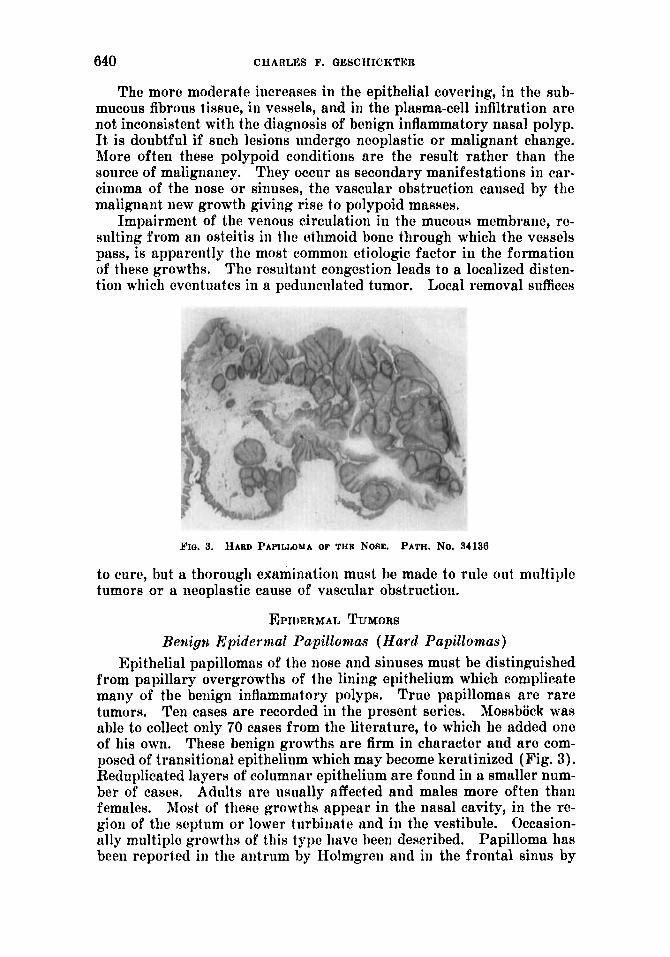
$'Iff. 3. HAED PAPILLOMA OF THE NOSE. PATH. NO. 34136
to cure, but a thorough examination must be made to rule out multiple tumors or a neoplastic cause of vascular obstruction.
EPIDERMAL TUMORS Bewigpi Epidermal Papillomas (Hard Papillomas)
Epithelial papillomas of the nose and sinuses must be distinguished from papillary overgrowths of the lining epithelium which complicate many of the benign iiiflammutory polyps. True papillomas are rare tumors. Mossbock was able to collect only 70 cases from the literature, to which he added one of his own. These benign growths are firm in character and are com- posed of transitional epithelium which may become keratinixed (Fig. 3). Reduplicated layers of columnar epithelium are found in a smaller num- ber of cases. Adults are usually affected and males more often than females. Most of these growths appear in the nasal cavity, in the re- gion of the septum or lower turbiiiate and in the vestibule. Occasion- ally multiple growths of this type have been described. Papilloma has been reported in the antrum by Holmgren and in the frontal s inw by
Ten cases are recorded in the present series.
TUMORS OF THE NASAL AND PARANASAL CAVITIES 641
Herxheimer. The tumors grow slowly and the outstanding symptom is nasal obstruction. Excision, preferably with a cautery, should suffice for a cure, but the specimen should be clieckcd microscopically to rule out malignant changc. Recurrent growths have bccn reported. These usually retain a benign character, showing the epithelium clearly demarcated from the underlying fibrous tissue.
The dividing line between papillomas and iiifltimmatory nasal polyps on the one hand and between papillomis and papillary carcinoma on the other hand is not distinct. Ewing believes that the inflammatory over- growths may pass imperceptibly into papillomas and thence into carci- noma. Recently Reuys, in a study of 12 cases, has tried to show a re- lation between nasal polyp, papilloma, and papillary carcinoma of the nose. In the present series the patients with benign papilloma who
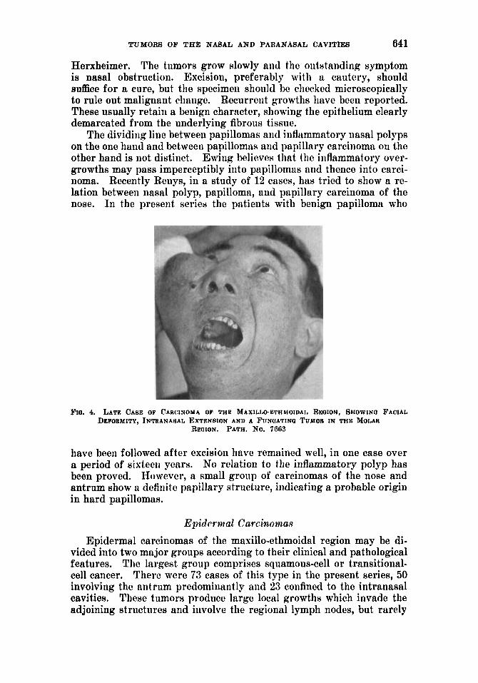
3.10. 4. LATE CA8E OF CAWINOMA OP THE MAXILLO-ETHMOIDAL RICOION, SHOWIN0 FACIAL DEFOUMITY, INTKANASAL EXTEN6ION AND A FUNOATINQ TUMOR IN THE MOLAR
REOION. PATH. No. 7063
have been followed after excision have remained well, in one case over a period of sixtecii yearn. No relation to the inflammatory polyp has been proved. However, a small group of carcinomas of the nose and antrum show a definite papillary structure, indicating a probable origin in hard papillomax.
Epidermal Carcinomas Epidermal carcinomas of the masillo-cthmoidal region may be di-
vided into two major groups according to their clinical and pathological features. The largest group comprises squamous-cell or transitional- cell cancer. There were 73 cases of this type in the present series, 50 involving the antrum predominantly and 23 confined to the intranasal cavities. These tumors produce large local growths which invade the adjoining structures and iiivolve the regional lymph nodes, but rarely
642 CHARLES F. GESCHICKTER
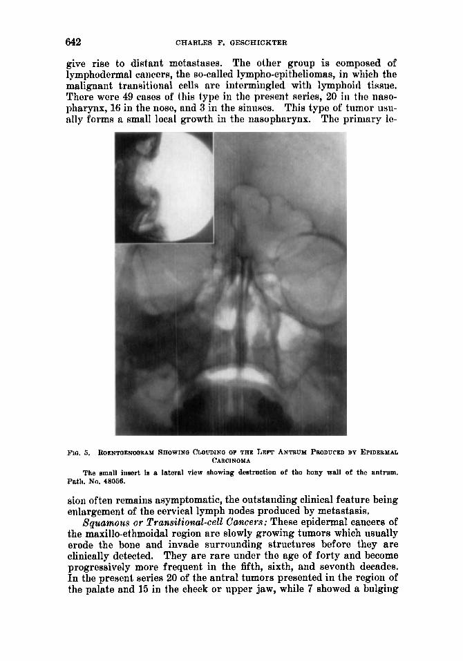
give rise to distant metastases. The other group is composed of lymphodermal cancers, the so-called lympho-epitheliomas, in which the malignant transitional cells are intermingled with lymphoid tissue. There were 49 cases of this type in the present series, 20 in the naso- pharynx, 16 in the nose, and 3 in the sinuses. This type of tumor uxu- ally forms a small local growth in the nasopharynx. The primary le-
F I O . 5. R.QENTOENOORAM 8AOWINO cLOL!DIIO OF THE LEFT ANTBUM PRODUCED B Y EPIDERMAL CARCINOMA
The small insert is a lateral view showing destruction of the bony wall of tho antrum. Path. No, 48056.
sion often remains asymptomatic, the outstailding clinical feature being enlargement of the cervical lymph nodes produced by metastasis.
Squamous or Transitional-cell Ca9~ers: These epidermal cancers of the maxillo-ethmoidal region are slowly growing tumors which usually erode the bone and invade surrounding structures before they are clinically detected. They are rare under the age of forty and become progressively more frequent in the fifth, sixth, and seventh decades. In the present series 20 of the antral tumors presented in the region of the palate and 15 in the cheek o r upper jaw, while 7 showed a bulging
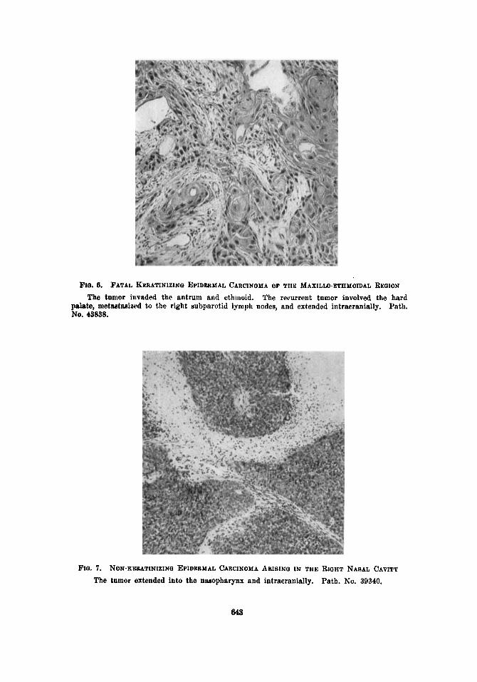
Fro. 6. FATAL KP.RATINIZINO EPIDERMAL CARCINOMA OF THE MAXILLO-ETHMOIDAL REGION The tumor invaded the antrum and ethiiioid. The reeurrent tumor involved the hard
palate, metsetseized to the right subparutid lyiiipli nodes, and extended intracranially. Path. NO. 43838.
FIG. 7. NON-KEBATINIZING EPIDERMAL CARCINOMA ARISING IN THE RIOHT NASAL CAVITY The tumor extended into the nasopharynx and intracranially. Path. No. 39340.
644 CHARLES F. QESCIIICKTER
in the orbit, I n the majority the outstanding symptoms were related to an iiitraiiasal tumor. The tumors growing in the nasal passages produce obstruction, interference with the voice and seme of smell, and epistaxis. Blocking of the naso-lacrimal duct may cause lacrimation. Ant ral tumors came swelling of the jaw, trigeminal neuralgia, niid fungution or bleeding in tlie regioii of the hard palate, nose, or orbit. Loosening of the upper molar teeth may be a late manifestation, arid exoplithalmos may occur. Nnsal polyps of the benign iiiflummatory type produced by vascular obstruction may be tlie first maiiifcstatioii of a more deep-seated maligiiniit growth. In the roentgenogram clouding of the antrum or of the iiasal cavities, with deflection of the seplum or bulging of the bony walls of the antrum, may be seen. Osscous destruc- tioii and decalcification are late signs (Figs. 4 and 5).
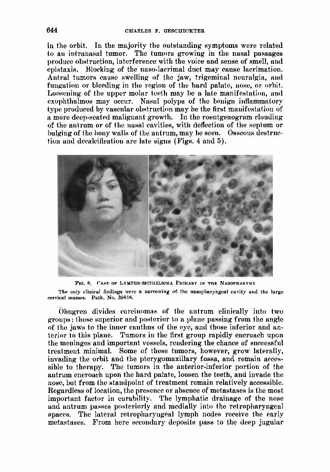
F I O . 8. ('ALIE OF IAYMPHO-EPITHCLIOJA PHIAIARY IN THE NABOPHAHY N S
The only c*linictrl findings were a narrowing of. the nasopharyngeal cavity arid tlic large cervical inaa~c's. Path. No. 39818.
ijliiigrcii divides carcinomas of the antrum clinically into two groups: those superior aiid posterior to a plane passing from the angle of the jaws to the iiirier caiithus of the eye, and those inferior and an- terior to this plane. Tumors in the first group rapidly encroach upon the meninges and important vessels, rendering the chance of successful treatment minimal. Some of these tumors, however, grow laterally, invading the orbit and the ptcrygomaxillary fossa, and remain acces- sible to therapy. The tumors iii the anterior-inferior portion of the antrum encroach upon the hard palate, loosen the teeth, and invade the nose, but from the standpoint of treatment remaiii relatively accessible. Regardless of location, the presence or absence of metastases is the most important factor in curability. The lymphatic drainage of the nose aiid antrum passes posteriorly and medially into the retropharyngeal spaces. The lateral retropliaryngeal lymph nodes receive the early metastases. From here sccoiidtiry deposits pass to the deep jugular
TUMORS OF THE NASAL A N D PARANASAL CAVITIES 645
chain of nodes at the bifurcation of the carotid. Palpation of the retro- pharyngeal nodes shows that the majority of cancers in this region metastasize relatively early. If palpation is restricted to the nodes at the bifurcation of the carotid, the false impression is obtained that metastasis occurs late. In a large number of the cases in the present series death resulted from intracraniul extension.
The commonest histologic variety is the so-called transitional-cell cancer arranged in islands or folds. Papillary epidermal carcinoma showing an origin in benign papilloma may occur (Figs. 6 and 7). Squnmous-cell carcinoma with epithelial pearls accounts for about one- sixth of the cases.
Cures of carcinoma of the nose and sinuses by means of surgery with or without thermal cauterization are exceedingly rare. Among
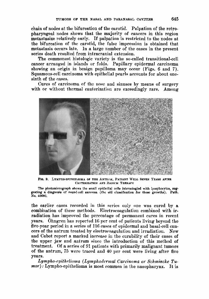
FIQ. 9. LYMPHO-EPITHELIOMA OF THE ANTRUM, PATIENT WELL SEVEN YEARS AFTEB
The photomicrograph shows the smnll cpithrlinl cells intermingled with lymphocytes, sup Path.
CAUTERIZATION AND HADIUbI THERAPY
gesting a diagnosis of round-cell sarcoma (the old clnssiflaation for these growths). No. 40680.
the earlier cases recorded in this series only one was cured by a combination of these methods. Electrocoagulation combined with ir- radiation has improved the percentage of permanent cures in recent years. Ohngren has reported 16 per cent of patients living beyond the five-year period in a series of 116 cases of epidermal and basal-cell can- cers of the antrum treated by electrocoagulation and irradiation. New and Cabot report a marked increase in the curability of their cases of the upper jaw and antrum since the introduction of this method of treatment. Of a series of 91 patients with primarily malignant tumors of the antrum, 75 were traced and 40 per cent were living after five years.
Lympho-cpithdioma (Lymphodcrwaal Carcinoma or Schmincke Tu- mor) : Lympho-epithelioma is most common in the nasopharynx. It is
646 CHARLES F. OESCHICKTER
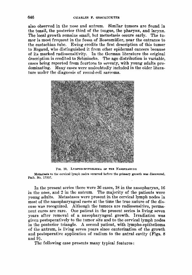
also observed in the nose and antrum. Similar tumors are found in the tonsil, the posterior third of the tongue, the pharynx, and larynx. The local growth remains small, but metastasis occurs early. The tu- mor is most frequent in the fossa of Rosenmiiller, near the entrance to the eustachian tube. Ewiiig credits the first description of this tumor to Regaud, who distinguished it from other epidermal cancers because of its marked radiosensitivity. In the German literature the original description is credited to Sehmiiicke. The age distribution is variable, cases being reported from fourteen to seventy, with young adults pre- dominating. Many ctises were undoubtedly included in the older litera- ture under the diagnosis of roimd-cell sarcoma.
5'10. 10. LYMPAO-EPITFIELIOMA OF THE NASOPHABYNX Metastaais to the cervical lympli nodcs occurred before the primary growth was discovered.
Path. No. 17557.
In the present series there were 36 cases, 18 in the nasopharynx, 16 in the nose, aiid 2 in the antrum. The majority of the patients were young adults. Metastases were present in the cervical lymph nodes in most of the nasopharyngeal cases a t the time the true nature of the dis- caw was recognized. Although the tumors are radiosensitive, perma- iieiit cures arc rare. One patieiit in the present series is living seven years after removal of a nasopharyngcal growth. Irradiation was given postoperatively to the tumor site and to the cervical lymph nodes in the posterior triangle. A second patient, with lympho-epithelioma of the antrum, is living seven years since cauterization of the growth aiid postoperative application of radium to the antral cavity (Figs, 8 and 9).
The following case presents many typical features :
TUMOR8 OF THE NASAL A N D PARANASAL OAVITIES 647
A colored girl, aged nineteen, was flrst seen hecausc of headache, pain in the right side of the face, and a sore throat. The tonsils were removed, but the sore throat con- tinued and was associated with a slight cough. Two months later the patient noticed a lump in the right jaw, which was painful and tender. A second lump appeared on the left side. Difficulty of breathing thi*ough the right nostril then developed, followed by a mucous discharge. A second examination showed tender and sensitive lymph nodes on both sides of the neck, and a polxpoid mass in the right nasal cavity apparently spring- ing from the ethmoidal and sphenoidal regions. The polyp was removed and the cervical lymph nodes were resected under the diagnosis of Hodgkin’s disense. Pathological ex- amination showed the polypoid mass to be inflnmmatory. The character of the lymph nodes was uncertain. The microscopic differentiation between inflammatory endothelial hyperplasia and possible metastatic carcinoma could not be made. Following the opera- tion radium was administered to the cervical region. The patient next complaiued of
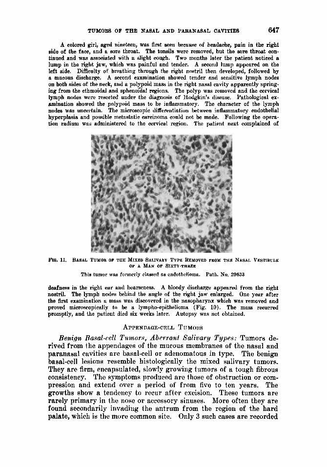
ma. 11. BASAL TUMOK OF THE MIXED SALIVARY TYPE REMOVED FBOM TBE NASAL VESTlBULX OF A M A N OF SIXTY-THREE
This tumor was forinerly classed as cndothclioma. Path. No. 29633
deafness in the right ear and hoarseness. A bloody discharge appeared from the right nostril. The lymph nodes behind the angle of the right jaw enlarged. One year after the flrst examination u mass was discovered in the nasopharynx which was removed and proved microsropically to be a lympho-epithelioma (Fig. 10). The muss recurred promptly, and the patient died six weeks later. Autopsy was not obtained.
APPENDAOE-CELL TUMORS Benign Rasal-cell Tumors, Aberrad Salivarp S’ypes: Tumors de-
rived from the appendages of the mucous membranes of the nasal and paranasal ctivities are basal-cell or adeiiomatous in type. The benign basal-cell lesions resemble histologically the mixed salivary tumors. They are firm, encapsulated, slowly growing tumors of a tough fibrous consistency. The symptoms produced are those of obstruction or com- pression and extend over a period of from five to ten years. The growths show a teiidency to recur after excision. These tumors are rarely primary in the nose or accessory sinuses. More often they a re found secondarily invading the antrum from the region of the hard palate, which is the more common site. Only 3 such cases a re recorded
648 CHARLES F. QESCHICKTER
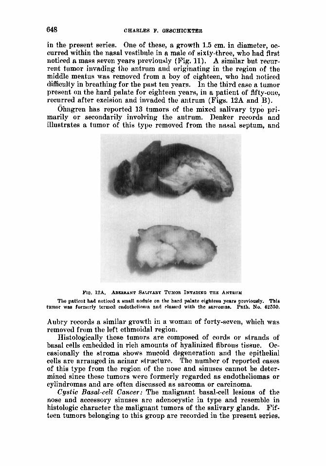
in the present series. One of these, a growth 1.5 cm. in diameter, oc- curred within the nasal vestibule in a male of sixty-three, who had first noticed a mass seven years previously (Fig. 11). A similar but recur- rent tumor invading the antrum and originating in the region of the middle meatus was removed from a boy of eighteen, who had noticed difficulty in breathing for the past ten years. In the third case a tumor present on the hard palate for eighteen years, in a patient of fifty-one, recurred after excision and invaded the antrum (Figs. 12A and B).
Ohngren has reported 13 tumore of the mixed salivary type pri- marily o r secondarily involving the antrum. Denker records and illustrates a tumor of this type removed from the nasal septum, and
PIG. 12A. ABJULMNT SALIVARY TUXOR INVADIN~ THE ANTRUM Tlie patient had noticed a small nodule on the hard palate eighteeu years previously. This
Path. No. 42550. tumor was formerly ternied cndothelionia trnd classed with the sarcomas.
Aubry records a similar growth in u womaii of forty-seven, which was removed from the left ethmoidal region.
Histologically these tumors are composed of cords or strands of basal cells embedded in rich amounts of hyaliiiized fibrous tissue. Oc- casionally the stroma shows mucoid degeneration and the epithelial cells are arranged in acinar structure. The number of reported cases of this type from the region of the nose and sinuses cannot be deter- mined since these tumors were formerly regarded as endotheliomas or cylindromas and are often discussed as sarcoma or carcinoma.
Cystic Basal-cell Cancer: The malignant basal-cell leeions of the nose and accessory sinuses are adenocystic in type and resemble in histologic character the malignant tumors of the salivary glands. Fif- teen tumors belonging to this group are recorded in the present series.
TUMORS OF THE NASAL AND PARANASAL CAVITIES 649
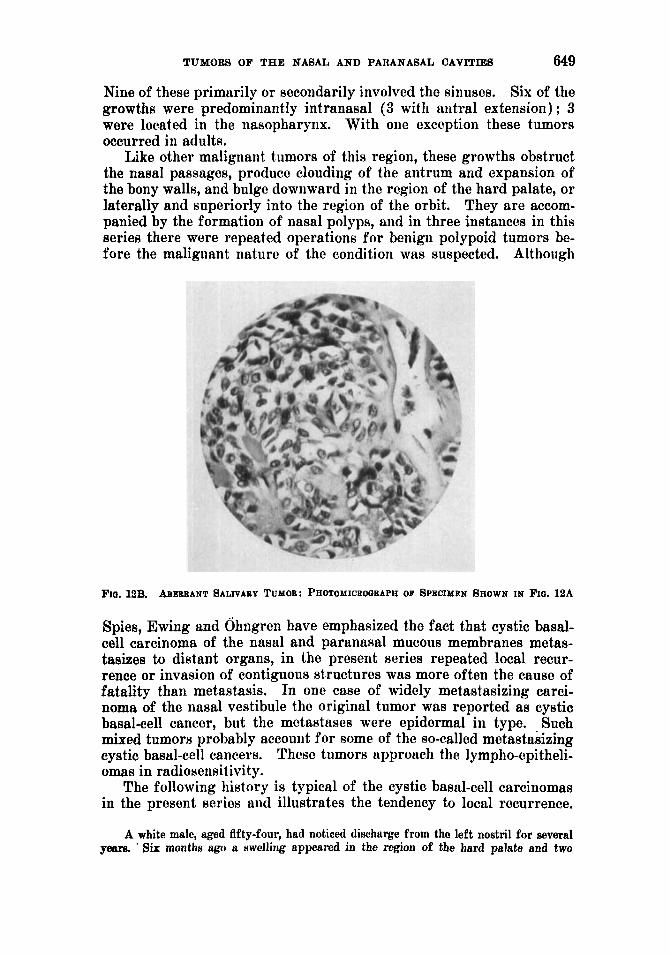
Nine of these primarily or secondarily involved the sinuses. Six of the growths were predominantly intranasal (3 with aiitral extension) ; 3 were located in the nasopharynx. With oiie exception these tumors occurred in adults.
Like other malignant tumors of this region, these growths obstruct the nasal passages, produce clouding of the antrum and expansion of the bony walls, and bulge downward in the region of the hard palate, or laterally and superiorly into the region of the orbit. They are accom- panied by the formation of nasal polyps, and in three instances in this series there were repeated operations for benign polypoid tumors be- fore the maligiiant nature of the condition was suspected. Although
Fro. 13B. ABERRANT SALIVARY TUYOF~: PHOTOMICROCIBAPH OF SPECIMEN SHOWN IN Fro. 12A
Spies, Ewing and Ohugren have emphasized the fact that cystic basal- cell carcinoma of the nasal and paranasal mucous membranes metas- tasizes to distant organs, in the present series repeated local recur- rence or invasion of contiguous structures was more often the cause of fatality than metastasis. In one case of widely metastasizing carci- noma of the nasal vestibule the original tumor was reported as cystic basal-cell cancer, but the metastases were epidermal in type. Such mixed tumors probably account for some of the so-called metastasizing cystic basal-cell cancers. These tumorB approach the lympho-epitheli- omas in radiosensitivity.
The following history is typical of the cystio basal-cell carcinomas in the present series and illustrates the tendency to local recurrence.
A white male, aged fifty-four, had noticed discharge from the left nostril for several years. ' Six months ago a swelling appeared in the region of the hard palate and two
650 CHARLES F. QESCHICKTER
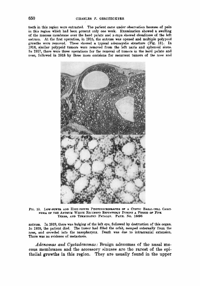
teeth in this region were extracted. The patient came under observation because of pain in this region which had been present only one week. Examination showed a swelling of the mucous inembrane over the hard palute and x-rays showed cloudiness of the left antrum. At the first operation, in 1915, the antrum was opened and multiple polypoid growths were removed. These showed a typical adenocystic structure (Fig. 13). In 1816, similar polypoid tumors werc removed from the left naris and sphenoid sinus. In 1917, there were three operations for the removal of tumors in the hard palate and nose, followed in 1918 by three more excisions for recurrent tumors of the nose and
FIG. 13. Low-POWEU AND IrrGII-PowEn PHOTOUICRWMPAB OF A CYSTIC BASAL-CELL CAncI- NOMA OF TXE ANTRIJM WHICH RECUBRED REPEATEDLY DURINO A PERIOD OF FIVE
YEAM, AND TERMINATED *'ATALLY. PATH. NO. 18090
antrum. I n 1919, there was bulging of the left eye, followed by destruction of this organ. 111 1920, the patient died. The tumor had fllled the orbit, escaped externally from the nose, and crowded into the nasopharynx. Death was due to intracranial extension. There was no evidence of metastasis.
Adenomas awd CystadePtomas : Benign aderiomas of the nasal mu- cous membranes and the accessory sinuses are the rarest of the epi- thelial growths in this region. They are usually found in the upper
TUMORS OF THE NASAL A N D PARANASAL UAVITIES 65 1
and posterior portion of the nasal fossa and occasionally the larger growths extend into the accessory sinuses. The region of the tu- berculum septi is cited by Eckert-Miibius as a favorite location. The present series includes 3 iiitranasal tumors of this type, 2 involving the antrum, and one in the frontal sinus. With one exception these 6 cases were in young adults, who complained of headache, neuralgia, nasal ob- struction or nasal discharge. Eckert-Mobius described 3 cases and
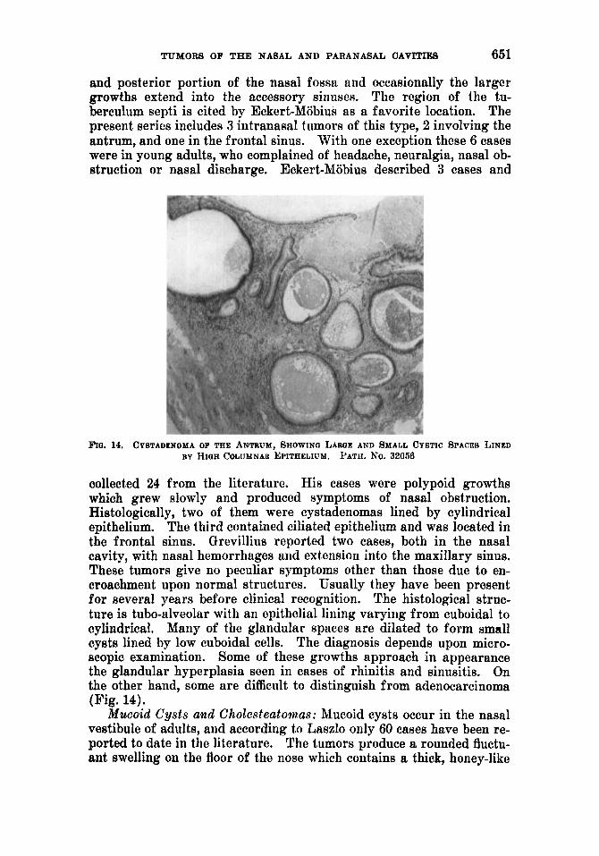
FlO. 14. CPSTADENOMA OP THE ANTRUM, SHOWINa LAROE AND SMALL CYSTIC SPACER LINED BY HIQH COLUMNAR EPITHELIUM. PATH. No. 32056
collected 24 from the literature. His cases were polypoid growths which grew slowly and produced symptoms of nasal obstruction. Histologically, two of them were cystadenomas lined by cylindrical epithelium. The third contained ciliated epithelium and was located in the frontal sinus, Grevillius reported two cases, both in the nasal cavity, with nasal hemorrhages and extension into the maxillary sinus. These tumors give no peculiar symptoms other than those due to en- croachment upon normal structures. Usually they have been present for several years before clinical recognition. The histological struc- ture is tubo-alveolar with an epithelial lining varying from cuboidal to cylindrical. Many of the glandular spaces are dilated to form small cysts lined by low cuboidal cells. The diagnosis depends upon micro- scopic examination. Some of these growths approach in appearance the glandular hyperplasia seen in cases of rhinitis and sinusitis. On the other hand, some are difficult to distinguish from adenocarcinoma (Fig. 14).
Mucoid Cysts and Cholesteatomms: Mucoid cysts occur in the nasal vestibule of adults, and according to Laszlo only 60 cases have been re- ported to date in the literature. The tumors produce a rounded fluctu- ant swelling on the floor of the nose which contains a thick, honey-like
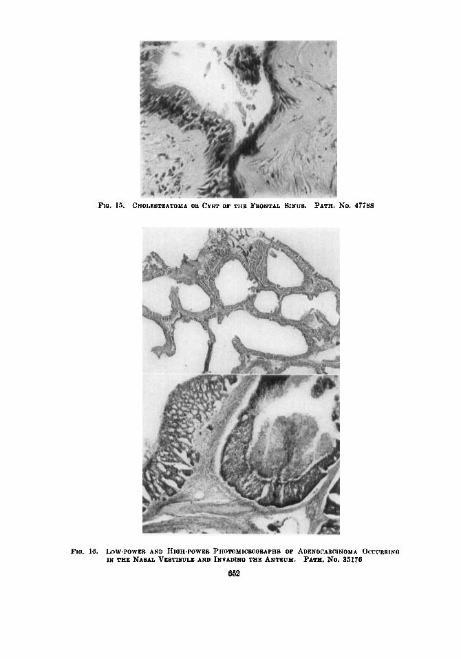
FIO. 15. CIIOLEBTEATOYA OB CYBT OF THE FRONTAL SINUS. PATH. KO. 47588
F I G . IG. IiOW-POWER AND HIOH-POWEB PHOMMICiKlQUAPHS OF ADENOCARCINOMA OCCIIBRINQ IN THE NASAL VESTIBULE AND INVADINO THE ANTUUM. PATH. NO. 35176
862
TUMORS OF THE NASAL A N D PARANASAL CAVITIES 663
fluid within an epithelial-lined sac. Histologically the lining membrane consists of stratified columnar epithelium. The majority of growths have been reported in women. Laszlo gives the various theories of their origin. One of these, proposed by Schaeffer, relates the growths to remnants of the nasopalatine canal which connects the nasal and oral cavities in fetal life. Romer relates the cysts to the epithelial dhbris of Malassez, and groups them with the dentigerous cysts of the jaws.
Cholesteatomas a re cystic tumors lined by stratified epithelium re- sembling epidermoid cysts of the skin. They are more often intra- cranial in origin. They have not been described in the nose, but Eckert-
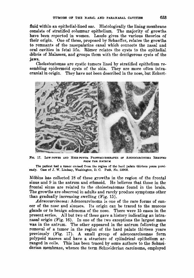
FIO. 17. LOW-POWER AND HIOH-POWER PHOTOMICROGRAPHS OF ADENOCABCINOYA REMOVEU
The patient had a tumor excised from the region of the hard palate thirteen yeare previ- FROM THE ANTRUM
ously. Case of J. W. Lindsay, Waeliington, D. C. Path. No. 53900.
Mobius has collected 18 of these growths in the region of the frontal sinus and 9 in the antrum and ethmoid. He believes that those in the frontal sinus are related to the cholesteatomas found in the brain. The growths are observed in adults and rarely produce symptoms other than gradually increasing swelling (Fig. 15).
Adenocarcinnoma: Adeiiocarcinomn is one of the rare forms of can- cer of the nose and sinuses. Its origin can be traced to the mucous glands or to benign adenoma of the nose. There were 15 cases in the present series. All but two of these gave a history indicating an intra- nasal origin (Fig. 16). In one of the two exceptions the largest mass was in the antrum. The other appeared in the antrum following the removal of a tumor in the region of the hard palate thirteen years previously (Fig. 17). A small group of adenocarcinomas form polypoid masses and have a structure of cylindrical epithelium ar- ranged in coils. This has been traced by some authors to the Sohnei- derian membrane, whence the term Schneiderian carcinoma, employed
654 CHARLES F. QESCHICKTER
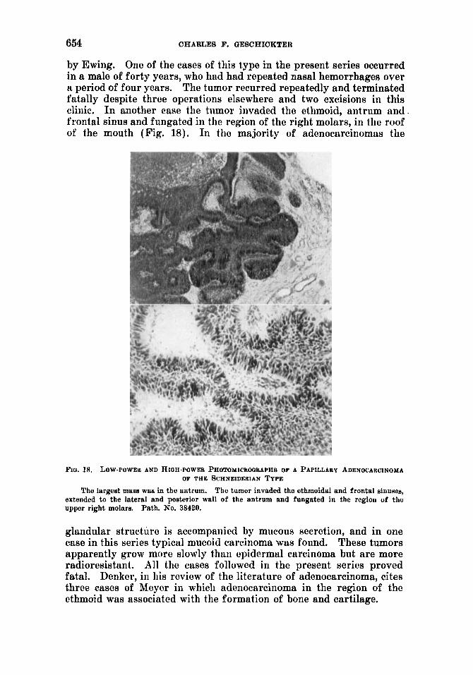
by Ewing. One of the cases of this type in the present series occurred in a male of forty years, who had had repeated nasal hemorrhages over a period of four years. The tumor recurred repeatedly and terminated fatally despite three operations elsewhere and two excisions in this clinic. I n another ca8e the tumor invaded the ethmoid, antrum and frontal sinus and fungatcd in the region of the right molars, in tlic roof of the mouth (Fig. 18). In the majority of adenocarcinomuv the
PIQ. 18. LOW-POWER AND HIOH-POWER PHOTQMICHOORAPHB OF A PAPILLARY ADENOCAHCINOMA OF T H E SCHNEIDERIAN TYPE
The largest mass waa in the antrum. The tumor invaded tho ethmoidal and frontal sinusee, extended to the lateral and posterior wall of the antrum and fungated in tho region of tho upper right molars. Path. No. 38420.
glandular structure is accompanied by mucous secretion, and in one case in this series typical mucoid carcinoma was found. These tumors apparently grow more slowly than epidermal carcinoma but are more radioresistant. All the cases followed in the present series proved fatal. Denker, in his review of the literature of adenocarcinoma, cites three cases of hleycr in which adenocarcinoma in the region of the ethmoid was associated with the formation of bone and cartilage.
TUMORS OF THE NASAL AND PARANASAL CAVITIES 655
BENIGN AND MALIGNANT CONNECTIVE-TISSUE TUMORS Benign connective-tissue tumors in the intranasal and accessory
cavities, including osteomas, angiomas, plasmocytomas and fibromas, are more common than benign epithelial tumors. Marschik and Send- ziak in their reviews of malignant tumors in this region find sarcoma more frequent than carcinoma. Sendziak records 450 sarcomas to 337
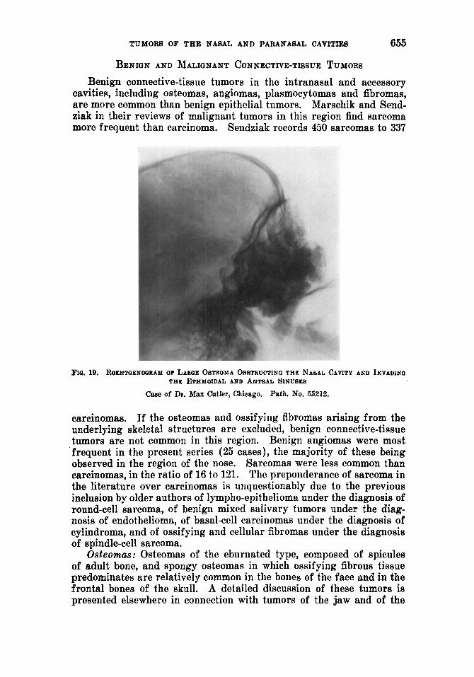
FIO. 19. ROENTGENOGRAM Or LAWE OSTEOMA ORATRUCTINO THE NANAL CAVITY AND INVADINO THE ETHMOIDAL AND ANTEAL SINUSES
Case of Dr. Max Cutler, Chicago. Path. No. 55212.
carcinomas. If the osteomas and ossifying fibromas arising from the underlying skeletal structures are excluded, benign connective-tissue tumors are riot common in this region. Benign angiomas were most frequent in the present series (25 cases), the majority of these being observed in the region of the nose. Sarcomas were less common than carcinomas, in the ratio of 16 to 121. The preponderance of sarcoma in the literature over carcinomas is unquestionably due to the previous inclusion by older authors of lympho-epi thelioma under the diagnosis of round-cell sarcoma, of benign mixed saIivary tumors under the diag- nosis of endothelioma, of basal-cell carcinomas under the diagnosis of cylindroma, and of ossifying and cellular fibromas under the diagnosis of spindle-cell sarcoma.
Osteomas .- Osteomas of the eburnated type, composed of spicules of adult bone, and spongy osteomas in which ossifying fibrous tissue predominates are relatively common in the bones of the face and in the frontal bones of the skull. A detailed discussion of these tumors is presented elsewhere in connection with tumors of the jaw and of the
656 CHARLES F. OEBCHICKTER
skull. They occur in young individuals and present most commonly in the frontal sinus. Eckert-Mobius reports 113 out of 276 osteomas in t.he frontal sinus, 53 in the ethmoid, 13 in the antrum, and 7 in the sphenoidal sinus. Of 70 osteomas in the region of the jaws recorded in this laboratory, 50 cases were in thc region of the upper jaws and, of these, 20 were primarily antral or intranasal (Fig. 19). Osteomas of
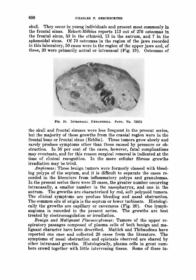
F I ~ . 20. INTEANASAL HEMANGIOMA. PATH. No. 52852
the skull and frontal sinuses were less frequent in the prcseiit series, but the majority of these growths from the cranial region were in the frontal bone or frontal sinus (Echlin). These tumors grow slowly arid rarely produce symptoms other than those caused by pressure or ob- struction. In 50 per cent of the cases! however, fatal complications may eventuate, and for this reason surgical removal is indicated at the time of clinical recognition. I n the more cellular fibrous growths irradiation may be tried.
Angiovnas: Thcsc benign tumors were formerly classed with bleed- ing polyps of the septum, and it is difficult to separate the cases re- corded in the literature from inflammatory polyps and granulomas. In the present series there were 25 cases, the greater number occurring intranasally, a smaller number in the nasopharynx, and one in the antrum. The growths are characterized by red, soft polypoid tumors. The clinical symptoms are profuse bleeding and nasal obstruct ion. The common site of origin is the septum or lower turbinate. Histologi- cally the growths arc capillary or cavernous (Fig. 20). One lymph- angioma is recorded in the present series. The growths are best treated by electrocoagulation or irradiation.
Benign and Malignant Plasnaocytomas: Tumors of the upper re- spiratory passages composed of plasma cells of both benign and ma- lignant character have been described. Mattick and Thibaudeau have reported one case and collected 20 cases from the literature. The symptoms of nasal obstruction and epistaxis observed are shared by other intranasal growths. Histologically, plasma cells in great num- bers crowd together with little intervening tissue. Some of these tu-
TUMORS OF THE NASAL AND PARANASAL CAVITIES 657
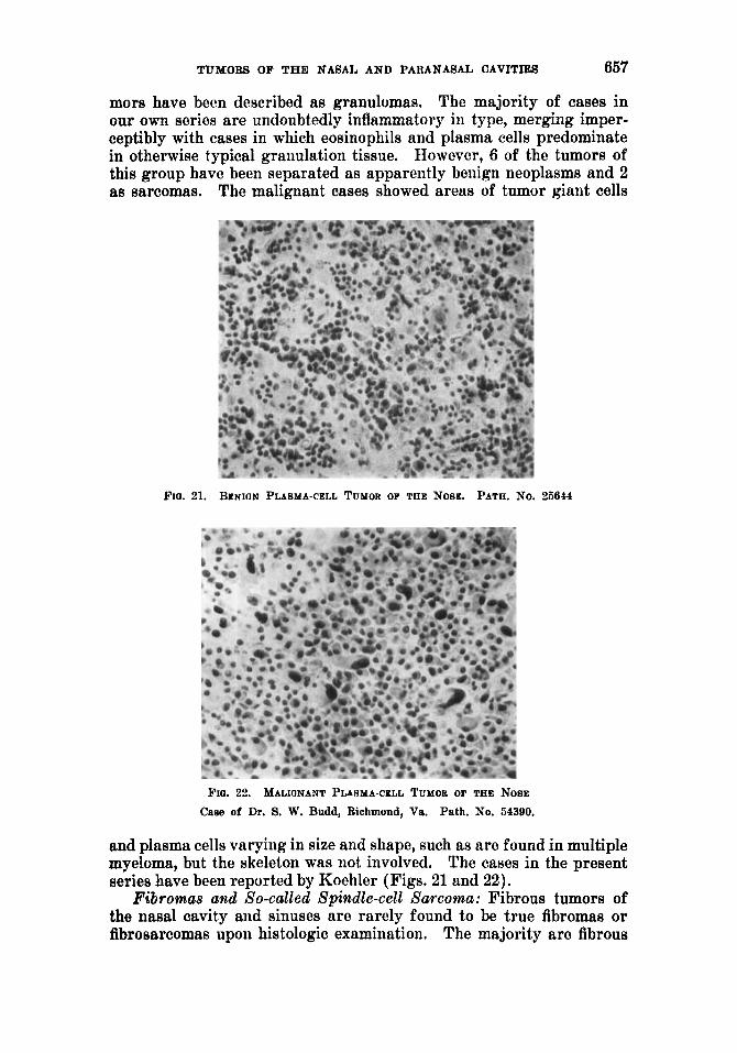
mors have been described as granulomas. The majority of cases in our own series are undoubtedly inflammatory in type, merging imper- ceptibly with cases in which eosinophils and plasma cells predominate in otherwise typical granulation tissue. However, 6 of the tumors of this group have been separated as apparently benign neoplasms and 2 as sarcomas. The malignant cases showed areas of tumor giant cells
Fro. 21. BENIGN PLASMA-CELL TUMOR OF THE NOSE. PATH. No. 35844
FIO. 22. MALIONANT PLASMA-CELL TUMOR of THE NOSE Case of Dr. 8. W. Budd, Richmond, Va. Path. No. 54390.
and plasma cells varying in size and shape, such as are found in multiple myeloma, but the skeleton was not involved. The cases in the present series have been reported by Koehler (Figs. 21 and 22).
Fibromas and So-called Spindle-cell Sarcoma: Fibrous tumors of the nasal cavity and sinuses are rarely found to be true fibromas or fibrosarcomas upoii histologic examination. The majority are fibrous
668 CHARLES F. QESCAICKTER
inflammatory polyps, fibrosed angiomas, or ossifying fibromas. The fibromas recorded in the literature (Sargnon) often respond to irradia- tion, which is not typical of truc fibromas or fibrosarcoma. Six fibro- mas were recorded in the present series and 3 fibrosarcomas. The growths occurred in young individuals and were found in the nose and nasopharynx. It is impossible to rule out the possibility of nerve sheath tumor in two of these cases.
The nasopharyngeal fibroma or basal fibroid presents many charac- teristic features. The tumor originates at the base of the skull or from
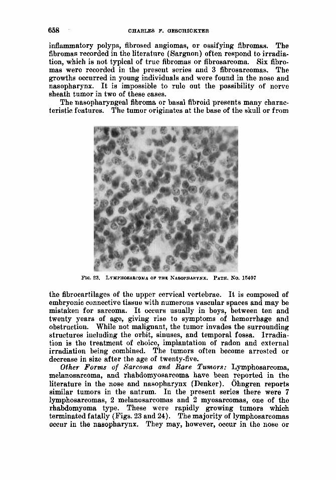
FIG. 23. LYMPHOSABCOMA OF THE ~ASOPHAIWNX. PATH. NO. 15407
the fibrocartilagcs of the uppcr cervical vertebrae. It is composed of embryonic connective tissue with numerous vascular spaces and may be mistaken for sarcoma. It occurs usually in boys, between ten and twenty years of age, giving rise to symptoms of hemorrhage and obstruction. While not malignant, the tumor invades the surrounding structures including the orbit, sinuses, and temporal fossa. Irradia- tion is the treatment of choicc, implantation of radon and external irradiation being combined. The tumors often become arrested or decrease in size after the age of twenty-five.
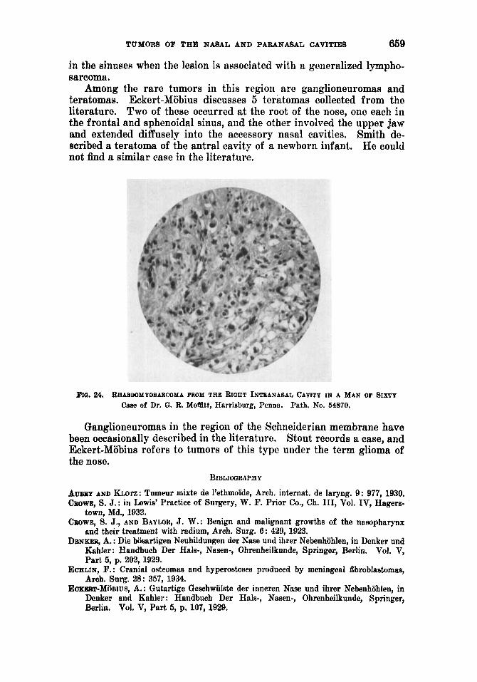
Other Forms of Sarcoma and Rare Tumors: Lymphosarcoma, melanosarcoma, and rhabdomyosarcoma have been reported in the literature in the nose and nasopharynx (Denker). Ohngren reports similar tumors in the antrum. In the present series there were 7 lymphosarcomas, 2 melanosarcomas and 2 myosarcomas, one of the rhabdomyoma type. These were rapidly growing tumors which terminated fatally (Figs. 23 and 24). The majority of lymphosarcomas occur in the nasopharynx. They may, however, occur in the nose or
TUMORS OF THE NASAL AND PARANASAL CAVITIES 659
in the sinusefi when the lesion is associated with a generalized lympho- sarcoma.
Among the rare tumors in this region are ganglioneuromas and teratomas. Eckert-Mobius discusses 5 teratomas collected from the literature. Two of these occurred at the root of the nose, one each in the frontal and sphenoidal sinus, and the other involved the upper jaw and extended diffusely into the accessory nasal cavities. Smith de- scribed a teratoma of the antral cavity of a newborn infant. He could not find a similar case in the literature.
FIG. 84. RHABDOMYOSABCOMA FROM THE RIOHT INTsANASAL CAVITY IN A MAN OF SIXTY Case of Dr. Gc. R. Moflltt, Harrisburg, Penna. Path. No. 54870.
Ganglioneuromas in the region of the Schneiderian membrane have been occasionally described in the literature. Stout records a case, and Eckert-Mobius refers to tumors of this type under the term glioma of the nose.
BIBLIWRAPHY
A U ~ Y AND K m : Tumeur mixte de l’ethmoi’de, Arch. internnt. de laryng. 9: 977, 1930. CBOWE, S. J.: in Lewis’ Practice of Surgery, W. F. Prior Co., Ch. 111, Vol. IV, Hagem-
town, Md., 1932. CBOWE, S. J., AND BAYMR, J. W.: Benign and malignant growths of the nasopharynx
and their treatment with radium, Arch. Surg. 6: 429, 1923. DENKER, A. : Die biisartigen Neubildungen der Nase und ihrer Nebenhohlen, in Denker und
Kahler: Handbuch Der Hals-, Nasen-, Ohrenheilkunde, Springer, Berlin. Vol. V, Part 5, p. 202,1929.
ECHLIN, F. : Cranial osteomas and hyperostoses produced by meningeal fibroblastomas, Arch. Surg. 28: 357, 1934.
EOKIOBT-M~BIUS, A.: Gutartige Qeschwiilste dcr inneren Nase und ihrer Nebenhohlen, in Dcnker and Kahler : Handbuch Der Hnls-, Nasen-, Ohrenheilkunde, Springer, Berlin. Vol. V, Part 5, p. 107, 1929.
660 CHARLES F. OESCHICKTER
EWINQ, J,: Lymphoepithelioma, Am. J. Path. 5: 99, 1929. EWINQ, J.: Neoplastic Diseases, W. B. Saunders Co., Philadelphia. Ed. 3, 1928, p. 763. EWINQ, J,: Lectures on Tumor Pathology, Cornell University Medical School, Class of
QREVILLIUS, AKE: De I’adbnonie dcs fosses nasales e t de leurs cavitbs accessories, Acta
HERXHEIYER, (3.: Uber das sog. harte Papillom der Nase, Ztschr. f. Laryng. 4: 249, 1912. HOLMQREN, Q.: Papillom im antrum Highmori, Hygiea (Stockholm) 87: 482, 1935. KOEHLER, H. P.: Benign and malignant plasmacytomas, to be published in Arch. Surg. LABZID, A. F. : So-called mucoid cyHts of the nose, Arch. Otolaryng. 21 : 41, 1935. MARSCHIK, H.: Die Pathologie u. Diagnostik d. malignen Geschwiilste der Nase und des
Nasenrachenraumcw mit Ausschluss cler Nasenrachenfihrome, Beitr. z. Anat., Physiol., Path. u. Therap. (1. Ohres 7 : 327, 1913-14.
MATTICK, W. L., A N D A. A. THIBAUDEAU: Extrameclullary plaama-cell tumors of the upper air passages, Am. J. Cancer 23: 513, 1935.
MOSSBOCK, F. : Beitrag zur pathologischen Anatomie und Klinik des harten Papilloms der Nase, Arch. f. Ohren-, Nasen-u. Kehlkopfh. 132: 254, 1932.
NEW, Q. B., AND CABOT, C. M.: Curability of malignant tumors of the upper jaw and antrum, Surg. Oynec. & Obst. 60: 971, 1935.
~ N ~ R E N , L. Ct . : Malignant tumonm of the maxillo-ethmoidal region, Acta Oto-Lnryngo- logica, Suppl. 19, 1933.
REUYS, H.: Uber Fibroepithelionie der Nase und ihre Beziehung zu den papillaren Car- cinomen, Ztschr. f. Hals-, Nasen- u. Ohrenh. 30: 421, 1932.
SARQNON, M.: Traitement des tumeurs du iiaso-pharynx B type fibromateux, Lyon m6d. 153 : 153,1934.
SENDZIAK, J. : Die malignen Tumoren der Ncbenhiihlen und des Rachens, Arch. internut. de laryng. 35: 371, 1913.
SMITH, R. T. : Teratoma of the antrum in the newborn, Ann. Otol. Rhin. I% Laryng. 41 : 886, 1932.
SPIEB, J. W. : Adenoid cystic carcinoma, Arch. Surg. 21 : 365, 1930. STOUT, A. P.: Human Cancer, Lea and Fehiger, Philadelphia, 1932, p. 71s.
1934, pp. 3 and 4.
Oto-Laryng. 18 : 177, 1932.