turning the pyramid of prenatal care - the fetal medicine ... · pdf fileturning the pyramid...
TRANSCRIPT

Fax +41 61 306 12 34E-Mail [email protected]
Original Paper
Fetal Diagn Ther 2011;29:183–196 DOI: 10.1159/000324320
Turning the Pyramid of Prenatal Care
Kypros H. Nicolaides a, b
a Harris Birthright Research Centre of Fetal Medicine, King’s College Hospital, and b Department of Fetal Medicine, University College Hospital, London , UK
provision of prenatal care [1, 2] . In 1929, the Ministry of Health in the UK issued a Memorandum on Antenatal Clinics recommending that women should first be seen at 16 weeks, then at 24 and 28 weeks, fortnightly thereaf-ter until 36 weeks and then weekly until delivery ( fig. 1 ) [3] . Although no explicit rationale was offered for either the timing or clinical content of the visits, these guide-lines established the pattern of antenatal care that is fol-lowed throughout the world even today. The high con-centration of visits in the third trimester implies that, firstly, most complications occur at this late stage of preg-nancy and, secondly, that most adverse outcomes are un-predictable during the first or even the second trimester.
In the last 20 years, it has become apparent that an in-tegrated first hospital visit at 11–13 weeks combining data from maternal characteristics and history with findings of biophysical and biochemical tests can define the pa-tient-specific risk for a wide spectrum of pregnancy com-plications, including fetal abnormalities, miscarriage and stillbirth, preeclampsia, preterm delivery, gestational di-abetes, fetal growth restriction and macrosomia. Early estimation of patient-specific risks for these pregnancy complications would improve pregnancy outcome by shifting prenatal care from a series of routine visits to a more individualized patient- and disease-specific ap-proach both in terms of the schedule and content of such visits. Each visit would have a predefined objective and the findings would generate likelihood ratios that can be used to modify the individual patient- and disease-spe-cific estimated risk from the initial assessment at 11–13 weeks.
Key Words First-trimester screening � Prenatal care � Preeclampsia � Preterm delivery � Gestational diabetes � Hypothyroidism � Small for gestational age � Macrosomia � Miscarriage � Stillbirth � Nuchal translucency
Abstract The current approach to prenatal care, which involves visits at 16, 24, 28, 30, 32, 34 and 36 weeks and then weekly until delivery, was established 80 years ago. The high concentra-tion of visits in the third trimester implies, firstly, that most complications occur at this late stage of pregnancy and, sec-ondly, that most adverse outcomes are unpredictable dur-ing the first or even second trimester. This review presents evidence that many pregnancy complications can now be predicted at an integrated first hospital visit at 11–13 weeks by combining data from maternal characteristics and history with findings of biophysical and biochemical tests. It is there-fore proposed that the traditional pyramid of care should be inverted with the main emphasis placed in the first rather than third trimester of pregnancy.
Copyright © 2011 S. Karger AG, Basel
Introduction
In the 19th century, pregnancy care was confined to the time of delivery and reserved for the wealthy. In the beginning of the 20th century, high maternal and infant mortality stimulated the establishment of institutions for
Received: December 14, 2010 Accepted after revision: January 11, 2011 Published online: March 8, 2011
Prof. K.H. Nicolaides Harris Birthright Research Centre for Fetal Medicine, King’s College Hospital Denmark Hill London SE5 9RS (UK) Tel. +44 203 299 8256, E-Mail kypros @ fetalmedicine.com
© 2011 S. Karger AG, Basel1015–3837/11/0293–0183$38.00/0
Accessible online at:www.karger.com/fdt

Nicolaides Fetal Diagn Ther 2011;29:183–196184
At 11–13 weeks, the great majority of women would be classified as being at low-risk for pregnancy complica-tions and a small proportion of women would be selected as being at high-risk ( fig. 2 ). In the low-risk group, the number of medical visits could be substantially reduced to perhaps three. One visit at 20–22 weeks would re-eval-uate fetal anatomy and growth, and reassess risk for such complications as preeclampsia and preterm delivery. An-other visit at 37–38 weeks would assess maternal and fetal well-being and determine the best time and method of delivery, which would be repeated at 41 weeks for the few that remain pregnant at this stage. The high-risk group could have close surveillance in specialist clinics both in terms of the investigations to be performed and the per-sonnel involved in the provision of care. In each of these visits, their risk would be reassessed and they would ei-ther remain high-risk or become low-risk, in which case the intensity of their care could be reduced.
This review summarizes the emerging evidence on the potential value of the 11 to 13 weeks’ assessment and sets the basis for a challenge to invert the 80-year-old pyramid of prenatal care.
Early Screening for Fetal Aneuploidies
Aneuploidies are major causes of perinatal death and childhood handicap. Consequently, the detection of chromosomal disorders constitutes the most frequent in-dication for invasive prenatal diagnosis. However, inva-
sive testing, by amniocentesis or chorionic villus sam-pling, is associated with a risk of miscarriage; therefore, these tests are carried out only in pregnancies considered to be at high risk for aneuploidies.
The Combined Test In the 1970s, the main method of screening for aneu-
ploidies was by maternal age. In the 1980s, it was done by maternal serum biochemistry and detailed ultrasono-graphic examination in the second trimester. In the 1990s, the emphasis shifted to the first trimester when it was realized that the great majority of fetuses with major aneuploidies can be identified by a combination of mater-nal age, fetal nuchal translucency (NT) thickness, mater-nal serum-free � -hCG and PAPP-A [4–14] . Screening by this combined test can identify about 90% of fetuses with trisomy 21 and other major aneuploidies for a false-posi-tive rate of 5%.
Studies in the last 10 years have shown that improve-ment in the performance of first-trimester screening can be achieved by (1) carrying out the biochemical test at 9–10 weeks and the ultrasound scan at 12 weeks and (2) inclusion in the ultrasound examination assessment of the nasal bone and flow in the ductus venosus, hepatic artery and across the tricuspid valve.
Timing of Ultrasound and Biochemistry One option in first-trimester combined screening for
trisomy 21 is to perform biochemical and ultrasono-graphic testing as well as counsel women in one-stop clin-
37 w 38 w 39 w 40 w 41 w
30 w 32 w 36 w 34 w
12 w
24 w 28 w 20 w
16 w
12 w
41 w
20 wSpecialist care 12–34 w
37 w
Fig. 1. Pyramid of traditional prenatal care established in 1929.w = Weeks.
Fig. 2. Proposed new pyramid of prenatal care. w = Weeks.

Turning the Pyramid of Prenatal Care Fetal Diagn Ther 2011;29:183–196 185
ics for assessment of risk (OSCAR) [9, 10, 15] . The ideal gestation for OSCAR is 12 weeks because the aim of the first-trimester scan is not just to screen for trisomy 21 but also to diagnose an increasing number of fetal malforma-tions. In this respect, the ability to visualize fetal anatomy is better at 12 weeks than at 10–11 or 13–14 weeks.
An alternative strategy for first-trimester combined screening is for biochemical testing and ultrasound scan-ning to be carried out on two separate visits, with the first done at 9–10 weeks and the second at 12 weeks [13, 14, 16] . It has been estimated that this approach would improve the detection rate from 90 to 93–94%. A third option would be to perform the scan at 12 weeks and optimize the performance of biochemical testing by measuring PAPP-A at 9 weeks and free � -hCG at the time of the scan at 12 weeks or even later, which would have an estimated detection rate of 95%. The cost and patient acceptability of the alternative policies of first-trimester testing will depend on the existing infrastructure of antenatal care. The potential advantage of two- or three-stage screening in terms of detection rate may be eroded by the likely in-creased noncompliance with the additional steps.
Additional Ultrasound Markers At 11–13 weeks, absence of the fetal nasal bone, re-
versed a-wave in the ductus venosus, tricuspid regurgita-tion and increased peak systolic velocity in the hepatic artery are observed in about 60, 66, 55 and 80% of fetuses with trisomy 21 and in 2.5, 3.0, 1.0 and 5% of euploid fe-tuses, respectively [17–26] .
In first-trimester combined screening, each of the ad-ditional ultrasound markers can be assessed in all pa-tients resulting in an increase in detection rate to 93–96% and a decrease in the false-positive rate to 2.5% [19, 21, 24, 27] . A similar performance of screening can be achieved by a contingent policy in which first-stage screening by maternal age, fetal NT and serum-free � -hCG and PAPP-A is offered to all cases ( fig. 3 ). Patients with a risk of 1 in 50 or more are considered to be screen-positive and those with a risk of less than 1 in 1,000 are screen-negative. Patients with the intermediate risk of 1 in 51 to 1 in 1,000, which constitutes 15–20% of the total population, have second-stage screening with nasal bone, ductus venosus or tricuspid blood flow, which modifies their first-stage risk. If the adjusted risk is 1 in 100 or more the patients are considered to be screen-pos-itive and those with a risk of less than 1 in 100 are screen-negative.
Early Diagnosis of Fetal Abnormalities
The 11 to 13 weeks’ scan evolved over the last 20 years from essentially a scan for measurement of fetal NT and crown-rump length to one which includes a basic check-list for examination of the fetal anatomy with the inten-tion of diagnosing major abnormalities which are either lethal or associated with severe handicap so that the par-ents can have the option of earlier and safer pregnancy termination.
Risk<1 in 100
Intermediate-risk (1/51–1,000)15% of the population
14% of trisomy 21
Chorionic villussampling
High-risk (≥1 in 50)1.5% of the population
85% of trisomy 21
Ultrasound scanat 22 weeks
Low-risk (<1 in 1,000)83.5% of the population
1% of trisomy 21
Risk≥1 in 100
Assessment of:nasal bone
ductus venosus flowtricuspid flow
hepatic artery flow
Maternal age, fetal NT and maternal serum-free �-hCG and PAPP-A
Fig. 3. Two-stage screening for fetal aneu-ploidies. In the first stage, all patients have screening using a combination of maternal age, fetal NT thickness and maternal se-rum-free � -hCG and PAPP-A; according to the results, they are classified into high-risk, intermediate-risk and low-risk groups. In the intermediate-risk group, second-stage screening is carried out by one or more sonographic markers, includ-ing nasal bone, blood flow in the ductus venosus, hepatic artery or across the tri-cuspid valve, and on the basis of these re-sults, they are then classified as high-risk or low-risk.

Nicolaides Fetal Diagn Ther 2011;29:183–196186
Major fetal abnormalities fall into essentially three groups in relation to whether they can be detected at the 11 to 13 weeks’ scan [28] . The first group consists of ab-normalities which are always detectable, and include body stalk anomaly, anencephaly, alobar holoprosen-cephaly, exomphalos, gastroschisis and megacystis. The second group consists of undetectable abnormalities be-cause they are manifested only during the second or third trimester of pregnancy, and include microcephaly, agen-esis of the corpus callosum, semilobar holoprosencepha-ly, hypoplasia of the cerebellum or vermis, cystic adeno-matoid malformation or pulmonary sequestration, and bowel obstruction. The third group includes abnormali-ties that are potentially detectable depending on, firstly, the objectives set for such a scan and, consequently, the time allocated for the fetal examination, the expertise of the sonographer and the quality of the equipment used. Additionally, the presence of an easily detectable marker for an underlying abnormality is also important for de-tection. A good example of such a marker in the first tri-mester is high NT, which is found in some fetuses with lethal skeletal dysplasias, diaphragmatic hernia and ma-jor cardiac defects.
Major Cardiac Defects Abnormalities of the heart and great arteries are the
most common congenital defects, accounting for about 20% of all stillbirths and 30% of neonatal deaths due to congenital defects [29] . Although most major cardiac de-fects are amenable to prenatal diagnosis by specialist fetal echocardiography, routine ultrasound screening in preg-nancy fails to identify the majority of affected fetuses [30–32] . Consequently, effective population-based prena-tal diagnosis necessitates improved methods of identify-ing the high-risk group for referral to specialists. The tra-ditional method of screening for cardiac defects, which relies on family history of cardiac defects, maternal his-tory of diabetes mellitus and maternal exposure to terato-gens, identifies only about 10% of affected fetuses [33] .
A major improvement in screening for cardiac defects came with the realization that the risk for cardiac defects increases with fetal NT thickness and is also increased in those with abnormal flow in the ductus venosus and across the tricuspid valve [34–40] . Reversed a-wave in the ductus venosus or tricuspid regurgitation, observed in about 2 and 1%, respectively, of normal fetuses is found in 30% of affected fetuses. Specialist fetal echocardiogra-phy for cases with NT above the 99th centile and those with reversed a-wave in the ductus venosus or tricuspid regurgitation, irrespective of NT, would require cardiac
scanning in about 4% of the population and would detect about 50% of major cardiac defects.
Open Spina Bifida In almost all cases of open spina bifida, there is an as-
sociated Arnold-Chiari malformation which is thought to be the consequence of leakage of cerebrospinal fluid into the amniotic cavity and hypotension in the sub-arachnoid spaces leading to caudal displacement of the brain and obstructive hydrocephalus. In the second tri-mester of pregnancy, the manifestations of the Arnold-Chiari malformation are the lemon and banana signs [41] .
It has recently been realized that in open spina bifida, caudal displacement of the brain is apparent at 11–13 weeks in the same midsagittal view of the fetal face as for measurement of fetal NT and assessment of the nasal bone [42, 43] . In this view, the lower part of the fetal brain between the sphenoid bone anteriorly and the occipital bone posteriorly can be divided into the brain stem in the front and a combination of the fourth ventricle and cis-tern magna in the back ( fig. 4 ). In fetuses with open spina bifida, the brain stem diameter is increased and the di-ameter of the fourth ventricle-cisterna magna complex is decreased.
It is possible that examination of the posterior fossa may also lead to the detection of at least some of the cas-es of cerebellar and vermian hypoplasia that are now missed in the first-trimester scan.
Early Screening for Miscarriage and Stillbirth
The rates of miscarriage and stillbirth after demon-stration of a live fetus at 11–13 weeks are about 1 and 0.4%, respectively [44] . Increased risk for miscarriage and still-birth are associated with certain maternal characteris-tics, including increasing maternal age and maternal weight, previous miscarriage or stillbirth, and African racial origin. Miscarriage and stillbirth are also associ-ated with abnormal results of first-trimester screening for aneuploidies, including increased fetal NT thickness, re-versed a-wave in the fetal ductus venosus and low mater-nal serum PAPP-A [44] .
Algorithms which combine maternal characteristics and biophysical and biochemical tests at 11–13 weeks could potentially identify about 35% of pregnancies that subsequently miscarry and 45 and 25% of stillbirths be-fore and after 34 weeks, respectively, with a false-positive rate of 10% [44] . Such performance of screening is poor

Turning the Pyramid of Prenatal Care Fetal Diagn Ther 2011;29:183–196 187
compared to combined testing for aneuploidies. Howev-er, unlike screening for aneuploidies where the endpoint is well defined, the heterogeneous etiology of miscarriage and stillbirth will hamper efforts to develop a high-per-formance screening test, unless the fetal losses are subdi-vided according to cause and we introduce disease-ori-ented biophysical and biochemical testing.
Use of the Algorithm for Miscarriage The model for prediction of miscarriage can be used
for the monitoring of risks from invasive antenatal inter-ventions, such as chorionic villus sampling. The same risk factors leading to chorionic villus sampling, includ-ing increased maternal age, high fetal NT, reversed a-wave in the fetal ductus venosus and decreased serum PAPP-A, are also associated with increased risk for mis-carriage. Consequently, these factors should be taken into account in monitoring the operator-dependent proce-dure-related risk of miscarriage.
Use of the Algorithm for Stillbirth Early identification of the group at high risk for still-
birth could lead to a reduction of this complication through closer monitoring of fetal growth and well-being and appropriate timing of delivery.
Early Screening for Preeclampsia
Preeclampsia, which affects 2% of pregnancies, is a major cause of maternal and perinatal morbidity and mortality. There is evolving evidence that both the degree of impaired placentation and the incidence of adverse fe-tal and maternal short-term and long-term consequences of preeclampsia are inversely related to the gestational age
at onset of the disease [45–50] . Consequently, the end-point in screening for preeclampsia should not be total preeclampsia, but the condition should be subdivided ac-cording to gestational age at delivery.
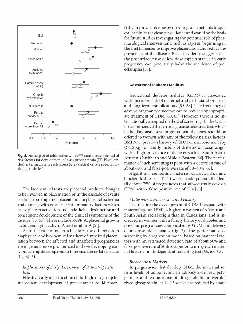
Algorithms which combine maternal characteristics and biophysical and biochemical tests at 11–13 weeks could potentially identify about 90, 80 and 60% of preg-nancies that subsequently develop early (before 34 weeks), intermediate (34–37 weeks) and late (after 37 weeks) pre-eclampsia, with a false-positive rate of 5% [51] .
Maternal Characteristics and History The risk for preeclampsia increases with maternal
weight and decreases with height, is higher in women of African and South Asian racial origin than in Cauca-sians, and is increased in women conceiving after the use of ovulation induction drugs, in those with a personal or family history of preeclampsia and in those with pre-ex-isting chronic hypertension or diabetes mellitus [51] . In general, the odds ratios for the factors in maternal his-tory which define the risk for preeclampsia are inversely proportional to the gestation at delivery, with higher ra-tios for early disease compared to intermediate and late preeclampsia ( fig. 5 ).
Biophysical and Biochemical Markers The biophysical tests are uterine artery pulsatility in-
dex and mean arterial pressure. Increased uterine artery pulsatility index reflects the underlying mechanism for the development of preeclampsia which is thought to be impaired trophoblastic invasion of the maternal spiral ar-teries and their conversion from narrow muscular vessels to wide nonmuscular channels independent of maternal vasomotor control [52–54] .
Fig. 4. Midsagittal view of the fetal brain in a normal (left) and a spina bifida (right) fetus at 12 weeks, demonstrating the mea-surement of brain stem (BS) diameter. In open spina bifida, the brain stem diameter is increased.
Nicolaides Fetal Diagn Ther 2011;29:183–196188
The biochemical tests are placental products thought to be involved in placentation or in the cascade of events leading from impaired placentation to placental ischemia and damage with release of inflammatory factors which cause platelet activation and endothelial dysfunction and consequent development of the clinical symptoms of the disease [55–57] . These include PAPP-A, placental growth factor, endoglin, activin-A and inhibin-A [51] .
As in the case of maternal factors, the differences in biophysical and biochemical markers of impaired placen-tation between the affected and unaffected pregnancies are in general more pronounced in those developing ear-ly preeclampsia compared to intermediate or late disease ( fig. 6 ) [51] .
Implications of Early Assessment of Patient-Specific Risk Effective early identification of the high-risk group for
subsequent development of preeclampsia could poten-
tially improve outcome by directing such patients to spe-cialist clinics for close surveillance and would be the basis for future studies investigating the potential role of phar-macological interventions, such as aspirin, beginning in the first trimester to improve placentation and reduce the prevalence of the disease. Recent evidence suggests that the prophylactic use of low-dose aspirin started in early pregnancy can potentially halve the incidence of pre-eclampsia [58] .
Gestational Diabetes Mellitus
Gestational diabetes mellitus (GDM) is associated with increased risk of maternal and perinatal short-term and long-term complications [59–64] . The frequency of adverse pregnancy outcomes can be reduced by appropri-ate treatment of GDM [60, 65] . However, there is no in-ternationally accepted method of screening. In the UK, it is recommended that an oral glucose tolerance test, which is the diagnostic test for gestational diabetes, should be offered to women with any of the following risk factors: BMI 1 30, previous history of GDM or macrosomic baby ( 1 4.5 kg), or family history of diabetes or racial origin with a high prevalence of diabetes such as South Asian, African-Caribbean and Middle Eastern [66] . The perfor-mance of such screening is poor with a detection rate of about 60% and false-positive rate of 30–40% [67] .
Algorithms combining maternal characteristics and biochemical tests at 11–13 weeks could potentially iden-tify about 75% of pregnancies that subsequently develop GDM, with a false-positive rate of 20% [68] .
Maternal Characteristics and History The risk for the development of GDM increases with
maternal age and BMI, is higher in women of African and South Asian racial origin than in Caucasians, and is in-creased in women with a family history of diabetes and previous pregnancies complicated by GDM and delivery of macrosomic neonates ( fig. 7 ). The performance of screening by a regression model based on maternal fac-tors with an estimated detection rate of about 60% and false-positive rate of 20% is superior to using each mater-nal factor as an independent screening test [66, 68, 69] .
Biochemical Markers In pregnancies that develop GDM, the maternal se-
rum levels of adiponectin, an adipocyte-derived poly-peptide, and sex hormone-binding globulin, a liver-de-rived glycoprotein, at 11–13 weeks are reduced by about
Odds ratio
Assisted conception
0.1 1 10
BMI
African
South Asian
Family history of PE
Chronic hypertension
Parous previous PE
0.2 0.4 2 4
Nulliparous
Parous no previous PE
Caucasian
Fig. 5. Forest plot of odds ratios with 95% confidence interval of risk factors for development of early preeclampsia (PE; black cir-cles), intermediate preeclampsia (grey circles) or late preeclamp-sia (open circles).

Turning the Pyramid of Prenatal Care Fetal Diagn Ther 2011;29:183–196 189
30 and 20%, respectively [68] . In contrast, the concentra-tion of visfatin, which is produced by adipose tissue, is increased by about 30% [70] . There is contradictory evi-dence that the serum concentration of follistatin-like-3 may also be reduced at 11–13 weeks [68, 71] .
In women who had a previous pregnancy affected by GDM, the risk of recurrence is very high and such wom-en can be automatically classified as screen-positive [72] . In nulliparous women and in those without a previous history of GDM, screening by a combination of maternal factors and serum adiponectin and sex hormone-binding globulin could identify about 65% of pregnancies that subsequently develop GDM, with a false-positive rate of 20%. A two-stage screening policy (screen positivity is defined by a history of previous GDM and the results of the combined test in those without such history) could identify about 75% of affected pregnancies at 11–13 weeks.
Diagnosis of Gestational Diabetes at 11–13 Weeks The widely accepted gestation of 24–28 weeks for
screening of GDM is based on an arbitrary recommenda-tion which attempts to achieve a balance between two opposing factors: (1) the need to maximize the detection rate of GDM by testing as late in pregnancy as possible
0
0.5
1.0
1.5
2.0
2.5
Pre
gnan
cy-a
ssoc
iate
d pl
asm
a pr
otei
n-A
(MoM
)
0
0.5
1.0
1.5
2.0
2.5
Pla
cent
al g
row
th fa
ctor
(MoM
)
0.7
0.8
0.9
1.0
1.1
1.2
1.3
1.4
1.5 M
ean
arte
rial p
ress
ure
(MoM
)
0
0.5
1.0
1.5
2.0
2.5
3.0
Ute
rine
arte
ry p
ulsa
tility
inde
x (M
oM)
Fig. 6. Box and whisker plots (median, interquartile range and range) of biophysical and biochemical markers, expressed as the multiple of the normal median (MoM) in pregnancies that develop early preeclampsia (black boxes), intermediate preeclampsia (grey boxes) or late preeclampsia (hatched boxes) compared to pregnancies with normal outcome (white boxes).
Age <25 years Age 25–34 years
Age >35 years
BMI <25 BMI 25–34
BMI >35
Caucasian South Asian
East Asian
Nulliparous Previous GDM Previous LGA
0.1 1 10 1000.5 5Odds ratio
202 3 4 50
Fig. 7. Forest plot of odds ratios with 95% confidence interval of risk factors for development of GDM. LGA = Large for gestation-al age.

Nicolaides Fetal Diagn Ther 2011;29:183–196190
because the diabetogenic effect of pregnancy increases with gestation, and (2) to maximize the duration of ther-apeutic intervention for reduction in the maternal and perinatal complications associated with GDM.
The desire to diagnose GDM in the first trimester of pregnancy could be achieved by lowering the currently used second-trimester cutoffs in plasma glucose levels both for screening and diagnosis of the condition. In screening for GDM in the first trimester, the cutoff for the 1-hour plasma glucose level after the oral administration of 50 g of glucose should be 130 rather than 140 mg/dl. In the diagnosis of GDM, the cutoffs for the 1-, 2- and 3-hour blood glucose levels after the oral administration of100 g of glucose should be 18–35% lower than the recom-mended cutoffs for the late second trimester of pregnan-cy [73] .
Implications of Early Assessment of Patient-Specific Risk Effective early identification of the high-risk group for
subsequent development of GDM is likely to improve pregnancy outcome because appropriate dietary advice and pharmacological interventions, with such drugs as metformin, can reduce the incidence of the disease and associated fetal macrosomia.
Small for Gestational Age Fetuses
Small for gestational age (SGA) fetuses with birth weight below the 5th centile for gestational age at delivery are at increased risk of perinatal death and handicap. These risks are substantially reduced in cases of SGA identified prenatally, compared to those detected after birth [74] .
Screening for SGA in the absence of preeclampsia by a combination of maternal characteristics and obstetric history with a series of biophysical and biochemical markers at 11–13 weeks could potentially identify (at a false positive rate of 10%) about 75% of pregnancies de-livering SGA neonates before 37 weeks and 45% of those delivering at term [75] .
Maternal Characteristics and History The risk for SGA increases with maternal age and de-
creases with maternal weight and height, is higher in women of African and Asian racial origin than in Cau-casians, and is increased in cigarette smokers, those with a medical history of chronic hypertension, women with a previous SGA neonate and those who had assisted con-
ception [76] . The estimated detection rate of SGA in the absence of preeclampsia with the use of the algorithm of maternal characteristics and obstetric history is about 35%, with a false-positive rate of 10%.
Biophysical and Biochemical Markers The risk for SGA is inversely related to fetal NT at 11–
13 weeks [76] . In pregnancies with SGA in the absence of preeclampsia, there is evidence of impaired placental per-fusion and function from the first trimester of pregnancy. Uterine artery pulsatility index and mean arterial pres-sure are increased and placental volume and serum PAPP-A, free � -hCG, PLGF, PP13 and ADAM12 are de-creased [75–77] . However, the magnitude of impairment in placental perfusion and function is considerably less than in preeclampsia. This is not surprising because, un-like preeclampsia which is a pathological disorder, SGA is a heterogeneous condition which includes constitu-tionally small fetuses, at no or minimally increased risk of perinatal death and handicap, and growth-restricted fetuses due to impaired placentation, genetic disease or environmental damage.
The impairment in placental function is greater for the subgroup of SGA delivering before 37 weeks than those delivering at or after 37 weeks [75] . Since the proportion of growth-restricted fetuses to constitutional SGA is like-ly to be higher in the preterm rather than term SGA, our findings imply that the early biophysical and biochemical markers could be identifying the growth-restricted sub-group amongst the SGA.
Implications of Early Assessment of Patient-Specific Risk Effective early identification of the high-risk group for
SGA could potentially improve pregnancy outcome by directing such patients to specialist clinics for regular monitoring of fetal growth and well-being. There is also evidence that the prophylactic use of low-dose aspirin started in early pregnancy can potentially halve the inci-dence of fetal growth restriction [58] .
Fetal Macrosomia
Fetal macrosomia is associated with increased risks for the mother, including cesarean section and trauma to the birth canal, and for the baby, including shoulder dys-tocia and consequent brachial plexus or facial nerve inju-ries, fractures of the humerus or clavicle, and birth as-phyxia [78, 79] .

Turning the Pyramid of Prenatal Care Fetal Diagn Ther 2011;29:183–196 191
Screening for macrosomia (birth weight above the 90th centile for gestational age at delivery) by a combina-tion of maternal characteristics and obstetric history with fetal NT and maternal serum free � -hCG and PAPP-A at 11–13 weeks could potentially identify, at a false-pos-itive rate of 10%, about 35% of women who deliver mac-rosomic neonates [80] . The detection rate is further im-proved to about 40% with the measurement of maternal serum adiponectin concentration at 11–13 weeks [81] .
Maternal Characteristics and History The risk for macrosomia increases with maternal
weight and height, and is higher in parous women who had previously delivered a macrosomic infant and/or have a medical history of diabetes mellitus; however, the risk is lower in women of African and South Asian racial origins, in cigarette smokers and in those with a medical history of chronic hypertension [80] .
Biophysical and Biochemical Markers The risk for macrosomia increases with fetal NT, ma-
ternal serum-free � -hCG and PAPP-A, and is inversely related to serum adiponectin. A possible mechanism for the association between serum PAPP-A and macrosomia is related to the proteolytic properties of PAPP-A, which cleaves insulin-like growth factor-binding proteins, thereby increasing the bioavailability of insulin-like growth factor, which is thought to play a key role in the control of placental growth and transfer of nutrients to the fetus [82–84] . There are no obvious explanations for the associations of macrosomia with high serum-free � -hCG and fetal NT. The likely mechanism underlying the association between low maternal serum adiponectin and neonatal macrosomia is increased insulin resistance and glucose intolerance [81] .
Implications of Early Assessment of Patient-Specific Risk The performance of early screening for macrosomia
is poor compared to that of screening for aneuploidies and preeclampsia. Future research should identify new biophysical and biochemical markers which could im-prove the performance of screening. Similarly, future studies should try to determine the extent to which knowledge of the individual patient-specific risk for macrosomia by first-trimester combined screening can improve antenatal surveillance and prevention of mac-rosomia itself or the intrapartum complications related to macrosomia.
Preterm Delivery
Preterm birth is the leading cause of perinatal death and handicap in children, with the vast majority of mor-tality and morbidity related to early delivery before 34 weeks [85, 86] . Delivery before 34 weeks occurs in about 2% of singleton pregnancies. In two thirds of the cases, this is due to spontaneous onset of labor or preterm prela-bor rupture of membranes. In the other third, it is iatro-genic and mainly due to preeclampsia [87] .
The rate of preterm delivery has not decreased in the last 30 years [88] . Although improvements in neonatal care have led to higher survival of very premature infants, a major impact on the associated mortality and morbid-ity will only be achieved through the development of a sensitive method to identify women at high risk of pre-term delivery and an effective strategy for prevention of this complication.
The risk of spontaneous preterm birth is increased in women with a previous late miscarriage or preterm deliv-ery and it is inversely related to cervical length measured by transvaginal sonography at 20–24 weeks’ gestation [87, 89–92] . In women with a short cervix, administration of progesterone reduces the risk of spontaneous early pre-term delivery by about 40% [93] . However, progesterone is not as effective in women with a cervical length less than 12 mm as in those with a length of 12–15 mm. An alternative treatment for women with a short cervix is cervical cerclage. This reduces the risk of spontaneous early preterm delivery by about 40% in women who had a previous preterm birth or second-trimester loss, but not in those without such history [94, 95] .
There are two disadvantages of measuring cervical length at 20–24 weeks. First of all, there is the inevitable failure to identify cervical incompetence leading to mis-carriage before this gestation. Secondly, the effectiveness of prophylactic administration of progesterone or cervical cerclage may be inversely related to the gestation at which treatment is initiated. Certainly in women who had a pre-vious preterm birth or second-trimester loss, cervical cer-clage is either carried out electively in the first trimester or is reserved for those where serial scans, beginning in the first trimester, demonstrate cervical shortening [96] .
Maternal Characteristics and History The patient-specific risk for spontaneous delivery be-
fore 34 weeks can be determined at 11–13 weeks by an algorithm combining maternal characteristics and ob-stetric history [97] . The risk for early delivery increases with maternal age and decreases with height, and is high-

Nicolaides Fetal Diagn Ther 2011;29:183–196192
er in women of African and South Asian racial origin than in Caucasians, in cigarette smokers, and in those conceiving after the use of ovulation induction drugs ( fig. 8 ). The risk is substantially influenced by the out-come of previous pregnancies: it is inversely related to the gestation at previous spontaneous delivery, decreasing
from about 7% if the gestation was 16–24 weeks to 3% if it was 31–33 weeks and 0.6% if all deliveries were at term. Additionally, the risk is affected by the number of previ-ous spontaneous deliveries at 16–30 weeks and increases from about 6 to 19% if there were two rather than one such delivery. In women with previous preterm deliver-ies, there is a protective effect against recurrence if they also had a delivery at term. For women with one or two deliveries at 16–30 weeks, the risk of recurrence decreas-es from about 6 to 1.5% and from 19 to 10%, respectively.
The estimated detection rate of spontaneous early de-livery with the use of the algorithm of maternal charac-teristics and obstetric history is 18% in nulliparous wom-en and 38% in parous women, with a false-positive rate of 10%.
Biophysical and Biochemical Markers Placental perfusion and function at 11–13 weeks are
not altered in pregnancies resulting in spontaneous early delivery [97] . Consequently, the performance of screen-ing provided by maternal characteristics and obstetric history is not improved by uterine artery pulsatility index and maternal serum or plasma concentration of PAPP-A, free � -hCG, placental growth factor, placental protein 13, a disintegrin and metalloprotease 12 (ADAM12), inhib-in-A or activin-A.
0.1 1 10 1000.5 5Odds ratio
Previous delivery at ≥37 weeks
Two deliveries at 16–30 weeks
NulliparousPrevious delivery at 16–23 weeks
Previous delivery at 24–27 weeksPrevious delivery at 28–30 weeksPrevious delivery at 31–33 weeksPrevious delivery at 34–36 weeks
Two deliveries at 16–30 weeks plus delivery ≥37 weeks
One delivery at 16–30 weeksOne delivery at 16–30 weeks
plus delivery ≥37 weeks
202 3 4 50
CaucasianAfrican
South Asian
Cigarette smokingAssisted conception
Fig. 8. Forest plot of odds ratios with 95% confidence interval of risk factors for spontaneous delivery before 34 weeks’ ges-tation.
Fig. 9. Ultrasound picture illustrating the measurement of the length of the endocervix (A to B) and the isthmus (B to C).

Turning the Pyramid of Prenatal Care Fetal Diagn Ther 2011;29:183–196 193
Recent evidence suggests that at 11–13 weeks the cer-vical length in pregnancies complicated by subsequent spontaneous delivery before 34 weeks is shorter than in those delivering after 34 weeks, and the risk for early de-livery is inversely related to cervical length [98] . In such an assessment, it is important to distinguish between the true cervix, characterized by the presence of the endocer-vical canal, which is bordered by the endocervical mu-cosa which is usually of decreased echogenicity com-pared to the surrounding tissues, and the isthmus ( fig. 9 ). It is likely that the measurement of cervical length at 1–13 weeks will be combined with the algorithm derived from maternal characteristics and obstetric history to provide an effective method for identification of the group at high risk for subsequent early delivery.
Implications of Early Assessment of Patient-Specific Risk Effective early identification of the high-risk group for
subsequent spontaneous early delivery could potentially improve outcome by directing such patients to specialist clinics for regular monitoring of cervical length and stimulating research for identification of potentially use-ful biomarkers and the investigation of the potential role of earlier intervention with such measures as prophylac-tic use of progesterone or cervical cerclage.
Conclusions
The scientific advances of the last 20 years raise the hope that many pregnancy complications can be detected as early as the 12th week of gestation. Future research will inevitably expand the number of conditions that can be identified in early pregnancy and define genetic markers of disease that will improve the accuracy of the a priori risk based on maternal characteristics and medical his-tory. Similarly, new biophysical and biochemical markers will be described that may replace some of the current ones and modify the value of others. In the future, it will become necessary to re-evaluate and improve the timing and content of each visit and the likelihood ratios for each test. Early identification of high-risk groups will also stimulate further research that will define the best proto-col for their follow-up and development of strategies for the prevention of disorders of pregnancy or their adverse consequences.
Acknowledgement
This study was supported by grants from the Fetal Medicine Foundation (Charity No. 1037116).
References
1 Ballantyne JW: A plea for a pro-maternity hospital. BMJ 1901; 2101: 813–814.
2 Ballantyne JW: The maternity hospital, with its antenatal and neo-natal departments. BMJ 1921; 3137: 221–224.
3 Ministry of Health Report: 1929 Memoran-dum on Antenatal Clinics: Their Conduct and Scope. London, His Majesty’s Stationery Office, 1930.
4 Nicolaides KH, Azar G, Byrne D, Mansur C, Marks K: Fetal nuchal translucency: ultra-sound screening for chromosomal defects in first trimester of pregnancy. BMJ 1992; 304: 867–889.
5 Snijders RJ, Noble P, Sebire N, Souka A, Nicolaides KH: UK multicentre project on assessment of risk of trisomy 21 by mater-nal age and fetal nuchal-translucency thick-ness at 10–14 weeks of gestation. Fetal Medi-cine Foundation First Trimester Screening Group. Lancet 1998; 352: 343–346.
6 Brizot ML, Snijders RJM, Bersinger NA, Kuhn P, Nicolaides KH: Maternal serum pregnancy associated placental protein A and fetal nuchal translucency thickness for the prediction of fetal trisomies in early pregnancy. Obstet Gynecol 1994; 84: 918–922.
7 Noble PL, Abraha HD, Snijders RJ, Sher-wood R, Nicolaides KH: Screening for fetal trisomy 21 in the first trimester of pregnan-cy: maternal serum free beta-hCG and fetal nuchal translucency thickness. Ultrasound Obstet Gynecol 1995; 6: 390–395.
8 Spencer K, Souter V, Tul N, Snijders R, Nico-laides KH: A screening program for trisomy 21 at 10–14 weeks using fetal nuchal translu-cency, maternal serum free � -human chori-onic gonadotropin and pregnancy-associat-ed plasma protein-A. Ultrasound Obstet Gy-necol 1999; 13: 231–237.
9 Bindra R, Heath V, Liao A, Spencer K, Nico-laides KH: One stop clinic for assessment of risk for trisomy 21 at 11–14 weeks: a prospec-tive study of 15,030 pregnancies. Ultrasound Obstet Gynecol 2002; 20: 219–225.
10 Spencer K, Spencer CE, Power M, Dawson C, Nicolaides KH: Screening for chromosomal abnormalities in the first trimester using ul-trasound and maternal serum biochemistry in a one stop clinic: a review of three years prospective experience. Br J Obstet Gynaecol 2003; 110: 281–286.
11 Wald NJ, Rodeck C, Hackshaw AK, Walters J, Chitty L, Mackinson AM, SURUSS Re-search Group: First and second trimester an-tenatal screening for Down’s syndrome: the results of the Serum, Urine and Ultrasound Screening Study (SURUSS). Health Technol Assess 2003; 7: 1–77.
12 Malone FD, Canick JA, Ball RH, Nyberg DA, Comstock CH, Bukowski R, Berkowitz RL, Gross SJ, Dugoff L, Craigo SD, Timor-Tritsch IE, Carr SR, Wolfe HM, Dukes K, Bianchi DW, Rudnicka AR, Hackshaw AK, Lambert-Messerlian G, Wald NJ, D’Alton ME, First- and Second-Trimester Evaluation of Risk (FASTER) Research Consortium: First-tri-mester or second-trimester screening, or both, for Down’s syndrome. N Engl J Med 2005; 353: 2001–2011.

Nicolaides Fetal Diagn Ther 2011;29:183–196194
13 Kagan KO, Wright D, Baker A, Sahota D, Nicolaides KH: Screening for trisomy 21 by maternal age, fetal nuchal translucency thickness, free beta-human chorionic go-nadotropin and pregnancy-associated plas-ma protein-A. Ultrasound Obstet Gynecol 2008; 31: 618–624.
14 Wright D, Spencer K, Kagan KO, Torring N, Petersen OB, Christou A, Kallikas J, Nico-laides KH: First-trimester combined screen-ing for trisomy 21 at 7–14 weeks’ gestation. Ultrasound Obstet Gynecol 2010; 36: 404–411.
15 Spencer K, Spencer CE, Power M, Moakes A, Nicolaides KH: One stop clinic for assess-ment of risk for fetal anomalies; a report of the first year of prospective screening for chromosomal anomalies in the first trimes-ter. BJOG 2000; 107: 1271–1275.
16 Borrell A, Casals E, Fortuny A, Farre MT, Gonce A, Sanchez A, Soler A, Cararach V, Vanrell JA: First-trimester screening for tri-somy 21 combining biochemistry and ultra-sound at individually optimal gestational ages. An interventional study. Prenat Diagn 2004; 24: 541–545.
17 Cicero S, Curcio P, Papageorghiou A, Sonek J, Nicolaides KH: Absence of nasal bone in fetuses with trisomy 21 at 11–14 weeks of ges-tation: an observational study. Lancet 2001; 358: 1665–1667.
18 Cicero S, Avgidou K, Rembouskos G, Kagan KO, Nicolaides KH: Nasal bone in first-tri-mester screening for trisomy 21. Am J Obstet Gynecol 2006; 195: 109–114.
19 Kagan KO, Cicero S, Staboulidou I, Wright D, Nicolaides KH: Fetal nasal bone in screen-ing for trisomies 21, 18 and 13 and Turner syndrome at 11–13 weeks of gestation. Ultra-sound Obstet Gynecol 2009; 33: 259–264.
20 Matias A, Gomes C, Flack N, Montenegro N, Nicolaides KH: Screening for chromosomal abnormalities at 10–14 weeks: the role of ductus venosus blood flow. Ultrasound Ob-stet Gynecol 1998; 12: 380–384.
21 Maiz N, Valencia C, Kagan KO, Wright D, Nicolaides KH: Ductus venosus Doppler in screening for trisomies 21, 18 and 13 and Turner syndrome at 11–13 weeks of gesta-tion. Ultrasound Obstet Gynecol 2009; 33: 512–517.
22 Huggon IC, DeFigueiredo DB, Allan LD: Tricuspid regurgitation in the diagnosis of chromosomal anomalies in the fetus at 11–14 weeks of gestation. Heart 2003; 89: 1071–1073.
23 Faiola S, Tsoi E, Huggon IC, Allan LD, Nico-laides KH: Likelihood ratio for trisomy 21 in fetuses with tricuspid regurgitation at the 11 to 13 + 6-week scan. Ultrasound Obstet Gy-necol 2005; 26: 22–27.
24 Kagan KO, Valencia C, Livanos P, Wright D, Nicolaides KH: Tricuspid regurgitation in screening for trisomies 21, 18 and 13 and Turner syndrome at 11 + 0–13 + 6 weeks of gestation. Ultrasound Obstet Gynecol 2009; 33: 18–22.
25 Bilardo CM, Timmerman E, Robles de Me-dina PG, Clur SA: Increased hepatic artery flow in first trimester fetuses: an ominous sign. Ultrasound Obstet Gynecol 2010, E-pub ahead of print. DOI: 10.1002/uog.7766.
26 Zvanca M, Gielchinsky Y, Abdeljawad F, Bi-lardo K, Nicolaides KH: Hepatic artery Dop-pler in trisomy 21 and euploid fetuses at 11–13 weeks. Prenat Diagn 2011; 31: 22–27.
27 Nicolaides KH, Spencer K, Avgidou K, Faio-la S, Falcon O: Multicenter study of first-tri-mester screening for trisomy 21 in 75,821 pregnancies: results and estimation of the potential impact of individual risk-orientat-ed two-stage first-trimester screening. Ul-trasound Obstet Gynecol 2005; 25: 221–226.
28 Syngelaki A, Chelemen T, Dagklis T, Allan L, Nicolaides KH: Challenges in the diagnosis of fetal non-chromosomal abnormalities at 11–13 weeks. Prenat Diagn 2011; 31: 90–102.
29 Office for National Statistics: Mortality Sta-tistics, Childhood, Infancy and Perinatal. 2007, series DH3, 40. Crown copyright li-censing and Public Sector information, 2010.
30 Bull C: Current and potential impact of fetal diagnosis on prevalence and spectrum of se-rious congenital heart disease at term in the UK. Lancet 1999; 35: 1242–1247.
31 Bricker L, Garcia J, Henderson J, Mugford M, Neilson J, Roberts T, Martin MA: Ultra-sound screening in pregnancy: a systematic review of the clinical effectiveness, cost-ef-fectiveness and women’s views. Health Tech-nol Assess 2000; 4:i–vi, 1–193.
32 Tegnander E, Williams W, Johansen OJ, Blaas HG, Eik-Nes SH: Prenatal detection of heart defects in a non-selected population of 30,149 fetuses-detection rates and outcome. Ultrasound Obstet Gynecol 2006; 27: 252–265.
33 Allan LD: Echocardiographic detection of congenital heart disease in the fetus: present and future. Br Heart J 1995; 74: 103–106.
34 Hyett J, Perdu M, Sharland G, Snijders R, Nicolaides KH: Using fetal nuchal translu-cency to screen for major congenital cardiac defects at 10–14 weeks of gestation: popula-tion based cohort study. BMJ 1999; 318: 81–85.
35 Atzei A, Gajewska K, Huggon IC, Allan L, Nicolaides KH: Relationship between nuchal translucency thickness and prevalence of major cardiac defects in fetuses with normal karyotype. Ultrasound Obstet Gynecol 2005; 26: 154–157.
36 Matias A, Huggon I, Areias JC, Montenegro N, Nicolaides KH: Cardiac defects in chro-mosomally normal fetuses with abnormal ductus venosus blood flow at 10–14 weeks. Ultrasound Obstet Gynecol 1999; 14: 307–310.
37 Maiz N, Plasencia W, Dagklis T, Faros E, Nicolaides K: Ductus venosus Doppler infetuses with cardiac defects and increased nuchal translucency thickness. Ultrasound Obstet Gynecol 2008; 31: 256–260.
38 Martinez JM, Comas M, Borrell A, Bennasar M, Gomez O, Puerto B, Gratacós E: Abnor-mal first-trimester ductus venosus blood flow: a marker of cardiac defects in foetuses with normal karyotype and nuchal translu-cency. Ultrasound Obstet Gynecol 2010; 35: 267–272.
39 Chelemen T, Syngelaki A, Maiz M, AllanL, Nicolaides KH: Contribution of ductusvenosus Doppler in first trimester screen-ing for major cardiac defects. Fetal Diagn Ther 2011, E-pub ahead of print. DOI: 10.1159/000322138
40 Pereira S, Ganapathy R, Syngelaki A, Maiz M, Nicolaides KH: Contribution of fetal tri-cuspid regurgitation in first trimester screening for major cardiac defects. Obstet Gynecol 2011, in press.
41 Nicolaides KH, Campbell S, Gabbe SG, Guidetti R: Ultrasound screening for spina bifida: cranial and cerebellar signs. Lancet 1986;2:72–74.
42 Chaoui R, Benoit B, Mitkowska-Wozniak H, Heling KS, Nicolaides KH: Assessment of in-tracranial translucency (IT) in the detection of spina bifida at the 11- to 13-week scan. Ul-trasound Obstet Gynecol 2009; 34: 249–252.
43 Lachmann R, Chaoui R, Moratalla J, Pic-ciarelli G, Nicolaides KH: Posterior brainin fetuses with spina bifida at 11–13 weeks. Prenat Diagn 2011; 31: 103–106.
44 Akolekar R, Bower S, Flack N, Bilardo CM, Nicolaides KH: Prediction of miscarriage and stillbirth at 11–13 weeks and the contri-bution of chorionic villus sampling. Prenatal Diagn 2011; 31: 38–45.
45 Witlin GA, Saade GR, Mattar FM, Sibai BM: Predictors of neonatal outcome in women with severe pre-eclampsia or eclampsia be-tween 24 and 33 weeks’ gestation. Am J Ob-stet Gynecol 2000; 182: 607–611.
46 Irgens HU, Reisaeter L, Irgens LM, Lie RT: Long term mortality of mothers and fathers after pre-eclampsia: population based co-hort study. BMJ 2001; 323: 1213–1217.
47 von Dadelszen P, Magee LA, Roberts JM: Subclassification of pre-eclampsia. Hyper-tens Pregnancy 2003; 22: 143–148.
48 Moldenhauer JS, Stanek J, Warshak C, Khoury J, Sibai B: The frequency and sever-ity of placental findings in women with pre-eclampsia are gestational age dependent. Am J Obstet Gynecol 2003; 189: 1173–1177.
49 Egbor M, Ansari T, Morris N, Green CJ, Sib-bons PD: Morphometric placental villous and vascular abnormalities in early- and late-onset pre-eclampsia with and without fetal growth restriction. BJOG 2006; 113: 580–589.
50 Yu CK, Khouri O, Onwudiwe N, Spiliopou-los Y, Nicolaides KH, Fetal Medicine Foun-dation Second-Trimester Screening Group: Prediction of pre-eclampsia by uterine ar-tery Doppler imaging: relationship to gesta-tional age at delivery and small-for-gesta-tional age. Ultrasound Obstet Gynecol 2008; 31: 310–313.

Turning the Pyramid of Prenatal Care Fetal Diagn Ther 2011;29:183–196 195
51 Akolekar R, Syngelaki A, Sarquis R, Wright D, Nicolides KH: Prediction of preeclampsia from biophysical and biochemical markers at 11–13 weeks. Prenat Diagn 2011; 31: 66–74.
52 Pijnenborg R: The placental bed. Hypertens Pregnancy 1996; 15: 7–23.
53 Meekins JW, Pijnenborg R, Hanssens M, Mc-Fayden IR, van Assche A: A study of placen-tal bed spiral arteries and trophoblastic inva-sion in normal and severe pre-eclamptic pregnancies. BJOG 1994; 101: 669–674.
54 Khong TY, De Wolf F, Robertson WB, Bro-sens I: Inadequate maternal vascular re-sponse to placentation in pregnancies com-plicated by pre-eclampsia and by small-for-gestational age infants. BJOG 1986; 93: 1049–1059.
55 Redman CWG: Pre-eclampsia and the pla-centa. Placenta 1991; 12: 301–308.
56 Roberts JM, Redman CW: Pre-eclampsia: more than pregnancy-induced hyperten-sion. Lancet 1993; 341: 1447–1451.
57 Granger JP, Alexander BT, Llinas MT, Ben-nett WA, Khalil RA: Pathophysiology of hy-pertension during preeclampsia linking pla-cental ischemia with endothelial dysfunc-tion. Hypertension 2001; 38: 718–722.
58 Bujold E, Roberge S, Lacasse Y, Bureau M, Audibert F, Marcoux S, Forest JC, Giguère Y: Prevention of preeclampsia and intrauterine growth restriction with aspirin started in early pregnancy. A meta-analysis. Obstet Gynecol 2010; 116: 402–414.
59 Casey BM, Lucas MJ, Mcintire DD, Leveno KJ: Pregnancy outcomes in women with ges-tational diabetes compared with the general obstetric population. Obstet Gynecol 1997; 90: 869–873.
60 Crowther CA, Hiller JE, Moss JR, McPhee AJ, Jeffries WS, Robinson JS: Effect of treat-ment of gestational diabetes on pregnancy outcomes. Australian Carbohydrate Intoler-ance Study in Pregnant Women (ACHOIS) Trial Group. N Engl J Med 2005; 352: 2477–2486.
61 Clausen TD, Mathiesen ER, Hansen T, Pe-dersen O, Jensen DM, Lauenborg J, Damm P: High prevalence of type 2 diabetes and pre-diabetes in adult offspring of women with gestational diabetes mellitus or type 1 diabe-tes: the role of intrauterine hyperglycemia. Diabetes Care 2008; 31: 340–346.
62 Metzger BE, Lowe LP, Dyer AR, Trimble ER, Chaovarindr U, Coustan DR, Hadden DR, McCance DR, Hod M, McIntyre HD, Oats JJ, Persson B, Rogers MS, Sacks DA, HAPO Study Cooperative Research Group: Hyper-glycemia and adverse pregnancy outcomes. N Engl J Med 2008; 358: 1991–2002.
63 Feig DS, Zinman B, Wang X, Hux JE: Risk of development of diabetes mellitus after diag-nosis of gestational diabetes. CMAJ 2008; 179: 229–234.
64 Bellamy L, Casas J-P, Hingorani AD, Wil-liams D: Type 2 diabetes mellitus after gesta-tional diabetes: a systematic review and me-ta-analysis. Lancet 2009; 373: 1773–1779.
65 Horvath K, Koch K, Jeitler K, Matyas E, Bender R, Bastian R, Lange S, Siebenhofer A: Effects of treatment in women with gesta-tional diabetes mellitus: systematic review and meta-analysis. BMJ 2010; 340:c1395.
66 National Institute for Health and Clinical Excellence: Diabetes in pregnancy: manage-ment of diabetes and its complications from pre-conception to the postnatal period. Clinical guideline 63, 2008. www.nice.org.uk/CG063fullguideline.
67 Waugh N, Scotland G, McNamee P, Gillett M, Brennan A, Goyder E, Williams R, John A: Screening for type 2 diabetes: literature review and economic modelling. Health Technol Assess 2007; 11: 1–125.
68 Nanda S, Savvidou M, Syngelaki A, Akolekar R, Nicolaides KH: Prediction of gestational diabetes mellitus by maternal factors and biomarkers at 11–13 weeks. Prenat Diagn 2011; 31: 135–141. DOI: 10.1002/pd.2636.
69 Van Leeuwen M, Opmeer B, Zweers E, van Ballegooie E, ter Brugge H, de Valk H, Visser G, Mol B: Estimating the risk of gestational diabetes mellitus: a clinical prediction mod-el based on patient characteristics and medi-cal history. BJOG 2010; 117: 69–75.
70 Ferreira AFA, Rezende JC, Vaikousi E, Akolekar R, Nicolaides KH: Maternal serum visfatin at 11–13 weeks of gestation in gesta-tional diabetes mellitus. Clin Chem 2011, in press
71 Thadhani R, Powe CE, Tjoa ML, Khankin E, Ye J, Ecker J, Schneyer A, Karumanchi SA: First-trimester follistatin-like-3 levels in pregnancies complicated by subsequent ges-tational diabetes mellitus. Diabetes Care 2010; 33: 664–669.
72 Kim C, Berger DK, Chamany S: Recurrence of gestational diabetes mellitus: a systematic review. Diabetes Care 2007; 30: 1314–1319.
73 Plasencia W, Garcia R, Pereira S, Akolekar R, Nicolaides KH: Criteria for screening and di-agnosis of gestational diabetes mellitus in the first-trimester of pregnancy. Fetal Diagn Ther 2011, in press.
74 Lindqvist PG, Molin J: Does antenatal iden-tification of small-for-gestational age fetuses significantly improve their outcome? Ultra-sound Obstet Gynecol 2005; 25: 258–264.
75 Karagiannis G, Akolekar R, Sarquis R, Wright D, Nicolaides KH: Prediction of small for gestation neonates from biophysi-cal and biochemical markers at 11–13 weeks. Fetal Diagn Ther 2010, E-pub ahead of print. DOI: 10.1159/000321694.
76 Poon LC, Karagiannis G, Staboulidou I, Shafiei A, Nicolaides KH: Reference range of birth weight with gestation and first-trimes-ter prediction of small for gestation neo-nates. Prenat Diagn 2011; 31: 58–65.
77 Plasencia W, Akolekar R, Dagklis T, Veduta A, Nicolaides KH: Placental volume at 11–13 weeks’ gestation in the prediction of birth weight percentile. Fetal Diagn Ther, in press.
78 Ferber A: Maternal complications of fetal macrosomia. Clin Obstet Gynecol 2000; 43: 335–339.
79 Grassi AE, Giuliano MA: The neonate with macrosomia. Clin Obstet Gynecol 2000; 43: 340–348.
80 Poon LCY, Karagiannis G, Stratieva V, Syn-gelaki A, Nicolaides KH: First-trimester pre-diction of macrosomia. Fetal Diagn Ther, E-pub ahead of print. DOI: 10.1159/000318565.
81 Nanda S, Akolekar R, Sarquis R, Mosconi AP, Nicolaides KH: Maternal serum adipo-nectin at 11–13 weeks’ of gestation in the pre-diction of macrosomia. Prenat Diagn 2011, in press
82 Lawrence JB, Oxvig C, Overgaard MT, Sot-trup-Jensen L, Gleich GJ, Hays LG, Yates JR 3rd, Conover CA: The insulin-like growth factor (IGF)-dependent IGF binding pro-tein-4 protease secreted by human fibro-blasts is pregnancy-associated plasma pro-tein-A. Proc Natl Acad Sci USA 1999; 96: 3149–3153.
83 Bonno M, Oxvig C, Kephart GM, Wagner JM, Kristensen T, Sottrup-Jensen L, Gleich GJ: Localization of pregnancy-associated plasma protein-A and colocalization of preg-nancy-associated plasma protein-A messen-ger ribonucleic acid and eosinophil granule major basic protein messenger ribonucleic acid in placenta. Lab Invest 1994; 71: 560–566.
84 Irwin JC, Suen LF, Martina NA, Mark SP, Giudice LC: Role of the IGF system in tro-phoblast invasion and pre-eclampsia. Hum Reprod 1999; 14(suppl 2):90–96.
85 Saigal S, Doyle LW: An overview of mortal-ity and sequelae of preterm birth from in-fancy to adulthood. Lancet 2008; 371: 261–269.
86 Centre for Maternal and Child Enquiries (CMACE): Perinatal Mortality 2008: United Kingdom. London, CMACE, 2010.
87 Celik E, To M, Gajewska K, Smith GC, Nico-laides KH, Fetal Medicine Foundation Sec-ond Trimester Screening Group: Cervical length and obstetric history predict sponta-neous preterm birth: development and vali-dation of a model to provide individualized risk assessment. Ultrasound Obstet Gynecol 2008; 31: 549–554.
88 Goldenberg RL, Culhane JF, Iams JD, Rome-ro R: Epidemiology and causes of preterm birth. Lancet 2008; 371: 75–84.
89 Iams JD, Goldenberg RL, Meis PJ, Mercer BM, Moawad A, Das A, Thom E, McNellis D, Copper RL, Johnson F, Roberts JM: The length of the cervix and the risk of spontane-ous premature delivery. National Institute of Child Health and Human Development Ma-ternal Fetal Medicine Unit Network. N Engl J Med 1996; 334: 567–572.

Nicolaides Fetal Diagn Ther 2011;29:183–196196
90 Heath VC, Southall TR, Souka AP, Elisseou A, Nicolaides KH: Cervical length at 23 weeks of gestation: prediction of spontane-ous preterm delivery. Ultrasound Obstet Gy-necol 1998; 12: 312–317.
91 Kagan K, To M, Tsoi E, Nicolaides KH: Pre-term birth: the value of sonographic mea-surement of cervical length. BJOG 2006; 113: 52–56.
92 To MS, Skentou CA, Royston P, Yu CK, Nico-laides KH: Prediction of patient-specific risk of early preterm delivery using maternal his-tory and sonographic measurement of cervi-cal length: a population-based prospective study. Ultrasound Obstet Gynecol 2006; 27: 362–367.
93 Fonseca RB, Celik E, Parra M, Singh M, Nicolaides KH: Progesterone and the risk of preterm birth among women with a short cervix. N Engl J Med 2007; 357: 462–469.
94 To MS, Alfirevic Z, Heath VC, Cicero S, Ca-cho AM, Williamson PR, Nicolaides KH, Fe-tal Medicine Foundation Second Trimester Screening Group: Cervical cerclage for pre-vention of preterm delivery in women with short cervix: randomised controlled trial. Lancet 2004; 363: 1849–1853.
95 Berghella V, Odibo AO, To MS, Rust OA, Al-thuisius SM: Cerclage for short cervix on ul-trasonography: meta-analysis of trials using individual patient-level data. Obstet Gynecol 2005; 106: 181–189.
96 Althuisius SM, Dekker GA, van Geijn HP, Bekedam DJ, Hummel P: Cervical Incompe-tence Prevention Randomized Cerclage Trial (CIPRACT): study design and preliminary results. Am J Obstet Gynecol 2000; 183: 823–829.
97 Beta J, Ventura W, Akolekar R, Syngelaki A, Nicolaides KH: Prediction of spontaneous preterm delivery from maternal factors and placental perfusion and function at 11–13 weeks. Prenat Diagn 2011; 31: 75–83.
98 Greco E, Lange A, Ushakov F, Rodriguez Calvo J, Nicolaides KH: Prediction of spon-taneous preterm delivery from endocervical length at 11–13 weeks. Prenatal Diagn 2011; 31: 84–89.