tutorial - blood cell morphology a clinical pathology 201 study module
TRANSCRIPT

Tutorial - Blood Cell Morphology
Louisiana State University Health Sciences Center
Department of Pathology
New Orleans, Louisiana
by
Carolyn Sue Walters, MHS, MT(ASCP)
A Clinical Pathology 201 Study Module
click here to continue
School of Medicine
© 2002 Do not reproduce this tutorial.
01-07-03

C. Sue Walters, MHS, MT(ASCP)
Associate Professor
Department of Pathology
LSU Health Sciences Center
New Orleans, LA
csw
lsuhsc
2002
click here to continue
It is illegal to reproduce the
text and images in this
computer exercise without
written permission by Ms.
Walters.

Special Acknowledgment
A special thanks and acknowledgment for the
generosity of Mrs. Angela Foley, MS, MT(ASCP),
Department of Clinical Laboratory Science,
School of Allied Health, LSU Health Sciences
Center New Orleans for the use of many of the
blood cell images used in this presentation.
click here to continue

Feedback
Feedback as to the quality and usefulness of this
exercise is solicited and suggestions for improve-
ment are welcomed. Please forward your remarks
by E-mail [email protected]
or via US MAIL:
C. Sue Walters, MHS, MT(ASCP)
LSU Health Sciences Center
Department of Pathology
1901 Perdido Street
New Orleans, LA 70112
csw
lsuhsc
2002
click here to continue

Directions
csw
lsuhsc
2002
The directions for navigating through the exer-
cise are given on the next 2 pages. They are the
same as those routinely used in Clinical
Pathology 202 study modules. Please click on:
to visit the directions before continuing
with the exercise.
to go directly to the first page of the
exercise.
or

Directions, continued
csw
lsuhsc
2002
in the upper left hand corner of every page to return
to the previous page
menu in the upper right corner of the page to return to
the Main Menu selection.
The following directional icons are provided throughout
the exercise for your convenience. You can click on:
csw
lsuhsc
2002
in the lower right corner of the page to continue.
in the lower right corner of the Main Menu page to
Quit (i.e., end the exercise). quit

Directions, continued
csw
lsuhsc
2002
“Hot points” (symbols, words, phrases) have
been inserted on the pages as navigational
tools and can be identified by their “gold”
color. If it’s “gold”, click on it to move to the
next text/data entry. Also, sounds have been
added in a few places for emphasis.
Remember, if it’s gold, click on it. Try it!
Caution, failure to follow the structured order
of the “hot points” may result in confusion. If
you use the mouse without placing the cursor
directly on the “hot point” , you may skip over
vital information.

Special Comments
csw
lsuhsc
2002
This exercise has numerous images. You may
note that, when a page contains images, there
may be a rather long delay before you regain
control of the cursor. Please be patient. I think
you will find the images are worth the wait.
NOTE:
Some animation and/or interactive affects may
be lost if you attempt to replay a page by re-
turning to the previous page and then advanc-
ing to that page again.
Now, click on the gold to begin.

csw
lsuhsc
2002
Hematologic Cells
Found in Peripheral Blood
and Bone Marrow

MAIN MENU
Introduction
csw
lsuhsc
2002
Erythrocytes
Leukocytes
quit
Platelets
Abnormal erythrocytes - terminology
Disorders – characteristic morphology

Introduction
csw
lsuhsc
2002

What is the purpose of this study
module?
csw
lsuhsc
2002
menu
This study module is designed for LSUHSC L2
students enrolled in Clinical Pathology 201. It
is intended as a reference for blood cell and
bone marrow morphology.
The presentation of illustrative cells in this
module is by no means a comprehensive study
of blood cells. It is limited to the material
covered in the lectures and laboratory sessions.
Unfortunately, a few cell illustrations are not
available at this time but will be added later.
Leukocytes
csw
lsuhsc
2002

How are the WBC identified and
classified?
csw
lsuhsc
2002
menu
Typical nuclear and cytoplasmic morphologic
features provide a means by which WBC can be
identified and classified as to cell line (i.e.,):
• lymphocytes
• granulocytes [neutrophils, eosinophils, or
basophils]
• monocytes
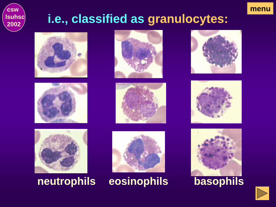
i.e., classified as granulocytes: csw
lsuhsc
2002
menu
neutrophils eosinophils basophils

i.e., classified as lymphocytes: csw
lsuhsc
2002
menu
shown with
normocytic RBC
shown with
macrocytic RBC
shown with
microcytic RBC

i.e., classified as monocytes: csw
lsuhsc
2002
menu
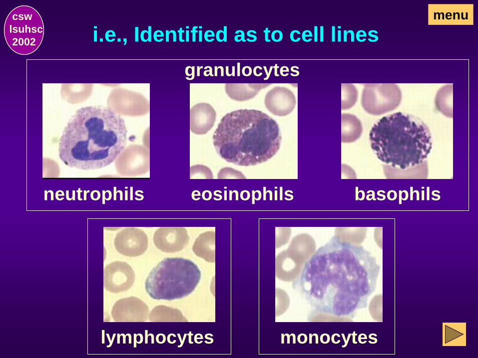
i.e., Identified as to cell lines csw
lsuhsc
2002
menu
neutrophils eosinophils basophils
granulocytes
lymphocytes monocytes

WBC can also be identified and
classified as to…
csw
lsuhsc
2002
menu
• maturity (i.e., mature cell or immature stage of
development).
next

Immature WBC, e.g.:
granulocytes
(various stages)
myeloblasts
lymphoblasts
menu
monoblasts
csw
lsuhsc
2002

e.g., Neutrophils in various stages
of maturation…
menu csw
lsuhsc
2002
myeloblast
metamyelocyte band PMN (mature)
myelocyte promyelocyte

menu csw
lsuhsc
2002 Neutrophilic Maturation
Mature neutrophil “PMN” Neutrophilic band “stab” Metamyelocyte Myelocyte
from immature blast to mature PMN
Promyelocyte Myeloblast

menu csw
lsuhsc
2002 Neutrophilic Maturation
Mature neutrophil “PMN” Neutrophilic band “stab” Metamyelocyte Myelocyte
From mature PMN to myeloblast
Promyelocyte Myeloblast

WBC can also be identified and
classified as to…
csw
lsuhsc
2002
menu
• abnormal morphology (i.e., nuclear or cyto-
plasmic alterations)

e.g., WBC with acquired non-
neoplastic alterations…
neutrophils
In bacterial
infections
with Döhle bodies
and/or toxic granulation
&
inherited disorders
menu csw
lsuhsc
2002
&
hypersegmented
neutrophils in
megaloblastic
anemias
reactive/atypical
lymphocytes (ATL)
In viral infections

e.g., WBC with inherited non-
neoplastic alterations…
menu csw
lsuhsc
2002
Pelger-Huet Anomaly
hyposegmented nuclei
&
May-Hegglin Anomaly
cytoplasmic blue bodies
&
&
cytoplasmic
black granules
Alder-Reilly Anomaly
cytoplasmic large
black granules
Chediak-Higashi Syndrome

myeloblasts w/ Auer rod(s)
in acute myelocytic leukemias
WBC with neoplastic alterations, e.g.…
hairy cell lymphocytes
hairy cell leukemia
&
&
menu csw
lsuhsc
2002
plasma cells
in multiple myeloma

Leukocytic Maturation
csw
lsuhsc
2002
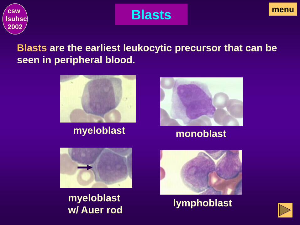
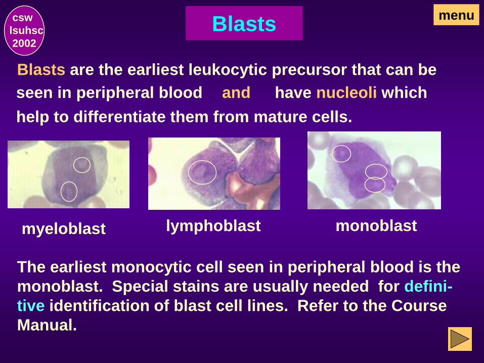
Blasts
Blasts are the earliest leukocytic precursor that can be
seen in peripheral blood.
monoblast
lymphoblast
menu csw
lsuhsc
2002
myeloblast
myeloblast
w/ Auer rod

Blasts
All blasts have nucleoli. and
myeloblast
myeloblast
monoblast
lymphoblast
Cell lines are difficult to differentiate on Wright’s stain
without a distinguishing feature (eg, Auer rod in AML).
Auer rod
menu csw
lsuhsc
2002

While the presence of nucleoli differentiates blasts
from more mature forms,
myeloblast myeloblast monoblast lymphoblast
Blasts
Leukoblasts must also be differentiated from
proerythroblasts.
menu csw
lsuhsc
2002
special stains are usually needed for definitive
identification of leukoblasts.

Proerythroblasts
myeloblast monoblast lymphoblast
Leukoblasts
How do you differentiate leukoblasts (WBC)
and proerythroblasts (RBC)?
menu csw
lsuhsc
2002
have nucleoli. &
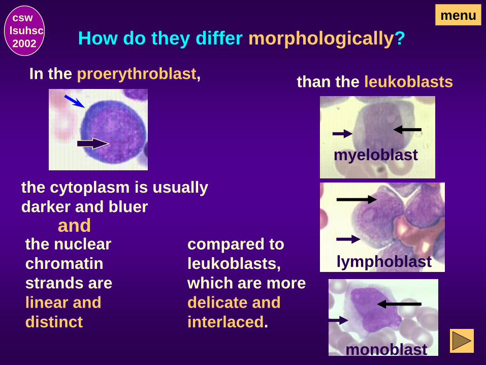
How do they differ morphologically?
myeloblast
monoblast
lymphoblast
In the proerythroblast,
menu csw
lsuhsc
2002
the nuclear
chromatin
strands are
linear and
distinct
than the leukoblasts
the cytoplasm is usually
darker and bluer
compared to
leukoblasts,
which are more
delicate and
interlaced.
and

Myelocytic (or Granulocytic) Series
csw
lsuhsc
2002

Myelocytic Maturation csw
lsuhsc
2002
menu
There are three types of cells in the myelocytic (or gran-
ulocytic) series: neutrophils, eosinophils, and basophils.
Myeloblasts (ie, the earliest precursor) originate in the
marrow from a stem cell common to erythroid, megakaryo-
cytic, and granulocytic cells. Prominent nucleoli are seen
in the nucleus and the cytoplasm is agranular. Morpho-
logically, they are difficult to differentiate from lympho-
blasts or monoblasts.
Examples of myeloblasts:
nucleoli
& agranular cytoplasm

Myelocytic Maturation, continued csw
lsuhsc
2002
menu
Nucleoli are also seen in the promyelocyte and the appear-
ance of large azurophilic (nonspecific) cytoplasmic gran-
ules in the early stage of its transition is an indication that
the cell is a granulocyte. However, morphologic determin-
ation as to neutrophilic, eosinophilic, or basophilic cannot
yet be made.
Examples of promyelocytes:
early promyelocyte
prominent
nucleoli and
cytoplasmic
non-specific
granules begin
to be visible
as the cell matures,
the nucleoli begin to
fade and the gran-
ules become more
numerous and
prominent
late promyelocyte

Myelocytic Maturation, continued
As the promyelocyte matures and reaches the myelocytic
stage, it has definitely and visually differentiated into one
of the three granulocytic types with characteristic
cytoplasmic “specific” or secondary granules. Nucleoli
are indistinct or not seen and this is the last mitotic
stage.
csw
lsuhsc
2002
menu
Examples of myelocytes with specific granules:
basophilic eosinophilic neutrophilic

Myelocytic Maturation, continued
Specific cytoplasmic granules:
csw
lsuhsc
2002
menu
neutrophilic
ill-defined reddish granules within the bluish
cytoplasm resulting in a lilac or pinkish
color
eosinophilic
relatively large, spherical, orange granules
basophilic
unevenly distributed large, blue-black
granules, which are usually also visible on
top of the nucleus
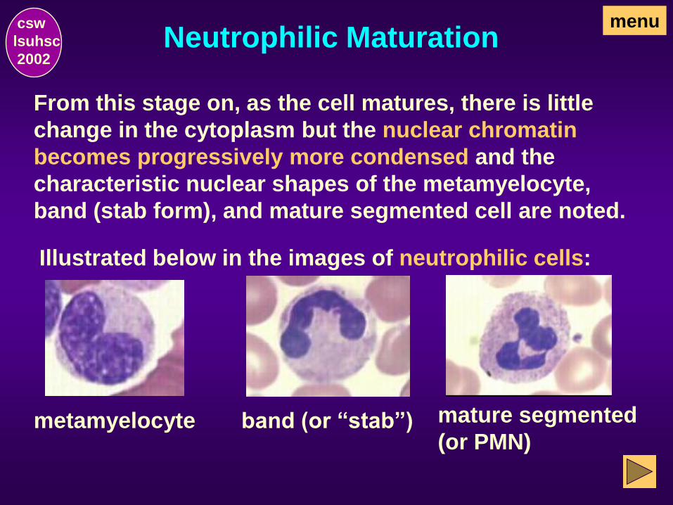
Neutrophilic Maturation
From this stage on, as the cell matures, there is little
change in the cytoplasm but the nuclear chromatin
becomes progressively more condensed and the
characteristic nuclear shapes of the metamyelocyte,
band (stab form), and mature segmented cell are noted.
Illustrated below in the images of neutrophilic cells:
metamyelocyte band (or “stab”) mature segmented
(or PMN)
csw
lsuhsc
2002
menu

Eosinophilic and Basophilic
Maturation
There is usually no differentiation made as to whether
eosinophils and basophils are myelocytes, metamyelo-
cytes, band, or mature cells. Regardless of the stage of
maturation, they are still referred to only as eosinophils
or basophils.
The granules observed in both cell lines are rather large,
frequently dense and, in many cases, obscure the
nucleus thus making it difficult to see the nuclear shape
as illustrated in the images of eosinophils and basophils.
csw
lsuhsc
2002
menu

Various Stages Maturation csw
lsuhsc
2002
menu
<---------Eosinophils---------> <----Basophils---->

Morphologic Features of
Granulocytes
csw
lsuhsc
2002
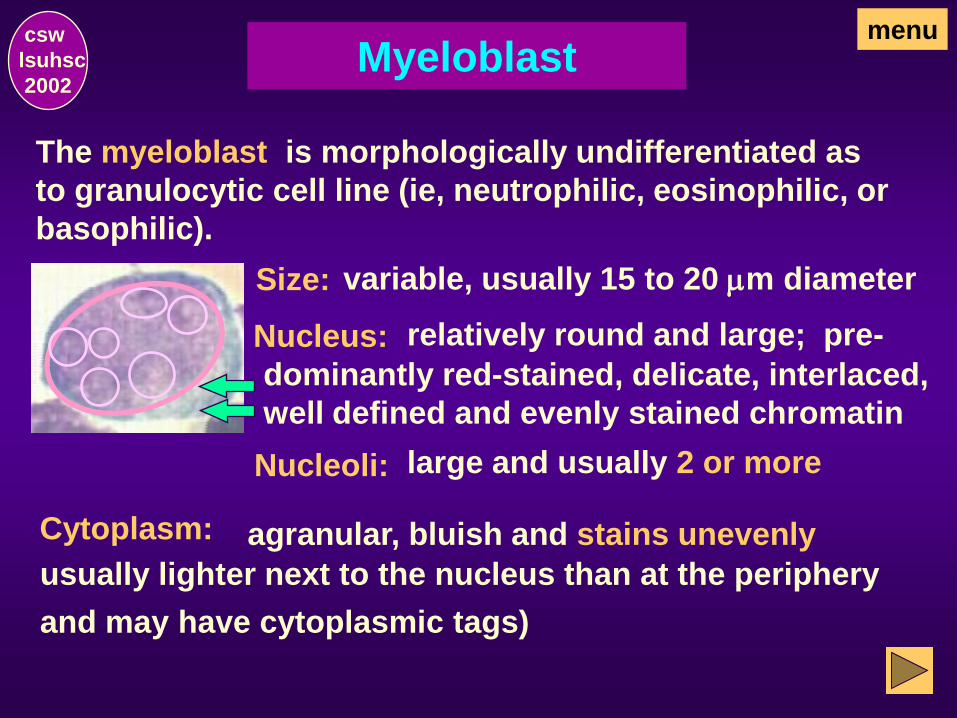
Myeloblast
The myeloblast is morphologically undifferentiated as
to granulocytic cell line (ie, neutrophilic, eosinophilic, or
basophilic).
menu
Size: variable, usually 15 to 20 mm diameter
Nucleus: relatively round and large; pre-
dominantly red-stained, delicate, interlaced,
well defined and evenly stained chromatin
Nucleoli: large and usually 2 or more
Cytoplasm: agranular, bluish and stains unevenly
usually lighter next to the nucleus than at the periphery
and may have cytoplasmic tags)
csw
lsuhsc
2002

Promyelocyte
The promyelocyte is still undifferentiated as to a specific
granulocytic cell line (ie, neutrophilic, eosinophilic, or
basophilic).
menu
Size: usually larger than blasts but variable
depending on the stage in the mitotic cycle
Nucleus: round and relatively large with
predominantly red-stained chromatin
Nucleoli: usually demonstrable
Cytoplasm: • Stains blue
• Granules - nonspecific (or primary) granules
and absence of secondary granules (ie, neutrophilic,
eosinophilic, or basophilic)
csw
lsuhsc
2002
with a relatively light area adjacent to the nucleus

Myeloblast
Promyelocyte
Myeloblast vs. Promyelocyte Nucleoli: two or more, large, prominent
usually demonstrable Nucleoli:
Cytoplasm: bluish, unevenly stained
menu
Cytoplasm: bluish, unevenly stained
Granules: none visible
Granules: distinct non-specific (or ,
primary), predominantly
dark blue or reddish blue
csw
lsuhsc
2002

Granulocytes menu csw
lsuhsc
2002
Granulocytes are neutrophilic, eosinophilic, or
basophilic. The cells cannot be morphologically
differentiated on Wright’s stain until they reach
the myelocyte stage and develop specific
granules.
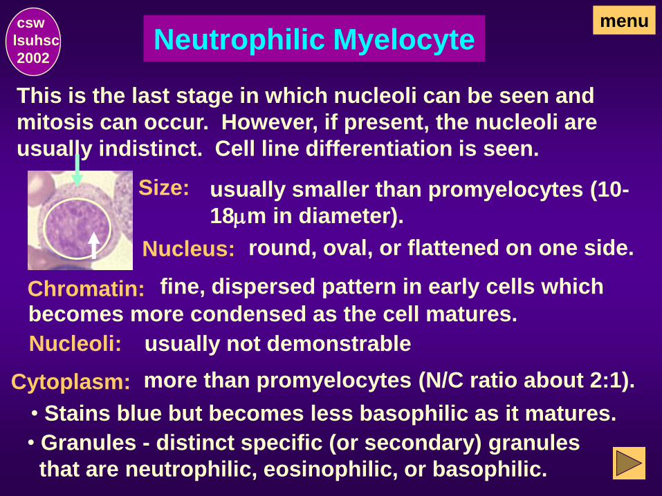
Neutrophilic Myelocyte menu
Size: usually smaller than promyelocytes (10-
18mm in diameter).
Nucleus: round, oval, or flattened on one side.
Chromatin: fine, dispersed pattern in early cells which
becomes more condensed as the cell matures.
Nucleoli: usually not demonstrable
Cytoplasm:
• Stains blue but becomes less basophilic as it matures.
• Granules - distinct specific (or secondary) granules
that are neutrophilic, eosinophilic, or basophilic.
more than promyelocytes (N/C ratio about 2:1).
This is the last stage in which nucleoli can be seen and
mitosis can occur. However, if present, the nucleoli are
usually indistinct. Cell line differentiation is seen.
csw
lsuhsc
2002

Myelocytes menu
The first morphologic evidence of specific or secondary
granules that provide a means of identifying the cell as a
neutrophil, eosinophil or basophil is seen in the
myelocyte.
Neutrophils - neutrophilic granules give the cytoplasm
a lilac or pinkish appearance
csw
lsuhsc
2002
NOTE: an enlargement of the cell
shown on the previous slide to
better illustrate the granules.

Myelocytes menu
The maturation sequence of the eosinophils and
basophils is the same as the neutrophils. The
nuclear features are identical but the color and/or
size of the cytoplasmic granules differentiates
these cells from the neutrophils.
Eosinophils - relatively large and spherical purplish-red
granules give the cytoplasm a reddish-orange color.
Basophils - large dark blue to black unevenly
distributed granules may fill the cytoplasm and,
when present in large numbers, may obscure the
nucleus.
csw
lsuhsc
2002

Neutrophilic, Eosinophilic, &
Basophilic Myelocytes
menu csw
lsuhsc
2002
basophilic
neutrophilic
eosinophilic

Metamyelocyte (Neutrophilic)
As soon as the nucleus of the myelocyte (which may be
neutrophilic, eosinophilic, or basophilic) becomes
indented, the cell is classified as a metamyelocyte. The
cell is no longer capable of mitosis.
menu
Size: usually slightly smaller than myelocytes
Nucleus: relatively smaller than myelocyte
and, as the cell matures, indentation increases.
Chromatin: less well defined and becomes more condensed clumped, and darkly stained as the cell matures.
Nucleoli: not demonstrable
Cytoplasm: progressively less basophilic than myelocytes
and distinct specific (or secondary) granules that are
neutrophilic, eosinophilic, or basophilic predominate.
csw
lsuhsc
2002

Metamyelocytes menu csw
lsuhsc
2002
neutrophilic
specific granules
eosinophilic
specific granules
basophilic
specific granules

Neutrophilic Band menu
As soon as the indention in the nucleus of the metamyelo-
cyte becomes greater than 1/2 the diameter, the cell is
classified as a band (neutrophil, eosinophil, or basophil).
Size: slightly smaller than metamyelocytes
Nucleus: indented greater than 1/2 diameter and opposite edges of the nucleus become
approximately parallel (horse-shoe shape).
Chromatin: dense and clumped, usually with a pyknotic
mass at each pole where the lobe will be.
Nucleoli: none present
Cytoplasm: no basophilia & may be slightly eosinophilic;
distinct specific (or secondary) granules that are
neutrophilic, eosinophilic, or basophilic predominate.
csw
lsuhsc
2002

menu csw
lsuhsc
2002 Bands
basophilic (blue-black)
specific granules
eosinophilic (orange)
specific granules
neutrophilic (lilac or pinkish) specific granules

MATURE SEGMENTED
NEUTROPHIL (PMN)
Mature neutrophils have nuclei that are separated into
definite lobes. They are frequently referred to as PMN
(polymorphonuclear neutrophils).
Size: 10-15 mm (about twice the size of RBC).
Nucleus: separated into definite lobes (usually
2 or 3 with occasional 4 or 5) which are con-
nected by a very narrow filament or strand.
Chromatin: dense and clumped
Nucleoli: not present
Cytoplasm: slightly eosinophilic or light pink with numer-
ous pink to bluish-black evenly distributed small granules.
menu csw
lsuhsc
2002

usually
bilobed
nucleus
but may have
3 or more lobes
eosinophils
Normal Mature Neutrophils,
Eosinophils, and Basophils
menu csw
lsuhsc
2002
segmented neutrophil (PMN)
usually 2-4 lobed nucleus but may have a few w/ 5 lobes
neutrophils
basophil
granules usually
obscure nucleus
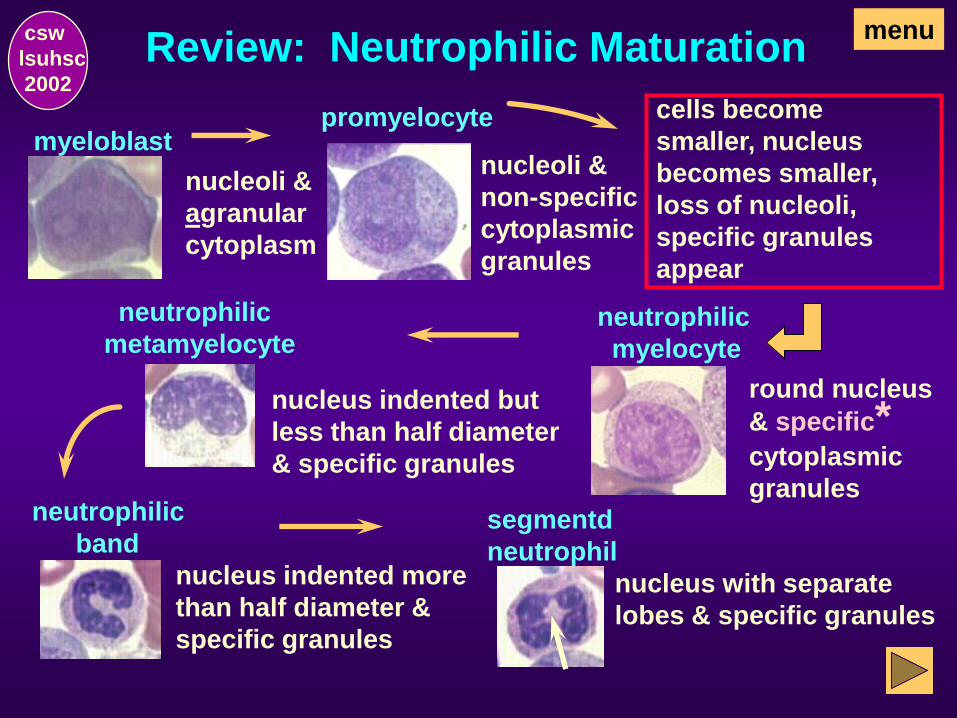
Review: Neutrophilic Maturation menu csw
lsuhsc
2002
myeloblast
nucleoli &
agranular
cytoplasm
neutrophilic
metamyelocyte
nucleus indented but
less than half diameter
& specific granules
neutrophilic
band
nucleus indented more
than half diameter &
specific granules
segmentd
neutrophil
nucleus with separate
lobes & specific granules
cells become
smaller, nucleus
becomes smaller,
loss of nucleoli,
specific granules
appear
neutrophilic
myelocyte
round nucleus
& specific* cytoplasmic
granules
nucleoli &
non-specific
cytoplasmic
granules
promyelocyte

Examples of granulocytes in
various stages of maturation:
1 early promyelocyte
1
2 late promyelocyte
or early myelocyte 2
3 myelocyte
3
3
4 metamyelocyte
4
4
4
4
4
5 band neutrophil
5
5
5
5
5
6 mature segmented
neutrophil (PMN)
6
6
7 eosinophil
7
8 Whoa!
8
That’s not a WBC. It’s a nucleated RBC but
will also be included in the total WBC count.
menu csw
lsuhsc
2002

Now, Can you Identify the stages
of granulocytes just illustrated?
1
1 early promyelocyte
2 2 late promyelocyte
or early myelocyte 3
3
3 myelocyte
4
4
4
4
4 4 metamyelocyte
5
5
5
5
5
5 band neutrophil
6
6
6 mature segmented
neutrophil (PMN)
7
7 eosinophil
8
menu
8 Remember, it’s a NRBC!
csw
lsuhsc
2002

Review: Eosinophilic & Basophilic
Maturation
menu csw
lsuhsc
2002
The maturation sequence for eosinophils and basophils
parallels that of neutrophils. Blast and promyelocyte
stages are morphologically undifferentiated as to
neutrophils, eosinophils, or basophils.
The cells can be differentiated in the myelocyte stage
with the appearance of specific cytoplasmic granules
(ie, neutrophilic pink, eosinophilic orange, or basophilic
dark blue-black) that remain through maturity.
neutrophil eosinophil basophil

Lymphocytic Series
csw
lsuhsc
2002

Blasts are the earliest leukocytic precursor that can be
seen in peripheral blood
myeloblast monoblast lymphoblast
Lymphoblasts
and have nucleoli which
help to differentiate them from mature cells.
The least mature lymphoid cell seen in peripheral blood is
the lymphoblast.
menu csw
lsuhsc
2002
Special stains and/or flow cytometry are usually needed
for definitive differentiation of the various leukoblast cell
lines (i.e., myeloblasts, lymphoblasts, and monoblasts).

Lymphocytic Cells
For Clinical Pathology 201, you will be expected
to be able to identify and differentiate:
and
atypical (or reactive) lymphocytes
lymphoblasts
menu csw
lsuhsc
2002
mature lymphocytes
and

Monocytic Series
csw
lsuhsc
2002
Blasts are the earliest leukocytic precursor that can be
seen in peripheral blood
myeloblast monoblast lymphoblast
Blasts
and have nucleoli which
help to differentiate them from mature cells.
The earliest monocytic cell seen in peripheral blood is the
monoblast. Special stains are usually needed for defini-
tive identification of blast cell lines. Refer to the Course
Manual.
menu csw
lsuhsc
2002
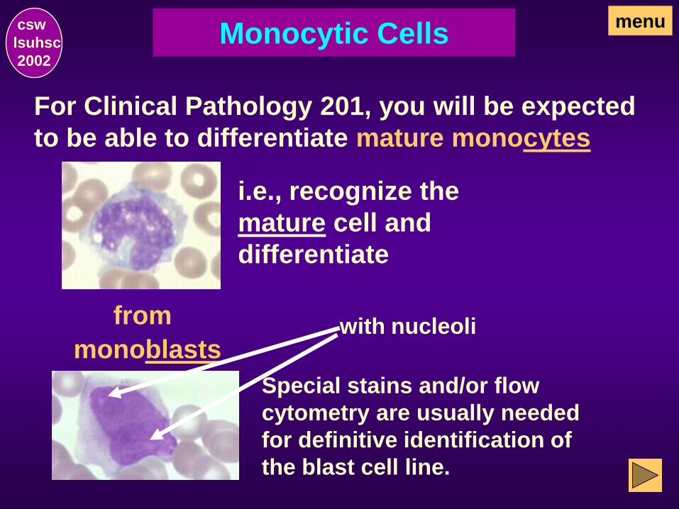
Monocytic Cells
For Clinical Pathology 201, you will be expected
to be able to differentiate mature monocytes
i.e., recognize the
mature cell and
differentiate
from
monoblasts
Special stains and/or flow
cytometry are usually needed
for definitive identification of
the blast cell line.
menu csw
lsuhsc
2002
with nucleoli

Plasma Cells
csw
lsuhsc
2002

Plasma Cells
Size: mature cells vary greatly
Shape: usually oval shape with relative-
ly smooth cytoplasmic margins, but, like
the lymphocyte, the plasmocyte is easily
traumatized and often has frayed or
nebulous margins and pointed or fila-
mentous cytoplasmic projections
Nucleus: relatively small and round and
eccentrically located
Cytoplasm: abundant
The cytoplasm adjacent to the nucleus stains more lightly
than the periphery of the cell which has a high saturation
of red and blue dyes. The area is called a “golgi”.
menu csw
lsuhsc
2002

Plasma Cells, continued
Plasma cells are never present in normal peripheral blood.
They constitute about 1% of the nucleated cells in normal
bone marrow where they tend to be grouped in small
islands around blood vessels. They may be present in
small numbers in chronic infections, in granulomatous and
allergic diseases and in plasma cell myeloma.
Plasmoblasts (not shown) are cells with relatively large
nuclei, nucleoli and delicate chromatin which takes a
predominantly red color. Plasmoblasts are not recogniz-
able except in malignancies of the plasmocytoid type.
menu csw
lsuhsc
2002

Leukocytes in Normal
Perpheral Blood
csw
lsuhsc
2002

Review: WBC found in normal
peripheral blood:
csw
lsuhsc
2002
menu
mature
neutrophils
eosinophils basophils
lymphocytes monocytes atypical lymphocytes
(<6% of lymphocytes)
band
neutrophils

Leukocytes with Acquired
Non-neoplastic Alterations
csw
lsuhsc
2002

Toxic granulation - dark blue to
purple cytoplasmic granules
toxic granulation
and/or Döhle bodies - small blue cyto-
plasmic inclusions
Döhle
bodies
Toxic granules may be seen in severe bacterial infec-
tions, burns, aplastic anemia, and following admin-
istration of toxic agents.
Frequently, Döhle bodies will also be seen concom-
itantly with toxic granulation.
Toxic Granulation & Dohle Bodies in
Neutrophils
menu csw
lsuhsc
2002
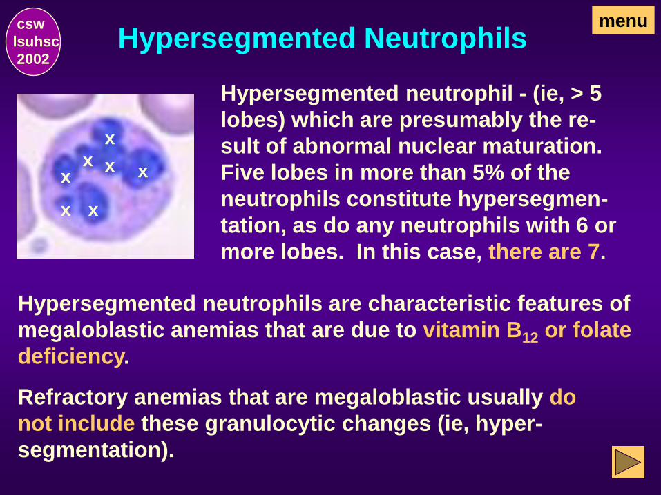
Hypersegmented neutrophil - (ie, > 5
lobes) which are presumably the re-
sult of abnormal nuclear maturation.
Five lobes in more than 5% of the
neutrophils constitute hypersegmen-
tation, as do any neutrophils with 6 or
more lobes. In this case, there are 7.
x x
x x
x
x x
Hypersegmented neutrophils are characteristic features of
megaloblastic anemias that are due to vitamin B12 or folate
deficiency.
Refractory anemias that are megaloblastic usually do
not include these granulocytic changes (ie, hyper-
segmentation).
Hypersegmented Neutrophils menu csw
lsuhsc
2002

Atypical lymphocytes may have abundant cytoplasm
with scalloped or indented rims ...
Atypical/reactive lymphocytes may be seen
most typically in viral disorders.
Atypical/Reactive Lymphocytes menu csw
lsuhsc
2002

…or have darker cytoplasm and more monocytoid
nuclear or plasmacytoid features ..
Atypical/Reactive Lymphocytes menu csw
lsuhsc
2002

Leukocytes with Inherited
Non-neoplastic Alterations
csw
lsuhsc
2002
Pelger-Huet Anomaly
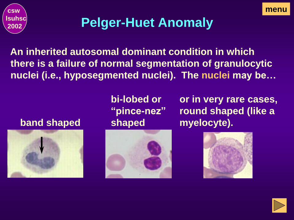
band shaped
menu csw
lsuhsc
2002
An inherited autosomal dominant condition in which
there is a failure of normal segmentation of granulocytic
nuclei (i.e., hyposegmented nuclei). The nuclei may be…
or in very rare cases,
round shaped (like a
myelocyte).
bi-lobed or
“pince-nez”
shaped

Pelger-Huet Anomaly, continued menu csw
lsuhsc
2002
next
The cell morphology persists through life and
the cells are functional.
Absence of symptoms of infection or other
cause of a “left shift”, history of persistent
blood morphology, and/or similar blood
morphology of other family members
suggests the anomaly.

Pseudo-Pelger-Huet Anomaly menu csw
lsuhsc
2002
Band forms, neutrophils with only two segments or “pince-nez” appearance (not shown), and/or neutrophils
with round non-segmented nuclei are seen. Neutrophils
with > 2 segments (lobes) will not be seen in this disorder.
There is asynchronism between the shape of the nucleus
and the maturity of the nucleus and cytoplasm.
An acquired disorder similar in appearance to Pelger-Huet
anomaly may occasionally be found in cases of granulo-
cytic leukemia, myeloproliferative disorders, some infec-
tions, and after exposure to certain drugs.

May-Hegglin Anomaly
Pale blue cytoplasmic inclusions that
resemble Dohle bodies but are larger
and more prominent. They may be
found in neutrophils, eosinophils,
basophils, and monocytes.
menu csw
lsuhsc
2002
Bluish aggregations (RNA) particles can be seen in the
cytoplasm of neutrophils. Giant platelets can also be
seen.
This is a rare autosomal dominant condition.
The cells are functional and the cytoplasmic inclusions
in May-Hegglin persist through life. Acquired Dohle
bodies are transient.
Alder-Reilly Anomaly menu csw
lsuhsc
2002
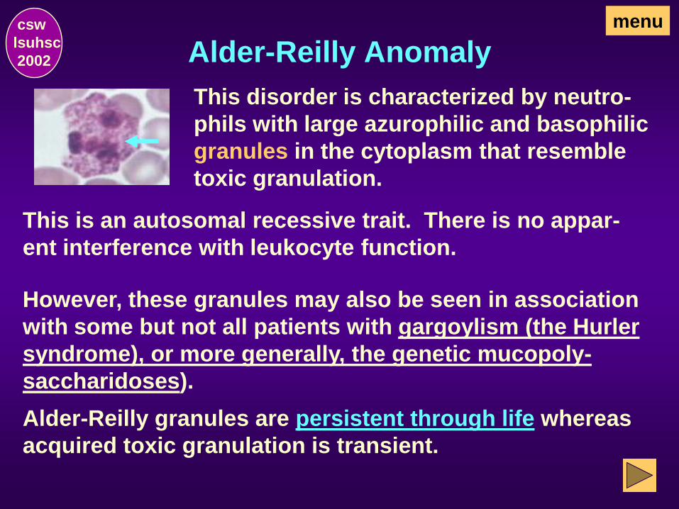
This disorder is characterized by neutro-
phils with large azurophilic and basophilic
granules in the cytoplasm that resemble
toxic granulation.
This is an autosomal recessive trait. There is no appar-
ent interference with leukocyte function.
However, these granules may also be seen in association
with some but not all patients with gargoylism (the Hurler
syndrome), or more generally, the genetic mucopoly-
saccharidoses).
Alder-Reilly granules are persistent through life whereas
acquired toxic granulation is transient.

Chediak-Higashi Syndrome
Abnormally large cytoplasmic black gran-
ules which appear to be abnormal lyso-
somes may be seen in granulocytes, mon-
ocytes, and lymphocytes.
menu csw
lsuhsc
2002
This is a rare sutosomal recessive disorder characterized
by paratial albinism, photophobia, and frequent pyogenic
infections. An accelerated lymphoma-like phase occurs,
with lymphadenopathy, hepatosplenomegaly, and pan-
cytopenia. Lymphoid infiltrates are widespread and death
ensues at an early age. Leukocyte functional abnormal-
ities exist.
The abnormal morphologic features are persistent
throughout life.

Leukocytes with Neoplastic
Alterations
csw
lsuhsc
2002

Hairy Cell Lymphocytes
The hallmark of hairy cell leukemia is the
presence of lymphocytes with irregular
long, delicate cytoplasmic projections
which give them a hairy appearance.
menu csw
lsuhsc
2002
This is an uncommon chronic, low grade lymphoprolifer-
ative disease (or CLL) that occurs about 5 times more
frequently in males than females.
Onset of disease is insidious; weakness and lethargy; or
may be asymptomatic (10-15% of patients). May be
bleeding and bruising.
Normocytic, normochromic anemia related to the
neoplastic cell mass, marrow hypoplasia, and hyper-
splenism is also seen. Thrombocytopenia in about
75% of patients and Coomb’s test may be positive.

Myeloblasts with Auer Rods menu
Auer rods in the cytoplasm of myeloblasts
are associated with acute leukemias hav-
ing a myeloid component. They appear as
cytoplasmic reddish rods with Wright’s or
Wright’s-Giemsa stains.
They may be seen in some, but not all,
myeloblasts in some, but not all, of the
variants of acute myelocytic leukemia.
They are not seen in blasts in chronic
myelocytic leukemia.
The presence of Auer rods in the cytoplasm of blasts
effectively rules out a lymphoid disorder.
csw
lsuhsc
2002

End of Leukocytes
csw
lsuhsc
2002
or
Erythrocytes to go to the next section of this
study module as designed.
This ends the section on leukocytes. Click on:
Menu to go back to the menu.
or
Quit to end the exercise.

Erythrocytes
csw
lsuhsc
2002

How are the RBC identified? csw
lsuhsc
2002
menu
Typical morphologic nuclear and/or cytoplasmic
features provide a means by which RBC can be
identified. For example:
Size
Shape
Color
Hemoglobin content
Inclusions (if any)
Maturity

How are RBC classified as to
maturity?
csw
lsuhsc
2002
menu
Characteristic nuclear and/or cytoplasmic
morphologic features allow red blood cells to
be classified as:
• basophilic normoblast (or prorubricyte)
• polychromatophilic erythrocyte (or diffusely
basophilic erythrocyte
• pronormoblast (or rubriblast), the earliest form
seen in peripheral blood (ie, least mature)
• polychromatophilic normoblast (or rubricyte)
• orthochromatic normoblast (or metarubricyte)
• mature erythrocytes
Summary of the key features of
erythrocyte development:
csw
lsuhsc
2002
menu
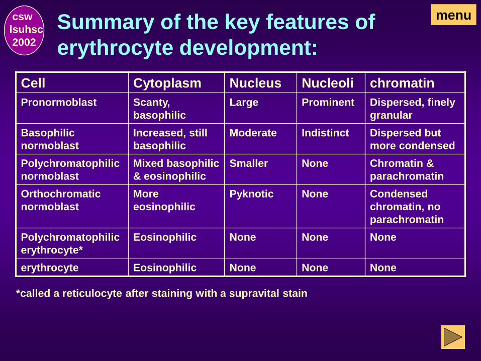
Cell Cytoplasm Nucleus Nucleoli chromatin
Pronormoblast Scanty,
basophilic
Large Prominent Dispersed, finely
granular
Basophilic
normoblast
Increased, still
basophilic
Moderate Indistinct Dispersed but
more condensed
Polychromatophilic
normoblast
Mixed basophilic
& eosinophilic
Smaller None Chromatin &
parachromatin
Orthochromatic
normoblast
More
eosinophilic
Pyknotic None Condensed
chromatin, no
parachromatin
Polychromatophilic
erythrocyte*
Eosinophilic None None None
erythrocyte Eosinophilic None None None
*called a reticulocyte after staining with a supravital stain
What are the characteristic features of
pronormoblasts?
csw
lsuhsc
2002
menu
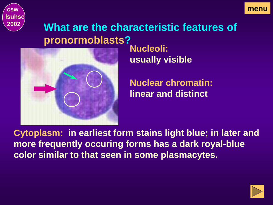
Nucleoli:
usually visible
Nuclear chromatin:
linear and distinct
Cytoplasm: in earliest form stains light blue; in later and
more frequently occuring forms has a dark royal-blue
color similar to that seen in some plasmacytes.

What are characteristic features of
basophilic normoblasts (prorubricytes)?
csw
lsuhsc
2002
menu
Nucleoli:
ill-defined or absent
Nuclear chromatin:
coarsening of the
chromatin pattern
Cytoplasm: deeply basophilic due to the abundance of
RNA with a reddish tinge produced by varying amounts
of hemoglobin present
& some cells may have a Golgi (clear) area adjacent
to the nucleus (not visible in this cell).
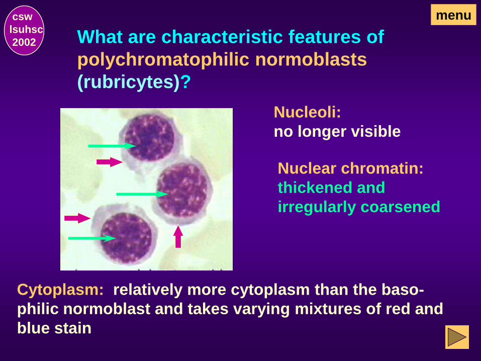
What are characteristic features of
polychromatophilic normoblasts
(rubricytes)?
csw
lsuhsc
2002
menu
Nucleoli:
no longer visible
Nuclear chromatin:
thickened and
irregularly coarsened
Cytoplasm: relatively more cytoplasm than the baso-
philic normoblast and takes varying mixtures of red and
blue stain

What are characteristic features of orthochro-
matic normoblasts (metarubricytes)?
csw
lsuhsc
2002
menu
Nucleoli: none
Nuclear chromatin:
nonlinear clumped
structure or, as shown in
this field, a solid reddish-
blue-black degenerated
nucleus
Cytoplasm: predominantly red cytoplasm with minimal
amounts of residual blue

What are characteristic features of poly-
chromatophilic erythrocytes (diffusely
basophilic erythrocytes)?
csw
lsuhsc
2002
menu
As the cell matures, the nucleus is
extruded and the cell becomes a
polychromatophilic erythrocyte
(erythrocytes do not have a nucleus)
Cytoplasm: predominantly red but may have a bluish
tinge due to the reticulum strands (RNA) still present.
orthochromatic erythroblast
(erythroblasts have a nucleus)

What is the correct name for this cell on
a Wright’s stained blood smear?
csw
lsuhsc
2002
menu
On a Wright’s stained blood smear,
the cell is called a polychromatophilic
erythrocyte.
When these cells are stained
with a supravital stain (e.g., new
methylene blue),
the residual RNA strands are
precipitated,
and the cell is then called a reticulocyte.

What are characteristic features of
normal mature erythrocytes?
csw
lsuhsc
2002
menu
Normal mature erythrocytes are anucleated
biconcave discs that stain a reddish buff color
with Wright’s (or Wright’s-Giemsa) stain and have
a small (about 1/3 of the cell) central pallor.
The intensity of the stain in
the center of the cell (i.e.,
the thin portion)
is less than at the outer rim
of the cell (i.e., the thicker
portion).

How are RBC classified as to size? csw
lsuhsc
2002
menu
Normocytic is the term used
to indicate RBC that are
normal size (6-8 m in
diameter) and normal shape.
Anisocytosis is a “generic” term
used to indicate a variation in
cell size, eg.,
microcytic macrocytic normocytic X
X
X X

Individual red cells can be classified
as…
csw
lsuhsc
2002
menu
Macrocytic (RBC > 8 m in diameter).
normal 6-8m <6m
>8m
Microcytic (RBC < 6 m in diameter).
Normocytic (RBC 6-8 m in diameter).

Depending upon the predominant cell
size, an RBC population can be
classified as…
csw
lsuhsc
2002
menu
Normocytic Microcytic Macrocytic

csw
lsuhsc
2002
menu Illustrative Mature Erythrocytes
microcytic
lymph
Comparison of erythrocytes with the
normal small mature lymphocyte
(which is about 6-10m in diameter) is
helpful in determining whether cells
are normocytic, microcytic, or
macrocytic. lymph
normocytic
lymph
macrocytic

How are RBC classified as to shape?
csw
lsuhsc
2002
menu
Normocytic is the term
used to indicate RBC
that are normal size
(6-8 m in diameter) and
normal shape (i.e., round,
biconcave).
Poikilocytosis is the
“generic” term used to
indicate variation in
shape.
Individual red cells can have numerous abnormal shapes, eg:
6-8m round, biconcave
What are some of the RBC
shape classifications?
Individual red cells can be classified as:
ovalocytes (elliptocytes)
spherocytes
target cells (leptocytes)
schistocytes (RBC fragments)
csw
lsuhsc
2002
menu

csw
lsuhsc
2002
menu
ovalocytes (elliptocytes) spherocytes
target cells schistocytes
Illustrative RBC Shapes

csw
lsuhsc
2002
menu
Schistocytes are RBC fragments and may have a variety
of shapes.
Schistocytes

What other erythroid shapes
can be seen?
csw
lsuhsc
2002
menu
Individual red cells can also be classified as:
echinocytes or crenated
sickle cells (trepanocytes or meniscocytes)
bitocytes (keratocytes)
acanthocytes

csw
lsuhsc
2002
menu
sickle cells (drepanocytes) bitocytes (keratocytes)
spiculated (acanthocytes) crenated (echinocytes)
Illustrative RBC Shapes

What about groups of RBC?
csw
lsuhsc
2002
menu
Groups or clumps of red cells can also be
classified as:
rouleaux
agglutination

Illustrative rouleau RBC formation: csw
lsuhsc
2002
Rouleau is an aggreation of RBC that is
aligned one upon the other resembling
stacks of coins and is caused by
elevated plasma fibrinogen or globulins.
This phenomenon causes an increased erythrocyte sedi-
mentation rate (ESR) and interferes with the hemogram
parameters. Rouleau is especially characteristic of para-
proteinemia (monoclonal gammopathy), in which case
plasma cells may also be seen.
menu

Illustrative agglutinated RBC: csw
lsuhsc
2002
Agglutination of red cells,
caused by cold agglutinins,
resembles rouleau but is
more irregular and may
appear in round clumps
rather than linear rouleau.
The large cell in the field is a degenerated neutrophil with
pyknotic nuclei and nuclear fragments.
menu

How are mature RBC classified as
to hemoglobin content?
csw
lsuhsc
2002
menu
Depending upon the hemoglobin content, mature RBC
may be classified as :
anucleated, pinkish cytoplasm
with a small central pallor
(about 1/3 of the cell diameter).
normochromic
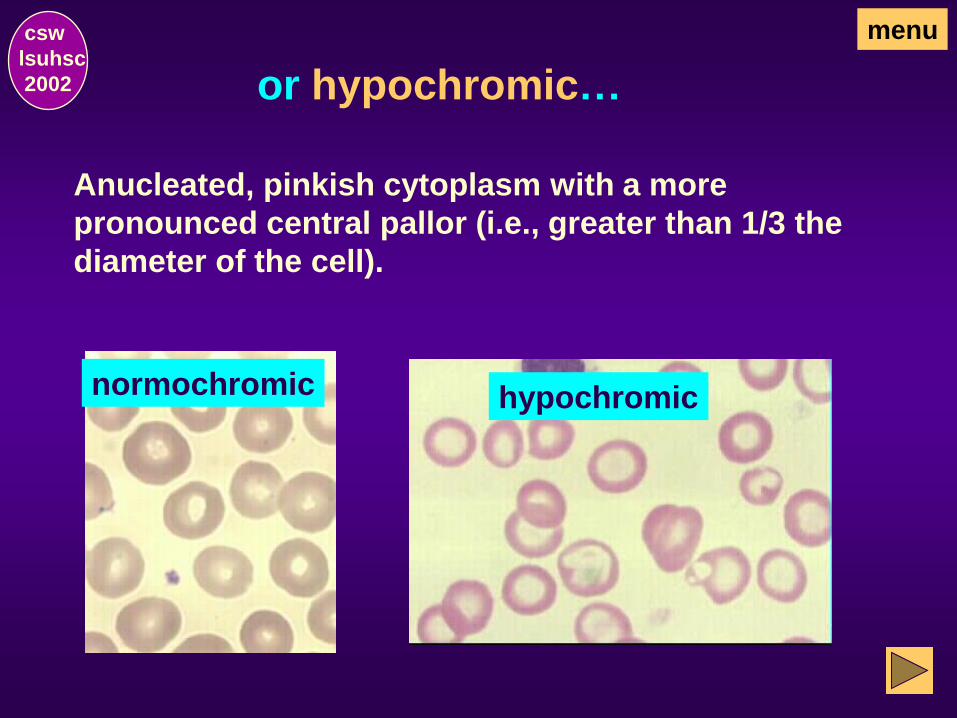
or hypochromic…
csw
lsuhsc
2002
menu
Anucleated, pinkish cytoplasm with a more
pronounced central pallor (i.e., greater than 1/3 the
diameter of the cell).
normochromic hypochromic
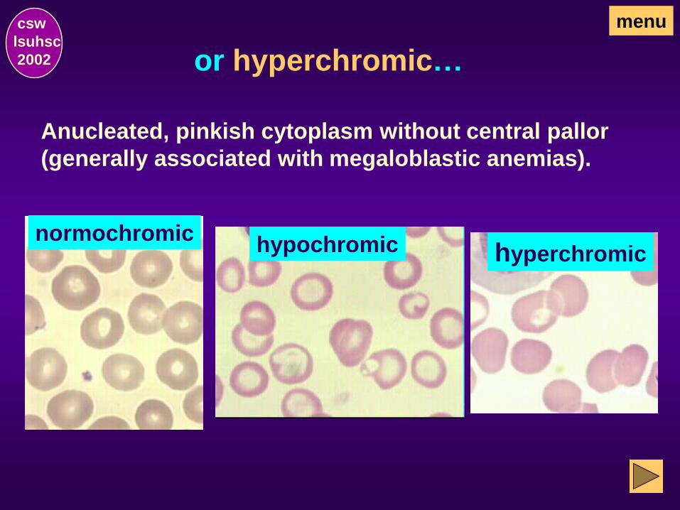
or hyperchromic…
csw
lsuhsc
2002
menu
Anucleated, pinkish cytoplasm without central pallor
(generally associated with megaloblastic anemias).
hyperchromic normochromic
hypochromic

csw
lsuhsc
2002
menu Illustrative Mature Erythrocytes
Normochromic (Normocyte)
lymph
Microcytic Hypochromic
lymph
Macrocytic Hyperchromic
lymph
Remember, comparison of the RBC
with normal small mature lympho-
cytes is helpful in classifying them
as normocytic, microcytic, or
macrocytic.

Cellular inclusions that may be found in
erythrocytes may include:
How are RBC inclusions classified?
csw
lsuhsc
2002
menu
Hemoglobin C crystals
Howell-Jolly bodies
Pappenheimer bodies
Cabot rings
Basophilic stippling
Heinz bodies

Illustrative RBC with inclusions:
HbC crystals basophilic
stippling
Howell-Jolly body
Pappenheimer bodies
Heinz bodies
Cabot rings
menu csw
lsuhsc
2002
(supravital stain)

Hemoglobin C Crystals csw
lsuhsc
2002
menu
Hexaganol shaped HbC crystals may be seen in
homozygous HbC disease but are not seen in
heterozygous HbC trait. The crystals may be
intracellular or extracellular.
Target cells are characteristically
seen in HbC disease and syndromes
and may be the only abnormality in
heterozygous HbC.

Hemoglobin C Crystals, cont’d csw
lsuhsc
2002
menu
Other shaped forms of the crystals (e.g.,
glove shaped) are seen in HbSC disease.

Basophilic Stippling csw
lsuhsc
2002
menu
Irregular basophilic granules, which may be coarse or fine,
dispersed throughout an erythrocyte is called basophilic
stippling. This finding is attributed to abnormal instability
of the residual RNA in the cell.
Fine stippling is commonly seen when
there is increased polychromatophilia,
and, therefore, with increased produc-
tion of red cells.
Coarse stippling may be seen in;
• lead poisoning or other diseases with impaired
hemoglobin synthesis
• megaloblastic anemia
• other forms of severe anemia (eg, thalassemia major,
sickle cell disease

Howell-Jolly Bodies csw
lsuhsc
2002
menu
Howell-Jolly bodies are smooth,
round, intracellular remnants of
nuclear chromatin (DNA) that may be
found in erythrocytes.
Their color may vary with the stain but are
usually the same color as the nuclei of
polychromatophilic erythroblasts.
Single Howell-Jolly bodies may be seen in megaloblastic
anemia, hemolytic anemia, hemoglobinopathies, thalas-
semia major, and after splenectomy.
Multiple Howell-Jolly bodies in a single cell is usually
indicative of abnormal erythropoiesis (e.g., megalo-
blastic anemia).

Looks Like Howell-Jolly Bodies csw
lsuhsc
2002
menu
Don’t confuse Howell-Jolly bodies with
platelets on top of a red cell.
Characteristically, platelets will appear to be surrounded
by a clear “halo” where the hemoglobin has been
displaced.
Howell-Jolly bodies usually have no halo.

Pappenheimer bodies: csw
lsuhsc
2002
menu
Pappenheimer bodies are few in a given
red cell and are usually clustered at the
edge of the cell membrane.
These cells are called siderocytes when observed
after staining with an iron stain (e.g., Prussian blue).
Pappenheimer bodies appear as dark blue intracellular inorganic iron-containing granules when seen on Wright-Giemsa stained blood smears.
Wright-Giemsa stain
Prussian Blue stain

Pappenheimer bodies are associated with
iron-loading disorders.
Wright-Giemsa stain
Pappenheimer bodies, cont’d. csw
lsuhsc
2002
menu
When Pappenheimer bodies are seen in peripheral
blood, there may be a concomitant increase of
siderocytes and sideroblasts in the bone marrow.
When the siderotic granules surround
at least 2/3 of the circumference of
the nucleus, the cell is called a
“ringed sideroblast”.
Prussian Blue stain
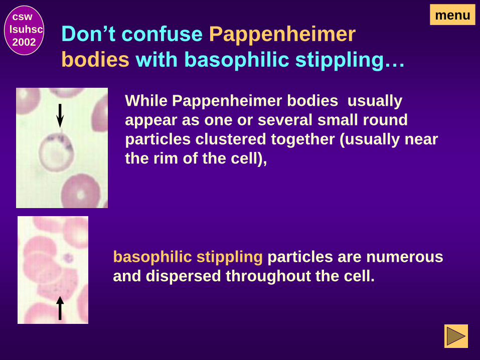
Don’t confuse Pappenheimer
bodies with basophilic stippling…
csw
lsuhsc
2002
menu
While Pappenheimer bodies usually
appear as one or several small round
particles clustered together (usually near
the rim of the cell),
basophilic stippling particles are numerous
and dispersed throughout the cell.

Heinz bodies are not visualized on Wright’s stained
blood smears but are seen only after staining with
supravital dyes. Even with these stains, exposure to
an oxidizing drug is often required before they are
detected.
Heinz Bodies csw
lsuhsc
2002
menu
Supravital stain

Wright’s Stain With removal of the Heinz body
by the spleen, the cells
observed on Wright-Giemsa
stained blood smears appear to
have had a bite taken out of the
cell membrane and are called
keratocytes (or “bitocytes”).
Heinz Bodies,cont’d csw
lsuhsc
2002
menu
Heinz bodies are most frequently associated with G6PD
and may be seen in hemolytic anemias and drugs such
as phenacetin. They may also be associated with
thalassemia major and hemoglobinopathies.
Cabot Rings csw
lsuhsc
2002
menu
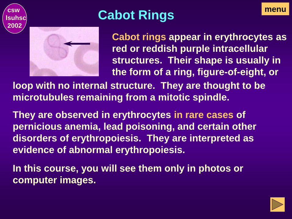
Cabot rings appear in erythrocytes as
red or reddish purple intracellular
structures. Their shape is usually in
the form of a ring, figure-of-eight, or
loop with no internal structure. They are thought to be
microtubules remaining from a mitotic spindle.
They are observed in erythrocytes in rare cases of
pernicious anemia, lead poisoning, and certain other
disorders of erythropoiesis. They are interpreted as
evidence of abnormal erythropoiesis.
In this course, you will see them only in photos or
computer images.
RBC Inclusions – Composition & Stains
csw
lsuhsc
2002
menu
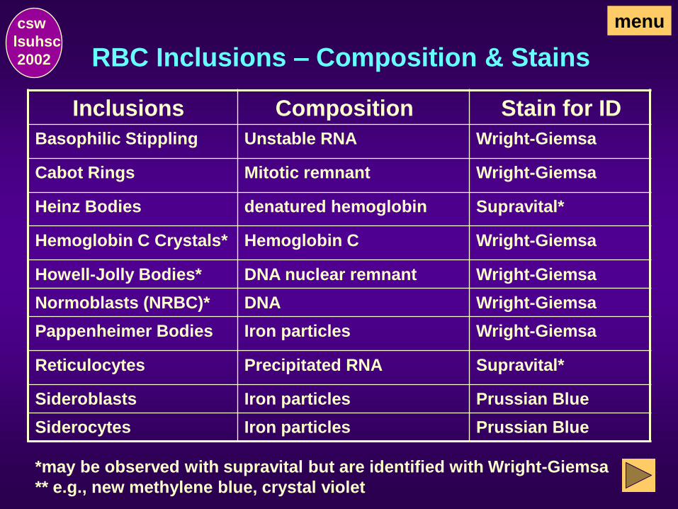
Inclusions Composition Stain for ID
Basophilic Stippling Unstable RNA Wright-Giemsa
Cabot Rings Mitotic remnant Wright-Giemsa
Heinz Bodies denatured hemoglobin Supravital*
Hemoglobin C Crystals* Hemoglobin C Wright-Giemsa
Howell-Jolly Bodies* DNA nuclear remnant Wright-Giemsa
Normoblasts (NRBC)* DNA Wright-Giemsa
Pappenheimer Bodies Iron particles Wright-Giemsa
Reticulocytes Precipitated RNA Supravital*
Sideroblasts Iron particles Prussian Blue
Siderocytes Iron particles Prussian Blue
*may be observed with supravital but are identified with Wright-Giemsa
** e.g., new methylene blue, crystal violet

Nucleated Red Blood Cells (NRBC)
csw
lsuhsc
2002
menu
NRBC are not normally present in peripheral blood of
adults. They may be seen normally in the peripheral
blood of newborns and in some diseases in adults. The
NRBC most commonly seen is the orthochromatophilic
erythroblast.
However, less mature stages may also
be seen.

How are immature RBC classified? csw
lsuhsc
2002
menu
Immature RBC precursors are classified as:
Normocytic - when they are of normal size.
Microcytic - when they are smaller than normal.
Macrocytic Non-megaloblastic - when they are
larger than normal with synchronized nucleus and
cytoplasm maturation (i.e., normoblastic bone
marrow).
Megaloblastic - when they are larger than normal (i.e.,
macrocytic) and have asynchronized nucleus and
cytoplasm maturation (I.e., megaloblastic bone
marrow).

Characteristic nuclear/cytoplasmic fea-
tures at various stages of RBC maturation.
proerythroblast
(earliest form w/ nucleoli)
basophilic erythroblast
(ill-defined or absent nucleoli)
polychromatophilic
erythroblast
(cytoplasmic evidence of HGB)
orthochromatophilic
erythroblast
(last stage before
extrusion of nucleus)
menu csw
lsuhsc
2002

Characteristic nuclear/cytoplasmic fea-
tures of megaloblastic RBC maturation.
The megaloblastic precursors are much larger
than normal with asynchrony between the nucleus
and cytoplasm maturation. The cytoplasm devel-
ops at the normal rate while the nucleus lags
behind.
Therefore, it is difficult to assign a specific stage
of development for an individual cell. For
example, the nuclear features may be consistent
with a basophilic megaloblast while the cytoplasm
may be more mature and be consistent with a later
stage.
menu csw
lsuhsc
2002

Examples of nuclear/cytoplasmic asyn-
chrony in megaloblastic precursors:
The very large early megaloblastic precurs-
or has a nucleus consistent with a pronorm-
oblast,
the more mature basophilic erythroblast
with visible evidence of hemoglobin (i.e.,
pink tinges).
but the cytoplasm is consistent with
The nucleus of this megaloblast is consis-
tent with a basophilic erythroblast …….
but the cytoplasm is more consistent
with a polychromatophilic erythroblast
with varying mixtures of red and blood
stain.
menu csw
lsuhsc
2002

Other examples of megaloblastic
precursors:
This nucleus is consistent with the thick-
ened and irregularly coarsened chromatin
of the polychromatophilic erythroblast ...
while the cytoplasm is predominantly red
with minimal amounts of residual blue that
is more consistent with the orthochromatic
erythroblast.
Another megaloblast in which the nucleus
looks consistent with a polychromatophilic
erythroblast ….
while the cytoplasm is already as mature
looking as the anucleated polychromato-
philic erythrocyte.
menu csw
lsuhsc
2002

More mature megaloblastic cells: menu csw
lsuhsc
2002
Polychromatophilic erythrocytes and mature RBC
are larger than normal (i.e., macrocytic) and
typically seen are:
macrocytes without central pallor
macroovalocytes
macro tear-drops
Abnormal Erythrocytes
Terminology (Definitions)
csw
lsuhsc
2002

Abnormal RBC are differentiated and
identified as part of the “diff”.
csw
lsuhsc
2002
Changes in size, shape, hemoglobin content,
and/or appearance of cellular inclusions may
occur as a result of a disease process. Such
changes are noted as part of the “diff”.
What terminology is used to indicate the
presence of abnormal red cells?
menu

Definitions: csw
lsuhsc
2002
Hypochromic erythrocytes that demonstrate a
central pale area that becomes larger
and paler as the hemoglobin content
diminishes (less than 1/3 of cell
diameter.
normochromic hypochromic
menu

csw
lsuhsc
2002
Anisochromic
or dimorphic
indicates the presence of both
normochromic and hypochromic
cells in the same blood film.
normochromic anisochromic
or dimorphic
normochromic
hypochromic
menu
Definitions:
csw
lsuhsc
2002
Polychromasia and polychromatophilia
view cells
changeable terms used to indicate the increased presence
of non-nucleated immature erythrocytes (polychromato-
philic erythrocytes) that contain residual RNA which gives
a blue-gray tint to the red cells. These cells, which remain
after ejection of the nucleus from the orthochromatic
erythroblast, are slightly larger than mature erythrocytes.
After exposure to a supravital stain, the cytoplasmic
organelles of these cells clump into an easily recognized
blue-staining reticulum and the cell is called a
reticulocyte.
are inter-
menu
Definitions:

continued: csw
lsuhsc
2002
Polychromasia (polychromatophilia)
normochromic
polychromatophilic
erythrocytes
menu
X
X
X
X

normocytic
RBC 6-8m diameter
csw
lsuhsc
2002
Microcytes are abnormally small erythrocytes (i.e.,
less than 6 m in diameter).
microcytic
(predominant)
menu
Definitions:
Compare with lymphocyte nuclei ( 8-10 m in diameter).

csw
lsuhsc
2002
Macrocytes are abnormally large erythrocytes (i.e.,
greater than 8 m in diameter.
normocytic
RBC 6-8m diameter
lymphocyte (with nuclei about 8-10 m in diameter)
macrocytic
menu
Definitions:

csw
lsuhsc
2002
Anisocytosis is a “generic” term used to indicate an
abnormal variation in size of
erythrocytes.
normocytic
RBC 6-8m diameter normocytic
microcytic
macrocytic
menu
Definitions:

csw
lsuhsc
2002
Poikilocytosis
normocytic
RBC round biconcave
is a “generic” term used to indicate
variation in shape of erythrocytes (e.g., oval, pear-
shaped, teardrop-shaped, saddle-shaped, helmet-
shaped, sickle-shaped, and irregularly shaped), eg:
RBC variable shapes
menu
Definitions:

csw
lsuhsc
2002
Elliptocytes
and
Ovalocytes
are interchangeable terms used to
indicate ovalshaped erythrocytes.
normocytic
RBC round biconcave
RBC predominantly
ovalocytes
x x x x
x
x
x x x
x x x x
x
x
x
x
x x
x
x x
x
x
menu
Definitions:

normocytic
round,
biconcave
RBC
spherocytes
csw
lsuhsc
2002
Spherocytes
x
x
x
x
x x
x x
x x x
menu
Definitions:
are nearly spherical erythrocytes which
usually have a diameter smaller than normal. They
lack the central pale area due to their spherical shape.

normocytic
round,
biconcave
RBC
target cells
csw
lsuhsc
2002
Target cells (leptocytes)
menu
Definitions:
thinner than normal which show a peripheral rim of
hemoglobin with a dark central hemoglobin-containing
area. A pale unstained ring containing less hemoglobin
separates the central and peripheral zones and gives the
cell a target appearance.
are erythrocytes that are

normocytic
round,
biconcave
RBC
schistocytes
csw
lsuhsc
2002
Schistocytes
menu
Definitions:
are fragmented red cell segments
that are the result of some hemolytic process. The
segments can be a variety of shapes but helmet cells
and triangularly-shaped cells are particularly
characteristic.
csw
lsuhsc
2002
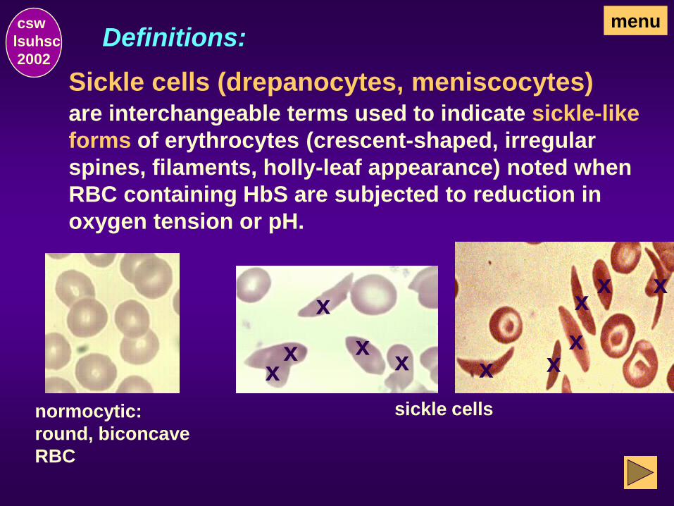
Sickle cells (drepanocytes, meniscocytes) are interchangeable terms used to indicate sickle-like
forms of erythrocytes (crescent-shaped, irregular
spines, filaments, holly-leaf appearance) noted when
RBC containing HbS are subjected to reduction in
oxygen tension or pH.
normocytic:
round, biconcave
RBC
menu Definitions:
x x x
x
x x
x x x
x x
sickle cells

csw
lsuhsc
2002
Keratocytes or “Bitocytes” interchangeable
terms used to indicate irregularly contracted erythro-
cytes which stain densely and have contraction of
hemoglobin from a part of the cell membrane, thereby
giving the appearance that a “bite” has been taken out
of the cell. These cells are thought to be cells from
which Heinz bodies have been removed by the spleen.
menu
Definitions:
normocytic
RBC, round,
biconcave
keratocytes
csw
lsuhsc
2002
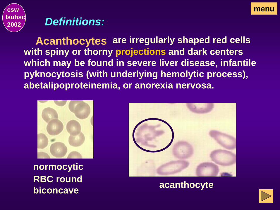
Acanthocytes
normocytic
RBC round
biconcave
are irregularly shaped red cells
with spiny or thorny projections and dark centers
which may be found in severe liver disease, infantile
pyknocytosis (with underlying hemolytic process),
abetalipoproteinemia, or anorexia nervosa.
menu
Definitions:
acanthocyte

csw
lsuhsc
2002
Crenated red blood cells
normocytic
RBC round
biconcave
are uniformly shrunken
red cells with uniform irregular, wrinkled cell membranes.
Their presence is frequently an artifact of storage and all
red cells in the field are usually affected. (By contrast,
ecinocytes are intermixed with normal red cells.)
crenated RBC
menu
Definitions:
X
X X
X X
X

csw
lsuhsc
2002
Echinocytes
normocytic
RBC round
biconcave
are irregularly shaped red cells with
spiny projections and preserved central pallor. While
their presence may be an artifactual phenomenon,
they may be seen in liver and renal disease,
hyperlipidemia, and red blood cell enzymopathies.
echinocyte
menu
Definitions:

csw
lsuhsc
2002
Rouleaux formation describes an aggregation
of erythrocytes that are aligned one upon the other,
resembling stacks of coins, caused by elevated plasma
fibrinogen or globulins. This phenomenon causes an
increased erythrocyte sedimentation rate. This finding
is especially characteristic of paraproteinemia
(monoclonal gammopathy).
rouleau
normal
menu
Definitions:

csw
lsuhsc
2002
Agglutination of red cells is caused by
agglutinins and resembles rouleaux but is more
irregular with round clumps rather than linear
rouleaux.
agglutination
of red cells
normal
menu
Definitions:

csw
lsuhsc
2002
Hemoglobin C crystals
Normocytic RBC
(round,
biconcave,
without
inclusions)
are hexagonal crystals
that may be found in individuals with HbC syndromes.
The crystals may be intracellular or extracellular.
intracellular
HbC crystals extracellular
HbC crystal
menu
Definitions:

basophilic
stippling (fine)
csw
lsuhsc
2002
Basophilic stippling
normocytic
RBC round
biconcave
is the term used to indicate
the presence of irregular basophilic granules in the
cytoplasm of erythrocytes. The granules are composed
of unstable RNA and may be fine or coarse.
basophilic
stippling (coarse)
menu
Definitions:
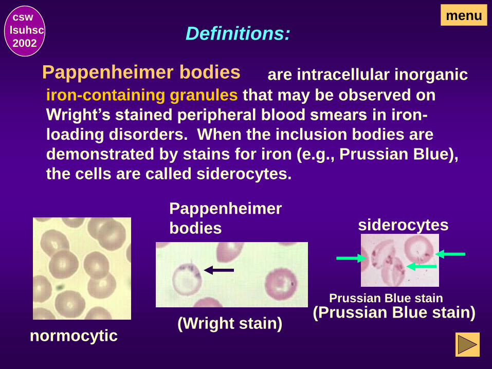
Pappenheimer
bodies
(Wright stain)
csw
lsuhsc
2002
Pappenheimer bodies
normocytic
are intracellular inorganic
iron-containing granules that may be observed on
Wright’s stained peripheral blood smears in iron-
loading disorders. When the inclusion bodies are
demonstrated by stains for iron (e.g., Prussian Blue),
the cells are called siderocytes.
menu
(Prussian Blue stain)
Definitions:
siderocytes
Prussian Blue stain

csw
lsuhsc
2002
Howell-Jolly bodies
normocytic
are intracellular particles
which are smooth, round remnants of nuculear
chromatin (DNA). Usually, only one per cell is seen
but, occasionally, there may be more than one.
Howell-Jolly
body (single)
Howell-Jolly
body (multiple)
image
pending
menu
Definitions:

csw
lsuhsc
2002
Nucleated red blood cells (NRBC) are precursors of the non-nucleated mature red cells,
usually orthochromatic erythroblasts when noted in
peripheral blood in disease states but earlier forms may
also be seen,eg:
mature RBC
orthochromatic
erythroblast polychromatophilic
erythroblast
basophilic
erythroblast proerythroblast
menu Definitions:

csw
lsuhsc
2002
Reticulocytes are anucleated slightly immature
erythrocytes, identified as polychromatophilic erythro-
cytes on Wright stained smears.
reticulocyte
(supravital stain)
The cells are identified as reticulocytes only after
exposure to a supravital stain which causes the
cytoplasmic organelles of the cells to clump into an
easily recognized blue-staining reticulum.
polychromatophilic erythrocyte
(Wright’s stain)
menu
Definitions:

Platelets
csw
lsuhsc
2002
lymphocyte
Platelets menu csw
lsuhsc
2002
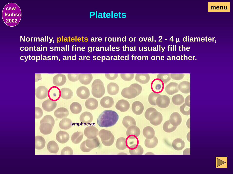
Normally, platelets are round or oval, 2 - 4 m diameter,
contain small fine granules that usually fill the
cytoplasm, and are separated from one another.

Estimated Platelet Count menu csw
lsuhsc
2002
An estimated platelet count can be made on a peripheral
blood smear. If the platelet count is normal, an average
of about one platelet per 10 to 30 red blood cells. Using
the oil immersion lens at 1000x magnification, that is
about 5 to 25 platelets per field.
In Clinical Pathology 201, < 5 platelets/oil immersion
field will be considered decreased and > 25 platelets/oil
immersion field will be considered increased.

Platelets menu csw
lsuhsc
2002
Platelet clumps may be found on blood smears that
have been improperly prepared. They may also be seen
in clotting disorders. Platelet estimates cannot be made
from blood smears with platelet clumps.
Platelets menu csw
lsuhsc
2002
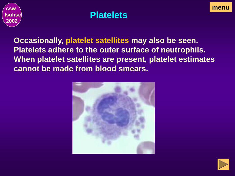
Occasionally, platelet satellites may also be seen.
Platelets adhere to the outer surface of neutrophils.
When platelet satellites are present, platelet estimates
cannot be made from blood smears.
Platelets menu csw
lsuhsc
2002
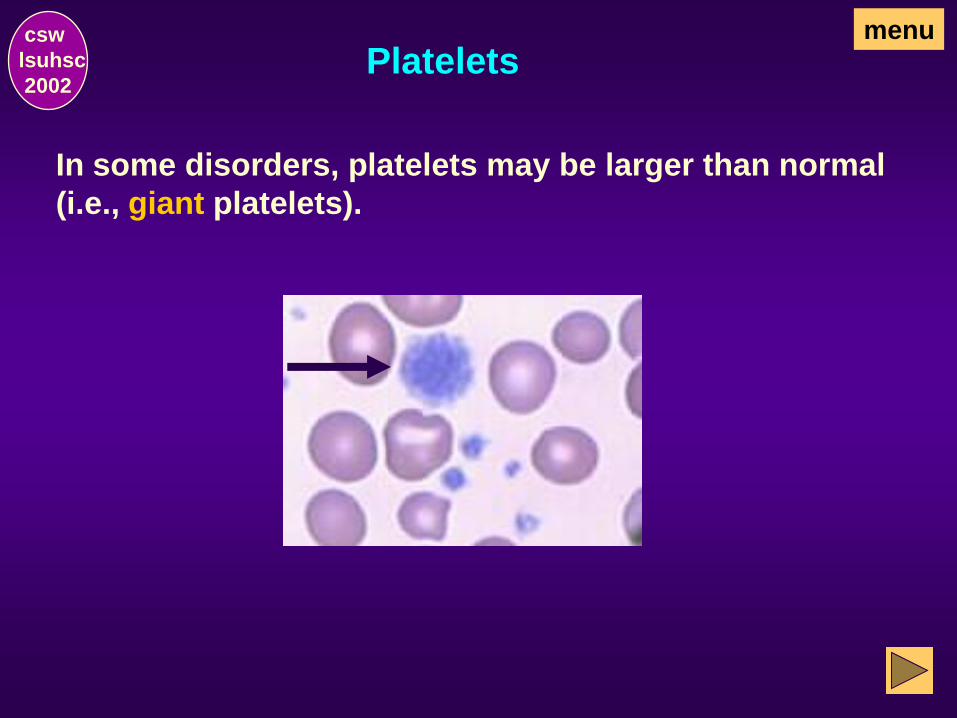
In some disorders, platelets may be larger than normal
(i.e., giant platelets).

Disorders
Characteristic Morphology
csw
lsuhsc
2002

Macrocytic Non-Megaloblastic Anemias
menu csw
lsuhsc
2002
Characteristic abnormalities associated with macrocytic
non-megaloblastic anemias in diseases associated with
reticulocytosis.
lymphocyte
normal numerous polychromatophilic
erythrocytes like the one
indicated by the arrow

Macrocytic Non-megaloblastic Anemia menu csw
lsuhsc
2002
Characteristic abnormalities associated with macrocytic
non-megaloblastic anemia in liver disease.
macrocytes
and
target cells

Macrocytic Megaloblastic Anemias menu csw
lsuhsc
2002
Examples of characteristic abnormalities associated
with megaloblastic macrocytic anemias
macrocytes
and
macro-tear drops
and
macro-ovalocytes
and erythrocytic precursors
with asynchrony in nuclear
and cytyplasm maturation
Normocytic RBC
and hypersegmented PMN

Microcytic Hypochromic Anemias
menu csw
lsuhsc
2002
Characteristic abnormalities associated with microcytic
hypochromic anemias (eg, iron deficiency, chronic
disease).
lymphocyte
predominant
cells are
microcytic
hypochromic
erythrocytes

Anemia in Sickle Cell Disease menu csw
lsuhsc
2002
Characteristic abnormalities associated with anemia in
sickle cell disease
Sickled cells (may be
crescent-shaped,
irregular spines,
filaments, holly-leaf
appearance)
and target cells
and also
NRBC Howell-Jolly
body
Pappenheimer
bodies
basophilic
stippling polychromasia

THE END
csw
lsuhsc
2002
Click on review to return to the main menu.
Click on to exit the program. quit