two earclips, wires, a little box and presto - the wonderful world of cranio-electro...
TRANSCRIPT

Mind Alive Inc Page 1 Copyright 2014
Two Earclips, Wires, a Little Box and Presto - the Wonderful World of Cranio-electro Stimulation
- by Dave Siever, C.E.T. 2014
Introduction
Cranio-Electro Stimulation (CES) is primarily a brain-calming technique which delivers small pulses of electrical current through the brain. CES is a subset of transcutaneous Electro-Neural Stimulation (TENS). TENS refers to any form of electrical stimulation that is delivered through the skin. Typically, TENS consists of a fairly strong pulse and is used to contract muscles. CES, on the other hand, involves a much weaker pulse and is typically applied bilaterally across the cranium via the placement of two small electrodes (one on each side of the head). Most studies have used electrode placements on the mastoid process, the ear lobes or temporal lobes. A few devices have also placed one electrode on the head and the other on a shoulder or arm. CES devices employ alternating currents (AC) in audio frequencies typically from 0.5 to 100 Hz, and one device uses 15 KHz. There are various theories as to how CES affects the brain. The most popular are that a direct action is enacted on the brain via the brain stem; the limbic system; the reticular activating system; and/or the hypothalamus, and this in turn affects neurotransmitter production and possibly the default-mode network.
History
The roots of simple brain electro-stimulation span back as far as 129 AD, when the philosopher and physician named Galen used eels to provide electric shocks to treat a variety of ailments including melancholia, depression and epilepsy (Kneeland & Warren, 1994). In 43 AD, Scribonius Largus, a Roman physician, used torpedo fish (also a type of electric eel), to treat various ailments including headache and gout (Kneeland & Warren, 1994). Modern CES research was begun by Leduc and Rouxeau in France in 1902 (McClintic, J., (1978). CES was initially studied for insomnia and was called electrosleep therapy (Gilula & Kirsch, 2005). In 1949, the Soviet Union expanded research of CES to include the treatment of anxiety as well as sleeping disorders. To date, approximately 40 CES devices have been marketed in the USA, Canada and Europe. About 200 studies have been published that cover a wide variety of clinical applications including improved drug abstinence and cognitive functioning in recovering alcoholics and drug users. On the regulatory side, the Food and Drug Administration (FDA) recognizes CES as a treatment for anxiety, depression and sleep (serotonin effect) but surprisingly not for pain (endorphin effect) even though the endorphin effect and efficacy for pain treatment have been well documented. Research suggests that CES affects the brain via the reticular activating system and/or the hypothalamus (Gibson & O’Hair, 1987; Brotman, 1989).
About Electricity and Nerve Physiology
To understand how transcranial electrical stimulation works, it’s important to know the basics of nerve and neuron physiology.
Mind Alive Inc Page 2 Copyright 2014
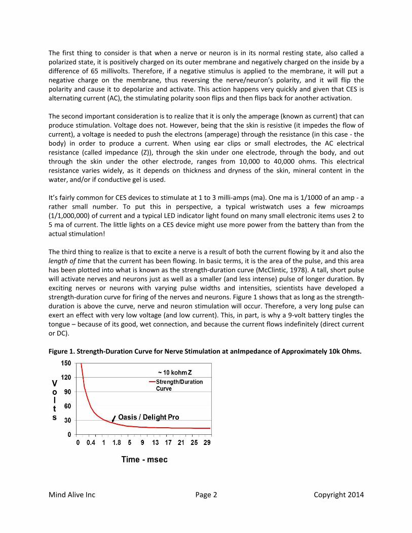
The first thing to consider is that when a nerve or neuron is in its normal resting state, also called a polarized state, it is positively charged on its outer membrane and negatively charged on the inside by a difference of 65 millivolts. Therefore, if a negative stimulus is applied to the membrane, it will put a negative charge on the membrane, thus reversing the nerve/neuron’s polarity, and it will flip the polarity and cause it to depolarize and activate. This action happens very quickly and given that CES is alternating current (AC), the stimulating polarity soon flips and then flips back for another activation. The second important consideration is to realize that it is only the amperage (known as current) that can produce stimulation. Voltage does not. However, being that the skin is resistive (it impedes the flow of current), a voltage is needed to push the electrons (amperage) through the resistance (in this case - the body) in order to produce a current. When using ear clips or small electrodes, the AC electrical resistance (called impedance (Z)), through the skin under one electrode, through the body, and out through the skin under the other electrode, ranges from 10,000 to 40,000 ohms. This electrical resistance varies widely, as it depends on thickness and dryness of the skin, mineral content in the water, and/or if conductive gel is used. It’s fairly common for CES devices to stimulate at 1 to 3 milli-amps (ma). One ma is 1/1000 of an amp - a rather small number. To put this in perspective, a typical wristwatch uses a few microamps (1/1,000,000) of current and a typical LED indicator light found on many small electronic items uses 2 to 5 ma of current. The little lights on a CES device might use more power from the battery than from the actual stimulation! The third thing to realize is that to excite a nerve is a result of both the current flowing by it and also the length of time that the current has been flowing. In basic terms, it is the area of the pulse, and this area has been plotted into what is known as the strength-duration curve (McClintic, 1978). A tall, short pulse will activate nerves and neurons just as well as a smaller (and less intense) pulse of longer duration. By exciting nerves or neurons with varying pulse widths and intensities, scientists have developed a strength-duration curve for firing of the nerves and neurons. Figure 1 shows that as long as the strength-duration is above the curve, nerve and neuron stimulation will occur. Therefore, a very long pulse can exert an effect with very low voltage (and low current). This, in part, is why a 9-volt battery tingles the tongue – because of its good, wet connection, and because the current flows indefinitely (direct current or DC). Figure 1. Strength-Duration Curve for Nerve Stimulation at anImpedance of Approximately 10k Ohms.

Mind Alive Inc Page 3 Copyright 2014
Some devices can deliver pulses above 150 volts with a very high current but for less than 0.1 millisecond (msec), which for CES, is a very short period of time. This approach has a tendency to utilize more of the capacitance of the body. Capacitance is the ability of any object, and in this case, a living body, to store a charge. As a result, this can reduce the risk of blistering an earlobe. But high voltages can push current through high impedances and might even accidentally trigger a heart contraction if the electrodes fell onto the user’s chest. The alternate approach is to use a low voltage and current, but for a longer period of time, typically a few milli-seconds. The plus side to this approach is that it is relatively safe. The down side is that there is a larger flow of electrons and it can burn and/or blister the skin if the intensity is turned up too high. Most CES devices use lower voltage-current and longer duration stimulation for the sake of safety. The Delight Pro, ALERT Pro and the Oasis Pro stimulate in the 1.5 to 2 msec range with a maximum of 30 to 40 volts and a maximum current of about 4 ma. The best scientific studies are double-blind, meaning neither the participant nor the researcher know when the real or placebo treatment are being applied. To accomplish this using CES, the real stimulation must be low enough that the participant cannot feel it and therefore differentiate it from the placebo stimulation. Double blind CES studies show excellent clinical results even when the person cannot feel the stimulation at all! CES is a powerful technique in that it does show effectiveness in double-blind studies. However, for actual clinical work, I suggest that the user feel the stimulation mildly.
The Electronics Behind CES
CES exerts its effects on the brain by presenting short negative (cathodic) electrical pulses into the cranium. The pulses alternate from side to side making the current flow back and forth. This back and forth motion constitutes alternating current (AC) in a similar fashion to how current alternates back and forth in a common power plug. Because the outside of a neuron is positively charged, a negative pulse will flip its resting state and cause an action potential to occur. This is not to be confused with transcranial DC Stimulation (tDCS), in which a direct current is applied through a group of neurons. The principles governing DC stimulation and AC stimulation are not the same. With tDCS, it’s the anode (positive) that enhances neuronal activity. With CES and TENS, it’s the negative going (cathodic) pulse that triggers action. With anodal tDCS, the voltage gradient becomes more negative as the current flows across our six layers of neurons. Given that the axons of our neurons lie away from the scalp, the axons will be more negatively charged than the soma, or neuron body. This reduces the overall membrane resting state from -65 millivolts to possibly -55 to -60 millivolts. So, whereas CES flips the resting state of a neuron, tDCS increases the probability that a neuron will fire when stimulated by adjacent axons (from other neurons) onto its dendrites (incoming branches). In Figure 2, we can see that when a stimulus is presented (a negative pulse), the other electrode provides the return path by being positive and vice versa. The polarity alternates back and forth as the electrodes take turns acting as a complete circuit: one electrode providing stimulation (negative pulse), while the other provides the return path (positive) and vice versa, as shown in Figure 2. So long as both pulses are not negative or positive at the same time, there will always be a current. In Figure 2, both sides are positive much of the time, so current flows during a downward pulse because the other circuit is positive. Figure 3 shows the same idea, but with a very long pulse. Some devices make pulses at 50% duty cycle, meaning that each electrode is negative or positive 50% of the time. This enhances the cup of wine effect, that fuzzy-headed sensation from consuming alcohol, which some people strive to experience as a therapy. Some people respond very well to this feeling, while others experience nausea

Mind Alive Inc Page 4 Copyright 2014
and must stop the stimulation. Regardless of the subjective experience, the pulses generating this effect are shown in Figure 3. In this case, the pulse has been randomized somewhat and like Figure 2, in Figure 3, the current flows only when the electrodes are of opposing polarity. This current may be flowing very little or very much of the time, depending on how the pulses are timed. Here we see that current flows ONLY during the red zones, as there is opposing polarities on the electrodes. When the polarities are the same (both positive or both negative), there is no current flow and no stimulation (no red line). Figure 2. Pulses of Short Duration at Any Given Frequency.
Figure 3. Pulses at a 50% Duty Cycle.
Household Power
To further our understanding of electricity and stimulation, let’s consider household power and what’s in a plug-in. Our household power alternates from positive to negative, back to positive and back to negative, 60 times per second, or 60 Hz AC (alternating current). If you should accidentally grab a “hot” power source, you will get a much more severe shock than from an electro-stimulator with the same

Mind Alive Inc Page 5 Copyright 2014
voltage. This is because at 60 Hz, the “stimulus” is on for about 8 msec (a very long time), then flips polarity for another 8 msec and a lot of continuous current can pass through a person. Due to the long stimulus time, a 60 Hz line shock can really excite nerves and make muscles contract harder than anyone could ever willfully contract his/her own muscles. As a result, torn muscles, heart attacks and burnt tissue are possible, depending on where the hazardous wire contacts the body, how well the connection is and how well the return connection is (reference or ground). All electricians must, by law, use non-metal ladders such as fiberglass or plastic to prevent electric shock from a current flowing through their body and into a grounded ladder (if placed on wet ground or any grounded metal) in case they were to accidentally grab a hot wire. Table 1 shows the shock hazard at varying currents of 60 AC. Table 1. 60 Hz AC Currents and Human Experience of Them. 1 ma Perception level 5 ma Mild shock felt, not painful but startling 6 - 30 ma Painful shock, but able to let go if through the hand 50 - 150 ma Extreme pain, unable to breathe, severe muscle contractions 1000 - 4000 ma Mild skin and internal tissue burns. Heart goes into ventricle fibrillation (heart flutters but does not pump blood) 5000 ma and up Cardiac arrest So, it’s not the voltage that does harm, but rather the amperage and the length of time that the amperage is present. With a low voltage, a better connection is needed to get the amperage through. A high voltage, however, can push an excessive current through the body even if the connection is poor.
CES Physiological Studies Methodology
Many clinicians experience confusion about the various frequencies and wave forms used with CES because some manufacturers create new buzz words for the sake of marketing. CES manufacturers generally use frequencies somewhere in the 0.1 to 40 Hz range plus 100 Hz and one device uses 15 KHz, modulated with 15 Hz and 500 Hz. However, it’s mainly the electrical nature of CES itself that produces most of the benefits. Frequency has some influence, but a minor amount. As a general guideline, it is believed that CES increases neurotransmitters at any frequency, but it is believed that 100 Hz increases serotonin more and therefore is used to reduce anxiety and depression and to improve sleep, while sub-delta (in the 0.5 to 3 Hz range) increases endorphins and therefore is used to reduce pain and promote feelings of well-being. A study by Datta, et al, 2013, used computer-based, high-resolution modelling to simulate the current flowing through the brain during CES. The modelling placed some of the current flowing through the temporal lobes (which makes sense as the temporal lobes are close to the ears where the electrodes are placed). It predicted that the bulk of the current path went right through the brain stem, in particular the medulla plus the posterior aspect of the hypothalamus (areas in red) as shown in Figure 4. This correlates with the liberation of the neurotransmitters such as serotonin, norepinephrine, dopamine and acetylcholine, which are produced within the brain stem and modulated by the hypothalamus as shown by Silverthorn, 2003, in Figure 5. Endorphins are thought to be liberated via the hypothalamus and other brain areas.

Mind Alive Inc Page 6 Copyright 2014
Figure 4. Peak electric field distribution from CES from 3 – 100 Hz.
Figure 5. Neurotransmitter Pathways Originating From the Brain Stem
Frequency Effects on Brain Waves
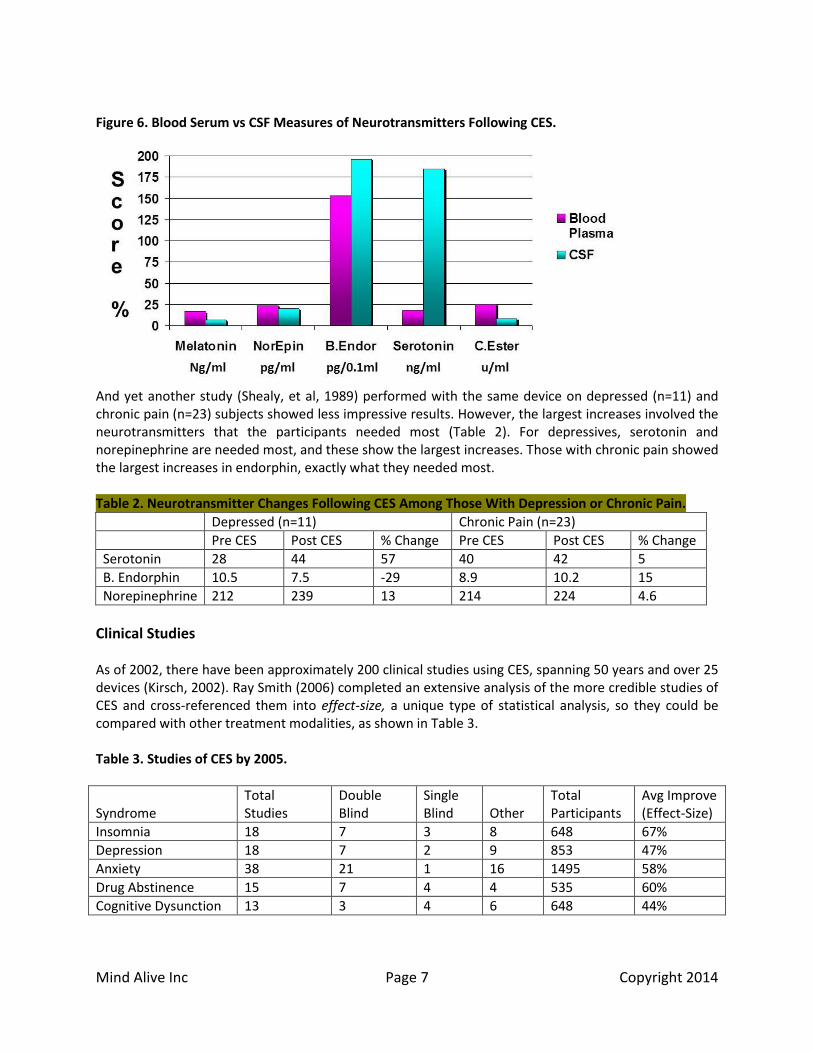
A Quantitative EEG (QEEG) study by Kennerly (2004) of 72 participants receiving 0.5 Hz (n=38) and another group receiving 100 Hz (n=34) found that both groups showed increases in alpha with decreased delta and beta activity. But the 0.5 Hz Group showed decreases in a wider range of delta EEG, whereas the 100 Hz Group showed decreases in a wider range of beta EEG (serotonin effect). This is consistent with the literature that CES at 100 Hz is more effective at increasing relaxation while reducing anxiety than the sub-delta frequencies. CES primarily modulates the brain via neurotransmitter production. In 1988, Shealy, et al, performed a study of the Liss Pain Suppressor (n =5) comparing blood-serum measures (drawn from blood) with that of cerebral spinal fluid (CSF) (drawn from lumbar subarachnoid space in the spine). His study was meant to prove that CSF measurements were considerably more accurate than blood serum measures. But in the process, he found that average increases of beta endorphin and serotonin production following CES were quite dramatic, as shown in Figure 6.
Mind Alive Inc Page 7 Copyright 2014
Figure 6. Blood Serum vs CSF Measures of Neurotransmitters Following CES.
And yet another study (Shealy, et al, 1989) performed with the same device on depressed (n=11) and chronic pain (n=23) subjects showed less impressive results. However, the largest increases involved the neurotransmitters that the participants needed most (Table 2). For depressives, serotonin and norepinephrine are needed most, and these show the largest increases. Those with chronic pain showed the largest increases in endorphin, exactly what they needed most. Table 2. Neurotransmitter Changes Following CES Among Those With Depression or Chronic Pain.
Depressed (n=11) Chronic Pain (n=23)
Pre CES Post CES % Change Pre CES Post CES % Change
Serotonin 28 44 57 40 42 5
B. Endorphin 10.5 7.5 -29 8.9 10.2 15
Norepinephrine 212 239 13 214 224 4.6
Clinical Studies As of 2002, there have been approximately 200 clinical studies using CES, spanning 50 years and over 25 devices (Kirsch, 2002). Ray Smith (2006) completed an extensive analysis of the more credible studies of CES and cross-referenced them into effect-size, a unique type of statistical analysis, so they could be compared with other treatment modalities, as shown in Table 3. Table 3. Studies of CES by 2005.
Syndrome
Total Studies
Double Blind
Single Blind
Other
Total Participants
Avg Improve (Effect-Size)
Insomnia 18 7 3 8 648 67%
Depression 18 7 2 9 853 47%
Anxiety 38 21 1 16 1495 58%
Drug Abstinence 15 7 4 4 535 60%
Cognitive Dysunction 13 3 4 6 648 44%

Mind Alive Inc Page 8 Copyright 2014
Drug Use Vs CES for the Treatment of Depression
Depression is a debilitating condition. Functional neuroanatomy studies of depression have shown a direct relationship with hyperactivation of the ventromedial prefrontal cortex (vmPFC) and hypoactivation of the left dorsal lateral pre-frontal cortex (dlPFC). As depression subsides, the vmPFC becomes less active while the left dlPFC becomes more active (Koenigs & Grafman, 2009). QEEGs that I have performed in my office reflect alpha activity at F3, and to a lesser extent at FP1 and FP2, in response to depression. There is a nutritional connection to depression. Shealy, et al. (1992) studied blood-serum levels of five neurochemicals (melatonin, norepinephrine, beta-endorphin, serotonin, and cholinesterase) in depressives. He found that 92% of depressives had abnormal levels in at least one of the five neurochemicals tested and 60% showed three or more abnormalities. In over half of the depressives, he found either elevated or low levels of norepinephrine/cholinesterase ratios. He also found magnesium deficiencies in 80% of depressed patients while 100% were deficient in taurine. Neurotransmitter levels also play a role in depression. Depletions in serotonin, norepinephrine and dopamine are well documented with major depressive disorder (Nutt, 2008). These widely varying factors involved in depression may well contribute in part, to the general failure of drug treatment. Because of the direct neurotransmitter effects of CES and the absence of negative side-effects, CES studies of over 1000 depressives, have shown relative success across the board. A meta-analysis study by Gilula & Kirsch (2005) of 290 depressives showed a direct comparison of CES against various depression medications. Table 4 shows the treatment-effect improvement in depression over placebo obtained from freedom-of-information data as provided to the FDA from pharmaceutical companies when seeking FDA approval. The CES data came from eight studies submitted to the FDA from Electromedical Products International, Inc., to reclassify CES from class III to class II for the treatment of depression, anxiety and insomnia. The CES studies had no reported negative side-effects, whereas the drug studies indicated side-effects ranging from interruption of liver metabolism to reactions with other medications and an increase in thoughts and behaviors related to suicide ideation. Table 4. Medication Treatments vs CES for Treating Depression.
Medication
Trials
n
Placebo Effect
Medication Effect
Proportion Placebo
%Meds Over Placebo
Prozac 5 1132 7.3 8.3 0.89 11
Paxil 12 1289 6.7 9.9 0.68 32
Zoloft 3 779 7.9 9.9 0.80 20
Effexor 6 1148 8.4 11.5 0.73 27
Serzone 8 1428 8.9 10.7 0.83 17
Celexa 4 1168 7.7 9.7 0.80 20
CES 8 290 --- --- 0.37 63
Although a table exemplifies relationships very well, there is nothing like a graph to see just how profound a relationship is. Figure 7 shows the relationship between drug treatments for depression vs CES. These show CES reduces depression better than the placebo response, which in the case of Prozac, is only 11% better than a fake pill.

Mind Alive Inc Page 9 Copyright 2014
Figure 7. Drug Treatment vs CES in the Treatment of Depression.
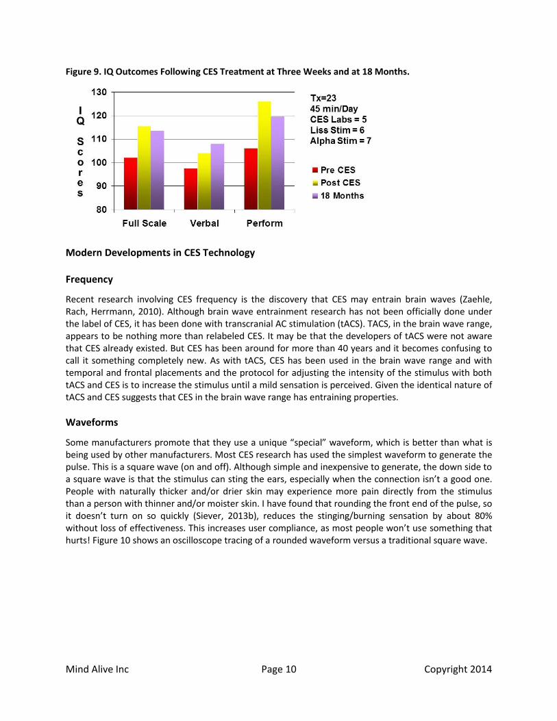
Several studies point to the success of using CES to help with cognitive dysfunction. In one study by Ray Smith (1998), he examined the emotional and IQ outcomes of 23 ADD/ADHD children following three weeks of 45 minutes of CES stimulation for five times per week and again at an 18-month follow-up with no treatment for 18 months, although some participants admitted to occasional use during the 18- month follow up, which may have influenced the outcome. However, in practical terms, this doesn’t really matter because they could use CES without negative side-effects and for no cost. Smith compared three different CES devices (CES Labs, Liss Stimulator and Alpha Stim), all with different stimulation parameters such as frequency, pulse-width and intensity, and found no statistical differences in treatment efficacy. Figure 8 shows the reduction in both state and trait anxiety and depression as measured on the IPAT Depression index and the STAI State and Trait Anxiety scales. Figure 9 shows the improvements in IQ as indicated on the WAIS-R and the WISC-R. Figure 8. Behavioral Outcomes Following CES Treatment at Three Weeks and at 18 Months.
Mind Alive Inc Page 10 Copyright 2014
Figure 9. IQ Outcomes Following CES Treatment at Three Weeks and at 18 Months.
Modern Developments in CES Technology Frequency
Recent research involving CES frequency is the discovery that CES may entrain brain waves (Zaehle, Rach, Herrmann, 2010). Although brain wave entrainment research has not been officially done under the label of CES, it has been done with transcranial AC stimulation (tACS). TACS, in the brain wave range, appears to be nothing more than relabeled CES. It may be that the developers of tACS were not aware that CES already existed. But CES has been around for more than 40 years and it becomes confusing to call it something completely new. As with tACS, CES has been used in the brain wave range and with temporal and frontal placements and the protocol for adjusting the intensity of the stimulus with both tACS and CES is to increase the stimulus until a mild sensation is perceived. Given the identical nature of tACS and CES suggests that CES in the brain wave range has entraining properties.
Waveforms
Some manufacturers promote that they use a unique “special” waveform, which is better than what is being used by other manufacturers. Most CES research has used the simplest waveform to generate the pulse. This is a square wave (on and off). Although simple and inexpensive to generate, the down side to a square wave is that the stimulus can sting the ears, especially when the connection isn’t a good one. People with naturally thicker and/or drier skin may experience more pain directly from the stimulus than a person with thinner and/or moister skin. I have found that rounding the front end of the pulse, so it doesn’t turn on so quickly (Siever, 2013b), reduces the stinging/burning sensation by about 80% without loss of effectiveness. This increases user compliance, as most people won’t use something that hurts! Figure 10 shows an oscilloscope tracing of a rounded waveform versus a traditional square wave.

Mind Alive Inc Page 11 Copyright 2014
Figure 10. Oscilloscope Tracing of a Square Waveform vs a Rounded Wave.
Another aspect of clinical effectiveness is to randomize the stimulus frequency as Heffernan (1997), who had demonstrated EEG smoothing and pain reduction following CES treatment. Figure 11 is an example of randomized frequency/time stimulation, (Siever, 2012; Siever 2013b). Figure 11. Example of Time-Frequency Randomization.
Conclusion
There is ample physiological evidence showing the effects of CES on the brain and why the clinical applications are widely varied. The best part of CES is that it is easy to use, the efficacy is quite high and the equipment is fairly inexpensive. CES is well proven and should be part of the therapeutic “tool chest” of all clinicians working with generalized cognitive challenges, affective disorders and pain, including prophylactic, acute and chronic applications.

Mind Alive Inc Page 12 Copyright 2014
References
Brotman, P. (1989). Low-intensity transcranial stimulation improves the efficacy of thermal biofeedback and quieting reflex training in the treatment of classical migraine headache. American Journal of Electromedicine, 6, (5), 120-123. Datta, A., Dmochowski, J., Guleyupoglu, B., Bikson, M., Fregni, F. (2013). Cranial electrotherapy stimulation and transcranial pulsed current stimulation: A computer based high-resolution modeling study. Neuroimage, 65, 280-287. Gibson, T., O’Hair, D. (1987). Cranial application of low level transcranial electrotherapy vs. relaxation instruction in anxious patients. American Journal of Electromedicine, 4, (1), 18-21. Gilula, M., and Kirsch, D. (2005). Cranial Electrotherapy Stimulation Review: A Safer Alternative to Psychopharmaceuticals in the Treatment of Depression. Journal of Neurotherapy, 9(2):7-26. Heffernan, M. (1997). The effect of variable microcurrents on EEG spectrum and pain control. Canadian Journal of Clinical Medicine, 4, 10, 2-8. Kennerly, R. (2004). QEEG Analysis of Cranial Electrotherapy: A Pilot Study. Journal of Neurotherapy, 8, 2, 112-113. Kirsch, D (2002). The science behind cranial electrotherapy stimulation – second edition. Medical Scope Publishing. Edmonton, Alberta, Canada. Kneeland TW , Warren CAB . (1994). Pushbutton Psychiatry: A history of electroshock in America. (Westport, CT: Praeger, 1994). Koenigs, M. & Grafman, J. (2009). The functional neuroanatomy of depression: Distinct roles for ventromedial and dorsolateral prefrontal cortex. Behavioural Brain Research, 201, 239-243. Leduc S., Rouxeau A. Influence du rythme et de la period sur la production de l'inhibition par les courants intermittents de basse tension. C.R. Seances Soc. Biol., 1903, 55, VII-X : 899-901 McClintic, J., (1978). Physiology of the Human Body, p103. John Wiley & Sons Inc., NY, NY. Nutt DJ, 2008, Relationship of neurotransmitters to the symptoms of major depressive disorder., Journal of Clinical Psychiatry, Vol:69 Suppl E1, ISSN:0160-6689, Pages:4-7. Shealy, N., Cady, R., Culver-Veehoff, D., Cox, R., Liss, S. (1988). Cerebrospinal fluid and plasma neurochemicals: Response to cranial electrical stimulation. Journal of Orthopedic Medical Surgery, 18, 94-97. Shealy, N., Cady, R., Wilkie, R., Cox, R., Liss, S. & Clossen, W. (1989). Depression: A diagnostic and Neurochemical Profile & Therapy with Cranial Electrotherapy Stimulation (CES). Journal of Neurological & Orthopaedic Medicine & Surgery, 10, 4, 319-321.

Mind Alive Inc Page 13 Copyright 2014
Shealy, N., Cady, R., Veehoff, D., Houston, R., Burnetti, M., Cox, R., & Closson, W. (1992). The neurochemistry of depression. American Journal of Pain Management, 2, 1, 13-16. Siever, D. September 11, 2012. Patent # 8,265,761. Improved Cranio-Electro Stimulator (Randomization at standard frequencies (0.5-40 Hz) and at 100 Hz). Siever, D. September 11, 2012. Patent: US 8,265,761. Improved Cranio-Electro Stimulator (Phase-shift randomization at 100 Hz.) Siever, D. (2013). Transcranial DC Stimulation. Neuroconnections, Spring Issue, 33-40. http://www.mindalive.com/1_0/article%2010.pdf. Siever, D. June 2013a. Patent: Canada 2,707,351. Improved Cranio-Electro Stimulator (Phase-shift randomization at 100 Hz.) Siever, D. December 17, 2013b. Patent: US 8,612,007B2. Cranio-Electro Stimulator (Rounded pulse and 3Hz randomization.) Silverthorn, D. U. (2003). Human Physiology: An Integrated Approach with Interactive Physiology, Third Edition, Chapter 9. As illustrated by Howard Booth, Eastern Michigan University. Pearson Education Inc., publishing as Benjamin Cummings, (2004). Smith, R. (1998). Cranial electrotherapy in the treatment of stress related cognitive dysfunction with an eithteen month follow-up. Journal of Cognitive Rehabilitation, 17, 6, 14-19. Smith, R. (2006). Cranial Electrotherapy Stimulation; Its First Fifty Years, Plus Three - A Monograph. Self published. Zaehle, T., Rach, S., Herrmann, C. (2010). Transcranial Alternating Current Stimulation Enhances Individual Alpha Activity in Human EEG. PlosOne, 5, 11, 1-7.

Mind Alive Inc Page 14 Copyright 2014
Biographical Sketch of Dave Siever, C.E.T. Dave graduated in 1978 as an engineering technologist. He later worked in the Faculty of Dentistry at the University of Alberta designing TMJ Dysfunction diagnostic equipment and research facilities. He organized research projects, taught basic physiology and an advanced TMJ diagnostics course. Dave had noted anxiety issues in many patients suffering with TMJ dysfunction, which lead him to the study of biofeedback.
In 1984, Dave designed his first audio-visual entrainment (AVE) device – the DAVID1 (Digital Audio/Visual Integration Device). Since this time, through his company, Mind Alive Inc., Dave has been researching and refining AVE technology, specifically for use in relaxation, and treating anxiety, depression, PMS, ADD, FMS, SAD, pain, cognitive decline and insomnia. He presents at many conferences and provides training throughout North America and Europe. Dave also designs Cranio-Electro Stimulation (CES), transcranial DC stimulation and biofeedback devices. Dave continues to conduct research and design new products relating to personal growth and wellness. Dave is the CEO of: Mind Alive Inc. Edmonton, Alberta, Canada TF: 800-661-6463 Ph: 780-465-6463 Email: [email protected]