typhimurium cells - journal of bacteriology - american society for
TRANSCRIPT
JOURNAL OF BACTERIOLOGY, Sept. 1987, p. 403040350021-9193/87/094030-06$02.00/0Copyright © 1987, American Society for Microbiology
Vol. 169, No. 9
Inhibition of Exogenous 3-Deoxy-D-manno-OctulosonateIncorporation into Lipid A Precursor of Toluene-Treated Salmonella
typhimurium CellsJOHN 0. CAPOBIANCO,* RICHARD P. DARVEAU,t ROBERT C. GOLDMAN, PAUL A. LARTEY,
AND ANDRE G. PERNETAnti-Infective Research Division, Abbott Laboratories, Abbott Park, Illinois 60064
Received 25 March 1987/Accepted 11 June 1987
Analogs of 3-deoxy-D-manno-octulosonate (KDO) were designed to inhibit CTP:CMP-KDO cytidylyltrans-ferase (CMP-KDO synthetase). Since these analogs lacked whole-cell antibacterial activity, a permeabilized-cellmethod was developed to measure intracellular compound activity directly. The method employed a mutant ofSalmonella typhimurium defective in KDO-8-phosphate synthetase (kdsA), which accumulated lipid A precursorat 42°C. Cells permeabilized with 1% toluene were used to evaluate inhibitor effect on 133H]KDO incorporationinto preformed lipid A precursor. KDO incorporation proceeded through the enzymes CMP-KDO synthetaseand CMP-KDO:lipid A KDO transferase. Optimum KDO incorporation occurred between pH 8 and 9 andrequired CTP, prior lipid A precursor accumulation, and a functional kdsB gene product, CMP-KDOsynthetase. The apparent Km for KDO in this coupled system at pH 7.6 was 1.38 mM. The reaction productsisolated and characterized contained 1 and 2 KDO residues per lipid A precursor molecule. Several KDOanalogs produced concentration-related reductions of KDO incorporation in toluenized cells with 50%inhibitory concentrations comparable to those obtained in purified CMP-KDO synthetase systems. Twocompounds, 8-amino-2-deoxy-KDO (A-60478) and 8-aminomethyl-2-deoxy-KDO (A-60821), competitivelyinhibited KDO incorporation, displaying K,s of 4.2 ,uM for A-60478 and 2.5 ,uM for A-60821. These dataindicated that the inactivity of the KDO analogs on intact bacteria was the result of poor permeation into cellsrather than intracellular inactivation.
The lipopolysaccharide (LPS) of gram-negative bacteriacontains 3-deoxy-D-manno-octulosonate (KDO), whichlinks lipid A with the polysaccharide chain (6). The pathwayfor LPS biosynthesis has been studied through the isolationof mutants defective in KDO biosynthesis. One such mutantof Salmonella typhimurium contains a temperature-sensitivedefect in the kdsA gene product, KDO-8-phosphate (KDO-8-P) synthetase, which results in the accumulation of lipid Aprecursor lacking KDO and ester-linked lauric and myristicfatty acid residues when cells are shifted to the nonpermis-sive temperature of 42°C (12, 13, 28, 30). Lipid A precursoraccumulated in the inner membrane, and this was proposedto result in growth stasis (13, 22, 30). An intermediate lipid Aprecursor containing two KDO residues was observed invivo following a shift back to the permissive temperature of30°C. This intermediate was found in the inner membrane,and when cells were shifted to the permissive temperature of23°C the intermediate converted to LPS (38). The KDOproduct formed in vitro with crude CMP-KDO:lipid A KDOtransferase (KDO-lipid A transferase) and cosubstratesCMP-KDO, a product of CTP:CMP-KDO cytidylyltransfer-ase (CMP-KDO synthetase), and purified lipid A precursorwas identical to the in vivo intermediate (18).
Antibiotic agents which inhibit the CMP-KDO synthetaseenzyme should block KDO incorporation into lipid A pre-cursor, disrupt LPS biosynthesis, and produce growth sta-sis. Several synthetic KDO analogs, selected for their abilityto inhibit purified CMP-KDO synthetase, did not havedetectable anti-bacterial activity when tested against intact
* Corresponding author.t Present address: Genetic Systems Corporation, Seattle, WA
98121.
cells (R. C. Goldman, W. E. Kohlbrenner, P. A. Lartey, andA. G. Pernet, Nature [London], in press). assay systemusing permeabilized cells was developed to evaluate the roleof transport, bacterial inactivation, or intracellular seques-tering as the cause of compound inactivity.Toluene treatment has been shown to effectively remove
the selective barrier function of cell membranes (9, 14, 17,23, 32). In the absence of a permeability barrier provided bytoluenization, we measured the ability of kdsA mutants ofSalmonella typhimurium to incorporate [3H]KDO into pre-formed lipid A precursor. This incorporation proceeded viaour target enzyme, CMP-KDO synthetase, and KDO-lipid Atransferase. Two competitive inhibitors of KDO incorpora-tion were identified and their Kis were determined.
MATERIALS AND METHODSBacterial strains and growth conditions. S. typhimurium
LT2 strains RG111 (wild type), RG109 (zdj-3602: :TnJOkdsA50 [temperature-sensitive mutant in KDO-8-P synthe-tase]), and RG106 (zbh-3602::TnJO kdsB91 [temperature-sensitive mutant in CMP-KDO synthetase]) were used in thisstudy. Strains were constructed after insertion of a TnJOtransposon near the kdsA and kdsB genes by methodsreported previously (8). The genes were then transduced intoRG111 by using phage P22. Transductants were selected fortetracycline resistance and screened for temperature-sensitive kdsA and kdsB alleles. Transduced strains wereconfirmed by enzymatic analysis of KDO-8-P synthetase (26)and CMP-KDO synthetase (7) isolated after growth at 30°Cand after the shift to 42°C. Lipid A precursor accumulationin both strains at 42°C was verified by thin-layer chromatog-raphy on Silica Gel H as previously described (25).
Cultures were grown in Lennox L broth at the permissive
4030
on April 9, 2019 by guest
http://jb.asm.org/
Dow
nloaded from
INHIBITION OF KDO-LIPID A BIOSYNTHESIS 4031
temperature of 30°C for three generations (roughly 2.5 x 10⁸cells/ml) prior to shifting for 45 min to the nonpermissivetemperature of 42°C, at which lipid A precursor accumulates(13, 29).
Cell preparation and KDO incorporation. Cells were har-vested by centrifugation at 4°C and washed once with cold 50mM HEPES (N-2-hydroxyethylpiperazine-N'-2-ethane-sulfonic acid) buffer, pH 7.6, containing 2 mM MgCl2.Washed pellets were suspended in HEPES buffer (1010cells/ml) and then warmed to room temperature beforepermeabilization with 1% toluene for 10 min. All cells were
centrifuged and washed once with HEPES buffer prior toresuspension in the same buffer (1011 cells/ml). Cells were
maintained at room temperature throughout the tolueniza-tion and wash procedures. Reactions took place at 30°C inmicrofuge tubes containing test compound (or H2O), 1 mMCTP, 0.5 mM [3H]KDO (2.5 ,uCi/l,mol), 2.2 mM MgCl2, 50mM HEPES, pH 7.6, and 90 ,u1 of toluenized or untreatedcells (6 mg of protein) in HEPES buffer in a final volume of200 p.l. Reactions were terminated at specified times byplacing the mixtures on ice followed by centrifugation or byadding 200 ,ul of cold 10% trichloroacetic acid and settingthem on ice for 60 min prior to centrifugation. Pellets were
washed once with cold HEPES buffer for the former or 5%trichloroacetic acid for the latter, taken up in H20, and thenadded to 10 ml of Instagel for radiometric analysis.Agar diffusion disk assay. Cells (0.1 ml; A420, 3.0) of S.
typhimurium RG111 grown overnight in MOPS (19) plusglucose (0.2%) were added to 3 ml of MOPS soft agar. Thesoft agar was then overlaid onto a MOPS minimal platecontaining 1 mM leucine. A paper disk (6.5 mm) containing1 p.mol of test compound was placed onto the plate. Plateswere examined for zones of growth inhibition after incuba-tion at 37°C for 24 h.
Isolation and characterization of reaction products. Imme-diately after a shift to 42°C, a 400-ml culture of strain RG109kdsA was incubated with D-[U-_4C]glucosamine (0.1 mM; 6.1,uCi/Iumol) in order to label the lipid A precursor molecules(31). Cells were harvested, washed, and toluenized prior todouble labeling with [3HJKDO (0.5 mM, 8.4 ,uCi/,umol) as
outlined above. Double-labeled toluenized cells were
washed in cold HEPES buffer containing 2 mM MgCl2 priorto pooling with a 3,600-ml culture processed with nonradio-active substrates. All cells were delipidated by a series ofextractions with 95% ethanol followed by acetone and thenby diethyl ether (28). Delipidated cells were extracted sev-
eral times in n-butanol-pyridinium acetate, pH 4.2 (34). Then-butanol-pyridinium acetate extracts were loaded onto a
DEAE-cellulose column (2.5 by 11 cm) equilibrated with99% methanol-1% acetic acid. The column was washed withequilibration, buffer and then eluted with a 500-ml lineargradient of 0 to 1 M ammonium acetate in the above buffer(18, 28). Fractions of 4 ml were collected, and 3H/14C ratioswere determined by using the dual-channel capabilities of a
Packard TRI-CARB 300 scintillation counter. Peak tubeswere pooled and products were extracted by methods pre-
viously described (18).Analytical procedures. Glucosamine content was deter-
mined after hydrolysis in 4 N HCI at 100°C for 14 h by a
modification of the Elson-Morgan reaction (15, 27). Totalphosphate and P1 were determined by the Ames procedure(1), and protein was estimated by the method of Lowry et al.(16). KDO was determined by the modified thiobarbituricacid assay (21).The fatty acid content of the DEAE-cellulose peaks was
determined from their methyl esters by gas chromatography.
Samples for analysis were heated to 100°C for 17 h inTeflon-sealed glass vials containing 24 jig of pentadecanoicacid and 200 .14 of methanolic HCI. A standard fatty acid mixincluding ,(-hydroxymyristic acid and the internal standard,pentadecanoic acid, was processed as above. Methyl estersin methanolic HCI were injected directly into a 6-f (ca. 2-m)glass column packed with 5% SP2100 on 100/120 Supel-coport (Supelco, Inc.). Analysis was accomplished with atemperature gradient of 170 to 230°C at the rate of 4°C permin. A Hewlett-Packard model 402 gas chromatographequipped with a flame ionization detector was used. Dataanalysis was aided by a Varian model 4270 integrator.Confirmation of peaks was accomplished by comparisonwith known standards and by mass spectrum analysis on aHewlett-Packard model 5985A GC/quadrupole mass spec-trometer (Packard Instrument Co.). The mass spectrometerwas operated at 70 ev ionization energy, 300 RA filamentemission, and 200°C source temperature.The mass molecular ion (M-H)- for DEAE-cellulose
peaks 2 and 4 was ascertained by fast atom bombardment(FAB) mass spectroscopy. Analysis was done on a KratosMS-50 mass spectrometer (AEI/Kratos) in the negativemode with 10 to 20 ,ug of sample (2.5 mg/ml in 99% methanol)mixed with a matrix of thioglycerol-glycerol (1:1) on theinstrument probe. A neutral beam of xenon was used, andthe translation energy varied between 6 and 8 kV. Collectionof data was done on paper scan at a 100-s/decade scan rateover a mass range of 1,300 to 2,000 daltons.
Chemicals. D-[U-'4C]glucosamine was purchased fromAmersham Corp., and D-[5-3H]arabinose was from ICNPharmaceuticals. [3H]KDO was prepared enzymaticallyfrom D-[5-3H]arabinose (18). Methanolic HCI kits were pur-chased from Alltech Assoc., Inc. Unlabeled KDO (5, 37) and,-hydroxymyristic acid (24) were synthesized chemically.DEAE-cellulose (DE52) was obtained from Whatman. KDOanalogs were synthesized at Abbott Laboratories (P. Lartey,D. Riley, R. Hallas, W. Rosenbrook, Jr., D. Norbeck, D.Grampovnik, W. Kohlbrenner, N. Wideburg, C. Moring,and A. Pernet, manuscript in preparation), and all otherchemicals were obtained from Sigma Chemical Co.
RESULTS AND DISCUSSION
Assay characterization. The kdsA mutant cells of S. typhi-murium, having accumulated lipid A precursor at 42°C,should be capable of incorporating exogenous [3H]KDOafter permeabilization. As observed with in vitro systems(18), KDO incorporation in toluenized cells required CTP,lipid A precursor, and a functional kdsB gene product,
TABLE 1. Requirements for incorporation of [3H]KDO into cells
Meanincorporation
Strain Assay componentsa (nmol ofKDO/mg of
protein) + SD
RG109 kdsA(Ts) Completeb 3.65 ± 0.14Minus lipid A precursorc 0.18 + 0.01Minus CTP 1.13 ± 0.03With nontoluenized cells 0.18 ± 0.01
RG106 kdsB(Ts) Completeb 0.003 ± 0.006
a Triplicate samples were incubated at 30°C for 60 min at pH 7.6.b Cells were shifted to 42°C (for lipid A precursor accumulation) and
toluenized.c Cells were grown at 30°C (no accumulation of lipid A precursor) and
toluenized.
VOL. 169, 1987
on April 9, 2019 by guest
http://jb.asm.org/
Dow
nloaded from

4032 CAPOBIANCO ET AL.
CMP-KDO synthetase (Table 1). When the same strain wasgrown at 30°C, preventing lipid A accumulation, and assayedfor KDO uptake, there was a 95% drop in KDO incorpora-tion. The exclusion of exogenous CTP resulted in a 69%decrease in KDO incorporation, with the residual activitypossibly due to endogenous CTP production. KDO incorpo-ration also decreased by 95% when cell membrane integritywas maintained (nontoluenized cells). An active kdsB gene
product, CMP-KDO synthetase, was required for incorpo-ration of exogenous KDO since substitution of kdsB cells forkdsA cells, both shifted to the nonpermissive temperature,resulted in complete loss of activity.The initial rate of KDO incorporation was 50-fold greater
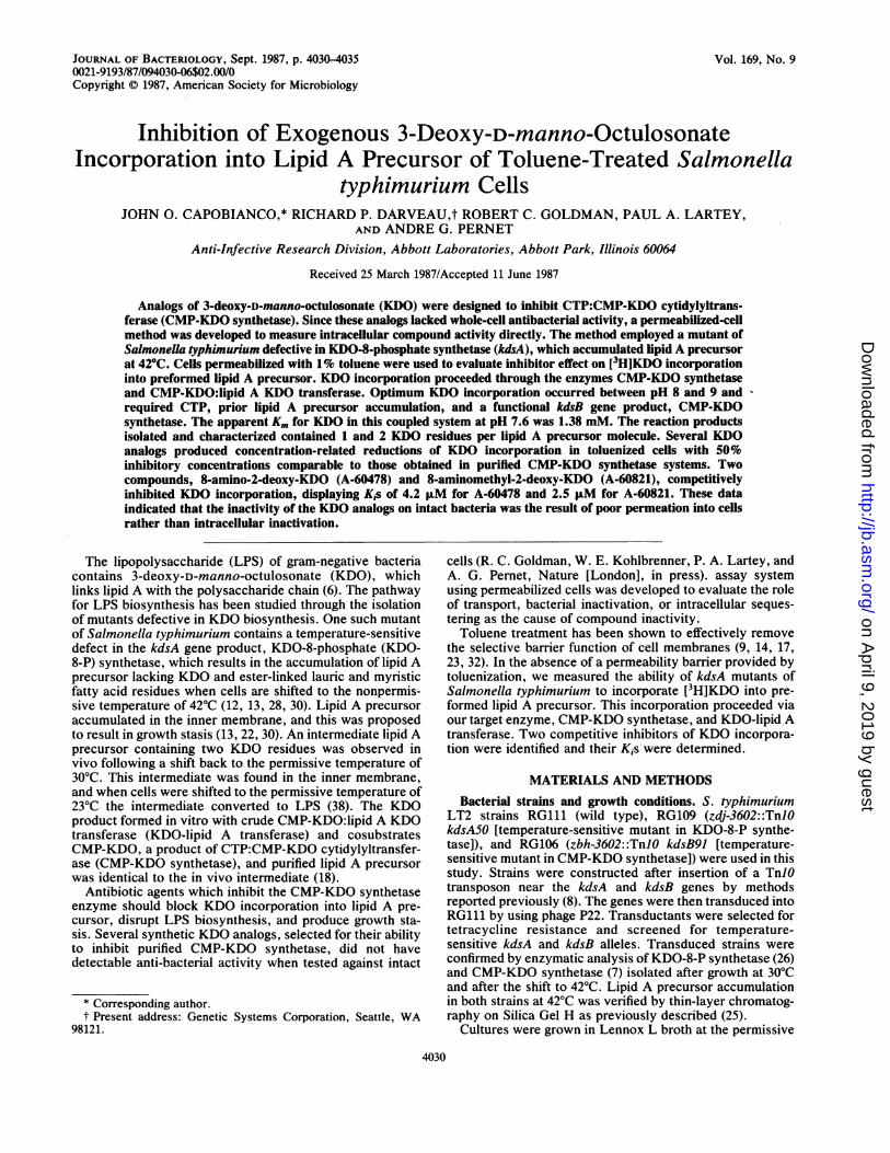
(363 versus 6.8 pmol/mg per min) in permeabilized cells thanin nontoluenized cells at pH 7.6 (Fig. 1). After 60 min at30°C, toluenized cells incorporated 22.5 nmol of KDO per1010 cells. The lack of linearity may- be related to theavailability of labile CMP-KDO (7, 10, 26) or lipid A precur-sor substrates. The kinetics of these two substrates were notinvestigated in this coupled system. Less than 5% of thelabel taken up by toluenized cells represented background,as determined from boiled or trichloroacetic acid-pretreatedcell controls. Uptake of KDO in toluenized cells was opti-mum at pH 8.0 to 9.0, was proportional to cell concentra-tions up to 2 x 10¹⁰ cells, and demonstrated hyperbolickinetics (unpublished observations). Significant KDO incor-poration (180 pmol/mg per min) was achieved in non-toluenized cells when the reaction took place in phosphatebuffer, pH 6.5. The possible pH dependency of KDO uptakewas not explored in this study.The latest reported apparent Km for KDO in a purified
CMP-KDO synthetase system was 0.29 mM (26). The appar-ent Km for KDO in the toluenized cell system was 1.38 mM,as determined by a Lineweavcr-Burk plot. Under the con-ditions of our coupled system, the apparent Km for KDO wasalmost five times that of the purified CMP-KDO synthetaseenzyme. The higher Km obtained in the coupled system maybe related to the effects of toluene or may reflect complexkinetics associated with the coupled cytosolic (CMP-KDOsynthetase) and membrane-bound (KDO-lipid A transferase)enzyme systemns. Further kinetic analysis of this system. wasnot attempted in this study.
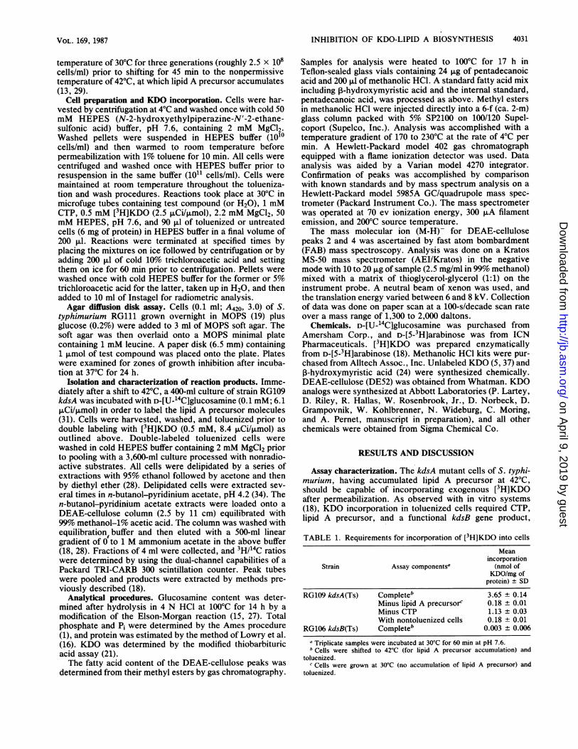
Reaction products. Two double-labeled products separatedon DEAE-cellulose (Fig. 2) were identified as lipid A pre-
z
w
0a:
0F)E
a
E
5 10 30Min. at 30C
60
FIG. 1. Incorporation of [3H]KDO into toluenized (x) andnontoluenized (0) cells at pH 7.6 as a function of incubation time.Cells (1010) were incubated and processed as described in Materialsand Methods. The amount of KDO incorporated was calculatedfrom the specific activity of exogenous KDO. Protein was deter-mined as described in Materials and Methods.
0
x
Qa-
c
x
20 40 60 80 100Fraction
FIG. 2. DEAE-cellulose chromatography of [14C]glucosamine-and [3H]KDO-labeled cell extracts. Toluenized cells were prepared,labeled, and extracted as outlined under Materials and Methods. Ofthe tritium counts incorporated into the cejls, 89% (3.8 x 10⁶ dpm)were extracted into n-butanol-pyridinium acetate. Extracts wereloaded onto a DEAE-cellulose column and eluted as described inMaterials and Methods. Animonium acetate concentrations weredetermined by conductivity. Ninety-six percent of the tritium loadedonto the column was recovered in peaks 2 to 4. Symbols: 0, 14C; X,3H; *, ammonium acetate gradient.
cursor molecules containing one or two KDO residues(Table 2). Four peaks were eluted from the column atammonium acetate concentrations of 0.24, 0.33, 0.42, and0.55 M. The 'H/14C ratio of peak 4 was almnost twice that ofpeak 3, indicating that the former may contain twice as manyKDO molecules per lipid A precursor molecule as the latter.Quantitative evaluation of the products from initial specificactivities was not attempted due to the probable dilution oflabel from intracellular unlabeled sources. Chemical analysispf peaks 2 to 4 for glucosamine, KDO, phosphate, andP,Bhydroxymyristic acid content revealed molar ratios ofglucosamine/phosphate and P-hydroxymyristic acid/gluco-samine indicative of lipid A precursor molecules (11, 28, 31).In addition to the appropriate molar ratios indicative of lipidA precursor, peaks 3 and 4 demonstrated glucosamine/KDOratios of 1.73 and 0.92, respectively, highly suggestive forKDO-lipid A intermediates (11), Peak 3 represents the firstreported isolation of a KDO incorporation product contain-ing 1 KDO molecule per lipid A precursor molecule. Peak 1contained the lowest concentration of glucosamine and the
TABLE 2. Analysis of chromatographic peaks for glucosamine,KDO, phosphate, and p-hydroxymyristic acida
Content (nmol)b Molar ratioPeak
GlcN KDO P I3tIMA GlcNIKDO GlcN/P ,BHMA/GlcN
1 24 _C 6,270 883 0.004 36.82 3,357 95 2,725 7,440 35.3 1.2 2.23 2% 172 284 459 1.7 1.0 1.64 448 488 485 889 0.9 0.9 2.0a Abbreviations: GlcN, glucosamine; P, phosphate; IHMA, 3-hydroxy-
myristic acid.b Values represent averages of three determinations.c-, None detected.
J. BACTERIOL.
on April 9, 2019 by guest
http://jb.asm.org/
Dow
nloaded from

INHIBITION OF KDO-LIPID A BIOSYNTHESIS 4033
t-zwI-z
w
-
lL
8~70 1800 17601623
l700 1600 1403 1530
1530 1500 1400 1360mlz
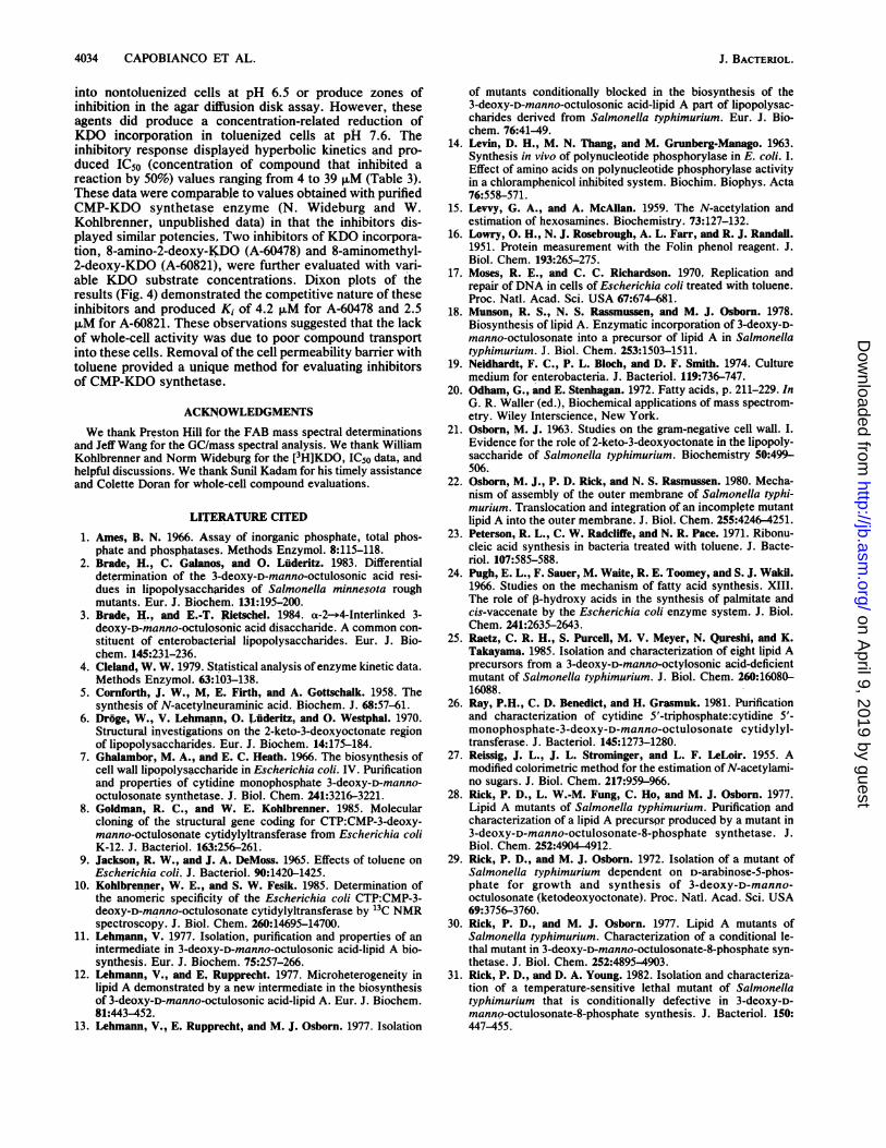
FIG. 3. Negative FAB mass spectrum of DEAE-cellulose peak4. Samples were prepared as described in Materials and Methods.Data were collected on paper at a scan rate of 100 s/decade over amass range of 1,300 to 2,000 daltons.
highest concentration of phosphate compared with the otherpeaks. The high phosphate content coupled with the pres-ence of unsaturated fatty acids (unpublished observation) asthe major fatty acids suggested that peak 1 was primarily aresidual phospholipid. The 3-hydroxymyristic acid presentin peak 1 may be due to contamination from peak 2. Thesmall amount of KDO present in peak 2 could not beassigned to a specific structure, although it displayed polarcharacteristics on DEAE-cellulose similar to those of lipid Aprecursor. The major fatty acid in peaks 2 to 4 was confirmedas 3-hydroxymyristic acid by gas chromatograph-mass spec-trum analysis. The electron impact spectrum (not shown)revealed a weak ion at m/z 240 (M-H20)+and a base peak atm/z 103 (HO=CH-CH2COOCH3) , which indicated thepresence of the 3-hydroxy group (20). Minor amounts ofcis-vaccenic and palmitic acids were also detected in peaks 2to 4 which may be attributed to residual phospholipidcontamination. Analysis of peaks 2 and 4 by negative FABmass spectroscopy provided mass molecular ion (M-H)-data for the major components isolated. The major compo-nent in peak 2 generated (M-H)- ions of mass 1,403 (spec-trum not shown), consistent with previous observations for aglucosamine disaccharide acylated with ,-hydroxymyristicacid at positions 2, 3, 2', and 3' and with monophosphates atpositions 1 and 4' (25) (C. R. H. Raetz, K. Takayama, L.Anderson, I. M. Armitage, and S. M. Stain, Fed. Proc.43:1567, 1984). Peak 4 generated (M-H)- ions of mass 1,843(Fig. 3), which differed from peak 2 by 440 atomic mass units(corresponds to 2 KDO residues minus their anomeric OHgroup); therefore, peak 4 differed from peak 2 by two KDOresidues. The minor ion at m/z 1623 represented (M-KDO)-,while the major fragment at m/z 1403 corresponded to lipid Aprecursor, (M-2KDO)-. The material from peak 3 wasconsumed in the chemical and gas chromatographic analysisand therefore was not available for FAB mass spectroscopicanalysis.Our attempts to pulse-chase peak 3 into peak 4, in
toluenized and nontoluenized cells were unsuccessful. Fur-thermore, the peaks were proportionally the same whetherthe KDO incorporation reaction was terminated after 1 or 60min. The explanation that peak 3 was the result of peak 4degradation during isolation could not be supported by theamount of free [3H]KDO detected after column chromatog-raphy, assuming that the tritium counts in free KDO wouldequal the tritium counts in peak 3. The two reaction productsmost likely were produced separately under our conditionsfrom similar enzyme systems.
TABLE 3. IC50s for KDO analogs
ICso (JIM)Abbottno. of Nomenclaturea Toluenized Purified
compound clSb CMP-KDOsynthetasec
A-60438 2-de-KDO 9.4 10.5A-60478 8-Amino-2-de-KDO 6.9 4.0A-60821 8-Aminomethyl-2-de-KDO 4.0 2.0A-62139 8-Thio-2-de-KDO 39.4 27.0A-63717 5,8-Diamino-2-de-KDO 33.3 14.6A-64654 5-Amino-8-aminomethyl-2-de-KDO 14.6 12.3
a 2-de-KDO, 2,3-Deoxy-D-manno-octulosonate.b Each compound was tested in triplicate at four inhibitory concentrations
against toluenized cells as outlined in Materials and Methods.c N. Wideburg and W. Kohlbrenner, unpublished data.
Earlier studies demonstrated the conversion of 2KDO-lipid A precursor to LPS (38). Reactions subsequent to theformation of our KDO products were affected by the limitedenergy source of the assay, since tritium-labeled LPS wasnot detected even after 60 min of incubation at 30°C. When[3H]KDO-labeled nontoluenized cells were suspended infresh Lennox L broth and incubated at 30°C for 60 min,KDO-lipid A precursors were no longer detectable by col-umn chromatography. Recent analysis of rough mutant LPSfrom members of the family Enterobacteriaceae showedcore regions containing KDO disaccharides (Re mutants) ortrisaccharides (Rd2 mutants) as a linear side chain (2, 3, 36).In Aeromonas and Rhodopseudomonas spp., a single KDOresidue was found in linkage between lipid A and thepolysaccharide (33, 35). The KDO-lipid A precursor productidentified in the present report may represent the precursorof a minor LPS species in S. typhimurium cQntaining a singleKDO in the core region.
Inhibitor evaluation. KDO analogs designed to inhibitCMP-KDO synthetase did not inhibit KDO incorporation
c 20
Rh15'C
E 10*io
-5vE
IIIs
I,/-
-5 0 5 10 20sM A-60478
B_ 35
Ei30-cm
25-a 20-~915-
E 10 00= 10 -g0/l' A S
-4 -2 0 2 4 8AM A-60821
16
FIG. 4. Dixon plots of inhibitors (A) 8-amino-2-deoxy-KDO(A-60478) and (B) 8-aminomethyl-2-deoxy-KDO (A-60821). Thecompound and toluenized cells were incubated at 30°C for 5 minwith 0.5 mM (x) or 1 mM (0) KDO substrate. Values representmean + standard deviation (N = 3). The apparent Kis for A-60478and A-60821 were 4.2 and 2.5 F±M, respectively.
VOL. 169, 1987
on April 9, 2019 by guest
http://jb.asm.org/
Dow
nloaded from
4034 CAPOBIANCO ET AL.
into nontoluenized cells at pH 6.5 or produce zones ofinhibition in the agar diffusion disk assay. However, theseagents did produce a concentration-related reduction ofKDO incorporttion in toluenized cells at pH 7.6. Theinhibitory response displayed hyperbolic kinetics and pro-duced IC50 (concentration of compound that inhibited areaction by 50%) values ranging from 4 to 39 ,M (Table 3).These data were comparable to values obtained with purifiedCMP-KDO synthetase enzyme (N. Wideburg and W.Kohlbrenner, unpublished data) in that the inhibitors dis-played similar potencies, Two inhibitors of KDO incorpora-tion, 8-amino-2-deoxy-KDO (A-60478) and 8-aminomethyl-2-deoxy-KDO (A-60821), were further evaluated with vari-able KDO substrate concentrations. Dixon plots of theresults (Fig. 4) demonstrated the competitive nature of theseinhibitors and produced Ki of 4.2 ,uM for A-60478 and 2.5,uM for A-60821. These observations suggested that the lackof whole-cell activity was due to poor compound transportinto these cells. Removal of the cell permeability barrier withtoluene provided a unique method for evaluating inhibitorsof CMP-KDO synthetase.
ACKNOWLEDGMENTS
We thank Preston Hill for the FAB mass spectral determinationsand Jeff Wang for the GC/mass spectral analysis. We thank WilliamKohlbrenner and Norm Wideburg for the [3H]KDO, IC50 data, andhelpful discussions. We thank Sunil Kadam for his timely assistanceand Colette Doran for whole-cell compound evaluations.
LITERATURE CITED
1. Ames, B. N. 1966. Assay of inorganic phosphate, total phos-phate and phosphatases. Methods Enzymol. 8:115-118.
2. Brade, H., C. Galanos, and 0. Luderitz. 1983. Differentialdetermination of the 3-deoxy-D-manno-octulosonic acid resi-dues in lipopolysaccharides of Salmonella minnesota roughmutants. Eur. J. Biochem. 131:195-200.
3. Brade, H., and E.-T. Rietschel. 1984. a-2--+4-Interlinked 3-deoxy-D-manno-octulosonic acid disaccharide. A common con-stituent of enterobacterial lipopolysaccharides. Eur. J. Bio-chem. 145:231-236.
4. Cleland, W. W. 1979. Statistical analysis ofenzyme kinetic data.Methods Enzymol. 63:103-138.
5. Cornforth, J. W., M, E. Firth, and A. Gottschalk. 1958. Thesynthesis of N-acetylneuraminic acid. Biochem. J. 68:57-61.
6. Droge, W., V. Lehmann, 0. LuAderitz, and 0. Westphal. 1970.Structural investigations on the 2-keto-3-deoxyoctonate regionof lipopolysaccharides. Eur. J. Biochem. 14:175-184.
7. Ghalambor, M. A., and E. C. Heath. 1966. The biosynthesis ofcell wall lipopolysaccharide in Escherichia coli. IV. Purificationand properties of cytidine monophosphate 3-deoxy-D-manno-octulosonate synthetase. J. Biol. Chem. 241:3216-3221.
8. Goldman, R. C., and W. E. Kohlbrenner. 1985. Molecularcloning of the structural gene coding for CTP:CMP-3-deoxy-manno-octulosonate cytidylyltransferase from Escherichia coliK-12. J. Bacteriol. 163:256-261.
9. Jackson, R. W., and J. A. DeMoss. 1965. Effects of toluene onEscherichia coli, J. Bacteriol. 90:1420-1425.
10. Kohlbrenner, W. E., and S. W. Fesik. 1985. Determination ofthe anomeric specificity of the Escherichia coli CTP:CMP-3-deoxy-D-manno-octulosonate cytidylyltransferase by 13C NMRspectroscopy. J. Biol. Chem. 260:14695-14700.
11. Lehnmann, V. 1977. Isolation, purification and properties of anintermediate in 3-deoxy-D-manno-octulosonic acid-lipid A bio-synthesis. Eur. J. Biochem. 75:257-266.
12. Lehmann, V., and E. Rupprecht. 1977. Microheterogeneity inlipid A demonstrated by a new intermediate in the biosynthesisof 3-deoxy-D-manno-octulosonic acid-lipid A. Eur. J. Biochem.81:443-452.
13. Lehmann, V., E. Rupprecht, and M. J. Osborn. 1977. Isolation
of mutants conditionally blocked in the biosynthesis of the3-deoxy-D-manno-octulosonic acid-lipid A part of lipopolysac-charides derived from Salmonella typhimurium. Eur. J. Bio-chem. 76:41-49.
14. Levin, D. H., M. N. Thang, and M. Grunberg-Manago. 1963.Synthesis in vivo of polynucleotide phosphorylase in E. coli. I.Effect of amino acids on polynucleotide phosphorylase activityin a chloramphenicol inhibited system. Biochim. Biophys. Acta76:558-571.
15. Levvy, G. A., and A. McAllan. 1959. The N-acetylation andestimation of hexosamines. Biochemistry. 73:127-132.
16. Lowry, 0. H., N. J. Rosebrough, A. L. Farr, and R. J. Randall.1951. Protein measurement with the Folin phenol reagent. J.Biol. Chem. 193:265-275.
17. Moses, R. E., and C. C. Richardson. 1970, Replication andrepair of DNA in cells of Escherichia coli treated with toluene.Proc. Natl. Acad. Sci. USA 67:674-681.
18. Munson, R. S., N. S. Rassmussen, and M. J. Osborn. 1978.Biosynthesis of lipid A. Enzymatic incorporation of 3-deoxy-D-manno-octulosonate into a precursor of lipid A in Salmonellatyphimurium. J. Biol. Chem. 253:1503-1511.
19. Neidhardt, F. C., P. L. Bloch, and D. F. Smith. 1974. Culturemedium for enterobacteria. J. Bacteriol. 119:736-747.
20. Odham, G., and E. Stenhagan. 1972. Fatty acids, p. 211-229. InG. R. Waller (ed.), Biochemical applications of mass spectrom-etry. Wiley Interscience, New York.
21. Osborn, M. J. 1963. Studies on the gram-negative cell wall. I.Evidence for the role of 2-keto-3-deoxyoctonate in the lipopoly-saccharide of Salmonella typhimurium. Biochemistry 50:499-506.
22. Osborn, M. J., P. D. Rick, and N. S. Rasmussen. 1980. Mecha-nism of assembly of the outer membrane of Salmonella typhi-murium. Translocation and integration of an incomplete mutantlipid A into the outer membrane. J. Biol. Chem. 255:4246-4251.
23. Peterson, R. L., C. W. Radcliffe, and N. R. Pace. 1971. Ribonu-cleic acid synthesis in bacteria treated with toluene. J. Bacte-riol. 107:585-588.
24. Pugh, E. L., F. Sauer, M. Waite, R. E. Toomey, and S. J. Wakil.1966. Studies on the mechanism of fatty acid synthesis. XIII.The role of ,B-hydroxy acids in the synthesis of palmitate andcis-vaccenate by the Escherichia coli enzyme system. J. Biol.Chem. 241:2635-2643.
25. Raetz, C. R. H., S. Purcell, M. V. Meyer, N. Qsreshi, and K.Takayama. 1985. Isolation and characterization of eight lipid Aprecursors from a 3-deoxy-D-manno-octylosonic acid-deficientmutant of Salmonella typhimurium. J. Biol. Chem. 260:16080-16088.
26. Ray, P.H., C. D. Benedict, and H. Grasmuk. 1981. Purificationand characterization of cytidine 5'-triphosphate:cytidine 5'-monophosphate-3-deoxy-D-manno-octulosonate cytidylyl-transferase. J. Bacteriol. 145:1273-1280.
27. Reissig, J. L., J. L. Strominger, and L. F. LeLoir. 1955. Amodified colorimetric method for the estimation of N-acetylami-no sugars. J. Biol. Chem. 217:959-966.
28. Rick, P. D., L. W.-M. Fung, C. Ho, and M. J. Osborn. 1977.Lipid A mutants of Salmonella typhimurium. Purification andcharacterization of a lipid A precursor produced by a mutant in3-deoxy-D-manno-octulosonate-8-phosphate synthetase. J.Biol. Chem. 252:4904-4912.
29. Rick, P. D., and M. J. Osborn. 1972. Isolation of a mutant ofSalmonella typhimurium dependent on D-arabinose-5-phos-phate for growth and synthesis of 3-deoxy-D-manno-octulosonate (ketodeoxyoctonate). Proc. Natl. Acad. Sci. USA69:3756-3760.
30. Rick, P. D., and M. J. Osborn. 1977. Lipid A mutants ofSalmonella typhimurium. Characterization of a conditional le-thal mutant in 3-deoxy-D-manno-octulosonate-8-phosphate syn-thetase. J. Biol. Chem. 252:4895-4903.
31. Rick, P. D., and D. A. Young. 1982. Isolation and characteriza-tion of a temperature-sensitive lethal mutant of Salmonellatyphimurium that is conditionally defective in 3-deoxy-D-manno-octulosonate-8-phosphate synthesis. J. Bacteriol. 150:447-455.
J. BACTERIOL.
on April 9, 2019 by guest
http://jb.asm.org/
Dow
nloaded from

VOL. 169, 1987 INHIBITION OF KDO-LIPID A BIOSYNTHESIS
32. Schrader, W. P., and D. P. Fan. 1974. Synthesis of cross-linkedpeptidoglycan attached to previously formed cell wall by tolu-ene treated cells of Bacillus megaterium. J. Biol. Chem.249:4815-4818.
33. Shaw, D. H., M. J. Squires, E. E. Ishiguro, and T. J. Trust. 1986.The structure of the heptose-3-deoxy-D-manno-octulosonic-acid region in a mutant form of Aeromonas salmonicida lipo-polysaccharide. Eur. J. Biochem. 161:309-313.
34. Stone, K. J., and J. L. Strominger. 1972. C55-isoprenyl pyro-
phosphate. Methods Enzymol. 38:306-309.35. Strittmatter, W., J. Weckesser, P. V. Salimath, and C. Galanos.
1983. Nontoxic lipopolysaccharide from Rhodopseudomonas
sphaeroides ATCC 17023. J. Bacteriol. 155:153-158.36. Tacken, A., E. T. Rietschel, and H. Brade. 1986. Methylation
analysis of the heptose/3-deoxy-D-manno-2-octulosonic acidregion (inner core) of the lipopolysaccharide from Salmonellaminnesota rough mutants. Carbohydr. Res. 149:279-291.
37. Unger, F. M. 1981. The chemistry and biological significance of3-deoxy-D-manno-2-octulosonic acid (KDO). Adv. Carbohydr.Chem. Biochem. 38:323-388.
38. Walenga, R. W., and M. J. Osborn. 1980. Biosynthesis of lipidA. Formation of acyl-deficient lipopolysaccharides in Salmo-nella typhimurium and Escherichia coli. J. Biol. Chem.255:4252-4256.
4035
on April 9, 2019 by guest
http://jb.asm.org/
Dow
nloaded from