u t i c a analy a p pharmaceutica analytica acta · title: nitrosothiol detection by hplc coupled...
TRANSCRIPT
Open AccessShort Communication
Shintani, Pharm Anal Acta 2013, 4:6 DOI: 10.4172/2153-2435.1000250
Volume 4 • Issue 6 • 1000250Pharm Anal ActaISSN: 2153-2435 PAA, an open access journal
*Corresponding author: Hideharu Shintani, Chuo University, School ofScience, 1-13-27, Kasuga Bunkyo 112-0003 Tokyo, Japan, Tel: +81425922336; E-mail: [email protected]
Received May 21, 2013; Accepted June 24, 2013; Published June 28, 2013
Citation: Shintani H (2013) Nitrosothiol Detection by HPLC Coupled with Flow Reactors of Hg2+ and Griess Reagent. Pharm Anal Acta 4: 250. doi:10.4172/2153-2435.1000250
Copyright: © 2013 Shintani H. This is an open-access article distributed under the terms of the Creative Commons Attribution License, which permits unrestricted use, distribution, and reproduction in any medium, provided the original author and source are credited.
Nitrosothiol Detection by HPLC Coupled with Flow Reactors of Hg2+ and Griess ReagentHideharu Shintani*
Chuo University, School of Science, 1-13-27, Kasuga Bunkyo 112-0003 Tokyo, Japan
Keywords: Nitrosothiol; HPLC; S-nitrosoproteins; Quantification
Introduction NO-related intermediates, including NO+ like species, can
participate in nitro-sating additions to nucleophilic centers of biological molecules. Sulfhydryl-containing molecules such as glutathione are particularly susceptible to nitrosation and form nitrosothiol adducts (nitrosothiols; RS-NOs). It seems that these adducts in biological systems play an important role in NO-mediated signalling cascades such as the downregulation of N-methyl-D-aspartate receptor, and the regulation of transcriptional factors [1]; they might also be involved in non-adrenergic and non-cholinergic neuronal responses [2]. lt is, therefore, essential to identify nitrosothiols specifically and to quantify them in biological systems.
Herein we describe a sensitive and specific HPLC method coupled with Hg2+ and Griess reagent for nanomolar quantification of a wide range of RS-NOs including low-molecular weight RS-NOs such as nitrosoglutathione (GS-NO) and nitrosocysteine (Cys-NO), and also S-nitrosoproteins, in particular S-nitrosoalbumin and S-nitrosohaemoglobin [3].
Preparation of S-nitrosoproteins Protocol
Reagents: Bovine serum albumin (BSA) (Nacalai Tesque, Osaka, Japan), dithiothreitol (DTT; Wako Pure Chemicals, Osaka, Japan), isoamyl nitrite (Wako), ethyllenediaminetetraacetic acid (EDTA; Dojindo Laboratories, Kumamoto, Japan), diethylenetriaminepentaacetic acid (DTPA; Dojindo), and 5, 5'-dithiobis[2-nitrobenzoic acid] (DTNB; Nacalai).
Procedure: Reduction of BSA
1. Add DTT (100 mM, 10 μL) to BSA (1 mM, 1 mL) in sodiumphosphate buffer (pH 7.0, 100 mM).
2. Incubate for 30 min at 37°C.3. Apply to a Sephadex G-100 (Pharmacia, Uppsala, Sweden)
column equilibrated with sodium phosphate buffer (pH 7.0,10 mM) containing EDTA (1 mM; buffer A), and elute withbuffer A.
4. Collect the fractions containing BSA and concentrate thefractions by ultrafiItration.
Nitrosation of the reduced BSA: 1. Add isoamyl nitrite (100 mM, 10 μL) to reduced BSA (0.1 mM, 1 mL) in sodium phosphate buffer (pH 7.8, 100 mM) containing DTPA (0.5 mM).
2. Incubate for 30 min at 37°C.
3. Apply to a Sephadex G-25 (Pharmacia) column equilibratedwith buffer A, and elute with buffer A.
4. Collect the fractions containing S-NO-BSA and concentratethe fractions by ultrafiltration.
Quantification of Nitrosothiols Protocol
Reagents: Nitrosoproteins (e.g., S-NO-BSA) prepared above, GS-NO (Dojindo), Cys-NO (Dojindo), HgCI2, and Griess reagent (naphthylethylenediamine; sulfanilamide (Wako)).
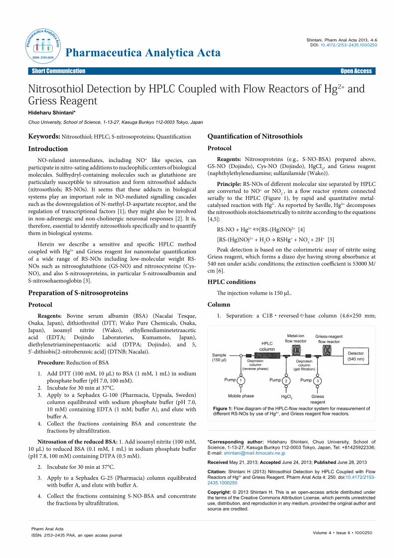
Principle: RS-NOs of different molecular size separated by HPLC are converted to NO+ or NO2
-, in a flow reactor system connected serially to the HPLC (Figure 1), by rapid and quantitative metal-catalysed reaction with Hg2-. As reported by Saville, Hg2+ decomposes the nitrosothiols stoichiometrically to nitrite according to the equations [4,5]:
RS-NO + Hg2+ ⇔[RS-(Hg)NOj2+ [4]
[RS-(Hg)NOj2+ + H2O → RSHg+ + NO2-+ 2H+ [5]
Peak detection is based on the colorimetric assay of nitrite using Griess reagent, which forms a diazo dye having strong absorbance at 540 nm under acidic conditions; the extinction coefficient is 53000 M/cm [6].
HPLC conditions
The injection volume is 150 μL.
Column
1. Separation: a C1B・reversedゃhase column (4.6×250 mm;
HPLCcolumn
PumpPump Pump
Mobile phase Griessreagent
Griess-reagentflow reactor
Detector(540 nm)
Sample(150 µl)
Metal-ionflow reactor
Deproteincolumn
(reverse phase)
Deproteincolumn
(get filtration)
1 2 3
HgCl2
Figure 1: Flow diagram of the HPLC-flow reactor system for measurement of different RS-NOs by use of Hg2+, and Griess reagent flow reactors.
Phar
mac
eutica Analytica Acta
ISSN: 2153-2435Pharmaceutica Analytica Acta
Citation: Shintani H (2013) Nitrosothiol Detection by HPLC Coupled with Flow Reactors of Hg2+ and Griess Reagent. Pharm Anal Acta 4: 250. doi:10.4172/2153-2435.1000250
Page 2 of 2
Volume 4 • Issue 6 • 1000250Pharm Anal ActaISSN: 2153-2435 PAA, an open access journal
TSKgel OOS.80Ts; Tosoh, Tokyo) for low molecular weight RS-NOs; a gel filtration column (8×300 mm; Oiol-120; YMC, Kyoto) for the nitrosoproteins.
2. Deproteinating column: small columns (3×10 mm) packedwith C1B-based resin are placed just before the separationcolumn and just after Hg2- flow reactor coil, in the reversed-phase and gel filtration systems, respectively.
Mobile phase
1. Pump 1 for HPLC: sodium acetate buffer (pH 5.5, 10 mM)containing DTPA (0.5 mM) and methanol (0-7%), for lowmolecular weight RS-NOs (reversed-phase HPLC); sodiumacetate buffer (pH 5.5, 10 mM) containing DTPA (0.5 mM) and NaCI (150 mM) for the nitrosoprotein (gel filtration HPLC).
2. Pump 2 for Hg2+-flow reactor: HgCl2 (1.75 mM) in sodiumacetate buffer (pH 5.5, 10 mM).
3. Pump 3 for Griess reagent flow reactor: naphthylethylenediamine (0.1%) in H2O; sulfanilamide (1 .0%) + phosphoric acid (2.0%)in H2O.
Flow rate: 0.55 mL/min (pump 1), 0.2 mL/min (pump 2), 0.24 mL/min (pump 3)
Detector: visible detector (Eicom, Kyoto) operated at 540 nm.
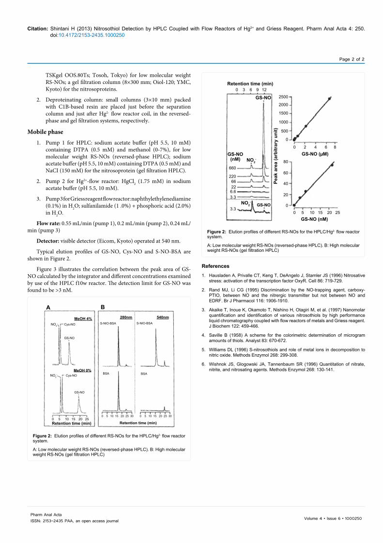
Typical elution profiles of GS-NO, Cys-NO and S-NO-BSA are shown in Figure 2.
Figure 3 illustrates the correlation between the peak area of GS-NO calculated by the integrator and different concentrations examined by use of the HPLC f10w reactor. The detection limit for GS-NO was found to be >3 nM.
References
1. Hausladen A, Privalle CT, Keng T, DeAngelo J, Stamler JS (1996) Nitrosativestress: activation of the transcription factor OxyR. Cell 86: 719-729.
2. Rand MJ, Li CG (1995) Discrimination by the NO-trapping agent, carboxy-PTIO, between NO and the nitrergic transmitter but not between NO andEDRF. Br J Pharmacol 116: 1906-1910.
3. Akaike T, Inoue K, Okamoto T, Nishino H, Otagiri M, et al. (1997) Nanomolarquantification and identification of various nitrosothiols by high performance liquid chromatography coupled with flow reactors of metals and Griess reagent. J Biochem 122: 459-466.
4. Saville B (1958) A scheme for the colorimetric determination of microgramamounts of thiols. Analyst 83: 670-672.
5. Williams DL (1996) S-nitrosothiols and role of metal ions in decomposition tonitric oxide. Methods Enzymol 268: 299-308.
6. Wishnok JS, Glogowski JA, Tannenbaum SR (1996) Quantitation of nitrate,nitrite, and nitrosating agents. Methods Enzymol 268: 130-141.
Retention time (min)
Peak
are
a (a
rbitr
ary
unit)
GS-NO GS-NO (µM)
GS-NO
GS-NO
GS-NO (nM)
(nM) NO2-
NO2-
0 3 6 9 12
2500
2000
1500
1000
500
0
0 2 4 6 8
0 5 10 15 20 25
80
60
40
20
0
660
2206622
6.63.3
3.3
Figure 2: Elution profiles of different RS-NOs for the HPLC/Hg2- flow reactor system.
A: Low molecular weight RS-NOs (reversed-phase HPLC). B: High molecular weight RS-NOs (gel filtration HPLC)
A B
MeOH 4%
MeOH 0%
280nm 540nmS-NIO-BSA S-NIO-BSA
BSA BSA
Retention time (min)Retention time (min)0 5 10 15 20 25
0 5 10 15 20 25 30 0 5 10 15 20 25 30
GS-NO
GS-NO
Cys-NO
Cys-NO
NO2-
NO2-
Figure 2: Elution profiles of different RS-NOs for the HPLC/Hg2- flow reactor system.
A: Low molecular weight RS-NOs (reversed-phase HPLC). B: High molecular weight RS-NOs (gel filtration HPLC)