ube3a reinstatement identifies distinct developmental...
TRANSCRIPT
The Journal of Clinical Investigation R e s e a R c h a R t i c l e
2 0 6 9jci.org Volume 125 Number 5 May 2015
IntroductionChildren with AS are typically diagnosed within the first year of life, due to developmental delay. The most prominent symp-toms include motor impairments, epilepsy, intellectual disability, and absence of speech (1). AS is caused by loss of function of the maternally inherited UBE3A allele. In neurons, the maternally inherited UBE3A allele is the only active allele, since the pater-nally inherited UBE3A allele is silenced through cell type–specific imprinting. This imprinting results in the allele-specific and neu-ronally restricted expression of a large antisense RNA transcript (UBE3A-ATS), which selectively interferes with paternal UBE3A transcription through a cis-acting mechanism (2–6).
There is currently no effective treatment for AS, but the unique silencing mechanism of the paternal UBE3A allele holds great promise for developing novel therapeutic strategies. Two recent studies have shown that the paternal UBE3A allele can be phar-macologically reactivated (7, 8), which offers a unique molecular target with high clinical potential for the treatment of AS.
Notably, however, activation of the paternal Ube3a gene in adulthood appears insufficient to rescue the majority of neurocog-nitive phenotypes in the AS mouse model. The failure of pheno-typic rescue in adult AS mice might have resulted from the incom-
plete reinstatement of UBE3A expression (35%–47% of WT levels) or, alternatively, because a neurocognitive rescue by UBE3A rein-statement requires early therapeutic intervention (8). Hence, for such a treatment strategy to be successful in the clinic, it is impera-tive to know whether there is a critical time window during which a disease-modifying therapy would be effective. This is particularly relevant for early-onset disorders, such as AS, whose causative gene is highly expressed in developing neural circuits.
Despite extensive knowledge of critical periods for the development of sensorimotor networks, much less is known about the critical periods for complex behaviors in neuro-developmental disorders (9, 10). An inducible mouse model for Rett syndrome showed that adult activation of the Mecp2 gene could rescue behavioral alterations and synaptic plasticity defi-cits, suggesting a broad window of therapeutic opportunity (11). In contrast, adult reactivation of the Syngap1 gene in a mouse model of intellectual disability and autism did not reverse any of the core behavioral deficits related to anxiety and behavioral flexibility (12). However, to our knowledge, no previous study has been performed to systematically investigate the influ-ence of critical developmental periods on the ability to rescue disease-relevant behavioral phenotypes. Here, we explore the effect of gene reactivation across multiple developmental win-dows in a novel mouse model for AS. Our results demonstrate an essential role for Ube3a in neurodevelopment and define a critical period for therapeutic intervention during which Ube3a gene reactivation ameliorates the neurocognitive impairments of AS model mice.
Angelman syndrome (AS) is a severe neurodevelopmental disorder that results from loss of function of the maternal ubiquitin protein ligase E3A (UBE3A) allele. Due to neuron-specific imprinting, the paternal UBE3A copy is silenced. Previous studies in murine models have demonstrated that strategies to activate the paternal Ube3a allele are feasible; however, a recent study showed that pharmacological Ube3a gene reactivation in adulthood failed to rescue the majority of neurocognitive phenotypes in a murine AS model. Here, we performed a systematic study to investigate the possibility that neurocognitive rescue can be achieved by reinstating Ube3a during earlier neurodevelopmental windows. We developed an AS model that allows for temporally controlled Cre-dependent induction of the maternal Ube3a allele and determined that there are distinct neurodevelopmental windows during which Ube3a restoration can rescue AS-relevant phenotypes. Motor deficits were rescued by Ube3a reinstatement in adolescent mice, whereas anxiety, repetitive behavior, and epilepsy were only rescued when Ube3a was reinstated during early development. In contrast, hippocampal synaptic plasticity could be restored at any age. Together, these findings suggest that Ube3a reinstatement early in development may be necessary to prevent or rescue most AS-associated phenotypes and should be considered in future clinical trial design.
Ube3a reinstatement identifies distinct developmental windows in a murine Angelman syndrome modelSara Silva-Santos,1,2,3 Geeske M. van Woerden,1,2 Caroline F. Bruinsma,1,2 Edwin Mientjes,1,2 Mehrnoush Aghadavoud Jolfaei,1,2 Ben Distel,4 Steven A. Kushner,2,5 and Ype Elgersma1,2
1Department of Neuroscience, Erasmus Medical Center, Rotterdam, Netherlands. 2ENCORE Expertise Center for Neurodevelopmental Disorders, Erasmus Medical Center, Rotterdam, Netherlands. 3Graduate Program in Areas of Basic and Applied Biology, Instituto de Ciências Biomédicas Abel Salazar, Universidade do Porto, Porto, Portugal. 4Department of Medical Biochemistry,
Academic Medical Center, Amsterdam, Netherlands. 5Department of Psychiatry, Erasmus Medical Center, Rotterdam, Netherlands.
Authorship note: Geeske M. van Woerden, Caroline F. Bruinsma, and Edwin Mientjes contributed equally to this work.Conflict of interest: The authors have declared that no conflict of interest exists.Submitted: December 26, 2014; Accepted: March 5, 2015.Reference information: J Clin Invest. 2015;125(5):2069–2076. doi:10.1172/JCI80554.

The Journal of Clinical Investigation R e s e a R c h a R t i c l e
2 0 7 0 jci.org Volume 125 Number 5 May 2015
We first investigated the efficiency of the tran-scriptional stop cassette in blocking Ube3a expres-sion. Female Ube3aStop/p+ mice were crossed to a constitutive Cre-expressing line with an early embryonic onset of recombination (ref. 13 and Fig-ure 1A). Because the paternal Ube3a allele is epige-netically silenced, mice with a maternally inherited stop cassette without Cre expression (Ube3aStop/p+; Cre– mice) showed a severe loss of UBE3A protein (also known as E6-associated protein [E6AP]), comparable to the reduction in UBE3A expression observed in the traditional AS mouse model with a maternally inherited deletion of Ube3a (Ube3am–/p+) (Figure 1B; see also Figure 2D for a comparison to Ube3am–/p+ mice). Immunohistochemical staining of Ube3aStop/p+;Cre– brain slices was indistinguishable from that of Ube3am–/p+ mouse samples (Supple-mental Figure 1B). These results confirm that the floxed stop cassette is highly effective in blocking transcription of the maternal Ube3a allele, while preserving the normal epigenetic silencing of the paternal allele.
Next, we investigated the efficiency of Ube3a reactivation upon Cre-mediated deletion of the floxed stop cassette. UBE3A protein levels were reinstated in Ube3aStop/p+;Cre+ mice to 89% of WT
levels in the hippocampus, 82% in the cerebral cortex, and 99% in the cerebellum. Furthermore, the subcellular distribution of UBE3A was indistinguishable between Ube3aStop/p+;Cre+ and WT;Cre+ mice, validating the functionality of the Ube3a reactiva-tion method (Figure 1B and Supplemental Figure 1B).
Early embryonic gene reactivation prevents the manifestation of AS phenotypes. Impaired motor coordination, autistic traits, anxi-ety, and epilepsy are hallmarks of AS patients, for which analogous phenotypes are well established in AS model mice (3, 14–16). As expected based on the loss of Ube3a expression, Ube3aStop/p+;Cre– mice exhibited significant alterations in rotarod performance (Fig-
ResultsGeneration and characterization of a conditional Ube3a mutant. To examine whether the therapeutic benefit of Ube3a gene reac-tivation is dependent upon the developmental stage at which gene expression is restored, we generated a conditional AS mouse model to allow temporally controlled reactivation of the Ube3a gene upon Cre-mediated deletion of a floxed transcrip-tional stop cassette inserted within intron 3 by homologous recombination (Ube3aStop/p+) (Supplemental Methods and Sup-plemental Figure 1; supplemental material available online with this article; doi:10.1172/JCI80554DS1).
Figure 1. Embryonic reactivation of Ube3a expression rescues AS-like behavioral phenotypes. (A) Schematic representation of Ube3a reactivation (indicated by the gray arrow) during mouse embryonic development and time point of behavioral testing. (B) Western blot analysis of hippocampus (n = 4 per genotype), cortex (n = 5), and cerebellum (n = 5) from Ube3aStop/p+ and WT littermates crossed with an embryonically active Cre line. (C–H) Ube3aStop/p+;Cre– mice show robust behavioral AS-relevant phenotypes, which can be fully rescued by embryonic reactivation of the Ube3a gene in the Ube3aStop/p+;Cre+ mice. Number of mice (WT;Cre+/Ube3aStop/p+;Cre–/Ube3aStop/p+;Cre+): accelerating rotarod, n = 14/8/17; marble burying test, n = 24/18/28; open field test, n = 14/8/17; nest building test, n = 7/7/7; forced swim test, n = 14/8/17; epilepsy test, n = 7/10/8. All data represent mean ± SEM. ANOVA with genotype as independent variable was used for statistical compar-isons. A significant effect of genotype was identified in all behavioral tests (see Supplemental Table 1). *P < 0.05, **P < 0.01; Bonferroni’s post hoc analysis.

The Journal of Clinical Investigation R e s e a R c h a R t i c l e
2 0 7 1jci.org Volume 125 Number 5 May 2015
stages of postnatal development. In particular, we induced Ube3a gene reactivation at 3 weeks (“juvenile mice”), 6 weeks (“adolescent mice”), and 14 weeks of age (“adult mice”), with behavioral testing performed at a mean age of 16 weeks, 22 weeks, and 28 weeks, respectively (Figure 2A). Across these developmental time points, UBE3A protein levels in tamoxifen-treated Ube3aStop/p+;CreERT+ mice were reinstated to 70%–100% of wild-type levels, which is comparable to those achieved by early embryonic reactivation (Figure 2, B–D, Sup-plemental Figure 2, and Figure 1B). Importantly, UBE3A expres-sion in vehicle-treated Ube3aStop/p+;CreERT+ mice was also similar to that observed in Ube3aStop/p+;Cre– and Ube3am–/p+ mice (Figure 2D and Supplemental Figure 3), demonstrating the tight control of gene reactivation.
Juvenile reactivation of Ube3a resulted in a full rescue of the motor coordination deficit. In contrast, adolescent reactivation only partially rescued motor coordination, while no improvement
ure 1C), marble burying (Figure 1D), open field exploration (Figure 1E), nest building (Figure 1F), and audiogenic seizure threshold (Figure 1H), all of which are also present in the classical Ube3am–/p+ mouse model of AS (3, 14). In addition, we identified a highly robust phenotype in the forced swim test present in both the classical Ube3am–/p+ mutant (data not shown) and the conditional Ube3aStop/p+ AS mouse model (Figure 1G).
Consistent with the therapeutic potential of Ube3a gene reac-tivation, Ube3aStop/p+;Cre+ mice exhibited a full rescue of all of these neurological and behavioral abnormalities, confirming that embryonic reactivation of UBE3A protein expression is sufficient to prevent the manifestation of AS phenotypes across multiple domains (Figure 1 and Supplemental Table 1).
Gene reactivation in juvenile, adolescent, and adult animals reveals the presence of distinct critical periods. We next crossed the Ube3aStop/p+ mice with a tamoxifen-inducible CreERT+ mouse line (17) to determine the efficacy of Ube3a reactivation at later
Figure 2. Molecular analysis of Ube3aStop/p+;CreERT+ mice reveals successful reactivation of the maternal Ube3a gene upon tamox-ifen induction. (A) Schematics representing Ube3a reactivation achieved by tamoxifen treatment (gray arrows) in each experimental group. (B–D) Ube3aStop/p+;CreERT+ mice with postnatally induced gene reactivation express UBE3A at levels comparable to those achieved with early embryonic gene reactiva-tion in hippocampus (juvenile, n = 3 per genotype; adolescent, n = 4 per genotype; adult, n = 4 per genotype), cortex (juvenile, n = 3 per genotype; adolescent, n = 4 per genotype; adult, n = 3–4 per geno-type), and cerebellum (juvenile, n = 3 per genotype; adolescent, n = 4 per genotype; adult, n = 5 per genotype). Data represent mean ± SEM.

The Journal of Clinical Investigation R e s e a R c h a R t i c l e
2 0 7 2 jci.org Volume 125 Number 5 May 2015
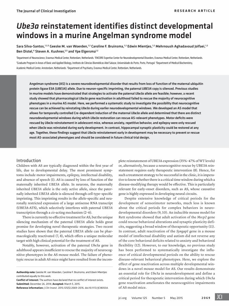
the epilepsy phenotype in AS mice is differentially respon-sive to treatment with antiepileptic drugs (AEDs) in mice with Ube3a reactivation. Adult (>8 weeks) Ube3am–/p+ and Ube3aStop/p+; CreERT– mice were treated for 5 days with either valproate or clonazepam using a within-subjects design. Prior to treatment, every AS mouse examined showed audiogenic seizures (Figure 4B). However, after 5 days of treatment with either of these AEDs, seizures were completely prevented in all mice. More-over, 3 days after the cessation of AED treatment (wash-out period), all AS mice again showed audiogenic seizures (Fig-ure 4B). These data confirm that seizures can be successfully treated in adult AS mice using conventional AEDs, but that they are nevertheless insensitive to postnatal Ube3a reactivation.
was observed with adult reactivation (Figure 3A). Together, these findings identify a critical period for Ube3a-dependent motor development, which closes between 3 and 6 weeks postnatally.
The critical window for rescuing motor coordination deficits was distinct from the window for rescuing autism- and anxiety-re-lated phenotypes such as the marble burying task, open field test, nest building test, and forced swim test, which could be rescued by embryonic reactivation (Figure 1, D–G, and Supplemental Table 1) but not upon juvenile, adolescent, or adult reactivation (Figure 3, B–E, and Supplemental Table 2).
The epilepsy phenotype was also refractory to postnatal Ube3a reactivation, as seizures persisted despite gene reactivation at a juvenile age (Figure 4A). Next, we sought to confirm whether
Figure 3. Postnatal reactivation of Ube3a expression reveals a critical period for behavioral rescue. (A–E) Behavioral testing of Ube3aStop/p+ and WT littermates treated with either vehicle (Veh.) or tamoxifen (Tamox.) shows distinct critical periods for recovery of the behavioral deficits. Numbers of mice (WT;CreERT+ Veh./ WT;CreERT+ Tamox./Ube3aStop/p+;CreERT+ Veh./Ube3aStop/p+;CreERT+ Tamox.): (A) Accelerating rotarod after juvenile (n = 22/20/22/22), adolescent (n = 11/11/10/11), and adult (n = 11/9/12/13) gene reactivation. (B) Marble burying test after juvenile (n = 21/20/20/20), adolescent (n = 20/20/21/23), and adult (n = 13/11/14/15) gene reactiva-tion. (C) Open field test after juvenile (n = 21/21/22/22), adolescent (n = 11/11/10/11), and adult (n = 10/8/9/10) gene reactivation. (D) Nest building test after juvenile (n = 12/13/14/13), adolescent (n = 14/13/16/17), and adult (n = 9/8/9/8) gene reactivation. (E) Forced swim test after juvenile (n = 17/19/18/19), adolescent (n = 11/11/10/11), and adult (n = 11/8/9/9) gene reactivation. All data represent mean ± SEM. Two-way ANOVA or repeated-measures 2-way ANOVA with genotype and treatment as indepen-dent variables was used for statistical comparisons. A significant effect of genotype was identified in all behav-ioral tests (see Supplemental Table 2). **P < 0.01 for genotype significance.
The Journal of Clinical Investigation R e s e a R c h a R t i c l e
2 0 7 3jci.org Volume 125 Number 5 May 2015
and the third postnatal week in order to prevent a wide spectrum of AS-like deficits, with the notable exception of defects in motor coordination and hippocampal LTP. To further refine the critical period window, we next induced gene reactivation immediately following birth by administering tamoxifen to lactating dams (Figure 5A). The efficacy of this method of tamoxifen adminis-tration was reduced compared with direct treatment of offspring, yielding 44%, 34%, and 63% of WT UBE3A levels, respectively, in the hippocampus, cortex, and cerebellum (Figure 5B). Notably, however, even with a reduced level of UBE3A reactivation, motor coordination was entirely rescued and performance in the open
To investigate the extent to which postnatal Ube3a reactivation is able to rescue electrophysiological phenotypes, we measured hippocampal long-term potentiation (LTP), a form of synaptic plasticity required for experience-dependent neurodevelopment. Intriguingly, we observed full recovery of hippocampal LTP follow-ing Ube3a gene reactivation at all time points examined (Figure 4, C and D, and Supplemental Table 2), indicating the absence of a crit-ical period window for rescue of this important cellular phenotype.
Partial gene reactivation in newborn animals rescues the motor coordination and open field phenotype. Our data suggest that UBE3A is required during a critical period between early embryogenesis
Figure 4. Ube3a reactivation in juvenile animals does not recover epilepsy susceptibility, but Schaffer collateral–CA1 LTP is fully recovered. (A) Epilepsy susceptibility in Ube3aStop/p+;CreERT+ mice persists after Ube3a gene reactivation at a juvenile age (n = 8 mice/group). (B) The induction of tonic-clonic seizures induced by audiogenic stimulation (indicated by the red arrows) is efficiently treated by administration of AEDs (blue rectangle illustrates treatment admin-istration and wash-out period) in adult Ube3aStop/p+;Cre– (n = 2) and Ube3am–/p+ (n = 12) mice. Seizures reappeared 3 days after cessation of treatment. Per-centages indicate the amount of mutant mice that developed seizures upon audiogenic stimulation (see also Supplemental Methods for more experimental details). Hippocampal plasticity deficit as measured by LTP in mutant mice is ameliorated upon gene reactivation at both (C) juvenile and (D) adult ages. Data represent mean ± SEM. Two-way ANOVA with genotype and treatment as independent variables was used for statistical testing. All tests showed a significant effect of genotype (see also Supplemental Table 2 for statistical comparisons). Number of slices/mouse used: juvenile reactivation: WT;CreERT+ Veh. (n = 16/4), WT;CreERT+ Tamox. (n = 25/4), Ube3aStop/p+;CreERT+ Veh. (n = 22/4), Ube3aStop/p+;CreERT+ Tamox. (n = 22/5); adult reactivation: WT;CreERT+ Veh. (n = 18/6), WT;CreERT+ Tamox. (n = 37/8), Ube3aStop/p+;CreERT+ Veh. (n = 23/4), Ube3aStop/p+;CreERT+ Tamox. (n = 15/4). **P < 0.01.

The Journal of Clinical Investigation R e s e a R c h a R t i c l e
2 0 7 4 jci.org Volume 125 Number 5 May 2015
field test was significantly improved (Figure 5C). However, AS-like deficits persisted in the other behavioral paradigms, suggesting that the Ube3a-dependent neurodevelopmental critical period for autism-related phenotypes in AS might not extend significantly beyond birth. Alternatively, given that we achieved only partial reactivation during the neonatal period, it remains distinctly pos-sible that functional plasticity may extend beyond 3 weeks of age but is only evident with a higher efficiency of Ube3a reactivation.
DiscussionOur results demonstrate an essential role for Ube3a in neuro-development and define critical periods during which Ube3a gene reactivation can ameliorate AS-like phenotypes. In particular, the window for improving motor coordination extends furthest into postnatal development, whereas the autism- and anxiety-re-lated phenotypes appear to be established much earlier. In con-
trast, at the cellular level, there appears to be no critical window for reversing plasticity deficits. The finding that LTP could be fully recovered at all ages is consistent with previous findings showing that the hippocampal LTP def-icit in adult AS mice is reversible upon acute pharmaco-logical treatment with an ErbB inhibitor or with ampak-ine cognitive enhancers (18, 19). However, expression of UBE3A in adult Ube3am–/p+ mice through a viral-mediated approach only partially recovered synaptic plasticity (20). This apparent discrepancy likely reflects the limited effi-ciency of in vivo virally mediated neuronal transduction, compared with the more homogeneous biodistribution of systemically administered pharmacological compounds.
Importantly, and consistent with our findings, to our knowledge no studies in AS mice have demonstrated the successful rescue of the behavioral phenotypes related to anxiety and repetitive behavior upon adult treatment, despite multiple efforts using a variety of different inter-ventions. In addition, it is notable that the behavioral deficits related to anxiety and behavioral flexibility in the Syngap1 mutant mouse model for intellectual disability were also not rescued by adult reactivation of Syngap1
gene expression (12). These findings could suggest that the win-dow during which gene activation can ameliorate autism-related phenotypes closes early in neurodevelopment. Importantly, how-ever, these observations do not exclude the possibility that directly targeting downstream signaling pathways could have a broader window for therapeutic intervention (21). In fact, this possibility is very well demonstrated by the highly effective intervention of AEDs in preventing audiogenic seizures, in contrast to the failure of postnatal gene reactivation to alter the susceptibility to audio-genic seizures. Therefore, at least for seizure susceptibility, the therapeutic benefit of restoring the etiological loss of UBE3A was inferior to that achieved by targeting downstream mechanisms, in this case with AEDs. This finding strongly supports investigation of the targets of UBE3A and their downstream signaling pathways in order to develop drugs that can be applied as part of a comple-mentary therapeutic strategy.
Figure 5. Partial reactivation of Ube3a expression during the first postnatal week attenuates the motor coordination and open field deficits. (A) Schematics representing Ube3a reactiva-tion achieved by tamoxifen administration (gray arrows) to the lactating dams starting on the day of delivery. (B) Western blot analysis of UBE3A expression in hippocampal (n = 4 per geno-type), cortical (n = 5), and cerebellar (n = 5) tissues of mutant mice and their WT littermates. The thin black lines on the hippocampus blot indicate noncontiguous samples run on the same gel. (C) Rescue of the accelerating rotarod and open field impairments. Number of mice used (WT;CreERT+ Veh./WT;CreERT+ Tamox./Ube3aStop/p+;CreERT+ Veh./Ube3aStop/p+;CreERT+ Tamox.) for accelerating rotarod, marble burying test, open field test, and forced swim test: n = 14/12/14/11; for nest building, n = 12/12/12/11. All data represent mean ± SEM. Two-way ANOVA or repeated-measures 2-way ANOVA with genotype and treatment as independent variables was used for statistical comparisons. A significant effect of genotype was identified in all behavioral tests (see Supplemental Table 2 for statistical comparisons). *P < 0.05; **P < 0.01.

The Journal of Clinical Investigation R e s e a R c h a R t i c l e
2 0 7 5jci.org Volume 125 Number 5 May 2015
Cre+ mice) or with Tg(CAG-cre/Esr1*)5Amc/J (The Jackson Labora-tory) (17) (herein referred as CreERT+), both kept in the C57BL/6J back-ground (Charles River), to generate heterozygous Ube3aStop/p+;Cre+ and Ube3aStop/p+;CreERT+ mutants and littermate controls in the F1 hybrid 129S2-C57BL/6 background.
Tamoxifen treatment. One-day to 8-month-old Ube3aStop/p+ mice and their WT littermates (both sexes) were used in this study. Ube3aStop/p+; CreERT+ mutants and WT mice were given tamoxifen to induce Cre-mediated deletion of the stop cassette. Tamoxifen (Sigma-Aldrich) was diluted in sunflower oil at a concentration of 20 mg/ml. Each mouse received 0.10 mg tamoxifen per gram body weight, by daily i.p. injec-tion. The control groups were treated with daily i.p. injections of sun-flower oil (vehicle). The newborn group received tamoxifen through the milk of the mother, who received daily i.p. injections of tamoxifen for 5 consecutive days starting at the day of delivery. Juvenile, adoles-cent, and adult groups received 7 daily i.p. injections of tamoxifen.
Behavioral analysis. All behavioral experiments were performed dur-ing the light period of the cycle. The experimenter remained blind to the genotype and treatment until final statistical analysis. For the accelerating rotarod test, mice were given two trials per day with a 45- to 60-minute inter-trial interval for 5 consecutive days. The maximum duration of a trial was 5 minutes. For the marble burying test, clean Makrolon cages (50 × 26 × 18 cm) were filled with bedding material at 4 cm thickness and 20 glass marbles, which were arranged in an equidistant 5 × 4 grid. Ani-mals were given access to the marbles for 30 minutes. Marbles covered for more than 50% by bedding were scored as buried. For the open field test, mice were placed in a brightly lit 120-cm-diameter circular open field for 10 minutes. For the nest building test, mice were singly housed for a period of 5–7 days before the start of the experiment. Subsequently, 12 g extra-thick filter paper (Bio-Rad) was added to the cage, and the unused nesting material was weighed for 5 consecutive days. From this, the per-centage of used nesting material was determined. For the forced swim test, mice were placed for 6 minutes in a cylindrical transparent tank (18 cm diameter) with water (at 26°C ± 1°C). The duration of immobility was assessed during the last 4 minutes of the test. For the epilepsy test, we used mice in the 129/Sv background, since epilepsy susceptibility in AS mice is dependent on the genetic background (16). Audiogenic seizures were induced by vigorously scraping scissors across the metal grating of the cage lid. This was done for 20 seconds or less if a tonic-clonic seizure developed before that time.
Electrophysiology. After the behavioral tests, animals were sacri-ficed, and hippocampal sagittal slices (400 μm) were obtained using a vibratome. Extracellular field recordings were obtained in a submerged recording chamber and perfused continuously with artificial cere-brospinal fluid (ACSF). LTP was evoked using the 10 theta burst proto-col (10 trains of 4 stimuli at 100 Hz, 200 ms apart), performed at two-thirds of the maximum field excitatory postsynaptic potential (fEPSP).
Western blot analysis. Blotted nitrocellulose membranes were probed with antibodies directed against E6AP (E8655 Sigma-Aldrich; 1:1,000) and actin (MAB1501R, Millipore; 1:20,000). A fluoropho-re-conjugated goat anti-mouse antibody (IRDye 800CW, Westburg; 1:15,000) was used as secondary antibody, and protein was quantified using a LI-COR Odyssey Scanner and Odyssey 3.0 software.
Immunohistochemistry. Forty-micrometer-thick frozen sections were subjected to hydrogen peroxidase (H2O2) treatment, placed in blocking solution (10% normal horse serum [NHS], 0.5% Triton X-100) for 1 hour and incubated overnight with the primary antibody
We believe that our results will be important for informing future AS clinical trials regarding the critical period for therapeutic intervention. However, there are two important limitations of our study. First, although our behavioral experiments were performed in an isogenic F1 hybrid background of 129/Sv and C57BL/6 mice, we cannot exclude an effect of the many heterozygous mutations that are contributed by each of these inbred strains, such as the Disc1 mutation, which is common to all 129/Sv substrains (22, 23). Such mutations may interact with the Ube3a mutation and inter-fere with the ability to obtain a behavioral rescue. To minimize such confounding effects, we included matched littermate control groups for all experiments performed. Moreover, we selected only those behaviors that consistently exhibited a robust and reliably reproducible phenotype across all experiments and for which we have demonstrated that a full rescue could be obtained upon early embryonic gene reactivation. A second translational limitation of our study is the obviously profound difference in brain develop-ment and systems-level functioning between mice and humans. Whereas a 3-week-old mouse can take care for itself and adult maturity is complete by 6–8 weeks of age, humans have a very extended childhood even compared with other primates. There-fore, it remains highly uncertain how and to what extent the pre-cise critical period windows we have identified can be translated to humans. A recent comprehensive comparative study of early brain maturation across multiple mammalian species estimated that the extent of brain maturation observed in a 3-week-old mouse pup is comparable to that in a 2-year-old human infant (24). Regarding critical period windows, among the most well-studied examples is ocular dominance plasticity. In mice, the critical period for acquir-ing binocular vision closes by 4 weeks of age. However, in humans this extends until approximately 7 years of age (25). Therefore, the window of therapeutic opportunity in human AS patients is likely to be much longer than in mice, and this may offer some reason for optimism that gene reactivation could be more effective in humans than we have observed in mice. However, regardless of the precise conversion of the developmental time scales, our stud-ies suggest that early intervention is very likely to determine the extent to which gene reactivation is therapeutically effective.
In addition to demonstrating an important developmental role for UBE3A, our study provides a notable contrast to a similar study of gene reactivation therapy in Rett syndrome (11), another neu-rodevelopmental imprinting disorder that is clinically reminiscent of AS. Whereas we demonstrate that adult reactivation of Ube3a is only minimally efficacious as a therapeutic intervention in AS, adult reactivation of Mecp2 appears to be highly effective for the treatment of Rett syndrome (11). This distinction not only empha-sizes the unique neurodevelopmental requirements for Ube3a and Mecp2, but also illustrates the importance of systematically inves-tigating disease-specific preclinical models, no matter how pheno-typically similar, when the goal is to accurately inform therapeutic discovery and human clinical trials.
MethodsFurther details are provided in Supplemental Methods.
Mice. For all behavioral experiments except the epilepsy test, we crossed female Ube3aStop/p+ mice (in the 129S2/SvPasCrl background; Charles River) with either TgCAG-Cre mice (13) (herein referred as

The Journal of Clinical Investigation R e s e a R c h a R t i c l e
2 0 7 6 jci.org Volume 125 Number 5 May 2015
1. Williams CA. Neurological aspects of the Angel-man syndrome. Brain Dev. 2005;27(2):88–94.
2. Rougeulle C, Cardoso C, Fontés M, Colleaux L, Lalande M. An imprinted antisense RNA over-laps UBE3A and a second maternally expressed transcript. Nat Genet. 1998;19(1):15–16.
3. Meng L, Person RE, Huang W, Zhu PJ, Costa-Mattioli M, Beaudet AL. Truncation of Ube3a-ATS unsilences paternal Ube3a and ameliorates behavioral defects in the Angel-man syndrome mouse model. PLoS Genet. 2013;9(12):e1004039.
4. Meng L, Person RE, Beaudet AL. Ube3a-ATS is an atypical RNA polymerase II transcript that represses the paternal expression of Ube3a. Hum Mol Genet. 2012;21(13):3001–3012.
5. Mabb AM, Judson MC, Zylka MJ, Philpot BD. Angelman syndrome: insights into genomic imprinting and neurodevelopmental phenotypes. Trends Neurosci. 2011;34(6):293–303.
6. Beaudet AL. Angelman syndrome: drugs to awaken a paternal gene. Nature. 2012;481(7380):150–152.
7. Huang HS, et al. Topoisomerase inhibitors unsi-lence the dormant allele of Ube3a in neurons. Nature. 2012;481(7380):185–189.
8. Meng L, Ward AJ, Chun S, Bennett CF, Beaudet AL, Rigo F. Towards a therapy for Angelman syndrome by targeting a long non-coding RNA. Nature. 2015;518(7539):409–412.
9. Meredith RM, Dawitz J, Kramvis I. Sensitive time-windows for susceptibility in neuro-developmental disorders. Trends Neurosci.
2012;35(6):335–344. 10. Suri D, Teixeira CM, Cagliostro MKC, Mahadevia
D, Ansorge MS. Monoamine-sensitive develop-mental periods impacting adult emotional and cognitive behaviors. Neuropsychopharmacology. 2015;40(1):88–112.
11. Guy J, Gan J, Selfridge J, Cobb S, Bird A. Reversal of neurological defects in a mouse model of Rett syndrome. Science. 2007;315(5815):1143–1147.
12. Clement JP, et al. Pathogenic SYNGAP1 muta-tions impair cognitive development by disrupting maturation of dendritic spine synapses. Cell. 2012;151(4):709–723.
13. Sakai K, Miyazaki JI. A transgenic mouse line that retains Cre recombinase activity in mature oocytes irrespective of the cre transgene transmission. Biochem Biophys Res Commun. 1997;237(2):318–324.
14. Huang HS, et al. Behavioral deficits in an Angel-man syndrome model: effects of genetic back-ground and age. Behav Brain Res. 2013;243:79–90.
15. Jiang YH, et al. Mutation of the Angelman ubiq-uitin ligase in mice causes increased cytoplasmic p53 and deficits of contextual learning and long-term potentiation. Neuron. 1998;21(4):799–811.
16. van Woerden GM, et al. Rescue of neurological deficits in a mouse model for Angelman syndrome by reduction of alphaCaMKII inhibitory phospho-rylation. Nat Neurosci. 2007;10(3):280–282.
17. Hayashi S, McMahon AP. Efficient recombina-tion in diverse tissues by a tamoxifen-inducible form of Cre: a tool for temporally regulated gene
activation/inactivation in the mouse. Dev Biol. 2002;244(2):305–318.
18. Kaphzan H, et al. Reversal of impaired hippo-campal long-term potentiation and contextual fear memory deficits in Angelman syndrome model mice by ErbB inhibitors. Biol Psychiatry. 2012;72(3):182–190.
19. Baudry M, et al. Ampakines promote spine actin polymerization, long-term potentiation, and learning in a mouse model of Angelman syn-drome. Neurobiol Dis. 2012;47(2):210–215.
20. Daily JL, et al. Adeno-associated virus-medi-ated rescue of the cognitive defects in a mouse model for Angelman syndrome. PLoS One. 2011;6(12):e27221.
21. Bhattacharya A, Klann E. Fragile X syndrome therapeutics S(C)TEP through the developmen-tal window. Neuron. 2012;74(1):1–3.
22. Koike H, Arguello PA, Kvajo M, Karayiorgou M, Gogos JA. Disc1 is mutated in the 129S6/SvEv strain and modulates working memory in mice. Proc Natl Acad Sci U S A. 2006;103(10):3693–3697.
23. Clapcote SJ, et al. Behavioral phenotypes of Disc1 missense mutations in mice. Neuron. 2007;54(3):387–402.
24. Workman AD, Charvet CJ, Clancy B, Darlington RB, Finlay BL. Modeling transformations of neu-rodevelopmental sequences across mammalian species. J Neurosci. 2013;33(17):7368–7383.
25. Levelt CN, Hübener M. Critical-period plas-ticity in the visual cortex. Annu Rev Neurosci. 2012;35:309–330.
tion for Scientific Research (NWO-ZoN-MW) to Y. Elgersma and B. Distel. We are grateful to the Dutch (Prader-Willi/Angelman Vereniging [PWAV]), Italian (Organizzazione Sin-drome di Angelman [OR.S.A.]), and French (Association Française du syndrome d’Angelman [AFSA]) Angelman parent organizations for financial support to generate the mice. S. Sil-va-Santos was supported by Fundação para a Ciência e Tecno-logia and Fundação Amélia de Mello. C.F. Bruinsma was sup-ported by the Nina Foundation. We thank Minetta Elgersma, Jolet van de Bree, Erica Goedknegt, and Sanne Savelberg for technical assistance.
Address correspondence to: Ype Elgersma, Department of Neu-roscience, Erasmus Medical Center, Rotterdam, Wytemaweg 80, 3015 CN, Netherlands. Phone: 31107043337; E-mail: [email protected].
(mouse α-E6AP [E8655 Sigma-Aldrich, 1:2,000] in 2% NHS, 0.5% Triton X-100). The next day the slices were incubated with the sec-ondary antibody (α–mouse HRP; Dako; 1:200), which was detected by DAB as the chromogen.
Statistics. All data were statistically analyzed using IBM SPSS soft-ware, and P values less than 0.05 were considered significant. Statisti-cal analysis was performed using 1-way ANOVA or 2-way ANOVA with Bonferroni’s post hoc comparison.
Study approval. All animal experiments were approved by the Dutch Animal Experiment Committee (Dierexperimenten commissie [DEC]) and in accordance with Dutch animal care and use laws.
AcknowledgmentsThis work was supported by grants from the Angelman Syn-drome Foundation (ASF), the Simons Foundation (SFARI, award 275234) to Y. Elgersma, and the Netherlands Organiza-