ubiquitination and degradation - the polak center for … · · 2011-06-19ubiquitination and...
TRANSCRIPT

1
Ubiquitination and Degradation
Yelena Kravtsova-Ivantsiv and Aaron Ciechanover*
Cancer and Vascular Biology Center,
The Rappaport Faculty of Medicine and Research Institute,
Technion – Israel Institute of Technology,
P.O. Box 9649, Haifa 31096, Israel
*Correspondence: [email protected]

2
ABSTRACT/SUMMARY
Modification by ubiquitin (Ub) and ubiquitin-like proteins (UBLs) is involved in the
regulation of numerous cellular processes, and has therefore become an important
subject of research in various areas of biomedicine. Being more “classical” biochemical
than molecular biological in its nature, studies of the system have encountered several
major difficulties. First and foremost, we lack information on the identity but mostly on
mechanisms of action for many of its basic components. For example, our knowledge
on modes of recognition of target substrates by ligases and consensus ubiquitination
sites is sparse. In addition, lack of basic tools such as antibodies directed against
specific internal polyubiquitin chain linkages or analytical methods to decipher the
structure of intact chains and their formation, made experimental approaches difficult if
not impossible. Here we describe selected basic procedures that allow one to become
acquainted with this rapidly evolving field, realizing that one cannot provide a
comprehensive coverage of all or even a small part of the methodologies related to this
research area. We provide information on how to set a cell free system for
ubiquitination - a powerful tool that enables researchers to reconstitute the modification
from purified components - and how to identify ubiquitin adducts in cells. Next, we
describe methods to follow stability (degradation) of proteins in cell free systems and in
cells. Last, we describe experimental approaches to identify “non-canonical” sites of
ubiquitination, such as N-terminal modification and tagging of amino acid residues other
than lysine.

3
INTRODUCTION
Covalent modification of proteins by ubiquitin (Ub) and ubiquitin-like proteins (UbL) is
involved in the regulation of numerous cellular pathways. Among them are cell cycle
and division, growth and differentiation, apoptosis, maintenance of the cell’s quality
control and response to stress. The ability of the ubiquitin system to regulate such a
broad array of processes is due to the fact that the tagging generates an extremely
diverse form of a protein-protein interaction module that, according to its structure,
enables it to associate with numerous different downstream effectors. Furthermore, the
same target protein can be modified by different modules under distinct
pathophysiological conditions, thus being targeted to disparate fates. In many cases,
the modification is followed by targeting of the tagged proteins to proteasomal or
lysosomal degradation, thus terminating their function1-4. Yet, in many other processes,
the modification does not lead to destruction of the target, and is therefore reversible.
Thus, mono- oligo- and polyubiquitination, generation of chains based on linkages to
different internal lysines within the ubiquitin molecule, along with mono- oligo- and poly-
modifications by SUMO and other UbLs, have converted this novel mode of post-
translational modification into an immense regulatory platform. Thus, ubiquitin and
UBLs can be regarded as modules that mediate interaction of the substrates to which
they are conjugated with other downstream effectors. In the case of degradation, this
downstream effector is the 26S proteasome complex. With the myriad of targeted
substrates and numerous diverse processes regulated, it has not been surprising to find
that aberrations in the system are implicated in the pathogenesis of many diseases –
among them malignancies, inflammatory and immune disorders, and
neurodegeneration. This has consequently led to efforts to develop mechanism- and

4
processes-based drugs; one successful anti-cancer drug is already in use. The present
protocols describe methods to monitor protein ubiquitination and degradation both in
cell free reconstituted systems and in intact cells.
The nature of the bond between Ub and its substrate has been well characterized: the
Ub C-terminal Gly76 carboxyl group forms in most cases an isopeptide bond with the
-amino group of an internal lysine residue in the target substrate. As for specificity of
the targeted lysines, for some proteolytic substrates, IB for example, the modified
lysines are highly specific5, whereas for others, e.g. cyclin B6 and the chain of the T
cell receptor7, any single lysine, whether native or artificially inserted, can serve as a
ubiquitin anchor.
Recent findings indicate that for several proteins, the first ubiquitin moiety is fused
linearly to the -NH2 group of the N-terminal residue8 or to residues other than lysine -
cysteine9, serine, and threonine10,11. We describe methods that allow the researcher to
distinguish between the different sites of ubiquitination.
When the protein is marked for degradation, a polyubiquitin chain is generated where
each moiety is typically bound via an isopeptide bond between Gly76 of one moiety and
the -NH2 group of internal lysine 48 of the previously conjugated moiety12. Recent
studies report a role for all other internal lysine residues in targeting proteins for
proteasomal degradation. Thus, Ubc6 synthesizes preferentially K11-based chains that
function in the ERAD (ER-associated degradation) pathway13. Heterogeneous mixed
chains based on lysines 11, 48 and 63 that target cyclin B1 for proteasomal degradation
have been recently described14. Interestingly, a similar result was obtained when the
chains lacked lysine 48-based linkages14, though a polyubiquitin chain that contains

5
Lys48 linkages is still regarded as the hallmark of the proteasomal recognition signal.
Conjugation of substrates of the ubiquitin fusion degradation (UFD) pathway requires
the presence of internal lysines 29 and 48 in the ubiquitin moiety15.
As we noted, other chains that serve as non-proteolytic signals have been described.
Thus, K63-based16 and linear, head to tail polyubiquitin chains17,18 have been shown to
be involved in NF-B activation. In another case it has been shown that the
autoubiquitinating activity of Ring1B generates an atypical, multiply branched mixed K6-
K27-, and K48-based polyubiquitin chain19 that serves to activate the Ring1B ligase
activity towards its natural substrate - Histone H2A - that is monoubiquitinated. In that
context it should be noted that monoubiquitination not only serves to epigenetically
modulate transcriptional activity by altering nucleosomal structure20, but also has other
functions such as routing proteins and endocytic vesicles to their proper subcellular
destinations21,22. Similarly, oligoubiquitination may also serve to route endocytic
vesicles23. The present protocols describe ways to distinguish between different types
of ubiquitin chains.

6
Experimental design
Preparation of cell extract and its sub-fractions for monitoring of conjugation and
degradation
Conjugation and degradation can be monitored in crude extract (prepared according to
Step 2). It should be noted that commercial reticulocyte lysate preparations used for in
vitro translation of specific mRNAs should not be used for monitoring degradation. For
one, they are expensive and packed in small aliquots that make fractionation difficult,
and the obtained fractions small and sufficient for a few reactions only. Also, and
probably more important, they contain hemin that is added to stimulate translation.
Hemin is a potent inhibitor of deubiquitinating enzymes, which can make it also an
inhibitor of proteolysis24. The extract prepared according to the protocol can be used
also to monitor ATP dependence of degradation, as ATP can be depleted from the cells
prior to their disruption by using inhibitors of anaerobic and aerobic respiration that are
later removed during dialysis. The extract can be re-supplemented with ATP (and ATP-
regenerating system to counteract the activity of ATPases). ATP can be also depleted
directly from the crude extract, though here, because respiration does not occur, one
needs to use a trap composed of hexokinase and 2-Deoxy-D-glucose. ATP depletion
from cells will be also important if one plans to fractionate the extract in order to monitor
ubiquitin dependence of degradation, or to follow the fate of exogenously added (e.g.
tagged) ubiquitin. Typically, the cell extract is fractionated over the anion-exchange
resin diethylaminoethyl (DEAE)-cellulose, where ubiquitin is eluted in Fraction I, the
unabsorbed, flow-through material that contains also certain E2 enzymes. Fraction II,
the high salt eluate, contains E1, the remaining E2s, all the E3s, and the 26S

7
proteasome, but not free ubiquitin. Depletion of ATP from cells inhibits ubiquitination,
whereas de-ubiquitination continues. This leads to release of ubiquitin from all
conjugated substrates and resolution of the free ubiquitin in Fraction I. If ATP is not
depleted, ubiquitin-conjugated proteins are resolved in Fraction II. The ubiquitin moiety
will be released upon incubation in the studied extract and will be conjugated to
substrates, among them the test substrate. Consequently, it will be impossible to
monitor ubiquitin dependence of conjugation and degradation in Fraction II.
Labeling of proteolytic substrates
In many cases, monitoring the conjugation and/or degradation of a specific protein
substrate requires its labeling (Step 14). The fate of an unlabeled protein can be also
followed after blotting to a nitrocellulose membrane, and its detection using a specific
antibody.
Biosynthetic labeling of proteins is a frequently used procedure to label substrates and
follow their fate in vitro. In principle, it is preferred to use wheat germ extract as a
source for the protein biosynthetic machinery. This extract lacks many, although not all,
of the mammalian E3 enzymes. Therefore, in most cases, a protein synthesized in this
extract can be used in experiments in which a cell-free system is reconstituted from
purified enzymes, and in particular, when the role of a specific E3 is tested. A protein
synthesized in reticulocyte lysate may be “contaminated” in many cases with
endogenous E2 and/or E3 enzyme(s) derived from the lysate. The enzymes, which are
being carried to the reconstituted conjugation/degradation system, may interfere with
the examination of the role of an exogenously added E2 or E3 in these processes. Yet,
at times, one must use the reticulocyte lysate, as the translation efficiency in the wheat

8
germ extract can be extremely low. In that case, if needed, the “contaminating” E2 or
E3 in the lysate can be inactivated after translation by N-ethylmaleimide (NEM; 10 min
incubation at room temperature in a final concentration of 10 mM of freshly prepared
solution). Because E1, all known E2s, and some of the E3s (HECT domain-containing)
have an essential –SH group, the alkylating agent inactivates them. The NEM is then
neutralized by the addition of DTT (final concentration of 7.5 mM). It should be noted
that this procedure can also denature/inactivate the substrate. In most cases, however,
the substrate can still be utilized and its behavior mimics faithfully that of the native
substrate.
Conjugation of proteolytic substrates in a cell free system
To demonstrate that the degradation of a certain protein proceeds in a ubiquitin-
dependent manner, it is essential to demonstrate the intermediates in the process,
ubiquitin–protein adducts (Steps 16-19). Typically, incubation of the labeled protein in a
complete cell extract in the presence of ATP will lead to the formation of high molecular
mass adducts that can be detected following resolution of the mixture in SDS-PAGE.
To increase the amount of the adducts generated, one can use two approaches. The
nonhydrolyzable ATP analog, adenosine-5'-O-(3-thiotriphosphate) (ATPS) can be used
instead of ATP25. The ubiquitin-activating enzyme, E1, can catalyze activation of
ubiquitin in the presence of the analog, as it utilizes the high-energy bond of the
nucleotide that is cleavable also in this derivative. In contrast, assembly and activity of
the 26S proteasome complex requires the of ATP bond that cannot be cleaved in the
analog. Caution should be exercised, however when utilizing the ATP analog. Often,
phosphorylation of the target protein is required in order for the ubiquitin ligase to

9
recognize it and conjugate it with ubiquitin26. In these cases, the analog cannot
substitute the hydrolyzable native ATP. An additional approach to increase the amount
of generated conjugates in a cell-free system is to use ubiquitin aldehyde (UbAl), a
specific inhibitor of certain ubiquitin C-terminal hydrolases, isopeptidases27.
Degradation of proteolytic substrates in a cell free system
As noted earlier, for accumulation of ubiquitin adducts in cell free systems, it is possible
to inhibit the activity of the proteasome by utilization of the nonhydrolyzable analog
ATPS (see above). Stabilization of a protein under such conditions strongly suggests
that the protein is indeed degraded by the 26S proteasome. To demonstrate more
directly the involvement of the 26S proteasome in protein degradation, it is
recommended to use inhibitors of the 20S proteasome. Inhibition of the proteasome in a
cell free system sometimes requires higher concentrations of the inhibitor (two- to
fivefold) compared to the concentrations used to inhibit the enzyme in cultured cells.
Ubiquitination and degradation of proteolytic substrates in cells
It appears that the vast majority of the proteolytic substrates of the ubiquitin-proteasome
system (UPS) is degraded by the 26S proteasome following generation of a covalently
conjugated polyubiquitin chain. One established exception is ornithine decarboxylase,
ODC28. This enzyme is degraded by the 26S complex without prior ubiquitination. A
non-covalent association with another protein, antizyme, renders ODC susceptible to
degradation by the proteasome. It appears that the cell cycle inhibitor p21 is degraded
via both ubiquitination-dependent and independent modes29-31.
The core catalytic subunit of the 26S enzyme is the 20S proteasome complex, and
inhibition of its active sites inhibits all proteolytic activities of the 26S proteasome. It

10
appears that the 20S proteasome by itself is not involved in targeting proteins in the
intact cell, despite being the core catalytic active complex, and all the activity - which is
regulated - is mediated by the 26S enzyme. To test whether a certain protein is
degraded by the 26S proteasome in vivo, the 20S complex should be inhibited. This
leads to accumulation of the intermediate ubiquitin adducts of the test protein that are
hard to detect when the proteasome is active. Demonstration of the intermediate
adducts serves as a strong evidence that the protein is degraded by the 26S
proteasome complex following tagging by ubiquitin. A complementary approach to the
utilization of proteasome inhibitors that results in stabilization of the substrate, is the use
of cells that harbor a temperature sensitive mutation in the ubiquitin-activating enzyme
E1, the first enzyme in the ubiquitin proteolytic cascade. At the non-permissive
temperature, the cells fail to conjugate the target proteins which are consequently
stabilized. Such cells can be, for example, cells derived from mouse mammary
carcinoma cell line (FM3A-wild type), ts85 (mutant)32, or the mouse embryonic fibroblast
cells BALB/3T3 (wild type) and ts20 (mutant)33. When using these cells, the
experimental approach can be either pulse-chase labeling and immunoprecipitation or
cycloheximide chase. It should be mentioned however that the mutation is “leaky”, and
it is hard to inhibit the enzyme completely. For each experiment, the level of inhibition
should be monitored by one of several methods, and it should be at least 20% or less of
the level in the untreated cells.
11
MATERIALS
REAGENTS
Adenosine 5’-[-thio]triphosphate tetralithium salt (ATPS; Sigma, cat. no. A1388)
Adenosine 5’-triphosphate disodium salt (ATP; Sigma, cat. no. A7699)
Albumin from bovine serum (BSA; Sigma, cat. no. A9430)
Ammonium sulfate [(NH4)2SO4; Sigma, cat. no. A4418]
Antibodies against the test protein or tag [mouse anti-Flag (M2; Sigma, cat. no.
F3165), mouse anti-Flag affinity agarose gel (M2; Sigma, cat. no. A2220), mouse
anti-HA (16B12; Covance, cat no. BIOT-101L), anti-Myc (9E10; Santa Cruz
Biotechnology, Inc., cat. no. sc-40), anti-Mdm2 antibody (a mixture of 4B2 and
2A9 monoclonal antibodies, was kindly provided by Dr. Moshe Oren, Weizmann
Institute, Israel)]
Chloramine-T hydrate (CH3C6H4SO2N(Cl)Na x 2H2O; Sigma, cat. no. C9887)
CAUTION Corrosive
Chloroform (CHCl3; Sigma, cat. no. C2432) CAUTION Harmful
clasto-Lactacystin -lactone(Enzo® Life Sciences, cat. no. PI108-0100, Boston
Biochem Inc., cat. no. I-100) or MG 132 (Z-Leu-Leu-Leu-CHO; Enzo® Life
Sciences, cat. no. PI102-0005, Boston Biochem Inc., cat. no. I-130), or
epoxomicin (Enzo® Life Sciences, cat. no. PI127-0100, Boston Biochem Inc., cat.
no. I-110), or Z-Leu-Leu-Leu-vinyl sulfone (Enzo® Life Sciences, cat. no.
ZW9170-0500), or MG 262 (Z-Leu-Leu-Leu-B(OH)2; Enzo® Life Sciences, cat.
no. PI109-0100, Boston Biochem Inc., cat.no. I-120)
Cultured cells in a monolayer or in suspension (tested for the absence of
mycoplasma contamination)
Coupled transcription–translation systems [TNT® Reticulocyte Lysate system
with SP6 (Promega, cat. no. L4600), T7 (Promega, cat. no. L4610) or T3
(Promega, cat. no. L4950) polymerase promoters or TNT® Wheat Germ Extract
system with SP6 (Promega, cat. no. L4130), T7 (Promega, cat. no. L4140) or T3
(Promega, cat. no. L4120) polymerase promoters and S30 for bacterially derived
extract (Promega, cat. no. L1130)].
Cycloheximide (Calbiochem, cat. no. 239764) CAUTION Toxic, dangerous for
the environment
2-Deoxy-D-glucose (C6H12O5; Sigma, cat. no. D8375)
Dialyzed serum (the same serum in which cells are grown)
Diethylaminoethyl cellulose (DEAE cellulose, DE-52; Whatman, cat no. 4057910)
2,4-dinitrophenol [(O2N)2C6H3OH; Sigma, cat. no. D198501]
1, 4-Dithiothreitol (DTT, C4H10O2S2; Sigma, cat. no. 43817) CAUTION Harmful

12
Ethylenediaminetetraacetic acid [EDTA, HO2CCH2)2NCH2CH2N(CH2CO2H)2;
Sigma, cat. no. 431788] CAUTION Harmful
E2: UbcH5c (Enzo® Life Sciences, cat. no. BML-UW9070), UbcH7 (Enzo® Life
Sciences, cat. no. BML-UW9080), UbcH8 (Enzo® Life Sciences, cat. no. BML-
UW9135)
H2O, double-distilled (ddH2O)
Hexokinase (HK, suspension in 3.2 M ammonium sulfate solution, pH~6.5;
Roche, cat. no. 11426362001)
In vitro translation–transcription coupled kit [TNT® reticulocyte lysate system with
SP6 (Promega, cat. no. L4600), T7 (Promega, cat. no. L4610) or T3 (Promega,
cat. no. L4950) polymerase promoters or TNT® wheat germ extract system with
SP6 (Promega, cat. no. L4130), T7 (Promega, cat. no. L4140) or T3 (Promega,
cat. no. L4120) polymerase promoters or S30 for bacterially derived extract with
T7 polymerase promoter (Promega, cat. no. L1130)]
Iodoacetamide (ICH2CONH2; Sigma, cat. no. I1149) CAUTION Toxic
Isoamyl alcohol [(CH3)2CHCH2CH2OH; Sigma, cat. no. 320021] CAUTION
Harmful
L-[35S]Methionine – for in vitro translation (specific activity 1000 Ci/mmol at 43.3
mCi/ml; PerkinElmer®, cat. no. NEG009005MC) CAUTION When handling
radioactive materials, appropriate safety precautions must be followed
L-[35S]Methionine – for pulse-chase labeling (EasyTag; specific activity 1000
Ci/mmol at 10.2 mCi/ml; PerkinElmer®, cat. no. NEG709A005MC) CAUTION
When handling radioactive materials, appropriate safety precautions must be
followed
L-Methionine (Sigma, cat. no. M9625)
MAb to Polyubiquitin (K63-linkage-specific, HWA4C4; Enzo® Life Sciences, cat.
no. BML-PW0600)
Magnesium chloride (MgCl2; Sigma, cat. no. M8266)
Methionine-free medium (the same medium in which the cells are growing but
that lacks methionine and used for labeling). Dulbecco’s modified Eagle medium
(DMEM), minimum essential medium (MEM), or Richter’s Improved MEM Insulin
(RPMI) medium are the media that are most frequently used.
Methylated ubiquitin (MeUb; Enzo® Life Sciences, cat. no. BML-UW855-0001,
Boston Biochem Inc., cat. no. U-502)
Methylene blue (C16H18ClN3S x 3H2O; Sigma, cat. no. M9140) CAUTION
Harmful
N-(2-Hydroxyethyl)piperazine-N'-(2-ethanesulfonic acid), pH 7.5 (HEPES; Sigma,
cat. no. H3537)
N-Ethylmaleimide (NEM, C6H7NO2; Sigma, cat. no. E-3876) CAUTION Toxic

13
New Zealand white rabbits (preferably females) of approx 2 kg body wt (2–3
months old) for preparation of reticulocyte lysate. CAUTION Experiments
involving live animals must be according to international, national and institutional
regulations
Nuclease-free water (Promega, cat. no. P1193)
Phenol (C6H5OH; Sigma, cat. no. 77613) CAUTION Toxic
Phenylhydrazine (C6H5NHNH2; Sigma, cat. no. 26252) CAUTION Toxic,
dangerous for the environment
Phosphate buffered saline (PBS; Sigma, cat. no. P5368)
Phosphocreatine (C4H8N3O5PNa2; Sigma, cat. no. P7936)
Phosphocreatine kinase (Sigma, cat. no. C3755)
Potassium chloride (KCl; Sigma, cat. no. P9541)
Potassium phosphate dibasic (K2HPO4; Sigma, cat. no. P2222) CAUTION
Harmful
Potassium phosphate monobasic (KH2PO4; Sigma, cat. no. P5655)
Protease Inhibitor Cocktail Set1 (Calbiochem, cat. no. 53913)
Proteins A and G (immobilized; Roche, cat. nos. 11134515001 and
11243233001, respectively)
Purified protein substrates (see main text)
Rabbit Reticulocyte Lysate for in vitro translation (Promega, cat. no. L4960)
Restriction enzymes
RiboMAX™ Express Systems for generation of mRNA from linearized cDNA
(RiboMAX™ Large Scale RNA Production System—SP6; Promega, cat. no.
P1280 or RiboMAX™ Large Scale RNA Production System T7; Promega, cat.
no. P1300)
Ribonuclease Inhibitor (RNasin®; Promega, cat. no. N2111)
Sodium 125I (Na[125I]; specific activity of 100–350 mCi/ml, PerkinElmer®, cat. no.
NEZ033A025MC) CAUTION When handling radioactive materials, appropriate
safety precautions must be followed
Sodium azide (NaN3; Sigma, cat. no. S8032) CAUTION Toxic, dangerous for the
environment
Sodium bicarbonate (NaHCO3; Sigma, cat. no. S6297)
Sodium chloride (NaCl; Sigma, cat. no. S7653)
Sodium citrate [HOC(COONa)(CH2COONa)2 x 2H2O; Sigma, cat. no. S1804]
Sodium dodecyl sulfate (SDS, CH3(CH2)11OS)3Na; Sigma, cat. no. L4390)
CAUTION Harmful, flammable
Sodium fluoride (NaF; Sigma, cat. no. S7920 ) CAUTION Toxic
Sodium iodide (NaI; Sigma, cat. no. 383112) CAUTION Harmful
Sodium metabisulfite (Na2S2O5; Sigma, cat. no. 255556) CAUTION Harmful

14
Sodium phosphate dibasic (Na2HPO4; Sigma, cat. no. S7907)
Sodium phosphate monobasic (NaH2PO4; Sigma, cat. no. S8282)
Trichloroacetic acid (TCA, Cl3CCOOH; Sigma, cat. no. T0324) CAUTION
Corrosive, dangerous for the environment
Triton® X-100 (Sigma, cat. no. T9284) CAUTION Harmful, dangerous for the
environment
Trizma® base (Tris, C4H11NO3; Sigma, cat. no. T1503)
Ubiquitin (Sigma, cat. no. U6253)
Ubiquitin aldehyde (UbAl; Enzo® Life Sciences, cat. no. UW8450-0050 or Boston
Biochem Inc., cat. no. U-201)
Ubiquitin with all its internal lysines substituted with arginines (UbK0; Boston
Biochem Inc., cat. no. UM-NOK)
Wheat Germ Extract Plus for in vitro translation (Promega, cat. no. L3250)
Wheat Germ Flexi® Vectors for independent mRNA-driven in vitro translation
(Promega, cat. nos. L5671, L5681)
EQUIPMENT (disposable)
Centrifugation filtration device (GE Healthcare, cat. no. 289322)
Desalting column PD MiniTrap G-25 (GE Healthcare, cat. no. 28918007)
Dialysis tubing
EQUIPMENT (permanent)
FPLC with a MonoQ column (GE Healthcare)
Luminescence Spectrometer LS 50S (PerkinElmer®)
Nitrogen Cavitation Bomb in a volume of 45 ml (Parr Instrument Company, cat.
no. 4639, 211 53rd Street, Moline, IL 61265- 9984. Tel: 1-800-872-7720 or 1-
309-762-7716; Fax: 309-762-9453)
PhosphorImager (Fuji, Japan)
β-scintillation counter
-counter
REAGENT SETUP
Buffer A. KPi pH 7.0 (3 mM), DTT (1 mM).
Buffer B. KPi pH 7.0 (3 mM), KCl (20 mM), DTT (1 mM).
Buffer C. Tris-HCl pH 7.2 (20 mM), KCl (500 mM), DTT (1 mM).

15
Buffer D. Tris-HCl pH 7.2 (20 mM), DTT (1 mM).
Chloramine-T. 10 mg/ml freshly dissolved in NaPi pH 7.5 (50 mM).
Chloroform:isoamyl alcohol. Mix the reagents in a 24:1 ratio, respectively.
Cycloheximide. Dissolve in ethanol (100 mg/ml) or H2O (20 mg/ml). Final
concentration in cultured cell medium is 20–100 g/ml. CRITICAL STEP Freshly
dissolved solution.
2-Deoxy-D-glucose. Dissolve in water. Stock solution is 1 M.
HEPES-saline. HEPES pH 7.5 (25 mM), NaCl (124 mM), KCl (4 mM), MgSO4 (1.2
mM), and CaCl2 (1 mM).
Hexokinase. Centrifuge the ammonium sulfate slurry and resuspend to the original
volume and concentration (10 mg/ml) in Tris-HCl buffer pH 7.6 (20 mM). Dilute in the
same buffer. Stock solution in the buffer can be stored at 4°C for at least 4 weeks.
“Hot” lysis buffer I. SDS (1%), EDTA (1 mM) in PBS.
“Hot” lysis buffer II. Triton® X-100 (2%), sodium deoxycholate (DOC) (0.5%), BSA
(1%), EDTA (1 mM), and protease inhibitors mixture (diluted according to the
manufacturer’s instructions) in PBS. CRITICAL STEP Protease inhibitors mixture
should be added to the lysis buffer just before use.
“Hot” lysis buffer III. Triton® X-100 (1%), SDS (1%), sodium deoxycholate (DOC)
(0.5%), BSA (1%), and EDTA (1 mM) in PBS.
Krebs-Ringer phosphate buffer (KRP). NaCl (130 mM), KCl (5 mM), CaCl2 (1.3 mM),
MgSO4 (1.3 mM), and NaPi pH 7.4 (10 mM).
KRP with ATP depleting reagents. KRP that contains also 2-Deoxy-D-glucose (20
mM diluted out of 1 M stock solution) and 2,4-dinitrophenol (2,4-DNP; 0.2 mM diluted
out of 20 mM stock solution. NaHCO3 should be added to allow dissolution of the 2,4-
DNP in the stock solution ).
Lysis buffer with iodoacetamide and N-ethylmaleimide (NEM). CRITICAL STEP
Iodoacetamide and NEM (final concentration of 5 mM each) should be freshly dissolved
(0.1 M) and added to the lysis buffer just before use.
Na-metabisulfite. 20 mg/ml freshly dissolved in NaPi pH 7.5 (50 mM).

16
Phenol:chloroform:isoamyl alcohol. Mix the reagents in a 25:24:1 ratio, respectively,
centrifuge for 5 min at 4,000 rpm, and use the lower phase.
Phosphocreatine kinase. Dissolve in Tris-HCl pH 7.6 (50 mM). Stock solution is 10
mg/ml.
Phosphocreatine. Dissolve in H2O. Stock solution is 1 M.
Potassium phosphate buffer (KPi) pH 7.0 (300 mM). Titrate K2HPO4 (300 mM) with
KH2PO4 (300 mM) to pH 7.0.
Proteasome inhibitors. Dissolve in DMSO. Stock solutions are of approximately 10
mM and final concentration in cultured cells is approximately 10–20 µM. Please refer to
the catalogs of the suppliers.
RIPA buffer. NaCl (150 mM), sodium deoxycholate (0.5%), Tris-HCl pH 8 (50 mM),
SDS (0.1%), NP-40 (1%), and protease inhibitors mixture. CRITICAL STEP Protease
inhibitors mixture should be added to the lysis buffer just before use.
Sample buffer x 2. Tris-HCl pH 6.8 (125 mM), SDS (4%), glycerol (20%), and 2-
mercaptoethanol (1.4 M).
Sodium acetate. pH 4.5 (3 M). Titrate acetic acid (stock solution is 17.5 M) to the
desired pH using NaOH, and bring to the desired volume using ddH2O.
Sodium phosphate (NaPi) pH 7.5 (100 mM). Titrate Na2HPO4 (100 mM) with
NaH2PO4 (100 mM) to pH 7.5.
Staining solution. Methylene blue (1 g), sodium citrate (0.4 g), sodium chloride (0.85
g), distilled water to 100 ml. Dissolve first sodium citrate in normal saline and then the
dye. Filter it and store at 4°C.
Tris-hydroxymethyl aminomethane (Trizma® base; Tris buffer). 1 M stock solution
brought to the desired pH with concentrated HCl. Please refer to the supplier’s catalog
for change in pH between 4, 25, and 37°C.
As a rule for all procedures carried out in the cold, one should use a Tris buffer
prepared at 25°C at pH 7.2. For reactions carried out at 37°C, one should use a Tris
buffer prepared at 25°C at pH 7.6.

17
PROCEDURES
1 For ubiquitination/degradation of proteolytic substrates in a cell free system, proceed
according to Steps 2-20 (see flow chart, shown in Figure 1); for
ubiquitination/degradation of proteolytic substrates in cells, proceed according to
Steps 21-28 (see flow charts, shown in Figure 2 and Figure 3).
Preparation of crude cell extracts for monitoring conjugation and degradation of
protein substrates in a cell free system
2 Rabbit reticulocyte lysate (Method A) contains all the enzymes required for
conjugation and degradation of most protein targets, and is therefore useful for a
starting experiment. Reticulocytes contain a relatively small number of proteins and
do not have lysosomes from which proteases can leak during preparation of the
extract. Unlike cultured cells extract, one can obtain reticulocyte lysate in a relatively
large amount. Also, the lack of requirement for tissue culture media and sera make
this lysate significantly less expensive than its nucleated cultured cells counterpart.
Yet, nucleated cell extract (Method B) can be useful in certain cases with specific
requirements. Thus, under certain conditions, ubiquitin system elements and/or
exogenous modifying enzymes have to be activated/modified (during NF-B
activation or -catenin degradation where certain proteins have to be
phosphorylated or modified by the UBL NEDD8), or certain proteins have to be
present (like the human papilloma virus E6 protein that is present in HeLa cells and
is necessary for E6-AP-dependent degradation of p53). In other cases one would
like to use a cell extract where a specific component(s) was inactivated (e.g. siRNA).

18
A. Preparation of reticulocyte lysate.
i. Inject rabbits subcutaneously with 10 mg/kg of phenylhydrazine (dissolved
in phosphate-buffered saline [PBS]) on days 1, 2, 4, and 6.
ii. Bleed the rabbits from the ear artery or vein, or from the heart (following
anesthesia) on day 8.
iii. Determine the % of reticulocytes in the peripheral blood (should be >90%).
[Incubate 0.3 ml of blood with 0.2 ml of staining solution (described under
REAGENT SETUP) for 15 min at 37°C. Prepare a thin smear of the
stained blood specimen by using a spreader slide. Air dry the smear.
Count reticulocytes under immersion objective in about 10 fields.
Reticulocytes are identified by fine deep violet filaments and granules
arranged in a network].
TIMING (i-iii) - 8 days
iv. Wash the cells (centrifuge at 1,000 x g for 10 min) three times with ice-
cold PBS. CRITICAL STEP Aspirate carefully the thin layer of white blood
cells (“buffy coat”) that overlays the pelleted red blood cells.
v. If it is necessary to deplete ATP (“Experimental design”), proceed as
described under this section, otherwise, move to vi. Resuspend the cells
in one volume of Krebs-Ringer phosphate buffer containing 2-Deoxy-D-
glucose (20 mM) and 2,4-dinitrophenol (0.2 mM). Following incubation
accompanied by gentle shaking for 90 min at 37°C, wash the cells twice in
ice cold PBS.

19
vi. Break cells open in 1.6 volumes (of pelleted cells volume) of ice-cold H2O
containing DTT (1 mM).
vii. Centrifuge at 80,000 x g for 1 hr at 4°C to remove particulate material.
viii. Collect the supernatant and freeze in aliquots at –70° C. PAUSE POINT
Reticulocyte lysate is stable at –70° C for several years.
TIMING (for iv-viii) – 5 hr
B. Preparation of extract from cultured cells
i. Seed cells and grow them to almost confluency (in case of adhering cells)
or to a density of 105-106 cells/ml (in case of cells in suspension)
TIMING for i 2-3 days
All procedures from here on are carried out at 4°C/on ice.
ii. Wash cells three times in HEPES pH 7.5 (20 mM) saline buffer, and
resuspend to a concentration of 107–108 cells/ml in HEPES pH 7.5 (20
mM), that contains DTT (1 mM).
iii. If it is necessary to deplete ATP, proceed as described under this section,
otherwise, move to iv. Resuspend the cells in Krebs-Ringer phosphate
buffer (to a density of 107 cells/ml in the presence of 2-Deoxy-D-glucose,
2,4-dinitrophenol (as described earlier), NaF (20 mM), and NaN3 (10 mM).
In case of adhering cells, add to the plate the same solution. Following
incubation for 60 min at 37° C, wash the cells twice in HEPES–saline and
resuspend in HEPES–DTT (1 mM).
iv. Cavitate the cells in a high-pressure nitrogen chamber. For HeLa cells,
the best conditions are 1,000 psi for 30 min. However, these conditions

20
can be different for different cells. Alternatively, cells can be frozen (in
liquid N2) and thawed several times. CRITICAL STEP Make sure that
most of the cells are disrupted by visualizing the suspension in a light
microscope before and after cavitation. Following disruption, one should
observe mostly intact nuclei and cell debris.
v. Centrifuge the homogenate successively at 3,000 x g and 10,000 x g for
15 min (each speed), and then at 80,000 x g for 60 min. The supernatant
is collected and frozen at –70° C. Extract should contain 5-10 mg/ml of
protein. PAUSE POINT Extract is stable at –70° C for several years.
TIMING for ii-v 4 hr
Fractionation of cell extract to Fraction I and Fraction II34 TIMING 24 h
All procedures are carried out at 4°C.
3 Swell the resin (DEAE cellulose; DE-52) in 0.3 M potassium phosphate pH 7.0, for
several hours (can be prepared a day before fractionation and kept at 4°C). Use
enough resin to absorb proteins in the extract. As a rule, use 0.6 resin volume per
volume of reticulocyte lysate or 1 ml resin/~5 mg of protein of nucleated cell extract
[in principle, one can use also a chromatographic system such as fast protein liquid
chromatography (FPLC; GE Healthcare) with an anion exchange resin-loaded
column (MonoQ or Q Sepharose), though, for resolution of large quantities, the
DEAE resin procedure is advantageous].
4 Load the resin onto a column and wash with 9 column volumes of buffer A without
DTT (can be done a day before fractionation if the column is kept at 4°C).
5 Wash the resin with 1 column volume of buffer A + 1 mM DTT.

21
6 Load the extract. Once all the material is loaded, elute Fraction I with buffer A.
CRITICAL STEP When resolving reticulocyte lysate, collect only the dark red
fraction. When resolving cell extract, collect only the fractions with the highest
absorption at 280 nm. Freeze Fraction I in aliquots at –70ºC.
7 Wash the column extensively with buffer B. CRITICAL STEP When resolving
reticulocyte lysate, make sure all the hemoglobin is eluted. When resolving
nucleated cell extract, wash until the absorbance at 280 nm returns to baseline.
8 Elute Fraction II with 2.5 column volumes of a buffer C. CRITICAL STEP Collect
Fraction II into a flask immersed in ice.
9 Add ammonium sulfate to saturation (~70 g/l of solution) and swirl on ice for 30 min.
10 Centrifuge at 15,000 rpm for 20 min.
11 Resuspend pellet in buffer D using 0.2–0.3 volume of the original extract. CRITICAL
STEP At times, it will be impossible to dissolve all the proteins. This is not essential.
Collect however the slurry of the pelleted proteins into the dialysis tubing; it will be
dissolved during dialysis.
12 Dialyze overnight against 100-500 volumes of buffer D (change buffer in the morning
for additional several hours).
13 Remove particulate material by centrifugation at 15,000 rpm for 15 min. Freeze in
aliquots at –70°C. PAUSE POINT Fraction I and Fraction II are stable at –70°C for
several years.
Labeling of proteolytic substrates
14 Two methods of labeling have proved to be useful, iodination (Method A) and
biosynthetic incorporation of L-[35S]methionine (Methods B and/or C). Iodination is

22
utilized mostly when a purified recombinant or a pure commercial protein are
available. The main advantage of the method is the high specific radioactivity that
can be obtained and the low cost. The disadvantage of the method is that one
needs a pure protein. Also, during iodination, unless it is carried out using the
Bolton–Hunter reagent, the protein can be damaged from chloramine T, the reagent
used to oxidize the iodide anion converting it to a free radical. In addition, during
storage, the labeled substrate may be subjected to radiochemical damage from the
isotope. A different method of labeling utilizes incorporation of 35S-labeled
methionine to a protein that is synthesized in a cell-free system from its
corresponding mRNA (and cDNA). The generated protein is native; however, the
specific activity obtained is relatively low. Also, the labeled protein is contained in the
crude extract in which it is synthesized, and therefore it is not pure. This extract
contains, among other proteins, enzymes of the ubiquitin system that may interfere
in the reconstitution of a cell-free system from purified components.
A. Radioiodination of proteins TIMING 1 hr
i. Add the reagents in the following order to 1.5 ml microcentrifuge tube.
The volume of the reaction mixture can vary from 20 to 100 µl.
a) NaPi pH 7.5 (100 mM).
b) Protein substrate, 10–500 µg dissolved in water or buffer CRITICAL
STEP Make sure that the buffer does not contain free amino or
hydroxyl groups, Tris-HCl, for example, as this may result in
iodination of these groups. As the buffer is in large molar excess
over the protein, the protein will not be labeled.

23
c) Unlabeled NaI (50 nmol).
d) Radiolabeled Na125I (0.1–2.0 mCi).
e) 10–50 µg of chloramine-T solution in NaPi pH 7.5 (50 mM). Mix
gently and incubate for 1–2 min at room temperature.
g) 20–100 µg of Na-metabisulfite (double the amount of the chloramine
T used) solution in NaPi pH 7.5 (50 mM). Mix gently.
ii. To remove unreacted radioactive iodine, resolve the mixture over a
desalting column equilibrated with Tris-HCl pH 7.6 (10 mM), and NaCl
(150 mM). CRITICAL STEP In case the amount of the iodinated protein is
less than 10 µg, it is recommended to add to the resolving buffer a protein
carrier to protect the labeled protein from absorbance to the tube and from
radio-damage. One can use 1 mg/ml of cytochrome C or ovalbumin.
These proteins are not ubiquitinated and are not degraded by the UPS,
and therefore do not compete with the labeled substrate on components of
the system.
iii. Collect fractions (in a fraction collector or manually), each of approx 10%
of the column volume. The radioactive protein is typically eluted in fraction
4 (void volume of the column which is ~35% of the column’s total volume).
iv. Store in aliquots at –18°C. PAUSE POINT Labeled proteins are stable at
–20°C for several weeks.
B. Biosynthetic labeling of proteins TIMING ~4 hr
i. Clone the cDNA of the target protein into a vector that contains T7, T3, or
SP6 RNA polymerase promoters. To increase translation efficiency in

24
wheat germ extract, it is recommended to use Wheat Germ Flexi® Vectors
(Promega, cat. nos. L5671, L5681; these vectors have T7 and SP6
promoters but lack T3).
ii. Linearize the DNA template by restriction digestion. CRITICAL STEP
Supercoiled DNA templates can be used for transcription, although
translation efficiency from the resulting RNAs may be lower. TIMING for ii
1 hr
iii. Transcribe the linear cDNA using systems that generate sufficient amount
of mRNA (typically 6-12 µg of mRNA is required for a standard translation
reaction), such as RiboMAX™ Large Scale RNA Production System SP6
(Promega, cat. no. P1280) or RiboMAX™ Large Scale RNA Production
System T7 (Promega, cat. no. P1300). TIMING for iii 30 min
iv. Purify the mRNA from the transcription reaction using either gel filtration
[for example PD MiniTrap G-25 columns (GE Healthcare, cat. no.
28918007)], TIMING 4 min, or phenol:chloroform extraction, TIMING 40
min. CRITICAL STEP Ethanol precipitation alone is not recommended.
Phenol:chloroform extraction:
Extract RNA with 1 volume of sodium acetate pH 4.5 (3 M)-saturated
phenol:chloroform:isoamyl alcohol (25:24:1). Vortex for 1 min and then
spin the sample at top speed in a microcentrifuge for 2 min.
Transfer the upper, aqueous phase to a fresh tube and add 1 volume
of chloroform:isoamyl alcohol (24:1). Vortex for 1 min and centrifuge
as described in the previous step.

25
Transfer the upper, aqueous phase to a fresh tube. Any transferred
chloroform can be removed by a quick spin (10 seconds) in a
microcentrifuge followed by removal of the bottom phase with a
micropipette. Add 0.1 volume of sodium acetate pH 4.5 (3 M), and 1
volume of isopropanol or 2.5 volumes of 95% ethanol. Mix and place
on ice for 2-5 min. Spin at top speed in a microcentrifuge for 10 min.
Carefully pour off or aspirate the supernatant and wash the pellet
carefully with 1 ml of 70% ethanol. Aspirate the ethanol, dry the pellet
under vacuum and dissolve the RNA in nuclease-free water to a
volume identical to that of the transcription reaction. Store in aliquots
at -70°C.
v. To translate the protein add the following reagents to 1.5 ml
microcentrifuge tube :
a) G-25-purified or phenol:chloroform-extracted mRNA template in
nuclease-free water – 6-12 µg.
b) [35S]methionine (1,000 Ci/mmol at 43.3 mCi/ml, PerkinElmer®) – ~40
Ci.
c) Wheat Germ Extract Plus (Promega, cat. no. L3250) / Rabbit
Reticulocyte Lysate (Promega, cat. no. L4960) – 30 µl.
d) Nuclease-free water to the final volume of 50 µl.
vi. Incubate at 30°C for 1 hr.
TIMING for v-vi ~ 70 min

26
C. Biosynthetic labeling of proteins using coupled transcription–translation kit
TIMING ~70 min
Use a coupled transcription–translation cell-free extract that synthesizes the
mRNA from its cognate cDNA and translates it in a coupled manner. Such
systems require only the addition of the cDNA and the labeled amino acid, and
are available commercially [TNT® Reticulocyte Lysate system with SP6
(Promega, cat. no. L4600), T7 (Promega, cat. no. L4610) or T3 (Promega, cat.
no. L4950) polymerase promoters or TNT® Wheat Germ Extract system with SP6
(Promega, cat. no. L4130), T7 (Promega, cat. no. L4140) or T3 (Promega, cat.
no. L4120) polymerase promoters and S30 for bacterially derived extract
(Promega, cat. no. L1130)]. Biosynthesis is carried out basically according to the
manufacturer’s instructions. The kit must be based on an RNA polymerase
promoter (T7, T3, or SP6) identical to that present in the cDNA.
TROUBLESHOOTING
Conjugation and degradation of proteolytic substrates in a cell free system
15 For conjugation of protein substrates, proceed with Steps 16- 19. For degradation
proceed with Step 20.
Conjugation of proteolytic substrates in a cell free system TIMING 2 hr
16 This section describes: (A) conjugation in crude extract using WT ubiquitin; (B)
conjugation in Fraction II which lacks ubiquitin and which allows inclusion of different
species of ubiquitin involved in formation of different chains; and (C) conjugation
using purified recombinant components.

27
A. The volume of the reaction mixture can vary from 10 to 1000 l dependent on the
need. For demonstration of conjugates, the volume is typically on the low range,
while for analytical purposes, such as mass spectrometry, the volume is larger.
Addition of all the reagents is carried out on ice. Described is a typical reaction
mixture of 10.0 l.
a. Tris-HCl pH 7.6 (50 mM).
b. MgCl2 (5 mM).
c. DTT (2 mM).
d. Reticulocyte lysate 2-4 l or complete cell extract 50-100 g.
e. Ubiquitin (5 g).
f. UbAl (0.5–1.0 g) - add only for conjugation assay.
g. ATPS (2 mM). For monitoring of ATP-dependence, ATPS is not added,
and residual ATP in the extract is depleted using 2-Deoxy-D-glucose (10
mM) and hexokinase (0.5 g). If phosphorylation is required for substrate
recognition, ATP (0.5 mM) and ATP regenerating system [phosphocreatine
(10 mM) and phosphocreatine kinase (0.5 g)] are substituting for ATPS.
h. Substrate. Use either a labeled protein (25,000–100,000 cpm) or an
unlabeled substrate in an amount that is sufficient for detection by Western
blot analysis (50–1,000 ng).
B. Conjugation using Fraction II.
Add all the reagents as described under Method A, but instead of reticulocyte
lysate or complete cell extract, add Fraction II (25-50 g) derived from
reticulocyte lysate or Fraction II (50-100 g) derived from nucleated cell extract.

28
Different ubiquitin species (typically mutated in all, in all but one, or in a single
lysine residue, or methylated ubiquitin in which also the N-terminal residue is
blocked) should be added (2.5-5.0 g). In addition, the reaction mixture should
be supplemented with E1 (0.25 g) and E2 (~0.5 g). In most cases, UbcH5c
will be sufficient, but certain reactions will require UbcH7 or UbcH8.
C. Conjugation using purified enzymes.
This reaction mixture contains instead of the lysate, crude cell extract or Fraction
II, purified E1 (~1 g), E2 (0.5 g) and E3 (should be titrated). In case of
monitoring self ubiquitination, no substrate is needed, and the labeled E3 can be
added.
17 Incubate the mixture for 30 min at 37°C, and resolve via SDS-PAGE (7.5–10%
acrylamide). CRITICAL STEP For detection of the high molecular mass ubiquitin
conjugates, do not remove the stacking gel.
18 Detect conjugates using a PhosphorImager (labeled proteins), or via enhanced
chemiluminescence (ECL) following Western blot (for unlabeled substrates) using a
specific primary antibody against the test protein and a secondary tagged antibody.
TROUBLESHOOTING
19 See also boxes 1, 2 and 3 for confirmation that the products of the reaction are
indeed ubiquitin conjugates, and for identification of ubiquitination sites and types.
Degradation of proteolytic substrates in a cell free system
20 For monitoring of degradation in a cell free system one can use – with a few
modifications - a similar assay to that used for monitoring of their conjugation
(Method A). For proteins that are degraded inefficiently or slowly, it is difficult to

29
follow the reduction in the density of a protein band in gel analysis. In this case it is
recommended to monitor the appearance of radioactivity in trichloroacetic acid
(TCA)-soluble fraction (Method B). Control reactions are complete mixtures that
have been incubated on ice, or mixtures that were incubated at 37°C in the absence
of ATP (ATP should be depleted) or ubiquitin (Fraction II is not completely depleted
of ubiquitin. Therefore some proteolytic activity - the level of which is dependent on
the substrate - can still be observed even in mixtures to which ubiquitin was not
added).
A. Monitoring of disappearance of the substrate TIMING 2-3 hr
i. Follow procedure 16A except for steps 16A f and 16Ag (addition of UbAl
and ATPS). Instead of ATPS use as a source of energy ATP (0.5 mM)
that its concentration is maintained constant by ATP-regenerating system
[phosphocreatine (10 mM) and phosphocreatine kinase (0.5 g)]. For
depletion of endogenous ATP, the system should contain, instead of ATP
and the ATP-regenerating system, 2-Deoxy-D-glucose (10 mM) and
hexokinase (0.5 g).
ii. Incubate the reaction mixture for 2–3 hr at 37°C and resolve it via SDS-
PAGE.
iii. Monitor the disappearance of the substrate either by using a
PhosphorImager (in case the protein substrate is radioactively labeled), or
via Western blot analysis (in case of unlabeled substrate).
TROUBLESHOOTING
B. Monitoring of the appearance of acid-soluble radioactivity TIMING 3-4 hr

30
i. Label the proteolytic substrate as described under 14A, 14B, or 14C.
ii. In case the protein was labeled synthetically [during translation of its
mRNA (14B) or in a coupled reaction (14C)], one needs to remove the
excess of unincorporated label as well as free ubiquitin (if this was
required). To remove the labeling amino acid, extensive dialysis against a
buffer that contains also 1 mM of the corresponding unlabeled amino
acids is sufficient [Tris HCl pH 7.2 (20 mM), DTT (1 mM)]. To remove
both the labeling amino acid and ubiquitin, fractionation on DEAE is most
efficient (steps 3-13). Typically, for a translation mixture of 50 l, a 200 l
column of DE-52 is sufficient (poured into 1 ml insulin syringe plugged with
glass wool). Wash the column after loading of the translation mix with 50
column volumes (10 ml) of buffer B. Elute the labeled protein in 3 column
volumes of buffer C, dialyze it extensively against a buffer D and
concentrate it in a centrifugation filtration device (GE Healthcare, cat. no.
289322) of a molecular weight cutoff (MWCO) lower by at least 3-fold than
that of the labeled protein. Instead of dialyzing, one can remove the
eluting salt by repeated concentration-dilution cycles in this device.
iii. For setting the degradation reaction mixture, follow procedure 20Ai-ii.
iv. At the end of the incubation, add a carrier protein (10–25 l of 100 mg/ml
solution of bovine serum albumin, BSA).
v. Add 0.6 ml of ice-cold TCA (20%).
v. Mix the reaction and incubate on ice for 10 min.
vi. Centrifuge for 5 min at 15,000 x g.

31
vii. Collect 0.5 ml of the supernatant.
viii. Count the radioactivity in either -counter (for iodine-labeled substrates;
14A) or a β-scintillation counter (for methionine-labeled substrates;
14B/C).
Ubiquitination/Degradation in cells
21 For ubiquitination of proteolytic substrates in cells, proceed according to Steps 22-
26, and for degradation in cells, proceed with Steps 27-28.
Ubiquitination of proteolytic substrates in cells. TIMING 72 hr
22 Transform the cells with cDNA coding for the studied substrate alone or with tagged
ubiquitin (HA-, Flag-, or Myc-). If you examine the ubiquitination of an endogenous
protein, you may use only tagged ubiquitin. TIMING 24-48 hr
23 After 24-48 hr, add the proteasome inhibitor: MG132 (20 M) or clasto-Lactacystin -
lactone (20 M) or epoxomicin (20 M) for 2-3 hr. TIMING 2-3 hr
24 Lyse the cells. To avoid deubiquitination, use the “hot” lysis (Method A) or add to
the lysis buffer inhibitors of deubiquitinating enzymes (Method B).
A. “Hot” lysis of cells. TIMING 15 hr
i. Aspirate medium from the dishes.
ii. Wash cells with ice cold PBS.
iii. Add “hot” lysis buffer I to cells (250 l/6 cm plate).
iv. Scrape cells into a microcentrifuge tube.
v. Boil for 5 min at 100°C; seal the tube (use a cap locker or cap locked
tubes).
vi. Shear lysate with 25G needle (3-5 times).

32
vii. Boil for 3 min at 100°C.
viii. Mix and centrifuge the sample for 5 min at room temperature.
ix. Transfer the supernatant to a clean tube.
x. Add 1 volume of “hot” lysis buffer II and antibody against the target protein
(using antibodies directed against the substrate or against its fused tag).
Alternatively, you can use an antibody against the tag fused to ubiquitin.
xi. Shake gently (rotate) overnight.
xii. Add ~20 l of a mixture of equal amounts of immobilized proteins A and G
suspended (50% beads/volume) in “hot” lysis buffer II.
xiii. Shake gently (rotate) for 1 hr.
xiv. Spin beads at 3,000 rpm for 1 min.
xv. Aspirate supernatant with a 30G needle.
xvi. Wash beads twice with “hot” lysis buffer III.
xvii. Wash beads twice with PBS.
xviii. Boil beads for 5 min at 100°C with 15-20 l of sample buffer.
PAUSE POINT The samples are stable at –20° C for several years.
B. Lysis with inhibitors of deubiquitinating enzymes. TIMING 15 hr
i. Lyse the cells with a RIPA buffer containing freshly dissolved
iodoacetamide and N-ethylmaleimide (5 mM each) to inhibit
deubiquitinating enzymes.
ii. Immunoprecipitate the tested protein (using antibodies directed against
the substrate or against its fused tag). Alternatively, you can use an
antibody against the tag fused to ubiquitin.

33
iii. Shake gently (rotate) overnight.
iv. Add ~20 l of a mixture of equal amounts of immobilized proteins A and G
suspended (50% beads/volume) in RIPA buffer.
v. Shake gently (rotate) for 1 hr.
vi. Centrifuge the samples at 3,000 rpm for 1 min.
vii. Aspirate the supernatant. CRITICAL STEP Make sure not to aspirate the
beads.
viii. Add 1 ml of RIPA buffer.
ix. Centrifuge the samples at 3,000 rpm for 1 min.
x. Aspirate the supernatant. CRITICAL STEP Make sure not to aspirate the
beads.
xi. Repeat Steps viii-x 5-6 times to thoroughly wash the precipitated protein.
xii. Boil the samples for 5 min at 100°C with 15-20 l of sample buffer.
PAUSE POINT The samples are stable at –20° C for several years.
25 Resolve the proteins via SDS-PAGE followed by blotting onto nitrocellulose
membrane. CRITICAL STEP In order to visualize conjugates in the entire range of
molecular weights, do not remove the stacking gel while transferring proteins to the
nitrocellulose membrane. TIMING 3.5 hr
26 In case proteins were precipitated with an anti-substrate antibody, conjugates can be
visualized using antibodies against the substrate or ubiquitin or against the tag fused
to either of them. In case anti-ubiquitin tag antibodies were used for
immunoprecipitation, conjugates can be visualized only by using antibodies directed
against the substrate or its fused tag. TIMING 4.5 hr

34
TROUBLESHOOTING
Degradation of proteolytic substrates in cells
27 Transform cells with cDNA coding for the substrate. If you examine the endogenous
protein, this step is not necessary. TIMING 24-48 hr
28 To monitor the degradation of a protein substrate, one can use either cycloheximide
chase, Method A, or pulse-chase labeling and immunoprecipitation, Method B. The
advantage of Method A is that it does not necessitate the use of radioactive material
and immunoprecipitation, and one can resolve a whole cell extract via SDS-PAGE.
The disadvantage is the potential interference of a drug in the proteolytic process.
Thus, if the drug inhibits the synthesis of a short-lived ubiquitin ligase (E3), an
inhibitor or an activator involved in the process, the test protein can be stabilized or
further destabilized, dependent on the role of the component affected.
A. Cycloheximide chase. TIMING 60 hr
i. In case of cDNA (coding for a substrate)-transfected cells (Step 27),
incubate the cells for 24-48 hr before adding cycloheximide (20-100 M).
Otherwise [testing the stability of an endogenous protein(s)], add the
cycloheximide when the cells reach the desired confluency.
ii. Add proteasome inhibitor for 2-4 hr (as a control for stabilization).
iii. Harvest the cells at the desired times after addition of cycloheximide.
iv. Monitor degradation/stabilization of the target protein via Western blot
analysis.
B. Pulse-chase labeling and immunoprecipitation. TIMING 60 hr

35
i. In case of cDNA (coding for a substrate)-transfected cells (Step 27),
incubate the cells for 24-48 hr before labeling. Otherwise [testing the
stability of an endogenous protein(s)], add the label when the cells reach
the desired confluency.
ii. Wash the cells twice in a methionine-free medium at 4°C.
iii. Add methionine-free medium that contains dialyzed serum (serum is
added in the concentration used for growing the cells).
iv. Incubate for 1 hr (to remove endogenous methionine), remove the
medium (by aspiration for adhering cells and following centrifugation at
800 x g for 10 min for cells in suspension), and add fresh methionine-free
medium with dialyzed serum. To save on labeled methionine, for
adherent cells add medium to barely cover the cells’ layer (1–1.5 ml for a
60 mm dish; cells can be rocked in the incubator. For non-adhering cells,
resuspend them to 2 × 10⁶/ml).
v. Add labeled methionine (50–250 µCi/ml), and continue the incubation for
0.5-1.0 hr (pulse).
vi. Add the proteasome inhibitor to the control dish (as described under Aii).
The inhibitor should be added for the last ~30 min of the labeling period
and should remain throughout the entire experiment.
vii. Remove the labeling medium.
viii. Add ice-cold complete medium that contains, in addition to the inhibitor (as
needed), also 2 mM of unlabeled methionine, and wash the cells twice in
the same medium.

36
ix. Add pre-warmed complete medium [that contains the inhibitor (as needed)
and 2 mM of unlabeled methionine], and continue the incubation for the
desired times (chase).
x. Withdraw samples at various time points and monitor
degradation/stabilization of the target protein(s) by immunoprecipitation
followed by SDS-PAGE and PhosphorImage analysis.
TROUBLESHOOTING

37
TIMING
Step 2, preparation of cell extracts: preparation of reticulocyte lysate (Method A) 9 days;
preparation of extract from cultured cells (Method B): 3 days
Steps 3-13, fractionation of cell extract: 24 hr
Step 14, labeling of proteolytic substrates: radioiodination of proteins (Method A): 1 hr;
biosynthetic labeling of proteins (Method B): 4 hr; biosynthetic labeling of proteins using
coupled transcription–translation kit (Method C): 70 min
Steps 16-18, conjugation of proteolytic substrates in a cell free system: 2 hr
Step 20, monitoring of degradation of proteolytic substrates in a cell free system:
monitoring of disappearance of substrates (Method A): 2-3 hr; monitoring of appearance
of acid-soluble radioactivity (Method B): 3-4 hr
Step 22, transformation of cells: 24-48 hr
Step 23, incubation with a proteasome inhibitor: 2-3 hr
Step 24, lysis of the cells and immunoprecipitation: “hot” lysis of cells (Method A): 15 hr;
lysis with ubiquitin hydrolases inhibitors (Method B): 15 hr
Steps 25-26, SDS-PAGE and Western blot analysis: 8 hr
Steps 27-28, degradation of proteolytic substrates in vivo: cycloheximide chase (Method
A): 60 hr; pulse-chase labeling and immunoprecipitation (Method B): 60 hr.

38
Box 1
How to determine that the generated high molecular mass adducts are indeed
ubiquitin derivatives of the test protein.
Adducts should not be generated in an ATP-depleted cell free system (or in
cells from which ATP was depleted).
The adducts should be stable to denaturing agents (e.g. SDS) and to prolonged
heating (60°C) in the presence of high concentrations of 2-mercaptoethanol35 or at
pH 12 (see however Box 2)
Generation of the conjugates of the specific substrate in a cell free system
should be inhibited reversibly by the addition of increasing concentrations of
methylated ubiquitin (MeUb36) or ubiquitin K0 (in which all internal lysines were
substituted with arginines). This reductively methylated or lysine-less derivative
of ubiquitin lacks free amino groups that can be targeted by ubiquitin, and
therefore cannot polymerize to generate polyubiquitin chains. They serve
therefore as chain terminators in the polyubiquitination reaction, and
consequently as inhibitors in this reaction. Their inhibition can be abrogated by
the addition of excess WT ubiquitin. For experiments in intact cells, expression
of K0 ubiquitin should also inhibit generation of high molecular mass adducts.
Use of ubiquitin K48R for inhibiting generation of high molecular mass adducts
can be misleading. That is because while the Lys48-based polyubiquitin chain is
the archetypical proteasomal degradation signal, chains based on other lysines
can also be recognized by the proteasome13, but also serve many non-
proteolytic functions. Such chains can be formed using ubiquitin K48R14,19.
Adducts can be precipitated from the reaction mixture with an antibody directed

39
against the test protein, and following SDS-PAGE, can be detected with an
anti-ubiquitin antibody (available from Enzo® Life Sciences, Boston Biochem Inc.,
Millipore, Sigma and several other suppliers). Alternatively, the reaction (or cell
transfection) can be carried out in the presence of HA-, Myc-, His-, or Flag-
tagged ubiquitin, and the gel-resolved immunoprecipitate can be analyzed with
one of the corresponding antibodies.
A cell-free system can be reconstituted from purified or isolated components of
the ubiquitin system, and the formation of conjugates should be dependent on
the addition of certain critical components. Rather than using a complete cell
extract, it is possible to use Fraction II, derived from ATP-depleted cells (see
above, step 16) and free or tagged ubiquitin (see above, step 16). Since Fraction
II is lacking ubiquitin, formation of conjugates that is dependent on the addition of
exogenous ubiquitin will strongly suggest that the high molecular mass
derivatives generated are indeed ubiquitin adducts of the test substrate.
Similarly, when using recombinant enzymes, conjugation should be dependent
on the addition of E1, E2, and E3.
Box 2
Distinction between sites of ubiquitination.
N-terminal ubiquitination
One should suspect that a ubiquitinated protein is modified at the N-terminal
residue if: (i) it does not contain any internal lysine residues, or (ii) all internal lysine
residues have been mutated (with Arg, for example). However these should not

40
always be the only considerations, as proteins with internal lysines can also
undergo N-terminal ubiquitination.
Several lines of experimental evidence support the assumption that a target
substrate is modified at the N-terminal residue:
Mass spectrometric analysis of tryptic digest of isolated adducts. Conjugates
should be preferably synthesized using non-polymerizable species of ubiquitin.
In a cell free system, one should use MeUB, while in cells, a tagged target
substrate should be expressed along with tagged K0 ubiquitin; the protein should
be precipitated and the adduct should be identified by Western blot or staining.
Utilization of a ubiquitin species that cannot polymerize allows synthesis of a
large amount of distinct and easy to identify monoubiquitin derivative. In contrast,
the WT protein will generate a broad array of adducts scattered along a wide
range of molecular masses. In case of N-terminal ubiquitination, it is expected
that tryptic digestion will generate a fusion peptide between the C-terminal diGly
of ubiquitin (-aa-aa-aa-aa-74R--75G-76G-COOH) and the N-terminal domain of
the target protein37. The example described above relates to trypsin digest. If a
tryptic site resides 2-4 residues downstream to the N-terminal residue of the
substrate, isolation and characterization of the tetra- or hexapeptide may be
difficult. In this case, digestion with a different proteolytic enzyme should be
considered, or the tryptic site in the substrate can be mutated, making the further
downstream site to become now the first cleavage site, thus generating a longer
peptide that can be easily identified.
Fusion of a tag to the N-terminal residue. Fusion of 6 x Myc to the N-terminal

41
residue but not to the C-terminal residue of the protein, will prevent the
formation of ubiquitin adducts. It should be noted that while Myc contains
lysine, it does not seem to be ubiquitinated.
Selective modification of the N-terminal domain/residue of the protein. This
can be done in two ways. First, the N-terminal domain can be altered so the
N-terminal residue will be recognized by one of several N-terminal acetyl
transferases (NATs)38-40. This will typically require substitution of 1-3 of the
first four N-terminal residues. For experiments in a cell free system one can
use the same mutant synthesized in an in vitro translation system (where
acetylation will also occur) or a substrate in which the N-terminal residue was
modified specifically by carbamylation at low pH. This modification blocks
selectively the NH2 group. Carbamylation is carried out in potassium
phosphate pH 6.0 (0.2 M), urea (6 M) and potassium cyanate (50 mM). Following
incubation for 8 hr at 37°C, the reaction is stopped by the addition of Gly-Gly to a
final concentration of 150 mM. The pH is adjusted to 8.1 with 30% (w/v) K2HPO4
pH 11, and the mixture is incubated for an additional 1 hr at 37°C. The latter
treatment releases carbamyl groups bound to non-amine residues within the
protein. Reagents are removed by dialysis against water.
Site directed mutagenesis or selective modification of all internal lysines.
Ubiquitination in the absence of internal lysine residues suggests that the protein is
modified at the N-terminal residue (see however below for modification of residues
other than lysines). All internal lysine residues can be modified by substitution
(site-directed mutagenesis) with arginines. Also, one can modify chemically the

42
internal lysines by guanidination. This modification selectively blocks the -NH2
groups that are typically targeted by ubiquitin. Guanidination is carried out in
-methylisourea (0.3 M) for 2 days at 4°C. The modifying reagent is removed by
dialysis for 2 days against four changes of KPi pH 7.0 (50 mM), and two changes
of ddH20.
The degree of modification of amino groups can be determined by the
Fluorescamine method41 , which monitors the content of free, primary amine
groups in proteins. Fluorescence measurements (Luminescence Spectrometer)
should be performed using an excitation wavelength of 390 nm and an emission
wavelength of 475 nm. The results can be normalized to equal concentrations of
WT and MeUb (Enzo® Life Sciences or Boston Biochem Inc.), the first having 8
free amino groups (7 internal lysines and one free N-terminal group), and the later
is supposed to have none.
If N-terminal ubiquitination is involved in targeting the substrate for degradation,
the modifications at the N-terminal residue/domain described above (rendering
the substrate susceptible to NATs activity and carbamylation at low pH) should
result in stabilization of the tested protein.
Truncation of the N-terminal segment of the test protein (first 15–30 residues)
may prevent its ubiquitination and degradation.
Identification of internal lysines targeted by ubiquitin
It appears that for many substrates, the ubiquitination sites are not specific, and
any single lysine can be targeted, even if inserted randomly6,7. For a smaller
group of proteins, IBp53 and Pax3, for example, specific lysines have been

43
identified, though the degree of specificity is varying. Thus for IB ubiquitination
must occur on lysines 21 and/or 2242, while for p53, it is a cluster of 6 lysines in the
C-terminal domain that were reported to be targeted43. For a third group of
proteins, the p105 precursor of NF-B for example, multiple rather than a single of
a few lysines must be ubiquitinated, regardless of their specific sites44,45. It is
possible that part of the “promiscuity” that we observe is due to the utilization of
cell free systems, where proteins are tested away from their natural cellular
environment and under conditions that are not physiologically relevant. One way
to identify a specific lysine(s) that serve as a ubiquitination anchor(s) is to mutate
systematically and sequentially and increasing number of lysines in the substrate.
Ubiquitination on the non- lysine residues
It has recently been demonstrated that cysteine, threonine and serine, can also be
targeted by ubiquitin9-11.
Cysteine. Incubation of the adducts in the presence of 2-mercaptoethanol (1.4 M)
at pH 11, or treatment with DTT (100 mM) under denaturing conditions should
cleave the thiol-ester bond between ubiquitin and cysteine, but not the isopeptide
bond between ubiquitin and the NH2 group of the internal lysine.
2-mercaptoethanol treatment. Add one volume of: Tris-HCl pH 11 (100 mM),
SDS (4%), glycerol (20%), 2-mercaptoethanol (1.4 M) and boil at 95° for 10 min.
DTT treatment. Add one volume of Tris-HCl pH 6.8 (100 mM), SDS (4%), glycerol
(20%), urea (8 M) along with DTT (100 mM). Boil samples at 95°C for 10 min.
Serine/threonine. Treatment of the adducts at basic pH should cleave the ester
bond between ubiquitin and serine/threonine.

44
Boil adducts in 0.5% SDS for 3 min followed by incubation in either sodium
hydroxylamine pH 9 (1 M) for 4 hr at 37°C, or sodium hydroxide (0.1 M) for 2 hr
at 37°C9,46. Prior to SDS-PAGE, dialyze the sodium hydroxylamine-treated
samples against PBS overnight at 4°C using low molecular weight cut-off mini
dialysis units (Pierce Chemical Co.).
Box 3
Distinction between different types of ubiquitin chains.
Several methods are available by which one can initially identify the type of chains
generated.
Analysis of isolated conjugates by mass spectrometry14,47. Here one can obtain
the percentage of each linkage in the chain, but not its localization and specific
order.
Utilization of two sets of ubiquitin molecules19,48 in which different lysine residues
were mutated. In the first set, all but a single lysine residue were substituted with
arginines (designated KX mutants, where X denotes the position of the singly
remained lysine). In the second set, single lysine residues were substituted with
an arginine moiety (designated RX, where X denotes the position of the singly
mutated lysine). When used in different combinations, these sets of mutant
ubiquitins can provide useful information on the type of chains that are generated
on different substrates by different ligases.
One should bear in mind however several difficulties that limit the usefulness of
such ubiquitin species: (i) inclusion in a reaction mixture of a single ubiquitin
species (in particular of the KX type) may not enable formation of mixed chains,

45
and (ii) similarly, such ubiquitin species may not enable formation of doubly (or
more) branched chains19.
Use of a yeast strain in which all endogenous ubiquitin genes were inactivated,
and, in a short time window, expression of a single mutant species can be
activated44,49. One can also express distinct ubiquitin species in mammalian cells.
However, in such cells, it is impossible to inhibit expression of the endogenous WT
ubiquitin which may participate in chain formation, in addition to the expressed
ubiquitin, giving rise to mixed chains.
Employment of antibodies directed against specific internal linkages in the
polyubiquitin chain. For now, only specific anti-K63 antibody is available50 (Enzo®
Life Sciences, cat. no. BML-PW0600)

46
TROUBLESHOOTING
Troubleshooting advice can be found in Table 1.
TABLE 1. Troubleshooting table.
Problem Possible reason Solution Inefficient labeling of protein Poor quality of DNA Use midi or maxi kit for DNA
preparation
Low methionine-containing substrate
Try to use a mixture of 35
S-Methionine and
35S-Cysteine that is
needed for metabolic labeling in cells. It does work in in vitro labeling in most cases Try to change to a different RNA polymerase promoter. SP6 is usually the most efficient, followed by T3 and then T7
There is no ubiquitination/ degradation in complete cell lysate
Lysate does not contain the appropriate E3 or a certain component is not activated
Change the cell line from which lysate is prepared or use extract derived from appropriately signaled cells
Low protein concentration of the extract
Use a more concentrated extract
There is no ubiquitination/ degradation in Fraction II
Fraction II does not contain appropriate E2
Try different E2 enzymes or add Fraction I (which contains also WT ubiquitin)
After “hot” lysis ubiquitin conjugates are not well resolved (smeared)
You did not shear the DNA
Use sonication instead of needle to shear the DNA
There is no degradation in vivo The cells are not suitable for your experiment
Monitor the half life in several cell lines (as it may vary)
The cycloheximide does not work
Use freshly dissolved cycloheximide. As a control use known short-lived proteins (e.g. c-Myc, Mdm2, MyoD): see Figure 7
The tested protein has longer half life then you expect
Change the time points

47
ANTICIPATED RESULTS
Typical ubiquitin conjugates generated in a cell free system are shown in Figure 4. It
should be noted that when using ATP, the conjugates are of somewhat lower molecular
mass compared to those generated using ATPS (compare lane 3 to lane 2). That
because in the presence of ATPS, the proteasome is inactive. The use of MeUB
generates multiply monoubiquitinated rather than polyubiquitinated substrate (lane 6).
Similarly, conjugates generated in cells in the presence of K0 ubiquitin are of lower
molecular mass than those generated in the presence of WT ubiquitin (Figure 5). When
monitoring degradation in a cell free system in the presence of WT ubiquitin, one can see
decline in the amount of conjugates along time, reflecting their degradation (compare
lanes 6 to lane 5 in Figure 6). That in contrast to time-dependent accumulation of
conjugates observed when MeUb is used (compare lane 9 to lane 8). Please note that in
monitoring the degradation of a protein using the radiolabeling pulse-chase and
immunoprecipitation method, the radioactive protein disappears (Figure 8i) but there is no
effect on the total amount of protein that is in steady state (Figure 8ii).

48
Figure 1. Summary of methods for detection of conjugation or degradation of
proteolytic substrates in reconstituted cell free system.
Choice of method for
preparation of cell extract
Preparation of
reticulocyte
lysate
Preparation of
extract from
cultured cells
DNA-RNA
coupled labeling
of proteins
Synthetic mRNA-
dependent labeling of
proteins
Conjugation/degradation of proteolytic substrates in reconstituted cell free
systems
Choice of method
for protein labeling
Fractionation of cell
extract to Fraction I &
Fraction II
Choice of method for protein
conjugation in a cell free
system
Conjugation
using ATPS
Conjugation using
ATP and ATP-
regenerating system
Conjugation using
Fraction II and
added components
Monitoring of disappearance
of the substrate from the
reaction mixture
Monitoring of the
appearance of acid-
soluble radioactivity
conjugation degradation
Radioiodination
Choice of method for monitoring protein
degradation in a cell free system
steps 2-20
9 days * 3-4 days *
24 h *
1 h * 4 h *~70 min *
2 h 2 h 2 h
2-3 h 3-4 h
* pause points between the
steps
Conjugation using
purified recombinant
components
2 h
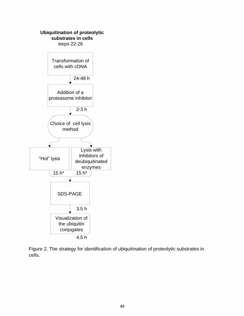
49
Figure 2. The strategy for identification of ubiquitination of proteolytic substrates in
cells.
“Hot” lysis
Lysis with
inhibitors of
deubiquitinated
enzymes
Transformation of
cells with cDNA
Addition of a
proteasome inhibitor
Choice of cell lysis
method
SDS-PAGE
Visualization of
the ubiquitin
conjugates
24-48 h
2-3 h
15 h* 15 h*
3.5 h
4.5 h
Ubiquitination of proteolytic
substrates in cellssteps 22-26

50
Figure 3. Summary of methods for detection of degradation of proteolytic substrates in
cells.
Transformation of
cells with cDNA
Choice of method for
monitoring protein
degradation
Cycloheximide
chase
Pulse-chase
labeling and
immunoprecipitation
24-48 h
60 h 60 h
SDS-PAGE
3.5 h
Visualization of
the protein
4.5 h
Degradation of proteolytic substrates in cells
steps 27-28

51
Figure 4. Ubiquitination of Ring1B in a cell free system.
In vitro-translated and 35S-labeled Ring1B was self ubiquitinated without addition of
ATP, Ub and E1/E2 (lane 1), in the presence of ATPS (lane 2), or ATP and ATP-
regenerating system (lane 3), in the absence of Ub (lane 4), in the absence of E1/E2
(lane 5), or in the presence of methylated ubiquitin (lane 6). “ATP +” denotes ATP and
ATP-regenerating system. MeUb denotes methylated ubiquitin, and Ub conj. denotes
ubiquitin conjugates.
Ub
co
nj.
– ++ –
+ + +
WT –
–
E1/E2
ATPS
1 2 3 4 5
ATP
Ub –
–
+
WT
6
+
WT Me
+ +
Ring1B
M
250
150
100
75
50
37
25

52
Figure 5. Ubiquitination of p105 in HEK 293 cells.
HEK 293 cells were transfected with cDNAs coding for Flag-p105 (lanes 1-2), and HA-
WT Ub (lane 1 and 3) or HA-UbK0 (lane 2). p105 and its conjugates were
immunoprecipitated using anti-Flag, and detected by either anti-Flag (i) or anti-HA (ii).
Flag-p105 + + + +
IP: Anti Flag
WB: Anti Flag
Flag-p105
Ub
co
nj.
1 2 3
IP: Anti Flag
WB: Anti HA
WT K0WTHA-Ub K0
(i) (ii)
1 2 3
–
WT WT
–
M
250
150
100
75
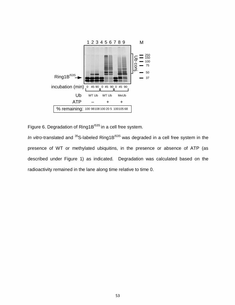
53
Figure 6. Degradation of Ring1BI53S in a cell free system.
In vitro-translated and 35S-labeled Ring1BI53S was degraded in a cell free system in the
presence of WT or methylated ubiquitins, in the presence or absence of ATP (as
described under Figure 1) as indicated. Degradation was calculated based on the
radioactivity remained in the lane along time relative to time 0.
Ub
1 2 3 4 5 6 7 8 9
incubation (min) 0 45
WT Ub MeUb
90 0 45 90 0 45 90
+
Ring1BI53S
% remaining:
Ub
co
nj.
WT Ub
ATP – +
100 2098 100108 5 100105 68
M
250150
100
75
50
37

54
Figure 7. Degradation of Mdm2 in HEK 293 cells using cycloheximide chase.
HEK 293 cells were grown to desired 75% confluency. Degradation of endogenous
Mdm2 was monitored following the addition of cycloheximide (lanes 1-3). Degradation
of Mdm2 was inhibited and the enzyme was accumulated following inhibition of the
proteasome by MG132 added for 2 hr (lane 4).
Tubulin was used as a marker to ascertain equal protein loading.
1 2 3 4
0 45 90Chx (min)
MG132 – – +
WB:Anti-HA
WB:Anti-tubulin
% remaining:
Mdm2
tubulin
100 33 9 311
M250150
100
75
50
M100
75
50

55
Figure 8. Monitoring of degradation of Myc-Ring1B using pulse-chase labeling
and immunoprecipitation.
Cos7 cells were transfected with a cDNA coding for Myc-Ring1B (lanes 1-3).
Stability of the protein was monitored in a pulse-chase labeling and
immunoprecipitation experiment. Immunoprecipitation was carried out using anti-
Myc antibody, and the proteins were visualized following SDS-PAGE, blotting
into nitrocellulose membrane. Radioactive Ring1B was detected using
PhosphorImaging (i) and total Ring1B in cells was detected using anti-Myc
antibody (ii).
Chase (hr) 0 4 6
WB:anti-9E10
100 60 31% remaining:
1 2 3(i)
(ii) 1 2 3
6 x Myc-Ring1B
6 x Myc-Ring1B
M250150
100
75
50
M100
75
50

56
REFERENCES
1. Mayer, R.J., Ciechanover, A., and Rechsteiner, M. Protein Degradation Handbook, (Weinheim, Germany: Wiley-VCH, 2005).
2. Mayer, R.J., Ciechanover, A., and Rechsteiner, M. Protein Degradation Handbook, (Weinheim, Germany: Wiley-VCH, 2006).
3. Mayer, R.J., Ciechanover, A., and Rechsteiner, M. Protein Degradation Handbook, (Weinheim, Germany: Wiley-VCH, 2006).
4. Mayer, R.J., Ciechanover, A., and Rechsteiner, M. Protein Degradation Handbook, (Weinheim, Germany: Wiley-VCH, 2008).
5. Scherer, D.C., Brockman, J.A., Chen, Z., Maniatis, T. & Ballard, D.W. Signal-induced degradation of I kappa B alpha requires site-specific ubiquitination. Proc. Natl. Acad. Sci. U S A 92, 11259-11263 (1995).
6. King, R.W., Glotzer, M. & Kirschner, M.W. Mutagenic analysis of the destruction signal of mitotic cyclins and structural characterization of ubiquitinated intermediates. Mol. Biol. Cell 7, 1343-1357 (1996).
7. Hou, D., Cenciarelli, C., Jensen, J.P., Nguygen, H.B. & Weissman, A.M. Activation-dependent ubiquitination of a T cell antigen receptor subunit on multiple intracellular lysines. J. Biol. Chem. 269, 14244-14247 (1994).
8. Ciechanover, A. & Ben-Saadon, R. N-terminal ubiquitination: more protein substrates join in. Trends Cell Biol. 14, 103-106 (2004).
9. Cadwell, K. & Coscoy, L. Ubiquitination on nonlysine residues by a viral E3 ubiquitin ligase. Science 309, 127-130 (2005).
10. Wang, X., et al. Ubiquitination of serine, threonine, or lysine residues on the cytoplasmic tail can induce ERAD of MHC-I by viral E3 ligase mK3. J. Cell Biol. 177, 613-624 (2007).
11. Tait, S.W., et al. Apoptosis induction by Bid requires unconventional ubiquitination and degradation of its N-terminal fragment. J. Cell. Biol. 179, 1453-1466 (2007).
12. Chau, V., et al. A multiubiquitin chain is confined to specific lysine in a targeted short-lived protein. Science 243, 1576-1583 (1989).
13. Xu, P., et al. Quantitative proteomics reveals the function of unconventional ubiquitin chains in proteasomal degradation. Cell 137, 133-145 (2009).
14. Kirkpatrick, D.S., et al. Quantitative analysis of in vitro ubiquitinated cyclin B1 reveals complex chain topology. Nat. Cell Biol. 8, 700-710 (2006).
15. Koegl, M., et al. A novel ubiquitination factor, E4, is involved in multiubiquitin chain assembly. Cell 96, 635-644 (1999).
16. Wang, C., et al. TAK1 is a ubiquitin-dependent kinase of MKK and IKK. Nature 412, 346-351 (2001).
17. Tokunaga, F., et al. Involvement of linear polyubiquitylation of NEMO in NF-kappaB activation. Nat. Cell Biol. (2009).
18. Rahighi, S., et al. Specific recognition of linear ubiquitin chains by NEMO is important for NF-kappaB activation. Cell 136, 1098-1109 (2009).
19. Ben-Saadon, R., Zaaroor, D., Ziv, T. & Ciechanover, A. The polycomb protein Ring1B generates self atypical mixed ubiquitin chains required for its in vitro histone H2A ligase activity. Mol. Cell 24, 701-711 (2006).
20. Robzyk, K., Recht, J. & Osley, M.A. Rad6-dependent ubiquitination of histone H2B in yeast. Science 287, 501-504 (2000).
21. Gregory, R.C., Taniguchi, T. & D'Andrea, A.D. Regulation of the Fanconi anemia pathway by monoubiquitination. Semin. Cancer Biol. 13, 77-82 (2003).

57
22. Vandenberg, C.J., et al. BRCA1-independent ubiquitination of FANCD2. Mol. Cell 12, 247-254 (2003).
23. Terrell, J., Shih, S., Dunn, R. & Hicke, L. A function for monoubiquitination in the internalization of a G protein-coupled receptor. Mol. Cell 1, 193-202 (1998).
24. Haas, A.L. & Rose, I.A. Hemin inhibits ATP-dependent ubiquitin-dependent proteolysis: role of hemin in regulating ubiquitin conjugate degradation. Proc. Natl. Acad. Sci. U S A 78, 6845-6848 (1981).
25. Johnston, N.L. & Cohen, R.E. Uncoupling ubiquitin-protein conjugation from ubiquitin-dependent proteolysis by use of beta, gamma-nonhydrolyzable ATP analogues. Biochemistry 30, 7514-7522 (1991).
26. Petroski, M.D. & Deshaies, R.J. Function and regulation of cullin-RING ubiquitin ligases. Nature reviews 6, 9-20 (2005).
27. Hershko, A. & Rose, I.A. Ubiquitin-aldehyde: a general inhibitor of ubiquitin-recycling processes. Proc. Natl. Acad. Sci. U S A 84, 1829-1833 (1987).
28. Murakami, Y., et al. Ornithine decarboxylase is degraded by the 26S proteasome without ubiquitination. Nature 360, 597-599 (1992).
29. Sheaff, R.J., et al. Proteasomal turnover of p21Cip1 does not require p21Cip1 ubiquitination. Mol. Cell 5, 403-410 (2000).
30. Chen, X., et al. N-acetylation and ubiquitin-independent proteasomal degradation of p21(Cip1). Mol. Cell 16, 839-847 (2004).
31. Amador, V., Ge, S., Santamaria, P.G., Guardavaccaro, D. & Pagano, M. APC/C(Cdc20) controls the ubiquitin-mediated degradation of p21 in prometaphase. Mol. Cell 27, 462-473 (2007).
32. Finley, D., Ciechanover, A. & Varshavsky, A. Thermolability of ubiquitin-activating enzyme from the mammalian cell cycle mutant ts85. Cell 37, 43-55 (1984).
33. Zeng, G.C., Donegan, J., Ozer, H.L. & Hand, R. Characterization of a ts mutant of BALB/3T3 cells and correction of the defect by in vitro addition of extracts from wild-type cells. Mol. Cell Biol. 4, 1815-1822 (1984).
34. Hershko, A., Heller, H., Elias, S. & Ciechanover, A. Components of ubiquitin-protein ligase system. Resolution, affinity purification, and role in protein breakdown. J. Biol. Chem. 258, 8206-8214 (1983).
35. Ciechanover, A., Heller, H., Elias, S., Haas, A.L. & Hershko, A. ATP-dependent conjugation of reticulocyte proteins with the polypeptide required for protein degradation. Proc. Natl. Acad. Sci. U S A 77, 1365-1368 (1980).
36. Hershko, A. & Heller, H. Occurrence of a polyubiquitin structure in ubiquitin-protein conjugates. Biochem. Biophys. Res. Commun. 128, 1079-1086 (1985).
37. Ben-Saadon, R., et al. The tumor suppressor protein p16(INK4a) and the human papillomavirus oncoprotein-58 E7 are naturally occurring lysine-less proteins that are degraded by the ubiquitin system. Direct evidence for ubiquitination at the N-terminal residue. J. Biol. Chem. 279, 41414-41421 (2004).
38. Polevoda, B., Norbeck, J., Takakura, H., Blomberg, A. & Sherman, F. Identification and specificities of N-terminal acetyltransferases from Saccharomyces cerevisiae. EMBO J. 18, 6155-6168 (1999).
39. Polevoda, B. & Sherman, F. Nalpha -terminal acetylation of eukaryotic proteins. J. Biol. Chem. 275, 36479-36482 (2000).
40. Boisson, B., Giglione, C. & Meinnel, T. Unexpected protein families including cell defense components feature in the N-myristoylome of a higher eukaryote. J. Biol. Chem. 278, 43418-43429 (2003).

58
41. Bohlen, P., Stein, S., Dairman, W. & Udenfriend, S. Fluorometric assay of proteins in the nanogram range. Arch. Biochem. Biophys. 155, 213-220 (1973).
42. Baldi, L., Brown, K., Franzoso, G. & Siebenlist, U. Critical role for lysines 21 and 22 in signal-induced, ubiquitin-mediated proteolysis of I kappa B-alpha. J. Biol. Chem. 271, 376-379 (1996).
43. Rodriguez, M.S., Desterro, J.M., Lain, S., Lane, D.P. & Hay, R.T. Multiple C-terminal lysine residues target p53 for ubiquitin-proteasome-mediated degradation. Mol. Cell Biol. 20, 8458-8467 (2000).
44. Kravtsova-Ivantsiv, Y., Cohen, S. & Ciechanover, A. Modification by single ubiquitin moieties rather than polyubiquitination is sufficient for proteasomal processing of the p105 NF-kappaB precursor. Mol. Cell 33, 496-504 (2009).
45. Cohen, S., Achbert-Weiner, H. & Ciechanover, A. Dual effects of IkappaB kinase beta-mediated phosphorylation on p105 Fate: SCF(beta-TrCP)-dependent degradation and SCF(beta-TrCP)-independent processing. Mol. Cell Biol. 24, 475-486 (2004).
46. Hershko, A., Ciechanover, A., Heller, H., Haas, A.L. & Rose, I.A. Proposed role of ATP in protein breakdown: conjugation of protein with multiple chains of the polypeptide of ATP-dependent proteolysis. Proc. Natl. Acad. Sci. U S A 77, 1783-1786 (1980).
47. Kim, H.T., et al. Certain pairs of ubiquitin-conjugating enzymes (E2s) and ubiquitin-protein ligases (E3s) synthesize nondegradable forked ubiquitin chains containing all possible isopeptide linkages. J. Biol. Chem. 282, 17375-17386 (2007).
48. Wu-Baer, F., Lagrazon, K., Yuan, W. & Baer, R. The BRCA1/BARD1 heterodimer assembles polyubiquitin chains through an unconventional linkage involving lysine residue K6 of ubiquitin. J. Biol. Chem. 278, 34743-34746 (2003).
49. Finley, D., et al. Inhibition of proteolysis and cell cycle progression in a multiubiquitination-deficient yeast mutant. Mol. Cell Biol. 14, 5501-5509 (1994).
50. Newton, K., et al. Ubiquitin chain editing revealed by polyubiquitin linkage-specific antibodies. Cell 134, 668-678 (2008).