ucd neuromuscular disease laboratory muscle biopsy...ucd neuromuscular disease laboratory 1 ......
TRANSCRIPT

UCD Neuromuscular Disease Laboratory
1
________________________________________________________________VETERINARY MEDICAL TEACHING HOSPITAL_
NEUROMUSCULAR DISEASE LABORATORY Monica Aleman, MVZ Cert., PhD, Dipl. ACVIM
Muscle biopsy
The appropriate muscle specimen to collect will depend on the clinical condition of the patient. If a generalized/diffuse myopathy (rhabdomyolysis, immune-mediated) is suspected, any muscle could be sampled. Whereas in focal myopathies (trauma, infection), the affected muscle must be collected to ensure a diagnostic specimen. The muscle biopsy should be obtained from a muscle that is definitely affected but no so severely wasted that much of it is replaced by fat or fibrous tissue. Ideally, the muscle should be one that has not been traumatized by injections or electromyographic (EMG) studies. In the horse, the gluteal muscle is routinely biopsied due to the ease of collection if appropriate instrument is used (example: Bergstrom biopsy needle or similar), and being one of the most studied muscles but other muscles are also useful. Muscle specimens approximately 1 cm long by 0.5 cm wide and 0.5 cm deep can be removed with minimal trauma (no cautery because it will destroy the sample). Block the skin and subcutaneous tissue but NOT the muscle itself and gently handle the sample (NO crushing, squeezing, or tearing). The best method for muscle collection is by an open surgical approach, but other modalities such as punch (superficial muscles) or Bergstrom biopsy needle (deep muscles such as gluteus medius) provide diagnostic specimens. The incision required to obtain a diagnostic muscle specimen is small and therefore the procedure is usually cosmetically innocuous. Complications secondary to the procedure are rare but may include local swelling, hematoma, and infection. Tetanus toxoid vaccination should be current and if not, vaccinate the horse.
(1) Top: scalpel blade and 8 mm skin punch biopsy. Bottom: Bergstrom biopsy needle with its components.
(2) Double Kelly forceps to be used with one hand to clamp a piece of muscle between the jaws of the forceps. This technique provides a 1.5 by 1.5 cm muscle specimen.

UCD Neuromuscular Disease Laboratory
2
Muscles commonly collected Semimembranosus muscle This muscle is commonly used for the diagnosis of exertional rhabdomyolysis including polysaccharide storage myopathy. After a local anesthetic and surgical preparation, a 2 cm vertical incision is made through the skin, subcutaneous tissue, and subcutaneous muscle (first exposed thin muscle). Incise fascia and once the semimembranosus muscle is exposed, two parallel vertical incisions 1 cm apart and 1 cm deep should be made. Grab both ends of the muscle with forceps and transect dorsally and ventrally to free the specimen. Close the subcutaneous tissue and skin. An alternative technique is by the use of an 8 mm skin punch biopsy. Approximately a 1.5 cm incision through the skin, and subcutaneous tissue is made. Once the semimembranosus muscle is visualized (not to confuse with the superficial subcutaneous muscle), a punch biopsy is collected. Both techniques provide diagnostic specimens. Gluteus medius muscle This muscle is useful for the diagnosis of exertional rhabdomyolysis and compartmental/pressure necrosis myopathy. Deeper muscles such as the gluteus medius may be more susceptible to ischemic or hypoxic events. For purposes of ease of collection and depth of this muscle, a Bergstrom biopsy needle or similar is preferred. This needle has an outer 6 mm diameter and a collection capacity of 250 mg of muscle 5 cm long. The landmarks are 20 cm along a line from the top of the dorsal tuber coxae to the base of the tail. The depth of collection is approximately about 7.5 cm in adult horses and mules, and less in younger horses, donkeys, zebras, ponies, and miniature breeds. Sacrocaudalis dorsalis muscle This muscle is the preferred muscle for the diagnosis of equine motor disease as it is composed of at least 20 to 30% of slow twitch fibers which are the most severely affected in this disorder. Other muscles such as the gluteus medius and semimembranosus muscles also have slow twitch fibers; however the ease of collection along with fiber type distribution of the sacrocaudalis dorsalis muscle makes this muscle ideal for collection and analysis. The muscle is located dorsal-lateral to the spinous processes of the coccygeal vertebra. Surgical incision or punch biopsy provides diagnostic specimens.

UCD Neuromuscular Disease Laboratory
3
(3) The shaved areas depict the area for gluteus medius muscle biopsy, and the black line indicates the site for biopsy of the semimembranosus muscle. The surgical incision required is smaller than the one shown here.
(4) Bergstrom needle biopsy for gluteus medius muscle biopsy. (5) Area for sacrocaudalis dorsalis lateralis muscle at the base of the tail.

UCD Neuromuscular Disease Laboratory
4
NDL Diagnostic tests The following diagnostic techniques are offered at our laboratory for various animal species. A few examples of diseases are provided. Histochemistry Cell types and morphology
Hematoxylin and eosin Inflammatory/immune-mediated myopathies*
Modified Gomori trichrome (connective tissue, nerve) Fiber typing
Myofibrillar ATPase at various pH (fiber type differentiation, non-neurogenic and neurogenic atrophy)
pH 9.8 Types 1 and 2 myofibers pH 4.6 Types 1, 2A, and 2B pH 4.3 Types 1, 2, 2C (immature, neonates, regenerating fibers) Enzymes: oxidative (mitochondria)
Nicotinamide adenine dinucleotide Succinate dehydrogenase Cytochrome oxidase Enzymes: glycolytic
Phosphorylase Phosphofructokinase Enzymes: hydrolytic
Esterase (macrophages—lysosomes, end plates) Acid phosphatase (macrophages—lysosomes, lipofuscin) Alkaline phosphatase (regenerating fibers, fibroblasts) Storage material Periodic acid Schiff for glycogen Amylase digestion assay Polysaccharide storage myopathy (PSSM) Genetic test for PSSM type 1 at the University of Minnesota (see link) Glycogen branching enzyme deficiency (GBED) Genetic test at UCD, Veterinary Genetics Lab (see link) Oil red O for lipid Pituitary Pars Intermedia Dysfunction (PPID) Immunohistochemistry *Cell type identification (examples: CD4, CD8, CD11a, CD20, and more) in collaboration with our pathology department
Presence of IgG antibodies Immune-mediated disorders
Dystrophin and spectrin Muscular dystrophy
Molecular diagnostics Equine malignant hyperthermia (MH) The test can be performed in various tissues including hair, blood, or muscle. Others There are additional tests that we can perform if indicated in collaboration with other laboratories.
UCD Neuromuscular Disease Laboratory
5
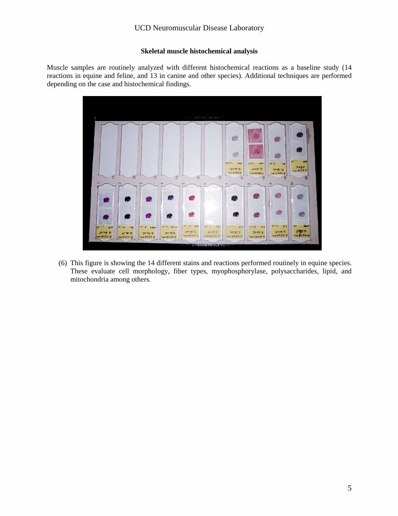
Skeletal muscle histochemical analysis Muscle samples are routinely analyzed with different histochemical reactions as a baseline study (14 reactions in equine and feline, and 13 in canine and other species). Additional techniques are performed depending on the case and histochemical findings.
(6) This figure is showing the 14 different stains and reactions performed routinely in equine species. These evaluate cell morphology, fiber types, myophosphorylase, polysaccharides, lipid, and mitochondria among others.
UCD Neuromuscular Disease Laboratory
6
Tissue collection and shipment
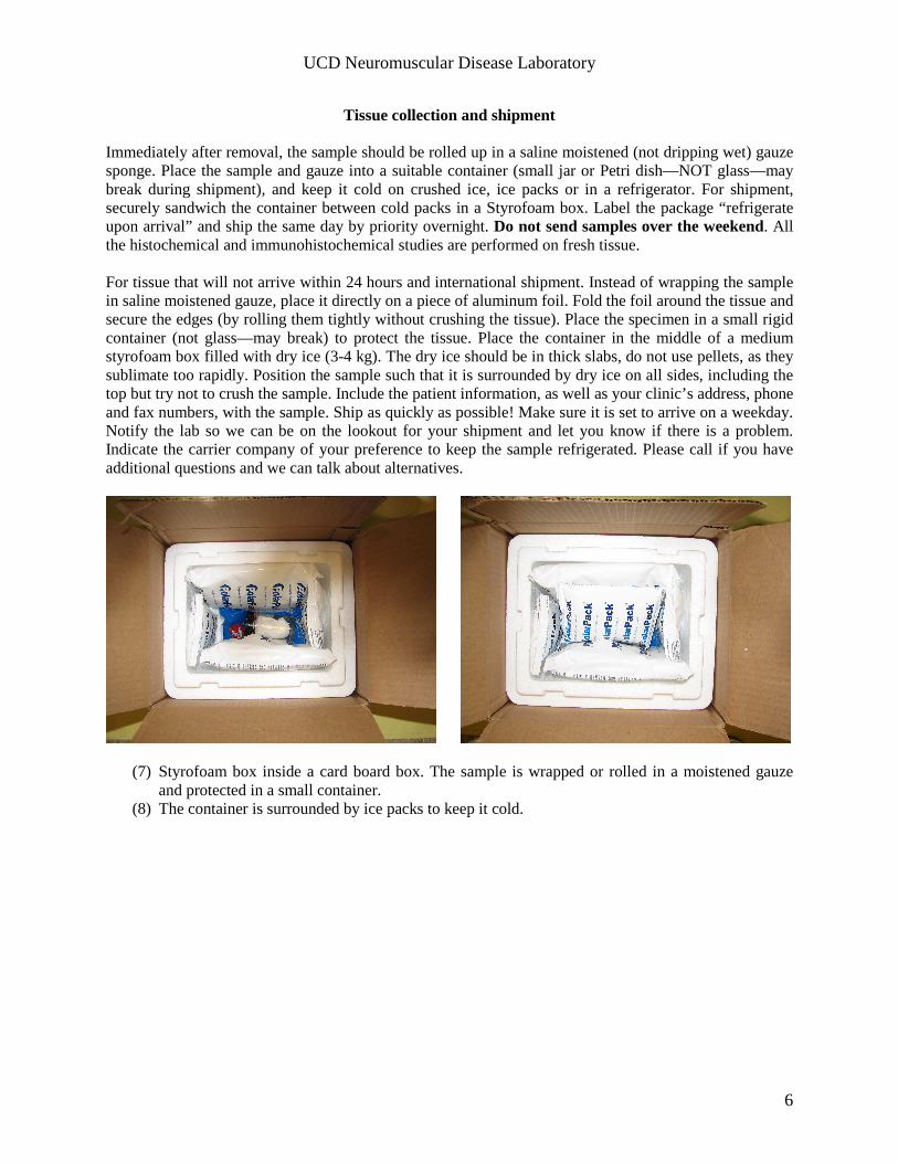
Immediately after removal, the sample should be rolled up in a saline moistened (not dripping wet) gauze sponge. Place the sample and gauze into a suitable container (small jar or Petri dish—NOT glass—may break during shipment), and keep it cold on crushed ice, ice packs or in a refrigerator. For shipment, securely sandwich the container between cold packs in a Styrofoam box. Label the package “refrigerate upon arrival” and ship the same day by priority overnight. Do not send samples over the weekend. All the histochemical and immunohistochemical studies are performed on fresh tissue. For tissue that will not arrive within 24 hours and international shipment. Instead of wrapping the sample in saline moistened gauze, place it directly on a piece of aluminum foil. Fold the foil around the tissue and secure the edges (by rolling them tightly without crushing the tissue). Place the specimen in a small rigid container (not glass—may break) to protect the tissue. Place the container in the middle of a medium styrofoam box filled with dry ice (3-4 kg). The dry ice should be in thick slabs, do not use pellets, as they sublimate too rapidly. Position the sample such that it is surrounded by dry ice on all sides, including the top but try not to crush the sample. Include the patient information, as well as your clinic’s address, phone and fax numbers, with the sample. Ship as quickly as possible! Make sure it is set to arrive on a weekday. Notify the lab so we can be on the lookout for your shipment and let you know if there is a problem. Indicate the carrier company of your preference to keep the sample refrigerated. Please call if you have additional questions and we can talk about alternatives.
(7) Styrofoam box inside a card board box. The sample is wrapped or rolled in a moistened gauze and protected in a small container.
(8) The container is surrounded by ice packs to keep it cold.

UCD Neuromuscular Disease Laboratory
7
International shipping Please MAKE SURE TO REQUEST the USDA permit for importation and transportation of animal tissue (total of 2 pages). The permit can be obtained with us at +(530)-752-1170 or +(530)-752-7267. The permit will be faxed to you immediately. You should add the permit to your shipment. Samples can also be frozen, please call us to instruct you on how to do this. Prepared/cut and mounted slides can also be sent to us for staining. Cost The cost for our routine evaluation (14 in the case of the horse) is of $95.00 per sample. If additional tests are required, there will be a separate charge for the specific test (call our laboratory for more information). The cost for the genetic test for malignant hyperthermia is of $65.00 per horse.