ufic file copy i - apps.dtic.mil · dence for water-born chemoreception is limited to a single...
TRANSCRIPT
(U) UFIC FILE CopyI SE, or, Fce
MENTATION PAGE Orm Aoproved
Ol lb RESTRICTIVE MARY NGS
T.AD-A222 648 k-iN/2aE 3 DISTRIBuTIOWAVAILABILITY OF REPORT
2b DECLASSIFICATIONDOWNGR 6 t a 19 Distribution Unlimited
4 PERFORMING ORGANIZATION NUMBE S) S MONITORING ORGANIZATION REPORT N ,MBFR(S)
Stanord nivesity -VbN/A6a NAME OF PERFORMING ORGANIZATION 6b OFFICL SYMBOL 7a NAME OF MONITORING ORGANZA' ON
(if applicable)
Stanford University N/A Office of Naval Research
6c ADDRESS (City, State, and ZIP Code) 7b ADDRESS (City, State, and ZIP Code)
Hopkins Marine Station 800 N.Quincy StreetPacific Grove, CA 93950 Arlington, VA 22217-5000
8a NAME OF -UNDING SPONSORtNG Bb OFFICE SYMBOL 9 PROCUREMENT INSTRuMENT DENT!F,CA TiON NUMBER
ORGANIZATION (If applicable)
Office of Naval Research ONR N00014-89-J-17448c ADDRESS (City, State, and ZIPCode) 10 SOURCE OF FUNDING NuMBERS
PROGRAM PROJECT TASK WORK UNiT
800 N. Quincy Street ELEMENT NO NO NO ACCESSION NO
Arlington, VA 22217-5000 61153N RR04108 441490611 TITLE (Include Security Classification)
(U) Evidence for chemoreception in squid olfactory organ'2 PERSONAL A,.,THOR(S)
Gilly, William Frank; Horrigan, Frank Theodore; Lucero, Mary Theresa13a 'PE OF REPORT T,3b TIME COVERED 14 DATE OF REPORT (Year, Month Day) T5 PACE CO ,jN
AnnualI ;ROM5 a9 TO 59O 5/29/90 2716 SuPPLEMEN'ARY NOTATION
17 COSATI CODES 16' SUBJECT TERMS .Conr nue on reverse if necessary and identify by block number)
;IELD GROUP SUB-GROUP SQUID, olfaction, behavior, chemoreceptor cells, ion
channels, electrophysiology, escape response motor controliet-propulsion, signal transduction . .
19 ABSTRACT (Continue on reverse if necessary and identify by block number)
'We have examined the effects of chemical stimuli on the putative olfactory organ of the squid Loligo opalescens.Chemosensory capabilities were studied both at the behavioral level in living squid and at the individual receptor cell levelusing whole-cell voltage clamp. We found that low concentrations of certain test substances reproducibly eficited escaperesponses in living, restrained squid. Taking advantage of this link between chemoreception and motor pathways, we wereable to map the region of highest chemosensitivity directly to the olfactory organ which is a small knob located in an ear-like flap lateral to each eye. 'Ablation' experiments, which were performed by treating the olfactory organ with a localanesthetic, further confirmed that the olfactory organ was the site of chemoreception. In examining isolated receptor cells,we found at least three morphologies, similar to those described by Emery in ultrastructural studies (1975). Voltage clampexperiments on the two most common cell types (pyriform and floriform) showed that they contain neuronal-like Na and Kchannels. We tested several chemicals including propyl-paraben, 4-amino-pyridine, methadone, and a snail hypobranchialgland extract, and found that every substance which elicited escape responses was also a potent blocker of K currents in thereceptor cells. Since the different chemical block K channels via different mechanisms, we propose that for thesesubstances, K channel block may be the relevant mode of signal transduction.
20 DiSTRIBIJTION AVALABtLITY OF ABSTRACT 21 ABSTRACT SECURITY CLASSIFICATION
2l UJNCLASSIFED UNLIMITED 0l SAME AS RPT [J OTIC USERS (U)
22a NAME OF RESPO)ISIBLE ,NDIVIOUAL 22b TELEPHONE (Include Area Code) .2 OFFICE SYMBOL
Dr. J.A. Maide (or other ONR Sci. Off.) .(202) 69-405 ONRDD Form 1473, JUN 86 Previous editions are obsolete -SECURITY _LASS FICATON OF THIS PAGE-
S/N U102-LF-014-6603 (U)

EVIDENCE FOR CHEMORECEPTION
IN SQUID OLFACTORY ORGAN
TJ3
William F. Gilly ®r
Frank T. Horrigan
Mary T. Lucero T W I]
J.
Annual ONR Progress Report
Grant # N00014-89-J-1744 D -,1:.
- I des
.... .. [.. ... ....1
INTRODUCTION
Cephalopods are arguably the most intelligent and responsive of
invertebrates. Squid, cuttlefish, and octopus are highly mobile
predators which are largely visually oriented (Packard, 1972), but are
also equipped with complex vestibular (Budelman, 1987), auditory
(Hanlon & Budelman, 1987) and tactile (Wells, 1964; Wells & Young,
1975) capabilities. These modalities converge in the central nervous
system to regulate specific motor outputs of great biological impor-
tance, e.g. escape jetting, chromatophore display, mating, homing, and
long-distance migration (Messenger, 1983; Boyle, 1986).
Although an important body of work exists concerning the chemotac-
tile sense in octopods (Graziadei, 1962; Wells, 1983; Wells, Freeman &
Ashburner, 1965), virtually nothing is known about chemoreception of
water-born molecules in these or other cephalopods (Boyle, 1986).
Morphological studies have revealed putative chemosensory cells in
several tissues, including an elaborate 'olfactory organ' (Graziadei,
1965; Woodhams & Messenger, 1974; Emery, 1975), but behavioral evi-
dence for water-born chemoreception is limited to a single report on
Octopus (Chase & Wells, 1984). In no case has a chemosensory function
for the olfactory organ been demonstrated, nor have receptor cells
been identified physiologically.
Squid have surprisingly received no behavioral or physiological
attention in these areas, despite an existant anatomical literature on
the olfactory organ (Emery, 1975) and on the central projections of
axons from the putative receptor cells of this organ (Messenger,
1979). Afferent tracts lead to the motor centers which control swim-
ming and escape jetting, and direct connections to the giant fiber
pathway may occur. The anatomical basis thus exists for an important
chemosensory element in the neural control of escape jetting in squid,
a subject which itself has only recently come to be reinvestigated
1

(Otis & Gilly, 1990) after a hiatus of 30 years (Wilson, 1960).
Although the basis of water-born chemoreception in cephalopods
and the behavioral outputs controlled by projections of the receptor
cells into the central nervous system are important questions, an
even more general set of questions concerns the primary mechanisms of
chemosensory transduction at the level of the receptor cell membrane.
Nothing is known about cephalopods in this regard, and very little is
presently known even in those groups, such as insects (Kaissling,
1986), which have historically provided much of our understanding of
the sensory physiology involved in olfaction. Much of the recent
progress in this area has come from vertebrate chemoreceptor (taste
and olfactory) cells dissociated from sensory epithelia of several
species and studied with patch voltage clamp techniques. Two major
themes on transduction mechanisms emerge from these studies.
First, vertebrate olfactory (Trotier, 1986; Maue & Dionne, 1987)
and taste (Kinnamon & Roper, 1988) receptor cells are excitable and
show a variety of conventional 'neuronal' ion channel types when
studied in vitro with patch clamp techniques. The idea that activity
of 'conventional' channel types can be directly modulated by odorant
molecules stems from experiments with model membrane systems and
channels reconstituted from olfactory epithelium which demonstrated
apparently direct activation of K-selective (Vodyanoy & Murphy, 1983)
and cation-selective channels (Labarca, Simon & Anholt, 1988) by
nanomolar levels of odorant molecules. In the patch clamp studies, a
decrease of voltage-dependent K conductance was found in taste cells
in response to sour (citric acid) and bitter (quinine) stimuli (Kinna-
mon & Roper, 1988). In experiments rn olfactory receptors, applica-
tion of odorant stimuli resulted in " a specific effects which could
not be associated with any particular type of channel (Trotier, 1986).
Another class of transduction mechanisms is thought to involve
2

indirect activation of ion channels by intracellular cyclic nucleo-
tides generated by odorant-dependent cyclases (Pace, Hanski, Salomon &
Lancet, 1985; Sklar, Anholt & Snyder, 1986). Although the specific
channels responsible have not yet been identified, micromolar levels
of cAMP significantly decrease the apparent membrane resistance of
excised patches from ciliary or somatic membrane of amphibian olfacto-
ry receptors (Nakamura & Gold, 1987). It has been suggested that cAMP
activation of a relatively non-selective channel may be a basis of
olfactory transduction, in analogy to the well established case for
cGMP in visual receptor cells (Yau & Baylor, 1988). In principle
other intracellular messengers could also act in this way.
What classes of odorants directly or indirectly activate normally
silent channels in receptor cells, block normally open channels, or
modulate voltage-dependent channels -- and where, and at what density,
these channels are located on the receptor cells -- remain as major
questions.
The first part of this paper addresses the chemosensory capabili-
ties of squid. We provide behavioral evidence for detection of water-
born chemical stimuli by the olfactory organ. These experiments also
demonstrate that the olfatory organ projections must converge on motor
centers controlling escape jetting. The second part of this paperdescribes voltage clamp studies of chemosensory cells dissociated from
the sensory epithelium of the olfactory organ. Like vertebrate chemo-
receptors, those in squid are excitable and very 'neuronal-like' in
their electrical properties. An important transduction mechanism may
involve the blocking of voltage-controlled K channels by a variety of
compounds, all of which act behaviorally to elicit escape responses
when applied to the olfactory organ in vivo.
METHODS
Behavioral experiments. All experiments were carried out on living,
but restrained, adult Loligo opalescens with techniques similar to
3

those described in Otis & Gilly (1990). Chemical stimuli were deliv-
ered by pressure ejection. 75-300 ms duration pulses delivered fluid
at a rate of 1 ul/ms from a port 0.65 mm in diameter. Test substances
were extruded only during pressure pulses; at other times the line was
closed to prevent leakage out of or siphoning back into the supply
line. The stimulating probe assembly also contained a pair of small
Pt wires and an optic fiber to transmit electrical and visual stimuli.
Pressure within the mantle cavity was monitored with a pressure trans-
ducer, and only responses which produced measurable pressure rises
were scored as positive escape responses. Some animals were also
videotaped in order to verify the spatial location of the stimulus
plume relative to the olfactory organ in the mapping experiments.
Dissociation of receptor cells and voltage clamp experiments. The
olfactory organ in Loligo, as in Lolliguncula (Emery, 1975), is a
small knob located at the bottom of the cavity formed by an ear-like
structure on the lateral aspect of the head (See Fig. 2 and Emery,
1975). The olfactory knob was excised and treated with non-specific
protease (10 mg/ml Sigma Type XIV) in sterile-filtered sea water at
18-20 0 C for 1 hr and then placed in a primary tissue culture medium as
described by Brismar & Gilly (1987). Cells were obtained for study by
simply dipping the piece of tissue into a drop of solution in the
experimental chamber. Cells were utilized immediately after dispersal
from olfactory knobs which had been cultured (150 C) for no more than
48 hrs. After this time cells were still viable, but morphological
characteristics (see below) became blurred.
Whole-cell voltage clamp experiments were carried out at 100 C
following standard procedures (Brismar & Gilly, 1987; Gilly & Brismar,
1989). The recording pipette was filled with an internal solution of
50 mM K glutamate, 50 mM KF, 400 mM tetramethylammonimum glutamate, 10
4
mM Na 2 -EGTA and 10 mM Hepes (pH 7.4). The external artificial sea
water (ASW) contained 450 mM NaCl, 10 mM KCI, 10 mM CaC 2, 50 mM
MgCl2 , and 10 mM Hepes (pH 7.6). The internal solution for studies on
Na channels contained 220 mM NaCl,230 mM TEA, 10 mM Na2 - EGTA , and 10
mM HEPES. External Na channel solution contained 450 mM NaCl, 10 mM
CsCl, 0 mM KCl, 10 mM HEPES, 10 mM CaC 2 and 50 mM MgCl 2 . Holding
potential was -70 mV.
RESULTS
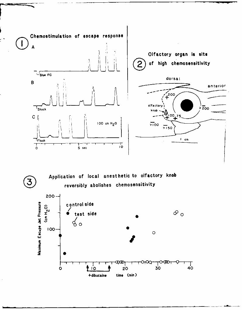
Identification of Chouoreoeption and Links to Motor Control.
Small volumes (< 100 ul) of sea water containing certain test
substances at low concentration were routinely found to elicit escape
responses when pressure ejected near the olfactory organ of a living
squid. Figure 1A shows mantle pressure following delivery of a 300 ms
pulse of blue food coloring (Schilling, McCormick, Inc., Baltimore,
MD) at a 1:500 dilution. The pressure transients are similar to those
following an electrical shock (Fig. 1B) or a strobe light flash (Fig.
IC) delivered to the same area between the olfactory organ and the
eye. All 3 forms of stimulation thus lead to escape behavior involv-
ing high pressure jet propulsion. Delays for chemical and electrical
stimuli are highly variable and always much longer than the delay for
visual stimulation. These differences are in large part due to which
motor system is first activated, the giant fiber pathway in the case
of visual stimuli vs. a small fiber system in the other two cases
(Otis & Gilly, 1990).
Blue food coloring was originally intended to serve as an inert
tracking dye, and its efficacy in stimulating escape responses was
surprising. This agent is a mixture of 2M propylene glycol, 20 mM
Brilliant Blue FCF and 10mM propyl paraben (4-hydroxybenzoic acid
propyl ester), an antifungal preservative. Each of these constituents
was independently tested in behaviorai experiments which showed unam-
biguously that propyl paraben was the only substance that could act
5

alone to stimulate escape responses.
In two different animals, a total of 15 trials with 1:500 blue
food coloring (in sea water) produced 9 positive escape responses,
whereas 11 trials with 50 uM Fast Green as a control yielded no re-
sponses. In a third animal 50 uM propyl paraben elicited escape jets
in 10 of 17 trials, and 100 uM Brilliant Blue and 100 uM Fast Green
were completely uneffective (4 trials each). Several experiments with
propylene glycol were also negative.
Threshold for activity of blue food coloring was 1:1000 (1 of 7
trials), corresponding to a propyl paraben a concentration of 10 uM.
Because of the method for scoring a positive reaction (high pressure
escape response), this figure is an upper limit for detectability.
Localization of the Chemoreceptive Site to the Olfactory Organ.
Two approaches were taken to localize the area of chemorecep-
tion. The first was to spatially map sensitivity to a potent stimulus.
With the stimulating probe positioned directly adjacent to the olfac-
tory knob (a few mm away), a threshold pulse duration of 100 ms was
observed for 100 uM propyl paraben to produce an escape response. The
probe was then moved to a new site, and another estimate of threshold
pulse duration was obtained. Figure 2 shows threshold duration at
various positions; + indicates a response for the given duration, -
indicates no response with the maximum duration used. Similar mapping
experiments were carried out on 4 additional animals, and in every
case, small displacements of the stimulus plume from the olfactory
knob decrease the efficacy of a given stimulus, consistent with the
proposition that the olfactory knob is a chemoreceptive organ which
can influence the motor centers that control escape jetting.
b A second way of localizing chemoreception to the olfactory knob
involved reversibly blocking function by treating this structure with
a potent, short-acting local anesthetic to impair transduction in the
6
receptor cells and/or afferent transmission in sensory axons of the
olfactory nerve. Anesthetic treatment was performed on one side of
the head; the contralateral untreated organ served as a control.
Figure 3 shows results obtained in such an experiment. High-
pressure escape jets are produced by every stimulus (150 ms pulse of
100 uM propyl paraben) on both the control and test organs prior to
the application of 10 ul of 0.5 mM dibucaine (in natural sea water) to
the right olfactory knob. Function on this side was abolished for
approximately 30 min, after which recovery occurred. The control side
remained responsive throughout the period of dibucaine block. These
'ablation' experiments thus lend additional support to the idea that
the olfactory organ mediates detection of propyl-paraben in the ambi-
ent sea water. Presumably this is a noxius stimulus, as evidenced by
the strong escape response.
Detection of Other Substances by the Olfactory Organ
Experiments similar to those described above in conjunction with
testing the efficacy of blue food coloring and propyl paraben as
activators of escape jetting were carried out with a variety of other
substances. Table I summarized results with several agents that
consistently produced positive responses. These particular substances
were focused on because of their ability to block voltage dependent K
channels, a property which they share with propyl paraben (See also
below). Spatial mapping of sensitivity to methadone also yielded
results in agreement with those described above for propyl paraben,
and presumably the olfactory organ mediates detection of all of these
substances.
Positive results were also obtained with a 1:10 dilution of a
crude extract of the hypobranchial gland from Calliostoma canilicula-
tum, a subtidal snail which produces a noxius defensive secretion in
response to stimulation by contact with predatory starfish (Smaby,
1988). In a single animal 11 out of 18 trials with this extract
7
elicited escape jets (0 out of 12 for sea water controls). Mapring
experiments were not carried out in this case, however.
Other substances tested did not produce escape jets, and these
results are summarized in Table II. Negative results reported here
cannot be taken to imply that the squid cannot detect any of these
substances, however, because our assay for a positive response proba-
bly rules out all but the most noxious stimuli. Upon exposure to some
of the substances in Table II animals would often appear to display a
change in respiratory rhythm, but quantitation of such responses was
not attempted.
Morphological Identification of Receptor Cell Types.
Figures 4A-F show examples of freshly dissociated olfactory knob
cells. At least three kinds of putative chemoreceptor cells are
recognizable. The most common type (Fig. 4A,B) is pyriform with a
long axonal process (ax) arising from the pole containing the nucleus
(n) and a spike-like cilium which projects from the opposite pole
(arrow). Most of the cell body appears to be filled with granular
material or cristae, and the bulk of the cell's 'interior' is thought
to be an invaginating extracellular cavity filled with cilia (Emery,
1975). These cells are undoubtedly equivalent to the 'Type 4' chemo-
receptor cells described on ultrastructural grounds by Emery (1975).
The inset to Fig. 4B is taken from his paper and illustrates this
receptor type. A low-power scanning electron micrograph of an alde-
hyde-fixed pyriform receptor is shown in Fig. 6G; the spike-like
cilium is evident.
Another receptor cell type is floriform (Figs. 4C,D). This type
also displays a prominent axonal process, but in this case a long,
thin neck extends from the cell body and is crowned by a swelling
covered with non-motile cilia arranged in petaloid fashion (arrow).
A close correspondence to Emery's Type 2 (Fig. 4C) or Type 5 (Fig. 4D)
8

receptor is apparent. Figures 4H,I are scanning electron micrographs
of a fixed floriform receptor.
A third, less common receptor may be a modified pyriform type
based on the presence of a 'spike'-cilium on the apical pole (Figs.
4E,F). Unlike the pyriform receptors described above, however, this
type is elongate with a constriction of varying severity between the
cell body proper, which contains the nucleus, and the apical portion
which is filled with the granular material. This type of receptor
appears equivalent to Emery's Type 3 (Fig. 4G).
Neuronal-Like Sodium and Potassium Currents in Receptor Cells
Voltage clamp experiments using the 'whole-cell' recording mode
of patch voltage clamp were carried out on both pyriform and floriform
receptor cells dissociated from the olfactory knob. Figure 5A shows
Na currents (obtained by TTX subtraction) recorded for voltage steps
ranging from -40 to + 70 mV in a pyriform receptor bathed in normal
artificial sea water and internally dialyzed with a high-sodium solu-
tion. The rapidly activating, transient currents show a definite
reversal potential of -+45 mV (Fig. 5B). In other experiments (not
illustrated) only inward currents flow if Na ions are omitted from the
internal solution. Inactivaton is steeply voltage-dependent between
-50 and 0 mV and is half-maximal at - -25 mV.
All of the above properties are those expected for-conventional,
neuronal-like Na channels, and additional analyses (not illustrated)
indicate that the TTX-sensitive Na current in pyriform receptors is
very similar to that found in squid giant axon and giant fiber lobe
(GFL) neurons (Gilly & Brismar, 1989). Figures 5C,D demonstrate that
similar Na currents also exist in floriform receptor cells, but these
are much smaller than the corresponding currents recorded in pyriform
cells.
Other experiments were designed to study voltage-dependent K
currents (i.e., Na-free solutions with TTX present), and a series of
9

currents recorded between -20 and +70 mV from another pyriform recep-
tor cell is illustrated in Fig. 6A. Outward currents activate with a
marked, voltage-dependent delay, and inward tail currents flow upon
termination of the pulses. Analysis of reversal potential indicates
that these currents are carried by K-selective channels (not illus-
trated), and the kinetic properties (Fig. 6A) and voltage-dependence
of the peak K conductance (Fig. 6C) are very similar to analogous data
obtained in squid giant axon or GFL neurons (Llano & Bookman, 1986).
As in squid GFL cells, the K currents in pyriform receptors inactivate
to a large degree during a long depolarization (Fig. 6B).
Floriform receptor cells also have voltage-dependent K currents
that are qualitatively similar to (but consistently smaller than)
those in pyriform cells (Figs. 6D-F). In this case, however, the
kinetics of inactivation are slightly more rapid than than those in
pyriform cells.
Pharmacological Block of Potassium Currents in Receptor Cells
4-aminopyridine (4AP) is both a classical blocker of (closed) K
channels (Yeh, Oxford & Narahashi, 1976) and a potent activator of
escape jetting in living squid. The effects of this substance were
therefore studied on K currents in dissociated receptor cells. Figure
7A shows Na and K currents at +30 mV in a pyriform receptor before and
after (+4AP) bath application of 5 mM 4AP. Outward K current is
reduced by roughly 50% (with no obvious change in time course), but
there is no detectable block of inward Na current. Figure 7C illus-
trates analagous results from a floriform receptor cell. 4AP thus
selectively blocks K currents in both types of receptor cells.
Methadone is a substance which is known to block open K channels
(Horrigan, 1990) and which also elicits strong escape jets in behav-
ioral experiments when it is applied to the olfactory organ. Figures
7B,D show the effect of 500 uM methadone on Na and K currents in a
10

pyriform and floriform cell, respectively. This drug, unlike 4AP, is
a potent blocker of both Na and K currents. Non-selective block of
both currents was also found in both types of receptor cells for
propyl paraben (not illustrated), a substance which also reliably
evokes escape responses in living squid.
DISCUSSION
The squid's 'olfactory organ' was not named for its chemosensory
capabilities (which were at that time untested) but for its location
and morphology (Emery, 1975). It is ideally located to detect water-
born chemicals during the inspiratory phase of respiration and is
coated with mucus and cilliated epithelial cells which may help to
capture and concentrate chemical stimuli.
The putative olfactory receptor cells themselves are reminescent
of olfactory cells in both vertebrates (Troiter, 1986) and inverte-
brates (Kaissling, 1986) with unipolar projections to the central
nervous system. Their morphology shows the classic pear-shaped cell
body with a narrowed stalk and a ciliated knob facing out into the
external mileau. Like other chemosensory cells, squid olfactory cells
contain voltage gated Na and K channels. Recent whole cell current
clamp experiments show that squid olfactory cells are electrically
excitable and capable of firing repetitive action potentials. (Data
not shown).
Our work represents the first behavioral and electropyhsiologi-
cal studies performed on squid olfactory organ. We find that the
'olfactory organ' of squid is indeed a chemosensory structure, at
least capable of sensing noxious water-born chemicals and translating
chemosensory information via higher pathways into an appropriate
escape response. Olfactory input to the squid's major defense system
(escape jetting) could increase survivability in nocturnal or low
visibility situations as well as toxic or heavily polluted waters.
Propyl-paraben, a component in blue food coloring, elicits strong
11

escape responses in living squid, and blocks K currents in isolated
voltage clamped olfactory cells. Assuming that blocking K channels
would depolarize the receptor cell causing it to become hyperexcitable
and/or increase its spontaneous firing rate, we tested the potassium
channel blockers 4AP and TEA, and found that they also elicit escape
responses. Voltage clamp experiments show that 4AP blocks only K
channels. Classically, TEA blocks only K channels but voltage clamp
experiments using TEA are not yet completed. It is likely that TEA
will be similar to 4AP Lased on each chemical's ability to block K
channels in other systems (Yeh, Oxford & Narahashi, 1976; Hille,
1984).
Methadone and propyl-paraben elicit escape responses and block
both Na and K currents in isolated olfactory receptor cells. Although
untested, it is possible that an escape response could be elicited by
a chanqe in spontaneous activ ty- whether it be an increase or a
decrease in activity.
Interestingly, 4AP, propyl paraben, TEA, and methadone, have very
different chemical structures and mechanisms for K channel block, but
all of these substances elicit escape responses in squid. We propose
that that K channel block itself may be important for signal transduc-
tion.
Without disregarding the possibility that block of Na channels
can also lead to signal transduction, one can also imagine that Na
channels in squid olfactory receptor cells may be spatially arranged
in a fashion similar to taste receptor (Kinnamon & Roper, 1988) where
in vivo tight junctions prevent exposure of specific channel popula-
tions to the external media. It will be interesting to conduct map-
ping experiments to test if ion channels are localized to specific
regions in squid olfactory receptor cells.
There are many activities besides escape and avoidance where
12

chemoreception may be important to squid (ie. mating, migration, and
feeding). Future experiments could make use of other behavioral assays
such as respiratory rate or mating behavior for identifying physiolog-
ically important substances. These substances in turn would be used
to investigate other mechanisms of chemosensory transduction in squid
olfactory cells.
13

REFERENCE8
Armstrong, C.M. & S.R. Taylor. 1980. Interaction of barium ions withpotassium channels in squid giant axons. Bioh . 3:473-488.
Boyle, P.R. 1986. Neural control of cephalopod behavior in TheMolluscs, v.9,pt. 2, A.O.D. Willows (ed.), Aacademic Press,NY, pp 1-99.
Brismar, T. & Gilly, W.F. 1987. Synthesis of sodium channels in thecell bodies of squid giant axons. Proc. Natl. A cad.S. USABA: 1459-1463.
Budelmann, B.U. 1987. Morphological diversity of equilibriumreceptor systems, in aquatic invertebrates, in Sensory biolo-gy of Acruatic Animals, J. Atema, R.R. Fay, A.N. Popper & W.N.Tavolga (eds.), Springer-Verlag, NY, 757-782.
Carbone, E., R. Fioravanti, G. Prestipino & E. Wanke. 1978. Action ofextracellular pH on Na and K membrane currents in the giantaxon of Loligo vulgaris. J. Memb. Biol. 43: 295-315.
Chase, R.J. & Wells, M.J. 1986. Chemotactic behavior in Octopus. J.Comp. Physiol. A 158: 375-381.
Emery, D.G. 1975. The histology and fine structure of the olfactory organ of the squid Lolliqucula brevis. Tissue & Cell 7:357-367.
Gilly, W.F. & Brismar, T. 1989. Properties of appropriately and inappropriately expressed sodium channels in squid giant axon andits somata. J. Neurosci. 9(4): 1362-1374.
Graziadei, P. 1965. Sensory receptor cells and related neurons in cephalopods. Cold Spr. Hrb. Symp. Quant. Biol. 30: 45-57.
Hanlon, R.T. & Budelman, B.-U. 1987. Why cephalopods are probably not"deaf." Am. Naturalist 129: 312-317.
Hille, B. 1984. Ionic Channels of Excitable Membranes. SinauerAssociates Inc., Sunderland, MA. 280-285.
Horrigan, F.T. 1990. Methadone block of neuronal K current. Biophysical Journal 57: 515a.
Kaissling, K.A. 1986. Chemo-electrical transduction in insectolfactory receptors. Ann. Rev. Neurosci. 9: 121-45.
Kinnamon, S.C. & Roper, S.D. 1988. Membrane properties of isolated mudputaste cells. J. Gen. Physiol. 91: 351-371.
Labarca, P., Simon, S.A. & Anholt, R.R.H. 1988. Activation byodorants of a multistate cation channel from olfactory cilia.
Proc. Natl. Acad. Sci. USA 85: 944-947.
Llano, I. & Bookman, R.J. 1986. Ionic conductances of squid giantfiber lobe neurons. J. Gen. Physiol. 88: 543-569.
1
Maue, R.A. & Dionne, V.E. 1987. Patch-clamp studies of isolated mouseolfactory receptor neurons. J, Gen. R 9Q: 95-125.
Messenger, J.B. 1979. The nervous system of Loligo. IV. The peduncleand olfactory lobes. Phil. Trans, &. Soc Lon. D 285: 275-309.
Messenger, J.B. 1983. Multimodal convergence and the regulation of motorprograms in cephalopods. Fort, Zool. 2&: 77-97.
Nakamura, T. & Gold, G.H. 1987. A cyclic nucleotide-gated conductance in olfactory receptor cilia. Nature 12k: 442-444.
Otis, T. & Gilly, W.F. 1990. Jet-propelled escape in the squid Loligoopalescens:Concerted control by giant and non-giant motoraxon pathways. Proc. Natl. Acd 2gi. USA, B7: 2911-2915.
Pace, U., Hanski, E., Salomon, Y. & Lancet. D. 1985. Odorant-sensitiveadenylate cyclase may mediate olfactory reception. Nature316: 255-258.
Packard, A. 1972. Cephalopods and fish: the limits of convergence. Biol.Rev. 47: 241-307.
Sklar, P.B., Anholt, R.R.H. & Snyder, S.H. 1986. The odorant-sensitiveadenylate cyclase of olfactory receptortor cells: differentialstimulation by distinct classes of odorants. J. Biol. Chem.261: 15538-15543.
Smaby, N. 1988. Biochemical characterization of Callistomadefensive yellow slime. Unpublished report for Biology 175H, Hopkins Marine Station of Stanford University.
Trotier, D. 1986. A patch-clamp analysis of membrane currents in salamanolfactory receptor cells. Pflugers Arch. 407: 589-595.
Vodyanoy, V. &Murphy, R.B. 1983. Single-channel fluctuations inbimolecularlipid membranes induced by rat olfactory epithelial homogenates. Scienc220: 717-719.
Wells, M.J. 1963. Taste by touch: some experiments with Octopus. J.Exp. Biol. 40: 187-193.
Wells, M.J. 1964. Tactile discrimination of surface curvature and shape byoctopuses. J. EXp. Biol. 41: 435-445.
Wells, M.J. 1965. Some experiments on the chemotactile sense of octopusExp. Biol. 43: 553-563.
Wells, M.J. & Young, J.Z. 1975. The subfrontal lobe and touch learning in theoctopus. Brain Res. 92: 103-121.
Wilson, D.M. 1960. Nervous control of movement on cephalopods. J.Ep. Biol. 37: 57-72.
Woodhams, P.L. & Messenger, J.B. 1974. A note on the ultrastructure of tOctopus olfactory organ. Cell Tiss. Res. 152: 253-258.
Yau, K.W. & Baylor, D.A. 1988. Cyclic GMP-activated conductance ofretinal photoreceptor cells. Ann. Rev. Neurosci. In press.
2

Yeh, J.Z., G.S. Oxford C.H. Wu & T. Narahashi. 1976. Interactions ofaminopyradines with potassium channels of squid axon mem-branes. iophys, J-. J&: 77-81.
3
TABLE 2
Chemicals which elicit positive escape responses in squid.
% Positive Responses and Number of Trials ()
Squid Control TEA 4AP Methadone Methadone20mM 20MM 1mM 500uM
15Janl 0 (9) 71 (7) 100 (1) ......
15Jan2 4 (26) 92 (12) 100 (4) ......
19Janl 0 (8) 50 (6) .........
19Jan2 0 (20) 88 (8) .........
22Janl 0 (16) 20 (10) --- 83 (6) ---
16Mayl 10 (21) .........- 80 (10)
27Mayl 0 (12) .........- 64 (14)
TABLE 2
Chemicals which do not elicit escape responses in squid.
% Positive Responses and Number of Trials
Chemical %Positive #of Trials #of Animals
5mM Isethionate 0 (3) 1
5mM Betaine 0 (13) 1
5mM Menthol 0 (2) 1
Egg Jelly Extract 0 (6) 1
ASW pH 5 0 (9) 1
20mM Ba + 2 ASW 11 (9) 1
20mM TMA 13 (18) 3

FIGURES
Figure 1. Chemical, electrical, and visual stimuli elicit escape
responses in living restrained squid. Changes in squid mantle
pressure reflect escape jetting of the squid in response to A.
blue food coloring, B. electrical shock, and C. strobe light
flash.
Figure 2. Mapping experiments on the squid's head using either
propyl paraben or methadone show that the olfactory organ is the
site of high chemical sensitivity.
Figure 3. Application of the local anesthetic betaine to the
olfactory knob reversibly abolishes chemosensitivity to propyl
paraben.
Figure 4. Olfactory receptor cells display at least 3 distinct
morphologies. A & B are light microscopy of acutely isolated
pyriform cells, C & D are floriform cells, and E & F are a modi-
fied pyriform cell. Drawings in inserts are from ultrastructural
studies by Emery (1975). G-I are SEM micrographs of a pyriform
cell, a floriform cell, and an enlarged floriform knob respec-
tively.

Figure 5. Neuronal-like Na channels are found in both pyriform
and floriform olfactory receptor cells. A & C. Whole cell TTX
subtracted Na currents are from a pyriform and floriform cell
respectively. Note the different scale bars for each. B & D.
Current-voltage relationships were obtained by plotting the peak
currents in A and C. The +45 mV reversal potential is close to
the Nernst potential for these solutions.
Figure 6. Delayed rectifier K currents are similar in pyriform
and floriform receptor cells. A & D. Outward K currents in
response to 30 ms pulses from -20 to +70 mV are shown for both a
pyriform (A) and floriform (D) receptor. B & E. K current inacti-
vates during the 300 ms pulse to +70 mV in the same cells as A &
D. K channels in both cell types activate with similar kinetics
as shown by the plot of relative conductance versus voltage in C
and F.
Figure 7. Effects of channel blockers on pyriform and floriform
receptor cells. Inward Na current is unaffected by 20 mM 4AP but
outward K currents in A. pyriform and C. floriform cells are
blocked by about 50%. B & D. Methadone blocks both channel types
in pyriform and floriform cells respectively.
2
Cleol,1mlalonof escape response
Olfactory organ Is siite
_ _ _ _ _ _ _ _ _ _ _ _ _ Fj L ~ IQ ) o f h ig h c h e m o s e n s itiv ity
dorsaIa ant e r ior
'200'+
olfactory - O
Shock knob re .20
r -- 41OO 75100 cm H2 0O10
F I Ia h cm
0 5 sec 10
(z~N~~Application of local anest hetic to olfactory knob
reversibly abolishes chemosensitivity
200-
0
0. 0 test side 50
0E/Ex
0 10.. 20 30 40+dibucaine time (min)

B5 F

I, (nA) -- 1.0
-40 -2020 4
Voltage Cmv)I nA
7
- -1.0
'5 mm
C DI,,a (nA) 0.33
-40 -210 1210 410a
0.2 nAVoltage Cmv)

B C
2 nA
-20' -20
__ _ __ _ __ _ io i0 0
30 me 300 me mtge('V
D EF
T 70 m
70 m0
S20 0.1n
-20D 2006Vollago (mv)

Q........
30 mV
+4AP2 nA.Methadone
30 me
1+ 0.2 nA Z Me t h adon e