uk guidelines on oesophageal dilatation in clinical practice · praful patel,5 stuart paterson,11...
TRANSCRIPT

1Sami SS, et al. Gut 2018;0:1–24. doi:10.1136/gutjnl-2017-315414
Guidelines
UK guidelines on oesophageal dilatation in clinical practiceSarmed S Sami,1 Hasan, N Haboubi,2 Yeng Ang,3,4 Philip Boger,5 Pradeep Bhandari,6 John de Caestecker,7 Helen Griffiths,8 Rehan Haidry,9 Hans-Ulrich Laasch,10 Praful Patel,5 Stuart Paterson,11 Krish Ragunath,12 Peter Watson,13 Peter D Siersema,14 Stephen E Attwood15
AbstrActThese are updated guidelines which supersede the original version published in 2004. This work has been endorsed by the Clinical Services and Standards Committee of the British Society of Gastroenterology (BSG) under the auspices of the oesophageal section of the BSG. The original guidelines have undergone extensive revision by the 16 members of the Guideline Development Group with representation from individuals across all relevant disciplines, including the Heartburn Cancer UK charity, a nursing representative and a patient representative. The methodological rigour and transparency of the guideline development processes were appraised using the revised Appraisal of Guidelines for Research and Evaluation (AGREE II) tool.Dilatation of the oesophagus is a relatively high-risk intervention, and is required by an increasing range of disease states. Moreover, there is scarcity of evidence in the literature to guide clinicians on how to safely perform this procedure. These guidelines deal specifically with the dilatation procedure using balloon or bougie devices as a primary treatment strategy for non-malignant narrowing of the oesophagus. The use of stents is outside the remit of this paper; however, for cases of dilatation failure, alternative techniques—including stents—will be listed. The guideline is divided into the following subheadings: (1) patient preparation; (2) the dilatation procedure; (3) aftercare and (4) disease-specific considerations. A systematic literature search was performed. The Grading of Recommendations Assessment, Develop-ment and Evaluation (GRADE) tool was used to evaluate the quality of evidence and decide on the strength of recommendations made.
summAry of recommendAtionsPatient preparation1. Predilatation investigations
1.1 Obtain biopsy specimens from all strictures for histological analysis to exclude ma-lignancy and eosinophilic oesophagitis (GRADE of evidence: moderate; strength of recommendation: strong).
1.2 Repeat biopsy after cross-sectional imaging (computed tomography (CT) or endo-scopic ultrasound (EUS)) in cases where biopsies are negative, but clinical or endo-scopic features are atypical or suspicious for malignancy (GRADE of evidence: low; strength of recommendation: strong).
1.3 Obtain oesophageal biopsy specimens in young patients with dysphagia or history of food impaction to exclude eosinophilic oesophagitis (GRADE of evidence: moder-ate; strength of recommendation: strong).
1.4 Perform barium swallow in patients with suspected complex strictures (such as post-radiation therapy or history of caustic injury) in order to establish the location, length, diameter and number of strictures (GRADE of evidence: low; strength of recommendation: strong).
2. Information and consent2.1 Counsel all patients about the benefits and
risks of dilatation and the likely need for multiple sessions before symptom resolu-tion can be achieved (GRADE of evidence: low; strength of recommendation: strong).
2.2 Tailor information to the individual pa-tient’s risk profile, based on the underly-ing cause, location, length and diameter of the stricture and coexistent adverse health problems (GRADE of evidence: low; strength of recommendation: strong).
2.3 Provide all patients with written informa-tion on oesophageal dilatation before the procedure and obtain written, signed consent. Inform patients about the perfo-ration risk and the potential need for en-doscopic or operative intervention should a perforation occur (GRADE of evidence: low; strength of recommendation: strong).
2.4 Inform patients— whenever appropriate—of any alternatives to dilatation, such as parenteral feeding or surgery in some cas-es (GRADE of evidence: low; strength of recommendation: strong).
2.5 Do not perform oesophageal dilatation in patients with active or incompletely healed oesophageal perforation as it may extend the oesophageal defect and pro-mote mediastinal soiling (GRADE of ev-idence: low; strength of recommendation: strong).
2.6 Perform dilatation in patients with a re-cent, healed perforation; recent upper gastrointestinal surgery; pharyngeal or cervical deformity; or bleeding disorders after careful consideration of the benefits, risks and alternatives of the procedure (GRADE of evidence: low; strength of rec-
to cite: Sami SS, Haboubi H,N, Ang Y, et al. Gut Epub ahead of print: [please include Day Month Year]. doi:10.1136/gutjnl-2017-315414
► Additional material is published online only. To view please visit the journal online (http:// dx. doi. org/ 10. 1136/ gutjnl- 2017- 315414).
For numbered affiliations see end of article.
correspondence toProfessor Stephen E Attwood, Department of Surgery, Durham University, Durham DH13HP, UK; seaattwood@ gmail. com
Received 5 October 2017Revised 3 January 2018Accepted 14 January 2018
Gut Online First, published on February 24, 2018 as 10.1136/gutjnl-2017-315414
Copyright Article author (or their employer) 2018. Produced by BMJ Publishing Group Ltd (& BSG) under licence.
on April 1, 2020 by guest. P
rotected by copyright.http://gut.bm
j.com/
Gut: first published as 10.1136/gutjnl-2017-315414 on 24 F
ebruary 2018. Dow
nloaded from

2 Sami SS, et al. Gut 2018;0:1–24. doi:10.1136/gutjnl-2017-315414
Guidelines
ommendation: strong).3. Fasting
3.1 Advise patients to fast for at least 6 hours before the pro-cedure in order to ensure emptying of the oesophagus and stomach. Patients with achalasia are likely to have oesophageal stasis and are therefore required to fast for a longer time based on the clinician’s judgement (GRADE of evidence: low; strength of recommenda-tion: strong).
4. Patient premedication4.1 Inform patients that dilatation is likely to be uncomfort-
able, in particular, when push dilators are used rather than dilatation balloons (GRADE of evidence: low; strength of recommendation: strong).
4.2 Offer patients intravenous sedation with a benzodiaze-pine and an opioid analgesic as a minimum. Propofol sedation (delivered by a suitably qualified person), or a general anaesthetic are valid alternatives based on the clinician’s and patient’s preference, procedure complexity as well as local availability and expertise (GRADE of evidence: low; strength of recommenda-tion: strong).
the dilatation procedure1. Personnel, training and equipment
1.1 Oesophageal dilatation should be undertaken only by (or under direct supervision of) an experienced operator who performs sufficient numbers to maintain their skills. The operator should be supported by at least two assistants (one of whom must be a trained nurse) in the endoscopy/radiology room (GRADE of evi-dence: moderate; strength of recommendation: strong).
1.2 Ensure that trainees performing dilatation have adequate knowledge and understanding of the indications for, contraindications to, and complications of, this ther-apeutic procedure. An understanding of the steps for recognition and management of complications is required. Trainees should be familiar with different dilatation techniques as well as alternative and com-plementary treatment options (GRADE of evidence: moderate; strength of recommendation: strong).
1.3 Perform the procedure in a dedicated, fully equipped endoscopy room with access to X-ray screening and surgical support, or a similarly equipped radiological suite (GRADE of evidence: moderate; strength of rec-ommendation: strong).
1.4 Units must have an agreed protocol to follow in case of a perforation with clear identification of a qualified surgeon (on or off site) to manage this complication in cases where luminal treatment, such as a covered stent, is not feasible or appropriate (GRADE of evi-dence: low; strength of recommendation: strong).
2. Oesophageal dilators2.1 Use either balloon or wire guided bougie dilators to
perform oesophageal dilatation (GRADE of evidence: high; strength of recommendation: strong).
3. The dilatation technique3.1 Consider limiting the initial dilatation to 10–12 mm in di-
ameter (corresponding to 30–36F) in cases of very nar-row strictures not passable by the adult gastroscope. The target for filiform strictures should be even lower (≤9 mm) (GRADE of evidence: very low; strength of recommendation: weak).
3.2 Consider using no more than three successively larger diameter increments in a single session for both bou-gie and balloon dilators. The precise restriction of 3×1 mm diameter increments is not evidence based (GRADE of evidence: low; strength of recommenda-tion: low).
3.3 Use wire-guided (bougie or balloon) or endoscopically controlled (balloon) techniques for all patients to en-hance safety (GRADE of evidence: moderate; strength of recommendation: strong).
3.4 Do not use weighted (Maloney) bougies with blind in-sertion, because safer dilators are available (GRADE of evidence: high; strength of recommendation: strong).
3.5 Perform dilatation without fluoroscopy for simple stric-tures as efficacy and safety have been shown in several studies (GRADE of evidence: moderate; strength of rec-ommendation: strong).
3.6 Use fluoroscopic guidance to enhance safety during dilatation of strictures that are either high risk (such as post-radiation and caustic); cannot be passed en-doscopically and are long; angulated; or multiple (GRADE of evidence: moderate; strength of recommen-dation: strong).
3.7 Perform repeat endoscopy or injection of contrast after dilatation in cases where perforation is suspected, to consider immediate treatment with a fully covered self-expanding metal stent (SEMS) (GRADE of evi-dence: low; strength of recommendation: strong).
3.8 Use carbon dioxide insufflation instead of air during en-doscopy whenever possible, in complex strictures to minimise luminal distension and postprocedural pain (GRADE of evidence: high; strength of recommenda-tion: strong).
3.9. Consider upper oesophageal sphincter dilatation in the treatment of dysphagia with disordered upper oesophageal sphincter opening, post-cricoid web, cricopharyngeal bar with or without the presence of a Zenker’s diverticulum, or to permit passage of radi-ofrequency ablation (RFA) catheters (GRADE of evi-dence: moderate; strength of recommendation: strong).
4. Aftercare and follow-up4.1 Monitor patients for at least 2 hours in the recovery
room and provide clear written instructions with ad-vice on fluids, diet and medications after the proce-dure (GRADE of evidence: moderate; strength of rec-ommendation: strong).
4.2 Do not perform imaging and contrast studies routinely after the procedure, unless patients—during recov-ery—develop persistent chest pain, fever, breathless-ness or tachycardia (GRADE of evidence: very low; strength of recommendation: weak).
4.3 Ensure that patients are well and tolerating water on leaving the hospital (GRADE of evidence: low; strength of recommendation: strong).
4.4 Suspect perforation when patients develop pain, breath-lessness, fever or tachycardia. Transient chest pain is not uncommon following dilatation but persistent pain should prompt a CT scan with oral contrast to look for perforation (GRADE of evidence: low; strength of rec-ommendation: strong).
4.5 Perform endoscopic re-inspection if the patient becomes symptomatic while in the procedure room, in order to assess for the presence of perforation and to undertake treatment which may include immediate endoscopic
on April 1, 2020 by guest. P
rotected by copyright.http://gut.bm
j.com/
Gut: first published as 10.1136/gutjnl-2017-315414 on 24 F
ebruary 2018. Dow
nloaded from

3Sami SS, et al. Gut 2018;0:1–24. doi:10.1136/gutjnl-2017-315414
Guidelines
stent placement (GRADE of evidence: low; strength of recommendation: strong).
4.6 Provide patients with contact information for the on-call team should they experience chest pain, breathlessness or become unwell (GRADE of evidence: low; strength of recommendation: strong).
4.7 Perform weekly or two-weekly dilatation sessions until easy passage of a ≥15 mm dilator is achieved along with symptomatic improvement (GRADE of evidence: moderate; strength of recommendation: strong).
disease-specific considerations1. Achalasia dilatation
1.1 Perform dilatation with pneumatic balloons 30–40 mm in diameter starting at 30 mm in the first session to reduce the risk of complications (GRADE of evidence: high; strength of recommendation: strong).
1.2 Perform a second dilatation session 2–28 days later with a larger size balloon of 35 mm (GRADE of evidence: high; strength of recommendation: strong).
1.3 Consider repeat dilatation (after the initial series) during follow-up to maintain symptom response (GRADE of evidence: high; strength of recommendation: strong).
1.4 Perform dilatation under endoscopic or fluoroscopic control based on clinician’s preference and local ex-pertise (GRADE of evidence: moderate; strength of rec-ommendation: strong).
1.5 Consider proton pump inhibitor (PPI) therapy after dil-atation as the technique has 10–40% rate of symp-tomatic gastro-oesophageal reflux disease (GORD) or ulcerative oesophagitis after treatment (GRADE of evidence: high; strength of recommendation: strong).
1.6 Consider performing a water-soluble contrast swallow after dilatation to screen for perforation, but it is not essential (GRADE of evidence: moderate; strength of recommendation: weak).
2. Peptic strictures2.1 Offer PPI therapy to patients with GORD and dysphagia,
as this treatment has been shown to reduce the need for oesophageal dilatation (GRADE of evidence: high; strength of recommendation: strong).
2.2 Offer PPI therapy after endoscopic dilatation for peptic strictures in order to reduce recurrence rate (GRADE of evidence: high; strength of recommendation: strong).
2.3 Offer PPI therapy rather than H2 receptor antagonists, which are ineffective in reducing the need for repeat dilatation (stricture recurrence), less effective in heal-ing of oesophagitis and in providing symptom relief from GORD and dysphagia (GRADE of evidence: high; strength of recommendation: strong).
3. Schatzki’s ring3.1 Do not offer dilatation for asymptomatic Schatzki’s rings
incidentally discovered on diagnostic endoscopy or contrast studies (performed for unrelated indication) (GRADE of evidence: low; strength of recommenda-tion: strong).
3.2 Consider exclusion of eosinophilic oesophagitis by dis-tal, mid and proximal oesophageal biopsies in sympto-matic Schatzki’s ring (GRADE of evidence: moderate; strength of recommendation: strong).
3.3 Offer a single dilatation session using graded dilatation to a relatively large diameter (16–20 mm) to treat dys-phagia related to Schatzki’s ring (GRADE of evidence:
moderate; strength of recommendation: strong).3.4 Offer PPI therapy after dilatation, as this reduces the
risk of relapse of Schatzki’s ring (GRADE of evidence: moderate; strength of recommendation: strong).
3.5 Consider electrosurgical incision as an effective alterna-tive treatment to oesophageal dilatation for relieving dysphagia related to Schatzki’s ring (GRADE of evi-dence: high; strength of recommendation: strong).
4. Post-endoscopic therapy strictures4.1 Inform patients of up to ~50% chance of developing
symptomatic stricture requiring endoscopic dilata-tion after endoscopic resection (ER) either following endoscopic mucosal resection (EMR) or endoscopic submucosal dissection (ESD) in any of the following situations: resection size >75% of the oesophageal circumference; and a longitudinal resection length of >40 mm (GRADE of evidence: high; strength of recom-mendation: strong).
4.2 Offer dilatation for the management of symptomatic post-mucosal resection strictures (GRADE of evidence: moderate; strength of recommendation: strong).
4.3 Inform patients that perforation rates of dilatation for post-ER strictures in the oesophagus can be up to 1.1% (GRADE of evidence: moderate; strength of rec-ommendation: strong).
4.4 Offer fully covered SEMS in carefully selected patients for the dilatation of refractory strictures after ER (GRADE of evidence: low; strength of recommenda-tion: weak).
4.5 Consider steroid injection at the resection site or oral prednisolone therapy in patients at high risk after large EMR or ESD to reduce stricture formation (GRADE of evidence: moderate; strength of recommendation: weak).
4.6 Offer fully covered SEMS in carefully selected patients for the prevention of refractory strictures after ESD of large lesions, placing the stent at the same time as the first stricture dilatation (GRADE of evidence: moder-ate; strength of recommendation: weak).
4.7 Inform patients that stricture formation requiring dilata-tion is significantly higher in the following situations: after photodynamic therapy (PDT) (36%) compared with other forms of ablation; where ER has preced-ed ablation (12% vs 6%); with use of higher doses of energy with RFA for Barrett’s oesophagus (BO); and after RFA for early squamous cell neoplasia (14–23%) (GRADE of evidence: moderate; strength of recommen-dation: strong).
4.8 Offer dilatation in patients with clinically significant stricture formation after RFA, with or without pre-vious ER (GRADE of evidence: moderate; strength of recommendation: strong).
4.9 Offer PPI therapy after ER or ablation to reduce stricture occurrence (GRADE of evidence: low; strength of rec-ommendation: strong).
5. Eosinophilic oesophagitis5.1 Offer dilatation along with other forms of disease mod-
ification using diet, topical steroids or other drugs (GRADE of evidence: moderate; strength of recommen-dation: strong).
5.2 Start other treatments for eosinophilic oesophagitis (EoE) before dilatation if possible, and those may be continued afterwards to prevent or delay recurrence of symptoms (GRADE of evidence: moderate; strength
on April 1, 2020 by guest. P
rotected by copyright.http://gut.bm
j.com/
Gut: first published as 10.1136/gutjnl-2017-315414 on 24 F
ebruary 2018. Dow
nloaded from

4 Sami SS, et al. Gut 2018;0:1–24. doi:10.1136/gutjnl-2017-315414
Guidelines
of recommendation: strong).5.3 Offer preliminary topical steroids followed by dilatation
as this is more cost-effective than using dilatation alone as first-line treatment (GRADE of evidence: moderate; strength of recommendation: strong).
5.4 Offer dilatation as first-line treatment in patients with acute symptoms such as food bolus obstruction and daily dysphagia (GRADE of evidence: moderate; strength of recommendation: strong).
5.5 Offer dilatation to patients with established tight stric-ture, narrow calibre oesophagus and those who do not respond to diet or drugs (GRADE of evidence: high; strength of recommendation: strong).
5.6 Reassure patients that dilatation of EoE is no more dan-gerous than dilatation for other oesophageal diseases, and the perforation rates are similar (GRADE of evi-dence: high; strength of recommendation: strong).
5.7 Inform patients with EoE that chest pain after dilatation is common (GRADE of evidence: high; strength of rec-ommendation: strong).
5.8 Inform patients that symptom response after dilatation usually lasts up to 1 year (GRADE of evidence: moder-ate; strength of recommendation: strong).
5.9 Repeat dilatation if needed (GRADE of evidence: high; strength of recommendation: strong).
5.10 Consider the use of special techniques such as Endo-FLIP and Balloon pull through to judge the optimal calibre and position of dilatation (GRADE of evidence: low; strength of recommendation: weak).
6. Postoperative strictures6.1 Consider performing upper GI endoscopy, manometry,
pH studies and barium swallow first in patients with post-fundoplication dysphagia, to understand the mechanism of dysphagia before dilatation or repeat surgery (GRADE of evidence: low; strength of recom-mendation: weak).
6.2 Consider treatment of concurrent delayed gastric emp-tying in order to reduce the need for redilatations (GRADE of evidence: low; strength of recommenda-tion: weak).
6.3 Consider the use of balloon dilatation to 30–40 mm (as with achalasia) in patients with post-Nissen dysphagia (GRADE of evidence: low; strength of recommenda-tion: weak).
6.4 Use steroid injections (0.5 mL aliquots of triamcinolone 40 mg/mL to the four quadrants) to reduce the fre-quency of repeat dilatations in anastomotic strictures refractory to initial dilatation approaches. This can be performed with a 4mm-long, 23-gauge needle imme-diately before bougie dilatation while leaving the nee-dle in for at least 1 min to minimise leakage of the drug and ensure delivery of the full dose to the target area (GRADE of evidence: moderate; strength of recommen-dation: weak).
6.5 Consider using needle knife incision for anastomotic strictures as an alternative to dilatation (GRADE of ev-idence: moderate; strength of recommendation: weak).
7. Post-radiation strictures7.1 Consider a combined anterograde and retrograde dila-
tation (CARD) or rendezvous approach under general anaesthetic as an alternative to surgery in treatment of the completely obstructed oesophagus, where local expertise is available (GRADE of evidence: moderate; strength of recommendation: weak).
7.2 Use fluoroscopic guidance to assist with the rendezvous procedure (GRADE of evidence: moderate; strength of recommendation: strong).
7.3 Use a guidewire to navigate through the obstruction when using the CARD approach to re-establish lumi-nal patency (GRADE of evidence: low; strength of rec-ommendation: strong).
7.4 After gaining luminal patency using the CARD procedure, perform subsequent dilatation using either balloon or bougie (GRADE of evidence: moderate; strength of rec-ommendation: strong).
8. Caustic strictures8.1 Perform upper gastrointestinal tract endoscopy within
the first 12–48 hours after caustic ingestion (GRADE of evidence: moderate; strength of recommendation: strong).
8.2 Ensure timely management of oesophageal strictures with dilatation as this plays a key role in affecting patient outcomes (GRADE of evidence: moderate; strength of recommendation: strong).
8.3 Consider avoiding dilatation within 3 weeks of initial caustic ingestion (GRADE of evidence: low; strength of recommendation: weak).
8.4 Consider a time interval between dilatations of <2 weeks (GRADE of evidence: very low; strength of recommen-dation: weak).
9. Refractory strictures9.1 Inform patients about the lack of good quality evidence
for best approach to treatment of refractory strictures (GRADE of evidence: low; strength of recommenda-tion: strong).
9.2 Consider discussion with, and/or referral to, centres with expertise in treatment and follow-up of patients with refractory strictures (GRADE of evidence: low; strength of recommendation: weak).
9.3 Ensure optimal management of ongoing inflammation with high-dose PPI therapy before defining a stricture as refractory (GRADE of evidence: high; strength of recommendation: strong).
9.4 Consider alternative neuromuscular causes in patients with ongoing dysphagia despite a seemingly adequate oesophageal diameter (GRADE of evidence: low; strength of recommendation: strong).
9.5 Use fluoroscopic guidance during dilatation of refractory oesophageal strictures (GRADE of evidence: very low; strength of recommendation: weak).
9.6 Use either bougie or balloon dilators with the decision individualised on a case by case basis dependent on the nature (length, location, cause) of the stricture (GRADE of evidence: low; strength of recommenda-tion: weak).
9.7 Use intralesional steroid therapy combined with dilata-tion in refractory strictures with evidence of inflam-mation (macro- or microscopically) on the assumption that anti-reflux therapy has been maximised previous-ly with no benefit (GRADE of evidence: high; strength of recommendation: strong).
9.8 Consider incisional therapy in patients with refractory Schatzki’s rings and anastomotic strictures at centres experienced in the use of such techniques (GRADE of evidence: very low; strength of recommendation: weak).
9.9 Offer temporary placement of fully covered self-ex-panding removable stents in patients where previous
on April 1, 2020 by guest. P
rotected by copyright.http://gut.bm
j.com/
Gut: first published as 10.1136/gutjnl-2017-315414 on 24 F
ebruary 2018. Dow
nloaded from

5Sami SS, et al. Gut 2018;0:1–24. doi:10.1136/gutjnl-2017-315414
Guidelines
methods have been unsuccessful in maintaining ade-quate oesophageal patency (GRADE of evidence: low; strength of recommendation: weak).
9.10 The optimum duration of stent placement is usually be-tween 4 and 8 weeks, but may vary depending on stric-ture aetiology and length, and type of stent (GRADE of evidence: very low; strength of recommendation: weak).
9.11 Consider biodegradable stent placement to reduce the frequency of dilatation in selected cases (GRADE of evidence: low; strength of recommendation: weak).
9.12 Consider teaching selected, self-motivated patients, with short proximal strictures to self-bougienage (GRADE of evidence: very low; strength of recommen-dation: weak).
9.13 Offer surgery to patients who do not respond or are in-tolerant to other measures (GRADE of evidence: low; strength of recommendation: weak).
introductionOesophageal dilatation is indicated in the treatment of symp-tomatic narrowing of the oesophagus, which may develop from of a wide range of anatomical and functional oesophageal disor-ders. The formation of benign strictures of the oesophagus is the end result of oesophageal inflammation and ulceration, which leads to deposition of collagen fibres that contract over time and cause narrowing of the oesophageal lumen.1 Reflux-induced strictures used to be a frequent indication for dilatation, but the increasing use of proton pump inhibitors (PPIs) has led to a relative decrease in their incidence.2 Other important causes of strictures include postoperative, post-endoscopic therapy (such as endoscopic resection (ER) and ablation), eosinophilic oesophagitis, post-radiation and corrosive strictures, as well as rings and webs.2–4 The characteristic symptom is dysphagia to solids more than liquids, in contrast to those with oesophageal motility disorders, in whom dysphagia to both solids and liquids occurs.1 In the latter group, dilatation does not consistently improve symptoms, with the exception of achalasia for which oesophageal dilatation is an effective treatment.5 The primary aim of oesophageal dilatation is to alleviate symptoms, permit maintenance of oral nutrition and reduce the risk of pulmonary aspiration.
Guideline develoPmentThis guideline is endorsed by the Clinical Services and Standards Committee of the British Society of Gastroenterology (BSG) under the auspices of the oesophageal section of the BSG. It has been extensively updated and modified since the original version published in 2004. The methodology and reporting of the guide-line were developed according to recommendations by BSG and the National Institute for Health and Care Excellence (NICE) with strict adherence to the Appraisal of Guidelines for Research and Evaluation (AGREE II) instrument as detailed below.
Assessing the quality of guidelines: AGree ii instrumentThe purpose of the AGREE II instrument is to provide a frame-work to assess the quality of the guideline, provide a method-ological strategy for its development and inform the reporting process of the guideline. It includes the following six domains which were used by our guideline development group (GDG): scope and purpose, stakeholder involvement, rigour of devel-opment, clarity and presentation, applicability and editorial independence.
Scope and purposeThis guideline will focus on the oesophageal dilatation proce-dure as a primary treatment for benign narrowing of the oesoph-agus using push or balloon dilators in adult patients (≥18 years). Palliative treatment of malignant strictures and the use of stents as a primary treatment option are outside the scope of this guide-line, but their role in dilatation failure will be discussed.6 Our objective is to provide a practical, evidence-based, guide for clini-cians (gastroenterologists, upper GI surgeons, radiologists and non-medical endoscopists) on how to undertake oesophageal dilatation, as well as pre- and postprocedure considerations in order to maximise safety and efficacy in patients with dysphagia. This document is intended for use once a decision to perform oesophageal dilatation has been made. This decision ultimately resides with the caring clinician after discussion with the patient.
As the practice of pure radiological dilatation is appropriate in some circumstances but not always advantageous for reasons of convenience, the term endoscopist and endoscopic dilatation can sometimes be interchanged with radiologist and radiological dilatation throughout this document. The decision should rest with a team approach by endoscopists and radiologists in each centre.
In addition to description of the dilatation procedure, specific aspects of management for different types of strictures will also be covered. This will include peptic strictures, post-endoscopic therapy (such as endoscopic resection and ablation), eosinophilic oesophagitis, anastomotic, post-radiation and corrosive stric-tures as well as rings and webs. The dilatation procedure for oesophageal achalasia will also be described.
Stakeholder involvementThe GDG included individuals who are representative of all the relevant professional groups, including gastroenterologists (including one international expert), an upper GI surgeon, a radiologist and a nurse endoscopist. We also included a patient representative (who had had several dilatations for strictures and a perforation) and the chair of a relevant patient charity (Heart-burn Cancer UK). All views were actively sought and incorpo-rated in the guideline at every stage of its development through regular meetings and teleconference discussions.
Rigour of developmentA systematic literature search strategy was developed with the aid of an expert librarian. We searched MEDLINE, Embase and the Cochrane Central Register of Controlled Trials (CENTRAL), from the database inception through to 31 October 2015 and updated 1 January 2017. Other sources searched included refer-ence lists of identified primary journal articles. All the guideline questions were designed in PICO (Problem/population, Inter-vention, Comparator, Outcome) format and incorporated into an extensive search strategy (online supplementary appendix 1) using all possible combination of search terms (both as free text and—where applicable—as Medical Subject Headings). No language limits were applied. The search results were divided among GDG members, who independently screened titles and abstracts relevant to their writing sections (two members for each section). Studies were included if they reported data on any aspect of the dilatation procedure that is relevant to the guide-line scope and purpose.
The quality of included evidence was assessed using the Grading of Recommendations Assessment, Develop ment and Evalua-tion (GRADE) system, which specifically separates the strength of evidence from the strength of a recommendation. While the
on April 1, 2020 by guest. P
rotected by copyright.http://gut.bm
j.com/
Gut: first published as 10.1136/gutjnl-2017-315414 on 24 F
ebruary 2018. Dow
nloaded from

6 Sami SS, et al. Gut 2018;0:1–24. doi:10.1136/gutjnl-2017-315414
Guidelines
strength of a recommendation may often reflect the evidence base, the GRADE system allows for occasions where this is not the case—for example, where it seems good sense to make a recommenda-tion despite the absence of high-quality scientific evidence such as a large randomised controlled trial (table 1).
To achieve transparency and simplicity, the GRADE system classifies the quality of evidence in one of four levels—high, moderate, low and very low (table 1). Evidence based on randomised controlled trials begins as high-quality evidence, but our confidence in the evidence may be decreased for several reasons, including: study limitations; inconsistency of results; indirectness of evidence; imprecision; reporting bias. The GRADE system offers two grades of recommendations: ‘strong’ and ‘weak’. When the desirable effects of an intervention clearly outweigh the undesirable effects, or clearly do not, guideline panels offer strong recommendations. On the other hand, when the trade-offs are less certain—either because of low-quality evidence or because evidence suggests that desirable and unde-sirable effects are closely balanced—weak recommendations become mandatory. In addition to the quality of the evidence, several other factors affect whether recommendations are strong or weak, such as: uncertainty about the balance between desir-able and undesirable effects, uncertainty or variability in values and preferences and uncertainty about whether the intervention represents a wise use of resources.
Areas of disagreement on the recommendation grade were resolved by discussion, using a Delphi process. The guideline drafts and final manuscript were critically reviewed by an inter-national expert (PDS) to ensure broader applicability and rigour. It is expected that a review and updating of this guideline will be required in 5 years in order to account for new developments.
Clarity and presentationWe formulated recommendations that are specific and unam-biguous. We considered both the general procedure of dilata-tion and specific aspects of stricture management according to the underlying health problem. Key recommendations are summarised at the beginning of the document.
ApplicabilityWe do not expect any barriers or resource implications to the implementation of this guideline, because described assessment and treatment techniques are already being performed in clinical practice. We will provide a quick reference guide and suggest a minimum requirement for procedure documentation in order to facilitate audit and monitoring of outcomes in individual units.
Editorial independenceGDG members have declared their conflicts of interest. The views of any funding body did not influence the content of this guideline.
PAtient PrePArAtionOesophageal dilatation is best undertaken as a planned proce-dure in patients who have been appropriately investigated, prepared and consented. An assessment of the patient’s nutri-tional status and referral to a dietician should also be considered.
Predilatation investigations(a) Should all strictures be biopsied before dilatation?
► Obtain biopsy specimens from all strictures for histo-logical analysis to exclude malignancy and eosinophilic oesophagitis7–9 (GRADE of evidence: moderate; strength of recommendation: strong).
Obtaining a definitive diagnosis before dilatation is desirable, because this will influence the overall management and estimation of perforation risk.8–10 Oesophageal biopsy samples can be safely obtained immediately before oesophageal dilatation7; however, if the stricture is tight or when the endoscopic features suggest malignancy, the results of biopsies are best awaited.4
► Repeat biopsy after cross-sectional imaging (CT or with endoscopic ultrasound (EUS) in cases where biopsies are negative, but clinical or endoscopic features are atypical or suspicious for malignancy11 (GRADE of evidence: low; strength of recommendation: strong).
EUS may be a more sensitive test than CT, but passage of the EUS probe is often not successful in the case of a stricture.11
► Obtain oesophageal biopsy specimens in young patients with dysphagia or history of food impaction to exclude eosino-philic esophagitis8 9 (GRADE of evidence: moderate; strength of recommendation: strong).
(b) Are there any other recommended tests? ► Perform barium swallow in patients with suspected complex
strictures (such as post-radiation therapy or history of caustic injury) in order to establish the location, length, diameter and number of strictures12 13 (GRADE of evidence: low; strength of recommendation: strong).
Barium swallow is also useful to evaluate the presence of asso-ciated pathology such as an oesophageal diverticulum or a hiatus hernia.12 13 This information will aid selection of the dilating technique, estimation of the number of sessions required and counselling the patient about the expected risks.13
information and consentThe overall consent process should be in line with the published BSG guidance.14
(a) What information should patients be given? ► Counsel all patients about the benefits and risks of dilatation
and the likely need for multiple sessions before symptom resolution can be achieved (GRADE of evidence: low; strength of recommendation: strong).
► Tailor information to the individual patient’s risk profile, based on the underlying cause, location, length and diameter
table 1 An overview of the GRADE system278
GrAde—strength of evidence GrAde—strength of recommendation
High quality: Further research is very unlikely to change our confidence in the estimate of effect
The trade-offs: Taking into account the estimated size of the effect for main outcomes, the confidence limits around those estimates and the relative value placed on each outcome
Moderate quality: Further research is likely to have an important impact on our confidence in the estimate of effect and may change the estimate
The quality of the evidence
Low quality: Further research is very likely to have an important impact on our confidence in the estimate of effect and is likely to change the estimate
Translation of the evidence into practice in a particular setting: Taking into consideration important factors that could be expected to modify the size of expected effects
Very low quality: Any estimate of effect is very uncertain Uncertainty about the baseline risk for the population of interest
on April 1, 2020 by guest. P
rotected by copyright.http://gut.bm
j.com/
Gut: first published as 10.1136/gutjnl-2017-315414 on 24 F
ebruary 2018. Dow
nloaded from

7Sami SS, et al. Gut 2018;0:1–24. doi:10.1136/gutjnl-2017-315414
Guidelines
of the stricture and coexistent adverse health problems (GRADE of evidence: low; strength of recommendation: strong).
Our patient representatives suggested that the use of a diagram to illustrate the location, length and shape of the stric-ture is desirable to enhance the patient’s understanding of the procedure. Moreover, the presence of a relative or a caregiver should be encouraged if deemed appropriate during the consent process.
► Provide all patients with written information on oesopha-geal dilatation before the procedure and obtain written, signed consent. Inform patients about the perforation risk and the potential need for endoscopic or operative interven-tion should a perforation occur (GRADE of evidence: low; strength of recommendation: strong).
► Inform patients— whenever appropriate—of any alterna-tives to dilatation, such as parenteral feeding or surgery in some cases (GRADE of evidence: low; strength of recommen-dation: strong).
(b) What are the possible complications?Complications include pulmonary aspiration, bleeding, perfo-
ration, risks of sedation and chest pain15; the last of these being more common in patients with eosinophilic oesophagitis.16 17 A UK regional audit published in 1995 reported an overall perfo-ration rate of 2.6% with a 30-day mortality of 1%.10 Perfora-tion was less common following dilatation of benign strictures (1.1% with a mortality of 0.5%) than following dilatation and/or intubation of malignant strictures (6.4% with a mortality of 2.3%).10 The risk of perforation was greater with less experi-enced endoscopists (performed <500 previous diagnostic endos-copies).10 Hernandez et al reported a perforation rate of 4 out of 348 procedures (1.1%); however, all these four perforations occurred when Maloney dilators (non-wire guided) were passed blindly into complex strictures. No perforations occurred with Savary-Gilliard (wire-guided) and balloon dilators.12 Hagel et al reported the most recent large retrospective case series of 1497 procedures on 368 patients over a 10-year period. Operators used Savary-Gilliard bougies (Cook Medical, Bloomington, Indiana, USA) or through the scope (TTS) balloons (Controlled Radial Expansion, CRE, Boston Scientific Ltd, Cork, Ireland and Eclipse Wire Guided Balloon Dilators, Cook Ireland Ltd, Limerick Ireland). Eight perforations (0.53%) occurred in malig-nant, post-radiation or caustic strictures. No perforations were reported in other types of strictures, such as peptic, postopera-tive and eosinophilic oesophagitis.18
The reported risk of perforation in achalasia has varied widely across studies from 0% to 8% (2–4% in most studies) with a mortality of 0–1%.19 20 Katzka et al19 pooled data from all 25 studies in the literature and reported a perforation rate of 2%; however, the balloon size, pressure, dilatation times and single or multiple dilatations varied in almost every study. The perfo-ration rate is lower with a graded approach to balloon dilatation and in experienced hands.20
(c) What are the contraindications/cautions for stricture dilatation?
► Do not perform oesophageal dilatation in patients with active or incompletely healed oesophageal perforation as it may extend the oesophageal defect and promote mediastinal soiling3 4 (GRADE of evidence: low; strength of recommen-dation: strong).
► Perform dilatation in patients with a recent, healed perfo-ration; recent upper gastrointestinal surgery; pharyngeal or cervical deformity; or bleeding disorders after careful consideration of the benefits, risks and alternatives of the
procedure3 4 (GRADE of evidence: low; strength of recom-mendation: strong).
Dilatation may be performed in patients who are having concurrent radiotherapy.3 4
fasting ► Advise patients to fast for at least 6 hours before the proce-
dure in order to ensure emptying of the oesophagus and stomach. Patients with achalasia are likely to have oesopha-geal stasis and are therefore required to fast for a longer time based on the clinician’s judgement3 4 (GRADE of evidence: low; strength of recommendation: strong).
Patient premedication ► Inform patients that dilatation is likely to be uncomfortable,
in particular, when push dilators are used rather than dilata-tion balloons (GRADE of evidence: low; strength of recom-mendation: strong).
► Offer patients intravenous sedation with a benzodiazepine and an opioid analgesic as a minimum. Propofol sedation (delivered by a suitably qualified person), or a general anaes-thetic are valid alternatives based on the clinician’s and patient’s preference, procedure complexity, as well as local availability and expertise (GRADE of evidence: low; strength of recommendation: strong).
Patients taking anticoagulants or antiplatelet agents and those at risk of endocarditisUK-based clinicians must adhere to the BSG and NICE guide-lines on management of anticoagulation and antibiotic prophy-laxis during endoscopy.21 22 Clinicians in other countries should follow the corresponding guidelines in their country or region of practice.22–24
the dilAtAtion ProcedurePersonnel, training and equipment(a) Who should perform dilatation?
► Oesophageal dilatation should only be undertaken only by (or under direct supervision of) an experienced operator who performs sufficient numbers to maintain their skills. The operator should be supported by at least two assis-tants (one of whom must be a trained nurse) in the endos-copy/radiology room4 25 26 (GRADE of evidence: moderate; strength of recommendation: strong).
The specific number of dilatation procedures required each year to maintain the operator’s skills remains unknown. It is good practice for independent operators to audit their outcomes and be regularly involved in the care of patients with upper GI disorder. Professional bodies such as Joint Advisory Group on Gastrointestinal Endoscopy (JAG) may define the suitable number of procedures for training and maintenance of practice standards.
► Ensure that trainees performing dilatation have adequate knowledge and understanding of the indications for, contraindications to, and complications of, this therapeutic procedure. An understanding of the steps for recognition and management of complications is required. Trainees should be familiar with different dilatation techniques as well as alternative and complementary treatment options27 (GRADE of evidence: moderate; strength of recommenda-tion: strong).
It is estimated that less experienced endoscopists (who have performed fewer than 500 diagnostic upper endoscopy
on April 1, 2020 by guest. P
rotected by copyright.http://gut.bm
j.com/
Gut: first published as 10.1136/gutjnl-2017-315414 on 24 F
ebruary 2018. Dow
nloaded from
8 Sami SS, et al. Gut 2018;0:1–24. doi:10.1136/gutjnl-2017-315414
Guidelines
procedures) are four times more likely to cause a perforation than their more experienced colleagues.10 In the UK, the JAG stipulates that training on therapeutic endoscopy should be undertaken only after competency in diagnostic procedures is achieved (minimum of 300 procedures) and the former should take place under direct supervision.27 No formal guidelines exist for interventional radiologists, but a similar level of experience in diagnostic and interventional proce-dures is recommended. Close collaboration of the radiolo-gists with interventional endoscopists and upper GI surgeons is essential for the appropriate management of complications.
Appropriate staffing and equipment are important for the provision of safe and successful therapeutic endoscopy procedures. Poor staffing levels are associated with worse outcomes from dilatation.10 Procedural assistants should be familiar with the dilatation equipment and capable of helping the operator in cases of an emergency. Qualified radiogra-phers must be present when the procedure is performed under X-ray screening.4 10 25
(b) Where should the procedure be performed? ► Perform the procedure in a dedicated, fully equipped endos-
copy room with access to x-ray screening and surgical support,10 25 or a similarly equipped radiological suite (GRADE of evidence: moderate; strength of recommenda-tion: strong).
► Units must have an agreed protocol to follow in case of a perforation with clear identification of a qualified surgeon (on or off site) to manage this complication in cases where luminal treatment, such as a covered stent, is not feasible or appropriate6 10 (GRADE of evidence: low; strength of recom-mendation: strong).
Access to radiographic screening must be available to assist dilatation if there are difficulties in passing a guidewire or balloon catheter through the stricture.25 When a perforation occurs, prompt treatment should be started and the relevant surgical team must be informed immediately.25
oesophageal dilatorsThere are two types of oesophageal dilators: the push (bougie) dilator and the balloon dilator.
Push dilatorsPush dilators are either wire guided (metal olives, Celestin-type dilators or polyvinyl bougies) or non-wire guided (tungsten-filled weighted rubber bougies).28
Several models of wire-guided bougie dilators are avail-able. The Savary-Gilliard (Cook Medical, Winston-Salem, North Carolina, USA) dilators are polyvinyl chloride, latex-free cylindrical solid tubes with a central channel to accommodate the guidewire. They are the most widely used (5–20 mm diameter). Each dilator has a 20 cm tapered tip and a radio-opaque band at the widest point of the dilator to aid radiological localisation. American Dilatation System (ConMed, Utica, New York, USA) and SafeGuide dilators (Medovations, Milwaukee, USA) are similar, but are totally radio-opaque throughout their length.28 Bougie dilators have external markers indicating the distance from the tip (American System) or from the point of maximal diameter (Savary-Gilliard) or both (SafeGuide). The InScope Optical Dilator (Ethicon Endosurgery Inc, Blue Ash, Ohio, USA) is a flexible, transparent, bougie with three dilating segments fitted over a standard endoscope to allow sequential dilata-tion under direct vision, but data on its efficacy are scarce.29
The Eder-Puestow dilators comprise a series of graduated metal olives (6.6–19.3 mm diameter) mounted on a flex-ible shaft. At one time, this was the only system available for dilating complex or resistant strictures, but is now rarely used.
Non-wire-guided weighted bougies are passed blindly after the application of local anaesthetic while the patient is in a sitting position. This approach has been used for self-dilatation in carefully selected patients, but it is seldom used now owing to concerns about safety.28 The Maloney dilator was the most commonly used. It has a tapered tip and is available in multiple sizes. Older versions were internally weighted with mercury, which has now been replaced by tungsten because of concerns about leakage and disposal of mercury.
Balloon dilatorsThrough the scope (TTS) balloon dilators are available in a variety of designs, lengths and diameters from various manufacturers. They are designed to pass through the endo-scope with or without wire guidance so that dilatation can be observed. The balloon is made of low-compliance, inflatable, thermoplastic polymers that allow uniform and reproducible expansion to the specified diameter. Most balloons allow for sequential expansion to multiple diameters. Larger ranges of wire-guided balloons are available for non-endoscopic dila-tation under fluoroscopy. The balloon size needs to be care-fully matched to the size of the stricture (see below). Dilating balloons are expanded by pressure injection of liquid (eg, water, radio-opaque contrast) using a handheld accessory device. Inflation with radio-opaque contrast allows for fluo-roscopic observation. The hydraulic pressure of the balloon is monitored manometrically to gauge the radial expansion force. Balloon dilators are single-use only.28
For achalasia dilatation, large-diameter (30, 35 and 40 mm) polyethylene balloon dilators with radio-opaque markers are used.28 They are all wire-guided, single use, and do not pass through the endoscope. They are positioned using fluoro-scopic guidance, and balloon insufflation pressure is monitored manometrically.28
(a) Is there a difference in clinical outcomes between balloons and bougie dilators?
► Use either balloon or wire-guided bougie dilators to perform oesophageal dilatation30–32 (GRADE of evidence: high; strength of recommendation: strong).
There is no difference in clinical outcomes—in particular, safety and efficacy, between wire-guided bougie and balloon dilators. Hence the choice should be based on clinician’s preference, local expertise, equipment availability, cost and the availability of robust methods of decontamination of reusable medical devices as many push dilators are not single use.30–32
Three randomised controlled trials compared these two techniques and found no difference in efficacy for dysphagia relief or safety at 1 year. Another retrospective study compared Maloney, balloon-type (both the hydrostatic and pneumatic type) and Savary-Gilliard dilators in 102, 156 and 90 sessions, respectively. The risk of oesophageal perfora-tion was higher with Maloney dilators when passed, blindly, into complex strictures; hence they should be avoided in these cases, and in patients with a tortuous oesophagus and large hiatus hernia.12 Savary-Gilliard and balloon dilators are currently the most frequently used.28 Although balloon dilators allow the procedure to be performed under direct vision, they are more costly than push dilators28 but there
on April 1, 2020 by guest. P
rotected by copyright.http://gut.bm
j.com/
Gut: first published as 10.1136/gutjnl-2017-315414 on 24 F
ebruary 2018. Dow
nloaded from

9Sami SS, et al. Gut 2018;0:1–24. doi:10.1136/gutjnl-2017-315414
Guidelines
is no direct cost-effectiveness comparison of the two tech-niques in clinical practice.
the dilatation techniqueThis section describes the general technique of benign stricture dilatation. Achalasia dilatation and disease-specific consider-ations will be discussed in subsequent sections.
Strictures can be simple or complex.33 34 Simple strictures are short (<2 cm), concentric, straight, and allow the passage of a normal diameter endoscope.33 34 Examples include Schatzki’s rings, oesophageal webs and peptic strictures.33 34 Overall, one to three dilatation sessions are sufficient to relieve dysphagia in simple strictures. Only 25–35% of patients require addi-tional sessions, with a maximum of five dilatations in >95% of patients.35 Complex strictures are usually longer (≥2 cm), angu-lated, irregular or have a severely narrowed diameter.33 34 These are more difficult to treat and have a tendency to be refractory or to recur despite dilatation.
(a) What size dilator should be initially chosen?The initial dilator choice should be based on the known or esti-
mated stricture diameter, length and the underlying pathology. ► Consider limiting the initial dilatation to 10–12 mm in diam-
eter (corresponding to 30–36F) in cases of very narrow stric-tures not passable by the adult gastroscope. The target for filiform strictures should be even lower (≤9 mm) (GRADE of evidence: very low; strength of recommendation: weak).
(b) What is the recommended number of dilatations or size increments per session?
► Consider using no more than three successively larger diameter increments in a single session for both bougie and balloon dilators. The precise restriction of 3×1 mm diameter increments is not evidence based3 4 36 (GRADE of evidence: low; strength of recommendation: low).
Although a ‘rule of three’ is recommended by many prac-tice guidelines and authorities,3 4 no studies have demonstrated improvement in safety or efficacy with this approach. On the other hand, a recent retrospective study showed that non-adher-ence to the rule of three did not appear to increase the risk of adverse events, particularly perforation, after oesophageal dilata-tion using bougie dilators, except for malignant strictures.36 For very tight or long strictures, it may be safer to limit the initial dila-tation to one or two size increments (2×1 mm) only. Conversely, larger increments may be safely used (4×1 mm or 3×2 mm) in less tight strictures or in those which have completely recurred after the first dilatation session.37 Patients usually need several sessions to achieve resolution of dysphagia and they should be informed of this possibility before the first procedure.
(c) The need for wire guidance or endoscopic control? ► Use wire-guided (bougie or balloon) or endoscopically
controlled (balloon) techniques for all patients to enhance safety18 38–40 (GRADE of evidence: moderate; strength of recommendation: strong).
► Do not use weighted (Maloney) bougies with blind insertion, because safer dilators are available41 (GRADE of evidence: high; strength of recommendation: strong).
(d) What is the role of fluoroscopy in stricture dilatation? ► Perform dilatation without fluoroscopy for simple stric-
tures as efficacy and safety have been shown in several studies18 38–40 (GRADE of evidence: moderate; strength of recommendation: strong).
► Use fluoroscopic guidance to enhance safety during dilata-tion of strictures that are either high risk (such as post-ra-diation and caustic); cannot be passed endoscopically and
are long; angulated; or multiple18 38–40 (GRADE of evidence: moderate; strength of recommendation: strong).
The use of radiographic screening in non-simple strictures gives additional assurance and control of the dilatation process. During wire-guided dilatation, it demonstrates that the wire has passed the stricture, and kinking of the wire has not occurred within or distal to the stricture. Fluoroscopy also shows that the dilator is following the line of the oesophageal lumen. During balloon dilatation, it indicates whether the balloon has slipped during inflation and whether obliteration of the stricture waist has occurred.3 4
Radiographic screening is particularly helpful when the stric-ture is tortuous or complex or associated with a large hiatus hernia or a diverticulum. It may also be of value when the guidewire meets resistance during passage through the stricture or when an adequate length of wire cannot be passed distal to the stricture. Although comparative trials are not available, the selective use of radiological screening appears safe and effective and is supported by extensive clinical experience.42 43 The use of small calibre gastroscopes should also be considered in narrow strictures.
► Perform repeat endoscopy or injection of contrast after dila-tation in cases where perforation is suspected, to consider immediate treatment with a fully covered self-expandable metal stent6 (GRADE of evidence: low; strength of recom-mendation: strong).
► Use carbon dioxide insufflation instead of air during endos-copy whenever possible, in complex strictures to minimise luminal distension and postprocedural pain44 (GRADE of evidence: high; strength of recommendation: strong).
Upper oesophageal sphincter disturbances, including motor disorders and mechanical disorders, can result in symp-toms of dysphagia.45 Pathologies associated with disordered neurally mediated opening of the upper oesophageal sphincter, including oculopharyngeal muscular dystrophies, may be amenable to dilatation to relieve symptoms.46 Furthermore, while a cricopharyngeal bar is often an incidental radiological finding in cricopharyngeal fibrosis, treatment of associated dysphagia by both balloon and bougie dilatation methods can be successful.47
► Consider upper oesophageal sphincter dilatation in the treatment of dysphagia with disordered upper oesophageal sphincter opening, post-cricoid web, cricopharyngeal bar with or without the presence of a Zenker’s diverticulum, or to permit passage of radiofrequency ablation (RFA) cathe-ters45–53 (GRADE of evidence: moderate; strength of recom-mendation: strong).
Aftercare and follow-up(a) Postprocedure and discharge instructions
► Monitor patients for at least 2 hours in the recovery room and provide clear written instructions with advice on fluids, diet and medications after the procedure10 25 (GRADE of evidence: moderate; strength of recommenda-tion: strong).
► Do not perform imaging and contrast studies routinely after the procedure, unless patients— during recovery—develop persistent chest pain, fever, breathlessness or tachycardia (GRADE of evidence: very low; strength of recommendation: weak).
► Ensure that patients are well and tolerating water on leaving the hospital4 (GRADE of evidence: low; strength of recom-mendation: strong).
on April 1, 2020 by guest. P
rotected by copyright.http://gut.bm
j.com/
Gut: first published as 10.1136/gutjnl-2017-315414 on 24 F
ebruary 2018. Dow
nloaded from

10 Sami SS, et al. Gut 2018;0:1–24. doi:10.1136/gutjnl-2017-315414
Guidelines
► Suspect perforation when patients develop pain, breath-lessness, fever or tachycardia.15 Transient chest pain is not uncommon following dilatation but persistent pain should prompt a CT scan with oral contrast to look for perfora-tion54 (GRADE of evidence: low; strength of recommenda-tion: strong).
A chest X-ray examination may show pneumothorax, pneu-momediastinum, air under the diaphragm or a pleural effu-sion but normal appearances do not exclude perforation. If clinical suspicion is high or if endoscopy examination raises the possibility of a deeper laceration than just mucosal, then ideally a CT scan with oral contrast should be performed. Conventional contrast studies are less sensitive and may miss small perforations.54–56 Moreover, CT can detect other compli-cations, such as pleural effusions, pneumopericardium and pneumoperitoneum.57
► Perform endoscopic re-inspection if the patient becomes symptomatic while in the procedure room, in order to assess for the presence of perforation and to undertake treatment which may include immediate endoscopic stent placement6 (GRADE of evidence: low; strength of recommendation: strong).
Iatrogenic perforation is a medical emergency. The patient should be assessed by an experienced physician and experienced surgeon in order to formulate an appropriate plan, which may include surgical, endoscopic or conservative management.4
► Provide patients with contact information for the on-call team should they experience chest pain, breathlessness or become unwell4 (GRADE of evidence: low; strength of recommendation: strong).
(b)Timing of next follow-up and procedure end pointsThe timing of subsequent dilatation sessions may depend
on the degree of success of initial dilatation and the patient's response to the procedure. Patients often require multiple sessions, especially if the stricture has a narrow diameter or any complex features.58 In the majority of studies, a repeat proce-dure was performed after 1 week and a subsequent follow-up at 2–4 weeks.31 59 In some patients, however, symptoms tend to recur rapidly following dilatation, hence they may require more frequent weekly or biweekly dilatations based on symptoms and stricture resolution.3 4
As a general rule, the last dilator size used in the previous session must be passed first. However, the degree of fibrosis and stricture healing is unpredictable and not all patients are expected to tolerate passage of the largest diameter dilator used during the previous session. Reassessment of stricture diameter is required and a smaller size dilator may be used if deemed appropriate.
Most patients respond well to oesophageal dilatation but outcomes are influenced significantly by the underlying pathology, with better clinical response rates achieved in peptic and post-surgical strictures than in caustic and post-radiation pathology.43
There is no consensus on the definition of end point for dilatation. In one case series of 321 patients, 98% of those in whom a 15 mm (45F) dilator was inserted, achieved clinical response over a mean follow-up period of 18.8 months.43 In one prospective study, all patients underwent dilatation to 15 mm then were randomised to either a subjective (end point is alle-viation of dysphagia, n=19) or objective (end point is passage of 12 mm barium pill, n=15) group. Patients in the objective group had less recurrent dysphagia (P=0.02) and required fewer redilatation sessions (P<0.05) than the subjective group.59 The size of the oesophageal lumen will vary depending on the height
and weight of the patient, and a 15 mm lumen end point is for someone of average size.
► Perform weekly or two-weekly dilatation sessions until easy passage of a ≥15 mm dilator is achieved along with sympto-matic improvement31 43 59 (GRADE of evidence: moderate; strength of recommendation: strong).
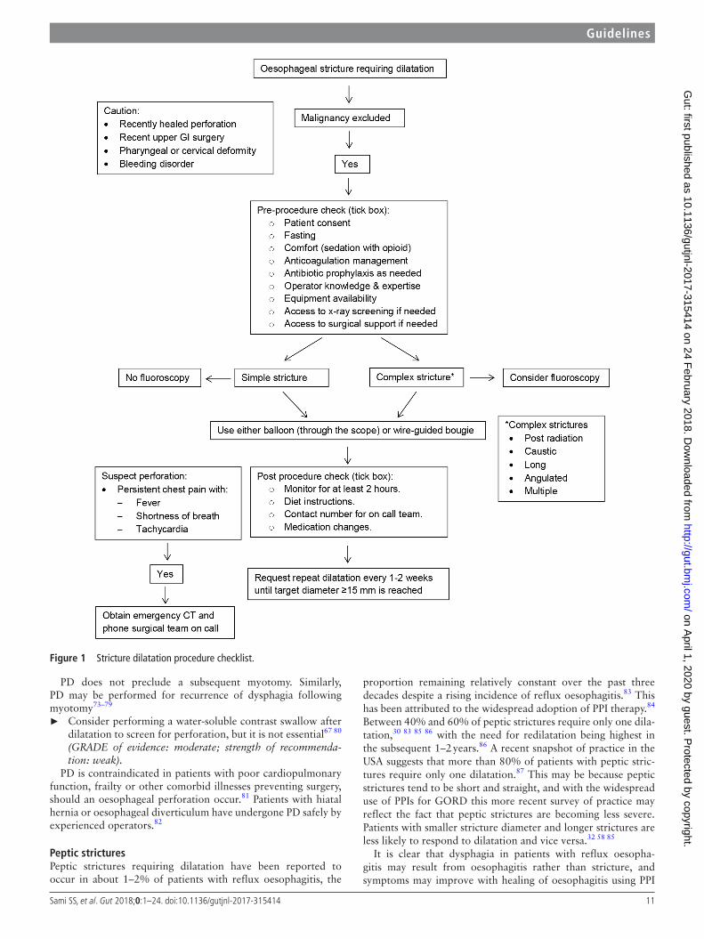
A brief checklist is shown in figure 1. This may be used as a guide.
diseAse-sPecific considerAtionsThis section discusses features of specific diseases, which may affect the dilatation procedure and patient outcomes.
Achalasia dilatationPneumatic balloon dilatation (PD) is one of a number of effective treatments for achalasia, which include surgical or endoscopic myotomy. However, the latter are outside the remit of this guide-line.60 Bougie dilators are not used for achalasia dilatation.
► Perform dilatation with pneumatic balloons 30–40 mm in diameter starting at 30 mm in the first session to reduce the risk of complications5 19 61 (GRADE of evidence: high; strength of recommendation: strong).
The dilatation technique varies across different studies and there is no consensus in the literature on the optimal method of performing pneumatic dilatation for achalasia. The balloon is usually positioned at the oesophagogastric junction and inflated according to the manufacturers’ instructions for 1–3 min.
► Perform a second dilatation session 2–28 days later with a larger size balloon of 35 mm5 (GRADE of evidence: high; strength of recommendation: strong).
Most authors advocate a third session either routinely or in cases where symptoms remain (Eckardt score >3) with the cautious use of 40 mm balloon if possible. If the Eckardt score remains >3 after the third session, the treatment is usually considered to have failed.5 Patients with a recurrence of symp-toms during follow-up may require further dilatation.5
► Consider repeat dilatation (after the initial series) during follow-up to maintain symptom response5 (GRADE of evidence: high; strength of recommendation: strong). The procedure is effective in 90% of patients in the first year
and this reduces to 86% in the second year.5 Up to one-third of patients may have recurrence of symptoms during 4–6 years of follow up.62 63 The vast majority can be successfully treated by repeat dilatation, achieving remission rates of up to 97% and 93% at 5 and 10 years, respectively.62
► Perform dilatation under endoscopic or fluoroscopic control based on clinician’s preference and local expertise64–66 (GRADE of evidence: moderate; strength of recommenda-tion: strong).
Fluoroscopic control is used in the majority of studies reporting safety and efficacy of balloon dilatation in achalasia; however, the safety of endoscopic control alone has been shown in a few studies.64–66 Comparative studies between the latter two approaches are lacking. Routine oesophagograms obtained after PD for achalasia did not reveal any clinically unsuspected perfo-rations and no perforations were missed in cases that were not followed by oesophagograms.67 Impedance planimetry may be a tool that improves decision-making in dilating achalasia.68
► Consider proton pump inhibitor (PPI) therapy after dila-tation as the technique has 10–40% rate of symptomatic gastro-oesophageal reflux disease (GORD) or ulcerative oesophagitis after treatment69–72 (GRADE of evidence: high; strength of recommendation: strong).
on April 1, 2020 by guest. P
rotected by copyright.http://gut.bm
j.com/
Gut: first published as 10.1136/gutjnl-2017-315414 on 24 F
ebruary 2018. Dow
nloaded from
11Sami SS, et al. Gut 2018;0:1–24. doi:10.1136/gutjnl-2017-315414
Guidelines
PD does not preclude a subsequent myotomy. Similarly, PD may be performed for recurrence of dysphagia following myotomy73–79
► Consider performing a water-soluble contrast swallow after dilatation to screen for perforation, but it is not essential67 80 (GRADE of evidence: moderate; strength of recommenda-tion: weak).
PD is contraindicated in patients with poor cardiopulmonary function, frailty or other comorbid illnesses preventing surgery, should an oesophageal perforation occur.81 Patients with hiatal hernia or oesophageal diverticulum have undergone PD safely by experienced operators.82
Peptic stricturesPeptic strictures requiring dilatation have been reported to occur in about 1–2% of patients with reflux oesophagitis, the
proportion remaining relatively constant over the past three decades despite a rising incidence of reflux oesophagitis.83 This has been attributed to the widespread adoption of PPI therapy.84 Between 40% and 60% of peptic strictures require only one dila-tation,30 83 85 86 with the need for redilatation being highest in the subsequent 1–2 years.86 A recent snapshot of practice in the USA suggests that more than 80% of patients with peptic stric-tures require only one dilatation.87 This may be because peptic strictures tend to be short and straight, and with the widespread use of PPIs for GORD this more recent survey of practice may reflect the fact that peptic strictures are becoming less severe. Patients with smaller stricture diameter and longer strictures are less likely to respond to dilatation and vice versa.32 58 85
It is clear that dysphagia in patients with reflux oesopha-gitis may result from oesophagitis rather than stricture, and symptoms may improve with healing of oesophagitis using PPI
figure 1 Stricture dilatation procedure checklist.
on April 1, 2020 by guest. P
rotected by copyright.http://gut.bm
j.com/
Gut: first published as 10.1136/gutjnl-2017-315414 on 24 F
ebruary 2018. Dow
nloaded from

12 Sami SS, et al. Gut 2018;0:1–24. doi:10.1136/gutjnl-2017-315414
Guidelines
therapy without the need for dilatation.88 In addition, oesoph-agitis as well as stricture diameter contribute to the severity of dysphagia,89–91 and healing of oesophagitis in patients with stric-tures is associated with a reduced need for redilatation.92 Finally, PPI therapy, but not H2 receptor antagonist treatment, reduces the need for, and frequency of, dilatation of peptic strictures after the initial dilatation.92–95
► Offer PPI therapy to patients with GORD and dysphagia, as this treatment has been shown to reduce the need for oesophageal dilatation83 84 89 92 (GRADE of evidence: high; strength of recommendation: strong).
► Offer PPI therapy after endoscopic dilatation for peptic strictures in order to reduce recurrence rate89 92–94 (GRADE of evidence: high; strength of recommendation: strong).
► Offer PPI therapy rather than H2 receptor antagonists, which are ineffective in reducing the need for repeat dila-tation (stricture recurrence), less effective in healing of oesophagitis and in providing symptom relief from GORD and dysphagia89 92–95 (GRADE of evidence: high; strength of recommendation: strong).
The management of refractory peptic strictures will be discussed in the relevant section.
schatzki’s ringSchatzki’s ring is an annular constriction at the gastro-oesoph-ageal mucosal junction, covered on its proximal side by squa-mous epithelium and distally by gastric mucosa.96 It was first appreciated on barium swallow radiology, provided the oesoph-agus was adequately distended,97 occurring in 6–14% of barium swallow examinations and often asymptomatic.98 99 The natural history of asymptomatic Schatzki’s ring is unknown so it is not established whether treatment (for instance, with a PPI) is indi-cated. Schatzki’s ring is less often seen at endoscopy100 unless the gastro-oesophageal junction is adequately distended by air insufflation. It is a common cause of intermittent dysphagia for solids and of food bolus obstruction: ‘Schatzki’s rule’ states that dysphagia is usual with ring diameters of ≤ 13 mm, and rarely occurs if the diameter exceeds 20 mm,101 with a ‘grey’ area in between where symptoms are less consistently observed.
Schatzki’s ring is associated with gastro-oesophageal reflux39 102 103 and with eosinophilic oesophagitis, even in the absence of other endoscopic oesophageal mucosal abnormali-ties.100 102 This may explain why patients with rings and acid reflux (demonstrated by pH monitoring), and also unselected patients with Schatzki’s rings appear to have fewer recurrences after dilatation when receiving PPI therapy.39 104 Dysphagia due to Schatzki’s ring was relieved by PPI therapy without the need for dilatation in a small retrospective case series.105
Dilatation therapy for symptomatic Schatzki’s ring is directed toward achieving rupture of the ring; therefore, larger calibre dilators may be needed.101 There is robust evidence for the effi-cacy of a single dilatation to 16–20 mm.106 Electrosurgical inci-sion of the ring has been reported to be at least as effective in relieving dysphagia due to Schatzki’s ring as a single large calibre dilatation in randomised trials.107 Two studies suggest that inci-sion may lead to longer remission of dysphagia than bougienage, proposing this treatment for patients with recurrence after a course of bougienage.108 109 Relapses still occur, but long-term PPI therapy (omeprazole 20 mg/day) significantly reduces the risk of relapse compared with placebo at up to 48 months of follow-up.39 104
Biopsy excision was shown to be effective and safe in a small feasibility study of 10 patients with dysphagia due to a Schatzki's
ring (six of whom had previously undergone bougie or balloon dilatation). Complete endoscopic obliteration of the ring and improvement of dysphagia was achieved (using jumbo biopsy forceps) in all 10 patients during 379 days (range 63–496 days) of follow-up with no serious complications.110
► Do not offer dilatation for asymptomatic Schatzki’s rings incidentally discovered on diagnostic endoscopy or contrast studies (performed for unrelated indication)97–99 (GRADE of evidence: low; strength of recommendation: strong).
► Consider exclusion of eosinophilic oesophagitis by distal, mid and proximal oesophageal biopsies in symptomatic Schatzki’s ring100 102 (GRADE of evidence: moderate; strength of recommendation: strong).
► Offer a single dilatation session using graded dilatation to a relatively large diameter (16–20 mm) to treat dysphagia related to Schatzki’s ring39 106 111 (GRADE of evidence: moderate; strength of recommendation: strong).
► Offer PPI therapy after dilatation, as this reduces the risk of relapse of Schatzki’s ring39 104 (GRADE of evidence: moderate; strength of recommendation: strong).
► Consider electrosurgical incision as an effective alternative treatment to oesophageal dilatation for relieving dysphagia related to Schatzki’s ring108 109 112 (GRADE of evidence: high; strength of recommendation: strong).
In studies reporting this technique, the incision was performed using a standard needle-knife papillotome with a 5 mm cutting wire passed through the accessory channel of the endoscope. Three to four longitudinal incisions were performed radially to the junction of the base of the ring and the oesophageal wall.109 Other experts recommend the use of standard endoscopic submucosal dissection (ESD) needle knife, IT knife or argon plasma coagulation.113 This procedure must be performed by a skilled operator who is familiar with the technique and uses it regularly (such as for ESD procedures).
Post-endoscopic therapy stricturesOesophageal stenosis can occur after Endoscopic Resection or after Endoscopic Mucosal Ablation for oesophageal neoplasia.
Post-endoscopic resection (ER): stricture dilatationER procedures performed in the oesophagus include both endo-scopic mucosal resection (EMR) and ESD.
It is generally accepted that once mucosal or submucosal resec-tion of the oesophageal wall has encompassed greater than 75% of the circumference then symptomatic stenosis will occur.114–120 Studies quote an OR for stricture formation of 44.2 (95% CI 4.4 to 443.6) once more than 75% of the circumference has been resected119 and frequency of 49.7% when the length of resection was >40 mm.
For the majority of post-ER strictures, dilatation will resolve symptomatic dysphagia, although repeat procedures are often needed. Pouw et al121 showed that by using either Savary bougienage or balloon dilatation, all (84) patients who developed symptomatic strictures after stepwise radical ER were adequately treated by a median of 3 (IQR 2–6) dilatation sessions, supple-mented by placement of a stent (n=2) or incision therapy (n=4). In 28 (33%) patients the stenosis was graded as severe since more than five endoscopic dilatations, stent placement or inci-sion therapy were required.
Endoscopic balloon dilatation has been shown to be an effec-tive and safe first-line intervention in patients with post -ER strictures, with a reported success rate of 90% and a perfora-tion rate of 0.3%122 in patients with symptomatic dysphagia
on April 1, 2020 by guest. P
rotected by copyright.http://gut.bm
j.com/
Gut: first published as 10.1136/gutjnl-2017-315414 on 24 F
ebruary 2018. Dow
nloaded from

13Sami SS, et al. Gut 2018;0:1–24. doi:10.1136/gutjnl-2017-315414
Guidelines
after EMR for early oesophageal cancer. Balloon dilatation has also been used to prevent strictures after ER. In one study, dila-tation was performed after ER and repeated once a week until the mucosal defect was completely healed. The remaining 12 cases were not treated and used as historic controls. Prophylactic dilatation decreased the incidence of stricture (59% vs 92%, P=0.04); reduced the severity of stricture; and shortened the duration required for resolving the stricture (29 days vs 78 days, P=0.04) even when stricture developed.
The main complications associated with post-ER stricture dilatation are perforation, bleeding and a low risk of bacter-aemia.118 120 The potential risk of perforation associated with dilatation of post-ER strictures is slightly higher (1.1%) that that seen in benign strictures (0.1%–1.02%).12 35 120 Also, strictures resistant to repeated dilatation do occur, and as in peptic stric-tures, a fully covered self-expanding metal stent (SEMS) may need to be employed, but there are limitations, including chest pain and stent migration.121 Further large-volume studies are needed to clarify the long-term outcome.
► Inform patients of up to ~50% chance of developing symp-tomatic stricture requiring endoscopic dilatation after ER either following EMR or ESD in any of the following situ-ations: resection size >75% of the oesophageal circumfer-ence; and a longitudinal resection length of >40 mm123 124 (GRADE of evidence: high; strength of recommendation: strong).
► Offer dilatation for the management of symptomatic post-mucosal resection strictures32 35 122 123 125–127 (GRADE of evidence: moderate; strength of recommendation: strong).
► Inform patients that perforation rates of dilatation for post-ER strictures in the oesophagus can be up to 1.1%120 (GRADE of evidence: moderate; strength of recommenda-tion: strong).
► Offer fully covered SEMS in carefully selected patients for the dilatation of refractory strictures after ER121 128 (GRADE of evidence: low; strength of recommendation: weak).
Post-ER: stricture preventionEndoscopic resection creates an ulcer which can be healed with PPI therapy based on previous studies on reflux oesophagitis and is known to reduce the risk of stricture formation.92 93 There are no comparative studies after endoscopic resection as PPI is a standard accepted treatment.
Endoscopic intralesional injections of steroids are applied based on the concept that inflammation and fibrosis after oesophageal ER are inhibited by the direct administration of steroids to the resection site. Takahashi et al129 carried out a randomised, controlled, open-label study to examine whether local steroid injection was an effective prophylactic treatment for oesophageal stenosis following extensive ESD. The authors found no difference in the frequency of stricture formation between the groups, but significantly fewer dilatation sessions were required in the steroid group than in the control group (6.1 (95% CI 2.8 to 9.4) vs 12.5 (95% CI 7.1 to 17.9); P=0.04).
Yamaguchi et al130 found that the stricture rate with oral pred-nisolone (5.3%, 1/19) was significantly lower than in the control group (31.8%, 7/22) (P<0.0001). The steroids were started at 30 mg/day on the third day after ESD, tapered gradually, and then discontinued 8 weeks later. Furthermore, the frequency of dilatation in the prednisolone group (mean 1.7, range 0–7) was significantly lower than in the control group (mean 15.6, range 0–48) (P<0.0001). Other studies have shown that using
a reducing regimen of oral prednisolone in patients after large EMR or ESD reduces the need for repeated dilatation.131 132
Wen et al133 performed a single-centre randomised controlled trial and showed that the stricture rate and number of additional dilatations during a short-term follow-up period were signifi-cantly lower in subjects treated by stent placement for 8 weeks immediately after ESD than in patients who were not treated with stents. Further studies are required to establish the bene-fits and risks of SEMS in this setting—in particular, problems of postinsertion hyperplastic regenerative changes, which are diffi-cult to distinguish from neoplastic residues.
Polyglycolic acid (PGA) is a synthetic compound that is completely degraded into a non-toxic degradation product, glycolic acid, over a period of 4–6 months in physiological conditions and has been employed in promoting tissue repair. Iizuka et al134 evaluated 15 patients after ESD in whom PGA sheets were placed, and oesophageal strictures occurred in only 1/13 patients. These very early data show that PGA sheets may prevent oesophageal strictures after large-scale EMR or ESD in the oesophagus. Other tissue engineering techniques, such as transplantation of autologous oral mucosal epithelial cell sheets into ESD sites, are also being evaluated.135
► Offer PPI therapy after ER or ablation to reduce stricture occurrence92 93 (GRADE of evidence: low; strength of recom-mendation: strong).
► Consider steroid injection at the resection site or oral pred-nisolone therapy in patients at high risk after large EMR or ESD to reduce stricture formation130–132 136–139 (GRADE of evidence: moderate; strength of recommendation: weak).
► Offer fully covered SEMS in carefully selected patients for the prevention of refractory strictures after ESD of large lesions placing the stent at the same time as the first stric-ture dilatation133 (GRADE of evidence: moderate; strength of recommendation: weak).
Post-ablative therapy: treatment and preventionField ablation is now accepted as an adjunctive treatment to endoscopic resection of oesophageal neoplasia and, in particular, neoplasia related to Barrett’s oesophagus (BO). Most interna-tional guidelines now support this approach with much of the data supporting the use of radiofrequency ablation (RFA).140 These treatments result in oesophageal ulceration which can be healed with PPI therapy based on previous studies on reflux oesophagitis, and PPI treatment is known to reduce the risk of stricture formation.92 93 There are no comparative studies after ablation as PPI is a standard accepted treatment.
Historically, photodynamic therapy (PDT) was the first-line ablation in patients with BO neoplasia. Owing to the toxicity and photosensitivity reactions associated with these agents its role is no longer advocated for the treatment of oesophageal neoplasia. One of the significant drawbacks of this technology was the high rate of stricturing in patients treated with PDT, which approached 36%.141 Most oesophageal strictures in the latter study were managed successfully through repeated dilata-tions, with one patient requiring an oesophagectomy after dila-tation-related perforation.
RFA has now been accepted as the 'gold standard' approach in patients with BO neoplasia. Ablation to a depth of 500 µm favours a reduced rate of stricture formation that is not the case with other ablation techniques. In a landmark randomised controlled trial, in which Shaheen et al142 compared RFA with a sham procedure, high disease eradication rates were seen and the stricture rate in the treatment group after a median of two
on April 1, 2020 by guest. P
rotected by copyright.http://gut.bm
j.com/
Gut: first published as 10.1136/gutjnl-2017-315414 on 24 F
ebruary 2018. Dow
nloaded from

14 Sami SS, et al. Gut 2018;0:1–24. doi:10.1136/gutjnl-2017-315414
Guidelines
treatments was 6%. All patients underwent successful endoscopic dilatation (mean of 2.6 sessions). Data from the UK registry of patients undergoing RFA for BO neoplasia reported a similar stricture rate. Haidry et al143 showed an overall stricture rate requiring therapeutic dilatation of 9% (30 of 335 patients). All these cases were managed endoscopically with a mean of 1.3 dilatations (range 1–6). Patients who underwent EMR before initiating RFA were more likely to develop strictures than those who did not (12% vs 5.9%; P=0.04).
The role of RFA for early squamous cell neoplasia has been controversial with early data supporting its use in well-selected patients,144 but it is associated with a higher stricture rate than with the same intervention in BO neoplasia. van Vilsteren et al144 showed a stricture rate of 23% and Bergman et al145 a stricture rate of 14%. Data from the UK registry, where 20 patients with early squamous neoplasia were treated with RFA, showed a stricture rate of 20%.146
► Inform patients that stricture formation requiring dilata-tion is significantly higher in the following situations: after photodynamic therapy (PDT) (36%) compared with other forms of ablation141 147 148; where ER has preceded ablation (12% vs 6%)143; with use of higher doses of energy with RFA for Barrett's oesophagus (BO)149 150; and after RFA for early squamous cell neoplasia (14–23%)144–146 151 (GRADE of evidence: moderate; strength of recommendation: strong).
► Offer dilatation in patients with clinically significant stricture formation after RFA, with or without previous ER142 143 152 153 (GRADE of evidence: moderate; strength of recommendation: strong).
► Offer PPI therapy after ablation to reduce stricture occur-rence92 93 (GRADE of evidence: low; strength of recommen-dation: strong).
eosinophilic oesophagitis (eoe)EoE is a common immune/antigen-mediated oesophageal disease characterised clinically by symptoms of oesophageal dysfunction and histologically, by eosinophil predominant inflammation.154 First described by Attwood and Straumann in 1993–94,155 156 EoE usually presents with dysphagia and is now the the most common cause of food bolus obstruction in patients under the age of 50 years.157 EoE is the the most common cause of spontaneous perforation of the oesophagus. It is the second most common benign disease of the oesophagus.158 It is often underdiagnosed and yet 50 times more common than achalasia.158
Preliminary medical treatment and diet elimination for EoEPPI, diet and topical steroids are all used as first-line treatments (in up to 50%).159 Dietary therapy with elimination of causative allergens, or topical corticosteroids may provide a durable long-term solution.160 161 Topical steroids (such as fluticasone) and budesonide are very effective in the short term, but longer-term studies are needed.162
► Offer dilatation along with other forms of disease modi-fication using diet, topical steroids or other drugs17 163–165 (GRADE of evidence: moderate; strength of recommenda-tion: strong).
► Start other treatments for EoE before dilatation if possible, and those may be continued afterwards to prevent or delay recurrence of symptoms163 164 166–169 (GRADE of evidence: moderate; strength of recommendation: strong).
► Offer preliminary topical steroids followed by dilatation as this is more cost-effective than using dilatation alone as
first-line treatment164 170 (GRADE of evidence: moderate; strength of recommendation: strong).
Indications for dilatation in EoEThe indications for dilatation in EoE include established tight stricture, narrow calibre oesophagus, food bolus obstruction and failure of symptoms to respond to diet or drugs.8 16 171 Dilata-tion of the oesophagus is now a standard treatment for EoE, along with other forms of chronic disease modification using diet, topical steroids or other drugs.17 163–165 Other treatments for EoE should be started before dilatation if possible, and continued afterwards to prevent or delay recurrence of symp-toms.163 166–170 172 A recent randomised controlled study by Kavitt et al170 has shown that preliminary topical steroids followed by dilatation is more cost-effective than using dilatation as the first treatment. In some patients, dilatation can be used as the only treatment.17 162 164 170 173–175 However, caution should be exer-cised with tight strictures of <7 mm calibre.170
► Offer dilatation as first line treatment in patients with acute symptoms such as food bolus obstruction and daily dysphagia17 164 173–175 (GRADE of evidence: moderate; strength of recommendation: strong).
► Offer dilatation to patients with established tight stricture, narrow calibre oesophagus and those who do not respond to diet or drugs8 16 163 168 (GRADE of evidence: high; strength of recommendation: strong).
Risks and complications of dilatation in EoEDilatation of EoE is no more dangerous than dilatation for other oesophageal diseases (such as peptic stricture or acha-lasia), and the perforation rates are similar if safe techniques and flexible endoscopy are employed by experienced oper-ators.17 158 162 163 176–178 Mucosal tears and associated minor bleeding are common after dilatation of EoE (8%), and can be managed conservatively.16 171 176 177
The risk of postprocedural chest pain is particularly common after EoE dilatation and may last for up to 48 hours.169 176 179 Patients should be warned about this possibility, and the need to take fluids and analgesia along with their maintenance EoE therapy.
► Reassure patients that dilatation of EoE is no more dangerous than dilatation for other oesophageal diseases, and the perfo-ration rates are similar16 17 163 174 176–178 (GRADE of evidence: high; strength of recommendation: strong).
► Inform EoE patients that chest pain after dilatation is common17 169 176 (GRADE of evidence: high; strength of recommendation: strong).
Follow-up treatment after dilatation in EoEA good clinical outcome is dysphagia resolution and avoidance of food bolus obstruction after dilatation.8 16 177 Most experts recommend gradual slow dilatation with sessions separated by 3–4 weeks and aim for a diameter of 16–18 mm, if possible.180 Repeat dilatation should be offered if symptoms of severe dysphagia recur or if a symptomatic stricture is diagnosed during follow-up.8 16 177 Symptom response after initial dilatations (about three sessions over several weeks) usually lasts for up to 1 year.167 169 Dilatation of the oesophagus in EoE may be repeated if needed.163 167 174 181
Maintenance treatment with either topical steroid or dietary elimination should continue after dilatation of EoE. A repeat endoscopy is recommended at 6–12 weeks.174 177
on April 1, 2020 by guest. P
rotected by copyright.http://gut.bm
j.com/
Gut: first published as 10.1136/gutjnl-2017-315414 on 24 F
ebruary 2018. Dow
nloaded from

15Sami SS, et al. Gut 2018;0:1–24. doi:10.1136/gutjnl-2017-315414
Guidelines
► Inform patients that symptom response after dilata-tion usually lasts up to 1 year167 169 (GRADE of evidence: moderate; strength of recommendation: strong).
► Repeat dilatation if needed167 171 174 181 (GRADE of evidence: high; strength of recommendation: strong).
Method of dilatation in EoEThere are no credible data comparing balloon with bougie dila-tation in EoE, but most observers prefer balloon to reassure the operator that a perforation is not occurring.16 174 179 181 Otherwise the technique remains the same as for other types of stricture.
Use of distensibility measure to guide dilatation in EoEEndoscopic Functional Luminal Imaging Probe (EndoFLIP, Crospon, Galway, Ireland) is used in some specialist centres to measure compliance and distensibility. EndoFLIP and Balloon pull through techniques may help in judging the optimal calibre to dilate to but require further study.166 182 183 These tech-niques are innovative, safe and seem effective. More research on their cost-effectiveness is warranted. The availability and cost-effectiveness will be the main considerations before formulating recommendations for their routine use in clinical practice. A recent study has shown that endoscopy has a sensi-tivity of only 25% in detecting narrowed oesophagus ≤15 mm in diameter among patients with EoE compared with barium radiology.184
► Consider the use of special techniques such as EndoFLIP and Balloon pull through to judge the optimal calibre and posi-tion of dilatation182 183 (GRADE of evidence: low; strength of recommendation: weak).
Postoperative stricturesFrequency and situationAll surgical interventions have the potential for fibrotic stric-turing at the anastomotic site as a complication. Oesophagec-tomy with oesophago-gastrostomy for oesophageal or gastric cardia malignancy can lead to stricturing in 4%185 to 66%.186 These strictures tend to be short and straight, but occasionally can be complicated.187 188 Methods for prevention of stricturing by surgical technique to maximise vascularisation of the anasto-mosis is a key strategy in the prevention of oesophageal anasto-motic strictures.185 186 188–191
Fundoplication for GORD, both partial and total, is also associated with postoperative dysphagia, affecting 5–43% of people.192 193 Mechanisms behind this include a tight wrap, a slipped wrap and paraoesophageal hernia.194 195 Partial wraps are less likely to cause significant postoperative dysphagia than a Nissen.195 196 Intraoperative evaluation of the oesophagus—for example, using the EndoFLIP device, could allow the oper-ator to determine the length of wrap and optimal tightness of the gastro-oesophageal junction during the fundoplication procedure,197 thereby reducing postoperative dysphagia and/or the need for dilatation.
Preliminary medical treatment and investigationsNutritional supplementation, particularly in patients with cancer, and PPI therapy following bariatric surgery198 are important. Specific investigations, including endoscopy and barium swallow, are helpful.
► Consider performing upper GI endoscopy, manometry, pH studies and barium swallow first in patients with persis-tent dysphagia post-fundoplication surgery, in order to assess the integrity of the wrap and understand the cause
of dysphagia prior to dilatation or repeat surgery194 195 (GRADE of evidence: low; strength of recommendation: weak).
Methods of dilatation of postoperative stricturesTreatment of postoperative stricture has used both bougienage188 and balloon dilatation, both under direct endoscopic visualisa-tion185 199 or with fluoroscopy,200 and no data exist to show which is optimal.186
Symptoms improve after the first dilatation session in up to 43% of individuals185 and therefore repeat dilatation is the norm. Typically, a median of three sessions is used before satis-factory relief of dysphagia is achieved.189
Treatment success is difficult to define, limiting comparisons between studies but may be obtained in between 62%195 and 100%.189 Delayed gastric emptying appears to be an additional risk factor for stricture formation, and treatment of this by pylo-roplasty or pyloromyotomy may help to reduce the frequency of dilatation sessions required to achieve successful treatment of anastomotic stricture.199
In the post-fundoplication setting, dilatation of the lower oesophageal sphincter typically requires 30–40 mm balloons, similar to those used in the management of achalasia.194 Success can be achieved in approximately two-thirds of patients, although non-responders may require revisional surgery.201
► Consider treatment of concurrent delayed gastric emptying in order to reduce the need for redilatations199 (GRADE of evidence: low; strength of recommendation: weak).
► Consider the use of balloon dilatation to 30–40 mm (as with achalasia) in patients with post-Nissen dysphagia194 (GRADE of evidence: low; strength of recommendation: weak).
Use of supplementary methodsThe use of steroid injection (0.5 mL aliquots of triamcinolone 40 mg/mL to four quadrants) has been evaluated in anastomotic strictures in a randomised double-blind study and found to be safe when injected with a 4 mm-long, 23-gauge needle imme-diately before bougie dilatation of anastomotic strictures.202 In this study, the steroid arm appeared superior in the length of time required between dilatation sessions (108 days vs 42 days in the saline group), and the total number of dilatation sessions required (two in the steroid group versus three in the saline group). However neither of these outcomes reached signifi-cance. Steroid-treated patients with stricture were found to have significantly more complications, specifically oesophageal candidiasis.
Needle knife incision has also been evaluated in refractory strictures and in the primary treatment of anastomotic stric-tures.203 204 In experienced hands, this technique is as safe as conventional dilatation methods, but evaluation of the success of this treatment strategy is more variable. Primary treatment with incisional therapy appears to confer no significant advan-tage over bougie dilatation in the mean number of dilatations or overall success rate at reducing symptoms of dysphagia.203 Specific methods used for incisional therapy include either an endoscopic needle knife or an ‘IT’ knife, incising in four quad-rants with the aid of a transparent cap, which enhances the safety of the procedure. The procedure is best done by an operator who is familiar with techniques of incision through experience with ESD or endoscopic retrograde cholangiopancreatography. Incisional therapy appears to maintain luminal patency, at 12 months' follow-up in 61.5% of cases compared with 19.8% in controls who underwent continued endoscopic balloon
on April 1, 2020 by guest. P
rotected by copyright.http://gut.bm
j.com/
Gut: first published as 10.1136/gutjnl-2017-315414 on 24 F
ebruary 2018. Dow
nloaded from

16 Sami SS, et al. Gut 2018;0:1–24. doi:10.1136/gutjnl-2017-315414
Guidelines
dilatation,205 and so has a role in the treatment of unresponsive anastomotic stricture.
► Use steroid injections (0.5 mL aliquots of triamcinolone 40 mg/mL to the four quadrants) to reduce the frequency of repeat dilatations in anastomotic strictures refractory to initial dilatation approaches. Use a 4 mm-long, 23-gauge needle immediately before bougie dilatation while leaving the needle in for at least 1 min to minimise leakage of the drug and ensure delivery of the full dose to the target area113 202 (GRADE of evidence: moderate; strength of recom-mendation: weak).
► Consider using needle knife incision for anastomotic stric-tures as an alternative to dilatation203 205–207 (GRADE of evidence: moderate; strength of recommendation: weak).
Risks and follow-upAfter treatment of postoperative strictures, complication rates such as bleeding or rupture have been reported in up to 30% of cases,200 208 but perforation with mediastinal leakage appears to be no more common than in other types of strictures.
There appears to be no evidence to warrant surveillance of patients postoperatively for anastomotic stricture.
Post-radiation stricturesRadiotherapy treatment has revolutionised the management of most solid organ malignancies, but may induce stricturing of the oesophagus. The risk of stricturing can be anticipated before radiotherapy in patients with greater tumour inva-sion (T-score) and greater circumferential involvement.209–211 Radiotherapy induces tissue ischaemia, fibrosis and subse-quent oesophageal stenosis. In contrast to other oesophageal strictures, the fibrosis often affects the tissues surrounding the oesophagus, creating a non-compliant mediastinum. As a conse-quence, radiation strictures are frequently refractory to dilata-tion and progressive. The risk is increased with higher radiation doses.212–217 Furthermore, radiotherapy with concurrent chemo-therapy increases the likelihood of stricture217 218 and is a cause of completely obstructed oesophagus,.212 219–221 Prior insertion of a gastrostomy should be considered to aid nutrition in already malnourished individuals and this offers an additional conduit through which dilatation attempts of subsequent oesophageal strictures can be made. Where antegrade passage of a guidewire is unsuccessful, a retrograde attempt through the gastrostoma can be made using catheter and guidewire under fluoroscopic control.
The endoscopic CARD approach can be considered in patients with a completely occluded oesophagus. In most circumstances, this requires a general anaesthetic, although conscious sedation has also been used.218 222 A CT scan is also recommended to exclude nearby vessels.223
Visualisation of the proximal aspect of the stricture is under-taken by insertion of an antegrade endoscope. A second endo-scope is inserted through the gastrostomy site and the stricture is trans-illuminated in order to identify the now obstructed lumen. Typically, a guidewire is passed from the distal oesophagus prox-imally under fluoroscopic guidance, puncturing through the stricture and then removed per-orally.209 212 219 222–224 A needle knife,225 straight catheter223 or EUS fine aspiration needle223 225 226 has also been shown to be effective at traversing the stricture by incision. Biplanar fluoroscopy or cone-beam CT may be used to facilitate an axial alignment of the endoscopes and needles and reduce the risk of perforation. Once a guidewire has been passed a bougie or balloon can dilate to 12–15 mm.219–221 223 224 226–229
In some patients, the placement of a temporary (up to 1 month) fully covered SEMS is necessary to maintain the luminal patency with or without a nasogastric tube for feedig.223
No large studies have been undertaken to evaluate the use of the CARD procedure, and it should be regarded as a last resort for use in expert hands only. Evidence from case series suggests it is safe, with no patients requiring emergency surgical inter-vention.223 The majority of larger series suggest good success (defined as discontinuation of percutaneous endoscopic gastros-tomy feeding), although in most instances, repeated dilatation is still required every 2–3 months.212 219 223
► Consider a combined anterograde and retrograde dilata-tion (CARD) or rendezvous approach under general anaes-thetic as an alternative to surgery in the treatment of the completely obstructed oesophagus, where local expertise is available209 212 218 219 221 222 225–228 (GRADE of evidence: moderate; strength of recommendation: weak).
► Use fluoroscopic guidance to assist with the rendezvous procedure209 212 218 219 221 222 225–227 (GRADE of evidence: moderate; strength of recommendation: strong).
► Use a guidewire to navigate through the obstruction when using the CARD approach to re-establish luminal patency209 212 219 222 225 (GRADE of evidence: low; strength of recommendation: strong).
► After gaining luminal patency using the CARD procedure, perform subsequent dilatation using either balloon224 226 229 or bougie212 219–221 228 (GRADE of evidence: moderate; strength of recommendation: strong).
caustic stricturesOesophageal injury can be caused by ingestion of caustic substances. Alkalis are the the most common caustic agents ingested in Western countries, while acid ingestion is more common in developing countries, such as India. Caustic oesoph-ageal injury can lead to the lower oesophageal sphincter pressure becoming impaired, which in turn leads to gastro-oesophageal reflux that can worsen stricture formation.230 231
After initial radiological assessment, an upper gastrointestinal tract endoscopy, with gentle air insufflations or preferably with CO2, should be considered after caustic ingestion within the first 12–48 hours to determine prognosis and management, including oesophageal stricture formation and subsequent requirement for dilatation.232 Contraindications to endoscopy include radio-logical suspicion of perforation or burns to the supraglottic and epiglottic areas. A CT grading system for caustic lesions has been proposed, but there is insufficient evidence to demonstrate its widespread utility.233 234
► Perform upper gastrointestinal tract endoscopy within the first 12–48 hours after caustic ingestion235–238 (GRADE of evidence: moderate; strength of recommendation: strong).
Several techniques have been used in an attempt to prevent stricture formation. These techniques include oral steroids,239 240 antibiotics, mitomycin C and intraluminal stents,241 but there is limited evidence on outcomes.
Late management makes dilatation more complex owing to fibrosis in the oesophageal wall. Dilatation can be carried out with either bougie or balloon dilators. There is no clear advan-tage of one technique over another.126 242–246 The Savary-Gilliard type was used in the majority of studies reporting on efficacy of bougie dilatation and is preferred by some practitioners in consolidated and fibrotic strictures.
► Ensure timely management of oesophageal strictures with dilatation as this plays a key role in affecting patient
on April 1, 2020 by guest. P
rotected by copyright.http://gut.bm
j.com/
Gut: first published as 10.1136/gutjnl-2017-315414 on 24 F
ebruary 2018. Dow
nloaded from

17Sami SS, et al. Gut 2018;0:1–24. doi:10.1136/gutjnl-2017-315414
Guidelines
outcomes242–246 (GRADE of evidence: moderate; strength of recommendation: strong).
Dilatation is more hazardous between 1 and 3 weeks after injury and some avoid this time interval. From approximately week 3, scar retraction begins, resulting in stricture formation. The time interval between dilatations varies in published reports but should be less than 2–3 weeks. The number of sessions required to achieve the desired outcome varies; usually three or four are required but the number can be significantly higher.
The perforation rate for caustic strictures is higher than the standard benign oesophageal stricture dilatation rate with anything from 0.4% up to 32% recorded in the literature. The complication rate is lower when performed by those with more experience, and reported to be 4.5%.
► Consider avoiding dilatation within 3 weeks of initial caustic ingestion242 246 (GRADE of evidence: low; strength of recom-mendation: weak).
► Consider a time interval between dilatations of <2 weeks246 247 (GRADE of evidence: very low; strength of recommendation: weak).
Refractory strictures are more common in caustic ingestion. Management has been reported with intralesional steroid injec-tion or topical mitomycin C at the time of dilatation, tempo-rary stent placement, biodegradable stents and a suspended oesophageal silicone prosthesis. If oesophageal dilatation fails then surgical resection can be considered. There is no consensus for the optimum management of these strictures.245 248–252
refractory stricturesA stricture should be considered refractory after an inability to maintain a luminal diameter of ≥14 mm after five sequential dilatation sessions 1–2 weeks apart or maintain the target diam-eter for 4 weeks once it has been reached.253 Refractory strictures are rare and challenging. Before embarking on any particular management the clinician and patient should be aware of the poor level of data for the outcomes and risks of most techniques.
► Inform patients about the lack of good quality evidence for best approach to treatment of refractory strictures (GRADE of evidence: low; strength of recommendation: strong).
► Consider discussion with, and/or referral to, centres with expertise in treatment and follow-up of patients with refrac-tory strictures (GRADE of evidence: low; strength of recom-mendation: weak).
► Ensure optimal management of ongoing inflammation with high dose PPI therapy before defining a stricture as refrac-tory83 84 89 92 (GRADE of evidence: high; strength of recom-mendation: strong).
► Consider alternative neuromuscular causes in patients with ongoing dysphagia despite a seemingly adequate oesoph-ageal diameter126 (GRADE of evidence: low; strength of recommendation: strong).
Complex strictures (>2 cm, angulated, irregular, severely narrowed) of the following aetiologies are most often implicated in refractory strictures: anastomotic, radiation induced, caustic, post-ablative therapy.33
Method of dilatationNo trials have compared types of dilators for refractory strictures.
► Use fluoroscopic guidance during dilatation of refractory oesophageal strictures126 (GRADE of evidence: very low; strength of recommendation: weak).
► Use either bougie or balloon dilators with the decision indi-vidualised on a case by case basis dependent on the nature
(length, location, cause) of the stricture126 (GRADE of evidence: low; strength of recommendation: weak).
Intralesional steroid therapyIntralesional steroid therapy involves the injection of 0.5 mL aliquots of 40 mg/mL triamcinolone to all four quadrants of the stricture. Experts advocate predilatation, although there are no studies directly comparing this with postdilatation injection. Two randomised controlled trials in patients with refractory stric-tures of mixed, but mostly peptic, aetiology, have demonstrated benefit, with reduction in the number of repeat dilatations and an increase in the dysphagia-free period.254 255 Results have been disappointing for refractory strictures of other aetiologies, with randomised controlled trials failing to show improvement in anastomotic202 and caustic256 aetiologies. In addition, an increase in complications was seen in the steroid-treated patients.202
► Use intralesional steroid therapy combined with dilata-tion in refractory strictures with evidence of inflamma-tion (macro- or microscopically) on the assumption that anti-reflux therapy has been maximised previously with no benefit202 254–256 (GRADE of evidence: high; strength of recommendation: strong).
Incisional therapyUse of a needle or IT knife to perform radial incisions of refrac-tory strictures parallel to the longitudinal axis of the oesoph-agus has been evaluated. The procedure is performed under direct visualisation with or without a transparent cap. For circumferential strictures, incisions in the four quadrants may be most effective. The outcomes are best for short strictures (<1.5 cm).257 Longer strictures may initially respond, but most will require re-treatment.258
► Consider incisional therapy in patients with refractory Schatzki’s rings and anastomotic strictures at centres expe-rienced in the use of such techniques257 258 (GRADE of evidence: very low; strength of recommendation: weak).
StentsThe benefit of self-expanding removable stents has been demon-strated in a recent systematic review and meta-analysis, with complete relief of dysphagia in approximately 40% (median follow-up 0–1281 days over the 18 included studies).259 The majority of refractory strictures will respond to placement of between 4 and 8 weeks, although longer periods of up to 3 months may be required.259 It is recommended that stents should not be left in longer than this owing to the risk of embedding. Recurrence rates of refractory strictures remain as high as 69% after stent removal, particularly in patients with long strictures (>7 cm),260 illustrating the difficulties in achieving a satisfactory long-term outcome.
In trials directly comparing metal and plastic stents, outcomes were more favourable with metal stents, with lower migra-tion rates and need for re-intervention.261 262 Stent migration is a considerable problem with both stent types, occurring in approximately 30% of cases.259 Stent placement is associated with adverse events (chest pain, bleeding, perforation, aspira-tion pneumonia) in 20% and thus patients should give consent accordingly.259 Use of partially or uncovered metal stents must be avoided owing to risk of embedding of the stent in the oesopha-geal wall, which may preclude safe removal.
► Offer temporary placement of fully covered self-expanding removable stents in patients where previous methods have been unsuccessful in maintaining adequate oesophageal
on April 1, 2020 by guest. P
rotected by copyright.http://gut.bm
j.com/
Gut: first published as 10.1136/gutjnl-2017-315414 on 24 F
ebruary 2018. Dow
nloaded from

18 Sami SS, et al. Gut 2018;0:1–24. doi:10.1136/gutjnl-2017-315414
Guidelines
patency259–265 (GRADE of evidence: low; strength of recom-mendation: weak).
► The optimum duration of stent placement is usually between 4 and 8 weeks, but may vary depending on stricture aeti-ology and length, and type of stent264 (GRADE of evidence: very low; strength of recommendation: weak).
Single biodegradable stent placement appears to be only temporarily effective in the majority of patients with refractory benign oesophageal stricture.266 267 It can, however, reduce the frequency of serial dilatation in selected patients. In the largest study to date, sequential placement of a first, second and then third biodegradable stent resulted in a median dysphagia free period of 90, 55 and 106 days, respectively.267 No patients were dysphagia free at the end of the study, suggesting that this strategy is not effective for long-term dysphagia relief.
► Consider biodegradable stent placement to reduce the frequency of dilatation in selected cases266 267 (GRADE of evidence: low; strength of recommendation: weak).
Options for failed stricture resolutionSelf-dilatation is safe and effective for short, proximal stric-tures.268–270 For appropriate, well-trained patients willing to undertake this, it appears to be well tolerated, can prevent surgery and the burden of repeated hospital visits.268–270
► Consider teaching selected, self-motivated patients, with short proximal strictures to self-bougienage268–270 (GRADE of evidence: very low; strength of recommendation: weak).
► Offer surgery to patients who do not respond or are intol-erant to other measures271 272 (GRADE of evidence: low; strength of recommendation: weak).
A brief summary of disease-specific considerations is provided in box 1.
economic imPlicAtionsOesophageal stricturing has a significant impact on quality of life owing to both dysphagia symptoms and the need for repeated dilatation. Quality of life scores are often further affected by underlying diagnosis, such as head and neck cancer.273 274
No studies have investigated the cost-effectiveness of dila-tation, but it is generally appreciated that bougienage has better economic benefits than balloon dilatation. This is owing to the absence of symptomatic or endoscopic differ-ence between the two modalities in addition to the non-signifi-cant difference in the need for repeated treatments. Hence the consumable costs of disposing of and replacing TTS balloons outweigh those of reusable Savary bougies. Furthermore, extrap-olation from studies in the lower GI tract comparing balloon versus bougienage in the management of postoperative anasto-motic strictures has shown similar findings (median cost €680 vs €22.30; P<0.001).275 These considerations obviously require to be balanced with expertise, local availability and endoscopist preference, for example, to allow direct visualisation during the dilatation procedure.
Use of oesophageal stenting is outside the remit of these guide-lines, but it may also be considered in cases where repeated dila-tation is required and can be considered to add cost-effectiveness when two or more dilatations are avoided.276
There are no other special economic effects of these guidelines, as they are a formalisation of current practice with the evidence base, and we do not advocate any changes or new directions in care that have a major economic impact.
Audit of effectivenessItems that could be subject to audit to establish good clinical standards of oesophageal dilatation include:
► Completeness of consent forms with clear documentation in each section (including procedure risks and benefits).
box 1 considerations in specific disease areas
Achalasia: ► Use pneumatic balloons 30–40 mm in diameter ► Start at 30 mm in the first session to reduce the risk of complications
► Perform a second dilatation 2–28 days later with a larger balloon (usually 35 mm)
► Consider PPI therapy after dilatationPeptic strictures:
► PPI therapy long term ► No role for H2 receptor antagonists
schatzki’s ring: ► Asymptomatic=no dilatation ► Symptomatic=single session (aim for 16–20 mm diameter) ► Exclude eosinophilic oesophagitis ► Consider PPI ► Consider needle knife incision if dilatation not effective
Post-endotherapy: ► Perforation risk may be higher (around 1.1%) ► Consider steroid injection to reduce risk of stricture formation
► Offer PPI to reduce risk of recurrenceeosinophilic oesophagitis:
► Consider additional treatments (PPI, steroids, elimination diet)
► Aim for histological remission ► Consider dilatation in narrow calibre oesophagus or failure of medical treatment in the absence of a visible stricture
► Inform patients that chest pain is commonPostoperative:
► Post-fundoplication: – Assess wrap integrity with upper endoscopy, barium
swallow, manometry and pH impedance – Use pneumatic balloon 30–40 mm as for achalasia
► Anastomotic: – Consider concomitant steroid injection in four quadrants – Consider needle knife incision as an alternative to
dilatationPost-radiation:
► Consider CARD in completely obstructed oesophaguscaustic:
► Careful evaluation as usually complex ► Dilatation not advised in first 3 weeks after ingestion
refractory: ► Ensure optimal dose PPI and control of inflammation ► Consider:
– Discussion with expert centres – Alternative causes/underlying pathology – Concomitant steroid injection four quadrants – Needle knife incision – Fully covered SEMS as temporary measure 4–8 weeks
► Offer patient the option of self-dilatation
CARD, combined anterograde retrograde dilatation; PPI, proton pump inhibitor; SEMS, self-expanding metal stent.
on April 1, 2020 by guest. P
rotected by copyright.http://gut.bm
j.com/
Gut: first published as 10.1136/gutjnl-2017-315414 on 24 F
ebruary 2018. Dow
nloaded from
19Sami SS, et al. Gut 2018;0:1–24. doi:10.1136/gutjnl-2017-315414
Guidelines
► Success rate, comfort scores, re-intervention rate (number of dilatations per patient), complication rates, 30-day readmis-sion rates and 30-day mortality rates.27
► Using a standard scoring system for dysphagia (such as Ogilvie, Dakkak and Bennett, etc) in >90% of patients to provide an objective measure of the efficacy of the thera-peutic intervention.27
► Timeliness of follow-up serial sessions for endoscopic dilata-tion (within 2 weeks).33 59
► Perforation rates. The overall perforation rate following dilatation should be <1% for benign strictures27 and 1–2% for achalasia.19 20
AreAs for future reseArch ► Identification of the learning curve, assessment parameters
required and direct observation of procedure or skills design for formative and summative assessments for trainees under-taking dilatation.
► The number of dilatation procedures performed each year required to achieve competency and maintain skill.
► Direct cost-effectiveness comparison between balloon and bougie dilators.
► Assessment of the optimal interval between repeat dilata-tions for tight, complex or refractory strictures.
► Available data on the role of acid suppression, proton pump inhibition, elimination diet and steroids in EoE are limited by variation in clinical practice and reported outcomes. Randomised controlled trials are needed to identify the optimum treatment to prevent oesophageal strictures in EoE.
► Clarification of the role of steroids and other methods in reducing the incidence and severity of post-ER strictures. There is an ongoing phase III clinical trial comparing oral steroid versus local steroid injection therapy for the preven-tion of oesophageal stricture after ESD.277
► Research into the true incidence of refractory strictures with details of aetiology and comparative outcomes after different treatments.
Author affiliations1Division of Gastroenterology and Hepatology, Mayo Clinic, Rochester, Minnesota, USA2Cancer Biomarker Group, Swansea Medical School, Swansea University, Swansea, UK3Department of GI Sciences, University of Manchester, Manchester, UK4Salford Royal NHS Foundation Trust, Salford, UK5Department of Gastroenterology, Southampton University Hospital, Southampton, UK6Department of Gastroenterology, Portsmouth University Hospitals NHS Trust, Portsmouth, UK7Digestive Diseases Centre, University Hospitals of Leicester, Leicester, UK8Department of Gastroenterology, Wye Valley NHS Trust, Wye Valley, UK9Department of Gastroenterology, University College Hospital, London, UK10Department of Radiology, The Christie NHS Foundation Trust, Manchester, UK11Department of Gastroenterology, NHS Forth Valley, Stirling, UK12NIHR Nottingham Digestive Diseases Biomedical Research Centre, Queens Medical Centre, Belfast, UK13Faculty of Medicine Health and Life Sciences, Queen’s University Belfast, Belfast, UK14Department of Gastroenterology and Hepatology, Radboud University Medical Centre, Nijmegen, The Netherlands15Department of Surgery, Durham University, Durham, UK
Acknowledgements We thank Mimi McCord (patient representative, chair of Heartburn Cancer UK) and Mark Brockhurst (patient representative) for providing advice, guidance and feedback on the planning, recommendations and discussion phases of the guideline.
contributors All authors contributed equally to the development of this guideline.
funding This research received no specific grant from any funding agency in the public, commercial or not-for-profit sectors.
competing interests PDS received research support from Boston Scientific and Cook Medical. YA received research support from Medtronic and is a member of the Clinical Services and Standards Committee of the British Society of Gastroenterology and the UK and Ireland RFA user group. SEA received consultancy fees from Dr Falk Pharma.
Provenance and peer review Not commissioned; externally peer reviewed.
open Access This is an Open Access article distributed in accordance with the Creative Commons Attribution Non Commercial (CC BY-NC 4.0) license, which permits others to distribute, remix, adapt, build upon this work non-commercially, and license their derivative works on different terms, provided the original work is properly cited and the use is non-commercial. See: http:// creativecommons. org/ licenses/ by- nc/ 4. 0/
© Article author(s) (or their employer(s) unless otherwise stated in the text of the article) 2018. All rights reserved. No commercial use is permitted unless otherwise expressly granted.
RefeRences 1 Richter JE. Peptic strictures of the esophagus. Gastroenterol Clin North Am
1999;28:875–91. 2 Piotet E, Escher A, Monnier P. Esophageal and pharyngeal strictures: report on 1,862
endoscopic dilatations using the Savary-Gilliard technique. Eur Arch Otorhinolaryngol 2008;265:357–64.
3 Egan JV, Baron TH, Adler DG, et al. Esophageal dilation. Gastrointest Endosc 2006;63:755–60.
4 Riley SA, Attwood SE. Guidelines on the use of oesophageal dilatation in clinical practice. Gut 2004;53 Suppl 1:1i–6.
5 Boeckxstaens GE, Annese V, des Varannes SB, et al. Pneumatic dilation versus laparoscopic Heller’s myotomy for idiopathic achalasia. N Engl J Med 2011;364:1807–16.
6 Spaander MC, Baron TH, Siersema PD, et al. Esophageal stenting for benign and malignant disease: European Society of Gastrointestinal Endoscopy (ESGE) Clinical Guideline. Endoscopy 2016;48:939–48.
7 Barkin JS, Taub S, Rogers AI. The safety of combined endoscopy, biopsy and dilation in esophageal strictures. Am J Gastroenterol 1981;76:23–6.
8 Rajagopalan J, Triadafilopoulos G. Ring(s)-related esophageal meat bolus impaction: biopsy first, dilate later. Dis Esophagus 2009;22:E14–16.
9 Arora AS. Management strategies for dysphagia with a normal-appearing esophagus. Clin Gastroenterol Hepatol 2005;3:299–302.
10 Quine MA, Bell GD, McCloy RF, et al. Prospective audit of perforation rates following upper gastrointestinal endoscopy in two regions of England. Br J Surg 1995;82:530–3.
11 Faigel DO, Deveney C, Phillips D, et al. Biopsy-negative malignant esophageal stricture: diagnosis by endoscopic ultrasound. Am J Gastroenterol 1998;93:2257–60.
12 Hernandez LV, Jacobson JW, Harris MS, et al. Comparison among the perforation rates of Maloney, balloon, and savary dilation of esophageal strictures. Gastrointest Endosc 2000;51(Pt 1):460–2.
13 Nagi B, Kochhar R, Thapa BR, et al. Radiological spectrum of late sequelae of corrosive injury to upper gastrointestinal tract. A pictorial review. Acta Radiol 2004;45:7–12.
14 Shepherd H, Hewett D. Guidance for obtaining a valid consent for elective endoscopic procedures. British Society of Gastroenterology 2008. [Epub ahead of print 20 Jun 2016].
15 Tulman AB, Boyce HW. Complications of esophageal dilation and guidelines for their prevention. Gastrointest Endosc 1981;27:229–34.
16 Dellon ES, Gibbs WB, Rubinas TC, et al. Esophageal dilation in eosinophilic esophagitis: safety and predictors of clinical response and complications. Gastrointest Endosc 2010;71:706–12.
17 Schoepfer AM, Gonsalves N, Bussmann C, et al. Esophageal dilation in eosinophilic esophagitis: effectiveness, safety, and impact on the underlying inflammation. Am J Gastroenterol 2010;105:1062–70.
18 Hagel AF, Naegel A, Dauth W, et al. Perforation during esophageal dilatation: a 10-year experience. J Gastrointestin Liver Dis 2013;22:385–9.
19 Katzka DA, Castell DO. Review article: an analysis of the efficacy, perforation rates and methods used in pneumatic dilation for achalasia. Aliment Pharmacol Ther 2011;34:832–9.
20 Lynch KL, Pandolfino JE, Howden CW, et al. Major complications of pneumatic dilation and Heller myotomy for achalasia: single-center experience and systematic review of the literature. Am J Gastroenterol 2012;107:1817–25.
21 Allison MC, Sandoe JA, Tighe R, et al. Antibiotic prophylaxis in gastrointestinal endoscopy. Gut 2009;58:869–80.
22 Veitch AM, Vanbiervliet G, Gershlick AH, et al. Endoscopy in patients on antiplatelet or anticoagulant therapy, including direct oral anticoagulants: British Society of
on April 1, 2020 by guest. P
rotected by copyright.http://gut.bm
j.com/
Gut: first published as 10.1136/gutjnl-2017-315414 on 24 F
ebruary 2018. Dow
nloaded from

20 Sami SS, et al. Gut 2018;0:1–24. doi:10.1136/gutjnl-2017-315414
Guidelines
Gastroenterology (BSG) and European Society of Gastrointestinal Endoscopy (ESGE) guidelines. Gut 2016;65:374–89.
23 Anderson MA, Ben-Menachem T, Gan SI, et al. Management of antithrombotic agents for endoscopic procedures. Gastrointest Endosc 2009;70:1060–70.
24 Khashab MA, Chithadi KV, Acosta RD, et al. Antibiotic prophylaxis for GI endoscopy. Gastrointest Endosc 2015;81:81–9.
25 The British Society of Gastroenterology. Report of a working party on the staffing of endoscopy units. London: The British Society of Gastroenterology, 1987.
26 Guidelines for the training, appraisal and assessment of trainees in gastrointestinal endoscopy. Joint advisory group on gastrointestinal endoscopy. 2004. http://www. bsg. org. uk/ pdf_ word_ docs/ jag_ recommendations_ 2004. pdf (accessed 14 Jan 2016).
27 BSG quality and safety indicators for endoscopy. Joint advisory group on gastrointestinal endoscopy. 2007. http://www. thejag. org. uk/ downloads% 5CUnit% 20Resources% 5CBSG% 20Quality% 20and% 20Safety% 20Indicators. pdf (accessed 14 Jan 2016).
28 Siddiqui UD, Banerjee S, Barth B, et al. Tools for endoscopic stricture dilation. Gastrointest Endosc 2013;78:391–404.
29 Jones MP, Bratten JR, McClave SA. The Optical Dilator: a clear, over-the-scope bougie with sequential dilating segments. Gastrointest Endosc 2006;63:840–5.
30 Cox JG, Winter RK, Maslin SC, et al. Balloon or bougie for dilatation of benign esophageal stricture? Dig Dis Sci 1994;39:776–81.
31 Saeed ZA, Winchester CB, Ferro PS, et al. Prospective randomized comparison of polyvinyl bougies and through-the-scope balloons for dilation of peptic strictures of the esophagus. Gastrointest Endosc 1995;41:189–95.
32 Scolapio JS, Pasha TM, Gostout CJ, et al. A randomized prospective study comparing rigid to balloon dilators for benign esophageal strictures and rings. Gastrointest Endosc 1999;50:13–17.
33 van Boeckel PG, Siersema PD. Refractory esophageal strictures: what to do when dilation fails. Curr Treat Options Gastroenterol 2015;13:47–58.
34 Lew RJ, Kochman ML. A review of endoscopic methods of esophageal dilation. J Clin Gastroenterol 2002;35:117–26.
35 Pereira-Lima JC, Ramires RP, Zamin I, et al. Endoscopic dilation of benign esophageal strictures: report on 1043 procedures. Am J Gastroenterol 1999;94:1497–501.
36 Grooteman KV, Wong Kee Song LM, Vleggaar FP, et al. Non-adherence to the rule of 3 does not increase the risk of adverse events in esophageal dilation. Gastrointest Endosc 2017;85:332–7.
37 Kozarek RA, Patterson DJ, Ball TJ, et al. Esophageal dilation can be done safely using selective fluoroscopy and single dilating sessions. J Clin Gastroenterol 1995;20:184–8.
38 Kadakia SC, Parker A, Carrougher JG, et al. Esophageal dilation with polyvinyl bougies, using a marked guidewire without the aid of fluoroscopy: an update. Am J Gastroenterol 1993;88:1381–6.
39 Sgouros SN, Vassiliadis K, Bergele C, et al. Single-session, graded esophageal dilation without fluoroscopy in outpatients with lower esophageal (Schatzki’s) rings: a prospective, long-term follow-up study. J Gastroenterol Hepatol 2007;22:653–7.
40 Wang YG, Tio TL, Soehendra N. Endoscopic dilation of esophageal stricture without fluoroscopy is safe and effective. World J Gastroenterol 2002;8:766–8.
41 McClave SA, Wright RA, Brady PG. Prospective randomized study of Maloney esophageal dilation--blinded versus fluoroscopic guidance. Gastrointest Endosc 1990;36:272–5.
42 Shami VM. Endoscopic management of esophageal strictures. Gastroenterol Hepatol 2014;10:389–91.
43 Raymondi R, Pereira-Lima JC, Valves A, et al. Endoscopic dilation of benign esophageal strictures without fluoroscopy: experience of 2750 procedures. Hepatogastroenterology 2008;55:1342–8.
44 Dellon ES, Hawk JS, Grimm IS, et al. The use of carbon dioxide for insufflation during GI endoscopy: a systematic review. Gastrointest Endosc 2009;69:843–9.
45 Cook IJ. Clinical disorders of the upper esophageal sphincter: GI Motil online. Nature Publishing Group, 2006.
46 Mathieu J, Lapointe G, Brassard A, et al. A pilot study on upper esophageal sphincter dilatation for the treatment of dysphagia in patients with oculopharyngeal muscular dystrophy. Neuromuscul Disord 1997;7 Suppl 1:S100–4.
47 Wang AY, Kadkade R, Kahrilas PJ, et al. Effectiveness of esophageal dilation for symptomatic cricopharyngeal bar. Gastrointest Endosc 2005;61:148–52.
48 Fleischer DE, Sharma VK. Endoscopic ablation of Barrett’s esophagus using the Halo system. Dig Dis 2008;26:280–4.
49 Hatlebakk JG, Castell JA, Spiegel J, et al. Dilatation therapy for dysphagia in patients with upper esophageal sphincter dysfunction--manometric and symptomatic response. Dis Esophagus 1998;11:254–9.
50 Hila A, Castell JA, Castell DO. Pharyngeal and upper esophageal sphincter manometry in the evaluation of dysphagia. J Clin Gastroenterol 2001;33:355–61.
51 Patel BJ, Mathur AK, Dehom S, et al. Savary dilation is a safe and effective long-term means of treatment of symptomatic cricopharyngeal bar: a single-center experience. J Clin Gastroenterol 2014;48:500–4.
52 Solt J, Bajor J, Moizs M, et al. Primary cricopharyngeal dysfunction: treatment with balloon catheter dilatation. Gastrointest Endosc 2001;54:767–71.
53 Zepeda-Gómez S, Montaño Loza A, Valdovinos F, et al. Endoscopic balloon catheter dilation for treatment of primary cricopharyngeal dysfunction. Dig Dis Sci 2004;49:1612–4.
54 The Royal College of Radiologists. iRefer Guidelines: Making the best use of clinical radiology. Version 8.0.1. 2017. https://www. irefer. org. uk/ (accessed 27 Dec 2017).
55 Lantos JE, Levine MS, Rubesin SE, et al. Comparison between esophagography and chest computed tomography for evaluation of leaks after esophagectomy and gastric pull-through. J Thorac Imaging 2013;28:121–8.
56 Wu CH, Chen CM, Chen CC, et al. Esophagography after pneumomediastinum without CT findings of esophageal perforation: is it necessary? AJR Am J Roentgenol 2013;201:977–84.
57 Søreide JA, Viste A. Esophageal perforation: diagnostic work-up and clinical decision-making in the first 24 hours. Scand J Trauma Resusc Emerg Med 2011;19:66.
58 Chiu YC, Hsu CC, Chiu KW, et al. Factors influencing clinical applications of endoscopic balloon dilation for benign esophageal strictures. Endoscopy 2004;36:595–600.
59 Saeed ZA, Ramirez FC, Hepps KS, et al. An objective end point for dilation improves outcome of peptic esophageal strictures: a prospective randomized trial. Gastrointest Endosc 1997;45:354–9.
60 Campos GM, Vittinghoff E, Rabl C, et al. Endoscopic and surgical treatments for achalasia: a systematic review and meta-analysis. Ann Surg 2009;249:45–57.
61 Mikaeli J, Bishehsari F, Montazeri G, et al. Pneumatic balloon dilatation in achalasia: a prospective comparison of safety and efficacy with different balloon diameters. Aliment Pharmacol Ther 2004;20:431–6.
62 Zerbib F, Thétiot V, Richy F, et al. Repeated pneumatic dilations as long-term maintenance therapy for esophageal achalasia. Am J Gastroenterol 2006;101:692–7.
63 Hulselmans M, Vanuytsel T, Degreef T, et al. Long-term outcome of pneumatic dilation in the treatment of achalasia. Clin Gastroenterol Hepatol 2010;8:30–5.
64 Chuah SK, Hu TH, Wu KL, et al. Endoscope-guided pneumatic dilatation of esophageal achalasia without fluoroscopy is another safe and effective treatment option: a report of Taiwan. Surg Laparosc Endosc Percutan Tech 2008;18:8–12.
65 Rai RR, Shende A, Joshi A, et al. Rigiflex pneumatic dilation of achalasia without fluoroscopy: a novel office procedure. Gastrointest Endosc 2005;62:427–31.
66 Lambroza A, Schuman RW. Pneumatic dilation for achalasia without fluoroscopic guidance: safety and efficacy. Am J Gastroenterol 1995;90:354–9.
67 Zori AG, Kirtane TS, Gupte AR, et al. Utility of clinical suspicion and endoscopic re-examination for detection of esophagogastric perforation after pneumatic dilation for achalasia. Endoscopy 2016;48:128–33.
68 Kappelle WF, Bogte A, Siersema PD. Hydraulic dilation with a shape-measuring balloon in idiopathic achalasia: a feasibility study. Endoscopy 2015;47:1028–34.
69 Chan SM, Chiu PW, Wu JC, et al. Laparoscopic Heller’s cardiomyotomy achieved lesser recurrent dysphagia with better quality of life when compared with endoscopic balloon dilatation for treatment of achalasia. Dis Esophagus 2013;26:231–6.
70 Felix VN, Cecconello I, Zilberstein B, et al. Achalasia: a prospective study comparing the results of dilatation and myotomy. Hepatogastroenterology 1998;45:97–108.
71 Leeuwenburgh I, Van Dekken H, Scholten P, et al. Oesophagitis is common in patients with achalasia after pneumatic dilatation. Aliment Pharmacol Ther 2006;23:1197–203.
72 Novais PA, Lemme EM. 24-h pH monitoring patterns and clinical response after achalasia treatment with pneumatic dilation or laparoscopic Heller myotomy. Aliment Pharmacol Ther 2010;32:1257–65.
73 Beckingham IJ, Callanan M, Louw JA, et al. Laparoscopic cardiomyotomy for achalasia after failed balloon dilatation. Surg Endosc 1999;13:493–6.
74 Bonavina L, Incarbone R, Reitano M, et al. Does previous endoscopic treatment affect the outcome of laparoscopic Heller myotomy? Ann Chir 2000;125:45–9.
75 Tsuboi K, Omura N, Yano F, et al. Preoperative dilatation does not affect the surgical outcome of laparoscopic Heller myotomy and Dor fundoplication for esophageal achalasia. Surg Laparosc Endosc Percutan Tech 2009;19:98–100.
76 Dolan K, Zafirellis K, Fountoulakis A, et al. Does pneumatic dilatation affect the outcome of laparoscopic cardiomyotomy? Surg Endosc 2002;16:84–7.
77 Eckardt AJ, Eckardt VF. Achalasia: Should pneumatic dilation be the primary treatment strategy? Nat Rev Gastroenterol Hepatol 2010;7:188–90.
78 Kumbhari V, Behary J, Szczesniak M, et al. Efficacy and safety of pneumatic dilatation for achalasia in the treatment of post-myotomy symptom relapse. Am J Gastroenterol 2013;108:1076–81.
79 Legros L, Ropert A, Brochard C, et al. Long-term results of pneumatic dilatation for relapsing symptoms of achalasia after Heller myotomy. Neurogastroenterol Motil 2014;26:1248–55.
80 Ciarolla DA, Traube M. Achalasia. Short-term clinical monitoring after pneumatic dilation. Dig Dis Sci 1993;38:1905–8.
81 Richter JE. Achalasia - an update. J Neurogastroenterol Motil 2010;16:232–42. 82 Metman EH, Lagasse JP, d’Alteroche L, et al. Risk factors for immediate
complications after progressive pneumatic dilation for achalasia. Am J Gastroenterol 1999;94:1179–85.
on April 1, 2020 by guest. P
rotected by copyright.http://gut.bm
j.com/
Gut: first published as 10.1136/gutjnl-2017-315414 on 24 F
ebruary 2018. Dow
nloaded from

21Sami SS, et al. Gut 2018;0:1–24. doi:10.1136/gutjnl-2017-315414
Guidelines
83 Nayyar AK, Royston C, Bardhan KD. Oesophageal acid-peptic strictures in the histamine H2 receptor antagonist and proton pump inhibitor era. Dig Liver Dis 2003;35:143–50.
84 Guda NM, Vakil N. Proton pump inhibitors and the time trends for esophageal dilation. Am J Gastroenterol 2004;99:797–800.
85 Persson ST, Fraser AG, Lane MR. Long-term follow-up of the management of benign oesophageal strictures at Auckland Hospital 1990-1994. N Z Med J 1999;112:28–30.
86 Patterson DJ, Graham DY, Smith JL, et al. Natural history of benign esophageal stricture treated by dilatation. Gastroenterology 1983;85:346–50.
87 Olson JS, Lieberman DA, Sonnenberg A. Practice patterns in the management of patients with esophageal strictures and rings. Gastrointest Endosc 2007;66:670–5.
88 Vakil NB, Traxler B, Levine D. Dysphagia in patients with erosive esophagitis: prevalence, severity, and response to proton pump inhibitor treatment. Clin Gastroenterol Hepatol 2004;2:665–8.
89 Silvis SE, Farahmand M, Johnson JA, et al. A randomized blinded comparison of omeprazole and ranitidine in the treatment of chronic esophageal stricture secondary to acid peptic esophagitis. Gastrointest Endosc 1996;43:216–21.
90 Dakkak M, Hoare RC, Maslin SC, et al. Oesophagitis is as important as oesophageal stricture diameter in determining dysphagia. Gut 1993;34:152–5.
91 Olson JS, Lieberman DA, Sonnenberg A. Empiric dilation in non-obstructive dysphagia. Dig Dis Sci 2008;53:1192–7.
92 Marks RD, Richter JE, Rizzo J, et al. Omeprazole versus H2-receptor antagonists in treating patients with peptic stricture and esophagitis. Gastroenterology 1994;106:907–15.
93 Smith PM, Kerr GD, Cockel R, et al. A comparison of omeprazole and ranitidine in the prevention of recurrence of benign esophageal stricture. Restore Investigator Group. Gastroenterology 1994;107:1312–8.
94 Barbezat GO, Schlup M, Lubcke R. Omeprazole therapy decreases the need for dilatation of peptic oesophageal strictures. Aliment Pharmacol Ther 1999;13:1041–5.
95 Farup PG, Modalsli B, Tholfsen JK. Long-term treatment with 300 mg ranitidine once daily after dilatation of peptic oesophageal strictures. Scand J Gastroenterol 1992;27:594–8.
96 Goyal RK, Bauer JL, Spiro HM. The nature and location of lower esophageal ring. N Engl J Med 1971;284:1175–80.
97 Kramer P. Frequency of the asymptomatic lower esophageal contractile ring. N Engl J Med 1956;254:692–4.
98 Pezzullo JC, Lewicki AM. Schatzki ring, statistically reexamined. Radiology 2003;228:609–13.
99 Patel B, Han E, Swan K. Richard Schatzki: a familiar ring. AJR Am J Roentgenol 2013;201:W678–82.
100 Müller M, Eckardt AJ, Fisseler-Eckhoff A, et al. Endoscopic findings in patients with Schatzki rings: evidence for an association with eosinophilic esophagitis. World J Gastroenterol 2012;18:6960–6.
101 Jalil S, Castell DO. Schatzki’s ring: a benign cause of dysphagia in adults. J Clin Gastroenterol 2002;35:295–8.
102 Towbin AJ, Diniz LO. Schatzki ring in pediatric and young adult patients. Pediatr Radiol 2012;42:1437–40.
103 Müller M, Gockel I, Hedwig P, et al. Is the Schatzki ring a unique esophageal entity? World J Gastroenterol 2011;17:2838–43.
104 Sgouros SN, Vlachogiannakos J, Karamanolis G, et al. Long-term acid suppressive therapy may prevent the relapse of lower esophageal (Schatzki’s) rings: a prospective, randomized, placebo-controlled study. Am J Gastroenterol 2005;100:1929–34.
105 Novak SH, Shortsleeve MJ, Kantrowitz PA. Effective treatment of symptomatic lower esophageal (Schatzki) rings with acid suppression therapy: confirmed on barium esophagography. AJR Am J Roentgenol 2015;205:1182–7.
106 Eckardt VF, Kanzler G, Willems D. Single dilation of symptomatic Schatzki rings. A prospective evaluation of its effectiveness. Dig Dis Sci 1992;37:577–82.
107 Ibrahim A, Cole RA, Qureshi WA, et al. Schatzki’s ring: to cut or break an unresolved problem. Dig Dis Sci 2004;49:379–83.
108 DiSario JA, Pedersen PJ, Bichiş-Canoutas C, et al. Incision of recurrent distal esophageal (Schatzki) ring after dilation. Gastrointest Endosc 2002;56:244–8.
109 Wills JC, Hilden K, Disario JA, et al. A randomized, prospective trial of electrosurgical incision followed by rabeprazole versus bougie dilation followed by rabeprazole of symptomatic esophageal (Schatzki’s) rings. Gastrointest Endosc 2008;67:808–13.
110 Gonzalez A, Sullivan MF, Bonder A, et al. Obliteration of symptomatic Schatzki rings with jumbo biopsy forceps (with video). Dis Esophagus 2014;27:607–10.
111 Groskreutz JL, Kim CH. Schatzki’s ring: long-term results following dilation. Gastrointest Endosc 1990;36:479–81.
112 Guelrud M, Villasmil L, Mendez R. Late results in patients with Schatzki ring treated by endoscopic electrosurgical incision of the ring. Gastrointest Endosc 1987;33:96–8.
113 Siersema PD. Ask the expert: Endoscopic management of benign esophageal strictures, rings and webs. ASGE news 2011– http://www. asge. org/ uploadedFiles/ ASGE_ News/ AEMarch11. pdf
114 van Vilsteren FG, Pouw RE, Seewald S, et al. Stepwise radical endoscopic resection versus radiofrequency ablation for Barrett’s oesophagus with high-grade dysplasia or early cancer: a multicentre randomised trial. Gut 2011;60:765–73.
115 Pech O, May A, Gossner L, et al. Curative endoscopic therapy in patients with early esophageal squamous-cell carcinoma or high-grade intraepithelial neoplasia. Endoscopy 2007;39:30–5.
116 Katada C, Muto M, Manabe T, et al. Esophageal stenosis after endoscopic mucosal resection of superficial esophageal lesions. Gastrointest Endosc 2003;57:165–9.
117 Mizuta H, Nishimori I, Kuratani Y, et al. Predictive factors for esophageal stenosis after endoscopic submucosal dissection for superficial esophageal cancer. Dis Esophagus 2009;22:626–31.
118 Isomoto H, Yamaguchi N, Minami H, et al. Management of complications associated with endoscopic submucosal dissection/ endoscopic mucosal resection for esophageal cancer. Dig Endosc 2013;25(Suppl 1):29–38.
119 Shi Q, Ju H, Yao LQ, et al. Risk factors for postoperative stricture after endoscopic submucosal dissection for superficial esophageal carcinoma. Endoscopy 2014;46:640–4.
120 Takahashi H, Arimura Y, Okahara S, et al. Risk of perforation during dilation for esophageal strictures after endoscopic resection in patients with early squamous cell carcinoma. Endoscopy 2011;43:184–9.
121 Pouw RE, Seewald S, Gondrie JJ, et al. Stepwise radical endoscopic resection for eradication of Barrett’s oesophagus with early neoplasia in a cohort of 169 patients. Gut 2010;59:1169–77.
122 Yoda Y, Yano T, Kaneko K, et al. Endoscopic balloon dilatation for benign fibrotic strictures after curative nonsurgical treatment for esophageal cancer. Surg Endosc 2012;26:2877–83.
123 Ezoe Y, Muto M, Horimatsu T, et al. Efficacy of preventive endoscopic balloon dilation for esophageal stricture after endoscopic resection. J Clin Gastroenterol 2011;45:222–7.
124 Lewis JJ, Rubenstein JH, Singal AG, et al. Factors associated with esophageal stricture formation after endoscopic mucosal resection for neoplastic Barrett’s esophagus. Gastrointest Endosc 2011;74:753–60.
125 Siersema PD. Treatment options for esophageal strictures. Nat Clin Pract Gastroenterol Hepatol 2008;5:142–52.
126 Siersema PD, de Wijkerslooth LR. Dilation of refractory benign esophageal strictures. Gastrointest Endosc 2009;70:1000–12.
127 Wong VW, Teoh AY, Fujishiro M, et al. Preemptive dilatation gives good outcome to early esophageal stricture after circumferential endoscopic submucosal dissection. Surg Laparosc Endosc Percutan Tech 2010;20:e25–7.
128 Saito Y, Tanaka T, Andoh A, et al. Novel biodegradable stents for benign esophageal strictures following endoscopic submucosal dissection. Dig Dis Sci 2008;53:330–3.
129 Takahashi H, Arimura Y, Okahara S, et al. A randomized controlled trial of endoscopic steroid injection for prophylaxis of esophageal stenoses after extensive endoscopic submucosal dissection. BMC Gastroenterol 2015;15:1.
130 Yamaguchi N, Isomoto H, Nakayama T, et al. Usefulness of oral prednisolone in the treatment of esophageal stricture after endoscopic submucosal dissection for superficial esophageal squamous cell carcinoma. Gastrointest Endosc 2011;73:1115–21.
131 Sato H, Inoue H, Kobayashi Y, et al. Control of severe strictures after circumferential endoscopic submucosal dissection for esophageal carcinoma: oral steroid therapy with balloon dilation or balloon dilation alone. Gastrointest Endosc 2013;78:250–7.
132 Yamaguchi N, Isomoto H, Shikuwa S, et al. Effect of oral prednisolone on esophageal stricture after complete circular endoscopic submucosal dissection for superficial esophageal squamous cell carcinoma: a case report. Digestion 2011;83:291–5.
133 Wen J, Lu Z, Yang Y, et al. Preventing stricture formation by covered esophageal stent placement after endoscopic submucosal dissection for early esophageal cancer. Dig Dis Sci 2014;59:658–63.
134 Iizuka T, Kikuchi D, Yamada A, et al. Polyglycolic acid sheet application to prevent esophageal stricture after endoscopic submucosal dissection for esophageal squamous cell carcinoma. Endoscopy 2015;47:341–4.
135 Ohki T, Yamato M, Ota M, et al. Prevention of esophageal stricture after endoscopic submucosal dissection using tissue-engineered cell sheets. Gastroenterology 2012;143:582–8.
136 Hashimoto S, Kobayashi M, Takeuchi M, et al. The efficacy of endoscopic triamcinolone injection for the prevention of esophageal stricture after endoscopic submucosal dissection. Gastrointest Endosc 2011;74:1389–93.
137 Hanaoka N, Ishihara R, Takeuchi Y, et al. Intralesional steroid injection to prevent stricture after endoscopic submucosal dissection for esophageal cancer: a controlled prospective study. Endoscopy 2012;44:1007–11.
138 Machida H, Tominaga K, Minamino H, et al. Locoregional mitomycin C injection for esophageal stricture after endoscopic submucosal dissection. Endoscopy 2012;44:622–5.
139 Wang W, Ma Z. Steroid Administration is Effective to Prevent Strictures After Endoscopic Esophageal Submucosal Dissection: A Network Meta-Analysis. Medicine 2015;94:e1664.
140 Fitzgerald RC, di Pietro M, Ragunath K, et al. British Society of Gastroenterology guidelines on the diagnosis and management of Barrett’s oesophagus. Gut 2014;63:7–42.
on April 1, 2020 by guest. P
rotected by copyright.http://gut.bm
j.com/
Gut: first published as 10.1136/gutjnl-2017-315414 on 24 F
ebruary 2018. Dow
nloaded from

22 Sami SS, et al. Gut 2018;0:1–24. doi:10.1136/gutjnl-2017-315414
Guidelines
141 Overholt BF, Lightdale CJ, Wang KK, et al. Photodynamic therapy with porfimer sodium for ablation of high-grade dysplasia in Barrett’s esophagus: international, partially blinded, randomized phase III trial. Gastrointest Endosc 2005;62:488–98.
142 Shaheen NJ, Sharma P, Overholt BF, et al. Radiofrequency ablation in Barrett’s esophagus with dysplasia. N Engl J Med 2009;360:2277–88.
143 Haidry RJ, Dunn JM, Butt MA, et al. Radiofrequency ablation and endoscopic mucosal resection for dysplastic Barrett’s esophagus and early esophageal adenocarcinoma: outcomes of the UK National Halo RFA Registry. Gastroenterology 2013;145:87–95.
144 van Vilsteren FG, Alvarez Herrero L, Pouw RE, et al. Radiofrequency ablation for the endoscopic eradication of esophageal squamous high grade intraepithelial neoplasia and mucosal squamous cell carcinoma. Endoscopy 2011;43:282–90.
145 Bergman JJ, Zhang YM, He S, et al. Outcomes from a prospective trial of endoscopic radiofrequency ablation of early squamous cell neoplasia of the esophagus. Gastrointest Endosc 2011;74:1181–90.
146 Haidry RJ, Butt MA, Dunn J, et al. Radiofrequency ablation for early oesophageal squamous neoplasia: outcomes form United Kingdom registry. World J Gastroenterol 2013;19:6011–9.
147 Gray J, Fullarton GM. Long term efficacy of photodynamic therapy (PDT) as an ablative therapy of high grade dysplasia in Barrett’s oesophagus. Photodiagnosis Photodyn Ther 2013;10:561–5.
148 Dunn JM, Mackenzie GD, Banks MR, et al. A randomised controlled trial of ALA vs. Photofrin photodynamic therapy for high-grade dysplasia arising in Barrett’s oesophagus. Lasers Med Sci 2013;28:707–15.
149 Künzli HT, Schölvinck DW, Phoa KN, et al. Simplified protocol for focal radiofrequency ablation using the HALO90 device: short-term efficacy and safety in patients with dysplastic Barrett’s esophagus. Endoscopy 2015;47:592–7.
150 van Vilsteren FG, Phoa KN, Alvarez Herrero L, et al. Circumferential balloon-based radiofrequency ablation of Barrett’s esophagus with dysplasia can be simplified, yet efficacy maintained, by omitting the cleaning phase. Clin Gastroenterol Hepatol 2013;11:491–8.
151 He S, Bergman J, Zhang Y, et al. Endoscopic radiofrequency ablation for early esophageal squamous cell neoplasia: report of safety and effectiveness from a large prospective trial. Endoscopy 2015;47:398–408.
152 Phoa KN, Pouw RE, Bisschops R, et al. Multimodality endoscopic eradication for neoplastic Barrett oesophagus: results of an European multicentre study (EURO-II). Gut 2016;65:555–62.
153 Phoa KN, van Vilsteren FG, Weusten BL, BLaM W, et al. Radiofrequency ablation vs endoscopic surveillance for patients with Barrett esophagus and low-grade dysplasia: a randomized clinical trial. JAMA 2014;311:1209–17.
154 Liacouras CA, Furuta GT, Hirano I, et al. Eosinophilic esophagitis: updated consensus recommendations for children and adults. J Allergy Clin Immunol 2011;128:3–20.
155 Attwood SE, Smyrk TC, Demeester TR, et al. Esophageal eosinophilia with dysphagia. A distinct clinicopathologic syndrome. Dig Dis Sci 1993;38:109–16.
156 Straumann A, Spichtin HP, Bernoulli R, et al. [Idiopathic eosinophilic esophagitis: a frequently overlooked disease with typical clinical aspects and discrete endoscopic findings]. Schweiz Med Wochenschr 1994;124:1419–29.
157 Desai TK, Stecevic V, Chang CH, et al. Association of eosinophilic inflammation with esophageal food impaction in adults. Gastrointest Endosc 2005;61:795–801.
158 Dellon ES, Liacouras CA. Advances in clinical management of eosinophilic esophagitis. Gastroenterology 2014;147:1238–54.
159 Molina-Infante J, Bredenoord AJ, Cheng E, et al. Proton pump inhibitor-responsive oesophageal eosinophilia: an entity challenging current diagnostic criteria for eosinophilic oesophagitis. Gut 2016;65:524–31.
160 Warners MJ, Vlieg-Boerstra BJ, Bredenoord AJ. Elimination and elemental diet therapy in eosinophilic oesophagitis. Best Pract Res Clin Gastroenterol 2015;29:793–803.
161 Gonsalves N, Yang GY, Doerfler B, et al. Elimination diet effectively treats eosinophilic esophagitis in adults; food reintroduction identifies causative factors. Gastroenterology 2012;142:1451–9.
162 Lipka S, Kumar A, Miladinovic B, et al. Systematic review with network meta-analysis: comparative effectiveness of topical steroids vs. PPIs for the treatment of the spectrum of eosinophilic oesophagitis. Aliment Pharmacol Ther 2016;43:663–73.
163 Bohm M, Richter JE, Kelsen S, et al. Esophageal dilation: simple and effective treatment for adults with eosinophilic esophagitis and esophageal rings and narrowing. Dis Esophagus 2010;23:377–85.
164 Kavitt RT, Penson DF, Vaezi MF. Eosinophilic esophagitis: dilate or medicate? A cost analysis model of the choice of initial therapy. Dis Esophagus 2014;27:418–23.
165 Furuta GT, Katzka DA. Eosinophilic esophagitis. N Engl J Med 2015;373:1640–8. 166 Attwood SE, Wilson MS. Current hurdles in the management of eosinophilic
oesophagitis: the next steps. World J Gastroenterol 2013;19:790–6. 167 Enns R, Kazemi P, Chung W, et al. Eosinophilic esophagitis: clinical features,
endoscopic findings and response to treatment. Can J Gastroenterol 2010;24:547–51.
168 Attwood SE, Lamb CA. Eosinophilic oesophagitis and other non-reflux inflammatory conditions of the oesophagus: diagnostic imaging and management. Best Pract Res Clin Gastroenterol 2008;22:639–60.
169 Lee GS, Craig PI, Freiman JS, et al. Intermittent dysphagia for solids associated with a multiringed esophagus: clinical features and response to dilatation. Dysphagia 2007;22:55–62.
170 Kavitt RT, Ates F, Slaughter JC, et al. Randomized controlled trial comparing esophageal dilation to no dilation among adults with esophageal eosinophilia and dysphagia. Dis Esophagus 2016;29:983–91.
171 Bohm ME, Richter JE. Review article: oesophageal dilation in adults with eosinophilic oesophagitis. Aliment Pharmacol Ther 2011;33:748–57.
172 Straumann A, Schoepfer A. Therapy: Positioning of dilation in eosinophilic oesophagitis. Nat Rev Gastroenterol Hepatol 2016;13:192–4.
173 Kay M, Wyllie R. Direct endoscopic dilation in patients with eosinophilic esophagitis. Gastrointest Endosc 2011;73:409.
174 Lipka S, Keshishian J, Boyce HW, et al. The natural history of steroid-naïve eosinophilic esophagitis in adults treated with endoscopic dilation and proton pump inhibitor therapy over a mean duration of nearly 14 years. Gastrointest Endosc 2014;80:592–8.
175 Maradey-Romero C, Prakash R, Lewis S, et al. The 2011-2014 prevalence of eosinophilic oesophagitis in the elderly amongst 10 million patients in the United States. Aliment Pharmacol Ther 2015;41:1016–22.
176 Jung KW, Gundersen N, Kopacova J, et al. Occurrence of and risk factors for complications after endoscopic dilation in eosinophilic esophagitis. Gastrointest Endosc 2011;73:15–21.
177 Moawad FJ, Cheatham JG, DeZee KJ. Meta-analysis: the safety and efficacy of dilation in eosinophilic oesophagitis. Aliment Pharmacol Ther 2013;38:713–20.
178 Jacobs JW, Spechler SJ. A systematic review of the risk of perforation during esophageal dilation for patients with eosinophilic esophagitis. Dig Dis Sci 2010;55:1512–5.
179 Runge TM, Eluri S, Cotton CC, et al. Outcomes of esophageal dilation in eosinophilic esophagitis: safety, efficacy, and persistence of the fibrostenotic phenotype. Am J Gastroenterol 2016;111:206–13.
180 Richter JE. Eosinophilic esophagitis dilation in the community--try it--you will like It--but start low and go slow. Am J Gastroenterol 2016;111:214–6.
181 Saligram S, McGrath K. The safety of a strict wire-guided dilation protocol for eosinophilic esophagitis. Eur J Gastroenterol Hepatol 2014;26:699–703.
182 Madanick RD, Shaheen NJ, Dellon ES. A novel balloon pull-through technique for esophageal dilation in eosinophilic esophagitis (with video). Gastrointest Endosc 2011;73:138–42.
183 Nicodème F, Hirano I, Chen J, et al. Esophageal distensibility as a measure of disease severity in patients with eosinophilic esophagitis. Clin Gastroenterol Hepatol 2013;11:1101–7.
184 Gentile N, Katzka D, Ravi K, et al. Oesophageal narrowing is common and frequently under-appreciated at endoscopy in patients with oesophageal eosinophilia. Aliment Pharmacol Ther 2014;40:1333–40.
185 Fukagawa T, Gotoda T, Oda I, et al. Stenosis of esophago-jejuno anastomosis after gastric surgery. World J Surg 2010;34:1859–63.
186 Williams VA, Watson TJ, Zhovtis S, et al. Endoscopic and symptomatic assessment of anastomotic strictures following esophagectomy and cervical esophagogastrostomy. Surg Endosc 2008;22:1470–6.
187 Honkoop P, Siersema PD, Tilanus HW, et al. Benign anastomotic strictures after transhiatal esophagectomy and cervical esophagogastrostomy: risk factors and management. J Thorac Cardiovasc Surg 1996;111:1141–8.
188 Pierie JP, de Graaf PW, Poen H, et al. Incidence and management of benign anastomotic stricture after cervical oesophagogastrostomy. Br J Surg 1993;80:471–4.
189 Marjanovic G, Schrag HJ, Fischer E, et al. Endoscopic bougienage of benign anastomotic strictures in patients after esophageal resection: the effect of the extent of stricture on bougienage results. Dis Esophagus 2008;21:551–7.
190 Briel JW, Tamhankar AP, Hagen JA, et al. Prevalence and risk factors for ischemia, leak, and stricture of esophageal anastomosis: gastric pull-up versus colon interposition. J Am Coll Surg 2004;198:536–41.
191 van Heijl M, Gooszen JA, Fockens P, et al. Risk factors for development of benign cervical strictures after esophagectomy. Ann Surg 2010;251:1064–9.
192 Perdikis G, Hinder RA, Lund RJ, et al. Laparoscopic Nissen fundoplication: where do we stand? Surg Laparosc Endosc 1997;7:17–21.
193 Pessaux P, Arnaud JP, Delattre JF, et al. Laparoscopic antireflux surgery: five-year results and beyond in 1340 patients. Arch Surg 2005;140:946–51.
194 Hui JM, Hunt DR, de Carle DJ, et al. Esophageal pneumatic dilation for postfundoplication dysphagia: safety, efficacy, and predictors of outcome. Am J Gastroenterol 2002;97:2986–91.
195 Fumagalli U, Bona S, Battafarano F, et al. Persistent dysphagia after laparoscopic fundoplication for gastro-esophageal reflux disease. Dis Esophagus 2008;21:257–61.
196 Baigrie RJ, Cullis SN, Ndhluni AJ, et al. Randomized double-blind trial of laparoscopic Nissen fundoplication versus anterior partial fundoplication. Br J Surg 2005;92:819–23.
197 Ilczyszyn A, Botha AJ. Feasibility of esophagogastric junction distensibility measurement during Nissen fundoplication. Dis Esophagus 2014;27:637–44.
on April 1, 2020 by guest. P
rotected by copyright.http://gut.bm
j.com/
Gut: first published as 10.1136/gutjnl-2017-315414 on 24 F
ebruary 2018. Dow
nloaded from

23Sami SS, et al. Gut 2018;0:1–24. doi:10.1136/gutjnl-2017-315414
Guidelines
198 Sheppard CE, Sadowski DC, de Gara CJ, et al. Rates of reflux before and after laparoscopic sleeve gastrectomy for severe obesity. Obes Surg 2015;25:763–8.
199 Sutcliffe RP, Forshaw MJ, Tandon R, et al. Anastomotic strictures and delayed gastric emptying after esophagectomy: incidence, risk factors and management. Dis Esophagus 2008;21:712–7.
200 Lee J, Song HY, Ko HK, et al. Fluoroscopically guided balloon dilation or temporary stent placement for patients with gastric conduit strictures after esophagectomy with esophagogastrostomy. AJR Am J Roentgenol 2013;201:202–7.
201 Byrne JP, Smithers BM, Nathanson LK, et al. Symptomatic and functional outcome after laparoscopic reoperation for failed antireflux surgery. Br J Surg 2005;92:996–1001.
202 Hirdes MM, van Hooft JE, Koornstra JJ, et al. Endoscopic corticosteroid injections do not reduce dysphagia after endoscopic dilation therapy in patients with benign esophagogastric anastomotic strictures. Clin Gastroenterol Hepatol 2013;11:795–801.
203 Hordijk ML, van Hooft JE, Hansen BE, et al. A randomized comparison of electrocautery incision with Savary bougienage for relief of anastomotic gastroesophageal strictures. Gastrointest Endosc 2009;70:849–55.
204 Simmons DT, Baron TH. Electroincision of refractory esophagogastric anastomotic strictures. Dis Esophagus 2006;19:410–4.
205 Muto M, Ezoe Y, Yano T, et al. Usefulness of endoscopic radial incision and cutting method for refractory esophagogastric anastomotic stricture (with video). Gastrointest Endosc 2012;75:965–72.
206 Lee TH, Lee SH, Park JY, et al. Primary incisional therapy with a modified method for patients with benign anastomotic esophageal stricture. Gastrointest Endosc 2009;69:1029–33.
207 Brandimarte G, Tursi A. Endoscopic treatment of benign anastomotic esophageal stenosis with electrocautery. Endoscopy 2002;34:399–401.
208 Park JY, Song HY, Kim JH, et al. Benign anastomotic strictures after esophagectomy: long-term effectiveness of balloon dilation and factors affecting recurrence in 155 patients. AJR Am J Roentgenol 2012;198:1208–13.
209 Chapuy CI, Annino DJ, Tishler RB, et al. Success of endoscopic pharyngoesophageal dilation after head and neck cancer treatment. Laryngoscope 2013;123:3066–73.
210 Atsumi K, Shioyama Y, Arimura H, et al. Esophageal stenosis associated with tumor regression in radiotherapy for esophageal cancer: frequency and prediction. Int J Radiat Oncol Biol Phys 2012;82:1973–80.
211 Atsumi K, Shioyama Y, Nakamura K, et al. Predictive factors of esophageal stenosis associated with tumor regression in radiation therapy for locally advanced esophageal cancer. J Radiat Res 2010;51:9–14.
212 Maple JT, Petersen BT, Baron TH, et al. Endoscopic management of radiation-induced complete upper esophageal obstruction with an antegrade-retrograde rendezvous technique. Gastrointest Endosc 2006;64:822–8.
213 Alevronta E, Ahlberg A, Mavroidis P, et al. Dose-response relations for stricture in the proximal oesophagus from head and neck radiotherapy. Radiother Oncol 2010;97:54–9.
214 Caudell JJ, Schaner PE, Desmond RA, et al. Dosimetric factors associated with long-term dysphagia after definitive radiotherapy for squamous cell carcinoma of the head and neck. Int J Radiat Oncol Biol Phys 2010;76:403–9.
215 Ahlberg A, al-Abany M, Alevronta E, et al. Esophageal stricture after radiotherapy in patients with head and neck cancer: experience of a single institution over 2 treatment periods. Head Neck 2010;32:452–61.
216 Tuna Y, Koçak E, Dinçer D, et al. Factors affecting the success of endoscopic bougia dilatation of radiation-induced esophageal stricture. Dig Dis Sci 2012;57:424–8.
217 Caudell JJ, Schaner PE, Meredith RF, et al. Factors associated with long-term dysphagia after definitive radiotherapy for locally advanced head-and-neck cancer. Int J Radiat Oncol Biol Phys 2009;73:410–5.
218 Francis DO, Hall E, Dang JH, et al. Outcomes of serial dilation for high-grade radiation-related esophageal strictures in head and neck cancer patients. Laryngoscope 2015;125:856–62.
219 Garcia A, Flores RM, Schattner M, et al. Endoscopic retrograde dilation of completely occlusive esophageal strictures. Ann Thorac Surg 2006;82:1240–3.
220 Boyce HW, Estores DS, Gaziano J, et al. Endoscopic lumen restoration for obstructive aphagia: outcomes of a 25-year experience. Gastrointest Endosc 2012;76:25–31.
221 Dellon ES, Cullen NR, Madanick RD, et al. Outcomes of a combined antegrade and retrograde approach for dilatation of radiation-induced esophageal strictures (with video). Gastrointest Endosc 2010;71:1122–9.
222 Langerman A, Stenson KM, Ferguson MK. Retrograde endoscopic-assisted esophageal dilation. J Gastrointest Surg 2010;14:1186–9.
223 Gonzalez JM, Vanbiervliet G, Gasmi M, et al. Efficacy of the endoscopic rendez-vous technique for the reconstruction of complete esophageal disruptions. Endoscopy 2016;48:179–83.
224 Steele NP, Tokayer A, Smith RV. Retrograde endoscopic balloon dilation of chemotherapy- and radiation-induced esophageal stenosis under direct visualization. Am J Otolaryngol 2007;28:98–102.
225 Moyer MT, Stack BC, Mathew A. Successful recovery of esophageal patency in 2 patients with complete obstruction by using combined antegrade retrograde dilation procedure, needle knife, and EUS needle. Gastrointest Endosc 2006;64:789–92.
226 Giever T, Gottlieb K, Merg A. Endoscopic repair of a complete post-radiation esophageal obstruction. J Gastrointestin Liver Dis 2008;17:335–8.
227 Schembre D, Dever JB, Glenn M, et al. Esophageal reconstitution by simultaneous antegrade/retrograde endoscopy: re-establishing patency of the completely obstructed esophagus. Endoscopy 2011;43:434–7.
228 Küper MA, Stüker D, Königsrainer A, et al. Esophageal recanalization by combined antegrade/retrograde esophagoscopy for radiotherapy-induced complete esophageal occlusion. Endoscopy 2014;46:E510–1.
229 Gornals JB, Nogueira J, Castellvi JM, et al. Combined antegrade and retrograde esophageal endoscopic dilation for radiation-induced complete esophageal stenosis. Dig Endosc 2012;24:483.
230 Contini S, Scarpignato C. Caustic injury of the upper gastrointestinal tract: a comprehensive review. World J Gastroenterol 2013;19:3918–30.
231 Mutaf O, Genç A, Herek O, et al. Gastroesophageal reflux: a determinant in the outcome of caustic esophageal burns. J Pediatr Surg 1996;31:1494–5.
232 Zargar SA, Kochhar R, Mehta S, et al. The role of fiberoptic endoscopy in the management of corrosive ingestion and modified endoscopic classification of burns. Gastrointest Endosc 1991;37:165–9.
233 Ryu HH, Jeung KW, Lee BK, et al. Caustic injury: can CT grading system enable prediction of esophageal stricture? Clin Toxicol 2010;48:137–42.
234 Isbister GK, Page CB. Early endoscopy or CT in caustic injuries: a re-evaluation of clinical practice. Clin Toxicol 2011;49:641–2.
235 Poley JW, Steyerberg EW, Kuipers EJ, et al. Ingestion of acid and alkaline agents: outcome and prognostic value of early upper endoscopy. Gastrointest Endosc 2004;60:372–7.
236 Cabral C, Chirica M, de Chaisemartin C, et al. Caustic injuries of the upper digestive tract: a population observational study. Surg Endosc 2012;26:214–21.
237 Andreoni B, Farina ML, Biffi R, et al. Esophageal perforation and caustic injury: emergency management of caustic ingestion. Dis Esophagus 1997;10:95–100.
238 Cheng HT, Cheng CL, Lin CH, et al. Caustic ingestion in adults: the role of endoscopic classification in predicting outcome. BMC Gastroenterol 2008;8:31.
239 Fulton JA, Hoffman RS. Steroids in second degree caustic burns of the esophagus: a systematic pooled analysis of fifty years of human data: 1956-2006. Clin Toxicol 2007;45:402–8.
240 Pelclová D, Navrátil T. Do corticosteroids prevent oesophageal stricture after corrosive ingestion? Toxicol Rev 2005;24:125–9.
241 Wang RW, Zhou JH, Jiang YG, et al. Prevention of stricture with intraluminal stenting through laparotomy after corrosive esophageal burns. Eur J Cardiothorac Surg 2006;30:207–11.
242 Lahoti D, Broor SL, Basu PP, et al. Corrosive esophageal strictures: predictors of response to endoscopic dilation. Gastrointest Endosc 1995;41:196–200.
243 Broor SL, Raju GS, Bose PP, et al. Long term results of endoscopic dilatation for corrosive oesophageal strictures. Gut 1993;34:1498–501.
244 Chiu YC, Liang CM, Tam W, et al. The effects of endoscopic-guided balloon dilations in esophageal and gastric strictures caused by corrosive injuries. BMC Gastroenterol 2013;13:99.
245 Ilkin Naharci M, Tuzun A, Erdil A, et al. Effectiveness of bougie dilation for the management of corrosive esophageal strictures. Acta Gastroenterol Belg 2006;69:372–6.
246 Singhal S, Kar P. Management of acid- and alkali-induced esophageal strictures in 79 adults by endoscopic dilation: 8-years’ experience in New Delhi. Dysphagia 2007;22:130–4.
247 Jain RK, Gouda NB, Sharma VK, et al. Esophageal complications following aluminium phosphide ingestion: an emerging issue among survivors of poisoning. Dysphagia 2010;25:271–6.
248 Karakan T, Utku OG, Dorukoz O, et al. Biodegradable stents for caustic esophageal strictures: a new therapeutic approach. Dis Esophagus 2013;26:319–22.
249 Kim JH, Song HY, Kim HC, et al. Corrosive esophageal strictures: long-term effectiveness of balloon dilation in 117 patients. J Vasc Interv Radiol 2008;19:736–41.
250 Nagaich N, Nijhawan S, Katiyar P, et al. Mitomycin-C: ’a ray of hope’ in refractory corrosive esophageal strictures. Dis Esophagus 2014;27:203–5.
251 Ancona E, Guido E, Cutrone C, et al. A new endoscopic technique for suspension of esophageal prosthesis for refractory caustic esophageal strictures. Dis Esophagus 2008;21:262–5.
252 Berger M, Ure B, Lacher M. Mitomycin C in the therapy of recurrent esophageal strictures: hype or hope? Eur J Pediatr Surg 2012;22:109–16.
253 Kochman ML, McClave SA, Boyce HW. The refractory and the recurrent esophageal stricture: a definition. Gastrointest Endosc 2005;62:474–5.
254 Altintas E, Kacar S, Tunc B, et al. Intralesional steroid injection in benign esophageal strictures resistant to bougie dilation. J Gastroenterol Hepatol 2004;19:1388–91.
255 Ramage JI, Rumalla A, Baron TH, et al. A prospective, randomized, double-blind, placebo-controlled trial of endoscopic steroid injection therapy for recalcitrant esophageal peptic strictures. Am J Gastroenterol 2005;100:2419–25.
256 Camargo MA, Lopes LR, Grangeia TA, et al. [Use of corticosteroids after esophageal dilations on patients with corrosive stenosis: prospective, randomized and double-blind study]. Rev Assoc Med Bras 2003;49:286–92.
on April 1, 2020 by guest. P
rotected by copyright.http://gut.bm
j.com/
Gut: first published as 10.1136/gutjnl-2017-315414 on 24 F
ebruary 2018. Dow
nloaded from

24 Sami SS, et al. Gut 2018;0:1–24. doi:10.1136/gutjnl-2017-315414
Guidelines
257 Hordijk ML, Siersema PD, Tilanus HW, et al. Electrocautery therapy for refractory anastomotic strictures of the esophagus. Gastrointest Endosc 2006;63:157–63.
258 Yano T, Yoda Y, Satake H, et al. Radial incision and cutting method for refractory stricture after nonsurgical treatment of esophageal cancer. Endoscopy 2013;45:316–9.
259 Fuccio L, Hassan C, Frazzoni L, et al. Clinical outcomes following stent placement in refractory benign esophageal stricture: a systematic review and meta-analysis. Endoscopy 2016;48:141–8.
260 Kim JH, Song HY, Choi EK, et al. Temporary metallic stent placement in the treatment of refractory benign esophageal strictures: results and factors associated with outcome in 55 patients. Eur Radiol 2009;19:384–90.
261 Canena JM, Liberato MJ, Rio-Tinto RA, et al. A comparison of the temporary placement of 3 different self-expanding stents for the treatment of refractory benign esophageal strictures: a prospective multicentre study. BMC Gastroenterol 2012;12:70.
262 Dan DT, Gannavarapu B, Lee JG, et al. Removable esophageal stents have poor efficacy for the treatment of refractory benign esophageal strictures (RBES). Dis Esophagus 2014;27:511–7.
263 Thomas T, Abrams KR, Subramanian V, et al. Esophageal stents for benign refractory strictures: a meta-analysis. Endoscopy 2011;43:386–93.
264 Bakken JC, Wong Kee Song LM, de Groen PC, et al. Use of a fully covered self-expandable metal stent for the treatment of benign esophageal diseases. Gastrointest Endosc 2010;72:712–20.
265 Eloubeidi MA, Talreja JP, Lopes TL, et al. Success and complications associated with placement of fully covered removable self-expandable metal stents for benign esophageal diseases (with videos). Gastrointest Endosc 2011;73:673–81.
266 Repici A, Vleggaar FP, Hassan C, et al. Efficacy and safety of biodegradable stents for refractory benign esophageal strictures: the BEST (Biodegradable Esophageal Stent) study. Gastrointest Endosc 2010;72:927–34.
267 Hirdes MM, Siersema PD, van Boeckel PG, et al. Single and sequential biodegradable stent placement for refractory benign esophageal strictures: a prospective follow-up study. Endoscopy 2012;44:649–54.
268 Zehetner J, DeMeester SR, Ayazi S, et al. Home self-dilatation for esophageal strictures. Dis Esophagus 2014;27:1–4.
269 Wong KK, Hendel D. Self-dilation for refractory oesophageal strictures: an Auckland City Hospital study. N Z Med J 2010;123:49–53.
270 Dzeletovic I, Fleischer DE, Crowell MD, et al. Self dilation as a treatment for resistant benign esophageal strictures: outcome, technique, and quality of life assessment. Dig Dis Sci 2011;56:435–40.
271 Javed A, Pal S, Dash NR, et al. Outcome following surgical management of corrosive strictures of the esophagus. Ann Surg 2011;254:62–6.
272 Knezević JD, Radovanović NS, Simić AP, et al. Colon interposition in the treatment of esophageal caustic strictures: 40 years of experience. Dis Esophagus 2007;20:530–4.
273 Murphy BA, Ridner S, Wells N, et al. Quality of life research in head and neck cancer: a review of the current state of the science. Crit Rev Oncol Hematol 2007;62:251–67.
274 Nguyen NP, Vos P, Karlsson U, et al. Quality of life following chemoradiation and postoperative radiation for locally advanced head and neck cancer. ORL J Otorhinolaryngol Relat Spec 2007;69:271–6.
275 Xinopoulos D, Kypreos D, Bassioukas SP, et al. Comparative study of balloon and metal olive dilators for endoscopic management of benign anastomotic rectal strictures: clinical and cost-effectiveness outcomes. Surg Endosc 2011;25:756–63.
276 Martin RC, Woodall C, Duvall R, et al. The use of self-expanding silicone stents in esophagectomy strictures: less cost and more efficiency. Ann Thorac Surg 2008;86:436–40.
277 Mizutani T, Tanaka M, Eba J, et al. A phase III study of oral steroid administration versus local steroid injection therapy for the prevention of esophageal stricture after endoscopic submucosal dissection (JCOG1217, Steroid EESD P3). Jpn J Clin Oncol 2015;45:1087–90.
278 Guyatt GH, Oxman AD, Vist GE, et al. GRADE: an emerging consensus on rating quality of evidence and strength of recommendations. BMJ 2008;336:924–6.
on April 1, 2020 by guest. P
rotected by copyright.http://gut.bm
j.com/
Gut: first published as 10.1136/gutjnl-2017-315414 on 24 F
ebruary 2018. Dow
nloaded from