uk standards for microbiology investigations · 4 specimen processing/procedure ... whole document....
TRANSCRIPT
Issued by the Standards Unit, Microbiology Services, PHE Bacteriology | B 25 | Issue no: 6 | Issue date: 20.02.15 | Page: 1 of 21
© Crown copyright 2015
UK Standards for Microbiology Investigations
Investigation of Continuous Ambulatory Peritoneal Dialysis Fluid
Investigation of Continuous Ambulatory Peritoneal Dialysis Fluid
Bacteriology | B 25 | Issue no: 6 | Issue date: 20.02.15 | Page: 2 of 21 UK Standards for Microbiology Investigations | Issued by the Standards Unit, Public Health England
Acknowledgments UK Standards for Microbiology Investigations (SMIs) are developed under the auspices of Public Health England (PHE) working in partnership with the National Health Service (NHS), Public Health Wales and with the professional organisations whose logos are displayed below and listed on the website https://www.gov.uk/uk-standards-for-microbiology-investigations-smi-quality-and-consistency-in-clinical-laboratories. SMIs are developed, reviewed and revised by various working groups which are overseen by a steering committee (see https://www.gov.uk/government/groups/standards-for-microbiology-investigations-steering-committee). The contributions of many individuals in clinical, specialist and reference laboratories who have provided information and comments during the development of this document are acknowledged. We are grateful to the Medical Editors for editing the medical content. For further information please contact us at: Standards Unit Microbiology Services Public Health England 61 Colindale Avenue London NW9 5EQ E-mail: [email protected] Website: https://www.gov.uk/uk-standards-for-microbiology-investigations-smi-quality-and-consistency-in-clinical-laboratories UK Standards for Microbiology Investigations are produced in association with:
Logos correct at time of publishing.
Investigation of Continuous Ambulatory Peritoneal Dialysis Fluid
Bacteriology | B 25 | Issue no: 6 | Issue date: 20.02.15 | Page: 3 of 21 UK Standards for Microbiology Investigations | Issued by the Standards Unit, Public Health England
Contents ACKNOWLEDGMENTS .......................................................................................................... 2
AMENDMENT TABLE ............................................................................................................. 4
UK SMI: SCOPE AND PURPOSE ........................................................................................... 5
SCOPE OF DOCUMENT ......................................................................................................... 7
SCOPE .................................................................................................................................... 7
INTRODUCTION ..................................................................................................................... 7
TECHNICAL INFORMATION/LIMITATIONS ......................................................................... 10
1 SAFETY CONSIDERATIONS .................................................................................... 11
2 SPECIMEN COLLECTION ......................................................................................... 11
3 SPECIMEN TRANSPORT AND STORAGE ............................................................... 12
4 SPECIMEN PROCESSING/PROCEDURE ................................................................. 12
5 REPORTING PROCEDURE ....................................................................................... 16
6 NOTIFICATION TO PHE OR EQUIVALENT IN THE DEVOLVED ADMINISTRATIONS .................................................................................................. 16
APPENDIX: INVESTIGATION OF CONTINUOUS AMBULATORY PERITONEAL DIALYSIS FLUID ......................................................................................................................... 18
REFERENCES ...................................................................................................................... 19
Investigation of Continuous Ambulatory Peritoneal Dialysis Fluid
Bacteriology | B 25 | Issue no: 6 | Issue date: 20.02.15 | Page: 4 of 21 UK Standards for Microbiology Investigations | Issued by the Standards Unit, Public Health England
Amendment Table Each SMI method has an individual record of amendments. The current amendments are listed on this page. The amendment history is available from [email protected]. New or revised documents should be controlled within the laboratory in accordance with the local quality management system.
Amendment No/Date. 10/20.02.15
Issue no. discarded. 5.3
Insert Issue no. 6
Section(s) involved Amendment
Whole document. Hyperlinks updated to gov.uk.
Page 2. Updated logos added.
Whole document. Scientific content reviewed and no substantive changes made.
Table and Appendix. Antimicrobial substance testing removed.
References. References reviewed and updated.

Investigation of Continuous Ambulatory Peritoneal Dialysis Fluid
Bacteriology | B 25 | Issue no: 6 | Issue date: 20.02.15 | Page: 5 of 21 UK Standards for Microbiology Investigations | Issued by the Standards Unit, Public Health England
UK SMI#: Scope and Purpose Users of SMIs Primarily, SMIs are intended as a general resource for practising professionals operating in the field of laboratory medicine and infection specialties in the UK. SMIs also provide clinicians with information about the available test repertoire and the standard of laboratory services they should expect for the investigation of infection in their patients, as well as providing information that aids the electronic ordering of appropriate tests. The documents also provide commissioners of healthcare services with the appropriateness and standard of microbiology investigations they should be seeking as part of the clinical and public health care package for their population.
Background to SMIs SMIs comprise a collection of recommended algorithms and procedures covering all stages of the investigative process in microbiology from the pre-analytical (clinical syndrome) stage to the analytical (laboratory testing) and post analytical (result interpretation and reporting) stages. Syndromic algorithms are supported by more detailed documents containing advice on the investigation of specific diseases and infections. Guidance notes cover the clinical background, differential diagnosis, and appropriate investigation of particular clinical conditions. Quality guidance notes describe laboratory processes which underpin quality, for example assay validation. Standardisation of the diagnostic process through the application of SMIs helps to assure the equivalence of investigation strategies in different laboratories across the UK and is essential for public health surveillance, research and development activities.
Equal Partnership Working SMIs are developed in equal partnership with PHE, NHS, Royal College of Pathologists and professional societies. The list of participating societies may be found at https://www.gov.uk/uk-standards-for-microbiology-investigations-smi-quality-and-consistency-in-clinical-laboratories. Inclusion of a logo in an SMI indicates participation of the society in equal partnership and support for the objectives and process of preparing SMIs. Nominees of professional societies are members of the Steering Committee and Working Groups which develop SMIs. The views of nominees cannot be rigorously representative of the members of their nominating organisations nor the corporate views of their organisations. Nominees act as a conduit for two way reporting and dialogue. Representative views are sought through the consultation process. SMIs are developed, reviewed and updated through a wide consultation process.
Quality Assurance NICE has accredited the process used by the SMI Working Groups to produce SMIs. The accreditation is applicable to all guidance produced since October 2009. The process for the development of SMIs is certified to ISO 9001:2008. SMIs represent a good standard of practice to which all clinical and public health microbiology
# Microbiology is used as a generic term to include the two GMC-recognised specialties of Medical Microbiology (which includes Bacteriology, Mycology and Parasitology) and Medical Virology.
Investigation of Continuous Ambulatory Peritoneal Dialysis Fluid
Bacteriology | B 25 | Issue no: 6 | Issue date: 20.02.15 | Page: 6 of 21 UK Standards for Microbiology Investigations | Issued by the Standards Unit, Public Health England
laboratories in the UK are expected to work. SMIs are NICE accredited and represent neither minimum standards of practice nor the highest level of complex laboratory investigation possible. In using SMIs, laboratories should take account of local requirements and undertake additional investigations where appropriate. SMIs help laboratories to meet accreditation requirements by promoting high quality practices which are auditable. SMIs also provide a reference point for method development. The performance of SMIs depends on competent staff and appropriate quality reagents and equipment. Laboratories should ensure that all commercial and in-house tests have been validated and shown to be fit for purpose. Laboratories should participate in external quality assessment schemes and undertake relevant internal quality control procedures.
Patient and Public Involvement The SMI Working Groups are committed to patient and public involvement in the development of SMIs. By involving the public, health professionals, scientists and voluntary organisations the resulting SMI will be robust and meet the needs of the user. An opportunity is given to members of the public to contribute to consultations through our open access website.
Information Governance and Equality PHE is a Caldicott compliant organisation. It seeks to take every possible precaution to prevent unauthorised disclosure of patient details and to ensure that patient-related records are kept under secure conditions. The development of SMIs are subject to PHE Equality objectives https://www.gov.uk/government/organisations/public-health-england/about/equality-and-diversity. The SMI Working Groups are committed to achieving the equality objectives by effective consultation with members of the public, partners, stakeholders and specialist interest groups.
Legal Statement Whilst every care has been taken in the preparation of SMIs, PHE and any supporting organisation, shall, to the greatest extent possible under any applicable law, exclude liability for all losses, costs, claims, damages or expenses arising out of or connected with the use of an SMI or any information contained therein. If alterations are made to an SMI, it must be made clear where and by whom such changes have been made. The evidence base and microbial taxonomy for the SMI is as complete as possible at the time of issue. Any omissions and new material will be considered at the next review. These standards can only be superseded by revisions of the standard, legislative action, or by NICE accredited guidance. SMIs are Crown copyright which should be acknowledged where appropriate.
Suggested Citation for this Document Public Health England. (2015). Investigation of Continuous Ambulatory Peritoneal Dialysis Fluid. UK Standards for Microbiology Investigations. B 25 Issue 6. https://www.gov.uk/uk-standards-for-microbiology-investigations-smi-quality-and-consistency-in-clinical-laboratories
Investigation of Continuous Ambulatory Peritoneal Dialysis Fluid
Bacteriology | B 25 | Issue no: 6 | Issue date: 20.02.15 | Page: 7 of 21 UK Standards for Microbiology Investigations | Issued by the Standards Unit, Public Health England
Scope of Document Type of Specimen Continuous ambulatory peritoneal dialysis (CAPD) fluid
Scope This SMI describes the processing and microbiological investigation of continuous ambulatory peritoneal dialysis fluid. This SMI should be used in conjunction with other SMIs.
Introduction Continuous ambulatory peritoneal dialysis (CAPD) is used as an alternative to haemodialysis for the management of patients with end-stage renal failure. In this procedure the patient’s own peritoneal membrane is used to dialyse waste products from the patient’s blood. CAPD encompasses a closed system of commercially prepared sterile dialysate fluid in a bag, connected by silastic tubing to a Tenchkoff catheter which leads the fluid in and out of the peritoneal cavity. This achieves hyperosmolar ultrafiltration across the peritoneal membrane. Usually 1-2 litres of dialysate is infused every 6 hours and the effluent drainage is collected by gravity into the empty dialysate bag at the end of each cycle. CAPD has many advantages over haemodialysis. There is no requirement for vascular access or for specialised equipment in the home. Moreover, patients are more mobile and independent, and are able to carry out the bag changes without assistance. However, peritonitis is a frequent complication of CAPD1. Most CAPD infections arise from direct contamination of the catheter. On rare occasions infections may originate from an intra-abdominal focus such as diverticulitis1. The vast majority of CAPD infections are unimicrobial. Infection may involve the catheter exit site, subcutaneous tunnel, or the peritoneum. Clinical manifestations of infection in patients undergoing CAPD include:
• Cloudy dialysis effluent
• Abdominal pain and tenderness
• Fever
• Nausea
• Vomiting
• Chills
• Erythema at the catheter site
• Discharge at the catheter site
• Catheter malfunction and drainage problems

Investigation of Continuous Ambulatory Peritoneal Dialysis Fluid
Bacteriology | B 25 | Issue no: 6 | Issue date: 20.02.15 | Page: 8 of 21 UK Standards for Microbiology Investigations | Issued by the Standards Unit, Public Health England
Diagnosis of CAPD Peritonitis This requires high quality microbiological facilities and close liaison between the clinician and microbiology department. Clinical diagnosis is usually based on the presence of at least two of the following criteria:
• Cloudy dialysate effluent
• Symptoms of peritonitis
• Positive culture and/or Gram stain of peritoneal fluid
Microscopy Cloudiness generally represents a white blood cell (WBC) count of >100 x 106 per litre1. The presence of chyle, fibrin or blood may also cause turbidity, so microscopy is essential to confirm the presence of WBCs. Fluids with WBC counts of 50 - 100 x 106 per litre may be macroscopically clear. The presence of >100 WBC x 106 per litre correlates closely with infection1, although many false negative culture results have been reported. This is less likely with WBC counts of 500 x 106 per litre or above1. However, low WBC counts of <100 x106 per litre may be associated with the early stages of infection1. In most infected dialysates polymorphonuclear leucocytes (PMNs) predominate1,2. Routine differentiation of WBC morphology is of little diagnostic value3. However, >100 x106 eosinophils per litre can indicate allergic reaction, and occur in patients with the aetiologically unclear "eosinophilic peritonitis", with fungal peritonitis, or in those who have received intraperitoneal or systemic antibiotics1. There is no correlation between the WBC count and the number of bacteria present in dialysis effluent. Despite large numbers of WBCs, organisms may not be visible or they may be present in low numbers because of their sequestration within the phagocytes. Hence sensitivity of Gram stain is low (about 50%) except where there are large numbers of organisms present1.
Culture Recovery of organisms on culture may be difficult therefore the UK Renal Association recommends that the negative peritoneal fluid culture rates in patients with clinical peritonitis should be less than 10% although others have disputed this figure as too low. Recovery of organisms in culture can be increased by lysis of WBCs which releases sequestered organisms4. Various methods for lysing WBCs with varying degrees of success in recovering the organisms have been reported and these are covered in more detail below:
• Water lysis is recommended to minimise the problem of toxicity to delicate organisms that was encountered when lytic agents such as bile salts or Triton-X were used and reduces the risk of contamination found with broth methods5
• A lysis-centrifugation method will yield a positive culture rate of about 85%2,6,7. There is currently no satisfactory culture method for detecting the cause of the remaining 15% culture negative, clinically infected patients3
• Filtration of unlysed CAPD effluent and enrichment methods of culture are less sensitive than centrifugation after white cell lysis; both are more sensitive than centrifugation without white cell lysis5
Investigation of Continuous Ambulatory Peritoneal Dialysis Fluid
Bacteriology | B 25 | Issue no: 6 | Issue date: 20.02.15 | Page: 9 of 21 UK Standards for Microbiology Investigations | Issued by the Standards Unit, Public Health England
Coagulase negative staphylococci are the commonest causes of CAPD peritonitis, but also the commonest laboratory contaminants3. Direct inoculation of blood culture bottles by CAPD staff is popular8-11. This method of culture may be useful for the early detection of infection where there will be a delay in receipt of the CAPD dialysate in the laboratory12. A protocol to minimise ward-based contamination during sampling and inoculation of blood culture bottles should be agreed with clinical staff, similar to that used for regular blood cultures. For patients on treatment, blood culture bottles containing antimicrobial removal resins are reported as having a higher isolation rate than those without9,12. The bottles should be accompanied by a separate specimen for microscopy and direct culture. Inclusion of direct culture on blood-containing media is recommended to allow recovery of fastidious micro-organisms that will not grow in blood culture bottles that do not contain blood. Organisms most commonly isolated from CAPD dialysate are1,10:
• Coagulase negative staphylococci
• Staphylococcus aureus
• Enterobacteriaceae
• Pseudomonads
• Acinetobacter species
• Enterococci
• Streptococci
• Corynebacterium species This list is not exhaustive, and a wide range of unusual and fastidious organisms have been isolated from CAPD dialysate1. Anaerobes are a relatively uncommon cause of CAPD peritonitis, but do occur as a result of bowel perforation (eg in diverticulitis). Mycobacterium species - if routine cultures are negative and abnormal dialysate findings persist after treatment of presumed or documented bacterial peritonitis, evidence of infection with M. tuberculosis should be sought particularly if tuberculosis is endemic in the patients country of origin13-15. Non-tuberculous Mycobacterium species are identified, although rarely, as causes of infective peritonitis in patients undergoing CAPD. Fungal peritonitis occurs with the most common isolates being Candida species16,17. Cryptococcus neoformans may be isolated, although rarely18. Polymicrobial infections have been reported and should be considered in certain patients2. 16S rDNA PCR and other non-culture detection methods may be a useful diagnostic tool in addition to culture for certain cases19.
Investigation of Continuous Ambulatory Peritoneal Dialysis Fluid
Bacteriology | B 25 | Issue no: 6 | Issue date: 20.02.15 | Page: 10 of 21 UK Standards for Microbiology Investigations | Issued by the Standards Unit, Public Health England
Technical Information/Limitations Limitations of UK SMIs The recommendations made in UK SMIs are based on evidence (eg, sensitivity and specificity) where available, expert opinion and pragmatism, with consideration also being given to available resources. Laboratories should take account of local requirements and undertake additional investigations where appropriate. Prior to use, laboratories should ensure that all commercial and in-house tests have been validated and are fit for purpose.
Selective Media in Screening Procedures Selective media which does not support the growth of all circulating strains of organisms may be recommended based on the evidence available. A balance therefore must be sought between available evidence, and available resources required if more than one media plate is used.
Specimen Containers20,21 SMIs use the term “CE marked leak proof container” to describe containers bearing the CE marking used for the collection and transport of clinical specimens. The requirements for specimen containers are given in the EU in vitro Diagnostic Medical Devices Directive (98/79/EC Annex 1 B 2.1) which states: “The design must allow easy handling and, where necessary, reduce as far as possible contamination of, and leakage from, the device during use and, in the case of specimen receptacles, the risk of contamination of the specimen. The manufacturing processes must be appropriate for these purposes”.

Investigation of Continuous Ambulatory Peritoneal Dialysis Fluid
Bacteriology | B 25 | Issue no: 6 | Issue date: 20.02.15 | Page: 11 of 21 UK Standards for Microbiology Investigations | Issued by the Standards Unit, Public Health England
1 Safety Considerations20-36 1.1 Specimen Collection, Transport and Storage20-25 Use aseptic technique. Collect specimens in appropriate CE marked leak proof containers and transport in sealed plastic bags. Large volumes or whole dialysate bags may require special transportation according to local protocols. They should be transported in rigid, leakproof outer containers. Compliance with postal, transport and storage regulations is essential.
1.2 Specimen Processing20-36 Containment Level 2. Where Hazard Group 3 Mycobacterium species are suspected, all specimens must be processed in a microbiological safety cabinet under full containment level 3 conditions. Laboratory procedures that give rise to infectious aerosols must be conducted in a microbiological safety cabinet28. Prior to staining, fix smeared material by placing the slide on an electric hotplate (65-75°C), under the hood, until dry. Then place in a rack or other suitable holder. Note: Heat-fixing may not kill all Mycobacterium species37. Slides should be handled carefully. Centrifugation must be carried out in sealed buckets which are subsequently opened in a microbiological safety cabinet. Specimen containers must also be placed in a suitable holder. Refer to current guidance on the safe handling of all organisms documented in this SMI. The above guidance should be supplemented with local COSHH and risk assessments.
2 Specimen Collection 2.1 Type of Specimens Continuous ambulatory peritoneal dialysis (CAPD) fluid
2.2 Optimal Time and Method of Collection38 For safety considerations refer to Section 1.1. Collect specimens before antimicrobial therapy where possible38. Unless otherwise stated, swabs for bacterial and fungal culture should be placed in appropriate transport medium39-43. Collect specimens other than swabs into appropriate CE marked leak proof containers and place in sealed plastic bags. Receipt of the whole dialysate bag is preferable so that sampling under controlled laboratory conditions may be performed.
Investigation of Continuous Ambulatory Peritoneal Dialysis Fluid
Bacteriology | B 25 | Issue no: 6 | Issue date: 20.02.15 | Page: 12 of 21 UK Standards for Microbiology Investigations | Issued by the Standards Unit, Public Health England
Where safe transport and receipt of the whole bag is considered impractical, withdraw fluid aseptically from the injection port of the plastic dialysate bag with a sterile needle and syringe and transfer to a microbiologically approved container21,44. If blood culture bottles are used they should be inoculated aseptically with 5-10mL of dialysate according to local protocol agreed between the laboratory and clinical staff.
2.3 Adequate Quantity and Appropriate Number of Specimens38 A volume of 10-50mL of fluid is considered suitable. Blood culture bottles may also be inoculated and submitted to the laboratory in addition to the pure sample. Numbers and frequency of specimen collection are dependent on clinical condition of patient.
3 Specimen Transport and Storage20,21 3.1 Optimal Transport and Storage Conditions For safety considerations refer to Section 1.1. Specimens should be transported and processed as soon as possible38. If processing is delayed, refrigeration is preferable to storage at ambient temperature38.
4 Specimen Processing/Procedure20,21 4.1 Test Selection Microscopy and culture for Mycobacterium species if routine bacteriology cultures are negative and abnormal dialysate findings persist - see B 40 - Investigation of Specimens for Mycobacterium species.
4.2 Appearance Describe as clear or cloudy fluid.
4.3 Sample Preparation For safety considerations refer to Section 1.2. Standard Water-lysis method Centrifuge 25mL of dialysate at 1500 x g for 5min. Discard the supernatant or transfer to another microbiologically approved container for further testing if required leaving approximately 0.5mL deposit21. Resuspend the centrifuged deposit in 10mL of sterile distilled water by vigorous shaking for 30sec45.
Centrifuge at 1500 x g for 5min. Discard the supernatant, leaving approximately 0.5mL. Resuspend the centrifuged deposit in the remaining fluid.
Investigation of Continuous Ambulatory Peritoneal Dialysis Fluid
Bacteriology | B 25 | Issue no: 6 | Issue date: 20.02.15 | Page: 13 of 21 UK Standards for Microbiology Investigations | Issued by the Standards Unit, Public Health England
Blood Culture Bottles If blood culture bottles are used they should be inoculated aseptically with 5-10mL of dialysate10. Supplementary Mycobacterium species - B 40 - Investigation of Specimens for Mycobacterium species.
4.4 Microscopy
4.4.1 Standard Cell count Perform total cell count on uncentrifuged specimen3.
4.4.2 Supplementary Gram stain Place one drop of centrifuged deposit (see Section 4.5.1) with a sterile pipette on to a clean microscope slide3. Spread this with a sterile loop to make a thin smear for Gram staining. Differential leucocyte counts for eosinophils (for total counts of >100 x 106 WBC/L) Prepare a slide from the centrifuged deposit as for Gram stain, but allow to air dry because heat fixation distorts the cellular morphology. Fix in alcohol and stain with a stain suitable for WBC differentiation. Patients with low WBC counts have been shown to be culture negative10. Microscopy for Mycobacterium species - see B 40 - Investigation of Specimens for Mycobacterium species.
4.5 Culture and Investigation Inoculate each agar plate with centrifuged deposit using a sterile pipette (Q 5 - Inoculation of Culture Media for Bacteriology). For the isolation of individual colonies, spread inoculum with a sterile loop.
Investigation of Continuous Ambulatory Peritoneal Dialysis Fluid
Bacteriology | B 25 | Issue no: 6 | Issue date: 20.02.15 | Page: 14 of 21 UK Standards for Microbiology Investigations | Issued by the Standards Unit, Public Health England
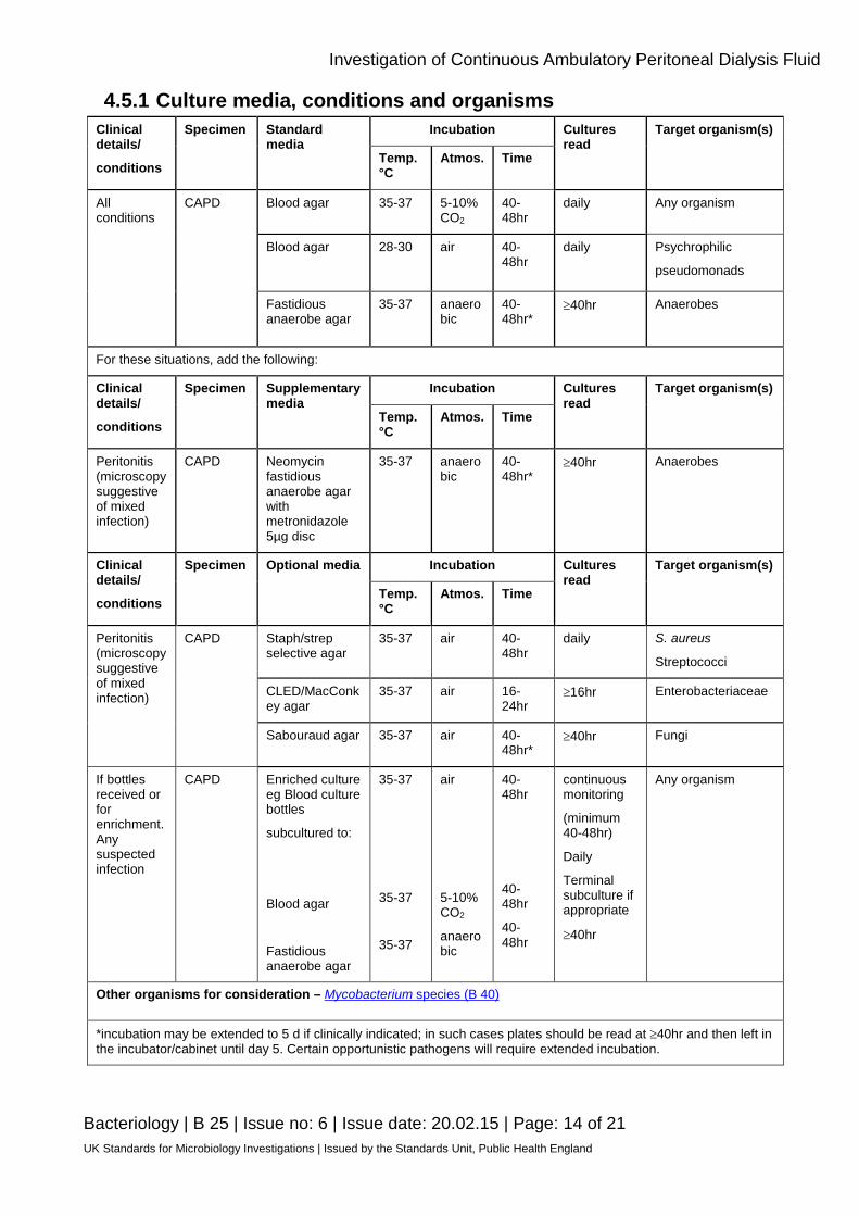
4.5.1 Culture media, conditions and organisms Clinical details/
conditions
Specimen Standard media
Incubation Cultures read
Target organism(s)
Temp. °C
Atmos. Time
All conditions
CAPD Blood agar 35-37 5-10% CO2
40-48hr
daily Any organism
Blood agar 28-30 air 40-48hr
daily Psychrophilic
pseudomonads
Fastidious anaerobe agar
35-37 anaerobic
40-48hr*
≥40hr Anaerobes
For these situations, add the following:
Clinical details/
conditions
Specimen Supplementary media
Incubation Cultures read
Target organism(s)
Temp. °C
Atmos. Time
Peritonitis (microscopy suggestive of mixed infection)
CAPD Neomycin fastidious anaerobe agar with metronidazole 5µg disc
35-37 anaerobic
40-48hr*
≥40hr Anaerobes
Clinical details/
conditions
Specimen Optional media Incubation Cultures read
Target organism(s)
Temp. °C
Atmos. Time
Peritonitis (microscopy suggestive of mixed infection)
CAPD Staph/strep selective agar
35-37 air 40-48hr
daily S. aureus
Streptococci
CLED/MacConkey agar
35-37 air 16-24hr
≥16hr Enterobacteriaceae
Sabouraud agar 35-37 air 40-48hr*
≥40hr Fungi
If bottles received or for enrichment. Any suspected infection
CAPD Enriched culture eg Blood culture bottles
subcultured to:
Blood agar
Fastidious anaerobe agar
35-37
35-37
35-37
air
5-10% CO2
anaerobic
40-48hr
40-48hr
40-48hr
continuous monitoring
(minimum 40-48hr)
Daily
Terminal subculture if appropriate
≥40hr
Any organism
Other organisms for consideration – Mycobacterium species (B 40)
*incubation may be extended to 5 d if clinically indicated; in such cases plates should be read at ≥40hr and then left in the incubator/cabinet until day 5. Certain opportunistic pathogens will require extended incubation.
Investigation of Continuous Ambulatory Peritoneal Dialysis Fluid
Bacteriology | B 25 | Issue no: 6 | Issue date: 20.02.15 | Page: 15 of 21 UK Standards for Microbiology Investigations | Issued by the Standards Unit, Public Health England
4.6 Identification Refer to individual SMIs for organism identification.
4.6.1 Minimum level of identification in the laboratory Anaerobes “anaerobes" level
β-haemolytic streptococci Lancefield group level
Enterococcus genus level
Coagulase negative staphylococci "coagulase negative" level
All other organisms species level
Organisms may be further identified if this is clinically or epidemiologically indicated. Note: Any organism considered to be a contaminant may not require identification to species level. Mycobacterium species see B 40 - Investigation of Specimens for Mycobacterium species.
4.7 Antimicrobial Susceptibility Testing Refer to British Society for Antimicrobial Chemotherapy (BSAC) and/or EUCAST guidelines. Prudent use of antimicrobials according to local and national protocols is recommended.
4.8 Referral for Outbreak Investigations N/A
4.9 Referral to Reference Laboratories For information on the tests offered, turn around times, transport procedure and the other requirements of the reference laboratory click here for user manuals and request forms. Organisms with unusual or unexpected resistance, and whenever there is a laboratory or clinical problem, or anomaly that requires elucidation should be sent to the appropriate reference laboratory. Contact appropriate devolved national reference laboratory for information on the tests available, turn around times, transport procedure and any other requirements for sample submission: England and Wales https://www.gov.uk/specialist-and-reference-microbiology-laboratory-tests-and-services Scotland http://www.hps.scot.nhs.uk/reflab/index.aspx Northern Ireland http://www.publichealth.hscni.net/directorate-public-health/health-protection
Investigation of Continuous Ambulatory Peritoneal Dialysis Fluid
Bacteriology | B 25 | Issue no: 6 | Issue date: 20.02.15 | Page: 16 of 21 UK Standards for Microbiology Investigations | Issued by the Standards Unit, Public Health England
5 Reporting Procedure 5.1 Microscopy
Cell count Report numbers of WBCs x 106 per litre.
Gram stain (if performed) Report on organisms detected.
Differential leucocyte count (if performed) Report numbers of eosinophils x 106 per litre. Microscopy for Mycobacterium species – see B 40 - Investigation of Specimens for Mycobacterium species.
Microscopy reporting time Urgent microscopy results to be telephoned or sent electronically. Written report, 16–72hr.
5.2 Culture Report the organisms isolated or Report absence of growth. Also, report results of supplementary investigations.
5.2.1 Culture reporting time Clinically urgent culture results to be telephoned or sent electronically. Written report, 16–72hr stating, if appropriate, that a further report will be issued. Supplementary investigations: Mycobacterium species - see B 40 - Investigation of Specimens for Mycobacterium species.
5.3 Antimicrobial Susceptibility Testing Report susceptibilities as clinically indicated. Prudent use of antimicrobials according to local and national protocols is recommended.
6 Notification to PHE46,47 or Equivalent in the Devolved Administrations48-51 The Health Protection (Notification) regulations 2010 require diagnostic laboratories to notify Public Health England (PHE) when they identify the causative agents that are listed in Schedule 2 of the Regulations. Notifications must be provided in writing, on paper or electronically, within seven days. Urgent cases should be notified orally and as soon as possible, recommended within 24 hours. These should be followed up by written notification within seven days. For the purposes of the Notification Regulations, the recipient of laboratory notifications is the local PHE Health Protection Team. If a case has already been
Investigation of Continuous Ambulatory Peritoneal Dialysis Fluid
Bacteriology | B 25 | Issue no: 6 | Issue date: 20.02.15 | Page: 17 of 21 UK Standards for Microbiology Investigations | Issued by the Standards Unit, Public Health England
notified by a registered medical practitioner, the diagnostic laboratory is still required to notify the case if they identify any evidence of an infection caused by a notifiable causative agent. Notification under the Health Protection (Notification) Regulations 2010 does not replace voluntary reporting to PHE. The vast majority of NHS laboratories voluntarily report a wide range of laboratory diagnoses of causative agents to PHE and many PHE Health protection Teams have agreements with local laboratories for urgent reporting of some infections. This should continue. Note: The Health Protection Legislation Guidance (2010) includes reporting of Human Immunodeficiency Virus (HIV) & Sexually Transmitted Infections (STIs), Healthcare Associated Infections (HCAIs) and Creutzfeldt–Jakob disease (CJD) under ‘Notification Duties of Registered Medical Practitioners’: it is not noted under ‘Notification Duties of Diagnostic Laboratories’. https://www.gov.uk/government/organisations/public-health-england/about/our-governance#health-protection-regulations-2010 Other arrangements exist in Scotland48,49, Wales50 and Northern Ireland51.
Investigation of Continuous Ambulatory Peritoneal Dialysis Fluid
Bacteriology | B 25 | Issue no: 6 | Issue date: 20.02.15 | Page: 18 of 21 UK Standards for Microbiology Investigations | Issued by the Standards Unit, Public Health England
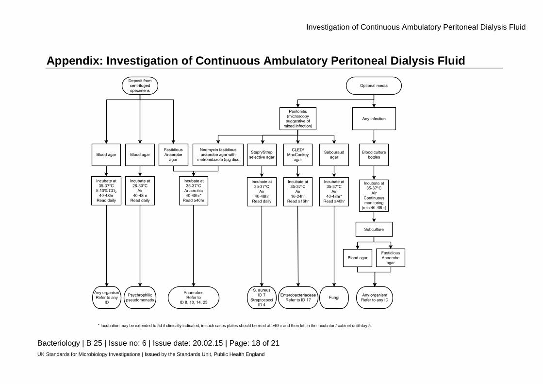
Appendix: Investigation of Continuous Ambulatory Peritoneal Dialysis Fluid Deposit from centrifuged specimens
Blood agar Blood agarFastidious Anaerobe
agar
Incubate at 35-37°C
5–10% CO2
40-48hrRead daily
Incubate at 28-30°C
Air40-48hr
Read daily
Any organismRefer to any
ID
Psychrophilic pseudomonads
Neomycin fastidious anaerobe agar with
metronidazole 5μg disc
Incubate at 35-37°C
Anaerobic40-48hr*
Read ≥40hr
AnaerobesRefer to
ID 8, 10, 14, 25
Peritonitis(microscopy suggestive of
mixed infection)
Optional media
Any infection
Staph/Strep selective agar
CLED/MacConkey
agar
Sabouraud agar
Blood culture bottles
Incubate at 35-37°C
Air40-48hr
Read daily
Incubate at 35-37°C
Air16-24hr
Read ≥16hr
Incubate at 35-37°C
Air40-48hr*
Read ≥40hr
Incubate at 35-37°C
AirContinuous monitoring
(min 40-48hr)
S. aureusID 7
StreptococciID 4
FungiEnterobacteriaceae
Refer to ID 17
Subculture
Blood agarFastidious Anaerobe
agar
Any organismRefer to any ID
* Incubation may be extended to 5d if clinically indicated; in such cases plates should be read at ≥40hr and then left in the incubator / cabinet until day 5.

Investigation of Continuous Ambulatory Peritoneal Dialysis Fluid
Bacteriology | B 25 | Issue no: 6 | Issue date: 20.02.15 | Page: 19 of 21 UK Standards for Microbiology Investigations | Issued by the Standards Unit, Public Health England
References 1. von Graevenitz A, Amsterdam D. Microbiological aspects of peritonitis associated with continuous
ambulatory peritoneal dialysis. Clin Microbiol Rev 1992;5:36-48.
2. Lin YC, Lin WP, Huang JY, Lee SY. Polymicrobial peritonitis following colonoscopic polypectomy in a peritoneal dialysis patient. Intern Med 2012;51:1841-3.
3. Ludlam HA. Infectious consequences of continuous ambulatory peritoneal dialysis. J Hosp Infect 1991;18 Suppl A:341-54.
4. Renal Association Standards Subcommittee, editor. Treatment of adults and children with renal failure: standards and audit measures. 3rd ed. London: Royal College of Physicians; 2002.
5. Ludlam H, Dickens A, Simpson A, Phillips I. A comparison of four culture methods for diagnosing infection in continuous ambulatory peritoneal dialysis. J Hosp Infect 1990;16:263-9.
6. Ludlam HA, Price TN, Berry AJ, Phillips I. Laboratory diagnosis of peritonitis in patients on continuous ambulatory peritoneal dialysis. J Clin Microbiol 1988;26:1757-62.
7. Gould IM, Casewell MW. The laboratory diagnosis of peritonitis during continuous ambulatory peritoneal dialysis. J Hosp Infect 1986;7:155-60.
8. Khare S, Yurack J, Toye B. Culture of dialysate in suspected CAPD associated peritonitis using the BacT/Alert system. Diagn Microbiol Infect Dis 1996;25:101-6.
9. Alfa MJ, Degagne P, Olson N, Harding GK. Improved detection of bacterial growth in continuous ambulatory peritoneal dialysis effluent by use of BacT/Alert FAN bottles. J Clin Microbiol 1997;35:862-6.
10. Iyer RN, Reddy AK, Gande S, Aiyangar A. Evaluation of different culture methods for the diagnosis of peritonitis in patients on continuous ambulatory peritoneal dialysis. Clin Microbiol Infect 2013.
11. Azap OK, Timurkaynak F, Sezer S, Cagir U, Yapar G, Arslan H, et al. Value of automatized blood culture systems in the diagnosis of continuous ambulatory peritoneal dialysis peritonitis. Transplant Proc 2006;38:411-2.
12. Howe PA, Fraise AP. Continuous ambulatory peritoneal dialysis: factors influencing recovery of organisms from effluents. Med Lab Sci 1991;48:114-7.
13. Lye WC. Rapid diagnosis of Mycobacterium tuberculous peritonitis in two continuous ambulatory peritoneal dialysis patients, using DNA amplification by polymerase chain reaction. Adv Perit Dial 2002;18:154-7.
14. Kwan JT, Hart PD, Raftery MJ, Cunningham J, Marsh FP. Mycobacterial infection is an important infective complication in British Asian dialysis patients. J Hosp Infect 1991;19:249-55.
15. Bendall RP, Drobniewski FA, Jayasena SD, Nye PM, Uttley AH, Scott GM. Restriction fragment length polymorphism analysis rules out cross- infection among renal patients with tuberculosis. J Hosp Infect 1995;30:51-6.
16. Quindos G, Cabrera F, Arilla MC, Burgos A, Ortiz-Vigon R, Canon JL, et al. Fatal Candida famata peritonitis in a patient undergoing continuous ambulatory peritoneal dialysis who was treated with fluconazole. Clin Infect Dis 1994;18:658-60.
17. Yuen KY, Seto WH, Ching TY, Cheung WC, Kwok Y, Chu YB. An outbreak of Candida tropicalis peritonitis in patients on intermittent peritoneal dialysis. J Hosp Infect 1992;22:65-72.
Investigation of Continuous Ambulatory Peritoneal Dialysis Fluid
Bacteriology | B 25 | Issue no: 6 | Issue date: 20.02.15 | Page: 20 of 21 UK Standards for Microbiology Investigations | Issued by the Standards Unit, Public Health England
18. Yinnon AM, Solages A, Treanor JJ. Cryptococcal peritonitis: report of a case developing during continuous ambulatory peritoneal dialysis and review of the literature. Clin Infect Dis 1993;17:736-41.
19. Kim SH, Jeong HS, Kim YH, Song SA, Lee JY, Oh SH, et al. Evaluation of DNA extraction methods and their clinical application for direct detection of causative bacteria in continuous ambulatory peritoneal dialysis culture fluids from patients with peritonitis by using broad-range PCR. Ann Lab Med 2012;32:119-25.
20. European Parliament. UK Standards for Microbiology Investigations (SMIs) use the term "CE marked leak proof container" to describe containers bearing the CE marking used for the collection and transport of clinical specimens. The requirements for specimen containers are given in the EU in vitro Diagnostic Medical Devices Directive (98/79/EC Annex 1 B 2.1) which states: "The design must allow easy handling and, where necessary, reduce as far as possible contamination of, and leakage from, the device during use and, in the case of specimen receptacles, the risk of contamination of the specimen. The manufacturing processes must be appropriate for these purposes".
21. Official Journal of the European Communities. Directive 98/79/EC of the European Parliament and of the Council of 27 October 1998 on in vitro diagnostic medical devices. 7-12-1998. p. 1-37.
22. Health and Safety Executive. Safe use of pneumatic air tube transport systems for pathology specimens. 9/99.
23. Department for transport. Transport of Infectious Substances, 2011 Revision 5. 2011.
24. World Health Organization. Guidance on regulations for the Transport of Infectious Substances 2013-2014. 2012.
25. Home Office. Anti-terrorism, Crime and Security Act. 2001 (as amended).
26. Advisory Committee on Dangerous Pathogens. The Approved List of Biological Agents. Health and Safety Executive. 2013. p. 1-32
27. Advisory Committee on Dangerous Pathogens. Infections at work: Controlling the risks. Her Majesty's Stationery Office. 2003.
28. Advisory Committee on Dangerous Pathogens. Biological agents: Managing the risks in laboratories and healthcare premises. Health and Safety Executive. 2005.
29. Advisory Committee on Dangerous Pathogens. Biological Agents: Managing the Risks in Laboratories and Healthcare Premises. Appendix 1.2 Transport of Infectious Substances - Revision. Health and Safety Executive. 2008.
30. Centers for Disease Control and Prevention. Guidelines for Safe Work Practices in Human and Animal Medical Diagnostic Laboratories. MMWR Surveill Summ 2012;61:1-102.
31. Health and Safety Executive. Control of Substances Hazardous to Health Regulations. The Control of Substances Hazardous to Health Regulations 2002. 5th ed. HSE Books; 2002.
32. Health and Safety Executive. Five Steps to Risk Assessment: A Step by Step Guide to a Safer and Healthier Workplace. HSE Books. 2002.
33. Health and Safety Executive. A Guide to Risk Assessment Requirements: Common Provisions in Health and Safety Law. HSE Books. 2002.
34. Health Services Advisory Committee. Safe Working and the Prevention of Infection in Clinical Laboratories and Similar Facilities. HSE Books. 2003.

Investigation of Continuous Ambulatory Peritoneal Dialysis Fluid
Bacteriology | B 25 | Issue no: 6 | Issue date: 20.02.15 | Page: 21 of 21 UK Standards for Microbiology Investigations | Issued by the Standards Unit, Public Health England
35. British Standards Institution (BSI). BS EN12469 - Biotechnology - performance criteria for microbiological safety cabinets. 2000.
36. British Standards Institution (BSI). BS 5726:2005 - Microbiological safety cabinets. Information to be supplied by the purchaser and to the vendor and to the installer, and siting and use of cabinets. Recommendations and guidance. 24-3-2005. p. 1-14
37. Allen BW. Survival of tubercle bacilli in heat-fixed sputum smears. J Clin Pathol 1981;34:719-22.
38. Baron EJ, Miller JM, Weinstein MP, Richter SS, Gilligan PH, Thomson RB, Jr., et al. A Guide to Utilization of the Microbiology Laboratory for Diagnosis of Infectious Diseases: 2013 Recommendations by the Infectious Diseases Society of America (IDSA) and the American Society for Microbiology (ASM). Clin Infect Dis 2013;57:e22-e121.
39. Rishmawi N, Ghneim R, Kattan R, Ghneim R, Zoughbi M, Abu-Diab A, et al. Survival of fastidious and nonfastidious aerobic bacteria in three bacterial transport swab systems. J Clin Microbiol 2007;45:1278-83.
40. Barber S, Lawson PJ, Grove DI. Evaluation of bacteriological transport swabs. Pathology 1998;30:179-82.
41. Van Horn KG, Audette CD, Sebeck D, Tucker KA. Comparison of the Copan ESwab system with two Amies agar swab transport systems for maintenance of microorganism viability. J Clin Microbiol 2008;46:1655-8.
42. Nys S, Vijgen S, Magerman K, Cartuyvels R. Comparison of Copan eSwab with the Copan Venturi Transystem for the quantitative survival of Escherichia coli, Streptococcus agalactiae and Candida albicans. Eur J Clin Microbiol Infect Dis 2010;29:453-6.
43. Tano E, Melhus A. Evaluation of three swab transport systems for the maintenance of clinically important bacteria in simulated mono- and polymicrobial samples. APMIS 2011;119:198-203.
44. Isenberg HD, Washington JA II, Doern GV, Amsterdam D. Specimen collection and handling. In: Balows A, Hausler WJ J, Herrmann KL, Isenberg HD, Shadomy HJ, editors. Manual of Clinical Microbiology. 5th ed. Washington D.C.: American Society for Microbiology; 1991. p. 15-28.
45. Fenton P. Laboratory diagnosis of peritonitis in patients undergoing continuous ambulatory peritoneal dialysis. J Clin Pathol 1982;35:1181-4.
46. Public Health England. Laboratory Reporting to Public Health England: A Guide for Diagnostic Laboratories. 2013. p. 1-37.
47. Department of Health. Health Protection Legislation (England) Guidance. 2010. p. 1-112.
48. Scottish Government. Public Health (Scotland) Act. 2008 (as amended).
49. Scottish Government. Public Health etc. (Scotland) Act 2008. Implementation of Part 2: Notifiable Diseases, Organisms and Health Risk States. 2009.
50. The Welsh Assembly Government. Health Protection Legislation (Wales) Guidance. 2010.
51. Home Office. Public Health Act (Northern Ireland) 1967 Chapter 36. 1967 (as amended).