ukrco.org.uk · web viewin february 2016, in conjunction with liverpool ccg, the primary care...
TRANSCRIPT

0
UKRO ORAL PRESENTATIONS
C1 Patient and Public Involvement (PPI)
Evaluations on the clinical effectiveness of a bespoke prostate PROMS baseline questionnaireGayan Chetiyawardana; Yat TsangMount Vernon Cancer CentreObjective: In prostate cancer management, it's prudent to assess patients' baseline status to accurately assess treatment toxicity post radiotherapy. A bespoke PROMS pre-radiotherapy baseline questionnaire was implemented at our department and this study aims to evaluate its clinical effectiveness in terms of patients' feedback and cost savings. Method: From January 2017 to May 2017, 50 prostate cancer radiotherapy patients filled out the PROMS pre-radiotherapy baseline questionnaire. If the patient indicated current function of genitourinary (GU), gastro-intestinal (GI) or sexual function (SF) were causing poor quality of life the patient had a telephone consultation and any suggestions recorded. Results: 45/50 patients indicated they would need further support prior to starting radiotherapy. 64% of patients needed advice on pharmacological intervention for current GU symptoms. 48% of patients needed further support for current GI symptoms and 17% needing advice on pharmacological intervention. 44% of patients needed advice on pharmacological intervention for current SF. The patients were asked to access the pharmacological intervention via their primary care providers. This resulted in departmental savings of £114.60 with the 50 patients in this study. With an estimation of 500 prostate radiotherapy patients per year at our department, this would result in a total annual saving of £1145. Conclusion: This study suggested that there was a role in using baseline PROMS to address prostate cancer patients’ physical/psychosocial needs prior to radiotherapy in terms of better patient-centred care and economical savings for our department.
Turning the corner: A mixed-methods investigation of the radiotherapy information needs of GPsKelsey Normand 1; Gareth Hill 2
1NHS Lothian; 2Queen Margaret UniversityBackground: NHS cancer strategy emphasises delivery of integrated care across primary and secondary environments. However, previous studies have highlighted a radiotherapy knowledge gap amongst GPs. This study aimed to identify the radiotherapy information needs of GPs and explore how these could be met by a large regional cancer centre. Method: A 10-item questionnaire developed by the researcher was distributed to all 123 practice managers in a single health board. To add depth and detail volunteers were then recruited for semi-structured interviews. Results: 93 valid questionnaires were received in the four week data collection period. Although 95% had cared for a patient undergoing radiotherapy, only 4% agreed that radiotherapy information was easy to access. Confidence in indications for emergency radiotherapy (65%) and indications for palliative radiotherapy (64%) were highest, while confidence in how radiotherapy interacts with other treatments (2%) and in managing acute side effects beyond skin reactions (14%) were lowest. 70% of GPs reported having radiotherapy education. This was correlated with confidence in explaining radiotherapy (p= 0.013), discussing long-term side effects (p= 0.036) and indications for palliative radiotherapy (p=0.02). GPs preferred easily accessible electronic information, and suggested adding radiotherapy information to an existing platform. They also perceived a division between specialist and primary care. Conclusion: The integrated care outlined in NHS cancer strategy is challenged in practice by lack of knowledge and an underlying perception among GPs of fragmentation of care. This affects patients and should be addressed as a priority with straightforward electronic information and more complex strategic interventions.1. Baart et al. (2009) GPs and referral for palliative radiotherapy. Rad and Onc. 91, 267-270. 2. Berendsen et al. (2015) The expanding role of primary care in cancer control. Lancelot Onc. Comm. 16, 1231-1272. 3. De Bock et al. (2012) Role of the GP during the active breast cancer treatment phase. Supportive Cancer Care (April), 705-714.
C10 The needs of contemporary knowledge based planning
Automated prostate radiotherapy scripting - a step towards quality improvementValerie Wilson; Clara Namelo; Douglas Etheridge; Joseph Snelling; Mau-Don Phan; Delia Pudney; Sarah Gwynne; Russell BannerSouth West Wales Cancer Centre, SwanseaBackground: Faced with an increase in the number of patients undergoing radical pelvic radiotherapy (RT) for urological cancers, methods of efficient, safe and reproducible target volume delineation are required. Automated scripts in pelvic radiotherapy planning could reduce human error, produce reproducible target volume delineation, increase consistency and reduce radiotherapy treatment planning times. We developed an automated multistep prostate RT planning script. Methodology: Predefined organs at risk (OARs) such as bladder, bowel Planning Risk Volume (PRV), rectum and Clinical Target Volumes (CTVs): CTVprostatic +/- CTVseminalvesicle, CTVnodes were outlined according to the PIVOTAL trial guidelines. Automated prostate scripts were developed and tested in conjunction with RT physics and Clinical Oncologists to mimic this trial's RT target volumes. The final script was run to generate corresponding PIVOTAL compliant planning target volumes (PTVs):
1UKRCO 2018 LIVERPOOL
UKRO ORAL PRESENTATIONS
PTVprostatic +/- PTVseminalvesicle and PTVpelvic, whilst bypassing the OARs. Script commands did not run if set critical target volumes were missing/duplicated. Results PTVs were generated that were compliant with the PIVOTAL trial and reduced clinician planning time by more than 50%. However, there was an increased dosimetrist time. To overcome this, bespoke target volume atlases including 'Bone-Muscle-Rim' were developed that decreased the dosimetrist time by approximately 30% and further improved consistency. Conclusion: This automated prostate script consistently and efficiently generated the expected PTVs. The script is now locally routinely used in clinical practice. There is potential to modify this script for use in adjuvant prostate bed radiotherapy and other pelvic malignancies and this has been exploited locally with gynaecological pelvic outlining.
The first UK survey of doses from radiotherapy treatment planning CT scans for adult patientsAnne T. Davis 1; Tim J. Wood 2; Matthew Williams 3; Rosy Plaistow 4; Rebecca Lindsay 5; Antony L. Palmer 1; Andrew Nisbet 1
1University of Surrey; 2Hull and East Yorkshire Hospitals NHS Trust; 3Velindre NHS Trust; 4Cambridge University Hospitals NHS Foundation Trust; 5St James Institute of OncologyBackground: The first UK wide dose survey for radiotherapy CT planning scans has been completed. The survey was initiated by a working party of the Institute for Physics and Engineering in Medicine (IPEM).Method: Patient dose metrics were collected for prostate, gynaecological, breast, 3D-lung, 4D-lung, brain and head/neck scans. Median values per scanner and examination type were calculated. National dose reference levels of CT dose index (CTDIvol) and dose-length-product (DLP) values for each examination type are proposed based on the third quartile values from the whole data set.Results: 68 radiotherapy CT scanners were included. Patient numbers per scan type ranged from 664 to 1527 across the seven examinations. The proposed reference levels for CTDIvol (mGy) and DLP (mGy.cm) respectively are prostate 16 and 570, gynaecological 16 and 610, breast 10 and 390, 3D-lung 14 and 550, 4D-lung 63 and 1750, brain 50 and 1500 and head/neck 49 and 2150. Head/neck and 4D-lung had the largest differences (18 times) in dose between lowest and highest dose scanners. Problems with the data collected included some older scanners indicating maximum CTDIvol not scan average; the lack of standardisation as to whether CTDIvol is indicated for a 16 cm or 32 cm phantom for head scans; the lack of patient weight information available in many centres.Conclusion: Evidence of clustering of results by scanner type suggests there is scope for protocol adjustment in some centres. Dose reference levels have been recommended to aid this.
The introduction of dedicated planning MR-CT fusion for radical radiotherapy of prostate cancerCiara Lyons 1; Lynn Graham 2; Bernadette McCafferty 2; Darren Brady 2; Patrizia Porcu 2; Lois McGinley 2; Stephen Gilroy 2; Aisling Haughey; Elaine Reilly 2; Andrew Reilly 2; David Stewart 2
North West Cancer Centre, Altnagelvin Area HospitalPurpose: Advances in radiotherapy planning and delivery have made target definition increasingly important. While CT images are required for plan calculation, MR fusion is increasingly used to more accurately define tumour and normal tissue. There is often significant variation seen between diagnostic and therapeutic imaging; hence, MR carried out in the treatment position is desirable. Method: A multidisciplinary team of diagnostic and therapeutic radiographers, treatment planners, medical physicists and clinicians was convened. Planning MR was integrated into the radiotherapy pathway and carried out in the days immediately following CT simulation. All men underwent identical preparation (administration of a micro-enema and drinking 300mL of water thirty minutes prior to imaging/treatment). Patients were set up in the treatment position using MR-compatible radiotherapy immobilisation. T2SE axial and sagittal images were acquired (Siemens Aera 1.5T E11, incorporating RT software platforms/LAP Laser Bridge/Civco RT Indexing Flat couch top/coil bridges), imported into the Eclipse planning system (V13.6, Varian), and fused to the planning CT for volume delineation. Results: The service opened in mid-September 2017. 26 patients were scanned to the end of November 2017. All patients tolerated preparation and imaging without difficulty. Conclusion: This service has been successfully introduced and will shortly expand to include other sites (rectum, lung, head and neck, complex palliative). A study is planned to assess the impact of the addition of MR on target delineation. Additional considerations include the need for dedicated radiology input and the potential role of collaboration with industry with a view to stand-alone MR simulation.
D8 Respiratory motion management
A respiratory motion management strategy for both abdominal and thoracic VMAT radiotherapyMark Bray-Parry; Joshua Gesner; Katrina Finnegan; Isabel Ho; Simon Stevens; Ashley Richmond; Jan KonieczekThe London Clinic
2UKRCO 2018 LIVERPOOL
UKRO ORAL PRESENTATIONS
Purpose or objective: When targeting with radiotherapy, it is important that this respiratory motion is accounted for. This is typically done by creating an Internal Target Volume (ITV). Alternative approaches include a breath-hold (BH) technique. This study investigates a motion management strategy which aims to provide the optimal motion management technique for each individual patient. This is shown in figure 1.
Material and methods: 43 patients were assessed within our motion management strategy were reviewed (mix of abdominal and thoracic sites). For each patient, Planning Target Volumes (PTV) were generated using both ITV and BH techniques and compared.Results: The difference in the PTV between the two techniques varied, with a mean volume difference for all patients of 51cc (15% relative change). For pancreas, BH was smaller in 7/11 patients with a mean reduction of 60cc (28.4%) and maximum of 143cc (35%). For liver, BH was smaller in 6/7 patients with a mean reduction of 93cc (14.9%) and maximum of 189cc (38.7%). For lung, BH was smaller in 4/7 patients with a mean reduction of 15cc (13.9%) and maximum of 38cc (46%). For oesophagus, ITV was smaller for 4/6 patients by a mean of 52cc (14.6%) and a maximum of 85cc (10.4%). For mediastinum, ITV was smaller for 4/5 patients by a mean of 12cc (4.5%) and a maximum of 14cc (5.6%).Conclusion: Results show that the optimal motion management strategy to minimise the irradiated volume is patient-specific. Therefore, it's important to have a flexible approach to motion management.
Controlling motion in radiotherapy: Rapid shallow ventilation for thoracic targetsNicholas West 1; Michael Parkes 2; James Prentis 1; Christopher Snowden 1; Jill McKenna 1; Shahid Iqbal 1; Christopher Walker 1
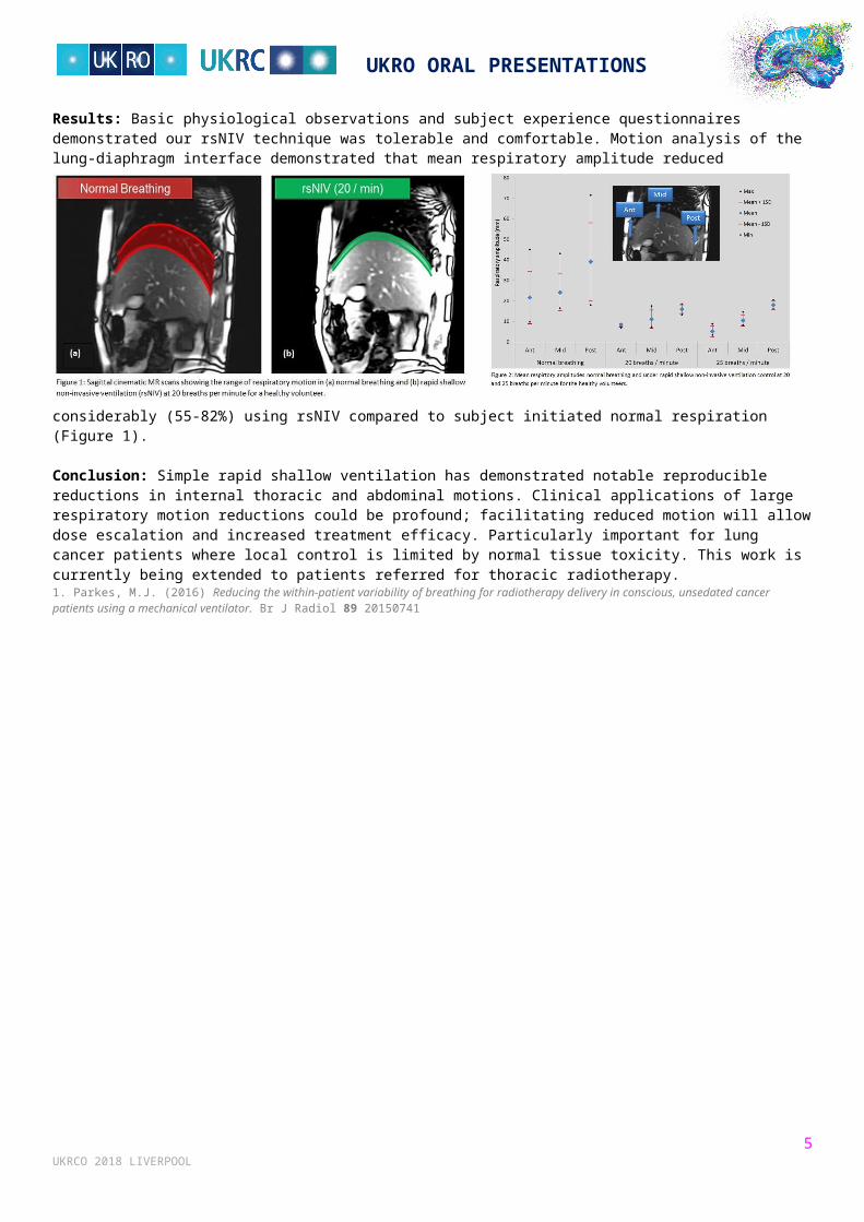
1Newcastle upon Tyne Hospitals Trust; 2University of BirminghamObjective: In radiotherapy, accounting for respiratory motion increases the volume of normal tissues irradiated, increasing healthy toxicity and constraining treatment efficacy.Aim: To assess rapid shallow non-invasive ventilation (rsNIV)[1] for controlling internal respiratory motion for radiotherapy purposes. To our knowledge, this is the first study to evaluate internal anatomical motion using rsNIV to regularise and minimise respiratory variations over a period long enough to image and deliver complex high dose radiotherapy.Materials and methods: 10 healthy volunteers (21.7-53.9yrs; mean 37.5yrs; 6f/4m) were scanned on an MR scanner in 3 respiratory modes; normal breathing and 2 non-invasive mechanically ventilated frequencies of 20 and 25 breathes per minute using a non-invasive ventilator. Sagittal and coronal cinematic datasets were acquired, and the resulting respiratory motions assessed. Respiratory amplitudes were measured across the lung-diaphragm interface and physiological parameters quantified tolerability of the mechanical ventilation.Results: Basic physiological observations and subject experience questionnaires demonstrated our rsNIV technique was tolerable and comfortable. Motion analysis of the lung-diaphragm interface demonstrated that mean respiratory amplitude reduced considerably (55-82%) using rsNIV compared to subject initiated normal respiration (Figure 1).
Conclusion: Simple rapid shallow ventilation has demonstrated notable reproducible reductions in internal thoracic and abdominal motions. Clinical applications of large respiratory motion reductions could be profound; facilitating reduced motion will allow dose escalation and increased treatment efficacy. Particularly important for lung cancer patients where local control is limited by normal tissue toxicity. This work is currently being extended to patients referred for thoracic radiotherapy.
3UKRCO 2018 LIVERPOOL

UKRO ORAL PRESENTATIONS
1. Parkes, M.J. (2016) Reducing the within-patient variability of breathing for radiotherapy delivery in conscious, unsedated cancer patients using a mechanical ventilator. Br J Radiol 89 20150741
4UKRCO 2018 LIVERPOOL
UKRO ORAL PRESENTATIONS
F1 Workforce challenges
Development of a consultant radiographer led radical prostate radiotherapy service: An effective use of skills for patient benefitTracey EllisLancashire Teaching Hospitals NHS Foundation TrustCurative treatment options for prostate cancer include surgery or radiotherapy, with neither modality being demonstrated as superior[1]. Prostate patients account for 25% of our radiotherapy department's workload and is set to rise. Consultant clinical oncologists are currently in short supply and so clinical capacity is reduced. Delays in oncology appointments to discuss radiotherapy as a treatment option cause anxiety for patients and their families, as well as resulting in breeches in cancer targets. Therefore, some patients opt for surgery as their treatment rather than waiting for an oncology appointment, thereby not making a fully informed treatment decision. In 2015, Macmillan supported the development of a consultant radiographer (CR) post with the aim of streamlining the pathway as well as offering additional oncology capacity. For two years training needs were addressed through shadowing consultant oncologists, clinical supervision and assessment and self-directed learning. Competency in roles outside of the traditional radiographer scope of practice such as consent, referral and contouring were evidenced through records of supervision, developed into a clinical portfolio. Aspects such as clinical review and non-medical prescribing were addressed through formal qualifications. A streamlined radical prostate radiotherapy service has now been developed. A radiographer led service mimics that offered by a consultant oncologist. The CR can carry out all aspects of the role autonomously. A new referral system is now in place to ensure patients receive timely appointments with either the CR or the clinical oncologist, ensuring patients are fully informed of their treatment options and cancer pathways are adhered to.1. Hamdy, F. C., Donovan, J.L., Lane, J. A., Mason, M., Metcalfe, C., Holding, P., Davis M., Peters, T. J., Turner, E. L., Martin, R.M., Oxley, J., Robinson, M., Staffurth, J., Walsh, E., Bollina, P., Catto, J., Doble, A., Doherty, A., gillatt, D. and Kockelbergh, R. (2016). 10 year outcomes after Monitoring, Surgery or Radiotherapy for localized prostate cancer. The New England Journal of Medicine, (375), 1415-1424.
I1 Adaptive radiotherapy
Hybrid I-123 MIBG SPECT/CT - radiotherapy planning CT scanning for neuroblastomaGrace Keane; Hazel McCallum; Emma Lethbridge; George Petrides; David McCulloch; Terry WatsonNorthern Centre for Cancer CareBackground: Neuroblastoma is the third most common tumour in children and I-123 MIBG SPECT/CT imaging is a well-established diagnostic tool that has not been previously used for radiotherapy target delineation at our centre, or routinely in the UK. This work will present our experience of two patients undergoing a hybrid SPECT/CT-planning CT scan, using a dedicated radiotherapy SPECT/CT scanner.Method: Hybrid SPECT/CT-planning CT scans were performed for two patients aged <7yrs on a Siemens Symbia-T16 SPECT/CT enabled for radiotherapy treatment planning. A sequential SPECT/CT-planning CT in the radiotherapy treatment position; to be used for diagnosis, delineation, planning and radiotherapy dose calculation, was acquired. Co-ordinating the patient pathway involved an extensive multi-disciplinary team from Radiotherapy, Nuclear Medicine and Children's Services. Virtual-Simulation Software ProSoma (MedCom) was used for image fusion and target delineation. Results: Clinicians reported increased confidence in outlined volumes using MIBG SPECT/CT compared to the CT planning scan alone. The benefits of a single imaging session were:
A decrease in appointment time with an average scan time of 80 mins; The CT and SPECT are implicitly registered and no uncertainties in spatial alignment are introduced; The patient was saved an additional hospital visit and general anaesthetic procedure with associated risks and costs; More efficient diagnostic work-up and treatment planning preparation.
Conclusion: For the first time at our centre, a hybrid I-123 MIBG SPECT/CT-planning CT scan has been acquired and used for radiotherapy planning. This process will be developed into a clinical service for all neuroblastoma patients.
Co-relationship between 3D surface imaging system and conventional volumetric registration in radiotherapy pelvis treatment positioningOi-Ching ChoiCancer Centre LondonBackground: 3D imaging has shown advantageous results on breast patients in detecting set-up errors without any radiation (Deantonio et al 2011 & Alderliesten et al 2013). This study aims at studying the co-relation of the surface registration with the cone beam computed tomography (CBCT) in radiotherapy pelvis treatment positioning. Method: 12 pelvis patients with 267 fractions were selected which all of them had CT planning scans. During treatment, 3D surfaces were captured by a surface imaging system (AlignRT) prior to subsequent setup procedure. The set-up errors were
5UKRCO 2018 LIVERPOOL
UKRO ORAL PRESENTATIONS
verified by the CBCT before treatment beam delivery. The discrepancies were calculated when comparing with the original planning CT scans. The resulting errors were compared with linear regression analysis and Bland-Altman plots. Results: The Pearson correlation between setup errors were 0.57, 0.51, 0.65 in left-right (LR), craniocaudal (CC) and anterior-posterior (AP) directions respectively. For the differences between setup errors: The group means, systematic errors and random errors were 0.10cm, -0.30cm, -0.09cm; 0.13 cm, 0.37 cm, 0.29 cm and 0.18 cm, 0.22 cm, 0.22 cm in LR, CC, AP directions respectively. The paired t-test for random errors showed a significant difference between the two systems along all direction (all with t<0.0001). Conclusion: The setup measurements by the 3D surface imaging has good correlation with the setup errors detected by CBCT. It can be used to assess the setup reproducibility for pelvis patients and reduce the number of setup corrections while using the CBCT.1. Alderliesten T, Sonke J, Betgen et al 2011. Accuracy Evaluation of a 3-Dimensional Surface Imaging System for Guidance in Deep-inspiration Breath-Hold Radiation Therapy. Int J Radiation Oncol Biol Phys. 85(2) 536-542 2. Deantonio L, Masini L, Loi G et al (2011). Detection of Setup uncertainties with 3D surface registration system for conformal radiotherapy of breast cancer. Rpt of Pract Onco and radiother 16:77-81
Dose painting for prostate cancer with external beam radiotherapy: factors affecting the feasibility of treatment planning and dose deliverySteve Blake 1; Serena Hilman 1; Alison Stapleton 1; Andrew Brown 1; Sian Curtis 1; Margaret Saunders 1; Janice Ash-Miles 2; Emma Dennis 1; Ron Hartley-Davies 1; Susan Masson 1; Dawn Bowers 1
1University Hospitals Bristol; 2North Bristol NHS TrustBackground: Dose painting is a promising technique[1,2,3] which enables dose escalation to tumour nodules within the prostate. This study aims to determine factors affecting treatment feasibility for 20 patients with intermediate-high risk disease.Method: Patients were imaged using a 3T MRI scanner and visible nodules outlined and registered with the planning CT. Plans were produced using OMP(Elekta). CHHIP constraints were used[4] and urethra and small bowel also delineated. Plans were assessed dosimetrically to determine whether the boosted distribution could be safely delivered. Results: MRI scans were successful for 19/20 patients. 14 showed 1-2 nodules with 11/14 overlapping the urethra and/or rectum, 1 abutting the urethra and 2 not overlapping. The target boost of 86 Gy was achieved in 6/14 plans (see figure). For one patient this was limited to 82 Gy due to the constraints for rectum and urethra and 80 Gy for 5 more patients whose GTV overlapped or abutted the urethra. For the remaining 2 patients it was difficult to match CT & MRI images using rigid registration due to changes in prostate position between modalities. Dosimetric measurements were made on 5 plans using Compass (IBA). One marginally failed the gamma comparison (3% 3 mm) with 3.9% of failing points within PTV1 (limit 3%). Conclusion: It was feasible to produce dose-painted plans for approximately half the patients with nodules. The main issue limiting the feasibility of dose painting was the proximity of organs-at-risk to the boost volumes. A strategy for improving CT/MRI registration issues is also required.1 Bauman G et al (2013) Boosting imaging defined dominant prostatic tumors: A systematic review. Radiother Oncol 107 (2013) 274-281 2 Monninkhof EM et al (2018) Standard whole prostate gland radiotherapy with and without lesion boost in prostate cancer: Toxicity in the FLAME randomized controlled trial. Radiother Oncol. 2018 Jan 11. [Epub ahead of print] 3 Uzan J, Nahum AE, Syndikus I (2016) Clin Oncol (R Coll Radiol). 2016 Mar;28(3):165-70. 4 CHHiP Conventional or Hypofractionated High Dose Intensity Modulated Radiotherapy for Prostate Cancer (2006) Protocol Version 3.0 ICR-CTSU, Sutton, Surrey UK.
UK clinical trials in the spotlight
Does the size of CTV-PTV margin in dysphagia-optimised intensity modulated radiotherapy (Do-IMRT) affect the quality of plan produced in the DARS head and neck cancer randomised trial?Justine TylerThe Royal Marsden NHS Foundation TrustThe DARS trial (CRUK/14/014) compares Do-IMRT versus standard IMRT in head and neck cancer treatment. For Do-IMRT, centres using a 5mm CTV-PTV margin experienced more difficulty meeting the pre-trial QA requirements than centres using a 3mm margin. This study aims to determine the effect of CTV-PTV margin on plan quality.Centres completing the Do-IMRT oropharyngeal QA case were required to meet mandatory DVH constraints and encouraged to try to achieve optimal constraints. Compromise in coverage was permitted in PlanPTV_5400 (54Gy PTV cropped from body and 65Gy PTV) only in the region of PlanSMPCM (Superior and Middle Pharyngeal Constructor Muscle cropped from 65Gy CTV) and PlanIPCM (Inferior Pharyngeal Constructor Muscle cropped from 65Gy CTV), see figure 1. DVH statistics for PTVs and OARs for the final plans were compared according to the CTV- PTV margin.PlanIPCM, Brainstem PRV and ipsilateral parotid (parotid_IL) dose statistics achieved by centres using a 3mm margin were statistically significantly lower than centres using a 5mm margin (table 1). Centres using a 3mm margin achieved poorer PlanPTV_5400 D99%(Gy) compared to centres using a 5mm margin.
6UKRCO 2018 LIVERPOOL
UKRO ORAL PRESENTATIONS
Initial experience in DARS suggests that the CTV-PTV margin affects plan quality for Do-IMRT plans; larger margins were associated with higher doses to some OARs. However, centres using a 5mm margin may be achieving better PlanPTV_5400 coverage at the expense of PlanIPCM dose, therefore accounting for some of the differences. These findings do not take into account the possible effect of other factors such as treatment planning system.Petkar I, Rooney K, Roe JWG, Patterson JM, Bernstein D, Tyler JM, Emson MA, Morden JP, Mertens K, Miles E, Beasley M, Roques T, Bhide SA, Newbold KL, Harrington KJ, Hall E, Nutting CM. (2016) DARS: a phase III randomised multicentre study of dysphagia- optimised intensity- modulated radiotherapy (Do-IMRT) versus standard intensity- modulated radiotherapy (S-IMRT) in head and neck cancer. BMC Cancer 16(770).DARS QA team. DARS Radiotherapy outlining and planning QA guidelines. Version 1.4. January 2016.
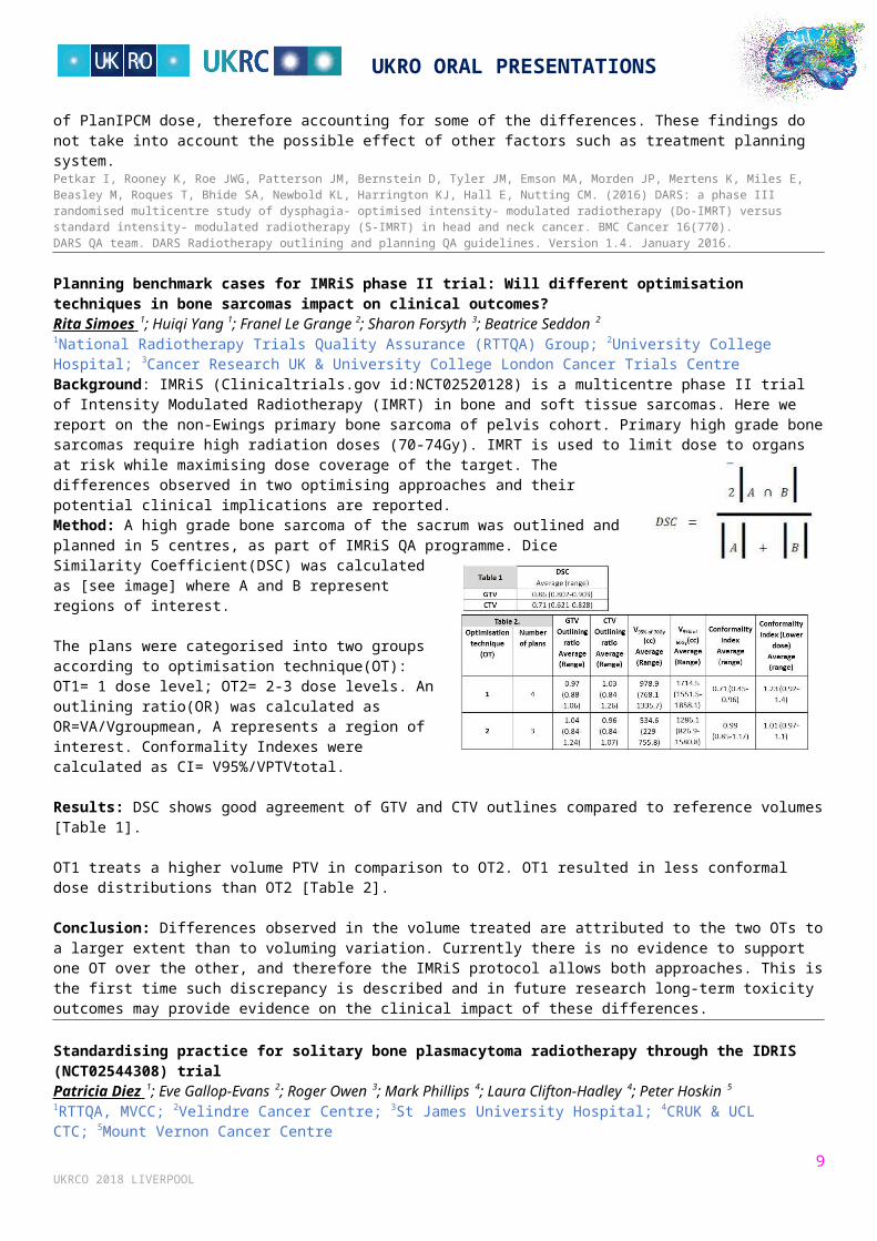
Planning benchmark cases for IMRiS phase II trial: Will different optimisation techniques in bone sarcomas impact on clinical outcomes?Rita Simoes 1; Huiqi Yang 1; Franel Le Grange 2; Sharon Forsyth 3; Beatrice Seddon 2
1National Radiotherapy Trials Quality Assurance (RTTQA) Group; 2University College Hospital; 3Cancer Research UK & University College London Cancer Trials CentreBackground: IMRiS (Clinicaltrials.gov id:NCT02520128) is a multicentre phase II trial of Intensity Modulated Radiotherapy (IMRT) in bone and soft tissue sarcomas. Here we report on the non-Ewings primary bone sarcoma of pelvis cohort. Primary high grade bone sarcomas require high radiation doses (70-74Gy). IMRT is used to limit dose to organs at risk while maximising dose coverage of the target. The differences observed in two optimising approaches and their potential clinical implications are reported. Method: A high grade bone sarcoma of the sacrum was outlined and planned in 5 centres, as part of IMRiS QA programme. Dice Similarity Coefficient(DSC) was calculated as [see image] where A and B represent regions of interest.
The plans were categorised into two groups according to optimisation technique(OT): OT1= 1 dose level; OT2= 2-3 dose levels. An outlining ratio(OR) was calculated as OR=VA/Vgroupmean, A represents a region of interest. Conformality Indexes were calculated as CI= V95%/VPTVtotal.
Results: DSC shows good agreement of GTV and CTV outlines compared to reference volumes [Table 1].
OT1 treats a higher volume PTV in comparison to OT2. OT1 resulted in less conformal dose distributions than OT2 [Table 2].
Conclusion: Differences observed in the volume treated are attributed to the two OTs to a larger extent than to voluming variation. Currently there is no evidence to support one OT over the other, and therefore the IMRiS protocol allows both approaches. This is the first time such discrepancy is described and in future research long-term toxicity outcomes may provide evidence on the clinical impact of these differences.
Standardising practice for solitary bone plasmacytoma radiotherapy through the IDRIS (NCT02544308) trialPatricia Diez 1; Eve Gallop-Evans 2; Roger Owen 3; Mark Phillips 4; Laura Clifton-Hadley 4; Peter Hoskin 5
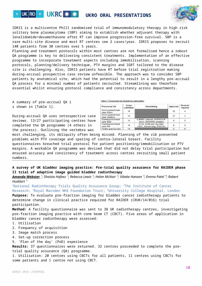
1RTTQA, MVCC; 2Velindre Cancer Centre; 3St James University Hospital; 4CRUK & UCL CTC; 5Mount Vernon Cancer CentreIDRIS is a multicentre PhIII randomised trial of immunomodulatory therapy in high risk solitary bone plasmacytoma (SBP) aiming to establish whether adjuvant therapy with lenalidomide+dexamethasone after RT can improve progression-free survival. SBP is a rare multi-site disease and most RT centres see 2 cases/year. IDRIS proposes to recruit 140 patients from 30 centres over 5 years. Planning and treatment protocols within most centres are not formalised hence a robust QA programme is key to delivering consistent treatments. Implementation of an effective programme to incorporate treatment aspects including immobilisation, scanning protocols, planning/delivery technique, PTV margins and IGRT tailored to the disease site is challenging. Also, IDRIS patients have RT before trial registration making during-accrual prospective case review unfeasible. The approach was to consider SBP patients by anatomical site, which had the potential to result in a lengthy pre-accrual QA process for a minimal number of patients recruited. Streamlining was therefore essential whilst ensuring protocol compliance and consistency across departments.
A summary of pre-accrual QA i
7UKRCO 2018 LIVERPOOL
UKRO ORAL PRESENTATIONS
s shown in [Table 1].
During-accrual QA uses retrospective case reviews. 13/27 participating centres have completed the QA programme (4 others in the process). Outlining the vertebra was most challenging, its obliquity often being missed. Planning of the rib presented problems with PTV coverage and sparing of contra-lateral breast. Facility questionnaires breached trial protocol for patient positioning/immobilisation or PTV margins. A workable QA programme was devised that did not delay trial participation but ensured accuracy and consistency of treatment across centres recruiting small patient numbers.
A survey of UK bladder imaging practice: Pre-trial quality assurance for RAIDER phase II trial of adaptive image guided bladder radiotherapyAmanda Webster 1; Shaista Hafeez 1; Rebecca Lewis 2; Helen McNair 3; Vibeke Hansen 3; Emma Patel 4; Robert Huddart 2
1National Radiotherapy Trials Quality Assurance Group; 2The Institute of Cancer Research; 3Royal Marsden NHS Foundation Trust; 4University College Hospital, LondonPurpose: To evaluate pre-fraction imaging for bladder cancer radiotherapy patients to determine change in clinical practice required for RAIDER (CRUK/14/016) trial participation. Method: A facility questionnaire was sent to 38 UK radiotherapy centres, investigating pre-fraction imaging practice with cone beam CT (CBCT). Five areas of application in bladder cancer radiotherapy were assessed: 1. Utilisation 2. Frequency of acquisition 3. Image match process 4. Set-up correction process 5. 'Plan of the day' (PoD) experience Results: 37 questionnaires were returned. 32 centres proceeded to complete the pre-trial quality assurance (QA) programme. 1. Utilisation: 20 centres using CBCTs for all patients, 11 centres using CBCTs for some patients and 1 centre not using CBCT. 2. Frequency: 4 centres acquired daily CBCTs for all bladder patients and 8 acquired daily CBCTs for some patients. 31 centres were compliant with minimum NRIG recommendation of CBCT acquisition for the first 3-5 fractions (1). 3. Image match process: 29 centres doing soft tissue match for all patients, 2 centres doing soft tissue match for some patients and 1 centre doing a mixture of bony and soft tissue match. 4. Set-up correction process: 30 centres incorporating an online process in bladder imaging and 2 centres had offline correction process. 5. PoD experience: 10 centres had experience in PoD. Conclusion: The changes in bladder imaging required reflect the complexity of the first UK multicentre radical adaptive trial. This has been supported by a comprehensive pre-trial and on-trial imaging QA programme. In on-going research, the impact on standard radiotherapy practice will be assessed.National Cancer Action Team (2012). National Radiotherapy Implementation Group Report. Image Guided Radiotherapy (IGRT) Guidance for implementation and use. NHS, pp.77-79.
8UKRCO 2018 LIVERPOOL
UKRC ORAL PRESENTATIONS
A3 Service optimisation and QA proffered papers
Assuring a representative research sample: importance of evaluating the demographics of those who decline to participate in researchCraig Roe 1; Maryann Hardy 2
1Leeds Teaching Hospitals NHS Trust; 2University of BradfordBackground: Research evidence based on a sample of patients recruited against defined inclusion criteria is often assumed to be representative of the wider population. However, rarely do studies overtly determine the representativeness of patient sample. This paper evaluates the demographic diversity (age, gender and socio-economic status) of patients accepting and refusing to participate in a research study and potential impact of systematic sample bias. Method: This study was undertaken at a large teaching hospital Trust in the North of England. The primary focus of the study was to determine patient anxiety prior to CT examination. A sample size of 60 was calculated and the age, gender and postcode data of all patients approached to participate was collected. Postcode data was used to determine socio-economic status of home neighbourhood (Index of Multiple Deprivation measure) as a proxy for individual socio-economic status. HRA ethical approval was received (16.LO.2211). Results: 230 patients were invited to participate in the study. Of the 170 patients approached but not included in the study, 62.3% (n=106) refused to participate. Systematic differences were noted in the age, gender and socioeconomic status of those recruited to the study (more likely to be female, younger and high socio-economic status). Discussion: The diversity within the recruited sample did not reflect the diversity of patients refusing to participate. Few research studies evaluate the demographics of non-participating invitees to establish the presence of sample bias. If researchers ignore this step in data evaluation, we may wrongly promote the generalisability and implementation of research.
Impact of a radiographer led teleradiology hot-reporting service on an emergency department missed-fracture ratePaul Simpson; Julie Howson; Cherise Lambert; Laura MallinsonCity Hospitals Sunderland NHS Foundation TrustBackground: It is recognised that there is an ever-increasing number of radiology examinations waiting for longer than 30 days before a formal report is issued1, despite current guidance recommending that all Emergency Department (ED) imaging is reported the same day, with urgent cases being reported within 30 minutes2. The use of reporting radiographers to reduce these delays is well established3,4, but is often a cold-reporting system5. The purpose of this study was to see if a small team of reporting radiographers could successfully use teleradiology to offer an extended hot-reporting service, and subsequently reduce the ED missed-fracture rate. Method: 3 reporting radiographers had reporting workstations installed in their homes, to allow them to offer a 14-hour weekday hot-reporting service, and a shorter weekend service. The number of ED missed-fractures was then measured preceding and following the start of the trial and compared. Results: Of the 10,935 musculoskeletal (MSK) examinations undertaken in the 9 months preceding the trial, 136 fractures were missed (miss rate = 1.24%). During the 8 months following the start of the trial, 13,737 MSK examinations were undertaken, with 60 fractures being missed (miss rate = 0.44%). However, the use of teleradiology had an impact on the reporting radiographer's ability to work as a team and consult on complex images. There were also a number of technical issues encountered regarding working remotely. Conclusion: The provision of a hot-reporting service reduced the missed-fracture rate by 65%, however the use of teleradiology has an impact on the service providers.1. Faculty of Clinical Radiology. (2016). Diagnostic radiology – our patients are still waiting. (Available from: https://www.rcr.ac.uk/sites/default/files/backlog_survey_feb_2016.pdf) [accessed 12 December 2017]. Royal College of Radiologists, London. 2. National Diagnostic Imaging Board (2008). Radiology Reporting Times Best Practice Guidance. (Available from: https://www.bnms.org.uk/images/stories/downloads/documents/radiology_reporting_times_september_2008.pdf) [Accessed 12 December 2017]. National Diagnostic Imaging Board, London. 3. Milner, R., Culpan, G. and Snaith, B. (2016). Radiographer reporting in the UK: is the current scope of practice limiting plain-film reporting capacity?. The British Journal of Radiology, 89(1065), p.20160228. 4. Hardy, M., Johnson, L., Sharples, R., Boynes, S. and Irving, D. (2016). Does radiography advanced practice improve patient outcomes and health service quality? A systematic review. The British Journal of Radiology, 89(1062), p.20151066. 5. Hardy, M., Spencer, N. and Snaith, B. (2008). Radiographer emergency department hot reporting: An assessment of service quality and feasibility. Radiography, 14(4), pp.301-305.
Radiological assessment of nasogastric tube position - a quality improvement projectNaomi Fenton; Steven Morgan; Paul McCoubrie; Michael DarbyNorth Bristol NHS TrustIntroduction: In December 2016, a patient within our hospital was fed through a misplaced nasogastric tube (NGT) following a suboptimal radiograph. The National Patient Safety Agency has provided guidance on the quality of radiographs taken for confirmation of safe NGT placement and also advises that reports should explicitly state whether or not the tube is safe for use. This 'Never Event' prompted an audit of our practice, to assess whether we are meeting the NPSA standards and implement changes in order to prevent another Never Event.
9UKRCO 2018 LIVERPOOL
UKRC ORAL PRESENTATIONS
Method: 100 NGT radiographs were reviewed retrospectively. Degree of rotation and tube visibility were assessed. Reports were assessed on their compliance with the NPSA guidance. Results: 19% of radiographs were sufficiently rotated to hinder interpretation. In 8% of radiographs the tube was not visible in its entirety. 2 tubes were radiolucent. 31% stated whether or not the tube was safe for use. Conclusion: Radiographer education about NGT radiographs was implemented. A specific examination code (XNASG) was introduced to improve workflow. A departmental 'traffic light' protocol was introduced to aid decision making and production of unambiguous reports. Using a phantom we compared visibility of multiple NGTs and are piloting the best feeding tube and a radiopaque Ryles tube. Quality of radiographs and reports has subsequently improved. A re-audit using the same method demonstrated that 100% of radiographs were diagnostic (degree of rotation within acceptable limits), with the entire NGT visible in 98%. 75% of reports stated suitability for use, an increase of 44%.
How do patients prefer to receive their radiology results in the 21st century?Amritha Ajith; Julie Cox; Yitka GrahamSunderland Royal HospitalBackground: We aimed to understand patient opinions relating to the way in which they receive results from radiological investigations and whether they would be willing to receive results via the internet or SMS messaging. Method: An objective and structured questionnaire was designed and distributed to patients undergoing CT or MRI scanning over a 2-month period. Fourty-six completed questionnaires were returned. Patients were given the option to provide additional free text comments. Results: Patients from all age groups and genders completed the questionnaire. The majority of respondents were within the 56-75 age groups (35%). Thirty-two patients (70%) expressed that they would expect to be provided with either a written report of their investigation or be shown images from their scan. Fourty-two patients (91%) expressed a preference towards being provided direct access to their own results. Nineteen patients (41%) stated they would be willing to receive their results either through email or through a website. Eighteen patients (39%) were willing to receive their results via SMS messaging.Conclusion: Our study suggests that patients want to access results from their radiological investigations directly. Providing patients with their radiology results may allow for better healthcare engagement and accountability. Further study, potentially through targeted focus groups could provide further data and allow for services to be developed accordingly.
Report requirements for specialist non-medical referrers requesting MRIDarren Hudson; Martin MitchellCanterbury Christ Church UniversityBackground: In many specialist MSK pathways it has become common place for non-medical referrers, such as Extended Scope Physiotherapists (ESP), to triage and refer patients on for MRI scanning to best utilise resources and management options. Following review of report complaints raised by 2 such referral groups and other internal audits, an increasing number of issues were highlighted specifically relating to report content, style and quality. Aim: The aim of this review was to engage with specialist non-medical referral groups to better understand what they want from a report of an MRI. Methods: SurveyMonkey was used as an online tool, and non-medical referrers from the identified MSK services were asked to rate the quality of the current reporting system, their requirements from a good report and their opinions on several different l.spine reporting styles. Conclusion: Results support the common themes being seen with queries and complaints around reports not answering the clinical question, lack of description on normal anatomy as well as the abnormal, and unhelpful recommendations. It also supports how important these areas are to the referral group in question so that they have sufficient detail in the reports to assure all anatomy has been assessed and that their clinical question is answered by the report. It also showed that a more structured report style commenting on all areas of interest with a summary that answers the clinical question is better suited for this referral group.
The accuracy of three-dimensional computed tomography images using different scanning protocolsRob Stroud 1; Richard Wellings 2; Gregory Gibbons 3
1Warwick Medical School; 2University Hospitals Coventry and Warwickshire; 3University of WarwickBackground: 3D images created from Computed Tomography scans are increasingly used in clinical practice. Modern scanning protocols allow slices to be overlapped with an apparent increase in accuracy of images. Only limited study has been conducted into whether overlapping slices improves spatial accuracy, and which image filters produce the most accurate 3D images (Whyms, 2013). This study investigates these issues further and makes suggestions for clinical applications.Method: Linear measurements of landmarks were taken on a test object to produce a set of fiducial measurements. The object was scanned using standard and overlapping methodologies with different image filters applied, and the resulting images were measured. Comparison was made using Absolute Relative Error (ARE) measurements, and Paired T-Tests were used to
10UKRCO 2018 LIVERPOOL

UKRC ORAL PRESENTATIONS
determine statistical significance. An ARE of ≤ 0.05 was used as the accuracy threshold following previous work.Results: Measurements of larger landmarks met the ARE accuracy threshold in all images. The most accurate images were the overlapped Boneplus and Edge filters, which were both capable of meeting a higher threshold of ARE ≤ 0.01. Measurements of the most geometrically complex landmark demonstrated a statistically significant difference between the standard and overlapping protocols, but no significant difference was observed for the landmarks combined. Conclusion: The threshold for accuracy of measurements should be varied according to the intended clinical use of the image. The use of overlapping protocols improves spatial accuracy for more complex features, which may be applicable in clinical scenarios.1. Whyms, B.J. et al. (2013) The effect of CT scanner parameters and 3D volume rendering techniques on the accuracy of linear, angular, and volumetric measurements of the mandible. Oral Surgery, Oral Medicine, Oral Pathology and Oral Radiology. 115 (5), 682-691.
Evaluation of occupational exposure from electromagnetic field radiation on mobile magnetic resonance imaging unitsAna Filipa SousaInHealthPurpose: The health staff exposure to electromagnetic fields in Magnetic Resonance Imaging (MRI) has been increasing and no evidence is found regarding the mobile MRI units and their exposure measurement. This study intends to measure the staff exposure to static magnetic fields on these units to assess compliance with exposure limits. Method: This investigation was performed in the United Kingdom, in 5 mobile MRI units, Siemens Symphony 1.5T and was divided in three phases: analyses of the examinations frequency; Measurement of the first operator exposure using a TAOMA TS/001/UB combined with a TS/002/BLF probe during routine protocols (n=98); Quantification of the exposure variation in different locations using a homogenous phantom.Results: The lumbar spine, knee and brain are the three most common anatomic regions examined. On the second phase, no significant differences were found between the anatomic region selected and the amplitude or frequency. However, significant differences were found (between the anatomic regions and the maximum value detected on the lumbar spine. On the third phase, the amplitude values shown significant differences between the amplitude value and the probe's position).Conclusion: The obtained results are in compliance with the Electromagnetic Field Directive. However, it would be interesting to promote training for MRI mobile workers, in order to present methods for their exposure reduction during patients attending. Further research on this subject would be helpful and interesting, not just on the mobile units but also at other facilities.
A7 History proffered papers
British mobile X-ray units in WWIFrancis DuckUniversity of BathThe British placed fewer military mobile X-ray units during WWI than other nations. This review will examine the evidence for those units that were deployed by the British Army and also those operated by charitable organisations such as the Red Cross and the Scottish Women's Hospitals. No single design was used. The Army units evolved from the small Mobile X-ray Unit No 1, first deployed in June 1915, to the large well-equipped Unit no 14, sent to Mesopotamia, which carried three interrupters of different designs and with at least one Coolidge tube. Most vehicles were Austins, but Daimler, Wolesley and Fiat chassis were also used. Critical to their success was a reliable dynamo, usually coupled to vehicle engine. Other design criteria included the dimensions of the van, tent and dark room, the selection of the radiological equipment and the provision of spare parts. They were used at first to support any military hospital without X-ray facilities. As these became better equipped, they were deployed to support casualty clearing stations. They also found use in rapidly-changing battle situations or in regions of rugged terrain. Units were eventually sent to France, Salonika, Serbia, Russia and Mesopotamia. Operational challenges included frozen batteries and dark-room chemicals in winter, electrical shorts from damp conditions and, in the Mediterranean, sufficiently light-tight protection for fluoroscopy and the dark room, and heat management. Funding, even for the army units, sometimes came from local fund-raising, examples including Cheltenham Ladies' College and Hull Royal Infirmary.Head H.C. (1918) Mobile x-ray wagon unit. J. Rönt. Soc. Jul 1918, 93-99.
William Hampson (1854-1926): An early radiologist from the far leftFrancis DuckUniversity of BathWilliam Hampson (1854-1926) is one of the lesser-known early radiologists. His practical radiological contributions included a method for improved platino-cyanide dosimetry using standard illumination, and a simplified method for X-ray localisation by using a fixed tube/screen distance, in both cases by using standardised conditions to improve speed and accuracy. However, he
11UKRCO 2018 LIVERPOOL
UKRC ORAL PRESENTATIONS
is now remembered primarily as the patentee of a method for liquefying air, developed while he was a medical student at St Bartholomew's Hospital. After qualification he retained his interest in physics, publishing ‘Radium Explained and Paradoxes of Nature and Science’. As honorary physician in the medical electricity department at Queen's Hospital for Children in Bethnal Green, he proposed a haemodynamic cardiac pacemaker using electrical stimulation of peripheral muscles, conceptually far ahead of its time. In his third book,’Modern Thraldom, A New Social Gospel’, he demonstrated his strong social conscience and a concern for the causes and effects of poverty. He explored how society could evolve, without revolution, into one without credit, removing finance as a central power base. Other proposals included an equitable allocation of housing and the transfer of responsibility for hospitals and schools from charities to the state. Hampson is an example of an early radiologist with a very wide range of talents who does not fit within the conventional mould, neither professionally nor politically.
Ian Donald and the 60th anniversary of his classic paper on ultrasoundArpan BanerjeeBirmingham Heartsland Hospital2018 is the 60th anniversary of Ian Donald's landmark paper on ultrasound which went on to revolutionise medical practice. In this talk I will reflect on his achievements and cover some of the important moments in the history of ultrasound imaging. Important figures in the development of ultrasound include Dussik, Howry, Edler and others who paved the way for the best known clinical pioneer in this new clinical field. Ian Donald was born in Cornwall, UK in 1910. He qualified in medicine in 1937 from St Thomas's Hospital' London. He served as medical officer in World War 2 and eventually performed research at the Hammersmith Hospital, London. In 1954 he was appointed to the Chair of Midwifery in Glasgow, Scotland. In 1958 he built the first ultrasound machine with Tom Brown from Kelvin and Hughes. Their 1958 Lancet paper became a classic and revolutionised medical practice. In 1955 he published his classic Practical Obstetric Problems which has continued through several editions even after his death. The many honours he received included the Gold Medal of the Royal College of Obstetrics and Gynaecologists, the CBE as well as honorary fellowship of the British Medical Ultrasound Society in 1984. He died in 1987.Thomas AMK Banerjee AK 2013 The History of Radiology ( OUP) Thomas AMK , Banerjee AK and Busch U 2005 Classic Papers in Modern Diagnostic Radiology (Springer)
Eponymous signs in plain film reporting - who were the eponymists?Arpan BanerjeeBirmingham Heartsland HospitalThroughout the history of medicine, diseases have been identified by their eponyms. Their usage is sometimes condemned by some but there is no getting away from the fact that eponyms are here to stay. The subject of radiology is no stranger to eponym usage. During radiology training, eponymous signs are used as important descriptors of disease. However little information is offered about who these people were and what was actually described by them and when. In this talk I will cover some of the common eponymous signs in plain chest X-ray and abdominal plain film reporting. An understanding by going back to the original sources helps clarify confusion which may have been propagated inadvertently down the line. An understanding of the pioneers' achievements helps inspire the future generations to make their own advances. The contributions of Kerley, Felson, Fleischner, Golden, Westermark and Rigler are some of the names whose signs will be described with short biographical vignettes, the original descriptions and current examples demonstrated.
B8 Radiation protection and dose proffered papers
Use of a digitally reconstructed radiograph (DRR) based computer simulation for optimisation of tube voltage for chest imaging using a digital radiography (DR) systemCraig Moore; Tim Wood; Ged Avery; Hiten Joshi; Najeeb Ahmed; Liam NeedlerHull & East Yorkshire Hospitals NHS TrustBackground: There is currently no published guidance that recommends optimised tube voltage (kVp) for chest imaging with digital radiography (DR) systems. Using a well-established digitally reconstructed radiograph computer simulator, this study presents preliminary results of a tube voltage optimisation exercise for chest imaging of adults with a DR imaging system. Method: Three experienced image evaluators blindly and randomly graded simulated images of average adult patients (n = 20) at different tube voltages on diagnostic reporting monitors. The quality of the images was evaluated using visual graded analysis on a flexible continuous scale. Quality of lung, hilar, spine, heart and diaphragm regions were assessed. Results: Image quality (VGAS) peaked between 80 and 90 kVp. This matches the physical absorption efficiency of caesium iodide (CsI) phosphors used in most DR systems. Conclusion: The preliminary results of this study demonstrate the optimum tube voltage for chest imaging of adults with DR systems lies between 80 and 90 kVp. We have since changed local clinical protocol to reflect this; real image quality is acceptable.
12UKRCO 2018 LIVERPOOL

UKRC ORAL PRESENTATIONS
13UKRCO 2018 LIVERPOOL

UKRC ORAL PRESENTATIONS
Optimisation of neonatal radiologyBelinda Gorell; Matthew WilliamsRadiation Protection Service CardiffChest X-rays are a key diagnostic tool in the healthcare of neonatal patients. Despite the legal requirement for additional special radiological consideration under IR(ME)R 2000, there is a paucity of evidence-based optimisation techniques. This project aimed to provide specific advice in respect of optimisation of neonatal exposures. For neonatal patients imaged within incubators exposure index and weight were audited, along with a literature review, to determine equivalent radiological chest thicknesses in terms of polymethyl methacrylate (PMMA). Image quality was assessed using the Artinis CDRAD contrast detail phantom and the results were subsequently used to inform recommendations for adjustment of radiological exposure factors. Recommendations were further verified by imaging a Gammex-610 neonate phantom. Premature and term neonate chests were found to be radiologically equivalent to 3.5cm and 5.0cm of PMMA respectively. The existing exposure parameters of 60kV and 1mAs, used for imaging the majority of neonates with computed radiology, could be reduced to 60kV with 0.5mAs following a transition to the digital radiology (DR) system tested, whilst maintaining clinically acceptable image quality. A further reduction to 0.32mAs could maintain the same image quality for premature infants, although further work, including specialist radiologist input, is required prior to clinical implementation. Preliminary results using Visual Grading Characteristics analysis of images of the Gammex-610 phantom support the proposed reduction in exposure parameters on transition to the DR system. This project concluded that local neonatal doses could be reduced based on transitioning to DR equipment, with scope for further dose reductions to premature infants.
A comparative study to evaluate dose and image quality for adult phantom chest radiography using 17 diagnostic radiography X-ray unitsSadeq Al-Murshedi; Peter Hogg; Andrew EnglandSalford UniversityBackground: Using routine acquisition factors for adult chest X-ray, this study evaluated image quality and radiation dose on 17 X-ray machines located in 8 hospitals. Method: The CDRAD phantom, with medical grade PMMA slabs, was used to acquire radiographic images of an adult chest radiography in 8 hospitals using 17 X-ray machines; routine local chest radiography protocols were used. Image quality was measured using the CDRAD analyser software and was represented by an inverse image quality figure (IQFinv). Signal to noise ratios (SNR), contrast to noise ratio (CNR) and conspicuity index (CI) were calculated as an additional measures of image quality. Incident air karma (IAK) was measured using a solid state dosimeter. A figure of merit (FOM) was calculated. Results: Image quality and radiation dose varied between hospitals and X-ray machines. IQFinv ranged from 0.83 to 2.18, SNR 15.39 to 58.88, CNR 2.26 to 6.92, CI 22.12 to 197.88, IAK 17.26 to 239.15 µGy and FOM from 0.01 to 0.14. The correlation between the IQFinv and IAK was observed to be equal to r=0.45 (p=0.06). Conclusion: Between the hospitals there was a wide variation in image quality and radiation dose and a weak correlation was observed between the IAK and IQFinv among the x-ray rooms.These results are likely to reflect the different types of X-ray imaging equipment and acquisition parameters used between the different hospitals and rooms.These results may have clinical consequences, in terms of potential lesion detection performance between hospitals or even between different X-ray rooms within the same hospital.
A comparative assessment of pathology visibility and radiation dose for routine neonatal chest radiography examinations in eight hospitalsSadeq Al-Murshedi; Peter Hogg; Andrew EnglandSalford UniversityBackground: To investigate pathology visibility and radiation exposure when imaging a phantom using routine neonatal chest radiographic protocols. Method: The Gammex RMI 610 phantom, which includes a collapsed lung and surfactant deficient lung disease, was used to simulate the neonatal chest. Images were acquired in 17 diagnostic radiography x-ray units using local routine protocols. Pathology visibility (PV) was evaluated visually using a relative visual grading analysis (VGA) by six observers. Furthermore, a signal to noise ratios (SNR) and contrast to noise ratio (CNR) were calculated as a physical method for assessing image quality. Dosimetry calculations were undertaken including measurements of the entrance surface dose (ESD) using a solid state dosimeter. A figure of merit (FOM) was calculated. Results: The range in ESD between hospitals ranged from 8.91 to 54.93 µGy. PV values ranged from 1.83 to 3.5. SNR values ranged from 31.48 to 97.99, CNR ranged from 7.65 to 33.18 and FOM ranged from 0.11 to 0.5. Correlation between the ESD and PV was r= 0.46 (p= 0.06). Conclusion: Between the hospitals there was a wide variation in pathology visibility and radiation dose and a weak correlation was observed between them among the X-ray rooms. These results are likely to reflect the different types of X-ray imaging equipment and acquisition parameters used between the different hospitals and rooms.
14UKRCO 2018 LIVERPOOL
UKRC ORAL PRESENTATIONS
Are we fatter when flatter? A prospective cohort study exploring technique change in pelvic radiographyKevin Flintham 1; Bev Snaith 2; Andrew England 3; Kholoud Alzyoud 3; Peter Hogg 3; Martine Harris 1
1The Mid Yorkshire Hospitals NHS Trust; 2University of Bradford; 3University of SalfordBackground: There is increasing evidence of the importance of spinopelvic alignment and consideration of the impact of weight-bearing on radiographic appearances. Hip morphology has been shown to vary in different anatomical positions, yet radiographic technique texts persist in only demonstrating supine acquisition. This study has considered the implication image acquisition parameters for pelvis radiographs from supine to erect positioning, focusing on changes in body morphology and dose. Method: Ethical approval was gained for 180 patients who were referred for pelvic radiographs to undergo measurements of body habitus, including height, weight, abdominal circumference and thickness in both erect and supine positioning. Stratification into differing body mass index groups from underweight to obese and modelling of the changes in body habitus between the different patient positions. Anthropomorphic phantom experimentation was also undertaken to produce a range of radiographs at different exposure techniques with the use of additional fat layering to reproduce different BMI groups of patients. Results: 180 participants were recruited. Variations in abdominal thickness were observed between the supine and erect positions. A lack of compressive force and gravitational influences are suggested as reasons for this change. Modelling of different body fat thicknesses at different kVp ranges demonstrated high levels of clinical image quality, giving confidence that the observed changes in body habitus will not adversely affect image quality. Conclusion: Changes in body habitus measurements for patients when changing between the erect and supine positions should be considered in clinical practice changes and the impact on radiation dose and image quality.
Construction and implementation of a low cost paediatric pelvic imaging phantom for dose optimisation studiesAli Mohammed Ali; Peter Hogg; Andrew EnglandUniversity of SalfordBackground: Imaging phantoms can be cost prohibitive and a need therefore exists to produce low cost alternatives which are fit for purpose. Consequently, this paper outlines the development and validation of a low cost dose/image quality pelvis phantom for a 5-year-old child. Method: Tissue equivalent materials representing paediatric bone (plaster) and soft tissue (PMMA) were used. PMMA was machined to match the bony anatomy identified from a CT scan of a 5-year-old child and cavities were created for plaster infusion. Phantom validation comprised physical and visual measures. Physical included CT density (HU) comparison between a CT scan of a 5-year-old male one of the phantom, a Signal to Noise Ratio (SNR) comparative analysis of AP DR phantom X-ray images against a commercially anthropomorphic phantom. Visual analysis used a psychometric image quality scale.Results: For HUs, the percentage difference between cortical bone and soft tissue and the equivalent tissue phantom substitutes were 88.4% and 86.1%, respectively. For SNR, (mAs response) there was a strong positive correlation between the two phantoms (r>0.95 for all kVps). For kVp response, there was a strong positive correlation (1-8mAs (r>0.85)), this decreased as mAs increased (r=-0.21 at 20 mAs). Psychometric scale results produced a Cronbach's Alpha of almost 0.8. Conclusions: Physical and visual measures suggest the low cost phantom has suitable anatomical characteristics for X-ray imaging. Our method produces a low cost phantom which could have utility in dose and image quality optimisation studies.
Focused CTPA: Dramatic dose reduction is achievable using a restricted field of viewAmy Greenwood 1; Helena Barton 2; Russell Bull 3; Rajiv Singh 1; Garrett McGann 1
1Cheltenham General Hospital; 2Bristol Royal Infirmary; 3Royal Bournemouth and Christchurch NHS Foundation TrustCT Pulmonary Angiograms (CTPAs) have traditionally been performed as a helical scan including the shoulders, lung apices and liver, areas of low diagnostic yield. With increasing CT detector arrays, single rotation scans gain a large volume of data with potentially reduced dose and movement artefact, but cannot cover the whole chest in one rotation. An initial retrospective study of 61 consecutive positive CTPAs was conducted, confirming that no solitary pulmonary embolus (PE) occurred outside the region of a single rotation 320 slice (16cm) scan centred around the hila. Following this, 50 single rotation CTPAs were performed. Diagnostic quality and scan dose were prospectively recorded. If the patient had a previous standard CTPA, the dose from this was used as a control. Where patients did not have a previous scan for comparison, a standard CTPA performed on another patient on the next available list on the same scanner was used. Average DLP in the single rotation group was 63mGy.cm vs 217 mGy.cm in the standard group, a dose reduction of 70%. Diagnostic quality of the scans was better in the single rotation group, with 82% of scans being deemed good quality, vs 66% in the standard CTPA group. Single rotation CTPAs can offer substantial dose reduction and scan quality improvements without diagnostic compromise. There appears to be a good case to consider restriction of the field of view, " focused CTPA", in scanners with narrower detector arrays to reduce dose in CTPA.
15UKRCO 2018 LIVERPOOL
UKRC ORAL PRESENTATIONS
16UKRCO 2018 LIVERPOOL
UKRC ORAL PRESENTATIONS
Foundation doctors' knowledge of radiation legislation and exposure: A completed audit cycleSzeyi Lai; Keng Peng Lim; Ratidzo ParirenyatwaNorth Tees and Hartlepool NHS Foundation TrustBackground: Radiological investigations provide clinical benefit as well as radiation risks. Junior doctors are duty-bound by Ionising Radiation (Medical Exposure) Regulations 2000 (IR(ME)R), yet it has been shown that they have limited understanding of radiation legislation and exposure. Our audit looked to evaluate the awareness and knowledge of these regulations surrounding radiation, as well as knowledge of dosages associated with common radiological investigations amongst foundation doctors. A re-audit was done following the implementation of an IR(ME)R teaching session to Foundation Year (FY) 1 doctors. Methods: A baseline audit was performed in October 2016, where the 'Foundation Doctors - Radiation Legislation Awareness' questionnaire produced by Royal College of Radiologists was distributed to the FY1 doctors at University Hospital of North Tees. A re-audit was done in April 2017 following an IR(ME)R-based teaching session delivered during FY1 weekly teaching. Results: In the initial audit, 64% of FY1s were aware of governmental regulations on radiation, while knowledge of radiation doses was poor (0%). Introduction of the teaching indicated significant improvement in awareness related to radiation legislation and exposure (100%) and knowledge of radiation doses (50%). The IR(ME)R legislation exists to ensure all aspects of patient safety surrounding radiation exposure. Conclusion: Our initial audit identified a deficit in knowledge and awareness of the regulations amongst foundation doctors, with significant improvement following an IR(ME)R teaching session. IR(ME)R training should be incorporated into the undergraduate and Foundation Programme curriculum, as well as at trust induction, with regular re-audits to ensure up-to-date knowledge and improved patient care.1. iRefer. Making the best use of clinical radiology. Royal College of Radiologists. London. 2012. http://guidelines.irefer.org.uk/ 2. Shiralkar, S., Rennie, A., Snow, M., Galland, R. B., Lewis, M. H., & Gower-Thomas, K. (2003). Doctors' knowledge of radiation exposure: questionnaire study. BMJ, 327(7411), 371–372. https://doi.org/10.1136/bmj.327.7411.371 3. The Ionising Radiation (Medical Exposure) (Amendment) Regulations 2011. http://www.legislation.gov.uk/uksi/2011/1567/contents/made (accessed 1 September 2016)
ASRT Exchange Lecture: On-table treatment adaptation and motion management using MR-guided radiotherapy: 4 Years of clinical implementationErin WittlandRoyalBarnes Jewish Hospital and Washington Univeristy and Siteman Caner Center, USAThis presentation will familiarize the attendees with the emerging technology of MR-guided radiotherapy and discuss considerations that the radiation oncology team must take into account when introducing MR into the treatment room, such as changes in immobilization methods and patient safety concerns. This session will present benefits of using MR guidance for treatment localization and real time tumor tracking, as seen in our 4 years of clinical experience with this treatment modality. Our current workflow for MR-IGRT hypofractionated stereotactic treatments and the role of the radiation therapist in the on-table adaptive radiotherapy (ART) process will be discussed. Participants will leave with a new understanding of how MR-guided radiotherapy and on-table treatment adaptation is changing the landscape of radiation oncology care.Presentation objectives1. Understand the impact of introducing MRI into the radiotherapy treatment room2. Understand the required changes in patient simulation and immobilization methods when treating with MR-IGRT3. Identify the treatment workflow when using MR-IGRT for on-table adaptive radiotherapy4. Learn the benefits and challenges of using real-time MR-guided motion management5. Understand patient satisfaction and compliance considerations when treating a patient with MR-IGRT
H4 Late breaking proffered papers
Lessons learned from the Manchester terrorist attackAmanda Martin Royal Bolton Hospital Major incident training for radiographers has historically focused on dealing with casualties from aeroplane crashes or multi-vehicle accidents. However, over recent years, the type of incident that we are dealing with has changed and their nature is unpredictable as outlined in a review of terrorist attacks within the UK over the years. We need to plan our response to these attacks, which bring with them injuries that we are not used to dealing with in civilian life, many requiring changes in our imaging practice. The attack at Manchester Arena was an event like we had never seen before. The detonation of an improvised explosive device (IED), packed with nuts and bolts, at a venue attended by teenager concert goers, took this major incident to another level. Through the personal reflections of 2 experienced radiographers, one leading the radiology input in a District General Hospital and the other dealing with seriously injured children in a Regional Children's Hospital, learning points from this event will be discussed so that they can be considered in major incident planning going forward. Such incidents require a different approach both radiographically and psychologically and the impact on those involved cannot be underestimated.
17UKRCO 2018 LIVERPOOL

UKRC ORAL PRESENTATIONS
Identification of vertebral fractures in Fracture Liaison Services (FLS) in the UKJo Sayer National Osteoporosis SocietyBackground: Fracture Liaison Services (FLS) prevent secondary fractures through systematic identification of fragility fractures using case finding, with assessment and treatment of osteoporosis where necessary. Services are measured for quality against the National Osteoporosis Society (NOS) Clinical Standards for FLS [1] . Standard 1 asserts that all patients over 50 with a newly reported vertebral fracture will be systematically and proactively identified. This analysis sought to establish to what extent this standard is being met in the UK.Methods: A rolling gap analysis of FLS provision for identification of vertebral fractures (VFX) in patients aged over 50 was undertaken. This measured service provision against the national standard. Data was collected at 110 sites across the UK between 2014 and 2018.Results:77% (85) of sites had no systematic process in place to identify VFX. Only 8% (9) sites identified all newly reported VFX. 15% (16) had procedures in place to identify some VFX, i.e. within certain cohorts. There was considerable disparity across the UK. Sites in Scotland were significantly more likely to have comprehensive processes in place (38%, 6/16) than in the rest of the UK (3%, 3/94).Conclusion: Gap analysis shows a lack of systematic identification of VFX. Responsibility for VFX identification in secondary care falls across a range of departments, which poses a challenge to clinicians. The NOS published clinical guidance in 2017 that recommends that Diagnostic Imaging departments identify VFX, report them unambiguously, and alert referring clinicians to the need for onward management or referral to FLS. 1. National Osteoporosis Society (2015) Effective Secondary Prevention of Fragility Fractures: Clinical Standards for Fracture Liaison Services. Bath:NOS.2. National Osteoporosis Society (2017) Clinical Guidance for the Effective Identification of Vertebral Fractures. Bath:NOS.
Frailty screening in patients with colorectal cancer using CT assessment of sarcopeniaCarina Brolund-Napier; Nirav Kaneria; David Shipway; Paul McCoubrieNorth Bristol NHS TrustBackground: Sarcopenia has been shown to be an objective measure of patient frailty and is associated with long term post-operative outcomes. Frailty screening of older patients with cancer is recommended to risk assess and optimise patient care through complex geriatric assessment, however this is not widely carried out. Method: Data collection was retrospective. We included patients newly diagnosed with colorectal cancer discussed in a local tertiary centre colorectal MDT between June and December 2017. Exclusion criteria included metastatic disease and previous spinal surgery. Clinical frailty was scored using patient records at first diagnosis using the Rockwood clinical frailty scale. Cross-sectional CT images were reviewed on Synapse PACS. Using freehand drawing at L3 vertebral level we measured the right and left (i) Total paraspinal muscle area (cm2), (ii) Total paraspinal muscle density (Hounsfield units), (iii) Total psoas muscle area and (iv) Total psoas muscle density. Results: Forty-one patients were included (median age 72). Clinical frailty scores ranged from 1 to 5 (median score 2). Most patients with a clinical frailty score above 4 were excluded due to metastatic disease. Only six patients (15%) had documentation of frailty screening. Clinical frailty scores had a better correlation with total psoas muscle area (R2=0.1027) compared to total paraspinal muscle area (R2=0.0457) and paraspinal muscle density (R2=0.0436). Conclusion: This study demonstrated a lack of frailty screening. CT assessment of psoas sarcopenia could be a useful simple frailty screening tool. Study limitations included a small sample size. We plan to carry out a larger prospective study.1. Buettner, S. et al. (2015) Inclusion of Sarcopenia Outperforms the Modified Frailty Index in Predicting 1-Year Mortality among 1,326 Patients Undergoing Gastrointestinal Surgery for a Malignant Indication.J Am Coll Surg 22 (4) 397 - 407. 2. Jeroen L.A. et al. (2015) The impact of sarcopenia on survival and complications in surgical oncology: A review of the current literature. J Surg Onc. 112 (6) 681 - 682. 3. Peng, P. et al.(2012) Impact of Sarcopenia on Outcomes Following Resection of Pancreatic Adenocarcinoma. J Gastrointest Surg. 16 (8), 1478-1486.
The utility of imaging for atypical endometrial hyperplasiaHannah Morley; Yvette Griffin; Nicola HartleyUniversity Hospitals of LeicesterBackground: Atypical endometrial hyperplasia (AEH) is a precancerous stage of endometrial cancer. There is currently no optimal imaging strategy. It is a tissue diagnosis1, managed definitively with hysterectomy or exceptionally with progesterone2. We aimed to evaluate whether imaging yields useful clinical information or contributes significantly to management. Method: A single-centre retrospective study of consecutive cases imaged for AEH. Cases retrieved from CRIS database search containing 'atypical endometrial hyperplasia', 'MR', 'CT' or 'US'. Pipelle and post-operative histology retrieved from electronic patient records (ICE). Imaging and histopathology results recorded. Results: 38 patients between 2010 and 2017. Mean age 56 (range 36 - 85). All but 3 patients scanned within 1 month of pipelle biopsy. 33 MR Pelvis, 4 CT chest, abdomen and pelvis, 2 US. 35 proceeded to hysterectomy - all within 1 month of the scan. No nodal/visceral metastases diagnosed at imaging or hysterectomy. Patients with normal imaging (61%) were not less likely to
18UKRCO 2018 LIVERPOOL

UKRC ORAL PRESENTATIONS
proceed to hysterectomy than those with thickened endometrium (96% vs 86%). Final histology was upgraded in 11/35 (31%) and downgraded in 7/35 (20%). Abnormal imaging did not correspond to upgraded histology (0/14 cases). Of 3 managed conservatively, 2 follow-up pipelle biopsy showed progesterone effect with no residual hyperplasia, atypia or cancer. 1 had insufficient sample. No recurrent disease documented. Conclusion: Preliminary results suggest imaging does not contribute to the routine diagnosis and management of AEH. We will be analysing a larger cohort of patients by increasing our date range on the CRIS search to substantiate these findings.1. Carinelli SG, Ellenson LH, Zaino R et al. Tumours of the uterine Corpus: epithelial Tumours and Precursors in Kurman RJ, Carcanglu ML, Herrington CS, Young RH, eds. WHO Classification of Tumours of female reproductive Organs. 4th ed. Lyon: WHO Press; 2014: 125â€"126 2. Royal College of Obstetrics and Gynaecology (2016) Endometrial Hyperplasia, Management of (Green-top Guideline No. 67). Available at: https://www.rcog.org.uk/en/guidelines-research-services/guidelines/gtg67/
The role of contrast enhanced ultrasound in the assessment of diagnostically challenging indeterminate lesions in native and transplant kidneysAia Mehdi; Philippa Lee; Ali Alsafi; Chris HarveyImaging Department, Imperial College NHS Healthcare TrustBackground: Microbubbles are safe (2) and well-tolerated intravascular ultrasound contrast agents that can be used in renal failure and obstruction where computed tomographic (CT) and magnetic resonance (MR) imaging contrast agents may be nephrotoxic or contraindicated (3). Their intravascular distribution allows assessment of the renal macro and microcirculation, interrogation of vascularity and enhancement patterns of renal lesions. Contrast enhanced ultrasound (CEUS) is advantageous in that it can be acquired in real time using low acoustic power, involves no ionising radiation and is cheaper in comparison to MR and CT. Purpose: We demonstrate the use of non-nephrotoxic microbubble ultrasound contrast agents in the assessment of indeterminate focal solid and cystic renal lesions as an adjunct/ alternative to CT and MR, especially in the presence of renal impairment and obstruction, where CT and MR agents may have deleterious effects. Using vascularity and flow enhancement patterns we demonstrate that CEUS can accurately distinguish pseudotumors from true tumours and help differentiate benign from malignant solid tumours and inflammatory lesions. Additionally, it can be used to classify Bosniak cysts (1).Its accuracy exceeds that of non-enhanced US and rivals contrast-enhanced CT, particularly in the assessment of challenging complex cystic lesions. In conclusion, CEUS is useful in the characterisation of indeterminate renal masses and cysts.Summary: This presentation demonstrates the use of real-time contrast-enhanced ultrasound in characterising indeterminate focal solid and cystic renal lesions found on MR, CT and B mode ultrasound and how CEUS can problem solve and facilitate accurate confident diagnosis.1. Harvey CJ, Alsafi A, Kuzmich S et al. Role of US contrast agents in the assessment of indeterminate solid and cystic lesions in native and transplant kidneys. Radiographics 2015; 35: 1419-1430. 2. Piscaglia F, Bolondi L; Italian Society for Ultrasound in Medicine and Biology (SIUMB) Study Group on Ultrasound Contrast Agents. The safety of Sonovue in abdominal applications: retrospective analysis of 23188 investigations. Ultrasound Med Biol 2006;32(9):1369-1375. 3. Sidhu P, Cantisani V, Dietrich CF, et al. The EFSUMB guidelines and recommendations on the clinical practice of contrast enhanced ultrasound (CEUS) in non-hepatic applications. Update 2017. Ultraschall Med 2018; 39: 2-44.
Clinical benefit of the lateral chest radiograph in addition to posteroanterior radiograph in routine first-line imaging of haemoptysisMeghavi Mashar; Maria Tsakok; Rachel Benamore; Victoria St NobleOxford University Hospitals NHS Foundation TrustBackground: Evaluation of haemoptysis includes imaging to classify the cause of bleeding. Using previous iRefer guidelines, the standard is to perform posteroanterior (PA) and lateral views as first-line. Our reporting experience was that the lateral rarely added value. Since the lateral radiograph has four times the radiation dose as the PA, we aimed to evaluate the clinical benefit it provided. Method: We undertook a retrospective study of 184 GP-requested PA and lateral chest radiographs requested over 34 months, of which 142 (78%) were for haemoptysis. Data was collected from PACS and CRIS, and processed using Excel and SPSS. Results: From the PA chest radiographs, 55 (39%) were abnormal: 47% infective change, 4% mass, 25% nodule, 16% cardiomegaly, 5% collapse and 2% bronchiectasis. No additional cancers were detected on lateral chest radiographs but 4 cases had additional findings: 2 perceived nodules with no corresponding findings on subsequent CT, a thoracic kyphosis and a Bochdalek hernia. There was no case of an abnormality detected on subsequent CT having been visible on preceding lateral, but not PA chest radiograph. Conclusion: Lateral chest radiographs did not add any clinically relevant findings in our case series. Given the higher radiation dose versus the PA film, and the fact patients with persisting or concerning symptoms proceed to cross-sectional imaging, we recommend cessation of the lateral chest radiograph in the routine investigation of haemoptysis to benefit patients by dose reduction and stream-lining care.
19UKRCO 2018 LIVERPOOL
UKRC ORAL PRESENTATIONS
20UKRCO 2018 LIVERPOOL

UKRC ORAL PRESENTATIONS
Iodinated contrast induced nephrotoxicityJamal Abdulkarim George Eliot Hospital NHS TrustPurpose: The aim of the study is to assess the effect of intravenous iodinated contrast media on renal function in patients undergoing CT pulmonary angiogram (CTPA), CT thorax abdomen and pelvis (CT-TAPC) or CT abdomen and pelvis (CTAPC) with contrast. Methods and materials: This was a retrospective study of 443 patients who underwent a CTPA, CT-TAPC or CTAPC in 2015/16 and were administered 60ml, 75 ml or of Omnipaque 350, respectively, during the procedure. Results: 33.9% (n=150) of CT studies requiring administration iodinated contrast showed a decrease in eGFR within 72 hours after the procedure with an average decrease in eGFR of -11.8 ± 10.9 (p<0.01), median -9.3. 36.2% (n=17) of CTPA patients, 48.2% (n=31) of CT-TAPC and 32.2% (n=102) of CTAPC patients showed a decrease in eGFR post procedure with an average reduction in eGFR of -11.3 ± 12.3 (p<0.01), median -6.3; -11.1 ± 8.9 (p<0.01), median -8.6; and -12.4 ± 11.6 (p<0.01), median -10.6, respectively. Furthermore, 6.9% (n=7) of CTAPC patients continued to have a reduced eGFR after 28 days. Conclusion: Administration of iodinated contrast medium in patients is associated with a decrease in eGFR in a significant proportion of patients undergoing CT studies. In patients receiving the higher dose of 75 ml, a significant proportion of patients continue to have a reduced eGFR after 28 days. 1. Goble E, Abdulkarim J. CT pulmonary angiogrpahy using a reduced volume of high concentration iodinated contrast medium and multiphasic injection to achieve dose reduction. Clinical Radiology January 2014 Vol 69 number 1 2. Mehran R. Nikolsky E. Contrast induced nephropathy: definition,epidemiology and patients at risk. Kidney Int Suppl. 2006; 100: S11-5 3. 1. Kim S. Kim JH. Han JK. Lee KH. Min BG. Prediction of optimal injection protocolol for tumour detection in contrast enhanced dynamic hepatic CT using simulation of lesion to liver contrast difference. Computerised medical imaging and graphics 2000; 24 (5): 317-327
I7 Body imaging proffered papers
Histological success rate of transjugular liver biopsiesNagushan Abimanue; Simon TravisNottingham University HospitalsBackground: In patients where the percutaneous approach is contraindicated, the transjugular approach is used for liver biopsies. We aimed to look at outcomes and complications of TJLB compared to standards set by previous national studies. Target success rate - >95%[1] should give histological diagnosis to be considered successful Target complication rate - Minor <7% and major <0.6%.Methods: Large retrospective audit done for all patients who underwent a TJLB from 1/1/2011 to 05/06/2017 which gave a total of 482 patients Patients identified using interventional radiology database. Used NOTIS to collect data on age, gender, indications for procedure, histology results and discharge summaries to look for success of procedure and complications Results/outcome: Commonest indication for procedure was for the staging of cirrhosis (56%). Success rate of TJLB at 96% (463/482) - able to give histological diagnosis. Optimal success rate (histological diagnosis but also able to comment on stage and grade of disease) - 446/482 (92.5%). Failures mainly due to small sample size and fragmented samples (75%). One patient with accessory hepatic vein giving renal tissue (image). Minor complication rate 2.7%, commonest complication – arrhythmias. Major complication rate 0.4%.Key messages: TJLB results comparable to other tertiary centres, despite large sample size. TJLB therefore, offer a viable option if the percutaneous approach is contraindicated. Plan in place for improvement especially to get greater percentage of optimal samples, by recording and increasing number of passes to obtain 3-4 cores and taking better care with samples to avoid fragmentation[2]. Disseminate data across specialties.References: Available on request (1) Behrens G, Ferral H. (2012) Transjugular liver biopsy. Semin Intervent Radiol. 29(2),111-7. (2) Vibhakorn, S. et al. (2009) A comparison of four- versus three-pass transjugular biopsy using a 19-G TruCut needle and a randomized study using a cassette to prevent biopsy fragmentation. Cardiovasc. Intervent. Radiol. 32, 508–513.
Fluoroscopic imaging appearances in bariatric surgeryChristopher Marsh; Michael Smith; Biju ThomasUniversity Hospital of North MidlandsThe surgical management of weight control in the severe to morbidly obese patients is becoming more common place [1]. Weight management is achieved through restriction of the available volume in the stomach, via removal or compartmentalisation of the stomach. Common procedures include laparoscopic roux-en y, gastric banding and sleeve gastrectomy, however these procedures are not without complications. Fluoroscopy is a fundamental imaging tool in the assessment of complications of bariatric surgery[10]. This abstract aims to present and discuss the post-surgical imaging appearances seen on fluoroscopy of the normal anatomy and complications seen in roux-en-y, gastric band and sleeve gastrectomy. Complications for roux-en-y include stricture, anastomotic leak, gastrogastric fistula, marginal ulceration[2]. Gastric band complications: GORD, ulceration, gastritis, slippage, oesophageal dysmotility[3]. Sleeve gastrectomy complications: staple line leak, strictures, gastric dilatation, GORD[4].
21UKRCO 2018 LIVERPOOL
UKRC ORAL PRESENTATIONS
1. Levine, M.S. and Carucci, L.R., 2014. Imaging of bariatric surgery: normal anatomy and postoperative complications. Radiology, 270(2), pp.327-341 2. Griffith, P.S., Birch, D.W., Sharma, A.M. and Karmali, S., 2012. Managing complications associated with laparoscopic Roux-en-Y gastric bypass for morbid obesity. Canadian Journal of Surgery, 55(5), p.329. 3. Naef, M., Mouton, W.G., Naef, U., van der Weg, B., Maddern, G.J. and Wagner, H.E., 2011. Esophageal dysmotility disorders after laparoscopic gastric banding—an underestimated complication. Annals of surgery, 253(2), pp.285-290 4. Rosenthal, R.J. and Panel, I.S.G.E., 2012. International Sleeve Gastrectomy Expert Panel Consensus Statement: best practice guidelines based on experience of> 12,000 cases. Surgery for Obesity and Related Diseases, 8(1), pp.8-19.
Migrating intra-abdominal mass on CT and MRI: Radiological appearances of large peritoneal loose bodiesKyungmin Kim; Milan Sapundzieski; Grazvydas GaikstasFairfield General HospitalBackground: Peritoneal loose bodies (PLBs) are thought to arise from torted, infarcted appendices of colonic epiploica that become detached from the colonic wall and form a calcified shell. These benign structures are classically 0.5-1.5cm in size and the patients tend to remain asymptomatic. However, large (>2cm) PLBs can be confused with gastrointestinal, urological or gynaecological tumours on imaging. This can cause unnecessary distress and/or surgical intervention for the patient. It is therefore important that radiologists consider PLBs as a differential diagnosis, when faced with a migrating intra-abdominal mass that does not arise from any internal organs. Purpose of educational pictorial review: The expected learning outcomes are:1. To understand the aetiology of PLBs;2. To outline the clinical presentation of PLBs;3. To become familiar with the radiological appearances and characteristics of PLBs on different imaging modalities; and4. To form a management plan for PLBs. Summary: This educational pictorial review presents a case of large PLB identified on multiple CT and MRI scans. Furthermore, this review discusses the aetiology, clinical presentation and radiological appearances/characteristics of peritoneal loose bodies.
A pictorial review of the role of multiparametric renal MRI in investigating renal massesJohn Spillane; Paul McCoubrieNorth Bristol TrustThe aim of this presentation is to detail the role of multiparametric MRI (mpMRI) in assessing renal masses by using relevant case studies identified after a retrospective review of 111 scans in 84 patients over a 5 year period (2012-2017). Computerized tomography (CT) is currently the standard modality for cross-sectional renal imaging, but MRI has many advantages over CT. Apart from the inherent lack of ionising radiation, valuable information can be obtained in patients who would be traditionally poorly served by CT such as in those with significant renal impairment or previous reactions to iodinated contrast media. When the superior soft-tissue contrast of MRI is complemented by functional information from Diffusion Weighted Imaging and Dynamic Contrast Enhancement, mpMRI aids the assessment and characterisation of renal lesions such as complex cysts and lesions that are borderline malignant on CT. The key benefits of mpMRI demonstrated include:1. The use of T2 weighted images in identifying the pseudocapsule of renal cell carcinomas; 2. The role of chemical shift imaging in diagnosing angiomyolipomas (particularly the fat-poor variant); 3. The use of diffusion weighted imaging and dynamic contrast enhancement in detecting difficult to see masses; 4. Improved imaging of complex cysts, improving the accuracy of Bosniak classification; 5. The added value of mpMRI in dense lesions that enhance poorly on CT.
Liverpool healthy lung project: radiologist perspectiveJohn Holemans 1; Caroline McCann 1; Sukumaran Binukrishnan 1; Diana Penha 1; Erica Thwaite 2; Madhu Paravasthu 2; Suzanne Amin 2; Alberto Alonso 3
1Liverpool Heart and Chest Hospital NHS FT; 2Aintree University Hospitals NHS Trust; 3Royal Liverpool and Broadgreen University Hospitals NHS TrustBackground: Liverpool has one of the highest respiratory morbidity rates in England, with double the national lung cancer incidence, particularly in lower socioeconomic groups. In February 2016, in conjunction with Liverpool CCG, the primary care sector, secondary care & Liverpool University a 3 year Liverpool Healthy Lung Programme was commenced.Method: In 5 boroughs with high deprivation scores individuals between the ages of 58-70 who have COPD, have smoked, or have been asbestos exposed are invited to a lung health check conducted by an experienced respiratory nurse. Spirometry is offered to those without a diagnosis of COPD, and a 5-year personal lung cancer risk calculated using the LLPv2 risk model. Those with > 5% risk were offered a low dose thoracic CT scan. The scans are read by expert chest radiologists applying BTS guidance i.e. ignoring nodules <5mm or with a benign appearance.Results: Up to September 2017, 8732 individuals were invited, 3637(42%) attended health check, spirometry 2169 (60% attenders), CT recommended 1300(36% attenders), CT performed 1102 (30% attenders), nodule investigations 132 (12%
22UKRCO 2018 LIVERPOOL
UKRC ORAL PRESENTATIONS
scanned), lung cancer 24 (2.2% scanned). 75 % of were stage I cancers and were resected/SABR, 4% radical oncological therapy, 12.5% palliative oncological therapy.Conclusion: This programme targeted deprived areas. Although more than half of people invited choose not to attend a clinic, of those who did, 85% offered a chest CT attended for the scan. Most lung cancers detected were at an early stage with a low rate of follow up scans compared to published screening studies.1. Cassidy A, Myles JP, van Tongeren M, et al (2008). The LLP risk model: an individual risk prediction model for lung cancer. Br J Cancer 98: 270-6.2. Raji OY, Duffy SW, Agbaje OF, et al (2012). Predictive accuracy of the Liverpool Lung Project risk model for stratifying patients for computed tomography screening for lung cancer: a case-control and cohort validation study. Ann Intern Med 157: 242-50.
Low diagnostic yield of haemoptysis referred initially for chest radiograph in the detection of lung cancerMaria Tsakok; Meghavi Mashar; Fergus Gleeson; Victoria St Noble; Rachel BenamoreOxford University Hospitals TrustBackground: 19% of cases of lung cancer present with haemoptysis. NICE guidelines state unexplained haemoptysis over the age of 40 should receive a 2 week-wait referral and CT chest. However, GPs may refer initially for CXR in the work-up to explain haemoptysis. The diagnosis and follow-up for these patients is unknown. This study aimed to characterise this cohort's outcomes. Method: We undertook a retrospective study of 142 GP-requested PA and lateral CXRs for haemoptysis. Data was collected using PACS and CRIS and processed using Excel and SPSS. Results: The mean age of the cohort was 67, with 98% over 40. From the PA films, 55 (39%) were abnormal and 100 (70%) reports recommended follow up: 57 chest clinic, 22 repeat CXR, 8 CT chest, 7 2-week-wait and chest CT and 3 CTPA. 49 (34%) had a follow-up CT, 25 in those with normal CXRs. 30 (61%) reports suggested a cause of haemoptysis. Of normal CXRs, 9/14 had a demonstrated cause on CT; all had findings consistent with infection. 22 (55%) recommended further follow-up. Lung cancers were identified in 5 cases and all had an abnormal PA CXR. 2 CXR reports stated likely bronchogenic carcinoma. Conclusion: Our data demonstrates a lower rate (4%) of lung cancer in patients over the age of 40 with haemoptysis who initially had a CXR, compared to the literature. Subsequent CT was performed in only 27% of patients initially presenting with haemoptysis, and only 17% with a normal CXR - this may highlight non-adherence to NICE.
An audit of lung cancer detection and the role of the radiology department within a large teaching hospitalPia Charters; Iara Sequeiros; Melissa WerndleSevern Deanery / University Hospitals BristolBackground: The incidence of Lung cancer in the UK is 46,403[1] accounting for 10% of deaths in Bristol under 75 year-olds[2].Nearly ¼ of chest X-rays (CXRs) requested by primary care in lung cancer patients are negative. Further investigation is warranted with continuing or changing symptoms, even if the CXR is not suggestive of malignancy[3]. To accommodate the high number of false negatives, reports generated within the Radiology department suggest one of two concluding templates. Each template prompts the GP referrer to refer for CT and 2 week wait respiratory hot clinic based on either a suspicious CXR finding (Template 1) or their clinical assessment of the patient with a normal CXR (Template 2).Aim: To determine whether GP referred CXRs, containing 'Template 1' or 'Template 2', are appropriately propagated along the lung cancer pathway. Results: 98 patients were reviewed. The average time taken to generate a CXR report was 3 days. If patients had a suspicious CXR, 93% were referred via 2 week wait pathway. 45% were found to have cancer giving template 1 detection rate of 40%. 26% of those with suspicious symptoms and a normal CXR were referred for CT and only 7 (50%) were within 2 weeks.Conclusion: Cancer suspicious CXRs are being referred on appropriately. Urgent referral with a normal CXR depends on GP clinical evaluation. All cancers picked up via GP referral were seen on CXR. 'Template 2' doesn't generate large volumes of unnecessary CT's. A standardised reporting tool can be helpful for communicating results with primary care.1. Cancer Research UK, (2016). Lung cancer incidence statistics [online] Available at: http://www.cancerresearchuk.org/health-professional/cancer-statistics/statistics-by-cancer-type/lung-cancer/incidence#heading-Zero [Accessed 20 November 2017].2. Bristol.gov.uk (2017). Living Well for Longer-The Case for Prevention. Director of Public Health Annual Report City of Bristol 2016 [online]. Available at: https://www.bristol.gov.uk/documents/20182/1032244/Director+of+Public+Health+annual+report+2016/b3dd70c2-72c8-4e2b-9fcb-a1aee6d08931 [Accessed 28 November 2017].3. 3. Stapley,S. Sharp, D. Hamilton, W. (2006). Negative chest X-rays in primary care patients with lung cancer. Br J Gen Pract, [online] Volume 56(529) p. 570-573. Available at: https://www.ncbi.nlm.nih.gov/pmc/articles/PMC1874519/ [Accessed 28 November 2017].
FDG PET-CT in haematological cancers: Are we adhering to RCR and NICE guidelines?Lucy DissBackground: Clinical use of PET-CT has grown considerably in recent years. This is due to its increased diagnostic accuracy compared with CT in detecting disease in certain subtypes of lymphoma and myeloma[1,2]. PET-CT is recommended by both RCR and NICE for staging, interim and remission assessment of high grade lymphomas and also as a prognostic value for certain myeloma subtypes[3,4]. This audit reviewed current practice and patient outcomes against RCR and NICE guidelines.
23UKRCO 2018 LIVERPOOL

UKRC ORAL PRESENTATIONS
Method: MDT data was collected retrospectively from January 2017 to June 2017 to include all patients who had PET-CT for various indications including lymphoma, myeloma subtypes and Hodgkin's disease. Results: From our cohort of 59 patients, 44% were high grade lymphoma, 13.5% Hodgkin's, 15.2% myeloma subtypes and 27% low grade lymphomas. 73% of the cases were compliant with recent national guidelines and 27% non-compliant. PET-CT resulted in significant change in management including initiating chemotherapy [33%]; change in type of chemotherapy [8.4%]; acheiving complete metabolic response [45.7%] and stable disease [13.5%]. Conclusion: Most patients [73%] who had PET-CT were compliant with recent guidelines. Of note, these PET-CT scans lead to significant change in patient management. A minority of patients [27%] had PET CT for various reasons including suspected lymphoma, routine surveillance and disease progression. In these patients, a standard CT scan would have been as helpful as a PET-CT and resulted in less radiation exposure compared to a PET-CT. The audit highlights the need for selective use of PET-CT in haematological cancers in compliance with current established guidelines.1) Barrington SF, Mikhaeel NG. When should FDG-PET be used in the modern management of lymphoma? Br J Haematol 2014; 164: 315–328. 2) Bartel TB, Haessler J, Brown TL et al. F18-fluorodeoxyglucose positron emission tomography in the context of other imaging techniques and prognostic factors in multiple myeloma. Blood 2009; 114: 2068–2076. 3) National Institute for Health and Clinical Excellence (2016). Myeloma: diagnosis and management. NICE guideline NG35. Available at https://www.nice.org.uk/guidance/ng35/chapter/Recommendations#imaging-investigations Accessed [1/12/17]. 4) Royal College of Radiologists (2016). Evidence-based indications for the use of PET-CT in the United Kingdom 2016. Available at https://www.rcr.ac.uk/system/files/publication/field_publication_files/bfcr163_pet-ct.pdf Accessed [1/12/17].
J10 – Hand and neck & neuro proffered papers
High-resolution ultrasound of the larynx: imaging technique, normal anatomy and spectrum of diseaseZiyad Saloojee; Susan Jawad; Sofia Otero; Simon Morley; Timothy BealeUniversity College London Hospital NHS TrustUltrasound is a vital adjunct to CT and MRI for accurate staging of laryngeal SCC as well as assessment of other laryngeal and anterior cervical masses. It can also be used as a non-invasive tool for assessing vocal cord movement. Despite its many advantages it remains an underutilised modality for assessment of pathology in this region, largely due to operator inexperience and unfamiliarity with the complex anatomy on ultrasound. This abstract will highlight the basic technique of performing an ultrasound of the larynx as well as some of the common benign and malignant pathologies encountered. Correlation between ultrasound and CT appearances for each case will be presented.
Orbital ultrasound: The good, the bad and the uglyLydia Guthrie 1; Chris Greenall 2; Rhian Rhys 1
1Cwm Taf University Health Board; 2Abertawe Bro Morgannwg University Health Board Orbital ultrasound is a procedure generally performed by ophthalmologists in the outpatient clinic. It is routinely used in the assessment of common pathologies such as retinal detachment, the 'swollen optic disc' and choroidal melanomas. Clinic based imaging however is ultimately limited by both user proficiency and the quality of outpatient ultrasound equipment. A national survey of all ophthalmology services found few offer formal orbital ultrasound. The benefits of a radiology led service would include greater investigator expertise in the use of ultrasonography, greater clinician exposure to the modality and access to superior technologies allowing for more accurate assessments, for example, choroidal melanoma tumour depth. This poster highlights how within our Radiology Department B mode ultrasound is commonly performed as an adjunct to that performed by our ophthalmology colleagues. The poster will describe the normal ultrasound anatomy of the eye. Additionally, it will demonstrate the ultrasound features of some commonly presenting pathologies, including melanoma, retinal detachment and optic nerve head drusen.
The evaluation and further investigation of thyroid nodules in accordance with the British Thyroid Association guidelinesVeena Vishwanath 1; Samuel Gregson 2; Herbert Imalingat 3; Niranjan Desai 3
1Central Manchester Foundation Trust; 2Salford Royal Foundation Trust; 3Pennine Acute NHS TrustIntroduction: Whilst thyroid nodules are found on clinical examination in 3-7% of the population, the incidence of detectable nodules on ultrasound (US) varies between 30-70%, increasing progressively with age. Clinicians are becoming increasingly reliant on ultrasound grading within their MDT approach. Cytological outcomes depend on several factors including nodule morphology, operator and cytopathologist experience. High inadequacy rates lead to repeat procedures with the associated added costs and patient pathway delays. At Pennine Acute Trust, we aimed to re-evaluate cytology outcomes following implementations such as stricter use of U classification criteria for determining which lesions to FNA (U3-U5) and changing FNA technique to include a third pass for scant samples/samples from mixed nodules. Methods: We retrospectively identified 203 patients between January-April 2017. Radiology reports, imaging and laboratory results were analysed for ultrasound grading (U1-5) of thyroid nodules, and cytology specimen outcomes. Results: Out of 203
24UKRCO 2018 LIVERPOOL

UKRC ORAL PRESENTATIONS
patients, 177 patients had thyroid nodules detected with grading varying from U1-U4. For nodules which had FNAC performed, 12% were ungraded with 28% inadequacy rate for cytology specimens. Discussion: Overall significant improvement has been demonstrated within 2 years, with up to 50% reduction in cytology inadequacy rates. This may be due to implementations such as including a third FNA pass for scant samples, samples from mixed nodules, and also using stricter criteria for performing FNAC (U3-U5). Minor improvements still need to be made with regard to ultrasound grading. This may be attributable to the novelty of the grading system, ambiguity of multinodular goitres and equivocal nodules(U3).1.Davies, L. & Welch, H.G. (2006) Increasing incidence of thyroid cancer in the United States, 1973– 2002. JAMA, 295, 2164–2167. 2. Frates, M.C., Benson, C.B. & Charbonneau, J.W. (2005) Management of thyroid nodules detected at US: society of radiologist in ultrasound consensus statement. Radiology, 237, 794– 80 of print]. 3. Kamran SC, Marqusee E, Kim MI, et al. Thyroid nodule size and prediction of cancer. J Clin Endocrinol Metab 2013;98:564-570. 4. Moon W-J et al. Benign and Malignant Thyroid Nodules: US Differentiation, a multi-center retrospective study. Radiology 2008; 247(3): 762 – 770. 5. Shrestha M, Crothers BA, Burch HB. The impact of thyroid nodule size on the risk of malignancy and accuracy of fine-needle aspiration: a 10-year study from a single institution. Thyroid 2012;22:1251-1256. 6. Smith-Bindman R, Lebda P, et al. Risk of Thyroid Cancer Based on Thyroid Ultrasound Imaging Characteristics: Results of a Population-Based Study. JAMA Intern Med. 2013 Aug 26. doi: 10.1001/jamainternmed.2013.9245. 7. Wharry LI, McCoy KL, Stang MT, et al. Thyroid Nodules (≥4 cm): Can Ultrasound and Cytology Reliably Exclude Cancer? World J Surg 2013 Oct 1. [Epub ahead of print].
Ultrasound-guided core biopsy as a preferred diagnostic technique for lymphoma - experience from a specialist centreZiyad Saloojee; Susan Jawad; Timothy Beale; Simon Morle ; Sofia OteroUniversity College London Hospital NHS TrustBackground: We present the data collated over the last 2 years from the largest centre for haematological malignancies in Europe. Traditionally lymphoma is diagnosed at many UK institutions with excisional biopsy (EB) of the suspected pathological lymph node under general anaesthetic (GA) which requires the patient having a day case procedure and pre-assessment to outline the potential risks of GA and surgery; all of this has a significant economic impact to the Trust. In addition, the patient is left with a visible scar after EB. In our institution we have moved away from excisional biopsy and instead perform ultrasound-guided core biopsy (CB) of the pathological node for diagnosis and subtyping of lymphoma to help guide further treatment. We also perform ultrasound-guided core biopsy to exclude lymphoma when other pathology is more likely, transformation from more indolent pathology and to confirm benign or reactive pathology. Aim and method: We audited the outcomes of over 700 patients that underwent CB rather than EB over a 2 year period from 2016-2018.Result: 98% of patients had their diagnosis and subtyping from the CB alone, which obviated the need for a EB procedure and has resulted in a significant financial saving for the Trust.
Learning from error in neuroradiologyMuhammad Yaman Adi 1; Sherafghan Ghauri 1; Tom Sulkin 2; Ben Rock 2; Nick Hollings 2
1Plymouth Hospitals NHS Trust; 2Royal Cornwall Hospitals NHS TrustMisses and misinterpretations are well recognised in radiology practice, around four percent of reports contain them. It is incumbent on us as radiologists to acknowledge and learn from these errors. Our own personal experience over many years in a medium sized department of over twenty radiologists has lead us to appreciate that 'general' CT and plain films form the lions' share of errors discussed. However, we also know that neuroradiology cases are equally open to misinterpretation but are under-represented as they are reported by relatively fewer individuals and second read less often. As such we have collated recent discrepancy cases and grouped them into tumours, infarcts, abnormal vessels and bleeds. Themes that emerge will be discussed with pictorial tips on what to look for in the future. This review will benefit all those reporting CT & MRI neuroradiology cases, sometimes, a neglected area of discrepancy analysis.
Imaging on time, when 'time is brain': A case study and image series outlining rapid and safe mechanical thrombectomy in hyperacute strokeGaneshan Ramsamy; Kurdow Nader; Don SimsUniversity Hospitals Birmingham NHS Foundation TrustBackground: The goal of hyperacute ischaemic stroke management is rapid arterial recanalisation as safely as possible. Mechanical thrombectomy has revolutionised stroke management across the UK. It has been shown to be a highly successful and cost-effective procedure for large artery occlusive stroke[1]. For neuro-radiologists and stroke physicians to achieve prompt revascularisation during thrombectomy, immediate imaging is recommended, either via CT angiography or MR imaging/angiography[2].Aims:
To illustrate and explain key radiological findings for an acute stroke patient who underwent thrombectomy and made a full neurological recovery.
To increase knowledge and awareness about the benefits possible with early intervention and appropriate imaging from the time of onset of stroke.
25UKRCO 2018 LIVERPOOL
UKRC ORAL PRESENTATIONS
To outline the considerable impact interventional neuro-radiology services can have on patient outcomes and the overall cost of stroke management.
Content: This paper will present the case of a 51-year-old male with sudden onset right-sided hemiparesis, facial droop and dysarthria. An intraluminal thrombus in M2 segment of the left middle cerebral artery was noted on CT [3]. A detailed, chronological image series of plain CT, CT angiography and thrombectomy will be presented and salient features explained in order to understand the excellent outcome achieved. The benefits of imaging and thrombectomy services available in this case will be discussed. Recommendations and future radiological considerations will be made for physicians and radiologists involved in managing acute stroke.1. Evans MRB, White P, Cowley P, et al. (2017) Revolution in acute ischaemic stroke care: a practical guide to mechanical thrombectomy Pract Neurol. 17:252-265.2. White PM , Bhalla A , Dinsmore J , et al. (2015) Standards for providing safe acute ischaemic stroke thrombectomy services. Clin Radiol. The Royal College of Radiologists. 72, e1-175 - e93. Saver JL , Goyal M , van der Lugt A , et al. (2016) Time to treatment with endovascular thrombectomy and outcomes from ischemic stroke: a meta-analysis. JAMA. 316, 1279-88
The mysterious cavern: Normal cross-sectional imaging appearances of the cavernous sinusHannah Marsh; Rebecca Hunt; Adam YoussefNorth Bristol NHS TrustThe cavernous sinus is a unique and complex paired venous channel, which is an important review area for radiologists interpreting cross-sectional imaging of the head. It is the site of potentially important pathology, yet this can be overlooked if the radiologist is not familiar with its normal imaging appearances. The cavernous sinus extends from the orbital apex anteriorly to Meckels cave posteriorly, and is bounded by dura superiorly and laterally. It receives tributaries from the cerebral and ophthalmic veins, petrosal veins, pterygoid plexus and contralateral cavernous sinus. Many of its borders are determined by adjacent structures, but the lateral border of the cavernous sinus is determined by its dural boundary and should be straight or concave in the axial plane. Convexity and/or asymmetry of the lateral border is highly suggestive of pathology. The contents of the cavernous sinus are well-described and have fairly constant cross-sectional anatomy. These include the internal carotid artery, cranial nerves 3, 4, 5a, 5b, and 6, and the sympathetic carotid plexus. The development of high resolution imaging makes visualisation of many of the important structures within the cavernous sinus feasible, particularly in the coronal plane. Areas of abnormal density or signal within the cavernous sinus may reflect pathology. This pictorial review aims to delineate the normal anatomy of the cavernous sinus and to describe and demonstrate the normal cross-sectional appearance of this important structure, its dural boundaries, its contents and its close anatomical relations.1. Yagi A, Sato N, Taketomi A, Nakajima T, Morita H, Koyama Y, Aoki J, Endo K. (2005) Normal cranial nerves in the cavernous sinuses: contrast-enhanced three-dimensional constructive interference in the steady state MR imaging. AJNR Am J Neuroradiol. 26 (4) 946-950. 2. Razek A.A.K and Castillo M. (2009) Imaging Lesions of the Cavernous Sinus AJNR Am J Neuroradiol. 30 (3) 444-452 3. Bakan AA1, Alkan A, Kurtcan S, Aralaşmak A, Tokdemir S, Mehdi E, Özdemir H. (2015) Cavernous Sinus: A Comprehensive Review of its Anatomy, Pathologic Conditions, and Imaging Features. Clin Neuroradiol. Jun;25(2):109-25. 4. Kline LB Acker JD, Donovan Post MJ, Vitek JJ. (1981) The Cavernous Sinus: A Computed Tomographic Study. AJNR Am J Neuroradiol. 2:299-305.
South West survey on the use of MRI for cholesteatoma follow upAnthony George; Hannah Marsh; Mandy WilliamsUniversity Hospitals Bristol NHS Foundation TrustBackground: Magnetic resonance imaging is commonly used to evaluate the presence or absence of cholesteatoma in post-operative imaging. However, we have noticed that many of the institutions in our region use varying protocols. In particular, some centres use intravenous contrast and there is differing practice with regards to diffusion weighted imaging parameters. Our aim was to look at the protocols used by various centres in our region for the MRI evaluation of cholesteatoma in the post-operative setting. In addition, we wished to see how useful post contrast and diffusion weighted imaging was perceived to be. Methods: A survey was sent out to all head and neck radiologists in the South West region to collect information regarding can protocols for cholesteatoma imaging. In addition, these radiologists were asked to grade the perceived usefulness of diffusion weighted imaging and post contrast imaging (if used) using a 5 point Likert scale. Results: A 100 % response rate was obtained from the 12 hospitals in the South West. 7 of these institutions use MRI for post-operative imaging for cholesteatoma. All centres use non echo planar diffusion weighted imaging however, only two centres use post contrast imaging. Overall diffusion weighted imaging was felt to be more useful than post contrast imaging. Conclusion: This survey highlights the heterogeneity of MRI protocols in post-operative cholesteatoma imaging within the South West. It would be useful to have a standardised protocol throughout the region. Given the results, we can suggest an optimal protocol based on the most used sequences.
K3 Education and development proffered papers
A comparison of diagnostic accuracy of parallel imaging versus conventional imaging in MRI practice: A systematic review26
UKRCO 2018 LIVERPOOL

UKRC ORAL PRESENTATIONS
John Paul Sahibbil; Catherine WestbrookAnglia Ruskin UniversityBackground: The advent of parallel imaging has changed the clinical use of MRI. It accelerates the scan time by skipping some phased-encoding lines resulting in improved spatial and temporal resolution. However, there are concerns about image quality including decreased SNR and increased reconstruction artefacts. Several studies explore the use of parallel imaging techniques but there is little consensus on its value compared to conventional imaging. A systematic literature review was therefore performed to investigate the comparative accuracy of parallel and conventional imaging with respect to image quality and diagnostic accuracy. Data sources: The study searched Science Direct, Springer, Wiley Library, IEEE Xplore, PubMed and Medline to identify relevant papers. An additional hand searching was carried out to identify eligible articles. Methodology: Methodological quality was assessed using QUADAS. Data were synthesised, tabulated and then analysed to make comparisons between parallel and conventional imaging. Results: Of 12, 481 publications on parallel imaging, 23 were identified to include in this review. The included reviews presented results of 6 neurology or head imaging studies, 3 spine imaging, 10 body imaging and 4 musculoskeletal imaging studies. Conclusion: The findings of this study suggest that parallel imaging has little effect on diagnostic accuracy compared to conventional imaging. Image quality in parallel imaging depends on coil design, geometry factor and maximum acceleration factor. However, the research design of some of the articles in this review was unclear and further research is needed to explore the benefits and weaknesses of this technique.
Transition from assistant practitioner to radiographerEmma Murdock; Sarah NaylorSheffield Hallam UniversityBackground: The research was chosen to explore why Assistant Practitioners decide not to stay in practice within their role. This pathway is of interest as to why APs are choosing to further fulfil their career and complete the degree in radiography. NHS trust are training Assistant Practitioners only for them to progress within a year or two of been in the role. Method: The method used for the research was qualitative. The researcher used a mixed group of purposefully selected individuals who had previously been Assistant Practitioners within NHS practice before going on to qualify as radiographers. The participants were interviewed via face-to-face interviews to determine how they found the process of going back into education and how the transition worked for them. These interviews were transcribed and thematically analysed. Results: The results identified that the Assistant Practitioners felt they had reached their full potential within their current role. Employers put some AP's forward to complete training and others decided to carry on with training/education rather than practicing as an AP. The AP role can be very limiting in its scope of practice. Conclusion: Although practice areas will continue to train Assistant Practitioners this is a very limiting role. Not all employers have a definitive role in the work place for AP's and after a while the Assistant Practitioner feels that they need more, so decide to go on and finish the radiography degree to become the fully trained professionals.1. Aveyard, H. (2007). Doing A Literature Review in Health (1st ed.). Open University Press. 2. Aveyard, H. (2010). Doing a literature review in health and social care: A practical guide (2nd ed.). Maidenhead: McGraw-Hill/Open University Press. 3. Aveyard, H., & Sharp, P. (2013). A beginner's guide to evidence-based practice in health and social care (1st ed.). Maidenhead, Berkshire, England: Open University Press. 4. Baker, D. (2016). Designing a curriculum for the assistant practitioner of the future: Ensuring interprofessional care aspects and other stakeholder requirements are met. Radiography, 22(2), 161-165. http://dx.doi.org/10.1016/j.radi.2015.10.004 5. Bell, J. (2010). Doing your research project: A guide for first-time researchers in education, health and social science (5th ed.). Maidenhead: McGraw-Hill Professional Publishing. 6. Bennion, C., & Irvine, F. (2011). Embedding the assistant practitioner role within the clinical department: A qualitative study. Radiography, 17(4), 292-296. http://dx.doi.org/10.1016/j.radi.2011.02.007 7. Berry, J., & Williams, P. (2000). What is competence? A new model for diagnostic radiographers: Part 2. Radiography, 6(1), 35-42. http://dx.doi.org/10.1053/radi.1999.0215 8. Brown, M., Mason, C,. & McAleavy, J. (2006). A new Assistant Practitioner role in critical care and theatre. Nursing Times. 102 (27) 32-34. 9. Burls, A. (2009). What is critical appraisal? - Whatis Series...?. Whatis Series...?. Retrieved 16 December 2016, from http://www.whatisseries.co.uk/what-is-critical-appraisal/ 10. Campbell, K., & McGowan, D. (2010). Enhancing care: Developing the assistant practitioner role. Cancer Nursing Practice, 9(2), 16-20. http://dx.doi.org/10.7748/cnp2010.03.9.2.16.c7678 11. College of Radiographers (2000). A Strategy for the Education and Professional Development of Therapy Radiographers. London: The College of Radiographers 12. Cormack, D. (2000). The research process in nursing (4th ed.). Cambridge, Mass., USA: Blackwell Science. 13. Critical Appraisal Skills Programme (CASP). (2016). Critical Appraisal Skills Programme (CASP). Retrieved 7 December 2016, from http://www.casp-uk.net 14. Gerrish, K., & Lacey, A. (2006). The Research process in nursing (1st ed.). Oxford: Blackwell Publishing. 15. HCPC - Health and Care Professions Council - Your registration. (2016). Hpc-uk.org. Retrieved 14 January 2017, from http://www.hpc-uk.org/registrants/ 16. Hyde, E. (2015). A critical evaluation of student radiographers' experience of the transition from the classroom to their first clinical placement. Radiography, 21(3), 242-247. http://dx.doi.org/10.1016/j.radi.2014.12.005 17. Jenkins, A. (2017) Your shout – Synergy News Available online: http://plus.yudu.com/item/details/3913928/Synergy-News-April-2017 18. Kings College London (2015) Ethics Available online: http://www.kcl.ac.uk/innovation/research/support/ethics/applications/apply.aspx 19. London South Bank University. (2017). Diagnostic Imaging – DipHE Lsbu.ac.uk. Retrieved 22 March 2017, from http://www.lsbu.ac.uk/courses/course-finder/diagnostic-imaging-diphe#course_tab_employability 20. Kittle, S., & Winnard, Y. (2008). Assistant practitioners in radiography. British Journal of Healthcare Assistants, 2(1), 42-44. http://dx.doi.org/10.12968/bjha.2008.2.1.28017 21. Miller, L., Williams, J., Marvell, R., & Tassinari, A. (2015). Retrieved 22 March 2017, from http://www.skillsforhealth.org.uk/images/resource-section/reports/Assistant%20Practitioners%20in%20England%20Report%202015.pdf 22. Morley, M. (2009). Contextual factors that have an impact on the transitional experience of newly qualified occupational therapists. The British Journal of Occupational Therapy, 72(11), 507-514. http://dx.doi.org/10.4276/030802209x12577616538753 23. Moule, P. (2015). Making sense of research in nursing, health and social care. London, United Kingdom: SAGE Publications. 24. Naylor, S., Ferris, C., & Burton, M. (2015). Exploring the transition from student to practitioner in diagnostic radiography. Radiography, 22(2), 131-136. http://dx.doi.org/10.1016/j.radi.2015.09.006 25. Newell, R., & Burnard, P. (2011). Research for evidence-based practice in healthcare (1st ed.). Chichester: Wiley-Blackwell. 26. Parahoo, K. (2014). Nursing research: Principles, process and issues. Palgrave Macmillan. 27.
27UKRCO 2018 LIVERPOOL

UKRC ORAL PRESENTATIONS
Pewsner, D,. Battaglia, M,. Minder, C,. Marx, M,. Heiner, C,. and Eggar, M,. (2004). Ruling diagnosis in and out with SpPlns and SnNouts: a note of caution. British Medical Journal pp329. 28. Price, R., Miller, L., Hicks, B., & Higgs, A. (2014). The introduction, deployment and impact of assistant practitioners in diagnostic radiography in Scotland. Radiography, 21(2), 141-145. http://dx.doi.org/10.1016/j.radi.2014.08.003 29. Price, R., Miller, L., Hicks, B., & Higgs, A. (2015). The introduction, deployment and impact of assistant practitioners in diagnostic radiography in Scotland. Radiography, 21(2), 141-145. http://dx.doi.org/10.1016/j.radi.2014.08.003 30. Ramlaul, A. (2010). Medical imaging and radiotherapy research (1st ed.). Edinburgh: Churchill Livingstone Elsevier. 31. Sharp, P., & Aveyard, H. (2013). A beginner's guide to evidence-based practice in health and social care (2nd ed.). Maidenhead: Open University Press. 32. Sim, J. (2010). Addressing conflicts in research ethics: Consent and risk of harm. Physiotherapy Research International, 15(2), 80-87. http://dx.doi.org/10.1002/pri.483 33. Society of radiographers (2012). Retrieved 9 January 2017, Available from https://www.sor.org/learning/document-library/scope-practice-assistant-practitioners 34. Society of Radiographers. (2016). Assistant practitioner accreditation Retrieved 23 March 2017, from http://www.sor.org/career-progression/assistant-practitioners/assistant-practitioner-accreditation 35. Society of Radiographers. (2017) Assistant Practitioner to Radiographer - CPD Course. Retrieved 23 March 2017, from https://www.derby.ac.uk/courses/modules/assistant-practitioner-to-radiographer/ 36. Society of Radiographers. (2012). Assistant Practitioners. Retrieved 21 February 2017, from http://www.sor.org/learning/document-library/education-and-professional-development-strategy-new-directions/assistant-practitioners 37. Society of Radiographers. (2016). Professional indemnity insurance Sor.org. Retrieved 17 March 2017, from http://www.sor.org/being-member/professional-indemnity-insurance 38. Society of Radiographers. (2013). Scope of Practice of Assistant Practitioners Sor.org. Retrieved 21 March 2017, from http://www.sor.org/learning/document-library/scope-practice-assistant-practitioners 39. Society of Radiographers. (2012). Retrieved from: https://www.sor.org/learning/document-library/scope-practice-assistant-practitioners [accessed 27th November 2016] 40. Standards of Proficiency. (2013). Society of Radiographers. Retrieved 21 March 2017, from http://www.hpc-uk.org/assets/documents/10000DBDStandards_of_Proficiency_Radiographers.pdf 41. Solowiej, K., Upton, D., & Upton, P. (2010). Supporting the transition from student to practitioner: a scheme to support the development of newly qualified practitioners. International Journal of Therapy and Rehabilitation, 17(9), 494-504. http://dx.doi.org/10.12968/ijtr.2010.17.9.78039 42. Stewart-Lord, A. (2011). Assistant practitioners (APs) perceptions of their developing role and practice in radiography: Results from a national survey. Radiography, 17(3), 193-200. http://dx.doi.org/10.1016/j.radi.2011.02.006 43. Stewart-Lord, A. (2014). Training and development of the assistant practitioners (APs) in radiography. Radiography, 20(1), 36-41. http://dx.doi.org/10.1016/j.radi.2013.08.003 44. Stewart-Lord, A., McLaren, S., & Ballinger, C. (2011). Assistant practitioners (APs) perceptions of their developing role and practice in radiography: Results from a national survey. Radiography, 17(3), 193-200. http://dx.doi.org/10.1016/j.radi.2011.02.006 45. The Code for Nurses and Midwives. (2017). Nmc.org.uk. Retrieved 21 March 2017, from https://www.nmc.org.uk/standards/code/ 46. Woodford, A. (2005). An investigation of the impact/potential impact of a four-tier profession on the practice of radiography – A literature review. Radiography, 12(4), 318-326. http://dx.doi.org/10.1016/j.radi.2005.09.008 47. Woodford, A. (2006). An investigation of the impact/potential impact of a four-tier profession on the practice of radiography – A literature review. Radiography, 12(4), 318-326. http://dx.doi.org/10.1016/j.radi.2005.09.008 48. University of Derby (2017). Assistant Practitioner. Available from: https://www.derby.ac.uk/nursing-courses/health-and-social-care-fdsc
The impact of a near-peer radiology teaching course on medical students' confidence at image interpretation and presentation at a District General HospitalRuhaid Khurram 1; Sherry Dutt 1; Pratik Solanki 2
1Barts and The London School of Medicine and Dentistry; 2Princess Alexandra Hospital NHS TrustBackground: The limited focus of radiology teaching in undergraduate medical curriculums does not reflect the increasing diagnostic significance of radiology in the NHS (Jacob et al., 2016). Increasing work pressures can limit adequate formal tutorials by senior radiologists during medical school clinical attachments. As a result, medical students and junior doctors lack confidence (Nyhsen et al., 2013) and their radiological skills can be insufficient (Christiansen et al., 2014), which may detrimentally affect patient management. We evaluate the impact of a near-peer radiology course on student confidence in interpreting and presenting medical images.Methods: Two final year medical students devised a radiology course for fourth year students on placement at a district general hospital. This included case-based tutorials on chest, abdominal and musculoskeletal radiographs as well as brain computed tomography and magnetic resonance images. The content was discussed and validated by a consultant radiologist. Two sessions were delivered, each containing 9 students. Post-session questionnaires were distributed to evaluate students' opinions.Results: Out of 18 students in total, 89% strongly agreed that their confidence improved at both interpreting and presenting medical images. Furthermore, 94% of students strongly agreed that the course was useful, well-delivered, relevant to their undergraduate curriculum and found teaching from a senior medical student highly beneficial. Peer-tutors were described as 'interactive', 'approachable' and explained content at an 'appropriate level' in free-text comments.Conclusion: A near-peer radiology course on a clinical attachment provides overall benefit and confidence to students and can address limitations in organising formalised teaching by senior radiologists.1. Christiansen, J. M., Gerke, O., Karstoft, J. and Andersen, P. E. (2014) 'Poor interpretation of chest X-rays by junior doctors', Dan Med J, 61(7), pp. A4875.2. Jacob, J., Paul, L., Hedges, W., Hutchison, P., Cameron, E., Matthews, D., Whiten, S. and Driscoll, P. (2016) 'Undergraduate radiology teaching in a UK medical school: a systematic evaluation of current practice', Clin Radiol, 71(5), pp. 476-83.3. Nyhsen, C. M., Steinberg, L. J. and O'Connell, J. E. (2013) 'Undergraduate radiology teaching from the student's perspective', Insights Imaging, 4(1), pp. 103-9.
Ensuring accuracy in diagnostic image interpretation: A new programme of peer review for plain film reporting radiographersNicholas Barlow; Lisa FieldMid Yorkshire NHS TrustIntroduction: Self-audit is vital in ensuring high standards of reporting and is fully endorsed by the Royal College of Radiologists (RCR) and the College of Radiographers. Current RCR guidelines (2016) suggest that a minimum of 5% of reports produced should be reviewed - a value far exceeding previous peer review numbers. The following system was adopted in plain film to ensure this standard was met, to keep the process easy and keeping impact on clinical work to a minimum. Purpose: This paper will outline the peer review process adopted by the plain film reporting radiography team, how discrepancies are categorised and dealt with, and the process of feeding this information back to the individuals. The peer
28UKRCO 2018 LIVERPOOL

UKRC ORAL PRESENTATIONS
review system was introduced in January 2017 and the number of reviewed images per month was below the 5% target. However, the number of reviews has increased over the following months, with all reporting radiographers fully engaged. By June the number of reviews totaled 5.8%, meeting the requirements of the RCR peer review standard. Despite this, the total reports reviewed by the team after 6 months is only 3.4% and therefore further promotion of the new system is needed to reach the desired target of 5.0% The accuracy rate is currently 99.91%, well within the national (95%) and local standard of 98%.Summary: Although current numbers are encouraging, clearly more work is needed to reach the annual review target of 5% (in line with national standards). We believe however that this system will adequately facilitate this.
Evaluation of shared teaching across MSc pre-registration and BSc (Hons) radiography studentsAlexandra Partner; Naomi ShinerUniversity of DerbyBackground: A new two year Masters (pre-reg) Diagnostic Radiography programme was introduced in 2016 at the University. It is one of only 4 courses of this type in the country. To date no literature has been published to evaluate the impact of such a course. The Masters students (level 7) share multiple teaching sessions with the undergraduate students (level 4); mixed level teaching is a new development for the current academic team. These cohorts undertake their clinical placement at the same NHS site over the same time period. This has provided an opportunity to evaluate the perceptions, expectations and experiences of the students engaging in this new programme. Method: The study used a questionnaire design to gather quantitative and qualitative data from all groups. Both the MSc (n=5) and BSc (n=38) students were included to provide comparative data. This will be enriched with qualitative data gained from small focus groups undertaken at the end of the MSc shared teaching. Results: Analysis is ongoing but provisional results from the BSc students is that the presence of level 7 MSc students within the classroom is enjoyable and adds depth to the learning as they pose more challenging questions. Conclusion: Mixed level teaching enriches discussion within the classroom, is more time and cost efficient. The results of the study will form part of the programme evaluation and provides opportunity to develop the curriculum in close partnership with placement providers.
A clinical academy for radiographer reporting - the trainee's perspectiveScott Raine 1; Emma Fitzpatrick 2; Emma Tattersall 3; Chris Osborne 4; Ryan Holmes 2; Sophie House 5; Lynsey Fowler 4; Melanie Dobson 5; James Harcus 6; Bev Snaith 7
1North Lincolnshire and Goole NHS Foundation Trust; 2Calderdale and Huddersfield NHS Foundation Trust; 3Barnsley Hospital NHS Foundation Trust; 4The Mid Yorkshire Hospitals NHS Trust; 5The Rotherham Foundation Trust; 6Sheffield Hallam University; 7University of BradfordBackground: Radiology continues to face significant capacity challenges and innovative solutions to delivery are required. Radiographer reporting is now accepted within a team approach but education places increased stress on departments and clinicians. A pilot academy for radiographer reporting training based in a clinical centre proposed a new model for training advanced practitioners. We present the experiences of the trainees in this novel training environment.Method: Alongside HEI education, the trainees attended a central academy for two days per week which provided the mentorship normally accessed through hospital sites. Ethical approval was obtained for an initial survey of expectations and follow up focus group to identify common themes and individual reflections on the trainee perspective of the scheme.Results: The focus groups identified a number of key strengths and benefits of the academy model, but also highlighted a number of areas whereby the programme could be developed or improved. The initiative was described as intensive but ensured dedicated reporting time. Peer-support and access to experienced tutors were seen as significant benefits but suggestions included greater integration with the academic programme and utilisation of image banks in a more stimulating and effective way.Conclusion: The academy model presents a number of benefits in terms of supporting departments in advanced practitioner development but also appears to provide an effective and supportive training environment for the trainees. The model will underpin future expansion of reporting capacity within the context of NHS workforce planning [1].1. Health Education England. Cancer workforce plan: Phase 1: Delivering the cancer strategy to 2021. 2017 (online) Available at: https://www.hee.nhs.uk/sites/default/files/documents/Cancer%20Workforce%20Plan.pdf [accessed 15 December 2017]
Supporting newly qualified radiographers: Are we getting it right?Jane Harvey-LloydUniversity of SuffolkIntroduction: The radiography profession is undergoing significant change in response to social, economic and political influences. This has resulted in increasing service demands and a requirement for graduates to possess a much wider range of skills (Decker, 2009). The pressures now being placed on newly qualified health and social care practitioners has initiated research in both nursing and medicine which has focussed on the transition of student to practitioner (Ross and Clifford 2002;
29UKRCO 2018 LIVERPOOL

UKRC ORAL PRESENTATIONS
Mooney, 2006). The aim of this PhD was to explore the experience of transition from student to practitioner in diagnostic radiography. Method: An interpretive phenomenological approach was used consisting of three face-to-face interviews of each participant at three months, six months and twelve months post qualification. These time intervals have been identified in the literature as critical times (Decker, 2009; Smith and Pilling, 2007). Thematic analysis was utilised and identified six themes the first entitled 'needing support' (Gibson and Brown, 2009). Results: The theme of needing support was comprised of four subthemes; reality hits, structured support, support from colleagues and peer support. Discussion: These subthemes will be discussed in view of current literature and contextualised using quotes from the participants. This discussion will be used to debate if the support given to newly qualified radiographers is sufficient in the current climate. Decker S (2009). The lived experience of newly qualified radiographers (1950-1985): An oral history of radiography. Radiography Vol 15, Issue 1, p72-77 Gibson WJ and Brown AB (2009). Working with qualitative data. London: Sage. Mooney M (2007). Facing registration; the expectations and the unexpected. Nurse Education Today, 27:840-7. Ross H and Clifford K (2002). Research as a catalyst for change: The transition from student to registered nurse. Journal of Clinical Nursing, 11:545-53 Smith RA and Pilling S (2007). Allied health program – supporting the transition from student to professional in an interdisciplinary program. Journal of Interprofessional Care, 21(3): 265-276.
30UKRCO 2018 LIVERPOOL

UKRO POSTER PRESENTATIONS
0001 ARENA: Improving training in target volume delineation for radiotherapyElin Evans 1; Concetta Piazzese 2; Emiliano Spezi 2; John Staffurth 3; Sarah Gwynne 4
1Velindre Cancer Centre, 2School of Engineering, Cardiff University; 3Dept of Cancer and Genetics, School of Medicine, Cardiff University; 4South West Wales Cancer Centre Background: Radiotherapy (RT) Target Volume Delineation (TVD) is outliner dependent and given potential inter-observer variation may be considered the 'weakest link' in the RT planning process. Accurate outlining is imperative to ensure patients receive optimal outcomes with minimal toxicity. The ARENA project has been launched by clinical oncologists in Swansea and Cardiff to facilitate a standardised approach to TVD training. The project will develop tumour site-specific TVD instructional modules with corresponding outlining modules, offering test cases for outlining and receipt of semi-automated feedback. Much experience has been drawn from the team's involvement with National Radiotherapy Trials Quality Assurance (RTTQA) group, providing feedback for submitted pre-trial RT outlining. Methods: To ascertain TVD training needs of clinical oncology trainees, 406 UK clinical oncology trainees were surveyed regarding TVD training quality and preferential format for TVD modules. Results: 131 trainees at ST3 to ST7 level responded. Most common method of TVD training was consultant led (123 trainees) followed by self-directed learning (109). Radiotherapy trial protocols were the most common self-directed used tool (97). Most trainees (92) report their supervising consultants spend 1 hour per week reviewing their outlining with them. 40 trainees felt highly competent in TVD for a specific tumour site, 113 trainees reasonably confident, 50 insufficiently competent. Most trainees (73) preferred step-by-step instructions for TVD modules with qualitative feedback (55). Conclusion: Given current reliance upon self-directed learning, ARENA aims to support consultant-led teaching, by developing TVD instructional and outlining modules to increase standardisation of training and competence.
0002 The introduction of the urology advanced practitioner roleAndrea Sykes, Amy TaylorWeston Park HospitalBackground: The challenges faced by the NHS lead to increased service demands and the recognition that there needs to be new models of healthcare provision. The four-tier model of assistant practitioners, practitioners, advanced practitioner and consultant practitioners[1], is aimed at meeting these service pressures with the overarching aim of improving patient care. The introduction of the role of Urology Advanced Practitioner defined by the Society of Radiographers [2] should work autonomously whilst continuously developing within their field of expertise. This increased level of knowledge, skills and ability endeavours to improve patient care and experience by enabling a more streamlined and effective care pathway. More departments are beginning to implement the advanced practitioner roles but there is not a defined pathway for successfully implementing this new model of healthcare. Purpose: By sharing the author's experience, the poster aims to provide guidance for others embarking on these roles. The poster will identify the process taken to introduce this new role, including training and competency to ensure the four domains of advanced practitioner are successfully met, and how practice will be evidenced. Summary: The poster will include an overview of the induction period and the benefits of this initial development period. The training and processes which were undertaken and how these align to the four domains of advanced practice; professional leadership, expert practice, education and practice and service development[2]. There will also be consideration of how achievements will be documented, to ensure the impact of these roles can be evidenced.1. The Department of Health. (2003) Radiography skills mix: a report on the four-tier service delivery mode [online]. Available at: http://webarchive.nationalarchives.gov.uk/+/http://www.dh.gov.uk/en/Publicationsandstatistics/Publications/PublicationsPolicyAndGuidance/DH_4007123 (Accessed 20th February 2018). 2. The Society of Radiographers. (2017) Advanced Practitioners [online]. Available at: https://www.sor.org/career-progression/advanced-practitioners (Accessed 20th February 2018).
0003 Interprofessional experts and service user involvement in study days to enhance radiotherapy student educationKerrie-Anne Calder; Marie PagettUniversity of LiverpoolAim: To demonstrate enhancement of student learning and engagement in bespoke study days using service users and interprofessional experts. The benefits of service users in health care education has been extensively researched [2, 4, 5, 6]. Similarly, interprofessional experts expose students to real-life scenarios and add enhancement to the academic theory [1] supporting the revised Standard of Education and Training (SETS) from the Health and Care Professional Council [3]. Learning and teaching using both academic and clinical resources is vital in developing deep learning experiences; amalgamating real, clinical scenarios with academic depth is an important aspect of radiotherapy education. Three student study days were delivered and evaluated for final year undergraduate and pre-registration postgraduate students, these were part of the teaching for modules designed to address challenging issues in cancer care. This method embraces the philosophy of combining academic depth with relevant clinical practice. Inclusion of real case information by speakers was encouraged to further enhance student experience. Each study day was positively evaluated, for example; "I feel that hearing opinions and stories/experiences from other professionals enable us to do better in our own profession" and "Wide variety and
31UKRCO 2018 LIVERPOOL

UKRO POSTER PRESENTATIONS
different presentations contributed to wider learning". Affirming the use of interprofessional experts and service users to enhance student learning within radiotherapy education and further supports continuing this for future cohorts. The poster will include full description of above material, including results from student evaluations of the study days. Learning outcomes for the associated modules and how the study day enhances these will be discussed.1. Cox, J. (2018) Classroom Management: Guest Speakers Support Learning. Available at www.teachhub.com assessed March 18 2. Gutteridge, R. and Dobbins, K., (2010). Service user and carer involvement in learning and teaching: A faculty of health staff perspective. Nurse Education Today, 30(6), pp.509-514. 3. Health & Care Professions Council, (2017). Standards of Education and Training. Available at https://www.hcpc-uk.org/assets/documents/10000BCF46345Educ-Train-SOPA5_v2.pdf accessed March 18 4. Hill, G., Thompson, G., Willis, S. and Hodgson, D., (2014). Embracing service user involvement in radiotherapy education: a discussion paper. Radiography, 20(1), pp.82.86. 5. Speed, S., Griffiths, J., Horne, M. and Keeley, P., (2012). Pitfalls, perils and payments: Service user, carers and teaching staff perceptions of the barriers to involvement in nursing education. Nurse Education Today, 32(7), pp 829-834. 6. Strudwick, R. and Harvey-Lloyd, J., (2016). Preparation for Practice through Service User involvement in the Diagnostic Radiography curriculum at University Campus Suffolk. International Journal of Practice-based Learning in Health and Social Care, 1(2), pp.37-46.
0004 Co-production: A shared sense of compassionAmy Taylor 1; Denyse Hodgson 2
1Sheffield Teaching Hospitals NHS Trust; 2Sheffield Hallam UniversityBackground: Historically, academic researchers carried out studies with little or no involvement of those who commissioned, provided or used health services [Heaton, et al 2006]. Consequently, findings were often deemed to not be relevant to or representative of those groups [HM Treasury, 2006. DoH, 2007]. Co-production is founded on the notion that users are not simply participants, instead are regarded as active agents' not merely passive subjects [Ostrom, 1996]. Using co-production in research can produce findings which hold significance and meaning within clinical environments by engaging those who both use and deliver the service. Purpose: The presentation will provide an overview of the co-production strategies employed within the authors PhD; Defining Compassion in Healthcare. The project brought together the researcher, student Therapeutic Radiographers, registered Therapeutic Radiographers and patients diagnosed with cancer and carers of those diagnosed. It aims to co-create a definition and a shared understanding of compassion and its associated behaviours. The work will provide delegates with both an understanding of the purpose of co-design and ways in which it can be embedded into healthcare research. It will outline the benefits this approach can have in strengthening research findings and reducing the potential for researcher bias. Summary: The presentation will include the rationale for its use within healthcare research, detail the co-production methods used in the Defining Compassion in Healthcare project, and discuss issues around aiding participant understanding and effective facilitation of the sessions. It will also identify the benefits reported by the co-production participants gained from their involvement1. Department of Health. Report of the high level group on clinical effectiveness, chaired by Professor Sir John Tooke. London: DH; 2007. 2. Heaton, J. Day, J. Britten, N. (2006) Collaborative research and the co-production of knowledge for practice: an illustrative case study. Implementation Science, 11: 20. 3. Ostrom E. (1996) Crossing the great divide: co-production, synergy, and development. World Development, 24(6):1073–87. 4. Treasury HM. A review of UK health research funding: Sir David Cooksey. London: HM Treasury; 2006.
0005 Audit: Retrospective analysis of diagnosis and management of Malignant Spinal Cord Compression (MSCC) at a district general hospitalNida Mushtaq; Gurjeet Pamma; Huzaifah Haq; Benoit RitzenthalerRussells Hall HospitalBackground: Malignant spinal cord compression (MSCC) is an oncological emergency and early management of this is key (McLinton et al. 2006). In 2008 NICE released guidance for the detection and management of this and we looked at whether this was being implemented in our trust (NICE CG75, 2008). Method: Using the Somerset cancer register and electronic patient records we were able to identify and analyse data from 96 patients who were referred to our acute oncology team as 'suspected MSCC' from May 2016-Dec 2017. Results: Data from 96 patients was analysed.12 patients were excluded as the imaging request did not specify to rule out MSCC. Around 77% (65/84) of imaging was done within 24 hours. Only 50% (21/42) of patients started definitive treatment within 24 hours of diagnosis. 62% (13/21) of delayed treatment was as a result of awaiting a decision from a spinal centre regarding whether the patient was suitable for surgery. A majority of these patients were not fit for surgery and proceeded to have radiotherapy the following day. Remaining delays (6/21) were due to patients awaiting anaesthetic assessments and transfer to a spinal centre for surgery. Conclusion: Our audit identified that our focus for improving management of MSCC patients should be to ensure that patients are receiving an opinion regarding suitability for surgery within 24 hours. These delays can be reduced with input from local oncology teams which could ensure that patients receive radiotherapy without having to wait for a surgical opinion.1. McLinton A, Hutchison C. Malignant spinal cord compression: a retrospective audit of clinical practice at a UK regional cancer centre. Br J Cancer. 2006 Feb 27;94(4):486-91 2. NICE. (2008). Metastatic cord compression in adults: Risk assessment, diagnosis and management. Available: https://www.nice.org.uk/guidance/cg75. Last accessed 23rd March 2018.
32UKRCO 2018 LIVERPOOL

UKRO POSTER PRESENTATIONS
0006 Exploring patient reported outcomes in relation to treatment planning data in lung radiotherapyCharlotte BrittonUniversity Hospitals Southampton NHS Foundation TrustBackground: The value of Patient Reported Outcomes (PRO's) in radiotherapy has been recognised[1,2]. PROs have been shown to depend on certain dose-volume parameters for prostate[5] and head and neck[3] radiotherapy. For lung, however, published studies only concern their development and evaluation in relation to clinician's toxicity scores [1,4] and not in relation to treatment planning data. If it could be shown that patient reported toxicity in lung radiotherapy increased with dose-volume parameters it would provide further evidence to support the use of PRO's in these patients and potentially influence lung planning objectives. We compared oesophageal and lung dose-volume values to patient reported severity scores for dysphagia, dyspnoea, chest pain, cough and haemoptysis pre- and 3 months post-radiotherapy for 14 patients. Whilst inspecting the data, unexpected score changes were identified and the patient's clinical picture explored.Purpose: The purpose is to explore the data, comparing and contrasting cases to illustrate the complexities involved, challenging a simple hypothesis that symptom scores will increase with dose as a result of toxicity. It will highlight clinical factors that may influence patient reported outcomes and should be considered in further studies.Summary: It will start with the background to the project, the aims and potential benefits. Data collected will be discussed before presenting excerpts for several cases showing interesting or unexpected changes. Breakout text and data boxes will be used to bring these confounding factors into consideration. The poster will conclude with recommendations to be taken forward in larger studies.1. Christodoulou, M. et al (2014) Investigation of a Patient Reported Outcome tool to assess radiotherapy-related toxicity prospectively in patients with lung cancer. Radiother Oncol, 112(2):244-9.2. Faithfull, S., Lemanska, A. & Chen, T. (2015) Patient-reported Outcome Measures in Radiotherapy: Clinical Advances and Research Opportunities in Measurement for Survivorship. Clin Oncol (R Coll Radiol), 27(11): 679-85.3. Kessel, K. A. et al (2016) Fractionated vs. single-fraction stereotactic radiotherapy in patients with vestibular schwannoma: Hearing preservations and patients self-reported outcome based on an established questionnaire. Strahlenther Onkol, Nov 1 4. McCarrier, K. P. et al (2016) Qualitative Development and Content Validity of the Non-small Cell Lung Cancer Symptom Assessment Questionnaire (NSCLC-SAQ), A Patient-reported Outcome Instrument. Clin Ther, 38(4): 794-810.5. Thor, M. et al (2015) Relationships between dose to the gastro-intestinal tract and patient-reported symptom domains after radiotherapy for localized prostate cancer. Acta Oncol, 54(9):1326-34.
0007 2D 3D imaging audit on rotational errors on pelvic radiotherapy patients with long PTVRobins PaulHull & East Yorkshire NHS TrustBackground: The maximum field of view of the CBCT is 16cm hence in some instances this length is not sufficient to cover the planning target volume and organ at risk structures. Rotational errors on CBCT can lead to PTV mismatch on superior end of the nodal treatment field so in order to check these coverage KV planar images have been taken. The options of stitched CBCTs are currently available with advance imaging package in some of the treatment machines but this will not provide any magnitude for the rotational errors (with currently available software). An imaging audit has been done to find out the relation with rotational errors of CBCT with translational errors (X, Y, Z directions) on KV planar images. The initial 3D anatomy matching was performed on the basis of bony match. This was then verified with soft tissue details. This process has followed by 2D/2D planar image matching. 68 imaging pairs were used to assess the magnitude of any displacement. Patient results show a maximum displacement of 0.5cm in the vertical when the pitch is within 3° which is in tolerance for total PTV match. So this audit has put forward a proposal of waiving 2D planar imaging (which was a routine practice locally) for long PTVs when the rotational errors are within 3 degree. Conclusion: This new proposal has enabled the centre to reduce appointment time for pelvic radiotherapy patients and also reduce patient concomitant doses.
0008 Inter-fractional uterine and cervix motion during radiotherapy for cervix cancerGillian Lewis; Sheela MacwanSheffield Hallam UniversityBackground: Studies have shown that the positional change of the uterus during radiotherapy for cervix cancer can be significant. This investigation quantified the inter-fractional movement of the uterus and cervix in patients with cervical cancer undergoing radiotherapy treatment and assessed the relationship between uterus and cervix positional change and bladder volume. Method: 85 retrospective CBCT images from 11 pre-operative cervix cancer patients who had undergone radiotherapy were fused with the planning CT scans. The change in the uterus and cervix positions on the CBCT scans compared to the planning CT scans was quantified. Changes in position were correlated with changes in bladder volume using linear regression.Results: The range of movement of the uterus was 0.02 cm to 3.61 cm in the superior/inferior direction (mean 0.71 cm). In the anterior/posterior direction (AP) it was 0.03 cm to 2.59 cm (mean 0.72 cm). The cervix had a range of 0.01 cm to 2.26 cm (mean
33UKRCO 2018 LIVERPOOL
UKRO POSTER PRESENTATIONS
0.48 cm) in the AP direction and the change in uterine angle was 0° to 23 ° (mean 6.68 °). A significant correlation was found between uterus and cervix positional change bladder volume change.Conclusion: Inter-fractional uterus and cervix movement can be substantial and can vary from patient to patient. Despite the use of a full bladder drinking protocol large variations in bladder volumes between fractions can occur and this can impact on the position of the uterus and cervix. 1. Ahmad, R., Hoogeman M.S., Bondar M. et al. (2011) Increasing treatment accuracy for cervical cancer patients using correlations between bladder-filling change and cervix and uterus displacements: proof of principle. Radiotherapy and Oncology. 98(3), 340-346.2. Buchali A., Koswig S., Dinges S., et al. (1999) Impact of the filling status of the bladder and rectum on their integral dose distribution and the movement of the uterus in the treatment planning of gynaecological cancer. Radiotherapy and Oncology. 52(1), 29-34.3. Taylor A., and Powell M.E.B. (2008) An assessment of interfractional uterine and cervical motion: implications for radiotherapy target volume definition in gynaecological cancer. Radiotherapy and Oncology. 88(2), 250-257.
0009 Implementing arterial-phase contrast for radiotherapy planning scansJames BarberGuy’s and St Thomas' NHS Foundation TrustBackground: Arterial-phase contrast significantly improves the definition of primary HCC liver lesions. Determining an appropriate method of delivering optimal arterial-phase enhancement is challenging when used in conjunction with breath-hold scanning techniques in a cohort of patients who often have compromised venous access and reduced cardiac output. Method: Diagnostic scanning protocols were deemed unsuitable for integration with radiotherapy scanning practices due to differences in scan procedures. Initially adjustments were made to the time delay between contrast injection and CT scan based on injection speed and scan duration which resulted in some increase in arterial-phase enhancement. However, a large volume of contrast often remained in the heart due to variation in injection speed and differing cardiac functions. To improve on this, a test-bolus of contrast was injected to ascertain a patient-specific delay derived from the time from contrast injection to detection of contrast by Hounsfield density in the target vessels. Results: Calculating a patient-specific time delay gives a robust method of delivering arterial-phase contrast. This negates the requirement for triggered CT scanning, allowing arterial-phase contrast to be used in conjunction with voluntary breath-hold techniques. Target volume definition for primary HCC liver lesions has subsequently been improved. Conclusion: Fixed time delays do not always give optimal arterial-phase contrast enhancement to images. Calculating a patient-specific time delay allows delivery of arterial-phase contrast in conjunction with voluntary breath-hold techniques. Having a robust method of producing a precise time reduces the risk of additional scans being required while improving the quality of arterial-phase enhancement.1. Xu, H., Gong, G., Wei, H., Chen, L., Chen, J., Lu, J., Liu, T., Zhu, J. and Yin, Y. (2014). Feasibility and potential benefits of defining the internal gross tumor volume of hepatocellular carcinoma using contrast-enhanced 4D CT images obtained by deformable registration. Radiation Oncology, 9(1).
0010 Implementing end exhalation breath hold as a standalone solution to abdominal structure motion in pre-treatmentJames BarberGuy’s and St Thomas' NHS Foundation TrustBackground: Treatment of abdominal structures with SBRT has historically been challenging due to respiratory-related motion. Steps must be taken to account for this, but use of an ITV often results in large treatment volumes, resulting in compromised or undeliverable treatment. An EEBH technique was introduced to pre-treatment, alongside a 4DCT scan on a Siemens CT scanner, utilizing the Anzai respiratory belt. This allowed no way of monitoring the patient's respiration at EEBH. When these scans were evaluated it was found that the EEBH dataset acquired was not comparable to the end-exhalation phase of the 4DCT scan. Method: Firstly, following the integration of the RPM system, an RPM trace was acquired for patients scanned in EEBH to assess end-exhalation hold. This revealed that current coaching techniques caused patients to hyper-ventilate, causing enlarged lung volumes at EEBH. Therefore, the coaching process was improved to utilise RPMs real-time monitoring of respiration, enabling patients to enter EEBH at a natural end-exhalation. Results: Comparison with 4DCT data-sets showed EEBH datasets comparable to end-exhalation phases of the 4DCT scans. It also shows improved image quality resulting from reduced respiratory-related organ motion during scanning. Conclusion: Next an EEBH technique will be implemented on treatment, allowing treatment to be planned without an ITV. This will significantly reduce the irradiated area and make optimal SBRT viable in a larger number of cases. Moving forward, this will be implemented on treatment using the RPM system integrated with Varian TrueBeam and an evaluation made of the target-volume reduction achieved.
0011 Should there be a standardisation of bladder and bowel preparation for prostate cancer patients undergoing external beam radiotherapy. A scope of practice of the South West of EnglandAmy WalkmanThe Christie NHS Foundation TrustBackground: Prostate cancer is the most common cancer in males in the UK, (Cancer Research UK, 2016) and Radiotherapy is one of the most frequently used radical treatments for intermediate/high-risk prostate cancer. As part of the treatment process
34UKRCO 2018 LIVERPOOL
UKRO POSTER PRESENTATIONS
patients are required to undertake bladder and bowel preparation to reproduce the bladder volume and rectal size obtained at the planning CT scan, throughout their treatment. With technical advancements rapidly occurring in radiotherapy and the implementation of the CHHiP dose-fractionation (60Gy/20#), there is even more scope for reproducing these volumes. Method: 9 NHS Radiotherapy departments within the South West of England were contacted regarding their bladder and bowel preparation protocols/patient advice leaflets. Themes were developed for discussion in the areas of bladder preparation and bowel preparation. The information collected from departments was compared to current literature, with the view to make recommendations for future practice. Results: 67% of NHS Radiotherapy departments contacted, responded to the written information request. 67% (n=4) departments use micro-enemas as a bowel preparation intervention, whereas 100% (n=6) of departments use a full bladder preparation protocol. Although 100% of departments used a full bladder protocol, all departments (n=6) used completely different filling volumes.Conclusion: Overall, from undertaking a comprehensive literature search and comparing this with the data collected the researcher could draw similarities between current practice and existing literature. Although this was the case, more research needs to be undertaken to identify if there is a need for national standardisation.1. Cancer Research UK (2017) Prostate cancer incidence statistics. Available from: http://www.cancerresearchuk.org/health-professional/cancer-statistics/statistics-by-cancer-type/prostate-cancer/incidence#heading-Zero [Accessed 23 February 2017].
0012 SPACEOAR - A high-risk planning case studyJoseph DrabbleGenesis CareBackground: From the concave shape and position of the seminal vesicles (SV), radiotherapy planning for high-risk prostate cancer is challenged in achieving high dose to the SV region whilst sparing rectal irradiation. A retrospective study by Ferrandis et al (2011) showed high gastrointestinal (GI) toxicity for high-risk prostate cancer patients with Radiation Therapy Oncology Group (RTOG) grade ⩾ 2, in 37.5% acute and 13.8% chronic. Aim: This case study poster will evaluate radiotherapy planning with a spacer on a high-risk prostate cancer patient. Method: A volumetric modulated arc therapy (VMAT) plan was made with a radiotherapy fractionation of 74Gy in 37 fractions. The planned rectal dose was compared against CHHIP trial recommended dose constraints and ICRU83 planning target volume (PTV) dose targets. The prostate-rectum separation and rectal volume in PTV measured and compared from pre and post spacer MRI images. Results: There was a clinically significant reduction in the planned rectal volumes using spaceOAR compared to CHHIP dose constraints with an absolute risk reduction of 38%, 38%, 22%, 11% and 3% in planned rectum volumes V50, V60, V65, V70 and V74 retrospectively. The post spacer MRI showed significantly less rectal volume in PTV with a 79.4% relative risk reduction (RRR) in PTV74 and a 41.0% RRR in PTV67. Conclusion: The use of a spacer with this case study patient lead to low planned rectal dose and increased prostate-rectum separation. Ferrandis, C. March, J.A. Martinez, J.M. Hernandez, J. Diez, N. Morillo, V, et al. (2011) Combined external radiotherapy and hormone therapy in patients with locally advanced prostate cancer: Predictive factors of genitourinary toxicity. Actas Urol Esp. 2. 35(3), 146–51.
0013 Small bowel dose reduction in rectal cancer patients with the clinical implementation of IMRTHai Trieu; Hugh Roulston; Claire Birch; Shanmugasundaram RamkumarUniversity Hospital Southampton NHS Foundation Trust Background: IMRT is known to achieve greater conformity and better sparing of organs at risk (OAR) compared to Conformal Radiotherapy (CRT). This study presents a comparison of small bowel dose for rectal cases planned with IMRT compared to CRT evidencing that IMRT results in a significant reduction in dose to the small bowel for rectal cancer patients.Method: Data for 47 locally advanced rectal cancer patients treated with long course pelvic IMRT between September 2016 and October 2017 were retrospectively reviewed. All the patients were inversely planned on XiOTMtreatment planning system (TPS) using a 6MV 5-field step-and-shoot technique. All patients were prescribed 45 Gy in 25 fractions to the pelvis and 14 were boosted to 50 Gy to the primary. D98%, D5%, and mean dose for the primary PTV and V40Gy and V45Gy for the small bowel were compared with 3D plans data taken from Urbano et.al, 2006 [1]. 10 IMRT patients treated at our centre will also be re-planned using a 3-field conventional technique to allow a more direct comparison.Results: Compared to CRT plans reported in Urbano's paper, IMRT plans delivered similar mean dose to the primary PTV while reducing small bowel V40Gy by a mean of 220.3 cc (90%) and V45Gy by a mean of 206 cc (96%). Data of local CRT plans are yet to be processed.Conclusion: IMRT can provide satisfactory target coverage while significantly reducing dose to the small bowel in patients with rectal cancer.1. Garofalo, M.C. (2011) RTOG 0822 A phase II evaluation of preoperative chemoradiotherapy utilizing intensity modulated radiation therapy (IMRT) in combination with capecitabine and oxaliplatin for patients with locally advanced rectal cancer.2. Mok, H. (2011) Intensity modulated radiation therapy (IMRT): differences in target volumes and improvement in clinically relevant doses to small bowel in rectal carcinoma. Radiation Oncology. 6:63.
35UKRCO 2018 LIVERPOOL
UKRO POSTER PRESENTATIONS
3. Urbano, M.T.G. (2006) Intensity-modulated radiotherapy in patients with locally advanced rectal cancer reduces volume of bowel treated to high dose levels. Int J Radiat Oncol Biol Phys. 65(3), 907-16.
36UKRCO 2018 LIVERPOOL

UKRO POSTER PRESENTATIONS
0014 Delayed symptomatic anaemia following treatment with Radium-223Benjamin Masters; Santhanam SundarNottingham City Hospital Background: Over 90% of patients with metastatic prostate cancer develop bone infiltration. Radium-223 dichloride (radium-223) is a targeted radio-therapeutic agent that aims to specifically target bone metastases. It has been shown to provide symptom relief and improve overall survival in patients with castrate-resistant prostate cancer with metastatic disease confined to their bones. Although trials suggest that Radium-223 has a favourable side effect profile in the short-term, the long-term effects of the drug remain largely unknown.Purpose: This case highlights that randomised controlled trials assessing novel radio-therapeutic treatments are an excellent method of assessing treatment effectivity and toxicity profile in the short term. However, they are often published prior to the long-term effects being identified. It is therefore essential for healthcare professionals to make patients aware that when commencing newly licenced treatments there may be longer-term side effects that have not yet been recognised.Summary of poster: We present a case of a patient with castrate-resistant metastatic prostate cancer who experienced minimal acute side effects during treatment with radium-223, however then proceeded to develop symptomatic anaemia six months following completion of his treatment. Despite being extensively investigated for an alternative cause for his anaemia, no other cause was identified. Therefore we postulate that the anaemia is most likely due to a delayed effect of the radium-223 on red cell precursors in the bone marrow. As a consequence, all our patients are now made aware for the potential risk of symptomatic long-term anaemia prior to starting Radium treatment.
0015 Implementation of a new verification technique for linac based SRS treatmentSamaneh Shoraka; Adam Dobsom; Kirsty Blythe; Clare Hartill; Chris ThomasGuy’s and St Thomas’ NHS Foundation Trust Purpose/objective: To implement a novel and innovative IGRT solution with non-invasive patient masks to deliver linac based SRS. A department's solution to ensure accuracy and monitor intra-fraction motion. Material/methods: SRS treatments are delivered on the Varian Truebeam STX with a 6DoF couch. These treatments are delivered in up to 4 non-coplanar arcs with a 10FFF beam. The patient is immobilised in a macromedics DSPS open faced mask and intra-fraction motion is monitored using AlignRT for all treatment arcs Initial treatment verification is done using CBCT, displacements are corrected for and the first arc delivered. Non-coplanar beams are verified using KV-KV orthogonal pairs at non-cardinal angles; ensuring a 3 dimensional verification image to review whilst avoiding collision with the component parts of the machine. Results: 26 patients have been treated using this technique and accuracy has been recorded within 0.2mm translationally and 0.3o rotationally. Surface guidance monitoring errors have have been observed in the sup-inf direction, mainly due to breathing and swallowing. The surface guidance findings thus far are to limit the inclusion of the cheeks for steroid patients and recommend patient's eyes are closed, as eye fluttering causes error. Treatment times can take an hour due to the complexity of non-coplanar treatment verification however this is well tolerated by the patient. Conclusion: SRS is a relatively new linac based radiotherapy technique and it is essential that verification is highly accurate. Using a combination of CBCT, KV imaging and surface guided monitoring this technique has been.1. Ma J, Chang Z, Wang Z, Wu J, Kirtkpatrick J, Yin F, 2009. ExactTrac 6 Degree of freedom image-guidance for intracranial non invasive sterotactic radiotherapy: Comparison with kilovoltage cone beam CT. Radiotherapy and Oncology 93 602–608 2. Rosenfelder N, Corsini L, McNair H, Pennart K, Burke K, Lamb C, Aitken A, Ashley S, Khoo V, Brada. 2013. Achieving the Relocation Accuracy of Stereotactic Frame-based Cranial Radiotherapy in a Three-point Thermoplastic Shell. Clinical Oncology 25 66-73
0016 Are treatment times with breast DIBH comparable to free breathing?Dawn Ledsom; Robert Biggar; Victoria ActonClatterbridge Cancer Centre Background: The use of deep inspiration breath hold (DIBH) at the author's centre was extended to treat all breast cancer patients irrespective of laterality or nodal status in March 2017. This audit investigated the duration of treatment before and after implementation of the DIBH technique for all breast cancer patients to determine if the standard 15 minute appointment slot was still achievable. Method: Varian Aria reports (v13.6) was used to identify treatment start and end time for all breast cancer patients treated March 2016 to February 2017 (left DIBH, right free breathing (FB)) and April 2017 to March 2018 (right and left DIBH). Data from March 2017 was excluded due to crossover of techniques. Results: For all breast patients treated between March 2016-February 2017 (n=801, right sided FB, left sided DIBH) median treatment time was 12m:08s (SD 5:09) compared to 12m:47s (SD 5:22) when all patients were treated in DIBH (n=1288, April 2017-March 2018). Data was stratified by laterality to compare FB and DIBH treatment times. For FB (n=418), median treatment time was 11m:50s (SD 5:01), versus DIBH (n=610) 12m:56s (SD 5:27). The difference was statistically significant (p<0.01). Outliers were excluded from data.
37UKRCO 2018 LIVERPOOL
UKRO POSTER PRESENTATIONS
Conclusion: Median treatment time increased by 1 minute with DIBH; although this is statistically significant it is not clinically significant as there was no substantial increase in treatment time and the 15 minute appointment slot was achieved. DIBH treatment time therefore does not impact on capacity or extend appointment times.0017 Surgically implanted markers for image guided radiotherapy in breast-only patients receiving a simultaneous integrated boost - a centres experience of setting up an implementation projectDaniel Blair; Lesley Woods; Louise Gately; Lisa Hallam; Liz Patchett; Alexander Hughes; Tracey Willems; Julie Kirk; Carolyn Dooley; Janette Simpson; Peter RobsonThe Clatterbridge Cancer Centre Background: A new breast technique was implemented at the host institution which uses a simultaneous integrated boost (SIB) for patients requiring additional tumour bed dose. The NRIG report (2012) and IMPORT HIGH trial recommend use of surgically implanted markers for tumour bed delineation, and for on-treatment Image Guided radiotherapy (IGRT).(Tsang et al, 2015). These national guidelines and work done locally by Hooton and Probst (2017, publication pending) led to the initiation of a project to assess the validity of using surgically implanted markers for IGRT in SIB patients.Method: A multi-disciplinary team was identified by a Radiotherapy Treatment Expert Practitioner (TEP), who was the project lead. The team contained Advanced Radiographers, Senior Physicists and a Clinical Oncologist. After the project initiation document (PID) outlined the proposed structure, the project team met, and the imaging protocol for the project was finalised. Clear objectives and targets were also set.Results: A pilot of 10 breast-only SIB patients is now in progress. This will assess the process of using surgically implanted markers for IGRT. The image process entails acquiring an orthogonal pair, matching to the clips, making shifts, checking breast clearance, and delivering treatment. A lateral MV treatment image will also be taken, and analysed offline to check correlation between techniques.Conclusion: National recommendations and local research should inform practice, and multi-disciplinary project teams are an excellent way to drive this development forward. New IGRT strategies must be thoroughly evaluated within institutional project frameworks to ensure they are introduced in the correct way.1. Hooton, R. and Probst, H. (2017). On-treatment imaging for breast irradiation: Evaluating accuracy using MV vs. KV techniques. MSc. Sheffield Hallam University.2.National Cancer Action Team (2012). National Radiotherapy Implementation Group Report Image Guided Radiotherapy (IGRT) Guidance for implementation and use.. London: NHS.3. Tsang, Y., Ciurlionis, L., Kirby, A., Locke, I., Venables, K., Yarnold, J., Titley, J., Bliss, J. and Coles, C. (2015). Clinical impact of IMPORT HIGH trial (CRUK/06/003) on breast radiotherapy practices in the United Kingdom. The British Journal of Radiology, 88(1056), p.20150453.
0018 Radiographer & patient experiences when implementing Halcyon, a new radiotherapy treatment platformEmily BorchardtBHR University Hospitals NHS TrustBackground: Our department installed the first Varian Halcyon linac in the UK in September 2017. Halcyon's design intends to be patient-centred in terms of comfort and treatment speed. Radiographer and patient experiences on this new treatment platform are presented. Method: The first clinical treatment was on October 18th, 25 days after installation. Varian trained the radiographer core team, which cascaded training to the department. Co-operation with Treatment Planning was essential, not only for selecting the first patients to be treated, but also for determining appropriate imaging which is mandatory for Halcyon and currently MV only, requiring its dose to be incorporated into the planning calculation. Time and motion studies compared differences in workflow between Halcyon and Clinacs. Patients who received treatment on both Halcyon and Clinacs were surveyed asking for comments on their experiences. Results: A radiographer can be competent in Halcyon use within two days. Halcyon workflow is simpler compared with Clinac, with IGRT incorporated. For example, seven-field IMRT treatments averaged 8 minutes for Halcyon and 15 minutes for Clinac. Radiographer daily run-up and QA on Halcyon takes half the time than that for a Clinac. On Halcyon we currently treat about 35 patients in a normal shift, and can treat at a rate of up to six patients per hour. All patients surveyed preferred treatment on the Halcyon, citing better comfort, speed, and ambience. Conclusion: With our Halcyon, we have implemented a new treatment platform in our department that enables faster patient throughput and improved patient experience.
0019 Life after simulation - the virtual realityKatie Williams; Lynn Bell; Robert BiggarClatterbridge Cancer Centre Background: The author's centre have utilised virtual simulation for all simple palliative intent patients since 2015, with over 3500 patients treated using this process to date. In July 2017 paperless electronic carepaths were introduced which enabled accurate auditing of the process. The purpose of this audit was to identify the duration of each stage of the carepath and identify unnecessary delays which could be minimised to improve efficiency.
38UKRCO 2018 LIVERPOOL

UKRO POSTER PRESENTATIONS
Method: Aria reports (v13.6) was used to extrapolate data relating to all patients with a palliative virtual simulation carepath since implementation in July 2017 to March 2018. Carepath tasks were analysed to determine duration to complete and the time between one task ending and the next starting. Tasks included CT scan, import into Eclipse, clinician prescribing and 1st and 2nd plan checks.Results: 781 patients were planned using the paperless virtual simulation process, 19% (n=150) received treatment to more than one area, resulting in a total of 980 plans. The mean duration to complete the carepath (CT scan - completion of 2nd check) was 13h:34m:36s (90% CI, median 01h:45m:05s). The mean total delay between tasks was 48h:43m:26s (90% CI, median 04h:29m:49s). The longest delay was import - clinician approval (mean 37h:15m:31s, 90% CI, median 00h:41m:15s). Conclusion: Paperless virtual simulation is a valid process for planning simple techniques; enabling fast-tracked commencement of treatment for palliative patients. Over 50% of patients could receive treatment within 2 hours of planning CT scan, however, clinician availability was the leading factor of delay.
0020 Does the implementation of a rapid access palliative clinic provide prompt radiotherapy treatment and a high-quality experience for patientsRachael BennettThe Christie NHS Introduction: Waiting time targets in radiotherapy are clearly defined by the Joint Council for Clinical Oncology and the Royal College of Radiographers (RCR); these reflect good practice guidance and should be the standard of care where possible. A rapid access clinic was established to provide prompt palliative radiotherapy to patients with symptomatic metastases and locally advanced disease to reduce the waiting times and the amount of appointments required to attend, whilst ensuring a high-quality experience. Method: The rapid access clinic commenced in the satellite radiotherapy department. The data was collected retrospectively over a one year period through audit trail; this was compared against the other satellite department in the trust which does not offer the service. Patient experience was assessed through a questionnaire. Results: The rapid access clinic treated 97% of patients within 14 days of the decision to treat, in comparison the Non-rapid access clinic treated 84% within the same time period; a difference of 13%. The questionnaire illustrated that 95% of patients were 'very satisfied' with both the wait between seeing the consultant to their treatment and their experience in the department overall. Conclusion: The clinic has shown to be an effective method of providing timely care to cancer patients with a limited life expectancy, demonstrating that waiting times fall within UK targets and that the patients are very satisfied with the service and quality of care they receive. It reduces the number of visits required, improving the efficiency of the patient pathway and reduces the requirement of travel.Joint Council for Clinical Oncology (1993). [Online]. Last accessed 16.07.2016 at https://www.rcr.ac.uk/sites/default/files/publication/reducingdelaysincancertreatment.pdf NHS England (2013). NHS Standard Contract for Radiotherapy (All Ages). [Online]. Last accessed 16.07.16 at https://www.england.nhs.uk/wp-content/uploads/2013/06/b01-radiotherapy.pdf
0021 Implementation of paperless working for image-guided brachytherapy (IGBT) for cervical cancer: an early timing audit comparing paper-based and paperless workflowsLouise Bagley; Rob Biggar; Rhydian Caines; Lucy Jewell; Louise Gately; Chris Lee; Mandy TaylorClatterbridge Cancer CentreThis project set out to implement paperless working for image-guided brachytherapy (IGBT) for cervical cancer. The paper-based workflow is long established, comprising 3 fractions of high dose rate (HDR) treatment individually planned using CT/MRI image fusion on one of two Oncentra planning workstations and delivered using Flexitron HDR treatment machine (Elekta, Sweden). The newly developed paperless workflow utilises ARIA Prescription, Care Path and Documents workspaces (Varian Medical Systems, CA). A retrospective audit was performed, comparing total duration between imaging and treatment for both workflows. Twenty-six fractions prior to the project were analysed (Jun-Sep 2017) and compared to the first twenty-six paperless fractions (Feb-Mar 2018). The number of patients treated per session was also compared, as the limited number of workstations is a known bottleneck. Median duration (imaging to treatment) was 253 [163 - 337] minutes using paper, compared with 264 [195 - 339] minutes paperless (p=0.14). However, the median number of patients treated per session was 2 [1 - 2] with paper, compared with 3 [2 - 4] paperless (p<0.01). All treatment was concluded before 6pm. Treatment duration has remained the same since going paperless, despite a significant increase in both the number of patients treated and the number of occasions where there were more patients than planning workstations. This suggests paperless working has improved throughput efficiency. We expect to demonstrate further improvement in subsequent audits as operators become more familiar with the new process, with the ultimate aim of improving the experience of patients undergoing IGBT for cervical cancer.
0022 Comparing the Venezia applicator with the Interstitial applicator using IC/IS IGABT for cervical cancerAlice Brain; Chris Lee; Louise Gately
39UKRCO 2018 LIVERPOOL
UKRO POSTER PRESENTATIONS
Clatterbridge Cancer Centre Background and purpose: In brachytherapy, a combination of intracavitary and interstitial (IC/IS) techniques can provide superior dosimetric coverage for patients with cervical cancer [1] [2] [3]. A new IC/IS brachytherapy applicator, VeneziaTM, has been produced by Elekta AB (publ), designed for targeting locally advanced cervical cancer (LACC) [4] [5] [6]. One of the unique design features of the applicator, its use of oblique needles, was tested in this theoretical study using data from previously treated patients at Clatterbridge Cancer Centre (CCC).Materials and methods: The dosimetric coverage achieved using the Venezia applicator model was compared to the Interstitial applicator model using IC/IS. Patients (n=52) were re-planned using Oncentra® Brachy v4.5 with both applicators using the IC/IS technique, and their resultant dosimetric coverage was then compared using a plan score that accounted for both organ at risk (OAR) and target dose.Results: The results show that 62% of plans had superior coverage with the Venezia applicator, but the results were not statistically significant (p=0.1). Conclusions: Regression analysis demonstrated that the target's volume (high-risk clinical target volume) and its lateral distance from the intrauterine tube could be used as predictors (p=0.04 and p=0.001 respectively) for the plan score difference using Venezia compared to Interstitial for a treatment plan.[1] L. Fokdal, "Image guided adaptive brachytherapy with combined intracavitary and interstitial technique improves the therapeutic ration in locally advanced cervical cancer: Analysis from the RetroEMBRACE study.," Radiother Oncol, 2016.[2] A. Olson, C. Oana, S. Meltsner, C. Jing, J. Z. Chang and J. Chino, "Dosimetry, feasability and acute toxicity of combined intracavitary brachytherapy and free hand interstitial needle placement for locally advanced cerix and uterine cancer," in Abstracts of the 2016 World Congress of Brachytherapy, 2016. [3] C. Haie-Meder, A. Berbaulet and R. Potter, "Interstitial brachytherapy in gynaecological cancer," in GEC-ESTRO Handbook of Brachytherapy: Endometrial Cancer, Brussels, ESTRO, 2016, pp. 417-433.
0023 Assessing MPC for daily output checksDenis Mostafa; Chris South; James EarleyRoyal Surrey County HospitalBackground: Varian's Machine Performance Check (MPC), on TrueBeam versions >2.0, performs automated QC checks through onboard imaging to verify beam and geometry performance. We compared MPC output measurements with our daily output measurements. Method MPC gives output as a percentage change from the baseline output measurement. MPC measurements were gathered weekly over several months on two linacs. The output results were compared with the outputs measured on the corresponding days with our daily output measurement device (a linacheck) or an ion chamber (used for weekly output checks). Results: Correlation between MPC and Chamber/Linacheck Outputs for Linac 1 (graph 1) Correlation between MPC and Chamber/Linacheck Outputs for Linac 2 (graph 2). Strong positive correlations between MPC and linacheck/chamber outputs are exhibited for all photon (and most electron) energies across both machines. Variances are not significantly different between MPC and linacheck/chamber outputs. Variations in intercept are likely to be caused by output variations at the time of MPC baseline acquisition. Conclusion: The strong correlations suggest MPC could potentially replace the linacheck outputs in the daily QC checks (thereby cutting down on the time required for the checks). The weak correlations for some electron energies may be due to noise and instability in output masking trends; increased data may improve correlations. Further investigation of the sensitivity of MPC to detect significant deviations in output is required prior to clinical implementation.
0024 Evaluation of the clinical use of diode and EPIgray in-vivo dosimetry system in breast radiotherapy treatmentOi-Ching Choi; Henry WeatherburnCancer Centre LondonBackground: Clinical use of EPID based in vivo dosimetry system, EPIgray, by DosiSOFT S.A., was implemented at Cancer Centre London. Celi et al (2016) had reported the worst EPIgray measurement results were in breast treatment in comparison with other sites. A comparative evaluation was conducted between the EPIgray and the semi-conductor diodes system for dose measurement of field-in-field breast treatment. Methods: 34 breast patients with 75 treatment beams were included prospectively over 9 months. All treatments were planned with XiO treatment planning system (TPS). Each patients' positioning was assessed by VisionRT to reduce the measurement errors from positioning. The primary endpoint was the dose deviation between the measured point dose from in-vivo dosimetry and the calculated dose by the TPS for patients. Results: A comparison of the TPS calculated doses and the doses measured by the EPIgray system and the diode system showed a mean of difference of -2.03% (SD: 0.032) and -0.47% (SD 0.033) respectively. The constructed mean dose difference of EPIgray over 7 points is -3.51% (SD: 0.0248), whereas diode over two points is -0.70% (SD 0.0243). Conclusion: Overall, the diode measured results were closer to TPS calculated doses than EPIgray measured results. This could be due to the FIF technique used that some of the measurement points may be shielded or partially shielded, while diode was put in a point at least 1cm away from every shielding. EPIgray may be more suitable for more complicated treatment which diode cannot measure.
40UKRCO 2018 LIVERPOOL
UKRO POSTER PRESENTATIONS
1. Celi, S., Costa, E., Wessels, C., Mazal, A., Fourquet, A., Francois, P. (2016) WPID based in vivo dosimetry system: clinical experience and results. J Appl Clin Med Phys. 17(3),262-276
41UKRCO 2018 LIVERPOOL
UKRO POSTER PRESENTATIONS
0025 Do lower dose KvCBCT protocols produce adequate quality images for bone match registration on head and neck cancer patients?Michelle Forshaw 1; Amy Taylor 2; Simon Temple 1; Helen Wong 1; Andrew Willett 1; Carl Rowbottom 1
1The Clatterbridge Cancer Centre NHS Foundation Trust; 2Sheffield Hallam University Background: Patients treated with volumetric arc therapy (VMAT) for head and neck cancer necessitates Daily Cone Beam Computerised Tomography (CBCT) in order to ensure treatment accuracy (Van Kranen et al, 2016) However it is imperative the imaging dose is minimised in line with the Ionising Radiation (Medical Exposures) Regulations 2000 (IR(ME)R).Method: A service evaluation was undertaken to determine whether lower dose KvCBCT head imaging modes provided comparable imaging quality for the purpose of treatment verification, in comparison to standard manufacturer settings. Reduction of imaging doses, were established by altering the manufacturer settings on the Linac. Two alternate settings were produced and tested on an anthropomorphic phantom. Patients who were receiving daily KvCBCT received one CBCT per week of a lower dose mode. 304 KvCBCT images were scored by three independent multidisciplinary image observers. Qualitative evaluation was used to evaluate the results. Calculations were completed to obtain the effective dose that the patient would receive with the differing imaging modes throughout the course of radiotherapy.Results: N= 38patients were included in the project. Statistical analysis was undertaken utilising Kappa analysis and comparison statistics. Kappa analysis determined that the intra-observer variability and there was agreement with the image observers. Comparison statistics were completed to determine if there was comparable image quality to that the standard settings.Conclusion: Findings identify that low dose imaging produces adequate quality imaging for the purpose that they are intended. The very low dose imaging modes are not suitable for volumes that extend past cervical vertebrae seven (C7), as this produces poor quality images.The ionising radiation (medical exposure) regulation 2000 Van Kranen, S. Hamming-Vrieze, O. Wolf, A. Damen, E. van Herk, M. Sonke, JJ. (2016) Head and neck margin reduction with adaptive radiation therapy: Robustness of treatment plans against anatomy changes. International Journal of Radiation Biology Physics 96(3), 653-660
O026 Comparison of the accuracy of immobilisation for Klarity green thermoplastic and a rigid PETG Shell for radical H&N patientsGeraldine Verschoor; Lucy Fitchett; Mark Dagless; Megan Aldus; Sarah Barber; Alison VinallNorfolk & Norwich University NHS Foundation Trust Background: When introducing a new thermoplastic immobilisation for radical H&N patients, it is important to compare it with a well-known system; especially as recent literature has shown that the PTV margins are highly dependent on departmental manufacture and set-up protocol[1]. We compare the errors of a commercial thermoplastic, 5-point Klarity Green with our gold standard rigid PETG (polyethylene terephthalate glygol) shell. Method: 21 radical H&N patients were allocated to the PETG shell, 22 to the Klarity mask. Shifts on EPID images relative to the CT image were analysed to calculate the components of the set up errors. These were combined according to the van Herk formula to give a set-up margin. Results: The estimated set-up margins for the CTV-PTV expansions ranged from 2.8-3.2 mm (2.9-3.6 mm) for the PETG (Klarity) in all directions. The overall mean systematic errors were <0.3 mm in all directions for both immobilisation systems apart from 0.7 mm in the Superior-Inferior direction for the Klarity mask. This shift is not accounted for in the margins calculated. Other reports in literature attribute any large systematic error to a difference in the CT scanning system and the treatment system [2]. However, we show here it is due to the less rigid thermoplastic immobilisation.Conclusion: Although the Klarity thermoplastic shows comparable set-up margins to rigid PETG, it has a large Superior-Inferior systematic error of ~1mm. This study highlights the importance of measuring set up errors for any new immobilisation and making a direct comparison with a well-known system. 1. Kapanen, M., Laaksomaa, M., Tulijoki, T., Kellokumpu-Lehtinen, P-L., Hyödynmaa, S. (2015) Effects of remedies made in patient setup process on residual setup errors and margins in head and neck cancer radiotherapy based on 2D image guidance. Reports of Practical Oncology and Radiotherapy 20(4) 292-298.2. Strbac, B and Jokic, V. S. (2013) Evaluation of set-up errors in head and neck radiotherapy using electronic portal imaging. Physica Medica 29(5) 531-536
0027 Head and neck radiotherapy verification: KV-CBCT to synthetic CT using varian velocityGRID™ softwareLisa Hay; Suzanne Currie; Aileen Duffton; Ronan Valentine; Eliane Miguel; Philip McLoone; Claire PatersonBeatson West of Scotland Cancer Centre Backround: To determine an appropriate interval for dose verification, by defining justification for adaptive planning, for patients with SCC of the oropharynx, using VelocityGRIDTM.Methods: 20 patients' weekly KV-CBCT scans (6 per patient), acquired post-treatment were reviewed retrospectively. Image registration (IR) between primary (pCT) and CBCT was undertaken and pCT structures duplicated to the CBCT. Parotid gland (PG) volumes were amended on the CBCT, verified by the consultant. The registration was imported to VelocityGRIDTM. A b-spline deformation model and CBCT Corrected Deformable, a 3-pass deformable registration (DR) was applied. The pCT volume was re-sampled, selecting the DR, and creating a new primary volume, where the volume boundaries matched the primary volume, creating a reshaped synthetic (sCT) volume possessing the unit values of the primary volume. The sCT was then imported to
42UKRCO 2018 LIVERPOOL
UKRO POSTER PRESENTATIONS
EclipseTM. IR between CBCT and sCT performed, and the amended structure set applied to the sCT. VMAT sCT plans were created, calculated and compared with the pCT plans, for equal treatment fractions.Results: The mean 95% dose to PTV1 for sCT and pCT was 95.9%, +/-3.0% and 97.3%, +/-2.4 respectively for week 1 plans. The ipsi-lateral and contra-lateral PG mean dose difference increases by 30.7cGy, +/-46.4 & 23.1cGy, +/-48.1, whilst the mean difference in volume decreases by -4.6cm3(16.2%), +/-4.5 & -4.6cm3(16.2%), +/-3.9, respectively at week 5.Conclusion: VelocityGRIDTM sCT plans were comparable with the pCT plans. Structures within the CBCT scan limits delivered DVH accuracy only. Further evaluation of all OARs is necessary.1. Van Kranen, Simon et al (2016). Head and Neck Margin Reduction With Adaptive Radiation Therapy: Robustness of Treatment Plans Against Anatomy Changes. International Journal of Radiation Oncology*Biology*Physics, 96(3), 653-660.
0028 National survey of review clinics for patients undergoing radiotherapy to the head and neckHannah Richardson; Joanne OsbornUniversity Hospital Southampton NHS Foundation Trust Background: It is recommended by the National Institute of Clinical Excellence (NICE) that patients receiving radiotherapy for head and neck (HN) cancer require multidisciplinary support throughout their treatment and for some protracted time due to their complex needs[3]. Analysis of the role of the treatment review radiographer has shown that they can best serve the needs of these patients; resulting in improved care, adding value to the provision of cancer services [1] while diversifying the role of the radiographer. Oncologists believe that with training and guidance, therapeutic radiographers can perform the task well [4], having highly comparable results in assessing common radiotherapy side effects[2]. There is currently no data on the format and frequency of treatment review clinics (TRC) available to HN patients receiving radiotherapy in the UK. The aim of this survey is to share a timely snapshot of the provision nationwide of HN TRC available to patients receiving radiotherapy, thereby helping to develop the HN service provision both locally and nationally in line with NICE guidelines and current UK best practice. Method: Questionnaires will be distributed to all UK NHS radiotherapy centres and the results will be analysed to assess the proportion of HN TRC which are radiographer-led. An overview of the size of centres offering radiographer-led TRC, duration, frequency and multidisciplinary input will also be highlighted.Conclusion: To follow (results are expected in May 2018)1. Colyer, H. (2000). The role of the radiotherapy treatment review radiographer. Radiography, 6(4), 253-260. 2. Lee, G., Harnett, N., Zychla, L., & Dinniwell, R. E. (2012). Radiotherapy treatment review: A prospective evaluation of concordance between clinical specialist radiation therapist and radiation oncologist in patient assessments. Journal of Medical Imaging and Radiation Sciences, 43(1), 6-10. 3. National Institute for Clinical Excellence. (2004). Guidance on cancer services: improving outcomes in head and neck cancers: the manual. National Institute for Clinical Excellence. 4. Shi, J., Cox, J., Atyeo, J., Loh, Y., Choung, W. L., & Back, M. (2008). Clinician and therapist perceptions on radiation therapist-led treatment reviews in radiation oncology practice. Radiotherapy and Oncology, 89(3), 361-367.
0029 A retrospective evaluation of reproducibility and beam direction shell (bds) fit for head and neck radiotherapy patients using hand poles for shoulder immobilisation during BDS makingStacey Wadsworth; Jordan Cook; Lauren Pevalin; Kim Harrison; Kirstie JohnsonHull & East Yorkshire NHS Trust Background: We found due to tension during mask making patients pulled their shoulders up, on treatment when more relaxed their shoulders didn't fit into the mask as well. Hand-poles pull the patient's shoulders down as far as comfortably achievable at the BDS making stage, preventing patients from being able to relax their shoulders down during treatment.Method: Using hand-poles at CT, patients were asked to reach as far as they can comfortably achieve to hold the hand-poles. The mask fit for the CT Planning scan was checked and noted with any comments to highlight specific issues. Any gaps visible on the CT planning scan were measured, and repeated for the first treatment. These results were compared to check reproducibility from CT to treatment, and compared to patients not using the hand-poles to check effectiveness of the equipment.Results: Variance for the combined Shoulder Gap measurement at CT was 0.07 for hand-poles and 0.12 without hand-poles (difference of 0.05), showing that both groups of patients are more reproducible in shoulder position at CT than at CBCT. The differences in variance for the combined Shoulder Gap measurement at CBCT for hand-poles versus without hand-poles was more pronounced (0.07 in the hand-poles group and 0.23 in the non hand-poles -difference of 0.16).Conclusion: With the importance of reproducibility in shoulder position at setup it is arguable that the lack of variation between CT and CBCT exhibited in the hand-poles group identifies that it has helped patients to maintain consistent shoulder position.
0030 Neutron production with flattening filter free beams from an Elekta linacRichard Delany; George TudorUniversity Hospitals Birmingham NHS Foundation Trust Background: Flattening Filter Free (FFF) Intensity Modulated Radiotherapy (IMRT) has become a widespread modality in radiotherapy treatment[1]. Linac vendors have taken different approaches to implementing this technique. Elekta Ltd. matches the mean energy of FFF beams to their Conventional Flattening Filter (cFF) beams, increasing the maximum energy [3]. At higher
43UKRCO 2018 LIVERPOOL
UKRO POSTER PRESENTATIONS
energies, greater than 10MV, there is the potential for increased unwanted neutron production [2]. This study investigated the neutron production from an Elekta linac for 10MV FFF and cFF beams for various machine geometries.Method: A NM2B neutron monitor was used to measure the neutron dose equivalent rates outside the bunker entrance of an Elekta Versa HD for 10MV cFF and 10 MV FFF beams. The effects of field size and gantry angle on neutron dose-rate were determined.Results: Averaging across gantry angles there was an increase in neutron dose-rate in the range of 104% - 155% across four field sizes compared to cFF beams. The maximum field size reduced neutron dose rate by 25% compared to smaller fields. Overall average neutron dose per MU reduced by 33% with FFF compared to cFF beams.Conclusion: Neutron dose rates for 10 FFF are higher compared to 10 MV cFF. This should be considered when determining radiation protection procedures and precautions. Field size dependence on neutron production is greater with FFF compared to cFF beams and is inversely related. Crucially, neutron dose per MU is less for FFF compared to cFF beams demonstrating the importance of the flattening filter in neutron production.1. Budgell, G., Brown, K., Cashmore, J., Duane, S., Frame, J., Hardy, M., Paynter, D. and Thomas, R., 2016. IPEM topical report 1: guidance on implementing flattening filter free (FFF) radiotherapy. Physics in Medicine & Biology, 61(23), p.8360.2. Kry, S.F., Howell, R.M., Titt, U., Salehpour, M., Mohan, R. and Vassiliev, O.N., 2008. Energy spectra, sources, and shielding considerations for neutrons generated by a flattening filter free Clinac. Medical physics, 35(5), pp.1906-1911.3. Paynter, D., Weston, S.J., Cosgrove, V.P., Evans, J.A. and Thwaites, D.I., 2014. Beam characteristics of energy matched flattening filter free beams. Medical physics, 41(5).
0031 Investigating online adaptive workflows for prostate patients on the MR-LinacSamuel Jones 1; Robert Chuter 2; Andrew Pollitt 2; Mark Warren 1; Alan Mcwilliam 2
1University of Liverpool; 2The Christie NHS Foundation Trust Background: With the MR-Linac system (Elekta Unity, Elekta AB, Stockholm, Sweden), changes in patient set-up are corrected using a 'virtual couch shift" (VCS), rather than physical bed movement. This study investigates VCS for prostate plans in the presence of set-up error and rectum volume change.Method: Four prostate IMRT plans, were created using a MR Linac beam model on Monaco research TPS, with the 1.5T magnetic field accounted for. Two changes were introduced:1. 5mm and 10mm setup error 2. rectal volume variation +/- 20%. Three re-optimisation methods were tested: Shift-only (SO); Segment Weight Optimization (SWO); and Segment Weight and Shape Optimization (SSO). DVH values and time taken to re-optimise were recorded; and change in dose from original plan calculated.Results: Figures 1 and 2 show PTV D95 from the original plan using the 3 optimisation methods. For all setup conditions SSO optimisation produced the smallest difference in PTV dose, SO optimisation produced the largest. Meanwhile changing rectal volume and increasing the setup error size meant the tools were less effective. Mean time taken for each method was 61, 64 and 239 seconds for SO, SW and SSO respectively.Conclusion: Whilst SSO was the optimal method for recovering the original plan parameters, there was a mean time increase of 3 minutes between this and the other methods. The efficiency of treatment speed and quality may be reliant on robust immobilisation at pre-treatment. Further work is needed to determine which cases are best suited to each method.
0032 Implementation of linac-based stereotactic radiosurgery - a single centre experience.Emma Johnson 1; Omar Al-Salihi 2; Mostafa El-Haddad 1; Mekala Chandrasekaran 1; Louis Eley 1
1University Hospital Southampton NHS Foundation Trust; 2Guy’s and St Thomas' NHS Foundation Trust Background: Stereotactic radiosurgery (SRS) technique allows a highly targeted dose of radiation to be delivered to small cranial lesions with the highest degree of precision. Patients requiring SRS within our region were required to travel to alternative centres, causing significant burden. Our centre was selected to implement linac-based SRS to benefit local patients [1]. Purpose: To share knowledge acquired from this multidisciplinary implementation project, disseminating best practices and lessons learned. Summary implementation process: A core team was established; a multidisciplinary approach was imperative to the safe introduction of SRS. The FraxionTM immobilisation system was procured and following training, a competency programme was developed. Detailed quality assurance including SRS beam modelling, completion of trial cases, and end-to-end testing, were reviewed externally. A bespoke SRS-specific contrast enhanced MRI protocol was developed enabling accurate volume delineation. A questionnaire was complied to gather patient feedback. Our pathway is a total 11 days from decision to treat to treatment delivery. The service began in November 2016. Outcomes: Between November 2016 and March 2018, a total of 90 patients have received SRS treatment (71 single brain mets, 13 multiple brain mets, 6 meningiomas). The FraxionTM system has been audited and provides excellent immobilisation and reproducibility. Patients treated would recommend their SRS care at our centre. This implementation programme has successfully enabled patients to receive SRS more locally, thus avoiding the time, expense and distress that may be associated with travelling further afield.
44UKRCO 2018 LIVERPOOL

UKRO POSTER PRESENTATIONS
1. NHS England. Stereotactic Radiotherapy and Radiosurgery service specification: Consultation outcome report. Available on: https://www.engage.england.nhs.uk/survey/srs-srt_service_revision/results/srs-srt-consult-outcome-rep.pdf; 2015.
0033 Delivered dose verification using Cone Beam CT (CBCT) images for Lung SABRSamantha Warren; Sundus Yahya; Helen Howard; David Stange; Kate Davies; Emma Wingate; Qamar Ghafoor; Steve Watkins; Robert StevensonUniversity Hospitals Birmingham Background: Stereotactic radiotherapy (SABR) has a key role in the management of inoperable lung cancer [1], with use of daily cone-beam computed tomography (CBCT) imaging for patient set-up. Using CBCT images for delivered dose verification is possible, but quantifying uncertainties is challenging. For lung SABR patients, poor lung function and emphysema mean that patient specific lung-density values could be required to avoid large dose uncertainties.Method: We retrospectively reviewed 17 lung SABR Volumetric-Modulated Arctherapy (VMAT) treatments calculated using Raystation Treatment Planning System v6.0. Predicted Forced Expiratory Volume (FEV1), visual grading of emphysema [2], and lung density from the planning CT scan were recorded. Patient-specific and standard lung density values [3] were applied and Planning Treatment Volume (PTV) D95 dose on the CBCT was compared to the plan CT value. Results: FEV1 range was 33-102%; emphysema was assessed as grade 1 sparse in 7 pts; grade 2 moderate in 7 pts, grade 3 moderate-severe in 3 pts. Lung density values were -652 to -846 HU. Use of patient-specific lung density gave CBCT PTV D95 < 3% different from planning CT for 16/17 pts (<5% for all pts). However, using standard lung density override values were < 5% for only 12/17 pts, with maximum observed difference of 10%. Conclusion: Verification of the PTV dose in lung SABR using CBCT images is possible with ~ 3% accuracy using patient-specific lung density values. This could provide assurance of the delivered dose or indicate when a treatment adaptation is required to ensure patient safety.1. Y Nagata. (2013) Stereotactic body radiotherapy for early stage lung cancer. Cancer Res Treat; 45:155-61 2. T Kimura, T Togami, H Takashima,Y Nishiyama, M Ohkawa and Y Nagata. (2012) Radiation pneumonitis in patients with lung and mediastinal tumours: a retrospective study of risk factors focused on pulmonary emphysema. The British Journal of Radiology, 85 , 135-141 3. S Chen, Q Le, Y Mutaf, W Lu, E Nichols, B Yong Yi, T Leven, K Prado, W D'Souza. (2017) Feasibility of CBCT-based dose with a patient-specific stepwise HU-to-density curve to determine time of replanning. J Appl Clin Med Phys 18:5, 64-69
0034 Commissioning extended cone beam CT on Varian TrueBeamAnna Hughes; Frances SmithBarts Health NHS Trust Background: In radiotherapy cone beam CT (CBCT) scans can be acquired using a diagnostic X-ray tube mounted onto a linear accelerator at the time of treatment. These scans are used 'online' to match patient anatomy to their planning CT scan to ensure geometrically accurate treatment delivery and 'offline' to monitor the setup and any anatomical changes that may make the current plan clinically unsuitable. These CBCT images have a limited scan length but a new "Extended length multi-scan CBCT" feature on Varian TrueBeam linacs allows multiple scans to be stitched together. This would be beneficial for long treatment sites which include nodes that extend superiorly or inferiorly to the primary tumour such as some head and neck or gynaecological sites. Our aim is to share the department's experience of commissioning the extended CBCT feature.Method: Commissioning tests performed included geometric accuracy, couch accuracy, image quality and image dose.Results: Couch positional tests showed the accuracy to be better than 1mm. A small discontinuity (<0.5 mm) was observed at the join when a straight edge was scanned but overall geometric accuracy was acceptable. Image quality measurements in a CatPhan were consistent with non-stitched images however at the intersection a line was visible in a homogeneous phantom.Conclusion: Extended CBCT commissioning measurements showed acceptable geometric accuracy and image quality for clinical review of patient anatomy in offline review. Further testing will be performed to assess dose in the overlap region.
0035 An evaluation of the accuracy and efficiency of techniques used in superficial radiotherapy for non-melanoma skin cancer to replicate the planned treatment areaMaria Vassiliou; Jenny Callender; Anthony Manning-StanleyThe University of LiverpoolBackground: Superficial radiotherapy is dependent on accurately replicating the original clinical mark-up. 18 UK Radiotherapy centres identified four replication techniques: acetate template + photograph [method 1]; acetate template with tracing holes + photograph [method 2]; photograph with anatomical measurements [method 3]; partial thermoplastic mask [method 4]). There is no published literature evaluating the accuracy of replication techniques. Method: 25 radiographers used each method to replicate an original 2.0cm x 2.5 cm ellipse field around the nasal ala of a surrogate patient. Measurements were recorded for lateral and longitudinal displacement, ellipse area, and time taken. A post-study questionnaire recorded participant preference and perceived confidence. Results: Ellipse area was comparable for methods 1-4, with no statistically significant difference (p=0.579 to p=0.999). Lateral and longitudinal displacements demonstrated a statistical significance between method 3, and methods 1 (p=0.008, p=0.036), 2 (p=0.002, p=0.000) and 4 (p=0.05, p=0.000). The mean time-taken revealed a significant difference (Friedman; p=0.00) between
45UKRCO 2018 LIVERPOOL
UKRO POSTER PRESENTATIONS
all methods. 22 participants completed the questionnaire. 48% preferred method 2 and 41% method 4. Method 3 was the least preferred (73%). A Likert scale (1-10) measured confidence, with those indicating ≥7 20/22 (methods 2 and 4); 18/22 (method 1) and 7/22 (method 3). Conclusion: Method 3 was the least accurate, most time consuming, and was least favoured by users. There was no significant difference in the accuracy of methods 1, 2, and 4, although methods 2 and 4 were the most favoured by radiographers. The clinical significance of accuracy will depend on margins used in local practice.
0036 Primary radiotherapy for cutaneous squamous cell carcinomas in a single institutionLouise Brookes; Helen Wyke; Judith Christian; Matthew Griffin; Eleanor James; Pat LawtonNottingham University Hospitals NHS Trust Introduction: Primary radiotherapy is the treatment of choice for cutaneous squamous cell cancers (SCCs) where surgery has a less satisfactory cosmetic or functional outcome[1]. Ensuring adherence to the the 31 day target from decision to treat to commencing radiotherapy for this subgroup of category 1 tumours is a key performance indicator [2,3].Methods: 25 patients were identified between November 2014 and October 2017, of which 64% were male and 36% were female. Mean age of the cohort was 76.4 years (range 43 - 96 years). 52% of patients treated had tumours of the lip, 16% of the pinnae, and tumours of cheek, temple and nose accounted for 4% each of the total.Results: Time to commencement of treatment ranged from 2 to 120 days, with 96% of patients treated within the 31 day target (mean 21.36 days). 8 fractionation regimes were used with the most common being 50Gy in 20# for 28%, all of whom were having treatment to the lip. 55Gy in 20# was used for 16%, 66Gy in 33# for 16%, 64Gy in 32# for 16%, 66Gy in 33# for 16% and 27Gy in 3# for 12 %. 20Gy in 5#, 40Gy in 10# and 60Gy in 30# were each used for a single patient.Conclusion: Good compliance with the national 31 day target for time between DTT and start of radiotherapy was achieved. Further work will include more detailed review of the patients' diagnostic pathway, including compliance with the 62 day target.Royal College of Radiologists (2016) Radiotherapy dose fractionation, second edition NHS Interim Management & Support (2014) Delivering cancer waiting times - a good practice guide Royal College of Radiologists (2008) The timely delivery of radical radiotherapy: standards and guidelines for the management of unscheduled treatment interruptions, third edition
0037 Axillary radiotherapy for breast cancer minimises overtreatment and has a low complication rateBlossom Lake 1; Sheena Khanduri 2; Amanda Welsh 1; Gaynor Wardle 1; Laura Pettit 1
1Shrewsbury & Telford NHS Trust; 2Clatterbridge Cancer Centre NHS Foundation Trust Background: Axillary lymph node dissection has been standard management of the involved axilla in invasive breast cancer with rates of arm lymphoedema of up to 30% [5,6] However there is growing evidence that there is need to minimise the overtreatment of the minimally involved malignant axilla [2,3,4]. Consensus statements have stated that the axillary management of sentinel lymph node-positive disease can be adjuvant axillary radiotherapy [1]. This audit was conducted to see if axillary radiotherapy reduces the complication rate. Method: A 2 year audit of axillary radiotherapy for breast cancer patients was conducted at Shrewsbury and Telford NHS Trust between March2015 - March 2017. Inclusion criteria was axillary radiotherapy alone or axillary radiotherapy with supraclavicular fossa (SCF) radiotherapy. Nodal volumes were contoured in accordance with RTOG atlas. Clinical Portal review for complications: arm lymphoedema, brachial plexus toxicity, shoulder problems, and axillary recurrence. Results: 182 patients had adjuvant radiotherapy to axilla or SCF as treatment for breast cancer. 83 patients had axillary radiotherapy +/- SCF radiotherapy, 15 patients (axilla and SCF) and 68 patients (axilla alone). 99 patients had SCF alone and were excluded. Arm lymphoedema rate was 4.8%, with 3 % in axilla group and 13% in axilla and SCF group. No brachial plexus toxicities were reported. Shoulder dysfunction rate was 1.2%. Axillary radiotherapy as adjuvant treatment for breast cancer is safe with a minimal complication rate and reduces the sequelae of arm lymphoedema.1. Association of Breast Surgery (2015) Management of the Malignant Axilla in Early Breast Cancer 2. ASCO Guidelines (2016) Sentinel Lymph Node Biopsy for Patients With Early-Stage Breast Cancer Update 3. Donker M et al (2014)Radiotherapy or surgery of the axilla after a positive sentinel node in breast cancer (EORTC 10981-22023 AMAROS): a randomised, multicentre, open-label, phase 3 non-inferiority trial Lancet 15(12):1303-1310 4. Giuliano AE et al (2011) Axillary dissection vs no axillary dissection in women with invasive breast cancer and sentinel node metastasis: a randomized clinical trial. JAMA 305(6): 569-575 5. NICE guidelines [CG80] (2009) Early and locally advanced breast cancer: Diagnosis and treatment 6. Tsai RJ et al (2009)The risk of developing arm lymphedema among breast cancer survivors: a meta-analysis of treatment factors Annals of Surgical Oncology 16(7):1959-72
0038 Re-scan significantly predicts for PEG dependence in patients undergoing radical radiotherapy for squamous cell carcinoma of the head and neckLaura Pettit; Kate Mcnamara; Joanna Santos; Amanda Welsh; Farhan AhsanRoyal Shrewsbury Hospital Background: Radical radiotherapy for squamous cell carcinoma of the head and neck (SCCHN) results in high incidences of oral mucositis. Prophylactic percutaneous endoscopic gastrostomy (PEG) tubes are used to aid nutrition and prevent weight loss. Prolonged placement of PEG tubes can be associated with worsening swallow function as a result of disuse. This study aimed to identify radiotherapy parameters that could predict PEG tube dependence (≥9 months).
46UKRCO 2018 LIVERPOOL

UKRO POSTER PRESENTATIONS
Methods: Patients undergoing radical radiotherapy with a 30# regime for SCCHN between 01/01/15 - 31/12/16 were identified. Oral cavity was contoured in accordance with Dean et al. Parameters collected included: mean and max. dose to oral cavity, volume of oral cavity (cc), percentage of oral cavity included in the PTV, percentage of oral cavity not in the PTV, oral cavity V35, re-scan and PEG tube duration. Multinominal analysis was performed in SPSS (version 22) to ascertain which factors were positive predictors of prolonged PEG tube. Results: 42 patients were identified. Mean dose to the oral cavity: 45.0 Gy (24.4 - 64.7Gy). Mean max dose: 66.8 Gy (52.6 - 69.9 Gy). Mean volume of oral cavity: 114.8 cc (72.5 - 158.9 cc). Mean V35 (percentage of volume receiving > 35Gy): 69.5% (25.4-100%). Odds ratio for the rescan coefficient was 0.463. Conclusion: Patients requiring rescans were much more likely to be PEG dependent. This group of patients can be targeted for a more intense programme of support from dietetic and speech and language teams. This requires increased resources for allied health professionals. The prevention of rescans is the ultimate aim.
0039 Halcyon™ quality assurance without a light field or front pointerYun Miao; Dom Withers; Ghirmay Kidane; Liz CreesBarking, Havering and Redbridge University Hospitals NHS Trust Background: The Halcyon™ linac (Varian Medical Systems, Palo Alto) is a new radiotherapy treatment platform, where the gantry is enclosed within a bore. The aim of this work is to present quality assurance (QA) methods for this machine, which does not have a light field or front pointer.Method: On Halcyon™, all QA checks are performed using imaging for set-up. The isocentre graticule and distance measurement tool on the images are used to replace the light field crosswire, and front pointer & optical distance indicator (ODI), respectively. For verification of source-surface distances (SSDs) other than 100cm, a trigonometry calculation determines the gantry angles required to image the phantom surface.Results: On a traditional linac, SSD verification and visualisation of isocentre requires ODI or front pointer and crosswire projection. With its imaging, the Halcyon™ is able to offer superior set-up precision (to the nearest 0.1mm) compared to traditional methods. Furthermore, where crosswires are used for visual set-up on a traditional linac, the Halcyon™ offers a quantitative approach, increasing precision and accuracy. Additionally, Halcyon™ QA is simpler to perform, reduces overall QA time by 50% and offers higher methodical repeatability.Conclusion: In this work, QA set-up methods using imaging on the Halcyon™ are presented. These methods offer superior set-up precision, and are simpler and faster to perform, compared to QA on a traditional linac. 1. American Association of Physicists In Medicine Task Group 142. (2009). Quality assurance of medical accelerators. The International Journal of Medical Physics Research and Practice, 36(9), pp. 4197-4212. 2. Institute of Physics and Engineering in Medicine. (2018). Report 81: Physics Aspects of Quality Control in Radiotherapy. 2nd ed. York: Institute of Physics and Engineering in Medicine.
0040 Paperless quality control with myQA - implementation and benefitRyan Fullarton; Ryan Hulley; Dom Withers; Ghirmay Kidane; Liz CreesBarking Havering and Redbridge University Hospitals TrustBackground: Quality control of modern radiotherapy machines requires a larger number of more complex tests than ever before. Analysis, recording and tracking of this quality control data can be time consuming. myQA (IBA Dosimetry GmbHB, Scwarzenbruck) is a platform that allows automatic analysis, digital storage and tracking of quality control data based on user customisable protocols.Methods: Quality control protocols were set up within the myQA platform for a Varian Halcyon treatment machine. The user-created protocols made use of the range of tests available and the automatic analysis plugins. myQA was used for the recording of quality control data to assess the feasibility of a paperless quality control workflow. The automatic analysis for profiles, and the picket fence MLC test images, were evaluated for sensitivity to establish tolerances.Results: For the Halcyon, picket fence MLC tests analysis requires a larger tolerance (0.8mm) than previously used for a Varian Clinac iX (0.5mm) due to the double-stacked MLC. The algorithm for analysis of the 6FFF beam is extremely sensitive to set-up, with a set-up error of approximately 1mm resulting in a significant discrepancy. The ability to import numeric data from spreadsheets allows quick calculation and transfer of data not directly measurable within myQA.Conclusion: By combining recording, analysis and tracking of quality control data, the myQA platform provides an effective solution for paperless quality control. The automatic analysis plugins are sensitive to set-up and deviations from standard treatment machines, but with reasonable adjustment of tolerances this can be accepted.
0041 Halcyon patient-specific quality assurance using ArcCHECK & portal dosimetryAhmed Ifthaker; Ryan Hulley; Debbie Farmakidis; Dom Withers; Ghirmay Kidane; Liz Crees; Yun MiaoBarking, Havering and Redbridge University Hospitals NHS Trust Background: The first UK Varian Halcyon Linac was installed and commissioned for Clinical use in October 2017. For patient treatment plan verification, ArcCHECK (Sun Nuclear, Melbourne, FL). and Varian's Portal Dosimetry were used. This study presents the gamma analysis results from these two techniques.
47UKRCO 2018 LIVERPOOL
UKRO POSTER PRESENTATIONS
Method: 137 treatment plans were independently verified using ArcCHECK and Portal Dosimetry (PD). Both techniques were used to evaluate the agreement between the dose delivered by the Halcyon linac and the planned dose from Eclipse. All plans were prepared using Eclipse 15.1.1 treatment planning system (Varian, Palo Alto). The treatment sites in this study are shown in the table below. Gamma analysis with criteria of 3%/3mm (ArcCheck) and 2%/2mm (PD) where more than 95% and 98% of the volume, respectively, was required to be less than 1 in order for the plan to be considered a pass. Results: Average gamma analysis results for ArcCHECK and Portal Dosimetry were 99.2 and 99.6% respectively. A comparison of dose delivered to the isocentre (measured using a Farmer chamber within ArcCHECK) and the planned isocentre dose from Eclipse showed an average agreement of 0.62% (95% CI: -3.2% to 4.6%). The confidence interval is larger than would be expected for agreement (±3%) due to the dose gradient across the isocentre.Conclusion: These results provide quantitative evidence of the accuracy of treatment delivery on the Halcyon. The gamma analysis value from ArcCHECK and portal dosimetry showed good agreement; dose measurements at the isocentre point were insufficient alone for plan verification.1. Low, D.A., Harms, W.B., Mutic, S., Purdy, J.A. A technique for the quantitative evaluation of dose distributions. Med Phys. 1998;25:656â€"6612. Sun Nuclear Corporation, (2009), ArcCHECK User's guide, Melbourne, FL 32940 U.S.A 3. Wouter van Elmpt et al., (2008), A literature review of electronic portal imaging for radiotherapy dosimetry, Radiotherapy and Oncology 88 (2008) 289â€"309
48UKRCO 2018 LIVERPOOL
UKRC POSTER PRESENTATIONS
MSK POSTER PRESENTATIONS
P042 The effects of vertebroplasty on pain and quality quality-of-life for patients with back pain and disability due to vertebral fracturesWinston Rennie; Hannah MorleyLeicester Royal Infirmary Background: NICE recommends vertebroplasty for refractory vertebral fracture pain despite two double-blind randomised controlled trials finding no benefit vs. placebo or alternative[1,2,3]. One of few UK centres performing vertebroplasty, we have recorded outcome data since 2007. We aim to contribute to the body of existing studies supporting its efficacy. Methods: A single centre retrospective analysis of qualitative data collected prospectively for patients undergoing vertebroplasty, 2007-2012. Data was collected using the EQD5 and EQVAS clinician-led questionnaire pre-procedure, and at one week, one month, 6 months and 1 year post-procedure. Patients self-score subjective quality of mobility, self-care, usual activities, pain/discomfort and anxiety/depression from 1-5 for the EQ5D and overall quality of health from 1-100 for the EQVAS. Scores for each domain at each interval were compared. Data was excluded if collected at <2 intervals. Results: Responses from 115 patients were included. Participation rates were variable; 88%; 64%; 79%; 43% and 61% at pre-procedure, 1 week, 1 month, 6 months and 1 year respectively. There was a sustained reduction in average score for pain; 2.46 1.93; 2.01; 1.89; 1.81 at pre-procedure, 1 week, 1 month, 6 moths, 1 year respectively. There was similar sustained improvement in EQVAS score; 46.9; 59.7; 61.9; 59.3; 59.6 at pre-assessment, 1 week, 1 month, 6 months and 1 year respectively. Conclusion: Vertebroplasty leads to a significant and sustained improvement in pain and subjective health ratings both short and long-term. 1. National Institute for Health and Clinical Excellence (2013) Occupational therapy interventions and physical activity interventions to promote the mental wellbeing of older people in primary care and residential care. Technology appraisal guidance [TA279]. 2. Buchbinder R, Osborne R, Ebeling P, et al. A randomized trial of vertebroplasty for painful osteoporotic vertebral fractures. N Engl J Med 2009; 361: 557-568. 3. Kallmes D, Comstock B, Heagerty P, et al. A randomized trial of vertebroplasty for osteoporotic spinal fractures. N Engl J Med 2009; 361: 569-579.
P043 Cervical spine fractures in the elderly: how can radiology aid in their prevention?Naomi Fuller 1; Oliver Stokes 2; Mary Brown 2; Karen Knapp 1
1University of Exeter; 2Royal Devon and Exeter Foundation NHS TrustBackground: Cervical spine fractures, particularly odontoid process fractures, result predominantly from ground level falls in the elderly; osteoporosis has been identified as an important predisposing risk factor. Bisphosphonates and other treatments for osteoporosis are effective at reducing fracture incidence in people with osteoporosis. Methods: A service evaluation was undertaken utilising a retrospective review of electronic hospital records for patients aged 65 and over who sustained a cervical spine fracture over a 3 year period. Results: 53 patients aged 65 and over (mean age: 81.91y, SD±9.88) with cervical spine fractures were identified. 74% sustained fractures from low level trauma. Mortality at 30 days and 1 year were 6% and 23% respectively. 9% had a prior DXA scan and 32% of patients had sustained at least one previous fracture, of these 35% had a diagnosis of osteoporosis and 18% had bisphosphonate use recorded. None of the patients without a prior fracture had a diagnosis of osteoporosis. A suspicion of osteoporosis or osteopenia had been mentioned in imaging reports prior to, or on the day of the cervical spine fracture, in 15% of patients. Conclusion: The one year mortality rate is similar to that published for cervical spine fractures in this population [1]. There were missed opportunities for fracture prevention in more than two thirds of patients who had sustained a previous fracture. Clinical risk factors for osteoporosis from radiology reports need to be highlighted and actioned by the wider healthcare teams to reduce the risk of future cervical spine and other fractures. 1.Radovanovic I et al. Patterns of C-2 fracture in the elderly: comparison of etiology, treatment, and mortality among specific fracture types. Journal of neurosurgery Spine. 27(5): 494-500.
P044 The trauma spine - what radiologists need to knowAhmed Ali 1; Andrew Swali 2; Mohammad Jawad Naqvi 1; Nawaraj Subedi 1
1Lancashire Teaching Hospitals Foundation Trust; 2Mid Cheshire NHS Foundation TrustBackground: Radiologists frequently interpret cross-sectional imaging of the spine in the setting of trauma. Mechanical stability of the traumatised spine is the single most important factor which guides further management. Several classification systems have been developed over the past to assist radiologists to judge the potentially unstable injuries. The radiologists are arguably most familiar with the Denis system of classification which is based on injury morphology and mechanism. This system has been criticised for being too simple, not prognostically valuable and lack of consideration of patients' neurological status. AO (Arbeitsgemeinschaft fur Osteosynthesefragen) and TLICS (Thoracolumbar Injury Classification and Severity Score) classification systems are the next major evolutions which highlight the importance of the posterior ligamentous complex (PLC) and neurological status of the patients in predicting the potentially unstable fracture requiring surgical fixation.
49UKRCO 2018 LIVERPOOL
UKRC POSTER PRESENTATIONS
Purpose: The proposed pictorial review (containing approximately 15 images) aims to familiarise radiologists with newer classification systems to improve their image interpretation skills and promote efficient communication with spinal surgeons. Ultimately, this will improve patient care in such settings. Summary: The poster will provide an overview of the AO and TLICS classifications for spinal injuries with the aid of relevant diagrams. Additionally, pictorial examples from real cases will illustrate the various injury types and how to classify them according to the aforementioned classification systems.
P045 The role of protocols and imaging in the diagnosis of blunt trauma cervical spine injury in adultsCharlotte Wealthy 1; Colette Bennion 2
1Aintree University Teaching Hospital; 2University of LiverpoolBackground: The aims of this review are to evaluate the current guidelines and protocols and to determine which imaging modality is superior in the diagnosis of cervical spine injury (CSI), in low severity patients and high severity, non-evaluable patients. Two clinical decision rules (CDR) - The National Emergency X-radiography Utilisation Study (NEXUS) and the Canadian Cervical Spine Rule (CCR) will be compared to show which CDR is most effective in clinical practice. Methods: Scopus was used to search for worldwide publications between the years of 2010 and 2017. The literature from this search was categorised into themes of; guidelines and protocols, imaging modalities, sensitivity & specificity, risk and cost. Results: NEXUS had a sensitivity range of 81.2%-100% and specificity ranging from 12.7%-46%. CCR had a sensitivity range of 90%-100% with a specificity of 0.62%-43%. CCR is superior in detecting CSI. CT has the highest radiation dose of 2.2 mSv however MRI carries the most risk for severely injured patients. Conclusion: An adequate primary imaging examination is sufficient for low severity CSI. The use of CT is appropriate in high severity cases, as it is the most cost effective and accurate imaging for diagnosis, with the risk of missed CSI greater than the risk of radiation induced cancer. MRI is recommended for the stable and alert patient to assess the resultant damage to the soft tissue and spinal cord. 1. McCutcheon, L., Schmocker, N., Blanksby, K., Bhandary, K., Deacon, B. and Reed, W., 2015. Best practice in diagnostic imaging after blunt force trauma injury to the cervical spine: A systematic review. Journal of Medical Imaging and Radiation Sciences, 46(2), pp. 231-240. 2. National Institute for Health and Care Excellence (NICE). Spinal injury: Assessment and initial management. 2016 [Cited 2017 Jan 10] Available from: http://www.nice.org.uk/guidance/ng41/chapter/recommendations#diagnostic-imaging
P046 Manubriosternal dislocation - an uncommon traumatic injuryFatima Ahmed; Khizer RanaSandwell & West Birmingham Hospitals NHS TrustBackground: We report a case of manubriosternal dislocation, an uncommon and easily overlooked injury usually encountered with high-energy chest trauma. The findings may not be visible on initial imaging in case of a subtle dislocation. Computed Tomography (CT) is the imaging modality of choice in trauma which provides optimal visualization of bony injury, also shedding light on any concurrent injuries.Method: Retrospective review of case notes, X-ray and Computed Tomography (CT) scans of the chest was performed.Result: An adult male patient presented to the accident and emergency department (A&E) after a road traffic accident and was investigated with CT scan as part of the trauma protocol. Manubriosternal injury sustained at the time was not picked up on initial imaging because of its subtlety and was only detected when he presented two weeks later with obvious chest deformity and pain around the manubriosternal joint; dislocation was confirmed on a plain radiograph. A retrospective review of the chest CT scan demonstrated minimal soft tissue swelling around the manubriosternal joint but no significant mal-alignment.Conclusion: An uncomplicated and asymptomatic manubriosternal injury can easily be missed on early imaging and can result in instability of the joint. Lateral chest radiographs and sternal reconstruction of CT scans should be included as part of routine trauma protocol. Timely detection of the injury aids in apt patient management.1. Van Hise ML, Primack SL, Israel RS, Muller NL. CT in blunt chest trauma: indications and limitations. RadioGraphics 1998;18:1071–10842. Schwagten V, Beaucourt L, Van Schil PV. Traumatic manubriosternal joint disruption: case report. J Trauma 1994;36:747–7483. Buckman R., Trooskin S. Z., Flancbaum L. & Chandler J.: The significance of stable patients with sternal fractures. Surg. Gynecol. Obstet. 164 (1987), 261.
P047 MR arthrography: A pictorial review of comparative techniquesDarren HudsonInHealthBackground: MR Arthrography is a well-established technique for the assessment of internal joint structures not easily seen on standard MRI. It involves the use of some form of contrast media within the joint cavity to better delineate internal structures and any associated damage. There are two approaches used in clinical practice; direct into the joint or indirect using the leakage of contrast media from intravenous administration into the joint space. Direct is the most commonly applied technique; predominantly using gadolinium based contrast media for visualisation on T1 images, although saline is used in some centres with more fluid based sequences such as PD or T2.
50UKRCO 2018 LIVERPOOL
UKRC POSTER PRESENTATIONS
Purpose: Using clinical images from various sites across the business, the three main techniques for MR arthography will be demonstrated and critiqued, highlighting the application and merits of each. Summary: Through a pictorial presentation, the different techniques applied to MR arthrography of either the hip and shoulder joints will be presented. Examples of pathology will be provided, with comparison in detection between the differing techniques. The pros and cons of the discussed approaches will also be summarised, along with any related considerations when applying the techniques.
P048 An audit of efficacy of MR arthrogram for labral tearsMichelle Ooi; Stephen Thomas; Muhammad Ahsan; Sarah JacksonSalford Royal Foundation TrustBackground: MR hip arthrograms are used to investigate structural defects in the hip joint, particularly labral tear. There is a wide range in efficacy of MR hip arthrograms in published literature[1]. This study compares the efficacy of MR hip arthrograms performed in our Trust against previous similar studies. Method: A retrospective study comparing hip arthroscopy and MR arthrogram detection of labral tear. 38 patients underwent hip arthroscopy from year 2014-2016. 24 of those patients had MR arthrogram before arthroscopy. MR images were examined separately by a post-FRCR qualified MSK fellow and MSK consultant. Contributing factors to finding discrepancies were identified. 3 cases were excluded due to images obtained at a different site and poor joint distension secondary to extravasation. Remaining cases were subdivided into two groups centred on median time interval between MR and hip arthroscopy. Referrer source and efficacy of both subgroups were analysed. Results: Overall sensitivity for labral tear detection from MR arthrogram was 93.75%; whilst specificity was 40%. This is within expected range from published literature. Median time interval between MR and arthroscopy in this study was 266 days. Sensitivity for both subgroups were 85.7% and 100%. All but one case were referred by a consultant. Conclusion: Referrer source and time interval between MR and arthroscopy did not influence efficacy in this study. The high sensitivity for both subgroups suggest that many positive MRs, initially ignored, proved to be true positive when arthroscopy was eventually done irrespective of time lapse. Further study into MR arthrograms without arthroscopy.1. Naraghi, AN, 2015. MRI of Labral and Chondral Lesions of the Hip. American Journal of Roentgenology, [Online]. 205, 479-490. Available at: http://www.ajronline.org/doi/pdf/10.2214/AJR.14.12581 [Accessed 10 December 2017].
P049 Clavicular lesions - a quick reference guide!L Smith; A Mistry; H Aniq; A JainRoyal Liverpool and Broadgreen University HospitalsBackground: Clavicle, a long bone which has some interesting developmental facts, is affected by various pathological processes. Even though it is one of the review areas on the chest radiograph, it still poses a diagnostic challenge. Thus, it is important for every radiologist to be aware of the imaging appearances and have an approach to diagnosing these lesions, in order to guide patient management. Purpose: Discuss the common pathologies affecting the clavicle. We will demonstrate imaging findings of various pathologies affecting the clavicle, encompassing degenerative change, infection and primary and secondary bone lesions. Furthermore, we will discuss imaging findings of lesions specific to the clavicle, such as Chronic Recurrent Multifocal Osteomyelitis. Summary: With our pictorial review we aim to give the reader a comprehensive multimodality approach to diagnosing clavicular lesions. We will highlight the individual characteristics of these bone lesions to aid radiological diagnosis, thus helping our clinical colleagues in management of these patients.
P050 Validation of a novel grading system to assess bone response around suture anchors following shoulder labral reconstructionHaseem Raja 1; Lennard Funk 2; Waqar Bhatti 2; Gulraiz Ahmad 3; Emma Torrence 2
1The University of Manchester; 2The Wilmslow Hospital; 3Manchester University NHS Foundation TrustBackground: Suture anchors are routinely used for shoulder labral reconstruction procedures. There is paucity of literature on how the response of bone to suture anchor should be measured following labral reconstruction. A new grading system, based on the use of magnetic resonance (MR) imaging, has been developed which grades bone signal changes around suture anchors using a five-point scale. We tested the reliability of the grading system in an independent dataset. Methods: Postoperative T1-weighted and T2 fat-saturated MR images of 10 patients who underwent labral reconstruction were retrospectively analysed. Out of the 10 patients there were in total 31 suture anchors selected for independent senior review (2 consultant shoulder surgeons and 4 consultant musculoskeletal radiologists) using the grading system. Four raters re-scored all the suture anchors after a week. Inter-rater and intra-rater agreement was calculated using weighted kappa statistics. Results: Ten patients with a mean age of 25.6 years (range 19--32 years) were included in this study. The average duration of follow-up MR scan after surgery was 331 days (range 21--1393 days). Inter-rater reliability in the first scoring exercise was fair to substantial (k=0.300--0.693) and in the second scoring exercise was fair to moderate (k=0.353--0.562). The intra-rater percentage agreement ranged from 55%--74%, with kappa values of k=0.569--0.790.
51UKRCO 2018 LIVERPOOL

UKRC POSTER PRESENTATIONS
Conclusion: We describe a validated new grading system for scoring bone response around shoulder suture anchors.P051 Turning the wrist: A multicentre interventional study examining radiographic techniqueBev Snaith 1; James Harcus 2; Melanie Dobson 3; Emma Fitzpatrick 4; Lynsey Fowler 5; Ryan Holmes 4; Sophie House 3; Chris Osborne 5; Scott Raine 6; Emma Tattersall 7
1University of Bradford; 2Sheffield Hallam University; 3The Rotherham Foundation Trust; 4Calderdale and Huddersfield NHS Foundation Trust; 5The Mid Yorkshire Hospitals NHS Trust; 6North Lincolnshire and Goole NHS Foundation Trust; 7Barnsley Hospital NHS Foundation TrustIntroduction: The accepted technique for musculoskeletal radiographs is two projections obtained at 90 degrees [1]. In the wrist this requires rotation of the whole limb to avoid isolated radioulnar pronation/supination [2]. Anecdotal evidence suggests that this principle is not being universally applied. This multicentre study aimed to evaluate practice before and after an educational intervention to assess impact.Method: We retrospectively reviewed wrist and scaphoid radiographs performed on 3 randomly selected days over a 3 month period across 8 sites. Exclusion criteria were recent surgical intervention or cast in situ. Radiographs were reviewed for ulna position on the dorsipalmar (DP) and lateral projections. Based upon this an educational poster was developed. The evaluation was then repeated after 2 weeks and 3 months to assess the longitudinal impact of the intervention.Results: In the initial phase 301 examinations were reviewed, trauma referrals predominated (249/301; 82.7%) although only half of these were abnormal (130/249; 52.2%). 62 examinations were excluded due to cast, surgery or poor visualisation of the ulna. When the remaining 239 examinations were scrutinised for change in ulna orientation, only 41 (17.2%) demonstrated a difference in position between the DP and lateral projections. Following the implementation of the educational poster in departments, an improvement in the rate of appropriate technique was demonstrated.Conclusion: Wrist technique is poor, but educational interventions such as posters can provide a simple and effective way to impact on technique and image quality. It is feasible to initiate and coordinate multicentre quality improvement programmes.1. Touquet R, Driscoll P, Nicholson D. Teaching in accident and emergency medicine: 10 commandments of accident and emergency radiology. BMJ 1995; 310: 642-5.2. Glick Y, Murphy A, et al. Effect of isolated pronation-supination (lateral wrist radiograph). Available at: https://radiopaedia.org/articles/effect-of-isolated-pronation-supination-lateral-wrist-radiograph [accessed 15 December 2017].
P052 Comparison of pre-revision and post-operation patients bone mineral density in total knee replacement compared to their contralateral kneesMichael Gundry 1; Patrick Hourigan 2; Susan Hopkins 1; Karen Knapp 1; Andrew Toms 2
1University of Exeter Medical School; 2Royal Devon and Exeter HospitalBackground: The association between total knee replacements (TKR) and post-operative bone mineral density (BMD) reductions are well reported, as is the increase in fracture risk (Gundry et al 2017). This research investigated the potential impact of knee replacement and revisions on knee BMD. Method: 19 patients 18 months post TKR and 8 patients who were awaiting a total knee revision (rTKR) had bilateral dual energy X-ray absorptiometry (DXA) scans of their knees. All DXA scans were divided into 8 regions for posteroanterior (PA) and 6 regions for lateral scans. Each ipsilateral region was compared to the contralateral knee. A t-test was used to compare the ipsi-contralateral differences between the two groups using STATA 15. Results: In post-operative patients the average PA BMD difference was -0.01416g/cm2 when compared to the contralateral knee, the lateral BMD showed an average difference of -0.00991g/cm2, with the average across both demonstrating a difference of -0.01240g/cm2. For the pre-revision patients, the scores were a difference of -0.01522g/cm2 for PA scans, -0.00784g/cm2 for lateral, and -0.01206g/cm2 for all regions. There were no significant differences between the two groups. Conclusion: The negative difference post TKR and pre rTKR is comparable in this study suggesting that patients have reduced BMD on the ipsilateral knee following primary TKR and prior to their next surgery. It is unclear whether the BMD improves after the 18-month period and then declined again as the patient offloads due to the need for a rTKR, or whether bone is never fully regained. Gundry M, Hopkins S, Knapp K (2017). A review on bone mineral density loss in total knee replacements leading to increased fracture risk. Clinical reviews in bone and mineral metabolism. 15(4),162-174.
P053 Use of dual energy computed tomography (DECT) in diagnosis of knee goutSafia Rehman; Mehreen SaleemOxford University HospitalsBackground: The use of DECT is becoming more prevalent as a non-invasive tool for the diagnosis of gout. This is especially important in cases with atypical and complicated presentations where DECT can help in resolving the diagnostic challenge. Aim: This pictorial presentation aims to show some complicated cases where DECT has been useful in establishing the diagnosis of gout. The aim is to make readers aware of it's usefulness. Discussion: DECT is based on the principle of differential absorption of X-rays by tissues at different photon energy level. Several studies have reported it's diagnostic accuracy in evaluation of gout. It can be helpful in both acute as well as chronic cases. It is
52UKRCO 2018 LIVERPOOL

UKRC POSTER PRESENTATIONS
of particular use in atypical disease with unusual symptoms or sites as well as excluding gout mimics. It can also be used to evaluate response to treatment.Conclusion: Despite all of it's benefits, it is underutilized as a diagnostic aid in the workup of gout and should be used more frequently.1. Carr A, Doyle AJ, Dalbeth N, Aati O, McQueen FM. Dual energy CT of urate deposits in costal cartilage and intervertebral disks of patients with tophaceous ‐gout and age matched controls. Am J Roentgenol 2016; 206: 1063–7 2. Bongartz T, Glazebrook KN, Kavros SJ, et al. Dual energy CT for the diagnosis of gout: An ‐ ‐accuracy and diagnostic yield study. Ann Rheum Dis 2015; 74: 1072–7 3. Choi HK, Burns LC, Shojania K, et al. Dual energy CT in gout: A prospective validation study. Ann Rheum Dis 2012; 71: 1466–71 4. Chou H, Chin TY, Peh WC. J Med Radiat Sci. 2017 Mar;64(1):41-51
P054 Use of classification systems and grading descriptors in radiographic reporting of knee osteoarthritis: Implications for effective management in the communityJames Yeomans; Dimitri AmirasImperial College Healthcare NHS TrustBackground: There are a number of classification systems for standardising the description of radiological appearances of osteoarthritis (OA) on plain films, including the IKDC system and the Kellgren and Lawrence system. These classifications are used in combination with clinical symptoms by GPs to guide management. Radiographic reports often do not describe the appearances of OA using classifications and there can be divergence between the plain film findings and the subsequent interpretation of the report by the GP (Lespasio MJ et al, 2017). This qualitative study aimed to examine if GPs valued the use of classification systems and simple grading descriptors in guiding their management of patients. Method: GPs were emailed a brief survey on the current usefulness of radiographic reports for OA management. The survey included direct questions to establish the GPs' opinions and culminated with a selection of genuine radiographic reports where some reports utilised grading scales and some did not. The participants were asked what severity of OA was suggested by the reports and these assessments were directly compared with the plain films to assess correlation. Results: GPs preferred reports that adopted a classification system in conjunction with grading descriptors such as "mild", "moderate" and "severe" when there remained ambiguities. The use of a grading scale increased the accuracy of GPs' interpretation of reports compared to the findings of the plain films. Conclusion: Routine use of standardised grading scales for OA in radiographic reports is valued by GPs and increases the accuracy of report interpretations in the community.1. Lespasio MJ et al. (2017) Knee Osteoarthritis: A Primer. Perm J. 2017; 21, 16-183
P055 ACL imaging - from derangement to reconstruction - what every radiologist should know!L Smith; A Mistry; H Aniq; A JainRoyal Liverpool and Broadgreen University HospitalsBackground: The anterior cruciate ligament is one of the commonest ligaments to be injured, occuring predominantly by a non-contact mechanism. If left untreated, these injuries can be debilitating and alters the kinematics of the knee predisposing to early osteo-arthritis. With the relentless increase in the number of knee MRI examinations performed, it is imperative that the reporting radiologists are aware of the radiological appearances of various pathologies affecting the native ACL, as well as the imaging appearances of the ACL graft, thus helping our orthopaedic colleagues in their clinical management. Purpose: Briefly discuss the normal anatomy and imaging appearances of the anterior cruciate ligament. Discuss the imaging findings of various pathologies ranging from mucoid degeneration to various grades of tear, whilst also making the reader aware of the ancillary/concurrent findings. Discuss the various treatment options and imaging appearances of the ACL graft reconstruction, along with the possible complications such as re-tear, arthrofibrosis etc. Summary: Our pictorial essay would help the reader identify the various pathological processes affecting the ACL from degeneration to trauma using imaging. Furthermore, it would help them identify the normal findings and various pathologies encountered in a reconstructed ACL graft, which in turn would translate into formulation of a more structured report, helping their clinical colleagues in patient management.
P056 Between a rock and a hard place: Pictorial review of unusual calcificationDavid Shatti; Nicholas RidleyGreat Western HospitalBackground: Calcium is fundamental to the interpretation of the musculoskeletal system with X-ray guided imaging. However, calcium is not limited to the skeleton and is frequently found at non-skeletal sites. Understanding sites of benign calcification is vital for the radiologist to prevent misdiagnosis. We are exposed to benign pathologies on a daily basis whether it be that calcified pineal gland, a calcified lymph node or chronic pancreatititis. Extra skeletal calcification is not limited to benign disease with more sinister and malignant pathologies being involved. Purpose: We present a pictorial review of unusual extra-skeletal calcification across plain film and computed tomography of less common disease but nonetheless important for radiographic and radiological diagnoses providing an education resource for radiologists and radiographers.
53UKRCO 2018 LIVERPOOL

UKRC POSTER PRESENTATIONS
Summary: Cases archived at Great Western Hospital were reviewed for unusual causes and sites of calcification and their clinicoradiological importance for the general radiologist in everyday acute and routine reporting. A variety of cases are presented in this review that include abnormal calcium deposition in autoimmune disease: dermatomyositis; infectious disease: schistosomiasis, cystercosis and guinae worm; vascular disease: haemangioma; trauma: hydroxyappetite deposition and Rider's bone; and idiopathic causes: paralysis. Unusual sites of calcium deposition span across an array of organ systems. In most cases the aetiology can be derived from pattern recognition. How we as imaging specialists interpret and generate a differential diagnosis of more abstract sites and morphologies relies heavily on a sufficient clinical history that must include chronic disease, social habits and foreign travel.
P057 Prediction of manipulation under anaesthesia success using local binary pattern featuresJulia MacMillan 1; Lucy Hoade 2; Karen Knapp 2; Constantino Carlos Reyes-Aldasoro 1; Greg Slabaugh 1; Andy Appelboam 3
1City, University of London; 2University of Exeter; 3Royal Devon and Exeter Hospital TrustBackground: Distal forearm fractures account for 16% of all skeletal fractures and the Colles' presentation is the most common deformity, associated with a fall on an outstretched hand. Colles' fractures regularly require manipulation under anaesthesia (MUA) to reduce the displacement of the distal fragment. In a recent audit up to a third of patients required internal fixation of their fracture following re-displacement up to two weeks post MUA. Method: A dataset of 48 patients with Colles' fractures was reviewed using a random forest machine learning approach for predictors of MUA success using local binary pattern analysis. Each patient had a pre-MUA lateral view radiographic image, from which a 5mmx5mm image patch in a homogeneous region of the radial bone was extracted. A random forest classifier was trained on the patches' LBP features, and evaluated using leave-one-out cross-validation. Results: The dataset was balanced with 24/48 (50%) successful MUA procedures and 24/48 (50%) unsuccessful procedures. The random forest classifier had an accuracy of 64%, which outperforms the baseline accuracy of 50% produced by random guessing. Conclusion: Local binary pattern may be a weak predictor of the success of an MUA. Further research is required before this could be developed to assist as a clinical decision making tool. 1. Jantzen, C. et al. (2016) Colles' fractures and osteoporosis - A new role for the Emergency Department. Injury 47(4), 930-933. 2. Meena, S. et al. (2014) Fractures of distal radius: an overview. J. Family Med Prim Care 3(4), 325-332. 3. Bruce, K.K. et al. (2016) Lack of agreement on distal radius fracture treatment. J. Amer. Board of Fam. Med., 29(2), 218-225.
P058 Many faces of calcaneal insufficiency fracturesJessica Gunn; Elizabeth Price; David Collins; Hyeladzira Thahal; Nicholas RidleyGreat Western HospitalBackground: Insufficiency fractures occur due to normal stressors on abnormal bone, secondary to a range of pathologies. They occur in a variety of locations, most commonly the spine and pelvis. Fracture of the calcaneus under any circumstance is rare, accounting for only two per cent of all fractures and insufficiency fractures are even more uncommon. Purpose: Calcaneal insufficiency fractures are uncommon and can sometimes have very subtle findings on X-ray. Importantly, these fractures can typically be present in patients with inflammatory arthritis, diabetes, anorexia nervosa, and methotrexate osteopathy. We report a case series of calcaneal insufficiency fracture, common radiographic appearances and review the literature on secondary causes of such fractures. Summary: A description of pertinent features of calcaneal insufficiency fractures on x-ray and MRI. Case series of patients with calcaneal insufficiency fractures secondary to a variety of causes including inflammatory arthritis, all presenting with subacute, atraumatic ankle pain. The take home message: In addition to osteoporosis other secondary causes of calcaneal insufficiency fracture are diabetes, anorexia nervosa, coeliac disease and methotrexate osteopathy. An underlying insufficiency fracture should be considered by both the radiologist and clinician when such patients present with atraumatic ankle pain.
P059 Reporting accuracySarah Johnson; Rachel Hurst; Lisa Ruffley-FullerBetsi Cadwaladr University Health BoardBackground: Monitoring reporting accuracy through peer review and identifying learning needs/opportunities. This is relevant in current diagnostic imaging departments because diagnostic accuracy is closely linked with patient management and outcome. It ensures consistent high standards of reporting and minimises potential harm to patients. Purpose: To provide a clear definition of reporting radiographer peer review (reporting accuracy) and benefits to stakeholders. To share a current working example of a peer review methodology and subsequent learning and discrepancy (L&D) meeting that is transferable to UKRC participants' own practise. Summary: The poster will have five sections, and will be displayed in accordance with UKRC Guidance.Aim: Provide a review of quality assurance (QA) practices employed by reporting radiographers in North Wales. Background: Reporting radiographers, like their radiologist colleagues, need to demonstrate objective assessment and reflection of their practice. Methodology: Proactive review, adhering to key principals of reporting QA established by the RCR.
54UKRCO 2018 LIVERPOOL

UKRC POSTER PRESENTATIONS
Results: A sample of reporting radiographers' performance demonstrated, which provides encouraging evidence supporting radiographer reporting accuracy. Observations: Peer review encompasses a wealth of practices, with collaborative working being central to this. Peer review is a supportive process through which learning and development are encouraged to improve standards, facilitated through dedicated reporting radiographer L&D meetings.Conclusion: Peer review acts as a valid and effective estimate of reporting radiographer performance. The process also serves as a continuing education tool, supporting the growth and development of the team.A sample of current references include: Harvey, H.B. et al. (2016) Key performance indicators in Radiology: You can't manage what you can't measure. Current problems in Diagnostic Radiology, 45(2):115-121. Harvey, H.B. et al. (2016) Radiologist peer review by group consensus. Journal of the American College of Radiology, 13(6): 656-662. Stephenson, P. et al. (2012) An evidence based protocol for peer review of radiographer musculoskeletal plain film reporting. Radiography, 18(3):172-178. The Royal College of Radiologists. Quality assurance in radiology reporting: peer feedback 2014
P060 Sonographers' experiences of work-related musculoskeletal disorder: The everyday consequences of physiological stress and injury in contemporary ultrasoundGareth Bolton; Lisa Boot ; Paul MillerUniversity of CumbriaBackground: By 2013, the UK government's Migration Advisory Committee had listed sonography as an official 'shortage specialty'[3,6]. As a consequence of the working stresses allied to this shortage, British sonographers have increasingly been reducing hours or leaving clinical practice entirely[7]. Moreover, among those who remain, incidences of reported chronic pain and active injury are also on the increase within a profession that was already synonymous with high rates of work-related musculoskeletal disorder (WRMSD)[2]. While contemporary research has described the rates of WRMSD among ultrasound practitioners[1], none has to date extensively explored its personal and professional impacts. Methods: Using a model of Interpretative Phenomenological Analysis with proven facility in medical imaging research [4,5], extended semi-structured interviews with N=10 experienced sonographers were analysed. Results: Participants routinely reported a sensation of guilt and depleted self-efficacy that not only permeated any working absence resultant of their own WRMSD, but also to taking legitimate leave when colleagues were suffering from WRMSD. An upshot of this was to recurrently "take one for the team" and work through excessive pain, even when this would likely result in greater prospective physical damage. While the basic shortage of sonographers was the core attribution for such behaviours, participants also cited (a)increasingly obese patients, (b)increasingly unhelpful (i.e. profiteering) equipment manufacturers, and (c)their own paternalism regarding healthcare. Conclusions: The present situation in ultrasound mirrors a culture of potentially dangerous pain acceptance that been noted in the psychology of sport for some time[8], albeit for largely altruistic, rather than egotistic, reasons.[1]. Bolton, G.C. & Cox, D.L. 2015, "Survey of UK sonographers on the prevention of work related musculoskeletal disorder (WRMSD)", Journal of Clinical Ultrasound, vol. 43, no. 3, pp. 145-152. [2]. Harrison, G. & Harris, A. 2015, "Work-related musculoskeletal disorders in ultrasound: Can you reduce risk?", Ultrasound, vol. 23, no. 4, pp. 224-230. [3]. Migration Advisory Committee 2013, Skilled Shortage Sensible: Full review of the recommended shortage occupation lists for the UK and Scotland, a sunset clause and the creative occupations. [4]. Miller, P.K., Booth, L. & Spacey, A. 2017, "Dementia and clinical interaction in frontline radiography: Mapping the practical experiences of junior clinicians in the UK", Dementia, . [5]. Miller, P.K., Woods, A.L., Sloane, C. & Booth, L. 2017, "Obesity, heuristic reasoning and the organisation of communicative embarrassment in diagnostic radiography", Radiography, vol. 23, no. 2, pp. 130-134. [7]. Parker, P.C. & Harrison, G. 2015, "Educating the future sonographic workforce: membership survey report from the British Medical Ultrasound Society", Ultrasound, vol. 23, no. 4, pp. 231-241. [6]. Society and College of Radiographers 2014, Sonographer workforce survey analysis, SCoR. [8]. Weinberg, R., Vernau, D. & Horn, T. 2013, "Playing through pain and injury: Psychosocial considerations", Journal of Clinical Sport Psychology, vol. 7, no. 1, pp. 41-59.
HEAD & NECK/NEURO
P061 Skull lesions on CT headThomas Peachey; Taymoor Asghar; Michael Paddock; Daniel Connolly; Matthew BullSheffield Teaching Hospitals NHS TrustBackground: The skull should be reviewed on bone windows for every CT head to look for potential skeletal abnormalities. 300 consecutive CT head scans were reviewed to evaluate the prevalence of skeletal lesions in an elderly population. We present common and important skull lesions with which every reporter of CT heads should be aware, with case examples.Purpose: We present the frequency of skeletal findings on CT heads in an elderly population, alongside a pictorial review of important and common bony lesions, including benign and malignant lesions.Summary: 13/300 cases (4%) had a suspicious lesion on CT head. On clinical review of these 13 cases, 4 were found to have definite myeloma, 3 had possible myeloma, and myeloma could not be excluded in the remaining 6 cases. We summarise that the referring clinician should be alerted to suspicious lesions seen on CT heads. We also include a pictorial review of common and important lesions, including: venous lakes, vascular channels, arachnoid granulations, myeloma lesions, lytic metastatic lesions, and sclerotic metastatic lesions.
55UKRCO 2018 LIVERPOOL

UKRC POSTER PRESENTATIONS
Venous lake Myeloma
P062 Spinal tuberculosis mimicking malignancyNuran Seneviratne; Viktor SerafimovDarent Valley Hospital, Dartford and Gravesham TrustIntroduction: This case illustrates the atypical appearances by which spinal tuberculosis (TB) can present, despite MRI having high sensitivity and specificity. In mimicking haematological malignancy, this patient was given radiotherapy inappropriately, and appropriate treatment interrupted. Case: A 29-year-old African gentleman developed gait disturbance after a year long history of back pain with no past medical history. MRI whole spine and CT Chest Abdomen Pelvis were strongly suggestive of multiple spinal and bony metastases secondary to haematological malignancy. Impending cord compression at T10 was seen. TB treatment was started, but with the imaging appearances, and initial microscopy of biopsies from rib lesions consistent with myeloma, these were stopped. He was given radiotherapy and dexamethasone. Histopathology later showed necrotizing granulomata and further biopsies from different sites confirmed the diagnosis of fully sensitive TB. He required extensive neurosurgery for spinal cord compression.
Discussion: Drivers for the diagnosis of malignancy were the involvement of multiple (12 in total) non-contiguous spinal levels including cervical and sacral regions, and intact intervertebral disks and endplates. In spinal TB, non-contiguous spread is present in 16.3%[1]. More than 5 spinal levels are involved in just 5%. Cervical and sacral segment involvement is also a rarity [2,3,4]. Involvement of endplates and intervertebral disks are very sensitive and specific signs [4] but are absent here despite the extensive disease. Lessons: 1. TB should be considered as a cause of multiple spinal lesions and may present with atypical MRI appearances.
2. Multiple biopsies from different sites may be needed for definitive diagnosis.(1) Polley, P., & Dunn, R. (2009). Noncontiguous spinal tuberculosis: Incidence and management. European Spine Journal, 18(8), 1096–1101. (2) Maurya, V. K., Sharma, P., Ravikumar, R., Debnath, J., Sharma, V., Srikumar, S., & Bhatia, M. (2018). Tubercular spondylitis: A review of MRI findings in 80 cases. Medical Journal Armed Forces India, 74(1), 11–17 (3) Sinan, T., Al-Khawari, H., Ismail, M., Ben-Nakhi, A., & Sheikh, M. (2004). Spinal tuberculosis: CT and MRI feature. Annals of Saudi Medicine, 24(6), 437–441. (4) Nasuda Danchaivijitr MD, Siriwan Temram MD, Kullathorn Thepmongkhol MD, MSc, Pipat Chiewvit MD. (2007). Diagnostic Accuracy of MR Imaging in Tuberculous Spondylitis, 90(8), 1581–1589.
P063 MRI protocol and surveillance in multiple sclerosis (MS) patients receiving TysabriHelen Cliffe; Marium Ahmed; Helen Ford; Mark IgraLeeds Teaching Hospitals TrustBackground: We assessed compliance with European Medicines Association (EMA) guidance on MRI screening for progressive multifocal leukoencephalopathy (PML) in patients receiving Natalizumab for multiple sclerosis (MS). Natalizumab is used to treat highly active MS. PML is a potentially fatal brain infection. Risk of developing PML is increased in patients on Natalizumab. MRI can detect asymptomatic PML, reducing time to diagnosis and improving prognosis. The EMA states all patients with MS treated with Tysabri require:
56UKRCO 2018 LIVERPOOL

UKRC POSTER PRESENTATIONS
1. Baseline MRI within 3 months before starting treatment;2. Annual surveillance scans;3. Surveillance scans should include FLAIR, PD/T2, DWI, T1 pre and post gadolinium. These 3 factors formed our audit standards and indicators. The target was 100% compliance. Method: Patients with MS receiving Tysabri were identified from neurology patient records. Data was collected on the standards using the Computerised Radiology Information System (CRIS), the Picture and Archiving Communication System (PACS) and electronic notes and collected and processed in MSExcel. Results: 46 patients were identified. 67% received a timely baseline scan. 87% of patients had at least annual follow-up scans on PACS. Of 97 annual surveillance MRI performed, 44% were obtained in a timely manner (within a 2 month window of the annual treatment 'anniversary'). 48% of scans included all recommended sequences. Conclusion: Our audit targets were not met. Reasons for this included lack of knowledge regarding guidance and departmental and workflows. Actions planned include education of the team members who request, protocol, book and perform the studies and consideration of a dedicated MRI protocol.
P064 A case presentation of a women with hirayama disease in the UKSurina Taneja 1; Sundip Udani 2
1University College London; 2National Hospital for Neurology and NeurosurgeryBackground: Hirayama Disease, a rare cause of cervical myelopathy with male preponderance is mainly reported from Asia. We present the clinical and radiological findings of a woman with Hirayama Disease in the UK. Case study: A 29-year-old female presented with headache and neck pain since the age of 11. At 22, she noticed right-hand weakness, prominently in her right thumb. This worsened over the years and she reported some twitching, affecting her right hand and later her left hand. Examination showed reduced power in right elbow extension, right finger abduction/adduction and weak pincer grip. The right thenar eminence was atrophic. Gait, lower limbs and sensory examination were normal. Initial MRI showed an expanded central canal and myelomalacic cord. An EMG study showed denervation from the anterior horn region and prompted a request for flexion cervical MRI. This showed spinal cord atrophy at C5/6. Dynamic views demonstrated widening of the posterior epidural space during flexion and posterior filling of the extradural space with venous blood. Clinical and radiological features lead to a diagnosis of Hirayama Disease. Purpose:
Highlight that a MRI flexion study is essential in patients with suspected Hirayama disease and should be considered in young patients presenting with focal upper limb atrophy
Demonstrate radiological features on neutral and flexion cervical MRI imaging This is important as early detection can allow for timely treatment and prevent disease progression
Content : Neuroradiological images including MRI in neutral and flexion Literature review and recommendations
1. Hassan, K.M., Sahni, H. & Jha, A., 2012. Clinical and radiological profile of Hirayama disease: A flexion myelopathy due to tight cervical dural canal amenable to collar therapy. Annals of Indian Academy of Neurology, 15(2), pp.106–12. 2. Parihar, A. et al., 2011. Role of dynamic MRI study in Hirayama disease. Annals of Indian Academy of Neurology, 14(2), pp.138–9. 3. Zhou, B. et al., 2010. Clinical features of Hirayama disease in mainland China. Amyotrophic Lateral Sclerosis, 11(1–2), pp.133–139
P065 Paraganglioma: what the general radiologist should knowMuhammad Yaman Adi 1; Sherafghan Ghauri 1; Ben Rock 2; Nick Hollings 2; Georgina Edwards 1
1Plymouth Hospitals NHS Trust; 2Royal Cornwall Hospitals NHS TrustParagangliomas have a varied clinical and radiological presentation. This poster is a pictorial review that will enable the general radiologist to better recognise their salient features. Paragangliomas are neuroendocrine tumours that arise from chromaffin cells of the autonomic ganglia. Those of sympathetic origin can arise anywhere along the sympathetic chain, from the neck to bladder and commonly release catecholamine. However, those of parasympathetic origin are most commonly seen along the course of the vagus and glossopharyngeal nerves. A variety of imaging modalities including angiography along with urinary markers may be used prior to consideration for surgery to confirm the diagnosis. The characteristic salt and pepper appearance of paragangliomas and their radiological manifestations in the thorax, neck, skull base and temporal bone will be illustrated in this pictorial review.
P066 A pictorial review of the cardinal signs of DESHStuart Baines; Sian Ebden; Shawn Halpin; Rhian RhysCwm Taf University Health BoardDisproportionately enlarged subarachnoid-space hydrocephalus (DESH) is an increasingly recognised variant of Normal Pressure Hydrocephalus (NPH) and can be easily recognised on routine CT and MRI scans. We describe the four cardinal signs of DESH
57UKRCO 2018 LIVERPOOL
UKRC POSTER PRESENTATIONS
Gan, C.L et al (2017) Association of imaging abnormalities of the subcallosal septal area with Alzheimer's disease and mild cognitive impairment. Clinical Radiology; 72: 915-922
P067 Diagnostic yield of magnetic resonance imaging in the ophthalmology department at a district general hospital for suspected neuro-ophthalmological disordersJoseph OlakkengilWatford General HospitalBackground: Imaging studies are frequently requested in ophthalmology clinics especially when investigating for suspected neuro-ophthalmological disorders, however there is little data demonstrating the diagnostic yield of these imaging studies. This study investigated the diagnostic yield of magnetic resonance imaging requested by a single ophthalmology department. Method: This retrospective study reviewed all patients who had magnetic resonance imaging as part of their diagnostic work-up in ophthalmology clinics over a one year period at a district general hospital for suspected neuro-ophthalmological disorders. Significant abnormal imaging studies were identified if they related to the patient's neuro-ophthalmic presentation and/or examination findings or if they elicited a significant change to the patient's management. These significant abnormal imaging studies were also further analsyed according to the patient's predominant complaint or examination finding to see which had the highest diagnostic yield. Results: One hundred and thirty-three imaging studies were analysed of which 14% had significant findings. When patients were subsequently classified by their chief clinical complaint or examination finding; imaging obtained for optic disc swelling and cranial nerve palsies had higher diagnostic yields than imaging studies performed for other clinical reasons with yields of 23% and 21% respectively. Conclusion: This study showed that the diagnostic yield of imaging in our ophthalmology department was lower when compared with other similar studies, however certain clinical indications provided a higher yield than others.1. Mehta S, Loevner LA, Mikityansky I, Langlotz C, Ying G-S, Tamhankar MA, et al. The diagnostic and economic yield of neuroimaging in neuro-ophthalmology. J Neuroophthalmol [Internet]. 2012 Jun [cited 2018 Apr 19];32(2):139–44. 2. 1. Pradhan E, Bhandari S, Ghosh YK. The indications for and the diagnostic yield of imaging in neuro-ophthalmic and orbital disorders. Nepal J Ophthalmol [Internet]. 2016 May 25 [cited 2018 Apr 19];7(2):159. Available from: http://nepjol.info/index.php/NEPJOPH/article/view/14966 3. 1. McClelland C, Van Stavern GP, Shepherd JB, Gordon M, Huecker J. Neuroimaging in patients referred to a neuro-ophthalmology service: the rates of appropriateness and concordance in interpretation. Ophthalmology [Internet]. NIH Public Access; 2012 Aug [cited 2018 Apr 19];119(8):1701–4. Available from: http://www.ncbi.nlm.nih.gov/pubmed/22484117
P068 The rate of acute intracranial haemorrhage in patients identified as being treated with either warfarin or novel oral anticoagulant agents (NOAC); a review of 1206 emergency CT head studiesLisley Salimin; Frederick Barber; Stuart Williams; Mohammad Limbada; Omar KhalilNorfolk and Norwich University HospitalBackground: Several prospective studies have demonstrated lower intracranial haemorrhage (ICH) rates of NOACs compared to warfarin[1,2,3,4]. There is however limited evidence comparing ICH rates of anticoagulated patients from a radiology service perspective. We aim to determine and compare the rates of acute ICH in emergency head CT studies performed on patients treated with either warfarin or a NOAC. Method: A retrospective automated search was undertaken via the hospital's radiology information system (RIS) for inpatient or emergency department CT head studies performed over a one year period where the clinical details indicated treatment with warfarin or a NOAC. The search terms included generic and proprietary drug names. The report of each scan was reviewed for the presence of unequivocal ICH. Duplicate and follow up scans were excluded. Other parameters (trauma history time of scan, GCS) were also reviewed. Results: Following exclusions 1206 cases were eligible for analysis. 901 patients were treated with warfarin and 305 treated with NOACs. Patients' age ranged from 21 to 101 years old with a median age of 83 years old. 48 CT heads, of which 45 were treated with warfarin and 3 treated with NOACs, were positive for various types of ICH. The positive rate for 'warfarin group' was 5% (NNT: 20) and 1% for 'NOACs group' (NNT: 100). Conclusion: Even in the acute setting, the rate of ICH in anticoagulated patients is low. This is particularly true in patients on NOAC drugs where the incidence of haemorrhage was one-fifth that of the warfarin group.1. Connolly, S., Ezekowitz, M., Yusuf, S., Eikelboom, J., Oldgren, J., Parekh, A., Pogue, J., Reilly, P., Themeles, E., Varrone, J., Wang, S., Alings, M., Xavier, D., Zhu, J., Diaz, R., Lewis, B., Darius, H., Diener, H., Joyner, C. and Wallentin, L. (2009). Dabigatran versus Warfarin in Patients with Atrial Fibrillation. New England Journal of Medicine, 361(12), pp.1139-1151. 2. Giugliano, R., Ruff, C., Braunwald, E., Murphy, S., Wiviott, S., Halperin, J., Waldo, A., Ezekowitz, M., Weitz, J., Špinar, J., Ruzyllo, W., Ruda, M., Koretsune, Y., Betcher, J., Shi, M., Grip, L., Patel, S., Patel, I., Hanyok, J., Mercuri, M. and Antman, E. (2013). Edoxaban versus Warfarin in Patients with Atrial Fibrillation. New England Journal of Medicine, 369(22), pp.2093-2104. 3. Granger, C., Alexander, J., McMurray, J., Lopes, R., Hylek, E., Hanna, M., Al-Khalidi, H., Ansell, J., Atar, D., Avezum, A., Bahit, M., Diaz, R., Easton, J., Ezekowitz, J., Flaker, G., Garcia, D., Geraldes, M., Gersh, B., Golitsyn, S., Goto, S., Hermosillo, A., Hohnloser, S., Horowitz, J., Mohan, P., Jansky, P., Lewis, B., Lopez-Sendon, J., Pais, P., Parkhomenko, A., Verheugt, F., Zhu, J. and Wallentin, L. (2011). Apixaban versus Warfarin in Patients with Atrial Fibrillation. New England Journal of Medicine, 365(11), pp.981-992. 4. Patel, M., Mahaffey, K., Garg, J., Pan, G., Singer, D., Hacke, W., Breithardt, G., Halperin, J., Hankey, G., Piccini, J., Becker, R., Nessel, C., Paolini, J., Berkowitz, S., Fox, K. and Califf, R. (2011). Rivaroxaban versus Warfarin in Nonvalvular Atrial Fibrillation. New England Journal of Medicine, 365(10), pp.883-891
58UKRCO 2018 LIVERPOOL
UKRC POSTER PRESENTATIONS
P069 In dementia, the cingulate sulcus is your friend!Sian Ebden; Stuart Baines; Rhian Rhys; Shawn HalpinCwm Taf University Health BoardWe describe the normal and pathological appearances of the cingulate sulcus, concentrating on the sagittal plane and how assessment of the cingulate sulcus can help distinguish between common causes of dementia including Alzheimer's disease (AD) and disproportionately enlarged subarachnoid-space hydrocephalus (DESH).Gan, C.L et al (2017) Association of imaging abnormalities of the subcallosal septal area with Alzheimer's disease and mild cognitive impairment. Clinical Radiology; 72: 915-922
P071 Effective methods to immobilise patients with head and neck cancer during external beam radiotherapyNaman Julka-AndersonMusgrove Park HospitalPurpose: Radiotherapy for H&N cancers is delivered using an immobilisation device, usually a thermoplastic mask to ensure accurate targeting and avoidance of OARs. A systematic literature review was undertaken to identify effective methods of head and neck immobilisation currently being used and the accuracy of each.Methods: The review was conducted and reported according to PRISMA. Search terms: radiotherapy immobilisation, head and neck cancer, comfort, set-up error, reproducibility, thermoplastic mask. The characteristic data derived from the different methods of immobilisation was tabulated and reviewed as per TIDieR guidelines. Expected outcomes:
1. A description of the method2. A description of the rationale and goal 3. The reproducibility of immobilisation method (systematic and random errors) 4. Requirements and challenges of using the immobilisation at radiotherapy treatment delivery.
Results: In all studies patients were immobilised supine with or without wearing a plastic mask and using ancillary equipment. Methods identified were:
1. Plastic mask a. Full head and neck/shoulders b. Head only c. Open face d. With modified head rest e. With modified mouth-bite
2. No mask a. Surface tracking + vacuum bag head rest b. Robotic technology + surface tracking + fixation device
Masks: systematic and random errors ranged from 1.3 to 6.4mm and 1.2 to 1.8mm respectively in all directions. No mask: systematic and random errors ranged from 0.8 to 6.3mm and 0.9 to 2.4mm respectively in all directions. Larger errors were observed in the lower neck for both. Greater requirements and challenges were observed when not using a mask.Conclusion: There was little difference in systematic or random errors between plastic mask and no mask systems. The lower neck immobilisation needs further development to reduce population errors.
P073 Imaging and management of vascular invasion in head and neck cancersGiulio Columbano; Michele Porcu; Paolo Garofalo; Filippo Carta; Roberto Puxeddu; Luca SabaUniversity of CagliariBackground: In 2002 the AJCC has introduced the unresectable T4b stage. However, in the last decades some authors demonstrated some improvements in terms of overall survival in those patients who underwent an aggressive surgical approach. Therefore, the purpose of this review was the collection of the present knowledge about the radiological findings and management of carotid artery involvement by and neck tumors.Method: Several works were review in literature using Google Scholar and PubMed as database. Different features are used to evaluate the involvement of this vessel: the encasement of the circumference of the vessel, obliteration of the fat between the metastases and the carotid artery or deformity of it.Results: In according to the literature, the correct identification of vascular invasion is challenging for radiologists, and intraoperative findings are still considered the gold standard in the diagnosis of vascular involvement. The correct choice of treatment in advanced head and neck tumors is debated.Conclusion: The diagnosis of this lesion is actually based on clinical evaluation, radiological imaging and operative findings. Unfortunately, the data in the literature are still controversial and further studies are necessary to improve the radiologist's ability in the identification of vascular involvement at early stages that can allow a safer surgical intervention in order to achieve a better loco-regional control and a better long-time survival. Although in literature are reported higher disease-free survival rates, the choice between surgical or not-surgical treatment in case of T4b head and neck tumor remains a surgeon's decision.
59UKRCO 2018 LIVERPOOL

UKRC POSTER PRESENTATIONS
Fig.3 Fig. 51. Greene FL, Page DL, Fleming ID et al. AJCC Cancer Staging Manual. 6th ed. Springer; 2002. P 17. 2. Snyderman CH, Damico F. Outcome of carotid artery resection for neoplastic disease: a meta-analysis. Am J Otolaryngal 1992;13:373-380.3. Manzoor NF, Russell JO, Bricker A et al. Impact of surgical resection on survival in patients with advanced head and neck cancer involving the carotid artery. JAMA Otolaryngol Head Neck Surg. 2013; 139:1219-25.4. Lodder WL, Lange CA, Teertstra HJ, Pameijer FA, van den Brekel MW, Balm AJ. Value of MR and CT Imaging for Assessment of Internal Carotid Artery Encasement in Head and Neck Squamous Cell Carcinoma. Int J Surg Oncol. 2013.
BREAST
P074 Managing fears of recurrence in a breast cancer patient population - preliminary findings from an innovative group intervention led by therapeutic radiography and clinical psychologyFiona Sinclair; Natalie Rooney; Chris Hewitt; Lisa HayBeatson West of Scotland Cancer CentreBackground: Fear of cancer recurrence (FCR) is defined as the fear or worry that cancer will return, progress or metastasise. FCR is one of the most commonly reported problems and one of the most prevalent areas of unmet need for cancer survivors and their families (Simard et al., 2013). Some patients can develop severe, long-term and debilitating levels of anxiety and stress. For this group of patients, FCR can have a significant impact on quality of life (Llewellyn, 2008) and can be implicated in treatment non-adherence, an inability to plan for the future (Hart et al, 2008), hyper - vigilence for symptom recurrence and the over-utilisation of medical and nursing resource. Therapeutic Radiography and Clinical Psychology have been delivering a 6 week group intervention for breast cancer patients to help equip them with practical and psychological techniques for managing fears of recurrence. Methods: Assessed in terms of its feasibility, acceptability and effectiveness. Pre and post outcome measures are completed with all participants as well as follow up at twelve weeks. Results: 7 group programmes have been completed since February 2017. Data collected so far suggests that although the fears of cancer recurrence have not significantly decreased at a statistical level, participants are reporting being less anxious about these concerns and being better able to live alongside them. Conclusions: Overall, group participants have described benefitting from the programme in helping them adjust emotionally after their active cancer treatment finished. The pilot project can inform how we deliver psychological support to breast patients.1. Simard, S., Thewes, B., Humphris, G., Dixon, M., Hayden, C., Mireskandari, S., Ozakinci, G. (2013) Fear of cancer recurrence in adult cancer survivors: a systematic review of quantitative studies. J. Cancer Survivorship. 7(3), 300-322. 2. Llewellyn, CD., Weinman, J., McGurk, M., Humphris, G. (2008) Can we predict when head and neck cancer survivors develop fear of recurrence? J. Psychosom Research. 65(6), 525 - 532. 3. Hart, SL., Latini, DM., Curran, JE., Carroll, PR., CaPSURE Investigators. (2008) Fear of recurrence, treatment satisfaction and quality of life after radical prostatectomy for prostate cancer. Support Care Cancer. 16(2), 161-169.
P075 Vacuum assisted biopsy: applications in breast radiologyKaterina Ntailiani; Caroline Parkin; Trupti KulkarniManchester University Foundation TrustAims: To increase awareness of vacuum assisted breast biopsy, modalities used and diagnostic and therapeutic applications.
60UKRCO 2018 LIVERPOOL
UKRC POSTER PRESENTATIONS
Purpose: This is a pictorial review of the indications for radiological breast interventions using vacuum assisted biopsy. This includes an educational commentary briefly outlining the principles and procedure of vacuum assisted breast biopsy using ultrasound or mammographic (stereo or tomographic) guidance. Use of this procedure for both diagnostic and therapeutic procedures is discussed. Summary: This review should enable radiology specialist trainees to achieve breast radiology competencies as specified in the Royal College of Radiologists specialty training curriculum in relation to breast interventional procedures while also informing the core trainee of specialist procedures in breast radiology. A selection of cases illustrating the value of this procedure will be depicted.
P076 A comparison of commonly-used SLN localisation protocols and proposed international guidelines: Benefits drawbacks and feasibilityLouise Sweeney; Laura Sweeney; Afshin NasoodiSt James' Hospital, DublinBackground: Accurate staging of breast cancer is extremely important for treatment planning and assessment of the sentinel lymph node (SLN) is an essential part of breast cancer staging. There are a number of ways to localise the SLN for breast cancer and there is much inter-departmental variation of localisation protocols. The European Association of Nuclear Medicine (EANM) and the Society of Nuclear Medicine and Molecular Imaging (SNMMI) have proposed guidelines on SLN localisation recommending the use of pre-operative radiotracer lymphoscintigraphic (LS) mapping after radiotracer injection for every SLN breast cancer case. Purpose: We discuss potential benefits and drawbacks/feasiblity of following the proposed guidelines. Strong arguments in favour of following the proposed guidelines of LS mapping for each case include improved accuracy in identification of the SLN quality control (e.g. injection failure) and detection of alternate or multiple SLN drainage pathways (e.g. along internal mammary pathway). Despite these numerous advantages we also must evaluate feasibility and potential disadvantages to the proposed technique which include time delays associated with delayed LS imaging (which can further off-set theatre timing delays) excess cost/accessibility to the gamma camera and possible increased morbidity with more invasive internal mammary node dissection. Summary: We review commonly used techniques of SLN localisation auditing our own departmental protocol and comparing these techniques with the proposed guidelines set out by the EANM and the SNMMI. We discuss the methods/resources involved in LS mapping (including pictorial examples) weighing up potential benefits and drawbacks of following proposed guidelines.1. Giammarile F Alazraki N Aarsvold JN Audisio RA Glass E Grant SF et al. (2013) The EANM and SNMMI practice guideline for lymphoscintigraphy and sentinel node localization in breast cancer. Eur J Nucl Med Mol Imaging. 40(12) 1932-47.
P077 A case study: A rare case of plasmacytoma of the breastJoleen Kirsty Eden; Rita Borgen; Rabea HaqEast Lancashire NHS TrustIntroduction: Differentiating plasmacytic lesions in the breast to primary breast carcinoma or metastases of the breast is often difficult due to the similar clinical and radiological presentation. Plasmacytoma of the breast is very rare with very few cases reported within the literature. Since 1928 only 63 cases have been reported (Majadob et al, 2013).Case Study: A 73 year old woman with a history of multiple myeloma was referred for assessment of the breast by a haematology specialist. This case study outlines secondary extramedullary manifestation of multiple myeloma within bilateral breasts without axillary nodal involvement. An overview of clinical presentation, radiological imaging and pathology is reported and discusses the difficulty in interpretation with clinical recommendations. Discussion: Clinically breast plasmacytoma often presents as a palpable mass, occasionally with inflammatory changes such as skin thickening which may suggest abscess or inflammatory carcinoma (Gupta et al, 2008). Presence of skin thickening was discordant in this case, however bruising to the soft tissue was a clinical indication and this is likely due to a low level platelet count within the blood known as thrombocytopenia which can lead to increased bruising (ACS, 2017). Definitive diagnosis is fundamental as clinical and radiological appearances are known to mimic benignity. Conclusion: Differentiating plasmacytic lesions in the breast to primary breast carcinoma or metastases of the breast is often difficult due to the similar clinical and radiological presentation. Overall, treatment options are improving; however it is the early diagnosis is essential to improving the patient quality of life.1. Majadob K, Al-Sakkaf W, Rezk F, Zegocki K, Al-Refaie F. (2013) "Recurrence of extramedullary plasmacytoma of the breast". Ecancermedicalscience. 28 (7), 322. 2. Gupta, A., Kumar, L., Aaron, M. (2008). "A Case of Plasmacytoma of the Breast Mimicking an Inflammatory Carcinoma". Clinical Lymphoma and Myeloma 8 (3): 191-192. 3. American Cancer Society (2017). Available at: https://www.cancer.org/cancer/multiple-myeloma/treating/chemotherapy.html. Accessed on [12.11.2017].
61UKRCO 2018 LIVERPOOL
UKRC POSTER PRESENTATIONS
LUNG & HEART
P078 Thoracic imaging and mammography as part of unprovoked VTE workupLaura Clarke; Andrew Yeung; Veena RaoNorth Tees University HospitalBackground: The 2012 NICE guideline on thromboembolic disease[1] sets out a strategy to investigate for occult malignancy in patients with unprovoked venous thromboembolism. A CXR is recommended for all patients. CT imaging limited to the abdomen and pelvis and mammography is to be considered in patients >40 years. We audited whether patients are receiving thoracic imaging and what form this takes in our centre. The diagnostic yield for combined thoracic and abdominopelvic imaging was determined. We also examined whether eligible patients were receiving mammography. Method: A radiology information system search was performed for CT scans containing the key word "unprovoked"performed between September 2012 and September 2015. 185 patients (84 DVT and 75 PE) undergoing CT abdomen and pelvis were identified and their imaging pathway examined. Results: Of the patients with PE; 59/75 received appropriate CTPA, 4/84 received appropriate VQ and CXR, 8/75 underwent CT thorax and 4/75 both CTPA and CT thorax. Of the DVT patients 12/84 received appropriate CXR, 50/84 received CT thorax and 22/84 received no thoracic imaging at all. The diagnostic yield of occult carcinoma on combined imaging was (5/185) 3.1%. Only (1/81) patients underwent mammography. Conclusion: Thoracic imaging as part of VTE work up is haphazard with duplicated thoracic CT imaging demonstrated in cases of PE and patients with DVT at risk of receiving no thoracic imaging. Mammography is not regularly carried out as part of occult malignancy screening in our trust.1. National Institute for Health and Care Excellence (2012) Venous Thromboembolic Disease: diagnosis, management and thrombophilia testing. Available at https://www.nice.org.uk/guidance/cg144 (DOI12/07/2017)
P079 Are we over-imaging the obese patent with suspected pulmonary embolismMary-Louise Gargan; Maeve O'Sullivan; William TorreggianiTallaght Hospital, DublinAims: To audit the amount of CT pulmonary angiograms performed in our department over a one year period in the age group 18-50 and determine if there was a sigificant difference in the number of positive studies for pulmonary embolism between obese and non obese patients.Method: A list was obtained of all CTPAs performed in our institution between the ages of 18-50 over a one year period. Due to the retrosepctive nature of the study, BMI data was not possible and a well recognised surrogate for BMI was used, the "fat bone ratio" which was obtained from the chest radiograph. Inclusion criteria: adequate chest radiograph, age 18-50. The following variables were audited: age, sex, BMI, diagnosis of pulmonary embolism, the presence of airways disease, D Dimer result, other sigificant findings. Results: 230 CTPAs were performed in our institution over a one year period in our patient cohort, of which 221 were included. 129 were male, 92 were female. 69 (31%) patients were classified as obese, and 11(16%) of these had positive studies. 28 (41%) had no sgnificant findings or airways disease. 152 patients were in the non obese category, of which 24 (15%) had positive studies and 75(49%) had no significant findings or airways diseaseConclusion: Our study concluded that we are not overimaging the obese patient, compared to the non obese patient, contrary to what was initially suspected. However, we are over imaging patients in general with suspected PE, and exposing a significant number, whether obese or non obese, to unnecessary radiation1. Cayley WE (2005). Diagnosing the cause of chest pain. Nov 15;72 (10): 2012-20212. Hawley, P (2011). Difficulties in diagnosing pulmonary embolism in the obese patient: A literature review. Vascular Medicine XX (X) 1-83.Kline, JA (2004). Clinical criteria to prevent unnecessary testing in emergency department patient with suspected pulmonary embolism. J. Thromb Haemostat. 2 1247-12554. Roh, A (2016). Fat bone ratio: A new measurement of obesity. In Practice Fall. Pages 6-11.5. Schembri, GP (2010). Radiation dosimetry and safety issues in the investigation of pulmonary embolism. Seminars in Nuclear Medicine. Nov; 40(6): 442-54.6. Wells, PS (2001). Excluding pulmonary embolism at the bedside without diagnostic imaging: management of patients with suspected pulmonary embolism presenting to the emergency department by using a simple clinical model and D dimer. Ann Intern Med. 135:98-107.7. Yang, D (2012). The effect of obesity on venous thromboembolism: A review. Journal of Preventive Medicine 2 499-509.
P080 Valley fever in the UK? The importance of a travel historyLaura CunliffeNorthern Lincolnshire and Goole NHS TrustCoccidioidomycosis, also known as Valley Fever, is a fungal infection caused by the inhalation of spores, and is endemic in the Southwestern United States, regions of Mexico, Central America and South America[1]. It therefore came as a surprise to isolate this infection from a patient in a seaside town in North Yorkshire, UK. Coccidioidomycosis is caused by either Coccidioides immitis or Coccidioides posadasii, and once inhaled they are highly infectious, and can cause a wide variation in clinical manifestation and imaging findings[1]. Most infections primarily involve the lungs, and are self-limiting and resolve over a period of weeks to months[1]. Occasionally the infection can spread to cause a very serious disseminated disease. The radiographic
62UKRCO 2018 LIVERPOOL
UKRC POSTER PRESENTATIONS
findings can be nonspecific and variable, often raising concern for many differential diagnoses such as malignancy, other infections or granulomatous conditions[1.3].
Image 1
Image 21. Jude CM, Nayak NB, Patel MK, Deshmukh M, Batra P. Pulmonary coccidioidomycosis: Pictoral review of chest radiographic and CT findings. RSNA RadioGraphics. 2014;34(4) [accessed 7 Nov 2016]. Available from: http://pubs.rsna.org/doi/ ref/10.1148/rg.344130134 2. Musil IL, Gilbertson-Dahdal D, Elliott SP. Disseminated coccidioidomycosis in an immunodeficient 16 year old female. Radiology Case Reports. 2008; 3(4) [accessed 7 Nov 2016]. Available from: https://www.researchgate.net/ publication/244937509_Disseminated_Coccidioidomycosis_in_an_ Immunodeficient_16YearOld_Female https://doi.org/10.2484/rcr.v3i4.237 PMid:27303563 PMCid:PMC4897600 3. Sharma S, Maycher B. Coccidioidomycosis Imaging. MedScape. [accessed 7 Nov 2016]. Available from: http://emedicine.medscape. com/article/2137779-overview 4. Gupta NA, Iv M, Pandit RP, Patel MR. Imaging manifestations of primary and disseminated coccidioidomycosis. Applied Radiology. The Journal Of Practical Medical Imaging Management. 2015 [accessed 7 Nov 2016]. Available from: http:// appliedradiology.com/articles/imaging-manifestations-of-primaryand-disseminated-coccidioidomycosis 5. Centre for disease control and prevention. Valley Fever Awareness CDC features. 2016 [accessed 7 Nov 2016]. Available from: http://www.cdc.gov/features/valleyfever/ 6. Hospenthal DR, Arsura EL, Oppenheimer AP, Thompson GR. Coccidiodomycosi. MedScape. 2016 [accessed 13 Nov 2016]. Available from: http://emedicine.medscape.com/article/215978- overview 7. Skalski M, Weerakkody Y. Pulmonary Coccidioidomycosis. Radiopedia.org. [accessed 7 Nov 2016]. Available from: https:// radiopaedia.org/articles/pulmonary-coccidioidomycosis 8. Centre for disease control and prevention. Valley Fever (Coccidioidomycosis) statistics. CDC fungal disease. [accessed 4 Jan 2016]. Available from: http://www.cdc.gov/fungal/diseases/ coccidioidomycosis/statistics.html 9. Hospenthal DR, Arsura EL, Oppenheimer AP, Thompson GR, Bronze BE. Coccidioidomycosis differential diagnoses. MedScape. 2016 [accessed 13 Nov 2016]. Available from: http://emedicine. medscape.com/article/215978-differential 10. Capone D, Marchiori E, Wanke B, Dantas K, Cavalcanti MAS, Filho AD, Esucissato DL, Warszawiak D. Acute pulmonary coccidiodiomycosis: CT findings on 15 patients. The British Institute of Radiology. 2008; 81(969) [accessed 7 Nov 2016]. Available from: http://www.birpublications.org/toc/bjr/81/969 11. Hot A, Maunoury C, Poiree S, Lanternier F, Viard JP, Loulergue P, Coignard H,Bougnoux ME, Suarez F, Rubio MT, Dupont B, Lecuit M, Faraggi M, Lortholary O. Diagnostic contribution of positron emission tomography with fluorodeoxyglucose for invasive fungal infections Clinical microbiology and infection. 2011;17(3) [accessed 7 Nov 2016]. Available from: http://www.sciencedirect.com/science/article/pii/ S1198743X14638775 https://doi.org/10.1111/j.1469-0691.2010.03301.x PMid:20636432
P081 Adequacy of contrast enhancement in CT pulmonary angiograms (CTPAs)Karen Pinto 1; Joseph Evans 2
1Guy's and St Thomas' NHS Foundation Trust; 2St Helens and Knowsley Teaching Hospitals NHS TrustBackground: CTPAs play a major role in diagnosis of Pulmonary Embolism(PE). To definitively confirm/exclude PE on CTPAs, the technical quality of images is critical. Of the different technical factors, inadequate enhancement has been recognised as a leading cause of suboptimal scans.Previous research validated a threshold of>210HU in the main pulmonary artery(MPA) for reliable identification of Pes [1]. It was also shown that up to 10.8% CTPAs may be suboptimal from all causes[2]. In line with the RCR audit template,the target for this project was set as <11% CTPAs having <210HU in the MPA[3].Method: Retrospective analysis of all CTPAs performed in the Trust from 01/08/16-01/10/16. For each scan, the average attenuation in the largest axial image of the MPA was measured. We also analysed the CTPA reports and recorded:
Reported outcome(positive,negative,indeterminate) Comments regarding image quality Excluded: repeat CTPAs following a poorly enhanced scan, follow-up CTPAs for known PEs.
Results: Following 4 exclusions, n=253. 50/253(19.8%) CTPAs were positive for PE, 176/253 (64.5%) negative, 27/253(10.7%) indeterminate. Overall, 16/253 (6.32%) CTPAs had <210HU in the MPA. 2 of these scans were positive for PE despite <210HU in the MPA. The remainder of these scans (14/253)were reported as indeterminate.6 scans with>210HU in MPA noted 'suboptimal opacification' in the report. Overall,35.6% reports incorporated comments on image quality.
63UKRCO 2018 LIVERPOOL
UKRC POSTER PRESENTATIONS
Conclusions: The Trust achieved the target of <11% scans with <210HU in the MPA.Review of reports showed that qualitative assessment of enhancement did not always correlate with quantitative measurements. This audit also showed that only 35.6% reports incorporated comments on image quality-a potential area for improvement, as such comments are useful in relaying degrees of diagnostic certainty to clinicians.1. Wittram C, Maher MM, Halpern E, Shepard JO (2005) Hounsfield unit values of acute and chronic pulmonary emboli. Radiology. 235; 1050-1054. 2. Jones SE, Wittram C. (2005) The indeterminate CT pulmonary angiogram: imaging characteristics and patient clinical outcome. Radiology; 237: 329-3373. Muller M, Beattie A (2013). Adequate Contrast Enhancement of CT Pulmonary Angiograms. rcr.ac.uk. Available at: https://www.rcr.ac.uk/audit/adequate-contrast-enhancement-ct-pulmonary-angiograms
P083 What a radiologist should know before performing a CT-guided lung biopsy?Cheng Xie; Peter Cox; Fiona Macleod; Victoria StNoble; Rachel BenamoreOxford University Hospital Foundation TrustBackground: In the management of cancer, tissue histology is often the key to both diagnosis and treatment. One of the main radiological services is to obtain this tissue sample. CT-guided biopsies of lung nodule/mass are on the rise due to early detection and rigorous imaging follow-up. To provide and maintain a safe CT-guided lung biopsy service, we investigated factors that may influence the risk of complications and subsequent chest drain/admission. Method: Retrospective review of 150 consecutive patients who had CT-guided biopsy of suspicious lung nodule/mass. Patient demographics, lung nodule/mass characteristics (size, location, skin to lesion distance, pleura to lesion distance), biopsy needle size and number of biopsies were investigated against post-procedure complications, and chest drain/admission. Statistical correlations were tested using Spearman's test (SPSS v.16). Results: 33% (50/150) patients had post-procedure pneumothorax. 9% (13/150) experienced haemoptysis. 42% (21/50) of those patients who had pneumothorax required either admission for monitoring and/or chest drain. As skin to lesion, and pleura to lesion distances increased, there is statistically significant correlation (P<0.05) with the occurrence of pneumothorax, and chest drain/admission. 90% (19/21) patients who were either admitted or required chest drain had skin to lesion distance of >5 cm. Other investigated factors showed no correlation with post-procedure complication or haemoptysis. Conclusion: Increasing distance from skin to lung lesion, and pleura to lesion showed statistically significant correlation with post-procedure pneumothorax and requirement for chest drain. These factors should be taken into account when planning a CT-guided lung biopsy particularly when there is high chance of hospital admission for further monitoring.
P084 The impact of national lung cancer awareness campaign 2012 on local servicesAnjali Sujith; Shaifali JainSouthend University Hospitals NHSFTBackground: The national lung cancer awareness campaign was undertaken in England in 2012 as a step towards improving England's relatively low cancer survival rates. This study was aimed at detecting the change in trend of lung cancer diagnosis at our institute following the campaign. The impact on local workload at various levels was also assessed. Methods: This was a retrospective study undertaken from May to October 2012, following initiation of the campaign. Comparison was made with the corresponding six months of 2011. Chest X ray [CXR] referrals made exclusively by General Practitioners [GP] were considered. New cases of suspicious CXR findings requiring chest clinic referral were included in the study and were further analysed regarding other diagnostic imaging and procedures undertaken and the outcome. There were 52 cases in the pre-campaign and 121 cases in the post-campaign groups. Results: We observed a 30% increase in GP CXR referrals following the campaign, leading to 2.3 times more chest clinic referrals and a significant rise in the various diagnostic and follow up investigations. This culminated in a 2.8 times rise in lung cancer diagnosis with a notable 2.6 times rise in detection at an early stage.Conclusion: This study reflects the impact the lung cancer awareness campaign has had at various levels with significant increase in the GP referrals resulting in improved lung cancer detection. The study also highlights the need for the concerned local services to be well equipped with adequate resources to cope with the resultant heavy impact on workload.1. Austoker J, Bankhead C, Forbes LJL, Atkins L, Martin F, Robb K, Wardle J, Ramirez AJ. Interventions to promote cancer awareness and early presentation: systematic review. Br J Cancer. 2009;101:S31–S39. 2. Gildea C, Ironmonger L, Ohuma E, Ormiston-Smith N, Peake MD, Thomson CS. An evaluation of the impact of large scale interventions to raise public awareness of a lung cancer symptom. Br J Cancer. 2015;112:207–216. 3. Power E, Wardle J. Change in public awareness of symptoms and perceived barriers to seeing a doctor following ‘Be Clear on Cancer' campaigns in England. Br J Cancer. 2015;112:S22–S26.
P085 Lung scar carcinoma: What do you know?Chris Loughran; Anu Datta; Soraya Campbell; Donal BradleyMacclesfield District General HospitalBackground: Lung scar carcinoma (LSC) develops around scars in the lung. It typically occurs as a subpleural site of pre-existing fibrosis in the upper lobes of the lungs. The fibrosis interrupts normal vascular and lymph drainage. This predisposes to metastatic lymph-node and small-vessel spread - even when the tumour is small - resulting in a lower 5-year survival rate. The early detection and diagnosis of LSC is critical for better treatment outcomes LSC should be suspected when a focal scar shows enlargement in follow-up images. Lung scars may occur in any area of fibrosis especially with old TB or pulmonary fibrosis.
64UKRCO 2018 LIVERPOOL

UKRC POSTER PRESENTATIONS
Misdiagnosis as an old lesion, often results in a delay in treatment. LSC is usually smaller than 3 cm, is commoner in men and mainly adenocarcinoma. Purpose: Five LSC cases are described. In two of these early metastatic spread - a feature of LSC - occurred while the primary tumour was small. Careful scrutiny of areas of lung scarring is essential. Changes in a pulmonary scar - enlargement, spiculation and/or associated ground glass opacity should alert the radiologist. By encouraging radiologists to seek out subtle changes in lung scars we seek to facilitate earlier detection and diagnosis of LSC. Summary: A selection of cases of LSC with plain radiograph, CT and PET CT findings are described. This poster highlights the condition and alerts the radiologist to subtle changes.1) Gao F, Ge X, Li M, et al. CT features of lung scar cancer. Journal of Thoracic Disease. 2015;7(3):273-280. doi:10.3978/j.issn.2072-1439.2015.02.07. 2) Bobba, Ravi K., et al. "Scar carcinoma of the lung: a historical perspective." Clinical lung cancer 12.3 (2011): 148-154. 3) Madri, Joseph A., and Darryl Carter. "Scar cancers of the lung: origin and significance." Human pathology 15.7 (1984): 625-631.
P086 The value of a lateral chest X-ray at an oncology centreSusan Bird; Fenella Wong; Susan Todd; Claire BarkerThe Christie Foundation TrustThe lateral chest X-ray is rarely performed, but for our patient cohort of oncology patients it is considered useful in both diagnosing and monitoring disease and treatment response. Although the majority of lesions/pathology can be visualised on the frontal view, some are more clearly identified on the lateral view in relation to position, size changes/appearances during/post treatment and to confirm whether findings are within the lungs or external to the patient. A frontal chest radiograph is a 2D image of a 3D structure and the lateral view assists with a more complete review of the normal anatomy as well as any pathology in the thorax, in line with other general radiology examinations where two views are the routine. The lateral view gives increased confidence as to where perceived lesions/pathology lie within the thorax and is a lower ionising radiation dose, more readily available and more economic than CT scanning. These factors make the lateral view a preferred option for follow up during treatment and beyond in certain patients to assess response and progression. This poster will demonstrate how valuable the lateral chest view is by identifying anatomy and pathology, including examples where the radiology reports have been more accurate because of the lateral view.
P087 Common patterns found in 51 chest X-ray discrepancy and missed errorsSarah J Touyz 1; Michael MacKenzie 1; Pavel Janousek 2; Kyungmin Kim 2; Grazvydas Gaikstas 2
1Fairfield General Hospital; 2Pennine Acute TrustDiagnostic errors are a recognised issue in radiology, with error rates of ~30% being replicated in several studies. Reviewing error cases potentiates performance outcomes by reducing reporting errors through education. Regular discrepancy meetings to recognise, assess, and analyse errors made in radiological practice is encouraged by local trusts and the Royal College of Radiologists. This study retrospectively analyses 51 chest X-ray cases referred to our department's discrepancy meetings from 2009-2017. A database with patient demographics, error classification (perceptual/cognitive/communicative), missed pathology, location, referring symptom, and referring professional was made, with subsequent statistical analysis. The aims of this study are: (i)to identify recurrent patterns of errors for quality improvement in the department, and (ii)to highlight anatomical areas worthy of further attention during reporting. Of 51 cases, the average age was 68.5. 56% were male and 44% were female. 22 were ex-smokers, while 17 were smokers, 5 non-smokers, and 7 unknown. 32 discrepancy errors were perceptual/observational (>60%), followed by 15 cognitive/interpretive errors (29%), and 4 communicative (8%). The most notable errors were missed primary cancer (>55%) and infections (14%), including TB and bronchopneumonia. Missed lesions ranged from the peripheral region (41%), hilar (30%) and parahilar (24%) regions. 40% of cases were in the upper zones. The most common symptom on referral was cough, and most referrals came from A&E.Errors in diagnostic radiology are common, and mostly perceptual. Determining patterns of error can educate reporters by influencing reporting technique, and improve diagnostic quality and accuracy (particularly for lung malignancies) in radiology.1. Donald, J.J, Barnard, S.A. (2012). Common patterns in 558 diagnostic radiology errors. J. Med. Imaging Radiat. Oncol. 56 173-178. 2. Gallagher, T.H., et al. (2003) Patients and physicians attitude regarding the disclose of medical errors JAMA 289 1001-1007. 3. Jett, J.R and Midthun, DE. (2004) Screening of Lung Cancer: Current Status and future direction. Thomas A Neff Lecture. Chest 125 1585-1625.
P088 'Lung fields or minefields': Can we learn from the retrospectoscope?William Brown; Karen LittonGreat Western Hospital, SwindonBackground: The chest X-ray (CXR) is often the first diagnostic test a patient will have in the investigation of suspected lung cancer. Diagnosis of early lung cancer on CXR is challenging as small lesions can be obscured by overlying structures. An awareness of the sites where difficulties may arise helps reduce the likelihood of these errors from occurring [1,2].
65UKRCO 2018 LIVERPOOL
UKRC POSTER PRESENTATIONS
Method: All lung cancer diagnoses for 2016 were obtained from the cancer registry. All CXR's performed in the year prior to diagnosis were reviewed using the RCR audit toolkit.3Where discordant reports were identified, radiographs and other relevant imaging were re-examined and classified.Results: A total of 241 CXR's were performed for 165 patients. 21 (15%) of patients had no CXR prior to diagnosis. 5 were diagnosed out of area. Of these 241 radiographs, 56 (23%) were reported as normal. The rest were reported as abnormal, either as a likely cancer or for further follow up. On review, 17 (7%) were felt to demonstrate visible lesions that had not been reported. 7 (3%) showed a subtle abnormality that could only be reported in retrospect. 32 (13%) were occult on CXR even with access to subsequent imaging. We show a selection of cases that were missed initially, but on review were visible. These cases are an invaluable learning resource and act as a reminder of the 'review areas'.Conclusion: Review of previous films where there has been a diagnosis of malignancy is an important tool in improving quality and reducing future error.1. Shah PK, Austin JH, White CS, et al. Missed non-small cell lung cancer: radiographic findings of potentially resectable lesions evident only in retrospect. Radiology. 2003;226:235-241.2. Del Ciello A, Franchi P, et al. Missed lung cancer: when, where, and why? Diagnostic and Interventional Radiology. 2017;23(2):118-1263. Nedumaran, PA, Greenhalgh R. (2016) Missed Lung cancers on Chest Radiographs.[ONLINE] Available at: https://www.rcr.ac.uk/audit/missed-lungcancers-chest-radiographs [Accessed 15 December 2017].
CARDIAC & VASCULAR
P089 Clinical relevance of incidental findings in lower limb CT angiogramsRomman Nourzaie; Hiba Abbas; Narayan Karunanithy; Panos Gkoutzios; Shahzad Ilyas; Slavka Kudrnova; Leo Monzon; Jeeban Das; Steve Moser; Athanasios DiamantopoulosInterventional Radiology Department, Guy’s and St Thomas’ NHS Foundation TrustIntroduction: The aim of this study is to report the incidence and the clinical relevance of extravascular incidental findings (EVIF) in patients who had lower limb CTA.Method: Consecutive lower limb CT angiograms performed between August 2015 and August 2017 were retrospectively reviewed. The clinical relevance of the non-vascular findings were categorized into A, B or C using guideline recommendations where Category A represents incidental findings of immediate clinical relevance such as suspicion of malignancy, Category B represents findings that may be clinically relevant but most probably benign and Category C represented purely incidental findings of no clinical significance.Results: A total of 1304 lower limb CTAs performed during the 2 years study period. 84 cases were excluded for missing reports. Mean age of 66± 14.2 years (range 8-98 years). A total of 1635 extravascular incidental findings were reported in 813 patients. Of these, 174 EVIFs (10.6%) were found in the chest, 1236 (75.6%) in the abdomen, 87 (5.3%) in the musculoskeletal system and 138 (8.4%) reported as 'other'. A total of 111 EVIFs (6.8 %) were Category A, 405 EVIFs (24.8%) of Category B and the remaining majority 1119 EVIFs (68.4%) were of Category C. No incidental findings were seen in 407 patients (27.5 %).Conclusion: The results confirms the importance of Radiology led reports of CTAs as they can lead to detection of serious pathology which would otherwise be missed or diagnosed late. (1) Adriaensen, M., Kock, M., Stijnen, T., van Sambeek, M., van Urk, H., Pattynama, P. and Myriam Hunink, M. (2004). Peripheral Arterial Disease: Therapeutic Confidence of CT versus Digital Subtraction Angiography and Effects on Additional Imaging Recommendations. Radiology, 233(2), pp.385-391. (2) Naidu, S., Hara, A., Brandis, A. and Stone, W. (2010). Incidence of Highly Important Extravascular Findings Detected on CT Angiography of the Abdominal Aorta and the Lower Extremities. American Journal of Roentgenology, 194(6), pp.1630-1634. (3) Preuß, A., Elgeti, T., Hamm, B. and Werncke, T. (2015). Extravascular incidental findings in run-off CT angiography in patients with acute limb ischaemia: incidence and clinical relevance. Clinical Radiology, 70(6), pp.622-629. (4) Preuß, A., Schaafs, L., Werncke, T., Steffen, I., Hamm, B. and Elgeti, T. (2016). Run-Off Computed Tomography Angiography (CTA) for Discriminating the Underlying Causes of Intermittent Claudication. PLOS ONE, 11(4), p.e0152780.
P090 The incidence and clinical relevance of extravascular incidental findings in upper limb CT angiogramsRomman Nourzaie; Hiba Abbas; Narayan Karunanithy; Panos Gkoutzios; Shahzad Ilyas; Slavka Kudrnova; Leo Monzon; Jeeban Das; Steve Moser; Athanasios DiamantopoulosInterventional Radiology Department, Guy’s and St Thomas’ NHS Foundation TrustIntroduction: CTA has been the gold standard in assessing the vascular system for pathology. It also has the invaluable advantage of imaging the extravascular structures allowing the detection of incidental findings. The aim of this study is to report the incidence and the clinical relevance of extravascular incidental findings (EVIF) in patients who had upper limb CTA.Methods: Consecutive upper limb CT angiograms performed between August 2015 and August 2017 were included. The clinical relevance of the non-vascular findings were categorized into A, B or C. Category A represents incidental findings of immediate clinical relevance, Category B represents findings that may be clinically relevant however most probably will be benign, whereas Category C findings were purely incidental findings of no clinical significance.Results: Seventy-nine upper limb CTAs were performed during the 2 years study period. Five were excluded for missing images. A total of 153 EVIFs were reported in 52 patients (70.3%). A total of 12 EVIFs (7.8%) seen in 11 patients were of Category A, 50
66UKRCO 2018 LIVERPOOL
UKRC POSTER PRESENTATIONS
EVIFs (32.3%) seen in 20 patients of Category B and the remaining majority 91 EVIFs (59.5%) seen in 21 patients were of Category C. No incidental findings were seen in 22 patients (29.7%).Conclusion: Our results signifies the importance of reporting both vascular and extravascular findings in CTAs, especially in this patient group of higher risk as they do lead to the detection of serious life threatening pathology which would otherwise be missed or diagnosed late.(1) Belgrano, M., Pozzi Mucelli, F., Spadacci, A., Pizzolato, R., Zappetti, R. and Cova, M. (2010). Prevalence of extravascular collateral findings during 64-slice CT angiography of the abdominal aorta and lower limbs. La radiologia medica, 115(6), pp.983-996.(2) Iezzi, R., Cotroneo, A., Filippone, A., Di Fabio, F., Merlino, B. and Bonomo, L. (2006). Extravascular incidental findings at multislice CT angiography of the abdominal aorta and lower extremity arteries: a retrospective review study. Abdominal Imaging, 32(4), pp.489-494.(3) McDougal, J., Valentine, R., Josephs, S., Trimmer, C., Clagett, G. and Modrall, J. (2006). Computed tomographic angiography has added value in patients with vascular disease. Journal of Vascular Surgery, 44(5), pp.998-1001.(4) Met, R., Bipat, S., Legemate, D., Reekers, J. and Koelemay, M. (2009). Diagnostic Performance of Computed Tomography Angiography in Peripheral Arterial Disease: a systematic review and meta-analysis. JAMA, 301(4), p.415.
P091 Geniculate artery embolisation for recurrent knee haemathrosis post total knee replacement: Experience so far in a busy DGH and review of the literatureLuke Bolton; Andrew Shawyer; Christopher Watt; Phil Petit; Ryan HigginRoyal Bournemouth and Christchurch NHS Foundation TrustGeniculate artery embolisation is a growing treatment for patients who have recurrent knee haemarthrosis following an uneventful total knee replacement. The procedure was first published in 2001 and since then there have been a number of small series published showing successful outcomes in a high percentage of patients who would otherwise have either supportive treatment or potentially revision surgery. Embolisation for recurrent joint related bleeding has been well documented for patients with haemophilia but post knee replacement it is not common practice and patients are rarely offered this treatment option. This is either due to lack of local expertise, or more commonly, due to lack of awareness of the procedure. We present our local experience of this procedure with details of the procedure, the technical challenges faced, follow-up and outcomes. We also use this opportunity to present a pictorial review of the procedure and review of the most up to date literature.1. Bagla S. (2015) Recurrent Hemarthrosis Following Knee Arthroplasty Treated with Arterial Embolization. Journal of Arthroplasty 30(11) 2004-2007 2. Chemelli-Steingruber I.E. (2012) Transarterial embolization for the management of hemarthrosis of the knee 8(2012) 2737-2740 3. Defreyne, L (2009) Angiographic findings and therapeutic embolization of late hemarthrosis after total joint arthroplasty. 38(1) 31-36 4. Thomson, K R (2008) Embolization of spontaneous hemarthrosis post total knee replacement.31(5) 986-988
P092 May-Thurner syndrome: A rare cause of lower limb DVTMohd Faiz Mohd Fauzi; Claudio Venturi; Soha KadhimCountess of Chester Hospital NHS Foundation TrustBackground: May-Thurner syndrome is an uncommon cause of unilateral left lower limb deep vein thrombosis (DVT). Symptoms and complication of this syndrome are due to the chronic compression of the left common iliac vein by the right common iliac artery[1]. Purpose: To highlight that correlation between clinical history and physical examination with radiographic findings is crucial in making a diagnosis. May-Thurner syndrome should be considered as one of the differential diagnoses in patients presenting with unilateral left lower limb DVT especially if they do not have any risk factors to develop venous thrombosis. Summary of poster: We describe here a case of a 24-year-old lady who presented to hospital with a sudden onset progressive swelling of the proximal left lower limb. She had no risk factors for DVT. A Doppler ultrasound was performed and confirmed the presence of an extensive left lower limb DVT from the left external iliac vein up to the distal inferior vena cava. A cross-sectional imaging of the abdomen and pelvis was performed to exclude any mass, which might have caused this extensive DVT. Correlation between radiographic features and clinical history suggested a diagnosis of May-Thurner syndrome that was later confirmed on angiographic venogram. The patient was thrombolysed and subsequently had a stent inserted into the proximal left common iliac vein to prevent reoccurrence.1. Barbaros E. Cil, Erhan Akpinar, Musturay Karcaaltincaba, and Devrim Akinci. (2004) May-Thurner Syndrome. Radiology. 233:2, 361-365
P093 The relationship between the presence of reflux and disease-specific quality of life measures in patients with chronic venous diseaseMaira Hameed; Sarah Onida; Roshan Bootun; Tristan Lane; Amanda Shepher; Alun DaviesImperial College LondonBackground: Chronic venous disease (CVD) is a highly prevalent condition. Venous duplex ultrasound (DUS) is the gold standard assessment tool in the investigation of CVD. Although quality of life correlates with severity of disease, the relationship with the presence of reflux is less well defined[1].Aim: The aim of this study was to compare objective duplex-derived outcomes with disease-specific quality of life, employing the Aberdeen Varicose Vein Questionnaire (AVVQ), in patients presenting with CVD.
67UKRCO 2018 LIVERPOOL
UKRC POSTER PRESENTATIONS
Method: Patients presenting with symptomatic CVD in a single unit were prospectively recruited and imaged with colour duplex ultrasonography of the symptomatic lower limb(s). Reflux constituted retrograde flow of > 0.5 seconds in the truncal veins. The AVVQ of the most symptomatic leg was recorded at presentation. Control subjects were similarly evaluated.Results: 420 patients were included (female n=271) with mean age 50.52 years (range 18-90 years). 105 control subjects were evaluated. The AVVQ showed a weak positive correlation with maximal vein diameter on duplex ultrasound (Spearman coefficient 0.102; P=0.04). No significant correlations were found between AVVQ and the number of trunks affected (Spearman coefficient 0.085; P=0.290), or age (Spearman coefficient 0.082; P=0.092). Conclusion: In a large cohort of CVD patients undergoing duplex ultrasound, no correlation between AVVQ and anatomical reflux was found, suggesting the presence of reflux alone is a poor surrogate marker for varicose vein patients' quality of life. Maximal vein diameter has limited utility. It is important to employ disease-specific quality of life tools in addition to imaging results.1. Lattimer, C., Kalodiki, E., Azzam, M. and Geroulakos, G. (2013). The Aberdeen Varicose Vein Questionnaire May be the Preferred Method of Rationing Patients for Varicose Vein Surgery. Angiology, 65(3), pp.205-209.
P094 Adaptive technique - congenital cardiac MRI challengesJenny Corden-Jolly; Anne DavisInHealthBackground: Over the past 20 years MRI has become increasingly important in the on-going management of congenital heart disease within the UK. The ability to image anomalies and disease for surgical planning or ongoing surveillance in greater detail, alongside Echocardiography, has reduced the incidence of open heart surgery and thereby improved outcomes and quality of life for patients. MRI is more widely accessible within the UK than ever before with mobile services providing a crucial role in increasing capacity and outreach services. Purpose: This poster aims to demonstrate in detail the adaptive techniques used by Cardiac MRI Radiographers at InHealth to obtain high quality diagnostic images in patients with rare congenital disease and post-surgical intervention. These cases include patients who have undergone Senning and Mustard procedures for transposition of the great arteries, Tetralogy of Fallot repair, Fontan procedures and coarctation of the aorta repair. Anatomy can vary widely in patients with congenital heart disease, therefore the case reviews will be used to illustrate some of the common variants seen and how these may be approached technically to achieve desired image appearances for diagnosis and assessment to be made. Summary: Not only must radiographers navigate the differing anatomical structures, they must also be conversant with related pathologies, adapting technique accordingly to ensure diagnostic efficacy. Although this can be technically challenging, it also proves to be incredibly rewarding.
GI & HEPATOBILIARY
P095 The evaluation of compliance with iRefer guidelines for abdominal imaging and the impact of the normal abdominal radiograph on the clinical confidence and decision making of emergency cliniciansPhilip Mowlem 1; Agnes Gouveia 1; Jennifer PInn 1; Maryann Hardy 2
1Poole Hospital NHS Foundation Trust; 2University of BradfordIntroduction: Attendance of adult patients to the Emergency Department (ED) with acute abdominal pain is a frequent event. Abdominal radiography (AXR) is commonly the first line of investigation but previous studies have suggested that the AXR has no place in assessing acute abdominal pain because of its low diagnostic yield and limited contribution to direct clinical decision making. However, no evaluation of the impact of a negative AXR on the clinical confidence and decision making of emergency clinicians has been undertaken. This study fills this gap. Method: A self-designed paper questionnaire was distributed to medical clinicians on ED placement at a single NHS trust in Dorset. The survey sought to explore the impact of the negative AXR on clinical confidence and decision making and compliance with iRefer guidelines for referring to alternative imaging modalities (ultrasound and computed tomography) should the option to refer for AXR be removed. Results: A total of 28 (n=28/41;68.3%) completed questionnaires were returned. Most clinicians (n=18/28; 64.3%) indicated that the negative AXR had little impact on their clinical decision making although a small majority (n=10/18; 55.6%) acknowledged it provided greater clinical confidence in their decision making. Variable compliance with iRefer guidelines for referral to ultrasound and computed tomography was noted. Conclusion: Whilst the negative AXR did not impact on the clinical decision making of most ED clinicians it did increase clinical confidence. Consequently, the AXR should remain a referral option in the workup for adult patients presenting with acute abdominal pain to the emergency department.1. Blackmore C. Mecklenburg R. and Kaplan G. (2011). Effectiveness of Clinical Decision Support in Controlling Inappropriate Imaging. J Am Coll Radiol. 8(1) 19-25. 2. Dauer L. Thornton R. Hay J. Balter R. Williamson M. and St. Germain J. (2011). Fears Feelings and Facts: Interactively Communicating Benefits and Risks of Medical Radiation with Patients. AJR Am J Roentgenol. 196(4) 756-761.
68UKRCO 2018 LIVERPOOL

UKRC POSTER PRESENTATIONS
3. Flood R. Strugnell M. and Moritz G. (2016). iRefer; are abdominal X-ray guidelines being followed? Clin Radiol. 71 S11-S25. 4. Gangadhar K. Kielar A. Dighe M. O’Malley R. Wang C. Gross J. Itani M. and Lalwani N. (2015). Multimodality approach for imaging of non-traumatic acute abdominal emergencies. Abdom Radiol. 41(1) 136-148. 5. Gans S. Pols M. Stoker J. and Boermeester M. (2015). Guideline for the Diagnostic Pathway in Patients with Acute Abdominal Pain. Dig Surg. 32 23–31. 6. Gans S. Pols M. Stoker J. and Boermeester M. (2012). Plain abdominal radiography in acute abdominal pain; past present and future. Int J Gen Med. 5 525–533. 7. Hughes C. Kramer E. Colamonico J. and Duszak Jr R. (2015). Perspectives on the Value of Advanced Medical Imaging: A National Survey of Primary Care Physicians. J Am Coll Radiol. 12 458-462 8. Laméris W. van Randen A. Wouter van Es H. van Heesewijk J. van Ramshorst B. Bouma W. ten Hove W. van Leeuwen M. van Keulen E. Dijkgraaf M. Bossuyt P. Boermeester M. and Stoker J. on behalf of the OPTIMA study group. (2009). Imaging strategies for detection of urgent conditions in patients with acute abdominal pain: diagnostic accuracy study. BMJ. 339b2431. 9. Meyer A. Payne V. Meeks D. Rao R. and Singh H. (2013). Physicians’ Diagnostic Accuracy Confidence and Resource Requests. JAMA Intern Med. 173(21) 1952-1959. 10. Microsoft Excel (2016) Redmond: Microsoft. 11. Morris-Stiff G. Stiff R. and Morris-Stiff H. (2006). Abdominal radiograph requesting in the setting of acute abdominal pain: temporal trends and appropriateness of requesting. Ann R Coll Surg Engl. 88 270-274. 12. Smith J. and Hall E. (2009). The use of plain abdominal x rays in the emergency department. Emerg Med J. 26 160-163. 13. Snaith B. and Hardy M. (2013). The perceived impact of an emergency department immediate reporting service: An exploratory survey. Radiography. 19(2) 92-96. 14. Sreedharan S. Fiorentino M. and Sinha S. (2014). Plain abdominal radiography in acute abdominal pain—is it really necessary? Emerg Radiol. 21 597–603. 15. The Royal College of Radiologists (2017). RCR iRefer Guidelines: Making the best use of clinical radiology 8th Ed. London: The Royal College of Radiologists. 16. The Royal College of Radiologists (2012). RCR iRefer Guidelines: Making the best use of clinical radiology 7th Ed. London: The Royal College of Radiologists. 17. van Randen A. Laméris W. Luitse J. Gorzeman M. Hesselink E. Dennis E. Dolmans G. Peringa J. van Geloven A. Bossuyt P. Stoker J. and Boermeester M. on behalf of the OPTIMA study group. (2011). The role of plain radiographs in patients with acute abdominal pain at the ED. Am J Emerg Med. 29 582–589.e2. 18. Zeina A. Shapira-Rootman M. Mahamid A. Ashkar J. Abu-Mouch S. and Nachtigal A. (2015). Role of Plain Abdominal Radiographs in the Evaluation of Patients with Non-Traumatic Abdominal Pain. IMAJ. 17 678-681.
P096 CT Colonography: Should we use it for polyp surveillance?Meghavi Mashar; Horace D'CostaOxford University Hospitals NHS Foundation TrustBackground: Colorectal cancer is the third most common cancer and polyps are known to be precursors up to 10 years prior. CT colonography (CTC) has high sensitivity for colorectal carcinoma (83-100%), but size dependent specificity for polyps (59-100%). It claims to be as accurate as colonoscopy for polyps >8mm and highlight extra-colonic findings. We aimed to characterise the sensitivity of CTC. Method: A retrospective analysis of 97 patients who had CTCs, analysed by a single operator over 2016-2017. Colonoscopy findings were the comparative gold standard. 79 patients had both CTC and colonoscopy. Results: Referrals for CTC were most commonly due to change in bowel habit (44%) and anaemia (24%). 10% of colonoscopy referrals were due to an abnormal CTC. CTC had 100% sensitivity for malignancy (2/2). Where documented (36/79), polyps were >5mm (17/34), 6-9mm (10/34) and >10mm (7/34). CTC showed 76% (13/17) sensitivity and 90% specificity (56/62). All 4 false negatives on CTC had polyps 4-7mm. 50% of CTCs showed extra-colonic findings: herniae (21%), gallstones (15%), and 1 possible lymphoma. Conclusion: Patients were appropriately referred for CTC as per RCR guidelines and we had a 100% malignancy detection rate. 50% of our cohort had at least 1 extra-colonic finding. Our sensitivities and specificities are within documented ranges for polyps 2-25mm. All false negatives were for polyps 4-7mm. Polyps <5mm are diminutive and CTC sensitivity is as low as 65%. Given the accuracy of CTC demonstrated, it confirms the use of CTC for malignancy detection and possible detection of large polyps.
P097 Patient anxiety prior to initial CT examination to investigate malignancy: The influence of patient demographicsCraig Roe 1; Maryann Hardy 2
1Leeds Teaching Hospitals NHS Trust; 2University of BradfordBackground: It is believed that patients experience anxiety prior to first time CT examination. However, as no published literature regarding patient anxiety prior to radiological examination in the UK has been identified, statements about patient anxiety are based on supposition rather than research evidence. This study aimed to address this evidence gap. Methods: This study was undertaken at a large teaching hospital Trust in the North of England. Any adult patient who had not previously experienced a CT examination and who had been referred for CT Colonography or an Abdomen/Pelvis scan (single, dual or triple phase) was eligible to participate. A sample size of 60 (30 in each group) was calculated. A study pack was sent to all eligible patients and participating patients completed the validated State-Trait Anxiety Inventory (STAI) questionnaire immediately prior to CT examination. HRA ethical approval was received (16.LO.2211). Results: Patients were recruited between March and October 2017. Systematic differences in the age and gender were noted between groups with patients in CT Colonography group more likely to be older and female. Acknowledging these differences, STAI analysis indicated that anxiety measures across both groups were greatest in females, elderly (over 65 years) and in those attending for CT Colonography.
69UKRCO 2018 LIVERPOOL

UKRC POSTER PRESENTATIONS
Discussion: This is the first study to explore patient anxiety prior to CT examination using a validated anxiety measurement tool. The findings confirm that patients do experience anxiety prior to CT examination but that this anxiety is not consistent across patient examination, gender or age groups. Mikocka-Walus, A. A., Moulds, L. G., Rollbusch, N. and Andrews, J. M. (2012) '"It's a tube up your bottom; it makes people nervous": the experience of anxiety in initial colonoscopy patients', Gastroenterology Nursing: The Official Journal of The Society of Gastroenterology Nurses and Associates. Vol 35(6), pp. 392-40. Spielberger, C. D., Gorsuch, R. L., Lushene, R., Vagg, P. R., & Jacobs, G. A. (1983). Manual for the State-Trait Anxiety Inventory. Palo Alto, CA: Consulting Psychologists Press.
70UKRCO 2018 LIVERPOOL

UKRC POSTER PRESENTATIONS
P098 Local accuracy of CT colonography for colon cancer staging: Comparison with the histopathology reportMichael Smith; Christopher Marsh; Ingrid BrittonUniversity Hospital of North MidlandsCT Colonography (CTC) is the imaging examination of choice for patients with a broad range of colorectal symptoms, with a high sensitivity and specificity. When colorectal cancer (CRC) is suspected on CTC, TNM (tumour, node, metastasis) staging is now routinely reported in order to assess patient suitability for prospective treatment planning, including surgery and trial participation. The aim of this study was to assess radiology reported tumour and nodal (TN) staging on CTC and compare this with histopathology reports. We already know that CT is very accurate when assessing metastatic distant spread. Previous studies suggest that on CT, tumour staging is variable and nodal staging is inaccurate (Andersen et al, 2011, da Fonte et al, 2012, Fillipone et al, 2004, Leufkens et al, 2011). No national standards are available. A search was conducted on the local radiology information system (CRIS), from 2014-2016, for all patients undergoing CTC examination and having a subsequent or known diagnosis of CRC. Each report was then reviewed for TN staging, and the accuracy evaluated against the final histopathology report. All assessments of TNM staging were performed without any prior knowledge of histopathological staging. TNM 7th, 6th and 5th editions were used for histology staging therefore any sub-stages were removed from the final evaluation. Main recommendation is that all T & N-staging on CTC is undertaken using MPR. Poster will consist of images and tables to demonstrate methods and results.
Andersen, E.M., Betts, M. & Slater, A. (2011). The value of true axial imaging for CT staging of colonic cancer. European Radiology, 21(6), pp 1286-1292. da Fonte, A.C., Chojniak, R., de Oliveira Ferreaira, F., Pinto, P.N, dos Santos Neto, P.j. & Bitencourt, A.G. (2012). Inclusion of computed tomographic colonography on pre-operative CT for patients with colorectal cancer 2012. European Journal of Radiology, 81(3), pp 298-303. Fillipone, A., Ambrosini, R., Fuschi, M., Marinelli, T., Genovesi, D. & Bonomo, L. (2004). Preoperative T and N Staging of Colorectal Cancer: Accuracy of contrast-enhanced multiâ€"detector row CT colonographyâ€"initial experience. Radiology, 231(1)pp 83-90. Epub Leufkens A.M., van den Bosch M.A., van Leeuwen M.S. & Siersema P.D. (2011). Diagnostic accuracy of computed tomography for colon cancer staging: a systematic review, Scandanavian Journal of Gastroenterology, 46(7-8), pp 887-94.
P099 The who's who of groin herniasMaged Mestrah; Jane BelfieldRoyal Liverpool University HospitalBackground: CT has historically played a minor role in differentiating between groin hernias, but the introduction of higher-resolution multidetector computed tomography has allowed radiologists to more accurately delineate the minute anatomical relations of these hernias that was previously not possible through imaging techniques alone. Correctly identifying the type of presenting groin hernia plays a major role in the patients' risk stratification, management plan and, if required, surgical approach; hence a methodical approach to an accurate diagnosis is vital. Purpose: To be able to correctly identify different types of groin hernias using the relevant pelvic anatomy on cross sectional imaging. Summary:
Introduction including a brief overview of the different management plans for each hernia Radiological anatomy of the inguinal region, femoral canal and relevant surrounding structures with illustrations and
cross sectional imaging examples Direct inguinal, indirect inguinal and femoral hernia characteristics on cross sectional imaging with cross-sectional
imaging examples containing overlays for clarification.
P100 Hepatic haemangioma and 18FDG PETCT: Case studyPeter Strouhal; Arash BakhtyariRoyal Wolverhampton NHS Trust
71UKRCO 2018 LIVERPOOL
UKRC POSTER PRESENTATIONS
Background: PETCT imaging has become commonplace, not least as problem solving tool for liver lesions identified on conventional imaging. Whilst malignant lesions usually show increased 18FDG avidity, some benign entities can also. Correlation with prior imaging and clinical background are vital in correct interpretation of focal hepatic uptake. Case reports have previously described increased 18FDG uptake in FNH and hepatic adenoma, and even circumferentially around cavernous haemangioma, but there have been no recent publications highlighting hepatic haemangioma doing the same. Indeed, older papers typically described hepatic haemangioma as showing poor FDG uptake (SUVmax of less than 2). This patient with lymphoma recurrence in paranasal sinuses and spleen (with splenomegaly) was referred for restaging baseline PETCT; disease recurrence confirmed on PETCT along with focal hepatic uptake - corresponding to known haemangioma that had been previously well characterised on serial CT (over 10 years) and also on more recent MR; but not apparent on low dose CT as part of PETCT. This lesion was unchanged on follow-up PETCT while lymphoma showed complete response. Purpose/summary: Highlights that haemangioma is a possible albeit rarer cause of focal hepatic uptake but which should still be part of the differential, as well as commoner causes such as malignant lesions (primary, metastasis, focal lymphoma) and other benign entities like FNH and adenoma.
1. Luk WH et al (2013). Imaging pattern of liver uptakes on PET scan: pearls and pitfalls. Nucl Med Review 16; 2; 75-81 2. Shimada K et al (2010). FDG PET for giant cavernous haemangioma: important clue to differentiate from malignant vascular tumor in the liver. Clin Nucl Med 35; 12; 924-6. 3. Sollaku S et al (2017). The rare case of positive FDG-PET for giant cavernous haemangioma of the liver. Brit J Res 4; 3; 19 4. Shen Tan GJ et al (2014). FDG PET/CT in the liver: lesions mimicking malignancies. Abdominal Imaging 39; 1; 198-195.
P101 Anorectal melanoma - a rare cause of rectal bleedingPriya Agarwal; Grazvydas GaikstasPennine Acute TrustBackground: Primary malignant anorectal melanoma is an extremely rare malignancy constituting to 1% of all malignant melanomas and less than 0.5% of all colorectal malignancies. Due to its rarity and non-specific presentation it is often misdiagnosed as a haemorrhoid or polyp. Purpose: We present an 81-year-old man with a solitary symptom of rectal bleeding post defecation. Colonoscopy revealed a 40mm malignant looking polypoid lesion in the distal rectum close to the anal verge. Biopsies were taken with histopathology results showing an ulcerated poorly differentiated tumour, with partial covering of anal squamous epithelium and focal brown pigment. The tumour was positive for HMB45, S100 and Melan- A and negative for CEA, CDX2, p63, CK5/6, Cam5.2, AE1/3, confirming malignant melanoma. Computed Tomography (CT) scan showed a rectal lesion with no signs of distant tumour spread. Magnetic Resonance Imaging (MRI) showed T2 intermediate signal intensity lesion with restricted diffusion on diffusion weighted (DW) images, radiological staging T2 N0 M0. The patient was listed for abdomino-perineal resection and consideration for adjuvant chemotherapy. Summary: Malignant anorectal melanoma provides diagnostic confusion due to its rarity, non-specific symptoms and amelanotic histological appearance as seen in 80% of cases. Therefore, 60% of cases will present with metastatic disease. This highlights the need for accurate imaging to assess disease extent for management and prognostic purposes. Studies have proven 18F-FDG-PET/CT's superiority in diagnostic accuracy in assessing for metastatic disease, and the importance of MRI scans in assessing tumour size and invasion into local tissue, with comparison to CT.1. Bello, D.M. and Smyth, E. Anal versus rectal melanoma: does site of origin predict outcome? Diseases of the colon and rectum (2013) 56(2): 150-157 2. Falch, C. and Mueller, S et al. Anorectal malignant melanoma: curative abdominoperineal resection: patient selection with 18F-FDG-PET-CT. World Journal of Surgical Oncology (2016) 14 (185) 3. Ferguson, H.J.M. Nandi, S. Hejmadi, R.K. and Ismail, H.T. A pilot study of extralevator abdominoperineal excision for primary melanoma of the anorectum. Techniques in coloproctology (2014) 18(11): 1113-1116 4. Horvat, C.M. and Rady, P.M. et al Prospective comparison of the impact of treatment decisions of whole-body magnetic resonance imaging and computed tomography in patients with metastatic malignant melanoma. European Journal of Cancer (2006) 42(3): 342-350 5. Kohli, S. and Narang, S. et al. Malignant melanoma of the rectum. Journal of Clinical Imaging Science (2014) 4(4) 6. Pfannenberg, C. and Aschoff, P et al. Prospective comparison of 18F- fluorodeoxyglucose positron emission tomography/ computed tomography and whole-body magnetic resonance imaging in staging of advanced malignant melanoma. European Journal of Cancer (2007) 43(3): 557-564 7. Purysko, A.S. and Coppa, C.F. et al Benign and malignant tumours of the rectum and perirectal region. Abdominal imaging (2014) 39(4): 824-852 8. Sahoo, M.R. Gowda, M.S. Kaladagi, R.M. Primary amelanotic melanoma of the rectum mimicking adenocarcinoma. The American journal of case reports. (2013) 14: 280-283.
72UKRCO 2018 LIVERPOOL
UKRC POSTER PRESENTATIONS
9. Schüle, S.C. Eigentler, T.K. et al. Influence of 18F-FDG PET/CT on therapy management in patients with stage III/IV malignant melanoma. European journal of nuclear medicine and molecular imaging. (2016) 43(3): 482-488 10. Tomioka, K. and Ojima, H. et al. Primary malignant melanoma of the rectum: report of two cases. Case reports in surgery (2012) 2012 Article ID 247348 11. Yeh, JJ. and Shia, J et al. The role of abdominoperineal resection as surgery therapy for anorectal melanoma. Annals of Surgery (2006) 244(6): 1012-1017
P102 Early low rectal tumour staging - a multi-modal pictorial reviewCarl Bradbury; Ingrid BrittonUniveristy Hospitals of North MidlandsEarly low rectal cancers prove difficult to provide accurate Radiological staging. The prognosis of low rectal tumours is different to that for higher tumours (Taylor et al, 2008). Surgical improvements have generally been made for both mid and upper rectal cancers; whereas low rectal lesions have been treated by abdominoperineal excision; which leads to high morbidity and permanent stoma (Christensen et al, 2011). There is a surgical challenge of ensuring a clear resection margin whilst attempting to maintain the anal sphincters (Weiser et al, 2009 & Rullier et al, 2013). Considering this, the provision of accurate information about the location and extent of the rectal tumour is essential for optimising the resection and retaining the anal sphincter function; low rectal cancer management proves an exceptional challenge due to the poor oncological outcomes and permanent stoma rates (Battersby et al, 2016); hence accurate radiological staging is of upmost importance to guide the appropriate management. The poster will present high quality MRI, endo-rectal ultrasound images, histology slides and endoscopic appearances of a variety T0, T1, T2 rectal tumours staged using the MINSTREL proforma (MINSTREL, 2016). The images will be presented in rows for each of the tumour stages detailed above and each slice of the multi-modal imaging/investigations will correspond to demonstrate the stage of the tumour. Each rectal cancer T stage example will be gold standard verified through the histology report. The poster will be presented as a cross-modality and cross-speciality teaching atlas; of verified low rectal cancer cases.Battersby, N.J., How, P., Moran, B., Stelzner, S., West, N.P., Branagan, G., Strassburg, J., Quirke, P., Tekkis, P., Pedersen, B.G. and Gudgeon, M., 2016. Prospective validation of a low rectal cancer magnetic resonance imaging staging system and development of a local recurrence risk stratification model: the MERCURY II study. Annals of surgery, 263(4), pp.751-760. Christensen, H.K., Nerstrøm, P., Tei, T. and Laurberg, S., 2011. Perineal repair after extralevator abdominoperineal excision for low rectal cancer. Diseases of the Colon & Rectum, 54(6), pp.711-717. MINSTREL (2016). MINSTREL CRF. Retrieved: http://minstrelstudy.co.uk/downloads/study-crf-v2.pdf accessed at 08.30am on 19/10/2017. Rullier, E., Denost, Q., Vendrely, V., Rullier, A. and Laurent, C., 2013. Low rectal cancer: classification and standardization of surgery. Diseases of the Colon & Rectum, 56(5), pp.560-567. Taylor, F. G., Swift, R. I., Blomqvist, L., & Brown, G. (2008). A systematic approach to the interpretation of preoperative staging MRI for rectal cancer. American Journal of Roentgenology, 191(6), 1827-1835. Weiser, M. R., Quah, H. M., Shia, J., Guillem, J. G., Paty, P. B., Temple, L. K., ... & Wong, W. D. (2009). Sphincter preservation in low rectal cancer is facilitated by preoperative chemoradiation and intersphincteric dissection. Annals of surgery, 249(2), 236-242.
P103 The cascade stomach revisited in the 21st century - what has changed?Carl Bradbury; Nagammapudur BalajiUniversity Hospitals North MidlandsBackground: Routine audit highlights discordance between the endoscopic appearances and the barium meal appearances of the presence or absence of a hiatal hernia. One area of disagreement is defining the appearance of a cup and spill stomach versus the presence of a hiatal hernia. Literature identifies a cup and spill stomach as the variant of the shape and topography of the stomach; cited often as a Radiological feature (Burdan et al, 2012). A cascade stomach is thought to be associated with symptoms of dyspepsia; with the shape of the stomach a risk factor (Miwa et al, 2015) and association of cascade stomach with Oesophageal reflux has been identified in previous studies (Kusano et al, 2012; Kusano et al, 2016); thus there is a close correlation of the symptomology of hiatal hernia and a variance in the topography of the stomach. Purpose: To highlight the variances in topography of the stomach, a cross-modality pictorial review will demonstrate barium meal, CT and endoscopic features which may account for the false positive presence of a hiatal hernia during endoscopic evaluation. Content: A small audit of Barium meal examinations which suggest the presence of a cup and spill stomach will be co-presented with the pictorial review. Summative findings explain that varying configurations of the cascade configuration (Classic, Reverse, Antral) may warrant a revised radiological classification of this uncommon but interesting anatomical variant of the stomach configuration. Endoscopic diagnosis of large hiatus hernia should be correlated with fluoroscopy to exclude a variance in topography.1. Burdan, F., Rozylo-Kalinowska, I., Szumilo, J., Zinkiewicz, K., Dworzanski, W., Krupski, W. and Dabrowski, A., 2012. Anatomical classification of the shape and topography of the stomach. Surgical and radiologic anatomy,34(2), pp.171-178. 2. Kusano, M., Hosaka, H., Moki, H., Shimoyama, Y., Kawamura, O., Kuribayashi, S., Mori, M. and Akuzawa, M., 2012. Cascade stomach is associated with upper gastrointestinal symptoms: a population based study.‐ Neurogastroenterology & Motility, 24(5), pp.451-455. 3. Kusano, M., Hosaka, H., Yasuoka, H., Kawamura, O., Kawada, A., Kuribayashi, S., Shimoyama, Y., Mizuide, M., Tomizawa, T., Ishihara, S. and Sagawa, T., 2016. New endoscopic classification of cascade stomach, a risk factor for reflux esophagitis. Journal of gastroenterology, pp.1-7.
URORADIOLOGY/GYNAECOLOGY/OBSTETRICS
P104 A review of genitourinary fluoroscopic studiesMichelle Ooi; Alistair Cowie; Syahminan Suut
73UKRCO 2018 LIVERPOOL
UKRC POSTER PRESENTATIONS
Salford Royal Foundation TrustIntroduction: Fluoroscopy has evolved from the early days of poor quality images on fluoroscopic screen requiring dark radiography room and red goggles for eye adaptation. It has improved substantially in our modern world both in quality and speed of image processing. It seems to be superseded in many respects especially by computed tomography (CT) scan, eg: CT nephrogram, CT loopogram and CT cystogram. Nevertheless, fluoroscopy should not be forgotten as it remains an important genitourinary dynamic test proving to be an effective modality. Purpose: This article aims to educate its readers on different fluoroscopic studies used in genitourinary (GU)radiology, highlighting its important role as a dynamic test in diagnosis and treatment of various genitourinary conditions. Summary of content: This article will contain spot films of GU fluoroscopic studies clearly demonstrating more than 10 different conditions. It covers a range of diagnostic and therapeutic procedures performed using fluoroscopy: nephrostomy, nephrostogram, percutaneous nephrolithotomy, ureteric stent insertion, loopogram, cystogram and micturating cystogram, urethrogram, and hysterosalpingogram. Several pathologies seen on the above mentioned fluoroscopic studies will be discussed.
P105 The accuracy of multi-parametric MRI in diagnosis of benign and malignant prostate lesions in Srinagarind hospitalChalida Aphinives; Chayanon Chiporncharoenpong; Kulyada Somsap; Vallop LaopaiboonKhon Kaen UniversityBackground: MRI was used to detect the prostate cancer, however different techniques showed various sensitivity and specificity. This study aimed to determine the diagnostic accuracy among various parameters of multiparametric MRI in detecting of prostate cancer.Material and methods: Radiographic findings of 28 patients underwent prostate MRI and transrectal ultrasound guided biopsy was retrospectively reviewed. Multiple parameters, including apparent diffusion coefficient (ADC), dynamic contrast enhanced MRI (DCE-MRI), Cho/cit and (Cho+creat)/cit ratios, were evaluated.Results: ADC was significantly lower in malignant lesions at peripheral zone (p < 0.01). If lesion sized more than 1cm at peripheral zone, (Cho+creat)/cit ratio was significantly higher in malignant lesion (p < 0.01). DCE-MRI combined with Cho/cit ratio showed the greatest accuracy of 83.3%. Combination of all three parameters showed no significant improvement in accuracy.Conclusion: Combined parameters improved the diagnostic accuracy of prostate cancer.1. Barentsz JO, Richenberg J, Clements R, et al. European Society of Urogenital Radiology. ESUR prostate MR guidelines 2012. EurRadiol 2012; 22:746-7572. Sadha V, Baris T, Naira, et al. Overview of Dynamic Contrast-Enhanced MRI in Prostate Cancer Diagnosis and Management. AJR:198 June 2012; 1277-12883. Maartan de Rooji, Esther H. J. Hamoen, et al. Accuracy of multiparameteric MRI for prostate cancer detection: A meta-analysis. AJR February 2014; 202
P106 Can MRI-based prostate volume-adjusted PSA density be used to reduce the number of unnecessary prostate biopsies? A single-centre retrospective studyTimothy Guest; Oliver ByassHull and East Yorkshire Hospitals NHS TrustBackground: NICE recommends prostate biopsy for men with raised PSA or abnormal DRE in the investigation of prostate cancer[1]. Prostate biopsy is expensive & invasive, with a complication rate of ~2.5% 2. Therefore, any alternative non-invasive test is worth considering. Several studies have documented a correlation between MRI-based prostate-volume (PV) adjusted PSA-density & Gleason grade on histology[3,4]. The authors investigated whether a safe PSA-density cut-off value could be demonstrated, thereby reducing the number of unnecessary prostate biopsies.Method: We retrospectively analysed 199 patients undergoing prostate MRI from June 2016 to May 2017, of whom 111 underwent subsequent prostate biopsy (mean age 63.6 years, range 37 - 79 years; mean PSA 6.9 ng/ml). PSA, PI-RADS score and Gleason grade were acquired from the local RIS and patient record system. MRI-based prostate measurements were obtained individually from PACS, and PV-adjusted PSA-density calculated thereafter.Results: Biopsy revealed high-grade prostate cancer in 35 patients (32%), low-grade prostate cancer in 30 patients (27%) and no prostate cancer in 46 patients (41%). Increasing PSA-density correlates with increasing Gleason grade on histology, with an average PSA-density for Gleason 3+3 cancers of 0.16 ng/ml/cm3, compared to 0.33 ng/ml/cm3 for Gleason 5+4 cancers. A PSA density cut-off of <0.1 ng/ml/cm3, would result in 30% fewer prostate biopsies. However, 1 case of clinically significant prostate cancer would have been missed.Conclusion: Whilst adjusted-PSA density does correlate with Gleason grade, a safe PSA-density cut-off, below which prostate biopsy can be avoided, has not been established. 1. National Institute for Health and Care Excellence (2014). Prostate cancer: diagnosis and management (NICE clinical guideline 175). Available at https://www.nice.org.uk/guidance/cg175/chapter/1-Recommendations. [Accessed 21 March 2018] 2. Pinkhasov GI, Lin YK, Palmerola R et al (2012). Complications following prostate needle biopsy requiring hospital admission or emergency department visits – experience from 1000 consecutive cases. British Journal of Urology 110(3), 369-374 3. Karademir I, Shen D, Peng Y et al (2013). Prostate Volumes Derived From MRI and Volume-Adjusted Serum Prostate-Specific Antigen: Correlation With Gleason Score of Prostate Cancer. American Journal of Roentgenology. 2013;201: 1041-1048. 4. Peng Y, Shen D, Shu Liao et al (2015). MRI based prostate volume adjusted prostate specific antigen in the diagnosis of prostate cancer . Journal of Magnetic Resonance Imaging. 2015; 42: ‐ ‐ ‐1733-1739
74UKRCO 2018 LIVERPOOL

UKRC POSTER PRESENTATIONS
75UKRCO 2018 LIVERPOOL

UKRC POSTER PRESENTATIONS
P107 The feasibility of apparent diffusion coefficient measurement as non-invasive biomarker for aggressiveness of prostate cancer: Correlation with gleason scoreManal WahbaCairo UniversityObjective: Our aim was to find the correlation between apparent diffusion coefficient (ADC) measurement and Gleason score (GS) in patients with prostate cancer. Materials and methods: Forty consecutive patients of prostate cancer were prospectively enrolled in this study. All patients underwent MRI examination of the prostate including DWI at b values of 0, 300, and 600 sec/mm2. MRI examinations were preformed before TRUS or at least 3 weeks after. ADC measurements of prostate cancer were obtained and correlated with the GS. Results: There was a significant negative correlation between ADC values of prostate cancer and Gleason score (P value < 0 .001). The mean ADC value of GS 4+3 (0.781× 10⁻³ mm2/s) was significantly lower than ADC value of GS 3+4 (0.812× 10⁻³ mm2/s). Receiver operating characteristic (ROC) curve analysis was performed to assess the accuracy of ADC measurement in prediction of tumor aggressiveness. An ADC value < 0.7725 indicated high grade tumor with GS > 7, whereas an ADC value > 0.8620 indicated low grade tumor with GS < 7. Both values reported sensitivity and specificity of 100%. Conclusion: ADC measurement can be used for assessment of the aggressiveness of prostate cancer and discriminating low and high grade tumors.1-Anwar S.S.M, Anwar Khan Z., Shoaib Hamid R, et al. Assessment of apparent diffusion coefficient values as predictor of aggressiveness in peripheral zone prostate cancer: comparison with Gleason score. ISRN radiology, 263:4-17 2. Caivano R., Rabasco, P., Lotumolo A, et al. (2013) Comparison between Gleason score and apparent diffusion coefficient obtained from diffusion-weighted of Prostate cancer patients. Cancer investigation 31.9: 625-629. 3.De Cobelli F, Ravelli S, Esposito A, et al. (2015) Apparent diffusion coefficient value and ratio as noninvasive potential biomarkers to predict prostate cancer grading: comparison with prostate biopsy and radical prostatectomy specimen.AJR Am J Roentgenol. Mar; 204(3):550-557. 4. Donati O.F., Mazaheri Y., Afaq A, et al. (2014) Prostate Cancer Aggressiveness: Assessment with Whole-Lesion Histogram Analysis of the Apparent Diffusion Coefficient. Radiology. Apr;271(1):143-152
P108 Reflections on the setting up a new MR-ultrasound fusion prostate biopsy service in a tertiary centre and evaluation of the first 32 casesLuke WheelerUniversity Hospital of WalesBackground: The MR-US fusion biopsy technique aims to provide superior accuracy to the cognitive biopsy technique. This is performed with real time correlation of the high-resolution MR image with the fused ultrasound image. Published data is lacking as to the best MR-US fusion platform and technique and also there is uncertainty as to the added value of MR-US fusion over the cognitive technique. Method: The MR-US fusion prostate biopsy service commenced at our institution in April 2016 - the first such service in Wales and one of the first in the UK. Setting up the service required multi-disciplinary input including assistance from the vendors specialists and overseas training of the practitioner. The first 32 cases performed have been evaluated - correlating the ultrasound appearance and biopsy result with the initial PIRADS lesion score. Results: 32 patients with 33 prostatic lesions were targetted with MR-US fusion biopsy. Of 13 PIRADS 5 lesions, 11 (85%) were positive for significant cancer. 5 of 9 (55%) PIRADS 4 lesions and 5 of 11 (45%) PIRADS 3 lesions were positive. 20/33 (60%) lesions were non-visible on ultrasound, with 12 of these (60%) positive for cancer. Conclusion: There are significant challenges to setting up a successful MR-US fusion biopsy service. We have achieved this and shown a high positive biopsy rate in the most concerning prostatic lesions for malignancy (PIRADS 5) on multiparametric prostate MRI. A drawback is the small number of prostatectomy specimens for use as a gold standard.
P109 Prostate MRI pre-biopsy - implementation of a new pathway at the University Hospital of Wales (UHW), CardiffAlex Powles; Luke WheelerUniversity Hospital of WalesBackground: For a long time, trans-rectal ultrasound (TRUS) has been the primary imaging modality used to assess the prostate in patients with an abnormal digital rectal examination and/or raised PSA. Its main use is at time of biopsy to ensure the prostate is adequately sampled. Recent evidence from the PROMIS Trial[1] has shown that multi-parametric MRI, that is using a combination of structural and functional imaging, is more sensitive than TRUS-guided biopsy at picking up clinically significant prostate cancers.Aims and methods: At UHW a new pathway was introduced in late 2015 such that all patients undergoing TRUS-guided biopsy have a prior MRI. We conducted a retrospective study correlating MRI reports with the histology from biopsy to look at two main questions:1. How good are we at detecting cancer with MRI?2. What has been the impact of MRI pre-biopsy on potential targeting of tumours?Results: Looking at 100 consecutive cases over a 5-month period in 2016 we found that the sensitivity of MRI for detecting clinically significant cancers, as defined by PIRADS, was 89% with a negative predictive value of 95%. Of the 100 MRI reports, there were only 3 false negatives.
76UKRCO 2018 LIVERPOOL

UKRC POSTER PRESENTATIONS
16 of the 100 cases underwent targeting at biopsy, with a quarter of these being upstaged by acquiring extra samples from the suspicious areas highlighted by MR.Conclusion: The sensitivity and negative predictive value of MRI for detecting clinically significant prostate cancers is excellent. For anterior tumours, MRI is crucial pre-biopsy to ensure appropriate sampling.1. Ahmed, H.U. et al. (2017) Diagnostic accuracy of multi-parametric MRI and TRUS biopsy in prostate cancer (PROMIS): a paired validating confirmatory study. Lancet, 389 (10071), 815-822.
P110 Value of CT and MR imaging with raised CA-125 and no ovarian mass on examination or USSabrina Alam; Bhavana Das; Yvette GriffinUniversity Hospitals of Leicester NHS trustBackground: CA-125 is a biomarker for screening, diagnosis and monitoring of treatment of ovarian cancer. It has low specificity, being raised in benign, malignant gynaecological and non-gynaecological diseases. Our aim is to assess the value of CT and MR with raised CA-125 and no ovarian mass on examination or US. Method: Our RIS system was searched with keywords 'raised CA-125’ on CT and MR requests between January 2016 to December 2018. All patients’ RIS notes, US in preceding 6 months, subsequent CT and MR, relevant blood and histology reports were reviewed. Patients with palpable abdomino-pelvic mass or mass on baseline US were excluded. Results: 90 patients had raised CA-125. Of these, 27 were excluded (17 had ovarian mass on US, 10 had palpable mass). Of 29 patients with prior US, 25 had CT and 4 had MR. These showed causes for raised CA-125 in 24/29 (83%) of patients. Of these, 17/29 (59%) were additional findings not demonstrated on US. All 34 patients with no history of palpable mass and no prior US underwent CT. All of these cases showed causes for raised CA-125. Pathologies included benign and malignant gynaecological disease, heart failure, liver disease and cancers including cholangiocarcinoma, gastric cancer, colon cancer and metastatic breast disease.Conclusion: CT and MR demonstrated alternative pathology in a significant number of patients in those with a raised CA-125 in the absence of a pelvic mass on examination or on US. CT and MR are therefore of value in this subset of patients.1. Pushparajasekaran, A, Freeman, S, Moyle P L, Addley H C (2016) Raised CA -125 and the spectrum of radiological observations - What the radiologists should know. ECR. [online] Availiable at URL http://www.myESR.org [Accessed 9 February 2018]. 2. Menon U, Gentry-Maharaj A, Hallett r et al. (2009) Sensitivity and specificity of multimodal and ultrasound screening for ovarian cancer, and stage distribution of detected cancers: results of the prevalence screen of the UK Collaborative Trial of Ovarian Cancer Screening (UKCTOCS). Lancet Oncol,10, 327–40. 3. Moss E L, Hollingworth J, Reynolds T M (2005) The role of CA125 in clinical practice. J Clin Pathol, 58, 308–312.
P111 The diagnostic potential of diffusion weighted and dynamic contrast enhanced MR imaging in the characterisation of complex ovarian lesionsManal Wahba; Sally Emadaldin; Marie GraceCairo UniversityPurpose: Our aim was to evaluate the diagnostic potential of diffusion-weighted (DW) and dynamic contrast enhanced MR (DCE-MR) imaging in the diagnosis and characterization of complex ovarian lesions. Methods: We prospectively evaluated 59 patients with 65 complex ovarian lesions detected at US. MRI examinations were performed using a 1.5T MRI machine. Masses were classified as benign (n=30), borderline (n=7) and malignant (n=28). Regions of interest were drawn and parameters were calculated such as apparent diffusion coefficient (ADC) values for the diffusion as well as maximum absolute enhancement (Simax), maximum relative enhancement (MRE), time to peak (Tmax) and wash-in-rate (WIR) for the dynamic parameters. Results: There was a significant difference in ADC values (P<0.001), SImax (P< 0.05), MRE% (P<0.001), Tmax (P<0.001) and WIR (P<0.001) between benign and borderline/invasive malignant groups. A cut-off ADC value ≤0.95 had a PPV of 81.8% for prediction of borderline/invasive malignant lesions; a cut-off Tmax≤141sec had a specificity of 86.7% and PPV of 86.7% for predicting malignancy. Conclusion: The addition of DW and DCE-MRI to the conventional MRI has improved its diagnostic value. They provide additional information for the tumor behavior. Thus, they are recommended to be added to the routine conventional MRI to help characterization of indeterminate masses. 1-Aliya Q.(2009) Diffusion-weighted Imaging in the Abdomen and Pelvis: Concepts and Applications. RadioGraphics . 29:1797–1810. 2-Hongju S, Shahid M , Jamal R, et al.(2011) Role of FDG PET/CT in Staging of Recurrent Ovarian Cancer .RadioGraphics.31:569–583. 3-Spencer JA and Ghattamaneni S. (2010) MR Imaging of the Sonographically Indeterminate Adnexal Mass Radiology 256 (3): 677-694. 4-Thomassin-Naggara I, Daraï E, Cuenod CA. et al. (2009) Contribution of diffusion-weighted MR imaging for predicting benignity of complex adnexal masses. Eur Radiol 19: 1544–1552. 5-Vargas HA, Tristan B, Sala E.(2013) MRI of Ovarian Masses. Journal Of Magnetic Resonance Imaging 37:265–281. 6-Whittaker C, Coady A, Culver L et al.(2009) Diffusion-weighted MR Imaging of Female Pelvic Tumors: A Pictorial Review. RadioGraphics 29:759–778.
P112 Inter-fractional uterine and cervix motion during radiotherapy for cervix cancerGillian Lewis; Sheela MacwanSheffield Hallam UniversityBackground: Studies have shown that the positional change of the uterus during radiotherapy for cervix cancer can be significant[1,3]. This investigation quantified the inter-fractional movement of the uterus and cervix in patients with cervical
77UKRCO 2018 LIVERPOOL
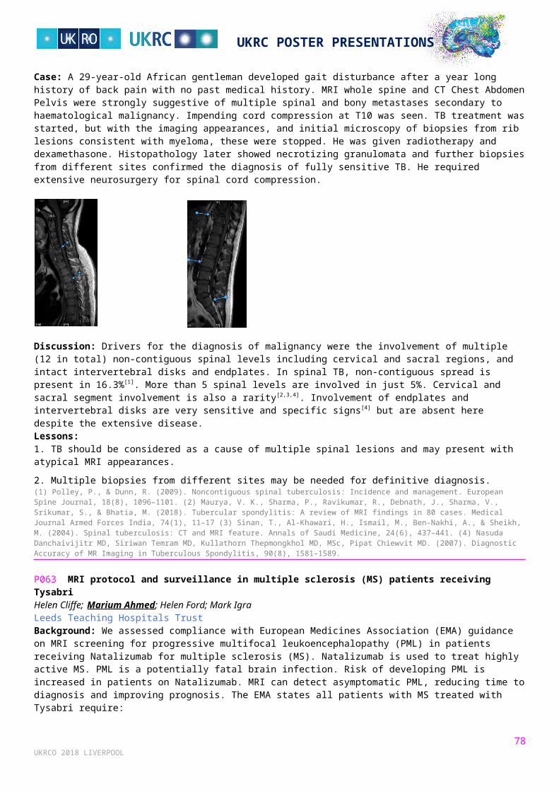
UKRC POSTER PRESENTATIONS
cancer undergoing radiotherapy treatment and assessed the relationship between uterus and cervix positional change and bladder volume. Method: 85 retrospective CBCT images from 11 pre-operative cervix cancer patients who had undergone radiotherapy were fused with the planning CT scans. The change in the uterus and cervix positions on the CBCT scans compared to the planning CT scans was quantified. Changes in position were correlated with changes in bladder volume using linear regression. Results: The range of movement of the uterus was 0.02 cm to 3.61cm in the superior/inferior direction (mean 0.71cm). In the anterior/posterior direction(AP) it was 0.03 cm to 2.59 cm (mean 0.72 cm). The cervix had a range of 0.01cm to 2.26cm (mean 0.48cm) in the AP direction and the change in uterine angle was 0o to 23 o (mean 6.68 o). A significant correlation was found between uterus and cervix positional change bladder volume change. Conclusion: Inter-fractional uterus and cervix movement can be substantial and can vary from patient to patient. Despite the use of a full bladder drinking protocol large variations in bladder volumes between fractions can occur and this can impact on the position of the uterus and cervix.1. Ahmad, R., Hoogeman, M.S., Bondar, M,. Dhawtal, V., Quint, S., De Pree, I., Mens, J.W., Heijmen, B.J. (2011) Increasing treatment accuracy for cervical cancer patients using correlations between bladder-filling change and cervix and uterus displacements: proof of principle. Radiotherapy & Oncology. 98(3), 340-346.2. Buchali, A., Koswig, S., Dinges, S., Rosenthal, P., Salk, J., Lackner, G., Böhmer, D., Schlenger, L., Budach, V. (1999) Impact of the filling status of the bladder and rectum on their integral dose distribution and the movement of the uterus in the treatment planning of gynaecological cancer. Radiotherapy & Oncology. 52(1), 29-34. 3. Taylor, A., and Powell, M.E.B. (2008) An assessment of interfractional uterine and cervical motion: implications for radiotherapy target volume definition in gynaecological cancer. Radiotherapy & Oncology. 88(2), 250-257.
P113 Benign ovarian lesions: A pictorial reviewSian Ebden; Jane Pollitt; Carys Jenkins; Kate Gower ThomasCwm Taf University Health BoardBackground: Ovarian abnormalities are common, both as symptomatic and incidental findings. For many Radiologists who do not have an interest in Gynaecological imaging, interpretation can be daunting. Ultrasound characterisation of ovarian lesions is vital in order to risk stratify patients and MRI is more commonly used as a problem-solving tool in indeterminate lesions. Purpose: Through a series of interesting clinical cases and selected radiological images, we will demonstrate characteristic features of a variety of ovarian lesions using both ultrasound and MRI. These will include characteristically benign entities, such as stromal tumours, as well as more indeterminate lesions for example, cystadenomas. Illustrating the key features will increase confidence in ascertaining which can safely be disregarded and those that require further follow up investigation. This will ultimately help prevent unnecessary additional imaging. Summary: We present a pictorial review of selected cases to display ovarian lesions that are both benign, and more radiologically indeterminate. Important features will be highlighted to help guide the necessity for further assessment.
P114 The normal cavum septum pellucidum during foetal developmentVivien MacRow-Wood; Elspeth WhitbyUniversity of SheffieldBackground: The Cavum Septum Pellucidum (CSP) is a cavity found in the foetal brain. Its visualisation is part of practice guidelines for performing the mid-trimester foetal ultrasound (US) scan[1] and an absent/abnormal CSP prompts further investigation. To date, our literature search uncovered no current papers which studied the size of the CSP on MRI in the normal foetal population. MRI studies examining the abnormal CSP used existing US measurements as a benchmark for normative size. This study aims to establish the normal size on MRI and creating a standardised approach to its measurement in a clinical scenario. Method: This is a retrospective study using data collected in routine clinical practice on over 300 normal foetal brains. The width and length of the CSP is measured in the coronal and axial planes following a set of specific repeatable anatomical boundaries.
Results: Initial scatterplots show that the width of the CSP on the coronal and axial plane stays fairly constant throughout gestation. Their values are comparable to each other and to major ultrasound studies - indicating their interchangeability if
78UKRCO 2018 LIVERPOOL
UKRC POSTER PRESENTATIONS
either view cannot be obtained. As yet no comparable studies for length have been found, however the initial graph of axial length to gestational age shows a clear trend of increasing as gestation increases. Conclusion: As this study is still ongoing, these results are taken from a preliminary analysis. Further work will be undertaken for more precise statistical analysis as well as prospective follow-up on the patients to confirm their normal developmental outcome.1. Salomon L, Alfirevic Z, Berghella V, Bilardo C, Hernandez-Andrade E, Johnsen S et al. Practice guidelines for performance of the routine mid-trimester fetal ultrasound scan. Ultrasound in Obstetrics & Gynecology. 2010;37(1):116-126.
P115 MRI volume-derived estimated fetal weight in the mid-pregnancy fetus: A comparison with current US standardsEmily Skelton; Jacqueline Matthew; Lisa Story; Alice Davidson; Chandni Gupta; Dharmintra Pasupathy; Mary RutherfordKing's College LondonBackground: An accurate estimated fetal weight (EFW) is essential for the delivery of good quality antenatal care. Current ultrasound (US) methods are susceptible to limiting factors (operator and technical) which can induce errors of up to 15% [2]. With the growing popularity of fetal magnetic resonance imaging (MRI), it has been suggested that volume-derived EFW may be a more accurate alternative to US methods, although current literature has yet to explore its applicability to the mid-pregnancy fetus.Method: Data were collected as part of the iFIND project (ISRCTN = 16542843). All participants gave written informed consent. 25 healthy mid-pregnancy (mean gestational age = 24 completed weeks of pregnancy) paired fetal US and MRI scans were retrospectively selected and EFW was calculated using US biometry-derived Hadlock[3] and MRI volume-derived (Baker[1] and Kacem[4]) formulae. The intra and interobserver variability of each method was assessed, as well as the systematic and random errors.Results: Compared to US biometry-derived EFW, Baker and Kacem MRI volumetry methods consistently and significantly under-measure EFW by a maximum of -13.6% (p=<0.001) in the mid-pregnancy fetus. However, there is excellent intra and inter-observer agreement in the MRI volume method suggesting a high reproducibility in the technique with no proportional bias (p=>0.05).Conclusion: The Baker and Kacem formulae should be interpreted with caution in relation to the clinical management of the mid-pregnancy fetus. Whilst our study demonstrates less variation within the MRI volumetry methods than US biometry-derived EFW, refinement of the MRI volume-derived EFW formula specific to the mid-pregnancy fetus is needed to compensate for varying fetal density at earlier gestational ages, and to provide additional description for interpretation of fetal lung, brain and liver volumes in growth-restricted fetuses.1. Baker P.N., Johnson I.R., Gowland P.A., Hykin J., Harvey P.R., Freeman A., et al. (1994) Fetal weight estimation by echo-planar magnetic resonance imaging. Lancet. 343(8898), 644-645. 2. Dudley N. (2013) A review of ultrasound fetal weight estimation in the early prediction of low birthweight. Ultrasound. 21(4),181-186. 3. Hadlock FP, Harris RB, Sharman RS, Deter RL, Park SK. (1985) Estimation of fetal weight with the use of head, body and femur measurements — a prospective study. Am J Obstet Gynecol.151, 333-337. 4. Kacem Y, Cannie MM, Kadji C, Dobrescu O, Lo Zito L, Ziane S, Strizek B, Evrad AS. Gubana F. Gucciardo L. Staelens R, Jani JC. (2013) Fetal weight estimation: comparison of two-dimensional US and MR imaging assessments. Radiology, 267(3), 902-910.
P116 Is fetal MRI more accurate than ultrasound in the second trimester assessment of non-central nervous system anomalies? A systematic reviewEmily Skelton; Lisa Story; Jacqueline MatthewKing's College LondonBackground: Early detection is essential to allow appropriate and timely management of a fetal anomaly [1]. Although ultrasound (US) is the preferred obstetric imaging modality, advances in fetal magnetic resonance imaging (MRI) technology have demonstrated its usefulness in the diagnosis of central nervous system (CNS) anomalies [2]. However, the value of second trimester fetal MRI in the diagnosis of non-CNS anomalies has yet to be fully explored.3
Method: A systematic review of available literature was performed (10-year period). Inclusion criteria were; non-CNS abnormalities suspected/diagnosed using US, gestational ages between 14-28weeks, MRI performed after US, and outcome by postnatal clinical findings/post-mortem. A modified QUADAS-2 tool was used to assess methodological quality for each included study, and individual patient data was also used to assess agreement with US and additional findings.Results: In most cases, MRI completely agreed with US findings. It diagnosed oro-facial anomalies more completely than US, particularly cleft lip and palate. MRI also provided more additional findings, particularly in relation to anomalies of the thorax, although US was better than MRI in the diagnosis of talipes.Conclusion: MRI has a similar accuracy to US in the diagnosis of non-CNS abnormalities. MRI is also more likely to detect additional findings, which may have an impact on patient counselling and management. This review was limited by the methodological quality and small sample sizes of included papers, therefore more evidence is required to further assess the usefulness of fetal MRI in comparison to US in the detection and diagnosis of non-CNS anomalies.1. National Institute for Health and Clinical Excellence. Antenatal Care: Guidelines CG62. 2018. 2. Rossi C A, Prefumo F. (2014) Additional value of fetal magnetic resonance imaging in the prenatal diagnosis of central nervous system anomalies: a systematic
79UKRCO 2018 LIVERPOOL
UKRC POSTER PRESENTATIONS
review of the literature. Ultrasound in Obstetrics and Gynaecology. 44(4). 3. We J S, Young L, Park I Y, Shin J C, Ah Im S. (2012) Usefulness of additional fetal magnetic resonance imaging in the prenatal diagnosis of congenital abnormalities. Archives of Gynecology and Obstetrics. 286(6), 1443-1452.PAEDIATRICS
P117 The neonatal chest X-ray: Common conditions and tips for non-paediatric radiologistsJames Ross; James HallsThe Great Western Hospital NHS Foundation TrustBackground: Neonatal respiratory distress is a common cause of admissions to special care units, affecting up to 7% of term newborns. The chest radiograph is the first line imaging test in the assessment of the neonate with respiratory distress and the ability to interpret it is key for prompt diagnosis and appropriate management. Radiologists without a specialist interest in paediatric radiology may feel unfamiliar with common lung abnormalities and their radiographic appearances. Purpose: This poster is designed as an educational aid to increase confidence in neonatal plain film reporting for all levels of radiologist from trainee to consultant. It will describe the basics of neonatal film reporting and differences compared to adult films. Common neonatal conditions including transient tachypnea of the newborn (TTN), meconium aspiration syndrome (MAS), neonatal pneumonia, pneumothorax and surfactant deficiency disease (RDS) will be described with typical radiological appearances and basic pathophysiology, with reporting tips and pitfalls to avoid. Neonatal lines and tubes will be displayed including umbilical catheters, ET tubes, chest drains and ECMO lines with advice on associated complications/misplacement. Summary: Content will be set out as a display of high quality plain radiographs, annotated to explain the basics of reporting and pathophysiology flanked by important learning points and take home messages.Arthur R. (2001) The neonatal chest x-ray. Paediatric respiratory reviews. 2 (4), 311-323. Liszewski, M.C. (2017) Respiratory distress in neonates. Underlying causes and current imaging assessment. Radiol Clin N Am, 55, 629-644. Pramanik A.K. (2015) Neonatal respiratory distress: a practical approach to its diagnosis and management. Pediatr Clin North Am. 62 (2), 453-69. Reuter, S. et al (2014) Respiratory distress in the newborn. Paediatrics in Review, 35 (10), 417-429.
P118 Vacuum immobiliser use for infant CT and MRI - is this the end of 'feed and wrap'?James Ross; James HallsThe Great Western Hospital NHS Foundation TrustBackground: Cerebral imaging with CT and MRI is commonly used to evaluate brain injury and development in neonates and infants within our institution. Traditionally, such imaging has required the use of a 'feed and wrap' technique to allow the scan to occur when the infant is sleeping following a feed or occasionally sedation. We now routinely use a vacuum immobilsation device that cocoons the infant and provides significantly improved image quality without the necessity for 'feed and wrap' or sedation. The technique has many advantages over the traditional 'feed and wrap' technique such as absence of motion-degradation, improved scan times and absence of repeated studies. Purpose: The poster will be of interest to both radiologists and radiographers, particularly those with an interest in paediatric imaging. It will demonstrate at length the safe usage of the vacuum immobilizer device; before, during and after a scan. We will describe the many benefits that we have observed and highlight the advantages the technique has over both the use of sedation and the traditional feed and wrap technique. Summary: This poster will be an educational visual display, utilising many images, explaining the contrasting techniques being discussed together with examples of the disparity in typical image quality from MR and CT studies and a summary of the benefits of immoblisation compared with traditional 'feed and wrap'. 1.Golan A. (2011) Imaging in the newborn: infant immobilizer obviates the need for anesthesia. Isr Med Assoc. 13(11):663-5 2.Ibrahim T. (2015) ‘Feed and wrap’ or sedate and immobilise for neonatal brain MRI? Archives of Disease in Childhood - Fetal and Neonatal Edition. Published Online First: 30 June 2015. doi: 10.1136/archdischild-2015-308847 3.Mathur, A.M. (2008) Transport, monitoring, and successful brain MR imaging in unsedated neonates. Pediatr Radiol. 38, 260-264.
P119 Procuring a new device for reducing intussusceptions in paediatric patientsAngela Staley; Vanessa WaspeNottingham University HospitalAim: The old equipment was no longer supported by clinical engineering so a suitable replacement was required to prevent patients being transferred to other centres for this procedure. Method: A risk assessment was performed to demonstrate the need for new equipment, and presented at the medical equipment planning group (MEPG). Paediatric centres contacted to find what devices are used elsewhere, and internet research carried out. Manufacturers who recommended their product for the procedure were contacted for advice and to arrange demonstrations. A suitable device was sourced, and purchased. Training competencies were developed with Clinical Engineering and the manufacturer prior to the equipment being implemented. Application training was provided for the Consultants and radiographers. Cascade training provided for rotating and new staff. Results: Robust training program implemented. Radiologists completed competencies and found equipment easy to use. Radiographers completed competencies and found equipment easy to set up as catheter packs provided contain all the
80UKRCO 2018 LIVERPOOL
UKRC POSTER PRESENTATIONS
equipment needed to use with the new machine. Ongoing audit shows short reduction times and reduced recurrence of intussusceptions. Conclusion: Thorough research prior to procurement ensured correct equipment for procedure chosen. Correct procurement process followed ensured swift approval to proceed. Support and advice provided to other centres that had purchased the equipment and those who were looking to procure.P120 Standards for radiological investigations of suspected non-accidental injury: A review of the updated guidelinesRebecca Murphy; Mark Thurston; Judith FosterPlymouth Hospitals NHS TrustBackground: New guidance for the radiological investigation of suspected physical abuse in children was produced by The Royal College of Radiologists (RCR) and The Society and College of Radiographers (SCoR) in September 2017 and replaces the 2008 Standards for radiological investigations of suspected non-accidental injury. The update is based on up-to-date evidence-based practice and guides referring clinicians, radiologists, and radiographers involved in imaging the child in suspected cases of physical abuse. Purpose: The new guidelines are detailed in a substantial and comprehensive 54 page document. We review and summarise the key areas addressed in the new guidance and highlight the changes from the previous 2008 guidance. We aim to educate the reader succinctly on current best practice for this critical area of imaging practice. Summary: We provide an overview for the general radiologist on the updated RCR/SoCR 2017 standards for radiological investigations of suspected physical abuse in children. Important changes as well as critical clinical pointers are covered. Illustrations with cases are included, where appropriate.1. Society and College of Radiographers and The Royal College of Radiologists. (2017) The radiological investigation of suspected physical abuse in children. The Royal College of Radiologists, London. 2. Royal College of Paediatrics and Child Health and The Royal College of Radiologists. (2008) Standards for Radiological Investigations of Suspected Non-accidental Injury. Royal College of Paediatrics and Child Health and The Royal College of Radiologists, London.
P121 AP vs PA spinal imagining: A comparison on the effective dose to radio sensitive organs in 13 year oldsBeckie PowellGreat Ormond Street HospitalBackground: Whole spine x-rays are imaged frequently in a paediatric setting due to the incidence of Scoliosis. This is found particularly in adolescents who are reaching puberty due to an increase in growth and can therefore exacerbate the degree of scoliosis. Around this time, the number of images required increases in respect to these growth periods. Patients can be imaged either antero-posterior (AP) or postero-anterior (PA) depending on the patients ability and images required. During these examinations the age of the patient and the dose implication, particularly to the radiosensitive organs, is of paramount importance. Aim: The aim of this study was to compare the dose to the radiosensitive organs when imaging the whole spine PA compared with AP. Additionally, the study also assessed if PA positioning is the gold-standard in terms of dose reduction to radiosensitive organs. Data was collected from whole spine images for 135 patients, aged 13 at the time of their examination, using RIS. Radiosensitive organs included thyroid, breast tissue,ovaries and testicles. The Purpose was to show the significant dose reduction to the patient when imaging was acquired PA. There was a 48% dose reduction factor to whole body, 87% to thyroid and most markedly, 91% to breast tissue. The cancer reduction risk was calculated with significant results, particularly for breast tissue. Summary: Introduction to AP vs PA whole spine imaging. Summary of results: Calculated comparison of cancer induction risk American Academy of Orthopedic Surgeons . (2010). Congenital Scoliosis. Available: www.orthoinfo.aaos.org/topic.cfm?topic=A00576. Last accessed: 22nd October 2017. Dartford and Gravesham NHS Trust. (2012). Postural Scoliosis. Available: www.dvh.nhs.uk/EasySiteWeb/GatewayLink.aspx?alId=266516. Last accessed: 20th October 2017. Javed, A Ltief, A. (2013). Development of the Human Breast.Available: https://www.ncbi.nlm.nih.gov/pmc/articles/PMC3706056/ . Last accessed: 24th October 2017. North American Spine Society. (2005). Adolescent Idiopathic Scoliosis.Available: www.knowyourback.org/Documents/scoliosis.pdf. Last accessed: 21st October 2017. NHS Choices. (2017). Scoliosis. Available: http://www.nhs.uk/conditions/scoliosis/Pages/Introduction.aspx. Last accessed: 24th October 2017.
P122 Radiodiagnosis of ischemic stroke in childrenMaksim Molodtsov; Igor Koltunov; Alexander Mazaev; Alexander GorbunovMorozov Children's City Clinical HospitalPurpose: Assessment of radiodiagnosis capabilities and aspects of ischemic stroke (IS) visualization in children. Methods and materials: 44 patients with cerebral IS (28 boys, 16 girls), aged 0 - 18 years, were examined. The patients underwent a comprehensive clinical investigation in combination with CT and MRI, sometimes under an anaesthetic (at the early age). Results: IS was mostly diagnosed in children aged 10 - 18 years (34% of patients). Hyperacute stage (HAS) was detected in 2 patients, acute - in 18, subacute - in 4, chronic - in 20. HAS was visualized only on DWI images. In 61% of cases, IS had a multifocal nature. Children under one year had mostly single focus (8 patients), children aged 1-5 years - one or two focuses (5 patients), children above 5 years had multifocal alterations (21 patients). Ischemic focuses predominantly affected the nuclei basalis (57%), frontal (55%) and parietal (43%) lobes. When IS at the chronic stage localized in central brain compartments, the
81UKRCO 2018 LIVERPOOL
UKRC POSTER PRESENTATIONS
cystic component predominated (10 patients), in peripheral compartments - the gliosis component prevailed (6 patients). In performing MR-angiography, a reduction in intensity of MR-signal from the artery feeding the area of lesion was observed in 34% of patients, which was caused by decreased blood flow.Conclusion: IS most commonly affects children aged 10 - 18 years, especially boys. In 61% of cases, IS is characterized by two and more focuses, mostly in the middle cerebral artery basin. Predominance of cystic or gliosis components depends on pathologic process localisation.P123 Estimating bone mass in children: A comparison between DXA and hand radiographs analysed using BoneXpert softwareKhalaf Alshamrani; Jean Russell; Amaka Offiah; N J BishopUniversity of SheffieldBackground: Dual-energy X-ray absorptiometry (DXA) is the current gold standard for assessing bone mineral density (BMD), however it does not predict fracture risk. BoneXpert computes bone health index (BHI) from hand radiographs by measuring cortical thickness, width and length of the three middle metacarpal bones. Objective: To compare BHI with BMD as measured from DXA scans. Materials & methods: We retrospectively identified DXA scans and hand radiographs for patients aged between 5 and 20 years acquired on the same day between 2008 and 2017. We documented absolute values and z-scores for whole body less head BMD (BMDTLH) and lumbar spine BMD (BMDLS1-LS4) and correlated these with BHI in different patient groups depending on their ethnicity and diagnosis. Pearson's correlation was performed using SPSS version 24 for PC (IBM, Armonk, New York). Results: 327 Caucasian (age 11.88 ±3.6 years) and 32 Asian (age 11.09 ±3.05 years) patients were included. BHI showed strong correlation with BMDTLH (r=0.64), BMDLS1-LS4 (r=0.71) in Caucasians, moderate correlation in patients treated with bisphosphonate BMDTLH (r=0.40), BMDLS1-LS4 (r=0.47) and weak correlation in Asians BMDTLH (r=0.15), BMDLS1-LS4 (r=0.19). Conclusion: In Caucasians, BHI has moderate to strong correlation with DXA absolute values. However, BHI has a weak correlation with DXA reads in patients who are of Asian ethnicity. Although numbers of Asians studied was small, our preliminary results suggest that normative BHI data is required for different ethnicities. The situation in Africans is unknown.
P124 Improving patient attendance for follow-up imaging after non-accidental injury skeletal surveyAngela Staley; Vanessa WaspeNottingham University HospitalAim: Follow up imaging is an essential part of the NAI skeletal survey, and has been shown to improve diagnostic yield. A system to ensure that patients returned for follow up imaging was designed, implemented, audited, changed and re-audited. Method: The standard for re-attendance is 100% as documented by the RCR and RCPCH guidelines. An initial audit was undertaken over two years to assess attendance for follow up imaging. Following this initial two year cycle a further two year cycle was undertaken after the following improvements were made to the process. Improvements included:
Appointment letter given to persons accompanying patient at the time of the skeletal survey Verbal communication on the importance of attending, emphasising safeguarding will be informed for nonattendance Clear documentation regarding the follow up appointment in CRIS, request card and departmental diary Appointment documented in patient notes, ensuring information is shared at strategy meetings, promoting
multidisciplinary team involvement Nonattendance is followed up with a phone call to Safeguarding, documented on CRIS and shared with the paediatric
imaging team, ensuring patient does attend. Results: Re-attendance in years one and two was 87% and 83% respectively. Following the implementation of the changes, re attendance improved to 92% and 97% in years three and four respectively. Conclusion: Changing our approach and practice has significantly reduced the nonattendance rate. The correct high quality follow up images are taken at the required time enable precise reporting including dating of any fractures.
P125 Is there evidence of gadolinium deposition in the brains of paediatric patients after multiple doses of contrast agent?Selene Rowe; Daniel Rodriguez; Paul Morgan; Robert Dineen; Timothy Jaspan; Eleanor CohenNottingham University Hospitals NHS TrustBackground: Since 2014 studies have demonstrated hyperintensities in the dentate nucleus (DN) and globus pallidus (GP) in patients after receiving multiple doses of gadolinium based contrast agent (GBCA). Only a small number of studies have been conducted on paediatric patients.Objectives: To establish if signal intensity (SI) increases in children after multiple doses of GBCA even when they received surgery only/no treatment. To compare linear and macrocyclic GBCAs and whether SI continues to increase when the GBCA was changed from linear to macrocyclic.Method: Regions of interest (ROIs) were drawn on the GP and the DN, normalised to the middle cerebellar peduncle, pons and cerebral white matter on unenhanced axial T1weighted images in 35 paediatric brain tumour patients. SI ratios were compared
82UKRCO 2018 LIVERPOOL
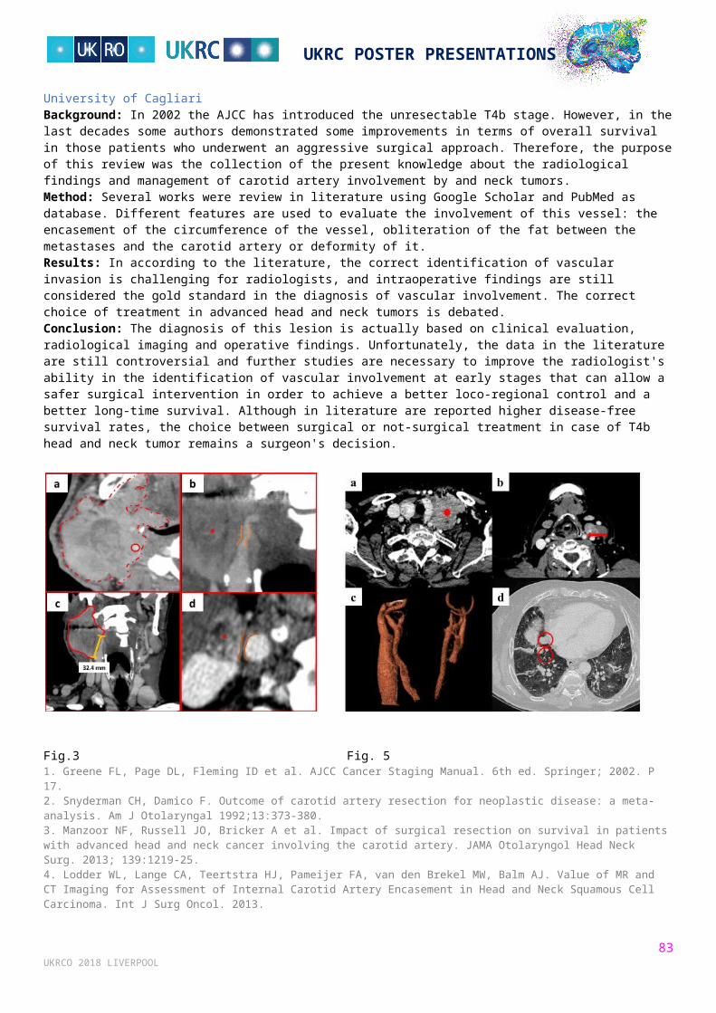
UKRC POSTER PRESENTATIONS
in the patients who had received treatment against those who had surgery only/no treatment and in patients who had received linear against those who had received macrocyclic agents.Results: There is a significant increase in SI ratios for all regions tested in patients who received linear agents: P<0.05. There was no significant difference in SI ratios between patients who had received chemo/radiotherapy and those who had surgery only/no treatment: P>0.05. There is a significant change in SI for the DN when patients changed from receiving a linear GBCA to a macrocyclic; P<0.05 Conclusion: Hyperintensities in the DN and GP of paediatric patients is caused by multiple doses of a linear but not macrocyclic GBCA regardless of whether they had chemo/radiotherapy.P126 Eye ultrasound in children a quality improvement exerciseMorag Green; Susie GoodwinNHS Greater Glasgow and Clyde, Royal Hospital for Children GlasgowEye Ultrasound is a recognised useful tool in distinguishing Optic Disc Drusen (ODD), a build up of protein and calcium salts inthe optic nerve head, from papilloedema. ODD is a benign condition, however, papilloedema can indicate raised intracranial pressure, an important finding which requires further crosssectional imaging. Audit of referrals for eye ultrasound in our centre was audited over a 2 year period. Between 2015/16 and 2016/17 there was a sharp increase of 76 % in paediatric eye ultrasound referrals within the RHC Glasgow, which may in part be due to some high profile national legal cases involving missed intracranial tumours. The majority of request were for optic disc drusen, however the requests were diverse and included retinal detachment and retinoblastoma. Due to the increased number and complexity of the referrals, the need for service improvement was recognised and a quality improvement program was implemented within the imaging department. The aim of the exercise was to improve eye ultrasound assessment accuracy for ODD from baseline to 95%. Using the plan, do, study, act (PDSA) cycle problem solving was undertaken which include introducing a standardised reporting tool.1 Ballantyne et al. 2002; Blaivas et al. 2003; Girisgin et al. 2007; Helmke and Hansen 1996; Newman et al. 2002; Tayal et al. 2007; Tsung et al. 2005. Optic nerve sheath diameter: An ultrasonographic window to view raised intracranial pressure? 2 Barnett SB, Ter Haar GR, Ziskin MC, Rott HD, Duck FA, Maeda K. (2000) International recommendations and guidelines for the safe use of diagnostic ultrasound in medicine. Ultrasound Med Biol. 26:355–66. [PubMed] 3 Ertl, Michael & Gamulescu, Maria-Andreea & Schlachetzki, Felix. (2012). Application of Orbital Sonography in Neurology. 10.5772/31181. 4 Helmke K, Hansen HC (1996) Fundamentals of transorbital sonographic evaluation of optic nerve sheath expansion under intracranial hypertension ii. Patient study. Pediatr Radiol 26:706–710
P127 Fluoroscopic guided oesophageal foreign body retrieval: 10 year review of practice at a paediatric centreMichael Jackson 1; John Fitzpatrick 1; Menelaos Philippou 2
1NHS Lothian; 2Glasgow HospitalsBackground: Ingested foreign bodies are a common problem in the paediatric emergency department. At our institution patients with smooth, radio-opaque foreign bodies (most commonly a coin) lodged in the upper oesophagus with a short duration history have undergone attempted retrieval using a Foley catheter under fluoroscopic guidance. In contrast to endoscopic removal, this procedure does not require general anaesthesia but departmental concerns related to the risks of the procedure and patient tolerance prompted this study to inform a new foreign body retrieval pathway. Methods: Retrospective analysis of all fluoroscopic guided retrieval procedures performed at our institution over a ten year period (2008-17) was conducted. Cases were identified via PACS, with the procedure report and clinical outcome reviewed via EPR. Results: 76 patients were identified during this period (F:M 45:31, age range 3 months to 13 years). Procedure was successful in 69 cases (90%) (foreign body retrieved in 52 cases, advanced into stomach in 17). Oesophageal tear occurred in one case. 7 patients underwent an unsuccessful attempt, requiring subsequent endoscopic retrieval. Minor complications included (vomiting in 4, gagging in 1, and minor bleeding in 1). A total of £13.79 and 2 US cents were retrieved. Conclusion: Data demonstrates a good success rate and only one serious complication. Risks of airway obstruction and oesophageal rupture, and concern regarding patient tolerability nevertheless persist. We anticipate the new local retrieval pathway will be complete by June 2018. This paper will contextualise the role of this procedure in this light.
P128 Determinants of radiographer abnormality detection error in paediatric patientsDarren Dewick; Philip CossonTeesside UniversityBackground: Diagnostic error can lead to missed, delayed or incorrect diagnosis, causing many potential problems (Singh et al, 2011). This is particularly important in paediatric patients where unidentified fractures can lead to misalignment and callus formations to occur which can affect growth development (Mounts et al, 2011). Smaller non-ossified bones and the presence of ongoing epiphyseal fusion might make it difficult for referrers to detect subtle fractures and radiographers who commonly engage in first line abnormality detection; the so called 'red dot system'. However, the determinants of radiographer red dot error are not fully understood. Therefore, the purpose of this study was to identify any patient characteristics or anatomical/pathological features that might predict this error.
83UKRCO 2018 LIVERPOOL
UKRC POSTER PRESENTATIONS
Method: A retrospective secondary data analysis was conducted identifying paediatric long bone radiographs from a non-clinical pseudonymised PACS. Prevalence of red dot was identified and compared with gold standard radiology reports. Fractures were classified using the Li-La paediatric long bone classification system. A binary logistic regression was performed identifying predictive factors of radiographer error. Results: Li-La non-articular fractures type l, ll and lll, Li-La articular fractures type V and the presence of joint effusions predicted radiographer false negative error. The ankle, wrist, elbow and foot had the most incorrect red dot cases.
Conclusion: These fracture types and joint effusions are often overlooked, despite joint effusions being consistently associated with the presence of occult fractures. Further training and clear policies for application of 'red dot' would be beneficial.Mounts, J., Clingenpeel, J., McGuire, E., Byers, E., and Kireeva, Y. (2011) Most frequently missed fractures in the emergency department. Clinical Paediatrics, 50(3), pp.183-186. Singh, H., Graber, M. L., Kissam, S. M., Sorensen, A. V., Lenfestey, N. F., Tant, E. M., Henriksen, K., and LaBresh, K. A. (2011) System-related interventions to reduce diagnostic errors: a narrative review. British Medical Journal Quality and Safety, pp.bmjqs-2011.
P129 Are we ALARP? An analysis of Dr paediatric exposure factorsTheresa NoonRoyal Bolton HospitalRadiographers have a professional and legal responsibility to adhere to NDRL's and local DRL's in accordance with IR(ME)R (2000). This ensures a mindful approach whilst maintaining a dose which is ALARP for all patients across all examinations. Across the UK the majority of children's examinations are not carried out at the specialist Children's hospitals. There is a clear relationship between dose and patient age and for this reason the NRPB recommends the adoption of reference levels for a range of patient ages. Aim: Asses the DAP readings for Paediatric examinations for all DR rooms. To ensure that all Radiographers are adhering to the ALARP principle for all examinations. To provide a baseline locally for Paediatric patients Standard: There is currently no standard set within the UK in relation to the use of DAP as a DRL, however there is with the use of ESD Methodology: DAP readings were audited over a 12-month period, from September 2015 to September 2016 for Paediatric examinations only. n= 8392 examinations. The data was collected retrospectively and was subject to strict inclusion criteria which adhered to standard protocols. Results: From the original sample, 5147 examinations met the inclusion criteria and demonstrated safe radiation protection practices across the team. Conclusion: As a department we are adhering to the ALARA principle, Radiographers do adjust their exposure factors dependent upon each patient. Recommendations: Update departmental IRMER document. Provide data for Christie's to compose DRL's.1.Diagnostic reference levels in medical imaging: review and additional advice. Ann ICRP 2001;31:33–52 http://www.icrp.org/docs/DRL_for_web.pdf 2.Medical Exposure Radiation Unit, HSE. Population Dose from General X ray and Nuclear Medicine. 2010. 3.Medical Council . Diagnostic Reference Levels Position Paper. ‐2004. 3. European Commission . Guidance on Diagnostic Reference Levels for Medical Exposures. 1999. 3.Diagnostic reference levels in medical imaging: review and additional advice. Ann ICRP 2001;31:33–52 http://www.icrp.org/docs/DRL_for_web.pdf 4.European guidelines on quality criteria for diagnostic radiographic images in Paediatrics. EUR 16261 EN (1996). 5. Hart D., Hillier M.C., Wall B F. NRPB – W14 (2002)Doses to patients from medical X-ray examinations in the UK – 2000 review 6.Evans, S. (2014). European diagnostic reference levels in paediatric imaging. Physica Medica, 30, pp.e14-e15. 7.Paulo, G., Vaño, E. and Rodrigues, A. (2016). Diagnostic reference levels in plain radiography for paediatric imaging: A Portuguese study.Radiography, 22(1), pp.e34-e39. 8. Strauss, K. J., & Kaste, S. C. (2006). The ALARA (as low as reasonably achievable) concept in paediatric interventional and fluoroscopic imaging: striving to keep radiation doses as low as possible during fluoroscopy of paediatric patients—a white paper executive summary. Paediatric Radiology, 36(Suppl 2), 110–112.http://doi.org/10.1007/s00247-006-0184-4 9.The 2007 recommendations of the International Commission on Radiological Protection. ICRP publication 103. Ann ICRP 2007;37:1–332 10.Wall, B. and Shrimpton, P. (1998). The Historical Development of Reference Doses in Diagnostic Radiology. Radiation Protection Dosimetry, 80(1), pp.15-19. 11. European Communities (Medical Ionising Radiation Protection) Regulations 2002. EEC Directive 97/43/Euratom. Health Protection of Individuals against the danger of Ionising Radiation in relation to Medical Exposures. S.I. 478 (2002).
OTHER
P130 Military field hospital versus UK Emergency Departments in major trauma: a radiographer's perspective of patient careChloe Shand; Kirsty WoodUniversity of DerbyThe aim of this poster is to demonstrate the variety of patient experiences through a range of trauma situations. The objective is to demonstrate where lessons have been learned and where patient care can be enghanced to improve the overall patient experience from a radiographer's perspective. The poster will demonstrate how each patient travels through the emergency department in a military field hospital and in a UK emergency department, this will create the viewer with a visual and direct comparison between the two. The process will include examples for good patient care and areas where improvements could be made, or is sometimes lacking. A timescale will be included to demonstrate the varying patient throughput speeds. Photographs of each department will be included for visual validity. The poster is relevant to all current emergency departments and
84UKRCO 2018 LIVERPOOL

UKRC POSTER PRESENTATIONS
radiographers who work within them. It can provide evidence of the lessons learned in the military setting and how these can be applied. The outcome should be to improve patient care in a trauma setting, both by radiographers and other healthcare professionals. It will also demonstrate that severe trauma can lead to an abrupt care approach and how this can be avoided. It will also demonstrate the importance of teamwork and having a strong structure of the trauma team. There is scope for further research in this topic area with the fluid nature of trauma departments. The patient journey is constantly evolving and it is important to keep up with the pace.1. Ehrlich, R.A., Coakes, D.M. (2017) Patient care in radiography: with an introduction to medical imaging. Missouri: Elsevier. 2. Harcke, T., Statler, J.D. and Montilla, J. (2006) Radiology in a hostile environment: Experience in Afghanistan.Journal of military medicine. 171(3), 194-199. 3. Sharma, V., Sreedhar, M. and Debnath, J. (2017) Combat radiology: Challenges and opportunities. Medical journal armed forces India. 73, 410-430.
P131 Review areas in the search for a primary: Learning points from our cancer of unknown primary MDT experienceNicholas Chua; Anjali Sujith; Anita LazarevskaBasildon University Hospital NHS Foundation TrustBackground: The incidence of cancer of unknown primary (CUP) occurs in about 0.5-9% of all cancer patients [2]. Identification of the primary lesion is vital to predict the natural progression, treatment options and prognosis for the patient. Patients with cancer of unknown primary have a significantly worse prognosis, with median survival between 6-14 months, than those with readily identifiable primaries either through time delay to treatment or unfavourable aggressive histology [1]. At our institution patients without a readily identifiable primary are referred to the CUP MDT upon which their imaging and histology are reviewed. Through our experience we note a small subset of patients referred to CUP MDT, upon review, actually have an identifiable primary that was not fully appreciated on initial reporting.Purpose: We describe a series of cases to highlight the various imaging features that radiologists need to be aware of in the search for a primary and thus to avoid delay in appropriate MDT referral and oncological treatment and to afford the patient the subsequent survival benefits.Summary: The most common radiological discrepancy was found to be pancreatic lesions followed by gastro-esophageal junction lesions and lung and pleural lesions. This study serves to highlight the important review areas for all radiologists with the view to reducing delays in cancer diagnosis and treatment.1. Pavlidis N. Cancer of unknown primary site: 20 questions to be answered. Annals of Oncology. 2010; 21:vii3003-vi307 2. van de Wouw AJ. Eur J Cancer 2002; 38:409-413
P132 Nodal staging with TNM 8 in head and neck cancer: How can the radiologist help?Rhys Thomas; Chris Greenall; Rhian RhysCwm Taf UHBWe review the new TNM 8 nodal staging for H&N cancer, concentrating on two important changes - extracapsular spread and the occult primary. We present a practical approach to these significant changes in the form of a pictorial review. We demonstrate their implications and how the radiology is key to correct staging and an accurate prognosis for the patient.1. Lydiatt, W.M. et al.(2017) Head and Neck Cancers—Major Changes in the American Joint Committee on Cancer Eighth Edition Cancer Staging Manual. CA Cancer J. Clin 67, 122-137.
RADIATION PROTECTION, DOSE OPTIMISATION & QUALITY ASSURANCE
P133 Saying sorry when a patient receives an unintended radiation dose in the imaging departmentNicholas Barlow; Lisa FieldMid Yorkshire NHS TrustThe Francis report highlighted requirements for openness and transparency when things go wrong. The Health and Social Care Act 2008 - ensures that providers are honest with patients and relatives in relation to their care. Other professional bodies have emphasised the importance of duty of candor (DoC); including the National Patient Safety Agency (NPSA), the Department of Health and the Royal College of Radiologists. The NPSA categorise harm into low/minor, moderate, severe and death and advise candour in those of moderate harm. There is confusion regarding this guidance; particularly for unintended doses of radiation as often no direct harm is caused. Radiation dose and risk are often weighted in relation to cancer risk, which causes anxiety and confusion amongst patients when investigation findings are discussed.This poster will discuss the responsibilities of duty of candour in radiology for unintended doses of radiation and clearly define the professional's role in this process. A comparison between dosage and relevant risk will be provided for reference in local department policy. To assist colleagues with patient feedback (and for everyday explanation of dose to patients) a table of comparative radiation dose sources will be provided that can be tailored to provide a more patient-friendly resource.The poster will contain the following:1. Introduction - The need for DoC in radiology2. Levels of harm - With relative radiation dose3. DoC Procedure - In flow chart form
85UKRCO 2018 LIVERPOOL

UKRC POSTER PRESENTATIONS
4. Patient feedback - Table of comparative doses5. Conclusion/Recommendations offering advice for clinical practice and scenarios.
P134 Understanding the patient journey in MRI: the good, the bad and the uglyDarren HudsonInHealthBackground: Understanding the experiences of patients undergoing MRI is fundamental to providing truly patient centred care. The nature of the modality itself is not the most patient friendly and many factors contribute to the ability of patients to successfully complete a scan and have a positive experience. It is an acknowledged barrier to patient compliance and the consequences are important as it can delay or inhibit diagnosis and treatment. Demand and throughput for MRI has increased rapidly and there is ever increasing pressure to do more for less which places tremendous time constraints on appointment slots. As a result, the imaging process has become more task focused meaning patients can't always be given the time they need. The short period of time in our care also means there is less time to establish a rapport and rapidly connect with patients. Purpose: To better understand the current patient journey in MRI, some observations of various locations across the business were conducted by an external organisation, the Patient Experience Network (PEN), which highlighted the good, the bad and the ugly. Summary of content: The patient journey map presented highlights some key themes around patients knowing what to expect, consideration over time and efficiency, how to get results, and recognition of the importance of visual cues. It also supports results from a 'What Matters...' survey conducted with patients to gain insight into what's important to their experience. All of which is helping us define patient experience in MRI.
P135 Radiation exposure and related risks for pediatric patients undergoing percutaneous intervention (PCI) proceduresAli Abdelrazig 1; Mohamed Abuzaid 2; Esameldeen Babikir 3; Esaam Mattar 3; Batil Alonazi 4; Abdelmoneim Sulieman 4
1Radiation Safety Institute, Sudan Atomic Energy Commission, Khartoum, Sudan; 2University of Sharjah; 3Department of Radiological Sciences, King Saud University, Riyadh, Saudi Arabia; 4
Department of Radiology and Medical Imaging, Prince Sattam bin Abdulaziz University, Alkharj, Saudi ArabiaCardiac catheterisation is an interventional procedure used for the diagnosis and treatment of coronary arteries diseases. Patients are exposed to prolonged radiation exposure during the procedure. Therefore, accurate dose optimisation is recommended to keep the radiation dose as low as reasonably achievable. The aim of this study was to determine patient radiation doses for interventional cardiology procedures, to identify procedures associated with high irradiation doses, and to determine the effects of various parameters on patient doses. A total of 51 patients were examined for a different clinical indication in this study from two cardiology departments. Calibrated X-ray machines were used to perform all the procedures. Patient dose measurements were performed using Dose Area Product (DAP) meter. The mean and range of patient age (year), weight (kg) were 4.9 (0.03-15) and 11.7 (10.0-46.0) respectively. While the mean and range exposure parameters were 63.2 (53-74.2) kVp, 173.2 (134-835) mA and 3.8 (1.7-21.6) min for tube potential, tube current and fluoroscopic time, in that order. The mean and range of the number of films per procedure are 3.8 (1.0-7.0). The mean cumulative average dose (CAD) (cGy/cm2) and effective doses (mSv) were34.08 (32.8-35.9) and 465.61 (380.65-501.13). The dose values are comparable than in the recent literature. Patient dose reduction is of a prime importance and practitioners should optimize the radiation dose for further dose reduction without compromising the diagnostic and therapeutic findings.1. M. Alkhorayef, E. Babikir, A. Alrushoud, H. Al-Mohammed,A. Sulieman.A Patient radiation biological risk in computed tomography angiography procedure. Saudi Journal of Biological Sciences. 24(2):235-240. (2017). 2. A. Sulieman, K. Alzimami K, R. Gafar.E. Babikir, K. Alsafi, I.I. Suliman. Occupational and patient exposure in coronary angiography procedures . Radiation physics and Chemistry Journal. Vol 104,68-72 (2014). 3. Nada A. Ahmed A. I. Nayel. Pediatric radiation dose during cardiac catheterization procedures in Sudan. Radiation Protection Dosimetry, Vol 174(4) 495–500 (2017).
P136 Image quality and radiation dose interrelationships during paediatric pelvis radiography - a factorial phantom studyAli Mohammed Ali; Peter Hogg; Andrew EnglandUniversity of SalfordBackground: Paediatric pelvic radiography exposure factor selection is difficult due to age/size variations; this study evaluates image quality and dose for a range of acquisition parameters. Method: 2,016 DR images were acquired using a 5-Year-old pelvis phantom. Acquisition parameters included: KVp (56-89, 3 kVp increments), mAs (1-16, 1 or 2 mAs increments), SID (100, 115, 130 and 145 cm) and filtration (0, 2 mm Al and 1mm Al + 0.1 mm Cu). Image quality (IQ) was assessed using physical and visual methods. Physical included SNR and CNR. Visual involved observers scoring sharpness and noise. Entrance surface dose was measured. Regression and main effect plots were conducted. Results: Adjusted (R2) was 0.76 for radiation dose; range = 0.31-0.78 for IQ. Increasing additional filtration had the greatest impact decreasing IQ (physical Beta coefficient (B)=-6.72 and visual B=-1.13) and radiation dose (B=-50.0). mAs increased radiation dose (B=17.32) and IQ (physical B=0.63 and visual B=17.32). Elevating KVp increases radiation dose (B=4.92); initially it increases IQ, then it reduces (physical B=-0.15 and visual B=0.49). As SID increases dose reduces (B=-2.60) and IQ remained
86UKRCO 2018 LIVERPOOL

UKRC POSTER PRESENTATIONS
unchanged (physical B=-0.80 and visual B=-0.09). Main effect plots for each exposure parameter showed different relationships when compared to others: either linear, or non-linear, with plateauing. SID=115cm produced the highest levels of image sharpness. Conclusion: Paediatric optimisation studies should consider a full factorial design to identify the impact of all acquisition parameters. Increasing KVp whilst reducing mAs produces lower dose and maintains IQ, but only for specific levels of filtration and SID.
87UKRCO 2018 LIVERPOOL
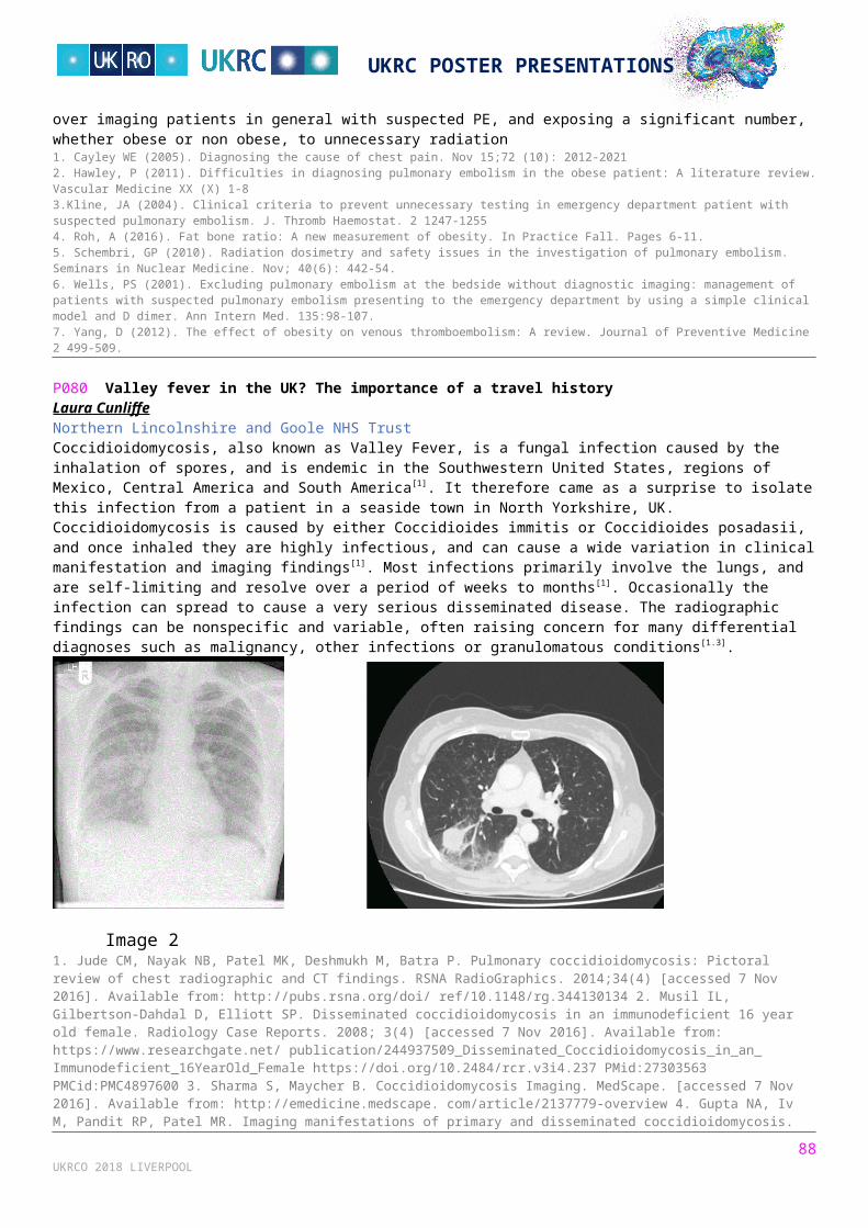
UKRC POSTER PRESENTATIONS
P137 An analysis of paediatric gonadal protection - a quality assurance audit based upon a local protocolFahad Rathore; Khalid KhanBarking, Havering and Redbridge University Hospitals NHS TrustBackground: The Institute of Physics and Engineering in Medicine (IPEM) in their Medical and Dental Guidance Notes state: "For all children, and for all persons of reproductive capacity, gonad shields should be used in examinations which are likely to give a significant gonad dose, unless the shields interfere with the proposed examination". There have been many studies that have highlighted the problem of high rates of inaccurate placement of gonad shielding in children having pelvic x-ray radiographs.Purpose: We have a protocol on paediatric gonadal shielding in our Trust. We audited it to check how well our practice adheres to the protocol as well as check how easy to implement the protocol is in real life. This was done to improve our practice & patient care as a result because...
It makes all relevant staff aware of the standards set by the protocol It allows the department to highlight areas where practice is falling below the standard set It also allows us to highlight areas of practice in which we are excelling.
Summary: Various issues arose with regards to paediatric gonadal protection at our Trust. These include... Issues with protocol compliance Issues with record keeping (which makes it harder to verify compliance) Odd exclusion criteria in the protocol which are hard to justify.
Our poster highlights our performance, our issues and a 7 point action plan for improvement in the future. We hope these can be used to help other Trusts evaluate their protocols too.1. Medical and dental guidance notes. (2002). York: Institute of Physics and Engineering in Medicine.2. Sikand, M., Stinchcombe, S. and Livesley, P. (2003). Study on the use of gonadal protection shields during paediatric pelvic X-rays. Annals of The Royal College of Surgeons of England, 85(6), pp.422-425.3. Fawcett, S. and Barter, S. (2009). The use of gonad shielding in paediatric hip and pelvis radiographs. The British Journal of Radiology, 82(977), pp.363-370.
P138 Evaluation of image quality, lesion visibility and entrance surface dose for routine adult chest radiography examinations in 8 hospitalsSadeq Al-Murshedi; Peter Hogg; Andrew EnglandSalford UniversityBackground: To investigate image quality and radiation exposure when imaging a Lungman adult anthropomorphic chest phantom using routine adult chest X-ray protocols and X-ray machines. Method: The chest phantom (with and without a 'fat jacket' to represent increased BMI) was used to acquire radiographic images in eight hospitals and 17 X-ray machines, using their existing routine adult chest X-ray protocols. Image quality (IQ) and lesion visibility (LV) were evaluated visually using a relative visual grading analysis (VGA) by six observers. Signal to noise ratio (SNR), contrast to noise ratio (CNR) and conspicuity index (CI) were measured as physical measures of image quality. The entrance surface dose (ESD) was measured using a solid state dosimeter. A figure of merit (FOM) was calculated. Results: For phantom without the fat jacket, IQ ranged from 10.00-21.83, LV 4.5-12, SNR 13.39-67.39, CNR 9.60-45.51, CI 18.75-66.51, ESD 28.34-200.35 µGy and FOM 0.18-1.19. With fat jacket, IQ ranged from 13.17-26.00, LV 5.83-13.67, SNR 11.33-66.25, CNR 7.48-30.10, CI 10.37-64.93 and ESD 40.25-565.56 µGy and FOM 0.08-1.07. Correlations between IQ and ESD for the phantom without and with fat jacket were r= 0.32 and r=0.60, respectively and the correlation between ESD and LV were r=0.31 and r=0.49 respectively. Conclusion: Between hospitals there was a considerable variation in image quality and radiation dose. A weak correlation between dose and IQ / LV existed. Differences are likely to be the result of different types of x-ray imaging equipment and protocols used.
P139 Inpatient chest X-rays: Frequently requested; rarely reportedPalany Parameshwaran 1; Essam Lakha 2; Qaiser Malik 3
1North Middlesex University Hospital; 2University College London Hospital; 3Basildon & Thurrock University HospitalImages are requested fundamentally because they impact patient management. IRMER states that any medical exposure should be clinically evaluated; and the resulting information, outcomes, and implications be recorded. Many trusts delegate the duty of reporting plain films to the responsible doctor. A retrospective audit was performed for inpatient chest X-ray requests at Basildon & Thurrock University Hospital during 2017. The aim was to assess the number of requests that had a corresponding evaluation documented in the notes - the expectation 100 %. A sample of 95 patients was obtained from across 14 departments and specialities, and information gathered from ORMIS, PACS, and patient records. Only 40 % (38/95) had been formally reported. There was an equal distribution for both males and females, with a mean time of 2 days (range 1 - 9 days) between the image being performed to documentation evidenced in the patient's notes.
88UKRCO 2018 LIVERPOOL
UKRC POSTER PRESENTATIONS
Chi-square analysis was performed between gender and whether the image was reported, and was not significant at p < .05. There were two outcomes of this audit - firstly, staff education; and secondly, patient safety. Consequently, a sticker was designed to prompt doctors to report images. If 60 % of patients' chest X-rays are not being reported - it must be considered if they are even being looked at, and whether they are actually necessary? Ideally, all images would be reported by a Radiologist, but there are obvious time and financial constraints. One factor that cannot be counted is the number of missed pathologies.1. Department of Health (2000). The Ionising Radiation (Medical Exposure) Regulations 2000. London: The Stationery Office. 2. Society and College of Radiographers, British Institute of Radiology (2015). A guide to understanding the implications of the Ionising Radiation (Medical Exposure) Regulations in diagnostic and interventional radiology. BFCR(15)2.
P140 An investigation into the upper limits of added filtration for projectional radiography of the lumbar spineRobert Meertens; Aaron Box; Ryan Knight; Paul Miles; Suleiman MohammedbrhanUniversity of ExeterBackground: The use of added filtration to reduce patient dose during projectional radiography examinations is established practice. However, most existing evidence is based pragmatically around levels of added filtration available in radiographic equipment, and involves film-screen or computed radiography (CR) systems. We aimed to investigate the extreme upper limits of added filtration and whether there may be scope to further reduce patient dose without detriment to image quality when using digital radiography (DR) systems. Methods: Repeated antero-posterior projections of the lumbar spine were acquired using an anthropomorphic phantom with increments of 0.1mm added copper filtration added up to a total thickness of 1.0mm added filtration. This was repeated at 70, 81 and 90 kVp. mAs was varied between exposures to ensure images of a comparable exposure index (EI). Dose Area Product and entrance surface dose measurements were recorded and image quality was subjectively reviewed by four experienced radiographers. Results: As expected, patient dose was reduced as added filtration increased, although the beneficial effects appeared minimal above 0.3mm of added copper filtration. All images were deemed diagnostically acceptable, but there was perceivable differences in image contrast with increasing levels of added filtration greater than 0.4mm of added filtration, despite DR post processing algorithms. Conclusion: Results suggest that the upper limits of 0.3mm copper filtration on our projectional radiography system are an appropriate balance between minimising patient dose whilst maintaining image quality and protecting against tube loading.
P141 The impact on image quality with variations in the placement of lead rubber during the lateral lumbar-spine radiographic examination: a phantom study with DR equipmentEllie Carter; Anthony Manning-Stanley; Anthony WardUniversity of LiverpoolBackground: The lateral lumbar-spine radiographic examination is a relatively high radiation dose technique [1]. Anecdotal evidence suggests that there is variation in practice as to the placement of lead rubber shielding during this examination; a phantom study with DR equipment was conducted to investigate the impact of this variation on image quality and radiation dose.Method: An anthropomorphic phantom was exposed at constant tube voltage (75kVp) and 19 tube current-time increments from 1mAs to 63mAs under three experimental conditions; 1) no lead rubber; 2) lead rubber placed against phantom lumbar-spine; 3) lead rubber placed on table. Post-processing was applied to optimise image quality and further collimate the resultant 57 images for blinded review by two reporting radiographers, who scored two aspects of the image exposure and overall image quality on a 3-point scale (1=inadequate, 2=adequate, 3=perfect), giving a maximum score of 9. 10% of images were re-reviewed to determine intra-rater reliability. Effective dose was calculated using PCXMC dose-modelling software.Results: Median image quality score was highest for setting 2 and lowest for setting 1 (6.0 v 4.5). The mAs beyond which overall image quality was consistently rated as adequate varied by setting; setting 2 the lowest (6.3mAs), setting 1 the highest (32mAs) with an effective dose 5 times higher. A high inter-class correlation coefficient (0.974) indicated reliable image quality ratings.Conclusion: Variation in the use of lead rubber during lateral lumbar-spine radiographic examinations has image quality and radiation dose implications; lead placed against the back is the optimal setting.1. Institute of Physics and Engineering in Medicine DRL Working Party. Guidance and Use of Diagnostic Reference Levels for Medical X-Ray Examinations. IPEM Report 88 (IPEM, York) 2004.
P142 Dose reduction in an alternate lumbar spine projectionMei Zi Henrietta Goh 1; Wei Yow Sim 1; Yew Mun Kwok 1; Mak May San 1; Lim Chee Yeong 1; Bak Siew Steven Wong 2
1Singapore General Hospital; 2Sengkang Health and Singapore General HospitalRadiology departments routinely perform weight-bearing antero-posterior (AP) lumbar spine X-ray examinations. The postero-anterior (PA) projection has been advocated for certain examinations as a means for dose reduction. Previous studies done on screen-film and computed radiography have compared dose reduction and image quality between AP and PA lumbar
89UKRCO 2018 LIVERPOOL
UKRC POSTER PRESENTATIONS
radiographs. However, no study investigating dose reduction has been done using digital radiography (DR). This study aims to compare radiation doses and image quality of the erect weight-bearing AP and PA radiographs of the lumbar spine using DR. Ethics approval was obtained. Conducted in two phases, phase one involved exposing an anthropological phantom at various kVp values. Annealed TLD-100 chips were placed in radiosensitive portions of the phantom to measure the absorbed doses of radiosensitive organs as well as entrance and exit doses. Phase two involved comparing image quality of AP and PA lumbar radiographs. Lumbar spine radiographs were obtained retrospectively from patients with supine and erect abdomen X-rays done from January 2010 to July 2016. Three blinded musculoskeletal radiologists evaluated AP and PA lumbar spine images (N=140) independently using a scale modified from the Commission of European Community (CEC) guidelines. Any discrepancy among reviewers was resolved by consensus. The PA position demonstrated mean reduction in entrance surface dose of 8.3% and a mean organ dose reduction of 33.7%. No significant differences in image quality were seen between the two positions. Absorbed dose and entrance surface doses can potentially be reduced for patients by performing their lumbar X-rays in the PA position.1. Brennan, P. and Madigan, E. (2000) Lumbar spine radiology: analysis of the posteroanterior projection. European Radiology. 10(7), 1197-1201.2. Davey, E. and England, A. (2015) AP versus PA positioning in lumbar spine computed radiography: Image quality and individual organ doses. Radiography. 21(2), 188-196.
P145 An audit on the compliance to the NICE guidelines on head injury and the quality of CT scan referralsPriyancaa Jeyabaladevan 1; Vishnu Naidu 2; Hayder Hassan 3
1King's College London; 2Barts Health NHS Trust; 3Lewisham and Greenwich NHS Trust, Queen Elizabeth HospitalBackground: Approximately 200,000 patients are admitted annually in the UK whereby the presenting complaint is head injury [2]. Although most recover, it's a major cause of physical, cognitive and psychological morbidity. Timely detection, diagnosis and intervention significantly improves prognoses. iRefer and NICE guidelines in 2014 (updated 2017) recommend CT head scans be completed within 1 hour of request, and for suitable patients, be reported within 1 hour of the scan [1,2]. Methods: 50 patients who had a CT head within the 1 hour protocol in A+E were randomized between 01/09/2017 - 30/09/2017. Duration from request to scan completion and scan completion to report availability were calculated. Furthermore, request quality was audited; adequate referrals had a clinical question. Results: Inadequate clinical information in the request caused 1 patient's data exclusion. 15/49 (31%) patients had CT scans within 1 hour of request. 34/49 (69%) of patients waited more than 1 hour. 44/49 (90%) patients had reports available within 1 hour of scanning with 5/49 (10%) taking more than 1 hour. 39/49 (80%) requests were adequate quality referrals; 10/49 (20%) were inadequate. Discussion: Although majority of scans were reported within 1 hour, there remains scope for improvement. Majority of referrals were adequate in quality, but 1/5 were not as they lacked a clinical question. Majority of scans also weren't completed within 1 hour. To identify and address sources of delay, a teaching session and discussion with the emergency department and radiologists on NICE guidelines has been organized. A re-audit will be completed in 2018. 1. iRefer. The Royal College of Radiologists. Making the best use of clinical radilogy services 7th Edition. 2012. www.rcr.ac.uk/content.aspx?pageid=995 2. National Institute for Health and Clinical Excellence. CG176. Head Injury :assessment and early management. London. January 2014. https://www.nice.org.uk/guidance/cg176
P146 Investigating the utility of a small field of view gamma camera for radioiodine dosimetryAlexandra Mackenzie 1; Sarah Bugby 2; Layal Jambi 2; Mohammed Alqahtani 2; Alan Perkins 3; John Lees 2
1University Hospitals of Leicester NHS Trust; 2University of Leicester; 3University of NottinghamBackground: Radioiodine therapy (RAIT) is a treatment for hyperthyroidism and thyroid cancer. Patients are administered a set activity of 131I, but differences in uptake, retention and clearance of the radionuclide result in a large variation of thyroid tissue dose between patients. Individual assessments of dose can be made using gamma camera imaging (MIRD method). However, less than 50% of NHS hospitals are currently performing dosimetry on RAIT patients. This may be due to lack of gamma camera availability, or the high cost of imaging every patient at multiple time-points. In this work we investigate whether a small-field-of-view gamma camera (SFOVGC) could be used for RAIT verification instead of a standard gamma camera (LFOVGC). The SFOVGC is portable, meaning that imaging could be performed in a patient side room or local community hospital. In addition, it is likely to be more affordable than a gamma camera. Method: In this study, we determine whether the technical parameters of the SFOVGC (sensitivity, resolution and count-rate capability) are suitable for RAIT dosimetry. We then use a 3-D printed thyroid phantom to determine whether the SFOVGC can measure thyroid activity as accurately as a commercial LFOVGC. Results: This study is currently in progress and is due to be completed in April 2018. Initial results demonstrate that the camera is able to detect 131I, and has a linear response up to 50MBq. Conclusions: The SFOVGC may present a user-friendly and cost effective alternative to standard gamma cameras for RAIT dosimetry. This may increase the uptake of dosimetry.
90UKRCO 2018 LIVERPOOL
UKRC POSTER PRESENTATIONS
[1] Bugby, S. L., Lees, J. E., Ng, A. H., Alqahtani, M. S., & Perkins, A. C. (2016). Investigation of an SFOV hybrid gamma camera for thyroid imaging. Physica Medica 32(1), 290–6.
91UKRCO 2018 LIVERPOOL
UKRC POSTER PRESENTATIONS
P147 Radiation dose optimization: Investigating how planning CT scans on inadequate scanogram affects radiation doseLi Jun Lim; Jia Jun Ng; Siew Teng BoonSengkang Health, SingaporeIntroduction: Scanograms are acquired to allow radiographers to plan CT scans of patients. Modern technology uses ATCM technique to dispense appropriate radiation dose to patient based on scanograms. However, at times acquired scanograms may missed areas which requires imaging. This gives radiographers to choose; if to estimate and plan their scans, or to repeat the scanogram. The study aims to investigate if there is a drastic difference in radiation dose to patient when the CT scan is planned on inadequate scanograms.Method: We reviewed 42 CT body scans over a period of 4 months on a Toshiba Aquilion 64 CT scanner. Adult CT body scans were collected based on the following criteria:
>18years old 50-80kg 140-175cm
From the data sets collected, half are planned upon an adequate scanograms while the other half of the studies was scanned based on inadequate scanograms. Patient information and radiation dose incurred were analyzed.Results: For studies where the scan was performed on adequate scanograms,the average CTDI was 11.3mGy and the average scan length was 41.0cm, while scans performed based on inadequate scanograms,the average CTDI was 11.1mGy and the average scan length was 41.9cm. The mean DLP based on adequate and inadequate coverages were 457.05 mGy.cm and 457.32 mGy.cm. Conclusion: In conclusion, CT scans planned on inadequate coverage has no significant difference compared to scans planned on adequate coverage.While common practice advocates proper patient positioning,which has its benefits and merits,radiographers should weigh the advantages and disadvantages of repeating scanograms,especially in urgent clinical settings where time is essential.
P148 Investigation of spatially diverse nodesets for CyberknifeJames Bedford; Peter Ziegenhein; Simeon Nill; Uwe OelfkeThe Institute of Cancer Research and The Royal Marsden NHS Foundation TrustBackground: Cyberknife is used to treat tumours from a large number of non-coplanar directions, allowing a high-quality dose distribution. This study investigates the use of spatially diverse nodesets (SDN) for delivery of a high-quality treatment in a short delivery time using a multileaf collimator (Asmerom et al. 2016).Methods: An Accuray-developed heuristic method was used to select either 20 or 25 nodes from the standard body nodeset of 110 nodes, such that orientations were as diverse as possible (SDN20 and SDN25). The heuristic was started with each of the 110 nodes in turn, leading to a variety of spatially diverse nodesets. These were then compared with a standard short path of 36 nodes (STD36). Five stereotactic cases were considered, consisting of prostate (homogeneous), prostate (brachytherapy-style), liver, lung and partial breast. Treatment plans were produced using a fast in-house direct-aperture optimisation scheme (Ziegenhein et al. 2013). For two cases, a beam-angle optimisation (Wild et al. 2015) was also used (BAO20 and BAO25). Treatment plans were compared using final objective value, dose-volume histograms and delivery time.Results: Figure 1 shows the objective values produced for prostate (brachytherapy-style), lower values representing higher quality. Table 1 shows the median objective values for all cases, normalised to STD36. Median estimated delivery time is 37 minutes (STD36), 39 minutes (SDN20) and 40 minutes (SDN25).
Conclusions: The spatially diverse nodesets are able to produce higher quality treatment plans than the standard short path, with comparable treatment time. The authors thank Accuray Inc. for funding this work.1. Asmerom, G., Bourne, D., Chappelow, J., Goggin, L.M., Heitz, R., Jordan, P., Kilby, W., Laing, T., Maurer, C.R. Jr, Noll, J.M., Sayeh, S. and Weber, A. (2016) The design and physical characterization of a multileaf collimator for robotic radiosurgery. Biomed. Phys. Eng. Express 2, 017003.2. Wild, E., Bangert, M., Nill, S. and Oelfke, U. (2105) Noncoplanar VMAT for nasopharyngeal tumors: Plan quality versus treatment time. Med. Phys. 42, 2157-2168.3. Ziegenhein, P., Kamerling, C.P., Bangert, M., Kunkel, J. and Oelfke, U. (2013) Performance-optimized clinical IMRT planning on modern CPUs. Phys. Med. Biol. 58, 3705-3715.
92UKRCO 2018 LIVERPOOL
UKRC POSTER PRESENTATIONS
P149 Proctography radiation dose reduction: An audit at St Peter's and Ashford District General HospitalChandni Patel; Antoni SergotAshford and St Peter's Hospital NHS Foundation TrustProctography is a dynamic fluoroscopic examination performed for anorectal or pelvic floor dysfunction with many young patients referred for the examination. Therefore, dose reduction should be optimised. This audit evaluates the dose-area product (DAP) of voiding proctography following a in change in examination protocol at St Peter's and Ashford district general hospital. The protocol for performing the examination was changed from multiple single exposure image acquisitions to fluoroscopic screen grab images. Screening times were correlated with radiation dose. With this change in protocol it was found that there was an overall reduction in average total dose for the examination (651 cGycm2) with 85% of examinations now falling under the diagnostic reference level. The overall dose reduction was observed without compromising the diagnostic quality of the examination.Hart D. Hillier M.C. and Shrimpton P.C.(2010) Doses to Patients from Radiographic and Fluoroscopic X-ray Imaging Procedures in the UK – 2010 Review. Health protection agency HPA-CRCE-034. Hare C. Halligan S. Bartram C.I. Gupta R. Walker A.E. and Renfrew I. (2001). Dose reduction in evacuation proctography. Eur Radiol. 11(3) 432-434.
P150 Measurement of operator scatter for endoscopy procedures employing tube over couch systemPaul Charnock; Ryan Jones; Alex McKillop; Jonathon MitchellIRS LimitedBackground: An optimised setup for endoscopy procedures when considering scatter to eye of operators is an undercouch setup. This is topical in light of the change in eye dose limits under IRR17. Manufacturers are now offering multi-purpose overcouch solutions which allow projection radiography, fluoroscopy and tomosythesis. This work will show scatter to staff for standard endoscopy procedures using an overcouch setup. Method: A Rando-Alderson head phantom was positioned in the approximate positions of the gastroenterologist and the head-end nurse. Mirion Instadose and Landauer TLDs were positioned around the head, neck and shoulders. The patient was simulated using two different anatomically correct tissue equivalent phantoms and 22 cm Perspex slabs from a standard physics QA kit. Results: Initial results show that the scatter per DAP to the eye at 1 metre was 10 µGy/Gycm2. This is in line with published values for other fluoroscopy procedures. There was no difference in results between the anatomical phantoms and the Perspex. Further results are being collected to determine how monitoring position effects eye dose measurements. Conclusion: Although a dedicated endoscopy room with undercouch system is preferable under ALARP, an overcouch system could be employed to supplement workload using a multi-purpose room. However, whichever system is used, a detailed risk assessment should be performed as shielding will be required. To inform the risk assessment, the scatter per DAP can be used to estimate eye dose, and where measurements are performed, Perspex slabs, which are more likely to be readily available, are sufficient1. Martin C, (2011) Personal dosimetry for interventional operators: when and how should it be done. Br J Radiol 84, 639-648 2. Statutory Instruments, (1999) The Ionising Radiations Regulations 1999, HMSO, London, 1999. 3. European Union, (2013). Council Directive 2013/59/Euratom . L13, 57.
P151 Hospital managers must acknowledge RPE and MPE roles, responsibilities and expertiseHugh WilkinsUniversity of ExeterRadiation Protection Experts (RPEs) and Medical Physics Experts (MPEs) play crucial roles in the safe and effective use of radiation in medicine. European Basic Safety Standards define an RPE as a recognized individual/group having the necessary knowledge, training and experience to give radiation protection advice and identify MPE responsibilities, which include responsibility for dosimetry of medical exposures[1].ICRP has recently consulted on a draft report on the ‘Ethical Foundations of the System of Radiological Protection’. The report notes that the system is built on three pillars: the science of radiological protection; ethical and social values; and experience accumulated from the day-to-day practice of radiation protection professionals[2]. IRPA has identified knowledge, skills and competences required by an RPE, noting that competences of an RPE include substantial elements of radiation safety management3. IRPA emphasize that radiation protection (RP) professionals within an organization must take the central role in supporting management to drive and embed RP culture throughout the organization[4].RPEs have been instrumental in creating the systems designed to ensure the safe use of radiation in medicine, and play a leading role in managing radiation safety in healthcare. There is a risk that these management roles and responsibilities may be overlooked, particularly during organizational change. Few general managers have a good understanding of radiation dosimetry/risk, and may be unfamiliar with fundamental radiation safety concepts. If managers ignore or over-rule the advice of RPEs they place the safety of patients and staff at risk, and must be prepared to be held to account.1. EUROPEAN COUNCIL (2013) Basic Safety Standards for protection against the dangers arising from exposure to ionising radiation. 2013/59/EURATOM.2. ICRP (2017) Ethical Foundations of the System of Radiological Protection [draft report for consultation] http://www.icrp.org/page.asp?id=348.3. IRPA (2016) Guidance on certification of a radiation protection expert http://www.irpa.net/page.asp?id=54718.4. IRPA (2014) Guiding principles for establishing a radiation protection culture (2014), http://www.irpa.net/.
93UKRCO 2018 LIVERPOOL

UKRC POSTER PRESENTATIONS
P152 Review of the IAEA international conference on radiation protection in medicine (Vienna, December 2017)Hugh Wilkins University of ExeterThe International Conference on Radiation Protection in Medicine: Achieving Change in Practice, organised by the International Atomic Energy Agency (IAEA) and co-sponsored by the World Health Organization and Pan American Health Organization, was held at the Vienna International Centre, 11-15 December 2017. This was five years after the landmark 2012 conference in Bonn, from which had emerged the Bonn Call-for-Action which identifies 10 priority actions to improve radiation protection in medicine.The purpose of the 2017 Conference was to review actions taken and developments since the 2012 Bonn conference, especially focusing on actions taken by all relevant parties in line with the Bonn Call-for-Action. This will enable review of the overall approach to implementation of these actions and harmonization of activities between international organizations and other stakeholders, as well as allowing stakeholders to look ahead at new developments impacting on radiation protection in medicine.Whilst the medical benefits of the use of ionizing radiation in medicine are unquestionable, there is growing evidence of unintended and unnecessary use of radiation in medicine. The conference dealt with justification and optimization in medical exposure; safety in medical use of ionizing radiation; and radiation protection of medical staff and public when ionizing radiation is used for diagnosis, intervention, therapy or research.The 2017 conference was attended by 534 participants from 97 countries. Invited papers from authorities in the field were supplemented by approximately 200 contributed papers and 80 posters. This presentation will outline the structure of the 2017 Conference, and offer a summary of key points.
IMAGING TECHNOLOGIES & INFORMATICS
P153 An investigation into the impact of aging on the performance of LCD 2.4 MP colour display monitor when visualising low contrast detail using a CDRAD phantomSadeq Al-Murshedi; Peter Hogg; khouloud Alzyoud; Andrew EnglandSalford UniversityBackground: To Investigate the influence of deterioration of 2.4MP LCD colour display monitor luminance as a result of the long-term use on its performance in visualising low contrast details with different image quality. Method: A 2.4 MP LCD colour display monitor was set at three different maximum luminance values (250, 140, and 116 cd/m2) to simulate the monitor in initial use, after 1 year and after 4.6 years. This was based on the monitor being used 24/7. Six CDRAD images were generated using adults chest radiography protocols with different levels of image quality. The images were assessed on the monitor by four observers in three different sessions with the three different levels of luminance to investigate its influence on image scoring. Results: Data analysis was conducted via repeated measures of variance (ANOVA). The overall ANOVA has shown that there is no significant difference (p=0.30) between the three ages (the three levels of luminance) of the monitor. Furthermore, the pairwise comparisons between the mean scores of monitor in initial use with that of the other two ages have shown that there is no significant difference among them p=1.00 and p=1.00 respectively. Conclusion: The study shows that the monitor aging has no significant influence on its performance for detection of low contrast details and the monitor can be used for clinical practice without needing to be replaced.
P154 An investigation of the influence of image viewing parameter settings on the performance of 5 MP monochrome liquid crystal display (LCD) monitor in visualising low contrast-detailSadeq Al-Murshedi; Peter Hogg; Andrew EnglandSalford UniversityBackground: To investigate the relation between the observer performance for low contrast-detail detectability and the image viewing settings (magnification, window setting) for a wide range of image quality on a 5 MP monochrome LCD monitor. Method: Six images of CDRAD phantom were generated, using adults chest radiography protocols with different image quality.Five observers evaluated the images on a 5 MP monochrome LCD monitor.The images were assessed in four different ways: firstly, the observers were not free in adjusting the contrast, intensity, and magnification of the image; secondly, only the magnification was allowed to be adjusted; thirdly, only the contrast and intensity were allowed to be adjusted; finally, the observers were free to adjust the contrast, intensity and magnification of the image. Results: Data analysis was conducted via repeated measures of variance (ANOVA). For the images with high quality, using magnification only, windowing only and using both magnification and windowing have a significant difference on improvement
94UKRCO 2018 LIVERPOOL

UKRC POSTER PRESENTATIONS
of image scoring p=0.001, p=0.002 and p=0.004 respectively. Furthermore, it was found that image viewing manipulation has a negative influence on image scoring for the ones characterised by low quality. Conclusion: The study demonstrates that adopting both magnification and windowing is of a significant impact on improving image scoring; employing windowing solely has the second highest impact. Hence, this study recommends the utilisation of image viewing manipulation only with the images that are of high image quality, while it should be avoided with the images that are of low quality.
P155 Promoting patient engagement through sharing diagnostic radiological imagesWilliam Cox 1; Penelope Cavenagh 2; Fernando Bello 3
1University of Portsmouth; 2University of Suffolk; 3Imperial College LondonIntroduction: Advancing technologies offer novel opportunities for sharing radiological imaging data with patients [1,2]. Products such as Sectra's 'Share with anyone[2]’ allow patients to access their own radiological imaging from home. However, little work explores the potential benefits and risks of such practices[3].Methods: A questionnaire was designed to measure respondent attitudes towards benefits and risks of image sharing utilising Likert scale type responses and a free text option. The questionnaire was distributed at a clinical imaging conference. Data were analysed using descriptive statistics. Results: 121 responses were received. The majority (81.2%, n=95) of respondents agreed that sharing images with patients was a good idea. Respondents indicated overall agreement with several factors which can contribute to patient engagement[4,5,6] including:
Understanding - understanding information shared with them (87.6%, n=106) Communication - talking with their clinician (76.03%, n=92) and working in partnership (85.13%, n=103) Adherence - following their care plan (57.85%, n=70) and managing their own health (53.72%, n=65).
Several risks were also identified. These were primarily concerned with: Images being difficult for patients to interpret (36.36%, n=45) The potential for causing patient anxiety (27.28%, n=33).
Conclusion: Respondents confirmed that sharing images with patients may promote engagement, but there are risks inherent to this process. There is a need, therefore, for further work in:
Clarification of how identified benefits and risks can be effectively managed Patients' perspectives surrounding this process.
1. Imperial College Healthcare NHS Trust. How it works. Available at https://www.careinformationexchange-nwl.nhs.uk/ (accessed on 07 November 2017) 2. Sectra. Share and collaborate. Available at https://sectra.com/medical/solutionarea/share-and-collaborate/ (accessed on 07 November 2017) 3. Cox, WAS, Cavenagh, P. & Bello, F. (2017) The diagnostic radiological image - identifying the benefits from the literature - Poster Presentation. UKRC/O Congress, 12 - 14 June 2017. Manchester Central Convention Complex. 4. Adams, RJ. (2010) Improving health outcomes with better patient understanding and education, Risk Manag Healthc Policy. 2010; 3, 61-72. 5. Thompson, L & McCabe, R. (2012) The effect of clinician-patient alliance and communication on treatment adherence in mental health care: a systematic review, BMC Psychiatry, 12:87 6. Coulter, A. (2012) Patient engagement - what works?, Journal of Ambulatory Care Management, 35(2)
P156 Clinical imaging experts' perspectives on the benefits and risks of sharing diagnostic radiological images with patientsWilliam Cox 1; Penelope Cavenagh 2; Fernando Bello 3
1University of Portsmouth; 2University of Suffolk; 3Imperial College LondonIntroduction: Advancing technologies offer novel opportunities to share with patients their diagnostic radiological images (Imperial College Healthcare NHS Trust 2017). This sharing may occur within the clinical environment under the supervision of a clinician, or may involve remote, unsupervised access for patients (Sectra 2017). However, the benefits and risks of such practices have not been widely explored (Cox, Cavenagh & Bello 2017).Methods: A questionnaire was designed to measure respondent attitudes towards benefits and risks of image sharing utilising Likert scale type responses and a free text option. The questionnaire was distributed to clinical imaging experts. Data were analysed using descriptive statistics.Results: 121 responses were received. The majority (81.2%, n=95) of respondents agreed that sharing images with patients was 'a good idea'. Respondents indicated overall agreement with several factors considered to contribute benefit (Adams 2010; Thompson & McCabe 2012; Coulter 2012) including:
Understanding - understanding information shared with them (87.6%, n=106) Communication - talking with their clinician (76.03%, n=92) and working in partnership (85.13%, n=103) Adherence - following their care plan (57.85%, n=70) and managing their own health (53.72%, n=65).
Several risks were also identified. These were primarily concerned with: Images being difficult for patients to interpret (36.36%, n=45)
The potential for causing patient anxiety (27.28%, n=33).Conclusion: Respondents confirmed that sharing images with patients may provide benefits, but there are risks inherent to this process. There is a need, therefore, for further work seeking:
95UKRCO 2018 LIVERPOOL
UKRC POSTER PRESENTATIONS
Clarification of how identified benefits and risks can be effectively managed Patients' perspectives surrounding.
1. Adams, RJ. (2010) Improving health outcomes with better patient understanding and education, Risk Manag Healthc Policy, 3: 61–72.2. Coulter, A. (2012) Patient engagement – what works?, Journal of Ambulatory Care Management, 35(2)3. Cox, WAS, Cavenagh, P. & Bello, F. (2017) The diagnostic radiological image - identifying the benefits from the literature – Poster Presentation. UKRC/O Congress, 12 - 14 June 2017. Manchester Central Convention Complex.4. Imperial College Healthcare NHS Trust. How it works. Available at https://www.careinformationexchange-nwl.nhs.uk/ (accessed on 07 November 2017)5. Sectra. Share and collaborate. Available at https://sectra.com/medical/solutionarea/share-and-collaborate/ (accessed on 07 November 2017)6. Thompson, L & McCabe, R. (2012) The effect of clinician-patient alliance and communication on treatment adherence in mental health care: a systematic review, BMC Psychiatry, 12:87
P157 Patient privacy and consent in radiology networksRobin BreslinForcareRadiology sharing networks are now the norm in the UK yet they are all based on simple RIS/PACS regional deployments. This means that access to the information is handled through the RIS and PACS applications - implicit consent to share the patients' information is assumed. But is this right? It is certain convenient for clinicians and makes deployment for suppliers simpler. Surely though the patients' rights to control who can see their images and other radiology documents should be in the control of the patient. GDPR will shine a spotlight on this. This presentation will discuss the current norm and compare that to an open-standards controlled Vendor Neutral Network approach - where privacy and consent controls are baked into the solution.
P158 West Midlands regional image sharing platformBarnaby Waters; Pankaj Das; Hilary Fanning; Simon BallUniversity Hospitals Birmingham NHS Foundation TrustSummary: Implementation of a 17 Trust Regional Image Sharing Platform (RISP) to support direct patient care and research. Method: Working collaboratively across 17 West Midlands NHS Trusts we procured and implemented a federated image sharing solution. RISP uses a smart HL7 message-driven automatic pull-based workflow or a manual direct referral push workflow to ensure that all of a patient's regional imaging and reports are available specifically to Trusts involved in their direct clinical care. Data was securely transferred between Trusts using an implementation of Dynamic Multi-point VPN. Good Information Governance was ensured with regionally agreed processes and responsibilities for sharing and a 17 Trust Data Sharing Agreement executed in counterparts to form one agreement. Collaborative regional networks of IT, IG and PACS leads were built to ensure a successful implementation. The Regional Image Sharing Platform was overseen and governed by a regional User Group. Results:
Clinicians have instant visibility of their patient's full regional imaging history and reports Clinicians can retrieve the relevant imaging from other Trusts themselves The solution was implemented at 17 Trusts in 17 months The solution also allowed the 100,000 Genomes research project to automatically collect imaging for 7000 consented
participants Resulting new regional networks and IT infrastructure form strong foundation for future collaboration and regional
programmes.Conclusion: This project has enabled secure clinician to clinician sharing of imaging and reports between 17 Trusts, targeted specifically at the patients for whom the Trusts were actively delivering shared clinical care.
P159 MRI of the lumbar spine: paraspinal muscle asymmetry in a healthy volunteer populationAbdullah ALQahtani 1; Jude Meakin 2; Karen Knapp 1; Jonathan Fulford 1
1University of Exeter, Medical School, Medical Imaging; 2University of Exeter, College of Engineering Mathematics Physical SciencesIntroduction: The lumbar paraspinal muscles are critical in providing spine stability, maintaining proper posture and assisting trunk movement. Paraspinal muscle asymmetry has been regarded as a potential indicator of localized spinal pathology and lower back pain (LBP). The current study aimed to examine the degree of cross-sectional area (CSA) asymmetry in a group of asymptomatic individuals. Method: 103 women (Mean±SD Age 42±10y, height 165.6±6.2cm, and weight 72.2±14.0kg) were recruited from the general population reporting no LBP during the prior year and no previous spinal fractures. All participants underwent MRI examination and had T2-weighted axial images collected for the L3-L4 level. CSA of the left and right psoas (Ps), multifidus (Mu) and erector spinae muscle (Es) were calculated using ImageJ. Paired t-tests were undertaken to assess differences between left and right values for individual muscles and the total (Ps+Mu+Es).
96UKRCO 2018 LIVERPOOL
UKRC POSTER PRESENTATIONS
Results: No significant differences between left and right areas were found for Es (Left 13.55±2.79cm2, Right 13.41±2.74cm2, p=0.261) or Mu (Left 4.90±1.31cm2, Right 4.78±1.26cm2, p=0.132). However, significant differences were found for Ps (left 8.73±1.78cm2, right 8.53±1.92cm2, p=0.048) and total muscle area (left 27.18±4.50cm2, right 26.72±4.46cm2, p=0.007). Conclusion: In this healthy female population a left-right asymmetry was found for the psoas and total muscle area. This suggests that a degree of muscle asymmetry may be a normal phenomenon and not related to specific pathology. However, further research is required to assess the potential importance of the magnitude of asymmetry and to examine the effect in a larger population with a greater age span. 1- FORTIN, M., YUAN, Y. & BATTIÉ, M. C. 2013. Factors Associated With Paraspinal Muscle Asymmetry in Size and Composition in a General Population Sample of Men. Physical Therapy, 93, 1540-1550. 2-NIEMELAINEN, R., BRIAND, M. M. & BATTIE, M. C. 2011. Substantial asymmetry in paraspinal muscle cross-sectional area in healthy adults questions its value as a marker of low back pain and pathology. Spine (Phila Pa 1976), 36, 2152-7.
P160 Impact of fixed tube current (FTC) and automatic tube current modulation (ATCM) objective and subjective evaluation of image quality in CT examinations of the abdomenMaily Alrowily; Andrew England; Andrew Tootell; Peter HoggUniversity of SalfordPurpose: To compare signal to noise ratio (SNR) objective and relative visual gradient analysis (VGA) subjective image quality between FTC and ATCM techniques modes when undertaking abdominal CT. Materials and Methods: A series of acquisitions using FTC and ATCM were performed using a Toshiba Aquilion 16 CT scanner of an adult abdominal anthropomorphic phantom (PH-5). Tube current was varied as follows: FTC: 100, 200, 250, 300 and 400mA; ATCM: low dose+, low dose, standard, quality and high quality; Pitch factors - 0.688, 0.938 & 1.438. SNR was calculated for 5 abdominal organs (liver, spleen, pancreas and left and right kidney). Relative VGA five abdominal axial images performed by observer. Results: Mean SNR and relative VGA scores for both ATCM and FTC demonstrated no statistical significant difference, except when100mA/low dose + and 200/low dose SNR values was higher for the FTC than ATCM and 0.938 pitch factor for the kidneys (P=0.002). However, 300 mA/quality and pitch of 1.438 was SNR value higher for the ATCM than FTC liver, spleen and pancreas (P = 0.035). This was similar for relative VGA scores higher for the ATCM than FTC on 300mA/quality and pitch of 1.438 for images 1, 2 and 3 (p<0.05). Conclusion: Phantom results demonstrated higher image quality ATCM than FTC using 300 mA/quality and pitch factors >1 upper and middle abdominal slices. However, higher SNR values FTC than ATCM tube current > 200mA and pitch factors =0.938 lower abdominal slices. This method can be used to compare image quality for abdominal CT within clinical practice.
P161 Analysis of epileptogenic changes detected in the PET/CT, electrophysiological and MRI studies and their correlation with post-surgical outcomesTomas Budrys; Algidas Basevicius; Rymante Gleizniene; Giedre Jurkeviciene; Ilona Kulakiene; Vincentas Veikutis; Tomas JureviciusLithuanian University of Health SciencesAims: 1. Compare and correlate the amount of epileptogenic foci found in EEG and PET/CT. 2. Determine most common localizations of epilepsy focal points in both functional and structural imaging methods.3. Determine the success rate of surgery in the operated patients when the focal points of epilepsy coincided in all three imaging methods. 4. Verify the concordance between the location of epileptogenic focal points found in EEG and PET/CT studies.Methods: We studied a group of 35 patients with clinically proven refractory epilepsy. All patients undervent an MRI scan with epilepsy protocol, Fluorodeoxyglucose-18-PET scan, and an EEG at least 1 hour prior to a PET/CT. Statistical data was analyzed with the SPSS 23.0 program. Assesment of normality was verified by the Kolmogorov-Smirnov and Shapiro-Wilk tests. The Wilcoxon Signal Criteria were used to compare the two dependent samples. Concordance was evaluated by using Cohen's kappa(k).Results: 1. According to Wilcoxon signed ranks test we can make assertion that there is a statistically significant difference between the number of epileptogenic foci found in PET/CT and EEG studies (Sig. 0.021<0.05). 2. Most common localization for epileptogenic activity in all three imaging methods was Right Temporal lobe (39.6-43.1%). 3. 10/14 patients who underwent surgery demonstrated excellent postsurgical outcomes, with 0 epileptic seizures not less than 1 year post operation; 3/14 patients had 1-2 seizures after surgery and 1/14 patient had same or more epileptic seizures. 4. Measure of Agreement Kappa value 0.637; Asymptotic Standardized Errora 0.096; Approximate Tb 6.253; Approximate Significance p<0.005.1. Fisher RS, Acevedo C, Arzimanoglou A, Bogacz A, Cross JH, Elger CE, et al. (2014) ILAE official report: a practical clinical definition of epilepsy. Epilepsia. 55:475-82. 2. Murray CJ, Vos T, Lozano R, Naghavi M, Flaxman AD, Michaud C, et al. (2012) Disability-adjusted life years (DALYs) for 291 diseases and injuries in 21 regions, 1990-2010: a systematic analysis for the Global Burden of Disease Study 2010. Lancet. 380:2197-223.3. Maganti RK, Rutecki P. (2013) EEG and epilepsy monitoring. Continuum (Minneap Minn) 19:598-622.
97UKRCO 2018 LIVERPOOL
UKRC POSTER PRESENTATIONS
4. Bronen RA, Fulbright RK, Spencer DD, Spencer SS, Kim JH, Lange RC, et al. (1996) Refractory epilepsy: comparison of MR imaging, CT, and histopathologic findings in 117 patients. Radiology. 201:97-105.5. Roy T, Pandit A. Neuroimaging in epilepsy. (2011) Annals of Indian Academy of Neurology. 14(2):78-80. 6. Cendes F. (2013) Neuroimaging in investigation of patients with epilepsy. Continuum (Minneap Minn) 19:623-42.
98UKRCO 2018 LIVERPOOL
UKRC POSTER PRESENTATIONS
P162 Using surface guided radiotherapy to improve the quality of care for lymphoma patientsClare HartillGuy's and St Thomas' HospitalBackground: Guy's and St Thomas' (GSTT) NHS Trust covers a population of 1.67 million. In 2014, two AlignRT systems were purchased to implement a deep inspiration breath hold (DIBH) technique for left sided breast patients to reduce cardiac toxicity. AlignRT is a patient motion management system that allows detects sub-millimetre differences in the patient surface compared to a reference surface and thus allows surface guided radiotherapy (SGRT). The GSTT lymphoma team wanted mediastinal lymphoma patients to benefit from the advantages of DIBH enabling a reduction of target volume and heart overlap, and irradiated lung tissue volume. The introduction of DIBH in 2015 assisted in the development of the butterfly lymphoma technique, now routine clinical practice. Once DIBH became routine in the delivery of mediastinal radiotherapy for lymphoma patients, the use of AlignRT for the set-up of these patients was investigated with the aim of reducing concomitant imaging dose for this group whose young age and good prognosis make long term side effects a concern. Method: A comparison was made of the translation and rotation displacements calculated by the AlignRT system compared to that calculated by the Varian TrueBeam imaging system for every fraction of DIBH lymphoma patients. Results: Early results indicate that the use of AlignRT to set-up mediastinal lymphoma patients allows a reduction in imaging frequency and hence concomitant dose. Conclusion: AlignRT is already a vital part of the DIBH service at GSTT and is anticipated to become an integral part of patient set-up to reduce concomitant imaging dose.
P163 Infra-red thermal imaging: evaluation of the healthy lower limbsCamille Langley; Luke Old; Adam Banks; Fin Hooker; Chris WrightUniversity of ExeterLower limb ischemia is common in peripheral artery disease and diabetes. This study explores the use of Infra-red (IR) thermal imaging to assess upper to lower leg temperature in healthy volunteers (n=4) to consider a normal point of reference. A FLIR T650 IR thermal imaging camera was used in a temperature controlled environment. BMI was calculated from height and weight. Phase 1; 12 single shot standing images acquired over several hours. Phase 2; 16 images with added thigh marker and foot positioner two days later. Reviewers measured the temperature in both legs from thigh to ankle. Assessments were made of intra and inter operator reliability using the root mean square standard deviation (RMSSD) and the root mean square coefficient of variation (RMSCV%). Both legs are the same temperature in healthy volunteers (+/-0.05 degrees), typically with a negative gradient from thigh to ankle and a warm 'spike' around the knee. Intra-operator RMSCV% (RMSSD) for Phase 1: 18.65% (0.74); Phase 2: 3.84% (0.28). Inter-operator RMSCV% (RMSSD) for Phase 1: 9.31% (0.38); Phase 2: 3.70% (0.24). A moderate correlation is noted between temperature gradient and BMI (r=0.45). IR Thermal imaging of the lower limbs is a non-invasive technique that is relatively easy to perform, with positioning technique closely related to projection imaging, and ideally suited to the extended radiographer role. Use of the thermo-sensitive thigh maker combined with development and application of a foot template to aid positioning reduced measurement error and deliver increased reliability.Gatt A et al (2014) Thermographic Patterns of the Upper and Lower Limbs:Baseline Data. International Journal of Vascular Medicine Volume 2015, 831369, http://dx.doi.org/10.1155/2015/831369 Viktor V. et al (2017) Multimodal optical measurement for study of lower limb tissue viability in patients with diabetes mellitus, J. Biomed. Opt. 22(8), 085003 (2017), doi: 10.1117/1.JBO.22.8.085003.
P164 Optimisation of image processing algorithms for bed-side chest radiography; a comparison of two generations of AGFA MUSICATim Wood; Craig Moore; Liam Needler; Claire Shepherdson; Alison Whittle; Hiten Joshi; Najeeb Ahmed;Ged AveryHull and East Yorkshire Hospitals NHS TrustBackground: Optimisation of chest radiography is a challenging task. Many studies focus on exposure parameters such as tube voltage, but in digital radiography image processing algorithms must also be considered. This study presents the results of an image quality investigation of the latest generation of AGFA MUSICA, compared with the previous version. Method: Four expert image evaluators blindly and randomly graded chest radiographs that were re-processed with five different settings (MUSICA2, MUSICA3default, MUSICA3weak, MUSICA3+ and MUSICA3strong) against a reference on diagnostic reporting monitors. Underlying clinical exposures were standard practice (80-90 kVp, 2-3 mAs, with or without grid). A flexible continuous grading scale and linear mixed-effects models were used to establish statistical significance. 'Overall' quality and individual structures (lung, hilar, spine, heart and diaphragm) were assessed. Results: It has been shown that both anatomy and processing settings are inter-dependent and statistically significant determinants of image quality. MUSICA3+ was found to give the best 'overall' image quality, along with structures in the less dense regions such as the lung and hilar. MUSICA3strong gave the highest scores in dense regions (diaphragm, ribs and spine), though in several cases the image evaluators indicated overall image noise was unacceptable. In most cases, MUSICA2 yielded the lowest scores.
99UKRCO 2018 LIVERPOOL
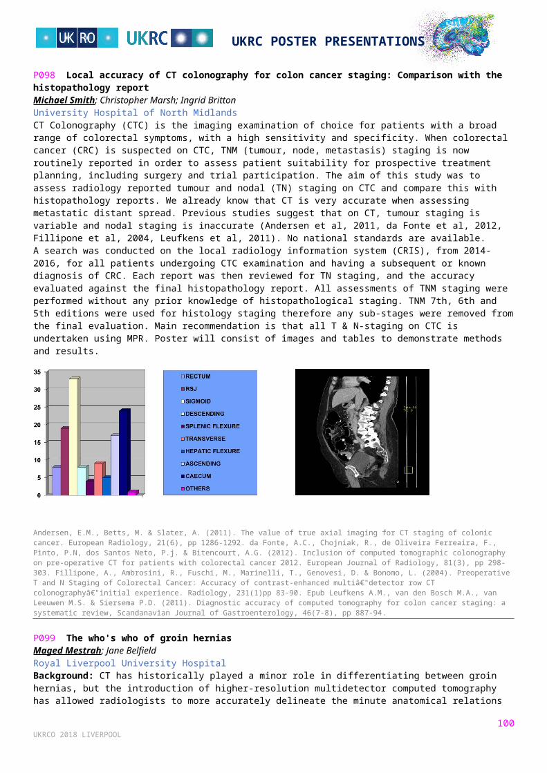
UKRC POSTER PRESENTATIONS
Conclusion: Image processing algorithms are a fundamental part of the optimisation process, and must not be neglected. For bed-side chest radiography, it has been demonstrated that MUSICA3+ gives the best 'overall' quality, but changes to image processing should be considered if dense regions are of clinical interest.
P165 Identifying anatomical regions of pelvic X-rays using open-source texture analysis and machine learning software: A proof of concept studyAndy CreedenUniversity Hospitals Coventry & Warwickshire NHS TrustBackground: The radiographic image texture of bone is influenced by density and trabecular pattern. Reporters generally assess texture qualitatively, but texture can also be assessed quantitatively. The open-source textural analysis software qMaZda (Szczypinski et al., 2009) can calculate approximately 300 texture parameters. These large datasets generated make conventional statistical techniques unfeasible for determining which (if any) of these parameters are useful for differentiating bone structures. However, machine learning algorithms are ideally suited to identify and 'learn from' patterns and relationships within large datasets. Bone texture varies between individuals but some bony structures have characteristic trabecular patterns. This study aimed to determine whether a quantitative approach using open-source software is more accurate than reporters at identifying anatomical regions of pelvic radiographs based solely on their texture. Method: 'Training' and 'test' datasets consisting of the textural parameters for 50 and 15 pelvic RoIs respectively were created using images downloaded from PACS. A variety of machine learning algorithms were trialled on the training set using the open-source data mining software Weka (Frank et al., 2009). Accuracy was then evaluated on the test set and compared to the performance of 5 experienced reporters undertaking the same task. Results: The 'Simple Logistic' algorithm identified the origins of the RoIs of the test set with an accuracy of 93%. This compared to 73% for experienced reporters. Conclusion: Open source textural analysis and machine learning software successfully identified anatomical regions within the pelvis. In future this approach could be used to identify pathological changes in bo1. Frank, E., Hall, M., Holmes, G., Kirkby, R., Pfahringer, B., Witten, I. H., and Trigg, L. (2009) 'Weka-a Machine Learning Workbench for Data Mining'. in Data Mining and Knowledge Discovery Handbook. ed. by Anon: Springer, 1269-12772. Szczypinski, P. M., Strzelecki, M., Materka, A., and Klepaczko, A. (2009) 'MaZda - a Software Package for Image Texture Analysis'. Computer Methods and Programs in Biomedicine 94 (1), 66-76
SERVICE INNOVATION AND OPTIMSATION
P166 Outsourcing CT scansJanki Patel; Sanjin IdrizRoyal Surrey County Hospital NHS TrustBackground: Many hospitals in the UK have introduced outsourcing of CT scans to supplement the demand for imaging required out-of-hours[1]. However, there is sparse data on how this has affected the number of imaging investigations done out-of-hours. This study addresses this gap by investigating the impact of implementing outsourcing via 'Four Ways' at an UK district general hospital on the number of out-of-hour CT scans requested.Method: This is a retrospective cross-sectional study. Data was collected on the number, type and indications of CT scans that were undertaken out-of-hours in January 2015, before the use of outsourcing, and compared to data from January 2016 and January 2017. Data was collected using HSS CRIS and analysed using Excel. Results: Following the introduction of outsourcing, the total number of Emergency Department CT scans that were vetted increased (74 in January 2015, 61 in January 2016 and 123 in January 2017). Furthermore, the total number of Inpatient CT scans vetted during outsourcing hours also increased (23 in January 2015, 18 in January 2016 and 46 in January 2017). This was despite the fact that the total number of CT scans during the non-outsourcing out-of-hour period remained stable from January 2016 and January 2017. Conclusion: These results indicate that since the introduction of outsourcing, the number of CT scans vetted out-of-hours has increased. This study did not look at the reasons for why outsourcing increased the number of scans, and this will form the basis of future work in this area. 1. Rcr.ac.uk. (2017). [online] Available at: https://www.rcr.ac.uk/sites/default/files/docs/radiology/pdf/BFCR%2810%298_Teleradiology_census.pdf [Accessed 14 Dec. 2017].
100UKRCO 2018 LIVERPOOL

UKRC POSTER PRESENTATIONS
P168 Detecting and reporting domestic abuse of the elderly: Mapping the concerns of experienced radiographersRachel Croft; Paul Miller; Lisa Booth; Elizabeth BatesUniversity of CumbriaBackground: While over 65,000 suspected cases of elder abuse are reported to English councils each year, it is estimated that upwards of 95% of incidences are either missed or not reported by healthcare professionals in emergency department settings [1]. Despite the call from Murray and Devos[2], two decades ago, for greater investigation of the extant and prospective role of radiographers in identifying abuse of the elderly, the broad phenomenon has continued to receive limited attention in medical imaging research. Methods: Using a standard model of Interpretative Phenomenological Analysis[3,4], extended semi-structured interviews with N=8 experienced plain radiographers were analysed. Results: In A&E contexts, where safeguarding issues have primarily been the responsibility of a physician, it was reported that the degree to which physicians take account of radiographers' concerns about elder abuse is inconsistent at best. This had sometimes resulted in a borderline defeatist attitude among radiographers, who would now only raise such a concern if they were uncategorically certain it would be taken seriously. In the outpatient domain, where radiographers felt more in control of the medical process itself, progressively higher levels of confidence to take a lead around these matters were reported. Even here, however, participants routinely argued that the available information and clinical communication necessary for them to recognise potential elder abuse was often lacking in a way it was not around other forms of domestic abuse. Conclusions: The analysis signposts some important issues around elder abuse and radiography that foregrounds, above all, the importance of clinical context and communication. 1. NHS Digital. Safeguarding adults: Annual report, England 2015-16 experimental statistics. London: Health and Social Care Information Centre; 2016. 2. Murray L, Devos D. The escalating problem of elder abuse. Radiol Technol 1997;68:351-353. 3. Miller PK, Woods AL, Sloane C, Booth L. Obesity, heuristic reasoning and the organisation of communicative embarrassment in diagnostic radiography. Radiography 2017;23:130-134. 4. Woods AL, Miller PK, Sloane C. Patient obesity and the practical experience of the plain radiography professional: On everyday ethics, patient positioning and infelicitous equipment. Radiography 2016;22:118-123.
P169 Retrospective review of Arian Teleheal organisation's international telemedicine and tele-education work; and defining the future vision by conducting world's first augmented reality telemedicine consultation using microsoft hololensWaheed Arian 1; Sami Khan 2; Kamran Turyalay 3; Fazale Hadi Wardag 3
1Arian Teleheal; 2Basildon Hospital; 3Basildon University HospitalArian Teleheal is a "pioneering" telemedicine charity which provides world-class healthcare advice to doctors in war zones and low-resource countries. The volunteer doctors of Arian Teleheal use everyday technology (such as smartphones and secure social media) to advise local doctors. Arian Teleheal also uses more complex technology to provide educational, learning and research opportunities, helping local doctors develop their abilities to further benefit their patients. Arian Teleheal is the first in the world to successfully implement international live telemedicine support using everyday technology on a 24/7, 365 days-a-year basis, with rapid response times for most acute cases. Furthermore, the work of Arian Teleheal provides educational and research opportunities for doctors in developed countries, through their work with colleagues around the world. The success of Arian Teleheal has been recognised with a number of awards, including being the first and only UK recipient of a UNESCO Global Hope Coalition award. Retrospective review conducted in form of anonymised detailed surveys shows that the work of Arian Teleheal has a positive impact on patients' healthcare management and on the participating local medics' education. Being the first in the world to successfully use augmented reality technology (Microsoft Hololens) to provide live international telemedicine consultation and teaching, Arian Teleheal sits the vision of the future of international telemedicine. Arian Teleheal is finalising collaboration plans to expand to Africa in 2018. The successful use of simple day to day technology, evolution of smartphones and increasing internet connectivity, complement survey evidence to scale up globally.
P171 WHO surgical safety checklist for radiology interventions: Improving compliance through action researchMartine Harris; Andrea Sanderson; Nicholas SpencerMid Yorkshire Hospitals NHS TrustPurpose: The overwhelming success of implementing the World Health Organisation (WHO) Safe Surgery Saves Lives Checklist [1] prompted the Royal College of Radiologists (RCR) to publish guidance for implementing the National Patient Safety Agency (NPSA) Safe Surgery requirement[2]. Subsequently a specific checklist adapted for radiological interventions has been developed with 100% target compliance encouraged by the quality care commission (CQC)[3].This work reflects on the action research process used during the introduction and implementation of this safeguarding checklist into clinical practice across a diagnostic imaging department.
101UKRCO 2018 LIVERPOOL

UKRC POSTER PRESENTATIONS
Method: This study has been undertaken in a single NHS Trust with data collected across its three hospital sites. An action research approach was adopted to allow collaboration between researcher and practitioners. Data collection included qualitative opinion of a small but varied group of radiology personnel responsible for the delivery and improvement of the service and quantitative safety checklist compliance data for imaging modalities undertaking interventional procedures. Results: Since its introduction locally, WHO safety checklist compliance has increased from 25% to 91%. Modality and Sub-speciality analysis demonstrates that some areas of practice out-perform others, requiring bespoke systems of work. Qualitative enquiry has informed several initiatives to improve compliance including the development of a formal policy and modified checklists as well as empowering staff to own the process. Conclusion: The multiprofessional team continues to strive for 100% compliance but this iterative action research cycle has been valuable in staff engagement, changing perceptions and embedding sustainable change. A potential limitation of the study is incomplete compliance data. 1. Haynes AB, Weiser TG, Berry WR et al. A surgical safety checklist to reduce morbidity and mortality in a global population. New Engl J Med 2009; 360: 491- 99. http://www.nejm.org/doi/full/10.1056/NEJMsa0810119#t=article 2. The Royal College of Radiologists. Standards for the NPSA and RCR safety checklist for radiological interventions. London: The Royal College of Radiologists, 2010. Available from https://www.rcr.ac.uk/publication/standards-npsa-and-rcr-safety-checklist-radiological-interventions. Accessed 13 April 2018. 3. The Royal College of Radiologists. Interventional Radiology Patient Safety Checklist, Last updated 2016. Available from https://www.rcr.ac.uk/audit/interventional-radiology-patient-safety-checklist. Accessed 13 April 2018.
P172 Responding to MRI safety incident trendsDarren HudsonInHealthBackground: Our internal MRI Safety reporting remains relatively low considering the number of patients seen in our facilities each day. Whilst this can be taken to mean safety is well managed, which generally it is, it is a fact that near misses and minor events are occurring more regularly and by luck don't result in significant harm. Staff are encouraged to report any near misses and failings in procedure or practices so the organisation can learn from these and work towards improvement to avoid potential for significant harm. Purpose: From review of our internal MRI safety events the main trends are around referral of contraindicated devices and failure in the screening process. Therefore, to help learn from these occurrences, improve practice and work to reduce incidents, some materials were developed to coincide with MRI Safety week. Summary: The poster will provide an overview of the materials produced: A short information leaflet which can be shared with referrers outlining the considerations when referring for MRI and the
importance of providing reliable patient history. These can then be given to those referrers sending patients to our services with contra-indicated devices in situ, and will proactively be shared where opportunity presents -- CCGs, Trust inductions etc.
Staff were asked to partake in some structured observational peer review around screening process and patter. This was intended to be an objective and non-judgmental means of reviewing and improving practice with one another.
102UKRCO 2018 LIVERPOOL
UKRC POSTER PRESENTATIONS
P173 Misplaced nasogastric tubes, a never event. A trust-wide three-cycle, closed loop, audit (Epsom & St Helier Hospitals Trust, London, United Kingdom)Jay Patel; Chandani Thorning; Julia HineEpsom and St Helier HospitalMisplaced nasogastric tubes are a never event. A national patient safety alert (NPSA) in 2016 revealed 95 incidents relating to the misplacement of nasogastric tubes, forty-five of which were related to the use of radiographs [1]. The safety alert provides criteria for optimal gastric placement and clear instructions for required actions if the tube is misplaced. We produced a three cycle audit investigating requests for radiographs, image quality and explicit statements in the resultant report. Secondary end-points included time taken to report radiograph, percentage of reports authorised within one hour and the number of requests out of hours. The final cycle of the audit included a total number of 226 adult patients with information obtained retrospectively via a trust-wide PACS at Epsom & St Helier Hospitals, London. The results showed an increase in all primary end-points by initiating discussion with consultant radiologists about the standards expected between each cycle. Particularly, the percentages of reports including an explicit statement regarding safety to feed increased from 63% to 80% (between cycle 2 and 3). We concluded 80% of reports in the final cycle were compliant with NPSA requirements in providing an explicit statement regarding safety to initiate feed, average time for radiograph to be reported decreased considerably and more checks were carried out out-of-hours resulting in higher use of teleradiology services.1. NHS Improvement (2016) Nasogastric tube misplacement: continuing risk of death and severe harm [online]. Available at: https://improvement.nhs.uk/news-alerts/nasogastric-tube-misplacement-continuing-risk-of-death-severe-harm/ (Accessed 19/04/2018)
P174 Improving quality of care and reducing unnecessary imaging by implementing new adrenalimaging guidelinesVikas Shah; Motahare Yadegarfar; Ali HabibUniversity Hospitals of Leicester NHS TrustBackground: Imaging follow up (FU) of "adrenal incidentalomas" (AI) is resource intensive. Our centre lacked clear guidance on managing AI. The American College of Radiology (ACR) recently published an algorithm for follow up of AI. We set out to (a) map heterogeneity of practice, and (b) identify resource implications of implementing the ACR guidelines. Methods: To map heterogeneity, data regarding dedicated adrenal imaging over a 1 year period was analysed. Data examined included reason for study (follow up (FU) of previously characterised lesion, or characterisation of newly found lesion), study modality (MRI or CT, +/- contrast), time since previous study if for FU, and number of previous studies. We applied three key questions from the ACR guidelines to our practice, to establish resource implications. Results: 90 studies were included (62 FU, 28 characterisation). Heterogeneity was found in all aspects of AI imaging; study modality, time from previous study to the FU study (0-84 months), number of adrenal studies preceding the FU study, nature of lesions being FU, and the size of lesions being followed up and characterised. If, by following the ACR guidelines, we hadn't followed up any lesion <10mm, benign lesions >10mm, or indeterminate lesions >10mm but unchanged over a year, we would have avoided 53 studies. Conclusion: The lack of local guidelines rendered our practice heterogeneous. By adopting the ACR guidelines, we estimate a workload reduction of 59% per year, as well as following a more consistent approach. We aim to re-assess our practice following a period of implementation.William W. Mayo-Smith, Julie H. Song, Giles L. Boland, Isaac R. Francis, Gary M. Israel, Peter J. Mazzaglia, Lincoln L. Berland, Pari V. Pandharipande, (2017), Management of Incidental Adrenal Masses: A White Paper of the ACR Incidental Findings Committee
P175 Real world experience of Nivolumab in a DGHPei King Teo; Akash Maniam; Cheng BoonWorcestershire Royal HospitalAim: An audit to assess response rate of treatment with Nivolumab in various tumour sites.Method: Data was obtained from pharmacist department for all patients receiving Nivolumab over the last 3 years. A spreadsheet on survival response rate indication of treatment and performance status at the start of Nivolumab treatment were recorded. Result: Data from a total of 17 patients were used to assess response to nivolumab treatment. The majority (total of 8) of these were patients with diagnosis of metastatic melanoma who failed the first line treatment. 2 patients with squamous cell carcinoma of the lung; 2 with head and neck cancer; remaining 5 with renal cell carcinoma. The overall survival is about 6 months and patients with performance status of 0-1 generally tolerate Nivolumab better than those with poor performance status. Of the toxicities 8 patients experienced no toxicity with the remaining 9 patients having immune related toxicities grade 3 and above. Immune mediated hepatitis contributes to the majority of the toxicities with 3 out of 9 patients having grade 3 immune hepatits requiring a temporarily stop to their treatment. 3 of the 17 did not survive long enough for response assessment with a scan. 3 patients out of 17 were on combination immunotherapy for metastatic melanoma. Conclusion: Nivolumab is a fairly well tolerated drug with limited toxicities and can be delivered in peripheral hospital as provided funding available on NICE.
103UKRCO 2018 LIVERPOOL
UKRC POSTER PRESENTATIONS
P176 Worthwhile and unnecessary radiology visits with general practitioner radiography referrals: an audit of local practiceSamuel Pettitt; Robert MilnerThe Rotherham NHS Foundation TrustBackground: The Radiology Department of an NHS Foundation Trust performs over 16,000 plain film referrals from General Practitioners (GPs) per year. Due to a recent local merger this number is predicted to rise to approximately 23,000 referrals per year. The Trust offers GP patients both an appointing and open access service. National guidance on radiological investigations has been published by the Royal College of Radiologists (2017), which aims to standardise practice. Anecdotally, radiographers' feel that a significant proportion of GP patients are turned away without imaging being performed. These represent unnecessary radiology visits and undoubtedly diminish trust in the healthcare system. Method: Utilising the Radiology Information System (RIS), all GP plain film radiography requests within a 12-month period from August 2016 to August 2017 were retrospectively reviewed, and the number of successfully preformed and cancelled requests were established for each anatomical area. Results: Over 5% of patients were turned away without imaging being performed, in eleven of the forty-three anatomical areas. All of these were due to unjustified clinical information, whilst almost all (96.3%) of these patients were pre-appointed which meant that the radiology department had time to 'vet' the request prior to arrival. If all requests for these eleven anatomical areas had been 'vetted' prior to booking, there would have been 2.9 requests to vet per working day. Conclusion: Implementation of a pre-appointment vetting system for GP radiography requests could significantly reduce the number of unnecessary visits to the radiology department, which would offset the slight increase in workload. 1. Cole, Andrew. (2014) GPs feel pressured to prescribe unnecessary antibiotics, survey finds. [Online]. http://www.bmj.com/content/349/bmj.g5238.full 2. Morgan, Myfanwy, Jenkins, Linda and Ridsdale, Leone. (2007) Patient pressure for referral for headache: a qualitative study of GPs' referral behaviour. Br J Gen Pract. 57(534), 29–35. https://www.ncbi.nlm.nih.gov/pmc/articles/PMC2032697/ 3. NHS England. (2016) NHS Diagnostic Waiting times and data. [Online]. https://www.england.nhs.uk/statistics/wp-content/uploads/sites/2/2016/06/DWTA-Report-April-2016.pdf 4. National Institute for Health and Care Excellence. (2017) Suspected Cancer: recognition and referral. NG12. [Online]. https://www.nice.org.uk/guidance/ng12 5. National Institute for Health and Care Excellence. (2016) Fractures (non-complex): assessment and management. NG38. [Online]. 6. Royal College of Radiologists. (2017) RCR I-refer: making the best use of clinical radiology. 8th ed., London, Royal College of Radiologists. 7. The Kings Fund. (2016) Understanding pressures in general practice. [Online]. http://cdn.basw.co.uk/upload/basw_53043-7.pdf
P177 Service improvement audit on the efficiency of supraorbital fracture diagnosisCaroline Coletto; Katherine SharkeySt Helens and Knowsley Teaching Hospitals TrustIntroduction: Supraorbital injuries are commonly caused by assaults, falls, sports-related injuries and road traffic collisions [1] thus encompassing a diverse range of patients that present to Accident and Emergency. A supraorbital injury should be identified as efficiently as possible to avoid further complications[2]. The audit focuses on the diagnostic value of our supraorbital protocol in combination with patient pathways through CT, supported by literature. Method: A retrospective audit was carried out from October 2016 - October 2017 evaluating supraorbital radiographic requests, the views performed and the role of CT. Findings: During this period 9% of all facial bone requests were undertaken for supraorbital injury. 40% of these patients had one view, 60% had 2 views but of that only 27% did a dedicated OF20 view as per protocol. Only 1 patient was recommended to have a dedicated supraorbital view following their examination. 11% of patients were referred to CT. Only 3 patients were diagnosed with a supraorbital fracture. Conclusion and recommendations: The protocol is not being followed. Contributing factors to the lack of completion of an OF20 view include; inexperience of an OF20, under appreciation of this dedicated view, referrer's knowledge and interpretation. However, with only 67 requests and of this only 3% reported fractured, raises the question is a dedicated view necessary? Many patients also have associated head injuries and as per NICE guidelines, automatically bypass having radiographs thus, further reducing the amount of supraorbital requests. An extended audit could be completed to complement these findings.References (1) Kin, J. and Huoh, K. (2010) ‘Maxillofacial Fractures’ Neuroimaging Clinics in North America, 20(4) pages 581-596. (2) Righi, S., Boffano, P., Guglielmi, V., Rossi, P., and Martorina, M. (2015) ‘Diagnosis and imaging of orbital roof fractures: a review of the current literature’, Oral and Maxillofacial Surgery, 19(1) pages 1-4.
P178 An audit of preliminary clinical evaluation practice in magnetic resonance imaging examinationsPaul Lockwood 1; Gill Dolbear 2
1Medical and Clinical Research Hub; 2Canterbury Christ Church UniversityAim: This study aimed to audit magnetic resonance imaging (MRI) preliminary clinical evaluations (PCE) of the brain, spine and knee completed by a cohort of diagnostic radiographers in postgraduate training. Methods: The audit of PCE data was taken from prospective clinical workloads in 27 MRI departments in England. The Radiographers (n=27) PCE commentaries (n=532 brain, n=592 spine, and n=496 knee examinations) were graded against reference standard answers from consultant radiologists. Applying true negative/positive and false negative/positive fractioned scoring. The audit statistical analysis assessed individual performance against the assessment of sensitivity, specificity, and accuracy. Interrater performance of sensitivity was further evaluated with d' prime. The reliability of independent interrater
104UKRCO 2018 LIVERPOOL
UKRC POSTER PRESENTATIONS
agreement applied Fleiss' Kappa coefficients. Interrater accuracy was verified with receiver operating curves (ROC) and Area under the Curve (AUC). Results: The radiographer's observer performance audit results for brain PCE demonstrated a sensitivity of 80% (d'=2.479), specificity of 89% and accuracy of 86%, p=0.046 (AUC 0.922, k=0.68). For the spine PCE commentaries were 88% sensitivity (d'=2.213), 70% specificity and 85% accuracy, p=0.143 (AUC 0.853, k=0.59). The knee PCE interpretations 85% sensitivity (d'=2.001), 74% specificity and 80.7% accuracy, p=0.213 (AUC 0.843, k=0.51). Conclusions: The findings of this limited audit indicate that a small cohort of radiographers demonstrated a consistent level of accuracy in the interpretation of 1,620 brain, spine and knee MRI examinations in a clinical environment. This innovative extension of radiographer roles in MRI has the potential to improve clinical practice and initial clinical decisions.1. Society and College of Radiographers. Preliminary Clinical Evaluation and Clinical Reporting by Radiographers: Policy and Practice Guidance. London: Society and College of Radiographers; 2013. 2. Snaith B, Hardy M. Radiographer abnormality detection schemes in the trauma environment—An assessment of current practice. Radiography 2008; 14(4):277-81. 3. Lancaster A, Hardy M. An investigation into the opportunities and barriers to participation in a radiographer comment scheme, in a multi-centre NHS trust. Radiography 2012; 18(2):105-8. 4. The Care Quality Commission. The state of health care and adult social care in England 2016/17 (Presented to Parliament pursuant to section 83(4)(a) of the Health and Social Care Act 2008). Newcastle upon Tyne: House of Commons; 2017. 5. Department of Health. Improving outcomes: A Strategy for Cancer. London: HMSO; 2011. 6. NHS England. Next steps on the NHS Five Year Forward View (Gateway number 06669). Leeds: NHS England; 2017 7. Schwartz LH, Panicek DM, Berk AR, Li Y, Hricak H. Improving communication of diagnostic radiology findings through structured reporting. Radiology 2011;260(1):174-81. 8. Royal College of Radiologists. Audit Live Peer Review- Using Double Reporting as a Tool for Revalidation. London: The Royal College of Radiologists; 2010. https://www.rcr.ac.uk/audit/peer-review-using-double-reporting-tool-revalidation 9. Royal College of Radiologists. Audit Live An audit of Radiology Report Quality. London: The Royal College of Radiologists; 2010 https://www.rcr.ac.uk/audit/audit-radiology-report-quality 10. Royal College of Radiologists. Audit Live Auditing reporting accuracy. London: The Royal College of Radiologists; 2011. https://www.rcr.ac.uk/audit/auditing-reporting-accuracy-template-spreadsheet-and-presentation 11. Berbaum KS, Franken JR EA, Anderson KL, Dorfman DD, Erkonen WE, Farrar GP, Geraghty JJ, Gleason TJ, Macnaughton ME, Phillips ME, Renfrew DL. The influence of clinical history on visual search with single and multiple abnormalities. Invest Radiol 1993; 1;28(3):191-201.
P179 An audit of preliminary clinical evaluation in nuclear medicinePaul Lockwood 1; Gill Dolbear 2
1Clinical and Medical Sciences Research Hub; 2Canterbury Christ Church UniversityAim: To assess the observer performance of preliminary clinical evaluations (PCE) in bone, lung, renal and thyroid nuclear medicine (NM) examinations among diagnostic radiographers. Methods: The audit reviewed 1,200 NM examination (n=519 bone scans, n=226 lung scans, n=282 renal scans, n=173 thyroid scans) PCE commentaries provided by a cohort of radiographers (n=20) participating in a nine month postgraduate training programme. All PCE commentaries were graded against formal radiologist reference standard reports. Applying true negative/positive and false negative/positive fractioned scoring. The audit statistical analysis assessed individual performance against the assessment of sensitivity, specificity, and accuracy. Interrater performance of sensitivity was further evaluated with Kappa coefficients. Interrater accuracy was verified with empirical and fitted receiver operating curves (ROC) and Area under the Curve (AUC) analysis. Results: The radiographer's observer performance in NM PCE demonstrated an overall sensitivity of 93% (91.3-94.4 .95% CI), specificity of 91% (89.7-93.0 .95% CI), and accuracy of 92% (90.5-93.7 95% CI), p> 0.001 (k= 0.84). All categories of examinations demonstrated high AUC scores as a metric for predictive modelling of diagnostic accuracy. Conclusions: An observed consistent level of accuracy in the interpretations of NM PCE examinations by a small sample of radiographers was demonstrated in a clinical environment. The main contribution of this paper is to present a new extension of radiographer roles in NM that has the potential to improve clinical practice, and to communicate urgent and unexpected findings for clinical management. 1. NHS England. (2017) Multi-professional framework for advanced clinical practice in England. 2. NHS England. (2017) Next steps on the Five Year Forward View. 3. Health Education England. (2017) Cancer Workforce Plan: Phase 1 delivering the cancer strategy to 2021. 4. The Kings Fund. (2017) Quality Monitoring Report November 2017: how is the NHS performing? 5. NHS England. (2017) Diagnostic Imaging Dataset Statistical Release: Provisional monthly statistics, July 2016 to July 2017. November 2017 6. Society and College of Radiographers. (2013) Preliminary Clinical Evaluation and Clinical Reporting by Radiographers: Policy and Practice Guidance. London: Society and College of Radiographers. 7. NHS Improvement. (2017) Quarter 2 2017/18 performance report: Performance of the NHS Provider Sector month ended 30 September 2017. 8. Royal College of Radiologists. (2010) Audit Live Peer Review- Using Double Reporting as a Tool for Revalidation. London: The Royal College of Radiologists. 9. Royal College of Radiologists. (2011) Audit Live Auditing reporting accuracy. London: The Royal College of Radiologists. 10. Royal College of Radiologists. (2010) Audit Live An audit of Radiology Report Quality. London: The Royal College of Radiologists. 11. O. Pinto A, Acampora C, Pinto F, Kourdioukova E, Romano L, Verstraete K. (2011) Learning from diagnostic errors: a good way to improve education in radiology. Eur J Radiol; 30;78(3):372-6. 12. Berbaum KS, Franken JR EA, Anderson KL, Dorfman DD, Erkonen WE, Farrar GP, Geraghty JJ, Gleason TJ, Macnaughton ME, Phillips ME, Renfrew DL. (1993) The influence of clinical history on visual search with single and multiple abnormalities. Invest Radiol; 1;28(3):191-201. 13. van Ooijen PM, Jorritsma W. (2017) Medical Imaging Informatics in Nuclear Medicine. In Quality in Nuclear Medicine (pp. 241-267). Springer International Publishing. 14. Larson DB, Towbin AJ, Pryor RM, Donnelly LF. (2013) Improving consistency in radiology reporting through the use of department-wide standardized structured reporting. Radiology. Apr;267(1):240-50.
P180 Adequate completion of computed tomography request formsDevleen Mukherjee; Archie KeelingCroydon Health Services NHS Trust
105UKRCO 2018 LIVERPOOL
UKRC POSTER PRESENTATIONS
Background: The motivation for this audit was to improve the standard of CT requesting. IR(ME)R provides a legal framework within which the referrer is required to supply sufficient information for the practitioner to justify the radiation exposure to patients. At our DGH, an electronic referral system is used. Methods: As per the standards set out by the RCR, 100% of CT request forms should include: patient's clinical information, relevant clinical question, patient details and referrer details. At our DGH, CT vetting is performed by radiographers, usually under the guidance of a consultant radiologist. During the vetting process, data was collected prospectively using a proforma based on RCR standards. This was used to scrutinise the quality of referral requests over a two week period. Results: 114 CT request forms were sampled. All the requests were made for the intended patient. 100% included the correct patient information. 91% included the relevant clinical information and question. However 20 out of the 114 (17%) did not include the referrer's details and 9% did not include details of the responsible clinical team. Conclusions: Despite using an electronic system, these results fell short of the standard set. We raised awareness of the importance of the referral process through presentation of these results at doctors' induction meetings and an intranet protocol on adequate radiology referral completion. Incomplete request details delays radiological diagnosis and subsequent action. The promotion of better interdepartmental communication, through radiology requests, subsequently leads to improved patient outcomes.1. Leonard, Graham, Bonacum. (2004) The human factor: the critical importance of effective teamwork and communication in providing safe care. BMJ Quality & Safety. 13: i85-i90. 2. Department of Health. The Ionising Radiation (Medical Exposure) Regulations 2000. London: The Stationery Office, 2000. 3. https://www.rcr.ac.uk/audit/adequate-completion-radiology-request-forms
P181 An audit assessing the diagnostic yield of acute abdominal radiographs in diagnosing bowel obstructionBarry StevensWalsall Healthcare NHS TrustBackground: Abdominal radiographs are commonly requested to investigate acute non-traumatic abdominal pain. However, routine use is reported as being of little value (Campbell & Gunn 1988), with the diagnostic yield previously being questioned (Kellow et al 2008). Specifically, abdominal radiographs are reported as being non-sensitive for investigating colonic obstruction as the cause of pain (Ahn et al 2002).Method: A retrospective audit of 50 abdominal requests from the Emergency Department (ED) with clinical indication of obstruction was undertaken to evaluate the propensity of positive findings of colonic obstruction.Results: Of the 50 abdominal radiographs, 47 were considered normal and three were positive, of which only one had a small bowel obstruction (SBO). Twenty-two patients had further imaging; 15 had CT and seven had US. Ten CT scans were positive but only two showed a SBO, demonstrating one that was not visible on the abdominal radiograph. Five US examinations were positive with non-obstruction findings.Conclusion: Abdominal radiographs to rule out an acute obstruction were seen to be negative in 98% of cases. Additional imaging with Computed Tomography (CT) was only positive for presence of colonic obstruction in 4% of cases. These findings correlate with previous work (Campbell & Gunn 1988; Ahn et al 2002). ED referrers need to be aware of the increased patient dose and length of hospital stay resulting from inappropriate abdominal requests for acute presentations. A confident clinical acumen will improve the quality of service whilst paying due respect to making the best use of radiology services.1. Campbell JP, Gunn AA. (1988) Plain abdominal radiographs and acute abdominal pain. British Journal of Surgery. 75(6):554–6. 2. Ahn SH, Mayo-Smith WW, Murphy BL, Reinert SE, Cronan JJ. (2002) Acute Nontraumatic Abdominal Pain in Adult Patients: Abdominal Radiography Compared with CT Evaluation. Radiology. 225(1):159–64. 3. Kellow ZS, MacInnes M, Kurzencwyg D, Rawal S, Jaffer R, Kovacina B, et al. (2008) The role of abdominal radiography in the evaluation of the nontrauma emergency patient. Radiology. 248(3):887–93. 4. Royal College of Radiologists (GB) (2017). iRefer: making the best use of clinical radiology
P182 Audit of CT guided nerve root injections cancellations secondary to incomplete peri-procedural anticoagulation managementElizabeth Robinson 1; Shahab Shahipasand 2; Panagiotis Liantis 1; Ramin Mandegaran 1; Ali Zavareh 1
1Guy's and St Thomas' Foundation Trust; 2London North West Healthcare NHS Foundation TrustBackground: CT guided steroid injection is a well-recognised, conservative treatment of localised spinal pain and radiculopathy[1,2]. An extremely rare complication is the development an epidural haematoma with potential to cause permanent neurological damage[3]. Anticoagulation at the time of procedure is therefore contraindicated. Routinely injections are performed as an outpatient requiring the referring physician to implement a peri-procedural anticoagulation plan. Anecdotal experience suggested cancellations were occurring as patients remained on anticoagulation at the time of their appointment.Purpose:
Identify the incidence of cancelled CT guided spinal injections secondary to incorrect peri-procedural anticoagulation management
Develop an intervention to help reduce the incident rate Re-audit to assess the effect of the intervention.
106UKRCO 2018 LIVERPOOL
UKRC POSTER PRESENTATIONS
Methods:Audit Standard: 100% of outpatients attending for a CT guided nerve root injection should have an appropriate anticoagulation plan implemented. Data collection:
Prospective data collection between 1st September-30th November 2016 Population: All elective CT guided spinal nerve root injections scheduled Requesting clinicians not specifically made aware of ongoing audit Descriptive analysis was completed.
Intervention: Revised electronic request form implemented (Figure1) New compulsory fields to be completed concerning anti-platelets and
anticoagulants.Re-audit following intervention:
Prospective data collection between 1st September-30th November 2017 Same data collection methods.
Results:Audit: 55 patients -3 cancellations (5%) Re-audit: 93 patients -0 cancellations (0%)
Conclusions: The new request form prevented 5% of all patients referred for CT guided nerve root injection being cancelled, secondary to incorrect peri-procedural anticoagulation management. Extrapolated over 1 year the potential savings through lost activity £51711. Benny B, Azari P. (2011) The efficacy of lumbosacral transforaminal epidural steroid injections: A comprehensive literature review. J Back Musculoskelet Rehabil ; 24:67-76. 2. Boswell MV, Trescot AM, Datta S et al. (2007) Interventional techniques: Evidence based practice guidelines in the management of chronic spinal pain. Pain Physician; 10:7-1113. Jankowski CJ. Complciations of regional anethesia. (2002) In: Raj PP,ed. Textbook of Regional anaesthesia. New York, NY; Churchill Livingstone;829-852
P183 Audit of adherence to trust guidance in elective vascular patients undergoing contrast proceduresElizabeth Robinson 1; Annelies Sweeney 1; Krishanthi Sathanandan 2; Judith Partridge 1
1Guy's and St Thomas' Foundation Trust; 2Barking and Havering NHS TrustBackground: Increasingly vascular patients are undergoing endovascular interventions, placing them at risk of contrast induced nephropathy (CIN)[1]. For the majority CIN is benign and transient however approximately 20% will experience a permanent deterioration in their renal function[2]. There is no treatment for CIN, however many prevention strategies exist (Table1). Local guidance advises strategies according to pre-procedure stratified risk (Figure1). Anecdotal experience suggested multi-morbidity was leading to admissions the day before for optimisation and guidance was not being adhered to.
Aims: Identify the incidence of CIN in elective vascular patients Establish adherence to trust guideline.
Methods: Audit Standard: Trust guidance-100% adherence expected.
107UKRCO 2018 LIVERPOOL

UKRC POSTER PRESENTATIONS
Data collection: Prospective data collection September 2017 of all elective vascular admissions for contrast procedures. Vascular team not specifically made aware. Data collected throughout admission (Table2).
Results: 40 patients (3 cancelled) Risk stratification: 28 Low risk, 8 Moderate risk, 1 High risk 3 cases of CIN (8%)
o Incidence did not correspond to contrast dose in any caseo Guidance not followed in each caseo 65% adherence to trust guidance (Table3)
Conclusions: Variable adherence to guideline Most commonly:
o Not post-hydratedo Continuation of nephrotoxic drugso Low risk patients admitted the day before procedure for IV hydration
Only 3 patients developed CIN despite non-adherence in multiple patients. Findings in line with AMACING trial[9]
o Suggested not giving prophylactic hydration is non-inferior to IV hydration in the prevention of CIN in high risk groups.
Estimated cost of beds for pre-hydration over 1 year £113,602Plan to change vascular admissions practice1) P. Grossman, S.Ali, H.Aronow et al. (2017) Contrast-induced nephropathy in patients undergoing endovascular peripheral vascular intervention: Incidence, risk factors, and outcomes as observed in the Blue Cross Blue Shield of Michigan Cardiovascular Consortium. J Interven Cardiol. 2) Maioli M, Toso A, Leoncini M, et al. (2012) Persistent renal damage after contrast-induced acute kidney injury: Incidence, evolution, risk factors, and prognosis. Circulation. 125:3099–103) Rudnick MR, Goldfarb S, Tumlin J. (2008) Contrast-induced nephropathy: is the picture any clearer? Clin J Am Soc Nephrol. 3: 261-262 4) Lameire N, Adam A, Becker CR et al. (2006) CIN Consensus Working Panel. Baseline renal function screening. Am J Cardiol. Sep 18;98(6A):21K-26K5) Lewington et al. (2013) Prevention of Contrast Induced Acute Kidney Injury (CI-AKI) In Adult Patients. Joint guideline from the Renal Association, British Cardiovascular Intervention Society and Royal College of Radiologists. Available from: http://www.rcr.ac.uk/docs/radiology/pdf/2013_RA_BCIS_RCR.pdf.6) Reed M, Meier P, Tamhane UU et al. (2009) The relative renal safety of iodixanol compared with low-osmolar contrast media: a meta-analysis of randomized controlled trials. JACC Cardiovasc Interv. 2:645–654. Epub 2009/07/25.7) Lameire N, Adam A, Becker CR, et al. (2006) Panel CINCW. Baseline renal function screening. Am J Cardiol. 98:21K–6K.8) Stacul F, van der Molen AJ, Reimer P, et al. (2011). Contrast Media Safety Committee of European Society of Urogenital R. Contrast induced nephropathy: updated ESUR contrast media safety committee guidelines. Eur Radiol. 21:2527–41.9) Nijssen, Estelle C et al. (2017) Prophylactic hydration to protect renal function from intravascular iodinated contrast material in patients at high risk of contrast-induced nephropathy (AMACING): a prospective, randomised, phase 3, controlled, open-label, non-inferiority trial. The Lancet , Volume 389 , Issue 10076 , 1312 - 1322
P184 Advancing practice in radiography: A focus on CT head reporting in ScotlandEmma GilmourRobert Gordon UniversityBackground: Computed Tomography (CT) head Radiographer Reporting is an area of advanced practice in Radiography that could benefit the profession, the service it provides and enhance patient satisfaction. However, this is an underutilised skill throughout Scotland, where there are currently little CT head Reporting Radiographers, unlike other countries in the UK. The inclusion of CT head Radiographer Reporting in daily practice throughout Scotland could enhance the patient pathway through medical imaging by decreasing waiting times and allowing more prompt treatment of conditions, such as cancer treatment. In turn, Scottish Radiographers will benefit through being able to advance their practice, from increased knowledge and enhanced job satisfaction. Also, Radiologists in Scotland will benefit from having more time to focus on highly specialised procedures and other forms of reporting that Radiographers cannot assist with such as MRI scans.Purpose: To explore the use of the CT head reporting role in Scotland and how it could enhance radiographic practice if more commonly utilised. To identify the need for change and highlight how overcoming the limitations will benefit the service and service users throughout Scotland. Identifying these aspects and acknowledging the barriers can promote service change and in turn result in increased uptake of the role and benefit the profession.Summary: This poster includes background information on CT head reporting and evidence of Radiographer competence, healthcare policy, regulations and guidance, drivers and barriers to the role and leadership and management considerations to be applied to CT head reporting1. CLARKE, R. et al., 2014. Implementing radiographic CT head reporting: The experiences of students and managers. Radiography, 20(2), pp. 117-120.2. HENDERSON, I., MATHERS, S.A. and MCCONNELL, J., 2017. Advanced and extended scope practice of diagnostic radiographers in Scotland: Exploring strategic imaging service imperatives. Radiography, 23(3), pp.181-186.
108UKRCO 2018 LIVERPOOL

UKRC POSTER PRESENTATIONS
3. HENDERSON, I. et al., 2016. Advanced and extended scope practice of radiographers: The Scottish perspective. Radiography, 22(2), pp. 185-193.4. LOCKWOOD, P., 2016. An economic evaluation of introducing a skills mix approach to CT head reporting in clinical practice. Radiography, 22(2), pp. 124-130.5. MILNER, R.C. and SNAITH, B., 2017. Are reporting radiographers fulfilling the role of advanced practitioner?. Radiography, 23(1), pp. 48-54.6. SOCIETY OF RADIOGRAPHERS, 2017. The Role of the Radiographer in Computed Tomography Imaging. London: Society of Radiographers.7. THE SCOTTISH GOVERNMENT, 2010. The Healthcare Quality Strategy for NHSScotland. Edinburgh: The Scottish Government.
P185 CT imaging for head injury in a busy Emergency department: Does the stage of training affect compliance to NICE guidelines?Muzzammil Hussain; Michael Roshen; Umber Shakil; Sana Fatima; Nicholas ReadingBart's HealthBackground: A large number of CT Head requests for 'head injury' are received at the Radiology department from a busy A&E department. A spot audit revealed some requests were not meeting NICE guidelines. The aim of this audit is to ascertain whether doctors working in A&E are complying with nice guidelines for head injuries. Secondly, whether the training grade determines the conformity to NICE guidelines. Method: Records of adults who had CT head requests in the A&E between 1st Feb to 31st 2018 were reviewed. A five point 'quality scoring' system was developed;
1. Sufficient information on indication from A&E documentation (1 point) 2. Sufficient information of indication from CT head request (1 point) 3. Clinical question to be answered (1 point) and 4. Clear adherence to nice guidelines (1 point) 5. GCS documented (1 point).
The training level of the doctor was recorded. Non-parametric statistical tests were used. Results: 265 CT Head scans (43.6%) were requested by A&E for head injury out of a total of 608 in Feb 2018. Registrars requested 106 scans, followed by SHOs (52), FY2s (31), Nurse Practitioners (10) and Consultants (2). 64 requests did not mention a referring clinician. The median 'quality score' for all requests was 4/5. 84.5% of requests met NICE head injury guidelines and 87% had good clinical documentation. There was no stastical difference between 'quality' scores or conformity to nice guidelines based on doctor grade (P>0.5). Conclusion: NICE guidelines are not currently met. Doctors of all grades need to improve conformity to NICE head injury guidelines and improve the 'quality' of requests for CT Head.Head injury: assessment and early management | Guidance and guidelines | NICE [Internet]. Nice.org.uk. 2018 [cited 12 April 2018]. Available from: https://www.nice.org.uk/guidance/cg176 Van Borsel, M.D., Devolder, P.J. and Bosmans, J.M., 2016. Software solutions alone cannot guarantee useful radiology requests. Acta Radiologica, 57(11), pp.1366-1371. Stetson, P.D., Morrison, F.P., Bakken, S., Johnson, S.B. and eNote Research Team, 2008. Preliminary development of the physician documentation quality instrument. Journal of the American Medical Informatics Association, 15(4), pp.534-541.
P186 Introduction of a direct booking system in the appointment of patients for CT colonography (CTC) examinations: A common sense approachMichael Smith; Christopher MarshUniversity Hospital of North MidlandsCTC requires rigorous and complicated bowel preparation. Not only is this often confusing and distressing to the patient, they are also required to re-attend the hospital to pick up bowel preparation. Many patients are attending clinic at the time of the request therefore collection of preparation at the same attendance, via a direct booking service, provides a common sense approach, enhancing patient experience. The system also accelerates the process of appointing 63 day cancer pathway patients referred for CTC as these examinations have to be performed and reported by Imaging 14 days from the initial referral. This target was not being achieved for all patients. Liaising closely with the Colorectal multi-disciplinary team, Imaging department line managers and appointments staff, it was decided that a dedicated team of administration and clerical staff and advanced practitioner radiographers (APR) would manage the appointments. Faecal tagging or bowel preparation has to be checked and signed by a nominated APR, following protocol of the request, according to our local standard operating procedure for distribution of bowel preparation. We have introduced a hospital mobile phone system so that an APR will be available at all times to check requests and sign for bowel preparation whilst the patient is present in the department. This poster will aim to describe the system of direct booking, initial difficulties in setting up the service, the advantages of such a system and the improvement made to patient experience utilising flow diagrams, standard operating procedures, charts, tables, images and text.
P187 C-Rad coding of CT colonography (CTC): Intra-observer agreement justifies appropriately trained advanced practitioner radiographers (APR) to perform and provide a preliminary interpretation of CT colonographyMichael Smith; Christopher MarshUniversity Hospital of North Midlands
109UKRCO 2018 LIVERPOOL

UKRC POSTER PRESENTATIONS
Using a modified C-Rads coding system to assess intra-colonic findings it has been possible to assess the accuracy of APR preliminary interpretation of CTC in comparison to the final radiology report. A recent study (awaiting publication) has concluded that APR C-coding of CTC is comparable to the final report by a radiologist. The evidence is supportive of radiographer reporting of intra-colonic pathology on CTC. Once an APR preliminary interpretation has been provided and is negative for colorectal cancer or its precursors, the patient can be removed from the 2 week wait cancer pathway. In addition, the APR can also provide patient triage at request and an on-table assessment of the acquired images. If positive pathology is detected, go on to autonomously perform a staging chest scan and therefore reduce the examination to treatment time on the cancer pathway. It is essential that an APR led CTC service is developed in each Trust to facilitate the enhanced patient pathway for colorectal cancer imaging and the bowel cancer screening programme (BSCP). APRs should be appropriately trained in line with the BCSP training and education guidelines (TBA). Poster will demonstrate how to set up and successfully maintain an APR led service and APR performing and reporting CTCs, utilising tables, images and text.
P188 Switching to a lower cost bowel preparation model for CT ColonographyChristopher Marsh; Michael Smith; Ingrid BrittonUniversity Hospital of North MidlandsCTCs require bowel preparation in the form of bowel cleansing to enable adequate detection of polyps and malignancies, the accuracy of the exam is improved with faecal tagging[1]. The ideal programme of bowel preparation remains a debated subject, however it is agreed that it must be safe, effective and well tolerated by the patient. At this trust CTCs are performed with a two day preparation regime with 100ml of Gastrografin given the day before the exam. At £19.80 per bottle and 3000 CTC exams performed this incurs a £60,000 cost to the service, in comparison switching to Citramag and E-Z-Paque based preparation would be significantly less at £8.18 equating to £24,540, this is less than half the cost of using Gastrografin. A pilot of 50 patients were given citramag with E-Z-Paque aliquoted into three 20 ml doses taken throughout the day before their exam. Exams where blind double reported with the adequacy of the bowel preparation and faecal tagging graded as inadequate, adequate and good. This poster aims to compare the adequacy, acceptability and cost saving implications of using Citramag bowel cleansing agent taken with E-Z-Paque (barium sulphate 40% w/v) against Gastrografin for Computed Tomography Colonography (CTC).1) Taylor, S.A., Slater, A., Burling, D.N., Tam, E., Greenhalgh, R., Gartner, L., Scarth, J., Pearce, R., Bassett, P. and Halligan, S., 2008. CT colonography: optimisation, diagnostic performance and patient acceptability of reduced-laxative regimens using barium-based faecal tagging. European radiology, 18(1), pp.32-42
P189 The appropriateness of lumbar spine X-ray referrals in a primary care trustVishnu Naidu Barts HealthBackground: Lower back pain is the leading cause of long term disability worldwide[1] and makes up 7% of GP consultations with a resultant loss of an estimated 4.1 million working days a year[4]. Lumbar spine X-rays (LSXr) expose patients to over 17 times the radiation received during a chest X-ray (0.07mSv vs 1.2mSv); the highest of all plain films [5]. Both iRefer and NICE have guidelines for when to refer for a LSXr[2,3]. This audit aims to measure adherence to the guidelines from a primary care trust as well as the referral quality. Methods: Adults who had received a LSXr between 1st July 2017- 31st September 2017 were selected. Manual screening of notes was undertaken to retrieve (a) presenting complaint, (b) clinical indication, (c) referral wording, (d) X-ray outcome. A good referral is one with a clear clinical question and a bad referral was one without. Results 40 patients were included in this audit. 22/40 (55%) referrals adhered to the guidelines. 18/40 (42.5%) did not meet guidelines. 24/40 (60%) of referrals were deemed poor, with no clinical questions asked. 13/40 were good (32.5%) and 3/40 could not be accessed (7.5%) Conclusion: Although most referrals met the guidelines, a significant portion did not; thus, were exposed to radiation unnecessarily. This may mean the knowledge of iRefer and NICE guideline are not well known in primary care. Furthermore, most referrals were poor, with no clinical question being asked of the radiologist. A re-audit is planned after education.1. Global, regional, and national incidence, prevalence, and years lived with disability for 310 diseases and injuries, 1990-2015: a systematic analysis for the Global Burden of Disease Study 2015. Lancet. 2016;388(10053):1545-602. 2. Irefer.org.uk. (2017) [cited 25 November 2017]. Available from: http://www.irefer.org.uk/ 3. NICE (2016) Low back pain and sciatica in over 16s: assessment and management [cited 25 November 2017] Available from :https://www.nice.org.uk/guidance/NG59 4. Parsons S IM, Clarke-Cornwell A, Symmons D. A Heavy Burden: the occurrence and impact of musculoskeletal conditions in the United Kingdom today. Arthritis Research UK Epidemiology Unit. 2011:39. 5. Vilar-Palop J, Vilar J, Hernandez-Aguado I, Gonzalez-Alvarez I, Lumbreras B. Updated effective doses in radiology. J Radiol Prot. 2016;36(4):975-90.
P190 A comparative analysis of PACS/RIS reporting efficiency from a user perspectiveAbhinaya Chandrashekar 1; Anand Devaraj 2; Luke Dixon 3; Simon Padley 2; Susan Copley 3; Carole Ridge 2
1Imperial College London; 2Royal Brompton & Harefield NHS Trust; 3Imperial College Healthcare NHS TrustBackground/aims: PACS and RIS are integral to radiology reporting in NHS hospitals. Despite efforts to improve efficiency in radiology, little is known about the impact of PACS/RIS on radiologists' reporting efficiency. This study evaluates the efficiency of two different PACS/RIS, comparing time taken to report CXRs on each system and the amount of 'useful' and 'wasted' reporting time between the systems.
110UKRCO 2018 LIVERPOOL
UKRC POSTER PRESENTATIONS
Method: 5 radiologists reported a total of 200 CXRs in batches of 5. 3 radiologists reported solely at Royal Brompton & Harefield (RBH) NHS Trust (2009 AGFA PACS/RIS), 1 reported solely at Imperial College NHS Trust (2016 Carestream PACS/Soliton RIS), and 1 reported on both systems. Each radiologist was timed using an electronic stopwatch with split-timer functionality. Directly looking at a patient's CXR and dictating the report were 'useful' time. All other time was 'wasted' time. The relative proportion of useful and wasted time was compared.Results: Average wasted time per batch of 5 CXRs was 51% and 33% for the two systems. For the radiologist reporting on both systems, a substantial difference in average time taken to report 5 CXRs was noted (9 minutes versus 2 minutes). Average overall time taken to report 100 CXRs was double using one system compared to the other. Conclusion: Depending on the PACS/RIS, up to ~50% of time spent reporting CXRs is 'wasted', not directly related to radiograph reporting. With shortages of radiologists resulting in backlogs of unreported radiographs, the detrimental effects of inefficient systems on reporting efficiency should not be underestimated.
P191 Installation of pacs workstations in a teaching hospital: The need for a team approach for reporting ergonomicsRichard TuckerDerby UniversityThis poster sets out the key aspects required for generic PACS workstations. The poster covers the background at a major teaching hospital in the East Midlands from the initial implementation of the PACS units in the early 2000's to the current set up, whereby PACS workstations are no longer assigned to individuals but have been utilised in a "hot desk" approach. This "hot desk" approach means that all the desks have to be versatile to support a multitude of users across Radiology. A study of the literature has been undertaken including key papers from the Royal college of Radiologists to the Health and Safety executive. This collaborate approach to ergonomics has standardised the reporting rooms to adopted best practice and to ensure that all reporters have access to the correct reporting conditions.Designing people-centric hospitals. (2014). [ebook] Koninklijke: Philips Medical. Available at: http://images.philips.com/is/content/PhilipsConsumer/PDFDownloads/Global/System/ODLI20161223_001Healthcare-Application-Guide.pdf [Accessed 16 Feb. 2018]. García-Lallana, A., Viteri-Ramírez, G., Saiz-Mendiguren, R., Broncano, J. and Dámaso Aquerreta, J. (2011). Ergonomics of the workplace in radiology. Radiología (English Edition), 53(6), pp.507-515. Goyal, N., Jain, N. and Rachapalli, V. (2009). Ergonomics in radiology. Clinical Radiology, 64(2), pp.119-126. Hse.gov.uk. (2013). Workplace health, safety. [online] Available at: http://www.hse.gov.uk/pubns/priced/l24.pdf [Accessed 16 Feb. 2018]. Hugine, A., Guerlain, S. and Hedge, A. (2011). User Evaluation of an Innovative Digital Reading Room. Journal of Digital Imaging, 25(3), pp.337-346. Leccese, F., Salvadori, G., Montagnani, C., Ciconi, A. and Rocca, M. (2017). Lighting assessment of ergonomic workstation for radio diagnostic reporting. International Journal of Industrial Ergonomics, 57, pp.42-54. Rcr.ac.uk. (2018). Ergonomic IT Guidance Documents. [online] Available at: https://www.rcr.ac.uk/system/files/publication/field_publication_files/BFCR%2812%297_Ergonomics_0.pdf [Accessed 1 Feb. 2018]. Rodrigues, J., Morgan, S., Augustine, K., Clague, G., Pearce, T., Pollentine, A., Wallis, A., Wilson, D. and McCoubrie, P. (2013). Musculoskeletal Symptoms Amongst Clinical Radiologists and the Implications of Reporting Environment Ergonomics—A Multicentre Questionnaire Study. Journal of Digital Imaging, 27(2), pp.255-261. Sutton, L. (2011). PACS and diagnostic imaging service delivery—A UK perspective. European Journal of Radiology, 78(2), pp.243-249. Working with display screen equipment (DSE). (2013). [ebook] Crown Copyright. Available at: http://www.hse.gov.uk/pubns/indg36.pdf [Accessed 16 Feb. 2018].
P192 A systematic review guide for chest reporting radiographersFrancesca Leonard; Nick WatsonUniversity Hospital North MidlandsIn an increasing number of centres appropriately trained and supported radiographers have an important role in the reporting of chest radiographs ensuring delivery of a timely and high-quality chest imaging service. "A Review Guide for Chest Reporting Radiographers" poster will demonstrate the importance of a logical system when reporting radiographs of the chest. The poster will aid radiographers, and others reviewing chest films, to formulate a systematic approach to chest radiograph interpretation. Due to the complexity of the anatomy on the chest radiograph, a systematic review is essential to ensure that all areas of the chest film are considered in a methodical way, particularly those areas where pathology can "hide". The classic review areas will be discussed and comprehensively illustrated with examples of pathologies which might otherwise be overlooked.
P194 Errors in voice recognition generated radiology reports: a re-auditAbul Haque; Moustafa El-Badawy; Sardar QasimBurton Hospitals NHS Foundation TrustBackground: Studies have shown that use of Voice Recognition Software (VRS) significantly reduces the turnaround time for production of radiology reports. With the ever-increasing workload on radiologists, departments are increasingly adopting the use of VRS. However, use of such technology is not without risk and this audit examined the error rate of such equipment. Aim: To look at the error rate of VRS reports generated by the Consultant Radiologists in a District General Hospital (DGH) within the UK. Methodology: An initial retrospective audit was carried out in November 2016 using the RCR Guidelines. The guidelines state that the Overall error rate should be <5% & Major errors should be 0%. The initial audit examined reports over a four-month period from 1st January 2016 to 30th April 2016. The results found the Trust was missing the RCR targets and so recommendations were made and a subsequent re-audit carried out six months later using the same method.
111UKRCO 2018 LIVERPOOL
UKRC POSTER PRESENTATIONS
Results: The initial audit showed that the department was missing the RCR targets of Overall error rate and Major error rate by 7% and 0.3% respectively. Following a number of recommendations which included proofreading reports at least once before verifying, a re-audit six months later found the department had successfully achieved the RCR standards - Overall error rate was 4.6% and Major errors was 0%. Conclusion: The re-audit demonstrated that if certain recommendations are successfully implemented into the daily practice of a Radiology department, the overall percentage of errors can be reduced to acceptable.P195 Written vs verbal consent for ultrasound guided biopsies - which one to use?Obaid HashmiHinchingbrooke HospitalBackground: Ultrasound (US) guided biopsies are commonly conducted in UK radiology departments. They provide a sample for histological diagnosis without the need for surgery. However, they are invasive and with potential complications thus requiring informed consent. There are 2 types of consent; verbal and written. We look at the following question: written VS verbal consent - which one to use? The Royal College of Radiologists standards for consent is generic and does not provide a list of procedures that require written consent. It states a judgment be made by the operator based on the nature of the procedure. Method: We retrospectively collected data on written vs verbal consent for fine need aspiration (FNAs) and core biopsies (CB) over a period of 10 months in 2017.Results: 127 US guided FNAs and 63 US guided CB were performed. All 127 FNAs were conducted with verbal consent. 33/63 CBs were conducted with written consent, 27 abdominal or pelvic biopsies, 2 thoracic, 3 neck and 1 arm.Conclusion: Procedures with potentially riskier complications, such as intra-abdominal bleeding, were conducted with written consent. Some advantages of written consent include a permanent document of patient agreement and it facilitates formal discussion of the procedure. Decisions regarding which procedures require written vs verbal consent are often made at a local or operator level. Without clear guidance, there needs to be more discussion regarding consent for US guided biopsies. We look at advantages and disadvantages of written consent and a potential decision-making process.
P196 Patient perceptions of a rapid access radiology-led lung escalation pathwayPeter Hewitt; Julie Cox; Jonathan Spratt; Ralph Marsh; Kelly Gribbon; Susan Pollock; Paula Goodson; Joanne AndersonCity Hospitals Sunderland NHS Foundation TrustPatient satisfaction is a key outcome measure in healthcare[1] within the UK, but this is often overlooked in radiology services. In April 2014, a new lung escalation pathway (LCEP) was implemented in CHSFT to improve access to diagnostic services for patients with suspected lung cancer. The design and results of this pathway were presented at UKRCO 2016 and published in the journal Clinical Radiology in 2017. The current evaluation aims to assess the patient perspective of the LCEP, focusing on the information provided, anxiety levels and delays[1]. The pilot evaluation obtained 16 responses from patients who had been part of the LCEP. Of the respondents, 88% (n=14) were aware that further tests could be arranged as part of the LCEP. The average time frame between chest X-ray and CT was 8 days. We have demonstrated concordance with the patient perception which averaged 5.4 days, with 94% (n=15) patients accepting the delay is "about right". 100% (n=15) understood the CT results but only 64% (n=9) felt a copy of the report would be useful. 63% (n=10) of patients rated their anxiety as 4-5/5. As a development of the LCEP we are implementing new strategies to ensure that patients are better informed and less anxious once the LCEP has been initiated. Information and posters are to be re-established in GP waiting rooms and a new patient leaflet is being produced. Contact details are to be made clearer, especially specialist nurse contacts for patients booked for CT. The evaluation is on-going.1. Putting patients first: Understanding what matters to lung cancer patients and carers’. UK Lung Cancer Coalition. November 2013
P197 An investigation into the concerns of pregnant women regarding the fetal anomaly scan using online forumsPenny Settle; Pauline ReevesSheffield Hallam UniversityRationale: The research aimed to provide insights into the concerns of pregnant women regarding the fetal anomaly scan offered at 18+0 to 20+6 weeks gestation in the UK.Methods: A qualitative study of comments on a UK-based parenting web forum was undertaken. Thematic analysis was conducted on 48 threads containing 591 posts made over the period of one year from January 2017 to January 2018.Results: Four main themes emerged from the analysis; advice and reassurance; physical aspects of the scan; finding out the sex of the fetus, and; a sense of wonder.Conclusion: Pregnant women post on web forums predominantly to seek advice and reassurance concerning all aspects of the scan. There is an opportunity for health professionals to engage with women in these forums and provide expert advice. The increase of availability and uptake of commercial, non-clinical, scans can be complementary to the fetal anomaly scan. The increase in use of commercial scans highlights the need for hospitals to have clear policies regarding how many people are
112UKRCO 2018 LIVERPOOL
UKRC POSTER PRESENTATIONS
permitted in the scan room, and the providing of information in writing or to third parties. The expansion of technological capabilities in ultrasound equipment continues to raise increasing ethical dilemmas, prompting a need to re-evaluate sonographer led pre-scan counselling.
P198 Radiographer led discharge: Implementation of a new pathway to release staff capacity and improve patient experience in accident & emergencyVictoria Ballard; Katherine DayBrighton and Sussex University Hospital NHS TrustBackground: Radiographer-Led Discharge (RLD) has been associated with improved waiting times, interpretations of X-rays and re-attendance rates[1], however the impact on patient experience is not yet known. This study aimed 1. To determine whether implementing a new RLD pathway at BSUH could reduce time in hospital and 2. Assess patient experiences of RLD. Method: The RLD pathway was developed by a multi-professional group. Patients aged 17-74 years attending A&E for assessment for closed bony injury of the clavicle, upper limb or lower limb excluding hips, were eligible for RLD. Time in hospital was compared between patients discharged using standard practice and those receiving RLD. Self-reported patient experience of RLD was assessed. Results: Fifty-two patients met the inclusion criteria and were referred for RLD (RLD group). Forty-six patients met the inclusion criteria for RLD but were discharged using standard practice (A&E group). The median time spent in hospital was 92.0 minutes (IQR 62.8; 117.8) for those in the RLD group and 147.0 minutes (IQR 99.8; 180.0) for the A&E group (median difference = 55.0 minutes) (p<0.0001). Twenty-eight (53.8%) of those in the RLD group completed feedback questionnaires of whom 27 (96.4%) indicated that they would be likely to refer the service to a friends/family and 24 (85.7%) indicated that the service should be continued. Conclusion: RLD significantly reduced the time patients spent in hospital and patient experience ratings were excellent. A planned audit of the service will compare the quality of care between patients receiving RLD and standard care.Henderson, D, Gray, WK, Booth,L. (2013) Assessment of a reporting radiographer-led discharge system for minor injuries: a prospective audit over 2 years. Emerg Med J 30:298-302
P199 Cystography and trial without catheter in the imaging department: Feasible and safe patient centred serviceHassan Rehman; Cherian GeorgeUniversity Hospitals of North MidlandsTrial without catheter (TWOC) is a procedure normally undertaken as a day case or inpatient by urology team, including specialist nurses, within a hospital setting. These patients have a cystography to ensure that the urinary bladder injury has healed following which TWOC may be done. We present our initial experience of 16 patients to see if TWOC was a safe and feasible service within the imaging department. Data was collected by searching radiology patient information system by using codes for cystography was done in the Imaging department from June 2015 to March 2017. Patients who only had cystogramphy without TWOC were excluded from the study. All eligible patients' reports were reviewed. Total of 16 patients (M:F=4:12; Age range was 27 to 86 years with a median age of 56 years) had TWOC immediately following the cystography within the imaging department. All 16 patients had a successful TWOC during their visit to the imaging department with none requiring further catheterisation. TWOC following a normal cystography routinely is on the same day in an ambulatory ward or on a later date. This has added costs to the health service in addition to inconvenience to the patient with regard to multiple appointments to the hospital. We propose that in the appropriate group of patients, cystography followed by TWOC within the Imaging department is feasible and beneficial for both the patients and the hospital.
P200 Post-mortem computed tomography in natural death investigation - a review of progressClaire Robinson 1; Bruno Morgan 2; Aparna Deshpande 1; Cathy Richards 1
1University Hospitals of Leicester NHS Trust; 2University of LeicesterBackground: Post mortem computed tomography (PMCT) is a relatively new radiographic speciality. Initial services diagnosed traumatic causes of death. With growing cultural and religious objections to the invasive autopsy, angiographic and ventilation techniques were developed to diagnose natural causes of death in up to 92% of cases [1]. We launched a PMCT service to investigate natural deaths in 2015, developing and expanding it in 2017 to become the first NHS, local authority funded, PMCT service. Method: A retrospective audit was conducted of all PMCT cases referred by HM Coroner for natural death investigation. The aims of the service set in 2015 were the standards used. Results 182 patients had PMCT scans and a cause of death was given in 176 cases (96.7%) exceeding the 90% target. A limited autopsy was predicted prior to the scan in 1 case and in a further 2 cases, the cause of death was unascertained after autopsy with histology and toxicology. 98.9% had the cause of death issued in 3 days of referral, meeting the target. The mean time from referral to radiology report being issued reduced to 10.5 hours in 2017.
113UKRCO 2018 LIVERPOOL
UKRC POSTER PRESENTATIONS
Conclusion: PMCT is now a viable alternative to some autopsies, providing cause of death in a comparable time scale to autopsy. Implementation of such services is possible within the NHS, funded by local authorities.1. Rutty G, Morgan B, Robinson C, Raj V, Pakkal M, Amoroso J, Visser T, Saunders S, Biggs M, Hollingbury F, McGregor A, West K, Richards C, Brown L Harrison R, Hew R (2017) Diagnostic accuracy of post mortem CT with targeted coronary angiography versus autopsy for coroner requested post mortem investigations: a prospective, masked comparison study. The Lancet; 390(10090); 145-154
114UKRCO 2018 LIVERPOOL

UKRC POSTER PRESENTATIONS
P201 Coronary artery calcification scoring could avoid angiography in post-mortem computed tomographyClaire Robinson 1; Aparna Deshpande 1; Guy Rutty 2; Bruno Morgan 2
1University Hospitals of Leicester NHS Trust; 2University of LeicesterPost-mortem computed tomography angiography (PMCTA) can give a cause-of-death (CoD) for most natural deaths investigated for HM Coroner, but angiography increases invasiveness and cost. Coronary artery calcium(CAC) scoring, in clinical practice, correlates with risk of coronary artery disease (CAD) events, although it cannot predict them. We hypothesised that although CAC would not predict a cardiac CoD, CAC over the clinical threshold could predict significant CAD, making angiography unnecessary. Consecutive PMCTA for natural death investigation were identified. CAC scoring was completed using Siemens SYNGO workstation. Cases were stratified based on clinical thresholds. The primary cause of death was recorded and whether there was significant CAD on the angiogram (moderate stenotic disease affecting all vessels or severe disease in any single vessel. 100 cases were recruited (M/F=56/44, average age 74(36-96)yrs). CAC was 0-10 (mild)=15, moderate (10-400)=38 and significant (400-3778)=47 cases. CAD was the cause of death in 55 cases: with 3(20%)mild, 19(50%) moderate, and 33(70%) significant CAC score. Fifteen cases had failed angiography. In the 85 remaining cases, 49(58%) had CAD as CoD and significant disease on angiogram in 61(72%) cases. 37 of 39 cases with a CAC>400 were confirmed as significant CAD on angiogram. The other two had CAC<600 and CAD not considered sufficient to cause death. Their CoD, based on PMCT and clinical history, was lung disease and cardiomegaly. Significant CAD could be assumed in nearly 50% of cases at PMCT avoiding angiography. In this series this would have resulted in 2 cases having CAD "over-reported" on their death certificate.
P202 Optimisation of 4dct with the siemens somatom confidence ct scanner™ and varian respiratory gating for scanners (rgsc™)Clare Ockwell; Hayley Dommett; James Early; Andy Barnard; Marco Yau; Lesley Banahan; Sandra ShawRoyal Surrey County HospitalBackground: In retrospective 4DCT there are often image artefacts due to irregular respiration and under sampling of prolonged breathing cycles during image acquisition. Optimisation of scanning parameters is known to minimise these artefacts. There is limited clinical experience with the combination of Varian RGSC and Siemen's Confidence CT scanner for 4DCT acquisition. This study is novel as optimal scanning protocols using these systems have not yet been documented. Method: A retrospective audit of ten patients scanned using Siemen's Confidence CT scanner and Varian RGSC was completed. Helical 4DCT acquisition included 'Care dose 4D' modulation and 'ADMIRE' iterative reconstruction. Average respiratory rate was monitored using the RGSC and trigger card to select the appropriate respiratory rate setting. Image quality and scanner parameters were evaluated. Results: Image quality was clinically acceptable in all patients in the average CT reconstruction and respiratory phases. Observed doses were lower than previous 4DCT experience. Interpolation artefacts were present in 5 patients. Review of actual respiratory rate during the scan and that selected, identified that a lower respiratory rate setting would have been beneficial for these patients. Conclusion: To reduce interpolation artefacts in 4DCT with this equipment it is suggested that the minimum respiratory rate is used to inform the selection of the respiratory rate setting. This was a small study, further study is required to improve image quality, identify optimal iterative reconstruction and dose parameters.
P203 Perceptions of radiology staff on appointing the first consultant radiographer within an NHS TrustRobert MilnerRotherham HospitalBackground: In 2016, an NHS Trust in the North of England, developed a business case seeking to employ their first consultant radiographer. The role was created in response to a difficulty in recruiting radiologists, and to manage a small, yet increasing backlog of plain film radiographs. Prior to commencing post, the successful candidate sought to gauge the sentiments of staff who already worked in the radiology department. Method: Prior to commencement, an email containing a link to an online questionnaire was sent to every staff member working within radiology department at the appointing NHS Trust. The inclusion criteria were wide and incorporated all staff employed within radiology, regardless of role. Results: Respondents were generally positive about the role, but themes were identified between professional groups: Radiographers perceived benefits such as "More approachable than radiologists", whilst radiologists hoped benefits would include "reduce interruptions (such as) radiographers disrupting us for trauma X-rays". Common themes included faster report turnaround times, more education and training, investment in reporting radiographers and improvement in standards and quality. Respondents suggested possible challenges would include breaking down traditional barriers, high workload, resentment to change, and pressure from radiologists.
115UKRCO 2018 LIVERPOOL

UKRC POSTER PRESENTATIONS
Conclusion: Several studies have evaluated new consultant radiographer roles from the perspective of the consultant; however, this is the first study that examines the role from the perception of the individuals already working in radiology. It captures their hopes, as well as some reservations; many similar themes were identified but there were also key differences between differing professional groups.Bogg, J. Hussain, Z. Radiographers perceptions of Equality and career progression in the National Health Service. Radiography 2010;16:262-267. Brayley, N. The need for radiographer reporting: an accident and emergency (A&E) perspective. Radiography 2000;6:227-229. Clarke, R. Allen, D. Arnold, P. Snaith, B. Implementing radiographic CT head reporting: The experiences of students and managers. Radiography 2014;20:117-120. Department of Health. Radiography skills mix. London: Department of Health; 2003. Ford, P. Consultant radiographers – Does the profession want them? Radiography 2010;16:5-7. b Ford, P. The role of the consultant radiographer – Experience of appointees. Radiography 2010;16:189-197. Hardy, M. Nightingale, J. Paper 1: Conceptualizing the Transition from Advanced to Consultant Practitioner: Career Promotion or Significant Life Event? Journal of Medical Radiation Sciences 2014;45:356-364. Hardy, M. Snaith, B. How to achieve consultant practitioner status: A discussion paper. Radiography 2007;13:265-270. Hardy, M. Spencer, N. Snaith, B. Radiographer emergency department hot reporting: An assessment of service quality and feasibility. Radiography 2008; 14:301-305. Harris, R. Patterson, A. Exploring the research domain of consultant practice: Experiences of consultant radiographers. Radiography 2015;22:e25-33. Hogg, P. Hogg, D. Henwood, S. Consultant radiographer leadership – A discussion 2008:14;e39-e45. Kelly, J. Hogg, P. Henwood, S. The role of a consultant breast radiographer: A description and a reflection. Radiography 2008;14:e2-e10. Milner, RC. Culpan, G. Snaith, B. Radiographer reporting in the UK: Is the current scope of practice limiting plain-film reporting capacity? Br J Radiol 2016:89;20160228. NHS England. Diagnostic Imaging Dataset Annual Statistical Release 2013/14. 2014. [Accessed 28.09.17] Available from: https://www.england.nhs.uk/statistics/wp-content/uploads/sites/2/2014/06/Annual-Statistical-Release-2013-14-DID-pdf-1118KB.pdf. Paterson, A.M. Price, R.C. Thomas, A. Nuttall, L. Reporting by radiographers: a policy and practice guide. Radiography 2004;10:205-212. Price, R.C. Edwards, H.M. Harnessing competence and confidence: Dimensions in education and development for advanced and consultant practice. Radiography 2008:14(1);e65-e70. Rees, Z. Consultant breast radiographers: Where are we now? An evaluation of the current role of the consultant breast radiographer. Radiography 2014;20:121-125. Royal College of Radiologists. Unreported X-rays, computed tomography (CT) and magnetic resonance imaging (MRI) scans: Results of a snapshot survey of English National Health Service (NHS) trusts. 2015. [Accessed 26.09.15]. Available from: https://www.rcr.ac.uk/sites/default/files/publication/Unreported_studies_Feb2015.pdf. Royal College of Radiologists. Unreported X-rays, computed tomography (CT) and magnetic resonance imaging (MRI) examinations: results of the September 2015 snapshot survey of English NHS acute trusts. 2015. [Accessed 18.02.16].Available from: https://www.rcr.ac.uk/sites/default/files/rcr_reporting_survey_sept15.pdf. Snaith, B.A. Radiographer-led discharge in accident and emergency – the results of a pilot project. Radiography 2007;13(1):13-17. Snaith, B. Hardy, M. Emergency department image interpretation accuracy: the influence of immediate reporting by radiology. Int J Emerg Nurs 2014:22;63e8. Stephens, G.K. Crossing internal career boundaries; the state of research on subjective career transitions. Journal of Management 1994:20(2);479-501.
P204 The voice of the AP: views on role and career prospectsDavid Palmer 1; Bev Snaith 2; Martine Harris 1
1The Mid Yorkshire NHS Trust; 2University of BradfordIntroduction: Assistant practitioners (APs) form a key part of the career framework in imaging colloquially termed the '4-tier' structure. However, there is little research exploring their roles and in particular their views on their place in the imaging team. This presentation represents the final analysis of a study considering the personal perspective of APs in the UK imaging field. Method: This was an electronic survey of APs (and trainees) working in diagnostic imaging and breast screening. The survey was open for 8 weeks in late summer 2017 and comprised closed questions on role, scope of practice, aspirations and allowed respondents to expand on their answers. Following exclusion of statements of fact the free-text responses describing personal views were analysed qualitatively with themes generated. Results: 193 responses were received from 95 organisations. 246 free-text statements were reviewed. Specific themes emerged evidencing a key sense of frustration with lack of career prospects and routes. Future opportunities, including Band 5 AP and in house developments (apprenticeships) were proposed. There are many who 'love' the role and perceive themselves as a 'valued team member', but role overlap with radiographers and scope creep has left a sense of exploitation particularly where departments are short staffed. The APs were not convinced of the value of voluntary accreditation with the professional body, with cost and restricted training as the main reasons. Conclusion: APs feel like the forgotten tier and let down by the lack of opportunities to progress in their chosen field.
P205 The role of diagnostic imaging departments in the identification of osteoporosisLeah Fenning; Elizabeth MiddletonSt Helens & Knowsley NHS TrustThe National Health Service (NHS) faces an increase in fragility fractures putting pressure on acute and community services [2]. Vertebral fractures are the most common osteoporotic fracture and the most predictive of subsequent hip fractures (HFs). The cost to the patient and the NHS of HFs can be avoided if systems are designed which allow quick identification and management following vertebral fractures[4]. The data below was collected over six months however the completed audit will address twelve months. All females aged forty-five and above who attended A&E for plain film imaging (PFI) of their pelvis were considered. Only patients with HFs and previous findings of osteopenia, bone demineralisation (BD) or fractures reported on thoracic and/or lumbar spine X-rays were included. The aim of this audit was to establish how frequently Dual Energy X-ray Absorptiometry (DEXA) scans were advised from PF spinal reports in cases where there was no known history of the disease. Twenty-three patients with previous findings of osteopenia, BD or fractures noted on spinal reports sustained HFs. In 52% of these cases, patients had no previous history of the disease and a DEXA scan was not advised or performed before a vertebral or subsequent HF was identified. Radiology teams need to understand the importance of fracture prevention[1].
116UKRCO 2018 LIVERPOOL
UKRC POSTER PRESENTATIONS
Recommendation for DEXA should be included in the report if there is no previous evidence of osteoporosis. In conjunction with a Fracture Liaison Service, this could prompt more referrals for DEXA and potentially reduce the number of HFs and subsequent hospital admissions[3]. 1. Das, C., Baruah, U. and Panda, A. (2014). Imaging of vertebral fractures. Indian Journal of Endocrinology and Metabolism, 18(3), p.295. 2. Flett, H. and Ingham, K. (2017). Engaging Stakeholders to Support Implementation of Guidelines. 3. National Osteoporosis Society (2014). Effective Secondary Prevention of Fragility Fractures: Clinical Standards for Fracture Liaison Services. [online] Bath: National Osteoporosis Society, pp.6-15. Available at: https://staging.nos.org.uk/media/1776/clinical-standards-report.pdf [Accessed 7 Nov. 2017]. 4. National Osteoporosis Society (2017). Clinical Guidance for the Effective Identification of Vertebral Fractures. [online] Bath: National Osteoporosis Society, pp.5-11. Available at: https://nos.org.uk/media/99100/vertebral-fractures-guidelines.pdf [Accessed 7 Nov. 2017].
P206 Engagement of the radiotherapy workforce through the use of staff surveyRosaleen CrouchWeston Park HospitalBackground: There is a growing body of evidence that demonstrates the importance of employee engagement in healthcare organisations and the strong links between staff and patient experience. Organisations with higher levels of employee engagement have increased levels of patient satisfaction and improved clinical outcomes[1,2,3,4].Purpose: This poster aims to share the process of creating and carrying out a series of department wide staff surveys of Therapeutic Radiographers working within the radiotherapy department at Weston Park Hospital Sheffield UK. The survey was originally designed back in 2013 following a series of national and trust wide staff engagement surveys. These surveys were by design generic and whilst they aimed to address issues on the wider scale of Sheffield Teaching Hospitals NHS Foundation Trust and the NHS our staff felt that some issues needed to be assessed on a more local level. Aim: The aim of these surveys was to enhance radiotherapy staff engagement and have proved to be a useful tool for identifying those issues that are pertinent to our team. Improved staff engagement has created a workforce who feels their contribution is valued improving staff morale and the service provided for our patients.Summary: The poster will detail the processes involved in the survey development analysis and dissemination of results. The strategies used for implementing the findings and subsequent changes will also be discussed with recommendations for future research.1. Point of Care Foundation. Jan 2014. Staff care: how to engage staff in the NHS and why it matters. www.pointofcarefoundation.org.uk/Downloads/Staff-Report-2014.pdf. 2. Social Partnership Forum. July 2014 SPF guidance on staff engagement and partnership working www.socialpartnershipforum.org/media/32850/spf-guidance-on-staff-engagement-and-partnership-working.pdf 3. Bailey C. Madden A. Alfes K. Fletcher L. Robinson D. Holmes J. . . . Currie G. (2015). Evaluating the evidence on employee engagement and its potential benefits to NHS staff: A narrative synthesis of the literature. Health Services and Delivery Research 3(26) 1-424. 10.3310/hsdr03260 4. Learning from listening: Engaging the healthcare workforce Retrieved from https://www.hsj.co.uk/learning-from-listening-engaging-the-healthcare-workforce/5043495.article
P207 Bridging between diploma and undergraduate radiography education: development of a short programme of studyChristopher AlveyUniversity of DerbyPurpose: We developed and implemented a short programme of study to enable 'diploma' level qualified radiographic staff to join our undergraduate radiography programme with advanced standing. A large proportion of routine (plain film) workload of an imaging department is performed by Assistant Practitioner's (AP) - with the high shortage of qualified Practitioner staff, we sought to develop those AP staff to become fully qualified radiographers. Methods and materials: We consulted with our clinical colleagues, and existing APs within our local geographical area to identify barriers to Practitioner status. We then developed a series of learning outcomes in the form of a short programme of 6 months to address the limitations in the AP scope of practice. This would consider both academic and clinical practice elements. The programme was approved by our University, and our Professional Body. Results: We are currently running the programme, which has been more widely embraced than we originally forecast. It does represent a longer time commitment, however, candidates do not have to choose between employment and education as a means of progressing their careers. In doing so, it widens access to the registered profession, for candidates who might otherwise not be empowered to address their full potential. Conclusion: Candidates are able to progress with advanced standing into the third year of the undergraduate degree programme. In doing so it can act as a catalyst for NHS trusts to 'grow' their own staff; it represents an alternative to the standard training and education pathway.
P208 Student radiographer's accuracy measuring Bohler's angle: Implications for training and abnormality detection systemsMike Potts; Anthony Manning-Stanley; Anthony Ward; Dean HarrisUniversity of LiverpoolBackground: Under the HCPC Standards of Proficiency, it could be reasonably expected that newly-qualified radiographers should have competence in angle measurement since it is an integral part of the radiography abnormality detection schemes. This study aimed to assess whether student radiographers in their final year of training prior to their first-post could accurately measure Bohler's angle to reliable indication of calcaneal fracture.
117UKRCO 2018 LIVERPOOL
UKRC POSTER PRESENTATIONS
Method: A total of 24 students were asked to measure Bohler's angle, following a dedicated tutorial, on 25 lateral foot/calcaneum radiographs and record their results. An experienced MSK reporting radiographer was then asked to measure the same images set to act as a gold standard measurement.Results: Results from the reporting radiographer showed a ~2° intra-participant variation in angle. Inter-participant results showed that a total of 14 participants estimated on average the angle to within 2° of the gold standard on average, although the standard deviation of each participant ranged between 2-9°, with the students as a whole generally overestimating the angle.Conclusion: The results showed that Bohler's angle was generally not measured accurately or reliably enough suggesting further training interventions are required. Reasons include lack of clinical experience measuring Bohler's angle and the absence of a radiography abnormality detection scheme at the student's placement site. It is recommended that time on placement is established for students to practice measuring anatomical angles and more stringent guidelines are established in relation to radiographer abnormality detection systems and the measuring of angles.
P209 Values-based practice (VBP) training for radiographersRuth Strudwick 1; Ann Newton-Hughes 2; Sue Gibson 3; Joanne Harris 4; Mark Gradwell 3; Emma Hyde 5; Jane Harvey-Lloyd 1; Tracy O'Regan 6; Julie Hendry 7
1University of Suffolk; 2University of Salford; 3Christ Church University, Canterbury; 4Royal Surrey County Hospital NHS Trust; 5University of Derby; 6College of Radiographers; 7Kingston UniversityVBP is consideration of a patient's values in decision-making. It is an approach that compliments evidence-based radiography to ensure a holistic service. By patient values we mean the unique preferences, concerns and expectations each patient brings to a practice encounter. VBP takes into account and highlights what matters to the patient (Fulford et al., 2012). A small team including committee members of the Association of Radiography Educators (ARE) and other interested radiography educators have adapted materials from a handbook, originally developed for medicine (Fulford and Handa, 2011), this has been adapted for radiography. The handbook has been produced with the intention that it will be used by and for radiographers. The scenarios included have been piloted with radiographers and undergraduates at study days and in teaching sessions. We are grateful to participants for their input. Raising the awareness of values is essential to enable contemporary person-centred care. Sustainable implementation, however, depends on a whole-systems approach where patients are at the centre of service delivery. The handbook introduces the concept and provides examples. ARE have facilitated two successful VBP radiography study days and also facilitated teaching sessions with student radiographers at their individual universities. Our aim has always been to share this material; we advocate that all radiographers must gain an understanding of VBP and adopt the approach in practice. The handbook is a method of raising awareness. This is the beginning of VBP conversations in radiography; when embedded, VBP will provide assurance we put the patient at the centre.Fulford K.W.M and Handa A (2011) Values Based Practice In Clinical Care – A Training Template. The Collaborating Centre for Values-Based Practice in Health and Social Care, Oxford. Fulford K.W.M, Peile E and Carroll H (2012) Essentials of Values-based Practice: Clinical Stories Linking Science with people. Cambridge University Press, Cambridge.
EDUCATION & RESEARCH
P210 Improving the experience of LGBTQ patients in the healthcare environment: An analysis of a Twitter chatSophia Thom 1; Amanda Bolderston 2; Julia Watson 3
1University Hospitals Leicester; 2BC Cancer; 3QUT & TRIBackground: Twitter provides a popular international platform for medical radiation professionals to connect and share their experiences. The medical radiation sciences' MedRadJournalClub (MRJC) attracts a global group of participants to monthly sessions to discuss selected articles. The September 2017 session explored discrimination against LGBTQ people within the healthcare environment. The aim of the chat was to establish what current training was available to medical radiation professionals how their organisations approached the issue and what participants would do differently at work or at home following the chat. Method: Data was extracted using the Twitter advanced search function with #MedRadJClub from the 19th-23rd September. A first review was conducted to eliminate tweets from authors or those pre/post chat. A second review allowed for each tweet to be categorized by their main theme using a spread sheet. A thematic analysis was then performed. Results: 44 participants took part in the September Twitter chat. 50 tweets were deemed appropriate for analysis. Almost all of the participants disclosed they had no undergraduate education or workplace training in this area. Workplaces of a limited few participants had specific approaches to improve experiences for LGBTQ patients. Many participants were eager to talk to managers about future training at their workplaces or lectures for their students following the Twitter chat. Conclusion: There is a great deal of work to be done to educate radiography staff to enhance their LGBTQ patients' experience. Specially tailored lectures e-learning modules or training sessions would prove beneficial to both students and qualified professionals.
118UKRCO 2018 LIVERPOOL
UKRC POSTER PRESENTATIONS
119UKRCO 2018 LIVERPOOL
UKRC POSTER PRESENTATIONS
P211 Learning radiology is not always black, grey and white Sarus Jain; Sophie Cheshire; Syed AliRoyal Preston HospitalBackground: Radiology training is intense and complex; there is a huge pressure to acquire a vast amount of theoretical and practical knowledge in a relatively short 5 year training programme. This is most acutely felt in the first two years of core training. Knowledge and skills can be acquired in many learning formats some of which are more effective than others. Different teaching methods include 1:1 teaching, didactic lectures, journal clubs, "hot seat" teaching and interactive group based multiple choice question sessions. Method: We sent a simple email survey tool to 40 radiology trainees in their first two years of training in the Northwest Radiology Deanery. We evaluated their responses and summarised the major findings. Results: We present the findings of our survey; we rank the learning techniques from the most popular and effective, to the least popular. We include comments of individual feedback of good and bad learning practice. We correlate the ranking of learning techniques with individual perception of confidence in preparation for formal exams. Conclusion: By highlighting the effective and favourable learning tools, trainees can have a more productive and valuable learning experience. Radiology departments and trainers can assess their training provision and consider how they may improve the experience for their trainees.
P212 Developing student clinical decision making competency in medical imagingCatherine Lyman; Karen Wallis; Jenny SimUniversity of AucklandIntroduction: Clinical decision making (CDM) is a complex cognitive process that has bearing on patient outcomes and the quality of care. The art of clinical decision making is an elusive practice that appears complex and seemingly lacks process and structure. It is therefore important to provide a structural approach to facilitate the development of students' clinical decision making skills. Methods: At the University of Auckland, a CDM model has been specifically developed to assist Medical Imaging students in their clinical reasoning. The model provides a structural approach to CDM, starting from pre-patient encounter, patient encounter, to hypothesis generation, hypothesis evaluation and ending with students reflecting on their CDM learning. The CDM model has been integrated as part of student learning activities during tutorials, as well as part of student online assessments. Results: This presentation focuses on student feedback on the effectiveness of the CDM model. Interviews were conducted to better understand students' approach to CDM learning. Preliminary findings indicate that students found the CDM model useful in the development of their clinical decision making competency. Through the interviews, we have identified a number of factors which impact upon student learning, which educators must consider in the CDM learning space. Conclusion: The CDM model provides the mental scaffolding and offers a structured step-by-step practical problem solving approach to clinical reasoning, paving the way for consistency and accuracy in student clinical judgment.
P213 A longitudinal study on the trait emotional intelligence development in diagnostic radiography and radiotherapy students studying degree programmesStuart Mackay; Elen PritchardUniversity of LiverpoolThe aim of this study was to determine any changes in student emotional intelligence (EI) across the diagnostic radiography and radiotherapy degree programmes. A before and after, questionnaire based, cohort study design was used. Students completed the short form of the published trait EI questionnaire at the start of year one and towards the end of year 3 of their degree. There were 159 students eligible to complete the questionnaire at the start of their degree. The data from two consecutive cohorts was combined giving a sample size of 159 at the start of the programmes giving a response rate of 58.5%. The results showed a statistically significant increase in Emotionality between the two time points. Emotionality mean score (SD) at time point 1 was 5.51(0.79) and 2 was 5.70 (0.70) P0.03. In addition, Global EI, Self-Control, Sociability, Emotionality and Well-being scores were higher for males than females. Global EI, Well-being, Emotionality and Sociability were all higher in DR students, but Self Control was higher in RT students although not statistically so. No association between age of the students and their trait EI was identified. It is suggested that the curriculum could be the cause of this change in EI. Further studies are required to confirm this finding.
P214 The effect of undergraduate radiology teaching on students' confidence and knowledge at interpreting radiographsVishnu Naidu 1; Habib Ismail 2; Faraz Hosseini-Ardehali 1; Sagar Kulkarni 3; Ruhaid Khurram 3
1Barts Health NHS Trust; 2University of Central Lancashire; 3Barts and the London School of Medicine and DentistryBackground: Undergraduate radiology teaching is inadequate; this is corroborated by various studies[1,3,4]. Only 6/32 medical universities in the UK were reported to have dedicated radiology placements [2]. The RCR produced a document highlighting the
120UKRCO 2018 LIVERPOOL
UKRC POSTER PRESENTATIONS
various imaging modalities foundation year doctors must be proficient at interpreting [5]. We aimed to provide students with a teaching day to improve both their knowledge and confidence in interpreting common radiographs. Methods: A one day lecture-based course teaching chest, abdominal and bone radiographs was organised and advertised to all medical students ranging from first to final year. Pre-course and post-course confidence ranging from 1-10 was recorded. Furthermore, a pre-and post 15-point quiz was given to measure knowledge improvement; these were of a spot-diagnosis nature. Results: 28 students attended; 22 completed the pre-course quiz/confidence scores and 20 completed the post-course assessments. Knowledge significantly increased by 46%, from an average pre-course score of 8.83 to 12.9 post-course (p=0.0001 95% CI 2.86-5.28). Confidence significantly increased in all imaging modalities, with overall confidence increasing by 43%, from 5.1 to 7.3 (p<0.00001). Confidence in interpreting CXR improved from 5.9 to 7.6 (p<0.001); AXR from 4.27 to 6.75 (p<0.001); upper limb from 4.05 to 6.75 (p<0.0001); lower limb from 4.09 to 6.75 (p<0.001) and pelvis from 3.72 to 6.45 (p<0.0001). Conclusion: We conclude that knowledge and confidence of medical students in interpreting common radiographs can be significantly improved by a one day teaching course. Radiology should be introduced as part of the medical school curriculum to provide the knowledge required by the RCR.1. Dmytriw, A. A., Mok, P. S., Gorelik, N., Kavanaugh, J. and Brown, P. (2015) 'Radiology in the Undergraduate Medical Curriculum: Too Little, Too Late?', Med Sci Educ, 25(3), pp. 223-227. 2. Kalami, T. R., Hood, A. and Craven, I. (2016) 'Undergraduate radiology teaching: starting from scratch', Clinical Radiology, 71(1), pp. S5-S6. 3. Nyhsen, C. M., Lawson, C. and Higginson, J. (2011) 'Radiology teaching for junior doctors: their expectations, preferences and suggestions for improvement', Insights Imaging, 2(3), pp. 261-266. 4. Nyhsen, C. M., Steinberg, L. J. and O'Connell, J. E. (2013) 'Undergraduate radiology teaching from the student's perspective', Insights Imaging, 4(1), pp. 103-9. 5. The Royal College of Radiologists. Undergraduate Radiology Curriculum London: The Royal College of Radiologists, 2012
P215 Simulation of the theatre environment to improve confidence of undergraduate radiography studentsClaire RichardsUniversity of DerbyBackground: Theatre can be challenging for newly qualified radiographers (Naylor et al., 2016). It is imperative that students gain confidence in this setting to ensure high quality, safe working practice and effective team working skills. High level simulation has been shown as an effective teaching method for faster learning of skills which are transferable to the clinical setting (Hravnak et al., 2007). Simulation can be defined as "a technique...to replace ... real experiences with guided experiences that evoke or replicate substantial aspects of the real world in a fully interactive manner" (Gaba, 2004 p.i2.). Purpose: Recognise the benefits/limitations of simulation in radiography education in the theatre setting. Modify the simulation scenario presented to enhance their own teaching practice. Summary: The students were briefed on the 45 minute theatre scenario and placed in peer groups of three. A Dynamic Hip Screw examination was chosen and students were able to interact with the environment, equipment and team. A timeout option was given, to allow students to interact with their group and teaching staff members before key decisions were made. Students were debriefed after the activity. Students were given an evaluation form to discuss how the simulation impacted on their knowledge, understanding and confidence of theatre radiography. These findings will be discussed alongside current literature. Benefits and limitations of the simulation will be considered. Images from the simulation will be included. 1.Gaba, D. M. (2004) The future vision of simulation in health care. Quality and safety in Health care, Quality Safety British Medical Journal, 13(suppl 1), p. i2-i10. 2.Hravnak, M., Beach, M., & Tuite, P. (2007) Simulator technology as a tool for education in cardiac care. Journal of Cardiovascular Nursing, 22(1), p. 16-24. 3. Naylor, S., Ferris, C., & Burton, M. (2016) Exploring the transition from student to practitioner in diagnostic radiography. Radiography, 22(2), p.131-136.
P216 Introducing first year diagnostic radiography students to open wounds: The findings of a doctoral pilot studyNaomi ShinerUniversity of DerbyPurpose: First year Diagnostic Radiography students can experience anxiety when imaging trauma patients (Hyde and Strudwick 2017), impacting negatively on the students' performance and as a result the patient experience. Higher Education Institutes have a duty of care to prepare students psychologically. Simulation based education provides experiential learning in a safe environment. The use of moulage (special effects make up) replicates the sight of open wounds increasing authenticity and immersion for participants (Stokes-Parish 2017). A gap in the literature was identified and led to this pilot study for a doctoral thesis; preparing students for open wounds prior to clinical placement. Method: Ethical approval was granted. A mixed method quasi-experimental study was designed. Six first year students formed the experimental group and would support a radiographer to image a patient's ankle. The patient had suffered an open fracture simulated using moulage. Three students formed a control group. Visual Analogue Scales were used to measure immediate emotions pre and post simulation. Focus groups will be held following the students first clinical placement to understand their experiences and evaluate the value of the simulation. Results: It is hypothesised that the experimental group will value the opportunity to explore and discuss imaging patients with open wounds. The control group will have similar experiences to previous cohorts indicating difficulties in this area.
121UKRCO 2018 LIVERPOOL
UKRC POSTER PRESENTATIONS
Conclusion: As work is in progress the findings of this study will be reported. This is an innovative use of moulage in this context, contributing to the evidence base supporting transitioning students.1. Hyde, E. and Strudwick, R. (2017) How prepared are students for the workplace? Imaging and Therapy Practice. Sept 5-11 2. Stokes-Parish, J. (2017) Does appearance matter? Current issues and formulation of a research agenda for moulage in simulation. Simulation in Healthcare. 12(1) 47-50
P217 How the implementation of a morning "huddle" enhanced the student experience at one of the biggest hospitals in Europe: A model for best practiceSharon Stewart; Jason Stanley; Jules SilvertonQueen Elizabeth Univeristy HospitalContext and aim: In one of the most radical redesigns of the National Health Service (NHS) three large hospitals were closed, and a new hospital built. Services moved to the new Hospital Site. This meant relocation for staff and students from three radiology sites to a new, very large radiology department in the 1,109 Adult bed hospital over 14 floors. Clinical placements are a core component of radiography education and are supported by experienced clinical practice supervisors. It is important that the learning experiences provided adheres to quality standards set by NES, HCPC and SCoR. Mason et al (2006) argue that students often feel stressed regarding placement, and that availability of the clinical instructors and other staff can alleviate this. It was recognised that transitioning caused issues regarding robust supporting mechanisms for students in this new clinical environment. An innovative solution was sought, and the daily huddle was born. The poster will outline this simple procedure and provide feedback from students and staff regarding the efficacy of the intervention.1. College of Radiographers ‘The approval and Accreditation of Educational Programmes and Professional Practice in Radiography’ 2006 Mason, S. L. (2006). Radiography student perceptions of clinical stressors. Radiologic Technology, 77(6), 437-50. 2. Health Care Professions Council ‘Standards of Education and Training’ 2017 https://www.hcpc-uk.org/publications/standards/index.asp?id=183 accessed 10/12/17 3. NHSScotland Quality Improvement hub 2017 Hospital Huddles available at http://www.qihub.scot.nhs.uk/quality-and-efficiency/whole-system-patient-flow/hospital-huddles/what-is-it.aspx accessed 11/12/17 4. NHS Education for Scotland ‘The Development of Quality Standards for Practice Placements’ http://www.nes.scot.nhs.uk/media/7057/Enhancing-quality0510.pdf accessed 10/12/17 5. NHSScotland Quality Improvement hub 2017 Hospital Huddles available at http://www.qihub.scot.nhs.uk/quality-and-efficiency/whole-system-patient-flow/hospital-huddles/what-is-it.aspx accessed 11/12/17 6. Price, R. Hopwood, N., Pearce, V. 2000 Auditing the clinical placement experience. Radiography, 6 (3) (2000), pp. 151-160
P218 A systematic flow chart to support radiographer commentingNaomi Shiner; Kirsty WoodUniversity of DerbyThe HCPC and SCoR require radiographers to be competent in differentiating between normal and abnormal findings and to communicate this to the referrer. The red dot system has been the accepted system since the 1980s. However, this is also known for its limitations, the main being a lack of detail. Radiographer commenting can overcome this limitation; yet the establishment of these systems can be hindered by a radiographer's confidence and limited training in producing a coherent comment (Howard 2013). The University of Derby have recently delivered a programme of Continual Professional Development study evenings. Focussing on improving the detection of abnormalities and structuring a radiographer comment using appropriate terminology for the appendicular and axial skeleton. To support the delegates a simple flow chart was developed to help structure a radiographer's comment. The flow chart is flexible, encourages the use of new terms and supports the use of the AABCS systematic approach to identifying an abnormality. Evaluation forms were completed for both CPD evenings and several positive comments were related to the use of this flow chart. The flow chart has also been incorporated into image interpretation modules within our BSc and MSc pre-registration Radiography programmes. Further to this we have received a recent request from a local trust to use the flow chart to support reporting radiographers in training. Following the positive experiences, a recommendation is made the flow chart is placed in radiology departments as a visual aid to support the practice of radiographer commenting, thus improving patient management.1. Howard, M.L. (2013) An exploratory study of radiographer's perceptions of radiographer commenting on musculo-skeletal trauma images in rural community based hospitals. Radiography. 19 (2) 137-141
P219 MedRadJClub: An impact analysis of an international Twitter journal clubAmanda Bolderston 1; Nick Woznitza 2; Julia Watson 3; Adam Westerink 4; Carly McCuaig 5; Lisa DiProspero 6; Charlotte Beardmore 7; Julie Nightingale 8
1BC Cancer; 2Homerton University Hospital and Canterbury Christ Church University; 3Foothills Medical Centre;4Royal Brisbane and Women's Hospital; 5Journal of Medical Imaging and Radiation Science; 6Radiation Therapy Odette Cancer Centre and University of Toronto; 7Society and College of Radiographers; 8Salford UniversityIntroduction: Online Twitter journal clubs are a popular recent innovation with the potential to increase research awareness and inform practice[1]. The medical radiation sciences' #MedRadJClub (MRJC) attracts a global group of participants to monthly sessions and is associated with three international journals[2]. Topics are wide-ranging and have included image interpretation, research productivity, and patient experience. This analysis will examine the impact of MRJC and its accompanying monthly blog. Methods: The 32 sessions from March 2015 to September 2017 were analysed for number of participants, numbers of tweets, and tweet impressions using Symplur. An analysis of journal website hits for the selected monthly articles was performed. Blog
122UKRCO 2018 LIVERPOOL
UKRC POSTER PRESENTATIONS
traffic was analysed by views per post and geographic reach. Research and networking activities associated with MRJC were also examined. Results: Monthly sessions averaged 43 participants (range 21-92). The most tweets sent per session were 907, and highest impressions per session were 1,341,602. The most popular session discussed early career researchers (July 2017). Research activities associated with MRJC included conference presentations, published papers, collaborative chats with post-graduate students, and organization of conference networking events. Journal articles were downloaded more frequently when they were selected for MRJC. Finally, the most popular blog (1,003 views) discussed research and consultant radiographers (October 2016). Blog visitors and chat participants came from over 100 different countries; the top three are the United Kingdom, Canada, and Australia. Conclusion: Our analysis indicated that MRJC is an impactful source of continuing professional development and networking with a wide global)1. Luby, M. Riley, J. Towne, G. (2006). Nursing Research Journal Clubs: Bridging the Gap Between Practice and Research. Medsurg Nurs. 15(2), 100-102. 2. Currie, G. Woznitza, N. Bolderston, A et al (2017). Twitter Journal Club in Medical Radiation Science. J Med Rad Sci. 48(1), 83-89.
P220 Coping and the plain radiography student: Professionalism and the crippling dilemma of accountability before and after graduationCharles Sloane; Paul MillerUniversity of CumbriaBackground: While the HCPC standards for radiography have not altered significantly since their initial publication, an examination of current NHS medical imaging workload data and case mix[1,2] reveals that real-world medical imaging practice itself is undergoing a period of sustained change and increasing professionalisation. This movement places enhanced accountability on even the most junior of clinicians, against an escalating variety of practical clinical tasks. Building on the work of Sloane and Miller[3] regarding radiography unit managers' perceptions around the "fitness for purpose" of new radiography graduates, the findings detailed herein explore in greater depth the relationship between contradictory structural pressures in UK Higher Education (HE) and the NHS. Methods: Using a Straussian model of Grounded Theory[3,4] extended accounts provided by N=20 radiography department leads were analysed. Results: Three themes emerged:
1. Participants voiced a degree of frustration around having to chase new graduates to undertake core roles and professional activities2. Newly qualified staff were regularly reported to have difficulty in maintaining a work life balance in 24/7 medical imaging services3. Recent graduates found difficulty in taking responsibility for their own mistakes.
Conclusions: The recent shifts in accountability-modelling in HE and the NHS place new radiography graduates in a profoundly difficult position regarding their adaptation/coping capacities. Shifting suddenly from an environment (HE) in which nearly all accountability is presently placed upon their lecturers and clinical tutors, into a professional context (NHS) in which all accountability is placed upon them, was noted to be crippling for them in many cases.[4]. Charmaz, K. 2008, "Grounded Theory" in Qualitative Psychology: A Practical Guide to Methods, ed. J.A. Smith, Sage, London, pp. 81-110. [1]. CREDO 2014, A White Paper investigation into the proposed commissioning of new PET-CT services in England, CREDO, London. [2]. NHS England 2014, NHS Imaging and Radiodiagnostic activity, NHS England Analytical Services, Leeds. [3.] Sloane, C. & Miller, P.K. 2017, "Informing radiography curriculum development: The views of UK radiology service managers concerning the ‘fitness for purpose’ of recent diagnostic radiography graduates", Radiography, vol. 23, no. S1, pp. S16-S22.
P221 Select me! preparation for employment using workshops and speed dating interviewsCatherine WilliamsDirectorate of Diagnostic Radiography, The QuadUK Diagnostic Radiography students study similar topics as a necessity to meet legislative and clinical requirements therefore it is essential that additional "soft" skills required to gain employment are enhanced to allow competition. Preparing year 3 students in these skills is challenging particularly with large cohorts and many will have limited interview experience. To meet this challenge a new method using a triple workshop approach and speed dating interviews has been introduced with support from the university Employability and Educational Opportunities Department; clinical partners and university lecturers. Workshop 1 introduces students to electronic job searches and selection of appropriate positions. Registration onto NHS jobs takes however direction to other opportunities is given eg private healthcare and the military. Workshop 2 involves small group work looking at ten key interview questions provided by clinical partners. Students discuss and produce answers to the questions. Workshop 3 takes place on the same day as workshop 2- groups of students (4-5) rotate around ten interview stations where an interviewer asks one key question to one student. Peer feedback is sought by the interviewer re the quality of the answer before giving additional feedback and guidance. A bell indicates time to rotate to the next station- each student will answer a minimum of 2 questions and will give peer feedback on 8
123UKRCO 2018 LIVERPOOL

UKRC POSTER PRESENTATIONS
others. This process has been well received by students with positive module reviews. Anecdotal evidence exists from external interviewers that our students out perform competitors leading to 100% employment each year.1. Health and Care Professions Council. (2017) Standards of Education and Training. Publication code: 20120801POLSETS (amended June 2017). HCPC. London 2. The Society and College of Radiographers. (2007) Clinical Imaging and Oncology; Learning and Development Framework for Clinical Imaging and Oncology; SCoR London. 3. Jackson D. (2012) Testing a model of undergraduate competence in employability skills and its implications for stakeholders. Journal of Education and Work. Volume 27Pages 220-242 ; 2014 - Issue 2; Pub. Taylor & Francis.
P222 Is it just in their bones?Charlotte BurnsideBirmingham City UniversityMorley (2001) argues that employability has become a performance indicator within higher education, which overlooks 'how social structures interact with labour market opportunities'. On vocational programmes students are undertaking vocational training for a specific career, and therefore need to demonstrate that they have the qualities and skills required for that working environment. As educators over the years we have become complacent regarding the area of employability hiding behind the numbers. Employment rates following graduation are consistently above 90% for diagnostic radiography graduates in the UK, with approximately two-thirds of qualifying students choosing to take up employment within the region. Despite this anecdotal evidence from radiographers states that the graduates aren't work ready, how did they qualify, they just don't fit in to the team, it is just a gut feeling, they don't have the x-factor, why don't they stay in the career? Work by Rich also echos this "I recognise that it takes time to settle in, but why nine months? Why not nine hours? Surely, graduates should be better able to hit the ground running?" This study investigates the important part that habitus and character have in the recruitment to Diagnostic Radiography programmes to the future employment of the student. From the reading it is apparent that there is lots to be done regarding a framework for employability and the notion of character or is it just in their bones?Andrews, J and Higsopn, H (2008) Graduate Employability, ‘Soft Skills versus ‘Hard’ Business Knowledge: A European Study. Higher Education in Europe, 33:4, 411-422 Atkins, M.J. (1999) Oven-ready and self-basting: taking stock of employability skills. Teaching in Higher Education 4 (2) 267-280 Birmingham City University Academic Plan 2020 http://cdn.bcu.ac.uk/human-resource/new-starter-documents/Strategic-Plan.pdf [Accessed 8th June 2017] Collini, S. (2012) What are universities for? The highest aspirations and ideals: Universities as a public good. London. Penguin. Cranmer, S. (2007) Enhancing graduate employability:best intentions and mixed outcomes. http://www.tandfonline.com/doi/abs/10.1080/03075070600572041 [Accessed 8th June 2017] de la Harpe, B., Radloff, A. & Wyber, J. (2000) Quality and generic (professional) skills. Quality in Higher Education. 6 (3) 231-243 Davies, A., and Jones, K., (2016) Mind the Gap: Exploring the needs of early career nurses and midwives in the workplace. [PDF] Birmingham. Health Education England. https://www.hee.nhs.uk/sites/default/files/documents/Mind%20the%20Gap%20Report_0.pdf [Accessed 20.01.2017]. Dictionary. Com http://www.dictionary.com/ [Accessed 20.01.2017]. Davis, S. (2014) What Are the Benefits of Competitive Sports for Youth? Last Updated: Jan 11, 2014 http://www.livestrong.com/article/134568-what-are-benefits-competitive-sports-youth/[Accessed April 2017) Francis, R. (2013) The Mid Staffordshire NHS Foundation Trust Public Inquiry. http://webarchive.nationalarchives.gov.uk/20150407084003/http://www.midstaffspublicinquiry.com/sites/default/files/report/executive%20summary.pdf [Accessed 20.01.2017]. Harvey, L. (2000b) An employability performance indicator? Perspectives… Harvey, L. (2001) Defining and measuring employability. Quality in Higher Education, 7 (2) 97-109 Harvey, L. & Knight, P (1996) Transforming Higher Education. Society for Research into Harvey, L., Plimmer, L., Moon, S. & Geall, V (1997) Student satisfaction manual. Open University Press, Buckingham. Higher education: teaching excellence, social mobility and student choice (2015) https://www.gov.uk/government/consultations/higher-education-teaching-excellence-social-mobility-and-student-choice [Accessed 8th June 2017] May A, L. (2009) Communities of Practice in Health and Social Care. London. Wiley-Blackwell. McLeod, S. (2010) https://simplypsychology.org/learning-kolb.html Kolb - Learning Styles. [Accessed April 2017] Nutbrown C. (2011) A box of childhood: small stories at the roots of a career. International Journal of Early Years Education. Vol. 19, pp. 233-248. O'Donnell, J. (2017) The Benefits of Non-Competitive Sports - Team Sports Aren't for Every Child. Updated February 28, 2017 https://www.verywell.com/tweens-and-non-competitive-sports-3287962 [Accessed April 2017) Picardo, J. (2012) Why students need a global awareness and understanding of other cultures. https://www.theguardian.com/teacher-network/2012/sep/25/students-global-awareness-other-cultures [Accessed April 2017) Reay D. (2004) ‘It’s all becoming a habitus’: beyond the habitual use of habitus in educational research. British Journal of Sociological Education. Vol. 25, No 4, pp. 431-444 Society and College of Radiographers (SCoR) (2013) Improving retention of the radiotherapy workforce – the role of practice placements in student attrition from pre-registration programmes in England: Full report. SCoR, London. Society and College of Radiographers (SCoR) (2009) Improving Student Retention: Guidelines and Good Practice. SCoR, London. Teaching Excellence Framework (REF) (2015). Teaching Excellence Framework. www.ref.ac.uk Accessed June 2017. Yorke, M. (2001) Employability in the first cycle higher education. A working paper for the ‘Skills plus’ Project. Liverpool John Moores University.
P223 The impact of radiographer led research on staff and patient experiencePaula Evans; Louise HardingWarrington and Halton HospitalBackground: Over recent years, there has been a growing movement in healthcare that is focused around the shift of conversations from 'What's the matter with you?' to 'What matters to you?' The aim of this shift is to support the development of high quality compassionate support, care or treatment focused on what people really want and need, and the importance of patient centred outcomes[1]. In the UK, Scotland has been leading the way with innovative work to develop reliable ways to identify what matters to patients, to listen to them and to act upon responses. Currently, there is very little evidence of this type of patient or staff involvement being undertaken within Radiology departments. Purpose: This poster will help demonstrate the impact of Radiographer led research utilising What Matters To You (WMTY) and Always Events to encourage change about the way we approach our patients and listen to our staff. It will also show how we have developed 'What matters to you?' within our department and demonstrate the outcomes that we have achieved and difficulties encountered.
124UKRCO 2018 LIVERPOOL
UKRC POSTER PRESENTATIONS
1. https://ihm.org.uk/the-kings-fund-has-published-a-new-briefing-paper-caring-to-change-how-compassionate-leadership-can-stimulate-innovation-in-health-care/ 2. Carlos, R.C. (2012) Patient Centered Outcomes in Imaging: Quantifying Value. J Am Coll Radiol. 9(10) 3. https://www.england.nhs.uk/ourwork/pe/always-events/
125UKRCO 2018 LIVERPOOL
UKRC POSTER PRESENTATIONS
P224 Exploring and understanding research pedagogy in radiography, in a UK universityLouise McKnightBirmingham City UniversityBackground: As a radiography educator studying for a Professional Doctorate in Education, research pedagogy in radiography is the focus of my study. The aims include addressing issues raised by The College and Society of Radiographers 'Research Strategy 2016-2021' (Society and College of Radiographers 2015) around embedding research in the curriculum. This research will explore how current practice in one educational setting endeavours to realise the aims of this research strategy from the perspective of educators and radiography students at all levels of education.Method: By developing an innovative use of imagery in both data collection and presentation of results, my method is symbolic of the practices of the radiography profession as it maintains the importance of images, their interpretation and use, in my research. Participants within the setting were invited to take part in individual interviews which included participant image making. Information gathered will be reported as a pictorial and written narrative of what is discovered, in an echo of our professional work of image making and reporting.Results: Preliminary results will be collated and presented.Conclusion: The findings will be used to inform future research pedagogy and curriculum development in radiography, helping us to embed research in the curriculum in a way that educators and students recognise. My aim is to make clear to students how important research is for them, their profession and overall, for our patients.1. Society and College of Radiographers (2015) Society and College of Radiographers Research Strategy 2016-2021, available: https://www.sor.org/sites/default/files/document-versions/research_strategy_final_4.pdf [accessed 04/11/2015].
P225 Communicating radiation risk to research participantsAndrea ShemiltNottingham University Hospitals NHS TrustWith healthcare research continuing to develop across the UK, more patients than ever are being given the opportunity to participate in research trials. All research carries both risks and benefits, however, and these must be articulated as well as possible prior to a patient's enrollment in the study. The process of communicating risks and benefits is key to giving informed consent for participation, a fundamental tenet for modern research ethics. Ionising radiation carries its own particular risks, and therefore has its own ethical and governance requirements. This specialist area of communication requires in-depth knowledge of radiation risk, UK policy framework and skills in describing technical information to the lay audience. This talk will discuss the relevant policies and legislation in England, and give examples of participant information that is both compliant and effective at communicating risk arising from ionising radiation exposure in research. Common pitfalls in preparing participant information will be discussed, aiming to support the audience in preparing research documentation that will satisfy both ethical and local R&D review.
P226 An account of silences in radiography: A cultural quilt stitched together with the threads of social defencesTracy O'Regan 1; Leslie Robinson 2; Ann Newton-Hughes 2; Ruth Strudwick 3
1The Society and College of Radiographers; 2University of Salford; 3University of SuffolkThe purpose of the study was to provide an account of cultures of silence that present in diagnostic radiography practice. Multi-professional disciplines contribute scholarly literature identifying typologies of silence. Approximately sixty different forms, functions and motives for silence are theorised. This study provides an interpretation of the form and functions of silence at three UK clinical imaging departments. A qualitative methodology, visual ethnography, was used to produce an account of silence in radiography practice. A main method of creative collage workshops and twelve interviews were supplemented with observations of twenty five staff working in accident and emergency and general practice (primary care) radiography. Thematic analysis results in five overarching themes grounded in the data. Silence in radiography clinical practice is related to:
1. Emotional labour and social defences; 2. Workload; 3. Avoiding conflict; 4. Legal and ethical dilemma and dichotomy; 5. Hierarchy.
Silence strategies were used to facilitate the smooth running of imaging services. Silence also functioned to reflect and enact empathy toward patients and colleagues; it facilitated staff and patient wellbeing, promoted harmonious teamwork and was also a strategy used to keep waiting times to a minimum. Silence reduced threat of litigation, decreased emotional anxiety and diminished the demands of emotional labour. The study results in knowledge of silence and silencing strategies used in diagnostic radiography. Data in the form of images will be presented in captioned pictorial form to raise awareness of emotional labour and social defences employed within radiography. Silence is both help and hindrance to service.
126UKRCO 2018 LIVERPOOL
UKRC POSTER PRESENTATIONS
127UKRCO 2018 LIVERPOOL
UKRC POSTER PRESENTATIONS
P227 Exploratory study to investigate the role of general consultant radiographers in the UKValerie MiddletonUniversity Hospitals of the North MidlandsBackground: Following publication of the 'The NHS Plan' (DoH 2000a) and 'Meeting the Challenge: a Strategy for the Allied Health Professions (AHP)' (DoH 2000b), consultant roles within AHP's have been developed. The role of AHP's and Consultant Radiographers (CRs) have been researched but there has been no specific research looking at 'general' CR's as a lone population. This study provides the opportunity.Aim and Objectives:
Scope the general CR population to identify and highlight key trends of practice. Produce profile of general CR population currently practicing. Ascertain whether the 4 core domains of CR practice (DoH 2000b) are being achieved. General CR's specialise within the general field? General CR's embrace SCoR accreditation?
Method:
Results and conclusions: 67% (n = 10) of predicted population was recruited. None were identified in Wales and NI, and 90% (n=9) practice in the trusts with 500 beds or larger, suggesting they are mainly based in the larger teaching trusts. All participants demonstrated a wide scope of practice. 70% (n=7) all plain imaging and 80% appendicular/axial/chest. No specific trends of specialisation within the general field found. 4 core domains were identified to be key to the general CR job plan and all domains were performed by all, however, the time afforded to each considerably varied. ECP afforded the largest, with research and evaluation the least. 50% perform research demonstrating a low level when CR's are being looked to for the research base. 50% SCoR accredited/30% have no plan to become accredited.1. Booth, L., Miller, P. and Henwood, S. (2016) Reflections on the role of consultant radiographers in the UK: What is a consultant radiographer?Radiography 22 (1), 38-43. 2. Department of Health (2000a) The NHS plan. London, HMSO. 3. Department of Health (2000b) Meeting the challenge: a strategy for the allied health professions. London. HMSO. 4. Field, L. and Snaith, B. (2013) Developing radiographer roles in the context of advanced and consultant practice. Journal of Medical Radiation Sciences 60, 11-15. 5. Harris, R. and Paterson, A. (2016) Exploring the research domain of consultant practice: Experiences of consultant radiographers. Radiography 22 (1). 6. Manning-Stanley (2017) Radiography research. SCoR. https://www.sor.org/learning/library-publications/imaging-therapy-practice/march-2017/radiography-research. Accessed 10.6.2017. 7. Milner, R. C. and Snaith, B. (2016) Are reporting radiographers fulfilling the role of advanced practitioner? Radiography. 8. NHS England (2014) Five Year Forward View. London, NHS England. 9. NHS England (2017) Allied Health Professions into Action. Using Allied Health Professionals to transform health, care and wellbeing. London, NHS England. 10. NHS Wales (2016) Modernising Allied Health Professions' Careers in Wales. A post registration framework. Cardiff, Llywodraeth Cymru Welsh Government. 11. Piorkowska, M. (2017) Post Brexit: challenges and opportunities for radiology beyond the European Union. London, British Institute of Radiology. 12. Radiologists, R. C. o. (2017) The Radiology crisis in Scotland: sustainable solutions are needed now.https://www.rcr.ac.uk/posts/radiology-crisis-scotland-sustainable-solutions-are-needed-now Accessed 10.6.2017. 13. Royal College of Radiologists and Society and College of Radiographers (2012) Team working in clinical imaging. London, The Royal College of Radiologists. 14. Society and College of Radiographers (2015) 2016-2021 society and college of radiographers research strategy. London, College of Radiographers. 15. Society and College of Radiographers (2017) Consultant Radiographer - Guidance for the Support of New and Established Roles. London
P228 Evaluating the roles of CT radiographers in the UKMartine Harris; Maryann HardyUniversity of BradfordPurpose: Despite ongoing advancement in imaging technologies, particularly in computed tomography (CT) the expectation for radiographer competency at registration, as defined by the UK Health and Care Professions Council (HCPC), is limited. Published generic skills and knowledge frameworks are inconsistent in their role expectations. This study presents the development and application of a unified knowledge and skills analysis tool to UK CT radiographer role descriptors. The findings form the first part of a multi-phase study exploring CT radiographer competencies. Methods and materials: A convenience sample of role descriptors from UK advertised radiographer vacancies requiring participation in CT were evaluated. This enabled comparison of current clinical roles with the theoretical framework. Systematic structured content analysis was undertaken to determine correlation with the themes of the unified framework tool. Results: Radiographer role descriptors were analysed from a range of recruiting organisations across the UK. Variation in role title, grade and experience required was noted. There were, key knowledge, skills and behaviors evident across all roles, but inconsistency in role expectations. Importantly, the language used to describe role characteristics was open to interpretation.
128UKRCO 2018 LIVERPOOL

UKRC POSTER PRESENTATIONS
Conclusion: Small changes in language across recognized levels of radiography practice may have a significant impact on the expectations of decision-making, responsibility and autonomy of radiographers in clinical practice. The majority of competencies reflect generic radiographic professional standards, whereas CT clinical task competencies remain largely undefined.
P229 Reducing anxiety in new ultrasound students: is peer-mentoring an effective method?Sara Copsey 1; Gill Harrison 2; Allison Harris 2
1Barts Health NHS Trust; 2City University LondonBackground: Returning to university after a period away from academia has been reported to bring a degree of stress and anxiety amongst ultrasound students. Peer support has been cited as a method of reducing anxiety in undergraduate students from a variety of disciplines, including those within the healthcare sector.Method: This study aims to identify whether peer discussion could be effective in supporting postgraduate ultrasound students. Nineteen new ultrasound students (65%), from a single institution, participated in a session of peer support as part of the induction programme. Peers from the previous cohort answered questions regarding the course in small groups. Anxiety levels before and after the session were measured using the short form Stait Trait Anxiety Inventory (STAI). Feedback was also collected via a short questionnaire. Non-research participating students were involved in the peer support session, so none were disadvantaged.Results: There was a significant reduction in anxiety scores following the session of peer mentoring (p=<0.0001). Student opinion favoured this method of support.Conclusion: Students valued the peer support session, which provided a relaxed environment to discuss their concerns. Anxiety levels were significantly reduced following the session. The use of peer support will be developed further in the ultrasound programme to evaluate other areas which might benefit the student learning experience. The value to the peers could also be investigated.
P230 The impact of a clinically-orientated approach to teaching physics in ultrasound to sonography studentsDean Harris 1; Shelley Smart 2; Robert Gill 3; Paul Miller 2; Gareth Bolton 2; Lorelei Waring 2; Amanda Marland 2
1University of Liverpool; 2University of Cumbria; 3School of Women’s and Children’s Health, University of New South Wales (UNSW)Background: It is recognised by professional bodies (eg CASE1) that student sonographers need to be educated in the science and technology of ultrasound equipment, both for patient safety and to obtain the best diagnostic image possible. Sonographers who study ultrasound physics are known by teaching practitioners to have difficulties in comprehending the topic [2]. The purpose of this action research was to evaluate if deeper learning might be achieved through more engaging activities which focussed on active learning, and incorporated stronger links to clinical applications. Method: A review of current ultrasound physics teaching methods was conducted via peer review. The student's preconceptions were explored using a survey. A newly designed module was purposefully incorporated small group tutorials led by members of the academic team and practical ultrasound lab activities. The impact of this intervention was evaluated via student feedback. Results: The majority of respondents had negative experiences learning ultrasound physics. Following the intervention, students generally felt they had an improved understanding of ultrasound physics and technology and that they were better equipped to apply this to their clinical work. Conclusion: This action research adopted qualitatively confirmed that the more engaging methods has improved student's perception of studying ultrasound physics and the belief that physics does indeed apply to their work as clinical practitioners. Overall, this makes students more likely to apply these principles in clinical practice, thereby aiding the development of safe and competent practitioners. Future studies can expand this approach to larger cohorts of students.1. Consortium for the Accreditation of Sonographic Education (2015) CASE Validation and Accreditation Handbook. Available at: http://www.case-uk.org/handbook/ (Accessed: 17th January 2013). 2. Oates, C.P., 2015. Reviewing the curriculum for physics and technology in postgraduate sonography courses. Ultrasound 23, 42–47. doi:10.1177/1742271X14567499
P231 An overview of academy based ultrasound training for speciality radiology registrarsSara Riley; Terry Humphrey; Ian CravenLeeds Teaching Hospitals NHS TrustBackground: Radiology academies were introduced with the aim of addressing the shortfall of Radiologists. The academy facilitates training over five years for an increased number of specialist trainees (ST1-5) by teaching large numbers of trainees on site to complement their clinical placements. Following the appointment of two Consultant Sonographers in 2016, we have implemented a new training programme for our ST1 to ST3 trainees accommodating increased numbers without detriment to quality. Purpose: In our aim to produce Radiologists with high quality ultrasound skills, this poster will give an overview of the US training of the 82 trainees within our Radiology Academy. The methods and resources used to tailor the teaching to the
129UKRCO 2018 LIVERPOOL
UKRC POSTER PRESENTATIONS
requirements of trainees at different stages of their training will be outlined. The importance of feedback in informing teaching will be discussed. Results: The Consultant Sonographers coordinate practical hands-on sessions from the first week of training complimented by use of an ultrasound simulator. This culminates in formative and summative assessments that are used to prepare trainees for on-call. One of the core learning drivers for our ST1-ST3 trainees is the FRCR Fellowship examinations. In addition to focussing on core practical competencies, we are now able to provide formal teaching and skills based workshops focussing on the Part 1, 2A and 2B examinations, informed by the RCR curriculum. For the higher-specialist trainees, the ultrasound training requirements are more focussed with individualised programmes dependent on the chosen speciality.
P232 BSc (Hons) Medical Ultrasound, direct entry undergraduate education for sonographersAnushka SumraBirmingham City UniversityIt is universally accepted that the ultrasound profession is becoming evermore diverse. As such a board spectrum of healthcare professions are choosing to adopt and embrace the use of ultrasound technology to create a competitive advantage within their respective fields. It must also be noted that alongside an exponential increase in the clinical requirement of CASE trained sonographers and the service needs, the ever-varying needs of the patients must also be addressed. It is accepted that a significant overhaul to the traditional Postgraduate intake route must be implemented, failure to do so, resulting in the above aspirations not being delivered upon. For those individuals wishing to pursue a profession in Sonography, there are a number of defined pathways available, namely, Post Graduate Study and Short Stand Alone Ultrasound courses (Focused Courses). In order to address the education, clinical shortfalls and provide a greater volume of skilled students, there must be focused effort by the education bodies to provide a wider and more varied pathway, without depleting other struggling professions such us Radiography, Midwifery, Nursing or Physiotherapy. The introduction of the Innovative new Direct Entry Course will ensure that a new generation of professionals are both attracted to the profession and set on a structured education pathway, resulting in a diverse workforce needed to address the constraints present in todays system. The poster will be utilised as the core medium of presenting the contents of the Direct Entry BSc (Hons) Medical Ultrasound plus PgCert Medical Ultrasound (preceptorship) programme.
P233 Growing capacity for sonographer training through effective clinical academic partnershipHeather Venables 1; Anthea Ferguson 2; Emma Hyde 1
1University of Derby; 2Derby Teaching Hospitals NHS TrustBackground: In response to well-documented staffing deficits, multiple alternative models for sonographer training have been proposed. However, attempts to increase trainee numbers are constrained by placement capacity. HEIs and clinical providers must explore alternative approaches that increase training capacity and reduce pressure on clinical departments, without compromising quality of patient care, student experience or outcome. Case report: The University*and a local NHS Trust** are working in partnership to establish a sustainable approach to sonographer training. Following successful validation and CASE accreditation of a full-time graduate entry MSc Ultrasound in 2016, the Trust and University have worked closely to develop innovative approaches to on-campus support for practical skills development.Key initiatives include:
Extensive use of state of the art simulation Dedicated training lists through establishment of an on-campus satellite ultrasound service Simulation using experts by experience Expansion of normal volunteer scanning to include second and third trimester obstetrics.
Discussion: To address current and projected sonographer shortages we need to think beyond the small scale 'home grown' approach to local sonographer training models that rely almost entirely on 'one to one' student-mentor working arrangements. However, the push to train 'at scale' is unfeasible and restricted by lack of credible alternative models of support for skills development. In this project we propose a range of achievable adjustments to clinical academic partnership that provide effective expansion of training capacity and shift support for early stage skills development in particular away from busy clinical departments.
P234 Empathy scores following an interactive service user session for sonography students: A pilot studyGill Harrison ; Allison Harris ; Jacquie TorringtonCity University of LondonBackground: Empathy and compassion are deemed to be important skills needed for working in the healthcare setting. Sonographers are expected to deliver difficult news to patients, often under challenging circumstances, without warning and in some cases when unsure of the actual diagnosis. This study aims to assess medical ultrasound students' opinions of a new
130UKRCO 2018 LIVERPOOL

UKRC POSTER PRESENTATIONS
interactive service user and carers session, which was introduced to the programme in June 2017. It also investigated whether empathy scores changed in response to the interaction with service users. Method: Students were invited to participate in the study by completing the Toronto Empathy Questionnaire (Spreng et al, 2009) before and after the service user session. Students and service users also completed a short questionnaire at the end of the afternoon, to evaluate the session and provide suggestions for future iterations. Students were asked to reflect on what they had learnt and how it might impact on their practice. Thirteen students (45%) participated in the study at a single institution.Results: Only 10 empathy scores were valid, so results need to be reviewed with caution, although there appears to be an increase in empathy score after the session. The event met or exceeded students' expectations, despite one student thinking they would not 'get anything out of it'. Conclusion: Students valued the 'candid and frank' exchange with service users. Empathy levels increased for most students. Suggestions for practice developments, which would impact on patient care and communication were highlighted.1. Fields, S. (2011). Measuring empathy in healthcare profession students using the Jefferson Scale of Physician Empathy: Health provider – student version. Journal of interprofessional care, 25 (4), p. 287.2. Spreng, R. McKinnon, M. Mar, R. and Levine, B. (2009) The Toronto Empathy Questionnaire: Scale development and initial validation of a factor-analytic solution to multiple empathy measures. Journal of Personality Assessment, 91(1), 62–71.
P235 Exploring experiences and perceptions of the ScanTrainer ultrasound simulatorPauline Reeves; Catriona HynesSheffield Hallam UniversityBackground: The role of simulation in healthcare education is increasing rapidly, and as technology has improved, high-fidelity simulators can provide a variety of learning experiences in ultrasound education. Increasing pressures in clinical departments, including staff shortages and an increased demand for services, mean that alternative methods of teaching clinical skills are being explored. However the effectiveness of any teaching methods requires assessment and monitoring. Method: A qualitative study was undertaken, using semi-structured interviews to investigate the experiences of a group of six MSc Medical Ultrasound programme ultrasound students and academic staff using the ScanTrainer simulator. Results: The findings confirm that ultrasound simulation provides learning opportunities in an unpressured environment, which can improve clinical skills, and therefore reduce risk to patients. However, simulation learning cannot support the development of the full range of skills required by sonographers, and the ScanTrainer is therefore not suitable as a replacement for clinical experience, or as a summative assessment tool. The fidelity of the simulation, and therefore the transferability of skills into the real clinical environment requires further study. This study found that there were concerns regarding the ergonomic design of the ScanTrainer equipment, and several suggestions for improvements to the equipment and software were made. The cost effectiveness of this expensive equipment also requires further evaluation. Conclusion: Simulation can play a useful role in ultrasound clinical training, however it remains a useful addition to clinical placements, rather than a replacement for learning in the real clinical environment.1. AHMAD, R, ALHASHMI, G, AJLAN, A, and ELDEEK, B, (2015). Impact of high-fidelity transvaginal ultrasound simulation for radiology on residents’ performance and satisfaction. Academic Radiology; 22; 234-239. 2. BURDEN, C, PRESHAW, J, WHITE, P, DRAYCOTT, T.J., GRANT, S and FOX, R (2013). Usability of virtual-reality simulation training in obstetric ultrasonography: a prospective cohort study. [online] Ultrasound in Obstetrics and Gynaecology, 42, 213-217. 3. CAROLAN-REES, G. and RAY, RF (2015). The ScanTrainer obstetrics and gynaecology ultrasound virtual reality training simulator: A cost model to determine the cost viability of replacing clinical training with simulation training. [online] Ultrasound.1-6. 4. CHALOUHI, Gihad, E., BERNARDI, Valeria, GUENEUC, Alexandra, HOUSSIN, Isabelle and STIRNEMANN, Julien, J (2016). Evaluation of trainees’ ability to perform obstetrical ultrasound using simulation: challenges and opportunities. [online] American Journal of Obstetrics and Gynaecology, 214: 525.e1-8. 5. DOH (2011). A Framework for Technology Enhanced Learning. [online] https://www.gov.uk/government/uploads/system/uploads/attachment_data/file/215316/dh_131061.pdf 6. DONALDSON, L (2008). 150 years of the Annual Report of the Chief Medical Officer: On the State of the Public Health. London. Department of Health. 7. ENNEN, Christopher S. and SATIN, Andrew J. (2010). Training and assessment in obstetrics: The role of simulation. [online] Best practice and research in clinical obstetrics and gynaecology, 24, 747-758. 8. GIBBS, Vivien (2015). The role of ultrasound simulators in education: an investigation into sonography student experiences and clinical mentor perceptions. Ultrasound, 23, 204-211. 9. GIBBS, Vivien (2014). An investigation into sonography student experiences of simulation teaching and learning in the acquisition of clinical skills. Ultrasound, 22, 173-178. 10. GIBBS, Vivien and GRIFFITHS, Marc (2013). Funding and commissioning issues for undergraduate and postgraduate healthcare education from 2013. [online] Imaging and Oncology, 56-61. http://www.sor.org/system/files/article/201305/I%26O%202013.pdf 11. HEER, I. M., MIDDENDORF, K., MULLER-EGLOFF, S., DUGAS, M., and STRAUSS, A. (2004). Ultrasound training: The virtual patient. Ultrasound in obstetrics & gynaecology: The official journal of the international society of ultrasound in obstetrics and gynaecology, 24. 440-444. 12. KYLE, R.R and MURRAY, W.B. (eds.) (2008). Clinical Simulation: Operations, Engineering and Management. London, Elsevier Academic Press 13. LASCHINGER, Susan, MEDVES, Jennifer, PULLING, Cheryl, MCGRAW, Robert, WAYTUCK, Brett and HARRISON, Margaret (2008). Effectiveness of simulation on health profession students’ knowledge, skills, confidence and satisfaction. [online] International Journal of Evidence Based Healthcare, 6: 278 – 302. 14. LEVINE, A. I., DEMARIA, S., SCHWARTZ, A.D. and SIM, A.J. (eds.) (2013). The comprehensive textbook of healthcare simulation. 1st edition, New York, Springer. 15. MEDAPHOR, 2016. Scantrainer 2016. [Online] at: http://www.MedaPhor.com/wp-content/uploads/2015/02/MedaPhor-ScanTrainer-Brochure-FINAL-17.02.16.pdf 16. MCKENNA, L, BOGOSSIAN, F, HALL, H, BRADY, S., FOX-YOUNG, S, and COOPER, S. (2011). Is simulation a substitute for real-life clinical experience in midwifery? A qualitative examination of perceptions of educational leaders. Nursing Education Today. 31; 682-686. 17. NITSCHE, Joshua F. and BROST, Brian C. (2013). Obstetric ultrasound simulation. Seminars in perinatology, 37, 199-204. 18. RUDOLPH, Jenny, W., SIMON, Robert, RAEMER, Daniel, B., 2007. Which Reality Matters? Questions on the Path to High Engagement in Healthcare Simulation. [online]. Simulation in Healthcare, 2 (3), 161-163. 19. SHANAHAN, M (2016). Student perspective on using a virtual radiography simulation. Radiography, 22, 217 – 222. 20. SIDHU, H.S., Olubaniyi, B.O, BHATNAGAR, G., SHUEN, V.S., DUBBINS, P. (2012). Role of Simulation-Based Education in Ultrasound Practice Training. Journal of Ultrasound in Medicine. 31; 785-791. 21. SOR (2014). Society of Radiographers Sonographer Workforce Survey Analysis. [online]. http://www.sor.org 22. WELLER, J.M (2004). Simulation in undergraduate medical education: bridging the gap between theory and practice. Medical Education. 38: 32-38. 23. WILLIAMS, C.J.; EDIE, J.C., FLINTON, D.M., HARRISON, G., MULLOY, B. (2013). Transvaginal ultrasound simulation and its effect on trainee confidence levels: a replacement for initial clinical training? Ultrasound. 21: 50-56.
131UKRCO 2018 LIVERPOOL
UKRC POSTER PRESENTATIONS
P236 Addressing the UK's sonographer shortage through new initiatives higher education: Evaluating the perspectives of ultrasound unit managersLorelei Waring; Paul Miller; Amanda Marland; Shelley SmartUniversity of CumbriaBackground: Over the last decade, progressively fewer available posts in UK sonography have been filled[1,2]. As such, interventions in higher education (HE) to attract new blood have become a matter of increasing interest for medical imaging research[3,4]. While this corpus of literature has produced a range of actionable findings to date, the views of employers in clinical ultrasound around how the issue might be addressed in HE have remained largely unresearched. Methods: Three models of ultrasound education were proposed to N=20 ultrasound department leads in public (n=17) and private (n=3) units: 1. The direct entry undergraduate model (DEUM); 2. The direct entry postgraduate model (DEPM); and 3. The 3+1 postgraduate model (31PM). Participants were encouraged to express a preference, reasons for their preference, and which components of each model were desirable/undesirable. Using a Straussian model of Grounded Theory[5,6], the extended accounts provided were analysed. Results: Of the participants, n=9 indicated a sole preference for the DEPM, while n=3 indicated a sole preference for the 31PM. However, n=8 found variable strengths/weaknesses in each. Qualitative concerns thematised as: 1. The feed of undergraduate entry programmes into extant pay banding.2. A lack of life, communication and time management skills synonymous with younger graduates. 3. Sustaining the current quality of sonographers without a prior background in plain radiography. 4. Condensing ultrasound learning into too brief a period. Conclusions: There is no simple solution in HE to the sonographer shortage. Unit managers' perspectives add depth to our understanding of what might be required.[5]. Charmaz, K. 2008, "Grounded Theory" in Qualitative Psychology: A Practical Guide to Methods, ed. J.A. Smith, Sage, London, pp. 81-110. [3]. Gibbs, V. 2012, "The long and winding road to achieving professional registration for sonographers", Radiography, vol. 19, no. 2, pp. 164-167. [4]. Parker, P.C. & Harrison, G. 2015, "Educating the future sonographic workforce: membership survey report from the British Medical Ultrasound Society", Ultrasound, vol. 23, no. 4, pp. 231-241. [6]. Sloane, C. & Miller, P.K. 2017, "Informing radiography curriculum development: The views of UK radiology service managers concerning the ‘fitness for purpose’ of recent diagnostic radiography graduates", Radiography, vol. 23, no. S1, pp. S16-S22. [1]. Society and College of Radiographers 2014, Sonographer workforce survey analysis, SCoR. [2]. Society and College of Radiographers 2009, Developing and growing the sonographer workforce: education and training needs, SCoR.
P237 Charting the practical dimensions of understaffing from a managerial perspective: Everyday consequences of the UK's sonographer shortageLorelei Waring; Paul Miller; Gareth Bolton; Charles SloaneUniversity of CumbriaBackground: The Society and College of Radiographers reports that. by 2014, 18.1% of UK ultrasound vacancies remained unfilled, a substantial rise from the 10.9% reported in 2011, and the 10.1% reported in 2009 [1,2]. Indeed by 2013, the UK government's Migration Advisory Committee had listed sonography as an official 'shortage specialty' [3,4]. The research reported herein is designed to lend qualitative depth to our current understanding of the "coal-face" situation in the UK's ultrasound units from the perspective of their managers.Methods: Using a Straussian model of Grounded Theory[5,6], extended accounts provided by N=20 ultrasound department leads in public (n=17) and private (n=3) units were analysed.Results: Three global themes emerged from the analysis. The first addresses how a lack of staff in the broader economy has created a migratory system that works chiefly to the advantage of the most junior and the most senior clinicians, often leaving mid-career professionals in a borderline impossible situation. The second highlights how the knowledge economy in many departments is being stymied by early retirement and late-career migration, rendering questions about how advanced expertise in ultrasound might be obtained and sustained by the remaining experienced clinicians. The third underscores how it is often workplace instability, rather than simple short staffing, that is most damaging to staff morale, planning capacity and clinical self-efficacy. Conclusions: This work ideally opens up debates on some largely undiscussed practical contingencies of the sonographer shortage, and can help ground future deductive research in the real-world experience of key actors.1. Society and College of Radiographers. Sonographer workforce survey analysis. SCoR; 2014. 2. Society and College of Radiographers. Developing and growing the sonographer workforce: Education and training needs. SCoR; 2009. 3. Migration Advisory Committee. Skilled shortage sensible: Full review of the recommended shortage occupation lists for the UK and Scotland, a sunset clause and the creative occupations. 2013. 4. Parker PC, Harrison G. Educating the future sonographic workforce: Membership survey report from the British medical ultrasound society. Ultrasound 2015;23:231-241. 5. Sloane C, Miller PK. Informing radiography curriculum development: The views of UK radiology service managers concerning the ‘fitness for purpose’ of recent diagnostic radiography graduates. Radiography 2017;23:S16-S22. 6. Charmaz, K., Grounded Theory, in: Smith, J.A., ed., Qualitative Psychology: A Practical Guide to Methods, Sage, London, 2008, 81-110.
132UKRCO 2018 LIVERPOOL

UKRC POSTER PRESENTATIONS
133UKRCO 2018 LIVERPOOL
UKRC POSTER PRESENTATIONS
P238 Ultrasound cases from a district hospital in Sierra LeoneThomas Peachey 1; Taymoor Asghar 1; Daniel Van Leerdam 1; Martelien Grootjans 1; Jonathan Van Nunes 2; Oliver Hamilton 2; Hanna Matheron 2; Thomas Gresnigt 2; Håkon Bolkan 1
1Capacare; 2Masanga HospitalBackground: Pathology in rural Sierra Leone is very different from that seen in the UK.Purpose: We present a summary of cases seen during a two week ultrasound course as part of the Capacare surgical training programme (Capacare, 2017) in a rural district hospital in Sierra Leone. We provide a pictorial review of the cases seen.Summary: We present a review of imaging findings in patients scanned during a two week ultrasound course in Sierra Leone. The cases include infectious diseases such as tuberculosis, schistosomiasis, eccinochosis, liver abscesses and pelvic inflammatory disease. Oncological disease included cervical cancer and breast cancer. Other diseases included nephrotic syndrome, gallstones and urinary tract stones (including kidney, ureter and urinary bladder).Capacare. 2017. Capacare website. [Online]. [12 December 2017]. Available from: https://capacare.org/about/
P239 The ABC of adrenal lesionsMubeen Chaudhry; Arparna MaddaliNorth Cumbria University Hospitals TrustIntroduction: With the marked increase in cross-sectional imaging over the last few decades, we have also seen an increase in the prevalence of 'incidentalomas' within the adrenal gland. It is important for the Radiologist to differentiate these from benign and malignant aetiologies. The following pictorial review will highlight the commonly encountered pathologies within the adrenal gland and provide for a reminder of their radiological appearances, as we have appreciated from our multi-disciplinary team meeting settings. Adrenal lesions: A Adenoma: Typically less than +10 HU on unenhanced imaging. Size is generally less than 5 cm. Found incidentally in 1% of CTs. B Blood - haemorrhage: May be traumatic or non-traumatic. B 'Blubber' - Myelolipoma Constitute predominantly fat. Look for drop in signal on the out-of-phase images on MR. C Cancer - Adrenocortical carcinoma/Metastases Large, aggressive lesions. Calcification noted. High T1 and T2 signals on MR. Metastases seen most commonly from lung carcinoma. C phaeoChromocytoma (Multiple Endrocrine Neoplasia, MEN and Von Hippel Lindau Disease, VHL) Chromaffin tissue tumour. Heterogenous with avid contrast enhancement. Commonly seen in MEN and VHL C Cushing Syndrome/Conns Syndrome Hyperfunctioning adenomas -- cortisol overproduction in Cushing's and aldosterone in Conns.Boland GW, Blake MA, Hahn PF, Mayo-Smith WW (2008). Incidental adrenal lesions: principles, techniques, and algorithms for imaging characterization. Radiology ; 249:756--775. Blake MA, Cronnin CG & Boland GW (2010). Adrenal imaging. American Journal of Roentgenology; 194:6, 1450-1460.
P240 Necrotising fasciitis: A case studyMelissa Dargue; Paula EvansWarrington and Halton Hospitals NHS Foundation TrustNecrotising Fasciitis (NF) is a serious condition. If untreated, it leads to sepsis, organ failure and death. Time is critical to diagnose and treat NF in order to prevent these consequences. I will be discussing the typical signs, symptoms, risk factors, diagnostic tests, treatment options and also the radiographic appearances of NF. I will also be linking this to the pathophysiology of sepsis. A 57 year old male presented to the Accident and Emergency Department with chest and shoulder pain. Initial X-rays revealed unusual and unexpected appearances. The case study follows the patient's journey of diagnosis and treatment, and highlights the significance of time in the diagnosis and treatment of NF if a favourable outcome is to be achieved. My aim in conducting this research and case study is to educate my fellow radiology professionals about this deadly; albeit uncommon; disease. This poster aims to display my findings, and to raise awareness of NF and how it links closely to sepsis.1. Andreasen TJ, Green SD, Childers BJ. (2001) ‘Massive infectious soft-tissue injury: diagnosis and management of necrotizing fasciitis and purpura fulminans.’, Plastic & Reconstructive Surgery 2001: 107: 1025-35. 2. Childers BJ, Potyondy LD, Nachreiner R, et al. (2002) ‘Necrotizing fasciitis: a fourteen-year retrospective study of 163 consecutive patients.’, American Surgeon 2002: 68: 109-16. 3. Richard R Clark, (2005), ‘Necrotising Fasciitis: Always use the finger.’, BMJ 2005;330:830 http://www.bmj.com/rapid-response/2011/10/30/necrotising-fasciitis-always-use-finger 4. Edlich RF1, Winters KL, Woodard CR, Britt LD, Long WB 3rd. (2005), Massive soft tissue infections: necrotizing fasciitis and purpura fulminans., Journal of long-term effects of medical implants, 2005; 15(1):57-65 (ISSN: 1050-6934) 5.Jacobi, (2002), ‘Pathophysiology of Sepsis’, American Journal of Health-System Pharmacy, https://www.ncbi.nlm.nih.gov/pubmed/11885412 6. NHS UK. (2017). Necrotising Fasciitis. [ONLINE] Available at: http://www.nhs.uk/Conditions/necrotising-fasciitis/Pages/Introduction.aspx#causes. [Accessed 4 October 2017]. 7. NHS UK. (2017). Sepsis. [ONLINE] Available at: https://www.nhs.uk/conditions/sepsis/. [Accessed 16 October 2017]. 8.NHS UK. (2017). Septic Shock. [ONLINE] Available at: https://www.nhs.uk/conditions/septic-shock/. [Accessed 30 October 2017]. 9.NHS UK. (2017). Causes of Sepsis. [ONLINE] Available at: https://www.nhs.uk/conditions/sepsis/causes/. [Accessed 17 November 2017].
P241 A critical review of physical psychological and social effects of breast cancer for younger womenTara Gallagher; Jo Edgerley; Mike KirbyUniversity of LiverpoolPurpose/objectives: Young breast cancer survivors (YBCSs) are a minority group in the breast cancer population yet breast cancer contributes to 43.4% of cancers in females aged 20-59 (2). YBCSs face age-specific challenges influencing their quality of life (QoL). Treatment can often have a significant negative impact on sexual function and their QoL [1]. This critical literature
134UKRCO 2018 LIVERPOOL

UKRC POSTER PRESENTATIONS
review aimed to examine the impact of breast cancer on the QoL of YBCSs under the age of 50 focusing on sexual function with these objectives:
Identifying the prevalence and severity of sexual functioning problems Examining associations between these problems and physical psychological and social QoL focussing on physical health
body image and relationships Identifying age-specific interventions aimed at alleviating psychosocial distress in YBCSs.
Materials/methods: Multiple databases were used to search for articles from 2006-2016 focusing on the effects of breast cancer on QoL of young women with regard to sexual function and body image. The final 12 papers were critically appraised. Results:
Physical Effects: 21-52% of YBCSs reported sexual problems affecting QoL after treatment. Psychological Effects: YBCSs were particularly susceptible to damaging effects of low body image. Social Effects: 41% of YBCSs reported a decline in sexual relationship. In terms of interventions most were tailored to
the older majority population of breast cancers survivors with YBCSs feeling under-supported. Conclusions: YBCSs appear to have a poorer QoL across physical psychological and social domains than older survivors. Cancer providers should give consideration to the unique barriers experienced1. Ahmad S. Fergus K. and McCarthy M. (2015). Psychosocial issues experienced by young women with breast cancer: the minority group with the majority of need. Supportive and Palliative Care. 9:271-278 2.Office for National Statistics. (2016a). Cancer registration statistics England. Available at: https://www.ons.gov.uk/peoplepopulationandcommunity/healthandsocialcare/co nditionsanddiseases/bulletins/cancerregistrationstatisticsengland/2014#themost-common-cancers-registered-were-breast-prostate-lung-and-colorectalcancers (Accessed on: 10/01/2017)
P242 A critical review of migration effects on cancer incidence and mortality in the UKTaka Mapimhidze; Jo Edgerley; Mike KirbyUniversity of LiverpoolBackground: Recent statistics show that nearly 500000 non-British immigrants entered the UK to the year-ending June 2017 resulting in changes in ethnic diversity as (e.g.) Asian Black and Chinese communities now represent 6.8 3.4 and 0.7 % of the current population respectively. This work endeavoured to critique the current literature for the effects of migration on cancer profiles. Methods: PubMed was used for searches using MeSH headings of ("Cancer" OR "Neoplasm") AND ("Immigrant" OR "Migrant" OR "Refugee") AND ("U.K." OR "United Kingdom" OR "England") over the past 20 years. The search was refined with relevant inclusion and exclusion criteria and standard CASP tools were used. Results: Nine key papers were identified all being longitudinal Cohort or Cross-sectional studies relating to Scottish Irish African Caribbean South Asian/Indian and Vietnamese migrants. Key findings included:
Epidemiological changes of the host country: caused by migrants arriving with contrasting risk levels from the native population. E.g. increased incidence of prostate cancer in the Caribbean and African migrant
Consequential changes in public health priorities. E.g. targeted PSA screening for high-risk groups; campaigns to discourage health behaviours which increased risk of developing these cancers.
Conclusions: There are statistically significant differences between migrants and native populations for certain cancers for incidence and mortality. Appropriate interventions such as targeted screening and health promotion campaigns could improve health for both populations although further research is needed to study aspects such as the generational evolution of risk and finding alternative methods for classifying ethnicity other than country of origin.
P243 Implementation of a lung cancer screening programme in the UK using low-dose Computed Tomography - does the literature support it?Adam Davies; Jo Edgerley; Colette Bennion; Mike KirbyUniversity of LiverpoolBackground: Lung cancer is the most common cause of cancer-related mortality in the UK, but there is presently no nationally approved screening programme. However, the 2011 results of the National Lung Screening Trial (NLST) in the US found a 20% reduction in overall mortality when using low dose computed tomography (LDCT) as a screening tool. Methods: A literature search was therefore conducted to identify research reporting on the efficacy of LDCT in screening for lung cancer. Papers were selected based on strict inclusion and exclusion criteria and critically appraised using CASP methodologies. Key papers were identified using keyword searches in Scopus, SCI and Medline and through 'snowballing', to identify suitable papers for in-depth critique. Results: Five key articles were identified. Critical review of these revealed positive findings such as up to 20% reduction in mortality using LDCT vs CXR (from NLST); significantly higher early-stage (I-II) detection and higher sensitivity of LDCT for lung abnormalities, from all five trials. Less-positively, some data was too immature to conclusively highlight mortality reduction; relatively low sample sizes were evident in some studies and lower socioeconomic group participation was challenging in others. Inter-observer variability was evident across all contributing to a number of false-positive cases.
135UKRCO 2018 LIVERPOOL
UKRC POSTER PRESENTATIONS
Conclusion: This critical review revealed a mixed economy of points for and against a national screening programme. Aspects such as poorer participation in lower socio-economic locations and also the potential negative impact of false-positives and overdiagnosis, must be addressed to firmly support a lung cancer screening programme in the UK.
P244 UFOs in pelvic imaging: a pictorial review of unrecognized foreign objects of surgical originLovis Kling 1; Alexander Clark 2
1Medical Faculty Mannheim of Heidelberg University; 2University Hospitals of North Midlands NHSInguinal hernia repair procedures are estimated to be among the most performed surgeries often including insertion of surgical meshes. A considerable number of meshes nowadays are also used for tension free vaginal tapes (TVT) and laparoscopic ventral mesh rectopexy (LVMR). Consistently radiologists are exposed to an increasing number of imaging studies from patients with meshes in the pelvic region either in order to judge mesh appearance hence function or incidentally. This work summarizes the imaging characteristics of inguinal TVT and LVMR meshes in magnetic resonance imaging (MRI) computed tomography (CT) and ultrasound scans (US).MRI visualizes mesh material with hypointense signal on T1 and T2 weighted sequences both producing excellent contrast to surrounding fat and some other soft tissues. Mesh representation on MRI was reported to be improved using an iron oxide containing material. By contrast CT demonstrates isoattenuation of meshes adjacent to muscle and soft tissue depending on the material. However, multiplanar reformation maximum intensity projection and hyperattenuating surgical clips enable mesh visualization. Adjacent fibrosis is a good indicator on CT too therefore often referred to as pelvic pseudolesions in the literature. On US hyperechoic signal often combined with acoustic shadowing discloses mesh localization. The twinkling artifact using Doppler US has been recognized useful in indirect mesh demonstration. Nonetheless US mesh identification is complicated in the sacral region as of relevance in LVMR due to limited penetration depth.1. Brocker K. A. Lippus F. Alt C. D. Hallscheidt P. Zsolt F. Soljanik I. Lenz F. Bock M. & Sohn C. (2015) Magnetic resonance-visible polypropylene mesh for pelvic organ prolapse repair. Gynecol Obstet Invest 79 101-6.2. Chernyak V. Rozenblit A. M. Patlas M. Kaul B. Milikow D. & Ricci Z. (2007) Pelvic pseudolesions after inguinal hernioplasty using prosthetic mesh: CT findings. J Comput Assist Tomogr 31 724-7.3. Eisenberg V. H. Steinberg M. Weiner Z. Alcalay M. Itskovitz-Eldor J. Schiff E. & Lowenstein L. (2014) Three-dimensional transperineal ultrasound for imaging mesh implants following sacrocolpopexy. Ultrasound Obstet Gynecol 43 459-65.4. Ford A. A. Rogerson L. Cody J. D. & Ogah J. (2015) Mid-urethral sling operations for stress urinary incontinence in women. Cochrane Database of Systematic Reviews.5. Girish G. Caoili E. M. Pandya A. Dong Q. Franz M. G. Morag Y. Higgins E. J. Rubin J. M. & Jamadar D. A. (2011) Usefulness of the twinkling artifact in identifying implanted mesh after inguinal hernia repair. J Ultrasound Med 30 1059-65.6. Lin B. H. Vargish T. & Dachman A. H. (1999) CT findings after laparoscopic repair of ventral hernia. AJR Am J Roentgenol 172 389-92.7. Sanders D. L. & Waydia S. (2014) A systematic review of randomised control trials assessing mesh fixation in open inguinal hernia repair. Hernia 18 165-76.
P245 Service evaluation project on the use of adaptive planning target volume (PTV) margins for prostate radiotherapyAnne McKenna 1; David Green 2; Peter Jenkins 1; Naomi Bulmer 1; Chris Foy 1
1Gloucestershire NHS Foundation Trust; 2Sheffield Hallam UniversityAims: To determine whether an adaptive treatment margin allows for the reduction in planning target volume (PTV) margins. The dosimetric impact of an adaptive PTV margin, and adaptive PTV with reduced margins (from 10mm to 7mm for PTV1) were assessed using dose data acquired from Cone Beam Computerised Tomography (CBCT) and planning scans. Methods: An adaptive PTV (10mm margin) and reduced margin adaptive (7mm margin) was derived from the first five fractions CBCTs and the planning CT. Coverage of the PTVs were assessed on subsequent weekly CBCTs. Dose Volume Histograms (DVHs) from the CBCTs were compared to the original planned DVHs to ascertain whether the delivered treatment varied from the original plan. Results: The mean prostate PTV1 D95 (in Gy) for the planned, adaptive and reduced volume margins were 62.3 (range 58.9-68.2), 60.7 (53.4-65.4) and 63.7 (57.2-68.1) respectively. No statistically significant difference was detected between the planned prostate PTV1 D95 and the adaptive prostate PTV1 (p=0.078). A statistically significant difference was detected between the planned prostate PTV1 D95 and the mean of the first five CBCTs (p=0.005). The mean centre of gravity of the first five CBCTs for all patients (in mm) were; in the lateral, anterior/posterior and superior/inferior directions -0.3, 0.1 and 1.6 respectively. Conclusion: No statistical difference was found between the planned prostate PTV1 D95 and the adaptive prostate PTV1 (p=0.078). However the variations between patients for the adaptive PTV1 D95 suggest that the adaptive margin would not be an adequate class solution for this group of patients.1. Dearnaley D P,Sydes M R, Graham, J D et al. Escalated-dose versus standard-dose conformal radiotherapy in prostate cancer: first results from the MRC RT01 randomised controlled trial. Lancet Oncol 2007; 8 (6): 475-487. 2. Kuban D A, Tucker S L, Dong L, et al. Long-term results of the M.D Anderson randomized dose-escalation trial for prostate cancer. Int J Radiat Oncol Biol Phys 2008; 70 (1): 67-74. 3. Button M R, Staffurth J N. Clinical application of image-guided radiotherapy in bladder and prostate cancer. Clin Oncol 2010; 2: 698-706. 4. Dearnaley D, Syndikus I, Mossop H et al. Conventional versus hypofractionated high-dose intensity-modulated radiotherapy for prostate cancer: 5-year outcomes of the randomised, non-inferiority, phase 3 CHHiP trial. Lancet Oncol 2016; 17 (8): 1047-1060. 5. Korreman S, Rasch C, McNair H, et al. The European Society of Therapeutic Radiology and Oncology- European Institute of Radiotherapy (ESTRO-EIR) report on 3D CT-based in-room image guidance systems: a practical and technical review and guide. Radiother Oncol 2010; 94: 129-144. 6. Royal College of Radiologists, Society and College of Radiographers, Institute of Physics and Engineerinf in Medicine. On target: ensuring geometric accuracy in radiotherapy. London: The Royal college of Radiologists. 2008. 7. Van der Wielen G J, Muntanga T F, Incrocci L, et al Gerard., et al. Deformation of prostate and seminal vesicles relative to intraprostatic fiducial markers. Int J Radiat Oncol Biol Phys 2008; 72 (5): 1604-1611. 8. McVicar, N, Popescu A, Heath E. Techniques for adaptive
136UKRCO 2018 LIVERPOOL
UKRC POSTER PRESENTATIONS
prostate radiotherapy. Phys Med 2016; 32: 492-498. 9. Yan D, Wong J, Vicini F, et al. Adaptive modification of treatment setup errors. Int J Radiat Oncol Biol Phys 1997; 38(1): 197-206. 10. Yan D, Vincini F, Wong J, Martinez A. Adaptive radiation therapy. Int J Radiat Oncol Biol Phys 1997; 42(1):123-32. 11. Ghilezen M, Yan D, Martinez A. et al. Adaptive Radiation Therapy for Prostate Cancer. Semin Radiat Oncol 2010; 20: 130-137. 12. Vargus C, Yan D, Kestin L L, et al Phase II dose escalation study of image-guided adaptive radiotherapy for prostate cancer: use of dose-volume constraints to achieve rectal toxicity. Int J Radiat Oncol Biol Phys 2005; 63 (1): 141-9. 13. Park S, Yan D, McGarth, et al. Adaptive image-guided radiotherapy (IGRT) eliminates the risk of biochemical failure caused by the bias of rectal distension in prostate cancer treatment planning: clinical evidence. Int J Radiat Oncol Biol Phys 2012;, 83 (3): 947-952. 14. Hoogeman M S, Van Herk M, de Bois J, Lebesque J V Strategies to reduce the systematic error due to tumour and rectum in radiotherapy of prostate cancer. Radiotherapy and Oncology, 2005; 74: 177-185. 15. Nijkamp J, Pos F J, Nuver T T, et al. Adaptive radiotherapy for prostate cancer using kilovoltage cone-beam computed tomography: the first clinical results. Int J Radiat Oncol Biol Phys 2008; 70(1): 75-82. 16. Ahunbay E E, Peng C, Chen G P, et al. An online adaptive replanning method for prostate radiotherapy. Int J Radiat Oncol Biol Phys 2008; 77 (5): 1561-1572. 17. Battista J J, Johnson C, Turnbull D, et al. Dosimetric and radiobiological consequences of computed tomography- guided adaptive strategies for intensity modulated radiation therapy of the prostate. Int J Radiat Oncol Biol Phys 2013; 87 (5): 874-880.22 18. Boggula, R, Lorenz F, Abo-Madyan Y, et al. A new strategy for online adaptive prostate based on cone-beam CT. Z Med Phys 2009; 19: 264-276. 19. Deutschmann H, Kametriser G, Steuninger P, et al. First clinical release of an online, adaptive, aperture-based image-guided radiotherapy strategy in intensity-modulated radiotherapy to correct inter- and intrafractional rotations of the prostate. Int J Radiat Oncol Biol Phys 2011; 83 (5): 1624-1632. 20. Li T, Thongphiew D, Zhu X, et al. Adaptive prostate IGRT combining online re-optimisation and re-positioning: a feasibility study. Phys Med Biol 2011; 56: 1243-58. 21. Yang C, Liu F, Chang Y W, et al. Combined online and offline adaptive radiation therapy: a dosimetric feasibility study. Pract Radiat Oncol 2014; (1):e75-83. 22. Liu H, Wu Q. Evaluations of an adaptive planning technique incorporating dose feedback in image-guided radiotherapy of prostate cancer. Med Phys 2011; 38(12):6362-70. 23. Ariyaratne H, Chesham H, Pettingell J, Alonzi R. Image-guided radiotherapy for prostate cancer with cone beam CT: dosimetric effects of imaging frequency and PTV margin. Radiother Oncol 2016; 121: 103-108. 24. Mayyas E, Chetty I J, Chetvertkov M, et al. Evaluation of multiple image-based modalities for image-guided radiation therapy (IGRT) of prostate carcinoma: A prospective study. Med Phys 2013; 40 (4). 25. Cheung P, Sixel K, Morton G, et al. Individualised planning target volumes for interfraction motion during hypofractionated intensity-modulated radiotherapy boost for prostate cancer. Int J Radiat Oncol Biol Phys 2005; 62(2):418-423. 26. Haverkort M A D, Van De Kamer, Pieters B R, Koning C C E, Van Herk M, Van Tienhoven G. Adaptive margin radiotherapy for patients with prostate carcinoma: What’s the benefit? Radiother Oncol 2012; 105: 203-206. 27. Kupelian PA, Langen KM, Willoughby TR, Image-guided radiotherapy for localized prostate cancer: treating a moving target. Semin Radiat Oncol. 2008;18:58-6 28. Van Herk M, Remeijer P, Rasch C, Lebesque, J V. The probability of correct target dosage: dose-population histograms for deriving treatment margins in radiotherapy. Int J Radiat Oncol Biol Phys 2000; 47: 1121-1135.
P246 Dysphagia, dyspepsia and dysphonia: The role of the barium swallowDavid MerrettMaidstone HospitalThe purpose of this poster was to highlight the diagnostic role of the barium swallow when evaluating and managing patients with the symptoms of dysphagia, dysphonia and dyspepsia. All three are symptoms surprisingly common complaints that can be longstanding and frustrating. They span both sexes and all age groups, whilst interfering with two of the most enjoyable social interactions, eating and speaking. Evaluation of these symptoms requires a multidisciplinary approach, utilizing a number of diagnostic investigations. These test are normally determined by clinical history and clinicians preference. Endoscopy is recommend as the first line investigation by the RCR and British Society of Gastroenterology when excluding esophageal abnormalities. This has resulted in the reallocation of the diagnostic burden by replacing the "older" barium swallow, with newer techniques such as manometry and cross sectional imaging.The poster looks at the presentation of these symptoms, differential diagnosis and a compares the barium swallow against the "gold standard". Pathological causes and radiographic appearances attributed to dysphagia, dysphonia and dyspepsia are highlighted, whilst evaluating the advantages and disadvantages of the barium swallow when compared to the "gold standard" endoscopy. Each investigation has its own distinct advantages but the poster highlights the need for a combined approach when patients present with one or all of the defined symptoms.1. Crary, M & Grother, M. (2003) Elsevier. 2. Jones B. (2003) Normal & Abnormal swallowing. 2nd Edition . Springer. 3. Khan A, Carmona R, Traube M.(2014). Dysphagia in the elderly. Clinical Geriatric Medline. Feb;30(1):43-53. 4. Levine, M, Rubesin, S and Laufer, I. (2000). Double Contrast Gastrointestinal radiology. 3rd edition. W.B. Saunders Company. Cahapter 4-6. pp: 61-148. 5. Logemann JA, Larsen K. (2012). Oropharyngeal dysphagia: pathophysiology and diagnosis of diseases of the Esophagus. Journal of disease of the esophagus. May;25(4):299-304
P247 Veterinary interventional radiography: Minimally invasive procedures improving outcomes for man's best friendSharyn Bray; Gerard McLauchlanFitzpatrick Referrals Oncology and Soft TissueRadiography in the veterinary field has advanced considerably over the last 15 years, with many procedures conducted on dogs and cats mirroring those performed in people. To illustrate, our specialist veterinary referral hospital employs seven radiographers and is equipped with a Siemens 1.5T MRI scanner, two Toshiba 160-slice CT scanners, ultrasound, digital radiography and a Ziem fluoroscopy unit.One emerging field is in the area of interventional medicine. Procedures are performed on anaesthetised animals in a dedicated operating theatre using a Ziem image intensifier. Some of the procedures being regularly performed in our hospital include:
Oesophageal strictures: Strictures are a rare complication arising from gastric reflux during anaesthesia. These are managed by repeated balloon-dilatation or with intraluminal stents.
Intra-arterial chemotherapy: Delivery of chemotherapy directly into the arterial supply of the tumour is performed for certain tumours to provide an enhanced cytotoxic effect, providing sustained tumour control and remission of clinical signs.
137UKRCO 2018 LIVERPOOL
UKRC POSTER PRESENTATIONS
Intravascular embolisation: Inoperable liver tumours in the dog are treated by targeted embolisation of the arterial supply, causing cessation of tumour growth and palliation of clinical signs.
Ectopic ureters: Congenital ectopic ureters can be readily corrected by laser-assisted ablation of the ectopic ureter, allowing for resolution of incontinence without the need for invasive surgery.
Kidney stones: Life-threatening ureteral obstructions in cats are effectively managed by placement of a subcutaneous ureteral bypass (SUB) system.
This presentation will share some of these interesting cases with you, and show you the procedures that can now be performed on your favourite family member.
P248 Reporting common cancers on CT: Advice for non-specialised radiologistsNeil McIntyre ; Sarah Higgins ; Karis McFeely ; David Buckley ; Rebecca GreenTorbay Hospital, NHS South Devon and TorbayBackground: Providing an imaging report which accurately diagnoses and describes a new malignancy can often be challenging and time-consuming, particularly when presenting as an incidental finding. Radiologists whose sub-specialty interests lie elsewhere from the origin of a particular cancer may not be aware of the salient details to include to be of most benefit to the referring specialist or multidisciplinary team.Purpose: The aim of this educational poster is to identify common pitfalls in the reporting of three common cancers, and to suggest relevant findings to include in reports which may be useful to referring clinicians and the wider multidisciplinary team. The poster will focus on lung, colorectal and breast cancers which together accounted for 39.4% of all new cancer diagnoses in England in 2016 (Office of National Statistics, 2018). This topic is particularly relevant when an increasing pressure on radiologists to commit to sub-specialisation risks detracting from their general reporting skills.Summary: The poster will have examples of annotated cross-sectional imaging with text descriptions exploring useful reporting technique and practical advice on how to convey relevant findings succinctly. These will be organised into three sections, one for each of lung, colorectal and breast cancer. This advice will draw on sources including Royal College of Radiology guidelines. 1. Office for National Statistics (2018). Cancer registration statistics, England: first release 2016.
P249 Type 2 chemical shift artefact in clinical applications of MRIClaudine HendersonUK BiobankCertain artefacts that occur in MRI images can aid diagnosis. One artefact is type 2 chemical shift artefact (CSA) also known as India ink artefact and is used for tissue characterisation, particularly for lesions containing both fat and water tissues. It is especially noticeable in the abdomen and can be seen at interfaces of normal structures (kidneys, bladder) as well as at borders with pathologic lesions such as lipid-containing dermoids. It appears as a black line around fat-water boundaries such as those between water-based tissues such as muscle and peritoneal fat which results in a sharp outlining of the muscle-fat boundary that is sometimes visually pleasing but not an anatomical structure. Type 2 CSA is dependent on the fat-water chemical shift and the TE used and happens in those voxels which contain both fat and water. It can be found at any field strength but is seen only in gradient echo sequences (GRE) at certain TE's. It is never seen with spin echo sequences (SE) as the phase shifts due to chemical shift are cancelled by the 180° refocusing pulse. Independent of spatial encoding it occurs in both the frequency-encode and phase-encode directions. The poster explains the physical principles of this artefact and why it occurs, how it can be valuable for tissue characterisation and its use in the diagnosis of focal fatty liver or adrenal adenomas. We aim to identify various methods employed to eliminate this effect including slice thickness, field of view, matrix size and receiver bandwidth.1. Elster AD (2015) Chemical Shift 2nd kind. [ONLINE] Available at: http://www.mri-q.com/chemical-shift-2nd-kind.html. [Accessed 15 February 2017]. 2. Elster AD (2015) In phase v out-of-phase. [ONLINE] Available at: http://www.mri-q.com/in-phaseout-of-phase1.html. [Accessed 15 February 2017]. 3. Elster AD, Burdette JH (2001) Questions and Answers in MRI, 2nd ed. St. Louis: Mosby 4. Goel A, Ballinger JR (2015) Black boundary artefact. [ONLINE] Available at: http://radiopaedia.org/articles/black-boundary-artifact. [Accessed 12 February 2016]. 5. Hammer M (2013) Chemical Shift of the "Second Kind". [ONLINE] Available at: http://www.xrayphysics.com/chem_sh.html. [Accessed 04 March 2016]. 6. Hindman N, Ngo L, Genega EM, Melamed J, Wei J, Braza JM, Rofsky NM, Pedrosa I (2012) Angiomyolipoma with Minimal Fat:Can it Be Differentiated from Techniques?Radiology. November, 265(2): 486-477 7. Hoa D (2014) MR Image quality and artefacts. [ONLINE] Available at: https://www.imaios.com/en/e-Courses/e-MRI/Image-quality-and-artifacts. [Accessed 21 February 2017]. 8. Kulkarni M, Joshi SR (2006) Chemical Shift Imaging in Diagnosis of Adrenal Adenoma. JAPI, January VOL. 54, pg 34 9. McRobbie DW, Moore EA, Graves MJ & Prince MR (2003) MRI From Picture to Proton. Cambridge University Press 10. Pokharel SS, Macura KJ, Kamel IR, Zaheer A (2013) Gastrointestinal Imaging. Current MR Imaging Lipid Detection Techniques for Diagnosis of the Lesions in the Abdomen and Pelvis. Radiographics. May/June, Volume 33, Issue 3 11. Westbrook C, Kaut Roth C, Talbot J (2011) MRI in Practice. 4th Ed. Wiley-Blackwell
P250 MR safety, RF heating, implants and MR scanning duration, MR Conditional labeling and total MR examination time - history and state of the artHans Engels; Gregor SchaefersMR:comp GmbH /MRI-STaR MR Institute for Safety, Research and Technology GmbHPurpose: There is still a lot of discussion on the "15 min continuous MR scanning" duration provided in MR Conditional labeling. We show the history and state of the art.
138UKRCO 2018 LIVERPOOL

UKRC POSTER PRESENTATIONS
Outline: The amount of RF energy, the MR scan duration, the anatomical area irradiated are critical for heating of tissue. This pulsed RF power used implies that the time provided by the MR Conditional labeling *Is NOT assigned to the duration of ONE MR sequence only as mentioned in Shellock et al '...it applies to only each particular pulse sequence that is used...' [4], but the whole MR examination (possibly consisting of multiple scans) must be taken into account (usually 15 min. duration, because of 15 min. ASTM RF heating testing. *Can be the duration of a number of subsequent MR sequences minus the time pauses between the scans. In IEC 60601-2-33(1995):Ed.1 the averaging times for the SAR of the patient of 15 min for the whole body, reduced in (2002):Ed.2 and Ed.3(2010) to 6 min. MR scanning time for device testing in ASTM is specified as 15 min. duration. Different tissues can handle different temperature rises, whereby also the duration of temperature stress/dose is critical to determine, if (permanent) tissue damage will occur. This is expressed via the value of Cumulative Equivalent Minutes at 43°C (CEM43) [5,6,7].Summary: Safety limitations have been set for MR examination time and exposure level for MR scanners and MR implant scanning, up until today discussing thermal dose concepts.1. IEC 60601-2-33, 'MEDICAL ELECTRICAL EQUIPMENT - Part 2-33: Particular requirements for the basic safety and essential performance of magnetic resonance equipment for medical diagnosis' 2. A Primer on Medical Device Interactions with Magnetic Resonance Imaging Systems, Food and Drug Administration Center for Devices and Radiological Health, 1997 3. ASTM F 2182 - 02a, 'Standard Test Method for Measurement of Radio Frequency Induced Heating Near Passive Implants During Magnetic Resonance Imaging', ASTM International, 2002 4. Shellock et al, 'MR Labeling Information for Implants and Devices: Explanation of Terminology', Radiology 2009; 253:26-30 5. L. S. Goldstein, M. W. Dewhirst, M. Repacholi, L. Kheifests,'Summary, conclusions and recommendations: adverse temperature levels in the human body' Int. J. Hyperthermia, Vol. 19, No. 3 (2003), pp. 373-384 6. Van Rhoon, et al, 'CEM43°C thermal dose Thresholds: a potential guide for magnetic resonance radiofrequency exposure levels?', Eur Radiol. 23 (2013) 7. Murbach, et al. 'Thermal tissue damage model analyzed for different whole-body SAR and scan durations for standard MR body coils'. Magn Reson Med, 71(1), 421-431 (2014)
P251 Intra orbital foreign body screening prior to MRI. Is it reliable? A case studyNicholas TaylorGreat Western HospitalX-ray screening for metallic intra orbital foreign bodies (IOFB) is sometimes necessary as part of an MRI safety previously and answered yes to metal splinters/shrapnel to the skin and eyes and no previous imaging or medical examination which could clear patient. IOFB films performed, hot reported and cleared for MRI however the MRI examination showed multiple image void / distortions consistent with metallic artefacts. The patient did not report any adverse effect either during or after the examination. The review will investigate computed and digital radiography and the particulate size used in fireworks in the context of this particular case to question the reliability of CR and DR for screening patients with this type of injury and the others with metallic foreign bodies below a certain size.
P252 What does patient centred care look like in diagnostic radiography?Emma Hyde 1; Maryann Hardy 2
1University of Derby; 2University of BradfordBackground: Patient Centred Care (PCC) is a term used within policy, professional and practice documentation[1,2,3]. Based predominately on the nursing philosophy of care, it is unclear whether common interpretation of the term is translatable to diagnostic radiography practice or whether understanding of the term is consistent across radiography service practice sub groups (managers, practitioners, educators and students) and radiography service users [4,5,6]. Further, it is unclear what PCC looks like from the perspective of these groups and how it might be observed and measured.Methods: This is a 2 stage mixed methods study using survey and focus group data collection methods. An attitudinal survey will provide a baseline measure of knowledge, understanding and attitudes to PCC. Attitudinal statements will be paired (negative and positive phrasing) and cross group responses analysed using Kruskal-Wallis one-way analysis of variance for non-parametric data. Focus groups will explore perceptions of PCC using situational vignettes developed from survey responses to prompt discussion and reflection. Focus group interviews will be digitally recorded, transcribed verbatim, and analysed using framework analysis to confirm and expand survey response themes and identify observable measures of PCC [8].Results: This national study is ongoing and initial findings including cross group variations in perceptions and attitudes to PCC will be presented.Conclusion: PCC is a central component of health policy but no robust data on what this means and looks like within diagnostic radiography exists. This study begins to fill this gap, suggesting observable measures of PCC as indicators of service quality.1. Health Education England, Skills for Health and Skill for Care (2017) Person Centred approaches: Empowering people in their lives and communities to enable an upgrade in prevention, wellbeing, health, care and support. Available at: http://www.skillsforhealth.org.uk/images/pdf/Person-Centred-Approaches-Framework.pdf. Accessed 24th August 2017 2. NHS England (2014). Five Year Forward View. Available at: https://www.england.nhs.uk/wp-content/. Accessed 24th August 2017 3. The Health Foundation. Patient-centred care made simple (2014) Available at: http://www.health.org.uk/sites/health/files/PersonCentredCareMadeSimple.pdf. Accessed 3rd March 2017 4. Bolster, D. & Manias, E. (2010) Person-centred interactions between nurses and patients during medication activities in an acute hospital setting: Qualitative observation and study. International Journal of Nursing Studies. 47: 154-165 5. Rathert, C., Williams, E.S., McCaughey, D. & Ishqaidef, G. (2012) Patient perceptions of patient centred care: empirical test of a theoretical model. Health Expectations. 18, 199-209 DOI: 10.1111/hex.12020 6. Olsson, LE, Karlsson, J, Berg, U, Karrholm, J & Hansson, E (2014) Person-centred care compared with standardized care for patients undergoing total hip arthroplasty-a quasi-experimental study. Journal of Orthopaedic Surgery & Research. 9:95 7. Oppenheim A. (1998) Questionnaire design, interviewing and attitude measurement. London: continuum-3PL. 8. Gale N.K., Heath G., Cameron E.,
139UKRCO 2018 LIVERPOOL
UKRC POSTER PRESENTATIONS
Rashid S., Redwood S. (2013) Using the framework method for the analysis of qualitative data in multi-disciplinary health research. BMC Medical Research Methodology 13: 117. Available at https://bmcmedresmethodol.biomedcentral.com/articles/10.1186/1471-2288-13-117. Accessed 1st September 2017
MISC
P253 Dawson Turner and the development of radiotherapyAdrian Thomas; Mark KynastonCanterbury Christ Church UniversityBackground: Dawson Turner made major contributions to the development of radiotherapy and his work is not sufficiently appreciated. Method: The writings of Dawson Turner and the contemporary literature has been reviewed. Results: Dawson Turner (1857-1928) was born in Liverpool graduating in Medicine in 1888. He had an interest in the application of physics to medicine and was appointed Lecturer in Medical Physics at Surgeons' Hall Edinburgh. On 5 February 1896 Dawson Turner showed radiograms using the Röntgen process to the Edinburgh Medico-Chirurgical Society. He was appointed Physician in Charge of X-Rrays at Edinburgh Royal Infirmary and remained in this role until ill health caused his retirement in 1925. Turner died of radiation related malignancy at Godalming in Surrey on Christmas Day 1928. Early in his career he lost the fingers of his left hand to radiation and also lost an eye and his name is one of three from Edinburgh to be listed on the X-ray Martyr's Memorial at Hamburg in Germany. In 1931 Edinburgh Royal Infirmary erected a memorial plaque to his memory in the radiology department. Dawson Turner made major contributions to the development of radiotherapy. His book 'Radium' was very influential and went through two editions. In 1913 he is one of the earliest recorded to use radiation to treat lymphosarcoma and this case will be discussed in detail. Conclusion. The life work and death of Dawson Turner provides a major insight into the origins of radiotherapy in Great Britain.Turner D.F.D. (1913). A Case of Lymphosarcoma Treated by Radium. Archives of The Roentgen Ray. 17 (11): 418–419. Turner D.F.D. (1914) Radium Second Edition. London:Baillère Tindall & Cox.
P254 Sonographic evaluation of the pelvic causes of female infertilityAlyaa Raheem 1; Luma Naji 1; Hawraa Emad 2
1Middle Technical University, Baghdad; 2Karbala Gynecologic Hospital, BaghdadUltrasound imaging plays major role in diagnostic evaluation of infertile women. Several ultrasound procedures have been used as an investigation method for the pelvic causes of infertility. These causes are diverse including tubal and peritoneal abnormalities uterine and cervical factors and ovarian disorders. In the majority of the cases the imaging procedures begin with transabdominal ultrasound. This technique may compensate any further investigation. The aim the study is to consider the role of ultrasound in the detection of pelvic causes of female infertility and to quantify the incidence of these causes for Iraqi women. This study was done retrospectively by reviewing the data during 12 months for all the infertile women who registered in a gynecological hospital. About 750 infertile women from (15-45) years old were investigated. However Only 255 cases were used according to the sample size calculator in order to deliver a 95% confidence level. Of the 255 cases 53.33% were normal and only 119 patients recognised as abnormal patients. Ovarian factors were present in 35.69 % of the selected population and the most common finding was the PCO which present in 88 patients. The second noticeable factor was the uterine factors presented in 9.8%. Other factors such as endometriosis and PID are presented in only 1.18% of the population. TAU is a valuable modality to deliver maximum information around the infertility causes associated with female pelvic organs. The main detected cause was PCO presents in more than one third of population followed by uterine fibroid.
P255 Role evolution of the advanced practitioner enema RadiographerSaminah Yunis 1; Gary Culpan 2; Michelle Ellwood 3
1Mid Yorkshire NHS Trust; 2Bradford University; 3Leeds UniversityPurpose: A pioneering area of advance practice for Radiographers in UK was in the field of performing Double Contrast Barium Enema (DCBE) examinations, however over the last 15 years the examination has become obsolete. This has been mainly due to the increasing availability of endoscopy and advanced cross sectional imaging modalities. The purpose of this study was to establish what became of these highly skilled radiographers. Did they carry on into other areas of advanced practice or were the skills and knowledge gained by the individuals lost. Material and methods: An online survey of Radiographers who have performed barium enema examinations was carried out. Questions included closed, semi structured and forced response question styles with an opportunity for free text comment. Results: Responses were received from 16 health regions across the United Kingdom. 87% of the respondents had performed DCBE for over 6 years. With the majority having completed a formal training course. 61% have moved on to perform other advanced practice. 40% gained a qualification at post graduate level and above. Conclusion: Inter professional patient-centred care skills gained whilst performing DCBE provided opportunities to progress into other advance practice roles. However these skills were often ignored when setting up CT Colonography services and there was
140UKRCO 2018 LIVERPOOL

UKRC POSTER PRESENTATIONS
no natural progression of the DCBE advanced practitioner into CT. Resistance by Radiologists into other areas of advanced clinical practice was also noted. Education opportunities were also in consistent across the country. Few Universities offering postgraduate courses that would complement the developing clinical practice.
141UKRCO 2018 LIVERPOOL
ePOSTER LISTING
To see the ePosters Visit www.// ukrco.org.uk/e-posters
e001 Electrotherapy and the origins of radiotherapyAdrian Thomas; Mark Kynaston, Canterbury Christ Church Universitye002 Imaging features in systemic lupus erythematosus - a pictorial review of three casesShikha Pandhi; Archie Keeling; David Sarma; Sally Zebari; Helena Blake, Croydon University Hospital e003 Benign mimics of soft tissue sarcomasElliott Rees; Philipp Riede; Holly Christopher; Dexter Valencia; Scott McDonald, Addenbrookes Hospital e004 Middle-East war refugee children: Overlooked foot pathology in the midst of the humanitarian crisisStavroula Theodorou 1; Daphne Theodorou 1; Helen Kostami 1; Asimina Demou 1; S Sakellariou 2; Konstantinos Papakostidis 1, 1University Hospital of Ioannina, Greece; 2National and Kapodistrian University of Athens, Greecee005 Meaningful assessment of the alignment of the foot and ankle in radiological practicePhilipp Riede; Dexter Valencia; Elliott Rees; Holly Christopher; Scott McDonald, Addenbrookes Hospital e006 Don't forget the nerves: Peripheral neurological causes of ankle and forefoot painMaira Hameed; Prashant Sankaye, Imperial Healthcare NHS Trust e007 Accessory ossicles of the foot - a pictorial reviewFatima Ahmed; Khizer Rana, Sandwell & West Birmingham Hospitals NHS Trust e008 Role of coronal stir during routine MR of cervical spineRajesh Botchu 1; Jose Yusta-Zato 1; Rachit Shah 1; Hiten Panchal 2,1The Royal Orthopedic Hospital; 2Sanya Pixel Diagnostics e009 MR imaging features of cervical spine synovial cystsDavina Mak 1; Alessandro Vidoni 2; Steve James 3; Munchi Choksey 4; David Beale 4; Rajesh Botchu 3,1Guy's and St Thomas’ NHS Foundation Trust; 2Cardiff and Vale Orthopedic Centre (CAVOC) University Hospital Llandough; 3Royal Orthopaedic Hospital NHS Foundation Trust; 4Heath Lodge Clinic e010 Osteochondromas of the cervical spine: An educational pictorial reviewJennifer Murphy; Anish Patel; Steven James; A M Davies; Rajesh Botchu, Royal Orthopaedic Hospital e011 Know your cystic and cyst-mimic lesions around the knee jointJenn Shiunn Wong; Rahul Anaspure, Royal Devon and Exeter NHS Foundation Trust e012 Musculoskeletal causes of groin pain: A pictorial reviewE McLoughlin 1; JA Yusta-Zato 1; S James 1; AM Davies 1; D Beale 2; R Botchu 1,1Royal Orthopaedic Hospital Birmingham; 2Heath Lodge Clinic, Solihulle013 When in doubt, gout: A pictorial review of the radiological musculoskeletal manifestations of goutMaira Hameed; Prashant Sankaye, Imperial Healthcare NHS Trust e014 Traumatic acromioclavicular injuries and postoperative managementEbrahim Y A Palkhi 1; Abbas Y A Palkhi 2; Edward Lightfoot 1; David Copas 1; Daniel Fascia 1; Jon Sharpe 1; Neal Larkman 1, 1Harrogate District Hospital; 2University of Leeds Medical School e015 Audit of a MRI fast track stroke protocol to improve imaging patient pathwaysJacqueline Pursey 1; Claire Currie 2, 1Queen Elizabeth University Hospital; 2Glasgow Caledonian University e016 'Fast' MRI brain in suspected stroke - implementation of a new pathway at the Princess of Wales Hospital (POW), BridgendAlex Powles; Sharon Donovan; Christopher Goodwin, Princess of Wales Hospital, Bridgende018 CT evaluation of adult epiglottitis - a case reportFatima Ahmed; Khizer Rana, Sandwell & West Birmingham Hospitals NHS Trust e019 Intracranial, intraventricular lesions: A pictorial reviewRebecca Hunt; Matthew Spurr; Fionn Williams, North Bristol NHS Trust e020 Paediatric CT head lens exclusion re-auditCleofina Furtado; Feyi Babatola; Nadir Khan, Royal Stoke University Hospital, University Hospitals of North Midlands NHS Trust e021 Documentation rates of thyroid nodule "U" classification scoreObaid Hashmi, Hinchingbrooke Hospitale022 U-turn in thyroid nodule management: The implications of using the U-score to guide fine needle aspiration cytologyEmily Guilhem; Tharsi Sarvananthan; Nicholas Hughes; Puja Patel, Frimley Park Hospitale023 Extra-laryngeal causes of vocal cord palsy: A 1 year review of casesAnthony George; Caroline Styles, Royal United Hospitale024 Osteonecrosis of the jawLucy Boyle; Hannah Marsh; Mandy Williams; Freya Smith-Jack, University Hospitals Bristol NHS Foundation Truste025 Breast imaging pregnant and lactating patients: A pictorial reviewArchita Gulati; Megan Bydder; Caroline Parkin, Manchester University NHS Foundation Trust
142UKRCO 2018 LIVERPOOL
ePOSTER LISTING
e026 Breast screening for transgender peopleArchita Gulati; Kulkarni Trupti; Gillian Hutchison; Caroline Parkin, Manchester University NHS Foundation Trust e027 Usefulness of Elkund technique among women with implants undergoing breast screening in a DGHLouise Robinson; Dhivya Murthy Paravasthu; Furhan Razzaq, Warrington and Halton Hospitals NHS Foundation Truste028 Applications of MRI in breast disease: A pictorial reviewKaterina Ntailiani; Caroline Parkin; Trupti Kulkarni, Manchester University Foundation Trust e029 Diagnostic accuracy of stereotactic and tomosynthesis guided breast biopsySoek Mui 1; Gillian Hutchison 2, 1Manchester Radiology Training Scheme; 2Manchester University Foundation Trust e030 Congenital agenesis of the pericardium: When the heart breaks freeStavroula Theodorou 1; Daphne Theodorou 2; Sachiko Kakitsubata 3; Yousuke Kakitsubata 3, 1Department of Radiology; University Hospital of Ioannina, Greece; 2Department of Radiology, 2General Hospital of Ioannina, Greece; 3Department of Radiology, Miyazaki Konan, Japan e031 Pictorial review of non-malignant lesions of the chest with radiology-pathology correlationMadhusudan Paravasthu; Gaurav Sundar; John Curtis; Erica Thwaite, Aintree University Hospitals NHS Trust e032 An audit into outcomes of inferior vena cava filtersAhmed Ali; Martin Swali; Steve Dsouza, Lancashire Teaching Hospitals Foundation Trust e033 Patient exposure to radiation during implantable cardioverter defibrillator implants: A comparative study of transvenous and subcutaneous systemsMichael LG Couzins; Benedict M Wiles; Paul R Roberts; Stephen P Harden, University Hospital Southampton NHS Foundation Truste034 Shear wave elastography for characterisation of carotid artery plaques: In vitro studyFahad Farhan m Almutairi, University of Leicestere035 Rupture of the abdominal aorta: A dreaded complication of pancreatic infectionStavroula Theodorou 1; Daphne Theodorou 2; Savvas Tsigas 2; Aggeliki Pappas 2; Thomas Tzimas 3, 1Department of Radiology, University Hospital of Ioannina, Greece; 2Department of Radiology, General Hospital of Ioannina, Greece; 3Department of Internal Medicine, General Hospital of Ioannina, Greece e036 Distribution of colonic cancers on CT colonoscopy in symptomatic referrals in relation to the presenting symptomsCatalin Ivan; Jay Pancholi; Sarah Hudson; Mosheir Elabassy; Ratan Verma; James A Stephenson, Gastrointestinal Imaging Group, Department of Radiology, University Hospitals of Leicestere037 Distribution of extra-colonic cancers identified at CT Colonoscopy in symptomatic non-screening patients referred from primary care with IDA and CIBHCatalin Ivan; Jay Pancholi; Sarah Hudson; Mosheir Elabassy; Ratan Verma; James A Stephenson, Gastrointestinal Imaging Group, Department of Radiology, University Hospitals of Leicestere038 Audit of CT colonography compared to optical colonoscopy with pictorial case reviewRobert Briggs; Senthilvel Nandini, County Durham and Darlington NHS Foundation Trust e039 Timing optimisation of the hepatic arterial phase for post-contrast dynamic liver magnetic resonance imagingKholoud Alwashmi 1; Martina Morrin 2; Sarah Traynor 3; Eavan Thornton 3, 1University College Dublin; 2Royal College of Surgeons in Ireland; 3Bon Secours Hospital e040 Forgotten but not gone: Delayed complications due to retained appendicoliths following appendicectomy; a seven year single centre experienceConor Barry; Ciaran Redmond; David Brophy, St. Vincent's University Hospitale041 Appropriateness of plain abdominal film requests in Sandwell and West Birmingham hospitals NHS trustAl-Assam Hayder; Rehaan Nensey; Keshav Kulkarni; Mikhil Mirajkar, Sandwell and West Birmingham Hospital NHS Truste042 An unusual case of bilateral hydronephrosis in an elderly manDavid Nicholson Thomas; Sally Zebari; Mohammad Daneshi, Croydon University Hospitale043 Percutaneous nephrostomy success and complications and comparison of local practice with standards of practice, quality improvement practice guidelines 2016Charlotte Jones1; Christopher Cook2, 1Taunton and Somerset NHS Trust; 2Weston Area Healthcare Trust e044 Audit of CT urograms for the invesigation of haematuriaEmily Guilhem 1; Colin Todd 2, 1Chelsea and Westminster Hospital; 2Kingston Hospital e045 Pictorial review of the role of radiology in the diagnosis and treatment of testicular varicocelesAndrew Shawyer; Clare Bent; Christopher Watts; David Beckett; John Oakes; Luke Bolton, Royal Bournemouth Hospital e046 A groin lump in pregnancy: Round ligament varicosities or inguinal hernia?Sally Zebari; Shikha Pandhi; Ambreen Irfan, Croydon University Hospital e047 Radiological assessment of pelvic endometriosisShikha PandhI; Devleen Mukherjee; Nelesh Jeyadevan; John Rendle, Croydon University Hospitale048 Hysterosalpingogram audit in a district general hospitalMichelle Roper, West Suffolk Foundation Trust
143UKRCO 2018 LIVERPOOL
ePOSTER LISTING
e049 Balanced steady state free precession in MRI foetal imaging of the abnormal placentaLindsay Walker, Wrightington, Wigan and Leigh NHS Foundation Truste050 Diagnostic yield of CT urograms for ureteric pathologyEmily Guilem; Colin Todd, Kingston Hospital e051 Adnexal masses: Correlation of MRI and pathological findingsJames Baren 1; Seichung Sak 2; Andrea Sanderson 2, 1Leeds/Bradford Radiology Academy; 2Mid Yorkshire NHS Trust e052 Clinical outcomes in paediatric intussusception reduction following human factors trainingPat Set; Tim Nye; Nicholas Heptonstall, Addenbrookes Hospital Cambridgee053 Contrast opacification in split-bolus single-pass CT protocol for paediatric traumaNeda Noroozian; Vincent Jinneng Leung; Nadir Khan; Hefin R Jones, Royal Stoke University Hospitale054 Automated bone age assessment yields comparable results to paediatric radiologist reportsPhilip Jarvis 1; Katie Giles 2; Simon Thorogood 2; Yadlapalli Kumar 2, 1Peninsula Radiology Academy; 2Royal Cornwall Hospitals NHS Trust e055 Dose length product of CT colonoscopy performed in screening and symptomatic patients and correlation with abdominal circumferenceMohamed Tofeig; James Stephenson; Catalan Ivan; Ratan Verma; Jay Pancholi, Hospitals of Leicestere056 Adequacy of trauma C-spine X-ray: A case for 'straight to CT'?Ishaana Munjal; Christopher Ng, South Tyneside NHS Foundation Truste057 Adequacy of clinical information on electronic request forms for trauma X-rays from emergency department, clinical auditNandini Senthilvel1; Sarath Bethapudi2, 1Darlington Memorial Hospital; 2County Durham and Darlington NHS Foundation Truste058 Audit to ascertain the accuracy of administered dose and degree of extravasation of injected 18FDG in PETAisha Syed; Shandana Qamar; Patrick Fielding, University Hospital of Walese059 A retrospective dose audit to compare dual energy to conventional protocols and discuss the advantages of using dual energy CT scanning on image qualityHannah Bergman; Paula Merry; Andrew Gogbashian, Paul Strickland Scanner Center e060 Treatment response assessment maps (TRAMs) in MRI: Adapting a published article into clinical practiceScott Robertson; Cheryl Richardson, Royal Marsden Hospital NHS Foundation Trust e061 Whole-body MRI imaging for myeloma - our initial experienceJamal A Abdulkarim 1; Claire Worrall 2; Victoria A Burrows 1, 1George Eliot Hospital; 2Warwick University e062 Knocking heads: Compliance to the NICE [CG176] guidance on use of standardised head injury pathways and CT imaging in adult traumatic head injuriesMuhammad Khan; Osman Janjua; Ali Rameto, Manchester University NHS Foundation Truste064 Every penny counts! Stock management and waste reduction of biopsy needlesNicola Spence; Katharine Forsyth; Gerald Lip, NHS Grampian e065 Using comments cards to improve patient experienceRachael Bowie; Teresa Letchford; Rupi Jillard; Vicki Major; WL Wong, Paul Strickland Scanner Centre e066 Audit on the adequacy of completion of radiology request cardsAbdelhamid Sabri Abdu, Shrewsbury and Telford NHS Truste067 Communications skills in the human factors curriculum of clinical radiology - the East of England experiencePat Set; Flora Daley; Saqib Butt; Nadeem Shaida, Addenbrookes Hospital Cambridge e068 Why is human factors training important in clinical radiology?Pat Set; Flora Daley; Saqib Butt; Nadeem Shaida, Addenbrookes Hospital Cambridgee069 Developing an app for MSK radiology and beyond - what do we need it for?Charles Bishop1; Francesca Mazzola2, 1Royal Sussex County Hospital; 2The Royal Marsden Hospital Trust e070 Social media: An invaluable radiology resource for medical studentsAbbas Y A Palkhi 1; Ebrahim Y A Palkhi 2, 1University of Leeds Medical School; 2Leeds Teaching Hospitals NHS Trust e071 Imaging features of body packing and its potential complicationsNirav Bhatt; Vitor Fialho-Lopes; Sheena McLaggan; Nicholas Bassett, Luton and Dunstable University Hospital e072 Intense diffuse skeletal muscle uptake in PET-CT patients imaged with 18F-FDGVicki Major; Teresa Letchford; Rachael Bowie; Christopher Shepherd; Hannah Bergman, Paul Strickland Scanner Centree073 Immunotherapy related synovitis and the role of radiological investigations: Two case studiesMeghavi Mashar; Safia Rehman, Oxford University Hospitals NHS Foundation Truste074 The RadSearching group: A community for all radiography learnersJulie Woodley; Janice St-John-Matthews; Simon King; Jan Chianese; Claire Bennett, Univeristy of the West of Englande075 Fluoroscopy: An essential modality in the age of high-resolution CT/MRI/USNathaniel Shalom; Gary Gong; Martin Auster, The Johns Hopkins Hospitale076 Unprovoked VTE - CT or not CT
144UKRCO 2018 LIVERPOOL

ePOSTER LISTING
Andrew Lynch; Alistair Gummow; Dominic Clarke, Royal Derby Hospitale077 Evaluation of MRI imaging post treatment in head and neck cancerHind Saffar; Michael Dykes; Yatin Jain, The Christie Foundation Truste078 Comparison of image quality and patient dose for contrast-enhanced abdomen-pelvis scans on three CT scanner modelsAna Pascoal; Michael Agyei; Ian Honey, Guy's and St Thomas' NHS Foundation Trust e079 Optimisation of abdomen-pelvis CT scanning protocol on Philips iCT scannersIan Honey; Ana Pascoal; Paul Woodburn; Hema Verma, Guy's and St Thomas’ NHS Foundation Trust e080 A service evaluation of radiotherapy patients in terms of travel time, diagnosis & treatment intent, at one centre in EnglandHelena Hayes1; Mike Kirby2, 1Lancashire Teaching Hospitals NHS Foundation Trust; 2University of Liverpool e081 The efficacy of a radiographer-led two weeks post radiotherapy telephone follow-up for rectum cancer patientsJulie Duong; Yat Man Tsang, Mount Vernon Cancer Centre e082 Review of the implementation of radiographer-led insertion of fiducial markers for prostate radiotherapyCaroline Chapman; Linda Bryant; Louise Codd; Yat Man Tsang, Mount Vernon Cancer Centre e083 Benefits of peer group supervision for support and development of the radiotherapy team in clinical practiceAngela Williams; Urvina Shah, East and North Hertfordshire NHS Trust e084 The use of internal coaching to support and develop therapeutic radiographersUrvina Shah, East and North Hertfordshire NHS Trust e085 Development of a novel 3D stereoscopic visualisation image-matching application for radiotherapyBrook Byrd; Pete Bridge; Mark Warren; John Fenwick, University of Liverpool e086 Assessment of spinal rotation; is a robotic couch necessary for vertebral SABR?Amy Wilson; Matthew Beasley; Alan Needham, Leeds Cancer Centre e087 On-treatment imaging for breast irradiation: Evaluating accuracy using MV vs. kV techniquesRoxanna Hooton 1; Heidi Probst 2, 1The Clatterbridge Cancer Centre NHS Foundation Trust; 2Sheffield Hallam University
Congress organisers
Profile Productions Ltd+44(0) 20 3725 5840
jointcongress@profileproductions
145UKRCO 2018 LIVERPOOL