ultrafastenergy-andmomentum-resolved ... · letters publishedonline:9may2016|doi:10.1038/nmat4641...
TRANSCRIPT

LETTERSPUBLISHED ONLINE: 9 MAY 2016 | DOI: 10.1038/NMAT4641
Ultrafast energy- and momentum-resolveddynamics of magnetic correlations in thephoto-doped Mott insulator Sr2IrO4
M. P. M. Dean1*†, Y. Cao1*†, X. Liu2,3*, S. Wall4, D. Zhu5, R. Mankowsky6,7, V. Thampy1, X. M. Chen1,J. G. Vale8, D. Casa9, Jungho Kim9, A. H. Said9, P. Juhas1, R. Alonso-Mori5, J. M. Glownia5, A. Robert5,J. Robinson5, M. Sikorski5, S. Song5, M. Kozina5, H. Lemke5, L. Patthey10, S. Owada11, T. Katayama12,M. Yabashi11, Yoshikazu Tanaka11, T. Togashi12, J. Liu13, C. Rayan Serrao14, B. J. Kim15, L. Huber16,C.-L. Chang17, D. F. McMorrow8, M. Först6,7 and J. P. Hill1
Measuring how the magnetic correlations evolve in dopedMott insulators has greatly improved our understandingof the pseudogap, non-Fermi liquids and high-temperaturesuperconductivity1–4. Recently, photo-excitation has been usedto induce similarly exotic states transiently5–7. However, thelack of available probes of magnetic correlations in the timedomain hinders our understanding of these photo-inducedstates and how they could be controlled. Here, we implementmagnetic resonant inelastic X-ray scattering at a free-electronlaser to directly determine the magnetic dynamics afterphoto-doping theMott insulator Sr2IrO4.We find that the non-equilibrium state, 2 ps after the excitation, exhibits stronglysuppressed long-range magnetic order, but hosts photo-carriers that induce strong, non-thermalmagnetic correlations.These two-dimensional (2D) in-planeNéel correlations recoverwithin a few picoseconds, whereas the three-dimensional (3D)long-range magnetic order restores on a fluence-dependenttimescaleof a fewhundredpicoseconds.Themarkeddi�erencein these two timescales implies that the dimensionality ofmagnetic correlations is vital for our understanding of ultrafastmagnetic dynamics.
In the layered perovskite Sr2IrO4, multiple interactions conspireto determine its electronic configuration. Strong spin–obit couplingsplits the Ir 5d states to form a narrow electronic band that can befurther split by the modest on-site Coulomb repulsion to generatean antiferromagnetic Mott insulating state with close structuraland electronic analogies to the superconducting cuprates2–4,8,9.It has been well established that when a perturbation destroysmagnetic order in aMott insulator, the resulting new state frequently
exhibits unusual properties10. For example, surface doping andRh–Ir substitution in Sr2IrO4 have generated novel Fermi-arc andpseudogap behaviour2–4, and some have argued that doped Sr2IrO4might host high-temperature superconductivity11,12. In both cases,magnetic correlations were argued to play a critical role in theformation of these states. Photo-doping a Mott insulator usingultrafast lasers provides an alternative route to create transientversions of these exotic states, with the advantage that the resultingstates are tunable and reversible. So far, however, there hasbeen a lack of appropriate tools to probe the momentum andenergy dependence of the electronic and magnetic correlationscharacterizing these ultrafast transient states.
Figure 1 illustrates our experimental approach. Sr2IrO4 wascooled to 110K, well below its Néel ordering temperature of 240K(ref. 13). Pump laser pulses with an energy of 620meV (2 µm) drivecarriers from the lowerHubbard band to the upperHubbard band14.The transient magnetic response to this pump was characterizedusing a free-electron laser. X-ray photons were tuned to the IrL3 resonance to couple to the spin degree of freedom throughthe resonant magnetic X-ray scattering mechanism and photonsscattered around 90◦ were measured as a function of momentumtransfer, Q, energy loss, E, and time delay, t . Further details areprovided in the Methods.
Figure 2a,b plots the time and fluence dependence of the(−3,−2, 28) magnetic Bragg peak intensity in Sr2IrO4, which issensitive to the presence of 3D magnetic order. This intensityis measured by an area detector without energy analysis of thescattered photons. We find that fluences of &5mJ cm−2 destroy the3D magnetic order based on the criterion of having.10% remnant
1Department of Condensed Matter Physics and Materials Science, Brookhaven National Laboratory, Upton, New York 11973, USA. 2Beijing NationalLaboratory for Condensed Matter Physics and Institute of Physics, Chinese Academy of Sciences, Beijing 100190, China. 3Collaborative Innovation Centerof Quantum Matter, Beijing, China. 4ICFO-Institut de Ciències Fotòniques, The Barcelona Institute of Science and Technology, 08860 Castelldefels(Barcelona), Spain. 5Linac Coherent Light Source, SLAC National Accelerator Laboratory, Menlo Park, California 94025, USA. 6Max Planck Institute for theStructure and Dynamics of Matter, D-22761 Hamburg, Germany. 7Center for Free Electron Laser Science, D-22761 Hamburg, Germany. 8London Centre forNanotechnology and Department of Physics and Astronomy, University College London, London WC1E 6BT, UK. 9Advanced Photon Source, ArgonneNational Laboratory, Argonne, Illinois 60439, USA. 10SwissFEL, Paul Scherrer Institut, CH-5232 Villigen PSI, Switzerland. 11RIKEN SPring-8 Center, Sayo,Hyogo 679-5148, Japan. 12Japan Synchrotron Radiation Institute, 1-1-1 Kouto, Sayo-cho, Sayo-gun, Hyogo 679-5198, Japan. 13Department of Physics &Astronomy, University of Tennessee, Knoxville, Tennessee 37996, USA. 14Department of Electrical Engineering and Computer Sciences, University ofCalifornia, Berkeley, California 94720, USA. 15Max Planck Institute for Solid State Research, D-70569 Stuttgart, Germany. 16Institute for QuantumElectronics, ETH Zurich, CH-8093 Zurich, Switzerland. 17Zernike Institute for Advanced Materials, University of Groningen, Groningen, NL 9747AG,The Netherlands. †These authors contributed equally to this work. *e-mail: [email protected]; [email protected]; [email protected]
NATUREMATERIALS | ADVANCE ONLINE PUBLICATION | www.nature.com/naturematerials 1
© 2016 Macmillan Publishers Limited. All rights reserved

LETTERS NATUREMATERIALS DOI: 10.1038/NMAT4641
2p
5dLHB
UHBEF
X-ray probe
Infrared pump
a
b
c
Time delay
Timedelay
Energy outEnergy
in
Ener
gy lo
ss(m
eV)
300
200
100
Momentum
(π, 0
)
(π, 0
)(π, π
)
(π/2, π
/2)
t < 0
t > 0
Figure 1 | Experimental configuration. a, The scattering set-up. The vertically polarized pump pulse (shown in red) is incident on the ab-face of Sr2IrO4.X-ray pulses from a free-electron laser (shown in purple) probe the resulting transient state. X-rays that are scattered close to 90◦ are either directlymeasured, to access the magnetic Bragg peak that probes the presence or absence of 3D magnetic order, or energy analysed to access the inelasticspectrum, which is particularly sensitive to the 2D magnetic correlations. The basic in-plane structural unit of Sr2IrO4 is outlined with a dotted black line.b, An illustration of the pump and probe processes. The 620 meV (2 µm) pump beam (in red) photo-dopes the sample, exciting an electron across theFermi energy, EF, from the lower Hubbard band (LHB) to the upper Hubbard band (UHB). Horizontally polarized 11.215 keV X-ray pulses from afree-electron laser (shown in purple) probe the resulting transient state. The incident X-ray pulses excite an Ir 2p core electron into the 5d valence band, tocouple to the spin degree of freedom. The resulting emitted photon encodes the magnetic and orbital configuration of the transient state23. c, Illustration ofthe detection of X-rays as a function of energy loss, momentum transfer and time delay, encoding the time-dependent magnetic correlations in thetransient state. The RIXS planes plot simple spin-wave calculations based on an increased thermal population of magnons after the pulse.
t = −1 ps
t = +1 ps
20 60Counts
100
1.0
a b c d
0.8
0.6
Inte
nsity
(a.u
.)
0.4
0.2
0.0−1 0 1 2
Delay (ps) Delay (ps)1 10 100 1,000
Delay (ps)−2
ΔR/R
(%)0.5 mJ cm−2
1.0 mJ cm−2
1.5 mJ cm−2
2.7 mJ cm−2
5.1 mJ cm−2
6.8 mJ cm−2
13.8 mJ cm−2
0.2 mJ cm−2
0.5 mJ cm−2
1.1 mJ cm−2
2.1 mJ cm−2
4.3 mJ cm−2
5.8 mJ cm−2
7.5 mJ cm−2
0 2 4 6 8 10
−2
−4
−6
0
Figure 2 | Destruction and recovery of charge and 3Dmagnetic order in Sr2IrO4. a, Intensity of the (−3,−2,28) magnetic Bragg peak 1 ps before (toppanel) and 1 ps after (bottom panel) excitation at 6.8 mJ cm−2. b,c, Intensity of the magnetic Bragg peak as a function of probe delay focusing on theshort (b) and long (c) timescales. The lines show the result of fitting the model, which incorporates one decay timescale and two recovery timescales.d, Relative change in the 800 nm optical reflectivity (∆R/R) of Sr2IrO4 after excitation with a 620 meV pump at di�erent fluences. All data are taken at110 K. Error bars represent the statistical uncertainty in the intensities assuming Poisson counting statistics.
intensity in the magnetic Bragg peak. This fluence correspondsto exciting a substantial fraction of all the lattice sites within theilluminated volume. Indeed, comparable fluenceswere also requiredto destroy long-range magnetic order in other strongly correlatedmaterials, including manganites15,16 and nickelates17–19.
To characterize the charge response to the 620meV (2 µm)pump excitation, we measured the optical reflectivity at 1.55 eV(800 nm) in Fig. 2d. The photo-carrier recombination is dominatedby processes in the picosecond or sub-picosecond regime, farfaster than the recovery of 3D magnetic order, suggesting that thecharge and magnetic recovery processes are largely independent ofone another.
A detailed understanding of ultrafast magnetic dynamics,beyond the presence or absence of 3D magnetic order, isseverely hampered by limited experimental information regardingthe short-range transient magnetic correlations. Other existing
techniques such as X-ray magnetic dichroism20, Faraday rotation21
and the magneto-optical Kerr effect22 capture only 3D magneticorder. This Letter breaks new ground by energy analysing thescattered X-rays, that is, by performing the first ever time-resolved (tr) magnetic resonant inelastic X-ray scattering (RIXS)experiment. RIXS probes the magnetic quasiparticle spectrumitself23,24. This is a fundamental expression of the nature of thecorrelated electron state—as it is the spatial and temporal Fouriertransform of the spin–spin correlation function and it encodesthe interactions present in the magnetic Hamiltonian. In thepresent case of the 5d valence electron compound Sr2IrO4, therelevant X-ray L-edge is in the hard X-ray regime, allowing fullaccess to reciprocal space. Such Q-space resolution is not availablein the complementary technique of time-resolved two-magnonRaman scattering, owing to the fact that visible photons carrynegligible momentum25.
2
© 2016 Macmillan Publishers Limited. All rights reserved
NATUREMATERIALS | ADVANCE ONLINE PUBLICATION | www.nature.com/naturematerials
NATUREMATERIALS DOI: 10.1038/NMAT4641 LETTERS
600a b c dQ = (π, 0) Q = (π, π) Q = (π, π)
1,000
800
600400200
0
−200
Phot
on c
ount
s
Phot
on c
ount
s
Equil.2 ps
Equil. ΔI (2 ps)ΔI (10 ps)2 ps
ΔI (2 ps)400
Phot
on c
ount
s
E(m
eV)
E
200
(−ππ, π)
((π, π)) (π, 0)
0
0010
0020
((π, −ππ) (0, π)
0
0 500 1,000Energy loss (meV)
0 500 1,000Energy loss (meV)
200
100
0
−100
−200 0 200 400
20
10
0
Change from equilibrium
(%)
−10
Energy loss (meV)
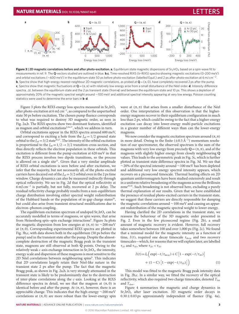
Figure 3 | 2D magnetic correlations before and after photo-excitation. a, Equilibrium state magnetic dispersions of Sr2IrO4 based on a spin-wave fit tomeasurements in ref. 9. The Q-vectors studied are outlined in blue. b,c, Time-resolved RIXS (tr-RIXS) spectra showing magnetic excitations (0–200 meV)and orbital excitations (∼600 meV) in the equilibrium state 50 ps before photo-excitation (labelled Equil.) and 2 ps after photo-excitation at 6 mJ cm−2.b, Spectra show that high-energy nearest-neighbour 2D magnetic correlations, as probed at Q=(π,0), have completely recovered 2 ps after the pump.c, Spectra show that magnetic fluctuations at Q=(π,π) with relatively low energy arise from a small disturbance of the Néel order. d, Intensity di�erencespectra,∆I, between the equilibrium state and the 2 ps transient state (from c) and between the equilibrium state and 10 ps. This shows a depletion ofapproximately 20% of the magnetic spectral weight around∼100 meV and additional spectral intensity appearing at very low energy. Poisson countingstatistics were used to determine the error bars in b–d.
Figure 3 plots the RIXS energy loss spectra measured in Sr2IrO4after photo-excitation at 6mJ cm−2, as compared to the unperturbedstate 50 ps before excitation. The chosen pump fluence correspondsto what was required to destroy 3D magnetic order, as seen inFig. 2a,b. The RIXS spectra show two dominant features, identifiedas magnon and orbital excitations9,26,27, which we address in turn.
Orbital excitations appear in the RIXS spectra around 600meV,and correspond to exciting a hole from the Jeff= 1/2 ground stateorbital to the Jeff=3/2 state26,27. The intensity of the orbital excitationis proportional to the Jeff=1/2→3/2 transition cross-section, andthus directly reflects the electron population in these orbitals. Thisexcitation is different from the pump excitation at 620meV in thatthe RIXS process involves two dipole transitions, so the processis allowed on a single site23. Given that a very similar amplitudeof RIXS orbital excitations is seen before and after excitation, weinfer that the majority, but not necessarily all, of the photo-excitedcarriers have decayed out of the Jeff=3/2 orbital even in the 2 ps timewindow. Charge dynamics can also be measured indirectly throughoptical reflectivity. We see in Fig. 2d that the optical reflectivity at6mJ cm−2 is partially, but not fully, recovered at 2 ps delay. Theresidual reflectivity change probably results from a non-equilibriumcharge distribution involving either spectral weight redistributionof the Hubbard bands or the population of in-gap charge states28,but could also arise from transient structural modifications due toelectron–phonon coupling.
The equilibrium excitation spectrum of undoped Sr2IrO4 can beaccurately modelled in terms of magnons, or spin waves, that arisefrom Heisenberg spin–spin exchange interactions9. Figure 3a plotsthe magnetic dispersion from low energies at (π,π) to ∼200meVat (π, 0). Corresponding experimental RIXS spectra are plotted inFig. 3b,c, with data shown both in the equilibrium (50 ps before thepump) and in the transient state after the pump. Despite the almost-complete destruction of the magnetic Bragg peak in the transientstate, magnons are still observed at both Q points. Owing to therelatively weak c-axis exchange interaction in Sr2IrO4, the intensity,energy scale and dispersion of thesemagnons ismost sensitive to the2D Néel correlations between neighbouring spins9. This indicatesthat 2D correlations largely retain their Néel-like nature in thetransient state 2 ps after the pump. The fact that the magneticBragg peak, as shown in Fig. 2a,b, is very strongly attenuated in thetransient state is likely to be predominantly due to the destructionof inter-plane correlations along the c-axis. Looking at the RIXSdifference spectra in detail, we see that the magnon at (π, 0) isidentical before and after the pump. At (π,π), however, there is anappreciable change. This indicates that the high-energy ∼200meVcorrelations at (π, 0) are more robust than the lower-energy spin
wave at (π,π) that arises from a smaller disturbance of the Néelorder. One interpretation of this observation is that the higher-energymagnons recover to their equilibrium configuration inmuchless than 2 ps, which could be owing to the fact that a higher-energyexcitation can decay into lower-energy multi-particle excitationsin a greater number of different ways than can the lower-energymagnons.
Wenow consider themagnetic excitation spectrumaround (π,π)in more detail. Owing to the finite (±0.5 Å−1) momentum resolu-tion of our spectrometer, the observed spectrum is the sum of themagnons with very low energy from preciselyQ=(π,π), and of themagnons with slightly higher energy from closely neighbouring Qvalues. This leads to the asymmetric peak in Fig. 3c, which is furtherplotted as transient state difference spectra in Fig. 3d. We see that∼20% of the spectral intensity around∼100meV has been depletedand additional very low energy spectral intensity appears, whichrecovers on a picosecond timescale. Thermal heating effects on 2Dquantum antiferromagnets have been studied extensively and resultin a uniform relative broadening of themagnons across the Brillouinzone29,30. Such broadening is not observed here, excluding a purelythermal explanation of our results. Given that we have establishedthe presence of residual photo-excited carriers in the transient state,we suggest that these carriers are directly responsible for dampingthe magnetic correlations around∼100meV and causing an appar-ent redistribution of the magnetic spectral weight to lower energy.
Having clarified the 2D correlations in the transient state, wereassess the behaviour of the 3D magnetic order presented inFig. 2. Even in the few picosecond regime (Fig. 2b), a smallamount of magnetic recovery is evident. However, full recoverytakes somewhere between 100 and over 1,000 ps (Fig. 2c). We foundthat a minimal model for the magnetic intensity as a function oftime, I(t), required one decay timescale τdecay and two recoverytimescales—which, for reasons that wewill explain later, are labelledτ2D and τ3D, where τ2D<τ3D
I(t)= I0(exp(−t/τdecay)+C
[1−exp(−t/τ2D)
]+ (1−C)
[1−exp(−t/τ3D)
])(1)
This model was fitted to the magnetic Bragg peak intensity datain Fig. 2b,c. In a similar way, we fitted the recovery of the opticalreflectivity, which also required two charge timescales, denoted Tfastand Tslow.
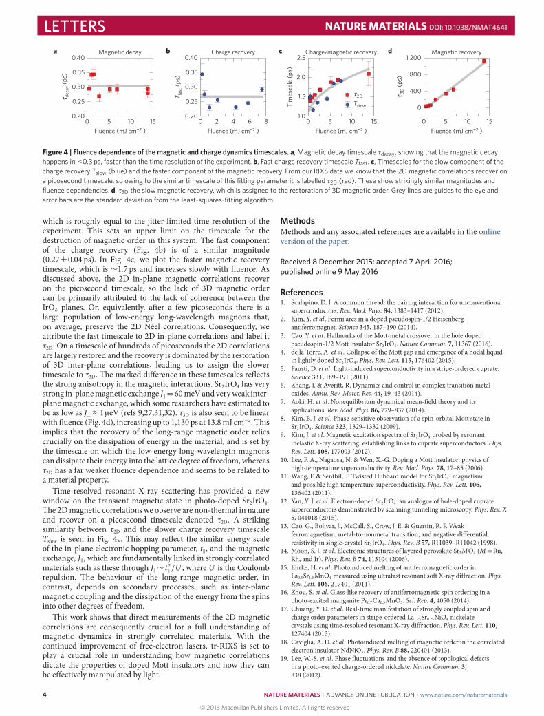
Figure 4 summarizes the magnetic and charge dynamics inSr2IrO4 after laser excitation. 3D magnetic order decays in0.30±0.03 ps approximately independent of fluence (Fig. 4a),
NATUREMATERIALS | ADVANCE ONLINE PUBLICATION | www.nature.com/naturematerials
© 2016 Macmillan Publishers Limited. All rights reserved
3
LETTERS NATUREMATERIALS DOI: 10.1038/NMAT4641
0.40
0.35
0.30
0.25
0.20
0.40
0.35
0.30
0.25
0.20
2.5
2.0
1.5
1.0
1,200
800
400
0
0 5 10 15 0 5 10 15 0 5 10 1586420
Magnetic decay
deca
y (ps
)τ 2Dτ 3D
(ps)
τT fas
t (ps
)
Tslow
Fluence (mJ cm−2 ) Fluence (mJ cm−2 ) Fluence (mJ cm−2 )
Charge recovery
Tim
esca
le (p
s)
Charge/magnetic recovery Magnetic recovery
Fluence (mJ cm−2 )
a b c d
Figure 4 | Fluence dependence of the magnetic and charge dynamics timescales. a, Magnetic decay timescale τdecay, showing that the magnetic decayhappens in≤0.3 ps, faster than the time resolution of the experiment. b, Fast charge recovery timescale Tfast. c, Timescales for the slow component of thecharge recovery Tslow (blue) and the faster component of the magnetic recovery. From our RIXS data we know that the 2D magnetic correlations recover ona picosecond timescale, so owing to the similar timescale of this fitting parameter it is labelled τ2D (red). These show strikingly similar magnitudes andfluence dependencies. d, τ3D the slow magnetic recovery, which is assigned to the restoration of 3D magnetic order. Grey lines are guides to the eye anderror bars are the standard deviation from the least-squares-fitting algorithm.
which is roughly equal to the jitter-limited time resolution of theexperiment. This sets an upper limit on the timescale for thedestruction of magnetic order in this system. The fast componentof the charge recovery (Fig. 4b) is of a similar magnitude(0.27±0.04 ps). In Fig. 4c, we plot the faster magnetic recoverytimescale, which is ∼1.7 ps and increases slowly with fluence. Asdiscussed above, the 2D in-plane magnetic correlations recoveron the picosecond timescale, so the lack of 3D magnetic ordercan be primarily attributed to the lack of coherence between theIrO2 planes. Or, equivalently, after a few picoseconds there is alarge population of low-energy long-wavelength magnons that,on average, preserve the 2D Néel correlations. Consequently, weattribute the fast timescale to 2D in-plane correlations and label itτ2D. On a timescale of hundreds of picoseconds the 2D correlationsare largely restored and the recovery is dominated by the restorationof 3D inter-plane correlations, leading us to assign the slowertimescale to τ3D. The marked difference in these timescales reflectsthe strong anisotropy in the magnetic interactions. Sr2IrO4 has verystrong in-planemagnetic exchange J‖=60meVand veryweak inter-planemagnetic exchange, which some researchers have estimated tobe as low as J⊥≈1µeV (refs 9,27,31,32). τ3D is also seen to be linearwith fluence (Fig. 4d), increasing up to 1,130 ps at 13.8mJ cm−2. Thisimplies that the recovery of the long-range magnetic order reliescrucially on the dissipation of energy in the material, and is set bythe timescale on which the low-energy long-wavelength magnonscan dissipate their energy into the lattice degree of freedom, whereasτ2D has a far weaker fluence dependence and seems to be related toa material property.
Time-resolved resonant X-ray scattering has provided a newwindow on the transient magnetic state in photo-doped Sr2IrO4.The 2Dmagnetic correlations we observe are non-thermal in natureand recover on a picosecond timescale denoted τ2D. A strikingsimilarity between τ2D and the slower charge recovery timescaleTslow is seen in Fig. 4c. This may reflect the similar energy scaleof the in-plane electronic hopping parameter, t‖, and the magneticexchange, J‖, which are fundamentally linked in strongly correlatedmaterials such as these through J‖∼ t 2‖/U , where U is the Coulombrepulsion. The behaviour of the long-range magnetic order, incontrast, depends on secondary processes, such as inter-planemagnetic coupling and the dissipation of the energy from the spinsinto other degrees of freedom.
This work shows that direct measurements of the 2D magneticcorrelations are consequently crucial for a full understanding ofmagnetic dynamics in strongly correlated materials. With thecontinued improvement of free-electron lasers, tr-RIXS is set toplay a crucial role in understanding how magnetic correlationsdictate the properties of doped Mott insulators and how they canbe effectively manipulated by light.
MethodsMethods and any associated references are available in the onlineversion of the paper.
Received 8 December 2015; accepted 7 April 2016;published online 9 May 2016
References1. Scalapino, D. J. A common thread: the pairing interaction for unconventional
superconductors. Rev. Mod. Phys. 84, 1383–1417 (2012).2. Kim, Y. et al . Fermi arcs in a doped pseudospin-1/2 Heisenberg
antiferromagnet. Science 345, 187–190 (2014).3. Cao, Y. et al . Hallmarks of the Mott-metal crossover in the hole doped
pseudospin-1/2 Mott insulator Sr2IrO4. Nature Commun. 7, 11367 (2016).4. de la Torre, A. et al . Collapse of the Mott gap and emergence of a nodal liquid
in lightly doped Sr2IrO4. Phys. Rev. Lett. 115, 176402 (2015).5. Fausti, D. et al . Light-induced superconductivity in a stripe-ordered cuprate.
Science 331, 189–191 (2011).6. Zhang, J. & Averitt, R. Dynamics and control in complex transition metal
oxides. Annu. Rev. Mater. Res. 44, 19–43 (2014).7. Aoki, H. et al . Nonequilibrium dynamical mean-field theory and its
applications. Rev. Mod. Phys. 86, 779–837 (2014).8. Kim, B. J. et al . Phase-sensitive observation of a spin-orbital Mott state in
Sr2IrO4. Science 323, 1329–1332 (2009).9. Kim, J. et al . Magnetic excitation spectra of Sr2IrO4 probed by resonant
inelastic X-ray scattering: establishing links to cuprate superconductors. Phys.Rev. Lett. 108, 177003 (2012).
10. Lee, P. A., Nagaosa, N. &Wen, X.-G. Doping a Mott insulator: physics ofhigh-temperature superconductivity. Rev. Mod. Phys. 78, 17–85 (2006).
11. Wang, F. & Senthil, T. Twisted Hubbard model for Sr2IrO4: magnetismand possible high temperature superconductivity. Phys. Rev. Lett. 106,136402 (2011).
12. Yan, Y. J. et al . Electron-doped Sr2IrO4: an analogue of hole-doped cupratesuperconductors demonstrated by scanning tunneling microscopy. Phys. Rev. X5, 041018 (2015).
13. Cao, G., Bolivar, J., McCall, S., Crow, J. E. & Guertin, R. P. Weakferromagnetism, metal-to-nonmetal transition, and negative differentialresistivity in single-crystal Sr2IrO4. Phys. Rev. B 57, R11039–R11042 (1998).
14. Moon, S. J. et al . Electronic structures of layered perovskite Sr2MO4 (M=Ru,Rh, and Ir). Phys. Rev. B 74, 113104 (2006).
15. Ehrke, H. et al . Photoinduced melting of antiferromagnetic order inLa0.5Sr1.5MnO4 measured using ultrafast resonant soft X-ray diffraction. Phys.Rev. Lett. 106, 217401 (2011).
16. Zhou, S. et al . Glass-like recovery of antiferromagnetic spin ordering in aphoto-excited manganite Pr0.7Ca0.3MnO3. Sci. Rep. 4, 4050 (2014).
17. Chuang, Y. D. et al . Real-time manifestation of strongly coupled spin andcharge order parameters in stripe-ordered La1.75Sr0.25NiO4 nickelatecrystals using time-resolved resonant X-ray diffraction. Phys. Rev. Lett. 110,127404 (2013).
18. Caviglia, A. D. et al . Photoinduced melting of magnetic order in the correlatedelectron insulator NdNiO3. Phys. Rev. B 88, 220401 (2013).
19. Lee, W.-S. et al . Phase fluctuations and the absence of topological defectsin a photo-excited charge-ordered nickelate. Nature Commun. 3,838 (2012).
4
© 2016 Macmillan Publishers Limited. All rights reserved
NATUREMATERIALS | ADVANCE ONLINE PUBLICATION | www.nature.com/naturematerials

NATUREMATERIALS DOI: 10.1038/NMAT4641 LETTERS20. Boeglin, C. et al . Distinguishing the ultrafast dynamics of spin and orbital
moments in solids. Nature 465, 458–461 (2010).21. Kampfrath, T. et al . Coherent terahertz control of antiferromagnetic spin
waves. Nature Photon. 5, 31–34 (2011).22. Malinowski, G. et al . Control of speed and efficiency of ultrafast
demagnetization by direct transfer of spin angular momentum. Nature Phys. 4,855–858 (2008).
23. Ament, L. J. P., vanVeenendaal, M., Devereaux, T. P., Hill, J. P.& van den Brink, J. Resonant inelastic X-ray scattering studies of elementaryexcitations. Rev. Mod. Phys. 83, 705–767 (2011).
24. Dean, M. P. M. Insights into the high temperature superconducting cupratesfrom resonant inelastic X-ray scattering. J. Magn. Magn. Mater. 376,3–13 (2015).
25. Batignani, G. et al . Probing ultrafast photo-induced dynamics of the exchangeenergy in a Heisenberg antiferromagnet. Nature Photon. 9, 506–510 (2015).
26. Ishii, K. et al . Momentum-resolved electronic excitations in the Mott insulatorSr2IrO4 studied by resonant inelastic X-ray scattering. Phys. Rev. B 83,115121 (2011).
27. Kim, J. et al . Excitonic quasiparticles in a spin–orbit Mott insulator. NatureCommun. 5, 4453 (2014).
28. Okamoto, H. et al . Ultrafast charge dynamics in photoexcited Nd2CuO4 andLa2CuO4 cuprate compounds investigated by femtosecond absorptionspectroscopy. Phys. Rev. B 82, 060513 (2010).
29. Manousakis, E. The spin-1/2 Heisenberg antiferromagnet on a squarelattice and its application to the cuprous oxides. Rev. Mod. Phys. 63,1–62 (1991).
30. Rønnow, H. M. et al . Spin dynamics of the 2d spin 12 quantum antiferromagnet
copper deuteroformate tetradeuterate (CFTD). Phys. Rev. Lett. 87,037202 (2001).
31. Fujiyama, S. et al . Two-dimensional Heisenberg behavior of Jeff=1/2 isospinsin the paramagnetic state of the spin-orbital Mott insulator Sr2IrO4. Phys. Rev.Lett. 108, 247212 (2012).
32. Vale, J. G. et al . Importance of XY anisotropy in Sr2IrO4 revealed by magneticcritical scattering experiments. Phys. Rev. B 92, 020406 (2015).
AcknowledgementsThe X-ray scattering work by M.P.M.D., Y.C., V.T. and X.M.C. was supported by the USDepartment of Energy Basic Energy Sciences Division of Materials Science andEngineering. X.L. acknowledges financial support fromMOST (No. 2015CB921302) andCAS (Grant No: XDB07020200) of China. P.J. acknowledges support by LaboratoryDirected Research and Development (LDRD) Program 12-007 (Complex Modeling). J.K.,D.C. and A.H.S. were supported by the US Department of Energy under ContractNo. DE-AC02-06CH11357. S.W. acknowledges financial support from Spanish MINECO(Severo Ochoa grant SEV-2015-0522), Ramon y Cajal programme RYC-2013-14838,Marie Curie Career Integration Grant PCIG12-GA-2013-618487 and Fundació PrivadaCellex. J.L. is sponsored by the Science Alliance Joint Directed Research andDevelopment Program at the University of Tennessee. Work in London was supported bythe EPSRC. The magnetic Bragg peak measurements were performed at the BL3 ofSACLA with the approval of the Japan Synchrotron Radiation Research Institute (JASRI)(Proposal No. 2014B8018). This research made use of the Linac Coherent Light Source(LCLS), SLAC National Accelerator Laboratory, which is a DOE Office of Science UserFacility, under Contract No. DE-AC02-76SF00515.
Author contributionsJ.P.H., X.L., M.P.M.D. and M.F. initiated and planned the project. M.P.M.D., Y.C., X.L.,S.W., D.Z., R.M., V.T., X.M.C., J.G.V., D.C., J.K., A.H.S., P.J., R.A.-M., J.M.G., A.R., J.R.,M.S., S.S., M.K., H.L., L.P., S.O., T.K., M.Y., Y.T., T.T., L.H., C.-L.C., D.F.M., M.F. and J.P.H.prepared for and performed the experiments. M.P.M.D., Y.C., X.L., S.W., M.F., D.F.M. andJ.P.H. analysed and interpreted the data. J.L., C.R.S. and B.J.K. prepared the samples.M.P.M.D. and Y.C. wrote the paper with contributions from X.L., S.W., D.F.M., M.F.and J.P.H.
Additional informationSupplementary information is available in the online version of the paper. Reprints andpermissions information is available online at www.nature.com/reprints.Correspondence and requests for materials should be addressed to M.P.M.D., Y.C. or X.L.
Competing financial interestsThe authors declare no competing financial interests.
NATUREMATERIALS | ADVANCE ONLINE PUBLICATION | www.nature.com/naturematerials
© 2016 Macmillan Publishers Limited. All rights reserved
5

LETTERS NATUREMATERIALS DOI: 10.1038/NMAT4641
MethodsSamples. The magnetic Bragg peak measurements were performed on 200 nmepitaxial films of Sr2IrO4, to match the volume of Sr2IrO4 to the penetration depthof the pump, as the X-ray penetration depth is longer than the pump. Thedisappearance of the magnetic Bragg peak in Fig. 2a,b confirms that the wholeprobed volume is excited. The film was deposited on SrTiO3 using pulsed laserdeposition, as described in the Supplementary Information and ref. 33.Supplementary Figs 1 and 2 demonstrate good sample crystallinity and the lack ofany detectable impurity phases. For RIXS,∼1◦ grazing incidence X-rays were usedto limit X-ray penetration depths to 80 nm on a bulk Sr2IrO4 crystal. Both sampleshave a c-axis surface normal. Reciprocal lattice notations are defined using the fullunit cell with lattice constants a=b=5.51Å and c=25.7 Å. The high-symmetrypoints in the in-plane Brillouin zone are defined in the reduced structural zone(which ignores the rotation of the IrO6 octahedra) as in ref. 9. The zone centre,denoted (π,π) and the zone boundary denoted (π, 0) correspond to (1,0,L) and(0.5,0.5,L), respectively, in the reciprocal lattice notation. In both experiments, thesample was cooled to about 110K with a nitrogen cryostream, well below the Néeltemperature of 240K (ref. 13).
Optical pump. For both tr-REXS and tr-RIXS experiments, 100 fs pump pulseswere generated at 620meV (2 µm) using an optical parametric amplifier. Thepulses were polarized vertically in the ab-plane of the sample and were incident at13◦ with respect to the sample surface. The choice of pump energy follows previousoptical conductivity measurements14 and resonates between the upper and lowerHubbard bands.
The time-resolved resonant elastic X-ray scattering (tr-REXS) set-up. Thetr-REXS experiment was performed at beamline 2 of the SPring-8 AngstromCompact free-electron LAser (SACLA) with a 30Hz pulse repetition rate. Weadopted a horizontal scattering geometry, as seen in Fig. 1a, and tuned the X-rayenergy to the peak in the Ir L3-edge resonance around 11.215 keV. A multi-portcharged coupled device (MPCCD) area detector was placed at 2θ=88.7◦ to observethe magnetic Bragg peak (−3,−2,28). This geometry is chosen to optimize theX-ray resonant magnetic scattering cross-section. We access the magnetic peak byrotating the sample around the vertical axis by φ=12.8◦, with the infrared andX-ray photons in an approximately co-linear geometry. The detector was read outshot by shot and the signal was thresholded to suppress the background comingfrom X-ray fluorescence and electrical noise. The peak intensity was determined bybinning the 2D MPCCD data into a 1D spectrum and fitting a Lorentzianlineshape with a uniform offset background. Each datapoint is the result of
summing 1,000–4,000 shots. Previous characterization of the beamline found thatthe time resolution of this experiment was jitter-limited to approximately 300 fs.
The minimal model for the fitting is outlined in the main text (equation (1)).This formula was convolved with a 100 fs Gaussian to account for the pump pulsewidth. The other major contribution to the effective time resolution was the X-raypulse jitter of approximately 300 fs, because this is only an approximate value thiswas not included in the fit, rather this is taken as an upper limit on the decay time.Apart from this quantity, all parameters were varied to fit the data in thelong-time-delay scans at 1.0, 2.7 and 13.8mJ cm−2 fluence in Fig. 2c, and these fitswere used to constrain τ3D in fits of the short-time-delay data in Fig. 2b byinterpolating the variation of τ3D and C as a function of fluence. In this way,equation (1) provides an accurate parametrization of the recovery dynamics at allfluences studied.
The time-resolved resonant inelastic X-ray scattering (tr-RIXS) set-up. Thetr-RIXS experiment was performed at the X-ray Pump Probe instrument34 at theLinac Coherent Light Source (LCLS) with a 120Hz repetition rate. We adopt ahorizontal scattering plane, similar to the set-up in the tr-REXS experiment. The(π, 0) and (π,π) data were measured at (−3.5,−3.5,24.1) and (−4,−3,23.9).Non-integer values of L were chosen to keep the X-ray incidence angle around 1◦,as the RIXS spectrum is known to be essentially independent of L9. A Si (333)monochromator produced a 50meV incident energy bandpass. The RIXSspectrometer is conceptually similar to that used at Sector 27 at the AdvancedPhoton Source. Scattered photons from the sample are reflected from a segmentedspherical Si(8,4,4) analyser in a near-backscattering configuration and detected bya Princeton CCD. The sample, the analyser crystal, and the photodetector areplaced on a Rowland circle with a radius of 1m in the vertical plane. The totalenergy resolution of the tr-RIXS set-up was∼70meV and theQ resolution wasdefined by the∼6◦ angular acceptance of the analyser. RIXS spectra were collectedin a stationary mode without moving the spectrometer, and the pixel-to-energyconversion was performed using well-established methods. The CCD was read outevery 1,800 shots. Jitter and timing drift were the main contributions to the timeresolution, which was on the order of 500 fs.
References33. Rayan Serrao, C. et al . Epitaxy-distorted spin-orbit Mott insulator in Sr2IrO4
thin films. Phys. Rev. B 87, 085121 (2013).34. Chollet, M. et al . The X-ray pump-probe instrument at the LINAC coherent
light source. J. Synchrotron Radiat. 22, 503–507 (2015).
© 2016 Macmillan Publishers Limited. All rights reserved
NATUREMATERIALS | www.nature.com/naturematerials