ultrasonography in diagnosis and analysis of chronic pain
TRANSCRIPT
RESEARCH ARTICLE Open Access
Ultrasonography in diagnosis and analysisof chronic pain following anterior openinguinal herniorrhaphyZY Qiu1† , Y Chen1*, JX Tang2 and L Chen1†
Abstract
Background: Chronic pain as a complication following inguinal herniorrhaphy has attracted increasing attention inrecent years. There is evidence that the chronic pain seriously affects patients’ quality of life. However, there are fewimaging studies and diagnostic techniques of the chronic pain. The aim of this study is to explore the etiology andto analysis ultrasonographic imaging description of chronic pain following anterior open inguinal herniorrhaphy.
Methods: One hundred fifty two patients with the chronic pain following anterior open inguinal herniorrhaphywere performed by ultrasonography to identify the main causes of postoperative chronic pain. Positive ultrasonicdiagnoses were confirmed to be correct by the pain relieved when the patients underwentre-operation and other clinical operations. Positive diagnoses which appeared simultaneously were grouped forpairwise comparisons.
Results: Two hundred sixteen positive ultrasonic diagnoses, 12 categories of postoperative chronic pain werefound. They were encapsulated effusion, scrotal wall edema, testitis, hydrocele testis, restricted motion of spermaticcord at the reconstructed deep inguinal ring, varicocele, scar sutured into pubic tubercle, shrinking mesh,accumulational mesh or mesh plug, recurrent hernia, cyst of spermatic cord and epididymal cyst. In the pairwisecomparison groups, encapsulated effusion with scrotal wall edema, varicocele with restricted motion of spermaticcord at the reconstructed deep inguinal ring, and shrinking mesh with recurrent hernia had significant differencesin each intragroup comparisons(P < 0.05).
Conclusions: Ultrasonography provieds important value in the diagnosis of chronic pain following anterior openinguinal herniorrhaphy. Some positive diagnoses occur simultaneously, which is necessary for doctors to considercomprehensively.
Keywords: Ultrasonography, Anterior open inguinal herniorrhaphy, Chronic pain
BackgroundIt was pointed out in Guidelines for diagnosis and treat-ment of adult inguinal hernia (2014 edition) [ 1] that al-most all cases of inguinal hernia are treated surgically,and anterior open inguinal herniorrhaphy is consideredto be the most common surgical procedure. Also, thechronic pain is a late complication of the surgery. In the1980s, the chronic pain was reported [2–4] to be a rareand occasional postoperative complication, and the
assessment of the surgery was limited to the postopera-tive acute phase. Nevertheless, studies in recent yearshave found that the incidence of chronic pain followinginguinal herniorrhaphy is approximately 54%, which isfar higher than that reported previously. Furthermore,up to 50% of patients experience the chronic pain formore than 1 year, which seriously affects the patients’quality of life [5–7]. The chronic pain has gradually be-come one of the long-term assessment indicators of theherniorrhaphy. Non-invasive, nonradiative and easilyperformed, ultrasound technique has been proposed as avaluable imaging method in examining patients beforeand after the inguinal herniorrhaphy [8].
* Correspondence: [email protected]†ZY Qiu and L Chen contributed equally to this work.1Ultrasound Department, Huadong Hospital Affiliated to Fudan University,221 West Yanan Road, Shanghai 200041, ChinaFull list of author information is available at the end of the article
© The Author(s). 2018 Open Access This article is distributed under the terms of the Creative Commons Attribution 4.0International License (http://creativecommons.org/licenses/by/4.0/), which permits unrestricted use, distribution, andreproduction in any medium, provided you give appropriate credit to the original author(s) and the source, provide a link tothe Creative Commons license, and indicate if changes were made. The Creative Commons Public Domain Dedication waiver(http://creativecommons.org/publicdomain/zero/1.0/) applies to the data made available in this article, unless otherwise stated.
Qiu et al. BMC Surgery (2018) 18:28 https://doi.org/10.1186/s12893-018-0361-z
The aim of our study was to explore the etiology andanalyze positive findings of chronic pain after anterioropen inguinal herniorrhaphy, thereby to assist in makingtreatment protocols in clinics.
MethodsSubjectsWe conducted a prospectively, observational study in asingle hernia center of Huadong Hospital, between August2009 and May 2014. 539 patients who felt uncomfortableafter their first herniorrhaphy were performed by ultra-sonography in our ultrasound department. 152 patientswho met the inclusion criteria agreed to participate in thisstudy and signed the informed consent. This study wasapproved by Fudan University Ethics Committee. Nostatistical power calculation was conducted prior to thestudy. The sample size was based on the previous litera-ture experience about the chronic pain review.The inclusion criteria: the patients had a history of
anterior open inguinal herniorrhaphy, during whichartificial material was patched in preperitoneal spaceor the back wall of the inguinal canal was recon-structed by suturing. According to the definition ofchronic pain by International Association of the Studyof Pain (IASP) [9], cases with “pain lasting for 3months or more” were included. Persistent chronicpain with or without local swell in the operativeregion or the ipsilateral scrotum was the clinicalsymptom of the disease. Sometimes the symptomcould be relieved by holding up the scrotum.The patients had laparoscope totally extraperito-
neal repair(TEP) and transabdominal preperitonealrepair(TAPP) were excluded. And the patients youn-ger than 16 years were excluded.The longest course of postoperative chronic pain in this
study was ten years, and the shortest course was 3 months.Among 31 patients who had a history of bilateral hernior-rhaphy, 12 patients presented bilateral chronic pain.
Ultimately, a total of 164 chronic pain sites met the inclu-sion criteria, included 157 sites with artificial mesh, 2 siteswith mesh plug, and 5 sites with suture repair.
Instruments and methodsThe Siemens ACUSON S2000™ color doppler ultrasoundinstrument, equipped with ultrasonic volumeauto-scan(UVAS) and a high-frequency linear transducerranging from 9 to 12 MHZ, were performed to scan theherniorrhaphic incisions and chronic pain sites in patients.The following parameters of UVAS were set: a volumeimage of 15.4 cm (length) × 16.8 cm (width) × 6.0 cm(depth) captured with a minimum slice of 0.5 mm and250 to 400 images were collected during a single scan.The axial sequence images were automatically transferredto the ultrasonic workstation for data analysis and pro-cessing. The patients were in supine position or standingposition as needed for ultrasound scanning, with the
Fig. 1 The seroma showed in the ultrasonography. The multilocularseroma was visible in front of the mesh
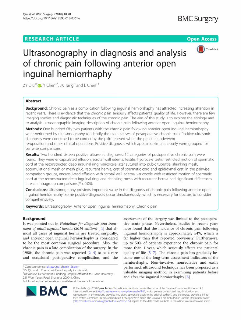
Fig. 2 The hematoma showed in the ultrasonography. Arrowsreferred to the old hematoma
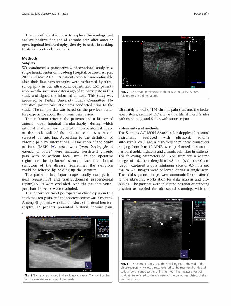
Fig. 3 The recurrent hernia and the shrinking mesh showed in theultrasonography. Hollow arrows referred to the recurrent hernia andsolid arrows referred to the shrinking mesh. The measurement ofstraight line referred to the diameter of the perito neal defect of therecurrent hernia
Qiu et al. BMC Surgery (2018) 18:28 Page 2 of 7

scrotums simultaneously scanned in male patients. Thelocation, shape, and spatial structure of the mesh, periton-eum, spermatic cords, etc., were observed, with the goal ofachieving ultrasonographic diagnosis of the etiology of thepostoperative chronic pain.All ultrasonographic results were confirmed by the
re-operation, the puncture or other clinical operations.Cases with two or more positive diagnoses were pair-
wise grouped for multiple comparisons.
Statistical analysisStatistical analyses were performed by STATA10 soft-ware. Values were presented as mean ± standard devi-ation for normally distributed variables. Positiveultrasonic diagnoses in the pairwise groups comparedusing a chi-square test. Statistical significance thresholdwas considered α = 0.05.
ResultsThe mean age in the study was 66.10 years(± 13.82SD),ranging from 22 to 93 years old. No statistical differencewas observed between genders in 93 men and 59 women(χ2 = 0.35, P > 0.05).
Ultrasonic diagnosis of chronic pain sites followinganterior open inguinal herniorrhaphyAmong 164 chronic pain sites, 121 sites showed positivefindings on ultrasonography and no ultrasonic abnor-mality was found in 43 sites.Among 121 sites with positive ultrasonic findings, 49
sites had a single positive diagnosis, 49 sites had 2 posi-tive diagnoses and 23 sites had 3 positive diagnoses,which resulted in a total of 216 positive diagnoses: 24cases of encapsulated effusion (11.1%), of which 14 caseswere confirmed by ultrasound guided aspiration to beseroma (Fig. 1) and 10 cases were confirmed to be
hematoma (Fig. 2). 59 cases of recurrent hernia(27.3%) (Fig. 3), 31 cases of shrinking mesh (14.4%)(Fig. 4), 5 cases of accumulational patch or mesh plug(2.3%) (Fig. 5), 2 cases of cyst of spermatic cord(1.0%)(Fig. 6) and 1 case of epididymal cyst (0.5%)were confirmed surgically. In addition, there appearedto be restricted motion of the spermatic cord at thereconstructed deep inguinal ring in 20 cases(9.2%)(Fig. 7),including 10 cases of the spermatic cord adhered in thedeep inguinal ring, 7 cases of patch compression of thespermatic cord, and 3 cases of reconstructed deep inguinalring stenosis, all of which were confirmed by adhesiolysisand inguinal neurotomy. Furthermore, ultrasound con-firmed 19 cases of the scrotal wall edema (8.8%), 16 casesof testitis (swelling with increased blood flow) (7.4%), 16cases of varicocele(7.4%)(Fig. 8), and 2 cases of mesh su-tured into pubic tubercle(1.0%), in which the mesh andscar hyperplasia were removed surgically, as well as 5
Fig. 4 The shrinking mesh showed in the ultrasonography. The shrinking mesh was showed on coronal consecutive image and 3D display.Arrows referred to the shrinking mesh
Fig. 5 Accumulational meshes showed in the ultrasonography
Qiu et al. BMC Surgery (2018) 18:28 Page 3 of 7
cases of hydrocele testis (2.3%)(3 cases were confirmed byurological surgery). Ultrasonography revealed 3 cases oflocal weakness at the reconstructed abdominal wall, whichneed further confirmation by clinical follow-up.The pain was relieved or even disappeared when pa-
tients had undergone re-operation, puncture, and otherclinical operations (such as anti-inflammation therapyand traditional Chinese medicine fomentation).
Comparison of pairwise combination of positivediagnosesUltrasonography revealed that 23 pain sites were found3 positive diagnoses in each site and 49 pain sites werefound 2 positive diagnoses in each site, which resulted in167 positive diagnoses (Table 1).The intragroup comparisons, grouped according to
pairwise combinations of 167 positive diagnoses, showed
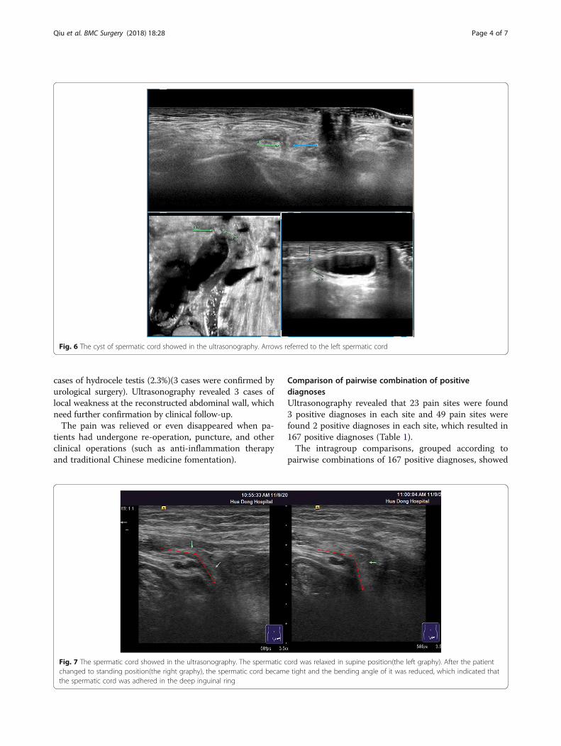
Fig. 6 The cyst of spermatic cord showed in the ultrasonography. Arrows referred to the left spermatic cord
Fig. 7 The spermatic cord showed in the ultrasonography. The spermatic cord was relaxed in supine position(the left graphy). After the patientchanged to standing position(the right graphy), the spermatic cord became tight and the bending angle of it was reduced, which indicated thatthe spermatic cord was adhered in the deep inguinal ring
Qiu et al. BMC Surgery (2018) 18:28 Page 4 of 7

that encapsulated effusion synchronized with scrotal walledema, varicocele synchronized with restricted motionof the spermatic cord at the reconstructed deep inguinalring, and shrinking mesh synchronized with recurrenthernia had significant differences within the respectivegroups (P < 0.05), which occurred more frequently thanother intragroup pairwise combinations.Meanwhile, other pairwise combinations of positive
diagnoses didn’t have statistically differences in theintragroup comparisons (P > 0.05).
There were 43 cases that postoperative chronic painoccurred, but no insignificant abnormalities were foundon ultrasonography. These cases required continuedfollow-up.
DiscussionUltrasonography is a fast, effective and radiation-freeexamination and suitable for various kinds of positions.UVAS, as a form of advanced ultrasound technology,supplies the coronal consecutive dynamic images on thebasis of the two-dimensional images, plays a comple-mentary role to the traditional ultrasound and providesthe most intuitive visual plane for clinicians to work outfurther surgical plans [10]. Ultrasonography can clearlyreveal the corrugation of the mesh, the motion of thespermatic cord and the continuity of the peritoneum,which is conducive to diagnosing the cause of thechronic pain after inguinal herniorrhaphy [11]and an ef-fective method of avoiding re-operation in the majorityof cases.There are two main steps of anterior open inguinal
herniorrhaphy. One is separation of the hernial sac andthe spermatic cord, and the other is to reconstruct theback wall of inguinal canal by suturing or patching anartificial mesh [12]. Ultrasonography shows the morph-ology and size of the mesh as well as the continuity ofthe peritoneal peritoneum, which can be used to detectthe recurrent hernia. According to the literature, thechronic pain following inguinal herniorrhaphy is mainly
Fig. 8 The varicocele showed in the ultrasonography
Table 1 Pairwise comparisons of positive diagnoses
Encapsulated effusion ① Varicocele ② Shrinking mesh ③ Recurrent hernia ④ Sum χ2
valueP value
Combined Un-combined
Combined Un-combined
Combined Un-combined
Combined Un-combined
Encapsulated effusion 23 0 0 23 0 23 5 18 23
Scrotal wall edema 19 0 0 19 0 19 1 18 19 ①41.37 <0.05
Testitis 8 2 0 10 0 10 1 9 10
Hydrocele testis 2 3 2 3 1 4 0 5 5
Mesh sutured intopubic tubercle
0 2 2 0 0 2 0 2 2
Cyst of spermaticcord / epididymalcyst
0 2 1 1 0 2 0 2 2
Accumulationalmesh
0 5 1 4 4 1 1 4 5
Varicocele 0 16 16 0 4 12 2 14 16
Restricted motionof spermatic cordat deep inguinalring
0 20 13 7 8 12 8 12 20 ②20.07 <0.05
Shrinking mesh 0 31 4 27 31 0 23 8 31 ④13.19 <0.05
Recurrent hernia 5 29 2 32 21 13 34 0 34 ③7.36 <0.05
Sum 57 110 41 126 69 98 75 92 167
Qiu et al. BMC Surgery (2018) 18:28 Page 5 of 7

from somatic, neurological, or visceral sources [13, 14].Cunningham et al. [15] found that the most commontype of postoperative chronic pain was somatic pain,which was mainly associated with pubic tubercle injurycaused by suturing the ligament and other tissues intothe pubic tubercle. Visceral pain mostly originates fromthe reproductive system. Ultrasonography can find thatthe motion of the spermatic cord decreased significantly,the flow of the spermatic vein is obstructed and thespermatic vein is expansive. Ultrasonography can alsoaccurately diagnose the varicocele when the blood flowspectrum of varicose vein was obviously reversed inValsalva test. Butler et al. [16] reported that the torsionand postoperative stenosis of the spermatic cord causedby scar hyperplasia, which might lead to ejaculatory dys-function or ejaculation pain. Wantz et al. [13, 17] foundthat the incidence of ischemic testitis following inguinalherniorrhaphy was 0.61%, which associated with thecompression caused by oversized mesh plug or anexcessively small patch hole to the spermatic cord. Theintraoperative damage or the ligation of spermatic arte-ries and veins resulted in the obstruction of venous fluxand the ischemia of the testis.Be careful to avoid the sensory nerves when the
mesh is being sewed. Neuropathic pain may be dueto the inguinal or genitofemoral nerves injury [18].Ultrasonography has limitation in scanning the nervebranches. This might be the reason for the 43 casesin this study in which postoperative chronic pain waspresent but no significant abnormalities were apparenton ultrasonography. Caliskan et al. [19] sought thecauses of pain by mean of relieving the pain by in-guinal neurotomy. Wijsmuller et al. [20] determinedthe possibly damaged nerves through intraoperativeidentification and protection of the ilioinguinal nerveand genitofemoral nerve genital branch. Surgicalnerve injury might cause the formation of neuromasor complete transection of nerve trunk, while postop-erative adhesion and local inflammation might causescars implanting into the nerve [21, 22].Therefore, following guidelines, standard operation
and normative process are the key to reduce the chronicpain and the other postoperative complications afteringuinal herniorrhaphy.
ConclusionsBoth UVAS and traditional ultrasonography have im-portant value in the diagnosis of the chronic pain follow-ing anterior open inguinal herniorrhaphy. Some causesof the chronic pain always occur simultaneously whichshould be comprehensively considered by clinical doc-tors. Therefore, it is necessary to conside ultrasoundtechnology as an objective basis for the assessment ofpostoperative chronic pain.
AbbreviationUVAS: Ultrasonic volume auto-scan
AcknowledgementsWe thank Yun Pang who helped us to collect and archived the ultrasonicimage data. I am also indebted to Professor Shaojie Li who provided thesurgical technical and theoretical guidance for me. My deepest gratitudegoes to Professor Jianxiong Tang and Yue Chen for their constantencouragements and illuminating instructions.
FundingThis work was supported by grants for a 2015 Clinical Capacity BuildingProject for Assistant Departments of the Shanghai Municipal ShenkangHospital Development Center (project no. SHDC22015013, Chen L).
Availability of data and materialsWe declared that all data and ultrasonic images described in the manuscript,including all relevant raw data, will be freely available to any scientistwishing to use them for non-commercial purposes, without breaching par-ticipant confidentiality.
Study designAll patients in the study who met the inclusion criteria were performed byultrasonography to identify the main causes of the postoperative chronic pain.Positive ultrasonic diagnoses were confirmed to be correct by the pain relievedor disappeared when the patients had undergone re-operation, puncture, andother clinical operations. Since some positive diagnoses occurred at the sametime, we combined these results pairwise with a list. The incidences of pairwisediagnoses were compared in different positive diagnose group. Based onmeaningful statistical results, we discussed the advantages and disadvantagesof ultrasonography in diagnosing the postoperative chronic pain.
Authors’ contributionsZQ carried out ultrasonic examinations and diagnosis, performed thestatistical analysis and drafted the manuscript. YC and JT participated in thedesign of the study. LC participated in coordination and helped to draft themanuscript. All authors read and approved the final manuscript.
Ethics approval and consent to participateAll patients had written the informed consent in which the purpose aboutthis study, the technical information about the ultrasound examination, theresearch process and the right of the participants were described clearly.They had been given the opportunity to ask questions about this study, andthey had been answered to their satisfaction. All 152 patients consented toparticipate in this study.The ethical implications of this study were adequately described in theapplication for approval of research protocol. This study was approved bythe Fudan University Research Ethics Committee.
Consent for publicationWe declared that written informed consents and any ultrasonic images wereobtained from the patients for publication of this study.
Competing interestsThe authors declare that they have no competing interests
Publisher’s NoteSpringer Nature remains neutral with regard to jurisdictional claims inpublished maps and institutional affiliations.
Author details1Ultrasound Department, Huadong Hospital Affiliated to Fudan University,221 West Yanan Road, Shanghai 200041, China. 2Department of GeneralSurgery, Huadong Hospital Affiliated to Fudan University, 221 West YananRoad, Shanghai 200041, China.
Qiu et al. BMC Surgery (2018) 18:28 Page 6 of 7

Received: 15 December 2017 Accepted: 9 May 2018
References1. Chinese Society for Hernia and Abdominal Wall Surgery CHCoS. Guidelines
for diagnosis and treatment of adult inguinal hernia (2014 edition). ChineseJournal of Surgery. 2014;52:481–3.
2. Mudge M, Hughes LE. Incisional hernia: a 10 year prospective study ofincidence and attitudes. Br J Surg. 1985;72(1):70–1.
3. Luijendijk RW, et al. A comparison of suture repair with mesh repair forincisional hernia. N Engl J Med. 2000;343(6):392–8.
4. Stoppa RE. The treatment of complicated groin and incisional hernias.World J Surg. 1989;13(5):545–54.
5. Poobalan AS, Bruce J, Smith WC, King PM, Krukowski ZH, Chambers WA.A review of chronic pain after inguinal Herniorrhaphy. Clin J Pain. 2003;19(1):48–54.
6. Poobalan AS, bruce J, King PM, et al. Chronic pain and quality of lifefollowing open inguinal hernia repair. Br J Surg. 2001;88:1122–6.
7. Callesen T, Bech K, Kehlet H. Prospective study of chronic pain after groinhernia repair. Br J Surg. 1999;86:1528–31.
8. Zhiying Q, Jianxiong T. Value of ultrasonography before and aftertension-free inguinal hernioplasty. Chinese Journal of General Surgery.2011;26(2):91–3.
9. International Association for the Study of Pain. Subcommittee on taxonomy.Classification of chronic pain: descriptions of chronic pain syndromes anddefinitions of pain terms. Pain. 1986;3(Suppl):1–226.
10. Zhiying Q, Yue C, Cai C, et al. Comparison of ultrasonic volume auto-scanand regular ultrasonography for the diagnostic classification of the hernia.Chinese. J Ultrasound Med. 2012;28(5):446–52.
11. Hua T, Lei Z, Hongyan Z, et al. The application of color dopplerultrasonography in diagnosis for the complication of tension-free inguinalhernia repair. Chin J Med Ultrasound (Electronic Edition). 2010;7(9):1483–9.
12. Joachim Conze, Anterior open repair of inguinal hernia in adults.Management of Abdominal Hernias. A.N. Kingsnorth and K.A. LeBlanc (eds.).chapter14.
13. ZibM GJ. Inguinal hernia repair : where to next. ANZ J Surg. 2002;72(8):573.14. Licheng Z, Junzhong S, Huijun S. Causes and strategies of chronic pain
following tension-free herniorrhaphy. Chinese. journal of general practice.2008;6(8):829–30.
15. Cunningham J, Temple WJ, Mitchell P, et al. Cooperative hernia study: painin the postrepair patient. Ann Surg. 1996;224:598–602.
16. Butler JD, Hershman MJ, Leach A. Painful ejaculation after inguinal herniarepair. J R Soc Med. 1998;91:432–3.
17. Wantz GE. The Canadian repair of inguinal hernia.//Nghus LM, Condon RE.Hernia. 3rd ed. Philadelphia: JB Lippincott Co,1989:236–252.
18. Wijsmuller AR, Lange JFM, Kleinrensink GJ, et al. Nerve-identifying inguinalhernia repair: a surgical anatomical study. World J Surg. 2007;31(2):414–20.
19. Caliskan K, et al. A method for the reduction of chronic pain after tension-free repair of inguinal hernia: iliohypogastric neurectomy and subcutaneoustransposition of the spermatic cord. Hernia. 2010;14(1):51–5.
20. Wijsmuller AR, van Veen RN, Bosch JL, et al. Nerve management duringopen hernia repair. Br J Surg. 2007;94:17–22.
21. Bower S, Moore BB, Weiss SM. Neuralgia after inguinal hernia repair. AmSurg. 1996;62:664–7.
22. Condon RE. Groin pain after hernia repair. Ann Surg. 2001;233(1):8.
Qiu et al. BMC Surgery (2018) 18:28 Page 7 of 7