ultrasound as a screening test for genitourinary anomalies...
TRANSCRIPT
Ultrasound as a Screening Test for GenitourinaryAnomalies in Children With UTI
WHAT’S KNOWN ON THIS SUBJECT: Current guidelinesrecommend renal ultrasound as a screening test after febrileurinary tract infection, with voiding cystourethrogram (VCUG) onlyif the ultrasound is abnormal. Few studies have evaluated theaccuracy of ultrasound as a screening test for VCUG-identifiedabnormalities.
WHAT THIS STUDY ADDS: This study shows that ultrasound isa poor screening test for genitourinary abnormalities identifiedon VCUG, such as vesicoureteral reflux. Neither positive nornegative ultrasounds reliably identify or rule out suchabnormalities. Ultrasound and VCUG provide different, butcomplementary, information.
abstractBACKGROUND: The 2011 American Academy of Pediatrics guidelinesstate that renal and bladder ultrasound (RBUS) should be performedafter initial febrile urinary tract infection (UTI) in a young child, withvoiding cystourethrogram (VCUG) performed only if RBUS shows ab-normalities. We sought to determine test characteristics and predic-tive values of RBUS for VCUG findings in this setting.
METHODS: We analyzed 3995 clinical encounters from January 1, 2006to December 31, 2010 during which VCUG and RBUS were performed forhistory of UTI. Patients who had previous postnatal genitourinary im-aging or history of prenatal hydronephrosis were excluded. Sensitivity,specificity, and predictive values of RBUS for VCUG abnormalities weredetermined.
RESULTS: We identified 2259 patients age,60 months who had UTI asthe indication for imaging. RBUS was reported as “normal” in 75%. OnVCUG, any vesicoureteral reflux (VUR) was identified in 41.7%, VURgrade.II in 20.9%, and VUR grade.III in 2.8%. Sensitivity of RBUS forany abnormal findings on VCUG ranged from 5% (specificity: 97%) to28% (specificity: 77%). Sensitivity for VUR grade .III ranged from 18%(specificity: 97%) to 55% (specificity: 77%). Among the 1203 childrenaged 2 to 24 months imaged after a first febrile UTI, positive predictivevalue of RBUS was 37% to 47% for VUR grade.II (13% to 24% for VURgrade .III); negative predictive value was 72% to 74% for VUR grade.II (95% to 96% for VUR grade .III).
CONCLUSIONS: RBUS is a poor screening test for genitourinary abnor-malities. RBUS and VCUG should be considered complementary as theyprovide important, but different, information. Pediatrics 2014;133:394–403
AUTHORS: Caleb P. Nelson, MD, MPH,a Emilie K. Johnson,MD, MPH,a,b Tanya Logvinenko, PhD,c and Jeanne S. Chow,MDd
aDepartment of Urology; bHarvard-wide Pediatric Health ServicesResearch Fellowship; cClinical Research Center; and dDepartmentof Radiology, Boston Children’s Hospital, Harvard Medical School,Boston, Massachusetts
KEY WORDSurinary tract infection, imaging, vesicoureteral reflux, pediatrics
ABBREVIATIONSAAP—American Academy of PediatricsGU—genitourinarySFU—Society for Fetal UrologyRBUS—renal and bladder ultrasoundUTI—urinary tract infectionVCUG—voiding cystourethrogramVUR—vesicoureteral reflux
Dr Nelson conceptualized and refined the study design,performed a substantial portion of data collection andinterpretation, and drafted the initial manuscript; Dr Johnsoncontributed substantially to data collection and interpretation,critically reviewed the manuscript, and incorporated revisionsfrom all the authors; Dr Logvinenko performed data analysisand critically reviewed the manuscript; Dr Chow contributed toconceptualization and refinement of the study design and datainterpretation and critically reviewed the manuscript; and allauthors approved the final manuscript as submitted.
www.pediatrics.org/cgi/doi/10.1542/peds.2013-2109
doi:10.1542/peds.2013-2109
Accepted for publication Nov 22, 2013
Address correspondence to Caleb P. Nelson, MD, MPH,Department of Urology, Boston Children’s Hospital, 300 LongwoodAve, HU-355, Boston, MA 02115. E-mail: [email protected]
PEDIATRICS (ISSN Numbers: Print, 0031-4005; Online, 1098-4275).
Copyright © 2014 by the American Academy of Pediatrics
FINANCIAL DISCLOSURE: The authors have indicated they haveno financial relationships relevant to this article to disclose.
FUNDING: Dr Nelson is supported by grant K23-DK088943 fromthe National Institute of Diabetes and Digestive and KidneyDiseases. Dr Johnson is supported by AHRQ/ARRA Recovery Act2009 T32 HS19485 National Research Service Award in ExpandingTraining in Comparative Effectiveness for Child HealthResearchers. Funded by the National Institutes of Health (NIH).
POTENTIAL CONFLICT OF INTEREST: The authors have indicatedthey have no potential conflicts of interest to disclose.
COMPANION PAPER: A companion to this article can be found onpage 535, and online at www.pediatrics.org/cgi/doi/10.1542/peds.2013-4158.
394 NELSON et al by guest on February 3, 2019www.aappublications.org/newsDownloaded from
Recommendations regarding appropri-ate evaluation of infants and youngchildren who have a first febrile urinarytract infection (UTI) continue to evolve.The 1999 American Academy of Pediat-rics (AAP) clinical practice guidelinesrecommended both voiding cystour-ethrogram (VCUG) and renal and blad-der ultrasound (RBUS) in this situation.1
The most recent AAP guidelines, how-ever, state that after an initial febrile UTIin an infant age 2 to 24 months, onlyRBUS should be performed and thatVCUG should only be performed aftera second febrile UTI, or if the RBUS“reveals hydronephrosis, scarring, orother findings that would suggest eitherhigh-grade vesicoureteral reflux (VUR)or obstructive uropathy.”2
Although the new guidelines do not ex-plicitly frame RBUS as a screening test,the guidelines do suggest that the de-cision to perform VCUG after a first fe-brileUTI shouldbebasedinpartonRBUSfindings. The implicit assumption is thatRBUS is a useful tool to identify patientslikely tohaveabnormalitiesonVCUG,andthat a normal RBUS effectively rules outclinically significant genitourinary (GU)abnormalities. However, most publishedstudies evaluating RBUS as a screeningtool in this context have significantlimitations.
The purpose of this study was to assessthe test characteristics of RBUS asa screening test for VUR and other GUconditions, and to determine the posi-tive and negative predictive value ofRBUS for these conditions, particularlyamong children age 2 to 24monthswhohave a history of first febrile UTI.
METHODS
Data Source
With Institutional ReviewBoardapprovaland a waiver of informed consent, wereviewed institutional billing records toidentify all clinical encounters betweenJanuary 1, 2006 and December 31, 2010
during which a patient underwent bothaVCUG(CurrentProceduralTerminology[CPT] code 74455) and RBUS (CPT codes76700 [abdominal], 76705 [abdominal,limited], 76770 [retroperitoneal], 76775[retroperitoneal, limited], 76856 [pel-vic], or 76857 [pelvic, limited]) on thesame day. Results of these studies wereabstracted directly from the text of theradiology report in the electronic med-ical record (imageswere not reviewed).Clinical information on specific patientswasabstractedfromthemedical record.
Patient Selection
The sample consisted of children age,60 months who underwent VCUG andRBUS on the same day. We excludedpatients who had previous postnatal GUimaging (VCUG, RBUS, or other ultra-sound or cross-sectional studies duringwhich the urinary tract was imaged),based on review of the medical record.The indication for VCUG/RBUS was cat-egorized as UTI (febrile or nonfebrile,initial or recurrent), history of prenatalGU abnormalities, or “other” indications.We then selected only those patientswhose indication for imaging was UTI.Children who had a history of prenatalhydronephrosis or other prenatal GUabnormalities were also excluded,even if they had not previously un-dergone postnatal GU imaging. Cir-cumcision status was also assessedamong males.
RBUS Data Abstraction andClassification
RBUSfindingswere categorized as renalor ureteral dilation, renal parenchymalfindings, bladder findings, and “other.”Synonyms for “hydronephrosis” in-cluded pelviectasis, pelvocaliectasis,caliectasis, and pelvic/calyceal dilation.Terms including “extra-renal pelvis,”“fullness,” and “prominence” wereconsidered dilation without hydrone-phrosis. Synonyms for ureteral dilationincluded hydroureter, ureterectasis,
and megaureter. Dilation was charac-terized on mild-moderate-severe scale.(At our institution, these terms approx-imated the Society for Fetal Urology[SFU] hydronephrosis scale3; “mild” =SFU grade 1–2, “moderate” = SFU grade3, “severe” = SFU grade 4. “Fullness” or“prominence” corresponded to SFUgrade 0–1). Renal parenchymal findingsincluded abnormal echogenicity, ab-normal cortico-medullary differentia-tion, cortical thinning/scarring, cysts,ectopia, duplication, hypotrophy or sizediscrepancy, agenesis, or calcifications.Bladder findings included wall thicken-ing, trabeculation, diverticulum, ure-terocele, dilated posterior urethra, anddebris. RBUS reports in which none ofthese findings are identified, or in whichthe “impression” section states that theultrasound is “normal,” were consid-ered to represent a “negative” RBUS.
We defined a range of thresholds fora “positive” RBUS screening test, basedon presupposed severity of specificfindings (Table 1). Using the most strin-gent threshold, only studies with themost severe findings (eg, “severehydroureter”) would be considered“positive.” At the most relaxed thresh-old, an RBUS with any abnormal findingsregardless of severity (eg, pelvic “full-ness”without hydronephrosis) would beconsidered “positive.”
VCUG Data Abstraction andClassification
VCUG findings were divided into 4 cate-gories: VUR, peri-ureteral diverticulum,other bladder findings, and urethralfindings. VUR was graded on the 5-pointinternational grading system,4 and lat-erality and duplication status wererecorded. Bladder findings included di-verticula, trabeculation, ureterocele,capacity above or below expected vol-ume, or wall thickening, as noted byincreased distance from pubic sym-physis on bladder filling. Any urethralfindings were considered clinically
ARTICLE
PEDIATRICS Volume 133, Number 3, March 2014 395 by guest on February 3, 2019www.aappublications.org/newsDownloaded from
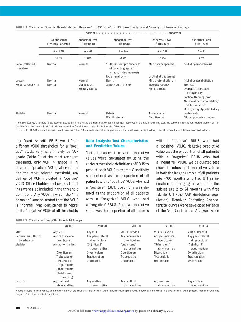
significant. As with RBUS, we defineddifferent VCUG thresholds for a “posi-tive” study, varying primarily by VURgrade (Table 2). At the most stringentthreshold, only VUR . grade III in-dicated a “positive” VCUG, whereas un-der the most relaxed threshold, anydegree of VUR indicated a “positive”VCUG. Other bladder and urethral find-ings were also included in the thresholddefinitions. Any VCUG in which the “im-pression” section stated that the VCUGis “normal” was considered to repre-sent a “negative” VCUG at all thresholds.
Data Analysis: Test Characteristicsand Predictive Values
Test characteristics and predictivevalues were calculated by using thevarious threshold definitions of RBUS to
predict each VCUG outcome. Sensitivity
was defined as the proportion of all
patientswith a “positive” VCUGwho had
a “positive” RBUS. Specificity was de-fined as the proportion of all patients
with a “negative” VCUG who had
a “negative” RBUS. Positive predictive
value was the proportion of all patients
with a “positive” RBUS who hada “positive” VCUG. Negative predictivevalue was the proportion of all patientswith a “negative” RBUS who hada “negative” VCUG. We calculated testcharacteristics and predictive valuesin both the larger sample of all patientsage ,60 months who had UTI as in-dication for imaging, as well as in thesubset age 2 to 24 months with firstfebrile UTI (the AAP guidelines pop-ulation). Receiver Operating Charac-teristic curveswere developed for eachof the VCUG outcomes. Analyses were
TABLE 1 Criteria for Specific Thresholds for “Abnormal” or (“Positive”) RBUS, Based on Type and Severity of Observed Findings
Normal ←←←←←←←←←←←←→→→→→→→→→→→→ Abnormal
No AbnormalFindings Reported
Abnormal LevelD (RBUS-D)
Abnormal LevelC (RBUS-C)
Abnormal LevelBa (RBUS-B)
Abnormal LevelA (RBUS-A)
N = 1694 N = 41 N = 135 N = 298 N = 91
75.0% 1.8% 6.0% 13.2% 4.0%
Renal collectingsystem
Normal Normal “Fullness” or “prominence”of collecting systemwithout hydronephrosis
Mild hydronephrosis .Mild hydronephrosis
Extra-renal pelvis Urothelial thickeningUreter Normal Normal Normal Mild ureteral dilation .Mild ureteral dilationRenal parenchyma Normal Duplication
Solitary kidneySimple cyst (single) Size discrepancy
Renal ectopiaStone(s)Dysplasia/increased
echogenicityCortical thinning/scarAbnormal cortico-medullary
differentiationMulticystic/polycystic kidney
Bladder Normal Normal DebrisWall thickening
TrabeculationDiverticulum
UreteroceleDilated posterior urethra
The RBUS severity threshold is set according to column furthest to the right that contains finding(s) observed in the RBUS screening test. The screening test is considered “abnormal” (or“positive”) at the threshold of that column, as well as for all those thresholds to the left of that level.a Threshold RBUS-B included findings categorized as “other”: 1 example each of acute pyelonephritis, renal mass, large bladder, urachal remnant, and bilateral enlarged kidneys.
TABLE 2 Criteria for the VCUG Threshold Groups
VCUG-E VCUG-D VCUG-C VCUG-B VCUG-A
VUR Any VUR Any VUR VUR . Grade I VUR . Grade II VUR . Grade IIIPeri-ureteral (Hutch)diverticulum
Any peri-ureteraldiverticulum
Any peri-ureteraldiverticulum
Any peri-ureteraldiverticulum
Any peri-ureteraldiverticulum
Any peri-ureteraldiverticulum
Bladder Any abnormalities “Significant”abnormalities
“Significant”abnormalities
“Significant”abnormalities
“Significant”abnormalities
Diverticulum Diverticulum Diverticulum Diverticulum DiverticulumTrabeculation Trabeculation Trabeculation Trabeculation TrabeculationUreterocele Ureterocele Ureterocele Ureterocele UreteroceleLarge volumeSmall volumeBladder wall
thickeningUrethra Any urethral
abnormalitiesAny urethral
abnormalitiesAny urethralabnormalities
Any urethralabnormalities
Any urethralabnormalities
A VCUG is positive for a particular category if any of the findings in that column were reported during the VCUG. If none of the findings in a given column were present, then the VCUG was“negative” for that threshold definition.
396 NELSON et al by guest on February 3, 2019www.aappublications.org/newsDownloaded from

performed by using SAS 9.3 (SAS In-stitute Inc; Cary, NC) and R 2.15.2(http://www.R-project.org/).
RESULTS
We identified 3995 clinical encountersduring which patients underwent RBUSand VCUG studies on the same date be-tween January 1, 2006 and December 31,2010. We excluded 930 patients who hadprevious postnatal GU imaging, leaving3065 subjects. Of these, 198 were age$60 months and were also excluded,leaving 2867 children. Among this group,the indications for imaging were UTI in2259 (78.8%), prenatally identified ab-normalities in 509 (17.8%), and otherindications in 99 (3.5%). The 2259patients who underwent initial GU im-aging for UTI are described in Table 3. Atotal of 79.0% were female, 75.3% wereaged 2 to 24 months, and 43% (975/2259) were seen clinically in the De-partment of Urology at our institution.Among the boys, most were un-circumcised. Among the group aged 2 to24 months, we confirmed that this wasan initial, febrile UTI episode in 1203patients.
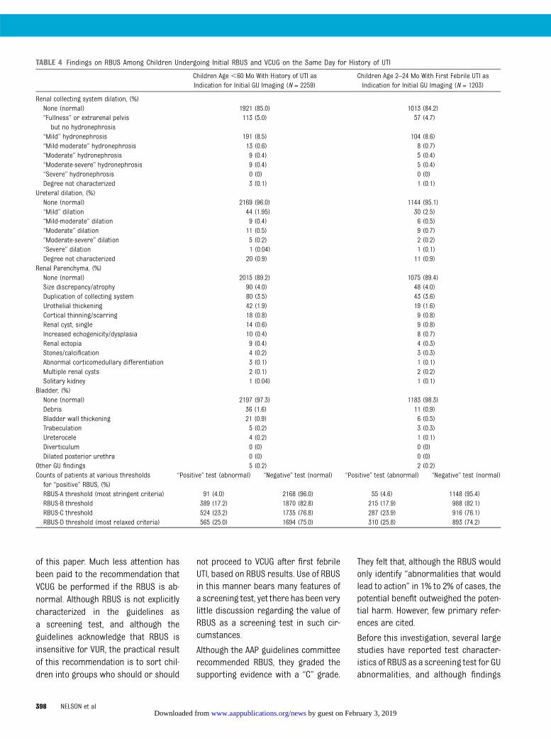
RBUS findings among both the wholecohort and “initial febrile UTI, age 2 to24 months” group are shown in Table 4,along with the proportions in eachgroup meeting thresholds for a “posi-tive” RBUS screening test. Dependingon the threshold, RBUS was “positive”(abnormal) in 4% (RBUS-A), 17% (RBUS-B),23% (RBUS-C), or 25% (RBUS-D) of chil-dren who had “any UTI.” Positive rateswere similar for the “initial febrileUTI” group. Overall, the RBUS findingswere notable for the small number ofpatients who had higher grades ofhydronephrosis; only approximately1.5% of children had hydronephrosisgreater than “mild” on either side.Similarly,,5% of children had ureteraldilation noted, in any degree. The mostcommon renal parenchymal findingswere size discrepancy and duplication;more discrete renal scarring or othercortical pathology was seen in ∼1% ofthe group.
VCUG findings among both patientgroups are shown in Table 5, along withthe proportions in each group meet-ing each threshold for a “positive”VCUG. Abnormalities of any kind were
identified in 43.9% of studies (49.2% ofthe initial febrile UTI group), and VURwas identified in 41.7% (47.5%). Signifi-cant numbers of children had dilatingVUR, with VUR grade .II seen in 20.9%(26.9%). However, high-grade VUR wasuncommon: VUR grade.III was presentin just 2.7% (2.6%). As with RBUS, theproportion of children who had a posi-tive VCUG depended on the thresholdused: VCUGwas “positive” (abnormal) in7% (VCUG-A), 23% (VCUG-B), 39% (VCUG-C), or 43% (VCUG-D) of the “any UTI, age,60months” group. Positive rateswereslightly higher for the “initial febrile UTI,age 2 to 24 months” group.
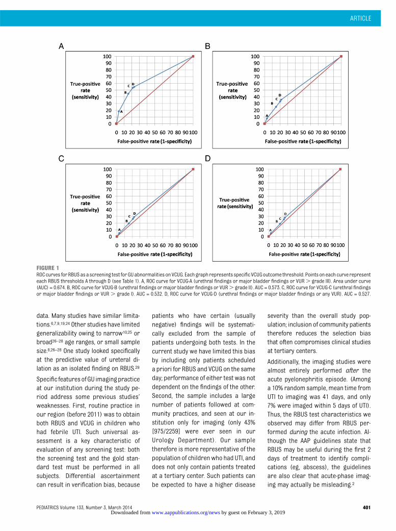
Test characteristics and predictive val-ues forRBUSasascreeningtest forVCUGfindings are shown in Table 6. ReceiverOperating Characteristic curves areshown in Fig 1 A–D. As expected, therewas little difference in test character-istics (sensitivity and specificity) be-tween the “any UTI” and “initial febrileUTI” groups. Predictive values differedsomewhat more, particularly for nega-tive predictive value. RBUS is not a sen-sitive test regardless of the thresholdused, with amaximumsensitivity of 55%(RBUS-D for the VCUG-A outcome).Specificity did reach high levels (maxi-mum 97%) but only at extremely lowlevels of sensitivity (,10%). Positivepredictive values were also low, sug-gesting that only a fraction of those whohave a positive RBUS have VCUG findingsat any “positivity” level. Finally, negativepredictive values were high, but only forthe highest grades of VUR.
DISCUSSION
The 2011 AAP guidelines regarding theevaluation of infants who have a firstfebrile UTI represent significant evolu-tion in management, with the mostsignificant change being the recom-mendation that VCUG be deferred untilafter a second febrile UTI.2 The dis-cussion regarding VCUG timing hasbeen vigorous5 and is beyond the scope
TABLE 3 Characteristics of Children Undergoing Initial RBUS and VCUG on the Same Day forHistory of UTI
Children Age ,60 Mo WithHistory of UTI as Indication
for Initial GU Imaging (N = 2259)
Children Age 2–24 Mo WithFirst Febrile UTI as Indication
for Initial GU Imaging (N = 1203)
Gender, (%)Female 1787 (79.1) 912 (75.8)Male: uncircumcised 306 (13.6) 209 (17.4)Male: circumcised 50 (2.2) 37 (3.1)Male: circumcision status unknown 116 (5.1) 45 (3.7)
Age, (%)0–1 mo 78 (3.45) 0 (0)2–6 mo 591 (26.16) 463 (38.5)7–12 mo 729 (32.27) 515 (42.8)13–18 mo 230 (10.18) 138 (11.5)19–24 mo 151 (6.68) 87 (7.2)25–59 mo 480 (21.25) 0 (0)
Previous UTI history, (%)First UTI 1557 (68.9) 1203 (100)Recurrent UTI 176 (7.8) 0 (0)Recurrence status unknown 526 (23.3) 0 (0)
UTI fever status, (%)Febrile UTI 2045 (90.5) 1203 (100)Nonfebrile UTI 89 (3.9) 0 (0)Febrile history unknown 125 (5.5) 0 (0)
ARTICLE
PEDIATRICS Volume 133, Number 3, March 2014 397 by guest on February 3, 2019www.aappublications.org/newsDownloaded from
of this paper. Much less attention hasbeen paid to the recommendation thatVCUG be performed if the RBUS is ab-normal. Although RBUS is not explicitlycharacterized in the guidelines asa screening test, and although theguidelines acknowledge that RBUS isinsensitive for VUR, the practical resultof this recommendation is to sort chil-dren into groups who should or should
not proceed to VCUG after first febrileUTI, based on RBUS results. Use of RBUSin this manner bears many features ofa screening test, yet there has been verylittle discussion regarding the value ofRBUS as a screening test in such cir-cumstances.
Although the AAP guidelines committeerecommended RBUS, they graded thesupporting evidence with a “C” grade.
They felt that, although the RBUS wouldonly identify “abnormalities that wouldlead to action” in 1% to 2% of cases, thepotential benefit outweighed the poten-tial harm. However, few primary refer-ences are cited.
Before this investigation, several largestudies have reported test character-istics of RBUS as a screening test for GUabnormalities, and although findings
TABLE 4 Findings on RBUS Among Children Undergoing Initial RBUS and VCUG on the Same Day for History of UTI
Children Age ,60 Mo With History of UTI asIndication for Initial GU Imaging (N = 2259)
Children Age 2–24 Mo With First Febrile UTI asIndication for Initial GU Imaging (N = 1203)
Renal collecting system dilation, (%)None (normal) 1921 (85.0) 1013 (84.2)“Fullness” or extrarenal pelvis
but no hydronephrosis113 (5.0) 57 (4.7)
“Mild” hydronephrosis 191 (8.5) 104 (8.6)“Mild-moderate” hydronephrosis 13 (0.6) 8 (0.7)“Moderate” hydronephrosis 9 (0.4) 5 (0.4)“Moderate-severe” hydronephrosis 9 (0.4) 5 (0.4)“Severe” hydronephrosis 0 (0) 0 (0)Degree not characterized 3 (0.1) 1 (0.1)
Ureteral dilation, (%)None (normal) 2169 (96.0) 1144 (95.1)“Mild” dilation 44 (1.95) 30 (2.5)“Mild-moderate” dilation 9 (0.4) 6 (0.5)“Moderate” dilation 11 (0.5) 9 (0.7)“Moderate-severe” dilation 5 (0.2) 2 (0.2)“Severe” dilation 1 (0.04) 1 (0.1)Degree not characterized 20 (0.9) 11 (0.9)
Renal Parenchyma, (%)None (normal) 2015 (89.2) 1075 (89.4)Size discrepancy/atrophy 90 (4.0) 48 (4.0)Duplication of collecting system 80 (3.5) 43 (3.6)Urothelial thickening 42 (1.9) 19 (1.6)Cortical thinning/scarring 18 (0.8) 9 (0.8)Renal cyst, single 14 (0.6) 9 (0.8)Increased echogenicity/dysplasia 10 (0.4) 8 (0.7)Renal ectopia 9 (0.4) 4 (0.3)Stones/calcification 4 (0.2) 3 (0.3)Abnormal corticomedullary differentiation 3 (0.1) 1 (0.1)Multiple renal cysts 2 (0.1) 2 (0.2)Solitary kidney 1 (0.04) 1 (0.1)
Bladder, (%)None (normal) 2197 (97.3) 1183 (98.3)Debris 36 (1.6) 11 (0.9)Bladder wall thickening 21 (0.9) 6 (0.5)Trabeculation 5 (0.2) 3 (0.3)Ureterocele 4 (0.2) 1 (0.1)Diverticulum 0 (0) 0 (0)Dilated posterior urethra 0 (0) 0 (0)
Other GU findings 5 (0.2) 2 (0.2)Counts of patients at various thresholdsfor “positive” RBUS, (%)
“Positive” test (abnormal) “Negative” test (normal) “Positive” test (abnormal) “Negative” test (normal)
RBUS-A threshold (most stringent criteria) 91 (4.0) 2168 (96.0) 55 (4.6) 1148 (95.4)RBUS-B threshold 389 (17.2) 1870 (82.8) 215 (17.9) 988 (82.1)RBUS-C threshold 524 (23.2) 1735 (76.8) 287 (23.9) 916 (76.1)RBUS-D threshold (most relaxed criteria) 565 (25.0) 1694 (75.0) 310 (25.8) 893 (74.2)
398 NELSON et al by guest on February 3, 2019www.aappublications.org/newsDownloaded from
have varied widely, none have foundRBUS to be an accurate screening test inthis setting. Sensitivity has ranged from18% to 79% and specificity from 41% to99%, depending on how a “positive”RBUS was defined and what VUR out-come was assessed (eg, any VUR, “di-lating VUR,” “high-grade VUR”).6–11 Manyother groups have reported GU imagingfindings among children who havea history of UTI. However, most of thesepapers have limitations that make it
impossible to determine the test char-
acteristics of RBUS; most common is
that many studies do not provide suffi-
cient data to directly compare RBUS
findings with VCUG findings in individual
patients.12–19 Other studies focus on the
value of RBUS to predict renal scinti-
graphic findings (scarring).13,20–22
Verification bias is a commonweaknessin the published literature. For example,Foresman et al assessed the correlation
betweenRBUSandVCUGamongpatientshospitalized for acute pyelonephritis.23
RBUS was performed in all patientsduring the hospitalization; however, notall patients subsequently underwentVCUG, and performance of the VCUGvaried depending on RBUS findings,with 67% of patients who had normalRBUS having VCUG, but 87% of patientswho had abnormal RBUS havingVCUG. Such differential assessmentintroduces an inherent bias into the
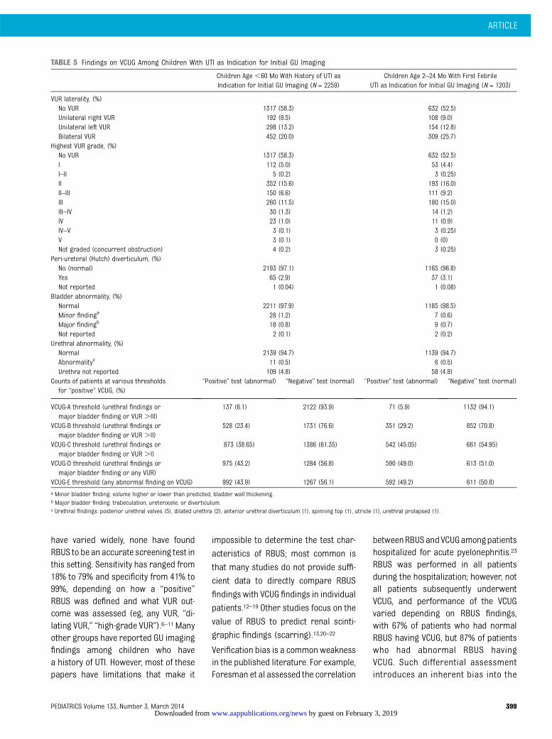
TABLE 5 Findings on VCUG Among Children With UTI as Indication for Initial GU Imaging
Children Age ,60 Mo With History of UTI asIndication for Initial GU Imaging (N = 2259)
Children Age 2–24 Mo With First FebrileUTI as Indication for Initial GU Imaging (N = 1203)
VUR laterality, (%)No VUR 1317 (58.3) 632 (52.5)Unilateral right VUR 192 (8.5) 108 (9.0)Unilateral left VUR 298 (13.2) 154 (12.8)Bilateral VUR 452 (20.0) 309 (25.7)
Highest VUR grade, (%)No VUR 1317 (58.3) 632 (52.5)I 112 (5.0) 53 (4.4)I–II 5 (0.2) 3 (0.25)II 352 (15.6) 193 (16.0)II–III 150 (6.6) 111 (9.2)III 260 (11.5) 180 (15.0)III–IV 30 (1.3) 14 (1.2)IV 23 (1.0) 11 (0.9)IV–V 3 (0.1) 3 (0.25)V 3 (0.1) 0 (0)Not graded (concurrent obstruction) 4 (0.2) 3 (0.25)
Peri-ureteral (Hutch) diverticulum, (%)No (normal) 2193 (97.1) 1165 (96.8)Yes 65 (2.9) 37 (3.1)Not reported 1 (0.04) 1 (0.08)
Bladder abnormality, (%)Normal 2211 (97.9) 1185 (98.5)Minor findinga 28 (1.2) 7 (0.6)Major findingb 18 (0.8) 9 (0.7)Not reported 2 (0.1) 2 (0.2)
Urethral abnormality, (%)Normal 2139 (94.7) 1139 (94.7)Abnormalityc 11 (0.5) 6 (0.5)Urethra not reported 109 (4.8) 58 (4.8)
Counts of patients at various thresholdsfor “positive” VCUG, (%)
“Positive” test (abnormal) “Negative” test (normal) “Positive” test (abnormal) “Negative” test (normal)
VCUG-A threshold (urethral findings ormajor bladder finding or VUR .III)
137 (6.1) 2122 (93.9) 71 (5.9) 1132 (94.1)
VCUG-B threshold (urethral findings ormajor bladder finding or VUR .II)
528 (23.4) 1731 (76.6) 351 (29.2) 852 (70.8)
VCUG-C threshold (urethral findings ormajor bladder finding or VUR .I)
873 (38.65) 1386 (61.35) 542 (45.05) 661 (54.95)
VCUG-D threshold (urethral findings ormajor bladder finding or any VUR)
975 (43.2) 1284 (56.8) 590 (49.0) 613 (51.0)
VCUG-E threshold (any abnormal finding on VCUG) 992 (43.9) 1267 (56.1) 592 (49.2) 611 (50.8)a Minor bladder finding: volume higher or lower than predicted; bladder wall thickening.b Major bladder finding: trabeculation, ureterocele, or diverticulum.c Urethral findings: posterior urethral valves (5), dilated urethra (2), anterior urethral diverticulum (1), spinning top (1), utricle (1), urethral prolapsed (1).
ARTICLE
PEDIATRICS Volume 133, Number 3, March 2014 399 by guest on February 3, 2019www.aappublications.org/newsDownloaded from

TABLE6
Test
CharacteristicsandPredictiveValues
ofEach
RBUS
“Positive”ThresholdforEach
oftheVCUG
Thresholds
Sensitivity(95%
CI)
Specificity(95%
CI)
PositivePredictiveValue(95%
CI)
NegativePredictiveValue(95%
CI)
VCUG
-Ethreshold(any
abnorm
alfindingon
VCUG
)RB
US-D
(mostrelaxed
criteria)
28.0(25.2–30.9)*/26.7(23.2–30.4)**
77.3(74.9–79.6)*/75.1(71.5–78.5)**
49.2(45.0–53.4)*/51.0(45.3–56.7)**
57.9(55.5–60.2)*/51.4(48.1–54.7)**
RBUS-C
25.5(22.8–28.3)*/24.2(20.8–27.8)**
78.6(76.2–80.8)*/76.4(72.9–79.7)**
48.3(43.9–52.7)*/49.8(43.9–55.8)**
57.4(55.0–59.7)*/51.0(47.7–54.3)**
RBUS-B
19.1(16.7–21.6)*/18.8(15.7–22.1)**
84.2(82.1–86.2)*/83.0(79.8–85.9)**
48.6(43.5–53.7)*/51.6(44.7–58.5)**
57.1(54.8–59.3)*/51.3(48.1–54.5)**
RBUS-A(m
oststringentcriteria)
4.9(3.7–6.5)*/5.2(3.6–7.4)**
96.7(95.5–97.6)*/96.1(94.2–97.5)**
53.8(43.1–64.4)*/56.4(42.3–69.7)**
56.5(54.4–58.6)*/51.1(48.2–54.1)**
VCUG
-Dthreshold(urethralfindings
ormajor
bladderfindingor
anyVUR)
RBUS-D
(mostrelaxed
criteria)
28.1(25.3–31.0)*/26.6(23.1–30.4)**
77.3(74.9–79.6)*/75.0(71.4–78.4)**
48.5(44.3–52.7)*/50.6(44.9–56.3)**
58.6(56.2–61.0)*/51.5(48.2–54.8)**
RBUS-C
25.5(22.8–28.4)*/24.1(20.7–27.7)**
78.6(76.2–80.8)*/76.3(72.8–79.7)**
47.5(43.2–51.9)*/49.5(43.6–55.4)**
58.2(55.8–60.5)*/51.1(47.8–54.4)**
RBUS-B
19.0(16.6–21.6)*/18.6(15.6–22)**
84.1(82.0–86.1)*/82.9(79.7–85.8)**
47.6(42.5–52.7)*/51.2(44.3–58)**
57.8(55.5–60.0)*/51.4(48.2–54.6)**
RBUS-A(m
oststringentcriteria)
4.9(3.7–6.5)*/5.3(3.6–7.4)**
96.7(95.5–97.6)*/96.1(94.2–97.5)**
52.7(42.0–63.3)*/56.4(42.3–69.7)**
57.2(55.1–59.3)*/51.3(48.4–54.2)**
VCUG
-Cthreshold(urethralfindings
ormajor
bladderfindingor
VUR.I)
RBUS-D
(mostrelaxed
criteria)
29.0(26.0–32.1)*/27.3(23.6–31.3)**
77.5(75.2–79.7)*/75.5(72.0–78.7)**
44.8(40.6–49.0)*/47.7(42.1–53.5)**
63.4(61.1–65.7)*/55.9(52.6–59.2)**
RBUS-C
26.5(23.6–29.5)*/24.7(21.1–28.6)**
78.9(76.6–81.0)/76.9(73.4–80.0)
44.1(39.8–48.5)/46.7(40.8–52.6)
63.0(60.7–65.3)/55.5(52.2–58.7)
RBUS-B
19.6(17.0–22.4)*/19.0(15.8–22.6)**
84.3(82.2–86.1)/83.1(80.0–85.8)
44.0(39.0–49.0)/47.9(41.1–54.8)
62.5(60.2–64.7)/55.6(52.4–58.7)
RBUS-A(m
oststringentcriteria)
5.2(3.8–6.8)*/5.4(3.6–7.6)**
96.7(95.6–97.6)/96.1(94.3–97.4)
49.5(38.8–60.1)/52.7(38.8–66.3)
61.8(59.7–63.9)/55.3(52.4–58.2)
VCUG
-Bthreshold(urethralfindings
ormajor
bladderfindingor
VUR.II)
RBUS-D
(mostrelaxed
criteria)
35.8(31.7–40.0)*/32.8(27.9–37.9)**
78.3(76.3–80.2)*/77.1(74.1–79.9)**
33.5(29.6–37.5)*/37.1(31.7–42.7)**
80.0(78.0–81.9)*/73.6(70.5–76.4)**
RBUS-C
33.1(29.1–37.3)*/29.9(25.2–35.0)**
79.8(77.9–81.7)*/78.6(75.7–81.3)**
33.4(29.4–37.6)*/36.6(31.0–42.4)**
79.7(77.7–81.5)*/73.1(70.1–76.0)**
RBUS-B
25.2(21.5–29.1)*/23.1(18.8–27.8)**
85.2(83.5–86.9)*/84.3(81.7–86.7)**
34.2(29.5–39.1)*/37.7(31.2–44.5)**
78.9(77.0–80.7)*/72.7(69.8–75.4)**
RBUS-A(m
oststringentcriteria)
7.8(5.6–10.4)*/7.4(4.9–10.7)**
97.1(96.2–97.8)*/96.6(95.1–97.7)**
45.1(34.6–55.8)*/47.3(33.7–61.2)**
77.5(75.7–79.3)*/71.7(69.0–74.3)**
VCUG
-Athreshold(urethralfindings
ormajor
bladderfindingor
VUR.III)
RBUS-D
(mostrelaxed
criteria)
54.7(46.0–63.3)*/54.9(42.7–66.8)**
76.9(75.1–78.7)*/76.1(73.5–78.5)**
13.3(10.6–16.4)*/12.6(9.1–16.8)**
96.3(95.3–97.2)*/96.4(95.0–97.5)**
RBUS-C
53.3(44.6–61.9)*/53.5(41.3–65.5)**
78.7(76.9–80.5)*/78.0(75.5–80.4)**
13.9(11.1–17.2)*/13.2(9.5–17.7)**
96.3(95.3–97.1)*/96.4(95.0–97.5)**
RBUS-B
44.5(36.0–53.3)*/46.5(34.5–58.7)**
84.5(82.9–86.1)*/83.9(81.7–86.0)**
15.7(12.2–19.7)*/15.3(10.8–20.9)**
95.9(94.9–96.8)*/96.2(94.8–97.3)**
RBUS-A(m
oststringentcriteria)
18.2(12.2–25.7)*/18.3(10.1–29.3)**
96.9(96.1–97.6)*/96.3(95.0–97.3)**
27.5(18.6–37.8)*/23.6(13.2–37.0)**
94.8(93.8–95.7)*/94.9(93.5–96.1)**
SeeTable2forexplanationofRBUS
thresholdcriteria.*Valuesam
ongchildrenage,120mowith
historyofUTIasindicationforimaging(N
=2259).**Values
amongchildrenage2–24
mowith
initialfebrile
UTIastheindicationforGU
imaging(N
=1203).CI,
95%confidenceinterval.
400 NELSON et al by guest on February 3, 2019www.aappublications.org/newsDownloaded from
data. Many studies have similar limita-tions.6,7,9,19,24 Other studies have limitedgeneralizability owing to narrow10,25 orbroad26–28 age ranges, or small samplesize.8,26–28 One study looked specificallyat the predictive value of ureteral di-lation as an isolated finding on RBUS.29
Specific featuresof GU imagingpracticeat our institution during the study pe-riod address some previous studies’weaknesses. First, routine practice inour region (before 2011) was to obtainboth RBUS and VCUG in children whohad febrile UTI. Such universal as-sessment is a key characteristic ofevaluation of any screening test: boththe screening test and the gold stan-dard test must be performed in allsubjects. Differential ascertainmentcan result in verification bias, because
patients who have certain (usuallynegative) findings will be systemati-cally excluded from the sample ofpatients undergoing both tests. In thecurrent study we have limited this biasby including only patients scheduleda priori for RBUS and VCUG on the sameday; performance of either test was notdependent on the findings of the other.Second, the sample includes a largenumber of patients followed at com-munity practices, and seen at our in-stitution only for imaging (only 43%[975/2259] were ever seen in ourUrology Department). Our sampletherefore is more representative of thepopulation of children who had UTI, anddoes not only contain patients treatedat a tertiary center. Such patients canbe expected to have a higher disease
severity than the overall study pop-ulation; inclusion of community patientstherefore reduces the selection biasthat often compromises clinical studiesat tertiary centers.
Additionally, the imaging studies werealmost entirely performed after theacute pyelonephritis episode. (Amonga 10% random sample, mean time fromUTI to imaging was 41 days, and only7% were imaged within 5 days of UTI).Thus, the RBUS test characteristics weobserved may differ from RBUS per-formed during the acute infection. Al-though the AAP guidelines state thatRBUS may be useful during the first 2days of treatment to identify compli-cations (eg, abscess), the guidelinesare also clear that acute-phase imag-ing may actually be misleading.2
FIGURE 1ROCcurves for RBUSas a screening test for GUabnormalities onVCUG. Each graph represents specific VCUGoutcome threshold. Points on each curve representeach RBUS thresholds A through D (see Table 1). A, ROC curve for VCUG-A (urethral findings or major bladder findings or VUR. grade III). Area under curve(AUC) = 0.674. B, ROC curve for VCUG-B (urethral findings or major bladder findings or VUR. grade II). AUC = 0.573. C, ROC curve for VCUG-C (urethral findingsor major bladder findings or VUR . grade I). AUC = 0.532. D, ROC curve for VCUG-D (urethral findings or major bladder findings or any VUR). AUC = 0.527.
ARTICLE
PEDIATRICS Volume 133, Number 3, March 2014 401 by guest on February 3, 2019www.aappublications.org/newsDownloaded from

The low overall incidence of significantGU anomalies in our sample warrantscomment.Previousstudieshavereporteda similar phenomenon. Hobermanet al examined imaging results among309 children who had febrile UTI, andnoted that patients who had high-gradeVUR were under-represented in theirsample, and stated that “the validity ofrenal ultrasonography in identifyingsuch children [with high-grade VUR]warrants further study.”30 A relativelylow incidence of high-grade VUR hasbeen seen in other series as well.8 Al-though our sample was much largerthan these studies, we too noted that,3% of our sample had high-gradeVUR (grade .III). Similarly, ,1.5% ofchildren had higher-grade hydro-nephrosis on RBUS. The low prevalenceof severe abnormalities may be at-tributable to the effect of prenatalscreening. We excluded children whohad a history of prenatally diagnosedabnormalities because (1) the AAP im-aging guidelines can be read as beingapplicable to children who have nohistory of prenatal abnormalities, and(2) such children usually undergo GUimaging as newborns, and we excludedchildren who had a history of previousGU imaging. Many, although not all,cases of high-grade disease are likelydetected prenatally, reducing the in-cidence of such anomalies amongchildren presenting with de novo UTIpostnatally. Fifty years ago, we pre-sumably would have observed muchhigher rates of such anomalies, be-cause many of the children who are
now diagnosed in utero would havepresented postnatally with clinical UTI.
The results of our investigation shouldbe interpreted in light of its limitations.This study was retrospective, subject tothe limitations of this design. For ex-ample, study subjects all had history ofUTI, but many of these diagnoses weremade elsewhere and could not be in-dependently verified. As some patientsmay have been misdiagnosed, theresults may not reflect those that wouldbe seenamong childrenwhohad strictlydefined and confirmedUTI. However, ourradiologists take a detailed history be-fore VCUG, to verify the history. Fur-thermore, irrespective of the diagnosticdetails, these are the patients beingreferred for GU imaging, and so reflectthe “real-world” screening populationseen in practice. With respect to imag-ing findings, we relied on the finalinterpretations of the imaging studiesas dictated by the clinical radiologist;independent confirmatory review ofimageswas not performed. The findingstherefore are subject to the relativevariability of interpretation (eg, gradingof VUR) that occurs in all clinical care.Furthermore, the radiologists readingeach study were not systematicallyblinded to the findings of the other test,so it is possible that interpretation of 1study could have been influenced byknowledge of the findings on the othertest. However, the radiologist interpret-ing the VCUG was usually a differentindividual than that radiologist inter-preting the RBUS, and in most patientsthe RBUS was performed (and usuallyread) before the VCUG, which would
minimize the impact of such unblind-ed interpretation. Furthermore, mostfindings on both RBUS and VCUG arerelatively objective. As noted, verifica-tion bias is a concern in any evaluationof a screening test. To minimize this, weincluded only patients who underwentboth VCUG and RBUS on the same day.However, it is likely that some patientsunderwent RBUS or VCUG separately,or underwent 1 test but not the other;such patients would be excluded fromour sample. If such patients were nu-merous, and if the decision not to ob-tain the second test was based on theresults of the first test, then bias couldhave been introduced into our sample.As we also noted, however, severalfeatures of our practice environmentduring the study period make this lesslikely, including the widespread ad-herence within our institution andcommunity to the 1999 AAP guidelines,the routine practice of scheduling bothRBUS and VCUG on the same day forpatients who have a history of UTI, andthe routine completion of both tests,regardless of findings.
CONCLUSIONS
Among young children who have a his-tory of UTI, RBUS is a poor screeningtest for GU abnormalities, with lowsensitivity/specificity. A negative RBUSdoes not rule out significant GU pa-thology (particularly VUR grades III andhigher), whereas a positive RBUS isa poor predictor. In such children, RBUSand VCUG should be considered com-plementary as they provide important,but different, information.
REFERENCES
1. American Academy of Pediatrics. Commit-tee on Quality Improvement. Subcommitteeon Urinary Tract Infection. Practice pa-rameter: the diagnosis, treatment, andevaluation of the initial urinary tract
infection in febrile infants and young chil-dren. Pediatrics. 1999;103(4 pt 1):843–852
2. Roberts KB; Subcommittee on Urinary TractInfection, Steering Committee on QualityImprovement and Management. Urinary
tract infection: clinical practice guidelinefor the diagnosis and management of theinitial UTI in febrile infants and children 2to 24 months. Pediatrics. 2011;128(3):595–610
402 NELSON et al by guest on February 3, 2019www.aappublications.org/newsDownloaded from
3. Fernbach SK, Maizels M, Conway JJ. Ultra-sound grading of hydronephrosis: in-troduction to the system used by theSociety for Fetal Urology. Pediatr Radiol.1993;23(6):478–480
4. Medical versus surgical treatment of pri-mary vesicoureteral reflux: report of theInternational Reflux Study Committee. Pe-diatrics. 1981;67(3):392–400
5. Wan J, Skoog SJ, Hulbert WC, et al; ExecutiveCommittee, Section on Urology, AmericanAcademy of Pediatrics. Section on Urologyresponse to new Guidelines for the di-agnosis and management of UTI. Pediatrics.2012;129(4). Available at: www.pediatrics.org/cgi/content/full/129/4/e1051–e1053
6. Rickwood AM, Carty HM, McKendrick T,et al. Current imaging of childhood urinaryinfections: prospective survey. BMJ. 1992;304(6828):663–665
7. Zamir G, Sakran W, Horowitz Y, Koren A,Miron D. Urinary tract infection: is therea need for routine renal ultrasonography?Arch Dis Child. 2004;89(5):466–468
8. Mahant S, Friedman J, MacArthur C. Renalultrasound findings and vesicoureteralreflux in children hospitalised with urinarytract infection. Arch Dis Child. 2002;86(6):419–420
9. Lee JH, Kim MK, Park SE. Is a routinevoiding cystourethrogram necessary inchildren after the first febrile urinary tractinfection? Acta Paediatr. 2012;101(3):e105–e109
10. Tsai JD, Huang CT, Lin PY, et al. Screeninghigh-grade vesicoureteral reflux in younginfants with a febrile urinary tract in-fection. Pediatr Nephrol. 2012;27(6):955–963
11. Preda I, Jodal U, Sixt R, Stokland E, HanssonS. Normal dimercaptosuccinic acid scin-tigraphy makes voiding cystourethrog-raphy unnecessary after urinary tractinfection. J Pediatr. 2007;151(6):581–584
12. Alon US, Ganapathy S. Should renal ultra-sonography be done routinely in childrenwith first urinary tract infection? ClinPediatr (Phila). 1999;38(1):21–25
13. Björgvinsson E, Majd M, Eggli KD. Diagnosisof acute pyelonephritis in children: com-parison of sonography and 99mTc-DMSAscintigraphy. AJR Am J Roentgenol. 1991;157(3):539–543
14. Giorgi LJ Jr, Bratslavsky G, Kogan BA. Fe-brile urinary tract infections in infants:renal ultrasound remains necessary. JUrol. 2005;173(2):568–570
15. Jakobsson B, Nolstedt L, Svensson L,Söderlundh S, Berg U. 99mTechnetium-dimercaptosuccinic acid scan in the di-agnosis of acute pyelonephritis in children:relation to clinical and radiological find-ings. Pediatr Nephrol. 1992;6(4):328–334
16. Jahnukainen T, Honkinen O, Ruuskanen O,Mertsola J. Ultrasonography after the firstfebrile urinary tract infection in children.Eur J Pediatr. 2006;165(8):556–559
17. Kass EJ, Fink-Bennett D, Cacciarelli AA,Balon H, Pavlock S. The sensitivity of renalscintigraphy and sonography in detectingnonobstructive acute pyelonephritis. J Urol.1992;148(2 Pt 2):606–608
18. Lavocat MP, Granjon D, Allard D, Gay C,Freycon MT, Dubois F. Imaging of pyelone-phritis. Pediatr Radiol. 1997;27(2):159–165
19. Montini G, Zucchetta P, Tomasi L, et al. Valueof imaging studies after a first febrile uri-nary tract infection in young children: datafrom Italian renal infection study 1. Pediat-rics. 2009;123(2). Available at: www.pediat-rics.org/cgi/content/full/123/2/e239–e246
20. Sreenarasimhaiah V, Alon US. Uroradiologicevaluation of children with urinary tractinfection: are both ultrasonograpy and re-nal cortical scintigraphy necessary? JPediatr. 1995;127(3):373–377
21. Rosenberg AR, Rossleigh MA, Brydon MP,Bass SJ, Leighton DM, Farnsworth RH.
Evaluation of acute urinary tract infectionin children by dimercaptosuccinic acidscintigraphy: a prospective study. J Urol.1992;148(5 pt 2):1746–1749
22. Biggi A, Dardanelli L, Pomero G, et al. Acuterenal cortical scintigraphy in children witha first urinary tract infection. PediatrNephrol. 2001;16(9):733–738
23. Foresman WH, Hulbert WC Jr, Rabinowitz R.Does urinary tract ultrasonography athospitalization for acute pyelonephritispredict vesicoureteral reflux? J Urol. 2001;165(6 pt 2):2232–2234
24. Ben-Ami T, Rozin M, Hertz M. Imaging of chil-dren with urinary tract infection: a tailoredapproach. Clin Radiol. 1989;40(1):64–67
25. Goldman M, Lahat E, Strauss S, et al. Imagingafter urinary tract infection in male neo-nates. Pediatrics. 2000;105(6):1232–1235
26. Smellie JM, Rigden SP, Prescod NP. Urinarytract infection: a comparison of four meth-ods of investigation. Arch Dis Child. 1995;72(3):247–250
27. Johnson CE, Shurin PA, Marchant CD, et al.Identification of children requiring radio-logic evaluation for urinary infection.Pediatr Infect Dis. 1985;4(6):656–663
28. Tappin DM, Murphy AV, Mocan H, et al. Aprospective study of children with firstacute symptomatic E. coli urinary tract in-fection. Early 99mtechnetium dimercapto-succinic acid scan appearances. ActaPaediatr Scand. 1989;78(6):923–929
29. Kenney IJ, Negus AS, Miller FN. Is sono-graphically demonstrated mild distalureteric dilatation predictive of ves-icoureteric reflux as seen on micturatingcystourethrography? Pediatr Radiol. 2002;32(3):175–178
30. Hoberman A, Charron M, Hickey RW, BaskinM, Kearney DH, Wald ER. Imaging studiesafter a first febrile urinary tract infectionin young children. N Engl J Med. 2003;348(3):195–202
ARTICLE
PEDIATRICS Volume 133, Number 3, March 2014 403 by guest on February 3, 2019www.aappublications.org/newsDownloaded from
DOI: 10.1542/peds.2013-2109 originally published online February 10, 2014; 2014;133;e394Pediatrics
Caleb P. Nelson, Emilie K. Johnson, Tanya Logvinenko and Jeanne S. ChowUTI
Ultrasound as a Screening Test for Genitourinary Anomalies in Children With
ServicesUpdated Information &
http://pediatrics.aappublications.org/content/133/3/e394including high resolution figures, can be found at:
Referenceshttp://pediatrics.aappublications.org/content/133/3/e394#BIBLThis article cites 29 articles, 10 of which you can access for free at:
Subspecialty Collections
http://www.aappublications.org/cgi/collection/urology_subUrologynt_subhttp://www.aappublications.org/cgi/collection/med_tech_advancemeMedical Technology and Advancemente_management_subhttp://www.aappublications.org/cgi/collection/administration:practicAdministration/Practice Managementfollowing collection(s): This article, along with others on similar topics, appears in the
Permissions & Licensing
http://www.aappublications.org/site/misc/Permissions.xhtmlin its entirety can be found online at: Information about reproducing this article in parts (figures, tables) or
Reprintshttp://www.aappublications.org/site/misc/reprints.xhtmlInformation about ordering reprints can be found online:
by guest on February 3, 2019www.aappublications.org/newsDownloaded from
DOI: 10.1542/peds.2013-2109 originally published online February 10, 2014; 2014;133;e394Pediatrics
Caleb P. Nelson, Emilie K. Johnson, Tanya Logvinenko and Jeanne S. ChowUTI
Ultrasound as a Screening Test for Genitourinary Anomalies in Children With
http://pediatrics.aappublications.org/content/133/3/e394located on the World Wide Web at:
The online version of this article, along with updated information and services, is
ISSN: 1073-0397. 60007. Copyright © 2014 by the American Academy of Pediatrics. All rights reserved. Print the American Academy of Pediatrics, 141 Northwest Point Boulevard, Elk Grove Village, Illinois,has been published continuously since 1948. Pediatrics is owned, published, and trademarked by Pediatrics is the official journal of the American Academy of Pediatrics. A monthly publication, it
by guest on February 3, 2019www.aappublications.org/newsDownloaded from