ultrasound assisted drug delivery · invest radiol 2000 jan 35:1 86-9 takeuchi m, ogunyankin k,...
TRANSCRIPT

Therapeutic Applications of Targeted Microbubbles
Evan C. Unger, M.D.a,b, Terry Onichi Matsunaga, Pharm.D., Ph.D.a, Thomas McCreery, M.Sa., Patricia Schumann, B.S.a, Robert Sweitzer, B.S.a
and Rachel Quigley, B.Sa
aImaRx Therapeutics, Inc. 1635 East 18th Street, Tucson, AZ 85705 USAbProfessor of Radiology, University of Arizona, Tucson, AZ 85715
Microbubbles can be targeted to specific receptors by attaching ligands to the surface of the microbubbles. We have synthesized a series of lipid bioconjugates, incorporated them into microbubbles and tested them for binding affinity, and potential imaging and therapeutic applications. Many ligands can be attached to each microbubble so that the microbubble functions as a polydentate ligand. The polydentate character of the microbubbles means that the binding affinity of the whole microbubble may be greater than for an individual ligand. This is analogous to white blood cells which express ligands and are know to bind avidly to a variety of different biological targets. Targeted microbubbles can be used as selective imaging agents to detect cells or tissues such as vascular thrombosis. Also, bioactive agents can be incorporated into microbubbles (viz acoustically active carriers). Thus combined diagnostic and therapeutic agents can be developed. Because of the acoustic properties of microbubbles such targeted therapeutic microbubbles afford unique therapeutic potential for treatment of thrombosis, drug and gene delivery.
INTRODUCTION
Several new ultrasound contrast agents have recently been developed. These new agents recirculate through the arterial and venous blood systems to function as blood pool agents. Our group is developing targeted ultrasound contrast agents, which bind to cell specific receptors or antigens. These “smart” microbubbles may be used as specific contrast agents for ultrasound to improve diagnosis, but also as therapeutic agents. Micro-bubbles lower the cavitation thresh-hold for ultrasound energy and can be used to increase the local absorption of ultrasound energy within a region to achieve a desired bioeffect. Our work is directed towards development of targeted ultrasound contrast agents to detect and treat disease.
METHODS
Bioconjugates comprising a targeting moiety, a linker and an anchor were developed. A series of differ-ent peptide-based targeting moieties were synthesized using solid phase chemistry. Thrombus-specific pep-tides, directed to the activated GPIIBIIIA receptor of platelets were evaluated for affinity to activated platelets by testing for the inhibition of platelet aggregation. The bioconjugates were compared to the native peptides for their inhibition on platelet aggregation. The prototypical bioconjugate, distearyl- diaminobutyryl -PEG-CRGDC was incorporated into microbubbles. The microbubbles
entrapping perfluoropropane gas were prepared from a blend of phospholipids by agitation. The blend of phos-pholipids and bioconjugate were optimized for binding activity by using a light obscuration assay studying the affinity of the microbubbles for activated platelets. In vitro ultrasound imaging was performed using a 7.5 MHz transducer in a flow-through phantom with human blood clot. In vivo ultrasound imaging was performed epicardially in dogs with clots in the atrial appendage. In vitro Sonothrombolysis was performed in vitro with human blood clot with and without targeted and non-targeted microbubbles and urokinase.
RESULTS
The greatest inhibition on platelet aggregation was achieved with cyclized peptides. Bioconjugates (com-prising peptides attached to a PEG linker and hydro-phobic anchor) had an about 50% reduction in binding compared to the respective peptides alone. Photomicro-graphs of microbubbles bound to activated platelets (see Figure 1); light obscuration assays and in vitro ultra-sound imaging of clot gave consistent results, i.e. the highest affinity ligands caused the greatest bubble bind-ing light obscuration and in vitro enhancement. In vivo imaging showed enhancement of atrial clot. In vitro sonothrombolysis studies showed greater clot lysis with targeted microbubbles.

DISCUSSION
A prototypical targeted microbubble therapeutic agent has been developed. The agent not only enhances detection of clots but also in concert with ultrasound enables rapid treatment via clot lysis. Sonothromboly-sis is enhanced by targeted microbubbles. Treatment can be performed with transcutaneous application of ultra-sound with or without thrombolytic agents (e.g. t-PA). When used with a thrombolytic agent targeted micro-bubble enhanced Sonothrombolysis enables clot to be lysed more rapidly and with a lower dose of thrombo-lytic agent. Targeted microbubbles represent a new class of thera-peutic agents. By selectively delivering microbubbles to precise sites in vivo we are able to localize cavitation nuclei for bioeffect. This can be used for drug and gene delivery and as shown above to achieve clot lysis.
ACKNOWLEDGEMENTS
The authors would like to acknowledge Noelia Navia-Ramírez and Terri New for their help in the preparation of this manuscript.
REFERENCES
Siegel RJ, Atar S, Fishbein MC, Brasch AV, Peterson TM, Nagai T, Pal D, Nishioka T, Chae JS, Birnbaum Y, Zanelli C, Luo H, Noninva-sive transcutaneous low frequency ultrasound enhances thrombolysis in peripheral and coronary arteries. Echocardiography 2001 Apr 18:3 247-57
Unger, Wu, McCreery and Matsunaga. Thrombus-specific contrast agents for imaging and thrombolysis. Ultrasound Contrast Agents, 2nd ed., Martin Dunitz, London, ed. Goldberg, Raichlen and Forsberg. 2001 pp. 337-345
Unger E, Metzger P, Krupinski E, Baker M, Hulett R, Gabaeff D, Mills J, Ihnat D, McCreery T, The use of a thrombus-specific ultrasound contrast agent to detect thrombus in arteriovenous fistulae. Invest Radiol 2000 Jan 35:1 86-9
Takeuchi M, Ogunyankin K, Pandian NG, McCreery TP, Sweitzer RH, Caldwell VE, Unger EC, Avelar E, Sheahan M, Connolly R. Enhanced visualization of intravascular and left atrial appendage thrombus with the use of a thrombus-targeting ultrasonographic con-trast agent (MRX-408A1): In vivo experimental echocardiographic studies. J Am Soc Echocardiogr 1999 Dec 12:12 1015-21
Wu Y, Unger EC, McCreery TP, Sweitzer RH, Shen D, Wu G, Vielhauer MD, Binding and lysing of blood clots using MRX-408. Invest Radiol 1998 Dec 33:880-5
FIGURE 1. Shown above is a photomicrograph of a slide binding activated human platelets. On the left the slide has been incu-bated with non-targeted microbubbles. Essentially no bubble binding is seen. The image on the right shows a slide incubated with targeted microbubbles containing a peptide bioconjugates. After washing, dense microbubble binding is observed.

Optison and Perfluorocarbon Exposed Sonicated DextroseAlbumin Are Not Equally Efficient in Enhancing
Ultrasound-Induced Gene Transfer In Vitro
S. V. Pislarua, R. R. Kinnickb, R. D. Simaria and J. F. Greenleafb
aCardiovascular Diseases, Mayo Clinic and Foundation, Rochester MN, USAbBasic Ultrasound Research Laboratory, Mayo Clinic and Foundation, Rochester MN, USA
Background. Various contrast agents have been shown to enhance ultrasound (US) induced gene transfer. The aim of our studywas to directly compare Optison and perfluorocarbon exposed sonicated dextrose albumin (PESDA), agents with very similarstructure. By doing so, any observed difference in gene transfer would be attributable to mechanisms beyond cavitation.Methods and results. A plasmid encoding for the firefly luciferase was used as a reporter. DNA mixed with either Optison orPESDA (25% v/v) was added to vascular smooth muscle cells or endothelial cells. US exposure was performed in continuouswave mode at 1 MHz, 0.5 or 0.75 W/cm2, for 5–30sec. Measurements of luciferase activity 24h after exposure showed a severalhundred-fold enhancement of gene transfer with both Optison and PESDA in comparison with plasmid alone, plasmid + US,plasmid + contrast agents without US, and several fold increase over plasmid lipofection.Conclusions. In this experimental setup, both Optison and PESDA enhanced gene transfer to levels superior to standard plasmidlipofection. However, PESDA was superior to Optison, suggesting that mechanisms beyond cavitation may be involved in USenhancement of gene transfer.
Several echocardiographic contrast agents have beenshown to enhance ultrasound (US) induced genetransfer [1-4]. The suggested mechanism responsiblefor this effect is lowering the threshold for inertialcavitation. However, the relative efficacy of differentcontrast agents was never evaluated. Furthermore, thepossible intervention of other mechanisms remainsunknown. The aim of our study was to directly compareOptison and Perfluorocarbon Exposed SonicatedDextrose Albumin (PESDA), two contrast agents withvery similar structure and size. By doing so, anyobserved difference in gene transfer would beattributable to mechanisms beyond cavitation.
METHODS
Reporter Genes and Cell Culture
We have used a plasmid encoding for the fireflyluciferase as reporter of gene transfection. Porcinevascular smooth muscle cells (VSMC) and humanendothelial cells (EC) were cultured in 199 and EmBMgrowth media, respectively. Cells grown in 6-wellplates to >70% confluence were used for gene transferexperiments.
Contrast Agents and Ultrasound Exposure
PESDA was prepared from human albumin, 5%glucose solution and decafluorbutane. Optison was
purchased and used within 24 hours after opening. Theconcentration of microbbubles in Optison and PESDAwas measured with a hemocytometer. The sizedistribution of microbubbles was evaluated onmicroscopic images of fresh dilutions (1:100 in 5%glucose) of Optison and PESDA with the ImageProPlus software. Four 35-mm diameter air-backed US transducerswere fixed in a frame so that the bottoms of the cornerwells on the 6-well culture plate were aligned parallelwith the transducers. The frame was immersed in awater tank; the distance between the top of thetransducers and the bottom of the well was threemillimeters. US exposure was performed in continuouswave mode at 1 MHz, 0.5 or 0.75 W/cm2 average
FPds
0%
5%
10%
15%
20%
25%
1 2 3 4 5 6 7 8 9 10
PESDA
Diameter (�m)
0%
5%
10%
15%
20%
25%
1 2 3 4 5 6 7 8 9 10
Optison
Diameter (�m)
Num
ber o
f mic
robu
bble
s (%
)
igure 1. Size distribution of Optison (left panel) andESDA (right panel). Over 90% of the microbubbles hadiameters in the 0-8 �m range. Both agents have albuminhells containing a perfluorocarbon gas.

power, for 5–30 seconds.
Gene transfer protocols and efficacy assays
A plasmid dose of 10 �g/well was used throughoutthe study. Luciferase plasmid was diluted in 750 �lserum-free media and mixed with 250 �l contrast. Themixture was added to the culture well, and exposure toUS was performed. After 2 hours, complete media wasadded to stop the transfection; cells were incubated foranother 24 hours prior to assessment of gene transferefficacy. Growth media, plasmid alone, plasmid plusUS, plasmid plus contrast media without US, andplasmid lipofection (20 �g lipofectamine mixed with10 �g DNA/well) were used as controls. Twenty-fourhours after transfection, the cells were lysed, luciferaseactivity was measured with a commercially availablekit (Promega) and expressed in light units permicrogram protein.
RESULTS
Luciferase activity measurements are summarized inTable 1 and Figure 2 (data from 6-8 experiments; mean±SEM). Exposure to US in the presence of bothOptison and PESDA enhanced gene transfer severalhundred-fold in comparison with plasmid DNA alone,and was superior to plasmid lipofection. Addition ofmicrobubbles in the absence of US, and US exposurein the absence of contrast agents were associated withluciferase activities similar to plasmid DNA alone. In both vascular smooth muscle cells and endothelialcells, PESDA was associated with significantly highertransfection efficacy than Optison (Figure 2). Thiseffect became evident for exposure times above 10seconds. Longer exposure to US resulted in higherluciferase activities, tending to reach a plateau at 30seconds. The endothelial cells were more difficult to transfectin vitro, with luciferase activities 3-5 times lower thanthose observed in vascular smooth muscle cells.
DISCUSSION AND CONCLUSIONS
Optison and PESDA are echocardiographic agentswith very similar structure. Both agents are based onalbumin shells containing a perfluorocarbon gas(octafluorpropane in Optison, decafluorbutane inPESDA). Both have microbubble concentrations of 4-8�108 /ml, and an average diameter of 2-6 �m.Therefore, their ability to enhance inertial cavitationshould be similar. In this experimental setting, Optison and PESDAenhanced US gene transfer to different extent. Wecould not find an obvious explanation for this effect.However, our results suggest that mechanisms beyondcavitation might also play a role in US induced genetransfer. Differences in DNA binding to the albuminshells, the presence in PESDA of microbubbles withdiameters above 8 �m, and different cellular uptake ofPESDA and Optison microbubbles could have beenresponsible for these results. Further studies of themechanisms responsible for plasmid DNA transportthrough the cellular and nuclear membranes uponexposure to US seem warranted.
REFERENCES
1. Greenleaf, W.J., Bolander, M.E., Sarkar, G., Goldring,M.B., Greenleaf, J.F., Ultrasound in Medicine andBiology 24:587-595 (1998).
2. Porter, T.R. Iversen, P.L., Li, S., Xie, F., Journal ofUltrasound in Medicine 15(8):577-584 (1996).
3. Lawrie, A., Briskin, A.F., Francis, S.E., CumberlandD.C., Crossman, D.C., Newman, C.M., Gene Therapy7:2023-2027 (2000).
4. Shohet, R.V., Chen, S., Zhou, Y.T., Wang, Z., Meidell,R.S., Unger, R.H., Grayburn, P.A., Circulation.101(22):2554-2556 (2000).
0
25
50
75
100
0 10 20 30
VSMC
Exposure time (sec)
Luci
fera
e ac
tivity
(LU
/�g)
0
5
10
15
20
25
0 10 20 30
EC
Exposure time (sec)
FIGURE 2. Luciferase activity 24 hours after transfection invascular smooth muscle cells (left panel) and endothelial cells(right panel). Both Optison (open circles) and PESDA (solidcircles) were superior to the level of plasmid lipofection(solid line). However, PESDA was associated with highertransfection efficacy in both cell cultures tested.
Table 1. Luciferase activity (in light units/�g protein) VSMC EC
Growth media 0.0 ± 0.0 0.0 ± 0.0DNA alone 0.1 ± 0.0 0.1 ± 0.2DNA + US 0.1 ± 0.0 0.3 ± 0.1DNA + Optison / PESDA 0.0 ± 0.0 0.1 ± 0.0DNA + lipofectamine 12.9 ± 4.1* 1.7 ± 0.3*DNA + Optison + US 37.6 ± 20.1*† 5.6 ± 0.6*†DNA + PESDA + US 72.6 ± 33.7*† 17.2 ± 9.9*†
All data given as mean ± SEM. VSMC: vascular smoothmuscle cells. EC: endothelial cells. DNA: luciferase plasmid;US: 30 seconds exposure at 0.75 W/cm2. * p<0.05 vs. DNAalone; † p<0.05 vs. DNA + lipofectamine
Effect of Cell Density and Contrast Agent Concentration on Ultrasound-Mediated Drug Delivery and Cell Viability
H.R. Guzmán, A. McNamara, D. Nguyen, and M.R. Prausnitz
School of Chemical Engineering, Georgia Institute of Technology, Altanta, GA 30332-0100
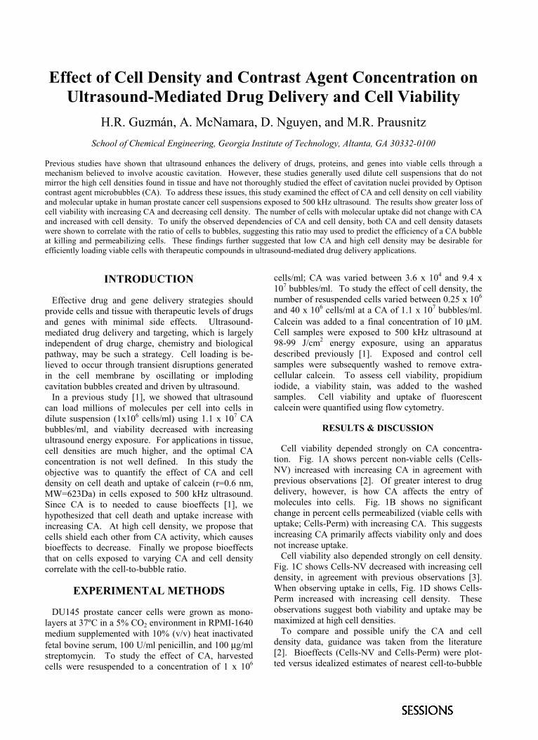
Previous studies have shown that ultrasound enhances the delivery of drugs, proteins, and genes into viable cells through a mechanism believed to involve acoustic cavitation. However, these studies generally used dilute cell suspensions that do not mirror the high cell densities found in tissue and have not thoroughly studied the effect of cavitation nuclei provided by Optison contrast agent microbubbles (CA). To address these issues, this study examined the effect of CA and cell density on cell viability and molecular uptake in human prostate cancer cell suspensions exposed to 500 kHz ultrasound. The results show greater loss of cell viability with increasing CA and decreasing cell density. The number of cells with molecular uptake did not change with CA and increased with cell density. To unify the observed dependencies of CA and cell density, both CA and cell density datasets were shown to correlate with the ratio of cells to bubbles, suggesting this ratio may used to predict the efficiency of a CA bubble at killing and permeabilizing cells. These findings further suggested that low CA and high cell density may be desirable for efficiently loading viable cells with therapeutic compounds in ultrasound-mediated drug delivery applications.
INTRODUCTION
Effective drug and gene delivery strategies should provide cells and tissue with therapeutic levels of drugs and genes with minimal side effects. Ultrasound-mediated drug delivery and targeting, which is largely independent of drug charge, chemistry and biological pathway, may be such a strategy. Cell loading is be-lieved to occur through transient disruptions generated in the cell membrane by oscillating or imploding cavitation bubbles created and driven by ultrasound. In a previous study [1], we showed that ultrasound can load millions of molecules per cell into cells in dilute suspension (1x10⁶ cells/ml) using 1.1 x 10⁷ CA bubbles/ml, and viability decreased with increasing ultrasound energy exposure. For applications in tissue, cell densities are much higher, and the optimal CA concentration is not well defined. In this study the objective was to quantify the effect of CA and cell density on cell death and uptake of calcein (r=0.6 nm, MW=623Da) in cells exposed to 500 kHz ultrasound. Since CA is to needed to cause bioeffects [1], we hypothesized that cell death and uptake increase with increasing CA. At high cell density, we propose that cells shield each other from CA activity, which causes bioeffects to decrease. Finally we propose bioeffects that on cells exposed to varying CA and cell density correlate with the cell-to-bubble ratio.
EXPERIMENTAL METHODS
DU145 prostate cancer cells were grown as mono-layers at 37ºC in a 5% CO2 environment in RPMI-1640 medium supplemented with 10% (v/v) heat inactivated fetal bovine serum, 100 U/ml penicillin, and 100 µg/ml streptomycin. To study the effect of CA, harvested cells were resuspended to a concentration of 1 x 10⁶
cells/ml; CA was varied between 3.6 x 10⁴ and 9.4 x 10⁷ bubbles/ml. To study the effect of cell density, the number of resuspended cells varied between 0.25 x 10⁶ and 40 x 106 cells/ml at a CA of 1.1 x 10⁷ bubbles/ml. Calcein was added to a final concentration of 10 µM. Cell samples were exposed to 500 kHz ultrasound at 98-99 J/cm2 energy exposure, using an apparatus described previously [1]. Exposed and control cell samples were subsequently washed to remove extra-cellular calcein. To assess cell viability, propidium iodide, a viability stain, was added to the washed samples. Cell viability and uptake of fluorescent calcein were quantified using flow cytometry.
RESULTS & DISCUSSION Cell viability depended strongly on CA concentra-tion. Fig. 1A shows percent non-viable cells (Cells-NV) increased with increasing CA in agreement with previous observations [2]. Of greater interest to drug delivery, however, is how CA affects the entry of molecules into cells. Fig. 1B shows no significant change in percent cells permeabilized (viable cells with uptake; Cells-Perm) with increasing CA. This suggests increasing CA primarily affects viability only and does not increase uptake. Cell viability also depended strongly on cell density. Fig. 1C shows Cells-NV decreased with increasing cell density, in agreement with previous observations [3]. When observing uptake in cells, Fig. 1D shows Cells-Perm increased with increasing cell density. These observations suggest both viability and uptake may be maximized at high cell densities. To compare and possible unify the CA and cell density data, guidance was taken from the literature [2]. Bioeffects (Cells-NV and Cells-Perm) were plot-ted versus idealized estimates of nearest cell-to-bubble

distance (CB-Distance) and the cell-to-bubble ratio (CB-Ratio) for the combination of cells and bubbles used (not shown). When all of the data for Cells-NV and Cells-Perm were plotted versus CB-Ratio, there was considerable scatter (R2 range: 0.23-0.18). Simi-larly, when the bioeffects were plotted as functions of CB-Distance, there was even greater scatter (R2 range: 0.03-0.04). Thus, these parameters which accounted for both CA and cell density could not unify the data. It occurred to us, however, that predicting the percent cells, non-viable or permeabilized, may not be appropriate given the wide variation in cell and bubble concentrations. Instead, the efficiency of a bubble to affect a cell may be a better metric. To test this, the efficiency of a bubble rendering cells non-viable was plotted versus CB-Ratio. Figure 1E shows non-viable cells per bubble increased linearly with increasing CB-Ratio in both the CA and cell density datasets (R2=0.93). Likewise, Figure 1F shows the efficiency of a bubble permeabilizing a cell increased linearly with increasing CB-Ratio (R2=0.87). This suggests low CB-Ratios may be equally as effective at making cells non-viable as high CB-Ratios. It also suggests that a relatively small number of bubbles is needed to permeabilize a large percent of cells. To further show that the efficiency of a bubble at causing bioeffects correlates with CB-Ratio, cell suspensions having four different combinations of CA and cell densities providing each of two CB-Ratios (10 and 0.1) were exposed to 98 J/cm2 ultrasound. The results showed that bioeffects did not vary at constant
CB-Ratio (p>0.05), but did vary between the two CB-Ratio used (p<0.001, not shown).
CONCLUSIONS
Cells-NV increased with CA and decreased with cell density. Although Cells-Perm increased with cell density, no change was observed with CA. Per bubble efficiency of achieving both bioeffects correlated with CB-Ratio; Cells-NV decreased and Cells-Perm increased, suggesting low CA and high cell density are desirable for drug delivery into viable cells. The data suggest that the efficiency of a bubble at inducing bioeffects can be predicted by the CB-Ratio.
ACKNOWLEDGEMENTS
We acknowledge the Whitaker Foundation, National Science Foundation, and National Institute of Health for financial support.
REFERENCES 1. Guzmán, H.R., Nguyen, D.X., Khan, S., and Prausnitz,
M.R., J. Acoustic Soc. Am., 110, In Press (2001) 2. Ward, M., Wu, J., and Chiu, J., Ultrasound Med. Biol.,
25, 1169-1175 (2000). 3. Ellwart, J.W., Brettel, H., Kober., L.O. Ultrasound Med.
Biol., 14, 43-50 (1988)
0%
25%
50%
75%
100%
0.1 1 10 100
0.01 0.1 1 10 100
0.01 0.1 1 10 100
0%
10%
20%
30%
40%
0.1 1 10 100
10-1 100 101 102 10-2 10-1 100 101 102
10-1 100 101 102 10-2 10-1 100 101 102
100
10-1
10-2
10-3
10-4
10-5
Perm
eabi
lzed
Cel
ls /
Bub
ble
Non
-via
ble
Cel
ls /
Bubb
le
102
101
100
10-1
10-2
10-3
CA (bubbles/ml) Cell Density (cells/ml) Cell to Bubble Ratio
0%
10%
20%
30%
40%
0%
25%
50%
75%
100%
CA (bubbles/ml) Cell Density (cells/ml) Cell to Bubble Ratio
Non
-via
ble
Cel
ls
Non
-via
ble
Cel
ls
Perm
eabi
lzed
Cel
ls
Perm
eabi
lzed
Cel
ls
A C
B
E
D F
104 105 106 107 108
104 105 106 107 108
Fig. 1. (A) Percent cells killed increase with CA concentration (One-way ANOVA p<0.05). (B) Percent permeabilized cells did not vary with CA concentration (p=0.14). (C) Percent cells killed decreased with increasing cell density (p<0.01). (D)Percent permeabilized cells increases with cell density (p<0.05). (E) Cells killed per bubble increases linearly with cell tobubble ratio (R2=0.93, slope = 0.87). (F) Cells permeabilized per bubble increases linearly with cell to bubble ratio (R2=0.86, slope = 1.20). The exposure conditions used were: ○ = 98 and ● = 99 J/cm2. Data is shown as mean ± SEM.

Rose Bengal Derivatives for Sonodynamic Treatment
K. Kawabata, N. Sugita, K. Sasaki and S. Umemura
Central Research Laboratory, Hitachi Ltd., 185-0081 Tokyo, Japan
Amphiphilic derivatives of rose bengal (RB) are synthesized to accumulate it into tumor tissues while maintaining its ability topromote acoustic cavitation. Such derivatives are very important for developing 'sonodynamic treatment' which utilizes thesynergistic effects of ultrasound and chemicals. Several kinds of derivatives with alkyl groups were synthesized and one ofthem were found to be amphiphilic (hydrophilic and lipophilic) while others were either hydrophilic or lipophilic. Theamphiphilic derivative reduced the cavitation threshold in water as lower as RB do. Tissue distribution of the derivative inmice were investigated and found that the derivative with alkyl group having more than 10 carbon atoms tends to accumulatein tumor tissues. Then we started therapeutic experiments with tumor bearing mice. In Preliminary experiments, a legion wasobserved by the combination of the RB derivative and ultrasound (1.0+2.0 MHz, 40 W/cm2 each 3min) while no clear changewas observed with ultrasound alone.
INTRODUCTION
Cavitation is a typical non-thermal bio-effectinduced by ultrasound. Although its therapeuticapplication is much less studied than thermal effects,cavitational effects may be useful for therapeuticapplication if it can be induced in controlled ways. Asan approach, we are studying for "sonodynamictherapy" [1] which utilizes chemical effects induced byacoustic cavitation and enhanced by certain sensitizers.Sonodynamic therapy has the potential for low-invasive and highly selective tumor treatment if it iscombined with suitable ultrasound exposure methodsand sensitizers.
These sensitizers must possess three properties:(1) lower acoustic intensity threshold of cavitation,(2) accumulate into tumor tissues, and(3) enhance cavitational anti-tumor effects.
Until now, certain porphyrin dyes have been foundto show properties (2) and (3)[1], and certain xanthenedyes have been found to show (1) and (3)[2]. Still,chemicals with all three properties have not yet beenfound. Accordingly, we synthesized new chemicalsaimed at attaining all three properties. Our approach isto add property (2) to xanthene dyes, which alreadypossess properties (1) and (3).
Results on their properties and effects on mousetumor will be described in this paper.
MATERIALAS AND METHODS
Chemicals
Figure 1 shows the structures of synthesized rosebengal (RB) derivatives. RB was reacted with
brominated alkane or brominated carboxylic acid inDMF at 70 °C for 4-8 hours. The resulting derivativeswere purified by open column chromatography. Thereagents for derivatives were purchased from WakoChemical Industries (Osaka, Japan).
FIGURE 1. Structures of RB derivatives
Measuring Concentration of Derivatives inMice Tissues
Tissue Preparation: Derivatives (10 mg/kg) wereintravenously administered into CDF1 mice (male, 5-weeks-old) inoculated with Colon26 tumor cells 1-2weeks before the administration. 2-6 hours afteradministration, the mice were sacrificed and theirtissues were sampled.Measurement: An index for concentrations ofderivatives in tissues was obtained by directmeasurement of fluorescence intensity in tissues.Intensities of the fluorescence specific to thederivatives (ex. 500 nm) and tissues (ex. 450 nm) weremeasured. The relative intensities of the derivatives totissues were used as the index for concentration.
Setup for Sonodynamic Treatment ofTumor bearing Mice
Derivatives were administered into tumor-bearingCDF1 mice under the same conditions as described inthe previous section. Mice were exposed to ultrasoundat 1.0 and 2.0 MHz (40 W/cm2 each) for 3 minutes 6hours after administration with a focused typeultrasound transducer (spherical curvature radius of 35mm, F number = 1) in a water tank filled withdegassed water.
RESULTS AND DISCUSSION
Solubility of Derivatives
Table 1. Solubility of RB and its derivatives
Table 1 shows the solubility of the derivatives towater and organic solvent (chloroform) measured at20ºC. Carbon chain length (n in Fig.1) was set to 7.RB, the starting material, is highly hydrophilic andpoorly lipophilic. On the other hand, derivative 1,which replaces RB's hydrophilic group (-COOH) witha lipophilic group (-CnH2n+1), is poorly hydrophilicand highly lipophilic. Derivative 2, which correspondsto derivative 1 except that is has an extra hydrophilicgroup, shows a little hydrophilicity but dissolvespoorly in chloroform. Derivative 3, which as the samenumber of hydrophilic group as derivative 2, dissolvesin both water and chloroform. Further experimentswere carried out by using derivative 3 because itshowed the best amphiphilicity.
Accumulation of Derivatives in TumorTissues
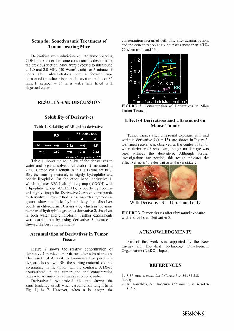
Figure 2 shows the relative concentration ofderivative 3 in mice tumor tissues after administration.The results of ATX-70, a tumor-selective porphyrindye, are also shown. RB, the starting material, did notaccumulate in the tumor. On the contrary, ATX-70accumulated in the tumor and the concentrationincreased as time after administration proceeded.
Derivative 3, synthesized this time, showed thesame tendency as RB when carbon chain length (n inFig. 1) is 7. However, when n is longer, the
concentration increased with time after administration,and the concentration at six hour was more than ATX-70 when n=11 and 13.
FIGURE 2. Concentration of Derivatives in MiceTumor Tissues
Effect of Derivatives and Ultrasound onMouse Tumor
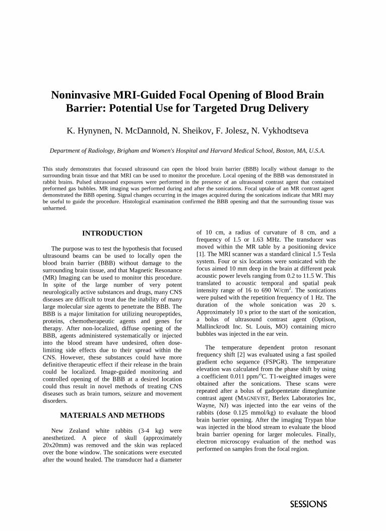
Tumor tissues after ultrasound exposure with andwithout derivative 3 (n = 13) are shown in Figure 3.Damaged region was observed at the center of tumorwhen derivative 3 was used, though no damage wasseen without the derivative. Although furtherinvestigations are needed, this result indicates theeffectiveness of the derivative as the sensitizer.
With Derivative 3 Ultrasound only
FIGURE 3. Tumor tissues after ultrasound exposurewith and without Derivative 3.
ACKNOWLEDGMENTS
Part of this work was supported by the NewEnergy and Industrial Technology DevelopmentOrganization (NEDO), Japan.
REFERENCES
1. S. Umemura, et at., Jpn J. Cancer Res. 84 582-588(1993)2. K. Kawabata, S. Umemura Ultrasonics 35 469-474
(1997)

Gene Transfer by Sonoporation
K. Tachibana
First Department of Anatomy, Fukuoka University School of Medicine, 814-0180 Fukuoka, Japan
Recently, in vitro and in vivo experiment have demonstrated that echo contrast agent microbubbles can be intentionallyruptured by diagnostic and therapeutic ultrasound. This acoustically induced destruction and collapse of the microbubblesproduces a high amplitude response. Violent microstreaming can be produced during microbubble collapse. Researchers havehypothesized that these microjets or microstreaming could be applied to promote diffusion of drugs into various tissues andlesions. Additionally, drug-filled or drug-coated microspheres carrying a therapeutic compound may be targeted to specifictissues through the use of sonic energy, which is directed to the target area and causes the microspheres to rupture and releasethe therapeutic compound. Targeted drug delivery methods are particularly important where the toxicity of the drug is an issue.Specific drug delivery methods potentially serve to minimize toxic side effects, lower the required dosage amounts, anddecrease costs for the patient. The most exciting application of this method is probably gene therapy and sonodynamic therapy.Recent advance on this topic will be discussed.
INTRODUCTION
Arterial occlusive diseases cause serious ischemicconditions in various organs, such as the heart, brainand leg. Therapeutic angiogenesis is believed to bebeneficial for such conditions. Intramuscular injectionof naked plasmid DNA encoding angiogenic growthfactors offers a promising new approach for suchpurposes, however, only a small amount will passthrough the cell membrane leading to low gene-transfer efficiency. Recent studies have shown thattherapeutic ultrasound can induce or increase cellmembrane permeabilization of various agents [1-5]. Itis currently suggested that the mechanism of thisphenomenon is closely related with acousticcavitation. High ultrasound intensities are required tocreate cavitation within tissues such as the skeletalmuscles and myocardium. This study addressed thehypothesis that commercially availableultrasonography contrast agent microbubbles could beused to increase gene transfection efficiency byrelatively low intensity ultrasound-mediatedmicrobubble destruction in vivo.
METHODS
Two different types of ultrasonography contrast agentmicrobubbles (0.02mL; Albunex or Optison, bothMolecular Biosystems) were separately mixed togetherwith a commercial reporter plasmid DNA (25µg;pGeneGrip, GTS Inc.) encoding green fluorescent
protein (GFP) immediately prior intramuscularinjection into mouse thigh quadriceps muscle. Afterthe mouse was anesthetized with 50mg/kg ketamineand 10 mg/kg xylazine, therapeutic ultrasound (1MHz) was irradiated to the plasmid DNA injectedmuscle site at an intensity of 2.0 W/cm2 for 2 minutes.Mice were sacrificed 7 days after ultrasound treatmentfor gene expression assay. Frozen cross-sections (7µmthick) were cut with a cryostat and affixed onto glassslides. Fluorescence distribution patterns wereobserved by confocal laser microscopy (Carl Zeiss)and quantified by computer image analysis. A dualwave length laser (480, 530nm) and digital imagesubtraction method was devised to eliminate autofluorescence of normal muscle tissue.
RESULTS
Muscle tissue irradiated with ultrasound in thepresence of albumin-coated, air-filled Albunexmicrobubble revealed no difference of the number ofGFP expressing muscle fibers as compared withultrasound alone (Fig 1). In contrast, albumin-coated,octafluoropropane gas-filled Optison microbubbleshowed a 10-fold increase in the number of GFPexpressing fibers. No significant enhancement wereobserved with Albunex alone or Optison alone.Histological examination of ultrasound irradiatedmuscle tissue revealed no evidence of inflammation ornecrosis.

CONCLUSION
Low intensity ultrasound increased the transfection ofnaked plasmid DNA of skeletal muscle in the presenceof octafluoropropane-filled Optison microbubbles butnot in air-filled Albunex. As Optison has a longer lifespan than Albunex as a bubble, gene transfection maybe attributed to repeated or slower bubble destructionduring ultrasound irradiation resulting in greaternumber of cell membrane poration. Although furtherinvestigation should be performed to find the optimaltype of gas-filled microbubble, already commerciallyavailable ultrasonography contrast agent, Optisonmight be used as a modality for efficient gene deliveryinto cells for induction of angiogensis and treatment ofvarious diseases in the near future.
ACKNOWLEDGMENTS
This work was supported in part by the FukuokaUniversity Central Research Institute grant 981001 andEKOS Corporation (Bothell, WA USA).
REFERENCES
1. Lawrie A, Brisken AF, Francis SE, Tayler DI,Chamberlain J, Crossman DC, Cumberland DC,Newman CM: Ultrasound enhances reporter geneexpression after transfection of vascular cells in vitro.Circulation, 99, 2617-2620 (1999).
2. Bao S, Thrall BD, Gies RA, Miller DL: In vivotransfection of melanoma cells by lithotripter shockwaves. Cancer Res, 58, 219-221 (1998).
3. Greenleaf WJ, Bolander ME, Sarkar G, Goldring MB,Greenleaf JF: Artificial cavitation nuclei significantlyenhance acoustically induced cell transfection.Ultrasound Med & Biol, 24, 587-595 (1998).
4. Tachibana K, Uchida T, Ogawa K, Yamashita N, TamuraK: Induction of cell-membrane porosity by ultrasound.Lancet, 353, 1409 (1999).
5. Manome Y, Nakamura M, Ohno T, Furuhata H:Ultrasound facilitates transduction of naked plasmidDNA into colon carcinoma cells in vitro and in vivo.Hum Gene Ther, 11, 1521-1528 (2000).
0
20
40
60
80
100
120
140
160
180
Num
ber
of G
FP e
xpre
ssin
g fib
ers *
FIGURE 1. Comparison of the number of GFP expressing fibers with or without ultrasonograpy contrast agents. Asteriskindicate significant differences (n=7, ANOVA, p<0.05).
Noninvasive MRI-Guided Focal Opening of Blood Brain Barrier: Potential Use for Targeted Drug Delivery
K. Hynynen, N. McDannold, N. Sheikov, F. Jolesz, N. Vykhodtseva
Department of Radiology, Brigham and Women's Hospital and Harvard Medical School, Boston, MA, U.S.A.
This study demonstrates that focused ultrasound can open the blood brain barrier (BBB) locally without damage to the surrounding brain tissue and that MRI can be used to monitor the procedure. Local opening of the BBB was demonstrated in rabbit brains. Pulsed ultrasound exposures were performed in the presence of an ultrasound contrast agent that contained preformed gas bubbles. MR imaging was performed during and after the sonications. Focal uptake of an MR contrast agent demonstrated the BBB opening. Signal changes occurring in the images acquired during the sonications indicate that MRI may be useful to guide the procedure. Histological examination confirmed the BBB opening and that the surrounding tissue was unharmed.
INTRODUCTION
The purpose was to test the hypothesis that focused ultrasound beams can be used to locally open the blood brain barrier (BBB) without damage to the surrounding brain tissue, and that Magnetic Resonance (MR) Imaging can be used to monitor this procedure. In spite of the large number of very potent neurologically active substances and drugs, many CNS diseases are difficult to treat due the inability of many large molecular size agents to penetrate the BBB. The BBB is a major limitation for utilizing neuropeptides, proteins, chemotherapeutic agents and genes for therapy. After non-localized, diffuse opening of the BBB, agents administered systematically or injected into the blood stream have undesired, often dose-limiting side effects due to their spread within the CNS. However, these substances could have more definitive therapeutic effect if their release in the brain could be localized. Image-guided monitoring and controlled opening of the BBB at a desired location could thus result in novel methods of treating CNS diseases such as brain tumors, seizure and movement disorders.
MATERIALS AND METHODS
New Zealand white rabbits (3-4 kg) were anesthetized. A piece of skull (approximately 20x20mm) was removed and the skin was replaced over the bone window. The sonications were executed after the wound healed. The transducer had a diameter
of 10 cm, a radius of curvature of 8 cm, and a frequency of 1.5 or 1.63 MHz. The transducer was moved within the MR table by a positioning device [1]. The MRI scanner was a standard clinical 1.5 Tesla system. Four or six locations were sonicated with the focus aimed 10 mm deep in the brain at different peak acoustic power levels ranging from 0.2 to 11.5 W. This translated to acoustic temporal and spatial peak intensity range of 16 to 690 W/cm2. The sonications were pulsed with the repetition frequency of 1 Hz. The duration of the whole sonication was 20 s. Approximately 10 s prior to the start of the sonication, a bolus of ultrasound contrast agent (Optison, Mallinckrodt Inc. St. Louis, MO) containing micro bubbles was injected in the ear vein.
The temperature dependent proton resonant frequency shift [2] was evaluated using a fast spoiled gradient echo sequence (FSPGR). The temperature elevation was calculated from the phase shift by using a coefficient 0.011 ppm/oC. T1-weighted images were obtained after the sonications. These scans were repeated after a bolus of gadopentetate dimeglumine contrast agent (MAGNEVIST, Berlex Laboratories Inc, Wayne, NJ) was injected into the ear veins of the rabbits (dose 0.125 mmol/kg) to evaluate the blood brain barrier opening. After the imaging Trypan blue was injected in the blood stream to evaluate the blood brain barrier opening for larger molecules. Finally, electron microscopy evaluation of the method was performed on samples from the focal region.

RESULTS
The focal signal intensity (SI) change after the MRI contrast agent injection increased with increasing pressure amplitude (figure 1). The magnitude images of FSPGR acquired during the sonications showed a reduction in SI at the focal locations. This reduction, which remained after the sonications, was pressure amplitude dependent. The contrast enhancement correlated with the signal intensity change observed in the magnitude images acquired during the sonications. The trypan blue was focally visible in the post mortem brains (figure 2). The three lowest power levels did not show any damage to the neurons in the histological evaluation.
FIGURE 1. T1-weighted image across the focal plane after sonications and contrast agent injection. The focal enhancement at the focal locations is clearly seen.
DISCUSSION
These results demonstrate that on-line MRI can be used to monitor the magnitude of the opening so, on-line monitoring may be feasible. This finding can have significant potential in targeted drug delivery.
ACKNOWLEDGMENTS
This research was supported by NIH grants #46627 and equipment from TxSonics.
REFERENCES
1. H.E. Cline et al. Radiology, vol. 194, pp. 731-737, 1995
2. A. Chung et al., Med. Phys, vol. 26, pp. 2017-2026, 1999.
3. Vykhodtseva, et al., Ultras. Med. Biol. 21 (7): 969-979,1995.
Trypan blue stained area
Hemorraghic areaTrypan blue stained area
FIGURE 2: Brain section at the focal plane across the focus demonstrating the diffusion of Trypan blue into the brain (2 areas) and one area of focal tissue damage and hemorrhage.

Self-Assembled Molecular Structures as Ultrasonically-Responsive Coating for Pulsatile Drug Delivery
C. S. Kwoka,b, T. J. Matulac, A. Braymanc, P. Mouradc, L. A. Cruma,c, andB. D. Ratnera,b
a Department of Bioengineering and b University of Washington Engineered Biomaterials (UWEB)c Center for Industrial and Medical Ultrasound (CIMU), Applied Physics Laboratory
University of Washington, Seattle, WA 98195, USA
A drug-containing polymer coated with a novel ultrasound-responsive overlayer was developed. The overlayer is a self-assembled molecular structure based on relatively impermeable, ordered n-alkyl chains that form an ultrasound-activated “on-off switch” in controlling drug release on demand, while keeping the drug inside the polymer carrier in theabsence of ultrasound. The chemical preparation and characterization of the overlayer as well as the ultrasound releaseprofiles have been recently published [Kwok et al., Biomacromolecules 1(1), 139-148, (2000) and Kwok et al. Journalof Biomedical Materials Research 57(2), 151-164, (2001)]. In this short communication, we were interested ininvestigating the acoustics-release mechanism. Both the power intensity and the Fourier Transformed waveforms athigh power intensities (~2.8 W/cm2) indicated that the cavitation was responsible for the enhancement in drug release.
INTRODUCTION
Ultrasound has been shown to be effective inenhancing a variety of drugs released frombiodegradable [1] and non-biodegradable [2]polymers as well as from polymeric micelles [3].Ultrasound has also been reported to be effective inassisting transdermal drug delivery for a number ofimportant biomolecules [4]. The advantage ofemploying ultrasound-assisted delivery is itsconvenient, on-demand feature that one can use tointervene and control the release rate externally,compared to the otherwise traditional, constantrelease devices. However, most polymer deliverysystems suffer from substantial backgroundleaching after sonication and shorten the lifetime ofthe device.
To address this issue, we have designed andsynthesized a drug-containing polymer gel surface-immobilized with a self-assembled coating basedon the long n-alkyl chains [5]. This coating isrelatively impermeable; however, when ultrasoundis applied, we hypothesize that the alkyl chains aretemporarily disrupted by cavitation and result inrapid release of the drug. Upon termination ofultrasound, the coating quickly re-assembles backinto an impermeable barrier and prevents furtherdrug leaching. It appears that such drug deliverydevice coated with an ultrasound-responsive and asmart self-healing barrier can potentially developinto a non-invasive, alternative treatment fordiabetics. Insulin was used as a model drug and itsultrasound-modulated release has been publishedelsewhere [6].
Although the augmented effect of acousticenergy on drug release has been clearlydemonstrated, the exact mechanism has not beenfully established. Current consensus is that theincreased drug release by ultrasound exposure isprimarily due to increased acoustic cavitation andacoustic microstreaming [7], which are closelydependent on the ultrasound frequency, presence ofexisting bubbles, duty cycle and ultrasoundtreatment time. In this short communication, wewere interested in varying the power intensity andattempted to correlate its effect with the insulinrelease.
EXPERIMENTAL
The polymer preparations [poly (hydroxyl ethylmethacrylate gel) pHEMA], the coating protocolsand the insulin release procedures were describedin detail elsewhere [6]. To study the effect ofpower intensity on insulin release, a resistance-variable rheostat was added to the power inlet ofthe 40 kHz sonicating bath (Aquasonic 40kHz,Model 75HT) to control the sonic power intensityat different variac settings of 10, 20, 40, 80 and115. A hydrophone (Bruel & Kjær, Type 8103,calibration factor = 25.1�V/Pa) was placed in thesonicating bath and measured the voltage deliveredat each setting. The voltage was then calibrated tothe appropriate pressure (Pa) and power intensity(Isapa, spatial average peak average power intensity,W/cm2). All the insulin released during the 5-minsonication was quantified by UVspectrophotometry and the release rates werecalculated accordingly.

RESULTS AND DISCUSSION
Insulin-containing pHEMA coated with self-assembled n-alkyl chains were exposed to theultrasound at various power intensities for 5minutes continuously. The procedure was repeateddaily for seven consecutive days. The ultrasound-induced release rates of all seven days at eachpower intensity were averaged and plotted againstIsapa as shown in Figure 1. It clearly shows that theinsulin peak release was power intensity dependentand two thresholds appeared to exist. At between 0to 0.5 W/cm2, a slight increase of insulin releasewas observed. Then at Isapa of 1.5 to 2.8 W/cm2,acoustic energy appeared to play a more effectiverole in triggering insulin release. The first moderaterelease was probably due to the stable inertialcavitation whereas the more effective release waslikely resulting from the transient inertialcavitation. Though operating at differentultrasound frequencies, other researchers [3, 8]have also observed similar threshold-dependentphenomenon and testified that the sudden enhancedbioeffect was primarily due to the onset of transientcavitation events.
Figure 1: Effect of power intensity on insulin releaserates.
To further investigate and confirm if thetransient cavitation was responsible for theeffective insulin peak release, the ultrasoundwaveform detected by the hydrophone at eachpower intensity was collected on an oscilloscopeand mathematically reduced with FourierTransform treatment. The fourier transformed(FFT) waveforms were shown in Figure 2, whichclearly verified that at low Isapa of 0.47 W/cm2
(Figure 2a), there were mostly narrow band noisewith increasing harmonics, indicating the existenceof stable cavitation; however, at high Isapaof 2.81W/cm2 (Figure 2b), the “broad-band” noise wasobserved, a strong indication of transientcavitation. The broadband noise was due theviolent collapse of the bubbles, leading to a“white” noise consisting of numerous ranges offrequencies. These evidence, including the
presence of a threshold for effective insulin peakrelease and the broad band in FFT waveform athigh Isapa, suggested that transient cavitation wasresponsible for the ultrasound-assisted drugrelease.
Figure 2: FFT of waveforms taken at (a) Isapa= 0.47W/cm2 (b) ISAPA = 2.81 W/cm2.
CONCLUSION
The acoustic and insulin release data concludedthat the transient cavitation played a major role intriggering the insulin peak release. Effectiveinsulin release was achieved by disorganizing orshearing the self-assembled coating on the pHEMAsurface via the oscillation and violent collapse ofthe bubbles surrounding it.
ACKNOWLEDGMENTS
The authors gratefully acknowledge the generousfunding from the UWEB and CIMU to support thisresearch work. UWEB grant number: EEC-9529161.
REFERENCES
1. Liu, L., et al., Macromolecules 25, 123-128 (1992).2. Lavon, I., and Kost, J., Journal of Controlled
Release 54, 1-7 (1998).3. Husseini, G.A., et al., Journal of Controlled Release
69, 43-52 (2000).4. Mitragotri, S., Blankschtein, D., and Langer, R.,
Science 269, 850-3 (1995).5. Kwok, C.S., et al., Biomacromolecules, 1(1), 139-
148 (2000).6. Kwok, C.S., et al., Journal of Biomedical Materials
Research 57(2), 151-64 (2001).7. Mitragotri, S., et al., Journal of Controlled Release
63, 41-52 (2000).8. Polichik, S.L., et al., Ultrasound in Med. and Bio.
25(6), 991-98 (1999).
(a) Isapa = 0.47W/cm2
020406080
100120140160
0 50 100 150 200 250 300Frequency, kHz
abs(
fft(a
mpl
))
42 kHz
12482
210166 247
(b) Isapa = 2.81W/cm2
0100200300400500600700
0 50 100 150 200 250 300Frequency, kHz
abs(
fft(a
mpl
))
40 kHz
12580
165 205
Mean insulin release rates at different power intensity,Isapa
0.0
1.0
2.0
3.0
4.0
5.0
6.0
7.0
0 0.5 1 1.5 2 2.5 3Power Intensity (Isapa), W/cm2
Ave
rage
rele
ase
rate
, ug/
min

Ultrasonic atomisers for the immunoprophylaxis of poultry diseases
A. Štimaca, B. Ivančevićb and K. Jambrošićb
a Brodarski Institute, Measurement, Vibration and Acoustics Department, Zagreb, Croatia
b Faculty of Electrical Engineering and Computing, Department of Electroacoustics, Zagreb, Croatia
The article deals with application of ultrasonic atomisation for immunoprophylaxis of poultry against viral diseases. Principles of liquid droplet formation from vaccine suspensions in which cavitation and resonant capillary wave break-ups are generated by means of an ultrasonic field have been analysed. A three-way optimisation process has been applied: results from medical inhalation theory have been used for optimisation of droplet size and distribution; frequency of the ultrasonic field has been determined which generates droplets of given size, and an ultrasonic transducer has been designed for generation of the ultrasonic field which could provide production of required aerosol quantity. Results of research in an experimental vaccination equipment show increased efficiency of the method in comparison to others.
INTRODUCTION The drug delivery with help of aerosols, has been known for a long time, especially for the asthma reme-dies and treatment of bronchitis, where application of aerosols has been particulary successful. Today, possible applications of aerosols in the treatment of chronic bronchitis, cystic fibrosis and migraine are investigatated. The main reasons for intensive investi-gations lies in the fact that inhalation with aerosols as a possible drug delivery has many advantages with respect to the gastrointestinal or the parenteral routes of drug delivery. The application of ultrasonic atomisers in the inhalation starts around 1970, when the main characteristic of application of ultrasonics for aerosol creation were acknowledged [1]. By application of high intensity ultrasonic power to the medium, with known operating frequency and intiensity of ultrasonic waves, the diameter of formed aerosol particles can be directly influenced [2]. Due to this characteristics, we have applied the ultrasonic atomisation theory to the procedure of mass vacination of one-day chickens.
REQUIREMENTS FOR AEROSOL DEPOSITION
On the basis of published literature and scientific articles covering inhalation theory, it is known that [3]: − aerosols droplets with a diameter greater then 30
µm do not enter bronchies, but stay in the upper parts of the respiratory tract,
− aerosols droplets with diameter between 0,5 µm and 5 µm are apsorbed in the alveolas, while
− aerosols droplets with diameter less then 0,5 µm are breathed out in a quantity of over 50 %.
On the basis of this facts, the aim of the project has been to substitute the usually applied expensive and less efficient method of parenteral vaccination of one-day old chickens with a mass vaccination aimed directly into the respiratory system. The current technology enables vaccine preparation for application by means of ultrasonic atomizers. On the basis of mentioned requirements, it follows that the ultrasonic atomiser must have the capability of producing an aerosol in which majority of the droplets will have a size that ensures absorption in the alveolas of the respiratory system. Production of aerosol by means of ultrasonic technology has an unique characte-ristic, that it enables influence on the median droplet diameter by controlling the operating frequency of the ultrasonic transducer, with known specific density and the surface tension of the medium [4,5,6]. According to the capillary wave theory, the corresponding droplet diameter d, can be calculated by means of Lang equation, as
3 2k834,034,0
fd
ρσπλ =≈ (1)
where kλ -capillary wave wavelength (m), σ-surface tension coefficient of the medium (N/m), ρ-specific density of the medium (kg/m3) and f–operating ultrasonic frequency (Hz). With respect to other vacci-ne manufacturers, for this experiment we have been using the vaccine MARIKAL®SPF by PLIVA, that does not need other diluents than sterile redistilled water (σ≈72,3·10-3 N/m; ρ≈1000 kg/m3). If we suppose that the basic requirement for the ultrasonic atomiser is to produce an aerosol with median particle diameter of d=3 µm, the necessary operating frequency is approx. 1,65 MHz. With this operating frequency and an effective electrical output power at the ultrasonic

transducer of Peff ≈ 30 W, droplet sizes in the range of (0,5-6) µm could be obtained, having in mind that in over 90 % of the total volume droplet diameters are under 4 µm [7]. Results in measurement of distribution of particle size are shown in Figure 1.
FIGURE 1. Particle size distribution
By means of an ultrasonic transducer with a radiation surface of 5,2 cm2, a production of (3-5) ml of vaccine suspension per minute could be acchieved. The amount depends on the height of medium above the transducer surface, which implies that a change of the electro-acoustic efficiency coefficent takes place. In order to find the optimal height of the medium above the trans-ducer, electrical input admittance of the transducer as a function of the medium height has been measured. Diagrams of input electrical admittance of the (un)-loaded transducer for optimal medium height of h = 3,5 cm are shown in Figure 2
FIGURE 2. Admitance curve of unloaded and loaded
ultrasonic transducer for ultrasonic atomiser SONOVAC
Method of vaccination and ultrasonic nebulizer for immunoprophylaxis of Marek's and other poultry diseases has been filed with the State Patent Institution of the Republic of Croatia under No. 381-03/45-01/1994; 559-03-91-01 of July 24, 1995.
In order to increase the productivity of the ultrasonic transducer and by that of the vaccination procedure as well, two transducers in parallel work have been used. The complete device (Figure 3.) is designed for vacci-nation of 100 one-day old chickens in a plastic box, with the possibility of continuous adjustment of the vaccination time. The fans are placed within the ultrasonic generator, and they must create an air flow which carries the produced aerosol vaccine into the vaccination chamber filled with chicken.
FIGURE 3. Ultrasonic atomiser SONOVAC
OBTAINED RESULTS By means of investigations performed at the Veterinary Faculty of the University in Zagreb and in production conditions following proofs could be provided: − the procedure did not cause any vaccinal reactions, − vaccinal viruses could be found in the lung tissue
already within six hours and in the bursa of Fabricius within 24 hours after vaccination,
− vaccine showed to be immunogenic, as confirmed by plaque reduction test,
− vaccine applied by atomisation significantly improved the protection of vaccinated poultry, and during both breeding and production period. There were no deaths caused by Marek's disease, while the mortality rate caused by other diseases was significantly reduced.
REFERENCES [1] Abramov, O.V., High-Intensity Ultrasonics, G&B Science
Publishers, Amsterdam 1998, pp. 237-286 [2] Gershenzon, E.L., Eknadiosyants O.K., Acoustic Journal
10, 156-162 (1964) (In Russian) [3] Topp, M.N., Eisenklam, P., Ultrasonics 21, 127-133 (1972) [4] Lang, R.J., JASA. 34, 6-9 (1962) [5] Peskin, R., Raco, R., JASA. 34, 1378-1381 (1963) [6] Drews, W.D., Elektronik 10, 83-86 (1979) [7] Perron, R., IEEE-UFFC 14, 149-153 (1967)
High Intensity Focused Ultrasound Selectively Disrupts theBlood-Brain Barrier in vivo
A.H. Mesiwalaa, L. Farrella, H. J. Wenzela, L. A. Crumb, D. L. Silbergelda, H.R.Winna, and P. D. Mourada,b
aDepartment of Neurological Surgery, University of Washington, Seattle, WA, USAbCenter for Industrial and Medical Ultrasound, Applied Physics Laboratory, University of Washington, Seattle,
WA USA
The blood-brain barrier (BBB) plays a protective role in the central nervous system (CNS), restricting the movement ofmolecules from the blood into the brain. The capillary endothelial cells, astrocytes, and basement membrane that form the BBBalso prevent the flux of therapeutic substances into the CNS. High intensity focused ultrasound (HIFU) has been shown todestroy CNS parenchyma, while disrupting the BBB in the periphery. The mechanism by which HIFU induces BBBdisruption, however, is unknown. Using a rat model, we show that HIFU can selectively disrupt the BBB in a particular regionof interest (ROI) in a reversible, controlled manner. This effect reverses within 72-96 hours, and may or may not be associatedwith tissue damage. Furthermore, using electron microscopy (EM), we demonstrate that HIFU preserves CNS architecture andpropose that HIFU disrupts the BBB by opening capillary endothelial cell tight junctions. This isolated ultrastructural effect ontight junctions is different from the cellular mechanisms through which hyperosmotic solutions, hyperthermia, and percussiveinjury disrupt the BBB. We believe that this work will lay the foundation for future studies on HIFU-mediated BBB disruption,and lead to development of novel CNS therapies designed to optimize and take advantage of HIFU.
BBB PROTOCOL
Cranial windows were created in adult Wistar rats toallow direct application of ultrasound (coupled viatransmission gel) to the cortical surface. Doseresponse curves (not shown) were generated for threeHIFU transducers operating at 2.0, 3.5, and 5.0 MHz; azone was identified in intensity/frequency/pulse-characteristic space that yielded either BBB disruption(characterized by the flux of Evans blue, a blood-bornedye that is normally unable to cross the BBB, intobrain tissue) with no damage or minimal damage involumes of brain treated with HIFU, or no observedeffect. Based on these results, the 2.0 MHztransducer, operating at 485 W/cm2 for 0.2 seconds,was selected for extensive studies of BBB disruption.
BBB OPENING: LIGHT MICROSCOPY
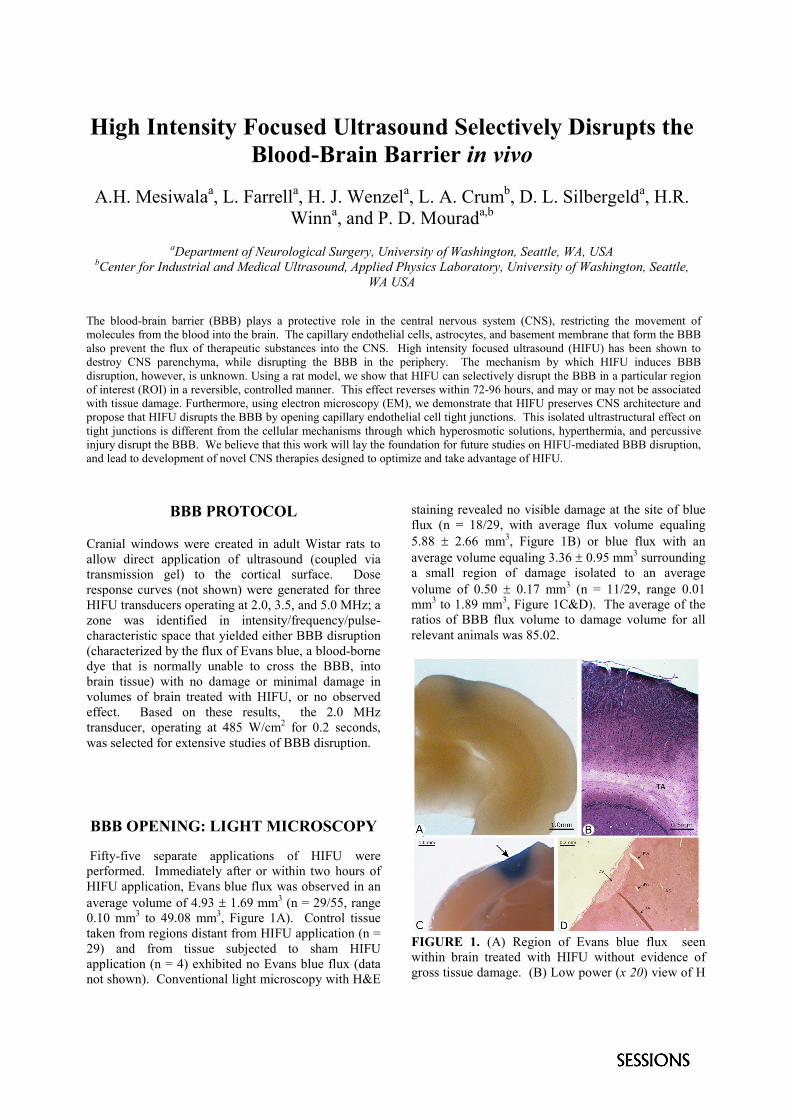
Fifty-five separate applications of HIFU wereperformed. Immediately after or within two hours ofHIFU application, Evans blue flux was observed in anaverage volume of 4.93 � 1.69 mm3 (n = 29/55, range0.10 mm3 to 49.08 mm3, Figure 1A). Control tissuetaken from regions distant from HIFU application (n =29) and from tissue subjected to sham HIFUapplication (n = 4) exhibited no Evans blue flux (datanot shown). Conventional light microscopy with H&E
staining revealed no visible damage at the site of blueflux (n = 18/29, with average flux volume equaling5.88 � 2.66 mm3, Figure 1B) or blue flux with anaverage volume equaling 3.36 � 0.95 mm3 surroundinga small region of damage isolated to an averagevolume of 0.50 � 0.17 mm3 (n = 11/29, range 0.01mm3 to 1.89 mm3, Figure 1C&D). The average of theratios of BBB flux volume to damage volume for allrelevant animals was 85.02.
FIGURE 1. (A) Region of Evans blue flux seenwithin brain treated with HIFU without evidence ofgross tissue damage. (B) Low power (x 20) view of H
& E stained region of BBB disruption shows noevidence of tissue damage, such as edema,disintegration of cellular membranes, or inflammation,only tissue processing artifact. (C) Gross evidence ofBBB opening with concurrent tissue damage (arrow).(D) Low power (x 20) view of H & E stained region ofbrain in image A showing damage to brain tissue dueto HIFU. Note that the stroma within this region isdestroyed and that astrocytes and neurons are invarious stages of degeneration or disruption, includingbeyond the region of stromal disruption. BV1 and BV2denote major blood vessels; BV1, in a zone of normaltissue, shows evidence of edema while BV2 and BV3do not. DS = disrupted stroma, NS = normal stroma,and TA = tissue processing artifact.
BBB OPENING: ELECTRONMICROSCOPY
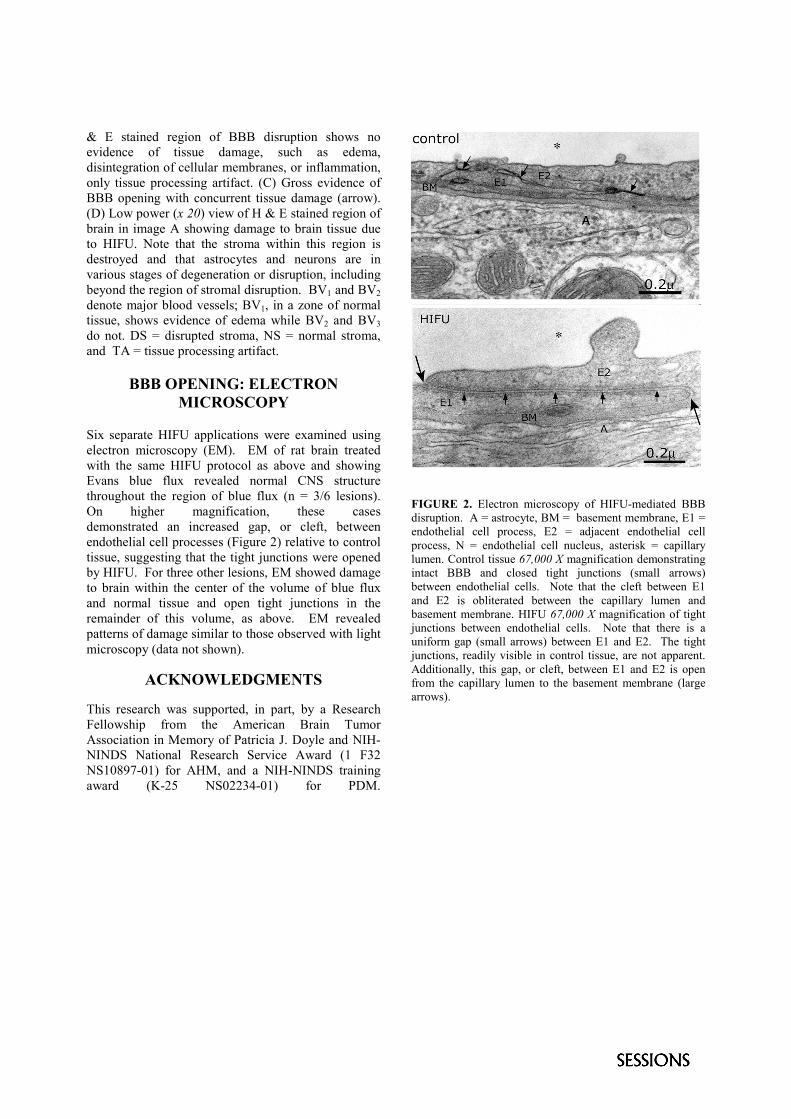
Six separate HIFU applications were examined usingelectron microscopy (EM). EM of rat brain treatedwith the same HIFU protocol as above and showingEvans blue flux revealed normal CNS structurethroughout the region of blue flux (n = 3/6 lesions).On higher magnification, these casesdemonstrated an increased gap, or cleft, betweenendothelial cell processes (Figure 2) relative to controltissue, suggesting that the tight junctions were openedby HIFU. For three other lesions, EM showed damageto brain within the center of the volume of blue fluxand normal tissue and open tight junctions in theremainder of this volume, as above. EM revealedpatterns of damage similar to those observed with lightmicroscopy (data not shown).
ACKNOWLEDGMENTS
This research was supported, in part, by a ResearchFellowship from the American Brain TumorAssociation in Memory of Patricia J. Doyle and NIH-NINDS National Research Service Award (1 F32NS10897-01) for AHM, and a NIH-NINDS trainingaward (K-25 NS02234-01) for PDM.
FIGURE 2. Electron microscopy of HIFU-mediated BBBdisruption. A = astrocyte, BM = basement membrane, E1 =endothelial cell process, E2 = adjacent endothelial cellprocess, N = endothelial cell nucleus, asterisk = capillarylumen. Control tissue 67,000 X magnification demonstratingintact BBB and closed tight junctions (small arrows)between endothelial cells. Note that the cleft between E1and E2 is obliterated between the capillary lumen andbasement membrane. HIFU 67,000 X magnification of tightjunctions between endothelial cells. Note that there is auniform gap (small arrows) between E1 and E2. The tightjunctions, readily visible in control tissue, are not apparent.Additionally, this gap, or cleft, between E1 and E2 is openfrom the capillary lumen to the basement membrane (largearrows).