ultrasound-guided supraclavicular versus infraclavicular ... · ultrasound-guided supraclavicular...
TRANSCRIPT

Egyptian Journal of Anaesthesia (2014) 30, 161–167
Egyptian Society of Anesthesiologists
Egyptian Journal of Anaesthesia
www.elsevier.com/locate/egjawww.sciencedirect.com
Research Article
Ultrasound-guided supraclavicular versus
infraclavicular brachial plexus nerve block
in chronic renal failure patients undergoing
arteriovenous fistula creation
* Corresponding author address: 2 Mostafa Darwish Street, 6th
District, Nasr City, Egypt. Tel.: +20 1221085448.
E-mail addresses: [email protected], [email protected]
(M.A. Mansour).
Peer review under responsibility of Egyptian Society of Anesthesiol-
ogists.
Production and hosting by Elsevier
1110-1849 ª 2014 Production and hosting by Elsevier B.V. on behalf of Egyptian Society of Anesthesiologists.
http://dx.doi.org/10.1016/j.egja.2013.12.006Open access under CC BY-NC-ND license.
Amany El-Sawy a, Nashwa Nabil Mohamed a, Mohamed Ahmed Mansour a,*,
Mona Ramadan Salemb
a Kasr Al-Ainy Hospital, Cairo University, Egyptb International Nozha Hospital, Cairo, Egypt
Received 8 November 2013; revised 17 December 2013; accepted 22 December 2013
Available online 21 January 2014
KEYWORDS
Ultrasound;
Supraclavicular;
Infraclavicular;
Brachial plexus block;
Arteriovenous fistula
Abstract Background: Most patients with chronic renal failure suffer from complications that
make brachial plexus block a good choice for providing anesthesia. The use of ultrasonography
increases the success rate and decreases complications. We compared the efficacy of ultrasound-
guided supraclavicular and infraclavicular brachial plexus block in providing anesthesia for crea-
tion of arteriovenous fistula.
Patients and methods: Sixty adult patients with chronic renal failure, scheduled for creation of arte-
riovenous fistula of the distal upper extremity were randomly divided into two equal groups: Supra
G (n= 30): ultrasonic guided supraclavicular brachial plexus block was given and Infra G (n= 30):
ultrasonic guided infraclavicular brachial plexus block was given. For both groups we used
20–25 cm 1:1 volumes of 0.5% bupivacaine and 2% lidocaine. The measured parameters were block
performance time and related pain, the degree and duration of sensory and motor block, patient
discomfort, first call for analgesics, complications and the patient’s satisfaction.
Results: There was no statistically significant difference between both groups as regard the block
performance time, the block related pain, the degree of sensory and motor block in the areas

162 A. El-Sawy et al.
supplied by the median, radial and musculocutaneous nerves at 10, 20 and 30 min. There was no
statistically significant difference as regard the sensory block grade in the area supplied by the ulnar
nerve at 10 min, but it was significantly higher in the Supra G than Infra G at 20 and 30 min. No
statistically significant difference as regard the motor block grade in the area supplied by the ulnar
nerve, the block duration, first call for analgesia, complications and patients’ satisfaction.
Conclusion: Both approaches can provide satisfactory sensory and motor block, very good analge-
sia that extends for a long time postoperatively in patients with chronic renal failure undergoing
creation of arteriovenous fistula.
ª 2014 Production and hosting by Elsevier B.V. on behalf of Egyptian Society of Anesthesiologists.
Open access under CC BY-NC-ND license.1. Introduction
Patients with chronic renal failure may suffer from serious
complications that represent a great challenge to the anesthesi-ologists. Complications like congestive heart failure, systemichypertension, electrolyte imbalances, metabolic acidosis, coag-
ulopathy, unpredictable intravascular fluid volume status andanemia obligate the anesthesiologist to avoid general anesthe-sia with its heroic risks in these patients and to think for alter-native methods [1].
Brachial plexus block is often used in chronic renal failurepatients to provide anesthesia for the creation or revision ofarteriovenous fistula for hemodialysis access. It provides anal-
gesia, sympathetic blockade, optimal surgical conditions andadequate duration of postoperative block that prevents arterialspasm and graft thrombosis. It provides higher blood flow in
the radial artery and arteriovenous fistula than is achieved withinfiltration anesthesia [2].
Many approaches can be used for brachial plexus block; ax-
illary, supraclavicular and infraclavicular approaches. Theywere commonly performed by blind techniques or neurostimu-lation which may be associated with high failure rate and seri-ous complications. Nowadays; the intraoperative use of
ultrasonography becomes more popular and much easier. Itsuse in these blocks increases the success rate and decreasescomplications [3].
Previous studies had compared ultrasonic guided supracla-vicular and infraclavicular block for upper limb surgery in nor-mal patients [3–5]. They hypothesized that the onset in
supraclavicular block is fast and the blockade is deep as thenerves are very tightly packed but pneumothorax can occurdue to the proximity of the pleura. Pneumothorax can be
avoided by ultrasonic visualization of the pleura and by propertechnique [6].
They also hypothesized that the infraclavicular block ischaracterized by compact anatomical distribution of the plexus
allowing single injection of local anesthetics and the decreasedincidence of pneumothorax. However, it may be associatedwith patient discomfort and technical difficulty, which can be
overcome by the use of ultrasonography [7–9].In a previous study; ultrasonic guided infraclavicular block
was compared with local infiltration anesthesia for creating
vascular access for hemodialysis in patients with chronic renalfailure [2]. But as far as we know; no study had compared be-tween ultrasonic guided supraclavicular and infraclavicularbrachial plexus block in this type of operation. This compari-
son would help if a local cause prevents the use of either ofthem like swelling, infection or obesity.
1.1. Aim of work
The aim of work was to compare the efficacy of ultrasound-guided supraclavicular versus infraclavicular brachial plexusblock in providing anesthesia for creation of arteriovenous fis-
tula in chronic renal failure patients.
2. Patients and methods
The Ethics Committee, Department of Anesthesiology, Fac-ulty of Medicine, Cairo University, approved the protocol ofthis study. This randomized study was conducted on sixty
adult patients with chronic renal failure scheduled for creationof arteriovenous fistula of the distal upper extremity. Patientsenrolled in the present study were of both sexes, aged
20–60 years, and with ASA physical status III. Every patientsigned an informed consent.
Exclusion criteria included the following: neurological, neu-romuscular, psychiatric disorders, hepatic, respiratory, or car-
diac diseases; uncontrolled seizures; coagulation disorders;infection at the block injection site; patients with a body massindex more than 30; or patients who refused the procedure.
All the patients included in the study were on chronichemodialysis and they had a hemodialysis session one day be-fore the block performance. Their routine preoperative labora-
tory investigations were within normal values especiallyprothrombin time (PT), partial thromboplastin time (PTT)and international normalized ratio (INR).
Patients were randomized using computer generated num-ber and concealed using sequentially numbered, sealed opaqueenvelope technique to two groups of 30 patients each:
Supra G (n = 30): Ultrasonic guided supraclavicular bra-chial plexus block group.Infra G (n= 30): Ultrasonic guided infraclavicular brachial
plexus block group.
In both groups the block was performed using a 50 mm 20
G nerve stimulator needle model (Braun). The needle was in-serted in-plane with a linear ultrasonic probe after the nervousand vascular structures were optimally visualized. A depth of
3–4 cm and a frequency of 10–12 Hz was used.The local anesthetic solution used in both groups consisted
of 1:1 volumes of 0.5% bupivacaine and 2% lidocaine (the to-tal volume injected was from 20–25 cm). This solution was
administered in increments with repeated aspiration in be-tween and its characteristic distribution around the nerveswas observed.

Ultrasound-guided supraclavicular versus infraclavicular brachial plexus nerve block 163
No premedication was given to the patients, since full coop-eration during block performance is required.
On arrival to the operating room, an intravenous catheter
was placed in the upper limb contra-lateral to the surgical siteand saline solution was started at 2 mL/kg/h. Standard anes-thesia monitors (ECG, Pulse Oximeter, Non-invasive Blood
Pressure) were applied. Supplemental oxygen (via nasal can-nula at 4 L/min) was used throughout the procedure.
The patients were positioned in the supine position with
the face turned to the contra-lateral side. Proper steriliza-tion of the block area was performed. After proper surgicaldraping and displaying the area of the block with the ultra-sound probe, a local anesthetic (Lidocaine 1%) was injected
subcutaneously.In the supraclavicular group, the ultrasound probe was
positioned in the supraclavicular fossa, pointing caudad and
moved laterally and medially in order to locate the subclavianartery. The hyperechoic first rib was identified deep to the ar-tery and the pleura was identified and its sliding move-
ment during respiration was noted. The plexus wasconsistently found with a characteristic ‘‘honeycomb’’ appear-ance lateral and superficial to the subclavian artery and supe-
rior to the first rib.The needle was introduced through the skin from lateral to
medial, in-plane with the transducer, with constant visualiza-tion, and directed toward the deep border of the nerve group.
Three separate injections were made at various sites in the bun-dle, tending to start deep, in the ‘‘corner pocket’’ close to theartery, and moving more superficially.
In the infraclavicular group, the arm was abducted to 90�with flexion of the elbow to bring the artery and plexus closerto the skin. The coracoid process was identified by palpating
the bony prominence just medial to the shoulder while thearm is elevated and lowered. Scanning was begun just medialto the coracoid process and inferior to the clavicle with the
transducer in the parasagittal plane to identify the axillary ar-tery by its thick wall and brisk pulsations. The pectoralis majorand minor muscles were identified just above the brachial ves-sels and plexus. The hyperechoic cords of the brachial plexus
and their corresponding positions relative to the artery wereidentified.
The needle was inserted in-plane from the cephalad aspect,
with the insertion point just inferior to the clavicle. The needleaimed toward the posterior aspect of the axillary artery andpassed through the pectoralis major and minor muscles. The
injectate used to spread cephalad and caudad to cover the lat-eral and medial cords, respectively. When injection of the localanesthetic with a single injection didn’t appear to result in ade-quate spread, additional needle repositions and injections
around the axillary artery were done. The goal of the techniquewas to inject local anesthetic until it spreads around the arteryin a U-shaped pattern.
2.1. Measured parameters
Block performance time which is defined as the interval be-tween the first needle insertion and its removal at the end ofthe block.
Block performance-related pain was evaluated immediatelyafter removal of the needle by asking the patient to verballyquantify the level of pain using a (VAS) score between 0 and
10, 0 meaning no pain and 10 meaning excruciating pain.
Evaluation of sensory and motor block was performedevery 10 min in musculocutaneous, median, radial, and ulnarnerve territories over a 30-min period beginning when the nee-
dle was withdrawn from the patient.Sensory block was evaluated by comparing the cold sensa-
tion elicited by ice in the central sensory region of each nerve
with the same stimulus delivered to the contra-lateral side.The median nerve block was tested on the skin of the radialhalf of the palm and palmar side of the lateral 3 digits. The ul-
nar nerve block was tested on the skin of the medial side of thewrist and hand; skin of the medial 1 digit. The musculocutane-ous nerve block was tested on the skin of the lateral side of theforearm. The radial nerve block was tested on the skin of the
posterior arm, forearm and hand. The sensory block was
graded as follows: 0 = no difference from the unblockedextremity, 1 = less cold than the unblocked extremity,
2= no sensation of cold.Motor block was evaluated using the forearm flexion,
thumb abduction, thumb and second digit pinch and finger
abduction (for the musculocutaneous, radial, median, and ul-nar nerves, respectively). The motor block was graded as fol-
lows: 0 = no loss of force, 1= reduced force compared with
the unblocked extremity, 2= incapacity to overcome gravity.Surgical anesthesia was defined as surgery without patient
discomfort or the need for supplementation of the block. If apart of the surgical territory was not completely anesthetized
at the time of surgery, the block was supplemented with localanesthetic infiltration. If the patient still experiences pain de-spite supplementation, general anesthesia was used and patient
excluded.The duration of the sensory and motor block was assessed.
The duration of the sensory block was defined as the time be-
tween the end of the local anesthetic injection and the totalrecovery of sensation. The duration of the motor block was de-fined as the time between the end of the local anesthetic injec-
tion and the total recovery of motor functions. The first call foranalgesics was recorded.
The side effects and complications, such as blood vesselpuncture, intravascular injection, overdose, dyspnea, Horner’s
syndrome, and pneumothorax, were noted.The patient’s satisfaction with the anesthetic technique was
assessed after the patient’s arrival in the post-anesthesia care
unit using a 2-point scale (0 = unsatisfied; 1 = satisfied).A post-block chest radiograph was obtained routinely after
surgery. If a patient complained of respiratory distress or any
signs of pneumothorax, a chest radiograph was done using theC arm in OR. All patients were examined after 24 h after sur-gery for the occurrence of complications (bruises/swellings atthe block site, chest pain, breathing difficulty, dysaesthesia,
or muscle weakness in the operated extremity). Surgeons werealerted to report any neurological problems not related to sur-gery during the follow-up visits.
2.2. Power Analysis
A total sample size of 60 patients randomly allocated into twoequal groups (30 patients per group) will have 80% power todetect a large effect size (W) of 0.45 (a error = 0.05, b er-
ror = 0.2, using Chi-Square test of independence). Statisticalpower calculations was performed using computer programG*Power 3 for Windows. (Franz Faul, Universitat Kiel,
Germany).

Table 1 Patients’ demographic criteria.
Demographic Criterion Supra G (n= 30) Infra G (n= 30) P value
Age (years) 44.4 ± 11.3 47.83 ± 7.80 0.17
Weight (kg) 81.83 ± 8.29 79.76 ± 6.15 0.27
Sex male/female 18/12 17/13
Statistically significant between-group difference (P < 0.05).
Table 2 Block performance time and block-related pain.
Parameter Supra G (n= 30) Infra G (n= 30) P value
Block performance time (min) 8.13 ± 1.30 7.73 ± 1.17 0.21
Block-related pain (VAS) 2.73 ± 0.90 2.43 ± 1.10 0.25
n= number of patients.
Statistically significant between – group difference (P < 0.05).
0
0.5
1
1.5
2
2.5
10 20 30
Sens
ory
Blo
ck G
rade
Measurement Time (min)
Supra GInfra G
*
* p < 0.05
*
Figure 3 Ulnar nerve sensory block.
0
0.5
1
1.5
2
2.5
10 20 30
Sens
ory
Blo
ck G
rade
Measurement Time (min)
Supra GInfra G
Figure 1 Median nerve sensory block.
0
0.5
1
1.5
2
2.5
10 20 30
Mot
or B
lock
Gra
de
Measurement Time (min)
Supra GInfra G
Figure 2 Median nerve motor block.
164 A. El-Sawy et al.
2.3. Statistical analysis
Statistical analysis of data was performed using Paired – Sam-ples T test for numerical variables and Chi-square test for
qualitative variables. P < 0.05 was considered statisticallysignificant.
3. Results
This study included 60 adult patients with chronic renal failurescheduled for creation of arteriovenous fistula of the distal
upper extremity, divided into 2 groups of 30 patients each: Su-pra G (supraclavicular brachial plexus block group) and InfraG (infraclavicular brachial plexus block). The block failed in 2
patients in the Supra G and in 3 patients in the Infra G. Those5 patients were excluded from the study and replaced by othersto compensate for failed cases. In the Supra G, one failure wasattributed to inability to clearly visualize the subclavian artery
and the other failure was due to an incomplete block in thearea supplied by the ulnar nerve after 30 min of the block.In the Infra G, 2 failures which included the distribution of
the 4 nerves were attributed to a possible subcutaneous injec-tion that was not recognized and the third failure was a partialblock that excluded the distribution of both the ulnar and the
median nerves.Patients in both groups were of comparable age, weight,
and sex (Table 1).As regard the block performance time and block related
pain: the block performance time was less than 10 min in bothgroups. The mean block performance time was comparable inthe Supra G and the Infra G. The block related pain was com-
parable in the Supra G and the Infra G (Table 2).As regard the median nerve block grade: the sensory and
motor block grades in the area supplied by the median nerve
showed no statistically significant differences between the 2groups at 10, 20 and 30 min measurement times (Figs. 1 and 2).
As regard the ulnar nerve block: The sensory block grade in
the area supplied by the ulnar nerve showed no statistically sig-nificant differences between the 2 groups at 10 min, but it washigher in the Supra G than the Infra G at 20 and 30 min mea-surement times. The difference was significant at 20 min and

0
0.5
1
1.5
2
2.5
10 20 30
Mot
or B
lock
Gra
de
Measurement Time (min)
Supra GInfra G
Figure 4 Ulnar nerve motor block.
0
0.5
1
1.5
2
2.5
10 20 30
Sens
ory
Blo
ck G
rade
Measurement Time (min)
Supra G
Infra G
Figure 7 Musculocutaneous nerve sensory block.
0
0.5
1
1.5
2
2.5
10 20 30
Sens
ory
Blo
ck G
rade
Measurement Time (min)
Supra GInfra G
Figure 5 Radial nerve sensory block.
0
0.5
1
1.5
2
2.5
10 20 30
Mot
or N
erve
Gra
de
Measurement Time (min)
Supra GInfra G
Figure 6 Radial nerve motor block.
0
0.5
1
1.5
2
2.5
10 20 30
Mot
or N
erve
Gra
deMeasurement Time (min)
Supra GInfra G
Figure 8 Musculocutaneous nerve motor block.
Ultrasound-guided supraclavicular versus infraclavicular brachial plexus nerve block 165
30 min. The motor block grade in the area supplied by the ul-nar nerve was comparable in the 2 groups at 10, 20 and 30 min
measurement times (Figs. 3 and 4).As regard radial nerve block: The sensory and motor block
grades in the area supplied by the radial nerve showed no sta-
tistically significant differences between the 2 groups at 10, 20and 30 min measurement times (Figs. 5 and 6).
As regard musculocutaneous nerve block: The sensory and
motor block grades in the area supplied by the musculocutane-ous nerve showed no statistically significant differences be-tween the 2 groups at 10, 20 and 30 min measurement times(Figs. 7 and 8).
As regard the motor block duration, sensory block dura-tion and first call for analgesia: they were comparable in SupraG and Infra G (Table 3).
Regarding patients’ satisfaction, 90% and 87% of patientswere satisfied in the Supra G and the Infra G respectively. Thedifference between the 2 groups was not statistically signifi-
cant. Three patients in the Supra G were unsatisfied, 2 wereunhappy about the idea of being awake during the surgeryand one was unhappy with the pain that accompanied the
block performance. Four patients in the Infra G were unsatis-fied, 2 patients were unhappy with the pain that accompaniedthe block performance and 2 patients did not like the feeling ofbeing unable to move their limb for a long time (Table 4).
None of the patients in both groups had intravascular injec-tion or developed local hematoma or pneumothorax. Surgeonsdid not report dysaesthesia, or muscle weakness in the oper-
ated extremity in the 1 week follow up visit of the patients.
4. Discussion
In our current study both the ultrasound-guided supraclavicu-lar and infraclavicular approaches to the brachial plexus havebeen compared in patients with chronic renal failure scheduled
for creation of arteriovenous fistula of the distal upperextremity.
The results of this study showed that the block performance
time was less than 10 min and was comparable in both groups.This result is consistent with many previous studies. In a
prospective randomized study comparing ultrasound-guidedinfraclavicular versus supraclavicular block, Arcand and his
colleagues [3] reported that although ultrasonic visualizationwas more rapid in the infraclavicular region than in the supra-clavicular region, block performance times were similar:
(4.0 ± 3.3) min and (4.7 ± 4.0) min for infraclavicular groupand supraclavicular group respectively. In a more recent study,Koscielniak-Nielsen and his colleagues [5] compared ultra-
sound-guided supraclavicular and infraclavicular blocks forupper extremity surgery and showed similar block perfor-mance times for both approaches: (5.0 ± 1.6) min in the infra-
clavicular group and (5.7 ± 1.6) min in the supraclaviculargroup.
Studies on ultrasound-guided supraclavicular block showedblock performance time of 9 min in a study done by Chan and
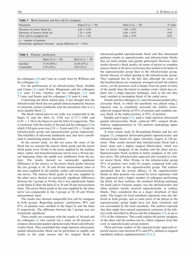
Table 3 Block duration and first call for analgesia.
Parameter Supra G (n= 30) Infra G (n= 30) P value
Duration of motor block (h) 6.26 ± 0.98 5.86 ± 0.97 0.11
Duration of sensory block (h) 7.36 ± 0.99 6.86 ± 0.97 0.05
First call for analgesia (h) 9.33 ± 1.02 8.86 ± 0.93 0.07
n= number of patients.
Statistically significant between – group difference (P < 0.05).
Table 4 Patients’ satisfaction.
Supra G (n= 30) Infra G (n= 30)
Satisfied 27/30 (90%) 26/30 (87%)
Unsatisfied 3/30 (10%) 4/30 (13%)
166 A. El-Sawy et al.
his colleagues [10] and 5 min in a study done by Williams andhis colleagues [6].
For the performance of an infraclavicular block, Sandhuand Capan [11] used 10 min, Dingemans and his colleagues[12] used 3.1 min, Gurkan and his colleagues [13] used
7.1 min, and Sauter and his colleagues [14] used 4.1 min.Concerning the block related pain, it was thought that the
infraclavicular block has not gained clinical popularity becauseof uncertain surface landmarks and the perception that it is a
more painful block [7].The block related pain in our study was comparable in the
Supra G and the Infra G, VAS was (2.73 ± 0.90) and
(2.43 ± 1.10) in the Supra G and the Infra G respectively. Thisis consistent with the results of Arcand and his colleagues [3] inwhich VAS pain score was (2.0 ± 2) min and (2.0 ± 2) min for
infraclavicular group and supraclavicular group respectively.The reliability of ultrasonic landmarks may also have contrib-uted to minimizing patient discomfort.
In the current study, we did not assess the onset of theblock but we assessed the sensory block grade and the motorblock grade every 10 min in the areas supplied by the median,ulnar, radial, and musculocutaneous nerves over a 30-min per-
iod beginning when the needle was withdrawn from the pa-tient. The results showed no statistically significantdifferences in the sensory or the motor block grades between
the two groups at 10, 20 and 30 min measurement times inthe areas supplied by the median, radial, and musculocutane-ous nerves. The sensory block grade in the area supplied by
the ulnar nerve showed no statistically significant differencesbetween the 2 groups at 10 min, but it was significantly higherin the Supra G than the Infra G at 20 and 30 min measurementtimes. The motor block grade in the area supplied by the ulnar
nerve was comparable in the 2 groups at 10, 20 and 30 minmeasurement times.
The results also showed comparable first call for analgesia
in both groups. Regarding patients’ satisfaction; 90% and87% of patients were satisfied in the Supra G and the InfraG respectively. The difference between the 2 groups was not
statistically significant.These results are consistent with the results of Arcand and
his colleagues [3] who carried out a study on 80 patients to
compare ultrasound guided supraclavicular block and infracla-vicular block. They concluded that single injection ultrasound-guided infraclavicular block can be performed as rapidly andresults in the same success rate for surgical block as
ultrasound-guided supraclavicular block and that ultrasoundguidance results in supraclavicular and infraclavicular blocks
that are both reliable and quickly performed. However, theirresults showed a block quality (in terms of partial or completesensory block of all nerve territories) that tended to be better inthe supraclavicular group than in the infraclavicular group,
mostly because of radial sparing in the infraclavicular group.They explained this by the fact that although the cords ofthe brachial plexus are compactly arranged around the axillary
artery, yet the posterior cord is deeper from the point of entryof the needle than the lateral or median cords, which may ex-plain why a single injection technique, such as the one they
used, resulted in incomplete block of the radial nerve.Ootaki and his colleagues [8], used ultrasound guided infra-
clavicular block, in which the anesthetic was placed using 2injection sites to completely surround the axillary artery,
achieved surgical blocks in 95% of patients and complete sen-sory block of the radial territory in 95% of patients.
Sandhu and Capan [11], used a triple injection ultrasound
guided infraclavicular block, achieved 90% surgical blockswithout supplementation in 126 patients undergoing upperextremity surgery.
A more recent study by Koscielniak-Nielsen and his col-leagues [5], compared ultrasound-guided supraclavicular andinfraclavicular blocks for upper extremity surgery in 120 pa-
tients. Their results showed that infraclavicular block had afaster onset and a higher surgical effectiveness, which wasdue to better analgesia of the median and the ulnar nerves.Supraclavicular block resulted in better analgesia of the axil-
lary nerve. The infraclavicular approach also resulted in a bet-ter motor block. After 30 min, in the infraclavicular group93% of patients were ready for surgery compared with only
78% of patients in the supraclavicular group. The authorsspeculated that the poorer efficacy of the supraclavicularblocks in their patients was caused by lower experience with
this approach and a higher number of colleagues performingthe block. In their institute, the standard blocking techniquefor hand and/or forearm surgery was the infraclavicular andobese patients mostly received supraclavicular or axillary
blocks. They considered this as a major drawback of theirstudy. The targets for local anesthetic injections were also dif-ferent in both groups, and so some parts of the plexus in the
supraclavicular group might have not been visualized andnot surrounded by the local anesthetic. The authors reportedthat they might have missed anatomical variations of the infe-
rior trunk described by Royse and his colleagues [15], in up to15% of the volunteers. This could explain the poorer analgesiaof the ulnar and the median nerves, which originate from this
cord, in supraclavicular group patients.
Three previous studies of the supraclavicular approach re-ported success rates between 85% and 95%, defined as surgicalanesthesia without supplementation [10,6,3].

Ultrasound-guided supraclavicular versus infraclavicular brachial plexus nerve block 167
Previous studies of the infraclavicular approach reportedsuccess rates of 80% [3], between 86% and 95% [12–14], andbetween 90% and 99% [8,9].
A recent study by Fredrickson et al. [4] compared an ultra-sound guided supraclavicular block using multiple injectionswith ultrasound guided triple injection infraclavicular block.
They reported that the corner pocket supraclavicular andinfraclavicular brachial plexus block were associated with sim-ilar onset times and sensory blockade at 30 min.
A more recent study by Yang and his colleagues [16], com-pared infraclavicular and supraclavicular approaches to thebrachial plexus using neurostimulation in 100 patients. Theirresults showed no significant differences in the evolution of
the sensory block over 50 min in the two groups but the sen-sory block was significantly better in the supraclavicular groupat 20 min in the ulnar nerve territory. The progression of the
motor block paralleled that of the sensory block and therewere no significant differences in the evolution of the motorblock with time. There was no significant difference in the pro-
portion of the complete sensory or motor block over time.There were no significant differences between the two groupsin the duration of the sensory and motor block. There were
no significant differences in the level of patients’ satisfactionbetween the two groups. The authors concluded that boththe supraclavicular and infraclavicular approach to the brachiaplexus had similar clinical efficacy.
As regards the possible complications in the current study,none of the patients in both groups had intravascular injectionor developed local hematoma or pneumothorax. Surgeons did
not report dysaesthesia, or muscle weakness in the operatedextremity in the 1 week follow up visit of the patients.
Perlas and his colleagues [17], reported that an ultrasound-
guided supraclavicular block was associated with a high suc-cess rate and low complication rate with no pneumothoraxin a series of 510 consecutive patients. They suggested that
ultrasound-guided supraclavicular block might reduce the riskof pneumothorax because the pleura and first rib are often easyto visualize.
Arcand and his colleagues [3] mentioned that no pneumo-
thorax has been reported in any study of supraclavicular orinfraclavicular block using ultrasound guidance.
Koscielniak-Nielsen and his colleagues [5], who compared
ultrasound-guided supraclavicular and infraclavicular blocksfor upper extremity surgery reported Horner’s syndrome in29% and suspected diaphragmatic paresis in 12% of patients
in the supraclavicular group. Diaphragmatic paresis was seenas a change in the breathing pattern and/or coughing difficulty.The incidence of vascular punctures was 2% in both groups.
5. Conclusion
The results of the current study showed that both supraclavic-ular and infraclavicular approaches to the brachial plexus were
comparable in providing very satisfactory sensory and motorblock in patients with chronic renal failure undergoing crea-tion of arteriovenous fistula of the distal upper extremity. Both
blocks provided very good analgesia that extended for a longtime postoperatively. Patients were satisfied with both blocksand no complications were reported.
The anesthesiologist can use either supraclavicular or infra-clavicular blocks satisfactorily. This helps a lot when a local
cause like swelling, infection, or obesity prevents the use ofeither of them. So, the other approach would work.
Conflict of interest
No conflict of interest.
References
[1] Holt NF. Renal disease. In: Hines Roberta L, Marschall
Katherine E, editors. Stoelting’s anesthesia and co-existing
disease, 6th ed.; 2012. p. 339–56.
[2] Sahin L, Gul R, Mizrak A, et al. Ultrasound-guided
infraclavicular brachial plexus block enhances postoperative
blood flow in arteriovenous fistulas. J Vasc Surg
2011;54(3):749–53.
[3] Arcand G, Williams SR, Chouinard P, et al. Ultrasound-guided
infraclavicular versus supraclavicular block. Anesth Analg
2005;101:886–90.
[4] Fredrickson MJ, Patel A, Young S, Chinchanwala S. Speed of
onset of ‘ corner pocket supraclavicular’ and infraclavicular
ultrasound guided brachial plexus block: a randomized
observer- blinded comparison. Anesthesia 2009;64:738–44.
[5] Koscielniak-Nielsen ZJ, Frederiksen BS, Rasmussen H, et al. A
comparison of ultrasound- guided supraclavicular and
infraclavicular blocks for upper extremity surgery. Acta
Anesthesiol Scand 2009;53:620–6.
[6] Williams SR, Chouinard P, Arcand G, et al. Ultrasound
guidance speeds execution and improves the quality of
supraclavicular block. Anesth Analg 2003;97:1518–23.
[7] Klaastad O, Lilleas FG, Rotnes JS, et al. A magnetic resonance
imaging study of modifications to the infraclavicular brachial
plexus block. Anesth Analg 2000;91:929–33.
[8] Ootaki C, Hayashi H, Amano M. Ultrasound- guided
infraclavicular brachial plexus block: an alternative technique
to anatomical landmark- guided approaches. Reg Anesth Pain
Med 2000;25:600–4.
[9] Sandhu NS, Manne JS, Medabalmi PK, Capan LM.
Sonographically guided infraclavicular brachial plexus block in
adults: a retrospective analysis of 1146 cases. J Ultrasound Med
2006;25:1555–61.
[10] Chan VWS, Perlas A, Rawson R, Odukoya O. Ultrasound
guided supraclavicular brachial plexus block. Anesth Analg
2003;97:1514–7.
[11] Sandhu NS, Capan LM. Ultrasound- guided infraclavicular
brachial plexus block. Br J Anaesth 2002;89:254–9.
[12] Dingemans E, Williams SR, Arcand G, et al. Neurostimulation
in ultrasound guided infraclavicular block: a prospective
randomized trial. Anesth Analg 2007;104:1275–80.
[13] Gurkan Y, Acar S, Solak M, et al. Comparison of nerve
stimulation vs. ultrasound- guided lateral sagittal infraclavicular
block. Acta Anesthesiol Scand 2008;52:851–5.
[14] Sauter AR, Dodgson MS, Stubhaug A, et al. Electrical nerve
stimulation or ultrasound guidance for lateral sagittal
infraclavicular blocks: a randomized, controlled, observer-
blinded comparative study. Anesth Analg 2008;106:1910–5.
[15] Royse CF, Sha S, Soeding PF, Royse AG. Anatomical study of
the brachial plexus using surface ultrasound. Anaesth Intensive
Care 2006;34:203–10.
[16] Yang CW, Kwon HU, Cho CK, et al. A comparison of
infraclavicular and supraclavicular approaches to the brachial
plexus using neurostimulation. Kor J Anesthesiol 2010;58(3),
260-26.
[17] Perlas A, Lobo G, Lo N, et al. Ultrasound guided
supraclavicular block: outcome of 510 consecutive cases. Reg
Anesth Pain Med 2009;34:171–6.