ultrasound of median nerve -...
TRANSCRIPT
Ultrasound of Median Nerve
Neuromuscular Conference
Dr. Hemang Shah
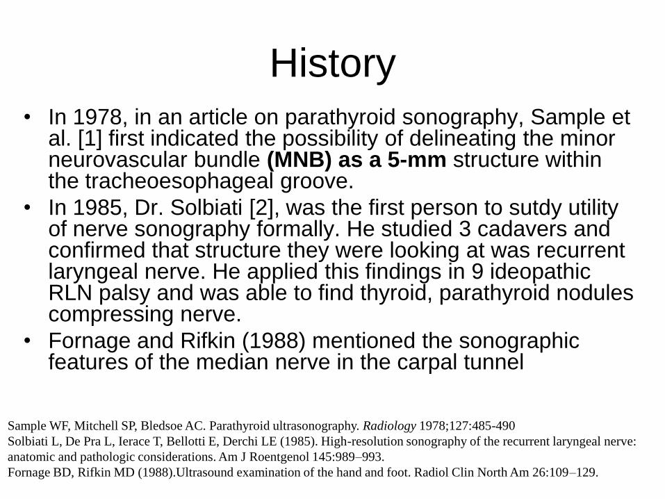
History• In 1978, in an article on parathyroid sonography, Sample et
al. [1] first indicated the possibility of delineating the minor neurovascular bundle (MNB) as a 5-mm structure within the tracheoesophageal groove.
• In 1985, Dr. Solbiati [2], was the first person to sutdy utility of nerve sonography formally. He studied 3 cadavers and confirmed that structure they were looking at was recurrent laryngeal nerve. He applied this findings in 9 ideopathic RLN palsy and was able to find thyroid, parathyroid nodules compressing nerve.
• Fornage and Rifkin (1988) mentioned the sonographic features of the median nerve in the carpal tunnel
Sample WF, Mitchell SP, Bledsoe AC. Parathyroid ultrasonography. Radiology 1978;127:485-490
Solbiati L, De Pra L, Ierace T, Bellotti E, Derchi LE (1985). High-resolution sonography of the recurrent laryngeal nerve:
anatomic and pathologic considerations. Am J Roentgenol 145:989–993.
Fornage BD, Rifkin MD (1988).Ultrasound examination of the hand and foot. Radiol Clin North Am 26:109–129.
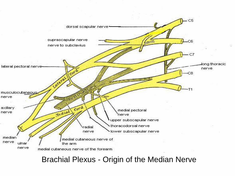
Brachial Plexus - Origin of the Median Nerve

Course of the Median Nerve- Arm and Forearm


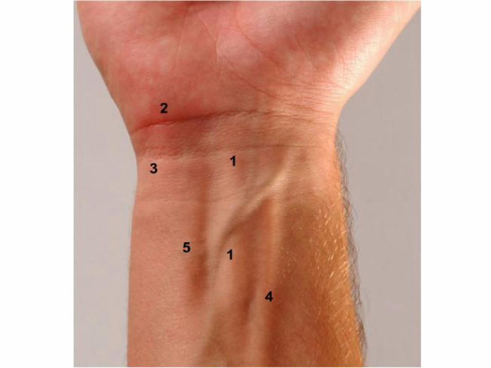
Course of the Median Nerve

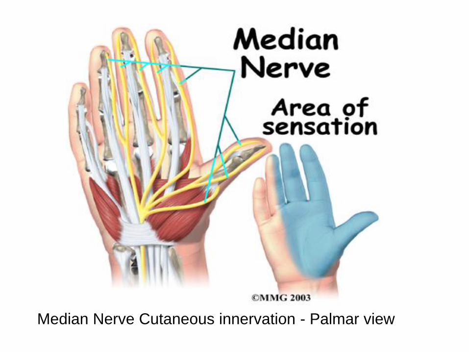
Median Nerve Cutaneous innervation - Palmar view
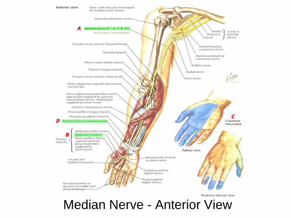
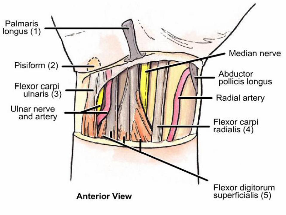
Median Nerve - Anterior View


Technical aspects of sonography
• Electromechanical transducer within a probe converts electrical signals into ultrasonic waves.
• Piezoelectric effect - At the boundary of two tissues with different echogenic properties (interface) the ultrasonic waves are reflected and subsequently received by the transducer again
• Average velocity of sound through various soft tissues is relatively constant (1540 m/s),
• Musculoskeletal ultrasound: linear array transducers
• Resolution: Contrast and Spatial (axial and lateral)

Technical aspects of sonography
• Peripheral nerves requires the use of transducers with high insonation frequencies (usually 7–12 MHz or more).
• How to improve axial resolution? (increase insonation frequecy, disadvantage)
• How to improve lateral resolution? (smaller beam width, decrease frame rate, multiple rows of crystals)
• Compound imaging: repeated overlapping sequences of frames of different predetermined view angles.

Technical Limitations
• Deeply situated nerves are difficult to visualize.
• Nerves are difficult to visualize if surrounded by fat or beneath bone.
• Relatively small field of view, extended-field-of-view technique to reconstruct a panoramic view.
• Correct positioning: If probe not perpendicular to nerve, may falsely be interpreted as being hypoechoic (anisotropy – change angle around)
• Pressure on the skin should be kept to a minimum in order to avoid artificial deformation of small superficial structures.
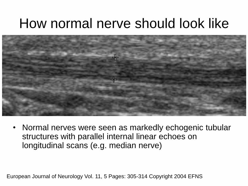
How normal nerve should look like
• Normal nerves were seen as markedly echogenic tubular structures with parallel internal linear echoes on longitudinal scans (e.g. median nerve)
European Journal of Neurology Vol. 11, 5 Pages: 305-314 Copyright 2004 EFNS
How normal nerve should look like
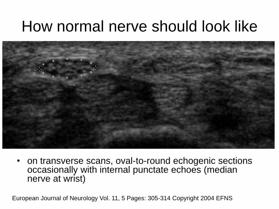
• on transverse scans, oval-to-round echogenic sections occasionally with internal punctate echoes (median nerve at wrist)
European Journal of Neurology Vol. 11, 5 Pages: 305-314 Copyright 2004 EFNS

How normal nerve should look like
• The internal structure of the cadaveric nerves as multiple hypoechoic parallel but discontinuous linear areas separated by hyperechoic bands when scanning in a longitudinal plane and scanning transversely as multiple rounded hypoechoic areas in a homogeneous hyperechoic background (fascicular or honeycomb pattern – not always seen).
• Hypoechoic areas – fascicles; hyperechoic background -epineurium
• Sonograms underestimated the number of fascicles may be due to:
(1)The inability to depict fascicles unless they are perpendicular to the ultrasound beam
(2)Poor lateral resolution that results in coalescence of adjacent structures of similar echogenicity.
How normal nerve should look like
Differentiation from muscles and tendons :
• Tendons appeared to have numerous fine parallel hyperechoic lines separated by fine hypoechoic lines (fibrillar pattern).
• Relative immobility of nerves during flexion–extension manoeuvres
• Perineurium of the sciatic nerve produced bright boundary echoes and variation of the insonation angle reduced echogenicity of the nerve to a lesser extent than that of muscles or tendons.*
* Grechenig W, Clement H, Peicha G, Klein A, Weiglein A (2000). Die Sonoanatomie des Nervus
Ischiadicus am Oberschenkel. Biomedische Technik 45: 298–303.
Normal Dimensions
• Modern ultrasound machines are usually equipped with software to easily perform these measurements accurate to the tenth of a millimeter.
• Many studies do not report whether they include or exclude the hyperechoic rim surrounding the nerve, most investigators perform measurements within this rim as it is not always present and its outer margin may be difficult to define.*
• A correlative cadaver study found that sonography was a very precise method to assess the dimensions (dorsopalmar diameter, radioulnar diameter, perimeter and area) of the median nerve at the carpal tunnel. (Kamolz et al. (2001)
• Nerve thickness has a Gaussian distribution and did not correlate with the subjects' height, weight or age.** (some studies found that normal values might be different in men and women.)
* Nakamichi K, Tachibana S (2002). Ultrasonographic measurement of median nerve cross-
ectional area in idiopathic carpal tunnel syndrome: diagnostic accuracy. Muscle Nerve 26: 798–803
** Heinemeyer O, Reimers CD (1999). Ultrasound of radial, ulnar, median, and sciatic nerves in
healthy subjects and patients with hereditary motor and sensory neuropathies. Ultrasound Med Biol
25: 481–485
Median nerve
• Ultrasonographic Reference Values for Assessing the Normal Median Nerve in Adults*
• Note they looked at whole course of median nerve.
• They also looked at amount of muscle in CT (lumbricles and FD).
• 4 sonographers, first established inter-rater reliability.
• 50 subjects, 100 arms looked at the six sites were: (1) distal wrist crease, (2) mid-forearm (mid-point between distal wrist crease and elbow crease), (3) proximal forearm (where the nerve enters the pronator teres muscle), (4) antecubital fossa, (5) mid-humerus (mid-point between elbow crease and axilla), and (6) axilla
* Francis O. Walker, MD @ Wake Forest, NC Journal of Neuroimaging, Volume
19, Number 1, January 2009 , pp. 47-51(5)

Both images are taken at the distal wrist crease and show a cross-section of the
median nerve. In the top image, the nerve is the oval hypoechoic structure in the
center, and in the bottom image the nerve is outlined with the trace function. This nerve
measures 10.2 mm2.
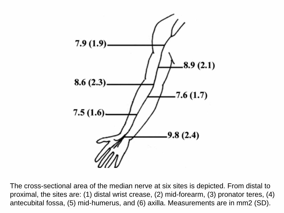
The cross-sectional area of the median nerve at six sites is depicted. From distal to
proximal, the sites are: (1) distal wrist crease, (2) mid-forearm, (3) pronator teres, (4)
antecubital fossa, (5) mid-humerus, and (6) axilla. Measurements are in mm2 (SD).

Really?
• the nerve was largest at the most distal region, the size of the nerve did not vary greatly throughout its entire length (7.5 to 9.8 mm2).
• 14.6 mm2 (the mean plus 2 SD) would be a cut off for the upper limit of normal. (others report a cut off value between 9 and 12 mm2). (too high, age and weight)
• the amount of muscle in the carpal tunnel in patients with CTS could be measured with ultrasound.
• Sonographers were not blinded
• nerve conduction studies were not performed to evaluate for subclinical neuropathy.
• Nerve ultrasound can provide anatomic information, such as the presence of intraneural masses or cysts
• Ultrasound as complementry to NCS. (e.g. ECHO to EKG)
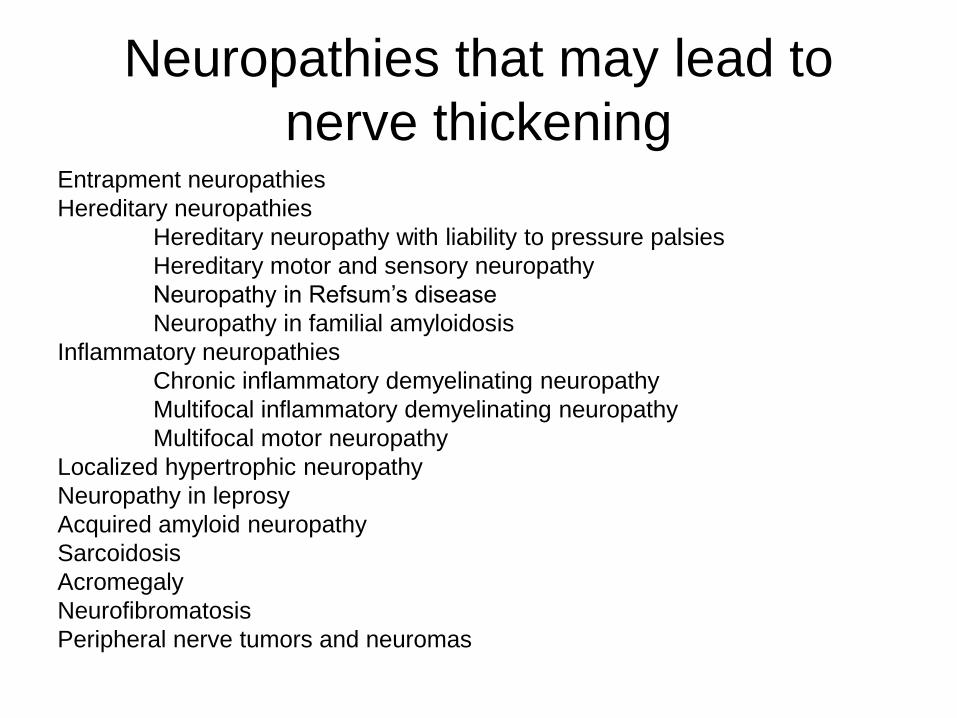
Entrapment neuropathies
Hereditary neuropathies
Hereditary neuropathy with liability to pressure palsies
Hereditary motor and sensory neuropathy
Neuropathy in Refsum’s disease
Neuropathy in familial amyloidosis
Inflammatory neuropathies
Chronic inflammatory demyelinating neuropathy
Multifocal inflammatory demyelinating neuropathy
Multifocal motor neuropathy
Localized hypertrophic neuropathy
Neuropathy in leprosy
Acquired amyloid neuropathy
Sarcoidosis
Acromegaly
Neurofibromatosis
Peripheral nerve tumors and neuromas
Neuropathies that may lead to
nerve thickening
Common clinical presentations
Carpal Tunnel Syndrome:
• In 1913, Marie and Foix first talked about compression of median nerve at carpal tunnel causing thenar atrophy.
• Presentation: both hands involvement, dominant more common, sensory changes in median distribution, nocturnal distribution, upward progression of pain, flick sign, tinel’s at wrist, phalen’s sign, weakness/atrophy of thenar muscles, might have double crush syndrome (cervical radiculopathy).
• Predisposing conditions: wt. gain, diabetes, hypothyroidism, arthritis.
• NCS: prove that conduction across the wrist is prolonged,
Pronator Terese Syndrome:
• Anatomy, presentation may mimic CTS,
weakness of muscles proximal to wrist, tinel’s at
site of entrapment, aggravated by pronation,
elbow felxion and 2nd digit flexion. Abnormal
needle exam of median innervated forearm
muscles.
Anterior Interosseous Syndrome:
• Pinch maneuver: FPL, PQ, 2nd digit FDP
Ligament of Struthers
• Need to know this because of interventional
cardiologist
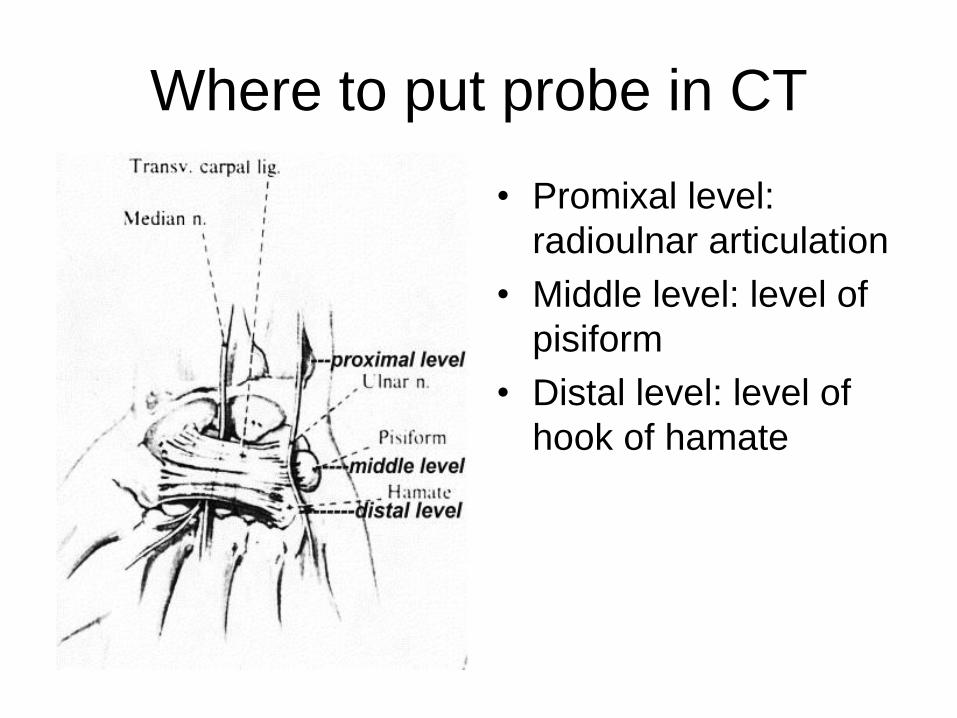
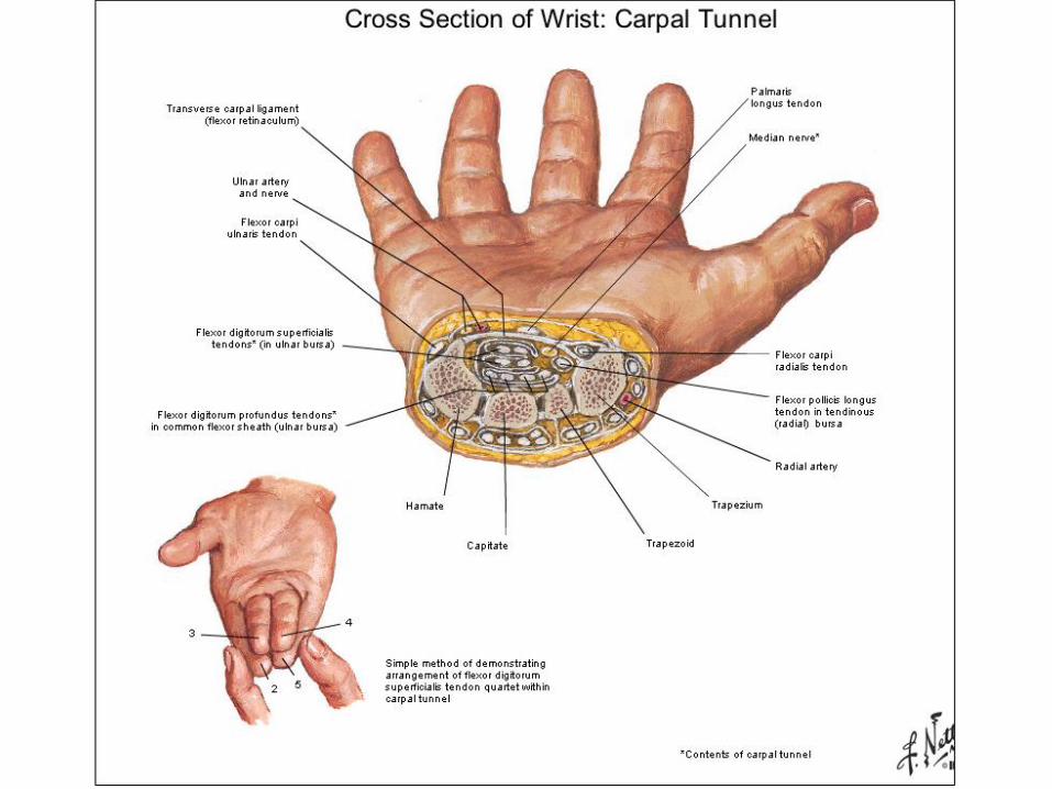
Where to put probe in CT
• Promixal level:
radioulnar articulation
• Middle level: level of
pisiform
• Distal level: level of
hook of hamate
Take home message
• The main pathological changes that can be demonstrated are nerve enlargement and increased hypoechogenicity.
• The best studied peripheral neuropathy is the carpal tunnel syndrome in which ultrasonography.
• US cheap, readily available, non invasive, safe (operator dependent, small field of view, can’t go deep)
• the final aim of all examinations in CTS is to determine the cause(s) of upper limb paresthesiae, not simply if there is a median nerve lesion at wrist or not.
• Most common cause of perepheral neuropathy world wide?
• Nerve imaging atlas
http://nerveatlas.ucsf.edu/atlas.html
• Let’s play with real thing