u.n.c. endo. lit. summary - college of diplomates · pdf filepoor correlation between clinical...
TRANSCRIPT
1
U.N.C.Endo. Lit. Summary
By
Peter Z. Tawil, DMD, MS, FRCD(C)Diplomate, American Board of Endodontics
U.N.C. Endo Lit Summary (V. 2014)By Peter Zahi Tawil, DMD, MS, FRCD(C)

2
Diagnosis
Smoking and EndoKrall et al 2006
• 811 patients VA in Boston• Smokers are 1.7 times more likely to have a root canal• There is a statistical dose-response relationship between cigarette smoking and the risk of root
canal treatment.
Bergstrom J 2004 (Journal of Oral Sciences)• Tobacco smoking not associated with apical periodontitis
HIV & EndoShetty 2006
• 157 HIV patients• No statistically significant differences were noted when the success of the root canal therapy
was related to the symptomatic clinical presentation, the antiretroviral therapy, or the viral load.
Suchina, Hicks et al 2006 (Houston, TX)• Despite obturation deficiencies and the immunocompromised state of the patients, endodontic
therapy has a relatively high degree of success in the majority of HIV/AIDS patients. HIV infection and AIDS should not be considered as a contraindication to endodontic therapy in this patient population.
Quesnell et al 2005 (JOE / Chicago)• 33 HIV pt and 33 healthy. PAI score no difference at 12 months.
Cooper et al1993• Short-term success was determined by follow-up appointments 1-3 months following
obturation. No complications were experienced in either group, except with one HIV infected patient. The results of this clinical study indicate that root canal treatment can be carried out following standard procedures and without antibiotic prophylaxis.
U.N.C. Endo Lit Summary (V. 2014)By Peter Zahi Tawil, DMD, MS, FRCD(C)

3
Age & EndoNg, Mann, Rahbaran, Lewsey & Gulabivala 2008
• Age decrease healing
Cardiovascular disease, Blood pressure & EndoChen et al 2007
• Hypertension decreases healingHersh 1993
• Maximum dose of epi for patients with mild or well-controlled cardiovascular disease and for patient on tricyclic antidepressants is 0.04mg (2 cartridges of 1:100 000)
Wang C et al 2011 (JOE)• An increased risk of tooth extraction after root canal therapy was significantly associated with
Diabetes Mellitus, Hypertension, and Coronary Artery Disease individually. Moreover, the constellation of systemic disease burden also manifests the importance in addition to other potential confounders.
Bleeding disorders AND RCT
Herman 1997• Dental treatment should be postponed if INR exceed 3.5
Sickle cell disease and RCT
Costa et al 2013 JOE• Sickle cell anemia is a potential risk factor for pulp necrosis in clinically intact permanent teeth
Kawa 2004• In sickle cell disease macrophages are too busy eating diseased cells and are not available to
kill bacteria• Spontaneous necrosis can happen due to blockage of the microcirculation.
U.N.C. Endo Lit Summary (V. 2014)By Peter Zahi Tawil, DMD, MS, FRCD(C)
4
Innate immune system & Endo
Marending, Peters, Zehnder, 2005 (OOOO, Switzerland)• Host factors were taken into account and Logistic regression was done• Diabetes, Renal insufficiency, Breast Cancer, etc. had effect on healing• Pre-op PAI, Innate immune system, and quality of root filling all had effect on outcome.• The integrity of a patient's nonspecific immune system, which has been neglected in earlier
investigations, is a significant predictor for endodontic treatment outcome, and should receive more attention in future studies.
Mindiola et al 2006 (JOE / Cleveland)• 5460 teeth done by Endo’s and GP• Diabetes, Hypertension, age and no restoration decrease retention of teeth after RCT.
Fouad 2003• Review Diabetes and RCT
Fouad, Burleson 2003• Reduction is success after RCT in diabetic patients who presents with CAP
Bender & Bender 2003• When Diabetes is under control, the healing of periapical lesions heal the same as a non-
diabetic.
Wang C et al 2011 (JOE)• An increased risk of tooth extraction after root canal therapy was significantly associated with
Diabetes Mellitus, Hypertension, and Coronary Artery Disease individually. Moreover, the constellation of systemic disease burden also manifests the importance in addition to other potential confounders.
Kidney Disease and RCT
Galili, Berger, Kaufman 1991De Rossi, Glick 1996
• Patients with end-stage renal disease or kidney transplants show a reduction in their pulp space.• Avoid prescribing drugs that are metabolized by the kidneys
U.N.C. Endo Lit Summary (V. 2014)By Peter Zahi Tawil, DMD, MS, FRCD(C)
5
Anesthesia:
Pogrel MA, 2000, 1995, 1993 1.3 % to 8 % of electrical sensation after mandibular block 15% can experience further prolonged paresthesia or dysthesia 81% of these case will heal within 2 weeks Spontaneous complete recovery can heal 85% to 94% within 8 weeks Patient with paresthesia lasting beyond 8 weeks have less chances of recovery. If problem after 2 weeks pt need to be referred to specialist.
Hsiao-Wu Grace, White & al 2007 (JOE / Harvard) 83 patients a negative response to the cold test after anesthesia were approximately 80%
less likely to experience pain during RCT compared to subjects with soft tissue signs of anesthesia alone.
X-rays:Matteson, S.R., Joseph, L.P., Bottomley, W., et., General Dentistry, July-August 1991, 264-269.
Full mouth series 1 mrem 9 month of pregnancy have 75 mrem from background radiation Need 10 000 to 25 000 (10 to 25 rads) to have chances for birth defects.
Goldman et al 1974✓ Agreement of pathosis on radiographs was 50% between evaluators✓ When case where re-evaluated by same observers several months later, same observers had
75 to 83% agreement with their previous assessment.
Bender & Seltzer 1961 (re-published in 2003 JOE)✓ Artificial lesions were created in Cadavers.✓ Radiographic changes in lower molars was observed once the bone loss extended to the
junction of the cortical and cancellous bone.✓ Depending on tooth location, some teeth are more prone to exhibit radiographic changes
compared to other, depending on the anatomic location.
U.N.C. Endo Lit Summary (V. 2014)By Peter Zahi Tawil, DMD, MS, FRCD(C)

6
CBCT:Youssefzadeh S et al 1999✓False negatives CT findings were related to metallic artifacts that obscured parts of the root.
Mora MA et al 2007✓Visible fractures created and analyzed on CBCT.✓Detection of VISIBLE fracture can be seen on CBCT.
Saulo Leonardo Sousa Melo et al 2010 (JOE)✓Human Dry skull, 180 endodontically treated teeth.✓The presence of cast-gold or gutta-percha cones reduced the overall CBCT diagnostic ability but no
significant association.✓0.3 mm voxel resolution were not reliable for the investigation of longitudinal root fractures.
Senem Yigit Ozer 2010 (JOE)✓60 teeth analyzed with 4 different voxel resolutions (0.125, 0.2, 0.3, 0.4mm)✓CBCT scans were reliable in detecting simulated VRF and 0.2mm voxel was the best protocol.
Pope O et al 2014 JOE 200 teeth analyzed with CBCT PDL space of a healthy tooth demonstrated significant variation when examined by CBCT
(Can lead to false positive readings)
U.N.C. Endo Lit Summary (V. 2014)By Peter Zahi Tawil, DMD, MS, FRCD(C)
7
CBCT to dx between Cysts VS GranulomasTrope, Pettigrew, Petras, Barnett, Tronstad 1989 (Endod Dent Traumat)✓ 60 human cadavers. 33 teeth with CAP.✓ Tomograms done on 8 teeth: Dark homogeneous dark area showed cyst.✓ Cyst can be differentiated from granulomas by computerized tomography due of a marked
difference in the density between the content of the cyst caviy and granulomatous tissue.
Simon & al 2006 (JOE/USC)✓ CBCT NewTom 3G VS histology done on 17 lesions✓ 13/17 of dx coincided✓ 4 cases CBCT dx cyst oral pathologist did not see on histology.✓ CBCT may provide a more accurate dx than biopsy without surgery.
Fishel 1976 Mental foramen can be superior to the apical level of the second premolar 61% of the time
Phillips 1992 Mental foramen is most often 2mm apically and mesially to 2nd premolar Most common location in apical to the second premolar
Lundy and Stanley 1969 In contrast to heat cold is easier and more reliable to use Vital molar might not respond to cold due to thick enamel A high EPT reading or negative does occur when pulp is necrotic. Response at any other value
does not seem to have any diagnostic importance other than vital state. Severe clinical responses usually accompanied an acute histopathologic state of the pulp where
leukocytes predominated. Time to respond to cold shortened in teeth with high histopathology scores. Saliva in contact with human unprotected dentin dentin may induce pulpal inflammation.
Warfvinge & Bergenholtz 1986 A large number of teeth demonstrated pulpal healing in spite of continuous bacterial challenge
of the dentin. (due to reparative dentine and source bacterial irritation exhausted)
Seltzer, Bender and Ziontz 1963 EPT accurate to state that some necrosis is present Complete obliteration of the root canal was never seen In teeth with periodontal disease, dystrophic calcification increased tremendously (some
increase in caries and restorations) Poor correlation between clinical signs, symptoms with histological appearances Negative EPT: 72% necrotic, 25.7% localized necrosis = Total 97.7% If negative response to percussion or palpation – does not necessarily mean inflammation is
absent.
U.N.C. Endo Lit Summary (V. 2014)By Peter Zahi Tawil, DMD, MS, FRCD(C)
8
Dummer 1980 Poor correlation between clinical signs, symptoms with histological appearances. Heat not more effective than cold to dx irreversible pulpitis
Fulling, Andreasen 1976 CO2, cold testing much more reliable in the young patient than EPT
Mumford 1976 (stimulus evoked pain in teeth)Andreasen 1972 (traumatic injuries on teeth)Ehrmann 1977(pulp testers and pulp testing with particular reference to the use of dry ice)
False negatives appear to occur primarily in young patients under 10years of age, traumatized teeth, and for cold the elderly
Fuss, Bender & al1986 Cold used as recommended will not cause pulpal or hard tissue damage and as effective as
heat. In young teeth cold better than EPT Most effective way for cold is CO2 (-78 d Celsius) or difluordichlormethane (DDM: Endo Ice)
over cotton pellet (-50 d Celsius)
Andreasen 1972 There is always a persisting narrow pulp canal, even in calcified traumatic teeth.
Reeves & Stanley 1966 Need of 1.11mm distance of dentine between bacteria and pulp to avoid path lesion. (When
reparative dentin was invaded by bacteria, pathosis of real consequence and of an irreversible Nature was found)
Kamal & al. 1997 Initial pulpal response correlated with Ia antigen-expressing cells beneath dentinal tubules
communicating with the superficial caries The intensity of the defense reactions correlated with the permeability of carious dentin Invasion of the reparative dentin increased the microbial challenges to the pulp.
Bergenholtz & al. 1984 (Human longitudinal study) Pulpal necrosis including periapical lesions developed with a significant higher frequency in
abutment teeth than in non-abutment teeth (bridges done after successful perio maintenance)
Glick 62, Travel 60 Pain never crossed mid line, Pain from lower area will spread around the ear, mandible, ramus Pain from upper teeth will spread to the temporal area Glick showed from tooth to area / Travel showed from area to tooth
U.N.C. Endo Lit Summary (V. 2014)By Peter Zahi Tawil, DMD, MS, FRCD(C)

9
Branstrom 1963, Gysi 1960 Described the hydrodynamic theory of pain
Yogi Bera, Okeson Observe and listen to history for dx
Okeson 1995 Referred pain, often in a vertical pattern (arch to arch) Consider selective anesthesia as additional dx tool If local anesthesia at the site of pain fails to reduce pain consider referred pain
Eriksen 1991 (11 years follow up) Less then 5% flare-ups of untreated CAP per year.
Petersson 1993 (15 years follow up) 50% chances of flare up in a period of 10 years. (for CAP)
Abbott PV 2004 (Aust Dent J) All restorations should be removed prior to endodontic treatment in order to remove
the common factors that may have caused the pulp and periapical disease, and to assess the tooth's prognosis and future treatment needs.
245 restored teeth evaluated Pre restoration removal: 19% Caries / 23% cracks / 39% marginal breakdown
Post restoration removal: 86% Caries / 66% cracks / 95 % marginal breakdown
U.N.C. Endo Lit Summary (V. 2014)By Peter Zahi Tawil, DMD, MS, FRCD(C)
10
Bisphosphonates
• Bisphosphonates are incorporated in the bone and they inhibit osteoclasts and favors osteoblasts.• It’s used for oncology: Bisphosphonates decrease the tumor growth.• Osteonecrosis can occur 5 to 6 months after extraction.• Worst cases have been on IV therapy• Pt with ORAL bisphosphonates, informed consent is the key.• For endo: try avoiding rubber dam clamps (Case reports of osteonecrosis from clamps). Avoid
patency files.
Marx 2003: First case reported on bisphosphonates and osteonecrosisRuggeiro 2004: First study on bisphosphonates and possible osteonecrosis
Marx 2006: CTX urine test to see susceptibility to osteonecrosis. But test still lacks specificity.
U.N.C. Endo Lit Summary (V. 2014)By Peter Zahi Tawil, DMD, MS, FRCD(C)
11
Access and Numbers if Canals
Kasahara & al 1990 JOE 510 Maxillary incisors 60% showed accessory canals
• 80% were the size of #10 reamer or smaller• 3% were thicker than a #40 reamer
Canal was adequately prepared when reached with a #60 reamer
Benjamin, Dowson 1974 OOO 41% of mandibular incisors have 2 canals Less then 2% have separate foramen.
Sert, Aslanalp, Tanal 2004 (Int Endod J, Turkish population) 1400 extracted teeth Presence of a second canal was detected in 68% of mandibular central incisors and 63% of
lateral incisors
Nallapti 2003 (Endodontic practice) 25% of maxillary premolars had 3 canals in a Jamaican population
Cleghron, Christie, Dong, 2007 (JOE: Review of lit, Canadian group) Literature review of Mandibular First Premolar Morphology 98% Single rooted 76% Single canal, 24% had 2 or more canals. 79% had single apical foramen and 21% had 2 or more foramens
Trope, Tronstad 1986 JOE Mandibular premolars shows 2 canals 33% in black population and 14% in white population.
U.N.C. Endo Lit Summary (V. 2014)By Peter Zahi Tawil, DMD, MS, FRCD(C)
12
Maxillary molars:
Kulild, Peters 1990 JOE 51 MB roots of first molar and 32 MB roots from second molars 95.2% of MB2 found in coronal half Hand instruments 54.2%, adding bur added 31.3%, microscope added 9.6%.
Stropko 1999 (Boston University) JOE 1732 conventionally treated molars over 8 years period 1096 first molars, 611 second molars and 25 third molars Without experience 73% first molar, 51% second molar. Once operator became experienced with enough time: MB2 canals were located in 93% of
first molars and 60.4% in second molars.
Corcoran 2007 JOE 37% of first year endo resident found MB2 62% of graduating second year found MB2
Use of microscope and magnification:
AAE position statement 2012 JOE States the benefits for the use of microscopes in Endodontics
Bowers, Glickman et al 2010 (JOE)✓Manual dexterity test was done without magnification, with 2.5X dental loupes and 8X microscope
magnification.✓A significant increase in accuracy was demonstrated with each level of magnification.✓The use of an operating microscope increased the time needed to complete the task for subjects with
less then 3 years of experience.
Meric Karapinar-Kazandag et al, 2010 (JOE)✓4.5X loupes VS microscope in detection of lower molar MM canal✓First molars showed 16 % with loupes and 18% with microscope✓Second molars showed 16% with loupes and 22% with microscope✓All negotiated canals merged with one of the main canals.
U.N.C. Endo Lit Summary (V. 2014)By Peter Zahi Tawil, DMD, MS, FRCD(C)
13
Mandibular molars:
Al-Nazhan S 1999 (Int Endond J / Saudi Arabia) Clinical study, 251 mandibular first molars 58% had 4 canals (2 mesial / 2 distal) 42% had 3 canals
Pomeranz & al 1981 JOE 14% incidence of middle mesial canal in lower molars
Seo, Park, 2004 (JOE / Korea) 32.7% occurrence of “C” shaped canals
Haddad, Nehme, 1999 (JOE) C-shaped canal in a Lebanese population. 94 mandibular second molars examined
radiographically and clinically. 19.1% had C-shaped. A true C with a single swath was the exception rather then the rule. Almost all preoperative radiographs showed common characteristics: radicular fusion, large
distal canal, narrow mesial canal, blurred image of a third canal in between.
Sabala 1994 (JOE) When present on one side, a C-shaped canal may be found in the contralateral tooth in over
70% of the individuals.
Jafarzadeh Hamid, Wu, 2007 (JOE) Lit review of C-Shaped root canal configuration C-shape first documented in 1979 by Cooke and Cox “;” semicolon shaped is the most common C-shaped is most often present in 2nd molars but was also seen in mandibular first molars and
first premolars and even in maxillary incisors.
U.N.C. Endo Lit Summary (V. 2014)By Peter Zahi Tawil, DMD, MS, FRCD(C)
14
ACCESS
La Turno, Zillich 1985 (Oral Surg) 198 extracted central incisors Only 6% of the central incisors had a canal whose projection was entirely palatal and could
therefore be approached successfully with an entirely palatal access. One can avoid involvement of the incisal edge in only 6% of cases to have a straight line
access to the apical third.
Zillich, Jerome, 1981 (Oral surg) 131 extracted lateral incisors 0.8% had a canal whose projection was entirely palatal and thus successfully approachable
through an entirely palatal access without having to involve the incisal margin. This is one of the reason for the high failure rate of this tooth.
Castagnola, Testori, Badino, 1991 (Int Endo) Lateral incisors always requires an access cavity that involves the incisal margin, with
prosthetic reconstruction of the tooth.
Accessory canalsDe Deus 1975, reprint 1997 (JOE, Brazil)
1,140 teeth of adult humans was made to verify the frequency, location, and direction of the accessory, secondary, and lateral canals located at the radicular-apical area, at the body of the root, and in the base of the root. In 27.4% of the teeth studied, some type of ramification was observed; these ramifications were usually located in the apical area of the root. The premolars and molars showed the greatest variety of ramifications.
Rubber Dam
European Society of Endodontology. Quality guidelines for endodotnic treatment: consensus report of European Society of Endodontology. Int Endo J 2006;39(12):921-930
American Association of Endodontists. AAE position statement: March 27, 2012
Ahmad IA 2009: Methods to popularize rubber dam amongst general practitioners are discussed
U.N.C. Endo Lit Summary (V. 2014)By Peter Zahi Tawil, DMD, MS, FRCD(C)
15
Bacteremia:
Pallasch 2000 Bacteremia more likely to be caused by daily oral manipulations than by dental treatment
procedures. Bacteremia directly proportional to the degree of inflammation and infection in pt.
Debelian 1995, 1998 26 human patients. 42 % had bacteremias subsequent to endodontic therapy. 7/13 from over-instrumented cases and 4/13 from in-canal instrumentation Phenotypic and genotypic homology between bacteria from root canal VS blood
(Propionibacterium acnes) suggesting that no skin contaminant factor.
Hunter William 1910: Focal infection. The role of sepsis and antisepsis in medicine.
Cecil & Angevine 1928: Believed in focal infection but their study have disproven this theory. Follow up study of 156 pt with rheumatoid arthritis. Of 52 pt who had teeth extracted, 47 did
not improve and 3 became even more ill.
Anibiotheapy:
Durack 1995 Sowed the effectiveness of antibiotics prophylaxis in animals. No studies available in humans – ethical reasons.
Lacassin 1995 No increased risk with dental procedures in the preceding 90 days
Storm 1998 A population based study. Only risk in dental procedure was tooth extraction. But cases often are infected with micro-organisms common to the oral microbiota and transient
bacteremias due to dental treatment cannot be excluded as a factor.
Hall, Heimdahl, Nord 1999 Anibiotic prohy can still be effective if given in conjunction with the procedure, but no later
than 2h after it was started. Oral surgery model for analysis done. Rationale is that the antimicrobial effect primarily is due to inhibition of bacterial growth on
the damaged hear valves and not to the colonization per se or to the killing of micro-organisms in the blood stream.
U.N.C. Endo Lit Summary (V. 2014)By Peter Zahi Tawil, DMD, MS, FRCD(C)
16
Root Fractures
Cvek 1974✓ 100% success when upper fragment tx with CaOH
Andreasen 1988✓ Occlusal and three bisecting films are the best for diagnosis
Andreasen 1989✓ Three types of healing : 1) hard tissue union 2) CT union 3) Granulation tissue (non-union)✓ Location of fracture did not affect result
Rud 1970✓ Fractures became visible when the x-ray bean is directed within 4 degrees of the fracture plane. ✓ When a vertical root fracture is present, it is observed in a radiograph only 35.7% of time.
Jacobsen and Zachrisson 1975✓ Longevity of fracture in coronal 1/3 not significant shortened: 77% fracture line repair, 20%
necrosis
Jacobsen and Kerekes 1980✓ Apical fragment in a fracture remains vital
Cameron 1976 Cracked tooth syndrome
U.N.C. Endo Lit Summary (V. 2014)By Peter Zahi Tawil, DMD, MS, FRCD(C)
17
Inflammatory ProcessBernick 1977✓ Showed that lymphatic system in the human pulp
Van HasselHeyeraas 1989✓ Disproved the strangulation theory. Vascular effects are localized and do not effect the whole
pulp.
Taylor & Byers 1988✓ Injury causes neural spouting & increased release of substance P and CGRP
Olgart 1990Gazelius 1987✓ Neurogenic inflammation is a key vascular mechanism in response to injury (activates T
lymphocytes, SP, CGRP,…)
Marsland 1970 & al. (Human study) Immediate damage to the dental pulp was greater in air-cooled than water-cooled high speed
cavity preparation.
Turner 1989 (Rat study) Odontoblasts for a physiological barrier between dentin and pulp in adult teeth. This barrier is perturbed following routine restorative procedure.
Branstrom & Lind 1965 Pulpal response quite early in enamel caries.
Reves & Stanley 1966 Caries into dentin – 0.5mm remaining dentin may lead to healing; only once reparative dentin
invaded is irreversible pathosis seen
Bergenholtz and Linde 1975 Cl V + plaque in monkey – vascular permeability -> migration of inflamed cells
Trowbridge 1981, Bergenhlotz 1990 Pulp tissue reacts to caries long before bacteria reach pulp
U.N.C. Endo Lit Summary (V. 2014)By Peter Zahi Tawil, DMD, MS, FRCD(C)
18
Izumi 1995 Varying levels of inflammation Adaptive immune response occur in irreversibly inflamed pulps separated by less than 2mm
from a deep carious front.1. enamel caries – increased T cells; little or no B, plasma, PMN2. dentin caries – dramatic T/B cell and PMN increase3. to 0.5 mm pulp – increase PMN, macrophages, plasma cells, B cells4. w/in 0.5mm – micro-abscesses formed, decrease in pulp cells and loss of ECM
Moller, Fabricius, Dahlen, Ohman, Heyden, 1981 (9 monkeys) After 6-7 months observation All infected teeth showed strong inflammatory reactions in PA region Non-infected necrotic pulp tissue did not induce inflammatory reactions in the apical tissues.
Stashenko 1995 Rats study with PGG glucan (enhances circulating neutrophils) showed the significant role of
neutrophils and monocytes in limiting the disease process. Less necrosis with PGG glucan enhanced rats for 20 days.
Stashenko 1998 During early phase helper T-cells (CD4+) predominate During chronic phase cytotoxic T-cells (CD8+) predominate
Pulver 1977 B-cells become important in well established lesions
Hou & Stashenko 2000 Antibody-mediated mechanisms (opsonisation and activation of the complement system) are of
great importance in confining root canal infection and preventing it from spreading.
Hahn, Liewehr 2007 (JOE / VCU) Relationship between pro-inflammatory and anti-inflammatory cytokines from bacteria
in caries High Lactobacilli: IL-10 (anti-inflam) no sensitivity Low Lactobacilli / High Prevotella: Indole/Ammonia Heat sensitivity Low Lactobacilli / High Streptococci: pre-inflammatory cytokines (TGF…) Cold
and Heat Sensitivity
U.N.C. Endo Lit Summary (V. 2014)By Peter Zahi Tawil, DMD, MS, FRCD(C)
19
EtiologyCox 1987
Pulpal damage associated with restorative materials is caused by bacterial leakage
Holman 1966 Described anaerobic culture technique
Kakehashi, Stanley and Fitzgerald 1965 The presence of microbial flora is the major determinant in the healing of the exposed pulp in
rodent pulps.
Bergenholtz 1974 Facultative -> anaerobes -> LPS, toxins -> host response -> osteoclasts -> lesion. Bacteria required to cause PA lesion
Moller 1981 Devitalized monkey pulps, necrotic tissue not enough for AP. Must be infected.
Fabricius 1982 Over time microbiological profile will become anaerobe. Monkeys, teeth mechanically traumatized and exposed to oral flora for 1 weeks then
closed and followed up for 3 years After 7 days 50% anaerobes After 1060 days 98% had anaerobes profile of bact same as infected teeth with CAP Aerobic bact need oxygen Facultatives bact need carbohydrates Anaerobes bact need proteins and amino acids.
Ando & Hoshino 1990, Baumgartner 1991, Sundvist 1994 Apical 5 mm root canal: Actinomyces, Lactobacillus, Bacteroides, Peptostrep,
Priopionnobacterium, Fusobacterium, Strep; 5 isolates avg
Sundqvist 1992 65 teeth intact chambers; F. Nucleatum, P. Intermedia, Pepto micros, Pepto anaerobicus,
Eubacterium, Wolinella
Sundqvist 1994 Fusobacterium, Bacteroides, Peptostrep : correlated highest with PA destruction
U.N.C. Endo Lit Summary (V. 2014)By Peter Zahi Tawil, DMD, MS, FRCD(C)
20
Dalton & Trope 1998 Uniform infection in necrotic teeth:
1) Strep, Staph, Actinomyces -> 2) Pseudomonas, Actinobacillus, Enterobacter ->3) Prevotella, Porphyromonas, Fusobacterium, Treponema
Stevens and Grossman 1983 Evaluation of the antimicrobial potential of calcium hydroxide as an intracanal medicament.
Not effective against E. Faecalis.
Love 1996 Laboratory experiments indicated that bacteria can enter through even minor cracks in enamel
and dentin following trauma
Berman & Kuttler 2010 JOE Pulp necrosis in the absence of restorations, caries or luxation injuries is likely caused by a
longitudinal fracture extending from the occlusal surface and into the pulp. Histology study showing 27 teeth with crack patterns.
Bergenholtz 1978, 1990 As long as pulp is vital, neither periodontal destruction nor plaque accumulation can causes
changes in the pulp. Vital pulp will eliminate bacteria following the healing of the dentine-pulp complex. But if tooth becomes necrotic, changes in the pulp will occur.
Torabinejad 1985 No correlation between perio disease and pulp tissue changes
Odell & Baumgartner 1999 Collagenase, a metalloproteinase associated with the spread of cellulitis. Found in strains of P. Gingivalis, no presence found in P. Endodontalis
Gomes, Lilley, Drucker 1996 70 root canals examined microbiologically and associated with clinical data Several different endodontic signs and symptoms are significantly associated with specific
bacterial species Anaerobes were associated with 70% of painful cases Pain was correlated to the presence of Prevotella and Peptostreptococci Tenderness to percussion Prevotella or anaerobes Swelling Eubacterium or with Prevotella
U.N.C. Endo Lit Summary (V. 2014)By Peter Zahi Tawil, DMD, MS, FRCD(C)
21
Gomes et al 2008 (JOE, Brazil)✓ PCR study from 45 canal samples✓ 77.8% of teeth had E faecalis✓ Porphyromonas nigrescens was associated with the presence of spontaneous pain and abscess✓ Porphyromonas endodontalis and Porphyromonas nigrescens were associated with purulent
exudates
Wang Z & Haapasalo M 2012 (JOE) Within dentin canals, bacteria in established biofilms are less easily killed by endodontic
medicaments than bacteria in young biofilms
U.N.C. Endo Lit Summary (V. 2014)By Peter Zahi Tawil, DMD, MS, FRCD(C)
22
Apical Periodontitis // Etiology:Rickert 1931
The hollow tube theory. Stasis of fluid in the apical third of root canal system with subsequent degradation and the formation of toxic by-products, induces an inflammatory response in the periradicular tissues
D. Miller 1883 First to investigate root canal micro-organisms.
Hunter 1911 Oral sepsis as a cause of disease. Focal Infection theory.
Moller 1966 Developed methods for sampling anaerobic microorganisms from canals.
Brynolf 1967 197 cadaver teeth; always bigger histo than radio
Krozen 1974 Rats, severity of PA inflammation related to length of exposure
Bergenholtz 1977 Nonspecific PMN and macrophage
Pulver 1978 Presence of IgG in PA lesions
Stashenko 1985 Th attract macrophages -> IL-1, TNFa, IL-1B – most responsible for resorption
Bergenholtz 1988 PA lesion consisting of T>B cells
Stashenko 1990, Nair 2000, Morton 2000 IL-1a, TNF, PG from macrophages; rapid bone resorption 7-20 days, slow thereafter. Main resorption from pro-inflammatory host-derived substances, minimal effect from bacterial
components.
U.N.C. Endo Lit Summary (V. 2014)By Peter Zahi Tawil, DMD, MS, FRCD(C)
23
Fukagawa et al 2002✓ RANK surface receptor on Osteoclast precursor cells✓ RANKL-RANK binding --> active osteoclast --> bone resorption✓ Osteoblast when active express and secrete OPG osteoprotegerin which act as a decoy receptor
and inhibits RANKL-RANK interaction and thus bone resorption.✓ Hormones and cytokines exert their effects largely by influencing RANKL-RANK interaction
directly or by changing the ratio of RANKL-OPG reciprocal gene expression.
Laux, Abbott, Nair 2000 Along with bone resorption, some apical parts of the root will be lost as well. Often just visible
only in microscopic sections.
Kawashima, Okiji 1996 T,B, PMN, Mo, plasma and mast cells in lesion
Nair 1996 256 periapical lesions, 9% apical true cysts and 6% apical pocket cysts. Classification of cysts into TRUE CYST and POCKET CYST 15% of periapical lesions are true apical cysts.
Nair 1997/1998: cyst formation hypotheses1. The Nutritional Deficiency Theory:
As islands of epithelium expand, more central epithelial cells are distanced from their nutritional supply and undergo necrosis. A cystic cavity results in the center of the cell mass as liquefaction necrosis occurs.
2. Abscess Theory:An abscess cavity is formed in the periapical connective tissues. Subsequently, the abscess is completely surrounded by epithelium because of the natural inclination of stratified squamous epithelium to line exposed connective tissue surfaces.
3. Merging of epithelial strands theory:As epithelial strands continue to grow, they merge to form a 3D ball mass. When connective tissue trapped inside the ball mass degenerates, a cyst is formed.
Jordan, Suzuki, Skinner, 1978 (Human teeth mean age 16.5) Radiographically demonstrable periapical lesions are not always associated with
irreversible pulpal pathologic conditions. 46% success rate after indirect pulp-capping of deep carious teeth.
Lin, Lageland 1984 Pulp biopsies from the teeth associated with periapical radiolucency; Some teeth
had vital tissue.
U.N.C. Endo Lit Summary (V. 2014)By Peter Zahi Tawil, DMD, MS, FRCD(C)

24
Byers et al. 1990 Periapical lesions were beginning to develop 5 to 8 days after pulpal exposure, even though
much of the pulp tissue was still vital. Sensory nerve fibers that contain calcitonin gene-related peptide (CGPR) have been shown to sprout into
inflamed tissue surrounding sites of pulpal injury but return to normal when healing occurs.
Yamasaki 1994 Inflammation cell mediators and bone destruction may occur before total necrosis
Morsani et al 2011 JOE✓Specific genetic markers associated with increased IL-1B production may contribute to increased
susceptibility to Persistent Apical Periodontitis.
U.N.C. Endo Lit Summary (V. 2014)By Peter Zahi Tawil, DMD, MS, FRCD(C)

25
Perio / Endo
Mazur and Massler 1964 No correlation between the severity of PD and the status of the pulp. True combined lesions: When the two entities meet and merge, the clinical picture is identical
to the other two lesions with secondary involvement. Periapical healing may be anticipated after successful endo tx. May look the same radiographically with a vertical root fracture.
Simon JH, Glick DH, Frank AL. 1972 Primary endo lesions:Drainage from the gingival sulcus area and/or swelling in the buccal attached gingiva. Pain is usually not present. Fistula may pass through PDL area, and may produce a radiolucency along the entire root length. These lesions will heal with endo tx alone.Primary endo, secondary perio:After a period of time, if the primary endo lesion is untreated, it may become secondarily involved with perio breakdown. Requires both endo and perio tx. Prognosis depends on the perio tx (provided the endo tx is good). With endo tx alone, only part of the lesion may heal.Primary perio lesions:Pulp is vital. Prognosis depends entirely on the perio tx.Primary perio, secondary endo:Perio lesion progresses toward apex, exposing lateral or accessory canals, which can lead to necrosis of pulp.Prognosis depends on perio tx once the endo tx has been done. Perio tx alone will not be sufficient.
Jansson et al 1993, 1995, 1998✓ Teeth with endodontic infection are associated with more attachment loss
Lageland & al 1974 Effect of perio disease on the pulp is degenerative in nature including increase in calcifications,
fibrosis, collagen resorption and inflammatory sequel. Pulpal necrosis occurred only when the apical foramen was involved.
Zehnder M, Gold SI, Hasselgren G. 2002✓ Review: Both endodontic and periodontal disease are caused by a mixed anaerobic infection. ✓ The pathways for the spread of bacteria between pulpal and periodontal tissues have been
discussed with controversy.
U.N.C. Endo Lit Summary (V. 2014)By Peter Zahi Tawil, DMD, MS, FRCD(C)

26
Euiseong Kim, Song, Jung, Lee, Kim 2008 JOE (Korea)✓ Endondontic microsurgery done on regular CAP cases and on Endo-Perio cases✓ 263 teeth with 2 year follow up✓ When buccal bone was lost, Calcium sulfate was used with CollaTape cover✓ Pure endo had 95.2% success / Endo Perio had 77.5% success
Setzer F et al 2010 (JOE)✓Molar endodontic treatments with crown placement.✓Information recorded was: crown lengthening, periodontal diagnosis, attachment loss, furcation
involvement, mobility, internal resorption, external resorption, periradicular resorption.✓4 year minimum follow-up.✓The only preoperative factors significant for the prognosis of restored endodontically treated molars
were related to periodontal prognostic value and attachment loss.
U.N.C. Endo Lit Summary (V. 2014)By Peter Zahi Tawil, DMD, MS, FRCD(C)

27
Pain controlTorabinejad et al 1994✓ 588 pt✓ An association was found between the intensity of pre- and postoperative pain. As the intensity
of preoperative pain increased the chances for more severe postoperative pain increased.
Genet, Wesselink 1986, 1987 443 pt 27% had post op pain, 5% were severe. Positive correlation between the preop pain with postop pain when necrotic pulp Women more frequent reports of pain then men. If pain after RCT 50% will disappear in 1 day, 90% after 2 days 3% had pain that lasted longer than 1 week
Cooper 1986Breivik &al 2000
Acetaminophen (650 to 1000 mg) supplements to ibuprofen significantly enhances NSAID analgesia in both oral surgery and post endodontic pain.
Dionne & Copper 1978 Third molar extraction surgery model 100 pt double-blind ibuprofen VS placebo It’s possible to delay the onset and lessen the severity of postoperative pain by
preoperative administration of ibuprofen.
Dionne 1999✓ Best analgesia provided by ibuprofen slight improvement when supplemented with oxycodone.
Miranda et al 2006✓ Synergism between Paracetamol (Acetaminophen) and NSAIDS (Ibuprofen)
Menhinick, Gutman et al 2004✓ Combination of ibubrofen and acetaminophen may be more effective than ibuprofen alone for
the management of postoperative endodontic pain.
Nusstein, Reader 2002 Intraosseous injection good adjunct in odontolgia patients
U.N.C. Endo Lit Summary (V. 2014)By Peter Zahi Tawil, DMD, MS, FRCD(C)
28
Corbett 2008 (JOE, Newcastle UK)✓ 4% Articaine infiltrations (buccal + lingual) on lower first mandibular molars had similar EPT
testing anesthesia as IANB (~60-70% rate of anesthesia)✓ Subjective tooth numbness was more with IANB
Remmers, Glickman et al 2008 (JOE, Dallas Texas)✓ 30 teeth with irreversible pulpitis✓ IntraFlow intra-osseous injections had 87% success EPT 80/80✓ IAN block had 60% success EPT 80/80
Stanley W et al 2012 JOE Mandibular teeth with irreversible pulpitis had a statistically significant increase in the success
of the IAN block when supplemented with 30-50% nitrous oxide sedation.
Aggarwal V et al 2012 JOE Increasing the volume of 2% lidocaine to 3.6ml improved the success rates as compared with
1.8ml. 54% success with 3.6ml VS 26% success with 1.8ml.
Fouad, Rivera, Walton 1996Henry, Al Reader, Beck 2001
The administration of antibiotic for symptomatic necrotic teeth did not significantly reduce post-op pain or swelling.
Yingling, Byrne, Harwell 2002✓ Antibiotic use by AAE members in the year 2000 a national survey✓ Gross overuse of antibiotic RX✓ For cases of irreversible pulpitis, 16.76% of responding endodontists prescribed antibiotics.
For the scenario of a necrotic pulp, acute apical periodontitis, and no swelling, 53.93% prescribed antibiotics. Almost 12% prescribed antibiotics for necrotic pulps with chronic apical periodontitis and a sinus tract. For the most part, the majority of the members of the AAE were selecting the appropriate antibiotic for use in orofacial infections, but there are still many who are prescribing antibiotics inappropriately.
Wang 1988✓ Dog study showed that ampicillin got in pulpal tissue with inhibitory concentration by the first
day.
Nagle, Reader, Beck and Weaver 2000✓ Double blind study, placebo controlled to see effect of Penicillin in pain with teeth with
irreversible pulpitis.✓ Antibiotics use does not relieve pain due to irreversible pulpitis
Flynn 2000✓ I&D was beneficial for pain control for localized AND DIFFUSE swellings.
U.N.C. Endo Lit Summary (V. 2014)By Peter Zahi Tawil, DMD, MS, FRCD(C)
29
Rosenberg et al 1998 Occlusion reduction should prevent postoperative pain in those patients whose teeth initially
exhibit pulp vitality, percussion sensitivity, preoperative pain and or periradicular radiolucency.
Birchfield and Rosenberg 1975✓ Back-Pressure is the key factor responsible for successful pulpal anesthesia.
Mattimore First to identify A-beta and A-delta in dogs and cats (did it by conduction)
Narhi 1982, 1985, 1992 A fibers and C fibers analysis
Teixeira LS & al, 2001 Clinical and radiographic evaluation of pulpotomies performed under intrapulpar injection of
anesthetic solution. In kids, 24 weeks follow up, intrapulpar injection, pulpotomy done, no difference at 24 weeks
follow up.
Lin, Lageland 1981 Even in the presence of a radiolucency, functional sensory nerve fibers may prevail in the
apical portion of the canal.
Li, Park, Kim, Oh 2005, 2006 (J restorative dent / South Korea) Eugenol inhibits voltage-gated potassium currents in trigeminal ganglion neurons.
Liem 2005✓ Red headed pt are harder to anesthetize
Polycarpou et al 2005✓The risk factors in the prevalence of persistent pain after endodontic treatment in cases with
radiographic healing are:o The presence and duration of preoperative pain lasting for at least 3 months.o Positive history of previous chronic paino Female gender
Oshima et al 2009 (JOE / Japan)✓ Neuropathic tooth pain after RCT is a rare type of chronic intractable endodontic pain.✓ Pain predominantly occurred in the maxilla (88%), in females (81%) more in re-treatments
with a mean age of 47 years old.
U.N.C. Endo Lit Summary (V. 2014)By Peter Zahi Tawil, DMD, MS, FRCD(C)
30
Post endo Flare ups:
Seltzer, Naidorf 1985, JOE Review of etiological factors of flare ups
Trope 1991Walton & Fouad 1992
Incidence of severe pain post RCT appears to be <5%
Eriksen 1991 (11 years follow up) Less then 5% flare-ups of untreated CAP per year.
Petersson 1993 (15 years follow up) 50% chances of flare up in a period of 10 years. (for CAP)
Tsesis, Fuss et al 2008 JOE✓ Review of the lit between 1966 to 2007✓ Average flare up post RCT is 8.4%✓ Insufficient date to investigate the effect of the influencing factors.
Pak et al 2011 JOE✓Systematic review of the literature.✓Pretreatment root canal-associated pain prevalence was high but dropped moderately within 1 day
and substantially to minimal levels in 7 days.
U.N.C. Endo Lit Summary (V. 2014)By Peter Zahi Tawil, DMD, MS, FRCD(C)

31
Corticosteroids and post-op pain control:
Grossi et al 2007 Third molars surgery model. Showed that 4mg and 8mg local injections at the extraction sites
is effective in the prevention of postoperative edema.
Stewart G.G. 1956 Endodontic surgery model with locally injected corticosteroid post-surgery Antihistamines and corticosteroids can reduce pain, swelling and discoloration and encourage
better wound healing when used individually.
Tsesis et al 2003 Endo surgery model Oral dexamethasone 8mg pre-op and 4mg 1 day post op and 4mg 2 day post op There was a low incidence of postoperative pain and swelling when using oral corticosteroids Patients with preoperative pain were more likely to have postoperative pain
Mehrvarzfar et al 2008 A single dose of dexamethasone infiltrated around the apex of a tooth with irreversible pulpitis
could be effective in reduction or prevention of postoperative endodontic pain during the first 24h
Nobuhara et al 1993 Following canal over-instrumentation, local infiltration of dexamethasone produced a
significant anti-inflammatory effect on periapical tissues of teeth with vital or partially necrotic pulp tissue
U.N.C. Endo Lit Summary (V. 2014)By Peter Zahi Tawil, DMD, MS, FRCD(C)
32
Pulp Capping
Kefah Mahmood Barrieshi-Nusair JOE 2006 Gray MTA was a suitable dressing agent for partial pulpotomy in cariously exposed young
permanent molars. Direct MTA 2-4 mm of Gray MTA / glass ionomer / (amalgam / silver crown) no cotton pellet
step needed. 28/31 teeth came for follow ups (12 to 26 months follow ups/average 17.5 month) 22/28 (79%) were OK at follow ups on x-ray and OK with vitality test. Remaining 6/28 did not respond to vitality testing but OK on x-ray. Bridge formed in 64% of the cases
Nandini Suresh & al 2007 (JOE/ India) Spectral analysis proved that placement of glass-ionomer cement over MTA after 45 minutes
did not affect its setting reaction and calcium salts may be formed in the interface of these tow materials.
Glass & Zander 1949 (human ortho) Capping with zinc oxide eugenol: no healing w/ chronic inflammatory reaction Capping with calcium hydroxide: rapid healing, no inflammation, new odontoblast and new
dentin formation in 4 weeks
Zander & Glass 1949 (human protho and ortho teeth) The use of phenol prior to capping exposed dental pulps does not interfere with nor does it
enhance the healing process (Calcium hyd vs. ZOE)
Sciaky & Pisanti 1960 (dog teeth) Calcium ions from calcium hydroxide do not enter into the formation of the new dentin roof.
Calcium for new dentin is therefore derived from the pulp itself.
Pisanti & Sciaky 1964 (dog teeth) The calcium in the newly formed secondary dentin comes from the blood stream
Kakehashi, Stanley, Fitzgerald & Bethesda 1969 (rat study) The application of the steroid formula immediately following pulpal exposure was neither
helpful nor harmful.
Mahmoud SH et al 2010 JOE Dog Study. Pulpal tissue responses in the group treated with prednisone were characterized by
inflammatory cell infiltration, limited tissue necrosis, as well as partial to complete hard tissue bridging.
U.N.C. Endo Lit Summary (V. 2014)By Peter Zahi Tawil, DMD, MS, FRCD(C)
33
Stanley & Lundy 1972 (human teeth) Dentinal bridge forms directly against the Dycal after the mummified tissue has been replaced
by granulation tissue which differentiates new odontoblasts (bridge formation found after 23 days).
Success depends on 1) depth of chemical cautery into pulp tissue (need more than 0.5mm of vital pulp if not do pulpotomy) 2) risk of embolization of calcium-hydroxide particles can cause a failure.
Magnusson, Sundell 1977 (human primary teeth) Step wise excavation (w/ calcium hydroxide) 15% fail, complete excavation 53% fail. Should
be an option of tx for primary molars
Bjorndal 1999: 84 teeth, 3 to 4 years follow up. 4 became necrotic. 92% success.
Cvek 1978 (human 7 to 16 years old) In Children and young adults, most teeth that have the pulp exposed by a crown fracture can be
treated successfully by the Cvek pulpotomy procedure. 96% successJensen & Handleman 1980
99.9% reduction in bacterial counts has been shown in carious dentin sealed with a resin material up to 1 year.
Mejare, Cvek 1993 Partial pulpotomy in young permanent teeth with deep carious lesions may be an adequate tx.
29/31 healing of asymptomatic exposure and 4/6 healing of symptomatic teeth with widened PDL.
Fitzgerald 1979 (monkey teeth) 3 steps for cellular reorganization in the pulp following mechanical exposure
1. Lysis and macrophage resolution of the clot2. Invasion if clot by fibroblasts and endothelial cells (granulation tissue)3. Organization and differentiation of cells into odontoblasts (9days)
Cox, Keall, Keall, Ostro , Bergenholtz 1987. Healing of dental pulp exposures is not dependent on the effect of a particular type of
medicament that provides calcium or hydroxyl ions such as calcium hydroxide, but on the capacity of the capping material to prevent bacterial leakage.
Heys & al. 1990 (229 human teeth)
U.N.C. Endo Lit Summary (V. 2014)By Peter Zahi Tawil, DMD, MS, FRCD(C)
34
Pulpal healing with hard-set Ca(OH)2 and Ca(OH)2 plus saline were the same for the rate and the sequence. Teflon shown same sequence up to but not including final differentiation of replacement odontoblasts as seen with Ca(OH)2.
Horsted & al. 1985 (human teeth prospective study) Five year survival of 82 % of calcium hydroxide pulpotomies Older teeth showed lower survival rate Survival rate of premolars was reduced (mesio-distal configuration)
Brannstrom in 1965 found a method dissecting free the affected enamel from the dentin prior to the
demineralization. Pulpal changes were found subjacent to enamel lesions in 50 out of 74 teeth. Pulpal reactions
were even noted subjacent to shallow white-spots lesions. Bjorndal & al in 1998
Developed a new under-mineralized method (Computerized histomorphometric analysis) to see the changes in enamel, dentin and pulp at the same time.
Revealed that the involved odontoblasts in active enamel lesions reaching the dentino-enamel junction were significantly smaller than were odontoblasts at the control site.
Haskell, Stanley & al 1978 356 pt, 149 had follow up (average 11.7 years) success 87.3% Calcium Hydroxide powder directly over exposure covered with ZOE. Pulp capping long term follow up success in not determined by age of pt. patient was 35 years
of age
Cox & Bergenholtz 1985 1 to 2 years follow up of pulp caps --- 50% complete healing
Murray, Cox & al 2002 Most significant factors identified by statistical analyses with respect to dentinal bridge
formation was the presence of odotoblast-like cells and the time elapsed since the pulp capping (monkey study)
Al-Hezaimi, Tay et al 2011, JOE✓3 year old Baboons ✓Pulps were exposed (4mm), left exposed for 30 min to the saliva, then rinsed with NaOCl 0.9% and
capped with White MTA, Grey MTA, calcium hydroxide and control.✓Reparative hard tissue formation for capped teeth with both MTA groups was thicker.✓No difference in the quality of the reparative hard tissue.
Marjorie Zanini et al 2012 JOE
U.N.C. Endo Lit Summary (V. 2014)By Peter Zahi Tawil, DMD, MS, FRCD(C)
35
Biodentine a new tricalcium silicate-based cement was tested on immortalized murine pulp cells.
Biodentine showed to be bioactive because it increased OD-21 cell proliferation and biomineralization in comparison with the controls
Because of its bioactivity, Biodentin can be considered a suitable material for clinical indications of dentin-pulp complex regeneration such as direct pulp capping.
Clinical outcome studies are still needed
Nowicka A. et al 2013 JOE In vivo patient model on virgin ortho extraction teeth model (28 caries free pulp exposure
capped with MTA and Biodentine). Pulp reaction on histology showed that Biodentine had similar efficacy in a clinical setting as
MTA
Ha et al 2014 JOE MTA reacts with atmospheric moisture, causing an increase in particle size that may adversely
affect the properties and shelf life of the material. ProRoot MTA had a 6-fold increase after 2 years MTA Angelus had a 2-fold increase after 2 years
U.N.C. Endo Lit Summary (V. 2014)By Peter Zahi Tawil, DMD, MS, FRCD(C)
36
TraumaJacobsen & Kerekes 1976 (Human prospective study)
Only teeth with total obliteration are susceptible to pulp necrosis. Rate of obliteration can be a sign of pathosis.
Jacobsen & Kerekes 1977 Teeth with completed root formation are more likely to develop pulpal necrosis than are teeth
with incompletely formed roots.
Andreasen & Hjorting-Hansen 1967 (human prospective study horizontal fractures) Dislocation of the coronal fragment negatively influenced the prognosis. No difference in prognosis between location of root fracture (apical, middle or coronal part of
root) Optimal reduction of the fragments enhanced prognosis Fixation gave better prognosis compared to non-fixation All apical fragments were vital
Breivik M, 1981 55 reimplanted extracted teeth in 11-12 year old children (teeth for ortho ext) During trauma, replantation, etc… first degeneration is of the odontoblastic layer is
accompanied via reduction in the width of predentin. Teeth with widen open apices and short roots, the odontoblasts survive and produce reactionary
dentin rather than osteodentin in the root portion
Cvek 1982, 1990Heide, Mjor 1983
It is expected that in the first 24h after the injury, a proliferative response with inflammation extending not more than 2mm in the pulp will be present
Katebzadeh, Dalton, Trope 1998 Necrosis of an immature tooth leaves it with thin dentinal walls that are susceptible to fracture
both during and after the apexification procedure Bonded restoration can internally strengthen the roots to fracture.
Andreasen 2002 Long-term calcium hydroxide may weaken the root/ more prone to fracture.
U.N.C. Endo Lit Summary (V. 2014)By Peter Zahi Tawil, DMD, MS, FRCD(C)
37
Martin, Pashley, Tay & al 2007 (Georgia JOE) MTA Orthograde apical plugs, 1 step apexification model. Group 1: 3-5mm plug with moist foam to stimulate periapical tissues Group 2: whole canal filled with MTA with moist foam apically Although MTA complete root filling exhibited a better seal after 48h, after 4 weeks no
statistically difference between the 2. MTA apexification is a valid effective technique. OK to fill all the root (will set)
Simon, Rilliard, Beral & Machtou 2007 (Int Endod Journal) 57 teeth on 50 pt / follow up 6 months and 12 months 81% success Canals were irrigated with 5% NaOCl, MTA plug was placed with wet cotton pellet and left for
7 days. At 7 days, canal was filled with GP and closed. Showed that MTA apical plug apexification is a predicatable TX
Budig, Eleazer 2008 JOE Alabama✓ In vitro model 33 single rooted-teeth✓ Canal packed with MTA dry powder, root was sealed apically and coronally with nail varnish
and immersed in water. After 72h 9/10 were set. Moisture absorbed through the root is enough to make MTA set.
Witherspoon et al 2008 JOE/Texas✓ 144 teeth were followed up in private practice where MTA apexification were done with no
apical barrier. 52/144 done with CaOH inter-appointment med and 92/144 done in 1 step. Average recall 19.4 months
✓ 1 step MTA apexification 93.5% success✓ 2 step MTA apexification 90.5% success --> not stat difference.
Skoglund & Tronstad 1981
After a trauma in an immature tooth, the pulp is necrotic but not infected. Necrotic pulp can act as a matrix into which the new tissue can grow. Apical part can stay vital and after re-implantation proliferate coronally replacing the necrotized portion of the pulp.
Andreasen 1988, 1989Jacobsen & Kerekes 1980
In horizontal root fractures: Apical pulpal circulation is not disrupted, pulp necrosis in the apical area is extremely rare. In the coronal segment, necrosis will occur in 25% of the cases.
Andreasen, Andreasen, Cvek, Mejare 2004 Splinting should be 2 to 4 weeks max.
U.N.C. Endo Lit Summary (V. 2014)By Peter Zahi Tawil, DMD, MS, FRCD(C)
38
Ritter, Trope 2004 In avulsed teeth immature tooth soaking in doxyxycline for 5 min or covering with
minocycline powder before replantation has been shown to double or triple the revascularization rate.
Commercial gel: Emdogain
Bryson, Trope 2002 Ledermix (Tetracycline-corticosteroid) inside the canal can shut down the inflammatory
response after replantation and allow healing
Gopikrishna et al 2008 JOE India• Coconut water kept significantly more PDL cells viable compared to propolis, Hank balanced
solution or milk. Teeth were left 30min dry and then immersed in solution for 45 min.
Hwang et al 2011 JOE Korea✓ Green tea extract was prepared using 10mg of green tea boiled in 100ml of hot water.✓ The ability of Green tea extract to maintaining human PDL cells is similar to Hank’s balanced
salt solution and can be considered as an alternative storage medium for avulsed teeth.✓ Commercial green tea bottles did not show the same positive effect.
Patel et al 2010 JOE✓Review of internal resorption.✓Process appears to be regulated by a process similar to bone resorption including similar cytokines
and transcription factors.✓OPG/RANKL/RANK transcription factor system is present.✓Odontoclasts and ostoclasts do not adhere to nonmineralized collagen matrices (Cementum and
odontobalstic layer is a protection)
Priyanshi Ritwik 2008 Dent Traumatol Systematic review concerning the time of pulp extirpation post avulsion Pulp extrirpation performed within 10 to 14 days after tooth replantation has a reduced
incidence of inlfammatory resorption.
Bastos et al JOE 2014 The risk of mature teeth of developing severe Inflammatory External Root Resorption was
directly affected by the timing of the pulpectomy.
U.N.C. Endo Lit Summary (V. 2014)By Peter Zahi Tawil, DMD, MS, FRCD(C)
39
Trauma / Decoronation
Malmgren B & Cvek 1984 (Scan J Dent Res)Malmgren B et al 2000 (Endod Dent Traumatol) Malmgren B. 2000 (Journal of the California Dental Association)
Describes the need and how to proceed with the Decoronation procedure
Shaul Lin et al 2013 JOE✓12 cases done: Helped maintain alveolar bone ridge width & hight (kept proper bone for future
implant placement without needed invasive ridge augmentation procedures)
Trauma / Ortho
Esteves Tarso & al 2007 (JOE / Brazil) 2500 charts analyzed, found 32 pt with a upper central incisor with RCT with the other upper
central incisor wnl. All these pt had ortho movement with brackets for a minimum of 20 months and RCT was
done at least 1 year before ortho. There was no statistically significant difference in apical root resorption found in the RCT
treated teeth compared to the group of vital teeth.
Thönen et al 2013 (JOE) Cervical Invasive Root Resorption in molar teeth of orthodontic patients have a low mid-term
occurrence 0.9% Long movement distances and/or long treatment duration may be related to the development of
these lesions
U.N.C. Endo Lit Summary (V. 2014)By Peter Zahi Tawil, DMD, MS, FRCD(C)
40
Apex locationSunada 1962
The first to apply to endodontics the principle that electrical resistance between the periodontal membrane and the oral mucosa was a constant value of 6.5kΩ
Pagavino & al 1998 Up to 100% accuracy +/- 0.5mm in teeth with an apical foramen in the long axis of the tooth.
(Root ZX)
Tselnik; JOE july 2005 Elements accuracy in determining WL same as ROOT-ZX (Elements was slightly better but
not stat. difference.) Locating the minor constriction the Root ZX was accurate 75% of the time to +/-0.5 mm,
83.3% +/-0.75 mm, and 88.9% to +/-1 mm. The Elements Diagnostic was accurate 75% of the time to +/-0.5 mm, 88.9% to +/-0.75 mm,
and 91.7% to +/-1 mm.
Trope, Tronstad 1985 127 roots in human teeth, apex locator reading VS X-ray 90.6% found at 0.5mm of radiographic apex.
Nguyen, Friedman 1996 21 extracted single rooted teeth / Root ZX Location of an apical constriction did not affect the reading of the apex locator even when the
anatomic constriction was eliminated. In enlarged canals, no difference between small and large files readings.
Herrera & al 2007 (JOE/ Spain, Seville) 10 single rooted teeth used with Root ZX / In vitro gel model File size did not make a difference in small and regular apical constriction sizes When apical constriction was 1.02mm results were inconsistent. Root ZX precision varies as a function of apical constriction diameter.
Lee, Kim 2002 Apex locator with built in compensation function built in 31 roots, reading, file cemented with glass-ionomer and tooth extracted There was no difference between small and large foramens readings No difference between vital and non-vital pulps +/- 0.5mm : 92% from CDJ, 94% from major foramen.
U.N.C. Endo Lit Summary (V. 2014)By Peter Zahi Tawil, DMD, MS, FRCD(C)
41
Mizutani et al 1992Kuttler 1955
Apical constriction lies 0.5-1mm from radiographic apex. In older pt cementum can be laid down and apical constriction can go up to 3mm
Burch & Lexington 1972 (Univ of Kentucky / Oral Surgery Journal) 877 teeth: 92.4% of the major foramina opened short of the anatomic apex with an average
distance of 0.59 mm.
Stabholz, Rostein, Torabinejad 1995 The ”feel” for the apical constriction with 15 or 20 K-file 75% accurate in pre-flared canals to detect apical constriction 32.3% accurate in non-flared canals
Camargo et al 2009 (JOE)• Precision of WL determination in increased after the pre-flaring procedure (with S1 and
SX Protaper system, 3mm short of WL and re-assessing)
Sanderink & al 1994 All sensor images were unacceptably inferior to film when size 10 file was used. File 15 gave comparable results.
Cianconi L et al 2010 (JOE)✓101 ex vivo human teeth / tested Endex, ProPex & Root-ZX✓Instrument sizes of hand files (between 10, 15 & 20) did not affect the accuracy of any electronic
apex locator.✓Electronic Apex Locators were more accurate in determining the WL than Radio videography.
U.N.C. Endo Lit Summary (V. 2014)By Peter Zahi Tawil, DMD, MS, FRCD(C)
42
Cleaning and shapingAbou-Rass, Frank, Glick 1980
The anti-curvature filing method to prepare the curved root canal.
Pettiette & Trope 1999 Less deviation of canal with NiTi files vs SS Less procedural errors with NiTi files vs SS
Pettiette & Trope 2001 Maintaining the original canal shape after instrumentation leads to a better prognosis of
endodontic treatment.
Nair 1990 Microorganisms were shown to prevent healing when hidden in apical branches of root canal
or voids adjacent to the root filling --- need larger apical size
Spangberg 2001 Reducing number of instruments and limiting apical preparations to small sizes does not
produce a clean apical prep in diseased teeth.
Shuping G, Orstavik D, Sigurdsson A, Trope M; 2000Siqueira 1999Wu M. 1995
Reduction of intracanal bacteria using NiTi rotary inst and various meds.
Sjogren 1990 Factors affecting long-term results (small size – disinfection is improper --- failure)
Sjogren 1997 55 single-rooted teeth, 5 years follow-up Single visit with bacterial sampling before filling 94% healing for canals with negative culture 68% healing for canals with positive culture Israelii major in failure cases. 10 overfilled cases were 100% success rate (even 5 with bact. Pre-obturation)
Hyeon-Cheol Kim et al 2010 (JOE)✓The stiffer file designs and bigger files generated higher stress concentrations in the apical root
dentin of the curved canals which raises the risk of dentinal defects that may lead do apical root cracking.
U.N.C. Endo Lit Summary (V. 2014)By Peter Zahi Tawil, DMD, MS, FRCD(C)

43
Card S, Sigurdsson A, Orstavik D, Trope M; 2002Rollison S, Barnett F. 2002Tan B. 2002Usman 2004
The effectiveness of increased apical enlargement in reducing intracanal bact. Still no longitudinal studies to prove clinical long term efficiency.
ElAyouti Ashraf et al 2011 JOE✓Increased apical enlargement of curved canals did not result in a complete apical preparation,
whereas it did lead to the unnecessary removal of dentin
Hans Raj Saini et al 2012 JOE Clinical outcome study with 167 patients with 12 months follow up Outcome assessed by Periapical index PAI score of the healing periapical radiolucencies The apical enlargement of the canal 3 sizes larger than the first apical biding file is
adequate, and further enlargement did not provide any additional benefit during endodontic treatment.
Khademi 2006 Minimum instrumentation size needed for penetration of irrigants to the apical third is a #30
file
Wu, Wesseling 1995 MB canals of lower molars with an average of 25 deg curvature Shaping should be carried out to at least a size ISO 30. 25 at apex with 35 at 1mm left debris seen under stereomicroscope Were able to enlarge to size 40-60 using the balance force technique without recognizable
transportation
Yusuf 1980 When instrument passes through apical foramen, may induce displacement of infected dentine
in periapical tissues and sustain inflammation and impair healing.
Schilder 1974 (reprint 2006) , Buchanan 1989 Apical patency, pass a small file through the constriction to avoid blockage Will stir up debris in irrigation fluid that is flushed out Will move irrigation fluid into the apical portion of the canal
U.N.C. Endo Lit Summary (V. 2014)By Peter Zahi Tawil, DMD, MS, FRCD(C)
44
Lask, Cunningham & al 2006 Analysis of 30/04 NiTi rotary files / In vitro Files tend to be larger than the nominal diameter (most files were 31 to 32 size) No significant effect on the nominal taper (0.39 to 0.40)
Cunningham, Lask & al 2006 Analysis of 30/04 Gutta-Percha cones / In vitro There was significant diameter and taper variability of taper and diameter Diameter varied from 0.226 to 0.365 and taper ranged from 0.035 to 0.047
Chesler et al 2013 JOE Files and Gutta-Percha cones were analyzed from EndoSequence, K3 and ProTaper Systems Variability between NiTi rotary file and Gutta-Percha cone sizes exists within manufacturers’
systems
Dentinal Defects created by instrumentation:
Shemesh, Wesselink et al 2011 JOE✓ Retreatment procedures with either Protaper Retreat files or Headstrom files result in more
dentinal root defects than initial treatment.✓ When assessing the outcomes of retreatment, this substantial damage should be taken into
consideration.
Liu, Shemesh et al 2013 JOE Rotary instrumentation caused more dentinal defects then hand instrumentation Instrumentation short of the apical foramen reduced the risk of dentinal defects
Topçuoğlu HS et al 2014 JOE In vitro on mandibular premolars. All retreatment file techniques (Mtwo R, D-RaCe, R-Endo, Hedstrom files) created dentinal
defects in the roots
Controlled Memory NiTi files:
Shen et al 2011 (JOE) In vitro model Instrument made of controlled memory wire had significantly higher fatigue resistance
Zhou et al 2013 (JOE) In vitro model Raw controlled memory NiTi wires showed greater flexibility then regular NiTi
U.N.C. Endo Lit Summary (V. 2014)By Peter Zahi Tawil, DMD, MS, FRCD(C)
45
Ninan & Berzins 2013 (JOE) In vitro model Shape memory files show greater flexiblity when compared with several othr NiTi files brands
Jung-Hong Ha et al 2013 (JOE) Heat treated R-phase K3XF files shoed improved cyclic fatigue without decline of the torsional
strength
U.N.C. Endo Lit Summary (V. 2014)By Peter Zahi Tawil, DMD, MS, FRCD(C)

46
Reciprocation:
Saber & El Sadat 2013 JOE Decreasing the reciprocation range of the WaveOne instruments (From 150CCW-30CW to
90CCW-45CW) resulted in an increased cyclic fatigue resistance with less canal transporation and more centered preparation
Lopes et al 2013 JOE Longer fatigque life was shown for instruments with higher flexibility was shown (Mtwo VS
Reciproc Files) Reciprocation prolong the fatigue life of rotary NiTi during instrumentation of the curved canal
Bürklein et al 2013 JOE In-Vitro: WaveOne and Reciproc were compared to mtwo and Protaper Reciprocation caused significantly more incomplete dentinal cracks in the apical level of the
canals then full-sequence rotary system
Mechanical rotary and reciprocation instrumentation and cracks/dentinal defects:
Bürklein et al 2013 JOE In-Vitro: WaveOne and Reciproc were compared to mtwo and Protaper Reciprocation caused significantly more incomplete dentinal cracks in the apical level of the
canals then full-sequence rotary system
Rui Liu et al 2013, JOE In vitro rotary K3 VS rotary Protaper VS Flex hand K-files --> effects on cracks/dentinal
defects creation in the roots Rotary instruments caused more dentinal defects than hand instruments Instrumentation short of the apical foramen reduced the risk of dentinal defects
Rui Liu et al 2013, JOE NiTi instruments may cause cracks on the apical root surface or in the canal wall Rotary ProTaper (Up to F2) caused the most cracks in this in vitro study
U.N.C. Endo Lit Summary (V. 2014)By Peter Zahi Tawil, DMD, MS, FRCD(C)

47
IrrigationUltrasonication:
Ram 1977 The irrigants can only progress 1mm beyond the tip of the needle
Abou-Rass 1982 Long thin needle is the best for reaching apex and flushing debris
Boutsioukis, Van der sluis et al, 2010 JOE✓The flow pattern of open ended needle tips was different from the close-ended tips resulting in more
irrigant replacement in front of the open-ended needle tips but also that created a higher apical pressure.
Shen, Haapasalo et al, 2010 JOE✓When placed 3mm from the apex the irrigant reach the apex in all 4 needle tip designs.✓Beveled tips had the most apical pressure and should be avoided.
Sedgley, Nagel, Hall, Appelgate 2005 (Michigan / Int Endodo J) 30 cuspids instrumented to size 60 with crown down 6ml of irrigant was significantly greater in reducing bacterial counts when delivered to 1mm to
the WL compared to 5mm.
Nguy & Sedgley 2006 JOE✓Real-time bioluminescent analysis of irrigation with a 30 gauge needle 1mm from WL. ✓Irrigation was less effective in 24 to 28 deg curvature canals prepared to size 27/04 compared to
46/04
Ahmad, Pitt Ford, Crum 1987 Defines acoustic streaming and the efficiency of it
Stamos 1987 JOEJensen 1999 JOESabins 2003 JOE✓ Ultrasound better than sonic / no cavitation with sonic, energy too low.
Goldman, White, Moser, Tenca 1988 A combination of low power ultrasonics with NaOCl was not more effective than NaOCl
alone. Need at least medium power as suggested by Cameron in 1988.
U.N.C. Endo Lit Summary (V. 2014)By Peter Zahi Tawil, DMD, MS, FRCD(C)
48
Gutarts, Nusstein, Reader & Beck 2005 Developed a ultrasonically activated irrigating needle. In vivo clinical study, 36 adults, vital lower mandibular molars / cases were going for
extractions. Hand and rotary instrumentation, 6% NaOCl used 1 minutes final ultrasonic irrigation VS
no ultrasonics. Histo and microscope analysis. In conclusion, the 1 min use of the ultrasonic needle resulted in significantly cleaner canals and
isthmuses in the mesial roots of mandibular molars.
Wiseman et al 2011 JOE✓ The combination of rotary instrumentation and passive ultrasonic activation for 3 periods of 20
sec resulted in significantly lower amounts of Ca(OH)2 remnants in the canal compared with sonic irrigation.
Vera et al 2011 JOE✓ 40 human roots were divided into 2 groups: One had apical patency + ultrasonic activation and the
other group had only ultrasonic activation.✓ Maintaining apical patency and then using Passive Ultrasonic Irrigation improves the delivery of
irrigants into the apical third
U.N.C. Endo Lit Summary (V. 2014)By Peter Zahi Tawil, DMD, MS, FRCD(C)

49
Burleson, Nusstein, Reader, Beck 2007 (JOE / Ohio) In vivo 48 lower mandibular molars. Mesial roots used. Roots instrumented to ISO 30 size Group 1: Hand/Rotary/NaOCl 6% 15ml group. Group 2: Hand/Rotary/1minute NaOCl 6%15ml ultrasonics (4 to 5mm from apex) Ultrasonic unit: Piezoelecric MiniEndo to maximum setting Cavitation + acoustic streaming. Histologic analysis showed cleanliness values significantly higher for ultrasonic group.
(Craver, Nusstein, Reader, Beck in press same but bacterial cultures instead of histo: 80% - ultrasonics & VS 27% - for rinse alone)
Gutarts and Burleson studies shows that ultrasonics can be effective 3mm from the tip, can be effective around an average 35 deg curve as lower molar model.
Lui & al 2007 (JOE / Singapore), In vitro testing on single root human premolars The use of ultrasonic with 17% EDTA improved smear layer removal
Van Der Sluis, Gambarini, Wu, Wesseling 2006 (Int Endod J, Netherlands)✓ In vitro, 15 canine teeth✓ Syringe delivery of 2% NaOCl 6ml and 12ml was as effective as a continuous flow of 2%
NaOCl (50ml) in regards to remaining debris while using ultrasonics
Van Der Sluis, Wu, Wesselink 2007 Int Endod J✓ Passive ultrasonic irrigation with 2% NaOCl was more effective in removing Ca(OH)2 paste
from artificial root canal grooves than syringe delivery of 2% NaOCl or water as irrigant.
Jian, Van der Sluis et al 2010 JOE✓Activation of the irrigant resulted in significantly more dentin debris removal✓ultrasonic activation was significantly more efficient than sonic activation✓the oscillation amplitude of the sonically driven tips is 1.2mm resulting in much wall contact and no
cavitation of the irrigant
U.N.C. Endo Lit Summary (V. 2014)By Peter Zahi Tawil, DMD, MS, FRCD(C)
50
EndoVac System:
Nielsen, Baumgartner 2007 (JOE / Oregon) 21 matched canals (19 teeth) in Vitro. 5 Controls. Serial section done. EndoVac system VS 30-gauge ProRinse side irrigating needle. 5.25% NaOCl and 17% EDTA used Significantly more irrigant was delivered with the EndoVac. Significanly better debridement at
1mm from working length with EndoVac.
Cesar de Gregorio et al 2010 (JOE)✓The Apical Negative Pressure system demonstrated limited activation of the irrigant into lateral
canals but reached the working length significantly more than the other groups tested.✓Passive ultrasonic group demonstrated significantly more penetration of irrigant into lateral canals
but not up to the working length.
Laser (Photo-initiated photoacoustic Streaming):
Peters ove et al 2011 JOE 70 human premolars were shaped to size 20/07 Contaminated with oral bacteria for 1 week and incubated for 2 more weeks. Irrigation was done with 6% NaOCl (control) Ultrasonic activation was added in a group for 60 sec. Pulsed erbium: YAG laser at 2940-nm wavelength for 60 sec at 10HZ and 50mJ with a newly
designed 21mm-long 400-um endodontic fiber. The laser activation generated the most negative bacteria samples and less apical bacteria/
biofilm.
Upadya M et al 2011 JOE✓ E faecalis biofilms were more susceptible to killing by Light-Activated Disinfection when
compared with Ca(OH)2 and chitosan nanoparticles.
Harry Huiz Peeters et al 2011 JOE The use of a laser with a plain fiber tip, which produces cavitation in the irrigant, has the
potential as an improved alternative method for removing of the smear layer from the apical region of straight root canals
U.N.C. Endo Lit Summary (V. 2014)By Peter Zahi Tawil, DMD, MS, FRCD(C)

51
NaOCl:
Christensen, McNeal, Eleazer 2008 JOE✓ NaOCl come from manufacturer at ph 12✓ pH 6 showed best dissolving properties✓ Higher concentrations and greater amounts had better effects
Grossman 1941 NaOCl as the best irrigant
Hand & Smith 1978 5.25% NaOCl superior in tissue dissolution in necrotic rat back
Rafael 1981 No difference in heating NaOCl
Bystrom & Sundqvist 1981 15 intact teeth saline irrigation; mechanical irrigation is not enough Adding NaOCl helps in bacterial reduction.
Bystrom & Sundqvist 1985 NaOCl superior to saline, 0.5% = 5% antibacterial (NaOCl/EDTA best)
Pahsley 1985 Cytotoxicity of NaOCl, not just necrotic tissue
Shuping 2000 NaOCl 2x reduction over saline (28% Dalton vs. 60% Shuping)
Moorer 1982 The speed of tissue dissolution in dependent on the extent of contact between active
solution and tissue. Thus, stirring or the use of ultrasonics will speed up the tissue-dissolving process considerably.
U.N.C. Endo Lit Summary (V. 2014)By Peter Zahi Tawil, DMD, MS, FRCD(C)
52
Sirtes, Waltimo, Schaetzle, Zehnder 2005 JOE (switzerland)✓ 10 ml syringes room temp 20 deg C, took 46 deg C in 7min and 60 deg in 20 min.✓ Solutions remained stable in the observation period of 1h✓ 1% NaOCl at 45 deg C was as effective as 5.25% NaOCl at 20 deg C for dissolving the pulp.
While the 1 % at 60 deg C was significantly more significant that the 5.25% at 20 deg C.✓ A 100-fold increase in killing efficacy was observed between solution at 20 deg C compared to
45 deg C.
Ling Zou et al 2010, JOE✓In vitro analysis of different aspect of NaOCl into dentin penetration ability✓Temperature, time and concentration all contribute to the penetration of NaOCl into the tubules.✓Temperature had the most modest (least) effect.
IKI (Iodine Potassium in Iodide): IKI proper disinfectant and can evaporates to reach far into the dentinal tubules,
crevices and fins of root canals
Spangberg 1979 IKI has a low cytotoxic potential
Orstavik 1990 In vitro in infected bovine dentin specimens Camphorated p-monochlorophenol was generally more efficient than Calasept,
and of the irrigants tested, iodine potassium iodide appeared more potent than sodium hypochlorite or chlorhexidine
Safavi, Spangberg, Lageland 1990 Human teeth in vitro 2% Iodine potassium-iodide for 10 minutes disinfected dentin effectively. In contrast,
bacteria remained viable in the dentin after relatively extended periods of calcium hydroxide treatment.
Peciuliene, Haapasalo & al 2001 40 asymptomatic root filled teeth with CAP Group A calcium hydroxide ~2weeks / Group B IKI for 5 minutes E. Faecalis in 21 of 33 positive culture teeth 64%. IKI improved antimicrobial effect of tx more than CaOH.
U.N.C. Endo Lit Summary (V. 2014)By Peter Zahi Tawil, DMD, MS, FRCD(C)
53
EDTA:
Shahravan Arash & al 2007 (JOE/ Iran) Review and Meta-analysis of: dye leakage, fluid filtration, electro-chemical, bacterial and
volumetric dye leakage test studies. Smear layer removal improves the fluid-tight seal of the root canal system whereas other
factors such as the obturation technique or the sealer, did not produce significant effects.
Calt & Serper 2002 JOE 6 human teeth 10 ml of 17% EDTA 1 min enough 10 minutes too much erosion NaOCl not effective
Crumpton & al 2005 JOE Final 1 minute rinse of 1ml of 17% EDTA followed by a 3ml of 5.25% NaOCl was good to
remove the smear layer.
Yamaguchi & al 1996 Powdered dentin resin mixture was more soluble to citric acid solutions than EDTA. Citric acid showed antibacterial effects on the tested bacteria
Lenarda & al 2000Citric acid was as effective in removing smear layer as EDTA.
Zaccaro & al 2003 Both 10% citric acid and 17% EDTA are good decalcifying agents.
MTAD:
Ruff & all 2006 6% NaOCl, 2% Chx were equally effective and statistically significantly superior to BioPure
MTAD and 17% EDTA in anti-fungal activity.
Torabinejad, Shabahang 2003 Superior antimicrobial effect when MTAD is added to NaOCl. In vitro studies.
U.N.C. Endo Lit Summary (V. 2014)By Peter Zahi Tawil, DMD, MS, FRCD(C)
54
Chlohexidine:
Faria & al 2007 (JOE/ Sao Paulo, Brazil) Mice paws injection of 0,01ml of CHX (0.125, 0.25, 0.5 & 1%) CHX (particularly at higher concentrations) induced severe toxic effects (necrotic changes in
the epidermis, dermis and subcutaneous tissue and a reactive inflammatory response. CHX may have unfavorable effects on tissue.
Zamany, Safavi, Spangberg 2003 24 teeth in vivo. 2% chlorhexidine rinse as a conventional treatment protocol. 1% NaOCl used during instrumentation, half of the cases had 4ml of 2% chlorhexidine and left
for 30 seconds. Additional rinse with 2% chlorhexidine resulted in enhanced disinfection of the root canal
system.
Gomes, Teixeira &al 2001 NaOCl (0.5%, 1%, 2.5%, 4%, 5.25%) and Chx gel and liquid (0.2%, 1%, 2%) In vitro, Showed that even if all tested irrigants possessed antibacterial activity, the time
required to eliminate E. Faecalis depended on the concentration and type of irrigant used. Chlorhexidine liquid had best results against E. Faecalis in vitro.
Nowicki JB, Sem DS, 2011; Thomas JE, SEM DS, 2010; Basrani et al 2010✓Mixing Chlorhexidine with NaOCl produces PCA (Parachloroaniline). ✓It is a known carcinogen
Krishnamurthy et al 2010 (JOE)✓The interaction between NaOCl and CHX resulted in an insoluble neutral salt as a precipitate called
Parachloroaniline.✓This salt can be prevented using absolute alcohol and minimized using saline / distilled water as
intermediate flushes.
U.N.C. Endo Lit Summary (V. 2014)By Peter Zahi Tawil, DMD, MS, FRCD(C)
55
Chloroform:Edgar, Marshall, Baumgartner 2006
In Vitro, showed negative cultures of chloroform against E Faecalis in 11 out of 17 samples. Control saline: 0 negative cultures.
Resilon and GP disinfection:
Royal & al 2007 5.25% NaOCl, MTAD and 2% chlorhexidine were all effective in the rapid disinfection of
Resilon (Polycaprolactone based) and gutta-percha pellets and 1 minute immersion was sufficient to disinfect. E. Facalis contamination model.
Pang & al 2007 (JOE / Korea) PCR to evaluate sterilization of GP cones by 5.25% NaOCl, 2% CHX, Chlorapercha. 19.4% of the cones from the clinic were contaminated and most by Staphylococcus genus. The 3 chemical were all effective in 1-minute immersion of GP cones.
U.N.C. Endo Lit Summary (V. 2014)By Peter Zahi Tawil, DMD, MS, FRCD(C)
56
Intracanal dressing (2 appt.)Pro-Calcium hydroxide papers:
✓Bystrom & Sundqvist 1985✓Trope 1999✓Shupping 2000✓Katebzadeh (1999,2000)
Siqueira, Roca 2008 JOE✓ Review article that stresses the point: For optimal outcome of the endodontic treatment to be
achieved, bacterial populations within the root canal should be ideally eliminated or at least significantly reduced to levels that are compatible with periradicular tissue healing.
Figini, Lodi, Gorni, Gagliani 2008✓ The Cochrane Collaboration, meta-analysis✓ 1 step better success but not stat diff with 2 steps✓ 1 step more post-op pain than 2 steps
Siquiera Junior JF 1999 CaOH kills bacteria and host cells by cell membrane protein denaturation and DNA damage
Delgado et al 2010 (JOE/Brasil)✓Ex vivo in extracted uniradicular teeth were infected with E faecalis ✓Ca(OH)2 both had antimicrobial effect on E faecalis✓2%Chlorhexidine had increased antimicrobial activity when compared with Ca(OH)2✓2%Chlorhexidine + Ca(OH)2 had best effect, but no stat difference with 2%CHX gel alone.
Khan, Hargreaves et al, 2008 JOE✓ Ca(OH)2 denatures IL-1, TNF and CGRP by 50%-100%.✓ Denaturation of these pro-inflammatory mediators is a potential mechanism by which Ca(OH)2
contributes to the resolution of periradicular periodontitis.
Sjogren 1991 Importance of inter-appointment medication (calcium hydroxide) for necrotic root
disinfection. 30 cases, initial sample taken. 18 cases CaHO for 7 days. 12 cases CaOH for 10 minutes. Bact sample After CaOH canals left empty, bact sample
U.N.C. Endo Lit Summary (V. 2014)By Peter Zahi Tawil, DMD, MS, FRCD(C)
57
Sato, Hoshino 1996Hoshino, Sato 1996
Tripe antibiotic paste: Ciprofloxacin, Metronidazole, Minocycline. No bacteria recovered from infected canals after 24h application Penetration through dentine and antibacterial efficacy of the drug combination can be expected
against bacteria infection the dentine of root canals. Other combinations to avoid staining from Minocycline:
• Ciprofloxacin, metronidazole and cefaclor• Other possible replacement are: Amoxicillin / cefroxadine / fosfomycin /
rokitamycin
Waltimo, Orstavik, Siren, Haapasalo 1999 In vitro study, showed that combining calcium hydroxide with sodium hypochloride or
chlorhexidine may provide a wide-spectrum antimicrobial preparation with a long-lasting effect. More efficient that CaOH alone.
Siren, Waltimo, Orstavik 2004 The addition of Chx or IKI did not affect the alkalinity of calcium hydroxide. These
combinations have the potential to be used as long-term medication. May prove to be of benefit in the treatment against E Faecalis.
In 1997 they showed that CaOH not effective against enterococci and yeast.
Safavi 1990Orstavik 1990
CaOH inefficient for against microorganisms in crevices and tubules.
Kvist, Molander, Dahlen, Reit 2004✓ From a microbiological point, teeth with apical periodontitis performed in 2-visits was not
more effective than the 1-vist procedure.✓ 1 visit: 10 minutes IKI dressing (5% iodine-potassium-iodide) --- 29% had microorganisms ✓ 2 visits: CaOH dressing --- 36% had microorganisms✓ 98% microorganisms present pre-instrumentation
Molander, Warfvinge, Reit, Kvist 2007 JOE Sweden. 2 year follow up 53 teeth 1 step with IKI, 48 teeth CaOH 1 step IKI: 65% healed 2 step CaOH 75% healed No stat difference between 1 step and 2 steps.
U.N.C. Endo Lit Summary (V. 2014)By Peter Zahi Tawil, DMD, MS, FRCD(C)

58
Peters L, Wesselink 2002 39 pt: 18 pt had calcium hydroxide (2 visits – 4 weeks), 21 teeth filled at first visit 87.5% had positive culture at time of filling 4.5 years follow up: complete healing one visit 81%, in 2 visit CaOH 71% No diff between in healing 1 visit VS 2 visits The presence of a bact culture did not influence outcome of tx
Weiger, Fan, Wesselink 2000 73 pt with CAP 67 where followed up to 5 years Success exceeded 90% in both groups, no stat diff. One-visit root canal treatment is an acceptable alternative to two-visit treatment for pulpless
teeth associated with an endodontically induced lesion.
Waltimo, Trope, Haapasalo, Orstaavik 2005 50 teeth 20 teeth single visit: 20 to 30% sampled bacterial growth 18 teeth CaOH 1 week: 33% sampled bacterial growth 12 empty canal 1 week: 67% sampled bacterial growth 1 year follow up; PIA used The present study indicates good clinical efficacy of sodium hypochlorite irrigation in the
control of root canal infection. Calcium hydroxide dressing between the appointments did not show the expected effect in
disinfection the root canal system and treatment outcome, indicating the need to develop more efficient inter-appointment dressings.
Penesis, Johnson et al 2008 (JOE, Illinois Chicago)✓ 97 patients: 1 visit VS 2 visit (with CaOH / CHX placed with lentullo)✓ Both groups exhibited equally favorable peri-apical healing at 12 months follow up.✓ 1 step had 85% healing VS 2 step had 80% healing✓ 1 step had 67% healed VS 2 step had 70% healed✓ No statistical difference.
U.N.C. Endo Lit Summary (V. 2014)By Peter Zahi Tawil, DMD, MS, FRCD(C)

59
Pulp Revascularization
Torneck 1966, 1967 Rat study with polyethylene tube implants Empty canal spaces do not support in-growth of new tissue
Thibodeau & al (JOE 2007 / UNC) Dog Study Revascularization of disinfected immature dog root canal systems is possible Used triple antibiotic paste: metronidazole, minocycline & ciprofloxacin Positive histologic outcomes: 44% thickened walls, 55% apical closure, 29% new luminal
tissue.
Wigler R et al (JOE 2013) Review paper regarding the Revascularization technique
Andreasen et al 1990 To obtain a vital pulp in a autotransplanted or replanted tooth, the apical foramen should not be
smaller than 1mm in diameter
Kling et al 1986, Johnson et al 1985, Laureys et al 2001, Andreasen et al 1995
Revascularization is most predictable in teeth with open apices (>1mm)
Nagy MM et al 2014 JOE
The use of artificial hydrogel scaffold and basic fibroblast growth factor was not essential when compared to the conventional technique of creating a blood clot scaffold.
U.N.C. Endo Lit Summary (V. 2014)By Peter Zahi Tawil, DMD, MS, FRCD(C)
60
Instrument Fracture
Yared & Machtou 1999, 2000Peters O. 2002
NiTi files may be used up to ten times (10 canals) or to prepare 4 molars with no increase fracture incidence. Studies done with Profile files.
Wolcott, Wolcott & al 2006 ProTaper rotary files may be safely reused at least four times Cohort clinical evaluation of 4652 treated canals
NiTiAustenite: high-temperature phase, stable energy condition. Original straight form of fileMartensitic: When external forced cause deformation, super-elasticity, stress induced transformation from a parent austenitic structure.
Berutti & al 2006 (Int J. Endod)Gambarini 2007
✓ Weakening if the NiTi files is due to the different metals in the cutting part and the shat. Cause galvanic corrosion.
✓ NaOCl effect on files is not significant.
Spili & Messer 2005✓ In a specialist office, rotary files fractured only slightly more frequently compared to SS hand
files.
Bossler & Peters 2007✓ Lubricants such as RCPrep or Glyde can increase torque for radial-landed ProFile rotaries.✓ Flooding canals with NaOCl not only reduces torque but also promotes disinfection.
Instrumentation with NaOCl as lubricant was recommended.
Tzanetakis et al 2008 JOE (University of Athens)✓ 4897 root canals✓ 1.83% rate of fracture for grad endo VS 7.41% of cases referred for DDS students✓ Fractures of SS files for Post-grad was 0.55% and NiTi 1.33%✓ Apical third separation was the highest✓ NiTi instrument fracture more then SS✓ Apical third retrieval or bypass the least successful
U.N.C. Endo Lit Summary (V. 2014)By Peter Zahi Tawil, DMD, MS, FRCD(C)

61
Strindberg L. 1956 Small sample of 15 cases 19% reduction in the rate of healing of apical tissues when separated instruments were present
Grossman LI 1969 Separated instruments in 66 cases with 2 year follow up When no apical lesion was present (necrotic or vital pulps) success rate of 89% was found When an apical lesion was present the success rate dropped by 47% Conclusion: Separated instruments affected the outcome only when a periapical lesion
was present
Spili P. et al 2005 JOE 8460 cases with separated instruments 92% success rates for teeth with separated instruments and 95% success for controls When an apical lesion is present the teeth with separated instruments had 86.7% success
and the controls had 92.9% success. Conclusion: Separated instruments affected the outcome only when a periapical lesion
was present
Panitvisai, Messer et al 2010 JOE✓Review / Meta-Analysis. When broken files area present, overall 80.7% success when CAP and
92.4% success when healthy PDL
G. Nevares et al 2012 (JOE) In vivo, clinical study: 112 patients Success in removing or bypassing fractured instruments with a dental operating microscope Overall success: 70.5% When the fragment was visible: 85.3% success When the fragment was non-visible: 47.7%
U.N.C. Endo Lit Summary (V. 2014)By Peter Zahi Tawil, DMD, MS, FRCD(C)

62
Perforation repair
De-Deus & al 2007 (JOE, Rio/ Brazil) In vitro fluid movement leakage model. Perforations was made in the floor with a #3 round bur (thickness 2.2 to 2.5 mm) Tested Portland Cement VS White Angelus-MTA VS MTA Bio Sealing ability of the 3 cements was similar, no cement was able to produce a fluid tight seal.
Jew RC, Weine et al 1982 Describes perforation and decreased prognosis. Tried repair with Cavit and had histologic evaluation in dogs Perforation in middle and apical third best outcome. If contacts oral environment decreased prognosis.
Pace, Giuliani, Pagavino JOE 2008 (italy)✓ 9 out 10 teeth / cases showed healing of MTA perforation repair in the furcation area on a 5
year follow up. Absence of radiolucent lesions and asymptomatic.
Pontius V. et al 2013 JOE✓70 perforation repairs with MTA done by endodontists in private practice. ✓Follow up done on 50/70 cases✓Follow up 6 to 116 months✓90% success (45/50)✓Prognosis factors were: Location and restorative status of the tooth prior to the perforation
Krupp et al 2013 JOE✓128 perforation cases repaired with MTA in private practice follow up done on 90/128✓1-10 years follow-up✓73.3% success/healed✓Prognosis factors were: The presence of a pre-operative lesion at the perforation site and direct
contact between the oral mucosa into the oral cavity
U.N.C. Endo Lit Summary (V. 2014)By Peter Zahi Tawil, DMD, MS, FRCD(C)
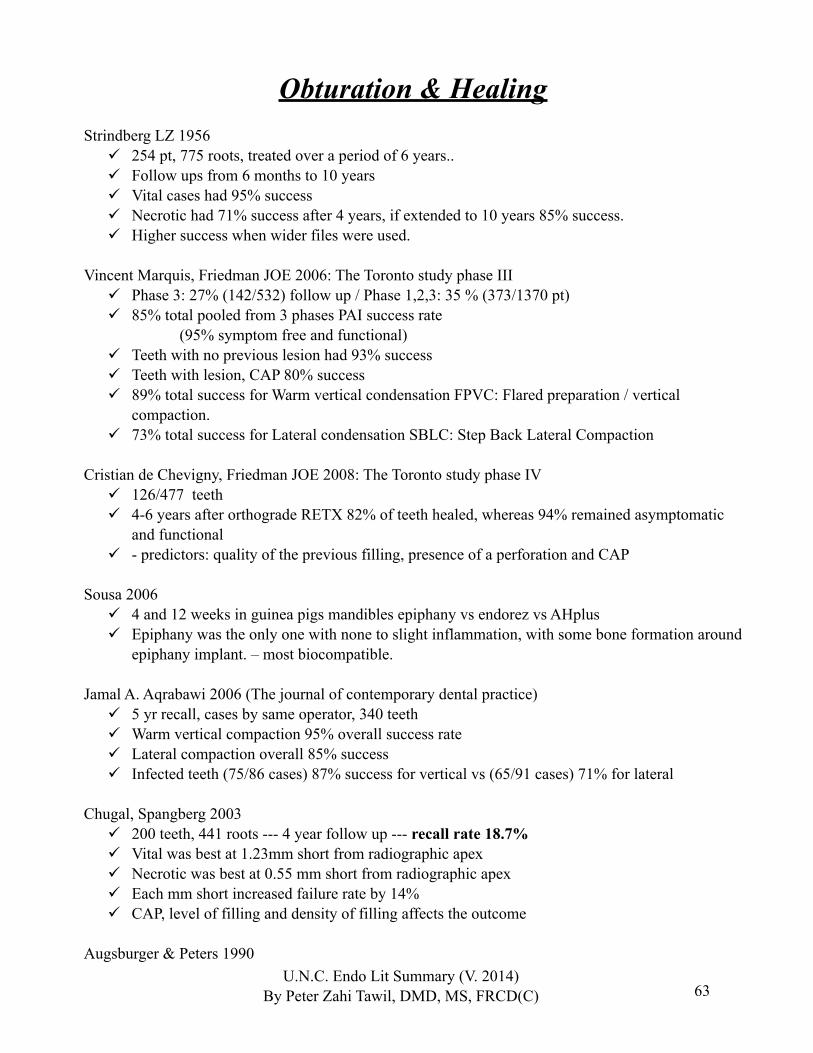
63
Obturation & HealingStrindberg LZ 1956
254 pt, 775 roots, treated over a period of 6 years.. Follow ups from 6 months to 10 years Vital cases had 95% success Necrotic had 71% success after 4 years, if extended to 10 years 85% success. Higher success when wider files were used.
Vincent Marquis, Friedman JOE 2006: The Toronto study phase III Phase 3: 27% (142/532) follow up / Phase 1,2,3: 35 % (373/1370 pt) 85% total pooled from 3 phases PAI success rate (95% symptom free and functional) Teeth with no previous lesion had 93% success Teeth with lesion, CAP 80% success 89% total success for Warm vertical condensation FPVC: Flared preparation / vertical
compaction. 73% total success for Lateral condensation SBLC: Step Back Lateral Compaction
Cristian de Chevigny, Friedman JOE 2008: The Toronto study phase IV 126/477 teeth 4-6 years after orthograde RETX 82% of teeth healed, whereas 94% remained asymptomatic
and functional - predictors: quality of the previous filling, presence of a perforation and CAP
Sousa 2006 4 and 12 weeks in guinea pigs mandibles epiphany vs endorez vs AHplus Epiphany was the only one with none to slight inflammation, with some bone formation around
epiphany implant. – most biocompatible.
Jamal A. Aqrabawi 2006 (The journal of contemporary dental practice) 5 yr recall, cases by same operator, 340 teeth Warm vertical compaction 95% overall success rate Lateral compaction overall 85% success Infected teeth (75/86 cases) 87% success for vertical vs (65/91 cases) 71% for lateral
Chugal, Spangberg 2003 200 teeth, 441 roots --- 4 year follow up --- recall rate 18.7% Vital was best at 1.23mm short from radiographic apex Necrotic was best at 0.55 mm short from radiographic apex Each mm short increased failure rate by 14% CAP, level of filling and density of filling affects the outcome
Augsburger & Peters 1990U.N.C. Endo Lit Summary (V. 2014)
By Peter Zahi Tawil, DMD, MS, FRCD(C)
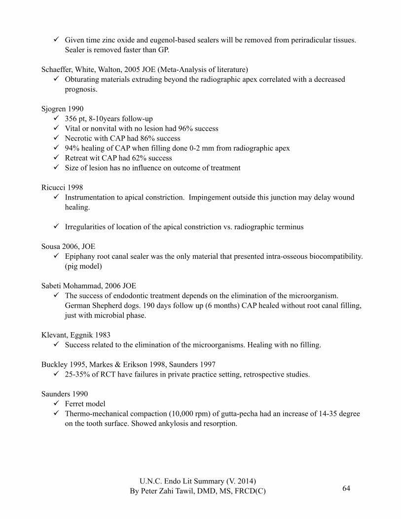
64
Given time zinc oxide and eugenol-based sealers will be removed from periradicular tissues. Sealer is removed faster than GP.
Schaeffer, White, Walton, 2005 JOE (Meta-Analysis of literature) Obturating materials extruding beyond the radiographic apex correlated with a decreased
prognosis.
Sjogren 1990 356 pt, 8-10years follow-up Vital or nonvital with no lesion had 96% success Necrotic with CAP had 86% success 94% healing of CAP when filling done 0-2 mm from radiographic apex Retreat wit CAP had 62% success Size of lesion has no influence on outcome of treatment
Ricucci 1998 Instrumentation to apical constriction. Impingement outside this junction may delay wound
healing.
Irregularities of location of the apical constriction vs. radiographic terminus
Sousa 2006, JOE Epiphany root canal sealer was the only material that presented intra-osseous biocompatibility.
(pig model)
Sabeti Mohammad, 2006 JOE The success of endodontic treatment depends on the elimination of the microorganism.
German Shepherd dogs. 190 days follow up (6 months) CAP healed without root canal filling, just with microbial phase.
Klevant, Eggnik 1983 Success related to the elimination of the microorganisms. Healing with no filling.
Buckley 1995, Markes & Erikson 1998, Saunders 1997 25-35% of RCT have failures in private practice setting, retrospective studies.
Saunders 1990 Ferret model Thermo-mechanical compaction (10,000 rpm) of gutta-pecha had an increase of 14-35 degree
on the tooth surface. Showed ankylosis and resorption.
U.N.C. Endo Lit Summary (V. 2014)By Peter Zahi Tawil, DMD, MS, FRCD(C)

65
Eriksson 1983 Rise of temperature of 10 degrees C for 1 minute is the critical point. Over 5 minutes more consistent bone damage will occur.
Salehrabi & Rotstein 2004, JOE 1 463 936 RCT treated teeth followed up to 8 years (Delta Dental insurance) 97% of the RCT treated teeth were retained within this 8year period 3% had re-treatments, apical surgeries or extractions mostly within 3years 85% of the extracted teeth had no full coronal coverage.
Alley, Eleazer & al 2004 (OOOO / Alabama school of dentistry) 195 teeth treated by Generalist success 89.7% 155 teeth treated by Endodontists success 98.1%
Imura, Gomes & al, 2007 JOE (Sao Paulo, Brazil) 2000 teeth done by endo specialist / follow up 18-24 months 624 Re-treatment: 85.9% success 1376 Initial RCT: 94% success
Zmener & al 2007 (JOE / Brazil) Compared the different level of moisture of canals and the coronal seal after filling with
EndoRez / GP and Resilon / Epiphany. In vitro 76 single-rooted human teeth. Dye leakage. Canals instrumented to ISO 40 EDTA and NaOCl rinse. GP fitted single cone (tug-back) and
Resilon single cone. Resilon had less leakage when the canal was slightly moist: drying with paper points with last
one appearing dry. GP had less leakage when the canal was dried with paper points and followed by dehydration
with 95% Ethanol.
U.N.C. Endo Lit Summary (V. 2014)By Peter Zahi Tawil, DMD, MS, FRCD(C)
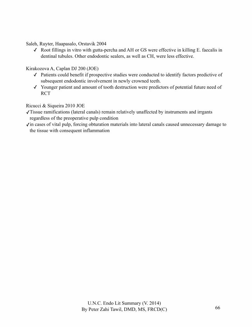
66
Saleh, Ruyter, Haapasalo, Orstavik 2004✓ Root fillings in vitro with gutta-percha and AH or GS were effective in killing E. faecalis in
dentinal tubules. Other endodontic sealers, as well as CH, were less effective.
Kirakozova A, Caplan DJ 200 (JOE)✓ Patients could benefit if prospective studies were conducted to identify factors predictive of
subsequent endodontic involvement in newly crowned teeth. ✓ Younger patient and amount of tooth destruction were predictors of potential future need of
RCT
Ricucci & Siqueira 2010 JOE✓Tissue ramifications (lateral canals) remain relatively unaffected by instruments and irrgants
regardless of the preoperative pulp condition✓in cases of vital pulp, forcing obturation materials into lateral canals caused unnecessary damage to
the tissue with consequent inflammation
U.N.C. Endo Lit Summary (V. 2014)By Peter Zahi Tawil, DMD, MS, FRCD(C)
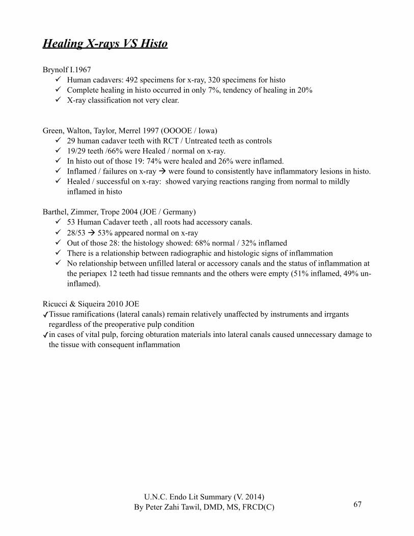
67
Healing X-rays VS Histo
Brynolf I.1967 Human cadavers: 492 specimens for x-ray, 320 specimens for histo Complete healing in histo occurred in only 7%, tendency of healing in 20% X-ray classification not very clear.
Green, Walton, Taylor, Merrel 1997 (OOOOE / Iowa) 29 human cadaver teeth with RCT / Untreated teeth as controls 19/29 teeth /66% were Healed / normal on x-ray. In histo out of those 19: 74% were healed and 26% were inflamed. Inflamed / failures on x-ray were found to consistently have inflammatory lesions in histo. Healed / successful on x-ray: showed varying reactions ranging from normal to mildly
inflamed in histo
Barthel, Zimmer, Trope 2004 (JOE / Germany) 53 Human Cadaver teeth , all roots had accessory canals. 28/53 53% appeared normal on x-ray Out of those 28: the histology showed: 68% normal / 32% inflamed There is a relationship between radiographic and histologic signs of inflammation No relationship between unfilled lateral or accessory canals and the status of inflammation at
the periapex 12 teeth had tissue remnants and the others were empty (51% inflamed, 49% un-inflamed).
Ricucci & Siqueira 2010 JOE✓Tissue ramifications (lateral canals) remain relatively unaffected by instruments and irrgants
regardless of the preoperative pulp condition✓in cases of vital pulp, forcing obturation materials into lateral canals caused unnecessary damage to
the tissue with consequent inflammation
U.N.C. Endo Lit Summary (V. 2014)By Peter Zahi Tawil, DMD, MS, FRCD(C)

68
Periapical Radiographs VS CBCT
Patel et al 2012 (IEJ) Diagnosis using CBCT revealed a lower healed and healing rate for primary root canal
treatment then periapical radiographs 123 teeth with 1 year follow up Healed 92.7% for periapical radiographs VS 73.9% for CBCT Healing 97.2% for periapical radiographs VS 97.2% for CBCT
U.N.C. Endo Lit Summary (V. 2014)By Peter Zahi Tawil, DMD, MS, FRCD(C)
69
Sealers
Kontakiotis, Wu, Wesselink 1997 In vitro and in vivo Thick layer of sealer showed more leakage ZOE sealing was poor and increased with storage time Epoxy sealer showed good sealing properties Calcium hydroxide leaked significantly after 2 years, can be due to fact that it dissolves over
time.
Abdulkader 1996 ZnOE sealer had strong antimicrobial activity, then glass ionomer, then calcium hydroxide.
Tested against anaerobic bacterias. ZnOE and formaldehyde effect…
Orstavik 1987, 1988 Zinc oxide sealers are cytotoxic and the effect can be long lasting Resin based sealer elicit initial severe inflammatory reaction and subsides after.
Balguerie E. et al 2011 JOE✓5 sealers were compared for their penetration and adaptation in the dentinal tubules under scanning
electron microscopy: Calcium hydroxide epoxy resin (Acroseal), Zinc oxide eugenol (Endobtur), Glass ionomer (Ketac-Endo), Epoxy resin (AH Plus), Silicon (RSA)✓AH Plus showed the most optimal tubular penetration and adaptation to the root canal wall
Osvaldo Zmener et al 2012 JOE MTA-Fillapex and Grossman sealer remained toxic to subcutaneous tissues in rats after 90 days
U.N.C. Endo Lit Summary (V. 2014)By Peter Zahi Tawil, DMD, MS, FRCD(C)
70
Follow ups / Recalls
Strindberg 1956 Healing in some cases took until 10 years to heal. Recommendation of 4 year follow up for necrotic cases
Orstavik 1996 500 teeth / follow up 1, 2, 4 year / PIA scoring Follow up recommendation of vital cases: 1year Most cases of failures were detected within 1 year 70% of new CAP will appear in 1 year. 90%
in 2 years Full CAP healing: 50% in 1 year, 80% 2 years, 90% 3 years Partial healing of CAP 90% in 1 year
Molven, Halse, Grung 1996 (Norway / JOE) 24 cases tx with periapical surgery that 2 to 6 years after surgery were classified as incomplete
healing (scar tissue) were followed for 8 to 12 years Lateral incisor most often found with scar tissue 1 case failed, 22 were still considered incomplete healing (scar tissue) The findings support the conclusion that cases clearly showing features of incomplete healing
(scar tissue) at the regular follow-up 1 yr after surgery can be regarded as successes. They need not be recorded for further systematic control.
U.N.C. Endo Lit Summary (V. 2014)By Peter Zahi Tawil, DMD, MS, FRCD(C)

71
Retreatment EtiologyFew strains 1 or 2.
Gram positive micro-organisms predominate. Dominance of facultataive over anaerobes.
E. Faecalis frequently found.
Molander 1998 (100 human RCT with CAP) 117 species recovered; 1-2 G+ facultative strains, E. Faecalis 47%, Lactobacillus Leaving root canals open for emergency can contribute to the establishment of a microflora,
including enteric bacteria and yeasts, which are hard to kill Micro-organisms have been recovered in root filled teeth without CAP.
Sundqvist & Figor 1998 1-2 spp. E. Faecalis (30%), A. israelii Leaving root canals open for emergency can contribute to the establishment of a microflora,
including enteric bacteria and yeasts, which are hard to kill
Hancock 2001 Mainly E. Faecalis (30%) If GP 2-4mm short = 75% bacteria recovered If lesion. 5mm = 79 % recovered If overextended = 100% recovery In retreament one should consider alternatives intra-medicaments and intra-canal irrigants.
Peciuliene, Haapasalo & al 2001 40 asymptomatic root filled teeth with CAP Group A calcium hydroxide ~2weeks / Group B IKI for 5 minutes E. Faecalis in 21 of 33 positive culture teeth 64%. IKI improved antimicrobial effect of tx more than CaOH.
Siqueira, Rocas 2004 PCR retreat analysis E. Faecalis found in 77% of cases. Fungi, C. albicans found in 9% of cases
Portenier, Waltimo, Orstavik, Haapasalo, 2005 (JOE) Series of experiments were done with E. Faecalis at different growth phases were challenged
by CaOH, 0.05% CHX and 0.0001% NaOCl In the logarithmic phase of growth, E. Faecalis was very sensitive to all medicaments and
was killed in just a few seconds. When E. Faecalis cells were in the stationary growth phase or starvation phase were more
resistant in all the experiments with the 3 medicaments and were not killed after the tested 10minutes range.
U.N.C. Endo Lit Summary (V. 2014)By Peter Zahi Tawil, DMD, MS, FRCD(C)
72
Nair & Schroeder 1984 Presence of actinomyces, A. Israelii in 2/45 PA lesions
Happonen 1986 13/16 sx cases demonstrated A. israelii
Bystrom 1987 2/79 cases which failed had PA actinomycosis
Lupo-Pegurier 2002 The majority of root fillings were poorly executed. There was a significant correlation between
the presence of periapical pathology and inadequate root-canal fillings (Nice, France population > 7000 teeth)
U.N.C. Endo Lit Summary (V. 2014)By Peter Zahi Tawil, DMD, MS, FRCD(C)

73
Retreatment
Bramante 2000 Use NiTi rotary at 1200rpm to remove gutta percha with heat generation (group 1, 350 r.p.m.; group 2, 700 r.p.m.; and group 3, 1500 r.p.m.). Cleanliness and residual debris were equivalent for each group, but the use of 1500 r.p.m.
speed was more rapid and fewer instruments fractured.
Wolcott, Himel, Hicks 1999 (J endod) System B technique for removal of Thermafill 225 deg C.
Huttula A. S. et al 2006 (JOE) The effect of Ultrasonic post instrumentation on root surface temperature was analyzed in vitro Post were vibrated with ultrasonic for 4 minutes at power 11 P5 booster Ultrasonic with water had a 3.2 to 5.0 deg C temperature increase Ultrasonic dry had 15.2 to 17.6 deg C temperature increase which could cause bone damage Irrigation during ultrasonic use had a significant impact on the temperature measured on the
external root surface
U.N.C. Endo Lit Summary (V. 2014)By Peter Zahi Tawil, DMD, MS, FRCD(C)

74
Failure etiology
1) interradicular infection : Nair 19902) Extraradicular infection: Sundqvist 1980, Sjorgren 1988,19973) Foreign body reaction : Koppang 1989, Nari 1990, Sjogren 19954) Cysts : Nair 19905) Fibrous scar tissue : Nair 1999
Extradradicular infection:
Tronstad, Barnett & al 1987 Anaerobic bacteria are able to survive and maintain an infectious disease process in periapical
tissues.
Sunde, Olsen, Debelian, Tronstad, 2002 36 teeth with refractory apical periodontitis analyzed Gram+ ~80% of the flora Sulfur granules were found in 9cases, 25%. This study demonstrated a wide variety of microorganisms, particularly gram-positive ones, in
the periapical lesions of teeth with refractory apical periodontitis.
Sunde, Olsen, Debelian, Tronstad & al 2003 39 periapcial lesions surgically removed, embedded in cold polymerizing resin and sectioned
for analysis fluorescence in situ hybridization (FISH) and epifluresecence and confocal laser scanning microscopy (CLSM)
A number of morphotypes occurred that could not be identified with the specific probes used, indicating the presence of additional bacterial species. CLSM confirmed that bacteria were located in different layers of the tissue. Accordingly, the FISH technique demonstrated mixed consortia of bacteria consisting of rods, spirochaetes and cocci in asymptomatic periapical lesions of root-filled teeth
Ricucci, Siqueira 2008 JOE✓ Case report of a extraradicular actinomycosis that clearly formed as a continuum with the
intraradicular infection. Histology shown.✓ Nonetheless, a review of the literature revealed that there is no clear evidence that apcial
actinomycosis is indeed an independent entity leading to persistent apical perodontitis lesion.
U.N.C. Endo Lit Summary (V. 2014)By Peter Zahi Tawil, DMD, MS, FRCD(C)
75
Diagnosis of periapical lesions, Cysts, OKC, etc:
Peters E, Lau M. 2003 JCDA✓ Review article✓ Granulation tissue is the main pathosis.✓ Although rare, other clinically confusing periapical lesions have been extensively documented
in numerous case reports and short case series. These lesions represent a wide range of pathosis, including various developmental cysts, infections, benign but locally aggressive lesions, and malignancies. The literature describing these lesions and the value of a histopathologic examination in diagnosis is reviewed.
Kuc I, Peters E, Pan J., 2000 OOO✓ 805 sequentially submitted periapical biopsy specimens over a 2 year period.✓ A histopathologic examination contributed clinically relevant information in 5.0% of submitted
cases✓ 805 cases, 788 (97.9%) granulation tissue, 9 (1.1%) complicated granulation tissue (with
infection or antral involvement), and 8 (1%) unrelated to the necrotic pulp, representing a range of locally aggressive but benign lesions and 1 malignancy.
Nobuhara WK, del Rio CE. 1993 JOE✓ Biopsy reports from 150 periradicular tissue specimens obtained from teeth refractory to
nonsurgical endodontic therapy were reviewed. ✓ The study found that 59.3% of the periradicular lesions were granulomas ✓ 22% cysts, ✓ 12% scars✓ 6.7% other pathoses.
Bhaskar SN. OOO 1966✓ 1108 cases, 48% granuloma 42% radicular cyst, 3.7% residual cyst, ✓ Other: scar, cementoma, dental abscess, etc.✓ In 1972: claims that instrumentation slightly beyond the foramen produces a transitory acute
inflammation which may destroy the epithelial lining of the radicular cyst and covert them into granulomas, thus leading to their resolution.
Tyler Koivisto et al 2012 (JOE) 9723 cases were analyzed 40.4% apical granulomas, 33.1% cysts, 8.8% Odontogenic keratocyst, 1.3% Central giant cell
lesions, 1.2% Amelomblastomas, <1% Metastatic lesions Most nonhealing lesions submitted for biopsy were classified as granulomas or cysts (73%)
often from the anterior maxillary jaw Nonhealing radiolucency jaw lesions other than granulomas or cysts were reported over 20%
of the time and may have more severe pathological implications
U.N.C. Endo Lit Summary (V. 2014)By Peter Zahi Tawil, DMD, MS, FRCD(C)
76
Surgical endo
Radiograph/CBCT for pre-surgical anatomy assessement:
Ferreira Costa F et al 2011 JOE✓ Small-volume CBCT scanning showed high accuracy in detecting horizontal root fracture
without a metallic post.✓ However, the presence of a metallic post significantly reduced the specificity and sensitivity of
the CBCT reading.
Bornstein et al 2011 JOE✓38 molars PA radiographs were compared to CBCT in their ability to assess the proximity of the
nerve and to assess periapical lesions.✓26% of periapical lesions were missed with PA radiographs and CBCT was more accurate in
determining location of IAN. ✓CBCT is useful for treatment planning in mandibular molars before apical surgery.
Kovisto et al 2011 JOE✓ 743 mandibular molars and premolars were assessed with CBCT in regards to the proximity of
the IAN.✓ The variable position of the IAN nerve bundle suggests that CBCT is useful to determine the
proximity of this nerve bundle prior to any invasive surgery in that area.
U.N.C. Endo Lit Summary (V. 2014)By Peter Zahi Tawil, DMD, MS, FRCD(C)
77
Flap:
Velvart Peter; JOE January 2005, 2003. Papillar preservation flap technique and microsurgical endodontics; showed better healing and
less scars.
Tarnow and Magner 1992 Factors affecting papilla hight 5mm from contact point to bone will have papilla filling 100% 6mm 56% / 7mm 27%
Matter 1980 Creeping of tissue after perio surgery will occur between 1month and 1 year.
Newman & Addy 1972, 1982 Preop Chx 0.2% rinse 1 week pre-surgery and 2 weeks post-surgery reduces plaque growth,
reduces post-operative discomfort and promotes healing.
Lang & Loe 1972 2mm of attached gingiva is necessary to maintain a stable position of the gingival margin in
submarginal incision.
Morman 1979 Paramedian releasing incisions around papilla followed by vertical are recommended to
minimize the risk of recession.
U.N.C. Endo Lit Summary (V. 2014)By Peter Zahi Tawil, DMD, MS, FRCD(C)
78
Harrison & Jurosky 1991, 1992 (part 1, 2, 3)1) The incisional wound * Few differences between sulcular and submarginal healing. Submarginal design showed less predictable results. 2) The dissectional wound *Scaling of root attached tissue and tags on the cortical bone should be avoided to allow rapid reattachment. Cortical retained periosteal tissues exert some protective influence which prevents necrosis of surface lamellae in underlying cortical bone. *The elevated periosteum does not survive the flap reflection, its cambium layer do not survive but becomes depolymerized and reforms laters. * Crestal bone osteoclastic activity occurs following submarginal and rectangular flaps. However osteoblastic repair occurs and creatal bone height is not altered.3) The osseous excisional wound: *osteoclastic activity not observed within the excisional wound site
*The endosteal tissue play the major role in osseous excisional wound healing* Periosteum does not function until the excisional wound is filled with woven bone
Von Arx et al 2011 JOE✓Periodontal parameters change mostly 1 year due to the surgery itself.✓0.29mm loss in Gingival margin✓0.20mm loss in Clinical attachment level
Bony access:
Calderwood 1964 Have to use sharp and clean burs to avoid heat generation Use of round bur better than fissure for heat generation
Lobene 1963 Diamond burs are inefficient and should not be used. Heat generation.
Eriksson & al 82, 83, 84 Heat generation effect on bone with implant model 44 to 47 degrees Celsius is the limit margin for 1 minute for impaired regeneration.
Moiseiwitsch 1998 Position of the mental foramen ranged from 8 to 21mm from the CEJ of premolars. Usually
located between premolars.
U.N.C. Endo Lit Summary (V. 2014)By Peter Zahi Tawil, DMD, MS, FRCD(C)
79
Phillips, Weller & Kulild, 1990, 1992 Mental foramen direction of exist is in a posterior-superior direction Location is inferior to the crown of the second premolar and approximately 60% of the
distance from the buccal cusp tip of that tooth to the inferior border of the mandible. When foramen not visualized, usually below the inferior edge of the film Position of the mental foramen was usually mesial and below the radiographic apex of the
second premolar
Von Arx T. 2007 JOE , Denmark The length of the bony access was the only significant factor affecting the outcome. 183 teeth
1 year follow up.
Pecora 2003 ✓ showed that calcium sulfate histologically in rats was good to stop the ingrowth of connective
tissue and promote osseous formation)
Hemostasis:
Gutmann 93, 96 Homeostatic effect of epinephrine in mucosa is primarily through alpha1-adrenegic receptors,
if in muscle actives Beta receptors and causes bleeding.
Buckley 1984 1:50000 epinephrine results in a good visualization of the surgical site, reduced surgery time
and decreased postoperative bleeding and blood loss.Kim 1984
Even though lower inferior nerve block reduces the blood flow, local administration of anesthetic with epi is needed for proper bleeding control.
Scarano A et al 2012 JOE The use of Calcium Sulfate (Plaster of Paris) completely eliminated the bleeding with very
good level of hemostasis.
U.N.C. Endo Lit Summary (V. 2014)By Peter Zahi Tawil, DMD, MS, FRCD(C)

80
Lemon, Jeansone 1993, Rabbit mandibles model Significant adverse effects on osseous healing was noted when ferric sulfate solution was left
in situ. When adequately curetted and irrigated from surgical site prior to closure, ferric sulfate did not
cause persistent inflammation or delay osseous repair.
Vickers, Baumgartner and Marshall 2002 Racellet VS 20% ferric sulfate (Viscostat) in patients No change in cardiovascular effects when using either of these agents. Both agents produced surgical hemostasis that allowed for a dry field for root-end filling.
Resection and retropep
Gilheany 1994; Tidmarsh 1989 Increase of leakage with increased angulation Decrease of leakage with increased depth of retrograde filling.
Wuchenich, Torabinejad & al 1994 20 anterior teeth in patient prepared, extracted and analyzed. Ultrasonic cavities produced more parallel walls, deeper depths for retention and followed the
direction of canal more closely than those prepared by burs.
Layton, Marshall, Morgan and Baumgartner 1996 Extracted human teeth model When root have crack more crack were generated with a high setting of the ultrasonic VS low
power.
Mehlahaff, Marshall, Baumgartner 1997 (Portland OHSU / JOE) 29 bilaterally matched pairs of human teeth cadavers Depth of ultrasonic better than high-speed bur 2.11mm VS 1.39mm Less bevel for ultrasonic 16 deg VS 35 deg Bony crypt needed was smaller for ultrasonic, more ease of access.
RetrofillHolt, Watts, Beeson, Kirkpatrick, Rutledge 2007 (JOE/TX)
MTA (both gray and white) mixed with CHX had lower compressive strength than the one mixed with sterile water
Watts, Holt, Beeson, Kirkpatrick, Rutledge 2007 (JOE/TX) MTA (both gray and white) mixed with local anesthetic (2% Lidocaine, 1:100 000) had lower
compressive strength than the one mixed with sterile water.
U.N.C. Endo Lit Summary (V. 2014)By Peter Zahi Tawil, DMD, MS, FRCD(C)
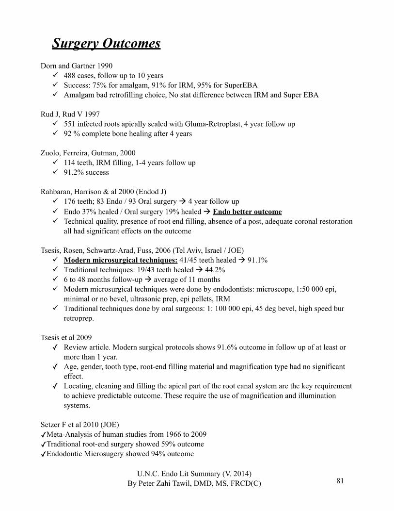
81
Surgery OutcomesDorn and Gartner 1990
488 cases, follow up to 10 years Success: 75% for amalgam, 91% for IRM, 95% for SuperEBA Amalgam bad retrofilling choice, No stat difference between IRM and Super EBA
Rud J, Rud V 1997 551 infected roots apically sealed with Gluma-Retroplast, 4 year follow up 92 % complete bone healing after 4 years
Zuolo, Ferreira, Gutman, 2000 114 teeth, IRM filling, 1-4 years follow up 91.2% success
Rahbaran, Harrison & al 2000 (Endod J) 176 teeth; 83 Endo / 93 Oral surgery 4 year follow up Endo 37% healed / Oral surgery 19% healed Endo better outcome Technical quality, presence of root end filling, absence of a post, adequate coronal restoration
all had significant effects on the outcome
Tsesis, Rosen, Schwartz-Arad, Fuss, 2006 (Tel Aviv, Israel / JOE) Modern microsurgical techniques: 41/45 teeth healed 91.1% Traditional techniques: 19/43 teeth healed 44.2% 6 to 48 months follow-up average of 11 months Modern microsurgical techniques were done by endodontists: microscope, 1:50 000 epi,
minimal or no bevel, ultrasonic prep, epi pellets, IRM Traditional techniques done by oral surgeons: 1: 100 000 epi, 45 deg bevel, high speed bur
retroprep.
Tsesis et al 2009✓ Review article. Modern surgical protocols shows 91.6% outcome in follow up of at least or
more than 1 year.✓ Age, gender, tooth type, root-end filling material and magnification type had no significant
effect.✓ Locating, cleaning and filling the apical part of the root canal system are the key requirement
to achieve predictable outcome. These require the use of magnification and illumination systems.
Setzer F et al 2010 (JOE)✓Meta-Analysis of human studies from 1966 to 2009✓Traditional root-end surgery showed 59% outcome✓Endodontic Microsugery showed 94% outcome
U.N.C. Endo Lit Summary (V. 2014)By Peter Zahi Tawil, DMD, MS, FRCD(C)

82
Von Arx et al 2010✓MTA showed good results on all treated teeth 91% success.✓Retroplast showed lower success, especially when used for mandibular premolars and molars.
overall 79.5% success.
Song, Shin et Kim 2011 JOE✓ Outcome study about Periapical Micro-RESURGERY. ✓ 42 cases recalled with 77.8% recall rate (42/54).✓ Overall success rate was 92.9% after 2 years.✓ The use of microsurgical techniques and biocompatible materials such as MTA and Super-EBA resulted in high clinical success rate, even in endodontic resurgery.
Kim & Solomon 2011 JOE✓Endodontic surgery was the most cost-effective among all the treatment modalities for a failed
endodontically treated molar.✓A single implant-supported restoration, despite its high survival rate, was shown to be the least cost-
effective treatment based on current fees.
Minju Song et al 2011 JOE✓493 teeth were analyzed for the cause of failure in root canals treatment when examined under a
microscope during endodontic microsurgery.✓Cause for failures were:
‣ 30.4% leakage around the root filling material‣ 19.7% Missed canal‣ 14.2% Underfilling‣ 8.7% anatomical complexity‣ 3% Overfilling‣ 2.8% Iatrogenic defects‣ 1.8% apical calculus‣ 1.2% cracks
Minju Song et al 2012 JOE Long term follow up of endodontic microsurgery Of the 172 treated teeth, 104 showed up to have long term follow of 6 to 10 years In this study 93.3% of endodontic microsurgery cases that were considered healed after 5 years
remained healed in the follow up period of 6 to 10 years
U.N.C. Endo Lit Summary (V. 2014)By Peter Zahi Tawil, DMD, MS, FRCD(C)
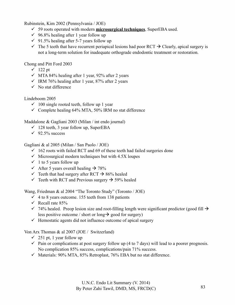
83
Rubinstein, Kim 2002 (Pennsylvania / JOE) 59 roots operated with modern microsurgical techniques, SuperEBA used. 96.8% healing after 1 year follow up 91.5% healing after 5-7 years follow up The 5 teeth that have recurrent periapical lesions had poor RCT Clearly, apical surgery is
not a long-term solution for inadequate orthograde endodontic treatment or restoration.
Chong and Pitt Ford 2003 122 pt MTA 84% healing after 1 year, 92% after 2 years IRM 76% healing after 1 year, 87% after 2 years No stat difference
Lindeboom 2005 100 single rooted teeth, follow up 1 year Complete healing 64% MTA, 50% IRM no stat difference
Maddalone & Gagliani 2003 (Milan / int endo journal) 128 teeth, 3 year follow up, SuperEBA 92.5% success
Gagliani & al 2005 (Milan / San Paolo / JOE) 162 roots with failed RCT and 69 of these teeth had failed surgeries done Microsurgical modern techniques but with 4.5X loupes 1 to 5 years follow up After 5 years overall healing 78% Teeth that had surgery after RCT 86% healed Teeth with RCT and Previous surgery 59% healed
Wang, Friedman & al 2004 “The Toronto Study” (Toronto / JOE) 4 to 8 years outcome. 155 teeth from 138 patients Recall rate 85% 74% healed. Preop lesion size and root-filling length were significant predictor (good fill
less positive outcome / short or long good for surgery) Hemostatic agents did not influence outcome of apical surgery
Von Arx Thomas & al 2007 (JOE / Switzerland) 251 pt, 1 year follow up Pain or complications at post surgery follow up (4 to 7 days) will lead to a poorer prognosis.
No complication 85% success, complications/pain 71% success. Materials: 90% MTA, 85% Retroplast, 76% EBA but no stat difference.
U.N.C. Endo Lit Summary (V. 2014)By Peter Zahi Tawil, DMD, MS, FRCD(C)
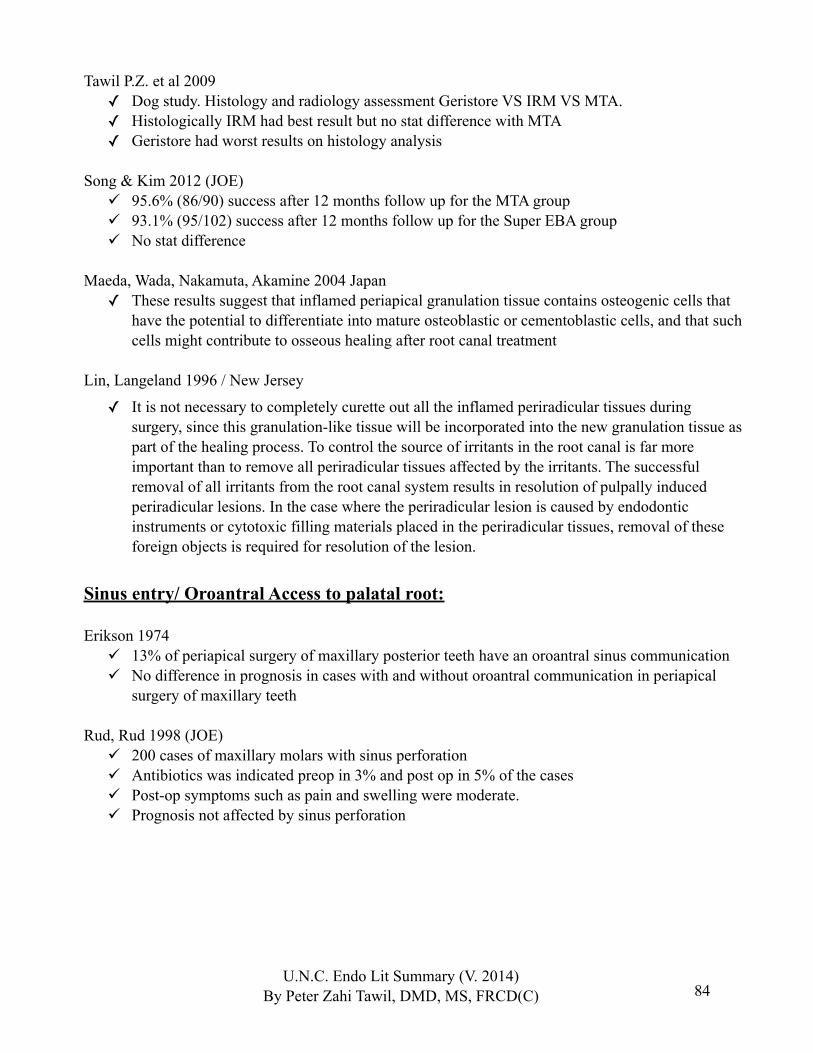
84
Tawil P.Z. et al 2009✓ Dog study. Histology and radiology assessment Geristore VS IRM VS MTA.✓ Histologically IRM had best result but no stat difference with MTA✓ Geristore had worst results on histology analysis
Song & Kim 2012 (JOE) 95.6% (86/90) success after 12 months follow up for the MTA group 93.1% (95/102) success after 12 months follow up for the Super EBA group No stat difference
Maeda, Wada, Nakamuta, Akamine 2004 Japan✓ These results suggest that inflamed periapical granulation tissue contains osteogenic cells that
have the potential to differentiate into mature osteoblastic or cementoblastic cells, and that such cells might contribute to osseous healing after root canal treatment
Lin, Langeland 1996 / New Jersey
✓ It is not necessary to completely curette out all the inflamed periradicular tissues during surgery, since this granulation-like tissue will be incorporated into the new granulation tissue as part of the healing process. To control the source of irritants in the root canal is far more important than to remove all periradicular tissues affected by the irritants. The successful removal of all irritants from the root canal system results in resolution of pulpally induced periradicular lesions. In the case where the periradicular lesion is caused by endodontic instruments or cytotoxic filling materials placed in the periradicular tissues, removal of these foreign objects is required for resolution of the lesion.
Sinus entry/ Oroantral Access to palatal root:
Erikson 1974 13% of periapical surgery of maxillary posterior teeth have an oroantral sinus communication No difference in prognosis in cases with and without oroantral communication in periapical
surgery of maxillary teeth
Rud, Rud 1998 (JOE) 200 cases of maxillary molars with sinus perforation Antibiotics was indicated preop in 3% and post op in 5% of the cases Post-op symptoms such as pain and swelling were moderate. Prognosis not affected by sinus perforation
U.N.C. Endo Lit Summary (V. 2014)By Peter Zahi Tawil, DMD, MS, FRCD(C)
85
Membranes and Guided Tissue Regeneration:
Lin, Chen, Ricucci and Rosenberg 2010 (JOE)✓Except in apico-marginal bony defects caused by combined perio-endo or endo-perio or in large
periapical lesions communicating with the alveolar crest, the use of membrane barriers in periapical surgery has not been shown to have a clear benefit in regenerating periapical tissues.✓Without using membrane barriers or bone grafts in periapical surgery for large apical periodontitis
lesions, complete periapical tissue regeneration has been observed histologically in many animal and human studies.✓Biologically, a blood clot is a better space filler. Without a blood clot tissue healing is impaired (like
a dry socket in an extraction model). Bone graft need to allow the blood clot or angiogenic factors to be able and promote perapical wound healing.✓Calcium sulfate is osteo-conductive.✓Many studies have clearly demonstrated that calcium sulfate can serve as scaffold for new bone
formation in periapical surgery.✓It appears that we still lack controlled clinical trials with a high level of evidence concerning
membrane barriers and bone grafts in periapical surgery.
Nyman et al 1979✓A membrane barrier is not required during periapical surgery because fibrous connective tissue re-
attachment will occur onto the root surface after reposition and suturing of a surgical flap.
Minju Song et al 2013 (JOE)✓Favorable prognosis can be expected when teeth/roots are covered with a buccal bone plate that is
>3mm in hight regardless of the amount of marginal bone loss
U.N.C. Endo Lit Summary (V. 2014)By Peter Zahi Tawil, DMD, MS, FRCD(C)

86
Dexamethasone and endo surgery post-op:
Grossi et al 2007 Third molars surgery model. Showed that 4mg and 8mg injections at the extraction sites is
effective in the prevention of postoperative edema.
Stewart G.G. 1956 Endodontic surgery model Antihistamines and corticosteroids can reduce pain, swelling and discoloration and encourage
better wound healing when used individually.
Tsesis et al 2003 Endo surgery model Oral dexamethasone 8mg pre-op and 4mg 1 day post op and 4mg 2 day post op There was a low incidence of postoperative pain and swelling when using oral corticosteroids Patients with preoperative pain were more likely to have postoperative pain
U.N.C. Endo Lit Summary (V. 2014)By Peter Zahi Tawil, DMD, MS, FRCD(C)
87
Retreat VS SurgeryHepworth & Friedman 1997
Weighted average calculation of a review of the literature 59% success for surgical endodontics 66% success for non-surgical re-treatments
Kvist & Reit 1999 95 endo failed cases randomly assigned to surgery or retreat groups 4 year follow up (6, 12, 24, 48 months) with bite-stents At 1 year (12 months), a statistically significant higher healing rate for surgical tx. No stat diff between the 2 groups in healing at 2 years (48 months) Surgical re-treatment seems to result in more rapid periapical bone fill but also may imply a
higher risk of “late failures”
Minju Song et al 2012 JOE Long term follow up of endodontic microsurgery Of the 172 treated teeth, 104 showed up to have long term follow of 6 to 10 years In this study 93.3% of endodontic microsurgery cases that were considered healed after 5 years
remained healed in the follow up period of 6 to 10 years No sign of significant “late failures” for the outcome of apical microsurgery
Kvist, Reit 2000 Surgical re-treatment resulted in more discomfort (swelling, discoloration, pain) 95 incisors pt randomly assigned to surgery or retreat groups
Fristad, Molven & Halse 2004 429 roots, 95.5% retreat success after a period of 20-27 years follow up. It was 85.7% afer
10-17years follow up. Increase of 10% over 10 years. Late periapical changes, with more successful cases, were recorded when a 10-17-year follow-
up after root canal treatment was extended for another 10 years. Persistent asymptomatic periapical radiolucencies, especially those with overfill, should generally not be classified as failures, as many of them will heal after an extended observation period.
Bergenholtz 1979 234 root filled teeth with CAP retreated 78% after 6 years follow up (48% after 1 year)
U.N.C. Endo Lit Summary (V. 2014)By Peter Zahi Tawil, DMD, MS, FRCD(C)
88
Sundqvist 1998 54 root filled teeth with CAP re-treated / 5 year follow up 74% success (6 cases were filled with + cultures) 80% success if we consider – bact samples roots at time of filling
Nair 1996 256 periapical lesions: 9% were true cysts, 6% apical pocket cyst. Pocket cyst (previously called bay cyst by Simon in 1980). Pocket cyst can heal after RCT.
True cysts are unlikely to resolve following RCT
U.N.C. Endo Lit Summary (V. 2014)By Peter Zahi Tawil, DMD, MS, FRCD(C)
89
RestorativeVital teeth:
Stanley 1968 A remaining dentinal thickness of more than 2mm is considered adequate to prevent pulpal
reactions to restorative procedures even if abusive techniques are used.
Pashley 1992 A hydrophilic resin may be used to seal the dentin to relieve the discomfort associated with
postoperative sensitivity.
Vojinovic & al. 1973 (Human study) The penetration of bacteria into dentinal tubules was massive only under the acid-treated
cavities. Acid etching opened and widened the dentinal tubules.
Wang & Hume 1988 (extracted human teeth 3rd molars) Hydrogen ions from strong acids penetrated dentine poorly relative to that from weak acids or
labeled water. (Buffering by hydroxyapatite and other mechanisms)
Bergenholtz, Cox 1982Cox 1992
ZOE seals effectively, prevents bacterial leakage and thus prevent pulpal reactions. Presence of bacteria / the seal not the restorative material.
Bergenhotlz G 2000 (Critical Reviews in Oral Bioogy & Medecine)✓ There is a lack of controlled clinical studies in resin-based restorations and confirmation that
pulpal health prevails over the long term following the use of the total-etch and resin-bonding techniques
Costa CA 2000 (Dental materials)✓ Self-etching adhesive systems may be useful and safe when applied on dentin but persistent
inflammatory reactions, delayed pulpal healing and failure of dentin bridging were evident in human pulps capped with bonding agents.
✓ The results observed in animal teeth cannot be directly extrapolated to human clinical conditions
✓ Vital pulp therapy using acidic agents and adhesive resins is contraindicated.
U.N.C. Endo Lit Summary (V. 2014)By Peter Zahi Tawil, DMD, MS, FRCD(C)
90
Glass Ionomer:
Peumans M, Van Meerbeek & al. 2005 Dent Mater (Meta-analysis) [Belgium] Bonding effectiveness of adhesives revealed that glass-ionomers most effectively and durably
bond to tooth tissue. Inefficient clinical performance was noted for the one-step self-etch adhesives.
Tay & Pashley 2001 Acid pretreatment dissolves the smear layer, creates a zone of partially demineralized dentin,
and allows the PAA to interact with dentin via the intermediate layer The use of acid pretreatment 10% polyacrylic acid improved the bond strength of glass
ionomer Glass ionomer has an intermediate layer: contains metallic salts contributed by both the GIC
and dentin
Berry & Powers 1994 Bond strengths of both glass ionomers were greater after dentin surface treatment with either
25% or 40% polyacrylic acid than with no treatment.
Inoue, Van Meerbeek & al 2001 [Belgium] 20% polyalkenoic acid conditioner removed the smear layer and improved the bond strength of
the adhesive GI to dentin
De Munck J, Van Meerbeek, Inoue & al 2004 [Belgium] Fuji Bond LC (GC) was applied without (i) and with pretreatment using (ii) a polyalkenoic
acid conditioner and (iii) a 37.5% phosphoric acid etchant. After 24 h and 4 yr, the lowest micro TBS was recorded when dentin was not pretreated. The
highest micro TBS was obtained following polyalkenoic acid pretreatment, although this was not significantly different from specimens that were pretreated using phosphoric acid.
Pretreating dentin with phosphoric acid intensified micromechanical interlocking at the expense of chemical bonding potential to hydroxyapatite. Nevertheless, correlating the micro TBS data with failure analysis through scanning electron microscopy and transmission electron microscopy indicated that combined micro-mechanical and chemical bonding involving pretreatment with the polyalkenoic acid conditioner yielded the most durable bond.
U.N.C. Endo Lit Summary (V. 2014)By Peter Zahi Tawil, DMD, MS, FRCD(C)

91
Resins:
Gwinnett AJ 1993 The contribution of the dentinal tubules as 15% of the bond
Tay, Loushine, Pashley & al 2005 JOE In root canals, C-factors can be over 1000
Opdam NJM, 2008 JOE Netherlands✓ A direct bonded composite resin restoration can be successful treatment for a cracked tooth. 7
years follow up VS full coverage crowns
U.N.C. Endo Lit Summary (V. 2014)By Peter Zahi Tawil, DMD, MS, FRCD(C)
92
Leakage:
Marshall & Massler 1961 First to stress the concept of coronal leakage causing failure of RCT
Madison & Wilcox 1988 If root canals exposed to oral environment, coronal leakage took place, in some cases
extending to the full length of the root.Torabinejad & al 1990
Coronal leakage of single-rooted teeth with P. vulgaris and S. Epidermis. 50% had entire length contaminated after 19 to 42 days.
Khayat & Torabinajed 1993 Same model showed completed contamination within 30 days whatever obturation technique
was used
Ray & Trope 1995 Integrity of the coronal part of the root canal system is paramount for success.
Tronstad 2000 Although coronal restoration is important, the quality of RCT is more significant
Ricucci 2000 Teeth with good RCT not adequately restored were OK over time Critiqued for low Number and when closed CAP might develop.
Hommez 2002 The importance of a good coronal restoration, as well as of a good root filling should be
emphasized as the technical quality of both influencing the periapical status.Hilton 2002
No current dental material provide a hermetic, leak proof seal
U.N.C. Endo Lit Summary (V. 2014)By Peter Zahi Tawil, DMD, MS, FRCD(C)
93
Orifice Plugs:
McInerney ST, Zillich 1992 In Vitro dye leakage, 2mm orifice plugs Cavit and IRM provided better internal sealing of the dentin than did zinc phosphate cement
Hansen-Bayless J, Davis R 1992 (97) reported Cavit as an orifice plug was a more effective barrier to leakage than IRM.
Hosoya N, Cox CF et al 2000 Hydraulic filling materials (Cavit and Coltosol) provided the most favorable cavosurface seal
when they were firmly packed into the cavity space to prevent microleakage, when compared with a photo-activated temporary resin material (Fermit), zinc oxide– eugenol cement, and a zinc oxide phosphate cement
Rotstein I et al 1992 In vitro with IRM, zinc oxide-eugenol, composite resin, or glass ionomer None of the materials tested in the bovine teeth showed H2O2 penetration with a base
thickness of 2 mm
Yamauchi et al 2006 In vivo with dogs tested 2mm composite plugs VS 2mm IRM plugs Both IRM and composite plugs decreased leakage
U.N.C. Endo Lit Summary (V. 2014)By Peter Zahi Tawil, DMD, MS, FRCD(C)
94
Post-Endo Restoration:
Uranga A, Blum JY, 1999 J. Endod. ; Permanent materials tend to leak less then temporary materials
Weber & Del Rio1978 Need at least 3.5mm of temporary filling to minimize leakage.
Pisani 1998: CAVIT plugs reduced leakage 85% / SuperEBA reduced leakage 65%
Rotstein 1992 2mm plug was enough with hydrogen peroxide, internal bleaching to protect tubilis from
external root resorption risk.
Lacy, Fowell, Watanabe 1991 No difference in bond strength between eugenol and non-eugenol cement pretreated teeth.
Tenure bonding system used.
Santos & al 2006 JOE Nov. There was a significant decrease in bond strength associated with NaOCl (even after EDTA
rinse) Chx had no effect on adhesion.
Kanca J 1996 The collagen in the preparation should have a shiny, wet surface, and the enamel should have a
frosty appearance
Gwinnett 1992 Bond on wet dentin far superior to bond on dry dentin. All Bond, Amalgambond, Mirage Bond
and Tenure all produced significant statistical improvement in shear bond strength values when applied to moistened dentin.
Ferrari 1995, Fonzi 2000 Cementum must be etched properly, higher organic content. Unlike dentin, cementum substrate will exhibit no resin tag formation, because of the presence
of the granular layer and the lack of tubules in the peripheral root dentin.
Gutmann 1978 Presence of accessory canals in the floor of the pulp chamber
Saunders & Saunders 1990 Gutta percha along floor should be removed to avoid leakage in furcation area.
U.N.C. Endo Lit Summary (V. 2014)By Peter Zahi Tawil, DMD, MS, FRCD(C)
95
Ferrule:
Libman & Nicholls 1995 25 extracted human centrals with cast post / the need of 1.5 to 2.0mm of ferrule 0.5 to 1.0 mm ferrule lengths failed at a significantly more than the 1.5 to 2.0 mm. Need 1.5mm minimum / recommendation of 2mm
Jelena Juloski et al 2012 JOE Literature review paper 1.5 to 2mm ferrule has a positive effect on fracture resistance of endodontically treated teeth If no ferrule is present, ortho extrusion or crown lengthening should be considered
Santos-Filho et al 2014 JOE In vitro model Post length influenced only the cast post strain and stress distribution The ferrule group always showed more satisfactory stress distribution and fracture resistance
Post placement:
Goldfein J et al 2013✓The use of a Rubber Dam during post placement provides a higher success rate✓93.3% success when Rubber dam was used✓73.6% success when Rubber dam was not used
Kvist, Rydin, Reit 1989 Retrospective study of teeth with post and with CAP Need at least 3mm of gutta percha after post space prep from proper seal.
Portel & Peters 1982; Bourggois & Lemon 1981;Karapanou et al 1996
Post-space preparation can be undertaken immediately following obturation to avoid twisting gutta-percha and breaking the seal when sealer is set.
Hayashi et al 2008 JOE✓ In-vitro, showed that Chemical-cured total-etch adhesive material showed stable bonding
performances throughout the entire post space and was better than dual cured systems.
Ambica K et al 2013Valdivia AD et 2012✓Fiber posts had no reinforcing effect against fractures
Sagsen B et al 2013 JOE✓Titanium posts and fiber posts had no reinforcing effect against fractures
U.N.C. Endo Lit Summary (V. 2014)By Peter Zahi Tawil, DMD, MS, FRCD(C)
96
Post-Endo: Internal Bleaching:
Arcari & al 2007 (J Adhes Dent, Brazil) In vitro 36 maxillary premolars. Sodium Perborate used for bleaching. Microtensile bond
strength testing was done. Definitive restoration can be accomplished immediately after non-vital bleaching treatment.
Thomas Attin & al 2005 (Dental materials, Denmark) Review. Bleaching may exert a negative influence on restorations and restorative materials.
Bleaching therapies may have negative effect on physical properties, marginal integrity, enamel and dentin bond strength.
Demarco FF & al 2001 Short-tem use of intracoronal calcium hydroxide medicament for 7 days after completing of
walking-bleach therapy was able to reverse the negative influences of the hydrogen peroxide application on microleakage of access cavities.
Texeira & al 2003, 2004 The bleaching of non-vital teeth affected the resin/dentin bonding strength. (In vitro studies
with shear bond strength and dye leakage)
Plotino 2008 JOE review✓ Review on internal bleaching✓ Sodium perborate is the safest. ✓ Discusses techniques, indications, complications, etc.
Felman & Parashos 2013 JOE White MTA induces the gray discoloration of teeth (when used around the crown) and the
effect is compounded in the presence of blood
U.N.C. Endo Lit Summary (V. 2014)By Peter Zahi Tawil, DMD, MS, FRCD(C)

97
Fracture Resistance and RCT:
Randow & Glantz 1986 In vivo, patient had controlateral teeth, vital vs root filled Pain loading level of root filled teeth was more than twice as high as vital teeth. There is changes in receptor properties in teeth with non-vital pulp leading to higher bite forces
than in vital teeth
Reeh & Messer 1989 42 Maxillary 2nd bicuspids used endodontic procedures have only a small effect on the tooth, reducing the stiffness by 5%. The
largest loss occurred with marginal ridge integrity. MOD cavity preparation resulted in an average of a 63% loss in relative cuspal stiffness
Composite restoration and enamel plus dentin etch were almost as strong as the unaltered tooth, while enamel-etch-only yielded lower stiffness.
Ausiello 1997, Hansen 1988, Oliveira 1987, Steele 1999 Found that intact marginal ridges with only a small access preparation are most resistant
against fracture and are not significantly weaker than intact teeth.
Sedgley & Messer 1992 23 matched pairs of contro-lateral teeth extracted for prostho reasons. Vital vs root filled. 2 slices of 0.3mm thick were made and analyzed for shear strength and
toughness. Teeth do not become more brittle following endodontic treatment.
Linn & Messer 1994 36 intact mandibular molars Endodontic access was followed by mesio-occlusal or mesio-occluso-distal preparation. Preserving a marginal ridge in molars did not fully conserve the strength of adjacent cusps, full
occlusal coverage with gold strengthened all cusps. It is more important to cover cusps than to preserve tooth structure (including a marginal ridge)
in endodontically treated molar teeth.
Panitvisai & Messer 1995 13 intact mandibular molars Increasingly extensive MO or MOD cavity preparations followed by endodontic access were
cut in each tooth. Cuspal deflection increased with increasing cavity size and was greatest following endodontic access
These findings reinforce the importance of cuspal coverage to minimize the danger of marginal leakage and cuspal fracture in endodontically treated teeth.
U.N.C. Endo Lit Summary (V. 2014)By Peter Zahi Tawil, DMD, MS, FRCD(C)
98
Andreasen , Munksgaard, Bakland 2006 JOE 30 immature sheep incisor teeth 80% of their root growth completed CaOH for 100 days had weakening effect on the strength of the root CaOH for 30 days + MTA did not affect strength of the root MTA did not affect the strength of the root Long term CaOH for apexification will weaken root structure.
Restoring over MTA:
Tsujimoto et al, 2013, JOE Composite resin with a bonding agent over MTA can be restored almost immediately after
MTA mixing during a single visit In Vitro, 3 different tested times: 10minutes, 1 day, 7 days All tested times with bonding agents placed over the MTA had no gaps between the MTA/resin
interface.
Periodontal Biologic Width for restoring teeth:
Gargiulo A et al 1995 Epithelial attachment is the most variable dimension Connective tissue attachment is more constant throughout the stages of passive eruption Connective tissue average length was 1.07mm 2.04mm is the mean average for epithelial and connective tissue attachment
Rosenberg et al 1980 2.04mm is needed for connective tissue and epithelial attachment We need to add 1-2mm for adequate seal of the restoration Recommended a 4mm guideline from bone crest to tooth margin when doing crown
lengthening
U.N.C. Endo Lit Summary (V. 2014)By Peter Zahi Tawil, DMD, MS, FRCD(C)
99
IMPLANTS+ implant success
Buser & al. 2002✓ 5 year success of 98.3 % success in augmented ridge cases (66 implants)
Bornstein & Buser 2005✓ 5 years success of 99%✓ Sandblasted and acid-etched (SLA) implants with early loading after 6 weeks✓ Evaluation on probing, attachment, radiographs: 3, 12, 24, 36, 48, 60 months.
Becker W, Becker BE 1999✓ 6 year success of 91.5% in mandibular molar area✓ 6 year success of 82.9% in maxillary implant area✓ 2-3 year success was 100% for both groups✓ 282 implants
Cooper, Felton 2001✓ 3 years success of 96.2% in single-tooth implants restored after 3 weeks✓ 52 pt.
Levin & Schwartz-Arad 2006✓ 92.6% success 6 to 125 months follow up✓ 81 patients, single molar implants✓ No relation was found among failure, complications, timing of implant placement, and
smoking habits
CAP does not affect healing of implant
Lindeboom & al. 2006✓ Immediate implant placement in chronic periapical lesions may be indicated.✓ 55 pt, 27 in lesions, 27 after 3 month healing of CAP✓ 92% success for immediate placement in CAP✓ 100% for 3 month healing period (no stat diff)✓ Evaluation of stability, aesthetics, x-ray, microbio of lesions.
Shabahang, Torabinejad 2004✓ Teeth with periradicular lesions do not adversely affect adjacent titanium solid root-form
implants. Dog study.✓ Teeth with no tx control 54%, teeth with lesion and RCT 74%, Lesion and no endo 68%.
U.N.C. Endo Lit Summary (V. 2014)By Peter Zahi Tawil, DMD, MS, FRCD(C)
100
- implant failure (mechanical failure)
Goodacre 2003 Meta-analysis (Clinical complications with implants and implant prostheses) 33% fail due to loosening of overdenture retentive mechanisms 25% fail due to irradiated maxillae 24% fail due to hemorrhage-related complications 22% have veneer fracture 21% implant loss with maxillary overdentures 19% needed to be relined 16% fail in type IV bone 16% Overdenture clip attachment fracture
Woodmansey et al 2009 JOE✓ 25 lower mandibular RCT VS 25 lower mandibular implants✓ Dental implants were found to have significantly lower maximum biting forces, reduced
chewing efficiency and small ocllusal contact and near contact.✓ RCT teeth may provide more effective occlusal contact during masticatory funtion.
Implant VS RCT studies:
Doyle & al 2006, JOE 196 post RCT followed up with 196 matched single tooth implants Both groups had a 94% success rate Implant group experienced a much greater incidence of post-operative complications.
Iqbal & Kim 2007, JOE Comprehensive review of the lit 57 studies for implants – 12000 implants 13 studies for RCT – 23 000 Outcomes were similar (high 90’s): The decision to treat a compromised tooth
endodontically or replace it with an implant must be based on factors other than treatment outcome.
Hannahan, Eleazer 2008 JOE✓ Implants were placed by periodontists and Root canals were done by endodontists in
private practice setting.✓ 129 implants, 36months follow up success of 98.4%✓ 143 RCT, 22months follow up success of 99.3%✓ The success of implant and RCT was essentially identical, but implants required more
postoperative treatments to maintain them.
U.N.C. Endo Lit Summary (V. 2014)By Peter Zahi Tawil, DMD, MS, FRCD(C)