unclassified ad number - apps.dtic.mil · (elisa) were developed and compared to the commercial...
TRANSCRIPT

UNCLASSIFIED
AD NUMBER
ADB081939
NEW LIMITATION CHANGE
TOApproved for public release, distributionunlimited
FROMDistribution authorized to U.S. Gov't.agencies only; Proprietary Information; 14JUL 1971. Other requests shall be referredto Commander, U.S. Army Medical Researchand Development Command, Attn: SGRD-RMS,Fort Detrick, Frederick, MD 21701.
AUTHORITY
USAMRDC/MCMR-RMI-S [70-1Y] ltr, 16 May1996
THIS PAGE IS UNCLASSIFIED

DEPARTMENT OF THE ARMYU.S. ARMY MEDICAL RESEARCH AND MATERIEL COMMAND
FORT DETRICK, FREDERICK, MD 21702-5012
IWK TO
MCMR-RMI-S (70-1y) ERR TA16.May 96
MEMORANDUM FOR Administrator, Defense Technical InformationCenter, ATTN: DTIC-OCP, Fort Belvoir,VA 22060-6218
SUBJECT: Request Change in Distribution Statement
I. Thv U.S. Army MtdicO! Research and M-iteriel Command hazreexamined the need for the limitation assigned to technicalreports written for Contract Numbers DAMDI7-81-C-1058 andDAMD17-82-C-2155. Request the limited distribution statement forAccession Document Numbers , N' Rao, ISAih19*L andAM5• gf be changed to *Approved for public release;distribution unlimited." A copy of these reports should bereleased to the National Technical Information Service.
4. Point of contact fcir this requesL is Mrs. Judy Pawlus at *DSN 243-7322.
FOR THE COMMANDER: ERRATA
SR. PAY III
Lieutenant Colozel. MSDeputy Chief of Staff
for' Infeomation Management
##

I' ADB08 1939'4
I SEARCH FOR HEPATITIS A VIRUSES BY NEW METHODS
*, Annual Progress Report
Joseph L. Melnick, Ph.D.
September 19,1
S•upported by
U.S. AR•Y MEDICAL 7E•zARCH & DEVELOPMENT COMMANDFort De.trick, F'e'derick, Maryland 21701
Cnntract No. DADA17-73-C-3074Contracc No. DAMDI7-81-C-1058
Baytor College of MedicineHouston, Texas 77030
DOD DISTRIBUTION STATEMENT
Distribution limited to U.S. Government Agencies only; (ProprietaryInformation) 14 July 1971. Other requests for this document must bereferred to the Commander, US Army Medical Research and DevelopmentComand, ATTN: SGRD-4HS, Fort Detrick, Frederick, Maryland 21701.
The finding8 in this report are not to be. construed as an officialDepartment of the Army position unless so designated by other
authorized documents.
LAJI
44 04 18 042
SE&UIITY CLASSI FICATION OF THIS PAGE (Wheni Dot* ffnt~td)
PAGE READ IN~STRUCTIONS______ REPORT___DOCUMENTATION______PAGE_ BEFORECOMPLETINGFORM
1. REPORT NUMBER 2GOT ACCESSION NO. S. RECiPIENT'S CATALOG NUMBER0
4. TTLE and ubtilo)5. TYPE OF REPORT & PERIOD COVERED* ~Annual (31 Aug 80-31 Mar 8Q,
Search for Hepatitis Viruses by New Methods Annual (I Apr 81-1 Sept 81)
6. PERFORMING ORG. REPORT NUMBER
7. AUTHOR(&) S. CONTRACT OR GRANT,*4UMBER(.)
Joseph L. Melnick, Ph.D. DDI-3C37DAMD17-81-C-1058. .
9 . PERFORMING ORGANIZATION NAME AND ADDRESS 10. PROGRAM ELENT POECT. TASKC.. AREA a WORI( UNIT NUMBERS
Baylor College of Medicine 6ll02A.3M16llO2BSO1.00.ol3Houston, Texas 77030
11I. CONTROLLING OFFICE NAME AND ADDRESS It. REPORT CATE
U.S. Army Medical Research and Devel-pment Commanc September 1981Fort Detrick (ATTN: SGRD-RMS) I). NUMMER OF PAGES r'1Frederick, ayad2714pp
14. MONITORIN aGC NAMC 4 ADORESS(I Witflu..aI gt CO&InIUAM Oflcf) ISN EC RMITEY CLASS. (01 lad* eWi)
Unclassified114k. DECkLASS11I FCATO ON/ DOWN GRAM NO
SCHEUL
IC CIST4kjbu 1I0i4 STATEMENT (of WeS MX4it
*Distribution limited to U.S. Government Agencies only; (Proprietary Information'.-*14 July 1971. Other requests for this document must be referred to the*Commander, US Army Medical Research and Development Command, ATTN: SGRD.-RMS.
F ort Detrick, Frederick, Maryland 21701
17. OISTUS~UTION ST ATEMENT (oil"I .bM,.e4 6WMimE 8804 11466 6 5 ftue* Rpe
I&B SUPf9tIWghTAftY MOVES
PHepatitis virus type Ao Hepatitis virus type B
HBsAgPolypeptide vaccineTetanus toxoid
Please see page 3.
* 2 5 ,13o osto m IWYC*&I.TaO :P~7De£.
W .350C fIWO 40IT

.' . *.- .. V.*? ... **.* *
3 1
DD Form 1473, Item 20: Abstract -
A preparation of HBsAg/ayw vaccine, which has been previously characterized inchimpanzees, was set aside by Dr. J. Gerin in cooperation with the National Institute ofAllergy and Infectious Diseases for characterization of an HBsAg-derived polypeptidevaccine. The major polypeptides (mol. wt. 22,000 and 25,000) were derived by SDS-polyacrylamide gel efectrophoresis (PAGE) and pooled. This was designated as a P22polypeptide vaccine. The immunogenicity of freshly prepared, as well as material storedat V and 201C for a period of 6 months, was tested in mice. Consistent antibody activitywas noted in mice immunized with P22, but the immunogenicity was markedly lower thanthat noted in mice inoculated with the intact particle vaccine. Inoculation of P22 vaccinein conjunction with tetanus toxoid significantly increased the immunogenicity of the P22 •component when saline suspensions were compared. In contrast, alum-adsorbed prepara-tions showed a decline in the geometric mean titer when tetanus toxoid was added.
Recent studies by hi*s a have shown that patients who have recovered fromHBV infections develop a specific cellular cytotoxicity tdward the PLC/PRF/5 hepatomacell line. These observations were extended by analysis of possible specific cellularcytotoxicity in three persons passively immunized with anti-HBs globulin and in 20individuals vaccinated with HBsAg vaccine. The three subjects with passively aidmlni-stered antibody activity were negative for cellular activity, but in 2 of 20 individualsvaccinated with formalirized HBsAg vaccine, a specific cytotoxicity for hepaturma targetcells was noted.
To facilitate testing of large numbers of mouse antisera, a micro solid-phase •radioimmunometric assay (micro-SPRIA) and an enzyme-linked immunosorbent assay(ELISA) were developed and compared to the commercial radioimmunoassay (Ausab) !ordetection of anti-HBs intibody. The two solid-phase antibody assays proved to be 200-35OX more sensitive for detection of antibody than the commercial antibody assay kit.
Preliminary data were presented In conjunction with an undenatured P22-P254! polypeptide vaccine prepared by the technology ot Zuckerman and coworkers. The
polypeptide material, isolated in micefle form, retained high levels of HBsAg activity whentested by solid-phase radioimmunoassay In contrast to the low activity observed with SOS-denatured material. This material has been Inoculated into mice to test Its relative levelof immunogenicity./tN
HAV has been purified fronf both marmoset liver tissue and from chimpanzee fecal p* material. Four weeks after In~culation of PLC/PRF/I, Niahlavu and MC-3 cells a
significant increase of HAV antig was noted In the Culture media by RIA procedures.
A waterborne outbreak of g troenteritis and hepatitis occurred in Georgetown,Texas, in June 1980. Along with h an enteric viruses, HAV was detected in the sewageand in one well water sample by mic o-SPRIA procedures.
* 5
I

I
Foreword:
In conducting the research described in this report, the investigator(s) adhered to the"Guide for Laboratory Animal Facilities and Care," as promulgated by the Committee onthe Guide for Laboratory Animal Resources, National Academy of Sciences-NationalResearch Co, uncil.
1ee*ssiOU_ J- ;
D)TIC TAB*-r,,nnomce'd
just ificatlo
Distribut iOn/ 0Availability C0des
1ýjst SpeCial
4 .
q S
rS
II
S... l ... t .... .9._•'t.. t•. • .I S

TABLE OF CONTENTS
Tite Page ... . . . . . . . .1
Report Documentation Page. ........... ..... 2
Foreword . . . . . .. . . . . . . . . . . . . ... 4
Table of Contents. . . . . . . . . . . . . . . . . . . 3
Body of the Report .......... .... ... . 6 p
L Immunogenic Characterization of the NIAID Stock of HBsAgloywVaccine to be Used as a Source for a Possible Polypeptide Vaccine . . . 6
It. Inoculation of a Particle HBsAg or an HBsAg Polypeptide Vaccinein Conjunction with Tetanus Toxoid . . . . . . . . . . . 7A. Humoral Immune Response to Tetanus Toxoid . . . . . . . . 9B. Humoral Immune Response to Hepatitis Polypeptide Vaccine . . . . 8C. Hurnoral immune Response to Hepatitis Subunit Vaccine . . . . . 9
III. Development of Anti-HBs Assay with Increased Sensitivity ..... 9IV. Testing of an Alternate Method of Isolation of Purified
Polypeptides from Solubilized HBsAg Preparations. . . . . . . . 10A. Polypeptide Purification from HBsAg Solubilized with
* Mercaptoethanol, Sodium Dodecyl Sulfate, Urea and4 Heat . .............. .. . . . |0
8. Fractionation of Undenatured P2-P23 Polypeptides by theMethod Described by Zuckerman etal . . . . . . . . . . 1I
V. Evaluation of Cellular Immunity in Hepatitis BImmunized Animals and Humans ....... 12
VI. Propagation of Hepatitis A Virus (HAV) In Cell Cultures:Proteolytic Enhancement . . .... ... . 13
VII. Detection of HAV and Rotavirus In a Community Water SupplyFollowing an Outbreak of Gastroenteritls and Infectious Hepatitis . . . 1*
VUL Preliminary Testing of Polypeptide Vaccine andSynthetic Peprde Vaccine in Mice . . . . . . . ..... 17
IX. Literature Cited 17
Tables 1-12 ..L.i . .. . . . . . . . . . . . . . . 19
SFigures 1-8 .. . . .. .. .. .. .. .. .. ..... 31
Publications .. . . .. .. .. . . . .. . . . . . . . 0
Distribution of the Annual Report. . ........... . . 41
• *_ . . .• _I... * 9 .1 - , /*t..._

6.
1. Immunogenic Characterization of the NIAID Stock of HBsAg/ayw Vaccine to be Usedas a Source for a Possible Polypeptide Vaccine
Dr. 3ohn Gerin, in cooperation with the National Institute of Allergy and InfectiousDiseases (NIAID), has set aside 60 mg of a purified preparation of HBsAg/ayw to beutilized as a possible source of material for a polypeptide vaccine suitab!e for humaninoculation. This will be referred to as the NIAID vaccine. Since the starting materialwas safety tested in chimpanzees and was found to contain no residual infectivity, It wasassumed that a polypeptide pool derived from this material could forego further testing inchimpanzees.
Preparation of the 22-nm particle antigen was carried out as previously described (1).The basic characteristics of this vaccine have been described (2). The purifiedpreparations were inactivated by the addition of formalin at a h:2000 dilution and wereincubated at 371C for 96 hr. Both antigenicity and efficacy studies were carried out inchimpanzees. Subsequent to the above studies, we have tested the Immunogenicity of theintact particle vaccine and of a derived P22-P25 polypeptide vaccine in mice.
The first series of experiments was designed to test the Immunogenicity of the intactparticles both as an aqueous preparation and as an alum precipitate. Alum precipitationwas performed as described in previous reports. A group of 6 mice was injected with thealum-precipitated preparation and 5 mice were immunized with an aqueous suspension,Each mouse was inoculated by the intraperitoneal route with 10 ug HBsAg In a totalvolume of 200 Jil. The postinoculation days of tail vein bleedings and the time of thesingle booster inoculation are shown in Table 1. The data are expressed as P/N valuesdetermined by testing 0.2 ml of a 1:4 serum dilution using a commercial Ausab kit (Abbott 1Laboratories, North Chicago, I1ll.). As noted in Table I, a vigorous antibody response wasseen 13 days after the primary inoculation of alum-precipitated material (P/N value rangefrom 18 to 45) which remained essentially unchanged or increased on days 31 and 73. Alarge elevation of antibody activity was noted In all 6 mice 17 days after a boosterinoculation of alum-precipitated vaccine. Although the titers were significantly lower inthose mice injected with the aqueous preparation, all 3 mice did have a positive responseby day 13. In 4 of these 3 mice, detectable levels of antibody were also noted on days 31and 73. The antibody response noted after a booster inoculation of aqueous vaccine wassirnificant, with P/N values ranging from 25 to 2.65.
Ouw next experiments were designed to test the immunogenicity of a P22-P25polypeptide preparation derived from the NIAID vaccine by the procedures describedabove. The purity of our polypeptide pool was monitored by analytical slab PAGE(polyacrylamde gel electrophoresis), and only the P22 and P23 bands were noted. Theprepar;.t'ons were adjusted to the s.ame protein concentration used above (10 ugprotelriu200 ' ). The material was ,'.reclpitated with alum. It was estimated that theoriginal , v,.'q -- ne :ontained a residual level of formalin (approximately 1:20,000).Thereto•., .•w too of t€e alum-precipltated polypeptide was treated with formalin togive a final concentration otf h20,000. The antibody responses noted after Inoculation ofmice are shown in Table 2. Several observations can be made from this experiment.First, essentially all immunogenic activity was destroyed by treatment of the HBsAgpolypeptide preparation with 1:20,000 formalin. Only I of 6 mice responded with a veryweak antibody response. Second, 6 of 6 mice produced detectable levels of antibody atdays 7 and 14 after the primary inoculation of untreated polypeptide vaccine. Unfortun-ate4y, formalin had been added to the major portion of our polypeptide preparation so bothgroups of mice were injected with a second inoculation containing formalin-treatedmaterial. No significant response was noted (Table 2).
i-S
, . • , •!t .• ft.'r • ..

74
Another question that was addressed during the current contract year dealt with the Istability of the alum-precipitated polypeptide vaccine after storage at 41C. In earlierstudies (3, 4), we noted an apparent loss of immunogenicity when a similar preparationwas held at VIC for a period of 3 months. -Therefore, we prepared a new alum-precipitated, NIAID-derived polypeptide vaccine that had not been treated with formalin.This material was used for animal inoculation after storage at V0C for 2 months. Theantibody responses (Table 3) were similar to those observed when mice were injected withan untreated polypeptide vaccine prepared freshly (Table 2). Again, 4 of 6 animalsdisplayed antibody activity 72 days after the primary inoculation, but the level of activitywas quite low.
The stability of this antigen will be retested for immunogenicity after storage of twoalum-precipitated polypeptide preparations for a period of 6 months at 4 and -20C,respectively.
11. Inoculation of a Particle HBsAg or an HBsAg Polypeptide Vaccine in Conjunction withTetanus Toxoid
Purification of 22-nm HBsAg particles was done as previously described (5, 6).
The HBsAg-derived P22-P25 polypeptide pool was obtained by preparative PAGE aspreviously described (7). This polypeptide pool will be designated as a P22 polypeptidevaccine in the remainder of this report. Purified HBsAg/adw particles were disrupted byheating at I009C for 2 min in the presence of 0.5 M urea, 1% sodium dodecyl sulfate and1% 2-mercaptoethanol (all from Bio-Rad Laboratories, Richmond, Calif.). PAGE was runon a 10% polyacrylamide gel (Bio-Rad Laboratories). The protein bands were localized on •a small wedge-shaped section cut longitudinally from the gel and were stained with 0.25%Coomassie brilliant'blue. The segment from the unstained gel, corresponding to P22, wascut out and homogenized. The protein was then eluted from the gel, concentrated bylyophilization, and checked for purity by analytical slab gel electrophoresis (8).
Tetanus toxoid (TT) (Connaught Laboratories, Inc., Swiftwater, Pa.) was purchased asa solution of fluid toxoid in isotonic sodium chloride containing approximately 8 Lf/O vgprotein/mi.
Four different types of preparations were used for each antigent (1) 10 ug proteinsuspended In 300 pii 0.9% sterile saline solution (HfBs-saline, P22-saline, TT-saline); (ii) 10ug alwu-precipitated protein 4spended In 300 ol phosphate-buffered saline (PBS) (HBs-
alum, P22-alum, TT-alumh (iii) 10 mg viral protein and 10 ug TT mixed in 0.9%sterile saline solution to a final volume of 300 ul (14BsTT-sallne, P22+TT-sallne); (iv) 10iug viral protein and 10 ug TT, coprecipitated on alum gel, suspended In 300 ul PBS(HBs.TT-alum; P22+TT-alum). Groups of 6 adult BALB/c mice were inoculatedintraperitoneally with each designated vaccine preparation, then boostered twice at 2-week intervals. Each inoculum consisted of the above-mentioned doses of protein orprotein mixture. The mice were evaluated for humoral immune response 10 days after thesecond booster.
Alum-precipitated preparations were p-epared by adsorbing the proteins to a gel ofaluminum potassium sulfate [AIK(SO,.)l - 12-101- Fisher Scienttific Co., Fair Lawn, N.J.)as described previously (3). Gel preparations with a single protein (HBs, P22 or TT), aswell as with mixed proteins (HBs+TT and P22+TT), were obtained. A 10% (0.21 %1)solution of hydrated aluminum potassium sulfate was prepared in 0.01 M PBS, pH 6.2. Thenecessary amount of 10% alum salt was calculated according to the amount of protein to
== _PI! . . •. I ,I• tt O • l•

4 be bound, to assure a proportion of 0.8 jig aluminum ion (All+) for 100 jig protein. Thesolution of protein(s) and the 10% aluminum salt solution were mixed to a final volume of10 ml with 0.01 M PBS, pH 6.2. The pH was adjusted to 5 with I N NaOH, and adsorptionproceeded for 2 hr at room temperature w;th gentle stirring. The gel was then washedtwice with 0.9% saline solution and resuspended in 0.03 M PBS, pH 7.2, to a finalconcentration of 10 jig protein in 300 uil for individual preparations and to 10 Ujg of eachprotein in 300 il for mixed preparations. S
The efficiency of adsorption of each individual protein onto the washed gels wasdetermined by monitoring the cpm of the trace labeled antigen at each step of theprocedure. The fin.2 washed gel preparations were suspended to a concentration of 10 jigprotein/300 pI based on the adsorption 2fficiency. The amount of HBs, P22, and TT boundin individual preparations was in excess of 95% (3). When HBs or P22 were coprecipitatedwith TT on the same gel, the percent of adsorption was lowered for each protein toapproximately 70-75%.
The iodination of HBsAg particles as well as proteins (P22, TT) has been described (9,10). Over 90% of the radioactivity was precipitated with 20% trichloroacetic acid. Thespecific activities of labeled preparations ranged from 3 to 5 uCi/ jiag for HBs, from 8 to16 for P22 (10), and from 7 to 15 for TT.
Mice were immunized against purified 22-nm HBsAg/adw particles (subunit vaccine),HBsAg/adw-deravtd P22 polypeptide (polypeptide vaccine), and TT, in individual orcombined vaccines.
A micro solid-phase radioimmunoassay (micro-SPRIA) was developed for titration ofboth ant--HBs and anti-TT antibodies and is described in greater detail below. 5
A. •Humoral Immune Response (Anti-TT) to Tetanus Toxoid
Anti-TT antibodies In mice Immunized with TT alone or In association with ahepatitis subunit or polypeptide vaccine were titrated by micro-SPRIA. The geometricmean antibody titers (GMT) of sera obtained from mice Immunized with the aluminum- Sprecipitated preparations were significantly higher than the GMT of the sera obtainedfrom animals injected with the corresponding antigens in saline solution for TT (p < 0.01;Table 4).
The Immunogenicity of TT was decreased In all of the combined antigen preparations.When Injected either in saline solution, or coprecipitated on aluminum gel, both P22.TT
4 and HBs+TT vaccines were significantly less immunogenic than TT alone (p C 0.001). •Antl-Tf GMT were similar when comparisons were made between HBs+TT-sallne andP22.TT-saline and between HBs+TT-alum and P22+TT-alti.
B. Hunoral Immune Response (Antl-HBs) to Hepatitis Polypeptide Vaccine
When injected as a single antigen, P22 precipitated on aluminum gel induced Ssignificantly higher anti-HBs titers than P22 In saline solution (p < 0.001; Table 5).
The immunogenicity of P22 In saline was significantly Increased when administered inconjLnction with TT (p < 0.01; Table 3). P22.TT-alum also gave significantly highertiters than P22-saline. In contrast, the P22-alum GMT was significantly greater than theGMT observed in animals inoculated with P22.TT In saline or alum (p, < 0.01).
L 1tIIl l I .. )

®q7.9
SC. Humoral Immune Response (Anti-HBs) to Hepatitis Subunit Vaccine
The GMT for the alum inum-precipitated HBsAg particles was higher than the GMTfor HBsAg particles in saline (Table 6), and both these preparations apparently were moreimmunogenic than the ý.orresponding polypeptide vaccines (Tables 5 and 6).
WVhen HBsAg particles and TT were administered together i'n saline solution, the •immunogenicity of HBsAg was markedly decreased when compared with the GMT for HBs-saline (p < 0.02). However, when both antigens were coprecipitated on alum gel, the GMTwas ,&;nificantly increased when compared to the saline preparation (p < 0.01).
These preliminary studies can be summarized as follows. In the case of the HBsAgparticle vaccine, when HBsAg and TT were administered as a mixture in saline solution, a •mutual competition inhibited the response to both antigens, since both anti-HBs and anti-TT titers were depressed compared to the titers induced by the same vaccines admini-stered alone. When the vaccines were precipitated on aluminum gel, the anti-HBs titersobtained with the antigen mixture were higher than the titers obtained with the individualsubunit vaccine, but the opposite was true for anti-TT titers. Under these specificexperimental conditions, HBsAg acted as the dominant antigen and TT as the suppressedone.
P22 was the dominant antigen when it was injected together with TT in salinesolution, as the anti-HBs titers were significantly higher than those Induced by theaqueous polypeptide vaccine alone, while the anti-TT titers were decreased. When P22and TT were coorecipitated on alum gel, the response to TT was again significantlydep-essed, as comoared to that obtained with TT-alum. Since the anti-HBs response to 5
0 ~ P22 also was significantly decreased when P22 was associated with alum, both componentsmay be playing a role in suppressing immunogenicity.
I1l, Development of Anti-Hbs Assay with Increased Sensitivity
Since it has been noted previously (11) that HBsAg has the same level of potency interms of immunogenicity In mouse and man, the studies with the candidate vaccine duringthe past year have been carried out in mice. We decided to develop a micro solid-phaseradiolmmunoassay (micro-SPRIA) because routine tests with the commercial Ausab kitswere too expensive and required the use of larger volumes of serum.
The final protocol of the micro-SPRIA for moune anti-i•Bs is as follows, PurifiedHBsAg/adw was diluted In 0.3 M carbonate-bicarbonate buffer, pH 9.J. Flat-bottom, 96-well plates (polystyrene; Cooke Microtiter System; Dynatech Laboratories, Inc., Alexan-dria, Va.) were coated with 200 ug of H~sAg/30 ul/well using an oýernight Incubation at
!S
iS
4 '
V0C. The plates were post-coated with 0.5% gelatin (Difco Laboratories, Detroit, Mich.)
in 0.05 M PBS, pH 7.2 (gelatin-PBS) for 2 hr at room temperature. Fivefold dilutions ofmouse antisera were added in duplicate wells and incubated overnight at 41C. The platesthen were washed 3 times with 0.01% gelatin-PBS. lodinated goat IgG anti-mouse IgG(' 25 I-GtciI,), diluted to 50,000 cpm/50 WI, was added to ea::h well and the plates werefurther incubated for 2 hr at 37MC. The plates were then washed 3 times as above, sealed,cut off, and counted.
The mouse anti-TT antibodies were titrated in a similar micro-SPRIA. A buffercontaining 0.05 M PBS, pH 7.2 + 0.5% bovine serum albumin (BSA; Sigma Cnemical Co.,St. Louis, Mo.) + 0.5% Tween 20 (Sigma) was used instead of gelatin-PBS for washings andfor dilution of the 12 3l-GtcoM to decrease background counts. Incubation of the firstantibody for 4 hr at 37C was as efficient as the overnight incubation and was preferred toshorten the test duration.
For both micro-SPRIA, a serum dilution was considered positive if the P/N (countsper minute of the sample/mean counts per minute of 5 negative control mouse sera) wasequal to or greater than an arbitrarily chosen cut-off value of 2.1. The titers werecalculated by extrapolation of the titration curves, as the reciprocal of the dilution whichcorresponded to a P/N of 2.1.
lj e also have developed an enzyme-linked immunoadsorbent assay (ELISA) whichutilizes the same goat anti-mouse IgG, coupled to alkaline phosphatase (AP), which waslabeled with 1231 above. Early comparative tests of these two assays (micro-SPRIA andELISA) showed that the ELISA was at least 10 times less sensitive than the micro-SPRIA.Our initial AP coniugated antibody reagents had been prepared with a glutaraldehydecross-linking reaction described earlier (12). In light of the difficulties we have *encoLntered with th, uncontrollable activity of g.utaraldehyde, we investigated the use ofa thio-disulfide bond exchange reaction after reactino both ISG and AP with N-succinimnidyl-3-(2.pyridyldithio)propionate (SPOP; Pharmacia) by a method described pre-viously (13). This method has the distinct advantage of allowing control over themolecular size of the resulting conjugate and therefore the specific activity of theconjugated lgG molecule. We have prepared conjugates In which I molecule of IgG islinked to I molecule of AP. "
At this point we compared the relative sensitivities of the micro-SPRIA and ELISAsystems with an Ausab test kit for measurement of anti-H~s activity. 23 mouse antiserawere tested in the experiment by all three methods; the results are given in Figure I andin Table 7. It is obvious that both of our te%ts offer a greater sensitivity than thatafforded by the Ausab test kit. A second point which should be made is that tNrough the
*l tee of the newer AP-IgG conjugate, the ELISA is more sensitive than the mctro-SPRIA,which is the opposite of that observed with the original glutaraldehyda-prepared conju-gate.
IV. Testin% of an Alternate Method of Isolation of Purified Polypeptides from Soluoilized-HfsAj Preparations
A. Polypeptide Purificatwin from HBs& Solubilized with \iercaptoerhanol. SodiumDodecyl Sulfate, Urea and Heat Q2 klin at 10M)T
The work described in this prcgress report was done with polypeptides isolated byfractionation in 14.5 x 12 mm. cylindrical tubes containinig 10% polyacrylamide gels (7).Individual protein bands were localized and the gel seg-nents were cut out. The protein
All
6I

was then extracted from the homogenized gel with 0.05 M PBS. Approximately 60% of'e original HBsAg protein was isolated as a P22-P25 pool by this procedure. The majorconcern with this procedure is that the final product may contain residual fragments ofacrylamide gel.
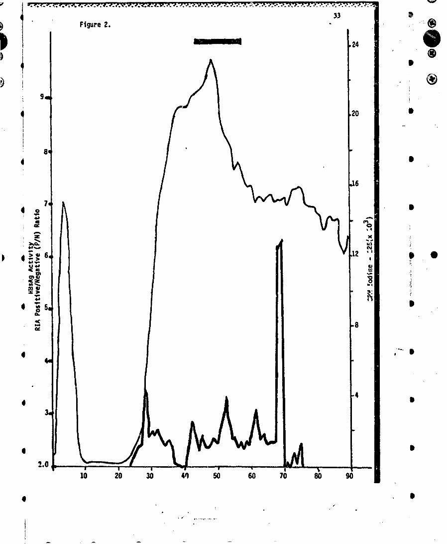
To circumvent the above problem, we fractionated a 20-mg preparation of solubilizedHBsAg on a Buchler electrophoretic apparatus as previously described (14). A 15%separation gel was used. The elution pattern is shown in Figure 2. Each fraction wasanalyzed for distribution of label (125s cpm), for antigenic activity (monitored by Ausria-ITtesting) and by analytical slab gel PAGE. The elution of protein was similar to that notedpreviously (14), and antigenic activity was noted throughout. The low antigenic activitywas expected since we have previously shown that antigenicity associated with polypep-tide populations is relatively inefficient when assayed with a sandwich technique such asAusria (4). Characterization of each fraction was carried out by analytical slab PAGE.The P22 and P25 polypeptides were localized in fractions 44-57 (Figure 2, solid bar). Thisrepresents a yield of 30% of the total protein mass which is free of all gel pieces. Thus,this method may be used to fractionate larger quantities of polypeptides free ofextraneous material.
B. Fractionation of Undenatured P22-P25 Polypeptides by he Method Described byZuckerman et al. (15)
Another approach to obtain :'22-P25 HBsAg-derived polypeptides under nondenatur-ing conditions was by the procedu'es described by Simons et al. (16) and Skelly et al. (17),with slight modifications.
HBsAg (0 mg) was disrupted under nondenaturing conditions with a final concentra-ton of 2% Triton X-100 in 0.01 M Tris-HCI, 0.5 M NaCI, pH 7.3, for 36 hr at 370C. 1231_
labeled HBsAg was added as tracer before the detergent treatment. The disrupted HBsAgwas applied to a I x 30 cm column of lentil lectin-Sepharose 4B (Pharmacia FineChemicals), equilibrated with 0.01 M Tris-HCI, 0.5 M NaCI, 1 mM CaCI 2, I mM MnCI 2,2% Triton X-100, pH 7.3. The sample was allowed to react with the gel for 30 min atroom temperature and then the column was flushed with the above buffer to removeunbound material. The bound material was eluted with 0.01 M Tris-HCI, 0.5 M NaCI, 2%Tritorn X-100, 5% ca-methyl-D-mannoside, pH 7.3. We collected 1-ml fractions andmonitored the proteins by ODago and radioactivity.
The data are presented in Figure 3. A major peak of unbound proteins eluted betweenfractions 12 and 29, and a minor peak of lentil-lectin-bound material eluted with themannoside buffer. This material is tnought to be a soluble complex of P22-P25 whichbound to lentil lectin through the glycosylated moiety of P25.
The fractions of the second peak were pooled, dialyzed extensively against 0.01 M-Tris-H10l, pH 7.3, at V0C, and concentrated through an Amicon YM 10 membrane toapprc',ximately 4 ml. This material was layered over a preformed linear 20-50% sucrose
* gradient and spun in an SW4I Beckman rotor at 36,000 rpm fnr 24 hr. Fractions (0.6 ml)were coilected from the top of the tube. Density and H1' sAg activity were determined foreach fraction. Three peaks were obtained at OD2 @0 . HBsAg activity, as measured byAusria-Il, was present in each peak. The top fractions of the peaks (4, 12 and 20) wereexamined by Jectron microscopy for the presence of micelles. Fractions 4 and 12contai:ted-aimcit no micelles. Fraction 20 was loaded with individual and aggregatedmicelles (figure 0), The buoyant density of the micelles in sucrose was 1.19 g/cm3, ascompared with a ,density of 1.17 g!cm3 reported for 22-nm HBsAg particles In sucrose.The diameter of the micelles ranged irom 50 to 250 nm. 30-33% of protein in micelle
* * 0 00 0

-. * p* -*. . .. ¼ .S * . iA*. * */ . *** *=4 _-- .. j•, • s
120
form was recovered from the original HBsAg particles, as estimated by r_ -rein -'etermin-ation (Lowry method) and percent of radioactivity in peaks. The mico-lles retz.ined higl1HBsAg activity, as demonstrated by high P/N ratios obtained by thw- Ausria test, incontrast to the low activity shown by the polypeptides (see Section IV.A). To furthercharacterize the retention of specific HBsAg activity, the micelles were iodinated usingthe chloramine-T method and used as labeled antigen for detection of anti-HBs. Two secsof beads from the Ausab kit were first incubated with normal and anti-fif3s sera fromdifferent species (guinea pig, rabbit and goat) and then one set was incubated with toý!1251-labeled HBsAg from an Ausab kit and the second set with 1231-labeleci micelle-HBsAg. The P/N ratios of both radioimmunoassays were similar.
V. Evaluation of Cellular Immunity in Hepatitis B Immunized Animals and Humans
SWe have recently shown that peripheral blood mononuclear cells (PBMCs) frompatients who had recovered from HBV infections showed cellular cytotoxicity toward thePLC/PRF/5 HBsAg-producing hepatoma cell line. The elfector cell responsible for thiseffect had the characteristics of a natural killer cell. Similar studies were instituted inmice and guinea pigs and in 20 young %d-uit volunteers who were vaccinated with purifiedHBsAg in a study on the reactogenicity and irnmunogenicity of an HBV vaccine. TheHBsAg vaccine used in the human studies consisted of purified, formalin-inactivated,alum-adsorbed HBsAg, subtype adw. In some individuals, blood was initially obtainedbefore the first vaccine dose was given. Subsequent samples were then drawn aftercirculating levels of anti-HBs antibodies were detected. lri five subjects, however, noproduction of antibodies had developed alter three inoculations of 40 ug of HBsAg eachdose. Blood was also obtained from chese indaviduals and tested fnr the production of -suppressive I ictors as described below.
Ferrnale BALB/c mice were immunized with 40-160 tig of purified HBsAg intraperi-toneally either once or twice. One t- two months alter thi=,r last inoculation, an -:
intravenous booster of 5-20 ýg HBsA' z -iven and the mictr were sacrificed 3 dayslater. Before they were sacrificed, bl_.. "-as obtained and the presence of circulating
S anti-HBs antibodies was confirmed by radloimmunoassay. These experiments wereconducted in conjunction with studies on monoclonal antibody production for HBsAg. Atotal of 6 immunized mice and 4 control mice (immunized with tetanus toxoid) weretested for lymphocyte cytotoxicIty toward the PLC/PRF/I cr Mahlavu cell lines. Noneshowed cytotoxicity toward any of the targets (data not shown). In contrast, two micethat were immunized with 10 x 106 PLC/PRF/I cells twice intraperitoneally did possesscytotoxicity toward both the PLCIPRF/3 and Mahlavu cell lines (data not shown).Interestingly, these mice did not develop anti-HBs antibodies. -Most likely, the targetantigens were human hepatocyte antigens common to both cell lines. Further experimentsexamining this xenogeneic system were not performed since the original purpose was todetermine whether- lymphocytes from immunized mice could show specific cytotoxicity.Positive results would have led to determination of the cells responsible and hopefullywould have complemented the results obtained with humans.
Previous studies performed by Cabral et al. (7, t9) and Sanchez et al. (10) showed !hepresence of cellular immunity to HBsAg in immunized guinea pigs. Spleenm were obtainedfrom guinea pigs that had been inoculated with purified HBsAg or one of its polypeptides(P68). Spleen cells from these animals also did not show cytotoxicity toward the humanhNtotna cell lines, although circulating levels of anti-HBs were present (data not shown).
0A
4i
4i @ 0 • • S S 6
136°..
4 .: Three human subjects who had been passively immunized with commercial high-titered anti-HBs globulin (H-BIG) were studied. All had been exposed to HbV-contamina-ted material and were therefore at high risk for subsequent HBV infection. All developed
* moderately high amounts of anti-HBs antibodies after intramuscular inoculation of H-BIG(mean radioimmunoassay positive-to-negative (P/N) of 58.4 t 8.61. Two to four weeks
"a fter inoculation of H-BIG, peripheral blood lymphocytes were obtained and studied for%- cytotoxicity. No specific cytotoxicity was observed (data not shown), although lympho-
* P," cytes from one subject did show nonspecific cytotoxic activity toward both cell lineswhich did not persist after an overnight incubation at 371C.
About 200 heaithy young adUt volunteers were participating in a separate study onthe immunogenicity of a purified, formalir-inactivated HBsAg vaccine. 20 of theseIndividuals were studied for lymphocyte cytotoxicity. Three subjects had been testedbefore the first dose. No difference in response was observed upon vaccination withHBsAg, although circulating levels of anti-HBs were present. In 15 individuals, nosignificant cytotoxicity was observed above a background of 5-15% specific lysis. In two,however, cytotoxicity was observed which was specific for the PLC/PRF/5 target cellsand appeared to be dose-dependent. Finally, 5 subjects who did not respond with theproduction of specific antibody after 3 inoculations were also not cytotoxic (data notshown).
.V. Propagation of Hepatitis A Virus (HAY) in Cell Cultures: Proteol_.5ic Enhancemernt
Our initial plan was to study mechanisms of replication for HAV that might enhancethe Infectivity of HAV using Q) proteolytic enzymes (pancreatin and trypsin) which areknown to exert their activity on virus particles by converting noninfectiow particles to *)infectious virions, xnd (ii) DEAE-dextran, a polycationic, diethyiarninoethyl ether of
; Jdaxtran which stimulates cellular uptake of nucleic acids and enhances plaque formation"by viruses and Infective RNA.
Continuous cell lines have been propagated and cell stocks stored in !iquid nitrogen?(I) MA- 104 - received from D0,. M.K. Estes and stored at passage 57; (ii) BGMK - stored
*.,', at passage 53; (1ii) PLC/PRF/J hepatoma cell line (HBsAg secretor) - courtesy of or."Stanley Lemon, Walter Reed Army Institute of Research; (iv) Mahlavu hepatoma cell line(HBsAg nonsecretor) - courtesy of Dr. 1. Milman; (v) MRC-5, (vi) HEF and (vii) Hep2 --all purchased from Microbiological Associates.
BGMK cells have been cultivated in MEM contalning 20 mM HEPES, 0.225% NaHCOJ,.. 0.25% lactalbumin hydrolysate, 0.5% bovine serum al3umin, antibiotics, and glutamine in S* "•the absence of serum for a period of at least I month without adverse effects when media
was changed weekly.
Marmoset-adapted HAV was prepared from about 5 grams of marmoset liver by6 either of two methods.
* -". (I) Procedure I (Figure A): Initial studies have shown that when HAV-intected liverwas homogenized in glyclne-HCI, pH 1.I, and then layered over 30% sucrose prepared inan identical buffer system prior to centrifugation at 24,000 rpm for 18 hr at 41C,antigen/antibody complexes presumably can be dissociated and/or kept from forming.Figure 6 shows the results obtained using this procedure.
.6
S
6) l ••••

, ~. (2) Procedure It (Figure 7): Marmoset liver tissue was homogenized with PBS, pH 7.4,,•.•. containing 0.05 M phosphate, saline, EDTA and Triton X-100. The homogenate was -;
sonicated for 30 sec and centrifuged at 5000 rpm for 13 min. The supernatant fluid was;', collected, and the pellet w~as resuspended in 20 ml of PES buffer, sonicated and• recentrifuged. The supertiatant fluids were then combined. Fractions were layered over".. 30% sucrose and a cushion of 60% sucrose. Centrifugation was performed at 25,000 rpm
".cr !5 hc at e,=C in an SW27 rotor. Fractions were collected from the bvttom of the b•." centrifuge tube and w'ere tested by radioimmunoassay for HAY antigen. Figure A shows
ta*e results obtained with this procedure.
In addition, HAV has been Isolated from fecal extracts of chimpanzees infected withHAV. Two procedures have been used.
(1) Precedu.e 111: 2.0 ml of a 20% chimpanzee fecal suspension containing HAV wasmixed with 1.0 ml lactaibumin hydrolysate and filtered through a 45-prn filter. Allsarmple. w,.r sto.,ed at -301C until used.
V (2) frrocedure IV: A 20% fecal suspension was clarified, and HAV was isolated using10% PEG 6000. The pellet was resolubilized with PB3 and the sample was placed on a"CsCI gradient and subjected to isopycnic centrifugation. Fractions were collected and •
Eltho.e containing the bulk of the HAV antigen were pooled for use as an inoculurn.
BkMK cells (passage 63) were treated with DEAE-dextran (10 or 100 uig/mI) for 30min at 37C. Partially purified marmoset-adapted HAV and HAY from fecal extracts(Prcedure I11) were incubated for 60 min with a solution of trypsin (10 uJ•Jml) prepared inHank BSS. The virus-trypsin samples were diluted 10-fold (10-1, 10 t and 10-3) to * **irnnish the toxic effect of the trypsin, then added to the DEAE-dextran-treated BGMK"cells. After an Incubation peri-- of 60 min at 37¶C, the plates were washed and"maintenance mneJia, without .rypsin, was added to the cells. Control cells receivedtrypsin solution only. CoverslIps are being evaluated weekly for HAV by immunofluores-
In anothev, set of ex-frinn.nts, five cell linea (P..C/PRF/5, M-JJavu, MRC-3, HEF and I° .,. Hep-2) were lnfe:tei with HAV obtained by Procedure IV Ihis material was inoculated
"Initially into a primary cell cult're derived from a neornatal hepatic carcinoma. After Iweek, the cells vwere scrapej from the plate, sonicated, and the :lartifed supernatant fluid"used as inoculum for the previously described •ell linel. Table 8 shows the results ofthese studies. During week 5, evidence of HAV rep•ization or expression wws detected In
Sthe PLCIPRF/3 and MRC-i ceil filtes when compared with the uninfected cells.
"VY Detection of HAV and Potavirus in a Connunity Water Supply Following an Outbreakof Gastroenteritis and infectious HepaZiti.
Georgetown, Texas (population 13,060), is onre of many small communities in theUnited Stawes which uses a groundwater :rpp;y with no water oreatment except chlorlia- •tion. Thterefcwe, If the raw water supply is unex;7.,;tedlv contaminated, the corrmunity is
. susceptible to a w3terborne disease outbreak. in a review of the problen, of viruses i•.groundwater, approxirnately 50% of the documented outbreiks of waterborne di-ease inthe United States were noted to be related to contaminated groundwater (0 C).
In June 1980 an outbreak of gastrointestinal illness char,.,:terized by an acute onset* ,; of diarrhea, abdominal cramps, nausea and fever occurr.d in Georgetown, Texas. Illness
was associated with drinking water from the central city vells in Georgetown. The attack
".5 •••••
1++15
4 :; rate among the 10,000 individuals living in areas supplied by these wells w'as approxim- Sately 79%. An increased number of cases of hepatitis A began to appear in 3uly 1980. 29cases of hepatitis were reported from 3uly 6-15, with a total of 36 cases reported during (the month. This compared to an expected incidence of 0-2 cases of hepatitis A per monthfor the Georgetown area.
Georgetown receives its water from 7 wells. There are 4 central city wells ranging4 from 186 to 210 feet deep which pump water to a common 750,000-gallon storage
reservoir. Georgetown is situated in a limestone region and draws its water from the.. Edwards aquifer. Due to the porous nature of the aquifer and the fact that the aquifer is
exposed to the surface by a fault line running just west of Georgetown, several"possibilities exist for contamination of the wells. These include: (4) leakage from nearbysewer lines; (ii) contamination from nearby abandoned or private wells or septic tanks; and(iii) relatively long-distance contamination from surface contaminants in the recharge S4 area of the Edwards aquifer.
During the second peak of gastroenteritis cases, samples of sewage, w-11 water andtap water were taken for virological analysis. Well and tap water samples of 400-1,000liters were concentrated by filtration through either positively charged depth filtersO(-MDS, AMF/CUNO, Meriden, Conn.) or negatively charged filters (Duo-Fine, Filterite
41 Corp., Timonium, Md.). With the Filterite filters it was necessary to adjust the pH to 3.5"by injecting I N HCI into the sample before filtration. Aluminum chloride was a. o added"through the injection port to a final concentration of 0.005 M. These conditions allow formaximum adsorption of the virus to the Filterite filters (20). With the I-MDS filters noaddi'ives were necessary since the positively charged filters adsorb the negativelycharged virus particles at ambient conditions. When tap water samples were taken, a
4! sodium thiosulfate solution was injected ahead of the filter to neutralize the residualchlorine.
Each flter cartridge was eluted with I liter of 3% beef extract at pH 10.0. Thiseluate was concentrated by an organic flocculation technique (21). Briefly, each eluatewas adjusted to pH 3.5 using I M glycine, pH 1.5. The floc that formed was collected bycentrifugation, and the virus that adsorbed to this floc was eluted with 0.05 M glycine at
4 pH 9.5. The resulting suspension was then clarified by centrifugation, treated withantibiotics, and Inoculated onto BGM cell monolayers to assay for enterovirus (22). An"aliquot of each concentrate was further concentrated by ultracentrifugation to assay forHAV antigen and rotavirus. Samples were assayed for rotavirus using an Indirect"immunofluorescence test (23) and for HAV antigen using a radloirmunoassay (24, 25).
"4. Table 9 shows the concentrations of enterovirus in sewage, well water and tap water 5samples taken on 3un-e 19. The concentration of enterovirtz in the five sewage samples"ranged from 1,200 to 7,400 plaque-forming units (PFU) per 100 liters, giving an average of.,M0 PFUJ100 liters.
Rotavirus, a major cause of Infantile diarrhea, was detected in two sewage samples, at a low level. Rotavirus was not detected in any of the well or tap water samples that• were tested. Seven fecal specitm•s from acutely IIl Georgetown residents were tested.. and were negative for rotavirus.
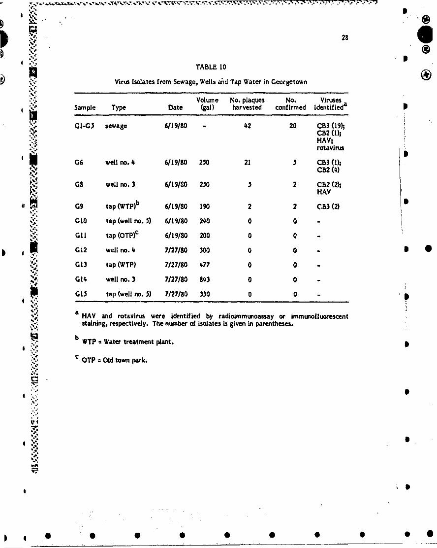
*" Table 10 shows the viral isolates that were obtained from each sample location.From sewage samples, 20 of 42 plaques were confirmed; 19 were identified as coxsackie-virus B3 (CB3) and I as coxsackievlrus B2 (C82). A total of 26 virus-like plaques wereharvested from cells inoculated with samples from Georgetown's central wells (no. 3 and 5
% ". 4h 7 of these were identified, with 6 identified as CB2 and I as CB3. The two .solates
44 00SS
16 6
4 ', from the tap water were both Identified as CB3. When the well and tap water were testedagain on July 27, no viruses were isolated. Serologic studies indicate that CB3 could havebeen partly responsible for the Georgetown outbreak.
HAV antigen was detected in 3 of 5 of the sewage concentrates and in I well water':oncentrate from samples taken on June 19. This preceded the outbreak of hepatitis
4 ""cases, which occurred in mid-July. Table 11 summarizes the hepatitis results. The Ppositive sewage and well water s3mples were neutralized by human IgG from a
* convalescent hepatitis patient. The convalescent IgG did not react with CB2 or CB3Isolated from the same samples or with other common enteroviruses. The buoyant densityof the sewage isolates in a CsCI gradient was 1.34 g/ml, which is in the range reportedfor HAV particles.
4 "Human enteric viruses were isolated from the potable water despite a total chlorineresidual of 0.8 mg/liter. This Is significant since the bacteriological samples of tap watertaken by local and state officials were consistently free of coliform bacteria. Thebacteriological results led city officials to believe that the water was safe when, in fact,viruses were surviving the chlorination process.
4. There are at least two other reports of gastroenteritIs outbreaks related to potable1 •water which met bacteriological standards. Wellings et rl. (26) Isolated echovirus 22/23
"from a disinfected groundwater supply during an outbreak at 4 migrant labor camp inFlorida. More recently, an lodinated (0.7-1.0 ppm) groundwater supply was Implicated inan outbreak of viral gastroenteritis at a summer camp in Maryland (27), even thoughdrinking water samples taken before, during and after the epidemic met bacteriologicalstandards. These findings support the recommendations recently made by a Scientific ICommittee of the World Health Organization (28) and by an American Water WorksAssociation T&P committee (29) whIch Include standards for viruses In water and possible
• " monitoing of viral contamination in certain situations.
* "The Georgetown Incident reemphasizes that the purity of groundwater cannot always"be relied on. Barriers against contamination of potable water supplies in communitiesthat depend on groundwater must be properly maintained and adequate for Intermittent S
4, contamination. These barriers may need to be stronger In certain areas, such as limestoneregions, which may be especially susceptible to contamination from distant sources.
The ability to detect hepatitis A virus antigen in a water source prior to an outbreakcould be valuable In the prevention of future outbreaks. Prophylactic treatment of thepopulation with gammaglobulin upon discovery of hepatitis In a water source could
4 decrease the severity of the outbreak. Unfortunately, in the Georgetown epidemic, thesamples were not assayed for hepatitis until after the outbreak, so no preventive measureswere taken. In the future, rapid analysis of water samples for hepatitis A virus may aid Inreducing the number of cases of morbidity and mortality from waterborne hepatitis.
4 II
II
*l •• 0 • 0 0

17 IVIII. Preliminary Testing of Polypeptide Vaccine and Synthetic Peptide Vaccine in Mice P
Preliminary results (Table 12) are available which strongly support the likelihood ofsuccess for our proposed work scope for the new contract year. Groups of mice wereimmunized with I dose of two difference types of vaccine. The first type was the micelle Apreparation prepared according to the Zuckerman procedure. The second type of vaccineinvolves injection of the first two sets of synthetic peptides made by Dr. Sparrow (BaylorCollege of Medicine). These two peptides consist of amino acid residues 117-137 (20residues) and amino acid residues 122-137 (15 residues).
It is evident that the micelle polypeptide preparation is highly immunogenic (Table12). In keeping with their report (Zuckerman et al., in Hepatitis B Vaccine, pp. 251-262,Elsevier-North Holland Biomed. Press) the same dose of micelles produced higher levels ofantibody than observed in mice immunized with intact particles (Tables I and 12). It wasof interest that the antibody levels dropped between days 7 and 14 after inoculation ofmicelles suspended in saline, whereas the titers increased during the same time period inthose mice inoculated with micelles precipitated in alum (Table 12).
The results of our preliminary work with synthetic peptldes (117-137 and 122-137) aremost exciting. In every group of mice injected, regardless of the adjuvant vehicle \ pemployed, at least one-half of the mice in each group produced detectable levels ofantibody at a serum dilution of 1:4, after I dose of peptide. In fact, in almost all casesthe antibody response was equal to that seen in mice injected with SDS.-denatured P22polypeptide material (Tables 2 and 12).
IX. Literature Cited I 6
1. Gerin, J.L., Holland, P.V., and Purcell, R.H. 1971. Australia antigen: large %calepurification irom human serum and biochemical studies of its proteins. 3. Virol.7:569-576.
2. Purcell, R.H. and Gerin, 3.L. 1978. Hepatitis B vaccines: a status report. In ViralHepatitis (G.N. Vyas, S.N. Cohen and R. Schmid, eds.), pp. 491-503. Fr'anklinInstitute Press, Philadelphia.
3. Hollinger, F.B., Dreesman, G.R., Sanchez, Y., Cabral, G.A., and Melnick, ý.L. 1978.Experimental hepatitis B polypeptide vaccine in chimpanzees. In Viral Hepatitis(G.N. Vyas, S.N. Cohen and R. Schmid, eds.), pp. 557-367. Franklin Institute Press,Philadelphia.
4. Dreesman, G.R., Hollinger, F.B., Sanchez, Y., Oefinger, P., and Melnick, 3.L. 1981.Immunization of chimpanzees with hepatitis 8 virus-derived polypeptides. Infect.tmmun. 32:62-67.
3. Dreesman, G.R., Hollinger, F.B., McComnbs, R..%, and Melnick, 3.1. 1972. Produc-tion of potent anti-Australia antigen sera of high specificity and sensitivity In goats.Infect. Immun. 5:213-221,
6. Hollinger, F.B. and Dreesman, G.R. 1980. Hepatitis viruses In Manual of ClinicalImmunology (N.R. Rose and H. Friedman, eds.), 2nd ed., pp. 678-691. American ,Society for Microbiology, Washington.
7. Cabral, G.A., Marciano-Cabrai, F., Funk, G.A., Sanchez, Y., Hollinger, F.B., Me.nick,3-L. and Dreesman, G.R. 1978. Cellular and humoral immunity in guinea pigs to twomajor polypeptides derived from hepatitis 8 surface antigen. 3. Gen. Viroh. 3#3391-350.
8. Chairez, R., Hollinger, F.B., Melnick, 3.L., and Dreesman, G.R. 1974. Biophysicalproperties of purified forms of hepatitis B antigen. Intervirology 33129-140.
". • I
0 0 • • 0 0
I
180
9. Greenwood, F.C., Hunter, W.M., and Glover, 3.S. 1963. The preparation of 1I11-Jlabeled human growth hormone of high specific radioactivity. Biochem. 3. 89.114-123.
10. Sanchez, Y., Ionescu-Matiu, I., Hollinger, F.B., Melnick, 3.L., and Dreesman, G.R.1980. Antigenic relationship of a hepatitis B surface antigen-derived polypeptide andhuman serum albumin. 3. Gen. Virol. 48:273-283.
11. Hilleman, M.R., Bertland, A.V., Buynak, E.B., Lampson, G.P., McAleer, W.3.,McLean, A.A., Roehrn, R.R., and Tytell, A.A. 1978. Clinical and laboratory studiesof HBsAg vaccine. In Viral Hepatitis (G.N. Vyas, S.N. Cohen and R. Schmid, eds.), pp.525-537. Franklin Istitute Press, Philadelphia.
12. Cabral, G.A., Gyorkey, F., Gyorkey, P., Melnick, 3.L., and Dreesman, G.R. 1978.Immunohistochemical and electron microscopic detection of hepatitis B surface andcore antigens. Exp. Molec. Pathol. 29:156-169.
13. Fields, H.A., Davis, C.L., Dreesman, G.R., Bradley, D.W., and Maynard, 3.E. 1981. SEnzyme potentiated radioimmunoassay (EPRIA) a sensitive third-generation test forthe detection of hepatitis B surface antigen. 3. Immunol. Methods, in press.
14. Dreesman, G.R., Chairez, R., Suarez, M., Hollinger, F.B., Courtney, R.J., andMelnick, 3.L. 1975. Production of antibody to individual polypeptides derived frompurified hepatitis B surface antigen. 3. Virol. 16:508-515.
15. Zuckerman, A.3., Howard, C.R., and Skelly, 3. 1981. Hepatitis B micelle vaccines.In Hepatitis B Vaccine (P. Maupas and P. Guesry, eds.), pp. 231-263. Elsevier/North -Holland Biomedical Press.
16. Simons, K., Helenius, A., Leonard, K., Sarvas, M., and Gething, M.3. 1978.Formation of protein micelles from amphilitic membrane proteins. Proc. Nati.Acad. Sci. USA 75:5306-5310.
17. Skelly, 3, Voward, C.R., and Zuckerman, A.3. 1981. Hepatitis B polypeptide vaccinepreparation in micelle form. Na:jre 290i51-54. 5
18. Cabral, G.A., Chalrez, R., Marciano-Cabral, F., Suarez, M., Dreesman, G.R.,Melnick, J.L., and Hollinger, F.B. 1975. Cell-mediated Immunity in guinea pigs tosubunits derived from hepatitis B surface antigen. Infect. Immun. 120564-70.
19. Keswick, B.H. and Gerba, C.P. 1980. Viruses in groundwater. Environ. Scd. Technol.14:1290-1297. /
20. Farrah, S.R., Gerba, C.P., Wallis, C., and Melnick, 3.L. 1976. Concentration ofviruses from large volumes of tap water using pleated membrane filters. Appl.Environ. Microbic. 31:221-226.
21. Katzenelson, E., Fattal, B., and Kostovesky, T. 1976. Organic flocculation: anefficient second-step concentration method for the detection of viruses in tap water.Appl. Environ. Mlcrobiol. 32.638.
22. Melnick, 3.L., Wenner, H.A., and Phillips, C.A. 1979. Enteroviruses. in DiagriosticProcedures for Viral, Rlcke:vsial .and Chlarnydial Infections (E.H. Lennette and N.J. SSchmidt, eds.), 5th ed, pp. 1471-334. American Public Health Association, New York.
23. Smith, E.1% et a. 1981, Development of a method for the detection of humanrotavirus in water. In preparation. .4
24. Hollinger# F.B., Bradley, D.W., Maynard, 3.E., Dreesman, G.R., and Melnick, 3.1.1975. Detection of hepatitis A viral antigen by radloimmunoassay. 3. Immunol.
25. Sanchez, Y., LaBelle, R., Gerba, C.P., Dreesman, G.R., and Fields, H. 1981.Detection and concentration of hepatitis A virus in water. In preparation.
26. Wellirngs) F.M,, Mountain, C.W., and Lewis, A.L. 1973. Virus In groundwater. Proc.2nd Natl. Conf. Individual Onsite Wastewater Systems. National Sanitation Founda-tion, Ann Arbor.
27. Woodward, W.E. 1981. Personal communication.28. World Health Organization Scientific Group. 1979. Human viruses in water,
wastewater and soil. WHO Tech. Rept. Series No. 639.29. P.merican Water Works Association T&P Committee. 1979. Viruses in drinking
water. 3. Am. Water Works Assoc. 71:-41.
0S* * * 0 0
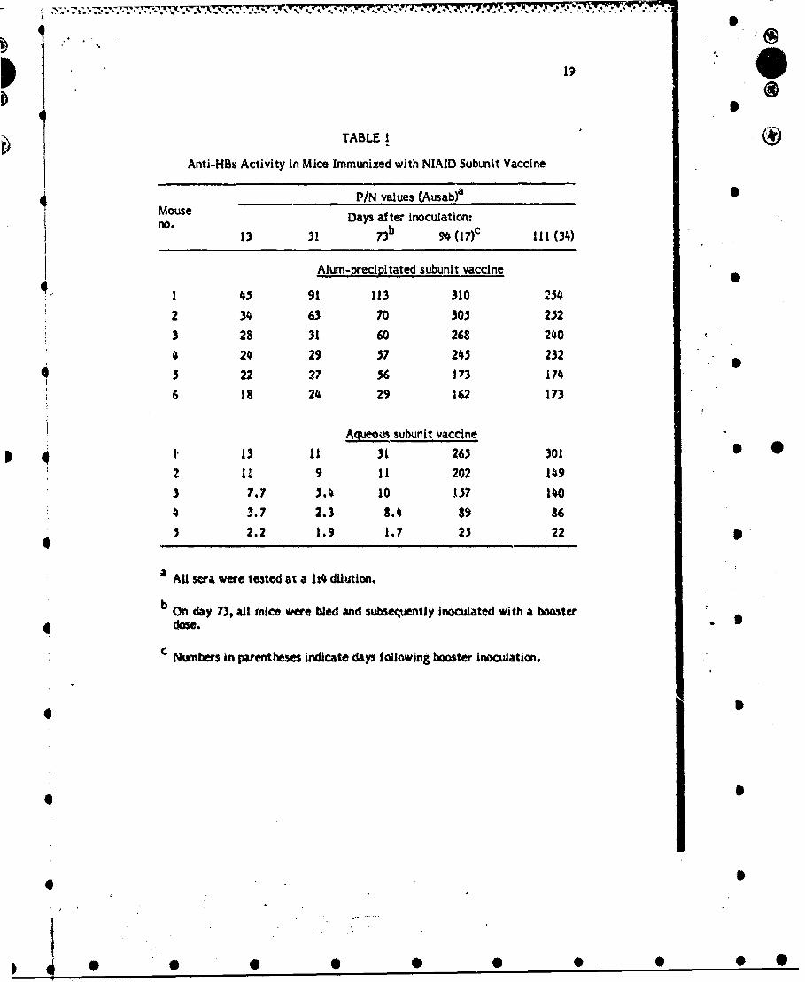
TABLE I
Anti-HBs Activity In Mice Immunized with NIAID Subunit Vaccine
P/N values (Ausab)a OMouse Days after inoculation-no0 13 31 73b 94 (17)c 111 (34)
Alum-precipitated subunit vaccine
I 45 91 113 310 254
2 34 63 70 305 252
3 28 31 60 268 240
4 24 29 37 245 232
3 22 27 36 173 174
6 18 24 29 162 173
Aqteous subunit vaccine
1 ., 13 11 31 265 301 S 02 11 9 11 202 149
3 7.7 3.4 10 137 1404 3.7 2.3 8.4 89 86
3 2.2 1.9 1.7 23 22 '
a AU ra were tested at a 1t4 dilution.
b On day 73, all mice were bled and subsequently inoculated with a booster
dose.
'Numbers in parentheses indicate days following booster inoculation.
0S
4 •
€ •
I
• • 0 9 * 0

206
TABLE 2
Anti-HBs Activity in Mice Immunized with a P22-P25Polypeptide Pool Derived from the NMAID Vaccine
P/N values (Ausab)a
Mouse Days after Inoculation:no* 7 14 21b 33 ( 12)c 51(30) 133 (112)
iI
Alum-precipitated, formailn-treated polypeptide vaccine
1 1.5 1.6 0.8 1.3 0.8 -d
2 1.3 0.8 0.6 0.6 0.6 -
3 1.1 2.0 1.2 0.8 1.2 -
4 1.0 1.4 0.7 1.6 0.7 -
3 2.9 2.5 1.2 1. 1.2 -
6 1.5 0.( 1.4 - - -
Alurn-precipitated, untreated polypeptide vaccine
I 3.1 2.- 0.3 1.3 1.4 1.4
2 3.0 2.9 0.7 1.3 0.8 1.2
3 k.6 3.2 1.9 1.4 0.8 2.6
4 2.1 1.3 0.8 0.7 0.7 1.2
3 8.6 7.4 9.1 5.6 C.2 5.6
6 6.1 4.8 4.3 3.3 2.1 3.0
a AU sea were tested at a 134 dilution.
b On day 21, all mice were bled and subsequently inoculated with a booster
dose. Both groups of mice were boostered with formalln-treated polypeptideva:cine.
€ Numbers in parr.nt eses indicate days following booster inoculation.
d Mice not alalable at that time.
• * .O * • S • S

p 21
TABLE 3
Anti-HBs Activity in Mice Immunized with a P22-P25Alum-Precipitated Polypeptide Vaccine after
Storage for 2 Months
P/N values (Ausab)aDays after inoculation
10 21 72I
1 1.7 2.7 3.0
2 2.8 2.4 2.1
3 1.0 0.9 2.9
4 1.6 3.0 3.8
5 4.3 2.4 3.0
6 3.3 2. 1.4
7 5.4 0.6 b *
a All sera were tested at a 1:4 dilution.
b Mouse not available at that time.
0D
4 • • ••O

22 ®
TABLE. 4
Anti-Tetanus Toxoid Titers in Mice Immunized with Tetanus Toxoid Aloneor in Association with a Hepatitis Subunit or Polypeptide Vaccine
Preparation No. mice Titer rangea Geometrictested mean titer
TT-saline 4 160,000 - 640,000 378,296
STT-alum 5 3000t0O00 - 60,000,000 42,875,516
P22+TT-saline 6 94,000 - 640,000 269,827
P22+TT-alum 6 1,900,000 - 3,800,000 2,822,768
HBs+TT-saline 6 31,250 - 700,000 196,217
HBS+TT-alum 6 781,250 - 7,600,000 1,892,364
a Titers are expressed as the reciprocal of that serum dilution which would i •
correspond to a P/N ratio of 2.1 in micro-SPRIA.
4
q S
4
o o
4I
f .
4 ... _

23 4
TABLE .5
Anti-HBs Titers in Mice Immunized with PolypeptideVaccine in Association with Tetanus Toxoid
aDPreparation No. mice Titer rangea Geometric
tested mean titer
P22-saline 6 1,500 - 13,000 5,647
P22+TT-saline 6 15,000 - 115,000 41,917 S
P22-alum 6 32,000 - 380,000 236,678
P22+TT-alum 6 22,000- 220,000 48,154
a Titers are expressed as the reciprocal of that serum dilution which 5
would correspond to a PIN of 2.1 in micro-SPRIA.
d 6
S
S
I)
S
-.- 4 . ... 4 ., •. 4 • . • • d .u •

w
I ~246
TABLE 6 0Anti-HBs Titers In Mice Immunized with Subunit HBsAg
Vaccine In Association with Tetanus Toxoid
Preparation No. mice Titer rangea Geometrictested mean titer
HBs-sallne 6 230,000 - 720j000 457,748
HBs+TT-saline 3 18,000 - 350,000 67,272
HBS-aium 4 *40,000 - 1,050,000 794,857
H~s+TT-alum 6 540,000 - 3,000,000 1,076,794
a Titers are expressed as the reciprocal of that serum dilution which
would correspond to a P/N ratio of 2.1 in micro-SPRIA.
6 •4
4
4 S
II

250 0
TABLE 7
Comparative Sensitivities of Antibody Assaysfor Anti-HBs Ac-" ,-i qy
Assay Mean titera Sensitivity
Ausabb 250
Micro-SPRIA 51,000 204
ELISA 89,268 357
SGeometric mean titer of 23 individual mouse anti-HBs sera.
b Commercial RIA kit purchased from Abbott Laboratories. 5
• 0
'-'4..
S
i''•'
IS
• e • •• • •

1~~ 266
) ~TABLE 8 )Solid-Phase (Microtiter) RIA for HAV Antigen
Deteclion in Supernatants from Five Human Cell Lines
Cell line SIN ratio In supernatantWeeks after infection:3 4
PLC./PRF/5 infected .1.03 2.24 3.24riot infected 1.15 1.20 1.27
Mahlavu I rf ected 1.30 1.87 2.87not infected 1.29 1.08 1.59
MRC-5 inf ected 1.16 1.09 3.07not Infected 1.11 1.16 1.00
HEF Wofcted A1.10 1.4 ~ 1.69neit Infected 1.10 ND' ND
Hep-2 Infected ND 1.83 1.90nmc Infected 1.91.60 ND*
Chimp stool(control S/N valuesfor each set ofassays) 6.19 7.30 1420
a ND Not cone.
%
41

,27
TABLE 9
Concentrations of Enterovirus in Georgetown Sewage, Well Waterand Tap Water on 3une 19, 1980
Enterovirus
Volume No. PFU PFU/Sample Type Filter (gal) observed 100 liters
GI sewage Filterite 23 14 6,400
G2 s&vage Filterite 23 7 4,300
G3 sewage Filterite 23 12 7,400
G4 sewage Filterite 25 6 3,600
G sewage I-MDS 27 3 1,200
G6 well no. 4 I-MDS 230 21 13!i+ G7 well no. 4 Fl~terite 100 0
S . GI well no. 3 I-MDS 230 4 3 S *%*b
G9 tap (WTP)b I-MDS 190 2 1
GIO tap (well no. 3) I-MDS 240 0 -
* Gil tap (OTP)c Filterite 200 0 -
.TowaJ plaque-forming units (PFU) observed; only a portion of each concentrate wasinoculated.
WTP c Water treatment plant.
c OTP mOld town park.
v4:
t." 4-9+%
4t 5
. ,,÷, .. . ..
* S•• S 0 0
28
TABLE 10
Virus Isolates from Sewage, Wells and Tap Water in Georgetown AD
Sme yDt Volume No. plaques No. Viruses" Sample Type Date (gal) harvested confirmed Identifieda
." GI-G5 sewage 6/19/80 - 42 20 CB3 (11CB2 (l);HAV;rotavirus
G 06 well no. 4 6/19/80 250 21 5 CB3 (l);CB2 (4)
G8 well no. 3 6/19/10 250 5 2 CB2 (2);HAV
G9 tap (WTPPb 6/19/80 190 2 2 C.B3 (2)
GI0 tap (well no. 5) 6/19/80 240 0 0 -
Gil tap (OTPf 6/19/80 200 0 0 -
G12 well no.4 7/27/90 300 0 0 - *0 G13 tap (WTP) 7/27/80 477 0 0 -
G14 well no. 3 7/27/80 143 0 0 -
G13 tap (well no. 5) 7/27/80 330 0 0 -
a HAV and rotavirus were identified by radloimmunoassay or immunoiluorescentstaining, respectively. The number of isolates Is given in parentheses.
b WTP = Water treatment plant.
C oTP = Old to pak.
4.
:.'::
• B

o . . .. . . . .- ._. . - . - . . . . . . . .. ...
29
4.. TABLE 11
Detection by Radjoimmunoassay of HAV Antigen
HAV antigen* Sample only Neutra9zed
Sample Type Enterovirus P/N8 P/N
GI sewage + 1.88 1.59
4 G2 sewage + 3.4• (+) 1.67 S
G3 sewage + 1.31 1.77
G4 sewage + 3.91(+) 1.19
G3 sewage + 4.36 (+) 1.99
G6 well no.4• + 0.90 1.00
G8 well no. 3 + 2.30 (+) 1.19
i G9 tap * 1.06 NDc
" GI1 tap 0.93 1.06
"Gi1 tap 1.78 NO
3 a Ratio of sample to negative control. A ratio > 2.0 Indicates a positive test (1). 5
* " bb Ratio of sample after reaction with human lgG anti-HAV to negative control. The
sample was reacted with a specilfc antibody prior to testing by radloimmunoassay.
c ND Not done.
*4
4
Q'I
"V"8~
4%
• ,•o
. 'I - " ;.
* 0 . 0".0S

30
b iTABLE 12
Aintl-HBs ActivAty in Mice Immunized with a Micelle PolypeptidePreparation (Groups A and B) and with a Synthetic HBsAg Peptide (Groups C-H)
Group Prepazation Antibody activi Antibody activitya b
at day 7 at day 14PosItive/total P/N range Positive/total P/N range
- A micelle -Wsaline 5/ 19-99 3/5 8-43
B rincelle-alum 6/6 19-131 6/6 53-220
C 117-137 -FCA 4/6 3.1-6.2 3/6 2.9-7.3
D 122-137-"FCA 3/6 3.2-4.3 3/6 4.6-82
"•E 117-137 -alum 2/6 2.8-5.4 4/6 2.1-L6
F 122-137- . .alum 4/6 2.3-9.8 4/6 23-7.0
G 122-137-.-/lllosmes 16 3.2-&4 3/6 3.54-3
H K 122-137- bliposomes * MDP 4/6 2.3-15.2 3/6 2.8.-13.6
a Positive antibody activity at P/N value of >2.1 at serum dilution of 14 when tested by,• ~Ausab. -/
S.'-. b Llposome - synthetic peptides entrapped In liposomes..5,.
o.5
7I
S..
- .
) 4 9 0 0 S 0 0 0 O O

%~ % % -. o F.- ;g.~ ;~r
p
31
FIGURE 1 Comparative sensitivities of testing of 23 individual mouse sera by diffeventassays for anti-HBs. (- )
Imp
&I ;E09
| o I L
:• :
so *
II
%o 1. *S' jA *
* I' -
• . I,' 1.°*o6.t• s " *o: ' ,$ , a
I I I'
* I I •, ' ,
a a
Auasab Mllcro-SP~lA ELISA
!-tI
- -- I - - . ; - .4 - . . .. 1 . . . . 1 .. 4 . . . . .4 • . . . .4 . • , .. .

32
FICURE 2 Elution profile of 20 mg of HBsAg/adw trace labeled with 121l, solubilized inSDS, urea, and mercaptoethanol by heating at 100*C for 2 min, from preparative PAGEutilizing ý5% polyacrylamide gels. A pool of fractions was made, as indicated by theheavy bar, which contained the P22 and P25 polypeptides.
S
I
I,
/
PN-.... 7. 77777.*777.-.7
Figure 2. . -
I.244 p
9
q 20 I
8
4 S
16
6.• , 12
. - .
IN
3a3
20
10 20 30 M so 60 so
4 t5

I
34 0I
FIGURE 3. Affinity chromatography of .5 mg disrupted HBsAg on lentil lectin-Sepharose43. The antigen was disrupted and the unbound material was removed from the columnby washing with the starting buffer (0.01 M Tris-HCI, pH 7.3, containing 2% Triton X-100, 0.5 M NaCI, I mM MnCI2, and I mM CaCI2). The arrow indicates the start ofelution of the h-und material with 0.01 M Tris-HCI, pH 7.3, containing 2% Triton X-100,0.5 M NaCI, and 5% ct-methyl-O-mannoside,
D
tOlt
fI A ey
II
S I
* , I
Io / ."k

3,
FIGURE 4. Electron micrographs of micelles, of liBsAg. Density 1.19 g/crn' in sucrose.Bar represents 200 nm.
4-
A
* Q -' 1 -.1'.!
01A,'t
V -. t'% ZIt'X A jti. '
ii-14,,64~ 4t-F "
W.Sý eIA
low\
Y, M,R5&V&A.s t
4 IY
Y.
A
.4 1 i
4'~~~ 1.vti#

36
si E
o 0
o
w z
4-4
.s..~.,.~o4.. -
.. a.~ rv

370
FIGURE 6. Extraction of HAV at low pH from Infected marmoset liver
44.
..... ....
f1H.
... .. . ... .. H
RHS
IE

00I..• oso•,
o 'm
tC
.0
01.Wi 0,
0u8
fto
Eo
0.

39..
FIGURE S. Extractionl of HAV at pHi 7.4 f rom inf ected marmoset liver.
I tiff
tilt WS
V4, I
wt M MI
0ý 1I11 I N * l

S44Va~4 66- . F P
®
Publications
Sanchez, Y., tonescu-Matiu, L, Dreesman, G.R.,-Kramp, W., Six, H.R., Hollinger, F.B. andMelnick, 3.L. Humoral and cellular immunity to hepatitis B virus-derived antigens:comparative activity of Freund complete adjuvant, alum, and liposomes. Infect. Immun.30:728-733, 1980. p
Dreesman, G.R., Hollinger, F.B., Sanchez, Y., Oefinger, P. and Melnick, 3.L. Immuniza-tion of chimpanzees with hepatitis B virus-derived polypeptides. Infect. Immun. 32:62-67,1991. •
Sanchez, Y., Ionescu-Matiu, I., Dreesman, G.R., Hollnger, F.B. and Melnick, 3.L.Evidence for the presence of repeating antigenic determinants in the major and minor Ppolypeptides derived from hepatitis B surface antigen. Virology, In press.
Benson, 3.R., Funk, G.A., Sanchez, Y. and Dreesman, G.R. Evidence for protein homologyol the two major hepatitis B surface antigen (HBsAg) derived polypeptldes. Submitted forpublication.
p
. .
411
9 .
41.0

* p.
11
ITTTA�TFT1I'n
* �JL��/LdL3IJUA
1 1
*
* I
* IO
* I
I
* I
UNCLASSIFIED*
*
* 0 0 0 0 0 0 0