understanding aspects of alginate biosynthesis and
TRANSCRIPT

Copyright is owned by the Author of the thesis. Permission is given for a copy to be downloaded by an individual for the purpose of research and private study only. The thesis may not be reproduced elsewhere without the permission of the Author.

i
Understanding aspects of alginate
biosynthesis and regulation by Pseudomonas
aeruginosa
A thesis presented in partial fulfilment of the
requirements of the degree of
Doctor of Philosophy
in
Microbiology
at Massey University, Palmerston North,
New Zealand
Yajie Wang
2017
ii
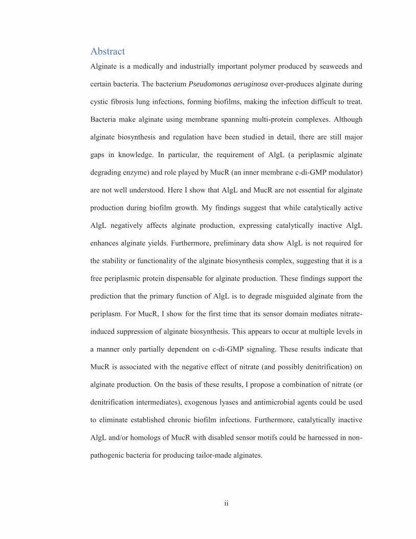
Abstract Alginate is a medically and industrially important polymer produced by seaweeds and
certain bacteria. The bacterium Pseudomonas aeruginosa over-produces alginate during
cystic fibrosis lung infections, forming biofilms, making the infection difficult to treat.
Bacteria make alginate using membrane spanning multi-protein complexes. Although
alginate biosynthesis and regulation have been studied in detail, there are still major
gaps in knowledge. In particular, the requirement of AlgL (a periplasmic alginate
degrading enzyme) and role played by MucR (an inner membrane c-di-GMP modulator)
are not well understood. Here I show that AlgL and MucR are not essential for alginate
production during biofilm growth. My findings suggest that while catalytically active
AlgL negatively affects alginate production, expressing catalytically inactive AlgL
enhances alginate yields. Furthermore, preliminary data show AlgL is not required for
the stability or functionality of the alginate biosynthesis complex, suggesting that it is a
free periplasmic protein dispensable for alginate production. These findings support the
prediction that the primary function of AlgL is to degrade misguided alginate from the
periplasm. For MucR, I show for the first time that its sensor domain mediates nitrate-
induced suppression of alginate biosynthesis. This appears to occur at multiple levels in
a manner only partially dependent on c-di-GMP signaling. These results indicate that
MucR is associated with the negative effect of nitrate (and possibly denitrification) on
alginate production. On the basis of these results, I propose a combination of nitrate (or
denitrification intermediates), exogenous lyases and antimicrobial agents could be used
to eliminate established chronic biofilm infections. Furthermore, catalytically inactive
AlgL and/or homologs of MucR with disabled sensor motifs could be harnessed in non-
pathogenic bacteria for producing tailor-made alginates.

iii
Acknowledgements I would like to thank Professor Bernd Rehm, Dr. Jan Schmid and Dr. Zoe Jordens for
their supervision, wisdom, guidance and support. I would also like to thank my
collaborators, Dr. Iain Hay, Dr. Zahid Rehman, Dr. Fata Moradali, Dr. Ian Sims, and
Dr. Ali Goudarztalejerdi. It has been a pleasure to work with you all.
I would like to thank Dr. Iain Hay and Dr. Zahid Rehman for their training and
construction of various plasmids (pBBR1MCS-5:mucR variants), Dr. Fata Moradali for
assistance with NMR, Dr. Ian Sims for SEC-MALLS analysis, and Dr. Ali
Goudarztalejerdi for generation of PDO300ΔalgL mutant. I would like to acknowledge
the current and former members of the laboratory team, Shuxiong, Patricia, Jinping, Jin,
Jean, Jason S, Jason L, Panan, Kampachiro, Shirin, David, Karin, Sasha, Leo, Lydia and
Andy, for their friendship and collegiality; it has been fun working alongside you.
Thank you to the Massey Genome Services for DNA sequencing, Manawatu
Microscopy and Imaging Centre for assistance with confocal laser scanning
microscopy, Mr. Mohsen Bagheri for operating the IFS sterilization and
decontamination facility, Mr. Paul Hocquard for procurement of reagents and
consumables, Ms. Ann Truter, Ms. Cynthia Creswell and Debra Creswell for making
administrative matters a breeze, Dr. Natisha Megan for compliance and health and
safety training, Professor Kathryn Stowell for being a supportive postgraduate
coordinator, Professor Simon Hall for being a supportive Head of Institute, and
Professor Gill Norris and Professor Geoff Jameson for technical and moral support.
I thank the Massey University Doctoral Research Scholarship, IFS Postgraduate
Scholarship and IFS Postgraduate Travel Fund for financial support.

iv
I would like to thank all my supportive friends (you know who you are): Lucy, Paulo,
Yilin, Leo, Lilian, Logan, Dam, Tuck, Jaired, Brian, Ricky, Dylan, Shao, Jennifer,
Kayla, Andrew, Isaac and Iain and all my friends at Palmerston North Overseas
Christian Fellowship for your friendship and Friday night and weekend fun. I would
like to thank my girlfriend, Lucy, for her support.
I would like to thank my father Qiao Wang, late-mother Li Yuan Chen, step-mother
Xiao Ling Chen, and half-brother George Zi Ming Wang for their unconditional love
and support.
Thank you to my Creator, God the Father, my Saviour Jesus Christ, and the Holy Spirit
that guide me with wisdom, strength and perseverance.

v
Dedication This thesis is dedicated to my late-grandfather Jiheng Wang, a former Professor of Plant
Breeding, who passed away on the 2nd of November 2016, aged 95.

vi
Table of Contents Abstract ..................................................................................................................................... ii
Acknowledgements .................................................................................................................. iii
Dedication .................................................................................................................................. v
Table of Contents ..................................................................................................................... vi
List of Abbreviations.................................................................................................................. x
List of Figures .......................................................................................................................... xi
List of Tables.......................................................................................................................... xiv
1. Chapter One: Introduction ..................................................................................................... 1
1.1 Structure and applications of alginate .................................................................................. 2
1.1.1 Structure and properties ........................................................................................ 2
1.1.2 Extraction and applications .................................................................................. 4
1.2 Pseudomonas aeruginosa and cystic fibrosis .................................................................... 10
1.2.1 Pseudomonas aeruginosa ................................................................................... 10
1.2.2 Cystic fibrosis and host defenses ........................................................................ 12
1.3 Biofilm formation and antimicrobial resistance ................................................................. 15
1.4 Alginate biosynthesis ......................................................................................................... 17
1.4.1 Polymerization .................................................................................................... 18
1.4.2 Modification ....................................................................................................... 19
1.4.3 Translocation and secretion ................................................................................ 20
1.5 Genetics and regulation of alginate production ................................................................. 22
1.5.1 Regulated intramembrane proteolysis cascade ................................................... 22
1.5.2 Transcriptional regulation .................................................................................. 24
1.5.3 Post-transcriptional regulation............................................................................ 27
1.5.4 Post-translational regulation ............................................................................... 27
1.6 Function of AlgL in alginate production ............................................................................ 28
1.7 Function of MucR in regulating alginate production ......................................................... 32
1.8 Aims and scientific questions ............................................................................................ 34
2. Chapter Two: Materials and Methods .................................................................................. 36
2.1 Strains, plasmids and oligonucleotides .............................................................................. 36

vii
2.1.1 Long term storage of strains ............................................................................... 42
2.2 Media, growth conditions and antibiotic concentrations ................................................... 42
2.2.1 Luria-Bertani (LB) medium ............................................................................... 42
2.2.2 X-Gal medium .................................................................................................... 43
2.2.3 Nutrient Broth ..................................................................................................... 43
2.2.4 Pseudomonas Isolation (PI) medium ................................................................. 43
2.2.5 Modified alginate-producing (MAP) medium ................................................... 43
2.2.6 Supplementation of media with antibiotics ........................................................ 44
2.2.7 Growth conditions .............................................................................................. 44
2.3 DNA manipulation ............................................................................................................. 44
2.3.1 Isolation of plasmid DNA .................................................................................. 44
2.3.2 Determination of DNA concentration and purity ............................................... 44
2.3.3 Polymerase Chain Reaction ................................................................................ 45
2.3.4 PCR dependent site-directed mutagenesis ......................................................... 47
2.3.5 Hydrolysis of DNA by restriction endonucleases .............................................. 50
2.3.6 Agarose gel electrophoresis ................................................................................ 50
2.3.7 Isolation of linear dsDNA .................................................................................. 51
2.3.8 DNA ligation ...................................................................................................... 52
2.3.9 DNA sequencing ................................................................................................ 52
2.3.10 Transformation of E. coli ................................................................................. 52
2.3.11 Transconjugation of P. aeruginosa .................................................................. 53
2.4 Generation of isogenic marker free mutants and complemented strains ........................... 55
2.5 Alginate analysis ................................................................................................................ 57
2.5.1 Preparation of samples from solid medium ........................................................ 57
2.5.2 Preparation of samples from liquid medium ...................................................... 59
2.5.3 Uronic acid analysis ........................................................................................... 59
2.5.4 Molecular mass determination ........................................................................... 60
2.5.5 Compositional analysis ....................................................................................... 60
2.5.6 Alginate lyase assay ........................................................................................... 61
2.6 Protein analysis .................................................................................................................. 62
2.6.1 Cultivation of strains for protein analysis .......................................................... 62
2.6.2 Protein extraction ............................................................................................... 62
2.6.3 Protein quantification ......................................................................................... 63
2.6.4 Crosslinking ........................................................................................................ 63

viii
2.6.5 Hexahistidine Pull Down .................................................................................... 64
2.6.6 SDS-PAGE ......................................................................................................... 65
2.6.7 Immunoblot ........................................................................................................ 66
2.7 Swarming Motility ............................................................................................................. 68
2.8 Biofilm analysis ................................................................................................................. 69
2.8.1 96 well plate assay .............................................................................................. 69
2.8.2 Flow chamber set-up .......................................................................................... 70
2.8.3 Confocal laser scanning microscopy and IMARIS analysis .............................. 72
2.9 LacZ reporters .................................................................................................................... 73
2.9.1 Promoter reporter ............................................................................................... 73
2.9.2 c-di-GMP reporter .............................................................................................. 73
2.9.3 Beta-galactosidase assay .................................................................................... 73
2.10 Statistical analysis ......................................................................................................... 74
3. Chapter Three: Results - The role of AlgL in alginate production and biofilm growth ...... 75
3.1 The role of AlgL in alginate yield during biofilm mode .................................................... 75
3.2 Effect of AlgL on alginate polymer length ........................................................................ 79
3.3 Effect of AlgL on alginate composition ............................................................................. 83
3.4 Role of AlgL in stability of biosynthesis complex............................................................. 85
3.5 Pull-down and immunoblot experiments to identify AlgL interaction partners ................ 88
3.6 Effect of AlgL on cell attachment, biofilm growth and dispersal ...................................... 91
3.7 Effect of O-acetylation on cell attachment, biofilm growth and dispersal ........................ 93
4. Chapter Four: Results - Insight into the functions of MucR in the regulation of
alginate production ................................................................................................................... 96
4.1 MucR’s DGC and PDE domains are important for alginate biosynthesis ......................... 99
4.2 Nitrate impairs alginate production through MucR ......................................................... 107
4.3 MucR’s MHYT sensor domain is involved in nitrate perception .................................... 109
4.4 Effect of nitrate on MucR oligomeric state ...................................................................... 114
4.5 Effect of MucR and nitrate on alginate promoter activity ............................................... 118
4.6 Effect of MucR and nitrate on intracellular c-di-GMP levels .......................................... 123

ix
4.7 Effect of nitrate and intracellular c-di-GMP levels on alginate promoter activity .......... 126
4.8 Effect of nitrate and MucR on other phenotypes sensitive to c-di-GMP ......................... 129
4.8.1 Swarming and attachment ................................................................................ 129
4.8.2 Expression of Psl and Pel biosynthesis genes .................................................. 131
4.8.3 Effect of nitrate and MucR on biofilm characteristics ..................................... 133
4.9 Effect of MucR and nitrate on alginate production during planktonic growth ................ 134
5. Chapter Five: Discussion and conclusion .......................................................................... 137
5.1 Function of the alginate degrading enzyme AlgL ............................................................ 137
5.2 Function of the inner membrane protein MucR ............................................................... 144
5.3 Conclusion and Outlook ................................................................................................... 149
References .............................................................................................................................. 150
Research outputs .................................................................................................................... 173

x
List of Abbreviations 1H-NMR Proton nuclear magnetic resonance
ANOVA Analysis of variance
APS Ammonium persulfate
BSA Bovine serum albumin
c-di-GMP Bis-(3´-5´)-cyclic dimeric guanosine monophosphate
CLSM Confocal laser scanning microscopy
DGC diguanylate cyclase
DMSO Dimethyl sulfoxide
dNTP Deoxynucleotide triphosphates
DSG disuccinimidyl glutarate
DTT Dithiothreitol
EDTA Ethylenediaminetetraacetic acid
HEPES 4-(2-Hydroxyethyl)piperazine-1-ethanesulfonic acid
IPTG Isopropyl β-D-1-thiogalactopyranoside
MOPS (3-(N-morpholino)propanesulfonic acid)
NIAC nickel ion affinity chromatography
O.D. Optical density
PCR Polymerase Chain Reaction
PDE phosphodiesterase
poly-M polymannuronic acid
RE Restriction endonuclease
SDS-PAGE sodium dodecyl sulfate polyacrylamide gel electrophoresis
SE Standard error
SEC-MALLS Size Exclusion Chromatography-multi-Angle Laser Light
Scattering
SLIM Site-directed, Ligase-Independent Mutagenesis
TBE Tris/Borate/EDTA
TBST Tris-buffered-saline + Tween 20
TEMED Tetramethylethylenediamine
X-GAL 5-bromo-4-chloro-3-indolyl-β-D-galactopyranoside

xi
List of Figures Figure 1.1 Chemical structure of alginate……….....................…………………..…3
Figure 1.2 Isolation of alginate from seaweed....................…………......…………..6
Figure 1.3 Alginate applications………………...........…….……………………….9
Figure 1.4 Pseudomonas aeruginosa vs CF human lung…………..........…...…….11
Figure 1.5 Cystic fibrosis, stages of biofilm development and mechanisms of
antibiotic resistance……...……...………………..……………………….……………14
Figure 1.6 A schematic representation of the biochemical pathway for alginate
biosynthesis in P. aeruginosa.........………........…………………………….…………18
Figure 1.7 A schematic representation of the alginate biosynthesis apparatus and
alginate biosynthesis operon…………..…………………………………………....…..19
Figure 1.8 The ‘switch’ for alginate production……......……...…….….…………23
Figure 1.9 Schematic representation of various regulatory mechanisms of alginate
biosynthesis….……………………………….…………..……………….……..……..26
Figure 1.10 Preliminary models for the role of AlgL in alginate biosynthesis……...31
Figure 1.11 Proposed model for the role of MucR in alginate biosynthesis...……....33
Figure 2.1 Schematic of SLIM site-directed-mutagenesis……….....………….......49
Figure 2.2 Schematic of flow cell chamber experimental set up……………..…....71
Figure 3.1 Confirmation of algL deletion mutant and complemented strains….....76
Figure 3.2 Effect of AlgL on alginate yield………………......……………………77
Figure 3.3 Alginate molecular weight (MW) averages and polydispersity indices as
determined by SEC-MALLS…………………………………….………………….….81
Figure 3.4 Alginate lyase assay of strains grown on solid media…………….....…82
Figure 3.5 Immunoblots for detection of components of the alginate biosynthesis
apparatus in various alg mutants...…………………....................………….……….…86

xii
Figure 3.6 Immunoblots showing the detection of hexahistidine-tagged AlgL and its
interaction partners……......…………………………….....................…………..….…89
Figure 3.7 Effect of AlgL on cell attachment, biofilm biomass and dispersal….…92
Figure 3.8 Effect of alginate O-acetylation on cell attachment, biofilm biomass and
dispersal efficiency…………………………………………………………………..…94
Figure 4.1 Overview of the proposed working model for the function of nitrate and
MucR in alginate production, c-di-GMP levels and other phenotypes sensitive to c-di-
GMP…………....……………………………………………………………………….98
Figure 4.2 Agarose gel electrophoresis of restriction digests to confirm plasmid
DNA isolated from PDO300ΔmucR trans-conjugants receiving pBBR1MCS-5 vectors
harboring variants of mucR gene…….......………………….……...............................101
Figure 4.3 Effect on alginate yield of deleting and plasmid-borne expression of
mucR and variants of mucR with mutated DGC and PDE domains……..………....…103
Figure 4.4 Immunoblot detection of MucR in inner membrane fractions of cell
lysates....................……………………………………………………………………105
Figure 4.5 Section 4.1 results suggest that MucR’s DGC & PDE domains are
important for alginate production……...…………………………..…………….........106
Figure 4.6 The role of MucR in nitrate perception and alginate production…......108
Figure 4.7 The role of MucR sensor motifs in nitrate perception and alginate
production…………………………………………………………………………......110
Figure 4.8 Schematic representation of how the MHYT motifs and nitrate affect
alginate yield...……………………………..……………………………...…………..113
Figure 4.9 Crosslinking and immunoblot detection of MucR to examine effect of
nitrate on MucR oligomeric state………………………………………...…………...116
Figure 4.10 Summary of results from Section 4.1 to 4.4……………..……..…..…117
Figure 4.11 PCR confirmation of integrated Palg-lacZ and c-di-GMP-sensitive lacZ
constructs and restriction confirmation of introduced plasmids..……..........................119
Figure 4.12 Effect of nitrate and MucR on alginate promoter activity….....…........121

xiii
Figure 4.13 Effect of MucR and nitrate on global c-di-GMP levels……......……...125
Figure 4.14 Effect of elevating intracellular c-di-GMP levels on alginate promoter
activity...........................................................................................................................127
Figure 4.15 Results summarized for Sections 4.1 to 4.6………..………….......….128
Figure 4.16 Effect of MucR and nitrate on swarming motility and surface
attachment…………………………………………………………………………......130
Figure 4.17 Effect of MucR and nitrate in transcriptional regulation of Psl and Pel
polysaccharide biosynthesis operons………………………………………...………..132
xiv
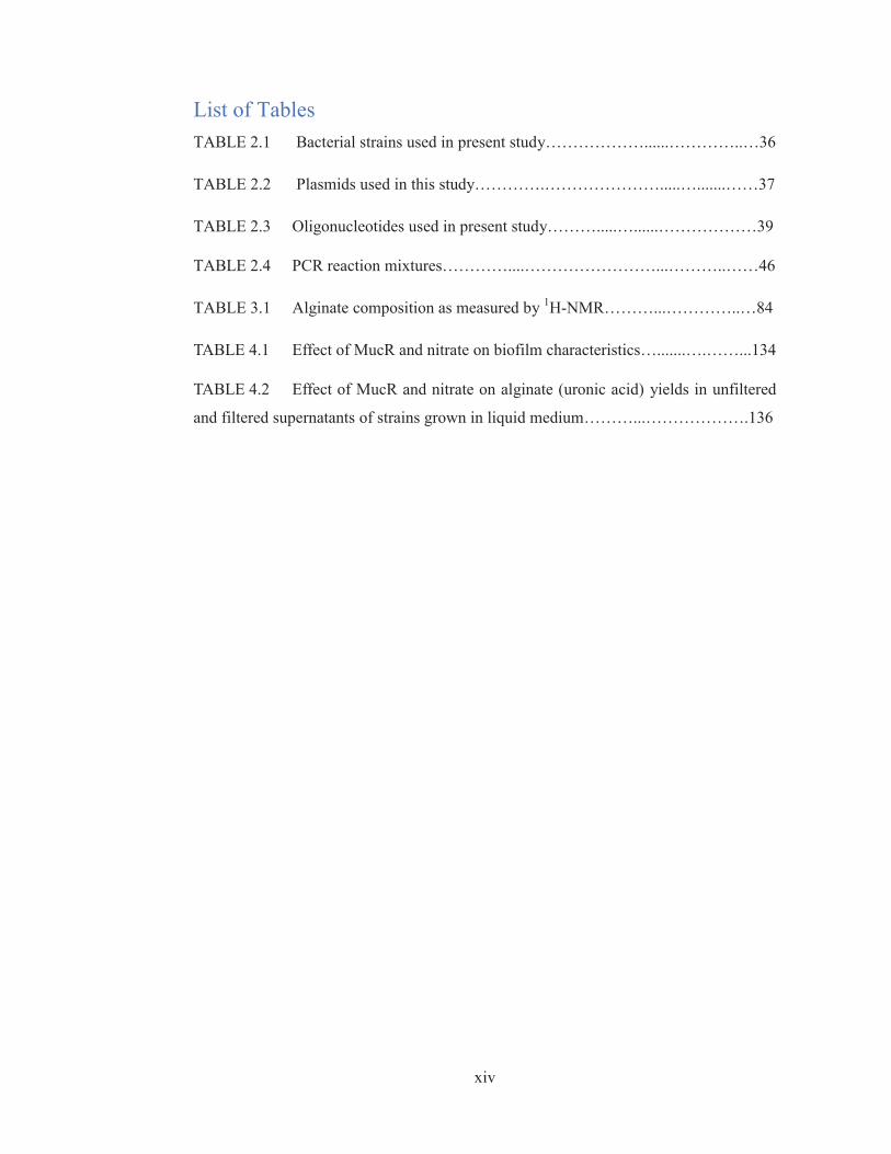
List of Tables TABLE 2.1 Bacterial strains used in present study………………......…………..…36
TABLE 2.2 Plasmids used in this study………….………………….....….......……37
TABLE 2.3 Oligonucleotides used in present study……….....…......………………39
TABLE 2.4 PCR reaction mixtures…………....……………………...………..……46
TABLE 3.1 Alginate composition as measured by 1H-NMR………...…………..…84
TABLE 4.1 Effect of MucR and nitrate on biofilm characteristics….......….……...134
TABLE 4.2 Effect of MucR and nitrate on alginate (uronic acid) yields in unfiltered
and filtered supernatants of strains grown in liquid medium………...……………….136

1
1. Chapter One: Introduction Alginate is a collective term for a family of polysaccharides produced by brown
seaweeds and bacteria (Hay et al. 2010a). As a major cell wall constituent of brown
seaweeds, alginate plays an important structural role in algal tissues and is harvested for
many industrial and medical applications (Schmid et al. 2016). For instance, it is used in
foods, cosmetics, pharmaceuticals and medical products as a stabilizer/thickener and
encapsulation/drug delivery agent, and more recently for regenerative therapy (Section
1.1).
Alginate also plays an important role in bacteria. For instance, in Azotobacter spp., it is
required for formation of desiccation resistant cysts (Campos et al. 1996). Recently, an
important Gram-positive bacterium, Sulfobacillus thermosulfidooxidans, has been found
to secret alginate during bioleaching of chalcopyrite (Yu et al. 2017). It is also produced
by various species of Pseudomonas such as Pseudomonas syringae, Pseudomonas
putida and Pseudomonas fluroescens. In P. syringae alginate is produced to increase its
epiphytic fitness and resistance to desiccation (Yu et al. 1999).
The opportunistic human pathogen, P. aeruginosa, over-produces alginate during
chronic cystic fibrosis (CF) lung infections, growing as a biofilm (cells embedded in
self-secreted polymeric matrix) to evade antibiotics and host immune responses,
clogging patients’ airways, causing chronic inflammation, tissue damage, pulmonary
deterioration and death (Hoiby et al. 2010). Owing to its medical importance, alginate
biosynthesis and regulation in P. aeruginosa has been studied in great detail. It is also
used as a general model to help understand biofilm formation, antibiotic resistance, and
to develop strategies to eliminate biofilms which are a major challenge in both medical
and industrial settings (del Pozo and Patel 2007; Langsrud et al. 2003).

2
In this chapter, I start by providing a brief overview of the chemical structure,
properties, commercial production and applications of alginate (Section 1.1), and the
challenges posed by P. aeruginosa in clinical and industrial settings (Sections 1.2 and
1.3). As the foundation for my PhD studies, I review the mechanisms behind the
biosynthesis (Section 1.4) and regulation (Section 1.5) of alginate production in
bacteria. I then outline the foci of the thesis, AlgL, a periplasmic alginate lyase (Section
1.6), and MucR, an inner membrane regulatory protein (Section 1.7), and then present
my aims and research questions in Section 1.8.
1.1 Structure and applications of alginate
1.1.1 Structure and properties
Alginate is an anionic polysaccharide consisting of variable ratios of β-D-mannuronate
(M) and its C-5 epimer α-L-guluronate (G) linked by 1–4 glycosidic bonds. In contrast
to seaweed alginate, bacterial alginate is O-acetylated, offering increased shear-thinning
properties (Skjakbraek et al. 1989). The chemical structure of alginate is illustrated in
Figure 1.1 on page 3. Alginate is biocompatible and biodegradable, and has excellent
gel-forming, scaffold-forming and viscosifying properties (Schmid et al. 2016).

3
Figure 1.1 Chemical structure of alginate. It is composed of β-D-mannuronate (M) and its C-
5 epimer α-L-guluronate (G) linked by 1–4 glycosidic bonds. They form MM, MG and GG
blocks. The polymer can be O-acetylated (by AlgI, AlgJ, AlgF and AlgX) and M residues can
be converted to G residues by epimerases (AlgG, AlgE1-7 of Azotobacter spp). Reuse
permission obtained from John Wiley and Sons (Hay et al. 2013).

4
1.1.2 Extraction and applications
Each year 30,000 metric tons of alginate are harvested from brown seaweeds for
numerous industrial and medical applications. Here I briefly describe the extraction
process of alginate from seaweed, highlight some of the most recent applications for
alginate, and emphasise the potential use of bacterial factories (or enzymes) to
manufacture tailor-made alginate for high value medical applications.
The rationale behind extraction of alginate from seaweed is converting all alginate salts
in the seaweed into the sodium salt, dissolving this in water, and removing the seaweed
residue by filtration. The extraction process is summarised in Figure 1.2 on page 6.
Firstly, the seaweed is chopped into pieces and mixed with sodium carbonate, obtaining
a crude sodium alginate solution. After removal of insoluble debris, crude sodium
alginate is enriched by precipitation with either calcium chloride or acid. Resulting
calcium alginate/alginic acid fibres are further treated with sodium carbonate to obtain
the enriched sodium alginate.
Alginate harvested from seaweed is used in many industrial and medical applications
(Figure 1.3, page 9) (Norouzi et al. 2015; Ruvinov and Cohen 2016; Tavassoli-Kafrani
et al. 2016; Venkatesan et al. 2015). Some of the most recent applications include
alginate as an anticoagulation agent (Arlov and Skjak-Brak 2017), an encapsulation
agent for delivery of probiotics (Jose Martin et al. 2015) and pesticides (Nuruzzaman et
al. 2016), a safe and renewable material for food packaging (Tavassoli-Kafrani et al.
2016), and a recovery agent for critical, toxic and precious metals from the environment
(Dodson et al. 2015).
In addition, alginate’s mild gelation properties and hydrogel resemblance to
extracellular matrix have made it an attractive vehicle for stem cell delivery (infused
with bioactive molecules and regenerative factors) to regenerate damaged tissues

5
resulting from cardiac arrest (Ruvinov and Cohen 2016). Alginate is also used in wound
dressings, skin substitutes and bioactive scaffolds – laced with growth factors,
antibiotics and other medicines to encourage skin healing and regeneration (Kamoun et
al. 2015; Norouzi et al. 2015). One recent exciting application for polysaccharides is the
transplantation of the first synthetic trachea – developed using a novel composite
scaffold containing alginate (Crowley et al. 2015).

6
Figure 1.2. Isolation of alginate from seaweed. Seaweeds are harvested, chopped and
converted to soluble sodium alginate by mixing with sodium carbonate. By filtration, residue
seaweed is removed and discarded. The crude sodium alginate solution is further enriched by
either the calcium alginate process or alginic acid process, where essentially the alginate is
made insoluble by calcium ions or reduced pH. The alginate is then precipitated and dried into
a paste and mixed with sodium carbonate again to obtain sodium alginate of greater purity.

7
Reused with permission from Food and Agriculture Organization of the United Nations
(McHugh 1987).
However, alginate from seaweed suffers from seasonal and environmental variation in
molecular mass, composition and physicochemical properties (Rosell and Srivastava
1984; Saraswathi et al. 2003). For instance, different parts of the seaweed plant,
different species, geographical location and season of harvesting can impact alginate
properties (Haug et al. 1974). Such variability makes crude alginate isolated from
seaweed cheap ($US5 kg−1). However, enriching pharmaceutical grade alginate of high
purity and homogeneity in molecular weight (MW), mannuronic to guluronic acid
(M/G) ratios and defined material properties from crude alginate is costly, with the end
product reflecting this, costing up to $US3,000 g−1 (Rhein-Knudsen et al. 2015).
On the other hand, alginate-producing bacteria or bacterial alginate-modifying enzymes
could be harnessed to produce alginate of defined properties, potentially alleviating this
cost-burden. Using bacteria to produce commercial alginate is an attractive alternative
because bacterial alginates are generally more homogeneous than that of seaweed;
bacteria can be readily engineered and cultivated under controlled conditions to produce
alginates of defined chemical structure and physicochemical properties. However,
because of the potential pathogenic nature of P. aeruginosa, production of commercial
bacterial alginates would most likely rely on Azotobacter vinelandii or non-pathogenic
species of Pseudomonas genera.
While many breakthroughs have been made in understanding the mechanisms of
bacterial alginate production and regulation, we are still far away from producing
industrial quantities of pharmaceutical grade alginate of high purity, defined structure
and physicochemical properties from bacteria. Nevertheless, pioneering studies have

8
developed strains and growth conditions offering reasonable control over alginate yield,
molecular mass, composition and associated properties. For instance, A. vinelandii
strains with increased transcription from alginate biosynthesis gene cluster have been
engineered to increase alginate production levels. Moreover, shutting down of
competing metabolic pathways and/or optimization of growth conditions (e.g. nutrients,
oxygen availability) have also increased yields (e.g. up to 9.5 g Alg/L in 50 h) and
improved control over alginate composition and properties (Bonartseva et al. 2017;
Castillo et al. 2013; Diaz-Barrera et al. 2007; Diaz-Barrera et al. 2010; Flores et al.
2015; Gaytan et al. 2012; Pena et al. 2006). For example, a mutant of P. fluorescens
lacking the epimerase function of AlgG has been generated that produces poly-M
(Gimmestad et al. 2003). Furthermore, O-acetylation levels can also be controlled by
using specific strains/mutants or altering the growth media and cultivation conditions,
including aeration, pH and temperature (Diaz-Barrera et al. 2010; Gaytan et al. 2012;
Pena et al. 2006).
Nature has provided a plethora of alginate modifying enzymes (reviewed in detail by
(Ertesvag 2015)), including epimerases, O-acetyl-transferases and depolymerases that
could be harnessed to modify seaweed alginates. For instance, A. vinelandii produces
several epimerases, each introducing specific ratios and patterns of G residues
(Gimmestad et al. 2003). These enzymes are used to modify alginate and can be used
for immobilization of living tissue (Morch et al. 2007). Moreover, P. syringae has been
used to O-acetylate seaweed alginate (Lee and Day 1995). Thus, there is huge potential
for using microbial factories and bacterial alginate-modifying enzymes to
manufacture/modify alginate for high value applications.

9
Figure 1.3. Alginate applications. Industrial and medical applications are summarised here.
Reused with permission from ArtMolds.com (ArtMolds 2016a; ArtMolds 2016b).

10
1.2 Pseudomonas aeruginosa and cystic fibrosis
1.2.1 Pseudomonas aeruginosa
P. aeruginosa is a Gram negative rod-shaped bacterium found in soil, water, skin flora
and most man-made environments. As a facultative anaerobe, it can utilize a range of
carbon sources and alternative terminal electron acceptors under low (no) oxygen
conditions. Moreover, its adaptability, numerous nutrient utilization pathways and
intrinsic resistance to antimicrobial agents allow it to survive in a wide range of settings.
Normally, environmental isolates are non-mucoid (i.e. do not over-produce alginate).
However, exposure to stressors including desiccation, osmotic imbalances and
antimicrobial agents elicits a stress response and a conversion to a mucoid phenotype
(i.e. alginate over-producing state). Although it is generally considered an accidental
pathogen to humans, P. aeruginosa can cause life-threatening nosocomial infections in
the elderly, organ transplant recipients and patients with cancer, severe burns or CF
(Lyczak et al. 2000). P. aeruginosa lung infections are the leading cause of death in CF
patients. Figure 1.4 (page 11) summarizes key virulence factors of P. aeruginosa and
host lung defenses (Gellatly and Hancock 2013).
For CF lung infections, failure to eradicate P. aeruginosa during the acute phase allows
it to adapt to the site by over-producing alginate and growing as a biofilm, resulting in
chronic infection. Studies on evolution of strains during the course of CF infection
(from acute to chronic) show vast variation in phenotypes and genotypes over time
(Hogardt et al. 2007; Smith et al. 2006). Often the chronic isolates display reduced
inflammatory effects and are less cytotoxic than their acute brethren. This conversion to
a chronic state involves mutations in mucA, B or D causing mucoid conversion, a loss of
flagella, pili and type three secretion systems (Mathee et al. 1999), as well as changes

11
affecting lipopolysaccharide composition and quorum sensing (Ernst et al. 2007;
Winstanley and Fothergill 2009).
Figure 1.4 Pseudomonas aeruginosa vs CF human lung. (A) Virulence factors of P.
aeruginosa include flagella (motility), type 4 pili (motility, adhesion and aggregation), type 3
secretion system (T3SS, during the acute stage it injects toxins into host cells damaging host
membrane integrity), proteases (degradation of host lung surfactant), quorum sensing molecules

12
(homoserine lactone = HSL and 2-heptyl-3-hydroxy-4-quinolone = PQS), toxins (exotoxin A,
pyocyanin), siderophores (pyoverdine), catabolic enzymes (phospholipase, alkaline
phosphatase, elastase) and biofilm formation. (B) Host defences include mucocilary clearance,
surfactants, antimicrobial peptides (α-defensins, lactoferrin, lysozyme), reactive oxygen (ROS)
and nitrogen (RNS) species, release of cytokines and chemokines (NFκB, IL6, IL8, IL10, IL23),
toll-like receptors (TLRs), and various immune cells (macrophage, dendritic cell, goblet cell,
lymphocyte, neutrophil). Reuse permission obtained from Oxford University Press (Gellatly and
Hancock 2013).
1.2.2 Cystic fibrosis and host defenses
CF patients have mutations in their cystic fibrosis transmembrane conductance gene
(CFTR) that encodes a membrane associated ATP-dependent chloride channel. Over
2,000 mutations of this gene have been catalogued in the Cystic Fibrosis Mutation
Database (http://www.genet.sickkids.on.ca/Home.html). These mutations have been
categorized into various classes depending on how severely they impact CFTR
expression/translation/localization/function and stability as well as disease
manifestations (Marson et al. 2016). Although most mutations can theoretically be
rescued by pharmacotherapy and/or gene therapy, this approach is extremely costly and
not always reliable. Hence, alternative strategies that target the bacterial infection by P.
aeruginosa and its ability to produce alginate should be considered.
Mutations of the CFTR gene causes a thickening of epithelial secretions due to
imbalanced transport of salt and water transport across cellular membranes (O'Sullivan
and Flume 2009). This obstructs the pancreatic duct, intestinal glands and bronchi,
preventing normal secretion of digestive enzymes and micro-ciliary clearance from
lungs, causing malnutrition and chronic lung infection (Rowe et al. 2005).
Manifestations of CF are illustrated in Figure 5A on page 14. Although nutritional

13
deficiencies can be treated by dietary and enzyme supplements, chronic lung infections
are much more serious and difficult to treat. They lead to chronic inflammation, cystic
bronchiectasis and severe airflow obstruction, often leading to mortality in patients’
thirties (Rowe et al. 2005; Williams et al. 2010).
Normally, the host lungs trap inhaled contaminants in epithelial mucus, sweeping them
out by beating of ciliated-epithelial cells (Knight and Holgate 2003). However,
thickening of epithelial secretions stalls this clearance mechanism (Knight and Holgate
2003). The host then responds to pathogen colonization by inflammation, secreting
antimicrobial peptides (e.g. lysozyme and beta-defensins) to attack the pathogen cell
surface (Chroneos et al. 2010). Cytokines and chemokines are also produced to recruit
and activate innate and adaptive immune responses, including a variety of immune cells
(Holt et al. 2008). Particularly, neutrophils play a key role in combating P. aeruginosa
infections. They phagocytose and kill bacteria, with antimicrobial peptides and reactive
oxygen and nitrogen species. However, during chronic CF lung infections, persistent
immune stimulation is often counter-productive, causing collateral damage to lung
tissues (Williams et al. 2010). This happens because during chronic infections, P.
aeruginosa overproduces alginate, protecting itself from antibiotics and host defenses,
including phagocytosis. This creates an infection that is extremely difficult to
treat/eradicate due to the combination of the inflamed host lung environment and the
difficult-to-penetrate bacterial biofilm.

14
Figure 1.5 Cystic fibrosis, stages of biofilm development and mechanisms of antibiotic
resistance. (A) CF affects function of sinus, lungs, skin, liver, pancreas, intestine and
reproductive organs. (B) Stages of biofilm formation, attachment, growth and dispersal. (C)
Antibiotic resistance mechanisms of P. aeruginosa biofilm. Images were reused with
permission from (A) National Heart Lung and Blood Institute, (B) Centre for Biofilm
Engineering at Montana State University P. Dirckx and (C) Elsevier (Stewart and Costerton
2001).
(A) (C)
(B)

15
1.3 Biofilm formation and antimicrobial resistance A conversion from a free living to biofilm forming state provides cells with distinct
advantages including resistance against physical, chemical and biological insults. This
conversion is facilitated by complex top-down changes in gene expression controlled by
numerous regulatory systems that respond to specific stress-inducing conditions,
including predation, starvation, antibiotics, reduced growth rate, dehydration, high
osmotic pressure and ionic strength (Devault et al. 1990; Evans and Linker 1973; Govan
and Fyfe 1978; Vandevivere and Kirchman 1993).
Biofilm formation has three stages - attachment, growth and dispersal (Figure 1.5B,
page 14). During attachment, cells stick to a surface by reversible (electrostatic/vander
waals forces) then irreversible interactions via cellular appendages (including type 4
pili, flagella and cup fimbria) and surface-associated polysaccharides (such as Psl and
Pel) (Mikkelsen et al. 2011; Wei and Ma 2013). As biofilms mature, they develop into
micro-colonies where cells aggregate in a matrix of exopolymeric substances including
polysaccharides, proteins, DNA and lipids. In the later stage of dispersal, free living-
cells are released by several mechanisms involving mechanical forces, matrix-degrading
enzymes and bacteriophages, allowing colonization of new surfaces (Harmsen et al.
2010).
Biofilm growth increases resistance to biocides and antibiotics, posing a major
challenge in clinical and industrial settings (Figure 1.5C, page 14) (Stewart and
Costerton 2001). Increased resistance occurs by metabolic slowing-down (i.e. reduced
oxygen availability and lower growth rate renders certain classes of antibiotics
ineffective since they only work on growing/dividing cells) as well as reducing
penetration and/or neutralization of antimicrobial agents within the matrix by steep
oxygen and chemical gradients. Moreover, resident cells often activate inherent

16
resistance mechanisms, including reduced membrane permeability, expression of
antibiotic degrading enzymes, mutation of antibiotic targets and expression of
membrane efflux pumps.
Current widespread use of biocides (in industry and hospitals) and antibiotics (in
humans and animals) has encouraged the emergence of highly resistant strains of P.
aeruginosa, already innately resistant to a wide range of antibiotics, and outbreaks of
infection with multi-resistant strains have been reported (Ashish et al. 2012). Thus,
alternative strategies should be considered. This study attempts to offer a deeper
understanding of alginate production and regulation in the hope of finding better
alternatives to counter P. aeruginosa biofilms. For instance, matrix degrading enzymes
(e.g. alginate lyase) and/or suppression of polysaccharide production (with specific
signals) might be an innovative way to better manage P. aeruginosa biofilms of clinical
and industrial significance.

17
1.4 Alginate biosynthesis In previous sections, I touched on the industrial and medical significance of alginate and
P. aeruginosa. Now I will review current knowledge about bacterial alginate
production. Alginate biosynthesis involves four steps: precursor assembly in the
cytoplasm, and polymerization, modification and translocation/secretion which are
facilitated by a membrane spanning multiprotein complex.
The alginate precursor, guanosine diphosphate (GDP)-mannuronic acid, is synthesized
from fructose-6-phosphate in four steps, catalyzed by three enzymes, AlgA, C and D in
the cytoplasm (summarized in Figure 1.6, page 18). Fructose-6-phosphate is converted
to mannose-6-phosphate (by AlgA), then to mannose-1-phosphate (by AlgC) and GDP-
mannose (by AlgA), and finally to GDP-mannuronic acid (by AlgD) (May et al. 1994;
Roychoudhury et al. 1989; Shinabarger et al. 1991; Tatnell et al. 1994; Zielinski et al.
1991).
The precursor is polymerized at the inner membrane by Alg8 and Alg44, forming poly-
mannuronate (poly-M) which is transported by a multi-protein complex (Alg44, X, G,
K) through the periplasm to the outer membrane (OM) protein, AlgE, for secretion
(Figure 1.7, page 19). In the periplasm, poly-M is modified by O-acetylation (AlgI, J, F
and X) and epimerization (AlgG).

18
Figure 1.6. A schematic representation of the biochemical pathway for alginate
biosynthesis in P. aeruginosa. Reuse permission obtained from John Wiley and Sons (Hay et
al. 2013).
1.4.1 Polymerization
Polymerization and translocation across the inner membrane involve the inner
membrane proteins Alg8 and Alg44 (Moradali et al. 2017; Oglesby et al. 2008;
Remminghorst and Rehm 2006a; Remminghorst and Rehm 2006b) (Figure 1.7, page
19). The glycosyltransferase Alg8 transfers the mannuronic acid from the precursor onto
a growing poly-M chain while Alg44 is a co-polymerase that aids this activity. The
latter has a cytoplasmic PilZ domain that binds to a common bacterial secondary
messenger molecule, bis-(3´-5´)-cyclic dimeric guanosine monophosphate (c-di-GMP)
(Merighi et al. 2007; Whitney et al. 2015). Alg44 also has a periplasmic domain thought
to bridge with periplasmic/outer membrane components of the biosynthesis complex

19
(Oglesby et al. 2008; Remminghorst and Rehm 2006a). In analogous systems (e.g.
cellulose biosynthesis), c-di-GMP binding causes local conformational changes that
allow activated precursor access to the catalytic site of the glycosyltransferase (Franklin
et al. 2011; Morgan et al. 2013; Steiner et al. 2013; Weinhouse et al. 1997; Whitney et
al. 2012).
Figure 1.7 A schematic representation of the alginate biosynthesis apparatus and alginate
biosynthesis operon. Subunit functions are colour coded. Modified from (Franklin et al. 2011).
1.4.2 Modification
In the periplasm the nascent polymer, poly-M, is O-acetylated by AlgX (and I, J and F)
via addition of acetyl groups to O2/O3 positions of M residues (Baker et al. 2014;
Franklin et al. 2004; Franklin and Ohman 1996; Franklin and Ohman 2002; Riley et al.
2013) (Figure 1.7, page 19). The polymer is also epimerized by AlgG through
conversion of M residues to G via protonation-deprotonation of C5 on the M residue in
Biosynthesis Operon

20
the polymer chain (Douthit et al. 2005; Jerga et al. 2006a; Jerga et al. 2006b; Wolfram
et al. 2014). These modifications substantially affect alginate material and biological
properties. For instance, O-acetylation increases alginate water binding capacity,
contributes to biofilm formation by Pseudomonads, and also protects against immune
responses and antimicrobial agents (Nivens et al. 2001; Pier et al. 2001; Tielen et al.
2005) while epimerization increases gel forming properties and also contributes to the
architecture of P. aeruginosa biofilms (Moradali et al. 2015; Morch et al. 2008). O-
acetylation also appears to influence the distribution of G residues along the alginate
chain (Schurks et al. 2002). While neither enzymatic activity (of AlgX or AlgG) is
essential for alginate production, these proteins are thought to form a structural
component of the periplasmic scaffold, hence being essential for alginate translocation
and secretion (Baker et al. 2014; Gimmestad et al. 2003; Gutsche et al. 2006; Jain et al.
2003; Riley et al. 2013; Robles-Price et al. 2004; Wolfram et al. 2014).
1.4.3 Translocation and secretion
A multi-protein complex is thought to translocate the polymer chain through the
periplasm to AlgE for secretion (Figure 1.7, page 19). Several proteins, Alg44, K, G, X
and E are thought to contribute to the translocation/secretion scaffold (Gimmestad et al.
2003; Jain et al. 2003; Jain and Ohman 1998; Rehman and Rehm 2013; Rehman et al.
2013; Robles-Price et al. 2004). One of these proteins AlgK, is supposed to help
assemble/stabilize the scaffold through its multiple tetratricopeptide-like repeats
implicated in protein-protein interactions (Keiski et al. 2010). AlgK is a lipoprotein
important for localization of AlgE to the outer membrane, and AlgE is also required for
assembly of the complex (Keiski et al. 2010; Rehman and Rehm 2013). Furthermore,
through mutual stability and interaction experiments using alg44, K, X and E mutants,
Rehman et al. (2013) have provided evidence for a membrane spanning multi-protein

21
apparatus, involved in alginate biosynthesis. Additional experiments have identified an
elaborate interaction network between subunits biosynthesis machinery (Moradali et al.
2015). When subunits of the apparatus are missing, the alginate leaks into the periplasm
where it is degraded by AlgL, an alginate lyase, that is thought to perform a
maintenance role (Bakkevig et al. 2005; Jain and Ohman 2005). However, the necessity
of AlgL for alginate production has been controversial. Hence, Chapter 3 was devoted
to investigating the biological function of AlgL in alginate production by P. aeruginosa
(please see Section 1.6 for more about AlgL).
Alginate is secreted through the outer membrane beta barrel porin, AlgE (Hay et al.
2010b; Rehm et al. 1994; Whitney et al. 2011). Structural and molecular dynamics
experiments have outlined the conformational landscape for alginate secretion,
suggesting that AlgK helps to align the polymer so that it can be threaded through the
pore of AlgE (Tan et al. 2014; Whitney et al. 2011).

22
1.5 Genetics and regulation of alginate production In the previous section, I reviewed the biosynthesis of alginate. Now, I will give an
overview about the genetics and regulation of microbial alginate production. While
studies have revealed many intricate details into the signalling pathways regulating
alginate biosynthesis, the specific signals perceived by these regulatory sensors remain
poorly understood. Nevertheless, some work has shown that alginate production is
induced under conditions of desiccation, high osmolarity, sub-inhibitory concentrations
of antibiotics and low oxygen conditions (Bragonzi et al. 2005; Chang et al. 2007; Li et
al. 2010; Wood and Ohman 2009)
It is widely accepted that alginate over-production provides a hydrated environment that
also protects against antimicrobial agents and host defences.
1.5.1 Regulated intramembrane proteolysis cascade
The ‘master switch’ for alginate production in P. aeruginosa is the regulatory operon
algU-mucABCD encoding: AlgU (an alternate sigma factor), its anti-sigma factors
MucA and MucB and negative regulators MucC and MucD. This operon responds to
envelope stress caused by antimicrobial and oxidizing agents, elevated temperatures,
and osmotic imbalances. Mutations in the muc genes typically cause a switch to the
mucoid phenotype, by unleashing AlgU, driving expression of its own operon, several
other genes involved in alginate regulation as well as the alginate biosynthesis gene
cluster (Firoved et al. 2002; Hay et al. 2014; Wozniak et al. 2003). Mutation of anti-
sigma factor MucA forces the system into a ‘permanent on state’ (Hay et al. 2014)
(Figure 1.8C, page 23). Normally, in the ‘off state’ (i.e. low stress) AlgU is sequestered
at the inner membrane (Figure 1.8A, page 23) by MucA-MucB (Cezairliyan and Sauer
2009; Wood and Ohman 2009).

23
Figure 1.8 The ‘switch’ for alginate production. Proteins and lines shown in green have a
positive effect on alginate production; proteins and lines shown in red have a negative effect on
alginate production. Dotted lines indicate an unknown/unclear mechanism. (A) Off state. The
switch is turned OFF in the absence of envelope stress. In this state, AlgU is sequestered at the
inner membrane by MucA–MucB, protecting MucA from proteolysis by AlgW and MucP. (B)
Induced On state. Envelope stress (thunderbolt) turns ON the switch. Mislocalized/misfolded
cellular components activate AlgW destabilizing the MucA–MucB interaction. MucD degrades
and/or repairs damaged OMPs that activate the cascade. Cleavage of MucA by AlgW is
followed by site-2 proteolysis by MucP, releasing the cytoplasmic domain of MucA with AlgU
bound into the cytosol where ClpXP degrade MucA, freeing AlgU to interact with RNA
polymerase and drive expression of alginate production genes. (C) Permanent On state.
Example of a clinical mucA mutant (mucA22) with a truncated MucA is no longer able to seek
MucB for protection, rendering it susceptible to proteolysis by AlgO and possibly other
proteases. Reuse permission obtained from John Wiley and Sons (Hay et al. 2014).
(A) (B) (C)
24
In response to envelope stress, the system is switched ‘on’; AlgU is released from its
anti-sigma factor complex by a regulated intramembrane proteolysis (RIP) cascade
involving various proteases (Figure 1.8B, page 23). Envelope stress causes mis-folding
and mis-localization of membrane components which disrupt the MucA-MucB
interaction and activate protease AlgW, cleaving MucA. (Cezairliyan and Sauer 2009;
Chaba et al. 2011; Kulp and Kuehn 2011; Lima et al. 2013; Qiu et al. 2007; Wood and
Ohman 2009). MucA is further hydrolyzed by MucP (Qiu et al. 2007), releasing
truncated MucA still bound to AlgU into the cytosol where it is rapidly degraded by
ATP-dependent cytoplasmic proteases, freeing AlgU to drive expression of alginate
biosynthesis genes (Qiu et al. 2008b).
MucD negatively regulates the RIP cascade by chaperoning and/or degrading misfolded
proteins that would otherwise activate AlgW or MucP proteases (Damron and Yu 2011;
Qiu et al. 2007; Yorgey et al. 2001) (Figure 1.8B, page 23). Intriguingly, MucD also
interacts with a component of the alginate biosynthetic machinery, AlgX (Gutsche et al.
2006; Hay et al. 2012); however, the significance of this interaction is unknown. In
clinical P. aeruginosa strains from CF lung infections, the alginate operon is in a
‘permanent on state’ (Figure 1.8C, page 23). This short-circuiting occurs by mutations
truncating MucA, preventing its interaction with MucB and exposing it to degradation
by alternative proteases such as AlgO (Qiu et al. 2007; Reiling et al. 2005). The
desiccating environment of CF lungs is thought to elicit this switch, first by the induced
and then by the permanent on state.
1.5.2 Transcriptional regulation
Alginate production is regulated transcriptionally by sigma factors, two component
signal transduction systems (TCST) and other DNA binding proteins (Figure 1.9, page
26). For AlgU to drive alginate production, other sigma factors are sequestered by their

25
cognate anti-sigma factors (Pineda et al. 2004; Yin et al. 2013; Yuan et al. 2008).
Certain sigma factors such as RpoN reduce alginate production under nitrogen rich
conditions (Boucher et al. 2000).
At least 2 TCST systems are involved in regulating alginate production: KinB-AlgB and
FimS-AlgR (Figure 1.9A, page 26). Generally, TCST systems have a sensor kinase
(SK) (e.g. KinB and FimS) that autophosphorylates itself in response to environmental
signals, and activates its cognate response regulator (RR) via phospho-transfer.
However, the exact signals of the SKs systems are unknown. Moreover, both these
systems behave non-canonically - RRs can enhance alginate production independently
of their SKs and phosphorylation (Ma et al. 1998; Wozniak and Ohman 1993). Also
KinB could be a phosphatase that is involved in AlgW-mediated degradation of the
MucA (Chand et al. 2012; Damron et al. 2009) (Figure 1.9A-D, page 26).
Numerous DNA-binding proteins control expression from PalgD promoter (Baynham et
al. 2006; Kato et al. 1990)(Figure 1.9B-C, page 26). These include the global regulators
AlgU and AmrZ (Jones et al. 2013; Pryor et al. 2012; Tart et al. 2006; Waligora et al.
2010), a histone-like DNA-binding protein, AlgP that enhances alginate production in
response to nitrogen availability (Deretic and Konyecsni 1990) an integration host
factor like heterodimer (IHFα and IHFβ) (DelicAttree et al. 1996), and various other
transcription factors including CysB and Vfr (DelicAttree et al. 1997) (Figure 1.9C,
page 26).

26
Figure 1.9 Schematic representation of various regulatory mechanisms of alginate
biosynthesis. Green proteins and lines have a positive effect on alginate production; red
proteins and lines have a negative effect on alginate production. Dotted lines indicate an
unknown/unclear mechanism. (A) Two-component signal transduction systems, FimS/AlgR and
KinB/AlgB. (B) Transcriptional regulation through DNA-binding proteins. (C) Schematic map
of the approximate binding sites of various transcriptional regulators on the P. aeruginosa algD
promoter. (D) Sigma/anti-sigma factors. (E) Posttranscriptional regulation through the Gac/Rsm
sRNA system in Azotobacter vinelandii. (F) Posttranscriptional regulation through a natural
antisense transcript (MucD-AS) that promotes alginate production by blocking the translation of
mucD mRNA. (G) Posttranslational regulation by c-di-GMP. MucR synthesizes a pool of c-di-
GMP near the alginate co-polymerase Alg44. Binding of c-di-GMP to Alg44 is essential for
alginate biosynthesis. (H) Posttranscriptional regulation by substrate competition. Reuse
permission obtained from John Wiley and Sons (Hay et al. 2014).

27
1.5.3 Post-transcriptional regulation
Mechanisms of post-transcriptional control of alginate production are shown in Figure
1.9E-F, page 26). In A. vinelandii, the central TCST system, GacS-GacA, activates
expression of noncoding small RNAs which sequester the translational regulatory
protein, RsmA - preventing it from binding and repressing translation from the algD
mRNA transcript (Manzo et al. 2011). In P. aeruginosa a natural antisense transcript
(mucD-AS), when overexpressed, significantly induces alginate production and biofilm
formation by blocking MucD production (Yang et al. 2011b).
1.5.4 Post-translational regulation
Mechanisms of post-translational control of alginate production are shown in Figure
1.9G-H. Substrate competition for GDP-mannose by alginate, Psl and B-band LPS
biosynthesis pathways may indirectly affect alginate yields (Byrd et al. 2009;
Shinabarger et al. 1991). Additionally, the generic bacterial secondary messenger, c-di-
GMP, drives alginate production post-translationally by binding to the PilZ domain of
Alg44, the alginate co-polymerase (Merighi et al. 2007; Whitney et al. 2015). Another
inner membrane protein, MucR, involved in c-di-GMP turnover is thought to impart a
localised pool of c-di-GMP (Hay et al. 2009b)(Figure 1.9G, page 26). However, it is
unknown how its activity is regulated nor is it known what signal it perceives. Thus, the
role of MucR was studied in detail in Chapter 4 (please see Section 1.7 for more about
MucR).
As mentioned earlier, two major gaps provide the foci of this study. AlgL, an alginate
lyase, and MucR, a sensor protein with c-di-GMP turnover activity (discussed in more
detail below).

28
1.6 Function of AlgL in alginate production As reviewed in Section 1.4, when components of the alginate biosynthesis apparatus are
missing, alginate is thought to leak into the periplasm, where it is degraded by alginate
lyases (e.g. AlgL), releasing free uronic acid oligomers. AlgL preferentially cleaves the
non-acetylated alginate chain via beta elimination, releasing dimeric and/or trimeric
alginate oligosaccharides with 4-deoxy-alpha-L-erythro-hex-4-enuronosyl groups at
their non-reducing ends and beta-D-mannuronate at their reducing end (Farrell and
Tipton 2012). Since mature alginate is O-acetylated, such activity is consistent with a
maintenance role, i.e. degrading misguided alginate from the periplasm. However,
because of the inconsistencies in the literature (outlined below), it has been difficult to
pinpoint the exact function of AlgL. Major inconsistencies revolve around its
requirement for alginate production. While several studies support the requirement of
AlgL for alginate biosynthesis, others suggest otherwise (Albrecht and Schiller 2005;
Bakkevig et al. 2005; Boyd et al. 1993; Jain and Ohman 2005; Monday and Schiller
1996; PenalozaVazquez et al. 1997; Trujillo-Roldan et al. 2003).
Boyd et al. (1994) and PenalozaVazquez et al. (1997) demonstrated that expression of
algA (an essential downstream gene) in transposon-generated polar algL mutants in P.
aeruginosa and P. syringae restored alginate production on solid media, and Trujillo-
Roldan et al. (2003) showed that a non-polar algL deletion mutant in A. vinelandii could
still produce alginate. In contrast, other studies support a requirement for AlgL in
alginate production. For instance, alginate yield in a polar algX mutant of the clinical
isolate, P. aeruginosa FRD1, could only be restored by plasmid-borne expression of
algX together with downstream genes algL and algA (Monday and Schiller 1996).
Similarly, Bakkevig et al. (2005) showed that expression of only algC (involved in
alginate precursor production) in an algC algL double mutant (generated in

29
Pseudomonas fluorescens) did not restore alginate production. Jain & Ohman (2005)
replaced the PalgD promoter of P. aeruginosa FRD1 with an inducible one prior to
swapping out the algL gene with a gentamicin resistance marker and revealed that
inducing alginate production in the absence of AlgL during planktonic growth led to an
accumulation of polymeric substances in the periplasm, eventuating in cell lysis. A later
study reached a similar conclusion for cells grown in liquid media, further emphasising
the need for AlgL lyase activity in alginate biosynthesis (Albrecht & Schiller 2005).
The above inconsistencies may relate to differences in experimental setup (parent
species/strain and mutant generation) and technical challenges. For example,
stabilization of alginate production in parent strains by chemical mutagenesis (Bakkevig
et al. 2005; Boyd et al. 1993; Darzins et al. 1986; Wang et al. 1987), the use of
uncharacterized plasmids (PenalozaVazquez et al. 1997) or placing alginate
biosynthesis genes under control of inducible promoters (Albrecht and Schiller 2005;
Bakkevig et al. 2005; Jain and Ohman 2005). These approaches introduced unknown
genetic changes and deregulated alginate production from normal cellular regulation.
Moreover, the methods used for generating algL mutants - transposon- and marker-
based strategies – often led to technical challenges in complementation due to polar
effects (Albrecht and Schiller 2005; Boyd et al. 1993; Jain and Ohman 2005; Monday
and Schiller 1996; PenalozaVazquez et al. 1997; Trujillo-Roldan et al. 2003).
Despite these issues, two preliminary models have been proposed for the role of AlgL in
alginate production (Figure 1.10, page 31). While both models assign AlgL a
maintenance role to degrade misguided alginate in the periplasm – to partner the
previous biochemical studies (Farrell and Tipton 2012), they disagree on whether it is a
subunit of the complex or a free periplasmic protein. Bakkevig et al. (2005) propose that
AlgL only plays a maintenance role (Figure 1.10A, page 31), and hence could be a free

30
periplasmic protein. However, Jain & Ohman (2005) suggest that AlgL is also involved
in secretion/translocation of the polymer (Figure 1.10B, page 31), and thus should be a
structural component of the complex. However, direct evidence of AlgL interacting
with the apparatus is still lacking. Furthermore, if and how AlgL influences alginate
polymer length and size distribution is poorly understood.
Although alginate is considered a major component of mucoid P. aeruginosa biofilms,
its function in attachment remains unclear. So far, whether alginate-degrading enzymes
contribute to cell dispersal remains highly controversial. For example, some researchers
postulate that alginase activity (supplemented extracellularly) could facilitate dispersal,
increasing susceptibility to antimicrobial agents (Alkawash et al. 2006). However,
findings from other researchers suggest otherwise (Germoni et al. 2016; Lamppa and
Griswold 2013). Moreover, while over-expression of AlgL increased sloughing from
biofilms grown on solid plates (Boyd and Chakrabarty 1994), it is not known how
deletion of algL could affect biofilm formation. While other researchers have shown
that P. aeruginosa produces PslG, which degrades Psl polysaccharide, leading to
biofilm disassembly (Yu et al. 2015), it is presently unknown if endogenously produced
alginate lyase participates in cell release from P. aeruginosa biofilms.

31
Figure 1.10 Preliminary models for the role of AlgL in alginate biosynthesis. In both
models, AlgL plays a maintenance role, degrading misguided alginate from the periplasm. (A)
In one model proposed by Bakkevig et al. (2005), AlgL is a free periplasmic protein. (B) In
another model proposed by Jain & Ohman (2005), AlgL is a structural component of the
complex. Images were reused with permission from the American Society of Microbiology
(Bakkevig et al. 2005; Jain & Ohman 2005).

32
1.7 Function of MucR in regulating alginate production MucR is thought to impart a localised pool of c-di-GMP, driving alginate production
post-translationally (Hay et al. 2009b) (Figure 1.11, page 33). It has an inner membrane
MHYT sensor domain, and cytoplasmic GGDEF and EAL domains that make and
break c-di-GMP via diguanylate cyclase (DGC) and phosphodiesterase (PDE) activity,
respectively (Hay et al., 2009b; Li et al. 2013). Li et al. (2013) suggest that the c-di-
GMP turnover function of MucR is growth-mode dependent, and that it may also be
involved in NO- and glutamate-induced dispersal. However, what exact signal is
perceived by MucR and how signal perception affects alginate production specifically
are still poorly understood.
The sensor domain of MucR has three MHYT motifs (named after its amino acid
sequence: Methionine, Histidine, Tyrosine, Threonine) predicted to co-ordinate a copper
ion for perceiving a diatomic gas – such as nitric oxide (NO) (Galperin et al. 2001; Hay
et al. 2009b). Nitric oxide and other nitro-active intermediates are produced by P.
aeruginosa during denitrification which occurs under microaerophilic conditions,
commonly encountered during biofilm growth, whereby the organism utilises nitrate as
an alternative electron acceptor. Denitrification intermediates are linked to reduced
alginate production (Wood et al. 2007; Zumft 1997).
However, whether nitrate, the initial substrate for denitrification (or other signals),
suppresses alginate biosynthesis in a manner dependent on MucR’s sensor (and output)
domains requires further elucidation.
Since most enzymes participating in c-di-GMP turnover form multimeric quaternary
structures—dimers or tetramers (Barends et al. 2009; De et al. 2009; Paul et al. 2007;
Phippen et al. 2014; Romling et al. 2013; Sharma et al. 2014; Tarutina et al. 2006;

33
Tchigvintsev et al. 2010) — the activity of MucR could be dictated by its oligomeric
state in response to its putative signal(s). However, it is unclear if (and how) MucR and
its signal control alginate production. In particular, it is unknown if this pathway is
specifically dependent on c-di-GMP at a post-translational and/or transcriptional level.
A loss of alginate production enhances swarming motility and surface attachment and
increases production of Pel and Psl exopolysaccharides (Ghafoor et al. 2011; Hay et al.
2009b). It is thus proposed that if the putative signal perceived by MucR suppresses
alginate production, then motility, attachment, and Pel and Psl production should
increase. Since Pel and Psl are involved in cell-cell and cell-surface interactions, the
effects of MucR and its signal may also affect biofilm characteristics, such as its
thickness, compactness and cell survival.
Figure 1.11 Proposed model for the role of MucR in alginate biosynthesis. A putative signal
is perceived by the MHYT sensor domain of MucR, this in turn affects the activity of its output

34
domain which is involved in c-di-GMP synthesis (GGDEF) and degradation (EAL). This affects
the concentration of intracellular c-di-GMP levels which activate alginate production by binding
to the PilZ domain of Alg44, the alginate co-polymerase. It is unclear if the c-di-GMP pool
imparted by MucR also impacts alginate production transcriptionally, nor is it known if the
MucR-signal pathway influences other phenotypes in a c-di-GMP dependent manner. Reused
and modified with permission from the American Society of Microbiology (Hay et al. 2009).
1.8 Aims and scientific questions Based on our current knowledge on alginate production in bacteria outlined above, I
proposed to work on two proteins, AlgL and MucR, that are considered to be involved
in alginate production. I designed and carried out a series of experiments (see Chapter
Two) to attempt to answer the following questions using P. aeruginosa as a model
organism:
1. What role do AlgL and its lyase activity play in alginate production, polymer
length control and composition?
2. Is AlgL a free periplasmic protein or a subunit of the biosynthesis complex?
3. What role does AlgL play in biofilm attachment and dispersal?
4. Are both sensor and output domains of MucR important for alginate
biosynthesis?
5. Is nitrate the putative signal perceived by MucR?
6. Does MucR-nitrate pathway regulate alginate production at post-
translational/transcriptional level through c-di-GMP?
In addition, I also explored the broader implications of MucR-nitrate pathway on the
biofilm lifestyle including swarming, attachment, expression of Psl and Pel biosynthesis
genes and biofilm thickness, compactness and survival.

35
The new information generated from the current study has contributed to the better
understanding of the functions of these two proteins. My findings in conjunction with
others’ research, could help contribute to the development of measures for disease
treatment and bacterial production of alginate in the future.

36
2. Chapter Two: Materials and Methods In Chapter 2, I outline and illustrate both general and specific materials and methods
used for this study.
2.1 Strains, plasmids and oligonucleotides Tables 2.1, 2.2 and 2.3 (pages 36-42) list the bacterial strains, plasmids and
oligonucleotides used in the present study. All oligonucleotides were synthesised by
Integrated DNA Technologies Ltd, USA.
TABLE 2.1 Bacterial strains used in present study
Strains Description Source
P. aeruginosa
PDO300 mucA22 mutant derived from PAO1. Encodes
truncated MucA, inducing alginate over-
production
(Mathee et al. 1999)
PDO300ΔmucR mucR marker-free deletion mutant derived
from PDO300
(Hay et al. 2009b)
PDO300ΔalgL algL marker-free deletion mutant derived from
PDO300
This study
PDO300Δalg44 alg44 marker-free deletion mutant derived
from PDO300
(Remminghorst and
Rehm 2006a)
PDO300ΔalgK algK marker-free deletion mutant derived from
PDO300
(Rehman et al. 2013)
PDO300ΔalgX algX marker-free deletion mutant derived from
PDO300
(Gutsche et al. 2006)
PDO300ΔalgE algE marker-free deletion mutant derived from
PDO300
(Hay et al. 2010b)

37
E. coli
TOP10 E. coli cloning strain Invitrogen
S17-1 thi-1 proA hsdR17 (rK- mK- recA1; tra gene
of plasmid RP4 integrated in chromosome
(Simon et al. 1983)
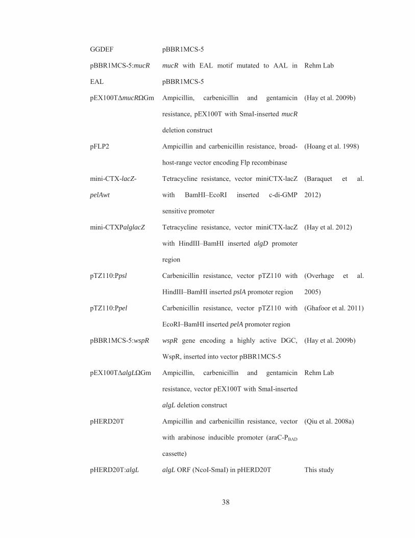
TABLE 2.2 Plasmids used in this study
Plasmids Description Source
pBBR1MCS-5 Gentamicin resistance, broad-host-range vector,
lacZ promoter
(Kovach et al. 1995)
pBBR1MCS-5:mucR KpnI-ClaI mucR fragment inserted into
pBBR1MCS-5
(Hay et al. 2009b)
pBBR1MCS-5:mucR
MHYT I
mucR with 1st MHYT motif to MAYT in
pBBR1MCS-5
Rehm Lab
pBBR1MCS-5:mucR
MHYT II
mucR with 2nd MHYT motif to MAYT in
pBBR1MCS-5
Rehm Lab
pBBR1MCS-5:mucR
MHYT III
mucR with 3rd MHYT motif to MAYT in
pBBR1MCS-5
Rehm Lab
pBBR1MCS-5:mucR
MHYT I, II
mucR with 1st & 2nd MHYT motif to MAYT
in pBBR1MCS-5
Rehm Lab
pBBR1MCS-5:mucR
MHYT I, III
mucR with 1st & 3rd MHYT motif to MAYT in
pBBR1MCS-5
Rehm Lab
pBBR1MCS-5:mucR
MHYT II, III
mucR with 2nd & 3rd MHYT motif to MAYT
in pBBR1MCS-5
Rehm Lab
pBBR1MCS-5:mucR
MHYT I, II, III
mucR with 1st, 2nd & 3rd MHYT motif to
MAYT in pBBR1MCS-5
Rehm Lab
pBBR1MCS-5:mucR mucR with GGDEF motif to GGAAF in Rehm Lab
38
GGDEF pBBR1MCS-5
pBBR1MCS-5:mucR
EAL
mucR with EAL motif mutated to AAL in
pBBR1MCS-5
Rehm Lab
pEX100TΔmucRΩGm Ampicillin, carbenicillin and gentamicin
resistance, pEX100T with SmaI-inserted mucR
deletion construct
(Hay et al. 2009b)
pFLP2 Ampicillin and carbenicillin resistance, broad-
host-range vector encoding Flp recombinase
(Hoang et al. 1998)
mini-CTX-lacZ-
pelAwt
Tetracycline resistance, vector miniCTX-lacZ
with BamHI–EcoRI inserted c-di-GMP
sensitive promoter
(Baraquet et al.
2012)
mini-CTXPalglacZ Tetracycline resistance, vector miniCTX-lacZ
with HindIII–BamHI inserted algD promoter
region
(Hay et al. 2012)
pTZ110:Ppsl Carbenicillin resistance, vector pTZ110 with
HindIII–BamHI inserted pslA promoter region
(Overhage et al.
2005)
pTZ110:Ppel Carbenicillin resistance, vector pTZ110 with
EcoRI–BamHI inserted pelA promoter region
(Ghafoor et al. 2011)
pBBR1MCS-5:wspR wspR gene encoding a highly active DGC,
WspR, inserted into vector pBBR1MCS-5
(Hay et al. 2009b)
pEX100TΔalgLΩGm Ampicillin, carbenicillin and gentamicin
resistance, vector pEX100T with SmaI-inserted
algL deletion construct
Rehm Lab
pHERD20T Ampicillin and carbenicillin resistance, vector
with arabinose inducible promoter (araC-PBAD
cassette)
(Qiu et al. 2008a)
pHERD20T:algL algL ORF (NcoI-SmaI) in pHERD20T This study

39
pHERD20T:algLH202A algL ORF with H202A mutation (NcoI-SmaI)
in pHERD20T
This study
pHERD20T:algLx6his algL ORF with C-terminal hexahistidine tag
(NcoI-SmaI) in pHERD20T
This study
pBBR1MCS-5:algX algX ORF in pBBR1MCS-5 (Moradali et al.
2015)
pBBR1MCS-5:
algXS269A
algX ORF in pBBR1MCS-5, with catalytic
serine 269 mutated to alanine
(Moradali et al.
2015)
TABLE 2.3 Oligonucleotides used in present study
Oligonucleotides Sequence Source
Complementation of mucR mutant
MucRN(KpSDNd) AGCAAGGTACCAGGAGACGCTCATATGCTTA
TCAGCA
(Hay et al.
2009b)
MucR(Cla) GAGTAATCCATAAATCAGG (Hay et al.
2009b)
Mutation of MHYT I to MAYT I
MHYT_1_F GGCATGCTCGCCTTCAGCCTGC Rehm Lab
MHYT_1_R GATGCCGAAGCCCATGGCGAAA Rehm Lab
MHYT_H59A_F TGGTCGATGGCCTTCGTCGGCATGCTCGCCTT
CAGCCTGC
Rehm Lab
MHYT_H59A_R GACGAAGGCCATCGACCAGATGCCGAAGCCC
ATGGCGAAA
Rehm Lab
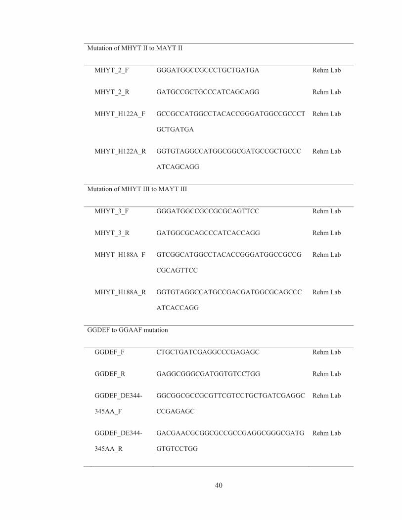
40
Mutation of MHYT II to MAYT II
MHYT_2_F GGGATGGCCGCCCTGCTGATGA Rehm Lab
MHYT_2_R GATGCCGCTGCCCATCAGCAGG Rehm Lab
MHYT_H122A_F GCCGCCATGGCCTACACCGGGATGGCCGCCCT
GCTGATGA
Rehm Lab
MHYT_H122A_R GGTGTAGGCCATGGCGGCGATGCCGCTGCCC
ATCAGCAGG
Rehm Lab
Mutation of MHYT III to MAYT III
MHYT_3_F GGGATGGCCGCCGCGCAGTTCC Rehm Lab
MHYT_3_R GATGGCGCAGCCCATCACCAGG Rehm Lab
MHYT_H188A_F GTCGGCATGGCCTACACCGGGATGGCCGCCG
CGCAGTTCC
Rehm Lab
MHYT_H188A_R GGTGTAGGCCATGCCGACGATGGCGCAGCCC
ATCACCAGG
Rehm Lab
GGDEF to GGAAF mutation
GGDEF_F CTGCTGATCGAGGCCCGAGAGC Rehm Lab
GGDEF_R GAGGCGGGCGATGGTGTCCTGG Rehm Lab
GGDEF_DE344-
345AA_F
GGCGGCGCCGCGTTCGTCCTGCTGATCGAGGC
CCGAGAGC
Rehm Lab
GGDEF_DE344-
345AA_R
GACGAACGCGGCGCCGCCGAGGCGGGCGATG
GTGTCCTGG
Rehm Lab

41
Mutation of EAL to AAL mutation
EAL_F CTGCGCTGGGAGCATCCGCAAC Rehm Lab
EAL_R CATCGGTCCGTTCGGCGCCAGT Rehm Lab
EAL_E469A_F ATCGGCGTGGCCGCGCTGCTGCGCTGGGAGC
ATCCGCAAC
Rehm Lab
EAL_E469A_R CAGCGCGGCCACGCCGATCATCGGTCCGTTCG
GCGCCAGT
Rehm Lab
Confirming mucR knockout
MucR upXout GACTCGTGCAATAATATCAGGTTG (Hay et al.
2009b)
MucR downXout GCATGTTTTCTCGTTATTGGATAGA (Hay et al.
2009b)
Generating c-di-GMP sensitive promoter
pPelfullfor CGGCGAATTCCTGGTGCGGTTCCTCGCACGCA
AC
(Baraquet et
al. 2012)
pPelfullrev GATCGGATCCACGGCGATTCCTTTCTTGCTG (Baraquet et
al. 2012)
Confirm integration of miniCTX based vectors
Pser Up CGAGTGGTTTAAGGCAACGGTCTTGA (Hoang et al.
2000)
Pser Down AGTTCGGCCTGGTGGAACAACTCG (Hoang et al.

42
2000)
Complementing algL knockout
algLN(NcoISDNd) CGATCCATGGGAGGAGATAATCGCTATGAAA
CGTCCCACCTGATCCGTATCG
This study
algLC(SmaI) CGCTATCCCGGGCACTCAACTTCCCCCTTCGC
GGCTG
This study
Confirming algL knockout
algL upXout GATCGAGCAGTCGAAAGCCGTCGATACC Rehm Lab
algL downXout CGTTTTCCTCTGGTGTTGCCTAAG Rehm Lab
2.1.1 Long term storage of strains
Bacterial strains were grown overnight in LB (Section 2.2.1) containing appropriate
antibiotics (Section 2.2.6). One millilitre of culture was transferred to a sterile 1.8 ml
cryotube vial (Thermo Scientific, USA) with 70 μl of DMSO. Strains were stored at -80
ºC and revived when required.
2.2 Media, growth conditions and antibiotic concentrations The following media were used in the present study. Solid media contained agar (~95%
purity, Neogen Cooperation, USA) at a final concentration of 1.5% (w/v), added before
autoclaving. All media were autoclaved at 121 °C for 20 min. When required,
antibiotics (Section 2.2.6) were added post-sterilization.
2.2.1 Luria-Bertani (LB) medium
LB medium was prepared by dissolving 20 g of LB powder (Acumedia, Neogen Co-
operation) in 1 litre of water.

43
2.2.2 X-Gal medium
This was prepared by supplementing 1 L of LB medium (Section 2.2.1) with 1 mL of
Isopropyl β-D-1-thiogalactopyranoside (IPTG) (Gold Biotechnology, USA) from a
stock (1 M in H2O) and 1 mL of 5-bromo-4-chloro-3-indolyl-β-D-galactopyranoside
(X-GAL) (Gold Biotechnology, USA) from a 4% (w/v) stock in DMSO.
2.2.3 Nutrient Broth
Nutrient Broth (NB) was made using Nutrient Broth Powder (Oxoid, England) as
described by the manufacturer.
2.2.4 Pseudomonas Isolation (PI) medium
Both liquid and solid PI media were used in this study. One litre of liquid PI medium
contained 20 g of peptone, 10 g of K2SO4, 1.4 g of MgCl2 x 6H2O, 25 mg of Triclosan
and 20 mL of glycerol (99% purity) in 1 L of water and adjusted to pH 7.0
(Remminghorst and Rehm 2006a).
Solid PI medium (PIA), was prepared according to the manufacturer’s instructions
(Becton, Dickinson and Company, USA). To 1 L of water, 45 g of dry medium, 20 mL
of glycerol and agar to a final concentration of 1.5% (w/v) was added prior to
autoclaving. Cells were cultivated during biofilm mode on solid PIA medium.
2.2.5 Modified alginate-producing (MAP) medium
To make 1 L of MAP medium, the following chemicals were dissolved in 1 L of water
(Franklin et al. 1994; Ohman and Chakrabarty 1981): 21.81 g of sodium D-gluconate,
18.70 g of monosodium glutamate, 1.17 g of NaH2PO4.2H2O, 2.93 g of K2HPO4, 1.20 g
of MgSO4, 0.60 g of (NH4)2SO4, and 0.20 g of NaCl. This solution was supplemented
with 1 ml of each of the following trace mineral solutions: at 0.2 g/100 ml for each of
ZnSO4.7H2O, CuSO4.5H2O, MnSO4.H2O and CoCl2.6H2O; at 0.6 g/100 ml for

44
FeSO4.7H2O, and at 0.05 g/100 ml for CaCl2.2H2O. The final MAP medium was filter-
sterilized.
2.2.6 Supplementation of media with antibiotics
When required, media used to cultivate E. coli were supplemented with antibiotics at
the following concentrations 100 μg/ml for ampicillin (sodium salt, Applichem,
Germany), 10 μg/ml for gentamicin (sulfate salt, Applichem, Germany) and 12.5 μg for
tetracycline (hydrochloride, Sigma Aldrich). For P. aeruginosa, antibiotics were used at
300 μg/ml for carbenicillin (disodium salt, Gold Biotechnology, USA) and gentamicin.
2.2.7 Growth conditions
All strains were grown at 37 ºC. Liquid cultures (planktonic mode) were grown in
Erlenmeyer flasks or universal tubes with shaking at 200 rpm in atmospheric conditions.
A ratio of 5:1 for container to culture volume was used to support sufficient aeration.
All strains cultivated on solid medium (biofilm mode) were grown at 37 °C for 24 to 72
h, as required.
2.3 DNA manipulation General procedures were followed for DNA manipulation (Sambrook et al. 1989).
2.3.1 Isolation of plasmid DNA
Plasmid DNA was isolated from cells from 3 mL of an overnight culture, grown in LB
(Section 2.2.1) with relevant antibiotics (Section 2.2.6) using the High Pure Plasmid
Isolation Kit (Roche, USA) according to manufacturer’s instructions.
2.3.2 Determination of DNA concentration and purity
DNA concentration and purity were assessed by fluorescence, spectrophotometry and
quantitative gel electrophoresis. The QubitTM fluorometer (Invitrogen Corporation,
USA) and Quant-iT DNA BR Assay Kit (Invitrogen Corporation, USA) was used in

45
conjunction with the provided DNA standard to measure DNA concentration
(emission/excitation wavelength of ~480/530 nm). DNA purity was assessed using a
Nano-drop ND-1000 spectrophotometer (Thermo Scientific, USA). An absorbance ratio
at 260/280 nm of 1.8 to 2.0 indicated high purity DNA. Quantitative gel electrophoresis
was also used to estimate the concentration and purity of linear double stranded DNA
fragments by comparing the relative intensity of DNA bands of serially diluted DNA
samples with that of MassRuler High Range DNA ladder (Fermentas, USA).
2.3.3 Polymerase Chain Reaction
Platinum ® Taq and Platinum ® Pfx Polymerases (Invitrogen Corporation, USA) were
used for analytical and preparative PCR, respectively. Reaction mixtures were prepared
as outlined below (Table 2.4, page 46-47) in 0.2 ml clean sterile thin-walled PCR tubes
(Axygen, USA).
Reactions were performed in a thermal-cycler as described below:
1) Primary denature: 94 °C (Pfx) or 95 °C (Taq) for 300 s
2) Denature: 94 °C (Pfx) or 95 °C (Taq) for 45 s
3) Anneal: ~5 °C below the lowest Tm of the primer pair for 30 s
4) Extend: 68 °C (Pfx) or 72 °C (Taq) for 60 s per 1 kbp
5) Cycle: steps 2-4 for 30 cycles
6) Hold: 10 °C

46
TABLE 2.4 PCR reaction mixtures
Pfx-DNA-polymerase reaction mixture (A)
10X Pfx Amplification Buffer 20 μl
PCRx Enhancer Solution 22.5 μl
MgSO4 (50 mM) 2 μl
DMSO 2.5 μl
Primer 1 (10 pmoles/μl) 3 μl
Primer 2 (10 pmoles/μl) 3 μl
dNTPs (10 mM each) 3 μl
Template DNA ~2 ng
Platinum® Pfx DNA Polymerase (2.5 U/ μl) 1 μl
H2O to 100 μl
Pfx-DNA-polymerase reaction mixture (B)
10X Pfx Amplification Buffer 20 μl
MgSO4 (50 mM) 5 μl
DMSO 5 μl
Primer 1 (10 pmoles/μl) 10 μl
Primer 2 (10 pmoles/μl) 10 μl
dNTPs (10 mM each) 10 μl
Template DNA ~2 ng
Platinum® Pfx DNA Polymerase (2.5 U/ μl) 1 μl
H2O to 100 μl
Taq-DNA-polymerase reaction mixture
Glycerol 50% (v/v) 20 μl
DMSO 5 μl

47
10X PCR Buffer (minus MgCl2) 10 μl
MgCl2 (50 mM) 10 μl
Primer 1 (10 pmoles/μl) 5 μl
Primer 2 (10 pmoles/μl) 5 μl
dNTPs (10 mM each) 10 μl
Template DNA ~2 ng
Platinum® Taq DNA Polymerase (5 U/μl) 1 μl
H2O to 100 μl
2.3.4 PCR dependent site-directed mutagenesis
To generate site-directed mutations, a previously described protocol was used with
adaptations (Chiu et al. 2004). For each site directed mutation, two primer pairs were
designed. One pair of primers were tailed (FT and RT), to introduce target mutations at
their 5’ ends, while the shorter primer pair (Fs and Rs) did not contain any mutations.
For each site-directed mutation, a PCR amplification of the entire vector containing the
WT mucR gene on pBBR1MCS-5 plasmid was performed using relevant primer pairs,
generating four linear PCR products. A typical PCR reaction was composed of 2.5 μL
of 10X Pfx Polymerase Buffer, 200 μM of each dNTP, 1 mM of MgSO4, 100 mM of
betaine, 10 pM of each primer, 103 pg of plasmid DNA (for 6.8 Kb template), 0.5 U of
Taq DNA polymerase and 0.25 U of Platinum Pfx polymerase with water to a final
volume of 46 μL. Reactions were run in five steps: Step 1 at 95 °C for 240 s, Step 2 at
95 °C for 15 s, Step 3 at 61 °C for 20 s, Step 4 at 68 °C for 210 s and Step 5 at 68 °C for
420 s. Steps 2-4 were repeated 25 times before Step 5, the final elongation.
To the completed PCR reaction, 4 μL of 10X buffer (Cutsmart) and 1 μL of DpnI
restriction enzyme (10 U) were mixed in, and the reaction was incubated for 1 h at
37 °C. DpnI only cleaves at its recognition site if it is methylated. Since template DNA
48
was produced from Dam+ E. coli strain, DpnI treatment would remove the parent
plasmid (which would be methylated), leaving the four PCR products intact. Figure
2.1A on page 49 schematically shows the four priming combinations and resulting PCR
products (identified in parentheses) FT/Rs (Product 1), Fs/RT (Product 2), FT/RT (Product
3), and Fs/Rs (Product 4). Products 1 and 2 (resulting from primer combinations of FT/Rs
and Fs/RT) are considered ‘productive’, because they carry the desired mutation at
opposite termini, i.e. at 3’ vs 5’ ends (Figure 2.1b, page 49). In contrast, Products 3 and
4 (generated from primer pairs FT/RT and Fs/Rs) are ‘non-productive’ because they
either have mutations at both termini (as in the case of Product 3) or no mutations at all
(Product 4).

49
Figure 2.1 Schematic of SLIM site-directed-mutagenesis. (A) PCR was performed using
template DNA (plasmid to be mutated) with two primer pairs (FT, Fs, and RT, Rs), yielding four
PCR products; product 1 (from primers FT/Rs), product 2 (Fs/RT), product 3 (FT/RT) and product
4 (Fs/RS). Template DNA was removed by DpnI treatment (not shown), and a re-hybridization
step was introduced (B), forming productive heteroduplexes between a strand of Product 1 with
its complementary strand in Product 2. These productive hybrids have complementary 5’ and 3’
overhangs that allow formation of stable circular DNA which was transformed and propagated
in E. coli on selective media.
(A)
(B)

50
After DpnI treatment, a re-hybridization step was introduced (65 °C for 5 min and 30 °C
for 15 min, for two cycles), allowing the PCR products to melt and re-hybridize,
generating 16 different hetroduplexes (not shown). Among these, only re-hybridization
events between one-strand of Product 1 with its complementary strand of Product 2
would generate complementary 3’ and 5’ overhangs at opposite termini. These
‘productive’ heteroduplexes form stable, non-covalently joined, DNA circles that upon
transformation (Section 2.3.9) into E. coli could propagate on selective media.
The MHYT, GGDEF and EAL motifs of MucR were mutated to MAYT, GGAAL and
AAL, respectively, using relevant primers, listed in Table 2.3, page 39-41.
2.3.5 Hydrolysis of DNA by restriction endonucleases
Plasmid DNA (Section 2.3.1) and PCR products (Sections 2.3.3 and 2.3.4) were
hydrolysed by restriction endonucleases (REs) for cloning and analytical purposes.
Restriction endonucleases were used according to the manufacturer’s instructions (New
England Biolabs, USA). Preparative and analytical digests were performed in clean
sterile microtubes containing 1X concentration of the relevant buffer.
In preparative digests 100 μg of DNA was digested with 100 U of enzyme in 50 μL
reactions while in analytical digests 10 U of enzyme was used to hydrolyse 1 μg of
plasmid DNA in 20 μL reactions. Unless otherwise stated, all digestions were
performed at 37 °C for 2-4 h and stopped by adding 0.2 vol of 6× stop-mix containing
Tris-HCl (pH 8.0) at 60 mM, EDTA at 60 mM, glycerol at 60% (v/v), and Orange G at
0.2% (w/v) and Xylene Cyanol FF at 0.05% (w/v) (Sambrook et al. 1989).
2.3.6 Agarose gel electrophoresis
Agarose gel electrophoresis (AGE) was used to assess DNA concentration and purity
(Section 2.3.2), and separate DNA fragments after PCR (Section 2.3.3) and restriction

51
endonuclease hydrolysis (Section 2.3.5). When needed, DNA could be recovered from
gels (Section 2.3.7). Agarose gels of 1% and 2% (w/v) were used to resolve double
stranded DNA fragments above and below 500 bp, respectively. Agarose (HYAgarose,
HydraGENE Co. Ltd. USA) was added to TBE electrophoresis buffer (50 mM Tris-
HCl, 50 mM boric acid, 2.5 mM EDTA, pH 8.3) and melted in a microwave oven,
poured into a gel chamber, and a well comb was inserted. Upon solidification (after 1 h)
the gel comb was removed. The chamber together with gel was placed into an
electrophoresis apparatus, submerging the gel in TBE buffer. To each volume of DNA
sample, 0.2 vol. of a 6× stop-mix was added (see section above) prior to loading.
A DNA molecular size standard was included in a separate well. Molecular size markers
used in this study were Lambda phage DNA hydrolysed with PstI (Sambrook et al.
1989), MassRuler High Range DNA ladder (Fermentas, USA), and 100 kb + Ladder
(Invitrogen, USA). Gels were run at 6-7 V/cm - distance between anode and cathode -
for 30-60 min. Gels were stained for 15 min in ethidium bromide solution (2 μg/ml) and
de-stained for 1 min in water. DNA bands were seen using an ultra violet (UV)
transilluminator and pictures generated (Bio-Rad, Gel Doc, USA).
2.3.7 Isolation of linear dsDNA
To isolate linear dsDNA from polymerase chain reactions (Section 2.3.3) or restriction
endonuclease digestions (Section 2.3.5), DNA was subject to AGE (Section 2.3.6) in
gels containing 1X concentration of SYBR™ Safe DNA Gel Stain (Invitrogen, USA).
Under UV light, the target DNA band was excised using a sterile scalpel blade. Target
DNA was purified from the gel using the Zymo Gel DNA Recovery Kit (Zymo
Research, USA) according to manufacturer’s instructions.
52
2.3.8 DNA ligation
Insert DNA obtained from PCR (Section 2.3.3) or RE digestions (Section 2.3.5) were
recovered from AGE (Section 2.3.7). Ligation reactions of insert with linearized vector
were prepared in 0.2 ml clean sterile thin-walled PCR tubes (Axygen, USA) containing
1 μl of T4 DNA ligase and 4 μl of 5× ligation buffer, and insert and vector DNA as
required to give a molar ratio of 6:1. DNA concentration was kept between 5 to 10
ng/μl. Water was added to give a final vol of 20 μl. Reactions were incubated at 16 °C
overnight.
2.3.9 DNA sequencing
All DNA sequencing of recombinant plasmids was provided by the Massey Genome
Service utilizing the dGTP BigDye Terminator v 3.0 chemistry in a capillary ABI3730
Genetic Analyser (Applied Biosystems Inc., USA). Results were provided in ABI
format and analysed using Vector NTI version 11 (Invitrogen Corporation, USA). Four
to six hundred nanograms of purified plasmid DNA (Section 2.3.1) was suspended in 20
μl of molecular grade water, containing 4 pMol of the relevant primer, 1 μL of DMSO,
and sent for sequencing in 0.2 ml clean sterile thin walled PCR tubes (Axygen, USA).
2.3.10 Transformation of E. coli
The recipient E. coli strain was cultivated in 50 ml of LB (Section 2.2.1), with
appropriate antibiotics (Section 2.2.6), to an optical density at 600 nm (O.D 600 nm) of
0.3. After keeping on ice for 10 min, cells were harvested by centrifugation (4,000 g at 4
ºC for 20 min). The cell pellet was suspended in 18 ml of RF1 solution and kept on ice
for 1 h. Cells were harvested again and suspended in 4 mL of RF2 solution.
Subsequently, 200 μL aliquots were transferred into 1.7 mL microtubes, frozen in liquid
nitrogen and stored at -80 ºC. The above RF1 and RF2 solutions were sterilized by
filtration (0.22 μm). RF1 solution contained RbCl at 100 mM, MnCl2 at 50 mM,

53
Potassium acetate at 30 mM and CaCl2 × 6 H2O at 10 mM dissolved in water and
adjusted to pH 5.8 with acetic acid. RF2 solution was composed of RbCl at 10 mM,
MOPS at 10 mM, CaCl2 × 6 H2O at 75 mM and glycerol at 15% (v/v), solubilized in
water and adjusted to pH 5.8 with NaOH.
For the transformation of E. coli, 50 to 500 ng of plasmid DNA was added to frozen
competent cells and incubated on ice for 1 h. After subjecting cells to heat shock (42 °C
for 90 s), tubes were incubated on ice for 5 min. Subsequently, 800 μL of LB medium
(Section 2.2.1) was added to the cells and incubated for 1 h at 37 °C. Cells were
harvested by centrifugation (15,000 g for 2 min), suspended in 200 μl of LB medium
and plated on LB agar containing relevant antibiotics (Section 2.2.6) to select for
colonies containing the plasmid of interest.
2.3.11 Transconjugation of P. aeruginosa
Transfer of conjugation-competent-plasmids into P. aeruginosa strains was performed
as outlined previously (Friedrich et al. 1981). The donor strains E. coli S17-1 was used
to transfer plasmids harbouring the Mob (oriT) region. Firstly, the plasmid of interest
was transformed into the E. coli donor strain (Section 2.3.9). Donor E. coli and recipient
P. aeruginosa strains were grown separately in 20 ml of LB medium (Section 2.2.1)
overnight, with appropriate antibiotics (Section 2.2.6). Cells were harvested by
centrifugation (4,000 g at 4°C for 20 min) and re-suspended in 0.9% (w/v) sterile saline.
Cells were washed thrice with sterile saline to remove extracellular polymeric
substances that may interfere with conjugation. Washed cells were re-suspended in 1 ml
of sterile saline, 200 μl of the donor and recipient suspensions were combined in sterile
1.7 ml micro-centrifuge tubes. After mixing by inversion, cells were harvested via
centrifugation (15,000 g for 2 min), re-suspended in 200 μl of sterile saline and dropped
carefully using a 1 ml pipette onto an NB agar plate (Section 2.2.3). After five minutes,

54
plates were gently transferred to 37 °C and incubated for 24 h. After incubation, a sterile
spatula was used to scrape biomass from plates. Cells were then suspended in 1 ml of
sterile saline and after a 1,000-fold dilution (no dilution required for pEX100T derived
plasmids), 100 μl of cell suspensions were plated on PIA medium (Section 2.2.4)
supplemented with relevant antibiotics (Section 2.2.6) and carbon source to select for
successful tranconjugants.

55
2.4 Generation of isogenic marker free mutants and complemented strains
A commonly used homologous recombination gene replacement strategy had been used
to knock out target genes involved in alginate biosynthesis (algL) and regulation
(mucR). Briefly, to disrupt each gene, plasmid borne ‘knockout cassettes’ were used.
For each gene, its respective ‘knockout cassette’ consisted of a gentamicin resistance
marker flanked by two regions of the gene; each region being ~300-500 bp long. These
fragments were obtained by PCR and ligated on either side of the resistance marker,
into the delivery vector, pEX100T. The marker itself is immediately flanked by two flip
recombinase recognition sites, allowing the marker to be removed after gene disruption.
The delivery vector pEX100T (Hoang et al. 1998), harbours an oriT (origin of transfer)
to facilitate conjugation and a sacB gene that confers lethality on sucrose containing
media. Final knockout vectors, pEX100TΔmucRΩGmR and pEX100TΔalgLΩGmR
were introduced into donor E. coli strain SM10 (Section 2.3.9) for conjugation into
PDO300 (Section 2.3.10).
Recipients were grown on PIA medium containing gentamicin (to select for integration
of the marker), and sucrose (to select for a double cross over event). Disruption of the
target gene (by a double cross over event, replacing part of the target gene with the
gentamicin resistance marker) was verified by growth on gentamicin containing
medium and colony PCR using primers flanking the target gene of interest (Table 2.3,
page 39). The marker was subsequently removed by introducing a flip recombinase
containing plasmid, pFLP2 by conjugation (Hoang et al. 1998). Successful recipients
were selected for on PIA medium containing carbenicillin. Excision of the marker was
confirmed by colony PCR and sensitivity to gentamicin. Since the pFLP2 plasmid also
harbours a sacB gene, it could be removed by counter selection on sucrose containing
media. Final strains, PDO300ΔmucR and PDO300ΔalgL, were confirmed by colony

56
PCR, growth on sucrose containing medium and susceptibility to carbenicillin and
gentamicin. The knockout plasmids pEX100T mucR and pEX100T algL were
constructed by Iain Hay and Uwe Remminghorst, respectively. The mutants were
PDO300ΔmucR and PDO300ΔalgL generated by Iain Hay and Ali Goudarztalejerdi,
respectively (Hay et al. 2009b; Wang et al. 2016a).
For complementation, genes of interest were expressed in trans on various plasmids.
For complementation of the algL mutant, the variants of the algL gene (encoding either
catalytically active or inactive variants of AlgL) were expressed from arabinose
inducible promoters on a medium copy number plasmid pHERD20T, that carried a
carbenicillin resistance marker. For the mucR investigation, variants of mucR were
expressed from a moderate constitutive promoter on a medium copy number plasmid
pBBR1MCS-5, carrying a gentamicin resistance marker. Variants of algL and mucR
were generated by a combination of PCR (Section 2.3.3), site-directed mutagenesis
(Section 2.3.4) and gene synthesis.
57
2.5 Alginate analysis Alginate yield, molecular weight and composition were assessed by a uronic acid assay,
SEC-MALLS and 1H-NMR, respectively. In some previous studies alginate production
was assessed by the gain/loss of mucoidity (Boyd et al. 1993), a colorimetric carbozole
assay (Albrecht and Schiller 2005; Jain and Ohman 2005; Monday and Schiller 1996) or
alginate lyase assay – based on increased absorbance at 230 nm associated with beta-
elimination (Bakkevig et al. 2005). In the present work, a previously described uronic
acid assay was used because it displayed greater sensitivity and specificity than the
carbozole assay, the latter of which is susceptible to interference by neutral sugars (May
and Chakrabarty 1994). Moreover, the loss/gain of mucoid phenotype can be subjective
and cannot reveal relative changes in alginate yield. Lastly the alginate-lyase assay is
susceptible to background absorbance of contaminants including other carbohydrates,
peptides, phenols or aromatic compounds. Thus, there is reason to believe that the
uronic acid assay here is more accurate and reliable than the methods used by previous
groups.
2.5.1 Preparation of samples from solid medium
Alginate produced by strains grown on solid medium (biofilm growth mode) was
harvested (Remminghorst and Rehm 2006a), with modifications, which helped
standardise biomass of the initial inoculum and eliminate effects of medium quantity
and supernatant carryover. A loop full of frozen culture stock (Section 2.1.1) was used
to inoculate 5 ml of LB medium containing antibiotics. After 14 h incubation, optical
density was measured (at 600 nm) and sufficient volume of this culture was added to 20
ml of fresh LB medium (Section 2.2.1) supplemented with appropriate antibiotics
(Section 2.2.6) to obtain an initial O.D. of 0.05. After 14 h incubation at 37 °C, cells
were harvested by centrifugation (8,000 g for 30 min), and washed twice with 20 ml

58
sterile saline. This washing step was included since previous studies have shown that
carry-over of supernatant components substantially affects alginate characteristics
(Trujillo et al. 2003b). Cells were re-suspended to an optical density of 6.0 and 1 ml of
cells was suspended in 200 μL of sterile saline and plated onto PIA medium (Section
2.2.4) containing relevant antibiotics (Section 2.2.6). For optical densities over 0.3, ten-
fold serial dilutions were used.
After 72 h incubation, biomass from plates was scrapped off, suspended in 150 ml of
sterile saline and left on a horizontal shaker for 60 min. Cells were removed by
centrifugation (9,000 g at 4 °C for 45 min) and the alginate in the supernatant was
precipitated with 1 vol of ice-cold isopropanol (96% v/v) (Sigma Aldrich, USA). The
cell pellet and crude alginate precipitates stored at -80 °C and then freeze-dried in a
floor-model freeze-dryer (Dura-Dry MP Model # FD2085C0000, FTS Systems, USA).
Alginate samples were solubilised in buffer (0.05 M Tris-HCl, 10 mM MgCl2) to a final
concentration of 0.5% (w/v). To remove co-precipitated nucleic acids and proteins,
samples were incubated with DNase I (15 μg/ml) and RNase A (15 μg/ml) for 6 h at
37 °C (with shaking at 200 rpm) followed by a further 18 h incubation with Pronase E
(20 μg/ml). Alginates were then dialysed (Dialysis Tubing, high retention seamless
cellulose tubing, 40 mm width, MW cut off: 12,400 kDa) against 5 litres of 0.9% w/v
NaCl in water for 24 h at 4°C. Dialysed alginate samples were precipitated with ice-cold
isopropanol, freeze-dried and weighed. These samples were dissolved in 200 μl of water
at 250 μg/ml and uronic acid content was measured (Section 2.5.3).
Alternatively, alginate samples for molecular mass determination (Section 2.5.4) and
compositional analysis (Section 2.5.5) were dialysed against 5 L of water prior to
precipitation, as above.

59
2.5.2 Preparation of samples from liquid medium
Alginate produced by strains grown in liquid medium (planktonic growth mode) was
measured as described previously (Rehman et al. 2013), with modifications. Strains
were grown for 14 h in LB medium containing antibiotics. From this pre-culture a
sufficient volume was added to 20 mL of MAP medium (Section 2.2.5) supplemented
with appropriate antibiotics (Section 2.2.6) to produce a culture with an initial O.D 600
nm of 0.05. Cultures were incubated for 24 h and cells were harvested by centrifugation
(15,000 x g for 1 h). The uronic acid (UA) content of filtered and unfiltered
supernatants was determined (Section 2.5.3). To filter supernatants a 10 kDa cut off
(Vivaspin® 500 Centrifugal Concentrator, GE Healthcare Life Sciences, USA) filter
was used, according to manufacturer’s instructions. UA content of unfiltered
supernatants represented the total alginate yield, inclusive of high and low molecular
weight (MW) alginate while the UA content of filtered supernatants represented the low
MW alginate that is poorly polymerised and/or degraded.
2.5.3 Uronic acid analysis
Uronic acid content of samples was measured using a colorimetric assay (Blumenkrantz
and Asboe-Hansen 1973) using alginic acid sodium salt from brown seaweed as a
standard (Sigma-Aldrich, USA). Samples (200 μL) prepared according to Sections 2.5.1
and 2.5.2 were added to 1.2 ml of tetraborate solution (0.0125 M tetraborate in
concentrated sulphuric acid), vortexed immediately and placed on ice for 10 min, at
100 °C for 5 min, and then put back on ice for another 5 min. To this mixture, 20 μl of
m-hydroxybiphenyl reagent (Sigma Aldrich, USA) (0.15% w/v m-hydroxybiphenyl in
0.125 M NaOH) was added and samples were vortexed for 1 min. For each sample and
standard, a negative control was included by substituting the m-hydroxybiphenyl

60
reagent with 20 μl of 0.125 M NaOH. Absorbances were measured at 520 nm for each
sample and standard, using the negative controls as blanks.
2.5.4 Molecular mass determination
Alginate molecular masses were determined as previously described (Moradali et al.
2015). For each strain, alginate from six PIA plates was harvested (Section 2.5.1),
pooled and subjected to Size Exclusion Chromatography-multi-Angle Laser Light
Scattering (SEC-MALLS) analysis. A Waters 2690 Alliance separations module and a
DAWN-EOS multi-angle laser light scattering detector with a laser at 690 nm (Wyatt
Technology Corp., Santa Barbara, CA, USA); Waters 2410 refractive index monitor)
were used. Purified samples (Section 2.5.1) were dissolved in 0.1 M NaNO3 (2 mg/mL)
and allowed to hydrate fully by incubating them overnight at room temperature.
Immediately prior to analysis, samples were pre-heated at 80 °C for 5 min, injected (100
μL) and eluted with 0.1 M NaNO3 (0.7 mL/min, 60 °C) from two columns (TSK-Gel
G5000PWXL and G4000PWXL, 300×7.8 mm, Tosoh Corp.) connected in series.
ASTRA software (version 6.1.2.84, Wyatt Technology Corp.) and dn/dc of 0.150 mL/g
was used for determining weight-average molecular weights (Mw) and number-average
molecular weights (Mn) and polydispersity index via the fraction Mw/Mn. In the case of
a uniform polymer, polydispersity index equals 1.0.
2.5.5 Compositional analysis
Alginate O-acetylation degree and M/G ratio and frequency of MM, MG/GM and GG
diads was determined by 1H-nuclear magnetic resonance (1H-NMR) spectroscopy
(Moradali et al. 2015). For each strain, alginate from six PIA plates was harvested
(Section 2.5.1), pooled and subjected to 1H-NMR analysis. Samples were prepared and
spectra were recorded at 90 °C with a JEOL 270 1H-NMR spectrometer (6.34 T)
operating at 270 MHz for proton. Chemical shifts were expressed in parts per million

61
downfield from the signal for 3-(trimethylsilyl) propanesulfonate (Sigma Aldrich,
USA). Compositional differences of de-acetylated and acetylated alginate samples were
determined by integration of 1H-NMR signals. Acetyl-groups were removed by alkaline
treatment where 30 ml of 1% alginate samples in saline solution were treated with 12 ml
of 1 M NaOH at 65 °C for 30 min followed by neutralization with 12 ml of 1 M HCl.
These samples were dialysed against 5 L of distilled water for 48 h and then freeze-
dried. De-acetylated and acetylated samples were compared by 1H-NMR to determine
degree of O-acetylation.
2.5.6 Alginate lyase assay
Detection of alginate lyase activity in cultures grown on solid media was performed as
described by (Sawant et al. 2015), with modifications. Strains were grown on PIA
medium for 72 h. Biomass was suspended in saline and cells were pelleted by
centrifugation. Supernatants were filter-sterilized and 100 μl of cell-free re-suspensions
were dropped onto alginate plates [LB medium supplemented with 1% w/v agarose and
0.1% w/v sodium alginate (Sigma Aldrich, USA)] for 24 h at 37 °C. Plates were flooded
overnight with 10% w/v cetylpyridinium chloride. The appearance of clearings
indicated alginate degradation. Commercial alginate lyase (Sigma Aldrich, USA) was
used as positive control (5 μL of 10 mg/ml stock).

62
2.6 Protein analysis
2.6.1 Cultivation of strains for protein analysis
Strains were cultivated in a similar manner as described in Section 2.5.1 for alginate
analysis. A loopful of frozen culture stock (Section 2.1.1) was used to inoculate 5 ml of
LB medium containing antibiotics. After 14 h incubation, optical density was measured
(at 600 nm) and sufficient volume of this culture was added to 20 ml of fresh LB
medium (Section 2.2.1) supplemented with appropriate antibiotics (Section 2.2.6) to
obtain an initial O.D of 0.05. After 14 h incubation, cells from this pre-culture were
harvested by centrifugation (8,000 g for 30 min), and washed twice with 20 ml sterile
saline. Afterwards, cells harvested from the pre-culture were re-suspended to an optical
density of 6.0. For each treatment, 1 ml of each standardized cell-preparation was
spread onto a PIA plate (Section 2.2.4) containing relevant antibiotics (Section 2.2.6),
using a sterilized glass spreader. Strains were grown on solid medium for 72 h, biomass
from plates was scrapped off, suspended in 150 ml of sterile saline and left on a
horizontal shaker for 60 min. Cells were harvested by centrifugation (9,000 g at 4 °C for
45 min).
2.6.2 Protein extraction
To obtain cell lysates, P. aeruginosa cell samples (Section 2.6.1) were treated on ice for
1 h with Buffer A (10 mM HEPES, 150 mM of NaCl, pH 7.4) supplemented with ×1
conc. of Roche EDTA free protease inhibitor and 1.5 μg/ml of lysozyme. Cells were
lysed by sonication (on ice, 12 cycles—15 s of sonication and 15 s cool down). Unlysed
cells and debris were removed by centrifugation (15,000 g for 30 min at 4 °C). To
sediment the whole envelope fraction, the clear cell lysate was ultra-centrifuged
(100,000 g for 90 min at 4 °C) and the pellet containing the membrane fraction was
suspended in 300 μl of Buffer A.

63
When required, the inner membrane fraction was further enriched by adding 0.7% w/v
N-lauroyl-sarcosine (Sigma Aldrich, USA) to Buffer A. Samples were mixed by gentle
agitation at 25 °C for 1 h to solubilize the inner membrane fraction. Subsequent ultra-
centrifugation (100,000 g for 90 min at 4 °C) was performed to sediment the outer
membrane fraction, which was discarded, leaving the inner membrane fraction in the
supernatant.
Protein samples were then flash frozen in liquid nitrogen in 50 μL aliquots and stored at
-80 °C for later analysis.
2.6.3 Protein quantification
Protein concentrations were determined using a commercially available Bradford Assay
according to manufacturer’s instructions (Bio-Rad, USA). Bovine serum albumin at
concentrations of 0.80 to 0.05 mg/ml (in the same buffer used to suspend protein
samples) was used to generate a standard curve. Serial two-fold dilutions of samples (up
to ×32) were made in 96 well plates. Ten microlitres of standards and samples was
individually mixed with 200 μl of Bradford reagent (Bio-Rad, USA) in separate wells.
Plates were incubated at room temperature for 5 min and the absorbance at 595 nm was
measured using a plate reader (ELx808iu Ultramicroplate Reader, Bio-Tek instruments,
USA).
2.6.4 Crosslinking
A crosslinking reagent was used to ‘capture’ protein-protein interactions and assess
their oligomeric states. The cross-linker used in this study, disuccinimidyl glutarate
(DSG) from Thermo Scientific Pierce, USA, is a water-soluble, homo-bifunctional N-
hydroxysuccinimide ester (NHS-ester) based crosslinker. This moiety reacts with
primary amines on the N-termini of peptides and the ε-amine of lysine residues, forming
stable covalently linked amide bonds, while releasing NHS groups. DSG harbours two

64
such moieties which are positioned separately at opposite ends of a linker spacer arm
with a length of 7.7 Å.
DSG was used in crosslinking reactions to investigate protein-protein interactions
and/or protein oligomeric states. In general, strains were grown for 72 h on PIA media
(Section 2.2.1) containing relevant antibiotics (Section 2.2.6). Biomass from three plates
were harvested and washed six times in buffer (10 mM HEPES, 150 mM NaCI, pH
7.7). To assess effect of nitrate on MucR oligomeric state, cells grown in the presence of
nitrate were washed with the same buffer supplemented with 50 mM of KNO3. Cells
were incubated for 30 min at 37 °C in 5 ml of crosslinking buffer (10 mM HEPES, 150
mM NaCl, pH 7.4, 20% v/v of DMSO, 5 mM of DSG). For each treatment, a negative
control (i.e. blank) was included; these cells were suspended in crosslinking buffer
minus DSG. Reactions were stopped by addition of Tris–HCl to a final concentration of
20 mM at pH 7.4. Cells were harvested by centrifugation and protein samples were
prepared and analysed as described in Sections 2.6.1 to 2.6.3.
2.6.5 Hexahistidine Pull Down
Another approach to studying protein-interactions is by expressing hexahistidine-tagged
‘bait’ proteins in cells followed by purification of the tagged-bait protein via nickel ion
affinity chromatography (NIAC), and searching for candidate interaction-partners in
eluted samples through SDS-PAGE and immunoblot analysis.
For such assays, strains were cultivated for 72 h on PIA medium supplemented with
relevant antibiotics. Biomass was harvested and cells were washed with saline. The
envelope fractions of cell lysates were obtained as outlined in Section 2.6.1 using Buffer
B (in place of Buffer A), supplemented with 0.1 mg/ml of lysozyme and 0.1 mg/ml of
DNAse A. (Buffer B is composed of 100 mM phosphate buffer, 150 mM NaCl, 5 mM
imidazole, 5 mM EDTA, 1X Roche EDTA-free protease inhibitor, 10% v/v glycerol).

65
Unlysed cells and cell debris were removed by centrifugation at 15,000 g for 45 min at
4 °C. From cleared cell lysates, envelope fractions were prepared by ultracentrifugation,
solubilized in Buffer C (Buffer B supplemented with 0.2% v/v of Triton X-100T) for 1
h on ice, and subject to NIAC purification using a commercially available resin
(cOmpleteTM His-Tag Purification Resin, Roche, USA), in conjunction with a Zymo-
Spin P1 Column and Collection Tube (Zymo Research, USA). Unbound proteins were
removed by centrifugation (10,000 g for 10 s) and impurities were removed by four
washes with Buffer C. To elute His-tagged proteins, resin was incubated with Buffer D
(Buffer B containing 400 mM imidazole) on ice for 10 min then centrifuged.
The purification resin used is fully compatible with metal chelators (e.g. EDTA) and
reducing agents (e.g. β-mercaptoethanol), and resilient to extreme pH (from 2-14) and
chaotropic agents (e.g. 8 M urea).
2.6.6 SDS-PAGE
Extracted protein samples (Section 2.6.2) were mixed with 0.2 vol of 6X SDS-PAGE
loading buffer and heated to 95 °C for 10 min. A milliliter of 6X SDS-PAGE loading
buffer contained 500 μl of glycerol, 120 mg of SDS, 93 mg of DTT, 0.2 mg of
Bromophenol blue and 500 μl of 4X Stacking Buffer (Laemmli 1970) After sample
preparation, 10-20 μg of each protein sample was run on SDS-PAGE.
A discontinuous buffering system was used for SDS-PAGE (Laemmli 1970). For this
method, stock solutions of 1X electrophoresis buffer (3 g of Tris, 1 g of SDS and 14.4 g
of glycine in water, adjusted to pH 8.5, in 1 L of water) and 4X stacking gel and 4X
separating gel were prepared. One liter of 4X stacking gel buffer contained 81.7 g of
Tris and 4 g of SDS in water, adjusted to pH 8.9. One liter of 4X separating gel buffer
contained 60.6 g of Tris and 4 g of SDS in water, adjusted to pH 6.8. Utilizing these
buffers, two standard 1.5 mm SDS-PAGE gels composed of 8.0 and 3.9% (w/v)

66
acrylamide separating and stacking gels were prepared as follows: For a 20 ml
preparation of an 8% (w/v) acrylamide separating gel, 5 ml of 4X Separating Gel
Buffer, 5.33 ml of 30% (w/v) acrylamide and 9.67 ml of water were thoroughly mixed
before a pinch of Na2SO3 was added to degas the mixture. Next, 10 μl of 40% (w/v)
APS and 10 μl of TEMED were added to the mixture and, the mixture was stirred for 10
s before it was poured into two 1.5 mm mini Novex® Gel Cassettes (Invitrogen, USA),
overlaid with isopropanol, and carefully placed on a level surface for 1 h to allow
polymerisation.
Upon polymerisation, the isopropanol was removed and the separating gel thoroughly
rinsed with water and dried with lint-free wipes. To prepare 5 ml of 3.9% (w/v)
acrylamide stacking gel, 1.25 ml of 4X Stacking Gel Buffer, 0.65 ml of 30% (w/v)
acrylamide and 3.1 ml of water were mixed thoroughly before a pinch of Na2SO3 was
added to degas the mixture. Then, 2.5 μl of 40% (w/v) APS and 5 μl of TEMED were
added, the mixture was stirred for 10 s and then poured onto the separating gels and a
well comb inserted. The stacking gel was left to solidify for 1 h.
SDS-PAGE gels were set up in XCell SureLockTM Mini-Cell electrophoresis gel
chambers according to the manufacturer’s instructions (Invitrogen Corporation, USA).
Gels were submerged in 1X SDS-PAGE electrophoresis buffer. Protein samples were
prepared and loaded into separate wells. For each gel, a protein molecular weight
standard (3-4 μL of either GangNam-STAIN Prestained Protein Ladder (intRON
Biotechnology, USA) or Novex ™ Sharp Pre-Stained Protein Standard (Invitrogen,
USA) was included. Pairs of 1.5 mm SDS-PAGE gels were run at 150 V for 60-90 min.
2.6.7 Immunoblot
To identify Alg proteins (Alg44, K, X, E and L), samples were run on SDS-PAGE
(Section 2.6.6), transferred to nitrocellulose membrane, probed with relevant primary

67
and secondary antibodies and visualized by chemical-luminescence through exposure
and development of auto-radiographic film.
Proteins from SDS-PAGE were transferred to nitrocellulose membrane using a semi-dry
transfer system (iBlot® Gel Transfer Stacks Nitrocellulose and iBlot® Gel Transfer
System, Invitrogen Corporation, USA). The iBlot® Anode Stack was placed on the
apparatus according to the company’s instructions. This Anode Stack has a layer of
nitrocellulose membrane resting on a buffer-infused mesh coated on the underside by a
thin sheet of copper – the anode. Onto the nitrocellulose membrane of the anode stack
the SDS-PAGE gel was placed followed by a layer of moistened iBlot® Filter Paper
(with molecular grade water). This was followed by the iBlot® Cathode Stack (a buffer-
infused mesh coated on the upper-side by a thin sheet of copper – the cathode) and the
iBlot® Sponge (to apply even pressure while completing the circuit). The supplied
‘roller’ was used between application of each layer to remove ‘air-bubbles’. The lid was
then securely fastened, and transfer programme No. 3 was initiated.
After transfer, the nitrocellulose membrane was washed thrice in a clean square-plastic-
petri dish with tris buffered saline supplemented with Tween 20 (TBST: 150 mM NaCl,
10 mM Tris-HCl and 0.1% (v/v) Tween 20 at pH 7.8) for 5 min and blocked at room
temperature for 1 h in TBST + 5% (w/v) BSA. The membrane was then washed three
times and incubated overnight at 4 °C with relevant rabbit anti-Alg antibody in TBST +
1% (w/v) BSA. For Anti-Alg44, K, E and L antibodies, antibody serum to buffer
volume ratios of 1:10,000 (v/v) were used, while for the Anti-AlgX antibody, 1:7,000
was used. After incubation, the membrane was washed again (x3 for 5 min) with TBST
and incubated in the dark with goat anti-rabbit antibody conjugated to horse radish
peroxidase (Abcam, England) at a 1:10,000 (v/v) ratio in TBST + 1% (w/v) BSA. After
washing three times again, membranes were incubated for 5 min in the dark with 3 ml

68
of substrate and 3 ml of enhancer solutions (Super Signal West Pico Stable Peroxide
Solution and Luminol/Enhancer Solution, Thermo Scientific, USA). All
incubations/washes were done with gentle agitation. To visualize bands, membranes
were exposed to BioMax XAR film (Kodak, USA) and images were developed (Kodak
X-Omat-100, USA).
For detection of his-tagged proteins a commercially available Anti-His antibody
conjugated to horse radish peroxidase (HisProbe-HRP Kit, Thermo Scientific, USA)
was used as described by the company. Nitrocellulose membranes were blocked
overnight in TBST + BSA at 1% (w/v). Membranes were washed twice in 15 ml of
TBST and incubated with the antibody at 1:5,000 (v/v) in TBST for 1 h at room
temperature in the dark. Following another four washes with TBST, the membrane was
incubated with the substrate and images were obtained as outlined above.
2.7 Swarming Motility Swarming motility was assessed as described previously (Tremblay et al. 2007). Five
microliters of an overnight culture were used to inoculate the centre of each swarming
plate. These plates consisted of modified M9 medium: 20 mM of NH4Cl, 12 mM of
Na2HPO4, 22 mM of KH2PO4, 8.6 mM NaCl, 1 mM MgSO4, 1 mM of CaCl2, 11 mM of
dextrose and 0.5% (w/v) of casamino acids (Difco) - solidified with agar (0.5% w/v).
Autoclaved medium was poured in petri dishes and allowed to solidify under laminar
flow for 1 h. Inoculated plates were incubated at 30 °C for 16 h. In the context of
assessing the effect of nitrate on motility, plates were supplemented with and without
1% w/v of KNO3. Motilities were recorded by photography. A negative result was
demonstrated by no swarming (no expansion of the colony, i.e. confinement of cells at
the initial site of inoculation). A positive result was scored if colonies expanded i.e.

69
forming a typical swarming appearance as defined by formation of dendritic fractal-like
patterns by migrating swarms moving away from initial location.
2.8 Biofilm analysis Biofilm growth was analysed by two methods, (1) a 96 well plate assay (Section 2.8.1)
and (2) via flow chamber growth (Section 2.8.2). In the latter case, biofilms grown on
glass plates were analysed by confocal laser scanning microscopy (Section 2.8.3) and
IMARIS software (Section 2.8.3).
2.8.1 96 well plate assay
A 96 well plate assay was used to study solid surface attachment, biomass growth and
biofilm dispersal (Merritt et al. 2005). For these assays, strains were grown in 10 mL of
PI medium containing carbenicillin and arabinose in 125 mL flasks at 200 rpm (shake
model) 37 ºC for 24 h. To assess initial attachment, cultures were standardized to an
O.D 600 nm of 2.0 using respective culture supernatants. 100 μL of standardized
cultures were added into eight wells of a clean sterile 96 well plate and incubated with
lid on for 2 h at 37 °C. Unbound cells were washed off by gently rinsing the plate twice
under tap water, after which plates were inverted and left to dry at ambient temperature.
Subsequently 150 μL of crystal violet solution (crystal violet at 1% w/v in water) was
added into each well. After 10 mins unbound stain was removed by gently rinsing the
plate twice with tap water and then plates were air dried. Subsequently 200 μL of
DMSO was added to each well, and after 10 mins the absorbance at 595 nm was
measured.
To assess biofilm growth, the above 24 h PI cultures were diluted with fresh PI medium
containing carbenicillin and arabinose to optical densities of 0.05. 100 μL of these
suspensions were used to inoculate individual wells of a 96 well plate (4 wells for each
70
treatment). To reduce the effect of medium evaporation on cell growth, the remaining
empty wells were filled with 200 μL of sterile water. After incubating plates for 72 h at
37 °C with the lid on, unbound cells were washed off and bound cells/biomasses were
stained as described above.
To investigate the dispersal efficiency of strains, 24 h PI cultures were diluted with
fresh PI medium containing carbenicillin and arabinose to an O.D of 0.200. 100 μL of
these suspensions were used to inoculate individual wells and incubated for 24 h. For
each treatment, four wells were used. To reduce the effect of medium evaporation on
cell growth, the remaining wells were filled with 200 μL of sterile water. After 24 h
incubation at 37 °C, unbound cells were removed by washing twice with water. To
examine dispersal efficiency, wells were incubated with 200 μL of sterile saline for 1 h.
Subsequently, 10 μL of supernatant was mixed with 990 μL of sterile saline in clean
sterile 1.7 ml Eppendorf tubes. Of this suspension, 100 μL was spread onto a PIA plate
containing relevant antibiotics. Agar plates were cultivated overnight and colony
forming units determined.
2.8.2 Flow chamber set-up
To study P. aeruginosa biofilm formation, strains were grown on a glass slide in
continuous-culture flow cell channels (with dimensions of 4 mm × 40 mm × 1.5 mm) at
37 °C (Hay et al. 2009a). This concept is illustrated schematically on page 71 in Figure
2.2 (Nielsen et al. 2011).

71
Figure 2.2 Schematic of flow cell chamber experimental set up. The growth chamber (flow
channel) was assembled and connected to the rest of the assembly. The media bottle, pump,
bubble trap, flow cells and waste bottle were connected by various tubing. Image reused with
permission from JoVE (Nielsen et al. 2011).
Flow cells were assembled by gluing the plastic-flow-chambers with a glass-cover-slip
using water-tight silicon-glue (Selleys All Clear Multipurpose Co-Polymer Sealant).
After allowing a couple of days for the glue to harden, chambers were submerged in
70% v/v ethanol to maintain sterility. Autoclavable parts of the system (media and
tubing) were prepared and sterilized by autoclaving. To assemble the flow-cell-chamber
system, the media input container was connected by tubing, and divided into eight
Marprene tubes using T-connectors, each clipped onto a laminar flow-pump to control
flow rate. These tubing were connected to separate bubble traps and which were
connected by further tubing to individual flow chamber channels units. Output tubing
from channels were connected to distribute waste-medium into a discharge container,
which was also kept ‘above’ the assembly to avoid accumulation of bubbles in growth
chambers. Overall, growth chambers were kept ‘below’ the rest of the assembly to
minimize bubbles.

72
The assembled system was sterilized by flushing twice then incubating overnight with
hypochlorite (0.5% v/v) solution. Prior to inoculating the system, hypochlorite was
washed out with sterile water, then flushed and infused with the growth medium.
Cultures used for inoculating growth chambers were grown overnight to early-
stationary phase in LB medium (Section 2.2.1) supplemented with relevant antibiotics
(Section 2.2.6). 0.5 ml of cultures were injected into the input tubing immediately
upstream of the growth chamber channel. To do this, immediately upstream of the
injection site, a bull-dog clip was used to clamp the tubing shut to prevent culture from
contaminating upstream media. Injection sites were sterilized with 70% (v/v) ethanol,
and cultures were slowly injected into tubing towards the growth chambers using sterile
1 mL syringe and needle. After delivering the injection, the site was promptly sealed
with water-tight silicon glue.
Injected cells were allowed to attach to the glass surface for 4 h at 25 °C without flow.
After allowing cells to bind to the surface, a flow rate of 0.3 ml/min (Reynolds number
of 5) was maintained for 20 h, using PI medium.
2.8.3 Confocal laser scanning microscopy and IMARIS analysis
Cells grown in flow cell chambers (Section 2.8.2) were then stained using the
LIVE/DEAD BacLight bacterial viability kit (Molecular Probes, Inc., Eugene, OR) to
visualize cells that were either living (green) or dead (red), using confocal laser
scanning microscopy (CLSM) (Leica SP5 DM6000B). This dye mixture contains two
fluorescent dyes SYTO 9 (green) and propidium iodide (red) which bind to DNA. The
former stain is membrane permeable (stains living cells) while the other is non-
permeable (stains dead/damaged cells). With excitation/emission maxima of 480/500
for SYTO 9 and 490/635 for propidium iodide, live cells would appear green and dead

73
cells red when viewed by CLSM. Biofilm characteristics were analysed by IMARIS
software as described (Ghafoor et al. 2011), obtaining the average biofilm height and
compactness. A ratio of dead to live cells was calculated by dividing the total red (dead)
with the total green (live) fluorescence (Ghafoor et al. 2011).
2.9 LacZ reporters Various promoter- and c-di-GMP-sensitive lacZ reporters were used in this study as a
convenient, non-disruptive way of measuring gene expression and intracellular c-di-
GMP levels.
2.9.1 Promoter reporter
Promoter activities of algD, pelA, and pslA were determined by previously constructed
promoter-specific lacZ fusion reporter plasmids: miniCTXPalglacZ, pTZ110:pelA, and
pTZ110:pslA (Ghafoor et al. 2011; Hay et al. 2012; Overhage et al. 2005). Promoter
activities were inferred from beta-galactosidase assay (Section 2.9.3).
2.9.2 c-di-GMP reporter
To assess global intracellular concentrations of c-di-GMP, a c-di-GMP-sensitive lacZ
reporter plasmid was made as described earlier (Baraquet et al. 2012). This plasmid,
mini-CTX-lacZ-pelAwt, was introduced into relevant P. aeruginosa strain and its
integration into the genome was confirmed by colony PCR using Pser-up and Pser-
down primers. Plasmid backbone removal was followed (Hoang et al. 2000). This
construct allowed measurement of intracellular c-di-GMP levels by inference of beta-
galactosidase activity (Section 2.9.3).
2.9.3 Beta-galactosidase assay
Beta-galactosidase (LacZ) activity was determined as described previously (Miller
1972; Zhang and Bremer 1995). Strains were grown on PIA medium (Section 2.2.4) for

74
72 h. Cells were washed twice with saline, and adjusted to an O.D of 0.7. LacZ activity
was measured in 96-well microtiter plates. Five microliters of sample were added to 20
μl of Buffer X [100 mM Na2HPO4, 20 mM KCl, 2 mM MgSO4, 0.8 mg/ml of
hexadecyltrimethylammonium bromide, 0.4 mg/ml of sodium deoxycholate, and 5.4
μl/ml of β-mercaptoethanol]. To this mixture, 150 μl of buffer Y [60 mM Na2HPO4, 40
mM NaH2PO4, 2.7 μl/ml of β-mercaptoethanol, and 1 mg/ml of o-nitrophenyl-β-D-
galactoside (ONPG) as substrate] was added and incubated for 30 to 90 min at 30 °C.
As a blank, Buffer Y minus ONPG was used for each treatment. Reactions were
terminated with 175 μl of Buffer Z (1 M sodium carbonate). Absorbance at 405 nm was
measured and LacZ activities were calculated in Miller Units (Zhang and Bremer 1995).
2.10 Statistical analysis Data were tested for normal distribution using Shapiro-Wilk test. Normally distributed
data (Shapiro-Wilk test: P > 0.05) were analysed using ANOVA followed by an LSD
Test for multiple comparison between treatments. However, data that were not normally
distributed (Shapiro-Wilk test: P < 0.05) were analysed using non-parametrical
ANOVA followed by Bonferroni (Dunn) t tests for multiple comparison between
treatments. All analyses were done using SAS 9.13. Rejection level was set at α < 0.05.
Values were reported as means ± standard error (SE).

75
3. Chapter Three: Results - The role of AlgL in alginate production and biofilm growth
3.1 The role of AlgL in alginate yield during biofilm mode To investigate the role of AlgL in alginate biosynthesis, an isogenic marker-free algL
deletion mutant was generated in the alginate over-producing strain, P. aeruginosa
PDO300 (Section 2.4). This ‘WT’ strain is a constitutive alginate over-producer,
derived by replacing the native mucA gene (encoding the MucA anti-sigma factor) with
a variant encoding a truncated MucA22 protein. This protein is rapidly degraded,
circumventing the RIP cascade (Section 1.5.1) and keeping the AlgU regulon in the
‘permanent on state’ (Mathee et al. 1999). The PDO300 algL mutant was confirmed by
colony PCR (Figure 3.1, page 76). Empty pHERD20T vectors harbouring a
carbenicillin resistance marker were introduced into the WT strain and its algL deletion
mutant as controls, while catalytically active and inactive variants of AlgL were
expressed in the algL mutant on pHERD20T plasmids from arabinose inducible
promoters (Section 2.4).
Since all strains have the plasmid-borne carbenicillin resistance marker, they could be
compared under identical growth conditions. As a control experiment to show that
arabinose did not have any unforeseen effects on alginate yield, I cultivated the WT
strain containing an empty plasmid as a biofilm, on PIA medium containing
carbenicillin in the absence and presence of arabinose. After three days’ cultivation, I
measured alginate yield via an uronic acid assay (Section 2.5). As expected, the
presence or absence of arabinose did not have any effect on alginate production levels in
the WT strain (Figure 3.2, page 77).

76
Figure 3.1 Confirmation of algL deletion mutant and complemented strains. (A) Colony
PCR of WT strain and its algL deletion mutant. The centre lane is a DNA size marker (PstI-
Lambda) (B) NcoI/SmaI restriction digestion of plasmids isolated from the WT strain and its
algL deletion mutant ( algL) carrying empty vectors (empty), or algL containing plasmid-
borne normal algL, catalytically inactive (algLH202A ), or algL with a hexahistidine tag at its c-
terminus (algLx6his ). Blue arrow = vector backbone (5.1 kb), Red arrow = algL (1.1 kb). The
DNA size marker included in the left and right panel of (B) is: MassRuler High Range DNA
ladder (Fermentas, USA). Lane 3 of left panel is empty.

77
Figure 3.2 Effect of AlgL on alginate yield. Mean alginate yield ± standard error for strains
grown on PIA medium (containing 300 μg/ml of carbenicillin) in presence (+) of arabinose
inducer (0.5% w/v). Mean alginate yield is presented as grams of alginate produced per gram of
cellular dry weight (g/g CDW). Different letters displayed above columns indicate significant
difference (p < 0.05) in alginate yield across treatments as determined by ANOVA followed by
an LSD Test (n = 3). Strains are identified as follows: WT = PDO300 containing an empty
vector (pHERD20T); ΔalgL, ΔalgL + algL, and algL + algLH202A = algL deletion mutant
containing an empty vector, plasmid-borne normal algL and catalytically inactive algL,
respectively. Reused with permission from Nature Springer (Wang et al. 2016b).
78
To examine the role of AlgL in determining alginate yield, I cultured the WT strain and
its algL deletion mutant – both harbouring empty vectors – on PIA medium containing
carbenicillin and arabinose. If AlgL was important for alginate production, then deletion
of algL would impair or stop alginate biosynthesis. Contrary to this expectation,
alginate production in the algL deletion mutant was higher, rather than lower than in the
wild type (Figure 3.2, page 77). This effect was completely reversed by reintroducing
algL on a plasmid expressed from an arabinose inducible promoter. Thus, AlgL is likely
to have a negative effect on alginate production because its absence increases alginate
yield while its over-expression (in the mutant) returns alginate yield to WT levels.
To investigate if it was AlgL lyase activity that negatively affected alginate yields, I
compared the alginate production levels of derivatives of the algL mutant expressing
plasmid-borne variants of AlgL that were either catalytically active or inactive. (The
inactive variant was generated by site-directed mutagenesis of its catalytic site (Albrecht
and Schiller 2005; Bakkevig et al. 2005) and both strains were verified by restriction
digestion (Figure 3.1, page 76)). If its lyase activity lowered alginate yield, then
presence of a lyase-inactive AlgL would lead to significantly higher alginate yields than
presence of catalytically active AlgL. After three days’ cultivation of these strains on
PIA medium containing carbenicillin and arabinose, alginate yields were measured. As
predicted, the strain expressing inactive AlgL produced significantly more alginate than
the strain expressing active AlgL (Figure 3.2, page 77). Since the only difference
between these two strains is the presence/absence of the catalytic site of AlgL, it is
reasonable to conclude that AlgL lyase activity has a direct negative effect on alginate
production levels. The increase in alginate yield of the mutant variant expressing

79
catalytically inactive AlgL suggests that some part of the protein could be having a
positive effect on alginate production.
3.2 Effect of AlgL on alginate polymer length Because AlgL negatively affected alginate yield (Section 3.1), I suspected that it might
also reduce alginate molecular weight. To test this, alginates from the WT strain and its
algL deletion mutant – both harbouring empty pHERD20T vectors – were analysed by
SEC-MALLS (Section 2.5.4). If AlgL was important for polymer length control, then
deletion of algL would detrimentally affect its molecular weight. For instance, if AlgL
is required for the production of uniform high molecular weight alginate, then deletion
of algL may result in alginates with increased average length and greater size
distribution. Theoretically, samples with a uniform molecular mass distribution should
have a polydispersity index (PI) of 1.00 and indices deviating from this value signal
increased sample heterogeneity (Stepto 2010). My preliminary findings showed that
deletion of algL did not have any effect on alginate molecular weight or its size
distribution (Figure 3.3, page 81), suggesting that AlgL is not important for controlling
alginate polymer length.
However, since the above experiment removed AlgL and its lyase activity from the
system, one could not conclude for sure whether removing only its enzymatic activity
would lead to the same (or a different) effect. Therefore, I further measured and
compared the average molecular masses and polydispersity indices of alginates
produced by the algL deletion mutant expressing plasmid-borne variants of AlgL that
were either catalytically active or inactive. If its lyase activity negatively affected
polymer length control, then the strain over-expressing active AlgL should yield
alginate of substantially lower molecular mass with greater PI value than the strain

80
expressing inactive AlgL. The preliminary SEC-MALLS analysis showed that while
over-producing inactive AlgL did not affect alginate molecular weight and
polydispersity, excess active AlgL produced alginate of much lower molecular mass
and wider size distribution (Figure 3.3, page 81). Based on this result and the fact that
the only difference between these strains was the presence/absence of AlgL catalytic
site, excess lyase activity might detrimentally affect alginate polymer-length control.
However, since knocking out algL alone did not have a direct effect on alginate
molecular mass, it is fair to conclude that AlgL is not directly involved in polymer
length control.
To rule out the possibility that excess periplasmic AlgL resulted in extracellular
degradation of the polymer, I attempted to detect alginate lyase activity in the
extracellular environment of biofilm. For this experiment, I grew the algL deletion
mutant (harbouring an empty vector) and its derivative expressing plasmid-borne active
AlgL on PIA medium (Section 2.2.4) containing carbenicillin and arabinose (Section
2.2.6). After three days of cultivation, biomasses were harvested and cell-free
suspensions were prepared and subject to an alginate lyase plate assay (Section 2.5.6).
My preliminary findings showed that over-expression of active AlgL did not lead to an
increase in alginate lyase activity of cell-free biofilm suspensions (Figure 3.4, page 82),
supporting previous reports that AlgL is an alginate degrading enzyme residing and
exerting its activity exclusively in the periplasm.

81
Figure 3.3 Alginate molecular weight (MW) averages and polydispersity indices as
determined by SEC-MALLS. (A) Alginate samples from strains grown on PIA medium
containing 300 μg/ml of carbenicillin and 0.5% (w/v) arabinose inducer were analysed by SEC-
MALLS to determine their average molecular mass and polymer length distribution
(polydispersity index = PI). PI values closer to 1.0 represent a narrow MW distribution (i.e.

82
uniform MW). Strains are identified as follows: WT = PDO300 containing an empty vector
(pHERD20T); ΔalgL, ΔalgL + algL, and algL + algLH202A = algL deletion mutant containing
an empty vector, plasmid-borne normal algL and catalytically inactive algL, respectively.
Alginate samples were harvested from six plates for each strain and then pooled for MW
analysis. Since this experiment was only done once, its result can only be considered as
preliminary. Graphic output of SEC-MALLS analysis is shown panel (B). Panel (A) reused with
permission from Nature Springer (Wang et al. 2016b).
Figure 3.4 Alginate lyase assay of strains grown on solid media. Shown are alginase assay
plates incubated for 24 h at 37 °C then flooded overnight with 10% w/v cetylpyridinium
chloride solution. Top-panel: Commercial alginate lyase (Sigma Aldrich, USA) and autoclaved
saline were used as positive (+) and negative (-) controls. Bottom-panel: 100 μL of cell-free re-
suspensions of biofilms grown on PIA medium. Samples were from the algL deletion mutant
containing an empty vector ( algL) and algL deletion mutant expressing plasmid-borne algL
( algL+algL). Clearings indicate alginate degradation. Since this experiment was only done
once, its result can only be considered as preliminary.

83
3.3 Effect of AlgL on alginate composition As noted earlier, expression of algL in trans in the algL deletion mutant did not restore
all phenotypes (i.e. polymer length) to WT levels. One of these phenotypes was O-
acetylation. To examine the effect of AlgL on alginate composition, alginates produced
by the WT strain and its algL deletion mutant – both harbouring empty vectors - were
analysed by 1H-NMR (Section 2.5.5). Bacteria were grown on PIA medium and alginate
from six plates for each strain were harvested and pooled for composition
determination. My preliminary findings showed that deletion of algL did not affect
epimerization degree, suggesting that AlgL might not have a direct role in controlling
alginate composition.
However, as alluded to above, the deletion was associated with a substantial drop in O-
acetylation levels (WT ~70% vs algL deletion mutant ~20%) and subsequent
complementation (by either expressing plasmid-borne active or inactive variants of
AlgL in the algL deletion mutant) could not restore O-acetylation to WT levels (Table
3.1, page 84). This preliminary result suggests that deletion of algL gene may have
caused polar effects on downstream genes involved in O-acetylation (algI, J and F).
Hence, additional experiments (e.g. RT-PCR or immunoblotting) could be done in the
future to see if the algL mutation caused any polar effects on downstream genes.

84
TABLE 3.1 Alginate composition as measured by 1H-NMR.
Strain FG GM FGM/MG FMM FGG AC%
WT 0.28 0.72 0.28 0.44 0 70
ΔalgL 0.35 0.65 0.35 0.30 0 17
ΔalgL + algL 0.36 0.64 0.36 0.28 0 20
ΔalgL + algLH202A 0.36 0.64 0.36 0.28 0 19
Alginate samples, obtained from strains grown on PIA medium containing carbenicillin (300 μg/ml) and
arabinose inducer at 0.5% (w/v), were analysed by 1H-NMR to investigate their level of epimerization
and O-acetylation. FG = mole fraction of guluronate residues in alginate chain. FM = mole fraction of
mannuronate residues in alginate chain. FGM/MG, FMM and FGG = mole fraction of GM/MG, MM and GG
diads. AC% = degree of O-acetylation. Strains are identified as follows: WT = PDO300 containing an
empty vector (pHERD20T); ΔalgL, ΔalgL + algL, and algL + algLH202A = algL deletion mutant
containing an empty vector, plasmid-borne normal algL and catalytically inactive algL, respectively.
Alginate samples were harvested from six plates for each strain and then pooled for compositional
analysis. Since this experiment was only done once, its result can only be considered as preliminary.
Table Reused with permission from Nature Springer (Wang et al. 2016b).

85
3.4 Role of AlgL in stability of biosynthesis complex Since deletion of algL did not appear to have compromised alginate yield (Section 3.1),
alginate molecular weight control (Section 3.2) or composition determination (Section
3.3), this gene might not be essential for the function or stability of the alginate
biosynthesis apparatus. To test this, I probed membrane fractions with various
antibodies to determine whether deletion of algL had any effect on the stability of
structural components of the complex. The WT strain and its algL deletion mutant were
grown on PIA medium for 72 hrs. After three days’ incubation, cells were harvested,
lysed and envelope fractions were prepared (Section 2.6.1 & 2.6.2) and subjected to
SDS-PAGE (Section 2.6.6) and immunoblot analysis (Section 2.6.7). If AlgL was
important for the stability of the alginate biosynthesis apparatus, then deletion of algL
would destabilise structural components. Deletion of algL did not affect the stability of
Alg44, AlgK, AlgX or AlgE (Figure 3.5, lanes 5 & 6 of each panel, page 86), implying
that AlgL might not be critical for the stability of the complex. However, since these
experiments did not include the complemented strains as controls, these results should
only be treated as preliminary.

86
Figure 3.5 Immunoblots for detection of components of the alginate biosynthesis
apparatus in various alg mutants. Shown are immunoblots of envelope fractions of various
strains using anti-Alg antibodies (top row, from left: α-Alg44, α-AlgK, α-AlgX; bottom row,
from left: α-AlgE and α-AlgL) to detect specific components of biosynthesis complex. Red
arrows highlight respective target proteins at expected molecular weight. Strains are identified
as follows: Δalg44 = PDO300Δalg44, ΔalgK = PDO300ΔalgK, ΔalgX = PDO300ΔalgX, ΔalgE
= PDO300ΔalgE, ΔalgL = PDO300ΔalgL, and WT = P. aeruginosa PDO300. MW markers are
shown in kDa.

87
If AlgL is not required for the stability of the complex, then deletion of other subunits
might also not affect AlgL stability. To test this, I cultured previously generated
(Gutsche et al. 2006; Hay et al. 2010b; Rehman et al. 2013; Remminghorst and Rehm
2006a) mutants of the WT strain lacking specific subunits (PDO300 alg44,
PDO300 algK, PDO300 algX and PDO300 algE) as above, and prepared and probed
membrane fractions with anti-AlgL (and other) antibodies. My results show that while
deletion of other subunits compromised alginate production and stability of some other
structural components, AlgL remained stable (Figure 3.5, lanes 1-4 of each panel, page
86), suggesting that AlgL could be a free periplasmic protein that is not essential for the
stability or function of the biosynthesis apparatus. However, in light of previously
encountered complementation issues (Sections 3.2 and 3.3) and the incomplete nature of
these experiments (i.e. lack of properly complementing strains), these results can only
be considered as preliminary.

88
3.5 Pull-down and immunoblot experiments to identify AlgL interaction partners Results thus far imply that AlgL is not essential for the function (Sections 3.1, 3.2 and
3.3) or stability (Section 3.4) of the biosynthesis apparatus. If this is the case, then AlgL
would not be expected to interact with other subunits of the apparatus. To test this, I
performed pull down and immunoblot experiments by expressing plasmid-borne algL
gene, encoding AlgL protein with a c-terminal hexahistidine tag, from an arabinose
inducible promoter in the algL deletion mutant (which was confirmed by plasmid
isolation and restriction digestion (Figure 3.1B, page 76)). This strain was grown on
PIA medium (Section 2.2.4) containing carbenicillin (Section 2.2.6) and arabinose for
three days, after which, envelope fractions were prepared and solubilized in Buffer C
(Section 2.6.5). Solubilized samples were subject to NIAC purification (Section 2.6.5).
After several washing steps to remove impurities and non-specifically interacting-
proteins, samples were eluted in elution buffer, ran on SDS-PAGE (Section 2.6.6),
transferred to nitrocellulose membrane and subjected to immunoblot analysis (Section
2.6.7). If AlgL was a free periplasmic protein, then none of the other subunits should be
detectable (by immunoblot) in the eluted fractions from pull down experiments. When
probed with various antibodies, none of Alg44, K, X and G was detected in eluted
fractions (Figure 3.6, page 89), lending further support to the preliminary data presented
above (Section 3.4), alluding to the possibility that AlgL could be a free periplasmic
protein.

89

90
Figure 3.6 Immunoblots showing the detection of hexahistidine-tagged AlgL and its
interaction partners. Strains were grown on PIA medium supplemented with 300 μg/ml of
carbenicillin (except for PDO300ΔalgL, lane 1) and 0.05% (w/v) arabinose for 72 h at 37 °C.
Whole Envelope (E) fractions were prepared and histagged-AlgL and its co-interacting proteins
were purified with Complete His-Tag Purification Resin (Roche). Envelope (E) and eluted (Elu)
fractions were run on SDS-PAGE, transferred to nitrocellulose membrane and subjected to
immunoblot with various antibodies (identified above each panel: α-Alg44, α-AlgK, α-AlgG, α-
AlgX and α-Histag), as described in methods section. In each panel, Lane 1 = Envelope fraction
of PDO300ΔalgL. Lanes 2 and 3 = Whole Envelope (E) and eluted (Elu) fractions of
PDO300ΔalgL(pHERD20T:algL), respectively. Lanes 4 and 5 = Whole Envelope (E) and
eluted (Elu) fractions of PDO300ΔalgL(pHERD20T algLx6his), respectively. Molecular weight
of protein standards are shown in kDa.

91
3.6 Effect of AlgL on cell attachment, biofilm growth and dispersal In earlier sections, it was suggested that AlgL negatively affected alginate yield (Section
3.1), without directly affecting alginate polymer length or composition (Section 3.2 &
3.3), and that AlgL is not an essential player (or component) of the alginate biosynthesis
apparatus (Section 3.4 & 3.5). Since AlgL is an alginate lyase and alginate makes up a
substantial proportion of P. aeruginosa PDO300 biofilms, it seemed reasonable to
hypothesize that excess AlgL activity would increase polymer degradation, potentially
affecting biofilm attachment, growth and dispersal.
To test this, I assessed the WT strain and its algL deletion mutant – both harbouring
empty pHERD20T vectors – along with the mutant over-expressing plasmid-borne
AlgL, using a 96-well plate biofilm assay (Section 2.8.1). If AlgL was important for
attachment, then deletion of algL would reduce attachment efficiency. While deletion of
algL reduced attachment, expressing the gene from a plasmid did not restore it to WT
levels (Figure 3.7A, page 92), suggesting that AlgL does not play a role in attachment in
the deletion mutant background.
Subsequent analysis of biofilm growth after 72 hrs of incubation showed no effect of
AlgL copy number (or alginate yield, molecular mass or composition) on biofilm
biomass (Figure 3.7B, page 92). However, although dispersal was increased in the
deletion mutant (Figure 3.7C, page 92), reintroducing the gene on a plasmid did not
restore dispersal to WT levels.

92
Figure 3.7 Effect of AlgL on cell attachment, biofilm biomass and dispersal. These biofilm
phenotypes were analysed in a microtiter plate assay. (A) Mean attachment efficiencies ± SE (n
= 8) at 2 h. (B) Mean biomass of biofilms ± SE (n = 4) at 72 h. Attachment and biomasses was
expressed as absorbance at 595 nm, as determined by crystal violet staining. (C) Mean dispersal
efficiencies ± SE (n = 4) at 72 h expressed as colony forming units (CFU × 104 per mL).
Different letters displayed above columns indicate statistically significant differences (p < 0.05).
Attachment efficiencies (A) were not normally distributed (Shapiro-Wilk test: P < 0.05), thus

93
analysed by non-parametrical ANOVA followed by Bonferroni (Dunn) t tests for multiple
comparison between treatments. Biofilm biomasses (B) and dispersal efficiencies (C) were
normally distributed (Shapiro-Wilk test: P > 0.05), thus analysed by ANOVA with an LSD Test
for multiple comparison between treatments. Strains are identified as follows: WT =
PDO300(pHERD20T), ΔalgL = PDO300ΔalgL(pHERD20T), and ΔalgL + algL =
PDO300ΔalgL(pHERD20T:algL). Reused with permission from Nature Springer (Wang et al.
2016b).
3.7 Effect of O-acetylation on cell attachment, biofilm growth and dispersal Even though plasmid-borne expression of algL did not restore O-acetylation (Section
3.3), attachment or dispersal (Section 3.6) to WT levels, the apparent correlation
between O-acetylation with attachment and dispersal raised the tentative possibility that
O-acetylation influences these phenotypes. To examine if O-acetylation levels truly
influenced attachment and dispersal, I conducted an experiment using two previously
described strains that produced o-acetylated (~10% AC) and non-o-acetylated alginate
of similar yield, molecular weight and epimerization degree. These two strains
generated by Moradali et al (2015) were subjected to the same 96 well plate assays
(excluding arabinose while substituting carbenicillin for gentamicin) (Section 2.8.1). I
hypothesized that if O-acetylation degree directly determined attachment and dispersal
efficiency, then a complete loss of this modification would influence both of these
phenotypes. Although abolishing O-acetylation did not affect biofilm growth (Figure
3.8B, page 94), it impaired both attachment (Figure 3.8A, page 94) and dispersal
(Figure 3.8C, page 94).

94
Figure 3.8 Effect of alginate O-acetylation on cell attachment, biofilm biomass and
dispersal efficiency. Attachment efficiencies, biofilm biomasses and dispersal efficiencies were
analysed in a microtiter plate assay. (A) Mean attachment efficiencies ± SE (n = 6) at 2 h, as
determined by crystal violet staining. Attachment efficiencies are expressed as absorbances at
595 nm. (B) Mean biomass of biofilms ± SE (n = 4) at 72 h, as determined by crystal violet
staining. (C) Mean dispersal efficiencies ± SE (n = 6) at 72 h expressed as colony forming units
(CFU x 104 per mL). In panels (A-C) different letters displayed above columns indicate
statistically significant differences (two-way, t-test, p < 0.05) in attachment efficiency, biofilm

95
biomass and dispersal efficiency, respectively. In panels (A-C), strains ΔalgX+algX and
ΔalgL+algXS269A are identified as PDO300ΔalgX(pBBR1MCS-5:algX) and
PDO300ΔalgX(pBBR1MCS-5:algXS269A), respectively. These strains produce o-acetylated
and non-acetylated forms of alginate, respectively.

96
4. Chapter Four: Results - Insight into the functions of MucR in the regulation of alginate production In this part of the study I investigated the biological function of MucR, an inner
membrane protein thought to be a chief driver of alginate production. Current models
suggest that it provides a local pool of c-di-GMP driving biosynthesis at a post-
translational level (Hay et al. 2009b). This protein has a membrane-embedded sensor
domain and two cytoplasmic output domains involved in c-di-GMP turnover. However,
whether both sensor and output domains are crucial for making alginate is unknown.
Here I explored the role of MucR, with special reference to its output and senor domains
and its response to nitrate.
The two cytoplasmic output domains of MucR, diguanylate cyclase (DGC) and
phosphodiesterase (PDE), have been shown in vitro to make and break c-di-GMP,
respectively (Li et al. 2013). In vitro studies using a truncated version of MucR,
consisting of only its output domains, revealed that the DGC activity was enhanced by
its PDE domain, suggesting that both contribute to intracellular c-di-GMP levels (Li et
al. 2013). However, the physiological relevance of this in vitro observation is currently
unclear, since normally the sensor domain is at the N-terminus of the protein, which
could regulate the activity of its output domains in vivo.
The sensor domain of MucR contains three MHYT motifs thought to co-ordinate a
copper ion for the perception of a signal (Galperin et al. 2001). A likely signal could be
nitrate because P. aeruginosa uses it in denitrification, releasing intermediates that are
linked to reduced alginate yields (Wood et al. 2007; Zumft 1997). One of these
intermediates, nitric oxide, also enhances biofilm dispersal through MucR by lowering
global c-di-GMP levels (Li et al. 2013). However, it is still not clear whether nitrate
97
could be a signal perceived by MucR, leading to reduced alginate yields from cells
cultivated in biofilm and planktonic growth modes.
Since elevated c-di-GMP levels up-regulate genes involved in biofilm formation (Jenal
et al. 2017), MucR and its putative signal (nitrate) could also affect alginate production
at a gene-expression level through modulation of global c-di-GMP levels. I also
postulated that any effect of MucR and its signal on c-di-GMP levels would also affect
other phenotypes sensitive to this ubiquitous bacterial secondary messenger - including
swarming motility, attachment, polysaccharide production and biofilm formation.
I carried out a series of experiments to explore the functions of MucR in the regulation
of alginate. Figure 4.1 (page 98) summarizes the proposed model that was explored in
this study for the function of MucR and nitrate in alginate production, modulation of c-
di-GMP and c-di-GMP sensitive phenotypes.

98
Figure 4.1 Overview of the proposed working model for the function of nitrate and MucR
in alginate production, c-di-GMP levels and other phenotypes sensitive to c-di-GMP. A
signal (nitrate?) is perceived by MucR (via its MHYT sensor domain?), affecting the activity of
its output domain (DGC and PDE?) involved in c-di-GMP turn-over. A local pool (global?) of
c-di-GMP imparted by MucR presumably drives alginate production at a post-translational (or a
transcriptional level?). The effect of nitrate and MucR on c-di-GMP levels may impact other
phenotypes including attachment, swarming, Psl & Pel production, biofilm formation which are
sensitive to this secondary messenger molecule.

99
4.1 MucR’s DGC and PDE domains are important for alginate biosynthesis Some preliminary data suggested that mucR was important for alginate biosynthesis
(Hay et al. 2009b) as deletion of mucR caused a substantial reduction in alginate yield.
Furthermore, plasmid-borne over-expression of MucR significantly increased alginate
production (Hay et al. 2009b). However, when the protein was truncated (i.e. both DGC
and PDE domains were removed), this effect did not occur (Hay et al. 2009b). Since in
vitro studies have shown that the output domains of MucR are involved in c-di-GMP
turnover, with one facilitating its synthesis and the other its degradation (Li et al. 2013),
I assumed that the failure of the truncated version of MucR to increase alginate yields
was due to the complete lack of domains involved in c-di-GMP turnover. If this
hypothesis is correct, then eliminating MucR’s c-di-GMP turnover activity by
introducing point mutations into each of the catalytic residues of MucR’s output
domains would stop it from promoting alginate biosynthesis.
To test this postulation, I expressed variants of MucR with mutated DGC and PDE
domains from constitutive lacZ promoters on a medium copy number plasmid
(pBBR1MCS-5) in a mutant where the native mucR gene had been deleted by
homologous recombination (Hay et al. 2009b). These variants were constructed and
cloned into pBBR1MCS-5, and DGC and PDE active sites disabled by alanine
substitution (GGDEF to GGAAF and EAL to AAL, respectively) (Wang et al. 2015).
Such mutations were expected to disable the output domains of MucR, preventing it
from synthesising and degrading c-di-GMP, respectively.
These specific mutations were introduced because when such changes were made to
other enzymes involved in c-di-GMP production/degradation, enzymatic activities were
lost (reviewed in (Jenal et al. 2017)). Variant-containing plasmids were verified by
DNA sequencing (Section 2.3.9) then introduced into E. coli S17-1 (Section 2.3.10) for
100
conjugation into the mucR deletion mutant (Section 2.3.11). Potential transconjugants
were selected for on PIA medium containing gentamicin. Received plasmids were
isolated and subjected to restriction digestion confirmation (Figure 4.2A, page 101).
Variants of the mucR deletion mutant harbouring either an empty vector or a plasmid-
borne mucR were included, along with the WT strain containing an “empty” plasmid
(i.e the cloning vector pBBR1MCS-5 containing the selectable gentamicin resistance
cassette). The presence of vector-borne gentamicin resistance markers in all strains
allowed me to compare the performance of different strains (and versions of MucR)
under identical growth conditions.
To determine the role of MucR’s DGC and PDE domains in alginate production during
biofilm growth, the above strains – mucR deletion mutant with: empty vector, plasmid-
borne MucR, plasmid-borne MucR with a defunct DGC domain, and plasmid-borne
MucR with a defunct PDE domain — were grown on PIA medium containing
gentamicin for 72 h, along with the WT strain containing an empty vector as a control.
Biomass was harvested and alginate samples prepared and quantified by an uronic acid
assay. If DGC and PDE domains of MucR enhanced alginate production, then plasmid-
borne expression of functional MucR would increase while expression of defunct MucR
would decrease (or not affect) alginate yields. My findings support this prediction, i.e.
expression of functional MucR from a plasmid increased alginate production levels and
plasmid-borne expression of defunct MucR failed to increase alginate yields (Figure
4.3, page 103).

101

102
Figure 4.2 Agarose gel electrophoresis of restriction digests to confirm plasmid DNA
isolated from PDO300ΔmucR trans-conjugants receiving pBBR1MCS-5 vectors harbour
variants of mucR gene. In panels (A) and (B), Plasmid DNA isolated from each strain were
(from left-to-right) not digested (uncut: lanes 1, 6, 11), cut with ClaI (lanes 2, 7, 12), cut with
KpnI (lanes 3, 8, 13) or cut with both ClaI and KpnI (lanes 4, 9, 14). As expected, plasmids cut
with either ClaI or KpnI produced one band ~6.8 kbp in size. In panel (C), Plasmid DNA was
either uncut or cut with both ClaI and KpnI. In all panels, samples digested with both ClaI and
KpnI produced two DNA fragments of ~4.7 (linearized vector) and ~2.1 kbp (variant of mucR
gene). A DNA size marker (PstI-lambda) was included in lanes 5 & 15 of panel (A), lanes 1 &
11 of panel (B), and lanes 1 & 8 of panel (C).

103
Figure 4.3 Effect on alginate yield of deleting mucR and plasmid-borne expression of
mucR and variants with mutated DGC and PDE domains during biofilm growth. Shown
are mean alginate yields ± SE, representative of three independent experiments. The strains
tested were: WT (PDO300; containing an empty pBBR1MCS-5 plasmid), a mucR deletion
mutant (ΔmucR; containing an empty pBBR1MCS-5), a mucR deletion mutant expressing
plasmid-borne mucR (ΔmucR+mucR), and a mucR deletion mutant expressing variants of mucR
encoding MucR with inactive DGC or PDE domains (ΔmucR+mucR DGC and ΔmucR+mucR
PDE, respectively). Alginate yields labelled with different letters differed significantly from
each other (LSD Test, p < 0.05).

104
To exclude alternative possibilities that these mutations destabilized MucR, leading to
its degradation, inner membrane fractions of cells were prepared and probed with anti-
MucR antibodies. Cells were grown as biofilms on PIA medium, harvested, lysed, and
inner membrane fractions were isolated (Section 2.6.2), protein concentrations
measured (Section 2.6.3), samples ran on SDS-PAGE (Section 2.6.6) and analysed by
immunoblot (Sections 2.6.7). My findings show that deletion of mucR gene abolished
the presence of the MucR protein (see Figure 4.4, lanes 3 & 4, page 105; red arrow
showing expected size of mucR gene) but inactivation of either DGC or PDE domain
did not jeopardize MucR stability (Figure 4.4, lanes 13-16, page 105).
My results thus strongly suggest that both DGC and PDE domains of MucR are
important for alginate production (Figure 4.5, page 106). This may be attributed to their
roles in making and breaking c-di-GMP, as demonstrated in vitro (Li et al. 2013).
Interestingly, it was previously reported that deletion of mucR reduced alginate
production (Hay et al. 2009) but my current work demonstrates that alginate yields of
the WT strain and its mucR deletion mutant were not significantly different (Figure 4.3,
page 103). The variance of findings between these two studies could result from
different experimental design (see discussion, Section 5.2).

105
Figure 4.4 Immunoblot detection of MucR in inner membrane fractions of cell lysates.
Shown are immunoblots using anti-MucR antibody to detect MucR in the inner membrane
fraction of the WT strain and mucR deletion mutant - harbouring various plasmids - grown in
the absence (-) or presence (+) of nitrate during biofilm growth on solid medium. WT strain
harbouring an empty vector (lanes 1-2). The mucR deletion mutant (ΔmucR) expressing various
plasmid-borne variants of mucR gene (lanes 3-16): empty vector (lane 3-4), normal mucR (lanes
5-6), mucR with defective senor motifs (mutated MHYT I, MHYT II or MHYT III motifs)
corresponding to lanes 7-8, 9-10 & 11-12), and MucR with defunct output domains (DGC or
PDE, lanes 13-14 & 15-16). MW ladder shown in kDa. Red arrow = expected MW of MucR
(74 kDa). Position of MW ladder bands are shown on the y-axis (Novex ™ Sharp Pre-Stained
Protein Standard, Invitrogen, USA). There is an anomaly in lane 4 - a faint band corresponding
to the same MW as MucR detected in the sample from a mucR mutant (with an empty vector)

106
grown in the presence of nitrate. This unexpected band could be a background signal, a protein
similar to MucR or accidental leakage from a neighbouring lane during loading.
Figure 4.5 Section 4.1 results suggest that MucR’s DGC & PDE domains are important
for alginate production. Green parts are supported by experimental findings. Grey parts are
hypothetical.

107
4.2 Nitrate impairs alginate production through MucR In the previous section, it was shown that both c-di-GMP synthesising and degrading
functions of MucR output domains were important for making alginate. However, so far
it is still not known if these activities are affected by any signals detected by the
putative sensor domain of MucR. The sensor domain is thought to harbour three MHYT
sensor motifs for the perception of a diatomic gas such as oxygen, carbon monoxide and
nitric oxide, which have been respectively linked to reduced alginate yields, impaired
biofilm formation and enhanced biofilm dispersal (Bragonzi et al. 2005; de la Fuente-
Nunez et al. 2013; Hassett 1996; Murray et al. 2012; Wood et al. 2007; Worlitzsch et al.
2002). It has been reported that P. aeruginosa can carry out denitrification under low
oxygen and high nitrate conditions, releasing nitric oxide endogenously (Line et al.
2014; Williams et al. 1978). However, it is not clear whether MucR facilitates a
suppression of alginate production under such conditions.
To investigate this, I grew the WT strain, its mucR deletion mutant (both harbouring
empty vectors) and a mucR deletion strain expressing plasmid-borne mucR on plain PIA
medium (containing gentamicin) or in PIA medium to which nitrate had been added as
potassium nitrate at 1% w/v (~ 100 mM). The latter thus provided levels of the initial
substrate for denitrification (i.e. nitrate) and at a concentration that maximizes cell
growth (Hassett 1996). After three days of incubation, alginate yields were measured. If
MucR mediated a negative effect of nitrate on alginate production, then adding nitrate to
the growth medium would lower alginate yields in both the WT strain and the plasmid-
borne expression strain but have no effect in the mucR deletion mutant. My findings
supported the prediction about the role of MucR in mediating nitrate-induced
suppression of alginate production (Figure 4.6, page 108), in that nitrate significantly

108
reduced alginate yields only in the strains containing an intact mucR gene (i.e. the WT
and complemented strains).
To rule out the possibility that nitrate blocked alginate production by destabilizing
MucR, leading to its degradation, I prepared inner membrane fractions and probed them
with anti-MucR antibodies. If nitrate led to degradation of MucR, then the intensity of
full length MucR might be reduced in immunoblots. However, my results show that
nitrate did not abolish the presence of full length MucR (Figure 4.4, lanes 7-12, page
105).
Figure 4.6 The role of MucR in nitrate perception and alginate production. Effect
of nitrate and mucR on alginate yield. Shown are mean alginate yields ± SE,
representative of three independent experiments. Strains were grown in the absence
(white) and presence (black) of nitrate. WT and ΔmucR = WT strain and mucR deletion
mutant strains harbouring empty pBBR1MCS-5 vectors. ΔmucR+mucR = mucR
deletion mutant expressing plasmid-borne mucR. Treatments with different letters are

109
significantly different (LSD Test, n = 3, p < 0.05). g/g CDW = grams of alginate per
gram of cellular dry weight. Reused with permission from Springer Nature (Wang et al.
2015).
4.3 MucR’s MHYT sensor domain is involved in nitrate perception Because MucR played a role in nitrate suppressing alginate production (Section 4.2), I
carried out experiments to determine whether MucR’s sensor domain was involved in
nitrate perception, leading to reduced alginate yields. Variants of MucR were generated
with single, double, and triple histidine to alanine substitutions at their MHYT motifs
(H59A, H122A, and H189A) - numbered I, II, and III from the N terminus. They were
made by SLIM PCR in combination with gene synthesis, sequence verified and then
cloned into pBBR1MCS-5 (Wang et al. 2015) (also see Section 2.3.4). I subsequently
introduced variant-containing plasmids into E. coli S17-1 for transconjugation into the
mucR deletion mutant (Section 2.3.11). Successful recipients were selected for on PIA
medium containing gentamicin and received plasmids were confirmed by plasmid DNA
isolation and restriction digestion (Figure 4.2B-C, page 101-102).
To investigate if the MHYT motifs of MucR’s sensor domain were involved in nitrate
perception, variants of MucR with individually mutated MHYT sensor motifs were
expressed from pBBR1MCS-5 plasmids in the mucR deletion mutant. These strains
were grown on PIA medium containing gentamicin with or without added nitrate. In
parallel, the mucR deletion mutant expressing vector-borne unmutated mucR cultivated
in the absence and presence of nitrate was included as control. After three days’
incubation, alginate yields were measured. As expected, alginate yields of the control
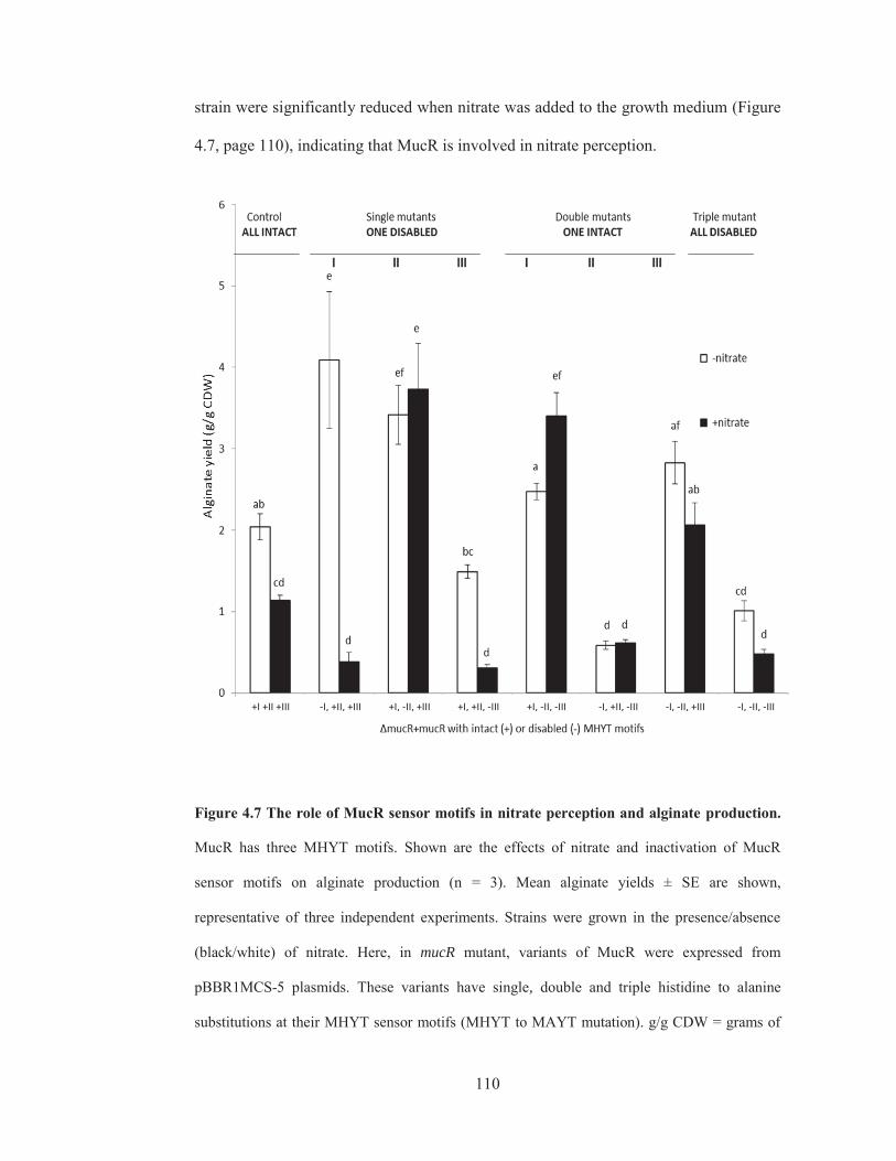
110
strain were significantly reduced when nitrate was added to the growth medium (Figure
4.7, page 110), indicating that MucR is involved in nitrate perception.
Figure 4.7 The role of MucR sensor motifs in nitrate perception and alginate production.
MucR has three MHYT motifs. Shown are the effects of nitrate and inactivation of MucR
sensor motifs on alginate production (n = 3). Mean alginate yields ± SE are shown,
representative of three independent experiments. Strains were grown in the presence/absence
(black/white) of nitrate. Here, in mucR mutant, variants of MucR were expressed from
pBBR1MCS-5 plasmids. These variants have single, double and triple histidine to alanine
substitutions at their MHYT sensor motifs (MHYT to MAYT mutation). g/g CDW = grams of
+I +II +III -I, +II, +III +I, -II, +III +I, +II, -III +I, -II, -III -I, +II, -III -I, -II, +III -I, -II, -III

111
alginate per gram of cellular dry weight. Control = ΔmucR+mucR i.e. the mucR deletion mutant
expressing plasmid-borne mucR with MHYT motifs ALL INTACT (+I, +II, +III). Single
mutants have only ONE DISABLED motif, double mutants have ONE INTACT motif, and
the triple mutant has its motifs ALL DISABLED. Notation on x-axis dictates which variant of
mucR was expressed in the mucR deletion mutant with roman numeral dictating which MHYT
motif was disabled (-) or left intact (+). Treatments with different letters are significantly
different (LSD Test, n = 3, p < 0.05).
Disruption of MHYT I in the absence of added nitrate or disruption of MHYT II
regardless of whether nitrate was added to the growth medium did not significantly
reduce alginate production. Strains expressing MucR with either inactive MHYT I or
MHYT III sensor motifs exhibited reduced alginate yields when grown in the presence
of nitrate compared to respective strains grown in the absence of added nitrate (Figure
4.7, page 110). These results suggest that (1) MHYT I and III are apparently not
necessary for perception of nitrate signal, (2) an intact MHYT II is involved in nitrate
perception, and (3) MHYT III enhances alginate production. To rule out the possibility
that the introduced mutations (or nitrate itself) impaired alginate biosynthesis by
destabilizing MucR, leading to its degradation, I probed inner membrane fractions with
anti-MucR antibodies (Section 2.6.7). My immunoblot experiments show that neither
nitrate nor single histidine to alanine substitutions detrimentally affected MucR stability
(Figure 4.4, lanes 7-12, page 105).
To consolidate above findings about the functions of MHYT motifs, I carried out
further experiments on MucR with double and triple mutations. As shown in Figure 4.7
on page 110, alginate yields of the strain containing an intact MHYT I motif (with
inactivated MHYT II & III motifs) did not respond negatively to nitrate. Alginate yields

112
of the strain with only an intact MHYT II motif (and disrupted MHYT I & III motifs)
was reduced to background levels regardless of whether nitrate was added to the growth
medium, and alginate production levels of the strain bearing the normal MHYT III
motif (with mutated MHYT I & II motifs) also did not respond to nitrate. Furthermore,
when all three motifs were knocked out from the sensor domain, alginate production
was reduced to background levels regardless of whether nitrate was added (Figure 4.7).
These findings support the conclusions derived from the single mutant experiments
(Figure 4.7, page 110).
Based on the results from this section, I proposed a model to illustrate how the MHYT
motifs and nitrate may affect alginate production (Figure 4.8, page 113). In this model
MHYT I and MHYT III motifs enhance alginate production while MHYT II impairs
alginate production, and the latter effect is enhanced in the presence of nitrate (Figure
4.8, page 113). In essence, a delicate balance between positive and negative effectors
dictates the eventual alginate production levels.
How this delicate balance is manifested mechanistically remained to be investigated. In
the later parts of this study, I attempted to explore potential mechanisms of nitrate-
induced suppression of alginate production, for example, whether nitrate affected MucR
output domain activity by influencing its oligomeric state (Section 4.4); whether MucR
and nitrate impacted alginate yield by influencing alginate promoter activity (Section
4.5), and whether MucR and nitrate influenced alginate production and promoter
activity by influencing intracellular c-di-GMP levels (Section 4.6).

113
Figure 4.8 Schematic representation of how the MHYT motifs and nitrate affect alginate
yield. A delicate balance between positive (MHYT I & III) and negative effectors (MHYT II &
nitrate) dictates final alginate yield. MHYT I & III enhance alginate production while MHYTII
impairs alginate production. Perception of nitrate by MHYTII amplifies this effect, suppressing
alginate production. The large nitrate box represents growth conditions in the presence of added
nitrate.

114
4.4 Effect of nitrate on MucR oligomeric state In the previous sections, I showed that MucR perceived nitrate through its MHYT
sensor domain, modulating alginate production levels. In principle, perception of nitrate
may have altered the activity of MucR’s output domain that is involved in c-di-GMP
turnover. This could occur through various mechanisms. One possibility could be that
nitrate affected the oligomeric state of MucR. To test if this was the case, I did
crosslinking and western blot experiments. Here, the WT strain containing an empty
vector was grown on PIA medium in the presence or absence of additional nitrate for
three days. Biomass from plates was harvested and washed with either saline (for cells
grown on normal PIA medium without additional nitrate) or saline supplemented with
potassium nitrate (for cells grown in on PIA medium containing additional nitrate). The
latter wash was used to help stabilise any interactions and/or oligomeric states that were
nitrate sensitive.
Washed cells were incubated with or without a crosslinking reagent (DSG) as described
in Section 2.6.4. The chosen reagent, DSG, was used to capture protein-protein
interactions (i.e. potential oligomeric states of MucR). This water soluble homo-
bifunctional NHS-ester-based crosslinker reacts with primary amines on the N-termini
of peptides and the ε-amine of lysine residues, forming amide bonds between
neighbouring (and within individual) proteins, stabilizing interactions that would
otherwise be destroyed during the harsh conditions of sample preparation. Hence, these
treatments would help stabilize multimeric complexes for immunoblot analysis with
anti-MucR antibodies.
As negative controls, another set of cells grown under each condition was prepared and
incubated without DSG. Subsequently, inner membrane fractions of DSG-treated and
untreated cells were prepared (Section 2.6.2), quantified (Section 2.6.3), boiled in a

115
denaturing buffer and ran on SDS-PAGE (Section 2.6.6) prior to probing with anti-
MucR antibodies (Section 2.6.7). If nitrate interfered with MucR activity by influencing
its oligomeric state, then adding nitrate to the cultivation medium would also affect
MucR oligomerization but this was not the case (Figure 4.9, page 116). Under both
conditions, MucR appeared as a monomer ( 75 kDa) in the non-crosslinked cells and a
multimer ( 300 kDa) in DSG-treated cells, suggesting that nitrate does not affect MucR
oligomeric state, which appears to be multimeric by default. These new findings
allowed me to revise the working model proposed earlier (Figure 4.5, page 106) into
Figure 4.10 (page 117), which shows that nitrate does not modulate alginate production
by altering the oligomeric state of MucR.

116
Figure 4.9 Crosslinking and immunoblot detection of MucR to examine effect of nitrate on
MucR oligomeric state. From left, WT strain harbouring an empty vector was grown in the
absence (-) or presence (+) of nitrate. Washed cells were either lysed directly (blank), or treated
with (+) or without (-) DSG crosslinker inner membrane fractions were prepared and probed
with anti-MucR antibodies. Left scale, MW ladder (kDa). Red arrow = expected MW of MucR.
Green arrow = MucR in higher MW complexes. Position of MW ladder bands are shown on the
y-axis (Novex ™ Sharp Pre-Stained Protein Standard, Invitrogen, USA).
blank

117
Figure 4.10 Summary of results from Section 4.1 to 4.4. I show that MucR’s DGC & PDE
domains are important for alginate production (Section 4.1), nitrate suppresses alginate
production through MucR (Section 4.2), MucR’s MHYT sensor domain is involved in nitrate
perception (Section 4.3), and nitrate does not modulate alginate production by affecting the
oligomeric state of MucR (Section 4.4).

118
4.5 Effect of MucR and nitrate on alginate promoter activity In the preceding sections it was proposed that MucR was necessary for nitrate-induced
suppression of alginate biosynthesis. To determine if this occurred at a gene expression
level, I introduced an integration-proficient alginate-promoter-lacZ-reporter construct
(miniCTX:Palg-lacZ) into the WT strain and its mucR deletion mutant, along with
empty pBBR1MCS-5 vectors. This Palg-lacZ reporter construct contained a lacZ
reporter gene controlled by the alginate promoter, a 1,000 bp region located directly
upstream of the alginate biosynthesis gene cluster. Since the construct specifically
integrates one copy of itself into the genome of recipient cells at the unique attB phage-
attachment located at 6.24 Mb of the PAO1 genome (Ramos 2011), a comparison of
alginate promoter activities between treatments would be possible by inference from
LacZ reporter enzyme activity. Successful integration of the Palg-lacZ reporter
construct into the genomes of recipient strains was confirmed by colony PCR (Figure
4.11A, page 119).

119
Figure 4.11 PCR confirmation of integrated Palg-lacZ and c-di-GMP-sensitive lacZ
constructs and restriction confirmation of introduced plasmids. (A) Colony PCR
confirming integration of Palg-lacZ reporter construct into genomes of WT strain and mucR
deletion mutant. Red arrow = expected MW. (B) ClaI/KpnI restriction confirmation of
pBBR1MCS-5 plasmids from strains harbouring genome-integrated Palg-lacZ constructs.
Plasmids were empty or expressing variants of MucR with a defunct sensor (MHYT II) or
output domains (DGC) or (PDE). Green arrow = mucR size. U = uncut, C = cut. (C) Colony
PCR confirming integration of c-di-GMP-sensitive-lacZ reporter construct into genomes of WT
strain and mucR mutant. Red arrow = expected MW. (D) XbaI/SacI confirmation of empty
pBBR1MCS-5 vectors or vectors with wspR gene isolated from WT strain containing genomic

120
Palg-lacZ reporter. U = uncut, C = cut. Yellow arrow = wspR size. (E) BamHI/EcoRI
verification of Ppel and Ppsl reporter plasmids (pTZ110Ppel-lacZ and pTZ110Ppsl-lacZ)
introduced into strains. Blue arrow = size of Ppel-lacZ and Ppsl-lacZ. In panel (A), left-panel of
(B), and panel (C) a MassRuler High Range DNA ladder (Fermentas, USA) was used. In right-
panel of (B), and panels (C) & (E), a PstI-lambda DNA size marker was used.
To determine if nitrate-induced suppression of alginate biosynthesis occurred at a
transcriptional level, the WT strain and its mucR deletion mutant – both harbouring
PalgD-lacZ reporter constructs and empty PBBR1MCS-5 plasmids – were grown on
PIA medium containing gentamicin, with or without additional nitrate. After three days’
incubation, cells were harvested and PalgD activity was inferred from beta-
galactosidase activity. If nitrate-induced suppression of alginate biosynthesis occurred at
a transcriptional level, then alginate yields should somewhat correlate with alginate
promoter (PalgD) activity, with a loss of alginate production in the WT strain (caused
by nitrate) expected to be linked to reduced (or abolished) PalgD activity. Furthermore,
deletion of mucR should not affect PalgD activity - since this mutant still produced
normal alginate yields (independent of nitrate) (Figure 4.6, page 108).
However, my results show that while nitrate abolished alginate production in the WT
strain (Figure 4.6, 108), it only reduced alginate promoter activity by 50% (Figure 4.12,
page 121), suggesting that the negative effect of nitrate on alginate production was only
partially caused by a drop in PalgD activity. Therefore, although nitrate suppressed
alginate production through MucR, this was only partially dependent on alginate
promoter activity. Hence, other forms of regulation are likely to be involved (Section
4.6). Since deletion of MucR also reduced alginate promoter activity (independent of

121
nitrate) (Figure 4.12, page 121), it could be important for expression of alginate
biosynthesis genes.
Figure 4.12 Effect of nitrate and MucR on alginate promoter activity. Shown are alginate
promoter activities (Miller Units, mean ± SE) of WT and mucR mutant strains grown in the
absence/presence (white/black) of nitrate. PalgD activity was measured using a chromosomally
integrated miniCTXPalgD-lacZ reporter. WT = PDO300 and ΔmucR = PDO300ΔmucR strains
harbouring empty pBBR1MCS-5 vectors. ΔmucR+mucR = mucR mutant harbouring variants of
mucR with inactivated MHYT II, DGC or PDE domains. n = 3.

122
To determine if PalgD activity was dependent on MucR sensor and output domains, I
also expressed variants of MucR with defunct sensor (MHYT II) (shown to impair
nitrate perception) and output (DGC or PDE) domains in the mutant. These strains were
confirmed by plasmid isolation and restriction digestion (Figure 4.11B, page 119). If the
sensor and output domains of MucR were important for PalgD activity, then expressing
defunct variants of MucR (with inactivated sensor or output domains) in the deletion
mutant would not restore PalgD activity to WT levels. Indeed, none of these variants
restored PalgD activity in the mucR deletion mutant (Figure 4.12, page 121), indicating
that the MHYT II, DGC and PDE domains of MucR were all necessary for full alginate
promoter activity.
My findings in this section show that alginate production yields were not entirely
determined by alginate promoter activity and that when MucR was completely absent or
when the above-mentioned domains were disabled, addition of nitrate did not affect
alginate promoter activity. These results suggest that both the sensor and output
domains of MucR are needed to elevate transcription of alginate genes beyond nitrate-
repressed levels. To further validate this observation in future experiments it would be
worth including as controls, measurements of PalgD activity for P. aeruginosa PAO1, a
that does not produce alginate, and PalgD activity of a mucR mutant expressing non-
mutated mucR.

123
4.6 Effect of MucR and nitrate on intracellular c-di-GMP levels Results from the previous sections suggested that both MucR’s sensor (MHYT) and
output (DGC and PDE) domains were required for full alginate promoter activity but
this activity did not directly determine final alginate yields. Therefore, other forms of
regulation could be involved in nitrate induced suppression of alginate production. One
possibility could be post-translational regulation. For instance, previous studies have
shown that polymerisation of alginate requires binding of c-di-GMP to Alg44,
activating the biosynthesis machinery at a post-translational level (Hay et al. 2009b;
Moradali et al. 2017; Whitney et al. 2015). Moreover, this pathway could be driven by a
local pool of c-di-GMP, presumably imparted by MucR (Hay et al. 2009b). However, to
date, it has been technically challenging to directly measure local subcellular
concentrations of this molecule.
An alternative approach could be used to obtain a proxy of local c-di-GMP
concentrations by measuring the global pool through a non-invasive c-di-GMP-sensitive
lacZ reporter gene (Section 2.9); non-invasive meaning that cells do not have to be
lysed to assess c-di-GMP levels. Assuming that local pools governing alginate
production substantially contribute to the global pool, this approach would give a
relatively good idea of local c-di-GMP concentrations. Therefore, substantial changes to
local concentrations – caused by nitrate or deletion of mucR – would also be detected as
changes to global levels, as reflected by changes in LacZ reporter activity.
To this end, a c-di-GMP-sensitive lacZ reporter construct, mini-CTX-lacZ–pelAwt
(Baraquet et al. 2012), composed of a lacZ reporter gene controlled by a promoter
region (driven by a c-di-GMP-sensitive transcription factor, FleQ), was delivered into
the WT strain and its mucR deletion mutant (Figure 4.11C, page 119). Since this
construct integrates a single copy of itself into a specific location of the genome

124
(Baraquet et al. 2012), it should be possible to compare the relative global c-di-GMP
levels between treatments of the same and different strains by inference from beta-
galactosidase activity.
Resulting strains were cultivated on PIA medium containing gentamicin, with or
without added nitrate. After three days’ incubation, cells were harvested and beta
galactosidase activity was measured. If nitrate blocked alginate production post-
translationally by lowering local c-di-GMP levels, then adding nitrate to the growth
medium of the WT strain would reduce global concentrations, as revealed by a drop in
LacZ activity. My results support this prediction, i.e. the LacZ reporter indicated that
nitrate significantly reduced global c-di-GMP levels in the WT strain (by 20%, Figure
4.13, 125), consistent with the possibility of a drop in local levels of this molecule and
its role in reduced alginate production.
To examine if MucR was responsible for this drop, c-di-GMP levels of the mucR
deletion mutant was also measured. If nitrate abolished alginate production through
MucR by reducing local c-di-GMP levels, then deletion of mucR should block this
effect. Indeed, when mucR was deleted, nitrate no longer significantly reduced c-di-
GMP levels (Figure 4.13, page 125). This would support a role for a reduction of c-di-
GMP levels in the nitrate–induced reduction of alginate synthesis, since, as described
earlier (Section 4.2), this reduction also failed to occur when mucR was deleted.
Deletion of mucR reduced global c-di-GMP levels by 40%, as inferred from beta-
galactosidase activity (Figure 4.13, page 125) (twice as much as the effect of nitrate in
the WT strain), suggesting that MucR contributes to both local and global
concentrations of this molecule in a nitrate-dependent manner.

125
Figure 4.13 Effect of MucR and nitrate on global c-di-GMP levels. Relative global c-di-
GMP levels (Miller Units, mean ± SE) of strains grown in the absence/presence (white/black) of
nitrate as inferred from beta-galactosidase activity of strains harbouring a c-di-GMP sensitive
lacZ reporter construct . WT and ΔmucR = WT strain and mucR deletion mutant containing c-
di-GMP sensitive lacZ reporter and empty pBBR1MCS-5 vector. n = 6, treatments with
different letters were significantly different (LSD test, p < 0.05). Reused with permission from
Springer Nature (Wang et al. 2015).

126
4.7 Effect of nitrate and intracellular c-di-GMP levels on alginate promoter activity Results from the above sections suggested that nitrate suppressed alginate production
through MucR. However, this was only partially linked to drops in c-di-GMP levels and
PalgD activity, suggesting that neither of these exclusively determines alginate yields.
Comparison of alginate yield, PalgD activity and c-di-GMP levels, revealed that the last
two appeared to be better-correlated with each other than with alginate yields, leading to
a prediction that PalgD activity could be driven by intracellular c-di-GMP levels. If so,
then artificially increasing the global pool by over-expressing a highly active DGC
would result in elevated PalgD activity. To test this hypothesis, I introduced a highly
active DGC on a plasmid (pBBR1MCS-5:wpsR) into the WT strain harbouring the
PalgD-lacZ reporter gene construct. This strain was cultivated on PIA medium
containing gentamicin, with or without added nitrate, along with the same parent strain
harbouring an empty vector as a negative control (these strains were confirmed by
plasmid isolation and restriction digestion) (Figure 4.11D, lanes 1-4, page 119). I
reasoned that if PalgD activity was sensitive to the intracellular c-di-GMP levels, then
raising them would also increase LacZ reporter activity. Indeed, elevating c-di-GMP
levels by over-producing WspR substantially increased PalgD-lacZ activity (Figure
4.14, page 127), suggesting that global c-di-GMP levels and PalgD activity were
positively correlated. The conclusions regarding the role of MucR in alginate
production resulting from all the above experiments are summarized in Figure 4.15
(page 128).

127
Figure 4.14 Effect of elevating intracellular c-di-GMP levels on alginate promoter activity.
Shown are alginate promoter activities (Miller Units, mean ± SE) of WT strains grown in the
absence/presence (white/black) of nitrate. PalgD activity was inferred from beta-galactosidase
(LacZ) activity using a chromosomally integrated PalgD-lacZ reporters. WT and WT+wspR =
WT strain (harbouring PalgD-lacZ reporter) with an empty vector or plasmid expressing a
DGC, WspR, to artificially increase global c-di-GMP levels. n = 3, treatments with different
letters are significantly different (LSD test, p < 0.05).

128
Figure 4.15 Results summarised for Sections 4.1 to 4.6. I show that MucR’s DGC & PDE
domains are important for alginate production (Section 4.1), nitrate suppresses alginate
production through MucR (Section 4.2), MucR’s MHYT sensor domain is involved in nitrate
perception (Section 4.3), nitrate does not modulate alginate production by affecting the
oligomeric state of MucR (Section 4.4), and in the WT strain, nitrate is sensed by MucR,
leading to a slight reduction in c-di-GMP levels which then negatively affect alginate
production (Section 4.5). It was also shown that nitrate, deletion of mucR or mutation of its
sensor (MHYT II) and output (DGC or PDE) domains negatively affect alginate promoter
activity (Section 4.6).
129
4.8 Effect of nitrate and MucR on other phenotypes sensitive to c-di-GMP At high concentrations, c-di-GMP suppresses motility, favouring a sessile lifestyle, and
in particular, the increase of c-di-GMP reduces swarming motility and enhances cell
attachment, polysaccharide production and biofilm formation (Baker et al. 2016;
Kuchma et al. 2015; Moradali et al. 2017; Zhu et al. 2016). Since addition of nitrate in
the WT strain reduced c-di-GMP levels, as did deletion of mucR (Figure 4.13, page
125), I hypothesized that the MucR-nitrate pathway could also have impact on these
other phenotypes.
4.8.1 Swarming and attachment
To test the role of MucR and nitrate on swarming motility, the WT strain and its mucR
deletion mutant harbouring empty vectors were subject to a swarm plate assay (Section
2.7). The results did not support such roles (Figure 4.16A, page 130), as comparable
swarming (i.e. lack of) occurred in both WT and mucR mutant, regardless of nitrate
addition.
Attachment, on the other hand, was reduced in the WT when nitrate was added, and
when mucR was deleted (Figure 4.16B, page 130), to a very similar extent as the
activity of the c-di-GMP-sensitive reporter (Figure 4.13, page 125). This suggests the

130
nitrate-MucR pathway influences attachment by impacting global c-di-GMP levels.
Figure 4.16 Effect of MucR and nitrate on swarming motility and surface attachment. (A)
Shown are swarming motilities of strains inoculated on swarming media supplemented with (+)
or without (-) nitrate. Photographs are representatives of three independent experiments.
PDO300 = PDO300(pBBR1MCS-5) and ΔmucR = PDO300ΔmucR(pBBR1MCS-5). (B)
Attachment efficiencies (absorbance at 595 nm, mean ± SE) of strains upon exposure to the
absence (white) or presence (black) of nitrate. Treatments with different letters are significantly
different (LSD test, n = 7, p < 0.05). .

131
4.8.2 Expression of Psl and Pel biosynthesis genes
The initial stage of biofilm formation, cellular attachment, is dependent on cellular
appendages and secretion of exopolysaccharides (Colvin et al. 2012; Ma et al. 2009;
Mikkelsen et al. 2011; Orgad et al. 2011; Wei and Ma 2013; Zhao et al. 2013). Aside
from alginate¸ P. aeruginosa also produces at least two other exopolysaccharides, Psl
and Pel (Colvin et al. 2012; Zhao et al. 2013). Previous studies showed that under
conditions of elevated c-di-GMP levels, production of these polymers is substantially
increased. Since the MucR-nitrate pathway also influences c-di-GMP levels, I
hypothesised that it could also affect expression of Psl and Pel biosynthesis genes. To
test this, I introduced plasmid-borne Ppsl- and Ppel-lacZ reporters, pTZ110:Ppsl-lacZ
and pTZ110:Ppel-lacZ, into the WT strain and its mucR deletion mutant by conjugation.
Successful transconjugants were confirmed by plasmid isolation and restriction
confirmation (Figure 4.11E, page 119) and then grown on PIA medium containing
carbenicillin in the presence or absence of additional nitrate, harvested, and relative
promoter activities determined by measuring LacZ activity.
Using these reporters, I showed that while psl promoter activity was insensitive to
nitrate, it was negatively influenced by mucR (Figure 4.17A, page 132). On the other
hand, pel promoter activity was enhanced by nitrate, but only in the mucR deletion
mutant (Figure 4.17B, page 132). These findings suggest that nitrate and MucR affect
psl and pel promoter activities differently, possibly through mechanisms that are
independent (or only partially dependent) on intracellular c-di-GMP levels.

132
Figure 4.17 Effect of MucR and nitrate in transcriptional regulation of Psl and Pel
polysaccharide biosynthesis operons. Promoter activities (Miller Units, mean ± SE) of (A)
pslA and (B) pelA, measured in PDO300 and ΔmucR using plasmid borne promoter-specific
lacZ fusion reporters. White/black = grown in the absence/presence of nitrate. Treatments with
different letters are significantly different (LSD Test, n = 3, p < 0.05). Reused with permission
from Springer Nature (Wang et al. 2015).

133
4.8.3 Effect of nitrate and MucR on biofilm characteristics
The previous sections suggested that the MucR-nitrate pathway affected c-di-GMP
levels, attachment efficiency and expression of genes involved in Psl and Pel
production. Since these phenotypes are important for biofilm formation, for instance,
Psl and Pel are involved in cell-surface and cell-cell interactions, it was hypothesized
that the MucR-nitrate pathway might influence the biofilm lifestyle of P. aeruginosa.
To see if this was the case, I cultured the WT and mucR mutant strains harbouring
empty pBB1MCS-5 vectors on flow cell chambers (Section 2.8.2) in liquid PIA
medium with or without added nitrate. After 24 h of cultivation, biofilms were stained
(with a live/dead stain) and viewed by CLSM and analysed by IMARIS software
(Section 2.8.3) to determine the biofilm thickness (height), cellular density, and ratio of
live to dead cells.
My preliminary results show that both addition of nitrate and deletion of mucR led to
formation of biofilms with greater thickness, compactness and survival (Table 4.1, page
134), supporting the possibility that the MucR-nitrate pathway influences biofilm
lifestyle of P. aeruginosa. Interestingly, conditions that increased these biofilm
parameters were also somewhat associated with increases in Psl and Pel promoter
activity.

134
TABLE 4.1 Effect of MucR and nitrate on biofilm characteristics
Nitrate Average height Average compactness Ratio dead/alive
Strain (±1% w/v) vol (μm3)/area (m
2) Total flu./vol (μm
3) Red/green flu.
WT − 4.43 167 0.996
+ 6.71 379 0.307
ΔmucR − 3.07 356 0.494
+ 8.17 528 0.267
flu. = fluorescence. vol. = volume. This experiment was performed once, hence can only be considered as
preliminary. Reused with permission from Springer Nature (Wang et al. 2015).
4.9 Effect of MucR and nitrate on alginate production during planktonic growth A recent study suggested that the function of MucR could be growth mode dependent
(Li et al. 2013). To see if this was the case for nitrate sensing, the WT strain and its
mucR deletion mutant (both harbouring empty pBBR1MCS-5 vectors) were grown in a
chemically defined liquid medium designed to induce alginate production (Sections
2.5.2 and 2.5.3). Supernatants were harvested and uronic acid (UA) content (alginate
yield) of unfiltered and filtered supernatants was determined. The quantity of UA in
unfiltered supernatants represented the total alginate yield (i.e. high and low MW
alginate) while UA of filtered supernatants represented the proportion of poorly made
and/or degraded alginate (i.e. low MW alginate).
My findings show that in the absence of added nitrate, the WT strain produced 627 ±
19.6 and 106 ± 13.7 μg/ml of alginate in unfiltered and filtered supernatants (Table 4.2,
page 136). These values are significantly different (Student t-test, n = 3, p < 0.05),

135
indicating that the majority (~83%) of alginate produced by the WT strain in the
absence of added nitrate was high molecular weight. However, when grown in the
presence of added nitrate, the WT strain produced significantly less alginate in both
fractions of the supernatant (i.e. 111 ± 16.2 μg/ml in unfiltered and 101.3 ± 13.1 μg/ml
in filtered supernatants). Thus, cultivation of the WT strain in liquid medium with added
nitrate led to production of small quantities of alginate that was essentially not made
properly and/or substantially degraded. Similarly, knocking out mucR led to production
of low quantities (<120 μg ml-1) of poorly-made and/or degraded alginate in culture
supernatants, regardless of whether nitrate was added to the growth medium (Table 4.2,
page 136). Taken together, these findings suggest that during planktonic mode MucR is
not only required for alginate production but it also mediates nitrate-induced
suppression of alginate biosynthesis.

136
TABLE 4.2 Effect of MucR and nitrate on alginate (uronic acid) yields in unfiltered and
filtered supernatants of strains grown in liquid medium
Strain ± Nitrate
UA of Unfiltered
Supernatant
(HMW+LMW alginate)
UA of Filtered
Supernatant
(LMW alginate)
% of high MW *p-value
WT - 627 ± 19.6 106 ± 13.7 ~83 0.0001
WT + 111 ± 16.2 101.3 ± 13.1 0 0.49
ΔmucR - 42.2 ± 16.4 42.2 ± 23.1 0 1
ΔmucR + 120 ± 19.0 109.1 ± 19.0 0 0.118
Shown are mean alginate yields (μg/ml) ± standard errors of unfiltered and filtered supernatants for the
WT strain and its mucR deletion mutant, grown in the absence and presence of added nitrate. WT and
ΔmucR = WT and mucR mutant harbouring empty pBBR1MCS-5 vectors. HMW and LMW = high and
low molecular weight, respectively. UA = uronic acid. % of high MW alginate was calculated as follows:
(UA of Unfiltered Supernatant – UA of Filtered Supernatant) / (UA of Unfiltered Supernatant x 100).
*Student t-test, n = 3, between alginate (uronic acid) yields of Unfiltered and Filtered supernatants
for each treatment.

137
5. Chapter Five: Discussion and conclusions
In this chapter, I attempt to interpret my major observations for the two proteins, AlgL
and MucR, from this study with special reference to examining why disparity between
my current results and previous reports occurred. I then summarise new and key
information generated in my study and discuss the potential implications in
manufacturing tailor-made bacterial alginate and management of P. aeruginosa
biofilms.
5.1 Function of the alginate degrading enzyme AlgL Encoded among the alginate biosynthesis genes is an alginate degrading enzyme, AlgL.
Although it has been studied by several groups, inconsistencies have made it difficult to
pin-point its role. Understanding the roles AlgL has in controlling alginate yield,
polymer length and composition may inform future strategies to produce tailor-made
alginate. Furthermore, if AlgL is essential for alginate production and/or biofilm
formation, then it could be a potential target for managing P. aeruginosa biofilms.
In Chapter 3, I presented the results from studies that (1) investigated the function of
AlgL and its lyase activity in alginate production, polymer length control and
composition, (2) explored if AlgL was a free periplasmic protein or a subunit of the
biosynthesis complex, and (3) examined its role in biofilm attachment and dispersal.
My findings suggest that the lyase activity of AlgL negatively influences alginate yield
on solid medium, supporting previous work that plasmid-borne expression of alginate
lyases detrimentally affect alginate production (Tatnell et al. 1996; Wang et al. 1987).
My preliminary results also reveal that AlgL is dispensable for alginate biosynthesis
during biofilm growth, which is aligned with previous findings from P. syringae and P.
aeruginosa 8830 (Boyd et al. 1993; PenalozaVazquez et al. 1997) but contrary to those

138
reported for P. aeruginosa FRD1 (Albrecht and Schiller 2005; Jain and Ohman 2005;
Monday and Schiller 1996). Recently it was suggested that the function of AlgL could
be growth mode dependent. It was proposed that AlgL is required for alginate
production during planktonic mode (Wang et al. 2016a). This finding is consistent with
several earlier reports which suggested that deletion of algL in various alginate
producing bacteria (P. aeruginosa FRD1 and PDO300, P. fluorescens and A. vinelandii)
reduced alginate yields and/or interfered with polymer length control and epimerization
degree during planktonic growth (Albrecht and Schiller 2005; Bakkevig et al. 2005; Jain
and Ohman 2005; Trujillo-Roldan et al. 2003; Wang 2016). Therefore, the requirement
of AlgL in alginate production could be species (strain) and/or growth mode dependent.
A species (strain) and/or growth mode dependent effect is exemplified by observations
that AlgL is completely dispensable for alginate production in A. vinelandii (Trujillo-
Roldan et al. 2003), dispensable only during biofilm mode in P. syringae
(PenalozaVazquez et al. 1997) and P. aeruginosa PDO300 (preliminary findings
presented here), or completely indispensable during both growth modes in P.
fluorescens (Bakkevig et al. 2005) and P. aeruginosa FRD1 (Albrecht and Schiller
2005; Jain and Ohman 2005). One explanation for these disparities is that in species
(strains) and/or growth modes where AlgL could be dispensable, the bacterium
produces an alternative alginate lyase that is functionally similar to AlgL. This is
plausible for A. vinelandii as it produces a bi-functional extracellular enzyme with both
epimerization and beta-elimination activities (Svanem et al. 2001). By extension, other
bacteria that can make alginate in the absence of AlgL could in principle also produce
other lyases. This is possible since numerous alginate lyases (of different size,
specificity or activity to AlgL) have been discovered in environmental Pseudomonas
(Kraiwattanapong et al. 1997; Lee et al. 2009; Lundqvist et al. 2012; Ott et al. 2001;

139
Wang et al. 2017; Zhu et al. 2015) and Azotobacter isolates (Davidson et al. 1977).
However, it is still unclear whether such enzymes are also produced by Pseudomonas
strains used in previous or in the present study. Hence, further experiments would be
required to validate this explanation.
Alternatively, the requirement for AlgL could be an exclusively growth mode dependent
phenomenon. Planktonic cells have low c-di-GMP levels which inherently suppress
alginate production (Romling et al. 2013). Keeping alginate production switched off
during planktonic mode would avoid the metabolic costs of producing a high molecular
weight polymer that would compete with the energy demands of cell division, reduce
oxygen and nutrient availability (by increasing supernatant viscosity) and innate
protection against antimicrobial agents (which are comparably limited to biofilm cells).
Therefore, making alginate during planktonic mode may not be physiologically
necessary, advantageous or even permissible. This is supported by the observation that
forcing planktonic cells to produce alginate in the absence of AlgL detrimentally affects
alginate biosynthesis and cell growth (Albrecht and Schiller 2005; Bakkevig et al. 2005;
Jain and Ohman 2005). As a result, it is possible that alginate production during
planktonic mode is error-prone, hence requiring an alginate lyase to perform a
maintenance role to clear the periplasm of misguided alginate. Conversely, during
biofilm growth AlgL activity could be dispensable because translocation/secretion may
be more reliable during this growth mode, or possibly other lyases are also expressed.
However, additional experimental work would be needed to substantiate these
explanations.
The third possible reason for why AlgL appears to be required for alginate production in
certain species (strains) of bacteria may come down to differences in experimental setup
(e.g., preparation of parent strains and generation of mutants). As outlined earlier on,

140
several different parent strains were used in previous studies, many of which were
modified from ‘original’ environmental or clinical isolates to stabilize their alginate
producing phenotype. For instance, stable mucoidity was maintained by introducing a
native plasmid - encoding unknown genetic elements into P. syringae
(PenezolaVazquez et al. 1997), or by chemical mutagenesis in P. fluorescens Pf201 and
P. aeruginosa 8830 – which would have also introduced unknown mutations (Bakkevig
et al. 2005; Boyd et al. 1993; Darzin et al. 1984). To avoid such effects, several research
groups placed the algC gene (in P. fluroescens Pf201) or alginate operon (in P.
aeruginosa FRD1) under the control of inducible promoters prior to disrupting the algL
gene (Albercht and Schiller 2005; Bakkevig et al. 2005; Jain & Ohman 2005). While
this approach offered excellent control over alginate biosynthesis, it completely
disconnected the system from native transcriptional control. In contrast, the present
study attempted to use the natural ‘switch’ of P. aeruginosa to stabilise mucoidity,
which may produce more reliable results. However, further studies are required to pin-
point how preparation of parent strains affects the functions of algL and other genes.
The present study endeavoured to make a non-polar isogenic marker-free algL mutant
with the intention to side-step potential technical challenges faced by other groups using
transposon or marker-based strategies (Albercht & Schiller 2005; Boyd et al. 1993; Jain
& Ohman 2005; Monday & Schiller 1996, PenazolaVazquez et al. 1997; Trujillo-
Roldan et al. 2003). Although the complementation method used in this study recovered
alginate yields, it failed to restore other phenotypes (e.g., O-acetylation degree and
biofilm formation), alluding to potential polar effects on downstream genes. Moreover,
since re-introducing algL on a medium-copy-number plasmid under a strong inducible
promoter appeared to lower average polymer length and widened its size distribution,
suggests that the complemented strain might be producing too much AlgL, negatively

141
affecting polymer length control, O-acetylation degree and biofilm formation. Thus a
different complementation approach, perhaps introducing a single copy of the algL gene
into the genome under its native promoter could be explored in future work.
Furthermore, the differences in growth conditions and methods for alginate yield
measurement between studies may also contribute to the above mentioned disparities.
For example, strains were often grown in (or on) different types of media -
supplemented with carbon sources and/or compounds to further enhance/stabilise
alginate production. However, it is difficult to tell whether these variations have
affected findings reported thus far. Therefore, it might be worth comparing alginate
yields of species (strains) grown under the same conditions. It is also noted that various
studies have used different methods for measuring alginate yields, for example,
mucoidy on solid medium (Boyd et al. 1993), a carbazole assay for strains grown in
liquid or on solid medium (Albercht & Schiller 2005, Monday & Schiller 1996, Jain &
Ohman 2005; PenazolaVazquez et al. 1997) and an end-point alginate lyase assay
(Bakkevig et al. 2005). Although all these methods are reasonably dependable, they
each have shortcomings. For instance, a mucoid phenotype does not show relative
changes in alginate yields, the carbazole assay is susceptible to interference with neutral
sugars and the end-point assay is also affected by other compounds absorbing at the
same wavelength. Therefore, to make findings comparable, the same measuring
methods should be used. Moreover, the method used to quantify alginate yields in the
present study offers greater specificity for uronic acids (Blumenkrantz and Asboe-
Hansen 1973). Although one cannot completely ignore the possibility that Pseudomonas
spp. could also produce other uronic acid containing compounds in the biofilm matrix.
Although the requirement of AlgL in alginate production could potentially be a
species/strain and/or growth mode dependent phenomenon, additional work would be

142
required to validate this possibility. Moreover, due to differences in experimental
design, methodology and technical challenges encountered in previous and current work
it is difficult to completely explain the discrepancies observed. Thus, more studies
would be required to fully appreciate the function of AlgL in alginate biosynthesis and
regulation.
While previous models propose that AlgL serves a maintenance role in the periplasm,
they disagree on whether it is a structural component of the complex or a free
periplasmic protein. Given its co-localization and (presumed) co-expression with other
alginate biosynthesis subunits and the fact that catalytically inactive AlgL can enhance
alginate yield during biofilm growth (Section 3.1), it is possible that AlgL is a structural
component involved in translocation/secretion, as originally proposed by Jain & Ohman
(2005). Indeed, AlgL might also be a structural component of the complex during
planktonic mode since deletion of algL during this growth mode impaired alginate
production, molecular mass and epimerisation degree in several different strains of
alginate producing bacteria (Albrecht and Schiller 2005; Bakkevig et al. 2005; Jain and
Ohman 2005; Trujillo-Roldan et al. 2003).
However, preliminary results of this study showed that the deletion of AlgL during
biofilm growth did not impair the function (alginate yield, molecular weight or
composition) or stability of the complex, suggesting that AlgL could be a free
periplasmic protein. Furthermore, AlgL was not found to interact with several different
subunits of the complex. However, given technical limitations of the interaction studies
performed here, at this stage I cannot exclude the possibility that AlgL is interacting
through weak/transient interactions that were difficult to detect, or with other subunits
that were not included in this experiment. Therefore, future studies should include all
relevant mutants and complemented strains. Additionally, using other tags and/or

143
tagging of other subunits could be explored because placing a highly positively charged
hexa-histidine tag at the c-terminus of AlgL may interfere with its protein interactions.
Moreover, a positive control should be included to ensure that the method used can
effectively detect interactions already previously demonstrated in the literature.
Although the apparent effects of AlgL and its lyase activity on alginate yield, molecular
weight and composition had no direct effect on biofilm attachment, growth or dispersal,
my study suggests an association between O-acetylation degree and attachment. This
finding supports previous claims that this modification enhances surface colonization,
micro-colony formation and cell-cell interaction (Nivens et al. 2001; Tielen et al. 2005).
In the present study, there was also a tentative effect of O-acetylation degree on biofilm
dispersal. However, its significance would require further experiments to elucidate.
Alginate lyases have been shown to work in synergy with antimicrobial agents in
eradicating mucoid P. aeruginosa biofilms (Alipour et al. 2009; Alkawash et al. 2006;
Bayer et al. 1992; Islan et al. 2015; Smyth and Hurley 2010). Degradation of alginate by
these enzymes not only enhances the penetration of antimicrobial agents into the
biofilm, but also releases unsaturated alginate oligomers which are a potent stimulant of
innate host immune responses including production of cytokines (TNF), nitric oxide and
reactive oxygen species (Fang et al. 2017; Iwamoto et al. 2005; Xu et al. 2015; Xu et al.
2014). Since nitric oxide concentration is low in CF lungs (de Winter-de Groot and van
der Ent 2005), release of alginate degradation products by lyase activity might enhance
inflammatory responses to help get rid of invading bacteria. Thus, alginate lyases not
only break down the biofilm, exposing bacterial cells to antimicrobial agents and
phagocytosis, but could also enlist other host immune responses to help defeat the
infection.

144
5.2 Function of the inner membrane protein MucR As described in the introduction, the alginate biosynthesis machinery is activated post-
translationally by c-di-GMP, imparted by the inner membrane protein MucR. However,
it was unclear how this occurred. In Chapter 4, I attempted to elucidate how MucR
regulated alginate production and biofilm formation to provide insights into ways of
harnessing MucR function to manipulate alginate biosynthesis and biofilm development
in favour of managing P. aeruginosa biofilms and production of tailor-made alginates.
Specifically, I (1) studied the role of MucR’s sensor and output domains in alginate
production, (2) explored if nitrate was a putative signal perceived by MucR, (3)
assessed whether MucR-nitrate pathway regulated alginate production through c-di-
GMP at post-translational/transcriptional level(s), and (4) examined the broader
implications of this pathway on biofilm lifestyle e.g. swarming, attachment, expression
of Psl and Pel biosynthesis genes, and biofilm thickness, compactness and survival.
I demonstrate for the first time that MucR was required for alginate production during
planktonic mode. However, my finding that MucR was dispensable for alginate
production during biofilm growth is contrary to a previous report (Hay et al. 2009b).
This disparity may be due to differences in experimental design. In both studies,
identical strains were initially grown under the same conditions as pre-cultures.
However, strains were prepared differently for inoculation on PIA plates. In particular, I
included additional steps in the present study to minimise potential effects associated
with supernatant components, inoculum size and medium quantity. Since the previous
study did not control for these variables, results of the present study could be more
reliable and accurate. In spite of these differences, both the current and previous studies
(Hay et al. 2009b) show that over-expression of MucR increased alginate production,
indicating that MucR has a positive effect on alginate yields during biofilm growth.

145
Here, my findings suggest that both DGC and PDE output active sites of MucR are
important for alginate biosynthesis. This finding refines previous observations that a
complete lack of the MucR output domain impaired alginate production (Hay et al.
2009b). While the majority of bi-modular (DGC + PDE) enzymes have only one active
domain, both domains can be simultaneously active in some cases e.g., MSDG1 of
Mycobacterium (Bharati et al. 2012; Sharma et al. 2014), alternately/conditionally
active e.g. ScrC in Vibrio parahaemolyticus (Ferreira et al. 2008) or completely inactive
e.g. FimX in Xanthomonas citri (Navarro et al. 2009).
My finding (and those of others) that the function of MucR was growth mode dependent
suggests that the output domains of MucR could be conditionally active (Li et al. 2013).
Such a mode of activation could be facilitated by protein-protein interactions or signal
perception by sensor domains. For example, the activity of the bi-modular enzyme,
ScrC of Vibrio parahaemolyticus (which is involved in the switch between swarming
and biofilm forming states) is activated alternately/conditionally by protein-protein
interactions (Ferreira et al. 2008). Specifically, its interaction with regulatory proteins
ScrA and ScrB activates its PDE activity while in the absence of ScrA and ScrB, ScrC
behaves as a DGC (Ferreira et al. 2008). Furthermore, blue light is a signal that
activates the DGC activity of BphG1, a bacteriophytochrome of Rhodobacter
sphaeroides (Tarutina et al. 2006). Here, it was shown that nitrate could be a signal that
mediates the activity of MucR output domains (see below).
The sensor domain of MucR has MHYT motifs thought to perceive specific signals that
modulate the activity of its output domain. In the present study, nitrate was identified as
a potential signal that suppresses alginate production through MucR’s second MHYT
sensor motif. I show here that MHYT I & III positively affect alginate yields while
MHYT II (and its perception of nitrate) negatively affect alginate yields, suggesting that

146
alginate production is dependent on a delicate balance between these positive and
negative effectors. Although it is unknown how this delicate balance is manifested
mechanistically, I made attempts to understand how the MucR-nitrate pathway
detrimentally affected alginate yields. Initially, it was hypothesised that nitrate
modulated the activity of MucR by affecting its oligomeric state. However, my results
rejected this prediction. Because MucR was detected as a multimer by default
independent of nitrate, it is conceivable that other proteins could be interacting with
MucR and negatively affecting alginate yields.
The MucR-nitrate pathway might negatively affect alginate production transcriptionally
by influencing intracellular c-di-GMP levels. However, my findings suggest that while
both c-di-GMP levels and PalgD activity are somewhat correlated to each other, neither
directly determines alginate yields. Thus, MucR-nitrate might impact alginate yields at
multiple levels through pathways partially dependent on c-di-GMP levels. Multiple
levels of regulation using different mechanisms might increase the robustness of cellular
signalling.
Although the current work provides compelling evidence that nitrate impairs alginate
production through MucR, elucidating whether it perceives nitrate directly or indirectly
will require further experiments. Since P. aeruginosa can utilise nitrate as an alternative
terminal electron acceptor during low oxygen conditions such as during biofilm growth
(Cutruzzola and Frankenberg-Dinkel 2016), and denitrification intermediates have been
linked to reduced alginate yields (Vollack and Zumft 2001; Wood et al. 2007;
Worlitzsch et al. 2002; Zumft 1997), it is plausible that MucR may be detecting nitrate
indirectly – possibly through denitrification intermediates and/or shifts in redox
potential. To clarify this, future work could measure the emission of such intermediates
to see if denitrification is indeed occurring. If it is, then cells could be grown in the

147
presence of specific intermediates to see if they lower alginate yields, and if they do,
perhaps one could knockout specific genes in the denitrification pathway to see which
denitrification product suppresses alginate production. Alternatively, other compounds
that alter cellular redox potential in a similar manner to nitrate and/or its denitrification
products could be tested to see if MucR elicits a more general effect on alginate yields
in response cellular redox potentials.
Previous reports show certain intermediates (i.e. nitric oxide and nitrite) at
physiologically tolerable concentrations – can be used to treat bacterial airway
infections (Major et al. 2010; Yoon et al. 2006; Zemke et al. 2014). One of these
intermediates, NO, is an important player of healthy innate host immune responses.
However, in the CF lung, both NO and its carrier, S-nitrosoglutathione (GNSO), are
virtually absent – potentially leaving patients susceptible to infection (Snyder et al.
2002). Thus, inhaling aerosols containing nitrate, denitrification intermediates and/or
GSNO may help treat or prevent these infections by increasing the effectiveness of
nitro-active host immune defences. Furthermore, since GSNO has been implicated in
proper functioning of the lungs [by enhancing ciliary motility, relaxing airway smooth
muscle and inhibiting airway epithelial amiloride-sensitive sodium transport (Snyder et
al. 2002)], administration of these compounds along with biofilm degrading enzymes
and/or antibacterial agents could be an effective recipe to manage CF lung infections.
Indeed, there are patents for such products under development (Gaston and Stamler
2004).
It is worth noting that the PIA medium used in this study also contains background
levels of nitrate (or denitrification intermediates), which MucR could be sensing.
Therefore, one could replace the complex PIA medium with a predefined medium
devoid of nitrate (subject to purity of ingredients supplied by manufacturers). This
148
would help determine if a complete lack of nitrate (or denitrification intermediates)
would affect alginate yields. Alternatively, simulated mucosal media or CF lung
infection models could be used to evaluate the physiological relevance of nitrate and
denitrification intermediates on alginate production by P. aeruginosa (Yu et al. 2000).
Moreover, since nitrate concentrations used in this study generally exceed normal levels
encountered in lungs, future studies could use more physiologically relevant (or
tolerated) concentrations to assess clinical applicability.
It has been suggested that elevations of c-di-GMP levels enhance attachment,
polysaccharide production and biofilm development (Romling et al. 2013). Consistent
with this understanding, my results indicate that the negative effects of MucR-nitrate
interaction on c-di-GMP levels also reduced attachment efficiency. Interestingly,
reduced c-di-GMP and attachment were not associated with reduced expression of
promoters controlling the expression of Psl and Pel biosynthesis genes – two
polysaccharides involved in cell-cell and cell-surface interactions. Instead, these
promoters responded differentially to MucR and nitrate, potentially contributing to the
already highly complex regulatory network controlling and fine-tuning the production
of exopolysaccharides and other matrix components during biofilm development
(Ghafoor et al. 2011).
Since Psl and Pel have somewhat similar functions in biofilm development (Colvin et
al. 2012; Ma et al. 2009; Vasseur et al. 2005; Yang et al. 2011a; Zhao et al. 2013),
increases in either (or both) of their promoter activities may enhance biofilm thickness,
compactness, and survival. My experiments supported this hypothesis (Table 4.1).
Moreover, these phenotypes were also enhanced by addition of nitrate, supporting the
preferential advantage for utilizing an alternative terminal electron acceptor under
hypoxic conditions of biofilm-growth.

149
5.3 Conclusion and Outlook In this study, I investigated the function of two proteins, AlgL and MucR, in alginate
production and biofilm formation. It was concluded that AlgL is likely to be a free
periplasmic protein serving a maintenance role to degrade misguided alginate from the
periplasm. In contrast, MucR, as a multimeric inner membrane protein, positively
affects alginate production. However, when nitrate is present, MucR suppresses alginate
biosynthesis at multiple levels through mechanisms dependent and independent on c-di-
GMP. Furthermore, while AlgL has no effect on biofilm attachment, growth or
dispersal, MucR and nitrate differentially affect expression of Psl and Pel production,
attachment and biofilm formation.
In the context of managing P. aeruginosa infections, the apparent necessity of MucR
and AlgL in producing alginate during planktonic mode (Wang et al. 2015; Wang et al.
2016a) – which is physiologically similar to the acute infection state – could be
exploited by therapeutic interventions that interfere with their function. For instance,
disabling either or both enzymes at early stages of infection might make it more
difficult for P. aeruginosa to transition into a persistent/chronic alginate over-producing
state that is much harder to treat. However, targeting these proteins in established
biofilms might not be effective. Hence for established biofilms, nitrate (or
denitrification intermediates) and exogenous lyases could be used to suppress alginate
production (and degrade the biofilm matrix), making the organism more susceptible to
antimicrobial agents and host immune responses.
In the context of using bacteria to make tailor-made alginates, expression of MucR with
an inactive MHYT II motif or catalytically inactive AlgL may help increase alginate
yields. While this information is derived from a potentially pathogenic microorganism,

150
similar regulatory mechanisms may be exploitable in non-pathogenic Pseudomonas or
Azotobacter spp. to develop microbial alginate factories.
References Albrecht MT, Schiller NL (2005) Alginate lyase (AlgL) activity is required for alginate
biosynthesis in Pseudomonas aeruginosa. J Bacteriol 187(11):3869-3872
doi:10.1128/jb.187.11.3569-3872.2005
Alipour M, Suntres ZE, Omri A (2009) Importance of DNase and alginate lyase for enhancing
free and liposome encapsulated aminoglycoside activity against Pseudomonas
aeruginosa. J Antimicrob Chemoth 64(2):317-325 doi:10.1093/jac/dkp165
Alkawash MA, Soothill JS, Schiller NL (2006) Alginate lyase enhances antibiotic killing of
mucoid Pseudomonas aeruginosa in biofilms. Apmis 114(2):131-138 doi:10.1111/j.1600-
0463.2006.apm_356.x
Arlov O, Skjak-Brak G (2017) Sulfated alginates as heparin analogues: a review of chemical and
functional properties. Molecules 22(5) doi:10.3390/molecules22050778
ArtMolds (2016a) Alginate Uses in Industry. Publisher. https://www.artmolds.com/alginate-
industrial-uses 2017
ArtMolds (2016b) Medical and Clinical Uses of Alginate. Publisher.
https://www.artmolds.com/alginate-medical-clinical 2017
Ashish A, Shaw M, Winstanley C, Ledson MJ, Walshaw MJ (2012) Increasing resistance of the
Liverpool Epidemic Strain (LES) of Pseudomonas aeruginosa (Psa) to antibiotics in
cystic fibrosis (CF)--a cause for concern? J Cyst Fibros 11(3):173-9
doi:10.1016/j.jcf.2011.11.004
Baker AE, Diepold A, Kuchma SL, Scott JE, Ha DG, Orazi G, Armitage JP, O'Toole GA (2016)
PilZ domain protein FlgZ mediates cyclic di-GMP-dependent swarming motility control
in Pseudomonas aeruginosa. J Bacteriol 198(13):1837-1846 doi:10.1128/jb.00196-16
Baker P, Ricer T, Moynihan PJ, Kitova EN, Walvoort MTC, Little DJ, Whitney JC, Dawson K,
Weadge JT, Robinson H, Ohman DE, Codee JDC, Klassen JS, Clarke AJ, Howell PL
(2014) P. aeruginosa SGNH hydrolase-like proteins AlgJ and AlgX have similar
topology but separate and distinct roles in alginate acetylation. Plos Path 10(8)
doi:10.1371/journal.ppat.1004334

151
Bakkevig K, Sletta H, Gimmestad M, Aune R, Ertesvag H, Degnes K, Christensen BE, Ellingsen
TE, Valla S (2005) Role of the Pseudomonas fluorescens alginate lyase (AlgL) in
clearing the periplasm of alginates not exported to the extracellular environment. J
Bacteriol 187(24):8375-8384 doi:10.1128/jb.187.24.8375-8384.2005
Baraquet C, Murakami K, Parsek MR, Harwood CS (2012) The FleQ protein from Pseudomonas
aeruginosa functions as both a repressor and an activator to control gene expression from
the pel operon promoter in response to c-di-GMP. Nucleic Acids Res 40(15):7207-7218
doi:10.1093/nar/gks384
Barends TRM, Hartmann E, Griese JJ, Beitlich T, Kirienko NV, Ryjenkov DA, Reinstein J,
Shoeman RL, Gomelsky M, Schlichting I (2009) Structure and mechanism of a bacterial
light-regulated cyclic nucleotide phosphodiesterase. Nature 459(7249):1015-U150
doi:10.1038/nature07966
Bayer AS, Park S, Ramos MC, Nast CC, Eftekhar F, Schiller NL (1992) Effects of alginase on the
natural-history and antibiotic-therapy of experimental endocarditis caused by mucoid
Pseudomonas aeruginosa. Infect Immun 60(10):3979-3985
Baynham PJ, Ramsey DM, Gvozdyev BV, Cordonnier EM, Wozniak DJ (2006) The
Pseudomonas aeruginosa ribbon-helix-helix DNA-binding protein AlgZ (AmrZ) controls
twitching motility and biogenesis of type IV pili. J Bacteriol 188(1):132-140
doi:10.1128/jb.188.1.132-140.2006
Bharati BK, Sharma IM, Kasetty S, Kumar M, Mukherjee R, Chatterji D (2012) A full-length
bifunctional protein involved in c-di-GMP turnover is required for long-term survival
under nutrient starvation in Mycobacterium smegmatis. Microbiol-Sgm 158:1415-1427
doi:10.1099/mic.0.053892-0
Blumenkrantz N, Asboe-Hansen G (1973) New method for quantitative determination of uronic
acids. Anal Biochem 54(2):484-9
Bonartseva GA, Akulina EA, Myshkina VL, Voinova VV, Makhina TK, Bonartsev AP (2017)
Alginate biosynthesis by Azotobacter bacteria. Appl Biochem Microbiol 53(1):52-59
doi:10.1134/s0003683817010070
Boucher JC, Schurr MJ, Deretic V (2000) Dual regulation of mucoidy in Pseudomonas
aeruginosa and sigma factor antagonism. Mol Microbiol 36(2):341-351
doi:10.1046/j.1365-2958.2000.01846.x
Boyd A, Chakrabarty AM (1994) Role of alginate lyase in cell detachment of Pseudomonas
aeruginosa. Appl Environ Microbiol 60(7):2355-2359

152
Boyd A, Ghosh M, May TB, Shinabarger D, Keogh R, Chakrabarty AM (1993) Sequence of the
AlgL gene of Pseudomonas aeruginosa and purification of its alginate lyase product.
Gene 131(1):1-8 doi:10.1016/0378-1119(93)90662-m
Bragonzi A, Worlitzsch D, Pier GB, Timpert P, Ulrich M, Hentzer M, Andersen JB, Givskov M,
Conese M, Doring G (2005) Nonmucoid Pseudomonas aeruginosa expresses alginate in
the lungs of patients with cystic fibrosis and in a mouse model. J Infect Dis 192(3):410-
419 doi:10.1086/431516
Byrd MS, Sadovskaya I, Vinogradov E, Lu H, Sprinkle AB, Richardson SH, Ma L, Ralston B,
Parsek MR, Anderson EM, Lam JS, Wozniak DJ (2009) Genetic and biochemical
analyses of the Pseudomonas aeruginosa Psl exopolysaccharide reveal overlapping roles
for polysaccharide synthesis enzymes in Psl and LPS production. Mol Microbiol
73(4):622-638 doi:10.1111/j.1365-2958.2009.06795.x
Campos ME, MartinezSalazar JM, Lloret L, Moreno S, Nunez C, Espin G, SoberonChavez G
(1996) Characterization of the gene coding for GDP-mannose dehydrogenase (algD) from
Azotobacter vinelandii. J Bacteriol 178(7):1793-1799
Castillo T, Heinzle E, Peifer S, Schneider K, Pena M CF (2013) Oxygen supply strongly
influences metabolic fluxes, the production of poly(3-hydroxybutyrate) and alginate, and
the degree of acetylation of alginate in Azotobacter vinelandii. Proc Biochem 48(7):995-
1003 doi:10.1016/j.procbio.2013.04.014
Cezairliyan BO, Sauer RT (2009) Control of Pseudomonas aeruginosa AlgW protease cleavage
of MucA by peptide signals and MucB. Mol Microbiol 72(2):368-379
doi:10.1111/j.1365-2958.2009.06654.x
Chaba R, Alba BM, Guo MS, Sohn J, Ahuja N, Sauer RT, Gross CA (2011) Signal integration by
DegS and RseB governs the sigma(E)-mediated envelope stress response in Escherichia
coli. Proc Nat Acad Sci USA 108(5):2106-2111 doi:10.1073/pnas.1019277108
Chand NS, Clatworthy AE, Hung DT (2012) The two-component Sensor KinB Acts as a
phosphatase to regulate Pseudomonas aeruginosa virulence. J Bacteriol 194(23):6537-
6547 doi:10.1128/jb.01168-12
Chang WS, van de Mortel M, Nielsen L, Nino de Guzman G, Li X, Halverson LJ (2007) Alginate
production by Pseudomonas putida creates a hydrated microenvironment and contributes
to biofilm architecture and stress tolerance under water-limiting conditions. J Bacteriol
189(22):8290-8299 doi:10.1128/jb.00727-07

153
Chiu J, March PE, Lee R, Tillett D (2004) Site-directed, ligase-independent mutagenesis (SLIM):
a single-tube methodology approaching 100% efficiency in 4 h. Nucleic Acids Res 32(21)
doi:10.1093/nar/gnh172
Chroneos ZC, Sever-Chroneos Z, Shepherd VL (2010) Pulmonary surfactant: an immunological
perspective. Cell Physiol Biochem 25(1):13-26 doi:10.1159/000272047
Colvin KM, Irie Y, Tart CS, Urbano R, Whitney JC, Ryder C, Howell PL, Wozniak DJ, Parsek
MR (2012) The Pel and Psl polysaccharides provide Pseudomonas aeruginosa structural
redundancy within the biofilm matrix. Environ Microbiol 14(8):1913-1928
doi:10.1111/j.1462-2920.2011.02657.x
Crowley C, Birchall M, Seifalian AM (2015) Trachea transplantation: from laboratory to patient.
J Tissue Eng Regen M 9(4):357-367 doi:10.1002/term.1847
Cutruzzola F, Frankenberg-Dinkel N (2016) Origin and impact of nitric oxide in Pseudomonas
aeruginosa biofilms. J Bacteriol 198(1):55-65 doi:10.1128/jb.00371-15
Damron FH, Qiu D, Yu HD (2009) The Pseudomonas aeruginosa sensor kinase KinB negatively
controls alginate production through AlgW-dependent MucA proteolysis. J Bacteriol
191(7):2285-2295 doi:10.1128/jb.01490-08
Damron FH, Yu HD (2011) Pseudomonas aeruginosa MucD regulates the alginate pathway
through activation of MucA degradation via MucP proteolytic activity. J Bacteriol
193(1):286-291 doi:10.1128/jb.01132-10
Darzins A, Frantz B, Vanags RI, Chakrabarty AM (1986) Nucleotide sequence analysis of the
phosphomannose isomerase gene (pmi) of Pseudomonas aeruginosa and comparison with
the corresponding Escherichia coli gene manA. Gene 42(3):293-302 doi:10.1016/0378-
1119(86)90233-7
Davidson IW, Lawson CJ, Sutherland IW (1977) Alginate lyase from Azotobacter vinelandii
phage. J Gen Microbiol 98(1):223-229
de la Fuente-Nunez C, Reffuveille F, Fairfull-Smith KE, Hancock REW (2013) Effect of
nitroxides on swarming motility and biofilm formation, multicellular behaviors in
Pseudomonas aeruginosa. Antimicrob Agents Ch 57(10):4877-4881
doi:10.1128/aac.01381-13
De N, Navarro MVAS, Raghavan RV, Sondermann H (2009) Determinants for the activation and
autoinhibition of the diguanylate cyclase response regulator WspR. J Mol Biol
393(3):619-633 doi:10.1016/j.jmb.2009.08.030

154
de Winter-de Groot KM, van der Ent CK (2005) Nitric oxide in cystic fibrosis. J cystic fibrosis :
official journal of the European Cystic Fibrosis Society 4 Suppl 2:25-9
doi:10.1016/j.jcf.2005.05.008
del Pozo JL, Patel R (2007) The challenge of treating biofilm-associated bacterial infection. Clin
Pharmacol Ther 82(2):204-209 doi:10.1038/sj.clpt.2007.6100247
DelicAttree I, Toussaint B, Froger A, Willison JC, Vignais PM (1996) Isolation of an IHF-
deficient mutant of a Pseudomonas aeruginosa mucoid isolate and evaluation of the role
of IHF in algD gene expression. Microbiol-Uk 142:2785-2793
DelicAttree I, Toussaint B, Garin J, Vignais PM (1997) Cloning, sequence and mutagenesis of the
structural gene of Pseudomonas aeruginosa CysB, which can activate algD transcription.
Mol Microbiol 24(6):1275-1284 doi:10.1046/j.1365-2958.1997.4121799.x
Deretic V, Konyecsni WM (1990) A procaryotic regulatory factor with a histone H1-like
carboxy-terminal domain: clonal variation of repeats within algP, a gene involved in
regulation of mucoidy in Pseudomonas aeruginosa. J Bacteriol 172(10):5544-5554
Devault JD, Kimbara K, Chakrabarty AM (1990) Pulmonary dehydration and infection in cystic
fibrosis: evidence that ethanol activates alginate gene expression and induction of
mucoidy in Pseudomonas aeruginosa. Mol Microb 4(5):737-745 doi:10.1111/j.1365-
2958.1990.tb00644.x
Diaz-Barrera A, Pena C, Galindo E (2007) The oxygen transfer rate influences the molecular
mass of the alginate produced by Azotobacter vinelandii. Appl Microbiol Biot 76(4):903-
910 doi:10.1007/s00253-007-1060-3
Diaz-Barrera A, Silva P, Berrios J, Acevedo F (2010) Manipulating the molecular weight of
alginate produced by Azotobacter vinelandii in continuous cultures. Biores Tech
101(23):9405-9408 doi:10.1016/j.biortech.2010.07.038
Dodson JR, Parker HL, Garcia AM, Hicken A, Asemave K, Farmer TJ, He H, Clark JH, Hunt AJ
(2015) Bio-derived materials as a green route for precious & critical metal recovery and
re-use. Green Chem 17(4):1951-1965 doi:10.1039/c4gc02483d
Douthit SA, Dlakic M, Ohman DE, Franklin MJ (2005) Epimerase active domain of
Pseudomonas aeruginosa AlgG, a protein that contains a right-handed beta-helix. J
Bacteriol 187(13):4573-4583 doi:10.1128/jb.187.13.4573-4583.2005
Ernst RK, Moskowitz SM, Emerson JC, Kraig GM, Adams KN, Harvey MD, Ramsey B, Speert
DP, Burns JL, Miller SI (2007) Unique lipid A modifications in Pseudomonas aeruginosa

155
isolated from the airways of patients with cystic fibrosis. J Infect Dis 196(7):1088-1092
doi:10.1086/521367
Ertesvag H (2015) Alginate-modifying enzymes: biological roles and biotechnological uses. Front
Microbiol 6 doi:10.3389/fmicb.2015.00523
Evans LR, Linker A (1973) Production and characterization of the slime polysaccharide of
Pseudomonas aeruginosa. J Bacteriol 116(2):915-924
Fang W, Bi D, Zheng R, Cai N, Xu H, Zhou R, Lu J, Wan M, Xu X (2017) Identification and
activation of TLR4-mediated signalling pathways by alginate-derived guluronate
oligosaccharide in RAW264.7 macrophages. Sci Rep 7 doi:10.1038/s41598-017-01868-0
Farrell EK, Tipton PA (2012) Functional characterization of AlgL, an alginate lyase from
Pseudomonas aeruginosa. Biochem 51(51):10259-10266 doi:10.1021/bi301425r
Ferreira RBR, Antunes LCM, Greenberg EP, McCarter LL (2008) Vibrio parahaemolyticus ScrC
modulates cyclic dimeric GMP regulation of gene expression relevant to growth on
surfaces. J Bacteriol 190(3):851-860 doi:10.1128/jb.01462-07
Firoved AM, Boucher JC, Deretic V (2002) Global genomic analysis of AlgU (sigma(E))-
dependent promoters (Sigmulon) in Pseudomonas aeruginosa and implications for
inflammatory processes in cystic fibrosis. J Bacteriol 184(4):1057-1064
doi:10.1128/jb.184.4.1057-1064.2002
Flores C, Diaz-Barrera A, Martinez F, Galindo E, Pena C (2015) Role of oxygen in the
polymerization and de-polymerization of alginate produced by Azotobacter vinelandii. J
Chem Tech Biotech 90(3):356-365 doi:10.1002/jctb.4548
Franklin MJ, Chitnis CE, Gacesa P, Sonesson A, White DC, Ohman DE (1994) Pseudomonas
aeruginosa AlgG is a polymer level alginate C5-mannuronan epimerase. J Bacteriol
176(7):1821-1830
Franklin MJ, Douthit SA, McClure MA (2004) Evidence that the algI/algJ gene cassette, required
for acetylation of Pseudomonas aeruginosa alginate, evolved by lateral gene transfer. J
Bacteriol 186(14):4759-4773 doi:10.1128/jb.186.14.4759-4773.2004
Franklin MJ, Nivens DE, Weadge JT, Howell PL (2011) Biosynthesis of the Pseudomonas
aeruginosa extracellular polysaccharides, alginate, Pel, and Psl. Front Microbiol 2
doi:10.3389/fmicb.2011.00167

156
Franklin MJ, Ohman DE (1996) Identification of algI and algJ in the Pseudomonas aeruginosa
alginate biosynthetic gene cluster which are required for alginate O acetylation. J
Bacteriol 178(8):2186-2195
Franklin MJ, Ohman DE (2002) Mutant analysis and cellular localization of the AlgI, AlgJ, and
AlgF proteins required for O acetylation of alginate in Pseudomonas aeruginosa. J
Bacteriol 184(11):3000-3007 doi:10.1128/jb.184.11.3000-3007.2002
Friedrich B, Hogrefe C, Schlegel HG (1981) Naturally occurring genetic transfer of hydrogen-
oxidizing ability between strains of Alcaligenes eutrophus. J Bacteriol 147(1):198-205
Galperin MY, Gaidenko TA, Mulkidjanian AY, Nakano M, Price CW (2001) MHYT, a new
integral membrane sensor domain. Fems Microbiol Lett 205(1):17-23 doi:10.1111/j.1574-
6968.2001.tb10919.x
Gaston B, Stamler JS (2004) Therapeutic use of aerosolized S-nitrosoglutathione in cystic
fibrosis. Google Patents US 6723703 B2.
Gaytan I, Pena C, Nunez C, Cordova MS, Espin G, Galindo E (2012) Azotobacter vinelandii
lacking the Na+-NQR activity: a potential source for producing alginates with improved
properties and at high yield. World J Microbiol Biot 28(8):2731-2740
doi:10.1007/s11274-012-1084-4
Gellatly SL, Hancock REW (2013) Pseudomonas aeruginosa: new insights into pathogenesis and
host defenses. Path Dis 67(3):159-173 doi:10.1111/2049-632x.12033
Germoni LAP, Bremer PJ, Lamont IL (2016) The effect of alginate lyase on the gentamicin
resistance of Pseudomonas aeruginosa in mucoid biofilms. J Appl Microbiol 121(1):126-
135 doi:10.1111/jam.13153
Ghafoor A, Hay ID, Rehm BHA (2011) Role of exopolysaccharides in Pseudomonas aeruginosa
biofilm formation and architecture. Appl Environ Microbiol 77(15):5238-5246
doi:10.1128/aem.00637-11
Gimmestad M, Sletta H, Ertesvag H, Bakkevig K, Jain S, Suh S, Skjak-Braek G, Ellingsen TE,
Ohman DE, Valla S (2003) The Pseudomonas fluorescens AlgG protein, but not its
mannuronan C-5-epimerase activity, is needed for alginate polymer formation. J Bacteriol
185(12):3515-3523 doi:10.1128/jb.185.12.3515-3523.2003
Govan JRW, Fyfe JAM (1978) Mucoid Pseudomonas aeruginosa and cystic fibrosis: resistance
of the mucoid form to carbenicillin, flucloxacillin and tobramycin and the isolation of
mucoid variants in vitro. J Antimicrob Chemeth 4(3):233-240 doi:10.1093/jac/4.3.233

157
Gutsche J, Remminghorst U, Rehm BHA (2006) Biochemical analysis of alginate biosynthesis
protein AlgX from Pseudomonas aeruginosa: purification of an AlgX-MucD (AlgY)
protein complex. Biochimie 88(3-4):245-251 doi:10.1016/j.biochi.2005.06.003
Harmsen M, Yang L, Pamp SJ, Tolker-Nielsen T (2010) An update on Pseudomonas aeruginosa
biofilm formation, tolerance, and dispersal. Fems Imm Med Microbiol 59(3):253-268
doi:10.1111/j.1574-695X.2010.00690.x
Hassett DJ (1996) Anaerobic production of alginate by Pseudomonas aeruginosa: Alginate
restricts diffusion of oxygen. J Bacteriol 178(24):7322-7325
Haug A, Larsen B, Smidsrod O (1974) Uronic acid sequence in alginate from different sources.
Carbohyd Res 32(2):217-225 doi:10.1016/s0008-6215(00)82100-x
Hay ID, Gatland K, Campisano A, Jordens JZ, Rehm BHA (2009a) Impact of alginate
overproduction on attachment and biofilm architecture of a supermucoid Pseudomonas
aeruginosa strain. Appl Environ Microbiol 75(18):6022-6025 doi:10.1128/aem.01078-09
Hay ID, Rehman ZU, Ghafoor A, Rehm BHA (2010a) Bacterial biosynthesis of alginates. J Chem
Tech Biot 85(6):752-759 doi:10.1002/jctb.2372
Hay ID, Rehman ZU, Moradali MF, Wang Y, Rehm BHA (2013) Microbial alginate production,
modification and its applications. Microb Biotech 6(6):637-650 doi:10.1111/1751-
7915.12076
Hay ID, Rehman ZU, Rehm BHA (2010b) Membrane topology of outer membrane protein AlgE,
which is required for alginate production in Pseudomonas aeruginosa. Appl Environ
Microbiol 76(6):1806-1812 doi:10.1128/aem.02945-09
Hay ID, Remminghorst U, Rehm BHA (2009b) MucR, a novel membrane-associated regulator of
alginate biosynthesis in Pseudomonas aeruginosa. Appl Environ Microbiol 75(4):1110-
1120 doi:10.1128/aem.02416-08
Hay ID, Schmidt O, Filitcheva J, Rehm BHA (2012) Identification of a periplasmic AlgK-AlgX-
MucD multiprotein complex in Pseudomonas aeruginosa involved in biosynthesis and
regulation of alginate. Appl Microbiol Biot 93(1):215-227 doi:10.1007/s00253-011-3430-
0
Hay ID, Wang Y, Moradali MF, Rehman ZU, Rehm BHA (2014) Genetics and regulation of
bacterial alginate production. Environ Microbiol 16(10):2997-3011 doi:10.1111/1462-
2920.12389

158
Hoang TT, Karkhoff-Schweizer RR, Kutchma AJ, Schweizer HP (1998) A broad-host-range Flp-
FRT recombination system for site-specific excision of chromosomally-located DNA
sequences: application for isolation of unmarked Pseudomonas aeruginosa mutants. Gene
212(1):77-86 doi:10.1016/s0378-1119(98)00130-9
Hoang TT, Kutchma AJ, Becher A, Schweizer HP (2000) Integration-proficient plasmids for
Pseudomonas aeruginosa: Site-specific integration and use for engineering of reporter
and expression strains. Plasmid 43(1):59-72 doi:10.1006/plas.1999.1441
Hogardt M, Hoboth C, Schmoldt S, Henke C, Bader L, Heesemann J (2007) Stage-specific
adaptation of hypermutable Pseudomonas aeruginosa isolates during chronic pulmonary
infection in patients with cystic fibrosis. J Inf Dis 195(1):70-80 doi:10.1086/509821
Hoiby N, Ciofu O, Bjarnsholt T (2010) Pseudomonas aeruginosa biofilms in cystic fibrosis.
Future Microbiol 5(11):1663-1674 doi:10.2217/fmb.10.125
Holt PG, Strickland DH, Wikstrom ME, Jahnsen FL (2008) Regulation of immunological
homeostasis in the respiratory tract. Nat Rev Imm 8(2):142-152 doi:10.1038/nri2236
Islan GA, Dini C, Bartel LC, Bolzan AD, Castro GR (2015) Characterization of smart auto-
degradative hydrogel matrix containing alginate lyase to enhance levofloxacin delivery
against bacterial biofilms. Int J Pharmaceut 496(2):953-964
doi:10.1016/j.ijpharm.2015.10.050
Iwamoto M, Kurachi M, Nakashima T, Kim D, Yamaguchi K, Oda T, Iwamoto Y, Muramatsu T
(2005) Structure-activity relationship of alginate oligosaccharides in the induction of
cytokine production from RAW264.7 cells. Febs Lett 579(20):4423-4429
doi:10.1016/j.febslet.2005.07.007
Jain S, Franklin MJ, Ertesvag H, Valla S, Ohman DE (2003) The dual roles of AlgG in C-5-
epimerization and secretion of alginate polymers in Pseudomonas aeruginosa. Mo
Microbiol 47(4):1123-1133 doi:10.1046/j.1365-2958.2003.03361.x
Jain S, Ohman DE (1998) Deletion of algK in mucoid Pseudomonas aeruginosa blocks alginate
polymer formation and results in uronic acid secretion. J Bacteriol 180(3):634-641
Jain S, Ohman DE (2005) Role of an alginate lyase for alginate transport in mucoid Pseudomonas
aeruginosa. Inf Imm 73(10):6429-6436 doi:10.1128/iai.73.10.6429-6436.2005
Jenal U, Reinders A, Lori C (2017) Cyclic di-GMP: second messenger extraordinaire. Nat Rev
Microbiol 15(5):271-284 doi:10.1038/nrmicro.2016.190

159
Jerga A, Raychaudhuri A, Tipton PA (2006a) Pseudomonas aeruginosa C5-mannuronan
epimerase: steady-state kinetics and characterization of the product. Biochem 45(2):552-
560 doi:10.1021/bi051862l
Jerga A, Stanley MD, Tipton PA (2006b) Chemical mechanism and specificity of the C5-
mannuronan epimerase reaction. Biochem 45(30):9138-9144 doi:10.1021/bi060748f
Jones CJ, Ryder CR, Mann EE, Wozniak DJ (2013) AmrZ modulates Pseudomonas aeruginosa
biofilm architecture by directly repressing transcription of the psl operon. J Bacteriol
195(8):1637-1644 doi:10.1128/jb.02190-12
Jose Martin M, Lara-Villoslada F, Adolfina Ruiz M, Encarnacion Morales M (2015)
Microencapsulation of bacteria: A review of different technologies and their impact on
the probiotic effects. Innov Food Sci Emerg 27:15-25 doi:10.1016/j.ifset.2014.09.010
Kamoun EA, Kenawy E-RS, Tamer TM, El-Meligy MA, Eldin MSM (2015) Poly (vinyl
alcohol)-alginate physically crosslinked hydrogel membranes for wound dressing
applications: Characterization and bio-evaluation. Arab J Chem 8(1):38-47
doi:10.1016/j.arabjc.2013.12.003
Kato J, Misra TK, Chakrabarty AM (1990) AlgR3, a protein resembling eukaryotic histone H1,
regulates alginate synthesis in Pseudomonas aeruginosa. Proc Nat Acad Sci USA
87(8):2887-2891 doi:10.1073/pnas.87.8.2887
Keiski C-L, Harwich M, Jain S, Neculai AM, Yip P, Robinson H, Whitney JC, Riley L, Burrows
LL, Ohman DE, Howell PL (2010) AlgK is a TPR-containing protein and the periplasmic
component of a novel exopolysaccharide secretin. Structure 18(2):265-273
doi:10.1016/j.str.2009.11.015
Knight DA, Holgate ST (2003) The airway epithelium: Structural and functional properties in
health and disease. Respirology 8(4):432-446 doi:10.1046/j.1440-1843.2003.00493.x
Kovach ME, Elzer PH, Hill DS, Robertson GT, Farris MA, Roop RM, Peterson KM (1995) Four
new derivatives of the broad-host-range cloning vector pBBR1MCS, carrying different
antibiotic-resistance cassettes. Gene 166(1):175-176 doi:10.1016/0378-1119(95)00584-1
Kraiwattanapong J, Ooi T, Kinoshita S (1997) Cloning and sequence analysis of the gene (alyII)
coding for an alginate lyase of Pseudomonas sp. OS-ALG-9. Biosci Biot Biochem
61(11):1853-1857
Kuchma SL, Delalez NJ, Filkins LM, Snavely EA, Armitage JP, O'Toole GA (2015) Cyclic di-
GMP-mediated repression of swarming motility by Pseudomonas aeruginosa PA14
Requires the MotAB Stator. J Bacteriol 197(3):420-430 doi:10.1128/jb.02130-14

160
Kulp A, Kuehn MJ (2011) Recognition of beta-strand motifs by RseB is required for sigma(E)
activity in Escherichia coli. J Bacteriol 193(22):6179-6186 doi:10.1128/jb.05657-11
Laemmli UK (1970) Cleavage of structural proteins during the assembly of the head of
bacteriophage T4. Nature 227(5259):680-& doi:10.1038/227680a0
Lamppa JW, Griswold KE (2013) Alginate lyase exhibits catalysis-independent biofilm
dispersion and antibiotic Synergy. Antimicrob Agents Ch 57(1):137-145
doi:10.1128/aac.01789
Langsrud S, Sidhu MS, Heir E, Holck AL (2003) Bacterial disinfectant resistance - a challenge
for the food industry. International Biodeter Biodegr 51(4):283-290 doi:10.1016/s0964-
8305(03)00039-8
Lee JH, Bae MJ, Kim Y-C, Nam S-W (2009) Identification and characterization of alginate lyase
producing Pseudomonas sp N7151-6. Korean J Microbiol Biot 37(4):350-354
Lee JW, Day DF (1995) Bioacetylation of seaweed alginate. Appl Environ Microbiol 61(2):650-
655
Li X, Nielsen L, Nolan C, Halverson LJ (2010) Transient alginate gene expression by
Pseudomonas putida biofilm residents under water-limiting conditions reflects adaptation
to the local environment. Environ Microbiol 12(6):1578-90 doi:10.1111/j.1462-
2920.2010.02186.x
Li Y, Heine S, Entian M, Sauer K, Frankenberg-Dinkel N (2013) NO-induced biofilm dispersion
in Pseudomonas aeruginosa is mediated by an MHYT domain-coupled
phosphodiesterase. J Bacteriol 195(16):3531-3542 doi:10.1128/jb.01156-12
Lima S, Guo MS, Chaba R, Gross CA, Sauer RT (2013) Dual molecular signals mediate the
bacterial response to outer-membrane stress. Science 340(6134):837-841
doi:10.1126/science.1235358
Line L, Alhede M, Kolpen M, Kuehl M, Ciofu O, Bjarnsholt T, Moser C, Toyofuku M, Nomura
N, Hoiby N, Jensen PO (2014) Physiological levels of nitrate support anoxic growth by
denitrification of Pseudomonas aeruginosa at growth rates reported in cystic fibrosis
lungs and sputum. Front Microbiol 5 doi:10.3389/fmicb.2014.00554
Lundqvist LCE, Jam M, Barbeyron T, Czjzek M, Sandstrom C (2012) Substrate specificity of the
recombinant alginate lyase from the marine bacteria Pseudomonas alginovora. Carb Res
352:44-50 doi:10.1016/j.carres.2012.02.014

161
Lyczak JB, Cannon CL, Pier GB (2000) Establishment of Pseudomonas aeruginosa infection:
lessons from a versatile opportunist. Microb Infect 2(9):1051-1060 doi:10.1016/s1286-
4579(00)01259-4
Ma L, Conover M, Lu H, Parsek MR, Bayles K, Wozniak DJ (2009) Assembly and development
of the Pseudomonas aeruginosa biofilm matrix. Plos Path 5(3)
doi:10.1371/journal.ppat.1000354
Ma S, Selvaraj U, Ohman DE, Quarless R, Hassett DJ, Wozniak DJ (1998) Phosphorylation-
independent activity of the response regulators AlgB and AlgR in promoting alginate
biosynthesis in mucoid Pseudomonas aeruginosa. J Bacteriol 180(4):956-968
Major TA, Panmanee W, Mortensen JE, Gray LD, Hoglen N, Hassett DJ (2010) Sodium nitrite-
mediated killing of the major cystic fibrosis pathogens Pseudomonas aeruginosa,
Staphylococcus aureus, and Burkholderia cepacia under anaerobic planktonic and
biofilm conditions. Antimicrob Agents Ch 54(11):4671-4677 doi:10.1128/aac.00379-10
Manzo J, Cocotl-Yanez M, Tzontecomani T, Martineza VM, Bustillos R, Velasquez C, Goiz Y,
Solis Y, Lopez L, Fuentes LE, Nunez C, Segura D, Espin G, Castaneda M (2011) Post-
transcriptional regulation of the alginate biosynthetic gene algD by the Gac/Rsm system
in Azotobacter vinelandii. J Mol Microbio Biot 21(3-4):147-159 doi:10.1159/000334244
Marson FAL, Bertuzzo CS, Ribeiro JD (2016) Classification of CFTR mutation classes. Lancet
Resp Med 4(8):E37-E38 doi:10.1016/s2213-2600(16)30188-6
Mathee K, Ciofu O, Sternberg C, Lindum PW, Campbell JIA, Jensen P, Johnsen AH, Givskov M,
Ohman DE, Molin S, Hoiby N, Kharazmi A (1999) Mucoid conversion of Pseudomonas
aeruginosa by hydrogen peroxide: a mechanism for virulence activation in the cystic
fibrosis lung. Microbiol-Sgm 145:1349-1357
May TB, Chakrabarty AM (1994) Isolation and assay of Pseudomonas aeruginosa alginate.
Methods in enzymol 235:295-304
May TB, Shinabarger D, Boyd A, Chakrabarty AM (1994) Identification of amino acid residues
involved in the activity of phosphomannose isomerase-guanosine 5'-diphospho-D-
mannose pyrophosphorylase. A bifunctional enzyme in the alginate biosynthetic pathway
of Pseudomonas aeruginosa. J Bio Chem 269(7):4872-4877
McHugh DJ (1987) Production, properties and uses of alginates. Production and utilization of
products from commercial seaweeds FAO Fish Tech Pap 288:58-115
Merighi M, Lee VT, Hyodo M, Hayakawa Y, Lory S (2007) The second messenger bis-(3 '-5 ')-
cyclic-GMP and its PilZ domain-containing receptor Alg44 are required for alginate

162
biosynthesis in Pseudomonas aeruginosa. Mol Microbiol 65(4):876-895
doi:10.1111/i.1365-2958.2007.05817.x
Merritt JH, Kadouri DE, O'Toole GA (2005) Growing and analyzing static biofilms. Current
protocols in microbiology Chapter 1:Unit 1B.1-Unit 1B.1
doi:10.1002/9780471729259.mc01b01s00
Mikkelsen H, Sivaneson M, Filloux A (2011) Key two-component regulatory systems that control
biofilm formation in Pseudomonas aeruginosa. Environ Microbiol 13(7):1666-1681
doi:10.1111/j.1462-2920.2011.02495.x
Miller JH (1972) Experiments in molecular genetics. Cold Spring Harbour Laboratory US.
Monday SR, Schiller NL (1996) Alginate synthesis in Pseudomonas aeruginosa: The role of
AlgL (alginate lyase) and AlgX. J Bacteriol 178(3):625-632
Moradali MF, Donati I, Sims IM, Ghods S, Rehm BHA (2015) Alginate polymerization and
modification are linked in Pseudomonas aeruginosa. Mbio 6(3)
doi:10.1128/mBio.00453-15
Moradali MF, Ghods S, Rehm BHA (2017) Activation mechanism and cellular localization of
membrane-anchored alginate polymerase in Pseudomonas aeruginosa. Appl Environ
Microbiol 83(9) doi:10.1128/aem.03499-16
Morch YA, Donati I, Strand BL, Skjak-Braek G (2007) Molecular engineering as an approach to
design new functional properties of alginate. Biomacromolecules 8(9):2809-2814
doi:10.1021/bm700502b
Morch YA, Holtan S, Donati I, Strand BL, Skjak-Braek G (2008) Mechanical properties of C-5
epimerized alginates. Biomacromolecules 9(9):2360-2368 doi:10.1021/bm8003572
Morgan JLW, Strumillo J, Zimmer J (2013) Crystallographic snapshot of cellulose synthesis and
membrane translocation. Nature 493(7431):181-U192 doi:10.1038/nature11744
Murray TS, Okegbe C, Gao Y, Kazmierczak BI, Motterlini R, Dietrich LEP, Bruscia EM (2012)
The carbon monoxide releasing molecule CORM-2 attenuates Pseudomonas aeruginosa
biofilm formation. PLoS One 7(4) doi:10.1371/journal.pone.0035499
Navarro MVAS, De N, Bae N, Wang Q, Sondermann H (2009) Structural analysis of the
GGDEF-EAL domain-containing c-di-GMP receptor FimX. Structure 17(8):1104-1116
doi:10.1016/j.str.2009.06.010
Nielsen MW, Sternberg C, Molin S, Regenberg B (2011) Pseudomonas aeruginosa and
Saccharomyces cerevisiae biofilm in flow cells. Jove-J Vis Exp (47) doi:10.3791/2383

163
Nivens DE, Ohman DE, Williams J, Franklin MJ (2001) Role of alginate and its O acetylation in
formation of Pseudomonas aeruginosa microcolonies and biofilms. J Bacteriol
183(3):1047-1057 doi:10.1128/jb.183.3.1047-1057.2001
Norouzi M, Boroujeni SM, Omidvarkordshouli N, Soleimani M (2015) Advances in skin
regeneration: application of electrospun scaffolds. Adv Healthc Mater 4(8):1114-1133
doi:10.1002/adhm.201500001
Nuruzzaman M, Rahman MM, Liu Y, Naidu R (2016) Nanoencapsulation, nano-guard for
pesticides: a new window for safe application. J Agr Food Chem 64(7):1447-1483
doi:10.1021/acs.jafc.5b05214
O'Sullivan BP, Flume P (2009) The clinical approach to lung disease in patients with cystic
fibrosis. Sem Resp Crit Care M 30(5):505-513 doi:10.1055/s-0029-1238909
Oglesby LL, Jain S, Ohman DE (2008) Membrane topology and roles of Pseudomonas
aeruginosa Alg8 and Alg44 in alginate polymerization. Microbiol-Sgm 154:1605-1615
doi:10.1099/mic.0.2007/015305-0
Ohman DE, Chakrabarty AM (1981). Genetic mapping of chromosomal determinants for the
production of the exopolysaccharide alginate in a Pseudomonas aeruginosa cystic
fibrosis isolate. Infect Immun 33(1):142-148
Orgad O, Oren Y, Walker SL, Herzberg M (2011) The role of alginate in Pseudomonas
aeruginosa EPS adherence, viscoelastic properties and cell attachment. Biofouling
27(7):787-798 doi:10.1080/08927014.2011.603145
Ott CM, Day DF, Koenig DW, Pierson DL (2001) The release of alginate lyase from growing
Pseudomonas syringae pathovar phaseolicola. Curr Microbiol 42(2):78-81
doi:10.1007/s002840010182
Overhage J, Schemionek M, Webb JS, Rehm BHA (2005) Expression of the psl operon in
Pseudomonas aeruginosa PAO1 biofilms: PslA performs an essential function in biofilm
formation. Appl Environ Microbiol 71(8):4407-4413 doi:10.1128/aem.71.8.4407-
4413.2005
Paul R, Abel S, Wassmann P, Beck A, Heerklotz H, Jenal U (2007) Activation of the diguanylate
cyclase PleD by phosphorylation-mediated dimerization. J Biol Chem 282(40):29170-
29177 doi:10.1074/jbc.M704702200
Pena C, Hernandez L, Galindo E (2006) Manipulation of the acetylation degree of Azotobacter
vinelandii alginate by supplementing the culture medium with 3-(N-morpholino)-

164
propane-sulfonic acid. Lett Appl Microbiol 43(2):200-204 doi:10.1111/j.1472-
765X.2006.01925.x
PenalozaVazquez A, Kidambi SP, Chakrabarty AM, Bender CL (1997) Characterization of the
alginate biosynthetic gene cluster in Pseudomonas syringae pv syringae. J Bacteriol
179(14):4464-4472
Phippen CW, Mikolajek H, Schlaefli HG, Keevil CW, Webb JS, Tews I (2014) Formation and
dimerization of the phosphodiesterase active site of the Pseudomonas aeruginosa MorA,
a bi-functional c-di-GMP regulator. Febs Lett 588(24):4631-4636
doi:10.1016/j.febslet.2014.11.002
Pier GB, Coleman F, Grout M, Franklin M, Ohman DE (2001) Role of alginate O acetylation in
resistance of mucoid Pseudomonas aeruginosa to opsonic phagocytosis. Infect Imm
69(3):1895-1901 doi:10.1128/iai.69.3.1895-1901.2001
Pineda M, Gregory BD, Szczypinski B, Baxter KR, Hochschild A, Miller ES, Hinton DM (2004)
A family of anti-sigma(70) proteins in T4-type phages and bacteria that are similar to
AsiA, a transcription inhibitor and co-activator of bacteriophage T4. J Mol Biol
344(5):1183-1197 doi:10.1016/j.jmb.2004.10.003
Pryor EE, Jr., Waligora EA, Xu B, Dellos-Nolan S, Wozniak DJ, Hollis T (2012) The
Transcription factor AmrZ utilizes multiple DNA binding modes to recognize activator
and repressor sequences of Pseudomonas aeruginosa virulence genes. Plos Path 8(4)
doi:10.1371/journal.ppat.1002648
Qiu D, Damron FH, Mima T, Schweizer HP, Yu HD (2008a) P-BAD-based shuttle vectors for
functional analysis of toxic and highly regulated genes in Pseudomonas and Burkholderia
spp. and other bacteria. Appl Environ Microbiol 74(23):7422-7426
doi:10.1128/aem.01369-08
Qiu D, Eisinger VM, Rowen DW, Yu HD (2007) Regulated proteolysis controls mucoid
conversion in Pseudomonas aeruginosa. Proc Nat Acad SciUSAa 104(19):8107-8112
doi:10.1073/pnas.0702660104
Qiu D, Esinger VM, Head NE, Pier GB, Yu HD (2008b) ClpXP proteases positively regulate
alginate overexpression and mucoid conversion in Pseudomonas aeruginosa. Microbiol-
Sgm 154:2119-2130 doi:10.1099/mic.0.2008/017368-0
Ramos JL (2011) Pseudomonas: Volume 1 Genomics, life style and molecular architecture.
Springer US

165
Rehm BHA, Boheim G, Tommassen J, Winkler UK (1994) Overexpression of algE in
Escherichia coli: subcellular localization, purification, and ion channel properties. J
Bacteriol 176(18):5639-5647
Rehman ZU, Rehm BHA (2013) Dual roles of Pseudomonas aeruginosa AlgE in secretion of the
virulence factor alginate and formation of the secretion complex. Appl Environ Microbiol
79(6):2002-2011 doi:10.1128/aem.03960-12
Rehman ZU, Wang Y, Moradali MF, Hay ID, Rehm BHA (2013) Insights into the assembly of
the alginate biosynthesis machinery in Pseudomonas aeruginosa. Appl Environ
Microbiol 79(10):3264-3272 doi:10.1128/aem.00460-13
Reiling SA, Jansen JA, Henley BJ, Singh S, Chattin C, Chandler M, Rowen DW (2005) Prc
protease promotes mucoidy in mucA mutants of Pseudomonas aeruginosa. Microbiol-
Sgm 151:2251-2261 doi:10.1099/mic0.27772.0
Remminghorst U, Rehm BHA (2006a) Alg44, a unique protein required for alginate biosynthesis
in Pseudomonas aeruginosa. Febs Lett 580(16):3883-3888
doi:10.1016/j.febslet.2006.05.077
Remminghorst U, Rehm BHA (2006b) In vitro alginate polymerization and the functional role of
Alg8 in alginate production by Pseudomonas aeruginosa. Appl Environ Microbiol
72(1):298-305 doi:10.1128/aem.72.1.298-305.2006
Rhein-Knudsen N, Ale MT, Meyer AS (2015) Seaweed hydrocolloid production: an update on
enzyme assisted extraction and modification technologies. Mar Drugs 13(6):3340-3359
doi:10.3390/md13063340
Riley LM, Weadge JT, Baker P, Robinson H, Codee JDC, Tipton PA, Ohman DE, Howell PL
(2013) Structural and functional characterization of Pseudomonas aeruginosa AlgX: Role
of AlgX in alginate acetylation. Journal of Biol Chem 288(31):22299-22314
doi:10.1074/jbc.M113.484931
Robles-Price A, Wong TY, Sletta H, Valla S, Schiller NL (2004) AlgX is a periplasmic protein
required for alginate biosynthesis in Pseudomonas aeruginosa. J Bacteriol 186(21):7369-
7377 doi:10.1128/jb.186.21.7369-7377.2004
Romling U, Galperin MY, Gomelsky M (2013) Cyclic di-GMP: the first 25 years of a universal
bacterial second messenger. Microbiol Mole Biol Rev 77(1):1-52
doi:10.1128/mmbr.00043-12
Rosell KG, Srivastava LM (1984) Seasonal variation in the chemical constituents of the brown
algae Macrocystis integrifolia and Nereocystis luetkeana. Can J Bot 62(11):2229-2236

166
Rowe SM, Miller S, Sorscher EJ (2005) Mechanisms of disease: cystic fibrosis. New Engl J Med
352(19):1992-2001 doi:10.1056/NEJMra043184
Roychoudhury S, May TB, Gill JF, Singh SK, Feingold DS, Chakrabarty AM (1989) Purification
and characterization of guanosine diphospho-D-mannose dehydrogenase – a key enzyme
in the biosynthesis of alginate by Pseudomonas aeruginosa. J Biol Chem 264(16):9380-
9385
Ruvinov E, Cohen S (2016) Alginate biomaterial for the treatment of myocardial infarction:
progress, translational strategies, and clinical outlook from ocean algae to patient bedside.
Adv Drug Deliver Rev 96:54-76 doi:10.1016/j.addr.2015.04.021
Sambrook J, Fritsch EF, Maniatis T (1989) Molecular cloning: a laboratory manual.
Saraswathi SJ, Babu B, Rengasamy R (2003) Seasonal studies on the alginate and its biochemical
composition I: Sargassum polycystum (Fucales), Phaeophyceae. Phycol Res 51(4):240-
243 doi:10.1111/j.1440-1835.2003.tb00191.x
Sawant SS, Salunke BK, Kim BS (2015) A rapid, sensitive, simple plate assay for detection of
microbial alginate lyase activity. Enzyme Microb Tech 77:8-13
doi:10.1016/j.enzmictec.2015.05.003
Schmid J, Farina J, Fiehm B, Sieber V (2016) Editorial: Microbial exopolysaccharides: from
genes to applications. Front Microbiol 7 doi:10.3389/fmicb.2016.00308
Schurks N, Wingender J, Flemming HC, Mayer C (2002) Monomer composition and sequence of
alginates from Pseudomonas aeruginosa. Int J Biol Macromol 30(2):105-111
doi:10.1016/s0141-8130(02)00002-8
Sharma IM, Prakash S, Dhanaraman T, Chatterji D (2014) Characterization of a dual-active
enzyme, DcpA, involved in cyclic diguanosine monophosphate turnover in
Mycobacterium smegmatis. Microbiol-Sgm 160:2304-2318 doi:10.1099/mic.0.080200-0
Shinabarger D, Berry A, May TB, Rothmel R, Fialho A, Chakrabarty AM (1991) Purification and
characterization of phosphomannose isomerase-guanosine diphospho-D-mannose
pyrophosphorylase – a bifunctional enzyme in the alginate biosynthetic pathway of P.
aeruginosa. J Biol Chem 266(4):2080-2088
Simon R, Priefer U, Puhler A (1983) A broad host range mobilization system for in vivo genetic
engineering: transposon mutagenesis in Gram negative bacteria. Nature Biot 1(9):784-
791 doi:10.1038/nbt1183-784

167
Skjakbraek G, Zanetti F, Paoletti S (1989) Effect of acetylation on some solution and gelling
properties of alginates. Carb Res 185(1):131-138 doi:10.1016/0008-6215(89)84028-5
Smith EE, Buckley DG, Wu ZN, Saenphimmachak C, Hoffman LR, D'Argenio DA, Miller SI,
Ramsey BW, Speert DP, Moskowitz SM, Burns JL, Kaul R, Olson MV (2006) Genetic
adaptation by Pseudomonas aeruginosa to the airways of cystic fibrosis patients. Proc
Nat Acad Sci USA 103(22):8487-8492 doi:10.1073/pnas.0602138103
Smyth AR, Hurley MN (2010) Targeting the Pseudomonas aeruginosa biofilm to combat
infections in patients with cystic fibrosis. Drug Future 35(12):1007-1014
doi:10.1358/dof.2010.35.12.1537937
Snyder AH, McPherson ME, Hunt JF, Johnson M, Stamler JS, Gaston B (2002) Acute effects of
aerosolized S-nitrosoglutathione in cystic fibrosis. Am J Resp Crit Care 165(7):922-926
doi:10.1164/rccm.2105032 Stepto RFT (2010) Dispersity in polymer science (IUPAC
Recommendation 2009). Polymer International 59(1):23-24 doi:10.1002/pi.2748
Steiner S, Lori C, Boehm A, Jenal U (2013) Allosteric activation of exopolysaccharide synthesis
through cyclic di-GMP-stimulated protein-protein interaction. Embo J 32(3):354-368
doi:10.1038/emboj.2012.315
Stepto RFT (2010) Dispersity in polymer science (IUPAC Recommendation 2009). Polymer Int
59(1):23-24 doi:10.1002/pi.2748
Stewart PS, Costerton JW (2001) Antibiotic resistance of bacteria in biofilms. The Lancet
358(9276):135-138 doi:10.1016/s0140-6736(01)05321-1
Svanem BI, Strand WI, Ertesvag H, Skjak-Braek G, Hartmann M, Barbeyron T, Valla S (2001)
The catalytic activities of the bifunctional Azotobacter vinelandii mannuronan C-5-
epimerase and alginate lyase AlgE7 probably originate from the same active site in the
enzyme. The J Biol Chem 276(34):31542-50
Tan J, Rouse SL, Li D, Pye VE, Vogeley L, Brinth AR, El Arnaout T, Whitney JC, Howell PL,
Sansom MSP, Caffrey M (2014) A conformational landscape for alginate secretion across
the outer membrane of Pseudomonas aeruginosa. Acta CrystallogrD 70:2054-2068
doi:10.1107/s1399004714001850
Tart AH, Blanks MJ, Wozniak DJ (2006) The AlgT-dependent transcriptional regulator AmrZ
(AlgZ) inhibits flagellum biosynthesis in mucoid, nonmotile Pseudomonas aeruginosa
cystic fibrosis isolates. J Bacteriol 188(18):6483-6489 doi:10.1128/jb.00636-06

168
Tarutina M, Ryjenkov DA, Gomelsky M (2006) An unorthodox bacteriophytochrome from
Rhodobacter sphaeroides involved in turnover of the second messenger c-di-GMP. J Biol
Chem 281(46):34751-34758 doi:10.1074/jbc.M604819200
Tatnell PJ, Goldberg JB, Gacesa P (1996) Expression of the Klebsiella pneumoniae alginate lyase
gene in Pseudomonas aeruginosa - Effect on alginate structure. Biochem Soc T
24(3):S407-S407
Tatnell PJ, Russell NJ, Gacesa P (1994) GDP-mannose dehydrogenase is the key regulatory
enzyme in alginate biosynthesis in Pseudomonas aeruginosa – evidence from metabolite
studies. Microbiol-UK 140:1745-1754
Tavassoli-Kafrani E, Shekarchizadeh H, Masoudpour-Behabadi M (2016) Development of edible
films and coatings from alginates and carrageenans. Carb Polymers 137:360-374
doi:10.1016/j.carbpol.2015.10.074
Tchigvintsev A, Xu X, Singer A, Chang C, Brown G, Proudfoott M, Cui H, Flick R, Anderson
WF, Joachimiak A, Galperin MY, Savchenko A, Yakunin AF (2010) Structural insight
into the mechanism of c-di-GMP hydrolysis by EAL domain phosphodiesterases. J Mol
Biol 402(3):524-538 doi:10.1016/j.jmb.2010.07.050
Tielen P, Strathmann M, Jaeger KE, Flemming HC, Wingender J (2005) Alginate acetylation
influences initial surface colonization by mucoid Pseudomonas aeruginosa. Microbiol
Res 160(2):165-176 doi:10.1016/j.micres.2004.11.003
Tremblay J, Richardson A-P, Lepine F, Deziel E (2007) Self-produced extracellular stimuli
modulate the Pseudomonas aeruginosa swarming motility behaviour. Environ Microbiol
9(10):2622-2630 doi:10.1111/j.1462-2920.2007.01396.x
Trujillo-Roldan MA, Moreno S, Segura D, Galindo E, Espin G (2003) Alginate production by an
Azotobacter vinelandii mutant unable to produce alginate lyase. Appl Microbiol Biot
60(6):733-737 doi:10.1007/s00253-002-1173-7
Vandevivere P, Kirchman DL (1993) Attachment stimulates exopolysaccharide synthesis by a
bacterium. Appl Environ Microbiol 59(10):3280-3286
Vasseur P, Vallet-Gely I, Soscia C, Genin S, Filloux A (2005) The pel genes of the Pseudomonas
aeruginosa PAK strain are involved at early and late stages of biofilm formation.
Microbiol-Sgm 151:985-997 doi:10.1099/mic.0.27410-0
Venkatesan J, Bhatnagar I, Manivasagan P, Kang K-H, Kim S-K (2015) Alginate composites for
bone tissue engineering: a review. Int J Biol Macromol 72:269-281
doi:10.1016/j.ijbiomac.2014.07.008

169
Vollack KU, Zumft WG (2001) Nitric oxide signaling and transcriptional control of
denitrification genes in Pseudomonas stutzeri. J Bacteriol 183(8):2516-2526
doi:10.1128/jb.183.8.2516-2526.2001
Waligora EA, Ramsey DM, Pryor EE, Jr., Lu H, Hollis T, Sloan GP, Deora R, Wozniak DJ
(2010) AmrZ beta-sheet residues are essential for DNA binding and transcriptional
control of Pseudomonas aeruginosa virulence genes. J Bacteriol 192(20):5390-5401
doi:10.1128/jb.00711-10
Wang M, Chen L, Zhang Z, Wang X, Qin S, Yan P (2017) Screening of alginate lyase-excreting
microorganisms from the surface of brown algae. AMB Express 7 doi:10.1186/s13568-
017-0361-x
Wang Y, Moradali MF, Goudarztalejerdi A, Sims IM, Rehm BHA (2016a) The alginate lyase
(AlgL) is dispensable for alginate production by Pseudomonas aeruginosa. Paper
presented at the ASM Microbe 2016, Boston, USA
Wang SK, Sacorreia I, Darzins A, Chakrabarty AM (1987) Characterization of the Pseudomonas
aeruginosa alginate (AlgG) gene region-II. J Gen Microbiol 133:2303-2314
Wang Y, Hay ID, Rehman ZU, Rehm BHA (2015) Membrane-anchored MucR mediates nitrate-
dependent regulation of alginate production in Pseudomonas aeruginosa. Appl Microbiol
Biot 99(17):7253-7265 doi:10.1007/s00253-015-6591-4
Wang Y, Moradali MF, Goudarztalejerdi A, Sims IM, Rehm BHA (2016b) Biological function of
a polysaccharide degrading enzyme in the periplasm. Sci Rep 6 doi:10.1038/srep31249
Wei Q, Ma LZ (2013) Biofilm matrix and its regulation in Pseudomonas aeruginosa. Int J Mol
Sci 14(10):20983-21005 doi:10.3390/ijms141020983
Weinhouse H, Sapir S, Amikam D, Shilo Y, Volman G, Ohana P, Benziman M (1997) c-di-GMP-
binding protein, a new factor regulating cellulose synthesis in Acetobacter xylinum. Febs
Letters 416(2):207-211 doi:10.1016/s0014-5793(97)01202-7
Whitney JC, Colvin KM, Marmont LS, Robinson H, Parsek MR, Howell PL (2012) Structure of
the cytoplasmic region of PelD, a degenerate diguanylate cyclase receptor that regulates
exopolysaccharide production in Pseudomonas aeruginosa. J Biol Chem 287(28):23582-
23593 doi:10.1074/jbc.M112.375378
Whitney JC, Hay ID, Li C, Eckford PDW, Robinson H, Amaya MF, Wood LF, Ohman DE, Bear
CE, Rehm BH, Howell PL (2011) Structural basis for alginate secretion across the
bacterial outer membrane. Proc Nat Acad Sci USA 108(32):13083-13088
doi:10.1073/pnas.1104984108

170
Whitney JC, Whitfield GB, Marmont LS, Yip P, Neculai AM, Lobsanov YD, Robinson H,
Ohman DE, Howell PL (2015) Dimeric c-di-GMP is required for post-translational
regulation of alginate production in Pseudomonas aeruginosa. J Biol Chem
290(20):12451-12462 doi:10.1074/jbc.M115.645051
Williams BJ, Dehnbostel J, Blackwell TS (2010) Pseudomonas aeruginosa: Host defence in lung
diseases. Respirology 15(7):1037-1056 doi:10.1111/j.1440-1843.2010.01819.x
Williams DR, Rowe JJ, Romero P, Eagon RG (1978) Denitrifying Pseudomonas aeruginosa –
some parameters of growth and active-transport. Appl Environ Microbiol 36(2):257-263
Winstanley C, Fothergill JL (2009) The role of quorum sensing in chronic cystic fibrosis
Pseudomonas aeruginosa infections. Fems Microbiol Lett 290(1):1-9 doi:10.1111/j.1574-
6968.2008.01394.x
Wolfram F, Kitova EN, Robinson H, Walvoort MTC, Codee JDC, Klassen JS, Howell PL (2014)
Catalytic mechanism and mode of action of the periplasmic alginate epimerase AlgG. J
Biol Chem 289(9):6006-6019 doi:10.1074/jbc.M113.533158
Wood LF, Ohman DE (2009) Use of cell wall stress to characterize sigma(22) (AlgT/U)
activation by regulated proteolysis and its regulon in Pseudomonas aeruginosa. Mol
Microbiol 72(1):183-201 doi:10.1111/j.1365-2958.2009.06635.x
Wood SR, Firoved AM, Ornatowski W, Mai T, Deretic V, Timmins GS (2007) Nitrosative stress
inhibits production of the virulence factor alginate in mucoid Pseudomonas aeruginosa.
Free Rad Res 41(2):208-215 doi:10.1080/10715760601052610
Worlitzsch D, Tarran R, Ulrich M, Schwab U, Cekici A, Meyer KC, Birrer P, Bellon G, Berger J,
Weiss T, Botzenhart K, Yankaskas JR, Randell S, Boucher RC, Doring G (2002) Effects
of reduced mucus oxygen concentration in airway Pseudomonas infections of cystic
fibrosis patients. J Clin Invest 109(3):317-325 doi:10.1172/jci13870
Wozniak DJ, Ohman DE (1993) Involvement of the alginate algT gene and integration host factor
in the regulation of the Pseudomonas aeruginosa algB gene. J Bacteriol 175(13):4145-
4153
Wozniak DJ, Sprinkle AB, Baynham PJ (2003) Control of Pseudomonas aeruginosa algZ
expression by the alternative sigma factor AlgT. J Bacteriol 185(24):7297-7300
doi:10.1128/jb.185.24.7297-7300.2003
Xu X, Bi D-C, Li C, Fang W-S, Zhou R, Li S-M, Chi L-L, Wan M, Shen L-M (2015)
Morphological and proteomic analyses reveal that unsaturated guluronate oligosaccharide

171
modulates multiple functional pathways in murine macrophage RAW264.7 cells. Marine
Drugs 13(4):1798-1818 doi:10.3390/md13041798
Xu X, Wu X, Wang Q, Cai N, Zhang H, Jiang Z, Wan M, Oda T (2014) Immunomodulatory
effects of alginate oligosaccharides on murine macrophage RAW264.7 cells and their
structure-activity relationships. J Agr Food Chem 62(14):3168-3176
doi:10.1021/jf405633n
Yang L, Hu Y, Liu Y, Zhang J, Ulstrup J, Molin S (2011a) Distinct roles of extracellular
polymeric substances in Pseudomonas aeruginosa biofilm development. Environ
Microbiol 13(7):1705-1717 doi:10.1111/j.1462-2920.2011.02503.x
Yang Z, Jin X, Rao X, Hu F (2011b) A natural antisense transcript regulates mucD gene
expression and biofilm biosynthesis in Pseudomonas aeruginosa. Mikrobiologiia
80(6):756-62
Yin Y, Withers TR, Wang X, Yu HD (2013) Evidence for sigma factor competition in the
regulation of alginate production by Pseudomonas aeruginosa. PLoS One 8(8)
doi:10.1371/journal.pone.0072329
Yoon SS, Coakley R, Lau GW, Lymar SV, Gaston B, Karabulut AC, Hennigan RF, Hwang S-H,
Buettner G, Schurr MJ, Mortensen JE, Burns JL, Speert D, Boucher RC, Hassett DJ
(2006) Anaerobic killing of mucoid Pseudomonas aeruginosa by acidified nitrite
derivatives under cystic fibrosis airway conditions. J Clin Inv 116(2):436-446
doi:10.1172/JCI24684
Yorgey P, Rahme LG, Tan MW, Ausubel FM (2001) The roles of mucD and alginate in the
virulence of Pseudomonas aeruginosa in plants, nematodes and mice. Mol Microbiol
41(5):1063-1076 doi:10.1046/j.1365-2958.2001.02580.x
Yu H, Nasr SZ, Deretic V (2000) Innate lung defenses and compromised Pseudomonas
aeruginosa clearance in the malnourished mouse model of respiratory infections in cystic
fibrosis. Infect Immun 68(4):2142-2147 doi:10.1128/iai.68.4.2142-2147.2000
Yu J, Penaloza-Vazquez A, Chakrabarty AM, Bender CL (1999) Involvement of the
exopolysaccharide alginate in the virulence and epiphytic fitness of Pseudomonas
syringae pv. syringae. Mol Microbiol 33(4):712-720 doi:10.1046/j.1365-
2958.1999.01516.x
Yu RL, Liu A, Liu Y, Yu Z, Peng T, Wu X, Shen L, Liu Y, Li J, Liu X, Qiu G, Chen M, Zeng W
(2017) Evolution of Sulfobacillus thermosulfidooxidans secreting alginate during

172
bioleaching of chalcopyrite concentrate. J Appl Microbiol 122(6):1586-1594
doi:10.1111/jam.13467
Yu S, Su T, Wu H, Liu S, Wang D, Zhao T, Jin Z, Du W, Zhu M-J, Chua SL, Yang L, Zhu D, Gu
L, Ma LZ (2015) PslG, a self-produced glycosyl hydrolase, triggers biofilm disassembly
by disrupting exopolysaccharide matrix. Cell Res 25(12):1352-1367
doi:10.1038/cr.2015.129
Yuan AH, Gregory BD, Sharp JS, McCleary KD, Dove SL, Hochschild A (2008) Rsd family
proteins make simultaneous interactions with regions 2 and 4 of the primary sigma factor.
Mol Microbiol 70(5):1136-1151 doi:10.1111/j.1365-2958.2008.06462.x
Zemke AC, Shiva S, Burn JL, Moskowitz SM, Pilewski JM, Gladwin MT, Bomberger JM (2014)
Nitrite modulates bacterial antibiotic susceptibility and biofilm formation in association
with airway epithelial cells. Free Rad Biol Med 77:307-316
doi:10.1016/j.freeradbiomed.2014.08.011
Zhang XG, Bremer H (1995) Control of the Escherichia coli rrnb P1 promoter strength by
ppGpp. J Biol Chem 270(19):11181-11189 doi:10.1074/jbc.270.19.11181
Zhao K, Tseng BS, Beckerman B, Jin F, Gibiansky ML, Harrison JJ, Luijten E, Parsek MR,
Wong GCL (2013) Psl trails guide exploration and microcolony formation in
Pseudomonas aeruginosa biofilms. Nature 497(7449):388-+ doi:10.1038/nature12155
Zhu B-W, Huang L-S-X, Tan H-D, Qin Y-Q, Du Y-G, Yin H (2015) Characterization of a new
endo-type polyM-specific alginate lyase from Pseudomonas sp. Biot Lett 37(2):409-415
doi:10.1007/s10529-014-1685-0
Zhu B, Liu C, Liu S, Cong H, Chen Y, Gu L, Ma LZ (2016) Membrane association of SadC
enhances its diguanylate cyclase activity to control exopolysaccharides synthesis and
biofilm formation in Pseudomonas aeruginosa. Environ Microbiol 18(10):3440-3452
doi:10.1111/1462-2920.13263
Zielinski NA, Chakrabarty AM, Berry A (1991) Characterization and regulation of the
Pseudomonas aeruginosa algC gene encoding phosphomannomutase. J Biol Chem
266(15):9754-9763
Zumft WG (1997) Cell biology and molecular basis of denitrification. Microbiol Mol Biol Rev
61(4):533-616

173
Research outputs Work from this thesis contributed to the follow research outputs:
Wang, Y. Moradali MF, Goudarztalejerdi A, Sims IM, Rehm BHA (2016) The Alginate
Lyase (AlgL) is Dispensable for Alginate Production by Pseudomonas
aeruginosa. Paper presented at the ASM Microbe 2016, Boston, USA.
Wang Y, Hay ID, Rehman ZU, Rehm BHA (2015) Membrane-anchored MucR
mediates nitrate-dependent regulation of alginate production in Pseudomonas
aeruginosa. Appl Microbiol Biot 99(17):7253-7265 doi:10.1007/s00253-015-
6591-4.
Wang Y, Moradali MF, Goudarztalejerdi A, Sims IM, Rehm BHA (2016) Biological
function of a polysaccharide degrading enzyme in the periplasm. Sci Rep 6
doi:10.1038/srep31249.
Hay ID, Wang Y, Moradali MF, Rehman ZU, Rehm BHA (2014) Genetics and
regulation of bacterial alginate production. Environ Microbiol 16(10):2997-3011
doi:10.1111/1462-2920.12389.