unique multipotent cells in adult human mesenchymal cell … · thus, nontumorigenic stem cells...
TRANSCRIPT
Corrections
IN THIS ISSUECorrection for “In This Issue,” which appeared in issue 16, April22, 2014, of Proc Natl Acad Sci USA (111:5755–5756; 10.1073/iti1614111).The authors note that, due to a printer’s error, on page 5755,
right column, first paragraph, line 2, “This preference is reflectedin adult speakers’ more frequent misperceptions of lbif than blifin psychological experiments and in the prevalence of the formersyllable type across languages” should instead appear as “Thispreference is reflected in adult speakers’ more frequent mis-perceptions of lbif than blif in psychological experiments and inthe prevalence of the latter syllable type across languages.”Additionally, on page 5756, right column, first full paragraph,
line 13, “Differences in brain connectivity reflect a biological traitrather than a technical artifact, and may lead to revisions of theinterpretations of imaging data in many neurodevelopmental,according to the authors” should instead appear as “Differencesin brain connectivity reflect a biological trait rather than a tech-nical artifact, and may lead to revisions of the interpretations ofimaging data in many neurodevelopmental studies, according tothe authors.” The online version has been corrected.
www.pnas.org/cgi/doi/10.1073/pnas.1408053111
NEUROSCIENCECorrection for “PDF and cAMP enhance PER stability inDrosophila clock neurons,” by Yue Li, Fang Guo, James Shen,and Michael Rosbash, which appeared in issue 13, April 1,2014, of Proc Natl Acad Sci USA (111:E1284–E1290; first pub-lished March 18, 2014; 10.1073/pnas.1402562111).The authors note that the reviewer name Patrick Edery should
instead appear as Patrick Emery. The online version has beencorrected.
www.pnas.org/cgi/doi/10.1073/pnas.1408057111
BIOCHEMISTRY, CHEMISTRYCorrection for “Crystal structure of PhnZ in complex with sub-strate reveals a di-iron oxygenase mechanism for catabolismof organophosphonates,” by Laura M. van Staalduinen, FernR. McSorley, Katharina Schiessl, Jacqueline Séguin, Peter B. Wyatt,Friedrich Hammerschmidt, David L. Zechel, and Zongchao Jia,which appeared in issue 14, April 8, 2014, of Proc Natl Acad SciUSA (111:5171–5176; first published March 21, 2014; 10.1073/pnas.1320039111).The authors note that on page 5175, right column, second full
paragraph, line 1 “A more complete mechanism can now beproposed for PhnZ. In the resting state, Y24 occupies the O2binding site of Fe2 (Fig. 5). Upon binding the substrate (R)-2at Fe2, E27 binds to the 2-amino group and triggers the re-lease of Y24 from the active site. The expulsion of Y24 mayalso be promoted by an electron transfer from Fe1 to Fe2.This would allow O2 to bind at Fe2 and be reduced to form aFe(III)-O2
•− species, which subsequently abstracts the C1hydrogen of (R)-2 to initiate CP bond cleavage (17).” shouldinstead appear as “A more complete mechanism can now beproposed for PhnZ. In the resting state, Y24 occupies the O2binding site of Fe1 (Fig. 5). Upon binding the substrate (R)-2at Fe2, E27 binds to the 2-amino group and triggers the releaseof Y24 from the active site. The expulsion of Y24 may also bepromoted by an electron transfer from Fe2 to Fe1. This wouldallow O2 to bind at Fe1 and be reduced to form a Fe(III)-O2
•−
species, which subsequently abstracts the C1 hydrogen of (R)-2to initiate CP bond cleavage (17).”
www.pnas.org/cgi/doi/10.1073/pnas.1408022111
www.pnas.org PNAS | June 3, 2014 | vol. 111 | no. 22 | 8311–8312
CORR
ECTIONS
Dow
nloa
ded
by g
uest
on
July
26,
202
0 D
ownl
oade
d by
gue
st o
n Ju
ly 2
6, 2
020
Dow
nloa
ded
by g
uest
on
July
26,
202
0 D
ownl
oade
d by
gue
st o
n Ju
ly 2
6, 2
020
Dow
nloa
ded
by g
uest
on
July
26,
202
0 D
ownl
oade
d by
gue
st o
n Ju
ly 2
6, 2
020
Dow
nloa
ded
by g
uest
on
July
26,
202
0
DEVELOPMENTAL BIOLOGYCorrection for “Unique multipotent cells in adult humanmesenchymal cell populations,” by Yasumasa Kuroda,Masaaki Kitada, Shohei Wakao, Kouki Nishikawa, YukihiroTanimura, Hideki Makinoshima, Makoto Goda, HideoAkashi, Ayumu Inutsuka, Akira Niwa, Taeko Shigemoto,Yoko Nabeshima, Tatsutoshi Nakahata, Yo-ichi Nabeshima,
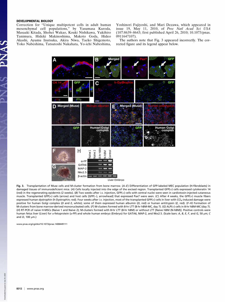
Yoshinori Fujiyoshi, and Mari Dezawa, which appeared inissue 19, May 11, 2010, of Proc Natl Acad Sci USA(107:8639–8643; first published April 26, 2010; 10.1073/pnas.0911647107).The authors note that Fig. 3 appeared incorrectly. The cor-
rected figure and its legend appear below.
www.pnas.org/cgi/doi/10.1073/pnas.1408449111
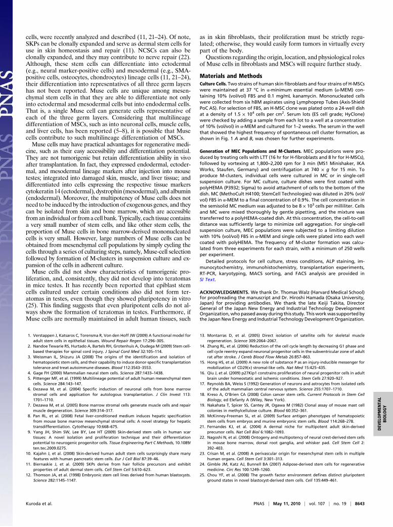
Fig. 3. Transplantation of Muse cells and M-cluster formation from bone marrow. (A–E) Differentiation of GFP-labeled MEC population (H-fibroblasts) indamaged tissues of immunodeficient mice. (A) Cells locally injected into the edge of the excised region. Transplanted GFP(+) cells expressed cytokeratin 14(red) in the regenerating epidermis (2 weeks). (B) Two weeks after i.v. injection, GFP(+) cells with central nuclei were seen in cardiotoxin-injected cutaneousmuscle. Transplanted GFP(+) cells (arrow) and host cells [GFP(−), arrowhead] that expressed Pax7 were seen. (C) After 4 weeks, the GFP(+) muscle fibersexpressed human dystrophin (h-Dystrophin; red). Four weeks after i.v. injection, most of the transplanted GFP(+) cells in liver with CCl4-induced damage werepositive for human Golgi complex (D and E, white); some of them expressed human albumin (D, red) or human antitrypsin (E, red). (F–H) Formation ofM-clusters from bone marrow-derived mononucleated cells. (F) M-clusters formed with 8-hr LTT (8-hr hBM-MC, day 7). (G) ALP(+) cells in 8-hr hBM-MC (day 7).(H) RT-PCR of naive H-MSCs (Naive 1 and Naive 2); M-clusters formed with 8-hr LTT (8-hr hBM) or without LTT [Naive hBM (N-hBM)]. Positive controls werehuman fetus liver (Liver) for α-fetoprotein (α-FP) and whole human embryo (Embryo) for GATA6, MAP-2, and Nkx2.5. (Scale bars: A, B, E, F, and G, 50 μm; Cand D, 100 μm.)
8312 | www.pnas.org
Dow
nloa
ded
by g
uest
on
July
26,
202
0
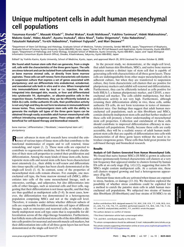
Unique multipotent cells in adult human mesenchymalcell populationsYasumasa Kurodaa,1, Masaaki Kitadaa,1, Shohei Wakaoa, Kouki Nishikawab, Yukihiro Tanimuraa, Hideki Makinoshimaa,Makoto Godac, Hideo Akashia, Ayumu Inutsukab, Akira Niwad, Taeko Shigemotoa, Yoko Nabeshimae,Tatsutoshi Nakahatad, Yo-ichi Nabeshimae, Yoshinori Fujiyoshib, and Mari Dezawaa,2
aDepartment of Stem Cell Biology and Histology, Graduate School of Medicine, Tohoku University, Sendai 980-8575, Japan; bDepartment of Biophysics,Graduate School of Science, Kyoto University, Kyoto 606-8502, Kyoto, Japan; dCenter for iPS Cell Research and Application, Kyoto University, Kyoto 606-8507,Japan; eDepartment of Pathology and Tumor Biology, Graduate School of Medicine, Kyoto University, Kyoto 606-8501, Japan; and cJapan BiologicalInformatics Consortium (Kyoto Branch Office), Oiwake, Kitashirakawa, Sakyo-ku, Kyoto 606-8502, Japan
Edited* by Yoshito Kaziro, Kyoto University, School of Medicine, Kyoto, Japan, and approved March 29, 2010 (received for review October 8, 2009)
We found adult human stem cells that can generate, from a singlecell, cells with the characteristics of the three germ layers. The cellsare stress-tolerant and canbe isolated fromcultured skinfibroblastsor bone marrow stromal cells, or directly from bone marrowaspirates. These cells can self-renew; form characteristic cell clustersin suspension culture that express a set of genes associated withpluripotency; and can differentiate into endodermal, ectodermal,and mesodermal cells both in vitro and in vivo. When transplantedinto immunodeficient mice by local or i.v. injection, the cellsintegrated into damaged skin, muscle, or liver and differentiatedinto cytokeratin 14-, dystrophin-, or albumin-positive cells in therespective tissues. Furthermore, they can be efficiently isolated asSSEA-3(+) cells. Unlike authentic ES cells, their proliferation activityis not very high and theydonot form teratomas in immunodeficientmouse testes. Thus, nontumorigenic stem cells with the ability togenerate the multiple cell types of the three germ layers can beobtained through easily accessible adult human mesenchymal cellswithout introducing exogenous genes. These unique cells will bebeneficial for cell-based therapy and biomedical research.
bone marrow | differentiation | fibroblasts | mesenchymal stem cell |pluripotency
Recent advances in stem cell research have revealed the exis-tence of various types of tissue stem cells that contribute to the
functional maintenance of organs and to cell renewal, tissueremodeling, and repair (1, 2). These stem cells are expected tocontribute to regenerative medicine, but this will require elucida-tion of their stem cell properties to control their proliferation anddifferentiation. Among the many kinds of tissue stem cells, hema-topoietic stem cells and neural stem cells have been characterizedmost extensively (i.e., their ability to self-renew and differentiateinto tissue-specific cell types has been clearly demonstrated at thesingle-cell level) (3, 4). In contrast, some of the properties ofmesenchymal stem cells remain obscure. For example, one mes-enchymal cell type, the bone marrow stromal cell (MSC), differ-entiates into cells of the same mesenchymal lineage, such asosteocytes, cartilage, and adipocytes, but also differentiates intocells of other lineages, such as neuronal cells and liver cells, sug-gesting that their differentiation is not tissue-specific, and that theyare thus qualified as multipotent cells (5–8). In most cases, how-ever, the differentiation was demonstrated in a heterogeneouspopulation comprising MSCs and not at the single-cell level.Therefore, it remains under debate whether different subsets ofcells are responsible for differentiation into cell types of differentlineages, such as osteocytes and neuronal cells, or whether a dis-tinctly multipotent stem cell type exists that is responsible for dif-ferentiation across all the oligo-lineage boundaries. Furthermore,hair follicle stemcells anddermal stemcells of the skindifferentiateinto cells positive for neuronal and smoothmuscle cellmarkers, buttheir differentiation into cells of all three germ layers has not beendemonstrated at the single-cell level (9–11).
In the present study, we demonstrate, at the single-cell level,that adult human skin fibroblasts, MSCs, and native bone marrowaspirates contain a distinct type of stem cell that is capable ofgenerating cells with characteristics of all three germ layers. Thesecells are indistinguishable from other major mesenchymal cells inadherent culture, but when they are transferred to suspensionculture, they form characteristic cell clusters that are positive forpluripotency markers and exhibit self-renewal and differentiation.Furthermore, they can be efficiently isolated as cells positive forboth SSEA-3, a human pluripotency marker, and CD105, a mes-enchymal cell marker. The cells exhibit multipotency, but theirproliferation activity is not very high. Furthermore, althoughretaining their differentiation ability in vivo, these cells, unlikeauthentic ES cells, do not form teratomas in testes of immuno-deficient mice. Our findings thus suggest that adult human mes-enchymal cell populations, such as skin fibroblasts and MSCs,contain distinctly multipotent stem cells and that further studies ofthese cells will promote a better understanding of mesenchymalstem cell properties. Collection and enrichment of these cellsshould contribute to improved differentiation efficiency in mes-enchymal cell populations. Finally, because these cells are easilyaccessible, they will be a realistic source of adult human multi-potent stem cells that are capable of differentiation into cells withcharacteristics of all three germ layers without the need to in-troduce exogenous genes. These cells thus hold great promise forcell-based therapy and biomedical research.
ResultsAnalysis of Cell Clusters Generated from Human Mesenchymal Cells.We found that naive human MSCs (H-MSCs) grown in adherentculture spontaneously formed characteristic cell clusters at a verylow frequency that appeared similar to clusters formed by humanES cells at an early stage (Fig. 1A) (12), suggesting that naive H-MSCs might contain multipotent cells. At a certain size, thesecell clusters stopped growing and had a heterogeneous appear-ance (Fig.1B).Dormant tissue stem cells are activated when tissues are exposed
to stress, burdens, or damage (13–16). We therefore explored thepossibility of whether stress conditions could be exploited fora method to enrich the putative stem cells in adult human mes-enchymal cell populations. We subjected two strains of humanskin fibroblasts (H-fibroblasts) and four strains of H-MSCs to six
Author contributions: M.D. designed research; Y.K., M.K., S.W., K.N., Y.T., H.M., M.G., H.A.,A.I., A.N., T.S., Y.N., T.N., Y.F., and M.D. performed research; Y.K., M.K., S.W., T.N., Y.-i.N.,Y.F., and M.D. analyzed data; and M.K., Y.F., and M.D. wrote the paper.
The authors declare no conflict of interest.
*This Direct Submission article had a prearranged editor.1Y.K. and M.K. contributed equally to this work.2To whom correspondence should be addressed. E-mail: [email protected].
This article contains supporting information online at www.pnas.org/lookup/suppl/doi:10.1073/pnas.0911647107/-/DCSupplemental.
www.pnas.org/cgi/doi/10.1073/pnas.0911647107 PNAS | May 11, 2010 | vol. 107 | no. 19 | 8639–8643
DEV
ELOPM
ENTA
LBIOLO
GY

different stress conditions, including long-term trypsin incubation(LTT) for 8 or 16 hr (Table S1 and SI Materials and Methods).Stem cells are often grown in suspension culture, which is an ef-
ficient and convenientmethod tomaintain their stemcell properties(17, 18). H-fibroblasts or H-MSCs that survived the stress treat-ments were therefore suspended in methylcellulose (MC) medium(19) at a density of 8,000 cells permilliliter (MCculture;SIMaterialsand Methods) and grown for 7 days (Fig. 1C). Each condition gaverise to cell clusters with sizes of up to 50–150 μm in diameter (Fig.1D). Using different filters, we separated the cell clusters accordingto their size and characterized themby immunocytochemistry.Mostof the clusters with a diameter larger than 25 μm contained cellspositive for the pluripotency markers Nanog, Oct3/4, SSEA-3,PAR-4, and Sox2 and were positive for alkaline phosphatase (ALP)staining (Fig. S1). We therefore only counted cell clusters largerthan25μm.Among the stress conditions tested, 16-hrLTTwasmostpotent in the formation of cell clusters in H-fibroblasts, and 8-hr
LTT was most potent in the formation of cell clusters in H-MSCs(Table S1 and SI Results). As expected, the formed clusters con-tained cells positive for the above pluripotencymarkers (Fig. 1E–J)and ALP staining (Fig. 1 K–M). We called these cells multilineagedifferentiating stress enduring (Muse) cells because they expresspluripotency markers; as described below, they differentiate intoectodermal, endodermal, and mesodermal cells; and they endurethrough stress conditions. We refer to H-fibroblasts and H-MSCstreated with 16-hr and 8-hr LTT, respectively, as “Muse-enrichedcell populations” (MEC populations).To calculate the frequency of Muse-cell-derived cell cluster
(M-cluster; SI Results) formation accurately, MEC populationsderived from both H-fibroblasts and H-MSCs were subjected tosingle-cell suspension culture after limiting dilution (Fig. S2 andSI Materials and Methods), showing that 11.6 ± 1.6% of the cellsin the H-fibroblast–MEC population and 8.1 ± 0.2% of the cellsin the H-MSC–MEC population proceeded to form M-clustersafter 7 days. Naive populations (without LTT) were also exam-ined and showed that 1.3 ± 0.1% (H-fibroblasts) and 1.1 ± 0.1%(H-MSCs) of the cells formed M-clusters in single-cell suspen-sion culture after limiting dilution.
Self-Renewal and Expansion of Muse Cells. After LTT, Muse cellsbegan to divide after 1–2 days in MC culture and continued todivide at a rate of ≈1.3 days per cell division until day 8, formingcell clusters. Cell proliferation gradually slowed by days 11–12and ceased around day 14, with cell clusters reaching a maximumsize of 150 μm (Fig. S3 and SI Materials and Methods).When M-clusters formed in single-cell suspension culture after
limiting dilution (day 12) were dissociated into single cells by a 5-mintrypsin treatment and returned to single-cell suspension culture, thecells survived but divided very slowly (5–7 days per cell division) orsometimes not at all (Fig. 1N1). However, transfer of single M-clusters to adherent culture reinitiated cell proliferation and pro-duced expanded cells. When cultures that had expanded to ≈3,000–5,000 cells were dissociated and subjected to single-cell suspensionculture without LTT, 48.0 ± 5.8% (H-fibroblasts) and 40.3 ± 9.1%(H-MSCs) of the cells formedM-clusters (Fig. 1N2). When cultureswereallowed toexpand to5–10×104 cells andwere then subjected toLTT to produce MEC populations (Fig. 1N3), 12.3 ± 1.3% (H-fibroblasts) and 8.5 ± 0.5% (H-MSCs) of these cells formed secondgeneration M-clusters. We repeated this culture cycle, consisting ofLTT→ suspension culture→ adherent culture, five times, and everycell generation showed similar behavior and a similar frequency ofM-cluster formation.We also confirmed that the fifth generationM-clusters were still positive for pluripotency markers and ALP stain-ing. Furthermore, karyotypes of cells expanded fromM-clusters didnot show detectable abnormalities (Fig. S4 and SI Results). In con-clusion, the proliferation activity of Muse cells is not very high, andproliferation stops in suspension culture when the cell clusters reacha defined size. Nevertheless, the proliferation of Muse cells can bereinitiated by transfer to adherent culture, which is followed by theformationof next-generationM-clusters, demonstrating the capacityof Muse cells for self-renewal and proliferation.
Differentiation of M-Clusters. Toanalyze their differentiation ability,single M-clusters formed in single-cell suspension culture afterlimiting dilution were transferred onto gelatin-coated dishes. After7 days of culture, immunocytochemistry revealed cells positive forneurofilament-M [an ectodermal marker; the ratio of positivecells was 3.5 ± 0.5% (H-fibroblasts) and 3.7 ± 0.6% (H-MSCs)],α-smooth muscle actin [α-SMA; mesodermal, 12.2 ± 1.8% (H-fibroblasts) and 8.0± 0.6%(H-MSCs)], α-fetoprotein [endodermal,2.7±0.1%(H-fibroblasts) and 3.2±0.3%(H-MSCs)], cytokeratin 7[endodermal, 5.5 ± 0.1% (H-fibroblasts) and 3.4 ± 0.6% (H-MSCs)], or desmin [mesodermal, 14.2 ± 0.4% (H-fibroblasts) and10.1± 0.5% (H-MSCs)] (Fig. 2A–E). RT-PCRof cells derived fromfirst- and third-generation M-clusters confirmed that these cells
Fig. 1. Characterization of M-clusters. (A and B) Characteristic cell clustersthat occur spontaneously in adherent cultures of naive H-MSCs. (C and D) MCculture of H-fibroblasts on day 7 showing an M-cluster (C, arrow). Immu-nocytochemical localization of Nanog (E and F), Oct3/4 (G), Sox2 (H), PAR4(I), and SSEA-3 (J) in M-clusters formed by H-fibroblasts (E, I, and J) and H-MSCs (F, G, and H). ALP(+) human ES cells (K), M-cluster (H-fibroblast) (L), andnaive H-fibroblasts (M). (N) Schematic diagram of the self-renewal of Musecells. (Scale bars: A–C, 100 μm; D–M, 50 μm.)
8640 | www.pnas.org/cgi/doi/10.1073/pnas.0911647107 Kuroda et al.
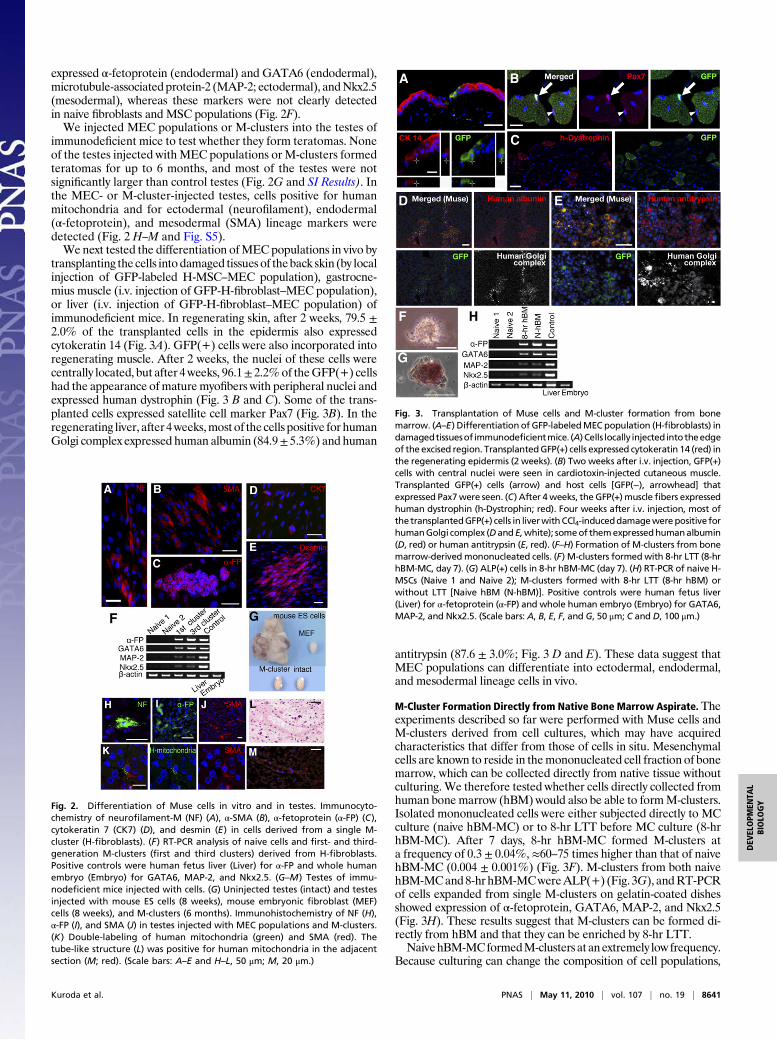
expressed α-fetoprotein (endodermal) and GATA6 (endodermal),microtubule-associated protein-2 (MAP-2; ectodermal), andNkx2.5(mesodermal), whereas these markers were not clearly detectedin naive fibroblasts and MSC populations (Fig. 2F).We injected MEC populations or M-clusters into the testes of
immunodeficient mice to test whether they form teratomas. Noneof the testes injected withMEC populations orM-clusters formedteratomas for up to 6 months, and most of the testes were notsignificantly larger than control testes (Fig. 2G and SI Results). Inthe MEC- or M-cluster-injected testes, cells positive for humanmitochondria and for ectodermal (neurofilament), endodermal(α-fetoprotein), and mesodermal (SMA) lineage markers weredetected (Fig. 2 H–M and Fig. S5).We next tested the differentiation ofMEC populations in vivo by
transplanting the cells intodamaged tissues of theback skin (by localinjection of GFP-labeled H-MSC–MEC population), gastrocne-mius muscle (i.v. injection of GFP-H-fibroblast–MEC population),or liver (i.v. injection of GFP-H-fibroblast–MEC population) ofimmunodeficient mice. In regenerating skin, after 2 weeks, 79.5 ±2.0% of the transplanted cells in the epidermis also expressedcytokeratin 14 (Fig. 3A). GFP(+) cells were also incorporated intoregenerating muscle. After 2 weeks, the nuclei of these cells werecentrally located, but after 4weeks, 96.1± 2.2%of theGFP(+) cellshad the appearance ofmaturemyofibers with peripheral nuclei andexpressed human dystrophin (Fig. 3 B and C). Some of the trans-planted cells expressed satellite cell marker Pax7 (Fig. 3B). In theregenerating liver, after 4weeks,most of thecells positive for humanGolgi complex expressed human albumin (84.9± 5.3%) and human
antitrypsin (87.6 ± 3.0%; Fig. 3 D and E). These data suggest thatMEC populations can differentiate into ectodermal, endodermal,and mesodermal lineage cells in vivo.
M-Cluster Formation Directly from Native Bone Marrow Aspirate. Theexperiments described so far were performed with Muse cells andM-clusters derived from cell cultures, which may have acquiredcharacteristics that differ from those of cells in situ. Mesenchymalcells are known to reside in themononucleated cell fraction of bonemarrow, which can be collected directly from native tissue withoutculturing.We therefore tested whether cells directly collected fromhuman bone marrow (hBM) would also be able to formM-clusters.Isolated mononucleated cells were either subjected directly to MCculture (naive hBM-MC) or to 8-hr LTT before MC culture (8-hrhBM-MC). After 7 days, 8-hr hBM-MC formed M-clusters ata frequency of 0.3 ± 0.04%, ≈60–75 times higher than that of naivehBM-MC (0.004 ± 0.001%) (Fig. 3F). M-clusters from both naivehBM-MCand8-hrhBM-MCwereALP(+) (Fig. 3G), andRT-PCRof cells expanded from single M-clusters on gelatin-coated dishesshowed expression of α-fetoprotein, GATA6, MAP-2, and Nkx2.5(Fig. 3H). These results suggest that M-clusters can be formed di-rectly from hBM and that they can be enriched by 8-hr LTT.NaivehBM-MCformedM-clusters at anextremely low frequency.
Because culturing can change the composition of cell populations,
Fig. 2. Differentiation of Muse cells in vitro and in testes. Immunocyto-chemistry of neurofilament-M (NF) (A), α-SMA (B), α-fetoprotein (α-FP) (C),cytokeratin 7 (CK7) (D), and desmin (E) in cells derived from a single M-cluster (H-fibroblasts). (F) RT-PCR analysis of naive cells and first- and third-generation M-clusters (first and third clusters) derived from H-fibroblasts.Positive controls were human fetus liver (Liver) for α-FP and whole humanembryo (Embryo) for GATA6, MAP-2, and Nkx2.5. (G–M) Testes of immu-nodeficient mice injected with cells. (G) Uninjected testes (intact) and testesinjected with mouse ES cells (8 weeks), mouse embryonic fibroblast (MEF)cells (8 weeks), and M-clusters (6 months). Immunohistochemistry of NF (H),α-FP (I), and SMA (J) in testes injected with MEC populations and M-clusters.(K) Double-labeling of human mitochondria (green) and SMA (red). Thetube-like structure (L) was positive for human mitochondria in the adjacentsection (M; red). (Scale bars: A–E and H–L, 50 μm; M, 20 μm.)
Fig. 3. Transplantation of Muse cells and M-cluster formation from bonemarrow. (A–E) Differentiation of GFP-labeledMEC population (H-fibroblasts) indamagedtissuesof immunodeficientmice. (A) Cells locally injected intotheedgeof the excised region. TransplantedGFP(+) cells expressed cytokeratin 14 (red) inthe regenerating epidermis (2 weeks). (B) Two weeks after i.v. injection, GFP(+)cells with central nuclei were seen in cardiotoxin-injected cutaneous muscle.Transplanted GFP(+) cells (arrow) and host cells [GFP(−), arrowhead] thatexpressed Pax7were seen. (C) After 4weeks, the GFP(+)musclefibers expressedhuman dystrophin (h-Dystrophin; red). Four weeks after i.v. injection, most ofthe transplantedGFP(+) cells in liverwithCCl4-induceddamagewerepositive forhumanGolgi complex (Dand E,white); someof themexpressedhumanalbumin(D, red) or human antitrypsin (E, red). (F–H) Formation of M-clusters from bonemarrow-derived mononucleated cells. (F) M-clusters formedwith 8-hr LTT (8-hrhBM-MC, day 7). (G) ALP(+) cells in 8-hr hBM-MC (day 7). (H) RT-PCR of naive H-MSCs (Naive 1 and Naive 2); M-clusters formed with 8-hr LTT (8-hr hBM) orwithout LTT [Naive hBM (N-hBM)]. Positive controls were human fetus liver(Liver) for α-fetoprotein (α-FP) and whole human embryo (Embryo) for GATA6,MAP-2, and Nkx2.5. (Scale bars: A, B, E, F, and G, 50 μm; C and D, 100 μm.)
Kuroda et al. PNAS | May 11, 2010 | vol. 107 | no. 19 | 8641
DEV
ELOPM
ENTA
LBIOLO
GY
cells in stable culture may have a different propensity to form M-clusters than cells fromnative tissues. To test this possibility, we grewhBM aspirate in adherent culture to collect primary MSCs andsubjected the cells directly to MC culture without 8-hr LTT. Thisprotocol resulted in amuch higher frequency ofM-cluster formationof 0.3 ± 0.08%. When primary MSCs were further cultured to thesecond and fifth passages, the frequency of M-cluster formationwithout LTT increased up to 0.5 ± 0.04% and 0.9 ± 0.1%, re-spectively. Consistent with this finding, 1.3 ± 0.1% of naive H-fibroblasts and 1.1 ± 0.1% of naive H-MSCs formed M-clusters insingle-cell suspension culture as described above. These resultssuggest thatMuse cellshave ahigh stress tolerance and canendure invitro culture and the subculture procedures.Bone marrow contains many cell types, including MSCs, he-
matopoietic lineage cells, and endothelial cells (19). To determinewhich fraction contains theMuse cells, we isolatedmononucleatedcells from hBM aspirate and subjected them to magnetic affinitycell sorting (MACS) using antibodies against CD34 and CD117[markers for hematopoietic cells (20)] and CD105 [marker forMSCs (5, 7)].We then subjected the cells to 8-hr LTT and allowedthem to grow inMC culture for 7 days. The CD34+/117+/105− cellfraction produced few M-clusters, but the CD34−/117−/105+
fraction produced 50 timesmoreM-clusters than theCD34−/117−/105− fraction (SI Results). This result suggests that the majority ofMuse cells belong to the CD105(+)mesenchymal cell population.
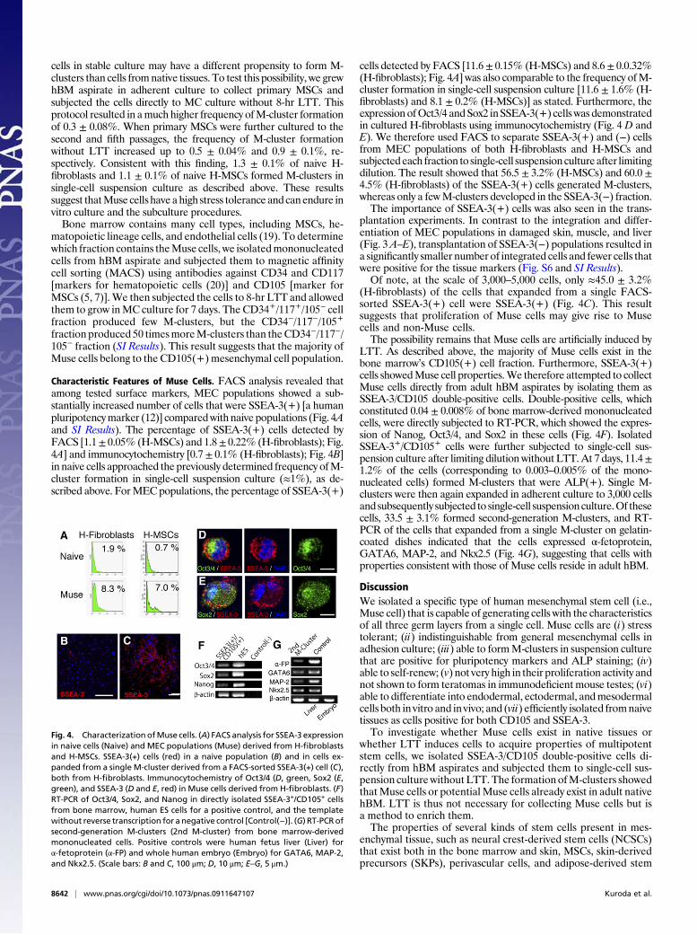
Characteristic Features of Muse Cells. FACS analysis revealed thatamong tested surface markers, MEC populations showed a sub-stantially increased number of cells that were SSEA-3(+) [a humanpluripotencymarker (12)] comparedwith naive populations (Fig. 4Aand SI Results). The percentage of SSEA-3(+) cells detected byFACS [1.1± 0.05% (H-MSCs) and 1.8± 0.22% (H-fibroblasts); Fig.4A] and immunocytochemistry [0.7 ± 0.1% (H-fibroblasts); Fig. 4B]in naive cells approached the previously determined frequency ofM-cluster formation in single-cell suspension culture (≈1%), as de-scribed above. ForMECpopulations, the percentage of SSEA-3(+)
cells detected by FACS [11.6± 0.15% (H-MSCs) and 8.6± 0.0.32%(H-fibroblasts); Fig. 4A] was also comparable to the frequency ofM-cluster formation in single-cell suspension culture [11.6 ± 1.6% (H-fibroblasts) and 8.1 ± 0.2% (H-MSCs)] as stated. Furthermore, theexpression ofOct3/4 and Sox2 inSSEA-3(+) cells was demonstratedin cultured H-fibroblasts using immunocytochemistry (Fig. 4 D andE). We therefore used FACS to separate SSEA-3(+) and (−) cellsfrom MEC populations of both H-fibroblasts and H-MSCs andsubjectedeach fraction to single-cell suspension cultureafter limitingdilution. The result showed that 56.5 ± 3.2% (H-MSCs) and 60.0 ±4.5% (H-fibroblasts) of the SSEA-3(+) cells generated M-clusters,whereas only a fewM-clusters developed in the SSEA-3(−) fraction.The importance of SSEA-3(+) cells was also seen in the trans-
plantation experiments. In contrast to the integration and differ-entiation of MEC populations in damaged skin, muscle, and liver(Fig. 3A–E), transplantation of SSEA-3(−) populations resulted ina significantly smaller number of integrated cells and fewer cells thatwere positive for the tissue markers (Fig. S6 and SI Results).Of note, at the scale of 3,000–5,000 cells, only ≈45.0 ± 3.2%
(H-fibroblasts) of the cells that expanded from a single FACS-sorted SSEA-3(+) cell were SSEA-3(+) (Fig. 4C). This resultsuggests that proliferation of Muse cells may give rise to Musecells and non-Muse cells.The possibility remains that Muse cells are artificially induced by
LTT. As described above, the majority of Muse cells exist in thebone marrow’s CD105(+) cell fraction. Furthermore, SSEA-3(+)cells showedMuse cell properties.We therefore attempted to collectMuse cells directly from adult hBM aspirates by isolating them asSSEA-3/CD105 double-positive cells. Double-positive cells, whichconstituted 0.04 ± 0.008% of bone marrow-derived mononucleatedcells, were directly subjected to RT-PCR, which showed the expres-sion of Nanog, Oct3/4, and Sox2 in these cells (Fig. 4F). IsolatedSSEA-3+/CD105+ cells were further subjected to single-cell sus-pension culture after limiting dilution without LTT. At 7 days, 11.4±1.2% of the cells (corresponding to 0.003–0.005% of the mono-nucleated cells) formed M-clusters that were ALP(+). Single M-clusters were then again expanded in adherent culture to 3,000 cellsand subsequently subjected to single-cell suspension culture.Of thesecells, 33.5 ± 3.1% formed second-generation M-clusters, and RT-PCR of the cells that expanded from a single M-cluster on gelatin-coated dishes indicated that the cells expressed α-fetoprotein,GATA6, MAP-2, and Nkx2.5 (Fig. 4G), suggesting that cells withproperties consistent with those of Muse cells reside in adult hBM.
DiscussionWe isolated a specific type of human mesenchymal stem cell (i.e.,Muse cell) that is capable of generating cells with the characteristicsof all three germ layers from a single cell. Muse cells are (i) stresstolerant; (ii) indistinguishable from general mesenchymal cells inadhesion culture; (iii) able to formM-clusters in suspension culturethat are positive for pluripotency markers and ALP staining; (iv)able to self-renew; (v) not very high in their proliferation activity andnot shown to form teratomas in immunodeficientmouse testes; (vi)able to differentiate into endodermal, ectodermal, andmesodermalcells both in vitro and in vivo; and (vii) efficiently isolated fromnaivetissues as cells positive for both CD105 and SSEA-3.To investigate whether Muse cells exist in native tissues or
whether LTT induces cells to acquire properties of multipotentstem cells, we isolated SSEA-3/CD105 double-positive cells di-rectly from hBM aspirates and subjected them to single-cell sus-pension culturewithout LTT.The formation ofM-clusters showedthatMuse cells or potential Muse cells already exist in adult nativehBM. LTT is thus not necessary for collecting Muse cells but isa method to enrich them.The properties of several kinds of stem cells present in mes-
enchymal tissue, such as neural crest-derived stem cells (NCSCs)that exist both in the bone marrow and skin, MSCs, skin-derivedprecursors (SKPs), perivascular cells, and adipose-derived stem
Fig. 4. Characterization ofMuse cells. (A) FACS analysis for SSEA-3 expressionin naive cells (Naive) and MEC populations (Muse) derived from H-fibroblastsand H-MSCs. SSEA-3(+) cells (red) in a naive population (B) and in cells ex-panded from a single M-cluster derived from a FACS-sorted SSEA-3(+) cell (C),both from H-fibroblasts. Immunocytochemistry of Oct3/4 (D, green, Sox2 (E,green), and SSEA-3 (D and E, red) in Muse cells derived from H-fibroblasts. (F)RT-PCR of Oct3/4, Sox2, and Nanog in directly isolated SSEA-3+/CD105+ cellsfrom bone marrow, human ES cells for a positive control, and the templatewithout reverse transcription for a negative control [Control(−)]. (G) RT-PCRofsecond-generation M-clusters (2nd M-cluster) from bone marrow-derivedmononucleated cells. Positive controls were human fetus liver (Liver) forα-fetoprotein (α-FP) and whole human embryo (Embryo) for GATA6, MAP-2,and Nkx2.5. (Scale bars: B and C, 100 μm; D, 10 μm; E–G, 5 μm.)
8642 | www.pnas.org/cgi/doi/10.1073/pnas.0911647107 Kuroda et al.
cells, were recently analyzed and described (11, 21–24). Of note,SKPs can be clonally expanded and serve as dermal stem cells foruse in skin homeostasis and repair (11). NCSCs can also beclonally expanded, and they may contribute to nerve repair (22).Although, these stem cells can differentiate into ectodermal(e.g., neural marker-positive cells) and mesodermal (e.g., SMA-positive cells, osteocytes, chondrocytes) lineage cells (11, 21–24),their differentiation into representatives of all three germ layershas not been reported. Muse cells are unique among mesen-chymal stem cells in that they are able to differentiate not onlyinto ectodermal and mesodermal cells but into endodermal cells.That is, a single Muse cell can generate cells representative ofeach of the three germ layers. Considering that multilineagedifferentiation of MSCs, such as into neuronal cells, muscle cells,and liver cells, has been reported (5–8), it is possible that Musecells contribute to such multilineage differentiation of MSCs.Muse cells may have practical advantages for regenerative medi-
cine, such as their easy accessibility and differentiation potential.They are not tumorigenic but retain differentiation ability in vivoafter transplantation. In fact, they expressed endodermal, ectoder-mal, and mesodermal lineage markers after injection into mousetestes; integrated into damaged skin, muscle, and liver tissue; anddifferentiated into cells expressing the respective tissue markerscytokeratin 14 (ectodermal), dystrophin (mesodermal), and albumin(endodermal). Moreover, the multipotency of Muse cells does notneed to be induced by the introduction of exogenous genes, and theycan be isolated from skin and bone marrow, which are accessiblefroman individual or fromacell bank.Typically, each tissue containsa very small number of stem cells, and like other stem cells, theproportion of Muse cells in bone marrow-derived mononucleatedcells is very small. However, large numbers of Muse cells can beobtained from mesenchymal cell populations by simply cycling thecells through a series of culturing steps, namely, Muse-cell selectionfollowed by formation of M-clusters in suspension culture and ex-pansion of the cells in adherent culture.Muse cells did not show characteristics of tumorigenic pro-
liferation, and, consistently, they did not develop into teratomasin mice testes. It has recently been reported that epiblast stemcells cultured under certain conditions also did not form ter-atomas in testes, even though they showed pluripotency in vitro(25). This finding suggests that even pluripotent cells do not al-ways show the formation of teratomas in testes. Furthermore, ifMuse cells are normally maintained in adult human tissues, such
as in skin fibroblasts, their proliferation must be strictly regu-lated; otherwise, they would easily form tumors in virtually everypart of the body.Questions regarding the origin, location, and physiological roles
of Muse cells in fibroblasts and MSCs will require further study.
Materials and MethodsCulture Cells. Two strains of human skin fibroblasts and four strains of H-MSCswere maintained at 37 °C in α-minimum essential medium (α-MEM) con-taining 10% (vol/vol) FBS and 0.1 mg/mL kanamycin. Mononucleated cellswere collected from six hBM aspirates using Lymphoprep Tubes (Axis-ShieldPoC AS). For selection of FBS, an H-MSC clone was plated onto a 24-well dishat a density of 1.5 × 10⁴ cells per cm2. Serum lots (ES cell grade; HyClone)were checked by adding a sample from each lot to a well at a concentrationof 10% (vol/vol) in α-MEM and cultured for 1–2 weeks. The serum in the wellthat showed the highest frequency of spontaneous cell cluster formation, asshown in Fig. 1 A and B, was chosen for further experiments.
Generation of MEC Populations and M-Clusters. MEC populations were pro-duced by treating cells with LTT (16 hr for H-fibroblasts and 8 hr for H-MSCs),followed by vortexing at 1,800–2,200 rpm for 3 min (MS1 Minishaker, IKAWorks, Staufen, Germany) and centrifugation at 740 × g for 15 min. Toproduce M-clusters, individual cells were cultured in MC or in single-cellsuspension culture. For MC culture, culture dishes were first coated withpolyHEMA (P3932; Sigma) to avoid attachment of cells to the bottom of thedish. MC (MethoCult H4100; StemCell Technologies) was diluted in 20% (vol/vol) FBS in α-MEM to a final concentration of 0.9%. The cell concentration inthe semisolid MC medium was adjusted to be 8 × 10³ cells per milliliter. Cellsand MC were mixed thoroughly by gentle pipetting, and the mixture wastransferred to a polyHEMA-coated dish. At this concentration, the cell-to-celldistance was sufficiently large to minimize cell aggregation. For single-cellsuspension culture, MEC populations were subjected to a limiting dilutionwith 10% (vol/vol) FBS in α-MEM and single cells were plated into each wellcoated with polyHEMA. The frequency of M-cluster formation was calcu-lated from three experiments for each strain, with a minimum of 250 wellsper experiment.
Detailed protocols for cell culture, stress conditions, ALP staining, im-munocytochemistry, immunohistochemistry, transplantation experiments,RT-PCR, karyotyping, MACS sorting, and FACS analysis are provided inSI Text.
ACKNOWLEDGMENTS. We thank Dr. Thomas Walz (Harvard Medical School)for proofreading the manuscript and Dr. Hiroshi Hamada (Osaka University,Japan) for providing antibodies. We thank the late Keiji Takita, DirectorGeneral of the Japan New Energy and Industrial Technology DevelopmentOrganization,whopassed away during this study. Thisworkwas supported bythe Japan New Energy and Industrial Technology Development Organization.
1. Verstappen J, Katsaros C, Torensma R, Von den Hoff JW (2009) A functional model foradult stem cells in epithelial tissues. Wound Repair Regen 17:296–305.
2. Nandoe Tewarie RS, Hurtado A, Bartels RH, Grotenhuis A, Oudega M (2009) Stem cell-based therapies for spinal cord injury. J Spinal Cord Med 32:105–114.
3. Weissman IL, Shizuru JA (2008) The origins of the identification and isolation ofhematopoietic stem cells, and their capability to induce donor-specific transplantationtolerance and treat autoimmune diseases. Blood 112:3543–3553.
4. Gage FH (2000) Mammalian neural stem cells. Science 287:1433–1438.5. Pittenger MF, et al. (1999) Multilineage potential of adult human mesenchymal stem
cells. Science 284:143–147.6. Dezawa M, et al. (2004) Specific induction of neuronal cells from bone marrow
stromal cells and application for autologous transplantation. J Clin Invest 113:1701–1710.
7. Dezawa M, et al. (2005) Bone marrow stromal cells generate muscle cells and repairmuscle degeneration. Science 309:314–317.
8. Pan RL, et al. (2008) Fetal liver-conditioned medium induces hepatic specificationfrom mouse bone marrow mesenchymal stromal cells: A novel strategy for hepatictransdifferentiation. Cytotherapy 10:668–675.
9. Yang JH, Shim SW, Lee BY, Lee HT (2009) Skin-derived stem cells in human scartissues: A novel isolation and proliferation technique and their differentiationpotential to neurogenic progenitor cells. Tissue Engineering Part C Methods, 10.1089/ten.tec.2009.0275.
10. Kajahn J, et al. (2008) Skin-derived human adult stem cells surprisingly share manyfeatures with human pancreatic stem cells. Eur J Cell Biol 87:39–46.
11. Biernaskie J, et al. (2009) SKPs derive from hair follicle precursors and exhibitproperties of adult dermal stem cells. Cell Stem Cell 5:610–623.
12. Thomson JA, et al. (1998) Embryonic stem cell lines derived from human blastocysts.Science 282:1145–1147.
13. Montarras D, et al. (2005) Direct isolation of satellite cells for skeletal muscleregeneration. Science 309:2064–2067.
14. Zhang RL, et al. (2006) Reduction of the cell cycle length by decreasing G1 phase andcell cycle reentry expand neuronal progenitor cells in the subventricular zone of adultrat after stroke. J Cereb Blood Flow Metab 26:857–863.
15. Hong HS, et al. (2009) A new role of substance P as an injury-inducible messenger formobilization of CD29(+) stromal-like cells. Nat Med 15:425–435.
16. Qiu J, et al. (2009) p27Kip1 constrains proliferation of neural progenitor cells in adultbrain under homeostatic and ischemic conditions. Stem Cells 27:920–927.
17. Reynolds BA, Weiss S (1992) Generation of neurons and astrocytes from isolated cellsof the adult mammalian central nervous system. Science 255:1707–1710.
18. Kreso A, O’Brien CA (2008) Colon cancer stem cells. Current Protocols in Stem CellBiology, ed Elefanty A (Wiley, New York).
19. Nakahata T, Spicer SS, Cantey JR, Ogawa M (1982) Clonal assay of mouse mast cellcolonies in methylcellulose culture. Blood 60:352–361.
20. McKinney-Freeman SL, et al. (2009) Surface antigen phenotypes of hematopoieticstem cells from embryos and murine embryonic stem cells. Blood 114:268–278.
21. Fernandes KJ, et al. (2004) A dermal niche for multipotent adult skin-derivedprecursor cells. Nat Cell Biol 6:1082–1093.
22. Nagoshi N, et al. (2008) Ontogeny and multipotency of neural crest-derived stem cellsin mouse bone marrow, dorsal root ganglia, and whisker pad. Cell Stem Cell 2:392–403.
23. Crisan M, et al. (2008) A perivascular origin for mesenchymal stem cells in multiplehuman organs. Cell Stem Cell 3:301–313.
24. Gimble JM, Katz AJ, Bunnell BA (2007) Adipose-derived stem cells for regenerativemedicine. Circ Res 100:1249–1260.
25. Chou YF, et al. (2008) The growth factor environment defines distinct pluripotentground states in novel blastocyst-derived stem cells. Cell 135:449–461.
Kuroda et al. PNAS | May 11, 2010 | vol. 107 | no. 19 | 8643
DEV
ELOPM
ENTA
LBIOLO
GY