unique phenotype in a patient with charge syndrome
TRANSCRIPT

CASE REPORT Open Access
Unique phenotype in a patient with CHARGEsyndromeShobhit Jain1, Hyung-Goo Kim2, Felicitas Lacbawan3, Irene Meliciani4, Wolfgang Wenzel4, Ingo Kurth5,Josefina Sharma6, Morris Schoeneman6, Svetlana Ten1, Lawrence C Layman2 and Elka Jacobson-Dickman1*
Abstract
CHARGE is a phenotypically heterogeneous autosomal dominant disorder recognized as a cohesive syndrome since theidentification of CHD7 as a genetic etiology. Classic features include: Coloboma, Heart defects, Atresia choanae,Retarded growth and development, Genitourinary abnormalities, and Ear anomalies and/or deafness. With greateraccessibility to genetic analysis, a wider spectrum of features are emerging, and overlap with disorders such asDiGeorge syndrome, Kallmann syndrome, and Hypoparathyroidism Sensorineural Deafness and Renal Diseasesyndrome, is increasingly evident. We present a patient with a unique manifestation of CHARGE syndrome, includingprimary hypoparathyroidism and a limb anomaly; to our knowledge, he is also the first CHARGE subject reported withbilateral multicystic dysplastic kidneys. Furthermore, with structural modeling and murine expression studies, wecharacterize a putative CHD7 G744S missense mutation. Our report continues to expand the CHARGE phenotype andhighlights that stringent fulfillment of conventional criteria should not strictly guide genetic analysis.
IntroductionThe CHARGE syndrome (MIM 214800) is an autosomaldominant or sporadic disorder of variable multisystemiccongenital anomalies that occurs with an incidence ofapproximately 1 in 10,000 [1,2]. Heterozygous CHD7(chromodomain helicase DNA-binding protein 7, MIM608892) mutations have been identified in approximately60%-70% of patients with clinically diagnosed CHARGESyndrome and are most commonly due to de novo trun-cating mutations. Furthermore, CHD7 mutations arereported throughout the entire coding sequence of thegene without an apparent pattern or cluster, and mean-ingful genotype-phenotype correlations have not beenrecognized [1-3].The first descriptions of this syndrome were provided by
Hall and Hitner independently in 1979 [4,5], though it wasin 1981 that Pagan and colleagues coined the acronymCHARGE to summarize its dominant features: coloboma,heart defects, atresia choanae, retarded growth and devel-opment, genital and/or urinary abnormalities, ear anoma-lies and/or deafness [6]. It was only in 2004 that the CHD7
gene was established as a genetic etiology for CHARGEsyndrome by Vissers et al [7]. A wider spectrum of asso-ciated features has since emerged, albeit less consistently,and includes hyposmia or anosmia, cleft lip and palate,hypocalcemia [8,9], and tracheoesophageal fistula [10]. Assuch, CHARGE Syndrome has several overlapping clinicalcharacteristics with DiGeorge syndrome [11], Kallmannsyndrome, and Hypoparathyroidism, Sensorineural Deaf-ness, and Renal Disease (HDR) (Barakat’s syndrome)[12,13]. As the CHARGE phenotype continues to expand,particularly into the clinical purview of other conditions, itsdiagnosis becomes more challenging as well as increasinglyinclusive.Herein, we report a patient with a unique presentation
of CHARGE syndrome, including primary hypoparathyr-oidism, bilateral multicystic dysplastic kidneys (MCDK),and an atypical limb anomaly; he carried a CHD7 muta-tion that has not been previously characterized. Our reportexpands the spectrum of phenotypes associated withCHD7 mutations.
Clinical ReportThe proband, an 18 year old African American male, pre-sented to us initially for management of refractory hypocal-cemia. He was born weighing 795 grams at 25 weeksgestation to a 31-year old woman with severe hypertension,
* Correspondence: [email protected] University of New York Downstate Medical Center, Children’s Hospitalat Downstate, Department of Pediatrics, Division of Pediatric Endocrinology,Brooklyn, NY 11203 USAFull list of author information is available at the end of the article
Jain et al. International Journal of Pediatric Endocrinology 2011, 2011:11http://www.ijpeonline.com/content/2011/1/11
© 2011 Jain et al; licensee BioMed Central Ltd. This is an Open Access article distributed under the terms of the Creative CommonsAttribution License (http://creativecommons.org/licenses/by/2.0), which permits unrestricted use, distribution, and reproduction inany medium, provided the original work is properly cited.

who died in the postpartum period of a myocardial infarc-tion. In infancy, he underwent cardiac surgery for a ventri-cular septal defect, and another to correct a right eyelidcoloboma. Additionally, he had congenital hypothyroidism,bilateral sensorineural hearing loss, and severe global devel-opmental delay; he began walking during his third year oflife, remains unable to independently dress or tie shoelaces, and he has a vocabulary of fewer than 5 words. At ayoung age, the family had been told that he had Down syn-drome, a diagnosis he carried until our meeting. Our initialconsultation for hypocalcemia was during a hospitalizationfor complications of end stage renal disease secondary toMCDK, which was diagnosed in early life based on X-raycomputed tomography (CT) showing dysplastic kidneyswith multiple cysts. Interestingly, both a full sister and
maternal half sister had severe hearing deficits and renaldisease (Figure 1A). Unfortunately, neither was availablefor further characterization.His physical examination revealed an overweight and
short young man (weight 65.9kg, height 150.5cm or -3.58SDS, body mass index 29.3 kg/m2), communicating pri-marily with hand gestures including pointing. He hadbrachycephaly with a flat facial profile, short foreheadand facial asymmetry, slightly upslanted eyes with mildright ptosis, a scar from eyelid coloboma corrective sur-gery, and mildly prominent ears with low nasal bridge,upturned nasal tip, and smooth philtrum (Figure 1B.Iand 1B.II). He had no cleft lip or palate. The chest wassymmetrical. He had scars from cardiac surgery on theleft anterior and posterior walls, and on the anterior
Figure 1 A patient with a unique presentation of CHARGE syndrome and a G744S CHD7 mutation. A. Pedigree of a CHARGE patient witha novel CHD7 mutation. circle: female; square: male; arrow: proband; +: wild type allele. B. Photographs of the facies (I), eyes (II), and hands (III) ofthe CHD7 G744S heterozygous proband. C. Evolutionary conservation of the residue Gly744. ClustalW multiple alignment of partial proteinsequence of CHD7 orthologs. The position of residue G744 altered by one heterozygous nucleotide change is marked by arrow and red lettersin the corresponding segment of the multiple alignment. The amino acid residues that differ from the sequence of the human CHD7 protein areindicated blue. Gly744 residue is evolutionarily fully conserved in all fifteen available CHD7 orthologs. D. Structural model of the amino acidregions spanning 651-794 of the CHD7 protein obtained by sequence homology to a bacterial flagellar filament. The site of the mutation(indicated in magenta), which is predicted to be detrimental by POLYPHEN, lies on a protein interaction surface as indicated by SSPIDER.
Jain et al. International Journal of Pediatric Endocrinology 2011, 2011:11http://www.ijpeonline.com/content/2011/1/11
Page 2 of 8

abdominal wall at the site of a neonatal gastric tube pla-cement. On examination of the extremities, his palmarcreases were normal and he had a flexion deformity ofthe right thumb whereby this digit was fixed in theadducted position (Figure 1B.III). His genital examinationrevealed Tanner V pubic hair, and bilaterally descendedtestes consistent in size with early to mid-pubertal range(5 mL right and 8 mL left, using Prader Orchidometer).A CT scan of the head showed bilateral basal ganglia
calcifications and scattered calcification in the frontalwhite matter and cerebellum, likely related to chronichypcalcemia. Olfactory structures could not be evalu-ated. X-ray of his hands revealed no osseous abnormal-ities to explain his right thumb deformity.The initial laboratory results included low serum cal-
cium (6.3 mg/dL, normal 8.3-10 mg/dL) normal albumin(4.2 g/dL, normal 3.5-5.8 g/dL), low phosphorus (1.1 mg/dL, normal 2.5-5 mg/dL), normal 25-hydroxy vitamin D(38 ng/mL, normal 20-100 ng/mL), slightly low magne-sium (1.6 mg/dL, normal 1.9-2.7 mg/dL) coincident withlow parathyroid hormone level (4.9 pg/mL, normal 15-65pg/mL), indicating primary hypoparathyroidism. His lowphosphorus was due to phosphate wasting associated withpolyuria of end stage renal disease (ESRD). He had normalcomplete blood cell counts with no evidence of white cellline depression. Additionally, despite having early to mid-pubertal sized testes, his LH, FSH and Testosterone werenot in the hypogonadal range (LH 4.1 mIU/mL, FSH 6.8mIU/mL, Testosterone 505 ng/dL), which is not consistentwith Idiopathic Hypogonadotropic Hypogonadism.In view of his dysmorphic features and questionable his-
tory of Down Syndrome (MIM 190685), a karyotype wasobtained which showed a normal 46, XY configuration in200 stimulated peripheral lymphocytes. Fluorescent in situhybridization (FISH) for a chromosome 22q11.2 deletion,associated with DiGeorge Syndrome (MIM 188400), wasnegative. To exclude a Calcium-Sensing Receptor defect(MIM 601199), sequencing of the CASR gene was con-ducted of the coding regions and exon/intron boundariesand did not reveal a mutation. In light of the patient’s con-stellation of hypoparathyroidism, renal disease, as well asdeafness, we performed microarray and then sequencinganalyses for mutations in GATA3 gene associated with theHDR syndrome (MIM 146255) [13], and none were found.Finally, CHD7 mutation analysis was performed on agenomic sample after PCR amplification of exons 2 to 38.The primers for all exons flanked the respective intron-exon junctions and direct sequencing was performed inboth forward and backward directions using automatedfluorescence di-deoxy sequencing methods. This revealed6 heterozygous unclassified variants in the CHD7 geneincluding 2 missense and 4 silent changes (Table 1).Among the missense changes, a G744S (NP_060250)resulting from a c.2230G®A nucleotide change (NM-
017780), was found to be conserved in available CHD7orthologs (Figure 1C) and was not identified in 192 con-trol subjects. The subject’s asymptomatic father, the onlyfamily member available for genetic analysis, was foundnot to carry the G744S substitution. An informed consentfor all genetic testing and for image publication wasobtained from father and from the subject’s legal guardian.
Structural Modeling of the CHD7 ProteinThe G744S mutation, which is located near chromodo-main 1 in exon 4, was hypothesized to possess functionalimplications in the pathogenesis of CHARGE Syndrome.We constructed a structural model for the portion of theCHD7 protein containing the mutation based on homol-ogy to a bacterial flagellar filament (pdb-code 1UCU) [14](See Figure 1D and additional file 1 for methods and align-ment). Based on this model, SPPIDER [15] identified thesite of the mutation as a possible binding site. G744 islocated at the protein surface, where mutations are mostlikely to affect protein function and signaling compared tomutations in the protein interior. In agreement with thisobservation, the bioinformatics based approach POLY-PHEN predicted a possible damaging effect of the muta-tion with a PISC SCORE: 1.58 (values below 0.5 areconsidered benign and values above 1.0 possibly/probablydamaging) [16]. The PISC score contains a sequencebased estimate of the accessibility of the mutation site,which is underestimated in comparison with the predic-tions of the structural model. Therefore, the sequencebased PSIC score likely underestimates the damagingeffect of our mutation.
Chd7 Murine Expression StudiesThroughout murine development Chd7 is broadlyexpressed, including organs classically anomalous inCHARGE Syndrome, such as the eyes, heart, and ears.To investigate the distribution of Chd7 transcripts inthe developing limbs, we performed in situ hybridizationanalysis using whole mount preparations. The following
Table 1 Sequence Analysis Revealed Six UnclassifiedVariants in the CHD7 Gene
Nucleotide ChangeNM_017780
Amion Acid ChangeNP_060250
SNP ID*
Missense changes
c.2230G®A p.G744S -
c.6478G®A p.A2160T rs61753399
Silent Changes
c.309G®A p.S103S rs41272435
c.657C®T p.G219G -
c.2124T®C p.S708S -
c.7590A®G p.K2530K rs61742801
*Reported in the Single Nucleotide Polymorphism database dbSNP:http:www.ncbi.nlm.nih.gov//SNP
Jain et al. International Journal of Pediatric Endocrinology 2011, 2011:11http://www.ijpeonline.com/content/2011/1/11
Page 3 of 8

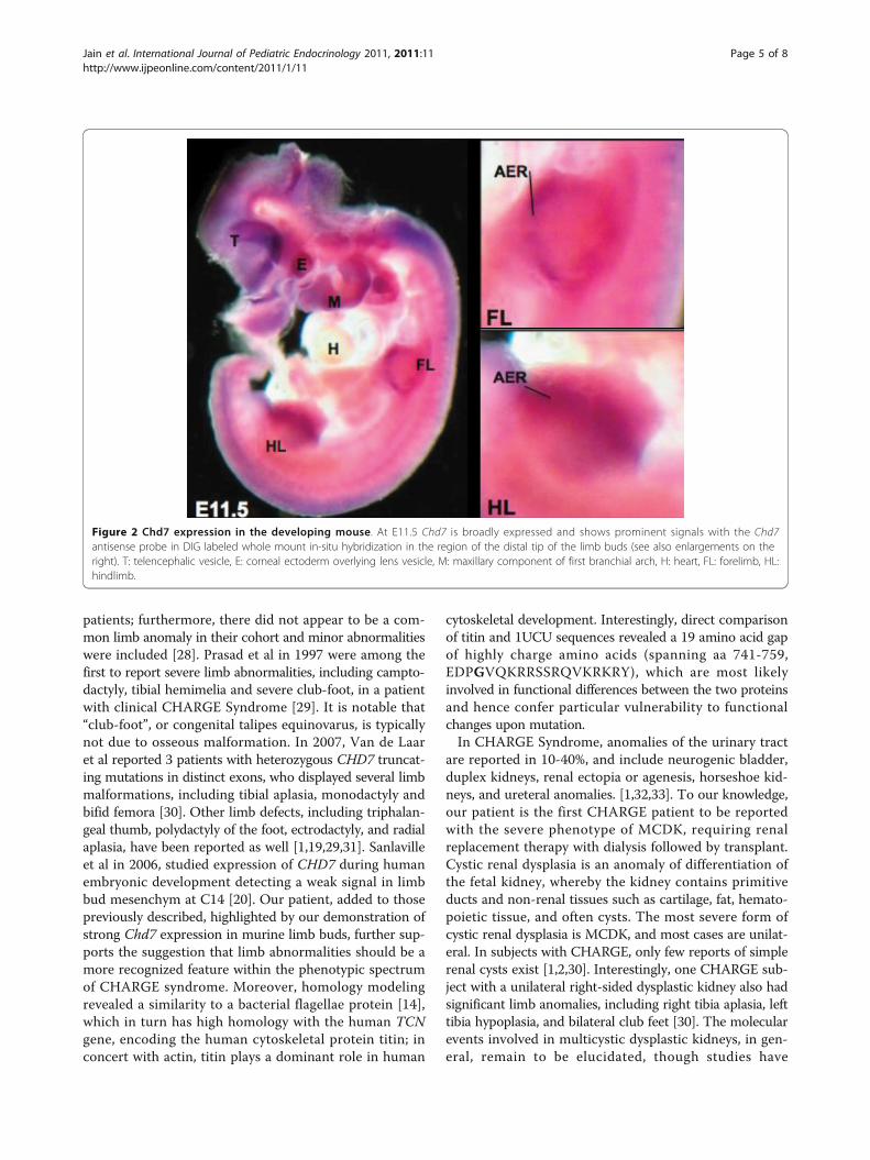
primers were used for amplification of the probe span-ning nucleotides 8305 to 9140 of the murine Chd7 tran-script (NM_001081417): 5′-CAGGTGGCTGGAGGAGAACCC-3′ and 5′-CTTTACAGGGCCCTCCCTCGGCC-3′. Amplicons were ligated to a Topo-TA vector (Invi-trogen) and subcloned into pBluescript via EcoRI andXbaI restriction sites. The probe was labeled withDigoxigenin (DIG) for whole-mount in situ hybridiza-tion following standard procedures. No specific signalswere detected using the respective sense probes. Byembryonic day E11.5, the limb buds are clearly dividedinto proximal and distal elements. By E12, the handplateshowed evidence of angular contours at its peripheral mar-gin, corresponding to the location of the future digits. AtE12.5-E13 early evidence of digital rays that are separatedby the digital interzones were apparent. We found highexpression levels of Chd7 throughout these steps of limbdevelopment in DIG-labelled whole mount embryos. Pro-nounced Chd7 expression was noted at the limb bud api-cal ectodermal ridge (AER) (Figure 2), a thickened layer ofectodermal cells at the distal tip of the developing limbbud, which is a crucial organizing region during limb for-mation. This Chd7 expression pattern supports a role forthis gene in limb development.
DiscussionCHARGE is a phenotypically heterogeneous autosomaldominant disorder that has been recognized as a non-random cohesive syndrome since the identification ofCHD7 mutations as an underlying etiology [1,2,7].CHD7, at 8q12.1, encodes a protein of the chromodo-main family [17,18]. The exact function of CHD7 has notbeen elucidated, however, in situ hybridization analysisduring human development has demonstrated expressionof this gene in the central nervous system, semicircularcanals and the neural crest of the pharyngeal arches;thereby implicating the embryologic role of CHD7 in thedevelopment of the respective organs [19,20]. Further-more, CHD7 mRNA expression was documented in thehypothalamus, pituitary and olfactory bulb in the rat andalso demonstrated in both migratory and post-migratoryGnRH neuronal cell lines [21]. Our subject carried a het-erozygous G744S, not seen in controls; this missensechange has been reported in one previous series anddesignated a polymorphism, though no functional studieswere conducted and controls were not tested [22]. In thisinstance, the G744S change was seen in a family withthree children with clinical CHARGE born to two differ-ent women and one man. A larger CHD7 rearrangementwas additionally seen in all affected children, and foundin mosaic form in the father’s sperm cell DNA but not inlymphocytes. The G744S change, however, was found intwo of the CHARGE children (the third was not tested)and in the completely unaffected father. Since the father
was heterozygous and not mosaic for G744S, the authorsconsidered G744S to be a non-pathogenic variant (Kohl-hase J, unpublished data). CHD7 mutations have clini-cally variable expression, and clear genotype-phenotypecorrelations are not observed, even among patients withidentical CHD7 mutations [1-3]. Asymptomatic carriersare reported as well, particularly in inherited forms [3].We can speculate that the disparate putative effect of thismutation is subject to yet undefined secondary genetic,epigenetic or environmental influences; this has beendemonstrated in other genetic disease models, such asidiopathic hypogonadotropic hypogonadism (IHH) andKallmann syndrome [23-25]. Our bioinformatics basedstructural analysis, protein alignment, and DNA sequen-cing in normal controls, provide supportive evidence thatthe G744S is a true deleterious mutation involving ahighly conserved amino acid, likely disrupting a crucialprotein interaction site.In this age of sophisticated and readily available genetic
analysis, the diagnosis of CHARGE appears to be trans-forming from the rigid fulfillment of conglomerates ofspecific major and minor criteria, towards an approachmore inclusive of patients with atypical or attenuatedphenotypes, as was demonstrated by Kim et al [26].CHARGE and Kallmann syndromes (KS, KAL5, MIM612370), though distinct developmental disorders, werenoted to share features of impaired olfaction and hypogo-nadism; thus CHD7 was hypothesized to be involved inthe pathogenesis of KS even in the absence of theCHARGE phenotype. Among 197 patients, they identifiedseven heterozygous mutations (two splice and five mis-sense, absent in ≥ 180 controls) in three sporadic KS andfour sporadic normosmic IHH patients. Thus, sporadicCHD7 mutations occurred in 6% of IHH/KS patientsstudied, allowing them to conclude that IHH/KS canrepresent a milder allelic variant of CHARGE syndrome.Furthermore, Jongmans et al also identified de novoCHD7 mutations in 3 of 56 mixed KS and nIHH sub-jects. Interestingly, in retrospect, their IHH patients withCHD7 mutations had some CHARGE features, includingcolobomas, deafness, ear anomalies, cleft palate and shortstature; however, not to the degree that would fulfill tra-ditional criteria [27]. Supported by this rational, we canconcluded that our subject, with an atypical eyelid colo-boma, hearing loss, severe developmental delay, ventricu-lar septal defect, short stature, and abnormal facies, inaddition to other more recently described features, suchas a limb anomaly, primary hypoparathyroidism andinterrupted pubertal development, may be included inthis designation.CHARGE syndrome was initially not considered to
involve the limb. Several subsequent reports have shownassociated limb anomalies and one series reported at leastone limb anomaly in over one third of 172 CHARGE
Jain et al. International Journal of Pediatric Endocrinology 2011, 2011:11http://www.ijpeonline.com/content/2011/1/11
Page 4 of 8
patients; furthermore, there did not appear to be a com-mon limb anomaly in their cohort and minor abnormalitieswere included [28]. Prasad et al in 1997 were among thefirst to report severe limb abnormalities, including campto-dactyly, tibial hemimelia and severe club-foot, in a patientwith clinical CHARGE Syndrome [29]. It is notable that“club-foot”, or congenital talipes equinovarus, is typicallynot due to osseous malformation. In 2007, Van de Laaret al reported 3 patients with heterozygous CHD7 truncat-ing mutations in distinct exons, who displayed several limbmalformations, including tibial aplasia, monodactyly andbifid femora [30]. Other limb defects, including triphalan-geal thumb, polydactyly of the foot, ectrodactyly, and radialaplasia, have been reported as well [1,19,29,31]. Sanlavilleet al in 2006, studied expression of CHD7 during humanembryonic development detecting a weak signal in limbbud mesenchym at C14 [20]. Our patient, added to thosepreviously described, highlighted by our demonstration ofstrong Chd7 expression in murine limb buds, further sup-ports the suggestion that limb abnormalities should be amore recognized feature within the phenotypic spectrumof CHARGE syndrome. Moreover, homology modelingrevealed a similarity to a bacterial flagellae protein [14],which in turn has high homology with the human TCNgene, encoding the human cytoskeletal protein titin; inconcert with actin, titin plays a dominant role in human
cytoskeletal development. Interestingly, direct comparisonof titin and 1UCU sequences revealed a 19 amino acid gapof highly charge amino acids (spanning aa 741-759,EDPGVQKRRSSRQVKRKRY), which are most likelyinvolved in functional differences between the two proteinsand hence confer particular vulnerability to functionalchanges upon mutation.In CHARGE Syndrome, anomalies of the urinary tract
are reported in 10-40%, and include neurogenic bladder,duplex kidneys, renal ectopia or agenesis, horseshoe kid-neys, and ureteral anomalies. [1,32,33]. To our knowledge,our patient is the first CHARGE patient to be reportedwith the severe phenotype of MCDK, requiring renalreplacement therapy with dialysis followed by transplant.Cystic renal dysplasia is an anomaly of differentiation ofthe fetal kidney, whereby the kidney contains primitiveducts and non-renal tissues such as cartilage, fat, hemato-poietic tissue, and often cysts. The most severe form ofcystic renal dysplasia is MCDK, and most cases are unilat-eral. In subjects with CHARGE, only few reports of simplerenal cysts exist [1,2,30]. Interestingly, one CHARGE sub-ject with a unilateral right-sided dysplastic kidney also hadsignificant limb anomalies, including right tibia aplasia, lefttibia hypoplasia, and bilateral club feet [30]. The molecularevents involved in multicystic dysplastic kidneys, in gen-eral, remain to be elucidated, though studies have
Figure 2 Chd7 expression in the developing mouse. At E11.5 Chd7 is broadly expressed and shows prominent signals with the Chd7antisense probe in DIG labeled whole mount in-situ hybridization in the region of the distal tip of the limb buds (see also enlargements on theright). T: telencephalic vesicle, E: corneal ectoderm overlying lens vesicle, M: maxillary component of first branchial arch, H: heart, FL: forelimb, HL:hindlimb.
Jain et al. International Journal of Pediatric Endocrinology 2011, 2011:11http://www.ijpeonline.com/content/2011/1/11
Page 5 of 8

suggested involvement of WNT-1 [34], FGFR3 [35], andPAX2 [36]. The PAX2 gene is associated with the renalcoloboma syndrome (MIM 120330), a syndrome charac-terized by renal hypoplasia and insufficiency, vesicoure-teric reflux, and optic disc coloboma. Interestingly,multicystic dysplastic kidney has been reported in onefamily with Renal Coloboma Syndrome [36]. In a study ofthe distribution pattern of the PAX2 gene in humanembryos, Tellier et al demonstrated that PAX2 geneexpression occurs in the primordia affected with CHARGEsyndrome. Therefore, PAX2 was further analyzed in 34patients fulfilling the diagnostic criteria of the CHARGEsyndrome, though no deletions or nucleotide variations ofthe coding sequence were detected, suggesting that muta-tions of the PAX2 gene was not a cause of the CHARGE[37]. Considering the embryonic expression of PAX2reported, and the common clinical features of Renal Colo-boma Syndrome with CHARGE, one can hypothesize thatCHD7 may have a role in regulating PAX2 gene and there-fore this overlapping pathway might be explored inCHARGE etiology, and perhaps contributes to the variableexpression observed.The significant clinical overlap and inherently variable
features of CHARGE and DiGeorge Syndromes can makedifferentiating these initial diagnoses particularly challen-ging. Hypocalcemia has been reported in CHARGE,though hypoparathyroidism, specifically, has been impli-cated in only few cases [38,39]. A study comparing 25CHARGE subjects with CHD7 mutations to a large cohortof subjects with 22q11.2 deletion syndrome, noted thatfeatures found more commonly in CHARGE syndromeincluded coloboma, choanal atresia, facial nerve palsy, tra-cheoesophageal fistula, and genital hypoplasia in boys.Interestingly, a high incidence of marked hypocalcemiawas observed in their CHARGE study group (72%), and apronounced spectrum of cell-mediated immunodeficiencyranging from lymphopenia (60%) to severe combinedimmunodeficiency (8%), was seen as well. Defects inhumoral immunity were documented in 4 CHARGEpatients and included severe hypogammaglobulinemiawith decreased T-cell numbers, transient hypogammaglo-bulinemia during infancy, and immunoglobulin A defi-ciency [40]. An accurate distinction between these twoentities can, therefore, be challenging but will influencegenetic counseling; CHD7 mutations more typically occursporadically, whereas 22q11.2 deletions are familial in 10%of cases [2,41].
ConclusionIn summary, we report an 18 year old male with CHARGEsyndrome and a unique phenotype, including primaryhypoparathyroidism, bilateral MCDK, a limb anomaly, dis-rupted testicular growth, and an atypical eyelid coloboma,who harbored a heterozygous G744S CHD7 mutation.
Our case emphasizes that CHARGE features are perhapseven more heterogeneous than previously described andshould include limb anomalies more universally. Addition-ally, the stringent fulfillment of the conventional CHARGEcriteria should not strictly guide genetic analysis. Further-more, our report highlights that the clinical overlap ofCHARGE with DiGeorge, HDR, and Kallmann Syndromescan pose a diagnostic challenge to the clinician, but thecorrect designation can have a critical impact on treat-ment, anticipatory guidance, and genetic counseling.
Additional material
Additional file 1: Additional file 1includes further elaboration ofchd7 protein modeling and alignement methods, as well as onefigure of the protein sequence alignement.
AcknowledgementsWe would like to acknowledge support of the German Academic ExchangeService (DAAD) and the Baden-Wurttemberg Stiftung GmbH, for thebioinformatic analysis.
Author details1State University of New York Downstate Medical Center, Children’s Hospitalat Downstate, Department of Pediatrics, Division of Pediatric Endocrinology,Brooklyn, NY 11203 USA. 2Institute of Molecular Medicine and Genetics, TheMedical College of Georgia, Section of Reproductive Endocrinology,Infertility, and Genetics, Department of Obstetrics and Gynecology, Augusta,GA 30912, USA. 3State University of New York Downstate Medical Center,Department of Pathology, Division of Molecular Pathology, Brooklyn, NY11203, USA. 4Institute of Nanotechnology, Karlsruhe Institute of Technology,PO Box 3640. 76021 Karlsruhe, Germany. 5Institute of Human Genetics,University Hospital Jena Kollegiengasse 1007743 Jena, Germany. 6StateUniversity of New York Downstate Medical Center, Children’s Hospital atDownstate, Department of Pediatrics, Division of Pediatric Nephrology,Brooklyn, NY 11203 USA.
Authors’ contributionsSJ lead and participated in the phenotyping and genotyping of our probandand in the characterization of the pedigree members; he also contributed towriting the manuscript. HK and LL lead the characterization of the CHD7G744S mutation. HK also contributed to writing this manuscript. IM and WWperformed the structural modeling of the CHD7 Protein. FL and ST guidedthe phenotyping and genotyping of the proband. IK conducted the Chd7murine expression studies. JS and MS conducted phenotyping of theproband’s renal pathology. EJD conceived of this study, oversaw thephenotyping and genotyping of the proband and his family, and was thesupervising writer of this manuscript. All authors read and approved the finalmanuscript.
Competing interestsThe authors declare that they have no competing interests.
Received: 5 October 2011 Accepted: 13 October 2011Published: 13 October 2011
References1. Jongmans MC, Admiraal RJ, van der Donk KP, Vissers LE, Baas AF, Kapusta L,
van Hagen JM, Donnai D, de Ravel TJ, Veltman JA, Geurts van Kessel A, DeVries BB, Brunner HG, Hoefsloot LH, van Ravenswaaij CM: CHARGEsyndrome: the phenotypic spectrum of mutations in the CHD7 gene.Journal of medical genetics 2006, 43(4):306-14.
2. Lalani SR, Safiullah AM, Fernbach SD, Harutyunyan KG, Thaller C,Peterson LE, McPherson JD, Gibbs RA, White LD, Hefner M, Davenport SL,
Jain et al. International Journal of Pediatric Endocrinology 2011, 2011:11http://www.ijpeonline.com/content/2011/1/11
Page 6 of 8
Graham JM, Bacino CA, Glass NL, Towbin JA, Craigen WJ, Neish SR, Lin AE,Belmont JW: Spectrum of CHD7 mutations in 110 individuals withCHARGE syndrome and genotype-phenotype correlation. Americanjournal of human genetics 2006, 78(2):303-14.
3. Jongmans MC, Hoefsloot LH, van der Donk KP, Admiraal RJ, Magee A, vande Laar I, Hendriks Y, Verheij JB, Walpole I, Brunner HG, vanRavenswaaij CM: Familial CHARGE syndrome and the CHD7 gene: arecurrent missense mutation, intrafamilial recurrence and variability.American journal of medical genetics 2008, 146A(1):43-50.
4. Hall BD: Choanal atresia and associated multiple anomalies. The Journalof pediatrics 1979, 95(3):395-8.
5. Hittner HM, Hirsch NJ, Kreh GM, Rudolph AJ: Colobomatousmicrophthalmia, heart disease, hearing loss, and mental retardation–asyndrome. Journal of pediatric ophthalmology and strabismus 1979,16(2):122-8.
6. Pagon RA, Graham JM, Zonana J, Yong SL: Coloboma, congenital heartdisease, and choanal atresia with multiple anomalies: CHARGEassociation. The Journal of pediatrics 1981, 99(2):223-7.
7. Vissers LE, van Ravenswaaij CM, Admiraal R, Hurst JA, de Vries BB,Janssen IM, van der Vliet WA, Huys EH, de Jong PJ, Hamel BC,Schoenmakers EF, Brunner HG, Veltman JA, van Kessel AG: Mutations in anew member of the chromodomain gene family cause CHARGEsyndrome. Nature genetics 2004, 36(9):955-7.
8. Sanka M, Tangsinmankong N, Loscalzo M, Sleasman JW, Dorsey MJ:Complete DiGeorge syndrome associated with CHD7 mutation. TheJournal of allergy and clinical immunology 2007, 120(4):952-4.
9. Wincent J, Holmberg E, Stromland K, Soller M, Mirzaei L, Djureinovic T,Robinson K, Anderlid B, Schoumans J: CHD7 mutation spectrum in 28Swedish patients diagnosed with CHARGE syndrome. Clinical genetics2008, 74(1):31-8.
10. Zentner GE, Layman WS, Martin DM, Scacheri PC: Molecular andphenotypic aspects of CHD7 mutation in CHARGE syndrome. Americanjournal of medical genetics 2010, 152A(3):674-86.
11. Sullivan KE: Chromosome 22q11.2 deletion syndrome: DiGeorgesyndrome/velocardiofacial Syndrome. Immunology and allergy clinics ofNorth America 2008, 28(2):353-66.
12. Hasegawa T, Hasegawa Y, Aso T, Koto S, Nagai T, Tsuchiya Y, et al: HDRsyndrome (hypoparathyroidism, sensorineural deafness, renal dysplasia)associated with del(10)(p13). American journal of medical genetics 1997,73(4):416-8.
13. Van Esch H, Groenen P, Nesbit MA, Schuffenhauer S, Lichtner P,Vanderlinden G, Harding B, Beetz R, Bilous RW, Holdaway I, Shaw NJ,Fryns JP, Van de Ven W, Thakker RV, Devriendt K: GATA3 haplo-insufficiency causes human HDR syndrome. Nature 2000,406(6794):419-22.
14. Yonekura K, Maki-Yonekura S, Namba K: Complete atomic model of thebacterial flagellar filament by electron cryomicroscopy. Nature 2003,424(6949):643-50.
15. Porollo A, Meller J: Prediction-based fingerprints of protein-proteininteractions. Proteins 2007, 66(3):630-45.
16. Ramensky V, Bork P, Sunyaev S: Human non-synonymous SNPs: serverand survey. Nucleic acids research 2002, 30(17):3894-900.
17. Delmas V, Stokes DG, Perry RP: A mammalian DNA-binding protein thatcontains a chromodomain and an SNF2/SWI2-like helicase domain.Proceedings of the National Academy of Sciences of the United States ofAmerica 1993, 90(6):2414-8.
18. Woodage T, Basrai MA, Baxevanis AD, Hieter P, Collins FS: Characterizationof the CHD family of proteins. Proceedings of the National Academy ofSciences of the United States of America 1997, 94(21):11472-7.
19. Eissenberg JC: Molecular biology of the chromo domain: an ancientchromatin module comes of age. Gene 2001, 275(1):19-29.
20. Sanlaville D, Etchevers HC, Gonzales M, Martinovic J, Clement-Ziza M,Delezoide AL, Aubry MC, Pelet A, Chemouny S, Cruaud C, Audollent S,Esculpavit C, Goudefroye G, Ozilou C, Fredouille C, Joye N, Morichon-Delvallez N, Dumez Y, Weissenbach J, Munnich A, Amiel J, Encha-Razavi F,Lyonnet S, Vekemans M, Attié-Bitach T: Phenotypic spectrum of CHARGEsyndrome in fetuses with CHD7 truncating mutations correlates withexpression during human development. Journal of medical genetics 2006,43(3):211-17.
21. Layman WS, McEwen DP, Beyer LA, Lalani SR, Fernbach SD, Oh E,Swaroop A, Hegg CC, Raphael Y, Martens JR, Martin DM: Defects in neural
stem cell proliferation and olfaction in Chd7 deficient mice indicate amechanism for hyposmia in human CHARGE syndrome. Humanmolecular genetics 2009, 18(11):1909-23.
22. Vuorela P, Ala-Mello S, Saloranta C, Penttinen M, Poyhonen M, Huoponen K,Borozdin W, Bausch B, Botzenhart EM, Wilhelm C, Kääriäinen H, Kohlhase J:Molecular analysis of the CHD7 gene in CHARGE syndrome:identification of 22 novel mutations and evidence for a low contributionof large CHD7 deletions. Genet Med 2007, 9(10):690-4.
23. Mitchell AL, Dwyer A, Pitteloud N, Quinton R: Genetic basis and variablephenotypic expression of Kallmann syndrome: towards a unifyingtheory. Trends Endocrinol Metab 2011.
24. Sykiotis GP, Plummer L, Hughes VA, Au M, Durrani S, Nayak-Young S,Dwyer AA, Quinton R, Hall JE, Gusella JF, Seminara SB, Crowley WF Jr,Pitteloud N: Oligogenic basis of isolated gonadotropin-releasinghormone deficiency. Proc Natl Acad Sci USA 2010, 107(34):15140-4.
25. Pitteloud N, Quinton R, Pearce S, Raivio T, Acierno J, Dwyer A, Plummer L,Hughes V, Seminara S, Cheng YZ, Li WP, Maccoll G, Eliseenkova AV,Olsen SK, Ibrahimi OA, Hayes FJ, Boepple P, Hall JE, Bouloux P,Mohammadi M, Crowley W: Digenic mutations account for variablephenotypes in idiopathic hypogonadotropic hypogonadism. J Clin Invest2007, 117(2):457-63.
26. Kim HG, Kurth I, Lan F, Meliciani I, Wenzel W, Eom SH, Kang GB,Rosenberger G, Tekin M, Ozata M, Bick DP, Sherins RJ, Walker SL, Shi Y,Gusella JF, Layman LC: Mutations in CHD7, encoding a chromatin-remodeling protein, cause idiopathic hypogonadotropic hypogonadismand Kallmann syndrome. American journal of human genetics 2008,83(4):511-9.
27. Jongmans MC, van Ravenswaaij-Arts CM, Pitteloud N, Ogata T, Sato N,Claahsen-van der Grinten HL, van der Donk K, Seminara S, Bergman JE,Brunner HG, Crowley WF Jr, Hoefsloot LH: CHD7 mutations in patientsinitially diagnosed with Kallmann syndrome–the clinical overlap withCHARGE syndrome. Clinical genetics 2009, 75(1):65-71.
28. Brock KE, Mathiason MA, Rooney BL, Williams MS: Quantitative analysis oflimb anomalies in CHARGE syndrome: correlation with diagnosis andcharacteristic CHARGE anomalies. American journal of medical genetics2003, 123A(1):111-21.
29. Prasad C, Quackenbush EJ, Whiteman D, Korf B: Limb anomalies inDiGeorge and CHARGE syndromes. American journal of medical genetics1997, 68(2):179-81.
30. Van de Laar I, Dooijes D, Hoefsloot L, Simon M, Hoogeboom J, Devriendt K:Limb anomalies in patients with CHARGE syndrome: an expansion ofthe phenotype. American journal of medical genetics 2007, 143A(22):2712-5.
31. Alazami AM, Alzahrani F, Alkuraya FS: Expanding the “E” in CHARGE.American journal of medical genetics 2008, 146A(14):1890-2.
32. Sanlaville D, Verloes A: CHARGE syndrome: an update. Eur J Hum Genet2007, 15(4):389-99.
33. Ragan DC, Casale AJ, Rink RC, Cain MP, Weaver DD: Genitourinaryanomalies in the CHARGE association. The Journal of urology 1999,161(2):622-5.
34. Arena S, Fazzari C, Scuderi MG, Implatini A, Villari D, Torre S, Arena F, DiBenedetto V: Molecular events involved in the morphogenesis ofmulticystic dysplastic kidney. Urologia internationalis 2010, 85(1):106-11.
35. Prontera P, Sensi A, Pilu G, Baldi M, Baffico M, Bonasoni R, et al: FGFR3mutation in thanatophoric dysplasia type 1 with bilateral cystic renaldysplasia: coincidence or a new association? Genetic counseling (Geneva,Switzerland) 2006, 17(4):407-12.
36. Fletcher J, Hu M, Berman Y, Collins F, Grigg J, McIver M, Jüppner H,Alexander SI: Multicystic dysplastic kidney and variable phenotype in afamily with a novel deletion mutation of PAX2. J Am Soc Nephrol 2005,16(9):2754-61.
37. Tellier AL, Amiel J, Delezoide AL, Audollent S, Auge J, Esnault D, Encha-Razavi F, Munnich A, Lyonnet S, Vekemans M, Attié-Bitach T: Expression ofthe PAX2 gene in human embryos and exclusion in the CHARGEsyndrome. American journal of medical genetics 2000, 93(2):85-8.
38. Sanka M, Tangsinmankong N, Loscalzo M, Sleasman JW, Dorsey MJ:Complete DiGeorge syndrome associated with CHD7 mutation. TheJournal of allergy and clinical immunology 2007, 120(4):952-4.
39. Wincent J, Holmberg E, Stromland K, Soller M, Mirzaei L, Djureinovic T,Robinson K, Anderlid B, Schoumans J: CHD7 mutation spectrum in 28Swedish patients diagnosed with CHARGE syndrome. Clinical genetics2008, 74(1):31-8.
Jain et al. International Journal of Pediatric Endocrinology 2011, 2011:11http://www.ijpeonline.com/content/2011/1/11
Page 7 of 8

40. Jyonouchi S, McDonald-McGinn DM, Bale S, Zackai EH, Sullivan KE: CHARGE(coloboma, heart defect, atresia choanae, retarded growth anddevelopment, genital hypoplasia, ear anomalies/deafness) syndromeand chromosome 22q11.2 deletion syndrome: a comparison ofimmunologic and nonimmunologic phenotypic features. Pediatrics 2009,123(5):e871-7.
41. McDonald-McGinn DM, Tonnesen MK, Laufer-Cahana A, Finucane B,Driscoll DA, Emanuel BS, et al: Phenotype of the 22q11.2 deletion inindividuals identified through an affected relative: cast a wide FISHingnet! Genet Med 2001, 3(1):23-9.
doi:10.1186/1687-9856-2011-11Cite this article as: Jain et al.: Unique phenotype in a patient withCHARGE syndrome. International Journal of Pediatric Endocrinology 20112011:11.
Submit your next manuscript to BioMed Centraland take full advantage of:
• Convenient online submission
• Thorough peer review
• No space constraints or color figure charges
• Immediate publication on acceptance
• Inclusion in PubMed, CAS, Scopus and Google Scholar
• Research which is freely available for redistribution
Submit your manuscript at www.biomedcentral.com/submit
Jain et al. International Journal of Pediatric Endocrinology 2011, 2011:11http://www.ijpeonline.com/content/2011/1/11
Page 8 of 8