unique signatures of natural backg round radiation on ...cricket.biol.sc.edu/papers/natural/premi et...
TRANSCRIPT

Unique Signatures of Natural Background Radiation onHuman Y Chromosomes from Kerala, IndiaSanjay Premi, Jyoti Srivastava, Sebastian Padinjarel Chandy, Sher Ali*
Molecular Genetics Laboratory, National Institute of Immunology, Aruna Asaf Ali Marg, New Delhi, India
Abstract
Background: The most frequently observed major consequences of ionizing radiation are chromosomal lesions and cancers,although the entire genome may be affected. Owing to its haploid status and absence of recombination, the human Ychromosome is an ideal candidate to be assessed for possible genetic alterations induced by ionizing radiation. We studiedthe human Y chromosome in 390 males from the South Indian state of Kerala, where the level of natural backgroundradiation (NBR) is ten-fold higher than the worldwide average, and that from 790 unexposed males as control.
Results: We observed random microdeletions in the Azoospermia factor (AZF) a, b and c regions in .90%, and tandemduplication and copy number polymorphism (CNP) of 11 different Y-linked genes in about 80% of males exposed to NBR.The autosomal homologues of Y-linked CDY genes largely remained unaffected. Multiple polymorphic copies of the Y-linkedgenes showing single Y-specific signals suggested their tandem duplication. Some exposed males showed unilocusduplication of DAZ genes resulting in six copies. Notably, in the AZFa region, approximately 25% of exposed males showeddeletion of the DBY gene, whereas flanking genes USP9Y and UTY remained unaffected. All these alterations were detectedin blood samples but not in the germline (sperm) samples.
Conclusions: Exposure to high levels of NBR correlated with several interstitial polymorphisms of the human Ychromosome. CNPs and enhanced transcription of the SRY gene after duplication are envisaged to compensate for the lossof Y chromosome in some cells. The aforesaid changes, confined to peripheral blood lymphocytes, suggest a possible innatemechanism protecting the germline DNA from the NBR. Genome analysis of a larger population focusing on greaternumbers of genes may provide new insights into the mechanisms and risks of the resultant genetic damages. The presentwork demonstrates unique signatures of NBR on human Y chromosomes from Kerala, India.
Citation: Premi S, Srivastava J, Chandy SP, Ali S (2009) Unique Signatures of Natural Background Radiation on Human Y Chromosomes from Kerala, India. PLoSONE 4(2): e4541. doi:10.1371/journal.pone.0004541
Editor: Simon Melov, Buck Institute for Age Research, United States of America
Received June 23, 2008; Accepted December 9, 2008; Published February 26, 2009
Copyright: ! 2009 Premi et al. This is an open-access article distributed under the terms of the Creative Commons Attribution License, which permitsunrestricted use, distribution, and reproduction in any medium, provided the original author and source are credited.
Funding: We thank Alexander Von Humboldt Foundation, Bonn, Germany for Equipment donation. This work was supported by DBT grant no. BT/PR2225/Med/13/077/2000 and DST grant no. SP/SO/DO3/99) to SA and a core grant from the Department of Biotechnology, Govt. of India to National Institute of Immunology,New Delhi. SP is thankful to the Council of Scientific and Industrial Research (CSIR), New Delhi for the Senior Research Fellowship. This work has been seen andapproved by all the authors and they do not have any conflict of personal communication or financial interests.
Competing Interests: The authors have declared that no competing interests exist.
* E-mail: [email protected]
Introduction
Natural background radiation (NBR) has been affecting allforms of life since the time of its inception, although itsgeographical scope has been varied. Semi-permanent exposureto ionizing radiation leaves a lasting imprint on the genome [1–3].Such change(s) may be used as biomarkers to monitor progressionof tumors, chromosomal lesions, minisatellite length polymor-phisms, and other alterations involving DNA [4]. Attempts havebeen made to establish a correlation between backgroundradiation and phenotypic changes in rats [5], cases of Down’ssyndrome [6], chromosomal aberrations [7], and congenitalmalformations [8]. However, experimental evidence for radia-tion-induced mutations/alterations in humans still remainscontroversial in the absence of sufficient data.The human Y chromosome, with fewer than 50 genes or gene
families coding for proteins, is not essential for life but harborsseveral testis-specific genes [9] necessary for sperm production,and hence continuation of the species. It is well known that at least
three non-overlapping regions of the human Y chromosome –AZFa, AZFb, and AZFc (Azoospermia factors a, b, and c) – areessential for spermatogenesis [10]. Microdeletions in these regionsaffecting one or more of the candidate genes (DAZ, RBMY, DBY,and USP9Y) cause male infertility [11]. Deletions of the DAZ genesin the distal Yq11 (AZFc) region are always associated withazoospermia [12–14]. The human Y chromosome abnormalitieshave also been attributed to the single copy SRY gene located onthe p11.3 region, which plays a predominant role in male sexdetermination [15]. Mutations in the conserved HMG boxdomain and its up/down stream sequences have been correlatedwith sex reversal or poor binding of the SRY protein to the targetDNA [15].Assessment of the effects of long-term experimental irradiation
on humans and its impact across the generations is not possibleowing to logistic and ethical constraints. Only a few studies havebeen carried out following the Chernobyl disaster focusing on theeffect of radiation on minisatellite mutation rate [16] or theradiation effect from nuclear weapon tests on the human germline
PLoS ONE | www.plosone.org 1 February 2009 | Volume 4 | Issue 2 | e4541

mutation rate [3]. In this context, coastal areas in Kerala (SouthIndia), which contain 10% thorium phosphate monazite, offer anatural setting to assess the radiogenomic effects of backgroundradiation. This radioactivity strip measuring an area of about10 km by 1 km supports a sizable population of fishermen [17].Earlier, we reported on the status of DAZ genes in 100 malesexposed to NBR [18]. Triggered by the initial results, weundertook analysis of the Y chromosome in 390 exposed malesand 790 unexposed ones as controls for possible structuralvariations. The results showed exclusive somatic microdeletionsand CNP of the Y chromosome–linked genes.
Results
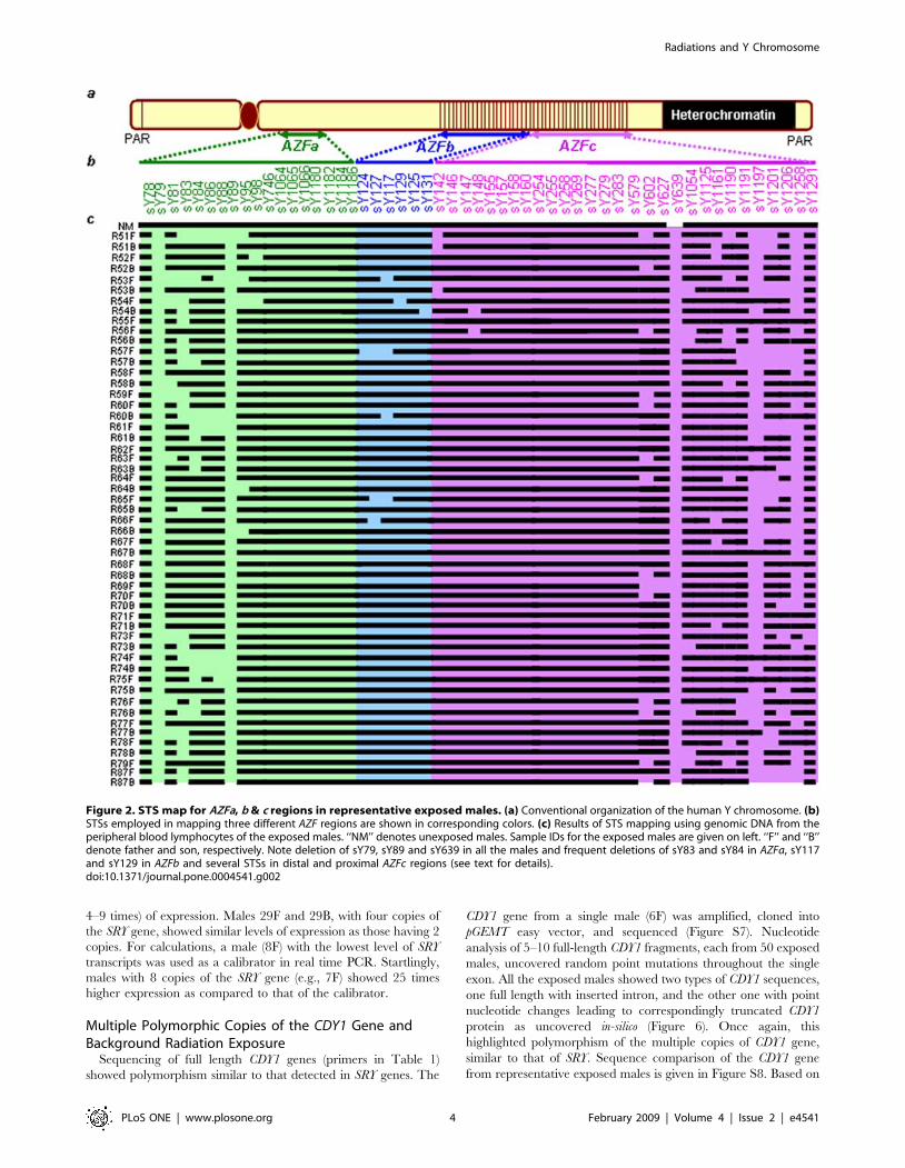
Random Y Chromosome Microdeletions in NBR-ExposedMalesSTS mapping showed randomly scattered microdeletions in the
AZFa, AZFb and AZFc regions of the Y chromosome in theexposed males. Frequency of microdeletions was higher in theAZFc region than in AZFa and AZFb. The AZFc microdeletionswere distributed mainly in the proximal and distal portionsthroughout the sample pool. These microdeletions includedsY1197, sY1258, sY1206 and sY1201 STSs in about 90% ofexposed males (Figures 1 and 2). None of the samples showed thecharacteristic STS profile of gr/gr (sY1291 negative; and sY1161,sY1206, sY1191 and sY1201 all positive) or b1/b3 (sY1161,sY1197, sY1191 and sY1291 all negative; and sY142, sY1258,sY1206 and sY1201 all positive) deletions/duplications, except fora single sample (34B) depicting gr/gr deletion. In the absence of ablood sample from this male, FISH analysis could not beconducted. The AZFa microdeletions were confined to theproximal region encompassing sY79 and sY81 STSs (deletioninterval 5A), sY88 (deletion interval 5D), and sY86 in about 85%of exposed males. Absence of sY117 in some exposed males hintsat the AZFb phenotype, but the presence of sY149, sY127, andother interstitial STSs proved the intactness of the AZFb region.STS mapping of AZFa, b and c regions in representative exposedmales is given in Figure 2.Besides random microdeletions, primers specific to DBY1 and
DBY2 genes did not show any amplification in 95 exposed males(Figure 3). This was confirmed by Southern blot hybridizationusing 32P-labeled PCR product of DBY gene(s) from the normalmale (not shown). Successful PCR amplification with sY83, sY86,sY84, DF1.5, sY87, UTY1, UTY3 and sY88 STSs showedbreakpoints located downstream of sY87 and upstream of UTY3.The DBY microdeletions were inconsistent between fathers andsons where father lacked them but son did not or vice versa. Studieson Human Endogenous Retroviral (HERV) elements also showedrandomly scattered microdeletions without a conclusive provirusA/B mediated recombination (Figure 4). The STSs sY1066,sY1182 and sY1185 that were absent in approximately 50–60% ofexposed males represented hotspots for microdeletions. Approx-imately 10% of AZFc and 4–5% of AZFa/b STSs showedadditional amplicons along with the expected ones (Figure S1)involving sY1201, sY1206, sY83 and sY84 STSs in more than98% of exposed males. Such amplicons were absent in the normalmales and, as expected, in the females, suggesting creation of newprimer binding sites in the exposed males.The intactness of 95% of STSs in germline (sperm) DNA in the
exposed males substantiated the somatic nature of microdeletions,corroborating our earlier study [18]. To rule out possibilities ofPCR reaction failure, single and multiplex PCRs were conductedthrice followed by Southern blot hybridization using [32Pa-dCTP]labeled PCR products of the corresponding STSs of the unexposed
males (not shown). Except for 2% of cases of the normal males thatshowed random microdeletions, all the unexposed control males(390 from Kerala and 400 from other parts of India) were freefrom microdeletions.
Copy Number Polymorphism (CNP) of the Y-LinkedGenes in Exposed MalesReal time PCR using TaqMan/SYBR green chemistries
uncovered duplications/multiplications of the Y-linked genesresulting in CNPs. Similar CNPs of the SRY and DAZ genes werereported earlier [18,19]. In the present study, TaqMan assays werestandardized for 11 genes listed in Table 1. A ten-fold dilutionseries of genomic DNA showed a difference of 3.32–3.6 Ct (cyclethreshold) per dilution and a standard curve with a slope of23.24,both of which reflected maximum efficiency of the assay system(Figure S2). Representative real time PCR amplification plots forfew assays are shown here (Figures S3, S4, and S5). As expected,no amplification was detected with female genomic DNA,confirming Y chromosome specificity of the primers and probes.All the samples were used in triplicate and each showed matchingCt values with a 60.05 difference. Owing to the haploid status ofthe Y chromosome, Ct value for the RNaseP gene was found to be21 as compared to that of the SRY gene (DCt=Ct SRY-CtRNaseP= 1) and +1 for DAZ genes (DCt=21) in blood DNA ofthe normal male [18,19].TaqMan probes uncovered CNPs in most of the Y-linked genes
(SRY, DAZ, VCY, CDY1, UTY, HSFY, PRY, XKRY, BPY2) andDYZ1-repeat showing a maximum of 2–3 rounds of duplication in,85% of exposed males (Table 2). Most frequently duplicatedgenes included DAZ, CDY1 and PRY. However, copies ofautosomal (CDYL1, CDYL2) genes largely remained unaffected.The autosomal genes CDYL1 and CDYL2 showed normal copies inabout 95% of cases, whereas those of retro-transposed homologCDY1 was found to be frequently altered. Often, there was nocorrelation between father and son (denoted as f = father, b= son)with respect to the number of copies of this gene. The germlineDNA of exposed males showed normal copies of these genes in.90% of males. In all the cases, copy number never decreased; iteither increased or remained unaltered.
Multiple Polymorphic Copies of the SRY Gene andExposure to NBRCopy number of the SRY gene varied among the exposed males.
Fathers and sons in several families showed 2 or more copies ofthis gene (Table 2). Of all the samples analyzed, ,85% showedSRY copies ranging from 2–8. Irrespective of number of copies inblood DNA, germline DNA showed a normal single copy in 90%of exposed males.Sequence analyses of PCR-amplified 20 SRY fragments (table 1)
from each of the selected 100 exposed males showed insertions,deletions and silent point mutations scattered upstream, down-stream and within the HMG box. Frequent transversions werenoticed from pyrimidine (C/T) to purine (A/G) throughout theSRY exon. Approximately 90% of the insertions were of adenine orguanine only (table 3). Surprisingly, amongst multiple copies ofSRY genes ranging from 4–8 detected in an individual, at least onecopy remained normal with respect to its nucleotide sequence.This was in accordance with our previous study showing multiplecopies of the SRY gene in one male 33F [19]. Multiple copies of theSRY gene have been reported in rodents [20,21] but not inhumans. Thus, in the present study, multiple polymorphic copiesof the SRY gene correlated with the effects of NBR. The aminoacid changes in some representative males are shown in Figure S6.
Radiations and Y Chromosome
PLoS ONE | www.plosone.org 2 February 2009 | Volume 4 | Issue 2 | e4541

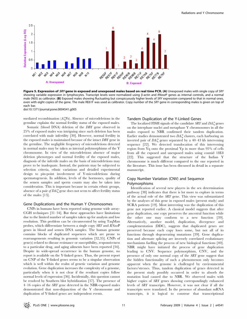
Expression Level of SRY Gene in the Exposed andUnexposed MalesIt is not possible to assess expression of Y-linked genes in human
tissues or gonads owing to logistic constraints. Therefore, westudied expression of the SRY gene in the blood. Normal fertilemales showed varying levels of SRY transcripts even amongmembers of the same family within and across the populations.
Similar variations were detected among exposed males, but ingeneral, the expression level was several folds higher than in theunexposed ones (Figure 5). The difference in the expression levelwas conspicuous in males with the same number of copies. Forinstance, males 3B and 10B, with 2 copies of the SRY gene, showed1.5–2 times the level of expression, whereas 9F, 10F, 31F, 32F,33F and 33B, also with two copies, showed higher levels (around
Figure 1. PCR-based assays of STSs with DNA of a few representative males exposed to natural background radiation (NBR). On theleft of the panels, ‘‘RE’’ denotes radiation exposed, F and B denote father and son, respectively, and numbers refer to family IDs. The STSs are given onthe right of the panels. Note de novo microdeletions in father but not in son, and vice-versa.doi:10.1371/journal.pone.0004541.g001
Radiations and Y Chromosome
PLoS ONE | www.plosone.org 3 February 2009 | Volume 4 | Issue 2 | e4541
4–9 times) of expression. Males 29F and 29B, with four copies ofthe SRY gene, showed similar levels of expression as those having 2copies. For calculations, a male (8F) with the lowest level of SRYtranscripts was used as a calibrator in real time PCR. Startlingly,males with 8 copies of the SRY gene (e.g., 7F) showed 25 timeshigher expression as compared to that of the calibrator.
Multiple Polymorphic Copies of the CDY1 Gene andBackground Radiation ExposureSequencing of full length CDY1 genes (primers in Table 1)
showed polymorphism similar to that detected in SRY genes. The
CDY1 gene from a single male (6F) was amplified, cloned intopGEMT easy vector, and sequenced (Figure S7). Nucleotideanalysis of 5–10 full-length CDY1 fragments, each from 50 exposedmales, uncovered random point mutations throughout the singleexon. All the exposed males showed two types of CDY1 sequences,one full length with inserted intron, and the other one with pointnucleotide changes leading to correspondingly truncated CDY1protein as uncovered in-silico (Figure 6). Once again, thishighlighted polymorphism of the multiple copies of CDY1 gene,similar to that of SRY. Sequence comparison of the CDY1 genefrom representative exposed males is given in Figure S8. Based on
Figure 2. STS map for AZFa, b & c regions in representative exposed males. (a) Conventional organization of the human Y chromosome. (b)STSs employed in mapping three different AZF regions are shown in corresponding colors. (c) Results of STS mapping using genomic DNA from theperipheral blood lymphocytes of the exposed males. ‘‘NM’’ denotes unexposed males. Sample IDs for the exposed males are given on left. ‘‘F’’ and ‘‘B’’denote father and son, respectively. Note deletion of sY79, sY89 and sY639 in all the males and frequent deletions of sY83 and sY84 in AZFa, sY117and sY129 in AZFb and several STSs in distal and proximal AZFc regions (see text for details).doi:10.1371/journal.pone.0004541.g002
Radiations and Y Chromosome
PLoS ONE | www.plosone.org 4 February 2009 | Volume 4 | Issue 2 | e4541

our data, we inferred that NBR exposure leads to the formation ofmultiple polymorphic copies of the Y-linked genes. This wassubstantiated by full length sequencing of the XKRY gene showingnucleotide changes spread all across its length, though frequentlyin the middle portions (not shown). Although there is no report onthe implications of this gene for male fertility, its small size makes itan attractive candidate for the assessment of point nucleotidechanges.
DAZ Genes (DAZ1–DAZ4) in Males Exposed to NBRAnalysis of SFV/SNV/STS in 390 exposed males showed all
the six intact SNVs, confirming the presence of four DAZ genes in
.85% of males. Similar SNV/SFV analysis of the remaining 15%showed either the absence of an allele, or absence of a single bandcorresponding to a specific SNV (Table 4). Another intriguingobservation was the loss of the restriction enzyme site for SNVIIsince the PCR product remained undigested in .55% of exposedmales. However, the majority of the NBR-exposed males showedintact DAZ genes.
Status of the Other AZFc CandidatesSNV/SFV analysis of the other AZFc candidates revealed
complete absence of STS Y-DAZ3 in blood DNA in approximately60% of the exposed males (Table 4). The TTY4/1 allele A was
Figure 3. Exclusive somatic deletion of DBY gene in ,25% of exposed males. (A) Representative males with their IDs shown on top. DNAfrom blood and semen samples are denoted as ‘‘b’’ and ‘‘s’’, respectively. Note deletion of DBY amplicons exclusively in the blood DNA of exposedmales compared to that of semen DNA. The SRY (sY14) and b-actin PCRs were used as positive controls. (B) Schematic representation of the AZFaregion. Candidate AZFa genes are given in ‘‘b1’’, STSs used in ‘‘b2’’, and the results of screening in ‘‘b3’’. Note the absence of amplicons with primerscorresponding to DBY1 and DBY2.doi:10.1371/journal.pone.0004541.g003
Radiations and Y Chromosome
PLoS ONE | www.plosone.org 5 February 2009 | Volume 4 | Issue 2 | e4541

absent in 167 of the 390 males. Similarly, allele B of the SNVBPY2/1 was absent in 15% of males, Likewise, allele A and a398 bp fragment of allele B of the SNV GOLY/1 were absent in.30% of exposed males (Figure 7). The other AZFc SNVs AZFc-P1/1 and RRM3 were normal in all the cases.
Tandem Duplication of the Y-Linked Loci in MalesExposed to NBRFluorescence in situ hybridization (FISH) conducted on
metaphase chromosomes and interphase nuclei of the normalmales showed a single signal in each of the SRY and DXZ1probes [19] (Figure 8a). As expected, a single signal was detectedin the centromeric region of each X chromosome in the normalfemale used as negative control (data not shown). Exposed malesalso showed a single signal with multiple copies of the SRY gene.A male (7F) carrying eight copies of SRY showed a relatively
stronger signal on the Y chromosome compared to that of anormal male. However, in several cases with 2–4 copies of SRY,differences in the signal intensity were not discernible.Depending upon the condensation status of the sister chromatidsin some cases, two signals of the SRY were seen. Interestingly,,5–12% of males lacked signals for the SRY gene, whereas asignal for DXZ1 was consistently detected. The absence of signalwas attributed to the loss of Y chromosome shown in the latersection.FISH probes (Cosmids 18E8, 46A6 and 63C9) used for the
DAZ genes have been explained in the literature [22]. Cosmidprobe 18E8 encompassing the 59 end of two neighboring DAZgenes showed two signals in the normal males, or one, if thesame were merged. In 20% of exposed males with 6 copies ofDAZ genes, three signals were discernible (Figure 9i). In theseexposed males, only one locus of DAZ underwent duplication
Figure 4. Analysis of the AZFa HERV elements in the exposed males. (A) Schematic representation of provirus elements with various STSmarkers used to study recombination mediated duplication or deletion. (B) Status of HERV elements in exposed males. Note presence of all the STSsin normal/unexposed males and randomly scattered microdeletions without any conclusive major deletion/duplication in the exposed ones. FemaleDNA was used as negative controls. Number of males showing deletion of a particular STS is given in percentage.doi:10.1371/journal.pone.0004541.g004
Radiations and Y Chromosome
PLoS ONE | www.plosone.org 6 February 2009 | Volume 4 | Issue 2 | e4541

Table 1. Details of the primers and probes used in this study#.
Primers and TaqMan Probes for Copy Number Assessment of the Respective Genes
Name Entrez ID Location Size (bp) Primers and Probes (59-39)
CDY1 9085 Yq11.23 3991 F (3626–4646) GTGGATGATGGCACCTTTGTG
R (3676–3700) GCAGCCTGTAAGATGGGTTTGTAAA
P (3656–3672) CTTGAGCCTGCTTTTC
CDYL 9425 6p21.5 250637 F (555–575) CCCTGACTGATGAGCAAACCA
R (609–632) TCACTGAACTCCATGTGTGTTACC
P (592–606) CCACTGGGCCTCTCG
CDYL2 124359 16q23.2 201738 F (102192–102216) GGTTTGCAAATAATGCCCCTGAAAA
R (120272–120293) AGTGCCTCTCATCCTTCTCAGA
P (120236–120250) CCGGCGTCCCCATTT
UTY 7404 Yq11 233495 F (1771–1787) CCATCACCCGCCTGGTT
R (1821–1839) TCATCAACGTGGGCAAGCT
P (1795–1811) CCTTCCCGGAGAGTATC
VCY 9084, 652821 Yq11.221 1942 F (1231–1253) CCTATCTCCCTGAGCAGCAACTA
R (1277–1297) CCCTGCTGGTGAGATCTCTGA
P (1254–1269) CAGCTGGGCCTAAACT
BPY 442868 Yq11.223 22403 F (21336–21356) TGGAGTCTGCCAAAACAAGGG
R (21447–21468) CAGAGCAGGAGAGTCTCATCAC
P (21362–21388) CACATATTGCGGAGTCCAGCACCCAGG
HSFY 442479 Yq11.222 4234 F (1060–1084) TCAATGAGGCTCCTTATCCTAACCT
R (1117–1141) GCAGCCGATGTATCAAATGTCATAG
P (1089–1103) CCAGCAGGCAACCAG
SRY 6736 Yq11.3 2045 Commercial assay from ABI (part number Hs00243216_s1)
DAZ 1617, 57135, sY587 (STS) F (220–249) TGACTGGACACCTAGTTTCATGAAC
Yq11.223 R (299–320) GTCAAGAGGCATCAAGTGAAAGTTG
P (256–279) CACCCTGTCTCCAACCC
PRY 9084 Yq11.223 25452 F (807–827) CAGGATGAAGGGATGCAGTGA
R (847–869) CTTAGAGGTGGGTGTCAGTGAAA
P (831–846) CAAGAGCCCAACCTTC
Primers Used for Cloning and Sequencing
Gene Primers Tm Amplicon
SRY SRY1F- 59GACAATGCAATCATATGCTTCTGC39 65 600
SRY1R- 59CTGTAGCGGTCCCGTTGCTGCGGT39
SRY3F- 59GAATCTGGTAGAAGTGAGTTTTGGA39 63 828
SRY3R- 59 TAAGGCCTTTATTAGCCAGAGAAAA39
SRY4F- 59CTTCTGCTATGTTAAGCGTATTCAA39 63 599
SRY4R- 59CAGCTTTGTCCAGTGGCTGTAG39
CDY1 CDY1F-59AAAGCTTTCTGTACTACACCAGAGGGTTG39 68 2763
CDY1R-59AAGAAGTTTCTGCCTTTAATAATGTGTCCA39
CDY2 CDY2F-59AAAGCTTTCTGTACTACACCAGAGGGTTG39 68.0 1954
CDY2R-59AGCAGACAGACTGACAATTAAAACTCATCA39
VCY VCY1F-59TAGTGGAGTGTTGACCAATCACAG39 65 960
VCY1R-59ACACACCACCTCTTCCTTCCTC39
HSFY HSFYF-59ATGCAGGCTGGAAGAGTAGCTAAAGAAATA39 68 2100
HSFYR-59TGGTGGAAGATATGTTGTAATTAGGGTGAT39
#F and R refer to forward and reverse primers; P is the TaqMan probe.doi:10.1371/journal.pone.0004541.t001
Radiations and Y Chromosome
PLoS ONE | www.plosone.org 7 February 2009 | Volume 4 | Issue 2 | e4541

Table 2. Copy number polymorphism of the Y-linked and autosomal genes in human males exposed to natural backgroundradiation (NBR)#.
Y-linked Autosomal
Sample ID SRY DAZ CDY1 PRY UTY HSFY VCY BPY2 XKRY TSPY DYZ1 CDYL CDYL2
Normal males 1 4 2 2 2 2 2 3 2 35–42 4000–4500 2 2
RE51F 2 4 4 8 2 1 4 6 4 52 5800 2 2
RE51B 2 4 4 4 2 1 4 4 4 45 5200 2 2
RE52B 2 8 4 4 1 1 2 4 4 49 6150 4 2
RE53F 1 6 4 4 2 1 2 6 4 48 5000 2 2
RE53B 2 6 4 4 2 1 4 6 4 65 5300 2 2
RE54F 2 8 4 4 2 1 2 9 4 60 6500 2 2
RE54B 2 16 4 16 2 2 2 6 4 55 6550 2 2
RE55F 1 4 2 4 1 2 2 4 4 58 5500 2 2
RE55B 2 4 4 8 1 1 4 4 6 42 5200 2 2
RE56F 2 8 8 4 2 1 4 3 4 43 4800 2 4
RE57F 8 4 4 4 1 1 2 6 4 56 5200 2 2
RE57B 2 8 8 4 2 1 4 4 4 51 6000 2 2
RE58F 1 8 8 4 2 1 2 4 6 68 5500 2 2
RE58B 2 8 4 4 2 1 2 4 6 40 5950 2 2
RE59F 2 8 8 8 2 2 4 6 4 68 5100 2 4
RE60F 2 6 4 8 2 2 2 6 4 66 5800 2 2
RE60B 2 6 32 4 1 2 4 6 4 53 4550 8 2
RE61F 1 6 4 4 2 1 2 5 4 44 4900 2 2
RE61B 1 8 4 2 2 1 2 3 4 48 4750 2 2
RE62F 1 8 4 8 1 2 2 3 4 50 6700 2 2
RE63F 1 8 8 8 1 2 2 4 4 42 6150 4 2
RE63B 1 6 8 8 2 2 2 6 4 58 5000 2 2
RE64F 1 8 4 8 1 2 2 3 4 50 4100 4 2
RE64B 1 8 16 8 4 2 2 3 4 66 4300 2 2
RE65F 1 6 8 8 4 2 2 3 4 40 4500 4 4
RE65B 1 4 8 4 2 2 4 4 4 38 4000 2 2
RE66F 1 6 4 8 4 2 4 3 4 46 3000 2 4
RE66B 2 4 1 4 2 1 1 3 2 44 3800 2 2
RE67F 1 4 4 4 2 2 2 3 4 48 5700 2 2
RE67B 1 4 4 2 2 2 2 4 4 62 6000 2 2
RE68F 1 4 8 8 2 2 4 4 2 60 3500 2 4
RE69F 1 8 4 4 2 2 2 6 2 55 4000 2 2
RE70F 2 8 4 4 2 1 2 5 6 40 6500 2 2
RE70B 2 8 4 4 2 2 2 4 6 42 6800 2 2
RE71F 4 8 4 4 2 1 2 6 6 44 5700 2 2
RE71B 2 8 2 4 2 2 4 5 4 58 5600 2 2
RE72F 1 8 4 1 4 1 2 9 4 60 3500 2 2
RE73F 2 8 2 1 16 2 2 6 4 44 3900 2 2
RE73B 2 8 2 2 1 1 2 4 4 48 5000 2 2
RE74F 2 6 2 4 8 2 2 4 4 56 5100 2 2
RE74B 4 8 2 2 2 2 2 4 4 54 5200 2 2
RE75F 4 4 2 2 1 2 2 4 2 64 5500 2 2
RE75B 4 4 2 2 1 1 2 3 4 44 5000 2 2
RE76F 8 8 2 4 2 2 2 3 4 48 4500 2 2
RE76B 2 4 2 4 2 2 2 5 6 42 4200 2 2
RE77F 1 4 2 4 2 1 2 4 4 40 4800 2 2
RE77B 2 8 4 8 1 1 2 4 2 46 4500 2 2
Radiations and Y Chromosome
PLoS ONE | www.plosone.org 8 February 2009 | Volume 4 | Issue 2 | e4541

(unilocus duplication), resulting in six copies. Males with 8 or 16copies showing correspondingly stronger signals compared tothat of normal males seem to have undergone single and doublerounds of tandem duplications, respectively (Figure 9). Exceptfor males with unilocus duplication, approximately 30% showedthree signals in about 9–12% of cells (not shown) and completeabsence of signals in 10–15% of interphase nuclei (explainedlater). The absence of DAZ signals was construed to be due tothe loss of Y chromosome. This inference was based on anumber of Y-specific probe combinations and painting describedin later sections. As expected, the mosaicisms affected Ct valuesof DAZ and SRY genes during real time PCR assay.
Structural (Re)organization of the DAZ GenesPreviously, two DAZ loci in the AZFc region, each with an
inverted pair of DAZ genes (amplicons r1, r2, r3, r4) and aninter-DAZ sequence were detected. We observed a similararrangement in the interphase nuclei of the exposed males. Butsubsequent hybridization of DAZ probes with metaphase Ychromosomes revealed a different organization. The cosmid18E8 (probe A) representing 59 DAZ exons 1 through 7 on oneside and the inter-DAZ region on the other [22] was localizedonto the proximal Yp instead of on the anticipated Yq region inall 390 exposed males (Figure 8). The other two DAZ cosmids46A6 and 63C9 (probes ‘‘B’’ and ‘‘C’’) representing DAZ exons2 through 11 and 39 DAZ, respectively, were localized on theiranticipated positions in the Yq region, overlapping with eachother on the metaphase Y chromosome (Figure 8). Similarresults were obtained with FISH conducted on interphase nuclei(Figure 9ii). Signals for probes ‘‘A’’ and ‘‘C’’ or ‘‘A’’ and ‘‘B’’never overlapped. Another probe (D) corresponding to neigh-boring g1/g2/g3 amplicons of the DAZ genes detected theexpected 3 signals on the interphase nuclei. On the metaphase Ychromosome, probe ‘‘D’’ detected the expected overlap withprobes ‘‘B’’ and ‘‘C’’. In addition, in each of 80–90% of exposedmales, 70–80% of cells showed an unexpected overlap of probeD signals with that of probe A on the proximal short arm of Ychromosome (Yp). Overall structural reorganization of the AZFcregion and DAZ genes in the exposed males is summarized inFigure 10. Similar (re)organization was detected in normal malesand thus this was not attributed to the effect of NBR.
Long Arm Heterochromatin of the Y Chromosome (Yqh)Prominent length variation of the Yq heterochromatin was
detected in the exposed males using a 3.4 kb FISH probe(Figure 8g–h) compared with an almost uniform signal in thenormal males. This variation was construed to be due to copynumber variation (CNV) of the 3.4 kb repeat units. In normalmales, the average number of copies of the DYZ1 repeat variesfrom 4000–4500. In the exposed males, this number was detectedin the range of 4500–6500 (table 2). The increase in DYZ1 copynumber among 390 males correlated with varying FISH signals.Interestingly, ,5–12% of cells per exposed male were devoid ofsignal, suggesting loss of Y chromosome.
Loss of the Y Chromosome and NBR ExposureConclusions on FISH were drawn based upon the analysis of
400 interphase/metaphases each from 390 exposed males. Asmentioned earlier, FISH conducted with SRY/DXZ1 showedDXZ1 signals on the X-centromere in all the cells, but SRY signalwas missing in 5–12% of cells (Figure 11). Similar observationswere made using different AZFc and DAZ probes individually andin combination with one another. Whole chromosome paintingconfirmed the loss of Y chromosome as mentioned earlier(Figure 11). In normal males, ,96–98% of cells were positivefor all the probes whereas in the remaining ones, signals were notdetected owing to technical constraints.
Discussion
Owing to its haploid status, repetitive nature and intrachromo-somal homologous recombination, the human Y chromosome ishighly prone to genetic variations [23,24], which are passed on tothe next generation un-repaired. However, this chromosome hasbeen implicated with male sex determination and spermatogen-esis. Thus, lethal mutations are usually not transmitted to the nextgeneration. The present study is an attempt to explore geneticvariations of the human Y chromosome in males exposed to NBR.
The AZF Microdeletions and Radiation ExposureIonizing radiation induces several types of DNA lesions through
single-strand (SSB) and double-strand (DSB) breaks, AP sites(either apyrimidinic or apurinic), and DNA–DNA and DNA–
Y-linked Autosomal
Sample ID SRY DAZ CDY1 PRY UTY HSFY VCY BPY2 XKRY TSPY DYZ1 CDYL CDYL2
RE78F 1 4 4 4 2 1 2 5 4 48 5800 2 2
RE78B 1 4 4 4 2 1 2 3 4 44 6000 2 2
RE82F 1 6 2 2 4 1 2 3 4 58 5300 2 2
RE82B 1 8 4 4 2 1 2 4 2 59 5400 2 4
RE83F 2 8 2 4 1 1 4 5 4 55 3700 2 2
RE83B 1 8 2 4 2 1 2 4 6 54 3500 8 2
RE84F 2 8 4 2 8 1 1 4 4 60 4800 2 4
RE84B 2 6 4 2 1 1 2 4 4 62 5000 2 4
RE85F 1 6 2 2 1 1 2 6 4 58 4200 2 2
RE85B 1 4 4 2 2 2 2 6 2 48 6550 2 2
RE87F 2 8 2 2 2 1 2 5 2 42 5300 2 2
RE87B 2 16 2 2 2 1 2 4 4 56 5600 2 2
#None of the NBR-exposed males contained normal copy number profiles for all the Y-linked genes studied.doi:10.1371/journal.pone.0004541.t002
Table 2. Cont.
Radiations and Y Chromosome
PLoS ONE | www.plosone.org 9 February 2009 | Volume 4 | Issue 2 | e4541

protein cross-linking, in addition to base modifications [25,26].Several alterations in the DNA induced by ionizing radiation arechemically identical to those caused by reactive oxygen species[27]. Ionizing radiation induces isolated as well as clustereddamages involving two or more lesions within one or two helicalturns of DNA. Base lesions within the clustered DNA enhancebiological severity of the damage. Highly localized DNA damagecaused by ionizing radiation leads to intrachromosomal breaks[28,29]. Several attempts have been made to establish acorrelation between background radiation, chromosomal aberra-tions and phenotypic changes [5–8]. However, due to insufficientdata, experimental evidence of radiation induced mutations inhumans still remains highly controversial.
Our study demonstrated a correlation between radiationexposure and microdeletions in the AZF regions. These micro-deletions did not follow a normal pattern of inheritance sincedeletions detected in fathers were absent in their sons and viceversa. This discrepancy, observed in most of the exposed males,was true for gene copy number variations as well. This can beexplained on the basis of unaffected germline DNA in the exposedmales. We postulate that radiation exposure caused localized DNAdamages but by some unknown protection mechanism, thegermline DNA remained intact. Scrutiny of microdeletionsshowed higher frequency in proximal and distal AZFc, and tosome extent in the AZFa region, but absence of any major deletionphenotypes such as gr/gr, b1/b3, b2/b4 (AZFc) and HERV
Table 3. Details of nucleotide and amino acid alterations in the SRY gene in some representative males exposed to NBR#.
S.N. Seq ID Point Nucleotide Changes Amino Acid Changes
1 RE53F Del A317, C410G NO CHANGE
2 RE54F Insertion T204, Insertion C363 and A381, T416C, C418T, A420C,G421A, Insertion G423, Insertion A800, T801, A219G, C231T,C468T
T48M
3 RE55B Del A317, C410G, Insertion C332, Insertion C577 DelL144–145, P146S, P149R, A150R, V152R, L153F, C154G, Del155–161,R162T, Y164Q, D166S, Del167–170, H173G, S174Q, R175Q, M176V,E177A, H178T, Q179G, L180M, G181T, H182V, L183R, Del184–204
4 RE55B C410G, T435G, A427T, C419T, Insertion C332, Insertion C577 DelL144–145, P146S, P149R, A150R, V152R, L153F, C154G, Del155–161,R162T, Y164Q, D166S, Del167–170, H173G, S174Q, R175Q, M176V,E177A, H178Q, Q179G, Del180–204
5 RE57F Del A317, C410G H182T, P184T, P185A, I186H, N187Q, A188R, A189S, S190Q,S191L, P192T, Q193A, Q194A, R195T, D196G, R197T, Y198L,S199Q, H200P, W201L, T202D, K203R, L204A
6 RE57B Del A317, C410G K203R
7 R31F Del A317, C410G, Insertion C578 and 577, C192T, InsertionC541,A657C, A671C, C677A, G679C, A684C
D166G, R165Q, Y164L, Del165–204
8 RE58F C410G, C419T, A427T, Del T435 NO CHANGE
9 RE58B A427T NO CHANGE
10 RE60F C410G, T435G F12I, D17E, N24D, I25N, L28F, S32F
11 RE60B C410G, A427T, T435G Del187–204
12 RE65F InsertionC332 DelL144–145, P146S, P149R, A150R, V152R, L153F, C154G, Del155–161, R162T, Y164Q, D166S, Del167–170, H173G, S174Q, R175Q,M176V, E177V, H178Q, Q179G, Del180–204
13 RE75F Del T82–83, Del336G, A596G, C733A, C808T Y44C
14 RE75B A140G, Del 336G,T458A,A670G S18G, Y44C
15 RE76F T226C, InsertionG235, T279C Y44C, D73K, Del49G-R72, Del75–204
16 RE80F InsertionC237, Insertion C365, Del T391, Insertion G397,Insertion A415, Insertion A448
Y44C
17 RE80B Y44C, Del84–85, R86Q, S88A, E89K, I90L, S91R, K92D, L94Q,Del95, Y96A, Q97S, K99D, M100T, L101S, T102G, E103K,A104C, E105Y, Del106–204
18 RE81F Insertion12C, 15G and 155T, A216G, InsertionT143, A228G, T467C,C535A, A495T, A573C, T636A, InsertionT638, InsertionC646,InsertionA/T670, C677A, C672G, A697T, G583T, C686A
A20L, V21C, L35P E47G, Y127H, K136M, Y164H, R165M, L183I,P184T, InsertionA185, I187S, N188T, A190Q, A191P, K203N,L204A, F110L, N141D
19 RE81B Insertion12C, 15G and 155T, A216G, InsertionT143, A228G, T467C,C535A, A495T, A573C, T636A, InsertionT638, InsertionC646,InsertionA/T670, C677A, C672G, A697T, G583T, C686A
A20L, V21C, L35P E47G, Y127H, K136M, Y164H, R165M, L183I,P184T, InsertionA185, I187S, N188T, A190Q, A191P, K203N,L204A, F110L, N141D
20 RE88F T188C, G292A, G313A no change
21 RE90B G371A G95R, Del152–160, N161G, R162T, Y164Q, D166S, Del167–170,G95R
22 RE94F T549C, T576C C154R, H173G, H174Q, R195T, D196G, R197P, Y198L, R199Q, H200P,W201L, T202V, L204A R175Q, M176V, E177V, H178Q, Q179G, Del180–204
23 RE94B G598T, C686A K203S, Del204, K43R, A171T
#Amino acid changes in bold are random, underlined are reported in the literature, and italicized are consistently present in several exposed males.doi:10.1371/journal.pone.0004541.t003
Radiations and Y Chromosome
PLoS ONE | www.plosone.org 10 February 2009 | Volume 4 | Issue 2 | e4541
mediated recombination (AZFa). Absence of microdeletions in thegermline explains the normal fertility status of the exposed males.Somatic (blood DNA) deletion of the DBY gene observed in
25% of exposed males was intriguing since such deletion has beencorrelated with male infertility [30]. However, normal fertility inthe exposed males is maintained because of the intact DBY gene inthe germline. The negligible frequency of microdeletions detectedin normal males may be taken as internal polymorphisms of the Ychromosome. In view of the microdeletions absence of majordeletion phenotypes and normal fertility of the exposed males,diagnosis of the infertile males on the basis of microdeletions mayprove to be inadequate. Instead, the patients may be subjected toselection criteria, ethnic variations and detailed experimentaldesign to pin-point involvement of Y-microdeletions duringspermatogenesis. In addition, levels of the hormones, quality ofthe semen samples and sperm counts may also be taken intoconsideration. This is important because in certain ethnic groups,absence of a pair of DAZ gene does not seem to affect fertility statusof the males [13].
Gene Duplications and the Human Y ChromosomesCNPs in humans have been reported using genome wide array-
CGH techniques [31–34]. But these approaches have limitationsdue to the limited number of samples taken up for analysis and lowresolution. This problem can be circumvented by using TaqManprobes, which differentiate between a single copy SRY and RNasePgenes in blood and semen DNA samples. The human genomecontains blocks of duplicated sequences which are prone torearrangements resulting in genomic variation [32,35]. CNPs ofgene(s) related to disease resistance or susceptibility, responsivenessto a particular drug, and aging ailments have been reported [34].Despite its wide-spread occurrence in the human genome, noreport is available on the Y-linked genes. Thus, the present reporton CNP of the Y-linked genes seems to be a singular observationwhich is well within the realm of genetic variation and genomeevolution. Gene duplication increases the complexity of a genome,particularly when it is not clear if the resultant copies follownormal levels of expression [36]. Incidentally, this question cannotbe resolved by Southern blot hybridization [37]. The presence of4–16 copies of the SRY gene detected in the NBR-exposed malesdemonstrated that non-disjunction of the Y chromosome andduplication of Y-linked genes are independent events.
Tandem Duplication of the Y-Linked GenesThe localized FISH signals of the candidate SRY and DAZ genes
on the interphase nuclei and metaphase Y chromosomes in all themales exposed to NBR confirmed their tandem duplication.Earlier studies demonstrated two DAZ clusters, each harboring aninverted pair of DAZ genes separated by a 40–43 kb interveningsequence [22]. We detected translocation of this interveningregion from Yq onto the proximal Yp in more than 95% of cellsfrom all the exposed and unexposed males using cosmid 18E8[22]. This suggested that the structure of the Indian Ychromosome is much different compared to the one reported inthe literature. This issue has been discussed in detail in a separatemanuscript.
Copy Number Variation (CNV) and SequencePolymorphismsIdentification of several new players in the sex determination
pathway [38] indicates that there is lot more to explore in termsof the actual role of the SRY gene. This view was substantiatedby the analyses of this gene in exposed males (present study) andSCRA patients [19]. Most interesting was the duplication of thisgene not reported earlier. A classical model suggests that aftergene duplication, one copy preserves the ancestral function whilethe other one may conform to a new function [39].Alternatively, another model, duplication, divergence andcomplementation (DDC), suggests that duplicated genes arepreserved because each copy loses some, but not all of itsfunctions through degenerating mutations [39]. Gene duplica-tion and alternate splicing are inversely correlated evolutionarymechanisms fuelling the process of new biological functions [40].NBR might have initiated the process of gene duplicationleading to CNV. Sequence polymorphism, CNV, and thepresence of only one normal copy of the SRY gene suggest thatthe hidden functionality of such a phenomenon only becomesapparent when the genome is challenged by environmentalfactors/stresses. Thus, tandem duplication of genes detected inthe present study possibly occurred in order to absorb themutation load caused due to NBR. We observed males withhigher copies of SRY genes showing correspondingly enhancedlevels of SRY transcripts. However, it was not clear if all thetranscripts were translated. In the presence of abundant mRNAtranscripts, it is logical to construe that transcriptional
Figure 5. Expression of SRY gene in exposed and unexposed males based on real time PCR. (A) Unexposed males with single copy of SRYshowing variable expression in lymphocytes. Transcript levels were normalized using b-actin and RNaseP genes as internal controls, and a normalmale (N05) as calibrator. (B) Exposed males showing fluctuating but conspicuously higher levels of SRY expression compared to that in normal ones,even with eight copies of the gene. The male RE61F was used as calibrator. Copy number of the SRY gene in corresponding males is given on top ofeach bar.doi:10.1371/journal.pone.0004541.g005
Radiations and Y Chromosome
PLoS ONE | www.plosone.org 11 February 2009 | Volume 4 | Issue 2 | e4541

machinery failed to discriminate between normal and alteredversions of a duplicated gene. Tandem duplication of SRYfollowed by its augmented transcription may well be a possiblemechanism of up-regulation of a gene.Polymorphism of multiplicated Y-linked genes became more
evident by sequence analysis of the SRY in other exposed malesshowing several known and novel mutations. Besides SRY,sequence polymorphism was observed in CDY1 and XKRY genes,supporting the hypothesis that in the exposed males, other Y-linked genes are also affected. This was corroborated by SNV/SFV typing of DAZ genes in males exposed to NBR. These males
showed either complete absence of a particular SNV allele orpresence of additional amplicons. Similar to DAZ SNVs/SFVs, theAZFc region also showed polymorphisms. All these somaticsequence variations in the males correlated with the NBRexposure.
Possible Mechanisms of Gene DuplicationA single copy gene gives rise to a number of mRNA
transcripts, some of which may be reverse transcribed andtransposed back into the DNA, resulting in multiple copies [41].Thus, besides putative tandem duplication, reverse transcriptase
Figure 6. Two different copies (6F_4, 6F_11) of CDY1 gene in NBR-exposed male (6F) are shown along with its normal sequence(top). Note the conspicuous difference between the two copies. Amino acid changes corresponding to nucleotides are highlighted in red withyellow background.doi:10.1371/journal.pone.0004541.g006
Radiations and Y Chromosome
PLoS ONE | www.plosone.org 12 February 2009 | Volume 4 | Issue 2 | e4541

activity on the mRNA may also contribute to CNV. However,this seemed to be a less likely event because SRY copies detectedwere in multiples of two to a maximum of 16. Owing to theavailability of a large number of mRNA transcripts, reversetranscriptase–like activity would have generated far greaternumbers of SRY copies.
Somatic Nature of Alterations in NBR-Exposed MalesNatural background radiation may be responsible for varying
alterations. However, intact germline DNA of the exposed malessuggested a strong protective mechanism to counter the effects ofNBR. Alternatively, it is likely that owing to high turnover ofmeiotic activities, defective cells undergo rapid apoptosis leavingbehind no trace of an alteration in the germline. We are aware ofthe fact that, based on the analysis of some Y-linked genes, nomajor conclusion on the effect of NBR can be drawn. Thus, moregenes involved in control and regulation of signal transduction,apoptosis, genome imprinting, tumor suppression and progressionmay be included to monitor the overall impact of NBR on thegenome. This would go a long way towards undertaking correctivemeasures to save the population at high risk, if any.
ConclusionsThe present study demonstrates the effects of NBR on gene
duplications and sequence polymorphisms, a possible genomestrategy to absorb the mutational loads. This is evident from themultiple polymorphic copies of the SRY and CDY1 genes.Moreover, from the enhanced SRY expression in blood, it islogical to construe that transcriptional machinery failed todiscriminate between normal and altered versions of a duplicatedgene. Absence of microdeletions and CNV of genes in germlineDNA of exposed males suggested that the germline DNA isprotected by some innate, still unexplored repair mechanism(s).Analysis of the additional Y-linked loci, and even the autosomalgenes, may uncover the overall impact of NBR on the structuraland functional attributes of the human genome.
Materials and Methods
Sample CollectionPeripheral blood and semen samples were collected with
informed consent from 390 males of two generations atChavara (lat. 8u57.89N, long. 76u31.89E) and Thevara (lat.9u589N, long. 76u169E) in Kerala, India [17]. This study wasreviewed and approved by the Institute’s Ethical and BiosafetyCommittee. Since the population at the site of the NBR samplecollection is illiterate, oral consent was obtained from theparticipants, facilitated by a recognized local clinician. In thecase of control samples from other parts of India, writtenconsent was obtained wherever possible. After due scrutiny, theclearance by the Institute’s Ethical Committee was accorded.Following this, no additional approval regarding a particulargene sequence/gene variant was required for publication of thedata.The semen analysis of the exposed males was conducted by a
commercial technician and the results are given in Tables S1 andS2. For control, blood and semen samples from 390 healthyunexposed males of matched age groups from Kochi city (SouthIndia) and 400 normal males from different parts of India wereincluded. DNA from blood and semen was isolated followingstandard protocols. RNA was isolated from lymphocytes separatedfrom blood with AccuspinTM System-HistopaqueH-1077 (SIGMA,cat no: A7054) using TRI-X reagent (MRC, cat no: TB-126-200)and cDNA synthesized using high cDNA Archive Kit (cat no:4322171, ABI, USA).
STS Mapping of the AZF RegionsThe AZF regions were screened in blood and semen DNA from
normal and exposed males for the presence or absence of STSmarkers, and details of the primers used are given in Figures 2 and3. The AZFc region was assessed for the intactness of the sitesP1.1/P1.2, GOLGA2LY, BPY2 and TTTY4 genes by the STSs andSNVs [12,13] (Table S3). PCR was carried out in 100 ml,precipitated with Na+-acetate/ethanol and used for restriction
Table 4. AZFc region SNV/SFV typing in males exposed to NBR#.
SNV Haplotypes Samples Number
DAZ SNVs
I A(709)+B(398) RE3, RE7, RE8, RE9, RE40, RE41, RE52, RE53, RE56, RE61, RE109, RE110 12
II A+B +ve in all
PCR amplicon not digested in more than 55% of the males
III A+B +ve in all
IV A+B(122) RE5, RE6, RE10, RE11, RE18, RE23, RE25, RE27, RE40, RE47, RE55, RE57, RE59, RE81,RE89, RE105–RE110
20
V A(195)+B RE111, RE122, RE123, RE124, RE127, RE143, RE144, RE146, RE147, RE157, RE158,RE159, RE167, RE168, RE170, RE172, RE173, RE189, RE190, RE191, RE197, RE200,RE201, RE202, RE203, RE205, RE206
27
AZFc SNVs
AZFc-P1/1 A+B +ve in all
GOLY/1 A+B (298) +ve in more than 90% of males
BPY2/1 A+B Allele B absent in ,15% of males
TTY4/1 A+B 167 out of 390 males 167
RRM3 +ve in all
Y-DAZ3 2ve in 60% of males 180
#The italicized haplotypes showed absence of a single band (size given), and the one shown in bold was absent.doi:10.1371/journal.pone.0004541.t004
Radiations and Y Chromosome
PLoS ONE | www.plosone.org 13 February 2009 | Volume 4 | Issue 2 | e4541

Figure 7. AZFc SNV/SFV typing in the exposed males. ‘‘NBRE’’ refers to natural background radiation–exposed males, and numbers representthe family IDs. (A) Loss of SNV TTY4/1 allele ‘‘A’’ (541 bp) in exposed males (arrows in panel ‘‘A’’). Sizes of digested DNA with HaeIII enzyme are givenon the right. Note the absence of 541 bp band in several males. Some showed complete absence of the SNV TTY4/1. (B) SNV for BPY2 gene showingits absence in some males (arrow) and loss of allele B (289+181 bp) in others (upper panel). (C) SNV typing of GOLGA2LY gene. Note the absence ofallele ‘‘A’’ and 289 bp of allele ‘‘B’’ in some males (arrows in ‘‘C’’).doi:10.1371/journal.pone.0004541.g007
Radiations and Y Chromosome
PLoS ONE | www.plosone.org 14 February 2009 | Volume 4 | Issue 2 | e4541

digestion with the recommended enzymes. The digested DNA wasresolved on 2–3% agarose gel. For human endogenous retrovirus(HERV) repeats, sY82, sY746, sY1064, sY86 sY85, sY84, sY1065,sY1066, and sY88 STSs were used [23].
PCR Amplification and Sequencing of the SRY and CDY1FragmentsThe SRY and CDY1 genes were amplified from semen and blood
DNA with primers given in Table 1. Approximately 20 fragmentsamplified with each primer set from each male were sequenced.Sequences were analyzed by NCBI Blast and ClustalW align-ments. Some PCR amplicons were cloned into p-GEMT easyvector (Promega, USA) for subsequent use.
Silver Staining of the DNA in Polyacrylamide GelFor small sizes of AZFc haplotype specific bands in the
SNV/SFV typing, a 12% polyacrylamide gel was run in1XTBE and silver stained for band visualization. Gel wastreated with fixative-1 (40% methanol, 10% acetic acid) for30 min followed by 2 washes with fixative-2 (10% ethanol, 5%acetic acid) for 15 min each. Gel was then rinsed in theoxidizer (1% potassium dichromate, 0.2% HNO3) for 5 minfollowed by 3 washes of de-ionized water until all the yellowcolor disappeared from the gel. Following this, gel was treatedwith 2% AgNO3 for 20 min and then washed with de-ionizedwater again until the brown precipitate was removed. Finally,gel was rinsed in developer (3% Na2CO3, 0.05% formaldehyde)for about 5 min or until the bands appeared. The action of
Figure 8. Structural organization of the human Y chromosome in exposed males. Probe combinations are described in table S4. Theabbreviations ‘‘fl’’ and ‘‘tr’’ refer to fluorescein and Texas red labels, respectively. Probe combination AtrBfl corresponds to dual color FISH with 18E8in red and 63C9 in green. (a) SRY-FISH showing a single signal with varying intensities, with its multiple copies representing tandem duplication. (b)FISH with AtrBfl showing localization of probe ‘‘A’’ onto the proximal Yp region in all the NBR-exposed and normal males. This was substantiatedusing AtrCfl and AtrDfl probe combinations shown in (c) and (d), respectively. FISH with probe ‘‘D’’ showed exclusive localization of one of the threegreen amplicons onto the short arm, overlapping with the signal for probe ‘‘A’’, which was substantiated using probe combinations AtrDfl, DtrCfl, andDfl shown in (d), (e), and (f), respectively. Such localization was detected in 85–90% of exposed males. Percentage of cells showing probe D signal onthe short arm was higher in the exposed males compared to unexposed ones. The ratios show percent of males to percent of cells (e.g., in probe D,panel f, 100/40% means only 40% cells of all the males showed alteration with respect to probe ‘‘D’’). (g–h) Variation in length and position of Yqheterochromatin detected using 3.4 kb repeat unit of DYZ1 as FISH probe. This fluctuation was more prominent in the exposed males compared withan almost uniform signal in normal males (NM).doi:10.1371/journal.pone.0004541.g008
Radiations and Y Chromosome
PLoS ONE | www.plosone.org 15 February 2009 | Volume 4 | Issue 2 | e4541

Figure 9. (i), Tandem duplication of DAZ genes in NBR-exposed males uncovered by FISH. Numbers in parentheses represent copies ofDAZ genes ranging from 4–16 in the exposed males with corresponding variation in signal intensity. Sample IDs are given in square brackets. Notethree signals in samples 3F, 24F and 13F, with two on one side and a single one on the other, highlighting the unilocus duplication of DAZ genes (seetext for details). (ii), Organization of DAZ genes in exposed males. Only six representative exposed males and a normal one (Nm) are shown,with their sample IDs on top, copies of DAZ in parentheses, and probes on the right of the panels. The normal male (Nm) showed the expectednumber of discernible signals for each probe (A–E) with combinations of AtrBfl and AtrCfl separating two signals. Exposed males showed higher DAZcopies corresponding to unilocus (RE53F & RE53B) or bilocus duplications (RE52F, RE54F, RE54B, & RE56F) evident from the number and intensity ofthe corresponding signals shown in (A), (B) and (D), respectively. Similarly, probe ‘‘D’’ specific for green amplicons showed three expected signalsoverlapping ‘‘B’’ with ‘‘C’’ in normal males. In exposed males, 80–90% of cells showed unexpected overlap of probe ‘‘D’’ signal with that of ‘‘A’’ (RE53F,54F & 56F), as shown in (C). Presence of three signals with probe D was confirmed by different probe combinations as shown in (D) and (E). (F)Schematic map of the AZFc amplicons given as a reference.doi:10.1371/journal.pone.0004541.g009
Radiations and Y Chromosome
PLoS ONE | www.plosone.org 16 February 2009 | Volume 4 | Issue 2 | e4541

developer was stopped by treating the gel in a stop solution(5% acetic acid) for 5 min.
Primers, TaqMan Probes, and Conditions for Real TimePCRFor real time PCR, ‘‘Assay on Demand’’ specific for SRY gene
(ID: Hs00243216_s1, nucleotide location 453) and an endoge-nous control, RNaseP gene (single copy per haploid genome;Catalog number: 4316831), having FAM/TAMRA and FAM/MGB probes, respectively (Applied Biosystems, USA), wereused. The TaqMan assay for DAZ genes was based on a 100 bpfragment of the sY587 sequence. Similarly, TaqMan assays weredesigned for other candidate genes by selecting a uniquefragment for each one of them. Primers and FAM/NFQ probeswere designed by ‘‘Primer Express Software (v 2.0)’’ andprocured commercially from the ‘‘Assay on Demand’’ service
of Applied Biosystems (ABI, USA). TaqMan universal PCRMaster Mix (P/N: 4304437), standard male genomic DNA (P/N: 4312660), and a kit (P/N: 4316831) having FAM/MGBprobe for RNaseP gene and CEPH family female genomic DNAwere purchased from ABI, USA. Using male genomic DNA andprimer for RNaseP gene, TaqMan assays were conducted. All thereactions were run in triplicate on the real time PCR (SequenceDetection System, 7000, ABI, USA). The efficiency andspecificity of each TaqMan Assay were established using 10-fold dilution series of the standard male genomic DNA. Femalegenomic DNA was used as a negative control. Temperatureprofile for PCR reaction included 50uC for 2 min, 95uC for10 min, and 40 cycles of 95uC for 0:15 min and 60uC for 1 min(referred to as universal PCR conditions by ABI, USA).Approximately 10–15 ng of genomic DNA per reaction, perindividual from blood and semen samples were used. Copies of
Figure 10. (i), Schematic representation of the AZFc region. (A) Arrangement of the AZFc amplicons as reported in the literature. Probes usedin this study are given below the corresponding amplicons. Cosmids common to all the DAZ genes are highlighted in pink. (B) Arrangement of theAZFc amplicons in the NBR-exposed and control males showing decreased intervening length between the two DAZ genes at each locus. Thisdecrease is hypothesized to be due to translocation of 18E8 sequences onto the Yp. (ii), (A) Diagrammatic illustration showing positions of DAZgenes and neighboring AZFc amplicons reported thus far. Red horizontal bars represent two DAZ loci. The amplicons g1, g2, and g3, detected withBAC 336F2 (probe D) are represented by green horizontal bars. (B) Translocation of probe ‘‘A’’ signal to the Yp shown as red dotted horizontal bar.(C) Duplication of either one or all the g1, g2, and g3 amplicons followed by translocation to the short arm with higher frequency in NBR-exposedmales compared to that in normal ones. (D) and (E) represent Y chromosomes of Sumatran orangutan and pygmy chimpanzee, respectively, whereDAZ genes are present on the short arm of the Y chromosome. The Yp localization of DAZ genes was not linked with the exposure to NBR, since asimilar arrangement was detected in the normal males.doi:10.1371/journal.pone.0004541.g010
Radiations and Y Chromosome
PLoS ONE | www.plosone.org 17 February 2009 | Volume 4 | Issue 2 | e4541

the genes were calculated by comparative Ct method in anabsolute quantitation assay following standard protocol [19]. Allthe samples were used in triplicate and reactions were repeatedat least three times to ensure error free consistent DCt values.The haploid status of Y-linked genes in comparison toautosomal RNaseP in case of DNA from blood was taken intoconsideration for copy number calculation. The details of thegenes, respective TaqMan assays, and target sites of theTaqMan probes are given in Table 1.
Copy Number Estimation of the Y-Linked Genes andExpression of the SRY geneCopies of the genes were calculated using the formula: Copy
Number = (1+E)2DCt, where E is the efficiency of the PCR andDCt= difference in threshold cycle value between the testsample and endogenous control. If the efficiency is maximum(one), the copy number of test gene is = 22DCt. The relativelevel of SRY transcripts in blood was calculated by the DDCtmethod following standard protocol (ABI). In this case, Ctvalues were normalized twice, first with the RNaseP gene as anendogenous control and second with the sample showinghighest Ct value (lowest expression) using SRY gene ascalibrator sample. All the reactions were conducted onSequence Detection System-7000 as mentioned above (ABI,USA).
Copy Number Calculation of DYZ1 RepeatCopy number of DYZ1 was calculated based on absolute
quantitation assay using SYBR green dye and SequenceDetection System-7000 (ABI, USA). A set of primers specific toDYZ1 (DYZ1F- 59 TGGAATGGAATCGAATGGAATGGAA39 and DYZ1R- 59 TGCCAAATCATTGCATTCCTTTCC 39)was designed using Primer Express Software V2.0 (ABI). Theefficiency of the primers was assessed using a 10-fold dilutionseries of the recombinant plasmid and standard male genomicDNA. All the PCRs were repeated thrice in triplicate. Copies ofthe DYZ1 arrays were calculated by extrapolation of thestandard curve obtained with known copies of the recombinantplasmid.
Southern HybridizationMicrodeletions present in AZF regions were confirmed by
Southern hybridization using 200 ng of genomic DNA and [32Pa-dCTP] labeled PCR product as probe. Deletion of DBY gene wasconfirmed following similar protocol of Southern hybridizationusing 50 mg genomic DNA.
Fluorescence in situ Hybridization (FISH)Approximately 400 ml of whole blood was cultured for
chromosome preparation following standard protocols [19]. LSISRY (Cat 32-191007) and WCP Y Spectrum green (Cat 32-
Figure 11. Loss of Y chromosome in 10–15% of cells in NBR-exposed males. Details of probes and probe combinations labeled with red(Texas red, tr) or green (fluorescein, fl) are given in parentheses. Probe symbols (A–D) are the same as given in Figures 1 and 2. SRY probecombination hybridizes simultaneously to SRY gene on the Y (Yp11.3) and centromeric region of the X chromosome (DXZ1). WCP refers to wholechromosome painting showing loss of the Y chromosome (arrows).doi:10.1371/journal.pone.0004541.g011
Radiations and Y Chromosome
PLoS ONE | www.plosone.org 18 February 2009 | Volume 4 | Issue 2 | e4541

122024) DNA FISH probes for SRY/CEP X and Y chromosome,respectively, were purchased from VYSIS (Illinois, USA). Cosmidprobes 18E8, 63C9 and 46A6 were purchased from the Geneservice, UK (www.geneservice.co.uk/home). BAC clones werepurchased from the Children’s Hospital Oakland ResearchInstitute (CHORI), and FISH was conducted following standardprotocol [19,22]. Biotynilated anti-fluorescein and anti-Texas redantibodies coupled with fluorescein and Texas red avidin DCS(Vector Labs) were used in dual probe FISH experiments. Detailsof all the FISH probes are given in Table S4.
Supporting Information
Figure S1 STSs amplifying additional bands. The ‘M’ is themolecular marker, ‘RE’ is radiation exposed, ‘N’ is normalunexposed males and, ‘F’ and ‘B’ denote father and son,respectively whereas the numbers state the respective families.Note the additional bands in case of sY1201, and sY1206 wherethe normal unexposed males showed only a single expectedband. Similarly, in case of sY84 and sY86, multiple bands wereobserved in the exposed males whereas the unexposed ones (notshown here) showed only a single expected band. The additionalbands were detected in several other STSs mentioned in thetext.Found at: doi:10.1371/journal.pone.0004541.s001 (0.40 MBPDF)
Figure S2 Validation of Real Time PCR primers and TaqManProbes for the DAZ genes. The assay was designed on the 100 bpfragment of sY587. (A) Real Time PCR plot for primers checkedwith SYBR green dye and 10 fold dilution series of humangenomic DNA and recombinant plasmid for sY587. Note the Ctdifference of ,3.3 among different dilutions suggesting maximumefficiency of the reaction. (B) Standard curve obtained on the basisof the 10 fold dilution series. Note the slope and R2 values, both ofwhich highlight the maximum efficiency of the primers. (C) Copynumber calculation of the DAZ gene in normal males (DCt=21)corresponding to 4 copies. (D) Copy number calculation of theDAZ gene in sperm (haploid DNA) of normal males. Note theDCt=22 corresponding to four copies (see text for details). TheX-Axis is cycle number and the Y, measure of Fluorescence.Found at: doi:10.1371/journal.pone.0004541.s002 (0.42 MBPDF)
Figure S3 Real time plots for the copy number calculation of theSRY gene in the males exposed to NBR. (A) Representative plotfor duplication of the SRY leading to 2 copies instead of 1(DCt= 0). (B) Representative plot for two rounds of duplication ofthe SRY leading to 4 copies (DCt=21). (C) Representative plotfor three rounds of duplication of the SRY leading to 8 copies(DCt=22). (D) Representative plot for 4 rounds of duplication ofthe SRY leading to 16 copies.Found at: doi:10.1371/journal.pone.0004541.s003 (0.49 MBPDF)
Figure S4 Real time plots for the copy number calculation of theautosomal CDYL and Y linked HSFY genes in males exposed toNBR. The color coding for the lines in the plots are given abovewhere red is for internal control RNaseP, brown, CDYL and bluefor HSFY. (A) Representative plot for single copy HSFY and twocopies of the CDYL genes. (B) Representative plot for duplicationof the HSFY leading to 2 copies (DCt=0) whereas the CDYLremains 2 copies. (C) Representative plot for the duplication ofCDYL whereas the HSFY remains unaffected.Found at: doi:10.1371/journal.pone.0004541.s004 (0.40 MBPDF)
Figure S5 Real time plots for the copy number calculation of theautosomal CDYL and Y linked CDY1 genes in males exposed toNBR. Note the Ct difference among RNAseP, CDYL and CDY indifferent plots. The copy number of each gene corresponding to Ctvalues is given on the plot. The color coding for the lines in theplots are given on top.Found at: doi:10.1371/journal.pone.0004541.s005 (0.52 MBPDF)
Figure S6 SRY amino acid sequence polymorphisms. Aminoacids constituting the HMG box are in green. The changes arehighlighted in red with yellow background. For the consequentphenotypes of these amino acid changes, see Harley et al., 2005[15].Found at: doi:10.1371/journal.pone.0004541.s006 (1.07 MBPDF)
Figure S7 The CDY1 gene in NBR exposed males. (A) PCRamplification of full length CDY1 (2800 bp) gene from the NBRexposed males. (B) Restriction analysis of the CDY1 recombinant,pGEMT-easy plasmid with Ecor1 showing the insert fall of2800 bp. (C) ClustalW alignment of two CDY1 nucleotidesequences from a male 6F. The two types of sequences wereconcluded on the basis of ,20 recombinant plasmids sequenced.Note the nucleotide differences between two sequences from 6F.Found at: doi:10.1371/journal.pone.0004541.s007 (0.04 MBPDF)
Figure S8 Amino Acid changes in the CDY protein in few NBRexposed males. Note that some of the amino acid changes areconsistent in all the NBR exposed males. Since most of thenucleotide changes were dual, all the amino acid changes detectedby the ORF finder of NCBI were observed as ‘‘X’’. All the aminoacid changes are highlighted in red with a yellow background.Found at: doi:10.1371/journal.pone.0004541.s008 (0.02 MBPDF)
Table S1 Semen Analysis in the males exposed to NBR. LF:Liquefaction, CL: Color, VL: Volume, VY: Viscosity, DT:Dropping Test, TC: Total Count, AM: Actively Motile, SM:Sluggish Motile, NM: Non Motile. SAA: Same as Above. In all therepresentative cases, the liquefaction took place before 30 seconds.The motility has been assessed with in 30 min and 60 min of thesample collection. Note the highly variable sperm count in NBRmales.Found at: doi:10.1371/journal.pone.0004541.s009 (0.03 MBPDF)
Table S2 Semen Analysis in the males exposed to NBR. Thenumbers given under each heading represents the percent spermwith that particular phenotype. Per HPF (/HPF) is per ‘‘highpower field’’Found at: doi:10.1371/journal.pone.0004541.s010 (0.02 MBPDF)
Table S3 SNV/SFV typing in males exposed to NBR. List of allthe SNVs and STSs used to assess the intactness of the DAZ genesand AZFc region in males exposed to natural backgroundradiations in addition to the routine STSs.Found at: doi:10.1371/journal.pone.0004541.s011 (0.03 MBPDF)
Table S4 Details of the BAC and Cosmid clones used as probesfor FISH and PCR primers used for the authentication of theclones.Found at: doi:10.1371/journal.pone.0004541.s012 (0.02 MBPDF)
Radiations and Y Chromosome
PLoS ONE | www.plosone.org 19 February 2009 | Volume 4 | Issue 2 | e4541

Acknowledgments
We thank Dr. Sangeeta Thatai and Shri Khem Singh Negi for technicalassistance. The equipment donation from the Alexander Von HumboldtFoundation, Bonn, Germany is gratefully acknowledged.
Author Contributions
Conceived and designed the experiments: SP SA. Performed theexperiments: SP JS. Analyzed the data: SP JS SA. Contributed reagents/materials/analysis tools: SPC. Wrote the paper: SP JS SA.
References
1. Sankaranarayanan K (1999) Ionizing radiations and genetic risks X. Thepotential ‘‘disease phenotypes’’ of radiation-induced genetic damage in humans:perspectives from human molecular biology and radiation genetics. MutationResearch 429: 45–83.
2. UNSCEAR United Nations Scientific Committee on the Effects of AtomicRadiations, The 1993 Report to the General Assembly WITH ScientificAnnexes, United Nations, New York 1993.
3. Dubrova YE, Bersimbaev RI, Djansugurova LB, Tankimanova MK,Mamyrbaeva ZZ, et al. (2002) Nuclear weapons tests and human germlinemutation rate. Science 295: 1037.
4. Hande MP, Azizova TV, Geard CR, Burak LE, Mitchell CR, et al. (2003) Pastexposure to densely ionizing radiation leaves a unique permanent signature inthe genome. Am J Hum Genet 72: 1162–1170.
5. Kuzin AM, Slozhenikina LV, Fialkovaskaia LA, Primak-Miroliubov VN (1983)possible effect of natural background radiation on the development of mammals.Radiobiologiia 23: 192–195.
6. Sundaram K (1977) Down’s syndrome in Kerala. Nature 267: 728–729.7. Cheriyan VD, Kurien CJ, Das B, Ramachandran EN, Karuppasamy CV, et al.
(1999) Genetic monitoring of the human population from high-level naturalradiation areas of Kerala on the southwest coast of India. II. Incidence ofnumerical and structural chromosomal aberrations in the lymphocytes ofnewborns. Radiat Res 152: 154–158.
8. Jaikrishan G, Andrews VJ, Thampi MV, Koya PK, Rajan VK, et al. (1999)Genetic monitoring of the human population from high-level natural radiationareas of Kerala on the southwest coast of India. I. Prevalence of congenitalmalformations in newborns. Radiat Res 152: 149–153.
9. Skaletsky H, Kuroda-Kawaguchi T, Minx PJ, Cordum HS, Hillier L, et al.(2003) The male-specific region of the human Y chromosome: a mosaic ofdiscrete sequence classes. Nature 423: 825–837.
10. Tiepolo L, Zuffardi O (1976) Localization of factors controlling spermatogenesisin the non-fluorescent portion of the human Y chromosome long arm. HumGenet 34: 119–124.
11. Foresta C, Moro E, Ferlin A (2001) Y chromosome microdeletions andalterations of spermatogenesis. Endocrine Reviews 22: 226–239.
12. Fernandes S, Huellen K, Goncalves J, Dukal H, Zeisler J, et al. (2002) Highfrequency of DAZ1/DAZ2 gene deletions in patients with severe oligospermia.Mol Hum Reprod 8: 286–298.
13. Fernandes S, Parachchini S, Meyer LH, Florida G, Tyler-Smith C, et al. (2004)A large AZFc deletion removes DAZ3/DAZ4 and nearby genes from men in Yhaplogroup N. Am J Hum Genet 74: 180–187.
14. Repping S, van Daalen SKM, Lange J, Silber S, van der Veen F, et al. (2002)Recombination between palindromes P5 and P1 on the human Y chromosomecause massive deletions and spermatogenic failure. Am J Hum Genet 71:906–922.
15. Harley VR, Clarkson MJ, Argentaro A (2005) The molecular action andregulation of the testis determining factors SRY (Sex determining region on the Ychromosome) and SOX9 [SRY-related high-mobility group (HMG) box 9.Endocrin Rev 24: 466–487.
16. Dubrova E, Grant G, Chumak A, Stezhka VA, Karakasian AN (2002) Elevatedminisatellite mutation rate in the post Chernobyl families from Ukraine.Am J Hum Genet 71: 801–809.
17. Forster L, Forster P, Lutz-Bonengel S, Willkomm H, Brinkmann B (2002)Natural radioactivity and human mitochondrial DNA mutations. Proc NatlAcad Sci 99: 13950–13954.
18. Premi S, Srivastava J, Sebastian PC, Ali S (2007) AZFc Somatic microdeletionsand copy number polymorphism of the DAZ genes in human males exposed tonatural background radiations. Hum Genet 121(3): 337–346.
19. Premi S, Srivastava J, Sebastian PC, Ahmad J, Ali S (2006) Tandem Duplicationand Copy Number Polymorphism of the SRY Gene in Patients with SexChromosome Anomalies and Males Exposed to Natural Background Radiation.Mol Hum Reprod 12(2): 113–121.
20. Lundrigen BL, Tucker PK (1997) Evidence for multiple functional copies of themale sex-determining locus, SRY, in African murine rodents. J Mol Evol 45(1):60–65.
21. Bullejos M, Sanchez A, Burgos M, Jimenez R, Guardia RD (1999) Multiplemono- and polymorphic Y-linked copies of the SRY HMG-box in microtidae.Cytogenet Cell Genet 86: 46–50.
22. Saxena R, De Vries JWA, Repping S, Algappan R, Skaletsky H (2000) FourDAZ genes in two clusters found in AZFc region of the human Y chromosome.Genomics 67: 256–267.
23. Sun C, Skaletsky H, Rozen S, Gromoll J, Nieschlag E, et al. (2000) Oates R andPage DC: Deletion of the azoospermia factor a (AZFa) region of human Ychromosome caused by recombination between HERV15 proviruses. Hum MolGenet 9: 2291–2296.
24. Jobling MA, Chi CI, Turner DJ, Bowden GR, Lee AC, et al. (2007) Structuralvariation on the short arm of the human Y chromosome: recurrent multigenedeletions encompassing Amelogenin Y. Hum Mol Genet 16: 307–16.
25. O’Neill P, Fielden EM 1993 (2007) Primary free radical processes in DNA. AdvRadiat Biol 17: 53–120.
26. Nakajima M, Takenchi T, Takeshita T, Morimoto K (1996) 8-Hydroxydeox-yguanosine in human leukocyte DNA and daily health practice factors: effects ofindividual alcohol sensitivity. Environ Health Perspect 104: 1336–1338.
27. Friedberg EC, Walker GC, Siede W (1995) DNA Rep. Mutagen. WashingtonDC: ASM Press.
28. Prise KM, Pinto M, Newman HC, Michael BD (2001) A review of studies ofionizing radiation-induced double stranded break clustering. Radiat Res 156:572–576.
29. Loucas BD, Eberle RL, Durante M, Cornforth MN (2004) Complex chromatid-isochromatid exchanges following irradiation with heavy ions? CytogenetGenome Res 104: 206–210.
30. Foresta C, Ferlin A, Moro E (2000) Deletion and expression analysis of AZFagenes on the human Y chromosome revealed a major role for DBY in maleinfertility. Hum Mol Genet 9(8): 1161–1169.
31. Iafrate AJ, Feuk L, Rivera MN, Listewnik ML, Donahoe PK, et al. (2004)Detection of large-scale variation in the human genome. Nat Genet 36:949–951.
32. Sebat J, Lakshmi B, Troge J, Alexander J, Young J, et al. (2004) Large-ScaleCopy Number Polymorphism in the Human Genome. Science 305(5683):525–528.
33. Buckley PG, Mantripragada KK, Piotrowski A, Stahl TD, Dumanski JP (2005)Copy-number polymorphisms: mining the tip of an iceberg. Trends Genet 21(6):315–317.
34. Nadeau JH, Lee C (2006) Genetics: Copies count. Nature 439: 798–799.35. Sharp AJ, Locke DP, McGrath SD, Cheng Z, Bailey AJ, et al. (2005) Segmental
duplications and copy-number variation in the human genome. Am J HumGenet 77: 78–88.
36. Xionglei H, Zhang J (2005) Gene complexity and gene duplicability. Curr Biol15(11): 1016–1021.
37. Murthy KS, Magliocco MA, Dmetrick JD (2005) Copy number analysis of c-erb-B2 (HER-2/neu) and topoisomerase II alpha genes in breast carcinoma byquantitative real-time polymerase chain reaction using hybridization probes andfluorescence in situ hybridization. Arch Pathol Lab Med 129(1): 39–46.
38. Ottolenghi C, Veitia R, Quintana-Murci L, Torchard D, Scapoli L, et al. (2000)The region on 9p associated with 46, XY sex reversal contains several transcriptsexpressed in the urogenital system and a novel doublesex-related domain.Genomics 64: 170–178.
39. Hoof VA (2005) Conserved functions of yeast genes support the duplication,degeneration and complementation model for gene duplication. Genetics 171:1455–1461.
40. Kopleman MN, Lancet D, Yanai I (2005) Alternative splicing and geneduplication are inversely correlated evolutionary mechanisms. Nat Genet 37(6):588–589.
41. Moran JV, Holmes SE, Naas TP, de Berardinis RJ, Boeke JD, et al. (1996) Highfrequency retrotransposition in cultured mammalian cells. Cell 29: 917–927.
Radiations and Y Chromosome
PLoS ONE | www.plosone.org 20 February 2009 | Volume 4 | Issue 2 | e4541