unit 2: bacterial genetics and the chemical control of ...faculty.ccbcmd.edu/~gkaiser/pdflg/unit...
TRANSCRIPT

Unit 2:
Bacterial Genetics and the Chemical Control of
Bacteria
I. Bacterial Genetics: Horizontal Gene Transfer; Quorum Sensing, Pathogenicity Islands, and Secretion Systems; Enzyme Regulation.
II. Using Antibiotics and Chemical Agents to Control Bacteria: Modes of Action for Control Agents; Bacterial Resistance to Control Agents.
III. The Eukaryotic Cell: Eukaryotic Cell Anatomy.

Horizontal gene transfer in bacteria
https://softchalkcloud.com/lesson/files/D1HmoqPQJ8AWYL/HGT_print.html[7/25/2017 2:45:00 PM]
Horizontal gene transfer in bacteria
HORIZONTAL GENE TRANSFER IN BACTERIA
Horizontal Gene Transfer in Bacteria
Fundamental Statements for this Softchalk Lesson:
1. Mutation is a modification of gene function within a bacterium and while it enables bacteria to adapt to new environments, it occurs relativelyslowly.2. Horizontal gene transfer enables bacteria to respond and adapt to their environment much more rapidly by acquiring large DNA sequences fromanother bacterium in a single transfer.3. Horizontal gene transfer is a process in which an organism transfers genetic material to another organism that is not its offspring.4. Mechanisms of bacterial horizontal gene transfer include transformation, transduction, and conjugation.5. During transformation, a DNA fragment from a dead, degraded bacterium enters a competent recipient bacterium and is exchanged for a piece ofDNA of the recipient. Typically this involves similar bacterial strains or strains of the same bacterial species.6. Transduction involves the transfer of either a chromosomal DNA fragment or a plasmid from one bacterium to another by a bacteriophage.7. Conjugation is a transfer of DNA from a living donor bacterium to a living recipient bacterium by cell-to-cell contact. In Gram-negative bacteria itinvolves a conjugation pilus.8. A conjugative plasmid is self-transmissible, that is, it possesses conjugation genes known as tra genes enable the bacterium to form a matingpair with another organism, and oriT (origin of transfer) sequences that determine where on the plasmid DNA transfer is initiated.9. Mobilizable plasmids that lack the tra genes for self-transmissibility can be co-transfered in a bacterium possessing a conjugative plasmid.10. Transposons ("jumping genes") are small pieces of DNA that encode enzymes that enable the transposon to move from one DNA location toanother, either on the same molecule of DNA or on a different molecule.11. Conjugative transposons carry the genes that enable mating pairs to form for conjugation.
12. F+ conjugation is the transfer of an F+ plasmid possessing tra genes coding only for a conjugation pilus and mating pair formation from a donorbacterium to a recipient bacterium. Mobilizable plasmids may be co-transfered during F+ conjugation.13. During Hfr conjugation, an F+ plasmid with tra genes coding for mating pair formation inserts into the bacterial chromosome to form an Hfr

Horizontal gene transfer in bacteria
https://softchalkcloud.com/lesson/files/D1HmoqPQJ8AWYL/HGT_print.html[7/25/2017 2:45:00 PM]
bacterium. This results in a transfer of some chromosomal DNA from the donor to the recipient which may be exchanged for a piece of therecipient's DNA through homologous recombination.
Common Course Objectives
1. Compare and contrast mutation and horizontal gene transfer as methods of enabling bacteria to respond to selective pressures and adapt to newenvironments.
2. Briefly describe the mechanisms for transformation in bacteria.3. Differentiate between generalized transduction and specialized transduction.4. Briefly describe the mechanisms for the transfer of conjugative plasmids, conjugative transposons, and mobilizable plasmids in Gram-negative bacteria.5. Differentiate between F+ conjugation and Hfr conjugation.6. Describe mechanisms of how a normally communalistic bacterium can become pathogenic.
Detailed Learning Objectives
1*. Compare and contrast mutation and horizontal gene transfer as methods of enabling bacteria to respond to selective pressures and adapt to newenvironments.
2*. Define horizontal gene transfer and state the most common form of horizontal gene transfer in bacteria.
3. Briefly describe the mechanisms for transformation in bacteria.
4. Briefly describe the following mechanisms of horizontal gene transfer in bacteria:
a. generalized transductionb. specialized transduction
5. Briefly describe the following mechanisms of horizontal gene transfer in bacteria:
a**. Transfer of conjugative plasmids, conjugative transposons, and mobilizable plasmids in Gram-negative bacteriab. F+ conjugationc. Hfr conjugation
6*. Describe R-plasmids and the significance of R-plasmids to medical microbiology.
(*) = Common theme throughout the course
(**) = More depth and common theme
TPS Questions
Mutation and Horizontal Gene Transfer in Bacteria
Bacteria are able to respond to selective pressures and adapt to new environments by acquiring new genetic traits as a result of mutation, a modification
of gene function within a bacterium, and as a result of horizontal gene transfer, the acquisition of new genes from other bacteria.
Mutation occurs relatively slowly. The normal mutation rate in nature is in the range of 10-6 to 10-9 per nucleotide per bacterial generation, although whenbacterial populations are under stress, they can greatly increase their mutation rate. Furthermore, most mutations are harmful to the bacterium.
For more information: Review of mutation.
Horizontal gene transfer , on the other hand, enables bacteria to respond and adapt to their environment much more rapidly by acquiring large DNA
sequences from another bacterium in a single transfer.
In this section we will look at horizontal gene transfer.
Horizontal gene transfer , also known as lateral gene transfer, is a process in which an organism transfers genetic material to another organism that is
not its offspring. The ability of Bacteria and Archaea to adapt to new environments as a part of bacterial evolution most frequently results from the acquisition ofnew genes through horizontal gene transfer rather than by the alteration of gene functions through mutations. (It is estimated that as much as 20% of the genome

Horizontal gene transfer in bacteria
https://softchalkcloud.com/lesson/files/D1HmoqPQJ8AWYL/HGT_print.html[7/25/2017 2:45:00 PM]
of Escherichia coli originated from horizontal gene transfer.)
Horizontal gene transfer is able to cause rather large-scale changes in a bacterial genome. For example, certain bacteria contain multiple virulence genes called
pathogenicity islands that are located on large, unstable regions of the bacterial genome. These pathogenicity islands can be transmitted to other bacteria byhorizontal gene transfer. However, if these transferred genes provide no selective advantage to the bacteria that acquire them, they are usually lost by deletion. Inthis way the size of the bacterium's genome can remain approximately the same size over time.
There are three mechanisms of horizontal gene transfer in bacteria: transformation, transduction, and conjugation. The most common mechanism for
horizontal gene transmission among bacteria, especially from a donor bacterial species to different recipient species, is conjugation . Although bacteriacan acquire new genes through transformation and transduction, this is usually a more rare transfer among bacteria of the same species or closely related species.
Transformation
Transformation is a form of genetic recombination in which a DNA fragment from a dead, degraded bacterium enters a competent recipient bacterium and
is exchanged for a piece of DNA of the recipient. Transformation usually involves only homologous recombination, a recombination of homologous DNAsequences having nearly the same nucleotide sequences. Typically this involves similar bacterial strains or strains of the same bacterial species.
A few bacteria, such as Neisseria gonorrhoeae, Neisseria meningitidis, Hemophilus influenzae, Legionella pneomophila, Streptococcus pneumoniae, andHelicobacter pylori tend to be naturally competent and transformable. Competent bacteria are able to bind much more DNA than noncompetent bacteria. Someof these genera also undergo autolysis that then provides DNA for homologous recombination. In addition, some competent bacteria kill noncompetent ones inorder to release DNA for transformation.
During transformation, DNA fragments (usually about 10 genes long) are released from a dead degraded bacterium and bind to DNA binding proteins on
the surface of a competent living recipient bacterium. Depending on the bacterium, either both strands of DNA penetrate the recipient, or a nuclease
degrades one strand of the fragment and the remaining DNA strand enters the recipient. This DNA fragment from the donor is then exchanged for a
piece of the recipient's DNA by means of RecA proteins. This involves breakage and reunion of paired DNA segments as seen in (see Fig. 1).
Fig. 1: Breakage and Reunion of DNA
Copyright © Gary E. Kaiser 1) A DNA endonuclease inserts a nick in one strand of the donor DNA.
2) The nicked strand is separated from its partner strand by proteins functioning as a helicase. Molecules ofsingle-stranded binding protein (yellow) then bind.
3) Rec A protein then binds to the single-strand fragment and promotes base pairing of the donor DNA with therecipient DNA (crossing over).
4) The linked molecules are separated by resolvases, enzymes that cut and rejoin the cross-linked DNAmolecules.
Transformation is summarized in Figs. 2A through 2D of the following Slideshow Activity below.

Horizontal gene transfer in bacteria
https://softchalkcloud.com/lesson/files/D1HmoqPQJ8AWYL/HGT_print.html[7/25/2017 2:45:00 PM]
Slideshow Activity
Flash animation showing transformation in bacteria.
Copyright © Gary E. Kaiser html5 version of animation for iPad showing transformation in bacteria.
Transformation is a form of genetic recombination in which a DNA fragment from a dead, degraded bacteriumenters a competent recipient bacterium and is exchanged for a piece of DNA of the recipient. Transformationusually involves only homologous recombination, a recombination of homologous DNA regions having nearly thesame nucleotide sequences. Typically this involves similar bacterial strains or strains of the same bacterialspecies.
During transformation, DNA fragments (usually about 10 genes long) are released from a dead degradedbacterium and bind to DNA binding proteins on the surface of a competent living recipient bacterium. Dependingon the bacterium, either both strands of DNA penetrate the recipient, or a nuclease degrades one strand of thefragment and the remaining DNA strand enters the recipient. This DNA fragment from the donor is thenexchanged for a piece of the recipient's DNA by means of RecA proteins and other molecules and involvesbreakage and reunion of the paired DNA segments.
Concept Map for Horizontal Gene Transfer
Transduction
Transduction involves the transfer of a DNA fragment from one bacterium to another by a bacteriophage. There are two forms of transduction: generalizedtransduction and specialized transduction.
a. Generalized transduction
During the replication of a lytic bacteriophage and temperate bacteriophages, the phage capsid sometimes accidentally assembles around a small
fragment of bacterial DNA. When this bacteriophage, or transducing particle, infects another bacterium, it injects the fragment of donor bacterial DNA it is
carrying into the recipient where it can subsequently be exchanged for a piece of the recipient's DNA by homologous recombination. Generalizedtransduction is summarized in Fig. 3A through 3G. Generalized transduction occurs in a variety of bacteria, including Staphylococcus, Escherichia, Salmonella,and Pseudomonas.
Plasmids, such as the penicillinase plasmid of Staphylococcus aureus, may also be carried from one bacterium to another by generalized transduction.
For more information: Review of the lytic life cycle of lytic bacteriophages.
Generalized transduction is summarized in Figs. 3A through 3G of the following Slideshow Activity below.
Slideshow Activity
Flash animation showing generalized transduction.
Copyright © Gary E. Kaiser Flash animation showing generalized transduction.
During the replication of lytic bacteriophages and temperate bacteriophages, occasionally the phage capsidaccidentally assembles around a small fragment of bacterial DNA. When this bacteriophage, called atransducing particle, infects another bacterium, it injects the fragment of donor bacterial DNA it is carrying intothe recipient where it can subsequently be exchanged for a piece of the recipient's DNA by homologousrecombination. Generalized transduction occurs in a variety of bacteria, including Staphylococcus, Escherichia,Salmonella, and Pseudomonas.
b. Specialized transduction
Specialized transduction may occur occasionally during the lysogenic life cycle of a temperate bacteriophage. During spontaneous induction, a small piece
of bacterial DNA may sometimes be exchanged for a piece of the bacteriophage genome, which remains in the bacterial nucleoid. This piece of
bacterial DNA replicates as a part of the bacteriophage genome and is put into each phage capsid. The bacteriophages are released, adsorb to recipient
Horizontal gene transfer in bacteria
https://softchalkcloud.com/lesson/files/D1HmoqPQJ8AWYL/HGT_print.html[7/25/2017 2:45:00 PM]
bacteria, and inject the donor bacterium DNA/phage DNA complex into the recipient bacterium where it inserts into its chromosome.
For more information: Review of the lysogenic life cycle of temperate bacteriophages.
Specialized transduction is summarized in Figs. 4A through 4F of the following Slideshow Activity below.
Slideshow Activity
Flash animation showing specialized transduction.
Copyright © Gary E. Kaiser html5 version of animation for iPad showing specialized transduction.
Specialized transduction occurs occasionally during the lysogenic life cycle of a temperate bacteriophage.During spontaneous induction, a small piece of bacterial DNA may sometimes be exchanged for a piece ofthe bacteriophage genome, which remains in the bacterial chromosome. This piece of bacterial DNAreplicates as a part of the bacteriophage genome and is put into each phage capsid. The bacteriophages arereleased, adsorb to recipient bacteria, and inject the donor bacterium DNA/phage DNA complex into therecipient bacterium where it inserts into its nucleoid.
Concept Map for Horizontal Gene Transfer
Conjugation
Conjugation is a transfer of DNA from a living donor bacterium to a living recipient bacterium by cell-to-cell contact. In Gram-negative bacteria it
typically involves a conjugation or sex pilus.
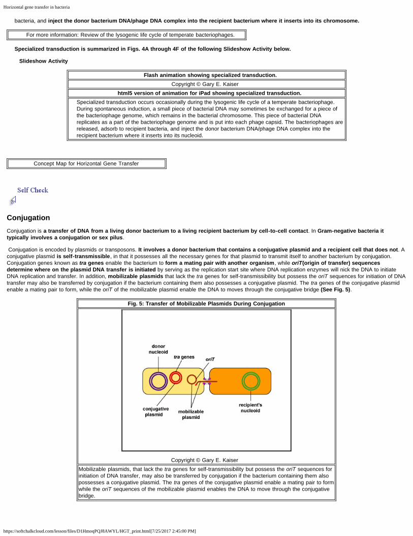
Conjugation is encoded by plasmids or transposons. It involves a donor bacterium that contains a conjugative plasmid and a recipient cell that does not. Aconjugative plasmid is self-transmissible, in that it possesses all the necessary genes for that plasmid to transmit itself to another bacterium by conjugation.Conjugation genes known as tra genes enable the bacterium to form a mating pair with another organism, while oriT(origin of transfer) sequences
determine where on the plasmid DNA transfer is initiated by serving as the replication start site where DNA replication enzymes will nick the DNA to initiateDNA replication and transfer. In addition, mobilizable plasmids that lack the tra genes for self-transmissibility but possess the oriT sequences for initiation of DNAtransfer may also be transferred by conjugation if the bacterium containing them also possesses a conjugative plasmid. The tra genes of the conjugative plasmidenable a mating pair to form, while the oriT of the mobilizable plasmid enable the DNA to moves through the conjugative bridge (See Fig. 5).
Fig. 5: Transfer of Mobilizable Plasmids During Conjugation
Copyright © Gary E. Kaiser Mobilizable plasmids, that lack the tra genes for self-transmissibility but possess the oriT sequences forinitiation of DNA transfer, may also be transferred by conjugation if the bacterium containing them alsopossesses a conjugative plasmid. The tra genes of the conjugative plasmid enable a mating pair to formwhile the oriT sequences of the mobilizable plasmid enables the DNA to move through the conjugativebridge.

Horizontal gene transfer in bacteria
https://softchalkcloud.com/lesson/files/D1HmoqPQJ8AWYL/HGT_print.html[7/25/2017 2:45:00 PM]
Transposons ("jumping genes") are small pieces of DNA that encode enzymes that enable the transposon to move from one DNA location to another, either
on the same molecule of DNA or on a different molecule. Transposons may be found as part of a bacterium's chromosome (conjugative transposons) or inplasmids and are usually between one and twelve genes long. A transposon contains a number of genes, such as those coding for antibiotic resistance or othertraits, flanked at both ends by insertion sequences coding for an enzyme called transpoase. Transpoase is the enzyme that catalyzes the cutting and resealing ofthe DNA during transposition.
For more information: Review of plasmids and transposons.
Flash animation illustrating transposons.
Copyright © Gary E. Kaiser html5 version of animation for iPad illustrating transposons.
Transposons (transposable elements or "jumping genes" are small pieces of DNA that encode enzymes thattranspose the transposon, that is, move it from one DNA location to another, either on the same molecule ofDNA or on a different molecule. A transposon contains a number of genes, coding for antibiotic resistance orother traits, flanked at both ends by insertion sequences coding for an enzyme called transpoase. Transpoase isthe enzyme that catalyzes the cutting and resealing of the DNA during transposition. Thus, such transposonsare able to cut themselves out of a bacterial chromosome or a plasmid and insert themselves into anotherchromosome or plasmid and contribute in the transmission of antibiotic resistance among a population ofbacteria.
Conjugative transposons, like conjugative plasmids, carry the genes that enable mating pairs to form for conjugation. Therefore, conjugative transposonsalso enable mobilizable plasmids and nonconjugative transposons to be transferred to a recipient bacterium during conjugation.
Many conjugative plasmids and conjugative transposons possess rather promiscuous transfer systems that enable them to transfer DNA not only to like
species, but also to unrelated species. The ability of bacteria to adapt to new environments as a part of bacterial evolution most frequently results from theacquisition of large DNA sequences from another bacterium by conjugation.
a. Transfer of conjugative plasmids by conjugation in Gram-negative bacteria
In Gram-negative bacteria, the first step in conjugation involves a conjugation pilus (sex pilus or F pilus) on the donor bacterium binding to a recipient
bacterium lacking a conjugation pilus. Typically the conjugation pilus retracts or depolymerizes pulling the two bacteria together. A series of membrane
proteins coded for by the conjugative plasmid then forms a bridge and an opening between the two bacteria, now called a mating pair.
Using the rolling circle model of DNA replication, a nuclease breaks one strand of the plasmid DNA at the origin of transfer site (oriT) of the plasmid and
that nicked strand enters the recipient bacterium. The other strand remains behind in the donor cell. Both the donor and the recipient plasmid strands
then make a complementary copy of themselves. Both bacteria now possess the conjugative plasmid.
For more information: Review of pili.
This process is summarized in Figs 6A through 6F of the following Slideshow Activity below.
Slideshow Activity
Flash animation showing transfer of conjugative plasmids by conjugation in Gram-negative bacteria.
Copyright © Gary E. Kaiser html5 version of animation for iPad showing transfer of conjugative plasmids by conjugation in Gram-
negative bacteria. Conjugation involves a donor bacterium that contains a conjugative plasmid and a recipient cell that does not. Aconjugative plasmid is self-transmissible, in that it possesses all the necessary genes for that plasmid totransmit itself to another bacterium by conjugation. Conjugation genes known as tra genes enable the bacteriumto form a mating pair with another organism, while oriT (origin of transfer) sequences determine where on theplasmid DNA transfer is initiated by serving as the replication start site where DNA replication enzymes will nickthe DNA to initiate DNA replication and transfer. In addition, mobilizable plasmids that lack the tra genes for self-transmissibility but possess the oriT for initiation of DNA transfer may also be transferred by conjugation if thebacterium containing them also possesses a conjugative plasmid. The tra genes of the conjugative plasmidenable a mating pair to form and the oriT of the mobilizable plasmid enable the DNA to replicate as it movesthrough the conjugative bridge.
This is the mechanism by which resistance plasmids (R-plasmids), coding for multiple antibiotic resistance and conjugation pilus formation, are

Horizontal gene transfer in bacteria
https://softchalkcloud.com/lesson/files/D1HmoqPQJ8AWYL/HGT_print.html[7/25/2017 2:45:00 PM]
transferred from a donor bacterium to a recipient. This is a big problem in treating opportunistic gram-negative infections such as urinary tract infections, woundinfections, pneumonia, and septicemia by such organisms as E. coli, Proteus, Klebsiella, Enterobacter, Serratia, and Pseudomonas, as well as with intestinalinfections by organisms like Salmonella and Shigella.
Flash animation showing transfer of R-plasmids by conjugation in Gram-negative bacteria.
Copyright © Gary E. Kaiser html5 version of animation for iPad showing transfer of R-plasmids by conjugation in Gram-negative
bacteria. R plasmids are conjugative plasmids coding for mating pair formation and also multiple antibiotic resistance. Aconjugative plasmid is self-transmissible, in that it possesses all the necessary genes for that plasmid totransmit itself to another bacterium by conjugation. Conjugation genes known as tra genes enable the bacteriumto form a mating pair with another organism, while oriT (origin of transfer) genes determine where on theplasmid DNA transfer is initiated. The plasmid also possess genes coding for resistance to a number of differentantibiotics.
There is also evidence that the conjugation pilus may also serve as a direct channel through which single-stranded DNA may be transferred during conjugation.
b. F+ conjugation
F+ conjugation results in the transfer of an F+ plasmid possessing tra genes coding only for a conjugation pilus and mating pair formation from a donorbacterium to a recipient bacterium. One strand of the F+ plasmid is broken with a nuclease at the origin of transfer (oriT) sequence that determines whereon the plasmid DNA transfer is initiated by serving as the replication start site where DNA replication enzymes will nick the DNA to initiate DNA replication andtransfer. The nicked strand enters the recipient bacterium while the other plasmid strand remains in the donor. Each strand then makes acomplementary copy. The recipient then becomes an F+ male and can make a sex pilus.
F+ conjugation is shown in Fig.7A through 7F of the following Slideshow Activity below.
Slideshow Activity
Flash animation showing F+ conjugation.
Copyright © Gary E. Kaiser html5 version of animation for iPad showing F+ conjugation.
F+ conjugation involves a donor bacterium that contains a conjugative plasmid known as an F+ plasmid and arecipient cell that does not. A conjugative plasmid is self-transmissible, in that it possesses all the necessarygenes for that plasmid to transmit itself to another bacterium by conjugation. Conjugation genes known as tragenes enable the bacterium to form a mating pair with another organism, while oriT (origin of transfer) genesdetermine where on the plasmid DNA transfer is initiated.The F+ plasmid codes only for formation of aconjugation pilus. Mobilizable plasmids, however, that lack the tra genes for self-transmissibility but possess theoriT genes for initiation of DNA transfer may also be transferred by conjugation if the bacterium containing themalso possesses a conjugative plasmid. The tra genes of the conjugative plasmid enable a mating pair to formand the oriT genes of the mobilizable plasmid enable the DNA to moves through the conjugative bridge.
In addition, mobilizable plasmids that lack the tra genes for self-transmissibility but possess the oriT sequences for initiation of DNA transfer may also betransferred by conjugation. The tra genes of the F+ plasmid enables a mating pair to form and the oriT sequences of the mobilizable plasmid enables the DNAto moves through the conjugative bridge (See Fig. 5 above).
c. Hfr (high frequency recombinant) conjugation
Hfr conjugation begins when an F+ plasmid with tra genes coding for mating pair formation inserts or integrates into the nucleoid to form an Hfr
bacterium. A nuclease then breaks one strand of the donor's DNA at the origin of transfer (oriT) location of the inserted F+ plasmid and the nicked strand
of the donor DNA begins to enter the recipient bacterium. The remaining non-nicked DNA strand remains in the donor and makes a complementary copy ofitself.
The bacterial connection usually breaks before the transfer of the entire chromosome is completed so the remainder of the F+ plasmid seldom enters therecipient. As a result, there is a transfer of some chromosomal DNA, which may be exchanged for a piece of the recipient's DNA through homologousrecombination, but not the ability to form a conjugation pilus and mating pairs.
Hfr conjugation is shown in Fig. 8A through 8E in the following Slideshow Activity below.
Slideshow Activity
Flash animation showing Hfr conjugation in bacteria.
Copyright © Gary E. Kaiser html5 version of animation for iPad showing Hfr conjugation in bacteria.
Hfr recombination begins when an F+ plasmid with tra genes coding for mating pair formation inserts or
Horizontal gene transfer in bacteria
https://softchalkcloud.com/lesson/files/D1HmoqPQJ8AWYL/HGT_print.html[7/25/2017 2:45:00 PM]
integrates into the chromosome to form an Hfr bacterium. A nuclease then breaks one strand of the donor'sDNA at the origin of transfer (oriT) location of the inserted F+ plasmid and the nicked strand of the donor DNAbegins to enter the recipient bacterium. The remaining non-nicked DNA strand remains in the donor and makesa complementary copy of itself. The bacterial connection usually breaks before the transfer of the entirechromosome is completed so the remainder of the F+ plasmid seldom enters the recipient. As a result, there isa transfer of some chromosomal DNA, which may be exchanged for a piece of the recipient's DNA throughhomologous recombination, but not the ability to form a conjugation pilus and mating pairs.
Class activity
Watch University of Texas at San Antonio microbiologist Karl Klose discussesthe problem of antibiotic resistance in a 2013 TED talk.
Use your notes to name and describe the bacterial mechanisms of horizontalgene transfer referred to in this talk as "funeral grab," "viral pass," and "making
whoopee."
TPS Questions
Concept Map for Horizontal Gene Transfer
Self Quiz for Horizontal Gene Transfer
Back to Unit 2 Table of Contents
Back to Softchalk Lessons Table of Contents

Bacterial quorum sensing and bacterial secretion systems
https://softchalkcloud.com/lesson/files/wLNsfX5laBoj7b/quorum_sensing_print.html[11/30/2016 3:51:37 PM]
Bacterial quorum sensing and bacterial secretion systems
BACTERIAL QUORUM SENSING, PATHOGENICITY ISLANDS, AND SECRETION SYSTEMS (INJECTISOMES)
Bacterial Quorum Sensing, Pathogenicity Islands, and Secretion Systems (Injectisomes)
Fundamental Statements for this Softchalk Lesson:
1. Pathogenicity is the ability of a microbe to cause disease and inflict damage upon its host; virulence is the degree of pathogenicity within a group orspecies of microbes.2. The pathogenicity of an organism is determined by its virulence factors.
3. Virulence factors enable that bacterium to colonize the host, resist body defenses, and harm the body.4. Most of the virulence factors are the products of quorum sensing genes.5. Quorum sensing involves the production, release, and community-wide sensing of molecules called autoinducers that modulate gene expression,and ultimately bacterial behavior, in response to the density of a bacterial population.6. The outcomes of bacteria-host interaction are often related to bacterial population density.7. At a low density of bacteria, the autoinducers diffuse away from the bacteria and there are insufficient quantities of these molecules to activate thequorum sensing genes that enable the bacteria to act as a population. As a result the bacteria behave as individual, single-celled organisms.8. Acting as individual organisms may enable a low density of bacteria to gain a better foothold in their new environment by enabling bacteria to usemotility and taxis to contact host cells, use pili to initially adhere to and crawl over host cell surfaces, use adhesins to adhere to host cells and resistflushing, and secrete a glycocalyx to form microcolonies.9. As the bacteria increase in numbers geometrically as a result of binary fission and reach high density, large quantities of autoinducers areproduced and are able to bind to the signaling receptors on the bacterial surface in sufficient quantity so as to activate the quorum sensing genes thatenable the bacteria to now behave as a multicellular population.10. By behaving as a multicellular population, individual bacteria within a group are able to benefit from the activity of the entire group.11. As the entire population of bacteria simultaneously turn on their virulence genes, the body's immune systems are much less likely to have enoughtime to counter those virulence factors before harm is done. Virulence factors such as exoenzymes and toxins can damage host cells enabling thebacteria in the biofilm to obtain nutrients.12. As the area becomes over-populated with bacteria, quorum sensing enables some of the bacteria to escape the biofilm and return to individual

Bacterial quorum sensing and bacterial secretion systems
https://softchalkcloud.com/lesson/files/wLNsfX5laBoj7b/quorum_sensing_print.html[11/30/2016 3:51:37 PM]
single-celled organism behavior in order to find a new sight to colonize.13. Quorum sensing enables bacteria to communicate with members of their own species, with other species of bacteria, and with their eukaryotichost cells.14. Most genes coding for virulence factors in bacteria are located in pathogenicity islands or PAIs and are usually acquired by horizontal genetransfer.15. Many bacteria involved in infection have the ability to co-opt the functions of the host cell for the bacterium's own benefit by producing secretionssystems that enable the bacterium to directly inject bacterial effector molecules into the cytoplasm of the host cell in order to alter the host cell'scellular machinery, cellular function, or cellular communication.
Common Course Objectives
1. Relate quorum sensing gene expression to the population of bacteria and how this can relate to bacterial pathogenicity.
2. Describe pathogenicity islands and how they are primarily transferred from one bacterium to another.
3. Describe how bacterial secretion systems can influence the ability of a bacterium to influence the action of other bacteria or of eukaryotic cells.
4. Describe mechanisms of how a normally communalistic bacterium can become pathogenic.
Detailed Learning Objectives
1*. Define the following:
a. pathogenicity
b. virulence
2**. Define and briefly describe the overall process of quorum sensing in bacteria and how it may enable bacteria to behave as a multicellular population.
3*. State at least two possible advantages of individual bacterial behavior.
4*. State at least two possible advantages of multicellular bacterial behavior.
5. State what is meant by intraspecies, interspecies, and interkingdom communication.
6*. State the function of bacterial secretions systems (injectosomes) such as the type 3 and type 6 secretion systems in bacterial pathogenicity.
(*) = Common theme throughout the course
(**) = More depth and common theme
TPS Questions
Bacterial Quorum Sensing
In this Learning Object we are going to look at several aspects of bacterial genetics that are directly related to bacterial pathogenicity, namely, quorum sensing,pathogenicity islands, and secretion systems. Pathogenicity and virulence are terms that refer to an organism's ability to cause disease. Pathogenicity is the abilityof a microbe to cause disease and inflict damage upon its host, whereas virulence is the degree of pathogenicity within a group or species of microbes as indicatedby case fatality rates and/or the ability of the organism to invade the tissues of the host. The pathogenicity of an organism, that is its ability to cause disease, isdetermined by its virulence factors.
Many of the virulence factors that enable bacteria to colonize the body and/or harm the body are the products of quorum sensing genes. Many bacteria are able tosense their own population density, communicate with each other by way of secreted chemical factors, and behave as a population rather than as
individual bacteria . This plays an important role in pathogenicity and survival for many bacteria.
Bacteria can behave either as individual single-celled organisms or as multicellular populations. Bacteria exhibit these behaviors by chemically "talking" toone another through a process called quorum sensing. Quorum sensing involves the production, release, and community-wide sensing of molecules called
autoinducers that modulate gene expression, and ultimately bacterial behavior, in response to the density of a bacterial population.
To initiate the process of quorum sensing, bacterial genes code for the production of signaling molecules called autoinducers that are released into thebacterium's surrounding environment.Th ese signaling molecules then bind to signaling receptors either on the bacterial surface or in the cytoplasm. When theseautoinducers reach a critical, threshold level, they activate bacterial quorum sensing genes that enable the bacteria to behave as a multicellular population

Bacterial quorum sensing and bacterial secretion systems
https://softchalkcloud.com/lesson/files/wLNsfX5laBoj7b/quorum_sensing_print.html[11/30/2016 3:51:37 PM]
rather than as individual single-celled organisms (see Fig. 1). The autoinducer/receptor complex is able to bind to DNA promoters and activate the transcriptionof quorum sensing-controlled genes in the bacterium. In this way, individual bacteria within a group are able to benefit from the activity of the entire group.
Fig. 1: Mechanism for Quorum Sensing
Copyright © Gary E. Kaiser Bacteria "talk" to one another through a process called quorum sensing. Bacterial genes code for theproduction of signaling molecules called autoinducers that are released into the surrounding environment.These signaling molecules then bind to signaling receptors either on the bacterial surface or in thecytoplasm, in this case, on the surface. When these autoinducers reach a critical, threshold level, theyactivate bacterial quorum sensing genes that enable the bacteria to behave as a multicellular populationrather than as individual single-celled organism. The autoinducer/receptor complex is able to bind to DNApromoters and activate the transcription of quorum sensing-controlled genes in the bacterium. In this way,individual bacteria within a group are able to benefit from the activity of the entire group.
Flash animation showing quorum sensing with a low density and a high density of bacteria.
Copyright © Gary E. Kaiser html5 version of animation showing quorum sensing with a low density and a high density of bacteria.
At a low density of bacteria, the signaling molecules (autoinducers) diffuse away from the bacteria. Sufficientquantities of these molecules are unavailable for binding to the signaling receptors on the bacterial surface andthe quorum sensing genes that enable the bacteria to act as a population are not activated. The bacterium thenutilizes genes that enable the bacterium to act as an individual organism rather than as a multicellular population.Acting as individual organisms may better enable that low density of bacteria to gain a better foothold in their newenvironment.
At a high density of bacteria, sufficient quantities of signaling molecules (autoinducers) are available for binding tothe signaling receptors on the bacterial surface and the quorum sensing genes that enable the bacteria to act as apopulation become activated. The outcomes of bacteria-host interaction are often related to bacterial populationdensity. Bacterial virulence, that is its ability to cause disease, is largely based on the bacterium's ability toproduce gene products called virulence factors that enable that bacterium to colonize the host, resist bodydefenses, and harm the body.
1. In Gram-negative bacteria, the autoinducers are typically molecules called acyl-homoserine lactones or AHL. AHLs diffuse readily out of and into bacterialcells where they bind to AHL receptors in the cytoplasm of the bacteria. When a critical level of AHL is reached, the cytoplasmic autoinducer/receptor complexfunctions as a DNA-binding transcriptional activator.
2. In Gram-positive bacteria, the autoinducers are oligopeptides, short peptides typically 8-10 amino acids long. Oligopeptides cannot diffuse in and out ofbacteria like AHLs, but rather leave bacteria via specific exporters. They then bind to autoinducer receptors on the surface of the bacterium. When a critical levelof oligopeptide is reached, the binding of the oligopeptide to its receptor starts a phosphorylation cascade that activates DNA-binding transcriptional regulatoryproteins called response regulators.
The outcomes of bacteria-host interaction are often related to bacterial population density. Bacterial virulence, that is its ability to cause disease, is largelybased on the bacterium's ability to produce gene products called virulence factors that enable that bacterium to colonize the host, resist body defenses, and
harm the body.

Bacterial quorum sensing and bacterial secretion systems
https://softchalkcloud.com/lesson/files/wLNsfX5laBoj7b/quorum_sensing_print.html[11/30/2016 3:51:37 PM]
A. At a low density of bacteria, the autoinducers diffuse away from the bacteria (see Fig. 2). Sufficient quantities of these molecules are unable to bind to thesignaling receptors on the bacterial surface and the quorum sensing genes that enable the bacteria to act as a population are not activated. This enables thebacteria to behave as individual, single-celled organisms.
Fig. 2: Quorum Sensing with a Low Density of Bacterial Cells
Copyright © Gary E. Kaiser At a low density of bacteria, the signaling molecules (autoinducers) diffuse away from the bacteria.Sufficient quantities of these molecules are unavailable for binding to the signaling receptors on thebacterial surface (Gram-positive bacteria) or in the cytoplasm (Gram-negative bacteria), and the quorumsensing genes that enable the bacteria to act as a population are not activated. The bacterium thenutilizes genes that enable the bacterium to act as an individual organism rather than as a multicellularpopulation. Acting as individual organisms may better enable that low density of bacteria to gain a betterfoothold in their new environment.
Possible advantages of individual bacterial behavior seen at low bacterial density: Producing virulence factors that better enable them to colonize the
body.
If a relatively small number of a specific bacterium were to enter the body and immediately start producing their virulence factors, chances are the body's immunesystems would have sufficient time to recognize and counter those virulence factors and remove the bacteria before there was sufficient quantity to cause harm.The bacterium instead utilizes genes that enable it to act as an individual organism rather than as part of a multicellular population.
Acting as individual organisms may better enable that low density of bacteria to gain a better foothold in their new environment in the following ways:
1. Many bacteria are capable of motility and motility serves to keep bacteria in an optimum environment via taxis.
Motility and chemotaxis probably help some intestinal and urinary pathogens to move through the mucous layer so they can attach to the epithelial cells
of the mucous membranes. In fact, many bacteria that can colonize the mucous membranes of the bladder and the intestines are motile. Motility probablyhelps these bacteria move through the mucus in places where it is less viscous.
Flash animation showing a motile bacterium contacting a host cell by swimming through the mucus.
Copyright © Gary E. Kaiser html5 version of animation for iPad showing a motile bacterium contacting a host cell by swimming
through the mucus. The mucosal surfaces of the bladder and the intestines constantly flush bacteria away in order to preventcolonization. Motile bacteria that can swim chemotactically toward mucosal surfaces may have a better chance tomake contact with the mucous membranes, attach, and colonize. Many bacteria that can colonize the mucousmembranes of the bladder and the intestines are motile. Motility probably helps these bacteria move through themucus in places where it is less viscous.
Movie of swimming Escherichia coli as seen with phase
contrast microscopy
Courtesy of Dr. Howard C. Berg from the Roland Institute atHarvard.
Movie of motile Escherichia coli with fluorescent-
labelled flagella
Courtesy of Dr. Howard C. Berg from the RolandInstitute at Harvard.

Bacterial quorum sensing and bacterial secretion systems
https://softchalkcloud.com/lesson/files/wLNsfX5laBoj7b/quorum_sensing_print.html[11/30/2016 3:51:37 PM]
2. One of the body's innate defenses is the ability to physically remove bacteria from the body through such means as the constant shedding of surfaceepithelial cells from the skin and mucous membranes, the removal of bacteria by such means as coughing, sneezing, vomiting, and diarrhea, and bacterialremoval by bodily fluids such as saliva, blood, mucous, and urine. Bacteria may resist this physical removal by producing pili (see Fig. 3), cell wall
adhesins proteins (see Fig. 4), and/or biofilm-producing capsules. Some pili, called type IV pili also allow some bacteria to "walk" or "crawl" along
surfaces to spread out and eventually form microcolonies.
Fig. 3: Adhesive Tip of Bacterial
PiliFig. 4: Bacterial Adhesins
Copyright © Gary E. Kaiser
Surface proteins called adhesins in the bacterial cell wall bind toreceptor molecules on the surface of a susceptible host cellenabling the bacterium to make intimate contact with the host cell,adhere, colonize, and resist flushing.
Copyright © Gary E. Kaiser
Flash animation showing a bacterium using adhesins to adhere to a host cell.
Copyright © Gary E. Kaiser html5 version of animation for iPad showing a bacterium using adhesins to adhere to a host cell.
Surface proteins called adhesins in the bacterial cell wall bind to receptor molecules on the surface of asusceptible host cell enabling the bacterium to make intimate contact with the host cell, adhere, colonize, andresist flushing.
Flash animation showing a bacterium using both pili and adhesins to adhere to a host cell.
Copyright © Gary E. Kaiser html5 version of animation for iPad showing a bacterium using both pili and adhesins to adhere to a host
cell. Pili enable some organisms to adhere to receptors on target host cells. The pilus has a shaft composed of aprotein called pilin. At the end of the shaft is the adhesive tip structure having a shape corresponding to that ofspecific glycoprotein or glycolipid receptors on a host cell. Because both the bacteria and the host cells have anegative charge, pili may enable the bacteria to bind to host cells without initially having to get close enough to bepushed away by electrostatic repulsion. Once attached to the host cell, the pili can depolymerize and this enablesbacterial cell wall adhesins to bind to adhesin receptors on the host cell. This allows the bacterial cell wall to makemore intimate contact with the host cell and enables the bacterium to colonize the host cell and resist flushing.There is also evidence that the binding of pili to host cell receptors can serve as a trigger for activating thesynthesis of some cell wall adhesins.
Movie of twitching motility of PseudomonasCourtesy of Dr. Howard C. Berg from the Roland Institute at
Harvard

Bacterial quorum sensing and bacterial secretion systems
https://softchalkcloud.com/lesson/files/wLNsfX5laBoj7b/quorum_sensing_print.html[11/30/2016 3:51:37 PM]
Scanning electron micrograph E. coli with pili; courtesy of Dennis Kunkel's Microscopy.
3. Many bacteria secrete an extracellular polysaccharide or polypeptide matrix called a capsule or glycocalyx that enables the bacteria to adhere to hostcells, resist phagocytosis, and form microcolonies.
As the bacteria geometrically increase in number by binary fission, so does the amount of their secreted autoinducers, and production of high levels ofautoinducers then enables the population of bacteria to communicate with one another by quorum sensing.
At a high density of bacteria, large quantities of autoinducers are produced (see Fig. 5) and are able to bind to the signaling receptors on the bacterial surface insufficient quantity so as to activate the quorum sensing genes that enable the bacteria to behave as a multicellular population (see Fig. 1 above).
Fig. 5: Quorum Sensing with a High Density of Bacterial Cells
Copyright © Gary E. Kaiser At a high density of bacteria, sufficient quantities of signaling molecules (autoinducers) are available forbinding to the signaling receptors on the bacterial surface (Gram-positive bacteria) or in the cytoplasm(Gram-negative bacteria), and the quorum sensing genes that enable the bacteria to act as a populationbecome activated. The outcomes of bacteria-host interaction are often related to bacterial populationdensity. Bacterial virulence, that is its ability to cause disease, is largely based on the ability of thebacterium to produce gene products called virulence factors that enable that bacterium to colonize thehost, resist body defenses, and harm the body.
Animation of the molecular cascade during bacterial quorum sensing.
Courtesy of HHMI's Biointeractive.
Advantages of multicellular behavior seen at high bacterial density: Better enabling the bacterial population to persist in the body.
1. By behaving as a multicellular population, individual bacteria within a group are able to benefit from the activity of the entire group. As the entirepopulation of bacteria simultaneously turn on their virulence genes, the body's immune systems are much less likely to have enough time to counter thosevirulence factors before harm is done.
2. This triggers production of an extracellular adhesive matrix (glycocalyx) enabling the bacteria to form microcolonies and irreversibly attachment to themucous membranes. Biofilm formation begins.
3. Virulence factors such as exoenzymes and toxins can damage host cells enabling the bacteria in the biofilm to obtain nutrients. The biofilm continues
to develop and mature.
4. As the area becomes over-populated with bacteria, quorum sensing enables some of the bacteria to escape the biofilm, often by again producing
flagella, and return to individual single-celled organism behavior in order to find a new sight to colonize.

Bacterial quorum sensing and bacterial secretion systems
https://softchalkcloud.com/lesson/files/wLNsfX5laBoj7b/quorum_sensing_print.html[11/30/2016 3:51:37 PM]
Pseudomonas aeruginosa is an example of a quorum sensing bacterium. P. aeruginosa causes severe hospital-acquired infections, chronic infections in people withcystic fibrosis, and potentially fatal infections in those who are immunocompromised.
1. When P. aeruginosa first enters the body, they are at a low density of bacteria. The autoinducers diffuse away from the bacteria (see Fig. 2 above),sufficient quantities of these molecules are unable to bind to the signaling receptors, and the quorum sensing genes that enable the bacteria to act as a populationare not activated. The P. aeruginosa continue to function as individual bacteria. Motility genes (coding for flagella) and adhesin genes (coding for pili and cellwall adhesins) are expressed. The flagella enable the initial bacteria to swim through mucus towards host tissues such as mucous membranes. Pili then enablethe bacteria to reversibly attach to host cells in order to resist flushing and begin colonization (See Fig. 6A). Type IV pili, which enable a twitching motility in somebacteria, then enable the bacteria as they replicate to crawl along and spread out over the mucous membranes (See Fig. 6B). The pili subsequently retract andbacterial cell wall adhesins enable a more intimate attachment of the bacterium to the mucous membranes (See Fig. 6C).
Fig. 6A: Development of a Biofilm by Pseudomonas aeruginosa: Step 1
Copyright © Gary E. Kaiser Planktonic Pseudomonas aeruginosa use their polar flagella and chemotaxis to swim towards hostmucous membranes. Pili then bind to host cell receptors for initial but reversible bacterial attachment.
Fig. 6B: Development of a Biofilm by Pseudomonas aeruginosa: Step 2
Copyright © Gary E. Kaiser As the bacteria begin to replicate, type IV pili enable the bacteria, by way of twitching motility, to crawlalong the surface of the mucous membranes and spread out.
Fig. 6C: Development of a Biofilm by Pseudomonas aeruginosa: Step 3

Bacterial quorum sensing and bacterial secretion systems
https://softchalkcloud.com/lesson/files/wLNsfX5laBoj7b/quorum_sensing_print.html[11/30/2016 3:51:37 PM]
Copyright © Gary E. Kaiser The pili retract and bacterial cell wall adhesins enable a more intimate attachment of the bacterium.
2. Once P. aeruginosa has colonized, it is able to replicate geometrically and achieve a high population density. Quorum sensing genes are activated and thebacteria function as a population. This triggers production of an extracellular polysaccharide called alginate to form microcolonies and enables irreversibleattachment to the mucous membranes (See Fig. 6D). Biofilm formation begins.
Fig. 6D: Development of a Biofilm by Pseudomonas aeruginosa: Step 4
Copyright © Gary E. Kaiser As the bacteria replicate, quorum sensing genes trigger production of an extracellular polysaccharidecalled alginate to form microcolonies and enable irreversible attachment to the mucous membranes.Biofilm formation begins.
3. Quorum sensing genes coding for enzymes and toxins that damage host cells are produced. These are injected into the host cells by way of aninjectosome. This releases nutrients for the bacteria in the biofilm. The bacteria continue to replicate as the biofilm continues to develop, mushroom up, andmature (See Fig. 6E).
Fig. 6E: Development of a Biofilm by Pseudomonas aeruginosa: Step 5

Bacterial quorum sensing and bacterial secretion systems
https://softchalkcloud.com/lesson/files/wLNsfX5laBoj7b/quorum_sensing_print.html[11/30/2016 3:51:37 PM]
Copyright © Gary E. Kaiser Quorum sensing genes coding for enzymes and toxins that damage host cells are produced. Thisreleases nutrients for the bacteria in the biofilm. The bacteria continue to replicate as the biofilmcontinues to develop, mushroom up, and mature.
4. As the bacteria replicate, the biofilm continues to mature (See Fig. 6F). Water channels form within the biofilm to deliver water, oxygen, and nutrients to thegrowing population of P. aeruginosa. The high density of bacteria bacteria are now acting as a multicellular population rather than as individual bacteria.
Fig. 6F: Development of a Biofilm by Pseudomonas aeruginosa: Step 6
Copyright © Gary E. Kaiser As the bacteria replicate, the biofilm continues to mature. Water channels form within the biofilm to deliverwater, oxygen, and nutrients to the growing population of P. aeruginosa.
Electron micrograph of a biofilm of Haemophilus influenzae from Biomedcentral.com
The biofilm enables bacteria to:
a. resist attack by antibiotics;
b.trap nutrients for bacterial growth and remain in a favorable niche;
c. adhere to environmental surfaces and resist flushing;
d. live in close association and communicate with other bacteria in the biofilm; and
e. resist phagocytosis and attack by the body's complement pathways.
5. When the population of P. aeruginosa begins to outgrow their local environment, quorum sensing enables them to turn off adhesin genes and turn on
flagella genes that allow some of the bacteria to spread out of the biofilm to new location within that environment via motility (See Fig. 6G and Fig. 6H).
Fig. 6G: Development of a Biofilm by Pseudomonas aeruginosa: Step 7
Copyright © Gary E. Kaiser As the population begins to overgrow the area and nutrients become limited, quorum sensing genestrigger some of the P. aeruginosa in the biofilm to again produce flagella.

Bacterial quorum sensing and bacterial secretion systems
https://softchalkcloud.com/lesson/files/wLNsfX5laBoj7b/quorum_sensing_print.html[11/30/2016 3:51:37 PM]
Fig. 6H: Development of a Biofilm by Pseudomonas aeruginosa: Step 8
Copyright © Gary E. Kaiser Planktonic P. aeruginosa leave the biofilm and move to a new location to begin new biofilms.
It turns out that bacteria are multilingual. They use quorum sensing not only to "talk" to members their own species (intraspecies communication), but also to"talk" to bacteria that are not of their genus and species (interspecies communication). Intraspecies autoinducers and receptors enable bacteria tocommunicate with others of their own species while interspecies autoinducers and receptors enable bacteria to communicate with bacteria of a different species orgenus (see Fig. 7). The autoinducers for interspecies communications are referred to as AI-2 family autoinducers and are different from the intraspecies (AI-1)autoinducers. In some cases bacteria use interspeciecies communication to work cooperatively with various other bacteria in their biofilm to the benefit all
involved; in other cases, bacteria may use interspecies communication in such a way that one group benefits at the expense of another.
Fig. 7: Intraspecies and Interspecies Communication
Copyright © Gary E. Kaiser Intraspecies autoinducers and receptors enable bacteria to communicate with others of their own specieswhile interspecies autoinducers and receptors enable bacteria to communicate with bacteria of a differentspecies or genus.
Furthermore, bacteria are capable of interkingdom communication, communication between bacteria and their animal or plant host. Increasing numbers ofbacteria are being found that have signaling receptors that recognize human hormones. For example, a number of bacteria that are pathogens of the humanintestinal tract have a sensing molecule called QseC that binds the human hormones adrenaline and noradrenaline. This, in turn, activates various virulence genes
of the bacteria. On the other hand, some bacterial autoinducers can enter human host cells and regulate human cellular function. For example, at lowconcentration some bacterial autoinducers suppress host immune responses thus better enabling those bacteria to better establish themselves in the body. At highconcentrations, however, they stimulate an inflammatory response in the host to help the bacteria to spread from the initial infection site. One bacterial autoinducerhas been found to initiate apoptosis (cell suicide) in phagocytes such as neutrophils and macrophages.

Bacterial quorum sensing and bacterial secretion systems
https://softchalkcloud.com/lesson/files/wLNsfX5laBoj7b/quorum_sensing_print.html[11/30/2016 3:51:37 PM]
Concept Map for Bacterial Quorum Sensing, Pathogenicity Islands, and SecretionSystems
Pathogenicity Islands
The genomes of pathogenic bacteria, when compared with those of similar nonpathogenic species or strains, often show extra genes coding for virulence
factors, that is, molecules expressed and secreted by the bacterium that enable them to colonize the host, evade or inhibit the immune responses of the
host, enter into or out of a host cell, and/or obtain nutrition from the host. These include virulence factors such as capsules, adhesins, type 3 secretionsystems, invasins, and toxins.
Most genes coding for virulence factors in bacteria are located in pathogenicity islands or PAIs and are usually acquired by horizontal gene transfer. These PAIsmay be located in the bacterial chromosome, in plasmids, or even in bacteriophage genomes that have entered the bacterium. The genomes of most pathogenicbacteria typically contain multiple PAIs that can account for up to 10 - 20% of the bacterium's genome. PAIs carry genes such as transpoases, integrases, orinsertion sequences that enable them to insert into host bacterial DNA. Transfer RNA (tRNA) genes are often the target site for integration of PAIs. Conjugative
plasmids are the most frequent means of transfer of PAIs from one bacterium to another and the transfer of PAIs can then confer virulence to a previously
nonpathogenic bacterium.
Concept Map for Bacterial Quorum Sensing, Pathogenicity Islands, and SecretionSystems
Type 3 Secretion Systems (T3SS or Injectisomes) and Type 6 Secretion Systems (T6SS)
Many bacteria involved in infection have the ability to co-opt the functions of host cells for the bacterium's own benefit. This is done by way of bacterialsecretions systems that enable the bacterium to directly inject bacterial effector molecules into the cytoplasm of the host cell in order to alter its cellular
machinery or cellular communication to the benefit of the bacteria.
The most common type is the type 3 secretion system or T3SS (see Fig. 8). A secretion apparatus in the cytoplasmic membrane and cell wall of the bacteriumpolymerizes a hollow needle that is lowered to the cytoplasmic membrane of the host cell and a translocon protein is then delivered to anchor the needle to the hostcell. Effector proteins in the bacterium can now be injected into the cytoplasm of the host cell. The delivery system is sometimes called an injectisome. (A type 4secretion system can transfer effector proteins and/or DNA into the host cell because it is similar to the conjugation transfer system initiated by tra genes discussedunder horizontal gene transfer.)
Fig. 8: The Bacterial Type 3 Secretion System

Bacterial quorum sensing and bacterial secretion systems
https://softchalkcloud.com/lesson/files/wLNsfX5laBoj7b/quorum_sensing_print.html[11/30/2016 3:51:37 PM]
Copyright © Gary E. Kaiser Many bacteria involved in infection have the ability to co-opt the functions of the host cell for thebacterium's own benefit. This is done by way of bacterial secretions systems that enable the bacterium todirectly inject bacterial effector molecules into the cytoplasm of the host cell in order to alter its cellularmachinery or cellular communication. The most common type is the type 3 secretion system. A secretionapparatus in the cytoplasmic membrane and cell wall of the bacterium polymerizes a hollow needle that is lowered to the cytoplasmic membrane of the host cell and a translocon protein is then delivered toanchor the needle to the host cell. Effector proteins in the bacterium can now be injected into thecytoplasm of the host cell. The delivery system is sometimes called an injectisome.
Article, Illustrations, and electron micrographs of injectisomes from the MarlovitsLab.
Electron micrograph of an injectisome from Wikipedia.
Some bacteria, such as Pseudomonas aeruginosa and Vibrio cholerae, produce a type 6 secretion system, or T6SS, that consists of a protein tube surroundedby a contractile sheath, similar to the tail of T4-bacteriophages. (A bacteriophage is a virus that only infects bacteria.) The type 6 secretion system not only
injects effector molecules into eukaryotic cells, but also is able to inject antibacterial effector molecules into other bacteria in order to kill those
bacteria. Predator bacteria can use their T6SS to kill prey bacteria. In fact, V. cholerae and P. aeruginosa have been shown to "duel" with one another via theirrespective T6SSs.
Concept map for Introduction to Pathogenesis and QuorumSensing
V. cholerae also uses its T6SS to promote horizontal gene transfer by way of transformation. Individual V. cholerae cells also use their T6SS to attack oneanother upon cell-to-cell contact. Most members of the population, however, produce immunity proteins that protect them from being killed by the effectormolecules that are injected. Not all strains of V. cholerae in the population, however, produce these immunity proteins and these non-immune cells aresubsequently lysed, releasing their DNA into the environment. This DNA can then be taken up by neighboring competent V. cholerae via transformation.
For more information: Review of Horizontal Gene Transfer
TPS Questions
iBiology YouTube Lecture on Microbial Pathogenicity

Bacterial quorum sensing and bacterial secretion systems
https://softchalkcloud.com/lesson/files/wLNsfX5laBoj7b/quorum_sensing_print.html[11/30/2016 3:51:37 PM]
Concept Map for Bacterial Quorum Sensing, Pathogenicity Islands, and SecretionSystems
Self Quiz for Quorum Sensing, Pathogenicity Islands, and Secretion Systems
Back to Unit 2 Table of Contents
Back to Softchalk Lessons Table of Contents

Enzyme regulation in bacteria
https://softchalkcloud.com/lesson/files/g7SExrjpOJPIby/enzyme_regulation_print.html[11/30/2016 3:51:56 PM]
Enzyme regulation in bacteria
ENZYME REGULATION
Bacterial Enzyme Regulation
Fundamental Statements for this Softchalk Lesson:
1. In living cells there are hundreds of different enzymes working together in a coordinated manner, and since cells neither synthesize nor break downmore material than is required for normal metabolism and growth, precise enzyme regulation is required for turning metabolic reactions on and off.2. There is tremendous diversity in the mechanisms bacteria use to regulate enzyme synthesis and enzyme activity.3. Ways in which enzymes can be controlled or regulated include controlling the synthesis of the enzyme (genetic control) and controlling the activityof the enzyme (feedback inhibition).4. In prokaryotes, genetic control of enzyme activity includes the induction or repression of enzyme synthesis by regulatory proteins that can bind toDNA and either block or enhance the function of RNA polymerase, the enzyme required for transcription.5. An operon is a set of genes collectively controlled by a regulatory protein.6. Regulatory proteins may function either as repressors or activators.7. Repressors are regulatory proteins that block transcription of mRNA by preventing RNA polymerase from transcribing the coding sequence for theenzymes.8. Some repressors, as in the case of the trp operon, are synthesized in a form that cannot by itself bind to the operator. This is referred to as arepressible system. The binding of a molecule called a corepressor, however, alters the shape of the regulatory protein to a form that can bind to theoperator and subsequently block transcription.9. Some repressors, as in the case of the lac operon, are synthesized in a form that readily binds to the operator and blocks transcription. However,the binding of a molecule called an inducer alters the shape of the regulatory protein in a way that now blocks its binding to the operator and thuspermits transcription. This is referred to as an inducible system.10. Activators are regulatory proteins that promote transcription of mRNA by enabling RNA polymerase to transcribing the coding sequence for theenzymes.11. Enhancers are regulatory proteins that bind to DNA located some distance from the operon they control by working with DNA-bending proteins.The DNA-bending proteins bend the DNA in a way that now allows the enhancer to interact with the promoter in such a way that RNA polymerase can

Enzyme regulation in bacteria
https://softchalkcloud.com/lesson/files/g7SExrjpOJPIby/enzyme_regulation_print.html[11/30/2016 3:51:56 PM]
now bind and initiate transcription12. Bacteria also use translational control of enzyme synthesis. One method is for the bacteria to produce noncoding RNA (ncRNA) molecules that arecomplementary to the mRNA coding for the enzyme, and when the small RNA binds to the mRNA by complementary base pairing, ribosomes cannotattach to the mRNA, the mRNA is not transcribed and translated into protein, and the enzyme is not made. In bacteria, these ncRNAs are often calledsmall RNAs (sRNAs).13. Feedback inhibition controls the activity of the enzyme rather than its synthesis and can be noncompetitive or competitive.14. In the case of non-competitive inhibition, the inhibitor is the end product of a metabolic pathway that is able to bind the allosteric site on theenzyme. Binding of the inhibitor to the allosteric site alters the shape of the enzyme's active site thus preventing binding of the first substrate in themetabolic pathway. In this way, the pathway is turned off.15. In the case of what is called competitive inhibition, the inhibitor is the end product of an enzymatic reaction. That end product is also capable ofreacting with the enzyme's active site and prevents the enzyme from binding its normal substrate. As a result, the end product is no longersynthesized.
Common Course Objectives
1. Compare and contrast the genetic control of enzyme activity (enzyme synthesis) in bacteria with the control of enzyme activity through feedback inhibition.
2. Compare and contrast an inducible operon with a repressible operon and give an example of each.
3. Compare and contrast competitive inhibition with noncompetitive inhibition.
Detailed Learning Objectives
1. Compare and contrast the genetic control of enzyme activity (enzyme synthesis) in bacteria with the control of enzyme activity through feedback inhibition.
2. Compare and contrast an inducible operon with a repressible operon and give an example of each.
3. Compare how the presence or absence of tryptophan affects the trp operon.
4. Compare how the presence or absence of lactose affects the lac operon.
5. Compare how the presence or absence of an inducer affects activators.
6. Briefly describe how small RNAs can regulate enzyme activity.
7. Define the following:
a. repressorb. inducerc. activatord. enhancere. small RNAs
8. Compare and contrast competitive inhibition with noncompetitive inhibition.
TPS Questions
In living cells, there are hundreds of different enzymes working together in a coordinated manner. Living cells neither synthesize nor break down more material
than is required for normal metabolism and growth. All of this necessitates precise control mechanisms for turning metabolic reactions on and off.
There is tremendous diversity in the mechanisms bacteria use to regulate enzyme synthesis and enzyme activity. For pretty much every step between the activationof a gene and the final enzyme reaction from that gene product there is some bacterial mechanism for regulation that step. Here we will look at several well studiedexamples.
1. Genetic Control of Enzyme Synthesis through Repression, Induction, or Enhancement of Transcription
Genetic control of enzyme activity refers to controlling transcription of the mRNA needed for an enzyme's synthesis. In prokaryotic cells, this involves the
induction, repression, or enhancement of enzyme synthesis by regulatory proteins that can bind to DNA and either induce, block, or enhance the function
of RNA polymerase, the enzyme required for transcription. The regulatory proteins are often part of either an operon or a regulon. An operon is a set of genestranscribed as a polycistronic message that is collectively controlled by a regulatory protein. A regulon is a set of related genes controlled by the same regulatoryprotein but transcribed as monocistronic units. Regulatory proteins may function either as repressors, activators, or enhancers.
For more information: Review of transcription

Enzyme regulation in bacteria
https://softchalkcloud.com/lesson/files/g7SExrjpOJPIby/enzyme_regulation_print.html[11/30/2016 3:51:56 PM]
a. Repressors
Repressors are regulatory proteins that block transcription of mRNA. They do this by binding to a portion of DNA called the operator (operators are often calledboxes now) that lies downstream of a promoter. The binding of the regulatory protein to the operator prevents RNA polymerase from binding to the operator
and transcribing the coding sequence for the enzymes. This is called negative control and is mostly seen in biosynthetic reactions where a bacterium onlymakes a molecule like a particular amino acid when that amino acid is not present in the cell.
Repressors are allosteric proteins that have a binding site for a specific molecule. Binding of that molecule to the allosteric site of the repressor can alter therepressor's shape that, in turn affects its ability to bind to DNA. This can work in one of two ways:
1. Some repressors are synthesized in a form that cannot by itself bind to the operator. This is referred to as a repressible system. The binding of amolecule called a corepressor, however, alters the shape of the regulatory protein to a form that can bind to the operator and subsequently blocktranscription.
An example of this type of repressible system is the trp operon in Escherichia coli that encodes the five enzymes in the pathway for the biosynthesis of theamino acid tryptophan. In this case, the repressor protein coded for by the trp regulatory gene, normally does not bind to the operator region of the
trp operon and the five enzymes needed to synthesize the amino acid tryptophan are made (see Slideshow Figs. 1A and B).
Flash animation of the repressible trp operon in the absence of a corepressor.
Copyright © Gary E. Kaiser html5 version of animation for iPad illustrating the repressible trp operon in the absence of a corepressor.
If tryptophan is not present in the E. coli, the enzymes required for tryptophan synthesis need to be made. In the absence oftryptophan (the corepressor), the bacterium produces an inactive repressor protein that is unable to bind to the operator of the trpoperon. This allows RNA polymerase, which binds to the promoter region of the operon located ahead of the operator region, totranscribe the trp operon structural genes trpE, trpD, trpC, trpB, and trpA that code for the enzymes that enable the bacterium tosynthesize tryptophan.
TrpE and TrpD are the two subunits for making anthranilate synthetase, the enzyme that catalyzes the first two reactions in thetryptophan pathway.TrpC is is indole glycerolphosphate synthetase, the enzyme that catalyzes the next two steps in the pathway.TrpB and TrpA are subunits for making tryptophan synthetase. the enzyme that catalizes the synthesis of tryptophan from indole-glycerol phosphate and serine.
Tryptophan, the end product of these enzyme reactions, however, functions as a corepressor. Once sufficient tryptophan has been synthesized, the cellneeds to terminate its synthesis.The tryptophan is able to bind to a site on the allosteric repressor protein, changing its shape and enabling it to interact withthe trp operator region. Once the repressor binds to the operator, RNA polymerase is unable to bind to the promoter and transcribe the genes for
tryptophan biosynthesis. Therefore, when sufficient tryptophan is present, transcription of the enzymes that allows for its biosynthesis are turned
off (see Slideshow Figs. 2A and 2B).
Flash animation of the repressible trp operon in the presence of a corepressor.
Copyright © Gary E. Kaiser html5 version of animation for iPad illustrating the repressible trp operon in the presence of a corepressor.
If tryptophan, the corepressor, is present in the E. coli, the enzymes required for tryptophan synthesis do not need to be made. Inthe presence of tryptophan, the bacterium produces an active repressor protein that is able to bind to the operator of the trpoperon. This prevents RNA polymerase from binding to the promoter region of the operon located ahead of the operator region,and the 5 structural genes (trpE, trpD, trpC, trpB, and trpA) that code for enzymes that enable the bacterium to synthesize theamino acid tryptophan are not transcribed and translated.
In addition to repression, the expression of the trp operon is also regulated by attenuation. The trpL gene codes for a mRNA leader sequence that controlsoperon expression through attenuation. This leader sequence mRNA consists of domains 1, 2, 3, and 4. Domain 3 can base pair with either domain 2 ordomain 4.
At high tryptophan concentrations, domains 3 and 4 pair in such a way as to form stem and loop structures that block the transcription of the remainder ofthe leader sequence mRNA and subsequently, the transcription of the structural genes for tryptophan biosynthesis (see Fig. 3A).
Fig. 3A: Attenuation in the Trp Operon of Escherichia coli: Excess Tryptophan

Enzyme regulation in bacteria
https://softchalkcloud.com/lesson/files/g7SExrjpOJPIby/enzyme_regulation_print.html[11/30/2016 3:51:56 PM]
Copyright © Gary E. Kaiser When excess tryptophan is available, there is a rapid translation of the early trp leader mRNA enablingdomain 2 to pair with domain 1 and form a pause loop. The ribosome pauses at a stop codon (arrow)causing domain 3 to pair with domain 4 and form a terminator loop. Transcription of the remainder of thetrp operon is terminated. Rapid initial translation is able to occur with excess tryptophan present becausethere is a sufficient quantity of Trp tRNA available to translate the two Trp codons (asterisks).
However, at low concentrations of tryptophan, domains 3 and 2 pair. This pairing allows for the full transcription of the leader sequence mRNA, as well asthat of the structural genes for tryptophan biosynthesis (see Fig. 3B).
Fig. 3B: Attenuation in the Trp Operon of Escherichia coli: Low Levels of Tryptophan
Copyright © Gary E. Kaiser When tryptophan is limited, there is a slow translation of the early trp leader mRNA which enables domain2 to pair with domain 3 and form an antiterminator loop. Transcription of the remainder of the trp operoncontinues and the enzymes required for tryptophan synthesis are made. Slow initial translation is able tooccur with low levels of tryptophan present because there is limited Trp tRNA available to translate thetwo Trp codons (asterisks) causing the ribosome to stall at the Trp codons and enabling domain 2 to pairwith domain 3 rather than domain 1.
2. Other repressors are synthesized in a form that readily binds to the operator and blocks transcription. However, the binding of a molecule called aninducer alters the shape of the regulatory protein in a way that now blocks its binding to the operator and thus permits transcription. This is referred to as aninducible system.
An example of an inducible system is the lac operon that encodes for the three enzymes needed for the degradation of lactose by Escherichia coli. E.coli will onlysynthesize the enzymes it requires to utilize lactose if that sugar is present in the surrounding environment. In this case, lactose functions as an inducer. In theabsence of lactose, the active repressor protein binds to the operator and RNA polymerase is unable to bind to the promoter and transcribe the genes
for utilization of lactose. As a result, the enzymes needed for the utilization of lactose are not synthesized (see Slideshow Figs. 4A and 4B).
Flash animation of the inducible lac operon in the absence of the inducer lactose.
Copyright © Gary E. Kaiser

Enzyme regulation in bacteria
https://softchalkcloud.com/lesson/files/g7SExrjpOJPIby/enzyme_regulation_print.html[11/30/2016 3:51:56 PM]
html5 version of animation for iPad of the inducible lac operon in the absence of the inducer lactose.
If lactose is unavailable in the environment for the E. coli to use as an energy source, there is no need to produce the enzymesrequired for lactose utilization. In the absence of lactose (the inducer) , the bacterium produces an active repressor protein thatbinds to the operator of the lac operon. This prevents RNA polymerase from binding to the promoter region of the operon locatedahead of the operator region and transcribing the lac operon structural genes that enable the bacterium to utilize lactose.
When lactose, the inducer, is present, a metabolite of lactose called allolactose binds to the allosteric repressor protein and causes it to change shape
in such a way that it is no longer able to bind to the operator. Now RNA polymerase is able to transcribe the three lac operon structural genes and the
bacterium is able to synthesize the enzymes required for the utilization of lactose (see Slideshow Figs. 5A and 5B).
Flash animation of the inducible lac operon in the presence of the inducer lactose.
Copyright © Gary E. Kaiser html5 version of animation for iPad of the inducible lac operon in the presence of the inducer lactose.
If lactose is available in the environment for the E. coli to use as an energy source, there is a need to produce the enzymesrequired for lactose utilization. In the presence of lactose (the inducer), a metabolite of lactose called allolactose (a combination ofglucose and galactose). The allolactose binds to the active repressor protein rendering it inactive and no longer able to bind to theoperator of the lac operon. This allows RNA polymerase, which binds to the promoter region of the operon located ahead of theoperator region, to reach and transcribe the lac operon structural genes that enable the bacterium to utilize lactose. The lacX genecodes for LacX, a betagalactosidase enzyme that splits the disaccharide lactose into glucose and galactose. The lacY gene codesfor LacY, a beta-galactoside permease that functions as a symporter to pump lactose into the bacterium. The lacA gene codes forLacA, a transacetylase. Its function in lactose catabolism, if any, is uncertain.
Concept Map for Bacterial Enzyme Regulation
TPS Questions
b. Activators
Activators are regulatory proteins that promote transcription of mRNA. Activators control genes that have a promotor to which RNA polymerase
cannot bind. The promotor lies adjacent to a segment of DNA called the activator-binding site. The activator is an allosteric protein synthesized in a form
that cannot normally bind to the activator-binding site. As a result, RNA polymerase is unable to bind to the promoter and transcribe the genes (see
Slideshow Fig. 6).
Flash animation of an activator in the absence of an inducer.
Copyright © Gary E. Kaiser html5 version of animation for iPad of an activator in the absence of an inducer.
Activators are regulatory proteins that promote transcription of mRNA. Activators control genes that have a promotor to which RNApolymerase cannot bind. The promotor lies adjacent to a segment of DNA called the activator-binding site. The activator is anallosteric protein synthesized in a form that cannot normally bind to the activator-binding site. As a result, RNA polymerase isunable to bind to the promoter and transcribe the structural gene and the enzyme is not made.
However, binding of a molecule called an inducer to the activator alters the shape of the activator in a way that now allows it to bind to the activator-
binding site. The binding of the activator to the activator-binding site, in turn, enables RNA polymerase to bind to the promotor and initiate transcription
(see Slideshow Figs. 7A and 7B). This is called positive control and is mostly seen in catabolic reactions where a bacterium only makes enzymes for the

Enzyme regulation in bacteria
https://softchalkcloud.com/lesson/files/g7SExrjpOJPIby/enzyme_regulation_print.html[11/30/2016 3:51:56 PM]
catabolism of a substrate when that substrate is available to the cell.
Flash animation of an activator in the presence of an inducer.
Copyright © Gary E. Kaiser html5 version of animation for iPad of an activator in the presence of an inducer.
Binding of a molecule called an inducer to the inactive activator alters the shape of the activator in a way that now allows it to bindto the activator-binding site. The binding of the activator to the activator-binding site, in turn, enables RNA polymerase to bind tothe promotor and initiate transcription of the structural gene and the enzyme is made.
Concept Map for Bacterial Enzyme Regulation
c. Enhancers
Enhancers are regulatory proteins that bind to DNA located some distance from the operon they control by working with DNA-binding proteins. The DNA-bendingproteins bend the DNA in a way that now allows the enhancer to interact with the promoter in such a way that RNA polymerase can now bind and initiatetranscription (see Fig. 8).
Fig. 8: Enhancers and DNA-Bending Proteins Initiating Transcription
Copyright © Gary E. Kaiser Enhancers are regulatory proteins that bind to DNA located some distance from the operon they controlby working with DNA-bending proteins. The DNA-bending proteins bend the DNA in a way that nowallows the enhancer to interact with the promoter in such a way that RNA polymerase can now bind andinitiate transcription.
2. Genetic Control of Enzyme Synthesis through Promoter Recognition and through DNA Supercoiling
a. Promoter Recognition
The specific sigma factors that bind to RNA polymerase determine which operon will be transcribed.
b. DNA Supercoiling
DNA supercoiling can change the tertiary shape of a DNA molecule from its normal form to one that has a left-handed twist called Z-DNA. The activities of somepromoters are decreased with Z-DNA while others are increased.

Enzyme regulation in bacteria
https://softchalkcloud.com/lesson/files/g7SExrjpOJPIby/enzyme_regulation_print.html[11/30/2016 3:51:56 PM]
3. Genetic Control of Enzyme Synthesis through the Translational Control of Enzyme Synthesis
a. RNA interference (RNAi)
RNA interference (RNAi) is a process whereby small non-coding regulatory RNAs (ncRNAs) such as microRNAs (miRNAs)regulate gene expression.ThesencRNAs are regulatory molecules that are complementary to an early portion of the 5' end of the mRNA coding for the enzyme. When the small RNA bindsto the mRNA by complementary base pairing, ribosomes cannot attach to the mRNA blocking its translation. As a result, the enzyme is not made (see Fig.
9). In bacteria these ncRNAs are often called small RNAs (sRNAs); in animal cells, plant cells, and viruses they are often called microRNAs (miRNA).
Fig. 9: Small RNA (sRNA)
Copyright © Gary E. Kaiser During translational control of enzyme synthesis, bacteria produce small RNA (sRNA) molecules that arecomplementary to an early portion of the mRNA coding for the enzyme. When the small RNA binds to themRNA by complementary base pairing, ribosomes cannot attach, the mRNA cannot be translated, andthe enzyme is not made.
b. Ribosomal Proteins (r-proteins)
Ribosomal proteins bind to rRNA to form ribosomal subunits. Because the nucleotide base sequence for the mRNA coding for the r-proteins has similarities to thatof the rRNA to which that r-protein binds during subunit formation, r-proteins not yet incorporated into ribosomal subunits can bind to that mRNA and blocktranslation.
For more information: Review of translation
Concept Map for Bacterial Enzyme Regulation
4. Controlling the Enzyme's Activity (Feedback Inhibition).
Enzyme activity can be controlled by competitive inhibition and non-competitive inhibition.
a. Non-competitive Inhibition
With what is termed non-competitive inhibition, the inhibitor is the end product of a metabolic pathway that is able to bind to a second site (the allosteric site
on the enzyme. Binding of the inhibitor to the allosteric site alters the shape of the enzyme's active site thus preventing binding of the first substrate in themetabolic pathway. In this way, the pathway is turned off (see Fig. 10).
Fig. 10: Non-competitive Inhibition with Allosteric Enzymes

Enzyme regulation in bacteria
https://softchalkcloud.com/lesson/files/g7SExrjpOJPIby/enzyme_regulation_print.html[11/30/2016 3:51:56 PM]
Copyright © Gary E. Kaiser When the end product (inhibitor) of a pathway combines with the allosteric site of the enzyme, this altersthe active site of the enzyme so it can no longer bind to the starting substrate of the pathway. This blocksproduction of the end product.
Flash animation showing non-competitive antagonism in the absence of an inhibitor.
Copyright © Gary E. Kaiser html5 version of animation for iPad showing non-competitive antagonism in the absence of an inhibitor. During an allosteric enzyme reaction in the absence of an inhibitor, a specific substrate binds to the active site ofthe bacterial enzyme forming an enzyme-substrate complex. The enzyme then catalizes the breakdown of thesubstrate into two end products required by the bacterium.
Flash animation showing non-competitive antagonism in the presence of an inhibitor.
Copyright © Gary E. Kaiser html5 version of animation for iPad showing non-competitive antagonism in the presence of an inhibitor. When the end product (inhibitor) of a pathway combines with the allosteric site of the enzyme, this alters theactive site of the enzyme so it can no longer bind to the starting substrate of the pathway. This blocks productionof the end product.
b. Competitive Inhibition
In the case of what is called competitive inhibition, the inhibitor is the end product of an enzymatic reaction. That end product is also capable of reacting
with the enzyme's active site and prevents the enzyme from binding its normal substrate. As a result, the end product is no longer synthesized (see Fig.
11).
Fig. 11: Competitive Inhibition of Enzyme Activity

Enzyme regulation in bacteria
https://softchalkcloud.com/lesson/files/g7SExrjpOJPIby/enzyme_regulation_print.html[11/30/2016 3:51:56 PM]
Copyright © Gary E. Kaiser The end product (inhibitor) of a pathway binds to the active site of the first enzyme in the pathway. As aresult, the enzyme can no longer bind to the starting substrate of the pathway.
Flash animation showing competitive inhibition.
Copyright © Gary E. Kaiser html5 version of animation for iPad showing competitive antagonism.
Competitive inhibition is a process whereby an inhibitor chemically resembles a substrate in a metabolic pathway.Because of their similarity, either the inhibitor or the substrate can bind to the substrate's enzyme. When thebacterial enzyme binds to its normal substrate, an enzyme-substrate complex forms and the end productsrequired by that bacterium are made. When the enzyme is bound to the inhibitor, it is unable to bind to its naturalsubstrate and that blocks the production of the end products of that metabolic pathway. If enough inhibitor ispresent in the bacterium, all of the bacterial enzyme - which is normally present in the cell only in limited amounts- is tied up with the inhibitor and the end products needed for the bacterium's metabolism are not produced
For more information: Review of enzymes
Self Quiz for Enzyme Regulation
Back to Unit 2 Table of Contents
Back to Softchalk Lessons Table of Contents

An Overview to the Control of Microorganisms
https://softchalkcloud.com/lesson/files/h5rdjsnXfFecWa/Control_print.html[11/30/2016 3:52:17 PM]
An Overview to the Control of Microorganisms
AN OVERVIEW Of THE CONTROL OF MICROORGANISMS
Fundamental Statements for this Learning Object:
1. Physical control includes such methods of control as high or low temperature, desiccation, osmotic pressure,radiation, and filtration.2. Chemical control refers to the use of disinfectants, antiseptics, antibiotics, and chemotherapeutic antimicrobialchemicals.3. Sterilization is the process of destroying all living organisms and viruses.4. Disinfection is the elimination of microorganisms, but not necessarily endospores, from inanimate objects orsurfaces.5. Decontamination is the treatment of an object or inanimate surface to make it safe to handle.6. A disinfectant is an agents used to disinfect inanimate objects but generally to toxic to use on human tissues.7. An antiseptic is an agent that kills or inhibits growth of microbes but is safe to use on human tissue.8. A sanitizer is an agent that reduces microbial numbers to a safe level.9. An antibiotic is a metabolic product produced by one microorganism that inhibits or kills other microorganisms.10. Synthetic chemicals that can be used therapeutically.11. An agent that is cidal in action kills microorganisms.12. An agent that is static in action inhibits the growth of microorganisms.13. Selective toxicity means that the chemical being used should inhibit or kill the intended pathogen withoutseriously harming the host.14. A broad spectrum agent is one generally effective against a variety of Gram-positive and Gram-negativebacteria.15. A narrow spectrum agent generally works against just Gram-positives, Gram-negatives, or only a few bacteria.
Common Course Objectives
1. Define terms used in describing the methods of microbial control.2. Identify appropriate methods of microbial control under a given specific circumstance.3. Recall the advantages and disadvantages of the different types of chemical control.
Detailed Learning Objectives
1. Define the following:
a*. selective toxicityb*. broad spectrum antibioticc*. narrow spectrum antibioticd*. antibiotice*. chemotherapeutic synthetic drug f*. cidalg*. statich*. sterilizationi*. disinfectionj*. disinfectantk*. antisepticl*. physical agent
(*) = Common theme throughout the course

An Overview to the Control of Microorganisms
https://softchalkcloud.com/lesson/files/h5rdjsnXfFecWa/Control_print.html[11/30/2016 3:52:17 PM]
AN OVERVIEW TO THE CONTROL OF MICROORGANISMS
Control of microorganisms is essential in order to prevent the transmission of diseases and infection, stop decomposition andspoilage, and prevent unwanted microbial contamination. Microorganisms are controlled by means of physical agents andchemical agents. Physical agents include such methods of control as high or low temperature, desiccation, osmotic pressure,radiation, and filtration. Control by chemical agents refers to the use of disinfectants, antiseptics, antibiotics, andchemotherapeutic antimicrobial chemicals.
Basic terms used in discussing the control of microorganisms include:
1. Sterilization
Sterilization is the process of destroying all living organisms and viruses. A sterile object is one free of all life forms,including bacterial endospores, as well as viruses.
2. Disinfection
Disinfection is the elimination of microorganisms, but not necessarily endospores, from inanimate objects or surfaces.
3. Decontamination
Decontamination is the treatment of an object or inanimate surface to make it safe to handle.
4. Disinfectant
A disinfectant is an agents used to disinfect inanimate objects but generally to toxic to use on human tissues.
5. Antiseptic
An antiseptic is an agent that kills or inhibits growth of microbes but is safe to use on human tissue.
6. Sanitizer
A sanitizer is an agent that reduces microbial numbers to a safe level.
7. Antibiotic
An antibiotic is a metabolic product produced by one microorganism that inhibits or kills other microorganisms.
8. Chemotherapeutic synthetic drugs
Synthetic chemicals that can be used therapeutically.
9. Cidal
An agent that is cidal in action will kill microorganisms and viruses.
10. Static
An agent that is static in action will inhibit the growth of microorganisms.
In this lesson we will concentrate on the chemical control of microbial growth with a special emphasis on the antibiotics andchemotherapeutic antimicrobial chemicals used in treating bacterial infections. Control of microorganisms by means of physicalagents will be covered in Lab 18 and control by means of disinfectants, antiseptics, and sanitizers will be discussed in Lab 19.
For more information: Preview of control of microorganismsby means of physical agents from Lab 18.
For more information: Preview of control of microorganismsby means of chemical agents from Lab 19.
An Overview to the Control of Microorganisms
https://softchalkcloud.com/lesson/files/h5rdjsnXfFecWa/Control_print.html[11/30/2016 3:52:17 PM]
The basis of chemotherapeutic control of bacteria is selective toxicity. Selective toxicity means that the chemical being usedshould inhibit or kill the intended pathogen without seriously harming the host. A broad spectrum agent is one generallyeffective against a variety of Gram-positive and Gram-negative bacteria; a narrow spectrum agent generally works againstjust Gram-positives, Gram-negatives, or only a few bacteria.
As mentioned above, such agents may be cidal or static in their action. A cidal agent kills the organism while a static agentinhibits the organism's growth long enough for body defenses to remove it.
There are two categories of antimicrobial chemotherapeutic agents: antibiotics and synthetic drugs. Antibiotics are metabolicproducts of one microorganism that inhibit or kill other microorganisms. Chemotherapeutic synthetic drugs are antimicrobialdrugs synthesized by chemical procedures in the laboratory. Many of today's antibiotics are now actually semisynthetic andsome are even made synthetically.
As mentioned above, antibiotics are metabolic products of one microorganism that inhibit or kill other microorganisms. Whythen do bacteria produce antibiotics? There is growing support for multiple actions for microbial antibiotic production:
If produced in large enough amounts, antibiotics may be used as a weapon to inhibit or kill other microbes in the vicinityto reduce competition for food.Antibiotics produced in sublethal quantities may function as interspecies quorum sensing molecules enabling a numberof different bacteria to form within a common biofilm where metabolic end products of one organism may serve as asubstrate for another. All the organisms are protected within the same biofilm.Antibiotics produced in sublethal quantities may function as interspecies quorum sensing molecules enabling somebacteria to manipulate others to become motile and swim away thus reducing the competition for food.Antibiotics action may result in the degradation of bacterial cell walls or DNA and these products can act as cues thattrigger other bacteria to produce a protective biofilm.Antibiotics produced in sublethal quantities may trigger intraspecies quorum sensing. Exposure to low concentrations ofan antibiotic may trigger bacteria to produce quorum sensing molecules that trigger the population to produce aprotective biofilm. The biofilm then protects the population from greater concentrations of the antibiotic.
In the following Softchalk lessons we will now look at the two sides of the story with regards to controlling bacterial infections bymeans of chemicals:
1. Ways in Which Our Control Agents May Affect Bacteria
2. Ways in Which Bacteria May Resist Our Control Agents
Self Quiz for Ways in which Our Chemical Control Agents may AffectBacteria
Back to Unit 2 Table of Contents

An Overview to the Control of Microorganisms
https://softchalkcloud.com/lesson/files/h5rdjsnXfFecWa/Control_print.html[11/30/2016 3:52:17 PM]
Back to Softchalk Lessons Table of Contents

USING ANTIBIOTICS AND Ways in which chemical control agents affect bacteria
https://softchalkcloud.com/lesson/files/jvCq56zdBmISwM/antibiotics_print.html[11/30/2016 3:52:40 PM]
USING ANTIBIOTICS AND Ways in which chemical control agents affect bacteria
WAYS IN WHICH OUR CHEMICAL CONTROL AGENTS MAY AFFECT BACTERIA
Ways in which Our Chemical Control Agents may Affect Bacteria
Fundamental Statements for this Softchalk Lesson:
1. Many antibiotics (penicillins, cephalosporins, vancomycin, bacitracin) inhibit normal synthesis of peptidoglycan by bacteria and cause osmoticlysis. They do this by inactivating the enzymes or the transporters involved in peptidoglycan synthesis.2. A few antimicrobial chemotherapeutic agents (INH, ethambutol) inhibit normal synthesis of the acid-fast cell wall.3. A very few antibiotics (polymyxin, colistin, daptomycin) alter the bacterial cytoplasmic membrane causing leakage of molecules and enzymesneeded for normal bacterial metabolism.4. Some antimicrobial chemotherapeutic agents (fluoroquinolones, sulfonamides, trimethoprim) inhibit normal nucleic acid replication in bacteria.5. Many antibiotics (tetracyclines, macrolides, oxazolidinones, streptogramins) alter bacterial ribosomes, interfering with translation of mRNA intoproteins and thereby causing faulty protein synthesis.6. There are 2 common antimicrobial modes of action for disinfectants, antiseptics, and sanitizers: damaging the lipids and/or proteins of thesemipermeable cytoplasmic membrane of microorganisms resulting in leakage of cellular materials; and denaturing microbial enzymes and otherproteins.7. A number of factors which influence the antimicrobial action of disinfectants and antiseptics, including the concentration of the chemical agent, thetemperature at which the agent is being used, the kinds of microorganisms present, the number of microorganisms present, and the nature of thematerial bearing the microorganisms.8. Endospore producers such as Bacillus species, Clostridium species, and acid-fast bacteria like Mycobacterium tuberculosis are harder to eliminate.
Common Course Objectives

USING ANTIBIOTICS AND Ways in which chemical control agents affect bacteria
https://softchalkcloud.com/lesson/files/jvCq56zdBmISwM/antibiotics_print.html[11/30/2016 3:52:40 PM]
1. Define terms used in describing the methods of microbial control.2. Recall the mechanism of action for each class of chemotherapeutic chemical agent and give specific examples for each class.3. Identify appropriate methods of microbial control under a given specific circumstance.4. Recall the advantages and disadvantages of the different types of chemical control.5. Identify clinically appropriate methods of growth inhibition when given a specific microbe
Detailed Learning Objectives
1**. Describe 6 different ways antibiotics or disinfectants may affect bacterial structures or macromolecules and state how each ultimately causes harm to thecell.
2*. State which of the following groups of antibiotics: 1) inhibit peptidoglycan synthesis; 2) inhibit nucleic acid synthesis; 3) alter bacterial 30S ribosomalsubunits blocking translation; or 4) alter bacterial 50S ribosomal subunits blocking translation.
a. macrolides (erythromycin, azithromycin, clarithromycin, dirithromycin, troleandomycin, etc.), oxazolidinones (linezolid), and streptogramins
b. penicillins, monobactams, carbapenems, cephalosporins, and vancomycin
c. fluoroquinolones (norfloxacin, lomefloxacin, fleroxacin, ciprofloxacin, enoxacin, trovafloxacin, etc.), sulfonamides and trimethoprim, and metronidazole
d. aminoglycosides (streptomycin, neomycin, netilmicin, tobramycin, gentamicin, amikacin, etc.) and tetracyclines (tetracycline, doxycycline,demeclocycline, minocycline, etc.)
(*) = Common theme throughout the course
(**) = More depth and common theme
TPS Questions
Ways in which Our Chemical Control Agents may Affect Bacteria
1. Many Antibiotics Inhibit Normal Synthesis of Peptidoglycan by Bacteria and Cause Osmotic Lysis.
Common Antibacterial Antibiotics
As learned earlier, in order for bacteria to increase their size following binary fission, links in the peptidoglycan must be broken, new peptidoglycan monomers mustbe inserted into the growing cell wall, and the peptide cross links must be resealed.
New peptidoglycan synthesis occurs at the cell division plane by way of a collection of cell division machinery known as the divisome. The following sequence ofevents occur at the divisome:
1. Bacterial enzymes called autolysins:
a) Break the glycosidic bonds between the peptidoglycan monomers at the point of growth along the existing peptidoglycan; and
b) Break the peptide cross-bridges that link the rows of sugars together (see Slideshow Fig. 1, steps 1-3 below).
Slideshow Activity
2. The bactoprenols transport the peptidoglycan monomers across the cytoplasmic membrane and insert the monomers into existing peptidoglycan (see
Slideshow Fig. 2, steps 1-6 below).
Slideshow Activity
3. Transglycosylase enzymes then insert and link new peptidoglycan monomers into the breaks in the peptidoglycan (see Slideshow Fig. 3, steps 1-2 below).
Slideshow Activity
4. Finally, transpeptidase enzymes reform the peptide cross-links between the rows and layers of peptidoglycan to make the wall strong (see Slideshow Fig. 4,
steps 1-2 below).
Slideshow Activity
Flash animation illustrating the synthesis of peptidoglycan

USING ANTIBIOTICS AND Ways in which chemical control agents affect bacteria
https://softchalkcloud.com/lesson/files/jvCq56zdBmISwM/antibiotics_print.html[11/30/2016 3:52:40 PM]
For more information: Peptidoglycan
Interference with this process results in the formation of a weak cell wall and osmotic lysis of the bacterium. Agents that inhibit peptidoglycan synthesis include thepenicillins (penicillin G, methicillin, oxacillin, ampicillin, amoxicillin, ticarcillin, etc.), the cephalosporins (cephalothin, cefazolin, cefoxitin, cefotaxime, cefaclor,cefoperazone, cefixime, ceftriaxone, cefuroxime, etc.), the carbapenems (imipenem, metropenem), the monobactems (aztreonem), and the carbacephems(loracarbef). Penicillins, monobactams, carbapenems, and cephalosporins are known chemically as beta-lactam antibiotics because they all share a molecularstructure called a beta-lactam ring (see Fig. 5). The glycopeptides (vancomycin, teichoplanin) and lipopeptides (daptomycin) also inhibit peptidoglycan synthesis.
Fig. 5: Beta-Lactam Rings of Penicillins, Cephalosporins, Imipenem, and Clavulanate
Copyright © Gary E. Kaiser Penicillins, monobactams, carbapenems, imipenems, and cephalosporins are known chemically as beta-lactamantibiotics because as part of their chemical structure they contain what is know as a beta-lactam ring. The aboveillustration shows the basic structure of penicillin, cephalosporin, imipenem, and clavulanate. "R" represents siteswhere different chemical side chains attach, depending on the particular antibiotic.
Some bacteria produce various beta-lactamases, enzymes that break the beta-lactam ring at the site indicated bythe arrows, and are able to inactivate some forms of these drugs.
Clavulanate and sulbactam are drugs that resemble beta-lactam antibiotics. These agents are sometimes addedto beta-lactam antibiotics to tie up and, therefore, inactivate the bacterial beta-lactamases.
a. Beta-lactam antibiotics such as penicillins and cephalosporins
Penicillins, cephalosporins, as well as other beta-lactam antibiotics, bind to the transpeptidase enzymes (also called penicillin-binding proteins) responsiblefor reforming the peptide cross-links between rows and layers of peptidoglycan of the cell wall as new peptidoglycan monomers are added during bacterial cellgrowth. This binding blocks the transpeptidase enzymes from cross-linking the sugar chains and results in a weak cell wall. In addition, these antibioticsappear to interfere with the bacterial controls that keep autolysins in check, with resulting degradation of the peptidoglycan and osmotic lysis of the bacterium (see
Fig. 6).
Fig. 6: The Role of Penicillins in Blocking Transpeptidase Enzymes
from Assembling the Peptide Cross-Links in Peptidoglycan

USING ANTIBIOTICS AND Ways in which chemical control agents affect bacteria
https://softchalkcloud.com/lesson/files/jvCq56zdBmISwM/antibiotics_print.html[11/30/2016 3:52:40 PM]
Copyright © Gary E. Kaiser During normal bacterial growth, bacterial enzymes called autolysins put breaks in the peptidoglycan in order toallow for insertion of new peptidoglycan monomers consisting of NAG, NAM, and a pentapeptide. As newmonomers are linked to the existing rows of peptidoglycan during cell wall synthesis, transpeptidase enzymes(also called penicillin-binding proteins) form a peptide bridge that cross-links the peptides coming off of eachNAM. These links connect each row of sugars with its adjacent rows and each layer of peptidoglycan with itsadjacent layers. This is what gives peptidoglycan its strength.
Flash animation illustrating how penicillins inhibit peptidoglycan synthesis.
Copyright © Gary E. Kaiser html5 version of animation for iPad illustrating how penicillins inhibit peptidoglycan synthesis.
During normal bacterial growth, bacterial enzymes called autolysins put breaks in the peptidoglycan in order toallow for insertion of new peptidoglycan monomers consisting of NAG, NAM, and a pentapeptide. As newmonomers are linked to the existing rows of peptidoglycan during cell wall synthesis, transpeptidase enzymes(also called penicillin-binding proteins) form a peptide bridge that cross-links the peptides coming off of eachNAM. These links connect each row of sugars with its adjacent rows and each layer of peptidoglycan with itsadjacent layers. This is what gives peptidoglycan its strength.
Penicillins, cephalosporins, and other beta-lactam antibiotics resemble the two terminal amino acids of themonomer's pentapeptide (D-Ala-D-Ala) to which transpeptidases normally bind. By binding to and tying up theactive site of the transpeptidases, these antibiotics block the formation of the peptide cross-links. This results in aweak cell wall and osmotic lysis of the bacterium.
Flash animation showing how penicillins inhibit peptidoglycan synthesis.
© Juliet V. Spencer, Stephanie K.M. Wong, authors, Licensed for use, ASMMicrobeLibrary.
b. Glycopeptides
Glycopeptides such as vancomycin and the lipoglycopeptide teichoplanin bind to the D-Ala-D-Ala portion of the pentapeptides of the peptidoglycan monomers andblock the formation of gycosidic bonds between the sugars by the transgycosidase enzymes, as well as the formation of the peptide cross-links by thetranspeptidase enzymes.This results in a weak cell wall and subsequent osmotic lysis of the bacterium (see Fig. 7).
Fig. 7: The Role of Vancomycin in Blocking Transpeptidase Enzymes
from Assembling the Peptide Cross-Links in Peptidoglycan
Copyright © Gary E. Kaiser During normal bacterial growth, bacterial enzymes called autolysins put breaks in the peptidoglycan in order toallow for insertion of new peptidoglycan monomers - consisting of NAG, NAM, and a pentapeptide. These
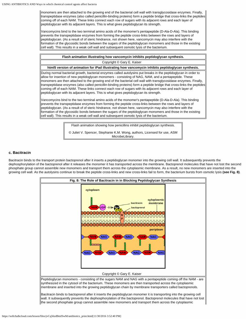
USING ANTIBIOTICS AND Ways in which chemical control agents affect bacteria
https://softchalkcloud.com/lesson/files/jvCq56zdBmISwM/antibiotics_print.html[11/30/2016 3:52:40 PM]
monomers are then attached to the growing end of the bacterial cell wall with transglycosidase enzymes. Finally,transpeptidase enzymes (also called penicillin-binding proteins) form a peptide bridge that cross-links the peptidescoming off of each NAM. These links connect each row of sugars with its adjacent rows and each layer ofpeptidoglycan with its adjacent layers. This is what gives peptidoglycan its strength.
Vancomycins bind to the two terminal amino acids of the monomer's pentapeptide (D-Ala-D-Ala). This bindingprevents the transpeptidase enzymes from forming the peptide cross-links between the rows and layers ofpeptidoglycan. (As a result of of steric hindrance, not shown here, vancomycin may also interfere with theformation of the glycosidic bonds between the sugars of the peptidoglycan monomers and those in the existingcell wall). This results in a weak cell wall and subsequent osmotic lysis of the bacterium.
Flash animation illustrating how vancomycin inhibits peptidoglycan synthesis.
Copyright © Gary E. Kaiser html5 version of animation for iPad illustrating how vancomycin inhibits peptidoglycan synthesis.
During normal bacterial growth, bacterial enzymes called autolysins put breaks in the peptidoglycan in order toallow for insertion of new peptidoglycan monomers - consisting of NAG, NAM, and a pentapeptide. Thesemonomers are then attached to the growing end of the bacterial cell wall with transglycosidase enzymes. Finally,transpeptidase enzymes (also called penicillin-binding proteins) form a peptide bridge that cross-links the peptidescoming off of each NAM. These links connect each row of sugars with its adjacent rows and each layer ofpeptidoglycan with its adjacent layers. This is what gives peptidoglycan its strength.
Vancomycins bind to the two terminal amino acids of the monomer's pentapeptide (D-Ala-D-Ala). This bindingprevents the transpeptidase enzymes from forming the peptide cross-links between the rows and layers ofpeptidoglycan. (As a result of of steric hindrance, not shown here, vancomycin may also interfere with theformation of the glycosidic bonds between the sugars of the peptidoglycan monomers and those in the existingcell wall). This results in a weak cell wall and subsequent osmotic lysis of the bacterium.
Flash animation showing how penicillins inhibit peptidoglycan synthesis.
© Juliet V. Spencer, Stephanie K.M. Wong, authors, Licensed for use, ASMMicrobeLibrary.
c. Bacitracin
Bacitracin binds to the transport protein bactoprenol after it inserts a peptidoglycan monomer into the growing cell wall. It subsequently prevents thedephosphorylation of the bactoprenol after it releases the monomer it has transported across the membrane. Bactoprenol molecules that have not lost the secondphosphate group cannot assemble new monomers and transport them across the cytoplasmic membrane. As a result, no new monomers are inserted into thegrowing cell wall. As the autolysins continue to break the peptide cross-links and new cross-links fail to form, the bacterium bursts from osmotic lysis (see Fig. 8).
Fig. 8: The Role of Bacitracin in in Blocking Peptidoglycan Synthesis
Copyright © Gary E. Kaiser Peptidoglycan monomers - consisting of the sugars NAM and NAG with a pentapeptide coming off the NAM - aresynthesized in the cytosol of the bacterium. These monomers are then transported across the cytoplasmicmembrane and inserted into the growing peptidoglycan chain by membrane transporters called bactoprenols.
Bacitracin binds to bactoprenol after it inserts the peptidoglycan monomer it is transporting into the growing cellwall. It subsequently prevents the dephosphorylation of the bactoprenol. Bactoprenol molecules that have not lostthe second phosphate group cannot assemble new monomers and transport them across the cytoplasmic

USING ANTIBIOTICS AND Ways in which chemical control agents affect bacteria
https://softchalkcloud.com/lesson/files/jvCq56zdBmISwM/antibiotics_print.html[11/30/2016 3:52:40 PM]
membrane. As a result, no new monomers are inserted into the growing cell wall. As the autolysins continue tobreak the peptide cross-links and new cross-links fail to form, the bacterium bursts from osmotic lysis.
Flash animation illustrating how bacitracin inhibits peptidoglycan synthesis.
Copyright © Gary E. Kaiser html5 version of animation for iPad illustrating how bacitracin inhibits peptidoglycan synthesis.
Peptidoglycan monomers - consisting of the sugars NAM and NAG with a pentapeptide coming off the NAM - aresynthesized in the cytosol of the bacterium. These monomers are then transported across the cytoplasmicmembrane and inserted into the growing peptidoglycan chain by membrane transporters called bactoprenols.
Bacitracin binds to bactoprenol after it inserts the peptidoglycan monomer it is transporting into the growing cellwall. It subsequently prevents the dephosphorylation of the bactoprenol. Bactoprenol molecules that have not lostthe second phosphate group cannot assemble new monomers and transport them across the cytoplasmicmembrane. As a result, no new monomers are inserted into the growing cell wall. As the autolysins continue tobreak the peptide cross-links and new cross-links fail to form, the bacterium bursts from osmotic lysis.
Flash animation showing how penicillins inhibit peptidoglycan synthesis.
© Juliet V. Spencer, Stephanie K.M. Wong, authors, Licensed for use, ASMMicrobeLibrary.
Common Antibacterial Antibiotics
Concept map for Chemical Agents that Inhibit Cell Wall Synthesis, Alter theCytoplasmic Membrane, or DNA Synthesis.
2. A few Antimicrobial Chemotherapeutic Agents Inhibit Synthesis of the Acid-Fast Cell Wall
A few antimicrobial chemotherapeutic agents inhibit normal synthesis of the acid-fast cell wall of the genus Mycobacterium
1. INH (isoniazid) appears to block the synthesis of mycolic acid, a key component of the acid-fast cell wall of mycobacteria (see Fig. 9).
2. Ethambutol interferes with the synthesis of the outer membrane of acid-fast cell walls (see Fig. 9).
Fig. 9: Structure of an Acid-Fast Cell Wall

USING ANTIBIOTICS AND Ways in which chemical control agents affect bacteria
https://softchalkcloud.com/lesson/files/jvCq56zdBmISwM/antibiotics_print.html[11/30/2016 3:52:40 PM]
Copyright © Gary E. Kaiser In addition to peptidoglycan, the acid-fast cell wall of Mycobacterium contains a large amount of glycolipids,especially mycolic acids. The peptidoglycan layer is linked to arabinogalactan (D-arabinose and D-galactose)which is then linked to high-molecular weight mycolic acids. The arabinogalactan/mycolic acid layer is overlaidwith a layer of polypeptides and mycolic acids consisting of free lipids, glycolipids, and peptidoglycolipids. Otherglycolipids include lipoarabinomannan and phosphatidyinositol mannosides (PIM). Like the outer membrane of thegram-negative cell wall, porins are required to transport small hydrophilic molecules through the outer membraneof the acid-fast cell wall.
Because of its unique cell wall, when it is stained by the acid-fast procedure, it will resist decolorization with acid-alcohol and stain red, the color of the initial stain, carbol fuchsin. With the exception of a very few other acid-fastbacteria such as Nocardia, all other bacteria will be decolorized and stain blue, the color of the methylene bluecounterstain.
Common Antibacterial Antibiotics
Concept map for Chemical Agents that Inhibit Cell Wall Synthesis, Alter theCytoplasmic Membrane, or Inhibit DNA Synthesis.
3. A very few Antibiotics Alter the Bacterial Cytoplasmic Membrane Causing Leakage of Molecules and Enzymesneeded for Normal Bacterial Metabolism.
A very few antibiotics, such as polymyxins, colistins, and daptomycin, as well as many disinfectants and antiseptics, such as orthophenylphenol, chlorhexidine,hexachlorophene, zephiran, alcohol, and triclosans, alter the bacterial cytoplasmic membrane causing leakage of molecules and enzymes needed for normal
bacterial metabolism.
a. Polymyxins and colistins act as detergents and alter membrane permeability in Gram-negative bacteria. They cannot effectively diffuse through the thickpeptidoglycan layer in gram-positives.
b. Daptomycin disrupts the bacterial cytoplasmic membrane function by apparently binding to the membrane and causing rapid depolarization. This results on a loss of membrane potential and leads to inhibition of protein, DNA and RNA synthesis, resulting in bacterial cell death.
c. Pyrazinamide inhibits fatty acid synthesis in the membranes of Mycobacterium tuberculosis.
For more information: Review of the bacterial cytoplasmic membrane
Common Antibacterial Antibiotics
Concept map for Chemical Agents that Inhibit Cell Wall Synthesis, Alter theCytoplasmic Membrane, or Inhibit DNA Synthesis.

USING ANTIBIOTICS AND Ways in which chemical control agents affect bacteria
https://softchalkcloud.com/lesson/files/jvCq56zdBmISwM/antibiotics_print.html[11/30/2016 3:52:40 PM]
4. Some Antibiotics and Synthetic Drugs Inhibit Nucleic Acid Replication in Bacteria
a. The fluoroquinolones
Fluoroquinolones (norfloxacin, lomefloxacin, fleroxacin, ciprofloxacin, enoxacin, trovafloxacin, gatifloxacin, etc.) work by inhibiting one or more of a group of
enzymes called topoisomerases, enzymes needed for supercoiling, replication, and separation of circular bacterial DNA (see Fig. 10). For example, DNAgyrase (topoisomerase II) catalyzes the negative supercoiling of the circular DNA found in bacteria. It is critical in bacterial DNA replication, DNA repair,transcription of DNA into RNA, and genetic recombination. Topoisomerase IV, on the other hand, is involved in the relaxation of the supercoiled circular DNA,enabling the separation of the interlinked daughter chromosomes at the end of bacterial DNA replication.
In Gram-negative bacteria, the main target for fluoroquinolones is DNA gyrase (topoisomerase II), an enzyme responsible for supercoiling of bacterial DNA duringDNA replication; in Gram-positive bacteria, the primary target is topoisomerase IV, an enzyme responsible for relaxation of supercoiled circular DNA andseparation of the inter-linked daughter chromosomes.
Fig. 10: How an Antimicrobial Agent May Inactivate a Bacterial Enzyme
Copyright © Gary E. Kaiser When an antibiotic binds to a bacterial enzyme, it may alter the activate site of the enzyme and prevent it fromreacting with its substrate.
Flash animation illustrating a normal bacterial enzyme reaction. Copyright © Gary E. Kaiser
html5 version of animation for iPad illustrating a normal bacterial enzyme reaction. When an antibiotic binds to a bacterial enzyme, it may alter the activate site of the enzyme and prevent it fromreacting with its substrate.
Flash animation illustrating how antimicrobial agents may inactivate a bacterial enzyme.
Copyright © Gary E. Kaiser html5 version of animation for iPad illustrating how antimicrobial agents may inactivate a bacterial
enzyme. When an antibiotic binds to a bacterial enzyme, it may alter the activate site of the enzyme and prevent it fromreacting with its substrate.
For more information: Review of the bacterial chromosome.
b. Sulfonamides
Sulfonamides ( sulfamethoxazole, sulfanilamide) and diaminopyrimidines (trimethoprim) block enzymes in the bacteria pathway required for the synthesis
of tetrahydrofolic acid, a cofactor needed for bacteria to make the nucleotide bases thymine, guanine, uracil, and adenine (see Fig. 11).
Fig. 11: Pathway for the Synthesis of Tetrahydrofolic Acid, a Cofactor
Needed for the Synthesis of DNA and RNA Nucleotides

USING ANTIBIOTICS AND Ways in which chemical control agents affect bacteria
https://softchalkcloud.com/lesson/files/jvCq56zdBmISwM/antibiotics_print.html[11/30/2016 3:52:40 PM]
Copyright © Gary E. Kaiser Sulfonamides such as sulfamethoxazole tie up the enzyme pteridine synthetase while trimethoprim ties up theenzyme dihydrofolic acid reductase. As a result, tetrhydrofolic acid is not produced by the bacterium. (Purines =adenine, guanine)
This is done through a process called competitive antagonism whereby a drug chemically resembles a substrate in a metabolic pathway. Because of theirsimilarity, either the drug or the substrate can bind to the substrate's enzyme. While the enzyme is bound to the drug, it is unable to bind to its natural substrate(see Fig. 12) and that blocks that step in the metabolic pathway. Typically, a sulfonamide and a diaminopyrimidine are combined. Co-trimoxazole, for example, isa combination of sulfamethoxazole and trimethoprim.
Fig. 12: Competitive antagonism
Copyright © Gary E. Kaiser The drug resembles a bacterial substrate and competes with that substrate for the limited bacterial enzyme. Thedrug ties up the enzyme making it unavailable for reaction with its normal substrate. As a result, end product is notmade.
Flash animation illustrating competitive antagonism.
Copyright © Gary E. Kaiser html5 version of animation for iPad illustrating competitive antagonism.
Competitive antagonism is a process whereby a drug chemically resembles a substrate in a metabolic pathway.Because of their similarity, either the drug or the substrate can bind to the substrate's enzyme. When the bacterialenzyme binds to its normal substrate, an enzyme-substrate complex forms and the end products required by thatbacterium are made. When the enzyme is bound to the drug, it is unable to bind to its natural substrate and thatblocks the production of the end products of that metabolic pathway. If enough drug is present in the bacterium,all of the bacterial enzyme - which is normally present in the cell only in limited amounts - is tied up with the drugand the end products needed for the bacterium's metabolism are not produced.
c. Metronidazole
Metronidazole is a drug that is activated by the microbial proteins flavodoxin and feredoxin found in microaerophilc and anaerobic bacteria and certain protozoans.Once activated, the metronidazole puts nicks in the microbial DNA strands.

USING ANTIBIOTICS AND Ways in which chemical control agents affect bacteria
https://softchalkcloud.com/lesson/files/jvCq56zdBmISwM/antibiotics_print.html[11/30/2016 3:52:40 PM]
For more information: Review of translation
Concept map for Chemical Agents that Inhibit Cell Wall Synthesis, Alter theCytoplasmic Membrane, or Inhibit DNA Synthesis.
d. Rifampin (rifamycin)
Rifampin blocks transcription by inhibiting bacterial RNA polymerase, the enzyme responsible for transcription of DNA to mRNA.
For more information: Review of transcription
Concept map for Chemical Agents that Inhibit RNA Polymerase, AlterProkaryotic Ribosomes, or Denature Enzymes
Common Antibacterial Antibiotics
5. Many Antibiotics Alter Bacterial Ribosomes, Interfering with Translation.
Many antibiotics alter bacterial ribosomes, interfering with translation of mRNA into proteins and thereby causing faulty protein synthesis. To learn more detailabout the specific steps involved in translation during bacterial protein synthesis, see the animation that follows.
For more information: Review of bacterial ribosomes
Flash animation illustrating the early stages of translation during bacterial protein synthesis.
Copyright © Gary E. Kaiser
Codon Sheet
html5 version of animation for iPad illustrating the early stages of translation during bacterial protein
synthesis.
a.The aminoglycosides
Aminoglycosides (streptomycin, neomycin, netilmicin, tobramycin, gentamicin, amikacin, etc.) bind irreversibly to the 16S rRNA in the 30S subunit of bacterialribosomes. Although the exact mechanism of action is still uncertain, there is evidence that some prevent the transfer of the peptidyl tRNA from the A-site to theP-site, thus preventing the elongation of the polypeptide chain. Some aminoglycosides also appear to interfere with the proofreading process that helps assure theaccuracy of translation (see Fig.13). Possibly the antibiotics reduce the rejection rate for tRNAs that are near matches for the codon. This leads to misreading ofthe codons or premature termination of protein synthesis (see Fig.14). Aminoglycosides may also interfere directly or indirectly with the function of the bacterialcytoplasmic membrane. Because of their toxicity, aminoglycosides are generally used only when other first line antibiotics are not effective.
Fig. 13: Aminoglycosides Interfering with the Translocation of tRNA from the A-site to the P-site

USING ANTIBIOTICS AND Ways in which chemical control agents affect bacteria
https://softchalkcloud.com/lesson/files/jvCq56zdBmISwM/antibiotics_print.html[11/30/2016 3:52:40 PM]
Copyright © Gary E. Kaiser The aminoglycosides (streptomycin, neomycin, netilmicin, tobramycin, gentamicin, amikacin, etc.) bind irreversiblyto the 16S rRNA in the 30S subunit of bacterial ribosomes. It has been proposed that some aminoglycosidesprevent the transfer of the peptidyl tRNA from the A-site to the P-site, thus preventing the elongation of thepolypeptide chain.
Fig. 14: Aminoglycosides Interfering with Translation by causing a Misreading of the Codons along the
mRNA
Copyright © Gary E. Kaiser The aminoglycosides (streptomycin, neomycin, netilmicin, tobramycin, gentamicin, amikacin, etc.) bind irreversiblyto the 16S rRNA in the 30S subunit of bacterial ribosomes. It has been proposed that some aminoglycosidesinterfere with the proofreading process that helps assure the accuracy of translation. Possibly the antibioticsreduce the rejection rate for tRNAs that are near matches for the codon. This leads to misreading of the codons orpremature termination of protein synthesis.
Flash animation illustrating aminoglycosides preventing the translocation of tRNA from the A-site to the
P-site of bacterial ribosomes.
Copyright © Gary E. Kaiser html5 version of animation for iPad illustrating aminoglycosides preventing the translocation of tRNA
from the A-site to the P-site of bacterial ribosomes. The aminoglycosides (streptomycin, neomycin, netilmicin, tobramycin, gentamicin, amikacin, etc.) bind irreversiblyto the 16S rRNA in the 30S subunit of bacterial ribosomes. It has been proposed that some aminoglycosidesprevent the transfer of the peptidyl tRNA from the A-site to the P-site, thus preventing the elongation of thepolypeptide chain.
Flash animation illustrating aminoglycosides causing a misreading of codons.
Copyright © Gary E. Kaiser html5 version of animation for iPad illustrating aminoglycosides causing a misreading of codons.
The aminoglycosides (streptomycin, neomycin, netilmicin, tobramycin, gentamicin, amikacin, etc.) bind irreversiblyto the 16S rRNA in the 30S subunit of bacterial ribosomes. It has been proposed that some aminoglycosides

USING ANTIBIOTICS AND Ways in which chemical control agents affect bacteria
https://softchalkcloud.com/lesson/files/jvCq56zdBmISwM/antibiotics_print.html[11/30/2016 3:52:40 PM]
interfere with the proofreading process that helps assure the accuracy of translation. Possibly the antibioticsreduce the rejection rate for tRNAs that are near matches for the codon. This leads to misreading of the codons orpremature termination of protein synthesis.
b. The tetracyclines
Tetracyclines (tetracycline, doxycycline, demeclocycline, minocycline, etc.) bind reversibly to the 16S rRNA in the 30S ribosomal subunit, distorting it in sucha way that the anticodons of charged tRNAs cannot align properly with the codons of the mRNA (see Fig. 15).
Fig. 15: The Role of Tetracyclines in Blocking Translation
during Bacterial Protein Synthesis
Copyright © Gary E. Kaiser The tetracyclines (tetracycline, doxycycline, demeclocycline, minocycline, etc.) block bacterial translation bybinding reversibly to the 16S rRNA in the 30S subunit and distorting it in such a way that the anticodons of thecharged tRNAs cannot align properly with the codons of the mRNA.
Flash animation illustrating how tetracyclines bind to the 30S ribosomal subunit and block translation.
Copyright © Gary E. Kaiser html5 version of animation for iPad illustrating how tetracyclines bind to the 30S ribosomal subunit and
block translation. The tetracyclines (tetracycline, doxycycline, demeclocycline, minocycline, etc.) block bacterial translation bybinding reversibly to the 16S rRNA in the 30S subunit and distorting it in such a way that the anticodons of thecharged tRNAs cannot align properly with the codons of the mRNA.
c. The macrolides
Macrolides (erythromycin, azithromycin, clarithromycin, dirithromycin, troleandomycin, etc.) bind reversibly to the 23S rRNA in the 50S subunit of bacterialribosomes. They appear to inhibit elongation of the protein by preventing the enzyme peptidyltransferase from forming peptide bonds between the aminoacids (see Fig. 15). They may also prevent the transfer of the peptidyl tRNA from the A-site to the P-site (see Fig. 16) as the beginning peptide chain on thepeptidyl tRNA adheres to the ribosome, creates friction, and blocks the exit tunnel of the 50S ribosomal subunit.
Fig. 15: Mode of Action of Macrolides in Blocking Translation by Blocking Peptidyltransferase
Copyright © Gary E. Kaiser

USING ANTIBIOTICS AND Ways in which chemical control agents affect bacteria
https://softchalkcloud.com/lesson/files/jvCq56zdBmISwM/antibiotics_print.html[11/30/2016 3:52:40 PM]
The macrolides (erythromycin, azithromycin, clarithromycin, dirithromycin, troleandomycin, etc.) bind reversibly tothe 23S rRNA in the 50S subunit.Th ere is evidence that they may inhibit elongation of the protein by thepeptidyltransferase, the enzyme that forms peptide bonds between the amino acids.
Fig. 16: Mode of Action of Macrolides in Blocking Translation by
Preventing the Transfer of the Peptidyl tRNA from the A-site to the P-site
Copyright © Gary E. Kaiser The macrolides (erythromycin, azithromycin, clarithromycin, dirithromycin, troleandomycin, etc.) bind reversibly tothe 23S rRNA in the 50S subunit. There is evidence that they may inhibit elongation of the protein by preventingthe enzyme peptidyltransferase from forming peptide bonds between the amino acids. They may also prevent thetransfer of the peptidyl tRNA from the A-site to the P-site as shown here.
Flash animation illustrating how macrolides bind to the 50S ribosomal subunit and block translation by
blocking peptidyltransferase.
Copyright © Gary E. Kaiser html5 version of animation for iPad illustrating how macrolides bind to the 50S ribosomal subunit and
block translation by blocking peptidyltransferase. The macrolides (erythromycin, azithromycin, clarithromycin, dirithromycin, troleandomycin, etc.) bind reversibly tothe 23S rRNA in the 50S subunit.Th ere is evidence that they may inhibit elongation of the protein by thepeptidyltransferase, the enzyme that forms peptide bonds between the amino acids.
Flash animation illustrating how macrolides bind to the 50S ribosomal subunit and block translation by
preventing the transfer of peptidyl tRNA from the A-site to the P-site.
Copyright © Gary E. Kaiser html5 version of animation for iPad illustrating how macrolides bind to the 50S ribosomal subunit and
block translation by preventing the transfer of peptidyl tRNA from the A-site to the P-site. The macrolides (erythromycin, azithromycin, clarithromycin, dirithromycin, troleandomycin, etc.) bind reversibly tothe 23S rRNA in the 50S subunit. There is evidence that they may inhibit elongation of the protein by preventingthe enzyme peptidyltransferase from forming peptide bonds between the amino acids. They may also prevent thetransfer of the peptidyl tRNA from the A-site to the P-site as shown here.
d. The oxazolidinones
Oxazolidinones (linezolid, sivextro), following the first cycle of protein synthesis, interfere with translation sometime before the initiation phases. They appear tobind to the 50S ribosomal subunit and interfere with its binding to the initiation complex (see Fig. 17).
Fig. 17: Mode of Action of Oxazolidinones Blocking the Attachment of the 50S Ribosomal Subunit to the
Initiation Complex

USING ANTIBIOTICS AND Ways in which chemical control agents affect bacteria
https://softchalkcloud.com/lesson/files/jvCq56zdBmISwM/antibiotics_print.html[11/30/2016 3:52:40 PM]
Copyright © Gary E. Kaiser The oxazolidinones (linezolid) bind to the 23S rRNA in the 50S ribosomal subunit and interfere with the formationof the initiation complex.
Flash animation illustrating how oxazolidinones block the binding of the 50S ribosomal subunit to the
initiation complex.
Copyright © Gary E. Kaiser html5 version of animation for iPad illustrating how oxazolidinones block the binding of the 50S
ribosomal subunit to the initiation complex.
The oxazolidinones (linezolid) bind to the 23S rRNA in the 50S ribosomal subunit and interfere with the formationof the initiation complex.
e. The streptogramins
Streptogramins (synercid, a combination of quinupristin and dalfopristin) bind to two different locations on the 23S rRNA in the 50S ribosomal subunit andwork synergistically to block translation. There are reports that the streptogramins may inhibit the attachment of the charged tRNA to the A-site or may block thepeptide exit tunnel of the 50S ribosomal subunit.
Common Antibacterial Antibiotics
Concept map for Chemical Agents that Inhibit RNA Polymerase, AlterProkaryotic Ribosomes, or Denature Enzymes
For a more detailed description of any specific antimicrobial agent, see the website of RxList - The Internet Drug Index.
TPS Questions
6. Modes of Action for Disinfectants, Antiseptics, and Sanitizers.
Disinfectants, antiseptics, and sanitizers
Disinfection is the elimination of microorganisms, but not necessarily endospores, from inanimate objects or surfaces, whereas decontamination is the treatment ofan object or inanimate surface to make it safe to handle. Sterilization on the other hand, is the process of destroying all living organisms and viruses. A sterile objectis one free of all life forms, including bacterial endospores, as well as viruses. Decontamination refers to the treatment of an object or inanimate surface to make itsafe to handle.
The term disinfectant is used for an agent used to disinfect inanimate objects or surfaces but is generally too toxic to use on human tissues. Antiseptic refers to anagent that kills or inhibits growth of microbes but is safe to use on human tissue. Sanitizer describes an agent that reduces microbial numbers to a safe level.Because disinfectants, antiseptics, and sanitizers often work slowly on some viruses - such as the hepatitis viruses, bacteria with an acid-fast cell wall such asMycobacterium tuberculosis, and especially bacterial endospores, produced by the genus Bacillus and the genus Clostridium, they are usually unreliable for
sterilization - the destruction of all life forms and viruses.

USING ANTIBIOTICS AND Ways in which chemical control agents affect bacteria
https://softchalkcloud.com/lesson/files/jvCq56zdBmISwM/antibiotics_print.html[11/30/2016 3:52:40 PM]
There are a number of factors which influence the antimicrobial action of disinfectants,antiseptics, and sanitizers, including:
1. The concentration of the chemical agent.
2. The temperature at which the agent is being used. Generally, the lower the temperature, the longer it takes to disinfect or decontaminate.
3. The kinds of microorganisms present. Endospore producers such as Bacillus species, Clostridium species, and acid-fast bacteria like Mycobacteriumtuberculosis are harder to eliminate.
4. The number of microorganisms present. The more microorganisms present, the harder it is to disinfect or decontaminate.
5. The nature of the material bearing the microorganisms. Organic material such as dirt and excreta interferes with some agents.
The best results are generally obtained when the initial microbial numbers are low and when the surface to be disinfected is clean and free of possibleinterfering substances.
Concept map for Lab 19 - Using disinfectants, antisepticics, and sanitizers tocontrol microorganisms
There are 2 common antimicrobial modes of action for disinfectants, antiseptics, and sanitizers:
1. They may damage the lipids and/or proteins of the semipermeable cytoplasmic membrane of microorganisms resulting in leakage of cellular materials
needed to sustain life.
2. They may denature microbial enzymes and other proteins, usually by disrupting the hydrogen and disulfide bonds that give the protein its three-dimensionalfunctional shape. This blocks metabolism.
A large number of such chemical agents are in common use. Some of the more common groups are listed below:
1. Phenol and phenol derivatives
Phenol (5-10%) was the first disinfectant commonly used. However, because of its toxicity and odor, phenol derivatives (phenolics) are now generally used. Themost common phenolic is orthophenylphenol, the agent found in O-syl®, Staphene®, and Amphyl®. Bisphenols contain two phenolic groups and typically havechlorine as a part of their structure. They include hexachlorophene and triclosan. Hexachlorophene in a 3% solution is combined with detergent and is found inPhisoHex®. Triclosan is an antiseptic very common in antimicrobial soaps and other products. Biguanides include chlorhexadine and alexidine. A 4% solution ofchlorhexidine in isopropyl alcohol and combined with detergent (Hibiclens® and Hibitane®) is a common hand washing agent and surgical handscrub. Theseagents kill most bacteria, most fungi, and some viruses, but are usually ineffective against endospores. Chloroxylenol (4-chloro-3,5-dimethylphenol) is a broadspectrum antimicrobial chemical compound used to control bacteria, algae, fungi and virus and is often used in antimicrobial soaps and antiseptics. Phenol andphenolics alter membrane permeability and denature proteins. Bisphenols, biguanides, and chloroxylenol alter membrane permeability.
2. Soaps and detergents
Soaps are only mildly microbicidal. Their use aids in the mechanical removal of microorganisms by breaking up the oily film on the skin (emulsification) andreducing the surface tension of water so it spreads and penetrates more readily. Some cosmetic soaps contain added antiseptics to increase antimicrobial activity.Detergents may be anionic or cationic. Anionic (negatively charged) detergents, such as laundry powders, mechanically remove microorganisms and othermaterials but are not very microbicidal. Cationic (positively charged) detergents alter membrane permeability and denature proteins. They are effective againstmany vegetative bacteria, some fungi, and some viruses. However, bacterial endospores and certain bacteria such as Mycobacterium tuberculosis andPseudomonas species are usually resistant. Soaps and organic materials like excreta also inactivate them. Cationic detergents include the quaternary ammoniumcompounds such as benzalkonium chloride, zephiran®, diaprene, roccal, ceepryn, and phemerol. Household Lysol® contains alkyl dimethyl benzyl ammoniumchloride and alcohols.
3. Alcohols
70% solutions of ethyl or isopropyl alcohol are effective in killing vegetative bacteria, enveloped viruses, and fungi. However, they are usually ineffective againstendospores and non-enveloped viruses. Once they evaporate, their cidal activity will cease. Alcohols denature membranes and proteins and are often combinedwith other disinfectants, such as iodine, mercurials, and cationic detergents for increased effectiveness.
4. Acids and alkalies
Acids and alkalies alter membrane permeability and denature proteins and other molecules. Salts of organic acids, such as calcium propionate, potassiumsorbate, and methylparaben, are commonly used as food preservatives. Undecylenic acid (Desenex®) is used for dermatophyte infections of the skin. Anexample of an alkali is lye (sodium hydroxide).
5. Heavy metals
Heavy metals, such as mercury, silver, and copper, denature proteins. Mercury compounds (mercurochrome, metaphen, merthiolate) are only bacteriostatic andare not effective against endospores. Silver nitrate (1%) is sometimes put in the eyes of newborns to prevent gonococcal ophthalmia. Copper sulfate is used tocombat fungal diseases of plants and is also a common algicide. Selinium sulfide kills fungi and their spores.
6. Chlorine
Chlorine gas reacts with water to form hypochlorite ions, which in turn denature microbial enzymes. Chlorine is used in the chlorination of drinking water,
USING ANTIBIOTICS AND Ways in which chemical control agents affect bacteria
https://softchalkcloud.com/lesson/files/jvCq56zdBmISwM/antibiotics_print.html[11/30/2016 3:52:40 PM]
swimming pools, and sewage. Sodium hypochlorite is the active agent in household bleach. Calcium hypochlorite, sodium hypochlorite, and chloramines (chlorineplus ammonia) are used to sanitize glassware, eating utensils, dairy and food processing equipment, hemodialysis systems, and treating water supplies.
7. Iodine
Iodine also denatures microbial proteins. Iodine tincture contains a 2% solution of iodine and sodium iodide in 70% alcohol. Aqueous iodine solutions containing2% iodine and 2.4% sodium iodide are commonly used as a topical antiseptic. Iodophores are a combination of iodine and an inert polymer such aspolyvinylpyrrolidone that reduces surface tension and slowly releases the iodine. Iodophores are less irritating than iodine and do not stain.They are generallyeffective against vegetative bacteria, Mycobacterium tuberculosis, fungi, some viruses, and some endospores. Examples include Wescodyne®, Ioprep®, Ioclide®,Betadine®, and Isodine®.
8. Aldehydes
Aldehydes, such as formaldehyde and glutaraldehyde, denature microbial proteins. Formalin (37% aqueous solution of formaldehyde gas) is extremely active andkills most forms of microbial life. It is used in embalming, preserving biological specimens, and in preparing vaccines. Alkaline glutaraldehyde (Cidex®), acidglutaraldehyde (Sonacide®), and glutaraldehyde phenate solutions (Sporocidin®) kill vegetative bacteria in 10-30 minutes and endospores in about 4 hours. A 10hour exposure to a 2% glutaraldehyde solution can be used for cold sterilization of materials. Ortho-phthalaldehyde (OPA) is dialdehyde used as a high-leveldisinfectant for medical instruments.
9. Peroxygens
Peroxygens are oxidizing agentes that include hydrogen peroxide and peracetic acid. Hydrogen peroxide is broken down into water and oxygen by the enzymecatalase in human cells and is not that good of an antiseptic for open wounds but is useful for disinfecting inanimate objects. The high concentrations of hydrogenperoxide overwhelm the catalase found in microbes. Peracetic acid is a disinfectant that kills microorganisms by oxidation and subsequent disruption of theircytoplasmic membrane. It is widely used in healthcare, food processing, and water treatment.
10. Ethylene oxide gas
Ethylene oxide is one of the very few chemicals that can be relied upon for sterilization (after 4-12 hours exposure). Since it is explosive, it is usually mixed withinert gases such as freon or carbon dioxide. Gaseous chemosterilizers, using ethylene oxide, are commonly used to sterilize heat-sensitive items such as plasticsyringes, Petri plates, textiles, sutures, artificial heart valves, heart-lung machines, and mattresses. Ethylene oxide has very high penetrating power and denaturesmicrobial proteins. Vapors are toxic to the skin, eyes, and mucous membranes and are also carcinogenic. Another gas that is used as a sterilant is chlorinedioxide which denatures proteins in vegetative bacteria, bacterial endospores, viruses, and fungi.
Self Quiz for Ways in which Our Chemical Control Agents may Affect Bacteria
Back to Unit 2 Table of Contents
Back to Softchalk Lessons Table of Contents

Antibiotic resistance
https://softchalkcloud.com/lesson/files/GBch1iD9gMq2wS/resist_print.html[7/25/2017 2:48:58 PM]
Antibiotic resistance
WAYS IN WHICH BACTERIA MAY RESIST CONTROL AGENTS
Ways in which Bacteria may Resist Chemical Control Agents
Fundamental Statements for this Softchalk Lesson:
1. Most bacteria become resistant to antibiotics by way of one or more mechanisms that are coded for by genes in the bacterial chromosome and/orin bacterial plasmids.2. Bacterial genes may code for production of an enzyme that inactivates the antibiotic.3. Bacterial genes may code for an altered target site receptor (ribosomal subunit, enzyme, etc.) for the antibiotic to reduce or block its binding.4. Bacterial genes may code for altered membrane components that prevent the entry of the antibiotic into the bacterium and/or using an effluxpump to transport the antibiotic out of the bacterium.5. Bacterial genes may code for modulated gene expression to produce more of the bacterial enzyme that is being tied up or altered by theantibiotic.6. When under stress from antibiotics, some bacteria switch on genes whose protein products can increase the mutation rate within the bacteriumcausing a hyperevolution to increase the chance of forming an antibiotic-resistant mutant that is able to survive.7. Horizontal gene transfer as a result of transformation, transduction, and conjugation can transfer antibiotic resistance from one bacterium toanother. Horizontal gene transfer enables bacteria to respond and adapt to their environment much more rapidly than mutation by acquiring largeDNA sequences from another bacterium in a single transfer.8. Another mechanism that protects some bacteria from antibiotics is antibiotic tolerance whereby the tolerant bacterium, called a dormant persister,is not killed but simply stops growing when the antibiotic is present.9. CDC estimates that "more than two million people in the United States get infections that are resistant to antibiotics and at least 23,000 people dieas a result."
Antibiotic resistance
https://softchalkcloud.com/lesson/files/GBch1iD9gMq2wS/resist_print.html[7/25/2017 2:48:58 PM]
Common Course Objectives
1. Define terms used in describing the methods of microbial control.2. Recall the mechanism of action for each class of chemotherapeutic chemical agent and give specific examples for each class.3. Identify appropriate methods of microbial control under a given specific circumstance.4. Recall the advantages and disadvantages of the different types of chemical control.5. Identify clinically appropriate methods of growth inhibition when given a specific microbe.6. Recall different mechanisms as a result of genetic changes in a bacterium that may enable that bacterium to resist an antibiotic.
Detailed Learning Objectives
1. Name 2 bacteria that have low-permeability membrane barriers and are thereby intrinsically resistant to many antibiotics.
2**. Briefly describe 4 different mechanisms occuring as a result of genetic changes in a bacterium that may enable that bacterium to resist an antibiotic.
3*. Describe R (Resistance) plasmids and state their significance to medical microbiology.
4. State what the following stand for: MRSA, VRE, CREs, ESBLs, and XDR TB.
5. Define dormant persisters and antibiotic tolerance.
(*) = Common theme throughout the course
(**) = More depth and common theme
TPS Questions
Ways in which Bacteria may Resist Chemical Control Agents
Some opportunistic pathogens, such as Pseudomonas aeruginosa, Mycobacterium tuberculosis, and Enterococcus species, have low-permeability membrane
barriers and are thereby intrinsically resistant to many antibiotics. Most bacteria, however, become resistant to antibiotics as a result of mutation or geneticrecombination. Mutation in bacterial DNA can alter the order of nucleotide bases in a gene and alter that gene product. Horizontal gene transfer can alter or addbacterial genes, again altering the bacterium's gene products.
For more information: See function of DNA.
Most bacteria, become resistant to antibiotics by way of one or more of the following mechanisms that are coded for by genes in the bacterial chromosome or inplasmids:
1. Producing an enzyme capable of inactivating the antibiotic;2. Altering the target site receptor for the antibiotic to reduce or block its binding;3. Preventing the entry of the antibiotic into the bacterium and/or using an efflux pump to transport the antibiotic out of the bacterium; and/or4. Modulating gene expression to produce more of the bacterial enzyme that is being tied up or altered by the antibiotic.
Nice 2013 summary of antibiotic resistant cases and
associated deaths; from the CDC
Article: Improving antibiotic use among hospitalized
patients; a 2014 report from the CDC
Article: Estimates of Healthcare-Associated Infections
(HCIs) 2011; from the CDC
Article: Getting Smart About Antibiotics; a 2015 report
from the CDC.
Preventing Antibiotic-Resistant Infections in Hospitals -United States, 2014; a 2016 report from CDC.
WHO publishes list of bacteria for which new antibiotics are urgently
needed. January, 2017

Antibiotic resistance
https://softchalkcloud.com/lesson/files/GBch1iD9gMq2wS/resist_print.html[7/25/2017 2:48:58 PM]
In the following pages we will look at each of these mechanisms of resistance.
1. Producing enzymes that inactivate the antibiotic
Bacteria may acquire new genes that code for an enzyme that inactivates a particular antibiotic or group of antibiotics (see Fig. 1).
Fig. 1: Producing Enzymes capable of Destroying or Inactivating the Antibiotic
Copyright © Gary E. Kaiser A bacterium producing enzymes that inactivate the antibiotic.
For example:
a. Bacteria typically become resistant to penicillins, monobactams, carbapenems, and cephalosporins are known chemically as beta-lactam antibiotics (see Fig.
2) and many bacteria become resistant to these antibiotics by producing various beta-lactamases that are able to inactivate some forms of these drugs. Beta-
lactamases break the beta-lactam ring of the antibiotic, thus inactivating the drug. (Penicillinase is a beta-lactamase that inactivates certain penicillins.)To overcome this mechanism of resistance, sometimes beta-lactam antibiotics such as amoxicillin, ticarcillin, imipenem, or ampicillin are combined with beta-
lactamase inhibitors such as clavulanate, tazobactam, or sulbactam (see Common Antibiotics) - chemicals that resemble beta-lactam antibiotic (see Fig. 2).These agents bind to the bacterial beta-lactamases and neutralize them.
Fig. 2: Beta-Lactam Rings of Penicillins, Cephalosporins, Imipenem, and Clavulanate
Copyright © Gary E. Kaiser Penicillins, monobactams, carbapenems, imipenems, and cephalosporins are known chemically as beta-lactamantibiotics because as part of their chemical structure they contain what is know as a beta-lactam ring. Theabove illustration shows the basic structure of penicillin, cephalosporin, imipenem, and clavulanate. Rrepresents sites where different chemical side chains attach, depending on the particular antibiotic.
Some bacteria produce various beta-lactamases, enzymes that break the beta-lactam ring at the site indicatedby the arrows, and are able to inactivate some forms of these drugs.
Clavulanate and sulbactam are drugs that resemble beta-lactam antibiotics. These agents are sometimes addedto beta-lactam antibiotics to tie up and, therefore, inactivate the bacterial beta-lactamases.
b. Bacteria may become resistant to aminoglycosides (streptomycin, neomycin, netilmicin, tobramycin, gentamicin, amikacin, etc.) and streptogramins by
enzymatically adding new chemical groups to these antibiotics, thus inactivating the drug.

Antibiotic resistance
https://softchalkcloud.com/lesson/files/GBch1iD9gMq2wS/resist_print.html[7/25/2017 2:48:58 PM]
Flash animation showing a bacterium producing an enzyme capable of inactivating the antibiotic
Copyright © Gary E. Kaiser html5 version of animation for iPad showing a bacterium producing an enzyme capable of inactivating
the antibiotic A gene in the chromosome or in a plasmid may code for production of an enzyme that is able to inactivate aparticular antibiotic.
Concept map for Ways in Which Bacteria Resist Antibiotics and ChemicalAgents
2. Altering the target site receptor for the antibiotic in the bacterium to reduce or block its binding.
Antibiotics work by binding to some bacterial target site, such as a 50S ribosomal subunit, a 30S ribosomal subunit, or a particular bacterial enzyme such as atranspeptidases or a DNA topoisomerase. Bacteria may acquire new genes that alter the molecular shape of the portion of the ribosomal subunit or the enzyme towhich the drug normally binds. For example:
a. Bacteria may become resistant to to macrolides (erythromycin, azithromycin, clarithromycin, dirithromycin, troleandomycin, etc.) by producing a slightly altered50S ribosomal subunit that still functions but to which the antibiotic can no longer bind (see Fig. 3).
Fig. 3: Altering the Target Site for the Antibiotic to Reduce or Block its Binding: Producing an Altered
Ribosomal Subunit
Copyright © Gary E. Kaiser A bacterium altering the target site of the antibiotic, in this case a 50S ribosomal subunit. The drug is no longerable to bind to the ribosome and block protein synthesis.
b. Bacteria may become resistant to beta-lactam antibiotics (penicillins, monobactams, carbapenems, and cephalosporins) by producing altered transpeptidases(penicillin-binding proteins) with greatly reduced affinity for the binding of beta-lactam antibiotics.
c. Bacteria may become resistant to vancomycin by producing altered cross-linking peptides in the peptidoglycan to which the antibiotic no longer bonds.
d. Bacteria may become resistant to fluoroquinolones (norfloxacin, lomefloxacin, fleroxacin, ciprofloxacin, enoxacin, trovafloxacin, etc.) by producing altered DNAgyrase or other topoisomerases to which the drug no longer binds (see Fig. 4).
Fig. 4: Altering the Target Site for the Antibiotic to Reduce or Block its Binding: Producing an Altered
Enzyme

Antibiotic resistance
https://softchalkcloud.com/lesson/files/GBch1iD9gMq2wS/resist_print.html[7/25/2017 2:48:58 PM]
Copyright © Gary E. Kaiser A bacterium altering the target site of the antibiotic, in this case an enzyme. The drug is no longer able to bind tothe enzyme and block the enzyme from reacting with its substrate.
Flash animation showing a bacterium producing an altered ribosomal subunit to which the antibiotic no
longer binds.
Copyright © Gary E. Kaiser html5 version of animation for iPad showing a bacterium producing an altered ribosomal subunit to
which the antibiotic no longer binds. Some antibiotics work by binding to a bacterial ribosomal subunit, altering the ribosome, and blocking bacterialprotein synthesis. This bacterium becomes resistant to the antibiotic by altering the antibiotic's target site, in thiscase a 50S ribosomal subunit. The drug is no longer able to bind to the ribosome and the bacterium can stillcarry out normal protein synthesis.
Flash animation showing a bacterium producing an altered enzyme to which the antibiotic no longer
binds.
Copyright © Gary E. Kaiser html5 version of animation for iPad showing a bacterium producing an altered enzyme to which the
antibiotic no longer binds. A bacterium can become resistant to an antibiotic by altering the antibiotic's target site, in this case the antibiotic-binding site of the bacterial enzyme. Since the drug is no longer able to bind to the enzyme, the active site ofthe enzyme is not altered and the enzyme is able to bind to its normal substrate.
For more information: Review of bacterial ribosomes
For more information: Review of peptidoglycan
For more information: Review of the bacterial chromosome and nucleoid
Concept map for Ways in Which Bacteria Resist Antibiotics and ChemicalAgents
3. Altering the membranes and transport systems to prevent the entry of the antibiotic into the bacterium and/orusing an efflux pump to transport the antibiotic out of the bacterium.
Antibiotics that target ribosomes or enzymes within the bacterium must first pass through the porins in the outer membrane of Gram-negative and acid-fastbacterial cell walls, and then the cytoplasmic membrane in the case of all bacteria. Subsequently, the antibiotic has to accumulate to a high enough concentrationwithin the bacterium to inhibit or kill the organism.

Antibiotic resistance
https://softchalkcloud.com/lesson/files/GBch1iD9gMq2wS/resist_print.html[7/25/2017 2:48:58 PM]
a. A Gram-negative or an acid-fast bacterium may block the entry of an antmicrobial drug by acquiring genes that alter the porins in the cell wall's outermembrane (see Fig. 5).
Fig. 5: Producing Altered Porins that Prevent Entry of the Drug
Copyright © Gary E. Kaiser A bacterium producing altered porins that block passage of the antibiotic through the outer membrane of aGram-negative bacterium.
b. A bacterium may block the entry of an antmicrobial drug by acquiring genes that alter the transport proteins used to transport the drug through thebacterium's cytoplasmic membrane (see Fig. 6). This is generally not a common mechanism of antibiotic resistance.
Fig. 6: Producing Altered Transport (Carrier) Proteins
Copyright © Gary E. Kaiser A bacterium producing an altered transport (carrier) protein that prevents transport of the antibiotic through thecytoplasmic membrane.
c. A bacterium may acquire genes coding for an energy-driven efflux pump in its the cytoplasmic membrane that is able to to pump the antibiotic out of thebacterium and preventing it from accumulating to a high enough concentration to inhibit or kill the organism (see Fig. 7). This is the most common methodbacteria use to prevent toxic levels of antimicrobial drugs from accumulating within the cytoplasm.
Fig. 7: Producing Transporter Molecules that Pump the Drug Out of the Bacterium (Efflux Pumps)

Antibiotic resistance
https://softchalkcloud.com/lesson/files/GBch1iD9gMq2wS/resist_print.html[7/25/2017 2:48:58 PM]
Copyright © Gary E. Kaiser A bacterium producing transporter molecules that pump the antibiotic out of the bacterium.
Flash animation showing a bacterium producing altered porins to block transport of the drug across the
outer membrane.
Copyright © Gary E. Kaiser html5 version of animation for iPad showing a bacterium producing altered porins to block transport of
the drug across the outer membrane.
Some Gram-negative bacteria become resistant to an antibiotic by producing altered porins in their outermembrane. The antibiotic is no longer able to pass through the outer membrane.
Flash animation showing a bacterium producing an altered transport protein to block transport of the
drug across the cytoplasmic membrane.
Copyright © Gary E. Kaiser html5 version of animation for iPad showing a bacterium producing an altered transport protein to
block transport of the drug across the cytoplasmic membrane.
Some bacteria become resistant to an antibiotic by producing an altered transport (carrier) protein in thecytoplasmic membrane. This new carrier protein is no longer able to transport the antibiotic across thecytoplasmic membrane into the cytoplasm.
Flash animation showing a bacterium producing a new transport protein able to pump the drug out of
the bacterium.
Copyright © Gary E. Kaiser html5 version of animation for iPad showing a bacterium producing a new transport protein able to
pump the drug out of the bacterium.
Some bacteria become resistant to an antibiotic by producing new efflux pump that pumps the antibiotic backout of the bacterium.
For more information: Review of the bacterial cytoplasmicmembrane
Concept map for Ways in Which Bacteria Resist Antibiotics and ChemicalAgents
4. Modulating gene expression to produce more of the bacterial enzyme that is being tied up or altered by theantibiotic.
Remember that enzymes function as catalysts and are present in cells in small amounts because they are not altered as they carry out their specific biochemicalreactions. As mentioned in the previous section, numerous antimicrobial drugs work by inactivating bacterial enzymes and blocking metabolic reactions. Making aparticular enzyme and the amount of enzyme that is made is under genetic control.
Genetic control of enzyme activity refers to controlling transcription of the mRNA needed for an enzyme's synthesis. In prokaryotic cells, this involves the inductionor repression of enzyme synthesis by regulatory proteins that can bind to DNA and either block or enhance the function of RNA polymerase, the enzyme requiredfor transcription.

Antibiotic resistance
https://softchalkcloud.com/lesson/files/GBch1iD9gMq2wS/resist_print.html[7/25/2017 2:48:58 PM]
Bacteria also use translational control of enzyme synthesis. In this case, the bacteria produce noncoding regulatory RNAs (ncRNAs) or antisense RNAs such asmicro RNAs (miRNAs) that are complementary to the early portion of a mRNA coding for the enzyme. When the noncoding regulatory RNA binds to the mRNA bycomplementary base pairing, ribosomes cannot attach to the mRNA, the mRNA cannot be translated into protein, and the enzyme is not made (See Fig. 8).
Fig. 8: Antisense RNA
Copyright © Gary E. Kaiser Bacteria also use translational control of enzyme synthesis. In this case, the bacteria produce noncodingregulatory RNAs (ncRNAs) or antisense RNAs such as micro RNAs (miRNAs) that are complementary to theearly portion of a mRNA coding for the enzyme. When the antisense RNA binds to the mRNA by complementarybase pairing, ribosomes cannot attach to the mRNA, the mRNA cannot be translated into protein, and theenzyme is not made
For more information: Review of enzyme regulation
Mutations or horizontal gene transfer may result in a modulation of gene expression or translational events that favor increased production of the
enzyme being tied up or altered by the antimicrobial agent (see Fig. 9). Since enzymes are normally produced in limited amounts, production of excessiveamounts of enzyme may allow for the metabolic activity being blocked by the agent to still occur.
Fig. 9: Producing Greater Amounts of the Enzyme that is being Inactivated by the Antibiotic
Copyright © Gary E. KaiserA bacterium producing greater amounts of the limited enzyme being tied up or inactivated by the antimicrobialagent. Although some enzyme is tied up by the drug, there is still enzyme available to react with its substrateand produce end products.
Flash animation illustrating competitive antagonism.
Copyright © Gary E. Kaiser html5 version of animation for iPad illustrating competitive antagonism.
Competitive antagonism is a process whereby a drug chemically resembles a substrate in a metabolic pathway.Because of their similarity, either the drug or the substrate can bind to the substrate's enzyme. When thebacterial enzyme binds to its normal substrate, an enzyme-substrate complex forms and the end productsrequired by that bacterium are made. When the enzyme is bound to the drug, it is unable to bind to its naturalsubstrate and that blocks the production of the end products of that metabolic pathway. If enough drug ispresent in the bacterium, all of the bacterial enzyme - which is normally present in the cell only in limitedamounts - is tied up with the drug and the end products needed for the bacterium's metabolism are not

Antibiotic resistance
https://softchalkcloud.com/lesson/files/GBch1iD9gMq2wS/resist_print.html[7/25/2017 2:48:58 PM]
produced.
Flash animation showing a bacterium producing more of a limited enzyme.
Copyright © Gary E. Kaiser Flash animation showing a bacterium producing more of a limites enzyme.
A bacterium becoming resistant to an antimicrobial agent by producing greater amounts of the limited enzymebeing tied up or inactivated by the drug. Although some enzyme is tied up by the drug, there is still enzymeavailable to react with its substrate and produce end products.
GIF animation illustrating antisense RNA
TPS Questions
Class activity
Watch University of Texas at San Antonio microbiologist Karl Klose discussesthe problem of antibiotic resistance in a 2013 TED talk.
Use your notes to name and describe the bacterial mechanisms of antibioticresistance referred to in this talk as "upchuck," "stealth mode," and "ballistic
missile."
Concept map for Ways in Which Bacteria Resist Antibiotics and ChemicalAgents
5. Many pathogenic bacteria, as well as normal flora, form complex bacterial communities as biofilms.
Bacteria in biofilms are often able to communicate with one another by a process called quorum sensing and are able to interact with and adapt to theirenvironment as a population of bacteria rather than as individual bacteria. By living as a community of bacteria as a biofilm, these bacteria are:
a. better able to resist attack by antibiotics, and
b. better able to resist the host immune system.
Why bacterium within a biofilm are more antibiotic resistant isn't completely understood but various mechanisms have been proposed. The extracellularpolysaccharide may make it more difficult for the antibiotic to reach all of the bacteria. Bacteria within a biofilm are generally in a metabolically more inert state andthis could slow down antibacterial action of the drug. Many antibiotics are static, not cidal in action; the body depends on phagocytes to remove the inhibitedbacteria. The biofilm structure makes engulfment by phagocytes pretty much impossible.
6. Dormant persisters
Another mechanism that protects some bacteria from antibiotics is antibiotic tolerance. In the case of antibiotic tolerance, the tolerant bacterium is not killed
but simply stops growing when the antibiotic is present. It then is able to recover once the antibiotic is no longer in the host. For example, Streptococcuspneumoniae tolerant to vancomycin appear to repress their autolysins in the presence of the drug and don't undergo osmotic lysis. Antibiotic tolerance is especiallysignificant in terms of bacteria that form biofilms associated with catheters, heart valves, orthopedic devices, and people with cystic fibrosis. These biofilms oftencontain a small percentage of dormant persisters that, because they are not dividing, tolerate the antibiotics.
Its been found that bacteria simultaneously produce toxins that inhibit their own growth and antitoxins that bind to the toxin and cause its neutralization. Small

Antibiotic resistance
https://softchalkcloud.com/lesson/files/GBch1iD9gMq2wS/resist_print.html[7/25/2017 2:48:58 PM]
numbers of bacteria in the population, however, become persisters because they produce lower levels of antitoxin or the antitoxin is degraded by stress. As aresult, the free toxin arrests bacterial growth enabling a persistent state that is able to survive stressors such as antibiotics and starvation.
The above changes in the bacterium that enable it to resist the antibiotic occur naturally as a result of mutation or as a result of horizontal gene transfer.
Mutation rate in bacteria
For example, when under stress from antibiotics, some bacteria switch on genes whose protein products can increase the mutation rate within the bacterium
10,000 times as fast as the mutation rate that occurs during normal binary fission. This causes a sort of hyperevolution where mutation acts as a selfdefense mechanism for the bacterial population by increasing the chance of forming an antibiotic-resistant mutant that is able to survive at the expense of themajority of the population. (Remember that most mutations are harmful to a cell.)
For more information: Review of mutation
Evolution of Escherichia coli Antibiotic Resistance via Mutation over Space andTime
Courtesy of Havard Medical School
In addition, horizontal gene transfer as a result of transformation, transduction, and conjugation can transfer antibiotic resistance from one bacterium to another.Horizontal gene transfer enables bacteria to respond and adapt to their environment much more rapidly than mutation by acquiring large DNA sequences fromanother bacterium in a single transfer.
For more information: Review of horizontal gene transfer
Concept map for Ways in Which Bacteria Resist Antibiotics and ChemicalAgents
Examples of bacterial antibiotic resistance
Exposure to the antibiotic typically selects for strains of the organism that have become resistant through these natural processes. Misuse of antibiotics, suchas prescribing them for non-bacterial infections (colds, influenza, most upper respiratory infections, etc.) or prescribing the "newest" antibiotic on the market whenolder brands may still be as effective simply increases the rate at which this natural selection for resistance occurs. According to the Centers for Disease Controland Prevention, as many as one-third (50 million out of 150 million) of antibiotic prescriptions given on an outpatient basis are unneeded. Patient noncompliancewith antimicrobial therapy, namely, not taking the prescribed amount of the antibiotic at the proper intervals for the appropriate length of time, also plays a role inselecting for resistant strains of bacteria.
The spread of antibiotic resistance in pathogenic bacteria is due to both direct selection and indirect selection.
1. Direct selection refers to the selection of antibiotic resistant pathogens at the site of infection.
2. Indirect selection is the selection of antibiotic-resistant normal flora within an individual anytime an antibiotic is given. At a later date, these resistant
normal flora may transfer resistance genes to pathogens that enter the body. In addition, these resistant normal flora may be transmitted from person
to person through such means as the fecal-oral route or through respiratory secretions.
As an example, many Gram-negative bacteria possess R (Resistance) plasmids that have genes coding for multiple antibiotic resistance through themechanisms learned above, as well as transfer genes coding for a conjugation (sex) pilus. It is possible for R-plasmids to accumulate transposons to increasebacterial resistance. Such an organism can conjugate with other bacteria and transfer to them an R plasmid. E. coli, Proteus, Serratia, Enterobacter, Salmonella,Shigella, and Pseudomonas are bacteria that frequently have R-factor plasmids.
Flash animation illustrating R-plasmid conjugation.
Copyright © Gary E. Kaiser html5 version of animation for iPad illustrating R-plasmid conjugation.
R plasmids are conjugative plasmids coding for mating pair formation and also multiple antibiotic resistance. A

Antibiotic resistance
https://softchalkcloud.com/lesson/files/GBch1iD9gMq2wS/resist_print.html[7/25/2017 2:48:58 PM]
conjugative plasmid is self-transmissible, in that it possesses all the necessary genes for that plasmid totransmit itself to another bacterium by conjugation. Conjugation genes known as tra genes enable the bacteriumto form a mating pair with another organism, while oriT (origin of transfer) genes determine where on theplasmid DNA transfer is initiated. The plasmid also possess genes coding for resistance to a number of differentantibiotics.
In addition to plasmids, conjugative transposons also frequently transmit antibiotic resistance from one bacterium to another. Conjugative transposons,like conjugative plasmids, carry the genes that enable mating pairs to form for conjugation. Therefore, conjugative transposons also enable mobilizable plasmidsand nonconjugative transposons to be transferred to a recipient bacterium during conjugation.
For more information: Review of horizontal gene transfer
Examples of resistant strains of bacteria of ever increasing medical importance include:
1. Antibiotic-Resistant Neisseria gonorrhoeae
Out of an estimated 820,000 Neisseria gonorrhoeae infections per year, 246,000 are antibiotic-resistant.
2.Carbapenem-Resistant Enterobacteriaceae (CRE)
More recently, carbapenemase-producing Klebsiella pneumoniae (KPC) strains are frequently being identified among nosocomial pathogens globally.Carbapenemase is a broad-spectrum beta-lactamase enzyme first found in K. pneumoniae isolates that results in resistance to all penicillins,
cephalosporins, carbapenems (i.e., imipenem, ertapenem, metropenem), and monobactams (i.e., aztreonam). These broad-spectrum beta-lactamases arealso known as extended spectrum beta-lactamases or ESBLs. These ESBLs are now being seen in a variety Enterobacteriaceae including Enterobacterspp., E. coli, Serratia spp., and Salmonella enterica. These ESBL-producing Enterobacteriaceae are known as carbapenem-resistant
Enterobacteriaceae, or CRE. Almost half of hospital patients who get bloodstream infections from CRE bacteria die from the infection.
3. Methicillin-resistant Staphylococcus aureus (MRSA)
Staphylococcus aureus resistance to methicillin confers resistance to all penicillins and cephalosporins.
4. Vancomycin-resistant Enterococcus (VRE)
Vancomycin-resistant Enterococcus (VRE) are intrinsically resistant to most antibiotics and have acquired resistance to the first line drug of choice,vancomycin.
5. XDR TB
Extensively drug-resistant tuberculosis (XDR TB), a relatively rare type of multidrug-resistant Mycobacterium tuberculosis that is resistant to almost all drugsused to treat TB, including the two best first-line drugs: isoniazid and rifampin. XDR TB is also resistant to the best second-line medications: fluoroquinolonesand at least one of three injectable drugs i.e., amikacin, kanamycin, or capreomycin.
6. Clostridium difficile infections
A 2015 CDC study found that "Clostridium difficile caused almost half a million infections among patients in the United States in a single year. Anestimated 15,000 deaths are directly attributable to C. difficile infections, making it a substantial cause of infectious disease death in the United States" with anexcess medical cost of $1,000,000,000 per year.
CDC's top 18 drug-resistant threats to the US
Bacteria such as E. coli, Proteus, Enterobacter, Serratia, Pseudomonas, Staphylococcus aureus, and Enterococcus mentioned above, are the leading cause ofhealth care-associated infections. According to the Centers for Disease Control and Prevention (CDC) Healthcare-associated infection's website, "In American
hospitals alone, healthcare-associated infections account for an estimated 1.7 million infections and 99,000 associated deaths each year" in the U.S.The CDC also estimates that "more than two million people in the United States get infections that are resistant to antibiotics and at least 23,000 people
die as a result."
Finally, Bacterial endospores, such as those produced by Bacillus and Clostridium, are also resistant to antibiotics, most disinfectants, and physical agents suchas boiling and drying. Although harmless themselves, they are involved in the transmission of some diseases to humans. Examples include anthrax (Bacillusanthracis), tetanus (Clostridium tetani), botulism (Clostridium botulinum), gas gangrene (Clostridium perfringens), and pseudomembranous colitis (Clostridiumdifficile).

Antibiotic resistance
https://softchalkcloud.com/lesson/files/GBch1iD9gMq2wS/resist_print.html[7/25/2017 2:48:58 PM]
Concept map for Ways in Which Bacteria Resist Antibiotics and ChemicalAgents
Self Quiz for Ways Bacteria Resist Chemical Control Agents
Back to Unit 2 Table of Contents
Back to Softchalk Lessons Table of Contents