unit 4 – maintaining dynamic equilibrium
TRANSCRIPT

Unit 4 – Maintaining Dynamic Equilibrium Unit Outline Homeostasis Ch. 9 pp. 298- 303 Circulatory system Ch. 9 pp. 304-331 Respiratory system Ch. 10 pp. 332-351 Digestive system Ch. 11 pp. 352- 373 Excretory system Ch. 11 pp. 374- 381 Immune system Ch. 11 pp. 382- 389 9.1 - Homeostasis
- means keeping internal body conditions the same, despite constant changes in the external environment
- dynamic equilibrium: state of balance achieved within an environment as a result of internal control mechanisms that continuously oppose outside forces that tend to change the environment
- in order to maintain homeostasis, several factors have to be maintained: 1) salinity and pH of intestinal fluid 2) composition and pH of blood 3) body temperature 4) hormone levels
Temperature Regulation - homeotherm – “warm-blooded”; animals that maintain their own temperatures - poikilotherm – “cold-blooded”; animals which depend on the environment to
regulate temperature - humans have several mechanisms to regulate temperature
1) the body generates heat as a by-product of metabolic processes 2) behavioural adaptations (finding shade on a hot day) 3) physiological adaptations (heat can be released through blood vessels
that run under the skin)

9.2 and 9.3 – The Circulatory System Organization in Biology A complete organism is comprised of various parts, depending on the complexity of the organism. While some organisms are simple and composed of one cell, many organisms are very complex and their cells are organized into groups with specific functions. Cell → tissue → organ → organ → whole system organism tissue: a group of cells that have a specific function ex. Muscle tissue connective tissue epithelial tissue (coverings) organ: a group of tissues that work together to perform a similar function ex. Heart – made of muscle tissue skin – made of epithelial tissue organ system: a group of organs that work together to perform a similar function ex. Circulatory system – heart and blood vessels digestive system – stomach, small intestine, large intestine, etc. The Circulatory System
- main job of the circulatory system is to transport materials throughout the body, such as oxygen, sugar, and other materials
- the human circulatory system is a closed, complete double system. o Two main types of circulation:
- Pulmonary – circulation that carries blood to and from the lungs. Important for picking up oxygen and dropping off carbon dioxide
- Systemic – circulation that carries blood to and from other parts of the body. Delivers oxygen and other materials to the body. There are various types:
• Hepatic portal: delivers to and from the liver • Renal: delivers blood to and from the kidneys • Cardiac: delivers blood to and from the heart
- the average man has 5-6 L of blood (woman has 4-5 L) - three main elements of any circulatory system:
• transport vessels to carry fluid • the transport medium (blood) • the pumping mechanism (heart)

(1) Transport Vessels (pp. 305-307) There are three main types of blood vessels: arteries, veins, and capillaries Arteries - carry blood away from the heart - carry oxygenated blood mostly (except the pulmonary artery) - arteries have three layers:
• outer layer of thick connective tissue • a thick middle layer of muscle • thin layer of epithelial cells
- arteries are very elastic and muscular to help pump blood throughout the body
Veins - carry blood to the heart - carries deoxygenated blood mostly (except the pulmonary vein) - veins also consist of three layers:
• outer – same as arteries • middle – less muscular • inner layer – same
- veins are less muscular and are not as elastic as arteries, so they do not help much in the movement of blood. However, they hold more blood (there is twice as much blood in the veins than in the arteries at any one time)
- to help blood return to the heart, external muscle help force blood back to the heart. As well, veins have valves to ensure blood does not flow backwards.
Capillaries - the smallest blood vessels - very thin; consist of a single layer of endothelial cells. They are only large
enough for a single red blood cell to fit through - exchange of materials between the blood and body cells occurs in capillaries
(see Fig 9.9, p. 307)
(2) Blood pp. 308-313 - blood consists of two main parts 1. plasma (55%) 2. blood cells (45%) Plasma
- mostly water - blood cells suspended in plasma - has dissolved protein components which are important in
various function - helps transport carbon dioxide (55% of all CO2 transport is by
plasma)
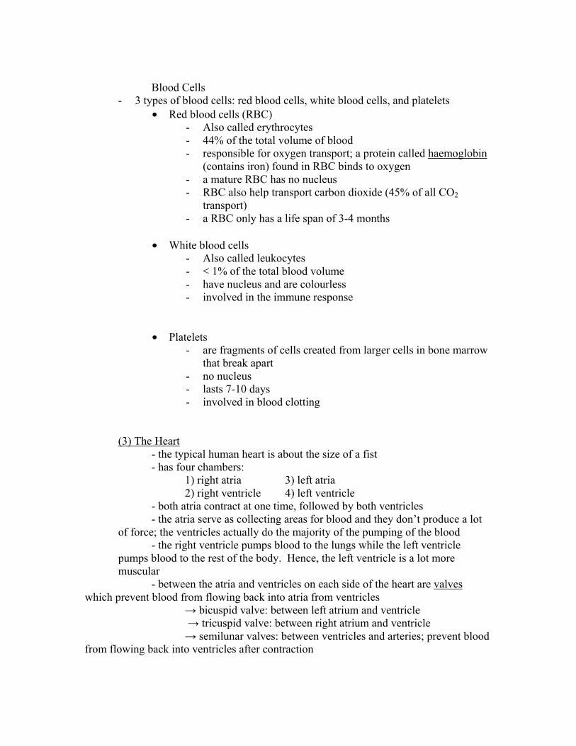
Blood Cells
- 3 types of blood cells: red blood cells, white blood cells, and platelets • Red blood cells (RBC)
- Also called erythrocytes - 44% of the total volume of blood - responsible for oxygen transport; a protein called haemoglobin
(contains iron) found in RBC binds to oxygen - a mature RBC has no nucleus - RBC also help transport carbon dioxide (45% of all CO2
transport) - a RBC only has a life span of 3-4 months
• White blood cells
- Also called leukocytes - < 1% of the total blood volume - have nucleus and are colourless - involved in the immune response
• Platelets - are fragments of cells created from larger cells in bone marrow
that break apart - no nucleus - lasts 7-10 days - involved in blood clotting
(3) The Heart - the typical human heart is about the size of a fist - has four chambers: 1) right atria 3) left atria 2) right ventricle 4) left ventricle - both atria contract at one time, followed by both ventricles - the atria serve as collecting areas for blood and they don’t produce a lot of force; the ventricles actually do the majority of the pumping of the blood - the right ventricle pumps blood to the lungs while the left ventricle pumps blood to the rest of the body. Hence, the left ventricle is a lot more muscular
- between the atria and ventricles on each side of the heart are valves which prevent blood from flowing back into atria from ventricles → bicuspid valve: between left atrium and ventricle → tricuspid valve: between right atrium and ventricle → semilunar valves: between ventricles and arteries; prevent blood from flowing back into ventricles after contraction

- septum: a wall that separates the left and right sides of the heart. This wall prevents mixing of oxygenated and deoxygenated blood.
The Cardiac Cycle
Flow of blood in a cardiac cycle Blood returning from body (deoxygenated) ↓ Superior vena cava and inferior vena cava ↓ right atrium ↓ right ventricle ↓ lungs (via pulmonary arteries) ↓ left atrium (via pulmonary veins) ↓ left ventricle ↓ aorta (carries blood to body) The pumping action of the heart has 2 main periods:
(1) diastole: the period of relaxation of the heart (2) systole: the period of contraction of the heart
During diastole, the A-V valves (bicuspid and tricuspid) are open. Blood flows
from the atria to the ventricles. By the end of diastole, the ventricles are about 70% filled. Systole begins with contraction of the atria. The contraction of the atria forces more blood into the ventricles, filling them. The ventricles then contract. While the ventricles contract, the atria relax.
As the heart valves open and close, they make a “lub-dup” sound. The “lub” sound is caused by the closing of the A-V valves. The “dup” sound is made by the closing of the semilunar valves.
Control of Heartbeat

Heart muscle has a built-in ability to contract without need from stimulus from the nervous system. These contractions are controlled by a structure called the sino-atrial node (S-A node), also called the pacemaker.
When the heart receives electric impulses from the S-A node, the atria contract. Then, the impulse reaches another structure called the atrioventricular node (A-V node) which causes the ventricles to contract.
The pacemaker itself can be regulated by certain nerves: - vagus nerves: slow down heartbeat - cardioaccelerator nerves: speed up heartbeat
Blood Pressure Pulse: the expansion and relaxation that can be felt in an artery each time the left ventricle of the heart contracts and relaxes Sphygmomanometer: device used to measure blood pressure (blood pressure cuff) Blood pressure is measured in terms of height of mercury in a tube of a sphygmomanometer. Average pressure in systole = 120 mmHg Average pressure in diastole = 80 mmHg Normal blood pressure = 120/80 Heart Defects
(1) Sepal defect → A hole in the septum of the heart → There is a mixing of oxygenated and deoxygenated blood → Can be treated by surgery
(2) heart murmur
→ one or more heart valves do not open or close properly → name comes from sound made by the blood escaping from the valve → some murmurs severe, others not as bad

Cardiac Output and Fitness Cardiac output: amount of blood pumped by the heart, usually measured in ml/min Stroke volume: amount of blood forced out of the heart with each heartbeat Cardiac output = stroke volume X heart rate A higher stroke volume and a lower heart rate indicates the heart is very efficient since the heart is not working very hard to maintain a certain cardiac output Ex. Individual Cardiac output
(ml/min) Stroke volume (ml/beat)
Heart rate (beats/min)
A 4900 70 70 B 4900 50 98 C 4900 140 35
In this example, individual C is exceptionally fit. Individual C can deliver the same amount of blood (and oxygen) to the body per minute as A and B, but their heart is not working as hard to do it.
Individual A represents an average person, with a stroke volume of 70 ml/beat and
70 beats/min Heart Rate and Fitness - maximum heart rate is not related to fitness
(1) max heart rate = 220-age - length of time it takes to return to resting heart rate is an indication of health.
If it takes a long time for a person’s body to return to a resting heart rate after exercise, then that person is in poor shape
- a higher resting heart rate usually indicates poor health as well
Stroke Volume and Fitness - two factors affect stroke volume:
(1) how easily the heart fills with blood (depends on the distendibility of ventricles and the amount of blood returned from the heart)
(2) how readily the heart empties (depends on strength of contraction and pressure on artery walls)

- regular cardiovascular exercise increases stroke volume by enlarging ventrical chambers, including distendibility (stretchiness) of ventricles, and strengthening ventricle walls.
- Strength training only may increase thickness of ventricle walls, but will reduce the elasticity of the ventricles.

9.4 – Transport Systems and Homeostasis lymphatic circulatory system – network of glands and vessels that carry lymph throughout the body lymph – colourless or pale yellow fluid that circulates throughout the lymphatic circulatory system. It is a lot like plasma in its composition (see fig 9.24, p. 323) → the lymphatic system has no pump; movement of external muscles helps circulate fluid → lymph is made of fluid that escapes from blood in the capillaries. This fluid gets absorbed into the vessels of the lymphatic system and eventually rejoins the main circulatory system → the lymphatic system also works with white blood cells (leukocytes) Blood Pressure Risks Hypertension – chronically elevated blood pressure, often associated with a number of health problems such as greatly increased risk of stroke and heart disease High blood pressure can be caused by several things, such as a diet high in salt, a diet high in cholesterol, certain drugs like nicotine, heredity, age, lack of exercise, smoking and obesity Atherosclerosis – narrowing of the arteries due to the deposit of cholesterol-rich plaques on the innermost layer of the wall of an artery; results in decreased blood flow Arteriosclerosis – a condition related to atherosclerosis in which cholesterol or other fatty material becomes deposited under the inner lining of the arteries; results in blockage of blood flow in the artery (see fig 9.27, p. 326) Angioplasty – surgical procedure in which a cardiologist inserts a fine plastic tube into a clogged artery and then inflates part of the tube; opens the artery Coronary bypass – surgical procedure that involves removing a segment of healthy blood vessel from another part of the body and using it to create a new path (shunt) around a blockage in a blood vessel near the heart.

Ch. 10 – The Breath of Life 10.1 – The Breath of Life Most organisms on Earth are aerobic; they require oxygen to survive. Simple organisms do not need a specific organ system to get oxygen because they are small. However, larger organisms need a complex organ system to get oxygen and get rid of carbon dioxide. Respiratory surface – any surface across which gases are exchanged for the purpose of respiration In humans, the respiratory surface is contained in the lungs. They are internal in order to keep the respiratory surface moist. Breathing – the taking in and releasing of air in and out of the lungs Inspiration: taking air in Expiration: the act of breathing out External respiration – the exchange of oxygen and carbon dioxide between the air and blood Internal respiration – the exchange of oxygen and carbon dioxide between the blood and the cells of the surrounding tissue Cellular respiration – the complex series of chemical reactions that may take place mainly in the mitochondria of cells Parts of the Human Respiratory System
1. Nostrils – openings in the nose where air enters. As well, the lining of the nostrils helps warm and moisten air and it also filters it
2. Pharynx – structure located just behind the mouth that connects the mouth and nasal cavity to the larynx and esophagus. Also known as the throat.
3. Larynx – a structure within the upper respiratory tract that contains the vocal cords. Also known as the “voice box”
4. Trachea – tube that carries air from the nasal passages or mouth to the lungs. Also known as the “windpipe”
5. Bronchi – the passageways that branch from the trachea into the lungs. One bronchus carries air to each lung.
6. Bronchioles – the passageways that branch from the bronchi into the separate lobes of the lungs. The bronchioles divide into smaller and smaller passageways that carry air into all portions of the lungs.
7. Alveoli – the gas exchange structures within the lungs. Alveoli are tiny air pockets with walls made of a membrane a single cell thick. Respiratory gases are exchanged across these cell membrane walls.

8. Diaphragm – a muscle layer that forms the floor of the thoracic cavity. The contraction of the diaphragm contributes to inspiration by increasing the volume of the thoracic cavity
9. Pleura – the membranes that envelop the lungs. Each lung is encased in two separate by a thin layer in fluid.
10. Intercostal Muscles – muscles of the rib cage. These muscles help to expand and contract the rib cage and play an important role in breathing.
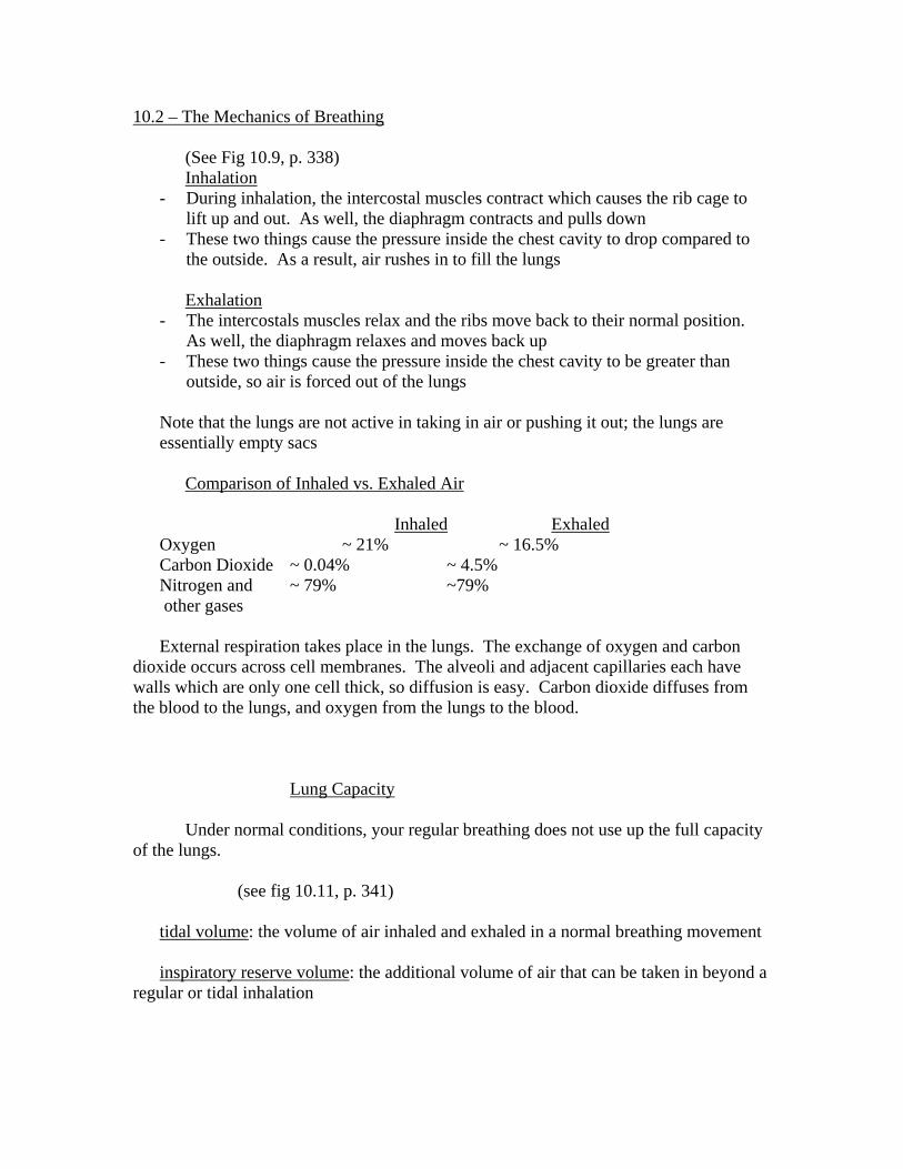
10.2 – The Mechanics of Breathing (See Fig 10.9, p. 338) Inhalation
- During inhalation, the intercostal muscles contract which causes the rib cage to lift up and out. As well, the diaphragm contracts and pulls down
- These two things cause the pressure inside the chest cavity to drop compared to the outside. As a result, air rushes in to fill the lungs
Exhalation
- The intercostals muscles relax and the ribs move back to their normal position. As well, the diaphragm relaxes and moves back up
- These two things cause the pressure inside the chest cavity to be greater than outside, so air is forced out of the lungs
Note that the lungs are not active in taking in air or pushing it out; the lungs are essentially empty sacs Comparison of Inhaled vs. Exhaled Air Inhaled Exhaled Oxygen ~ 21% ~ 16.5% Carbon Dioxide ~ 0.04% ~ 4.5% Nitrogen and ~ 79% ~79% other gases External respiration takes place in the lungs. The exchange of oxygen and carbon
dioxide occurs across cell membranes. The alveoli and adjacent capillaries each have walls which are only one cell thick, so diffusion is easy. Carbon dioxide diffuses from the blood to the lungs, and oxygen from the lungs to the blood.
Lung Capacity Under normal conditions, your regular breathing does not use up the full capacity
of the lungs. (see fig 10.11, p. 341) tidal volume: the volume of air inhaled and exhaled in a normal breathing movement inspiratory reserve volume: the additional volume of air that can be taken in beyond a
regular or tidal inhalation

expiratory reserve volume: the additional volume that can be forced out of the lungs beyond a regular or tidal exhalation
vital capacity: the total volume of gas that can be moved into and out of the lungs
vital = tidal + inspiratory + expiratory capacity vol. reserve vol. reserve vol. residual volume: the amount of gas that remains in the lungs and the passageways of the respiratory system even after a full exhalation. Respiratory efficiency: rate at which oxygen can be transferred from the respiratory surface to the internal transport system or tissues of an animal.

10.3 – Respiratory Health Respiratory Diseases
1. Lung Cancer → Lung cancer: uncontrolled and invasive group of abnormal cells in the
lungs → Carcinoma: malignant tumour → Carcinogen: a cancer-causing agent → Death not usually caused by difficulty breathing, but the cancer spread
to other parts of the body → 87% of lung cancer caused by cigarette smoking, 12% by radon
exposure
2. Pneumonia → Pneumonia: disease of the lungs that causes the alveoli in the lungs to
inflame and fill with liquid. This impairs their ability to take in oxygen, so body cells become starved
→ 2 types: i. lobar – affects a lobe of the lung
ii. bronchial – affects patches in both lungs → pneumonia can be caused by bacterial infections or viral infections.
Bacterial infections tend to be more serious
3. Asthma → Asthma: chronic obstructive lung disease that can develop at any age,
characterized by extreme sensitivity of the lungs to certain triggers that cause airways to react and become obstructed
→ Asthma can be mild or severe, and in some cases can lead to death → In asthma attacks, the airways of the lungs swell, the bronchial
muscles tighten and increased mucus is secreted in the airway. → Common triggers for asthma attacks include colds, exercise, exposure
to pollen, tobacco smoke, cold air, dust, and so on. → Medications used to treat asthma include anti-inflammatory agents and
bronchodilators
4. Chronic Bronchitis → Chronic bronchitis: common obstructive respiratory disorder in which
airways are inflamed and filled with mucus. Commonly, a cough brings up mucus and infection is likely to occur
→ Most common cause is smoking
5. Emphysema → Emphysema: chronic, obstructive incurable respiratory disease where
the alveoli are distended and their walls become so damaged that the

surface available for gas exchange is reduced and less oxygen is available to the brain and tissues
→ Only treatment for emphysema is reducing or eliminating smoking, exercise, some medications, and supplemental oxygen.

Ch. 11 – Digestion, Excretion, and Immunity 11.1 – The Chemical Foundation of Digestion essential nutrients: chemical substances taken in or derived from food that can be used by an organism to sustain its life processes. Autotroph: an organism that produces organic molecules from simple inorganic molecules and thus make their own food Heterotroph: organism that must derive some of its nutrients from organic molecules from autotrophs Digestive system: system inside animals in which food is taken in and broken down to the point at which useful substances can be absorbed and transported by the circulatory system to individual cells. Essential Nutrients 1) carbohydrates 4) minerals 2) fats (lipids) 5) vitamins 3) proteins 6) water Carbohydrates
- made of carbon, hydrogen, and oxygen - simple sugars are monosaccharides; more complex ones are
polysaccharides (starch, glycogen) - carbohydrates are “quick energy”
Lipids
- needed as a source of energy and in structures of cell membranes. Also insulation.
- Fats store certain vitamins
Proteins - made of amino acids - used to repair muscles and other protein structures, can be used for
energy
Minerals - inorganic compounds that the body needs in small amounts - allows certain chemical reactions to occur, help make up bone, and are
essential components to hemoglobin, hormones, and enzymes, and vitamins

Mineral Sources Functions Calcium Dairy products, leafy green
vegetables Bone formation, muscle contraction, blood clotting
phosphorous Dairy products, meat, eggs, vegeatbles
Bone formation, ATP synthesis, nucleic acid formation
Sodium Table salt, vegetables Nerve conduction, osmotic balance
Iron Meats, whole grains, raisins, vegetables
Hemoglobin synthesis
Iodine Seafood, table salt Thyroid hormone synthesis, thyroid gland function
Vitamins
- vitamins are organic compounds needed by the body in small amounts - two types: fat-soluble and water-soluble - some serve as coenzymes, which help enzymes function. Also
involved in tissue development and growth, and in helping the body fight disease
- the body can produce some vitamins, but the rest have to be in food o Vitamin D – sunlight o Vitamin K – bacteria in intestine o Vitamin B (some) – same as K
- only vitamins A and D can be stored in the body Vitamin Fat/Water soluble Sources Functions A (carotene) Fat Eggs, butter, whole
milk Antioxidant needed for healthy eyes
C (ascorbic acid) Water Citrus fruits, green vegetables
Antioxidant and helps in immune system
D Fat Milk, eggs, etc. Bone formation, nervous system repair
E Fat Green vegetables, nuts, fruits
Protects RBC and prevents blood clots
K Water Green vegetables Needed for blood clotting

11.2 – The Human Digestive System mechanical digestion: initial stage of digestion; physical breakdown of food into smaller particles ex. Teeth chewing food, stomach acid chemical digestion: second stage of digestion; the separation of food into its components by chemical means (enzymes) The human digestive tract (alimentary canal) is essentially a long tube with two openings, a mouth and an anus. The tube is divided up into different sections which perform different tasks. As well, there are accessory glands which aid in digestion but are not part of the alimentary canal. Parts of the Alimentary Canal:
1. Mouth → Mechanical and chemical digestion occurs here (mechanical by teeth and
tongue, chemical by saliva) → Saliva: watery secretion of the salivary glands which contains a starch-
digesting enzyme and also helps lubricate food so it may easily be swallowed
→ Salivary glands: three pairs of glands (parotid, sublingual, and submaxillary) that secrete saliva into the mouth
→ Papillae: tiny pimple-like structures on the upper surface of the tongue that holds the taste buds
→ Uvula: small, fleshy V-shaped extension of the soft palate above the tongue at the entrance of the throat. Prevents food from entering the pharynx when swallowing
→ The tongue forms the food into a ball called a bolus
2. Esophagus (Gullet) → Esophagus: tube which connects the mouth to the stomach, lined with both
circular and longitudinal muscles (push food along) → Esophagus also coated with mucin, which is a lubricant → No digestion occurs here
3. Stomach → Stomach: a “J” shaped sac lying between the esophagus and the small
intestine where muscles work to physically break down food; its inner lining also contains millions of gastric glands that release gastric juice (begins the chemical breakdown of proteins)
→ Mechanical digestion occurs as food is churned and physically broken down by hydrochloric acid; chemical digestion of protein occurs as well
→ Stomach has three muscle layers: circular, longitudinal, and oblique
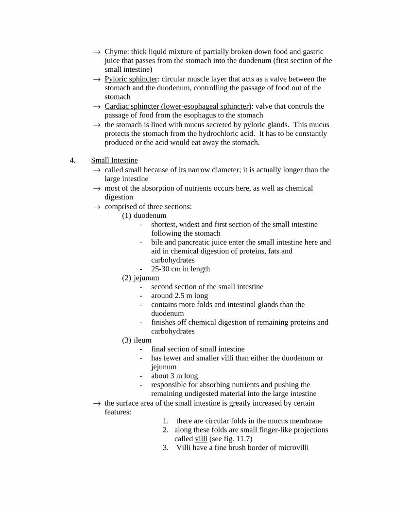
→ Chyme: thick liquid mixture of partially broken down food and gastric juice that passes from the stomach into the duodenum (first section of the small intestine)
→ Pyloric sphincter: circular muscle layer that acts as a valve between the stomach and the duodenum, controlling the passage of food out of the stomach
→ Cardiac sphincter (lower-esophageal sphincter): valve that controls the passage of food from the esophagus to the stomach
→ the stomach is lined with mucus secreted by pyloric glands. This mucus protects the stomach from the hydrochloric acid. It has to be constantly produced or the acid would eat away the stomach.
4. Small Intestine
→ called small because of its narrow diameter; it is actually longer than the large intestine
→ most of the absorption of nutrients occurs here, as well as chemical digestion
→ comprised of three sections: (1) duodenum
- shortest, widest and first section of the small intestine following the stomach
- bile and pancreatic juice enter the small intestine here and aid in chemical digestion of proteins, fats and carbohydrates
- 25-30 cm in length (2) jejunum
- second section of the small intestine - around 2.5 m long - contains more folds and intestinal glands than the
duodenum - finishes off chemical digestion of remaining proteins and
carbohydrates (3) ileum
- final section of small intestine - has fewer and smaller villi than either the duodenum or
jejunum - about 3 m long - responsible for absorbing nutrients and pushing the
remaining undigested material into the large intestine → the surface area of the small intestine is greatly increased by certain
features: 1. there are circular folds in the mucus membrane 2. along these folds are small finger-like projections
called villi (see fig. 11.7) 3. Villi have a fine brush border of microvilli

→ increasing surface area allows for better digestion and absorption of nutrients.
5. Large Intestine
→ consists of the caecum, colon, rectum and anus → caecum: sac-like part of the large intestine near the small intestine that has
a blind end → appendix: appendage that hangs from the caecum portion of the large
intestine. It has no function in digestion, but may help fight infections. → Colon: main part of the large intestine; its main job is to absorb minerals
and water from the leftover food. Bacteria in the colon are also responsible for producing vitamins like B12 and K.
→ Rectum: the last 20 cm of the large intestine (stores waste) → Anus: the opening of the digestive tract through which feces passes
through the body.
The Movement of Food Peristalsis: movement of food through the digestive tract, accompanied by a series of wave-like contractions and relaxations of the circular and longitudinal muscles that surround the parts of the digestive tract. Accessory Glands to the Alimentary Canal There are several glands which are not directly a part of the alimentary canal but they do help in digestion. They help in digestion by producing enzymes and other chemicals to aid in digestion. Enzymes: protein structures that help speed up chemical reactions in the body. Digestive enzymes help speed up the breakdown of food into its basic parts (see table 11.2, p. 365)
- Amylases – break down carbohydrates - Proteases – breaks down proteins - Lipases – breaks down fats
Main accessory organs to the alimentary canal: (1) liver
→ produces bile, which aids in fat digestion; only role in digestion. Bile released into the small intestine from the gall bladder.
→ Has several other roles in the body including recycling of RBC, storing fat-soluble vitamins, detoxification, and so on.
(2) salivary glands
→ produces salivary amylase which aids in carbohydrate digestion
(3) pancreas

→ produces several enzymes which aid in digestion of carbohydrates, proteins, and fats
• pancreatic amylase – CHO • pancreatic lipase – fats • carboxypeptidase – proteins • trypsin – proteins
→ also produces a basic solution that neutralizes the acidic chyme from the stomach before it reaches the duodenum
(4) gall bladder
→ stores bile made by the liver. Hormone signals from the duodenum cause the release of bile (CCK and secretin)
Digestive Disorders
1. Ulcers - Ulcer: slow-healing sore resulting from stomach acid penetrating the
mucus layer on the stomach wall and causing the wall to begin to erode - most ulcers are believed to be caused by an acid-resistant bacteria which
attacks the stomach wall, causing mucus production to stop - as well, other factors can contribute to ulcers (smoking, caffeine, alcohol,
and stress) - treatment includes medications and lifestyle adjustments
2. Gallstones
- Gallstones: small hard masses that form in the gall bladder, resulting from cholesterol precipitating out of the bile to form crystals that grow in size
- Factors that cause gallstones include obesity, alcohol use, and heredity - Treatment includes medications to lower cholesterol, lifestyle adjustments,
and in extreme cases gall bladder surgery
3. Inflammatory Bowel Diseases - Several kinds of inflammatory bowel diseases:
i. Ileitis: inflammation of the ileum. It causes pain and makes the bowels empty frequently causing diarrhea • Fever is often present, rectal bleeding can occur and there may
be loss of appetite and weight loss ii. Colitis: inflammation and ulceration of the innermost lining of the
colon • Symptoms include loose and bloody stool, abdominal pain,
skin lesions, joint pain, and so on. • In extreme cases, the entire bowel and rectum are removed and
an external opening called an ileostomy is created for waste

Eating Disorders Our North American society places a lot of importance on being thin. As a result, people develop unusual and sometimes extremely dangerous diets to become thin or stay thin. This usually results in those people not getting the proper amounts of nutrients. Anorexia nervosa: eating disorder characterized by a morbid fear of gaining weight, which causes the person to go on a very restrictive diet. Sufferers typically have a body mass less than 85% of their normal mass and, due to a distended body image, see themselves as emaciated.
- all symptoms of starvation are present including low blood pressure, irregular heartbeat, and constipation and so on
- in many cases, treatment of the anorexic is unsuccessful and the patient dies
Bulimia nervosa: dangerous eating disorder characterized by recurring episodes of binge eating followed by purging usually through vomiting or taking laxatives; may be associated with obesity or anorexia nervosa.

11.3 – The Human Excretory System (see Fig. 11.17, p. 374)
- kidneys – either of two organs that filter blood to remove waste (which is excreted in urine) and adjusts the concentration of salts in the blood.
- Ureters – either of a pair of ducts that carry urine from the kidneys to the bladder
- Urethra – the tube through which urine exits the bladder - Urinary bladder – stores urine until it can be excreted
When proteins are broken down in humans, a compound called ammonia (NH3) is
formed. Ammonia is highly toxic, so it is converted into urea by the liver, and this urea is the primary nitrogenous waste found in urine. Other waste products excreted in the urine include uric acid (from breakdown of DNA and RNA) and creatinine (from muscle action).
(see fig. 11.19, kidney and nephron, p. 375) The kidney has three sections: (1) cortex – the outer part of the kidney, where the glomerulus, Bowman’s
capsule, proximal tubule and distal tubule of nephrons are located. (2) medulla – the inner part of the kidney, where the loop of Henle and the
collecting ducts of nephrons are located. (3) pelvis – part of the kidney where urine accumulates before it enters the ureters The functional unit of the kidney is the nephron. Each kidney contains about 1
million or so nephrons. Parts of a nephron: 1. glomerulus – the blood vessel inside the Bowman’s capsule from which water,
salts, nutrient molecules, and waste molecules leave the blood to be filtered by the kidney 2. Bowman’s capsule – the receiving end of a renal (kidney) tubule at which
water and small solutes from the blood enter the proximal tubule from the glomerulus 3. proximal tubule – the tube between the Bowman’s capsule and the loop of
Henle. Reabsorption of important nutrients like water, glucose, and amino acids begins here.
4. loop of Henle – the long loop of the nephron that extends into the medulla of the kidney between the proximal tubule and the distal tubule. The main function of the loop of Henle is to remove water from the filtrate.
5. distal tubule – the tube that connects the loop of Henle to the collecting duct. The main job of the distal tubule is to secrete material like hydrogen ions, creatinine, and drugs out of the blood into the filtrate.
6. collecting ducts – carries urine to renal pelvis The amount of water found in urine depends on the permeability of the distal
tubule and the collecting duct. This permeability is controlled by a hormone (chemical messenger) called anti-diuretic hormone (ADH). ADH increases permeability, which
allows the body to reabsorb water so it doesn’t go out in urine. If you drink a lot of fluid, ADH is not produced so the kidneys do not reabsorb the water. As well, certain drugs like alcohol and caffeine block the release of ADH and increase urine volume.
Disorders of the Excretory System 1. Urinary tract infections - if bladder is infected, the infection is termed cystitis. If only the urethra
is infected, the term is urethritis. - urinary tract infections are more common in women than men - symptoms include painful urination, a need to constantly urinate, brown
or bloody urine, fever, nausea, etc. 2. Kidney stones - chemicals in urine precipitate and form crystals, mostly calcium oxalate - more common in men than women - factors that cause stones include urinary tract infections, insufficient
water intake, low activity level, and/or too much vitamin C or D - symptoms include severe pain, blood in the urine, nausea - small stones can pass in urine, but larger ones need to be broken down
(either by chemicals or sound) before they can pass. In extreme cases, they have to be removed by surgery
3. Renal failure - renal failure – the condition in which an individual’s kidneys no longer
function and waste accumulates in the blood - if waste levels get too high, a person can suffer loss of consciousness and
finally heart failure - hemodialysis – or kidney failure, medical treatment of kidney failure; the
process of removing blood from an artery, purifying it, adding vital substances and returning it to a vein
11.4 – The Immune System immune system – a homeostatic mechanism comprising of a variety of white blood cells and proteins that attack foreign invaders such as bacteria and viruses; recognizes and destroys damaged cells and irregular growths immunity – the ability to resist disease after having been exposed to it in the past pathogen – disease-causing agent (ex. bacteria and viruses) non-specific defenses – the ability to resist disease, even if there has been no prior exposure to it specific immune system – wide variety of cells, developed through exposure to a disease, that recognizes certain foreign substances and acts to neutralize or destroy them Classes of white blood cells (leukocytes): 1. Macrophages – large phagocytic cells that can pass through the walls of capillaries to engulf and digest pathogens such as bacteria circulating within the body 2. Neutrophils – small white blood cells that engulf bacteria through phagocytosis as an indirect immune response 3. Monocytes - white blood cells from which neutrophils and macrophages are derived. 4. Eosinophils – white blood cells that attack larger pathogens such as worm. They contain digestive enzymes. 5. Lymphocytes – type of white blood cell that plays a role in the body’s acquired immune response, enabling the body to recognize and fend off specific pathogens (see fig 11.26, p. 383) The body has 3 layers of defense when it comes to defending against pathogens: 1st layer of defense – physical barriers → Non-specific → outer layer of the skin is dry, skin’s oil has bactericide in it, and sweat makes the environment acidic → linings of respiratory and digestive pathways coated in mucus, which traps bacteria → stomach acid kills most of the microorganisms in food 2nd layer of defense – inflammatory response → non-specific → special cells arrive at infection site and release histamine, which causes blood vessels to dilate. Increased blood flow makes area swell and hot.

→ neutrophils and macrophages engulf invading bacteria. Pus forms as dead macrophages and bacteria accumulate. 3rd layer of defense – specific immunity including lymphocytes → two types of lymphocytes: (1) B-cells – mature in bone marrow; function primarily in antibody immunity (2) T-cells – mature in thymus gland; function primarily in cellular immunity → antibody – a Y-shaped protein produced by B-cells that “tag” foreign cells to be destroyed by macrophages; binds to antigens → antigen – protein or other large molecule on the surface of a non-self cell (attacking or foreign cell) that helps the body recognize its own cells → steps in cellular immunity (1) macrophage engulfs an antigen, breaking it down and placing part of it on its own cell surface (2) macrophage binds to helper T-cell, which activates cytotoxic (killer) T-cells and memory T-cells (3) cytotoxic T-cells destroy invading pathogen, while memory T-cells remain behind in case of infection by the same pathogen. → steps in antibody immunity: (1) Macrophage engulfs antigen (same as cellular immunity) (2) macrophage binds to helper T-cells, which activates special B-cells (plasma cells and memory B-cells) (3) plasma cells produce antibodies which “tag” foreign cells to be consumed by macrophages; memory B-cells stay around in case of infection by the same pathogen again. Disorders of the Immune System 1. Autoimmune disorders → when T-cells or antibodies mistakenly attack a person’s own cells → ex. rheumatoid arthritis 2. Allergies → an exaggerated response by the immune system to a harmless material such as pollen, mould, cat dander and so on. → symptoms include swelling, watery eyes, runny nose, and in the case of food allergies vomiting, cramps and diarrhea → usually caused by extra histamine released when not needed