unit iii chapter 6
TRANSCRIPT

Chapter 6UNIT III
We inhale and exhale air. Why is breathing
so important for life? What happens when
we breathe? Why energy is required for the
body to perform various life processes?
Where does the energy come from? We
eat food for energy. Though the above
raised questions look disconnected, we
should know that the process of breathing
is connected to the process of release of
energy from food. Oxygen is utilized by the
organisms to breakdown the biomolecules
like glucose and to derive energy. During
this breakdown carbondioxide, which is
a harmful gas is also released. It is very
obvious that oxygen has to be provided
to cells continuously and the CO2 to be
released immediately by the cells. So the
need of a respiratory system is essential
for life.
Respiration
Chapter Outline
6.1 Respiratory function
6.2 Respiratory organs in various organisms
6.3 Mechanism of breathing
6.4 Exchange of gases
6.5 Transport of gases
6.6 Regulation of respiration
6.7 Problems in oxygen transport
6.8 Disorders of respiratory system
6.9 Effects of smoking
• Learns to describe
the gross structure of
the human gaseous
exchange system
• Observes and
draws the tissues
and organs associated with the
respiratory system
• Understands the process of
gaseous exchange and transport of
respiratory gases
• Knows the problems associated
with oxygen transport
• Gains knowledge on the ill–effects
of smoking.
Learning Objectives:Learning Objectives:
Exercise increases the rate and depth of
breathing and supplies extra oxygen to the
muscles and removes more CO2 from the
tissues.
121
In animals like sponges, coelenterates
and flatworms exchange of gases takes
place through the body surface by simple
diffusion. Earthworms use their moist
skin, whereas insects have tracheal tubes.
Gills are used as respiratory organs in
most of the aquatic Arthropods and
Molluscs. Among verterbrates, fishes use
gills whereas amphibians, reptiles, birds
and mammals have well vascularised
lungs. Frogs spend most of their time in
water and also use their moist skin for
respiration along with lungs.
6.2.1 Human Respiratory SystemThe respiratory system includes the
external nostrils, nasal cavity, the
pharynx, the larynx, the trachea, the
bronchi and bronchioles and the lungs
which contain the alveoli (Figure 6.1). The
parts starting from the external nostrils
up to the terminal bronchioles constitute
the conducting zone, whereas the alveoli
and the ducts are called the respiratory
zone. The parts of the conducting zone,
humidifies and warms the incoming air.
In human beings, air enters the upper
respiratory tract through the external
nostrils. The air passing through the
nostrils is filtered by fine hairs and mucus
lining the passage. The external nostrils
lead to the nasal chamber which opens into
the nasopharynx which opens through
the glottis of the larynx region into the
trachea. The ciliated epithelial cells lining
the trachea, bronchi and bronchioles
secrete mucus. Mucus membrane lining
the airway contains goblet cells which
secrete mucus, a slimy material rich in
glycoprotein. Microorganisms and dust
particles attach in the mucus films and
We have discussed in the previous chapter
how food provides energy for growth and
repair of tissues. As mentioned earlier
along with food, oxygen is necessary for
breakdown of glucose to energy. In this
chapter we shall discuss the respiratory
organs of human, the mechanism of
breathing, exchange and transport of
gases and a few respiratory disorders.
The term respiration refers to the
exchange of oxygen and carbondioxide
between environment and cells of our
body where organic nutrients are broken
down enzymatically to release energy.
6.1 Respiratory functionsThe five primary functions of the
respiratory system are –
i. To exchange O2 and CO2 between
the atmosphere and the blood.
ii. To maintain homeostatic regulation
of body pH.
iii. To protect us from inhaled
pathogens and pollutants.
iv. To maintain the vocal cords
for normal communication
(vocalization).
v. To remove the heat produced
during cellular respiration through
breathing.
6.2 Respiratory organs in various organisms.Different animals have different organs
for exchange of gases, depending upon
their habitats and levels of organization.
The amount of dissolved oxygen is very
low in water compared to the amount of
oxygen in the air. So the rate of breathing
in aquatic organisms is much faster than
land animals.

122
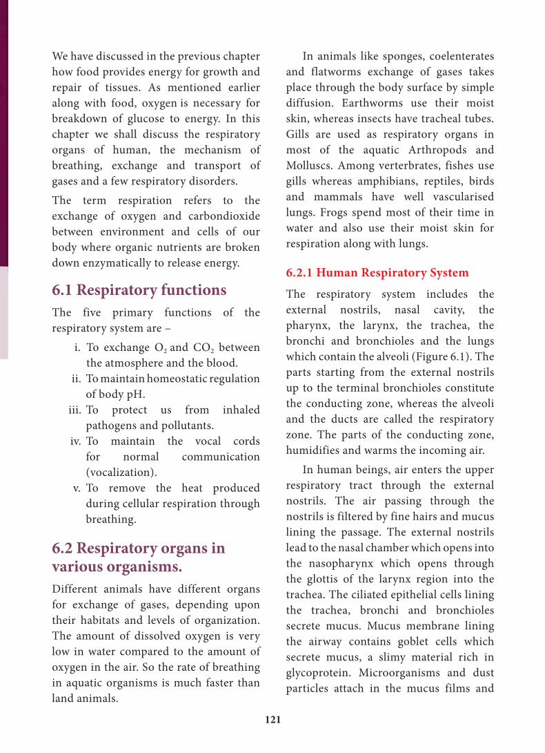
Bronchi have ‘C’ shaped curved
cartilage plates to ensure that the air
passage does not collapse or burst as the
air pressure changes during breathing.
The bronchioles are without cartilaginous
rings and have rigidity that prevent them
from collapsing but are surrounded by
smooth muscle which contracts or relaxes
to adjust the diameter of these airways.
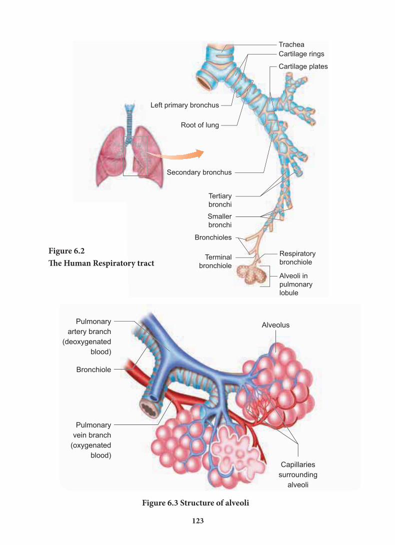
The fine respiratory bronchioles
terminate into highly vascularised thin
walled pouch like air sacs called alveoli
meant for gaseous exchange (Figure 6.2,
6.3). The diffusion membrane of alveolus
is made up of three layers – the thin
squamous epithelial cells of the alveoli, the
endothelium of the alveolar capillaries and
the basement substance found in between
them. The thin squamous epithelial cells
of the alveoli are composed of Type I and
are carried upwards to pass down the
gullet during normal swallowing. During
swallowing a thin elastic flap called
epiglottis prevents the food from entering
into the larynx and avoids choking of
food.
The trachea is semiflexible tube
supported by multiple cartilaginous rings
which extends up to the midthoracic
cavity and at the level of the 5th thoracic
vertebra where it divides into right and
left primary bronchi, one bronchus to each
lung. Within the lungs the bronchi divides
repeatedly into secondary and tertiary
bronchi and further divides into terminal
bronchioles and respiratory bronchioles.
It is advised not to talk or laugh louder
while eating. Can you give the reason?
Figure 6.1 The Human respiratory system
123
Figure 6.3 Structure of alveoli
Figure 6.2The Human Respiratory tract

124
6.3. Mechanism of breathingThe movement of air between the
atmosphere and the lungs is known as
ventilation or breathing. Inspiration
and expiration are the two phases of
breathing. Inspiration is the movement
of atmospheric air into the lungs and
expiration is the movement of alveolar air
that diffuse out of the lungs. (Figure 6.4)
Lungs do not contain muscle fibres but
expands and contracts by the movement
of the ribs and diaphragm. The diaphragm
is a sheet of tissue which separates the
thorax from the abdomen. In a relaxed
state, the diaphragm is domed shaped.
Type II cells. Type I cells are very thin
so that gases can diffuse rapidly through
them. Type II cells are thicker, synthesize
and secrete a substance called Surfactant.
The lungs are light spongy tissues
enclosed in the thoracic cavity surrounded
by an airtight space. The thoracic cavity is
bound dorsally by the vertebral column
and ventrally by the sternum, laterally by
the ribs and on the lower side by the dome
shaped diaphragm.
The lungs are covered by double
walled pleural membrane containing a
several layers of elastic connective tissues
and capillaries, which encloses the pleural
fluid. Pleural fluid reduces friction when
the lungs expand and contract.
Characteristic features of respiratory surface:• surface area must be very large and
richly supplied with blood vessels
• should be extremely thin and kept
moist
• should be in direct contact with the
environment
• should be permeable to respiratory
gases
The steps involved in respiration are
i. The exchange of air between the
atmosphere and the lungs.
ii. The exchange of O2 and CO2
between the lungs and the blood.
iii. Transport of O2 and CO2 by the
blood.
iv. Exchange of gases between the
blood and the cells.
v. Uptake of O2 by the cells for various
activities and the release of CO2.
Observe a live fish and find out how
many times it beats the operculum
per minute. Now check your rate of
breathing for a minute. The rate of
breathing will be more in fish than you
– Give reasons.
SURFACTANTS are
the thin non–cellular
films made of protein
and phospholipids
covering the alveolar membrane. The
surfactant lowers the surface tension in
the alveoli and prevents the lungs from
collapsing. It also prevents pulmonary
oedema. Premature Babies have low
levels of surfactant in the alveoli may
develop the new born respiratory
distress syndrome (NRDS) because
the synthesis of surfactants begins only
after the 25th week of gestation.

125
the lungs (intrapulmonary pressure)
is less than the atmospheric pressure
likewise expiration takes place when the
pressure within the lungs is higher than
the atmospheric pressure.
Ribs are moved by the intercostal muscles.
External and internal intercostal muscles
found between the ribs and the diaphragm
helps in creating pressure gradients.
Inspiration occurs if the pressure inside
Why do some people snore? – Breathing with a hoarse sound during sleep is caused
by the vibration of the soft palate. Snoring is caused by a partially closed upper air
way (nose and throat) which becomes too narrow for enough air to travel through the
lungs. This makes the surrounding tissues to vibrate and produces the snoring sound.
Figure 6.4 Mechanism of breathing
You are at high level in a mountain
above the sea level. Suddenly you get
palpitation and nausea. What condition
are you suffering from? What are the
other symptoms for this disease and
how can it be reduced?
Airinhaled.
Airexhaled.Rib cage
expands.
Rib cagegets
smaller.
Lung
Diaphragm
Inspiration Expiration
Inspiraton is initiated by the contraction
of the diaphragm muscles and external
intercostal muscles, which pulls the ribs
and sternum upwards and outwards and
increases the volume of the thoracic
chamber in the dorso–ventral axis, forcing
the lungs to expand the pulmonary volume.
The increase in pulmonary volume and
126
decrease in the intrapulmonary pressure
forces the fresh air from outside to enter the
air passages into the lungs to equalize the
pressure. This process is called inspiration.Relaxation of the diaphragm allows
the diaphragm and sternum to return to
its dome shape and the internal intercostal
muscles contract, pulling the ribs
downward reducing the thoracic volume
and pulmonary volume. This results in an
increase in the intrapulmonary pressure
slightly above the atmospheric pressure
causing the expulsion of air from the
lungs. This process is called expiration.On an average, a healthy human breathes
12–16 times/minute. An instrument called
Spirometer is used to measure the volume
of air involved in breathing movements for
Figure 6.5 Lung volumes and capacity
clinical assessment of a person’s pulmonary
function.
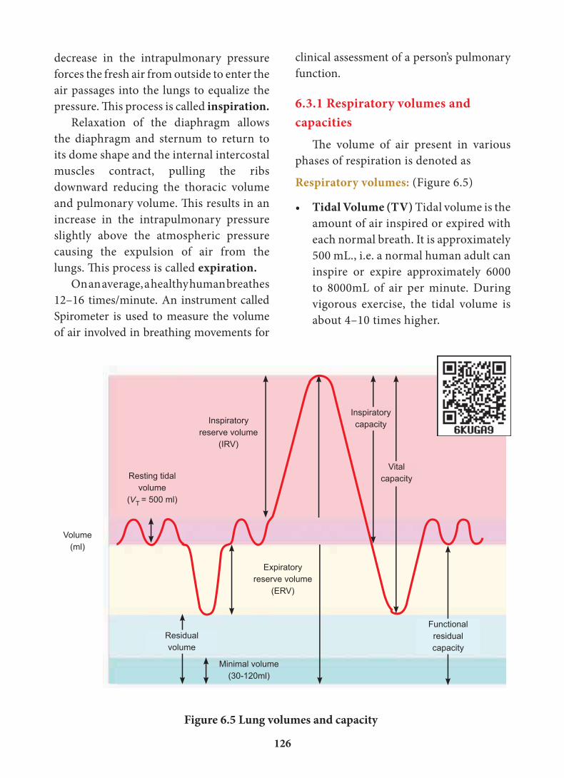
6.3.1 Respiratory volumes and capacities
The volume of air present in various
phases of respiration is denoted as
Respiratory volumes: (Figure 6.5)
• Tidal Volume (TV) Tidal volume is the
amount of air inspired or expired with
each normal breath. It is approximately
500 mL., i.e. a normal human adult can
inspire or expire approximately 6000
to 8000mL of air per minute. During
vigorous exercise, the tidal volume is
about 4–10 times higher.
Minimal volume(30-120ml)
Volume(ml)
Resting tidal volume
(VT = 500 ml)
Expiratoryreserve volume
(ERV)
Inspiratoryreserve volume
(IRV)
Residualvolume
Inspiratorycapacity
Vitalcapacity
Functional residual capacity
127
Healthy lungs contain
large amounts of
elastic connective
tissue around the
alveoli, containing
elastin, which makes the lung tissue
elastic. People with emphysema and
bronchitis have difficulty in exhaling
because the enzyme elastase destroys
the elastin around the alveoli and
reduces the elasticity of the lungs.
• Inspiratory Reserve volume (IRV)
Additional volume of air a person can
inspire by forceful inspiration is called
Inspiratory Reserve Volume. The
normal value is 2500–3000 mL.
• Expiratory Reserve volume (ERV) Additional volume of air a person
can forcefully exhale by forceful
expiration is called Expiratory
Reserve Volume. The normal value is
1000–1100 mL.
• Residual Volume (RV) The volume
of air remaining in the lungs after a
forceful expiration. It is approximately
1100–1200 mL.
Respiratory capacities:
• Vital capacity (VC) the maximum
volume of air that can be moved out
during a single breath following a
maximal inspiration. A person first
inspires maximally then expires
maximally. VC=ERV+TV+IRV
• Inspiratory capacity (IC) The total
volume of air a person can inhale
after normal expiration. It includes
tidal volume and inspiratory reserve
volume. IC=TV+IRV
• Expiratory capacity (EC) The total
volume of air a person can exhale
after normal inspiration. It includes
tidal volume and expiratory reserve
volume. EC=TV+ERV
• Total Lung Capacity (TLC) The total
volume of air which the lungs can
accommodate after forced inspiration
is called Total Lung Capacity. This
includes the vital capacity and the
residual volume. It is approximately
6000mL. TLC=VC+RV
• Minute Respiratory Volume The
amount of air that moves into the
respiratory passage per minute is called
minute respiratory volume.
Normal TV = 500mL; Normal
respiratory rate = 12 times/minute
Therefore, minute respiratory
volume = 6 Litres/minute (for a normal
healthy man).
Dead spaceSome of the inspired air never reaches the
gas exchange areas but fills the respiratory
passages where exchange of gases does not
occur. This air is called dead space.
Dead space is not involved in gaseous
exchange. It amounts to approximately
150mL.
6.4 Exchange of gasesThe primary site for the exchange of
gases is the alveoli. The uptake of O2
and the release of CO2 occur between
the blood and tissues by simple diffusion
driven by partial pressure gradient of O2
and CO2. Partial pressure is the pressure
contributed by an individual gas in a
mixture of gases. It is represented as
pO2 for oxygen and pCO2 for carbon–

128
Respiratory gases
Partial pressure mm HgAtmospheric
air Alveoli Deoxygenated Blood
Oxygenated blood Tissues
O2 159 104 40 95 40
CO2 0.3 40 45 40 45
Table 6.1 Partial pressure of Oxygen and Carbon dioxide (in mmHg) in comparison to
those gases in the atmosphere
Figure 6.6 Exchange of gases at the alveolus and the tissue with blood and transport of
oxygen and carbondioxide
(Carrying
deoxygenated
blood)
(Carrying
oxygenated
blood)
dioxide. Due to pressure gradients, O2
from the alveoli enters into the blood
and reaches the tissues. CO2 enters
into the blood from the tissues and
reaches alveoli for elimination. As the
solubility of CO2 is 20–25 times higher
than that of O2, the partial pressure of
CO2 is much higher than that of O2
(Tab.6.1 and Figure 6.6).
Respiratory pigmentsHaemoglobinHaemoglobin belongs to the class
of conjugated protein. The iron
containing pigment portion haem
constitutes only 4% and the rest
colourless protein of the histone class
globin. Haemoglobin has a molecular
weight of 68,000 and contains four
atoms of iron, each of which can
combine with a molecule of oxygen.
MethaemoglobinIf the iron component of the haem
moieties is in the ferric state, than
the normal ferrous state, it is called
methaemoglobin. Methaemoglobin
Breathing through nose is healthier
than through mouth– Why?

129
does not bind O2. Normally RBC contains
less than 1% methaemoglobin.
6.5 Transport of gases6.5.1 Transport of oxygenMolecular oxygen is carried in blood in
two ways: bound to haemoglobin within
the red blood cells and dissolved in
plasma. Oxygen is poorly soluble in water,
so only 3% of the oxygen is transported
in the dissolved form. 97% of oxygen
binds with haemoglobin in a reversible
manner to form oxyhaemoglobin
(HbO2). The rate at which haemoglobin
binds with O2 is regulated by the partial
pressure of O2. Each haemoglobin
carries maximum of four molecules
of oxygen. In the alveoli high pO2, low
pCO2, low temperature and less H+
concentration, favours the formation of
oxyhaemoglobin, whereas in the tissues
low pO2, high pCO2, high H+ and high
Figure 6.7 Oxygen dissociation curve
temperature favours the dissociation of
oxygen from oxyhaemoglobin.
A sigmoid curve (S–shaped) is obtained
when percentage saturation of haemoglobin
with oxygen is plotted against pO2. This
curve is called oxygen haemoglobin
dissociation curve (Figure 6.7). This
S–shaped curve has a steep slope for pO2
values between 10 and 50mmHg and then
flattens between 70 and 100 mm Hg.
Under normal physiological
conditions, every 100mL of oxygenated
blood can deliver about 5mL of O2 to the
tissues.
6.5.2 Transport of Carbon–dioxideBlood transports CO2 from the tissue cells
to the lungs in three ways
i. Dissolved in plasma About
7 – 10% of CO2 is transported in a
dissolved form in the plasma.
ii. Bound to haemoglobin About
20 – 25% of dissolved CO2 is
bound and carried in the RBCs as
carbaminohaemoglobin (Hb CO2)
CO2 + Hb Hb CO2
iii. As bicarbonate ions in plasma about 70% of CO2 is
transported as bicarbonate ions
This is influenced by pCO2
and the degree of haemoglobin
oxygenation. RBCs contain a high
concentration of the enzyme,
carbonic anhydras, Whereas small
amounts of carbonic anhydrase is
present in the plasma.
At the tissues the pCO2 is high
due to catabolism and diffuses into
the blood to form HCO3– and H+

130
CO2 + H2O carbonic anhydrase H2CO3
carbonic anhydrase HCO3– + H+
The HCO3– moves quickly from the RBCs
into the plasma, where it is carried to the
lungs. At the alveolar site where pCO2 is
low, the reaction is reversed leading to the
formation of CO2 and water. Thus CO2
trapped as HCO3– at the tissue level it is
ions. When CO2 diffuses into the RBCs,
it combines with water forming carbonic
acid (H2CO3) catalyzed by carbonic
anhydrase. Carbonic acid is unstable and
dissociates into hydrogen and bicarbonate
ions.
Carbonic anhydrase facilitates the
reaction in both directions.
Inspiration Expiration
Respiratory centre initiates the stimuli
during inspiration.
Impulses are carried to the inspiratory
muscles through nerves.
Diaphragm and inspiratory muscles
contract.
The thoracic volume increases as the
chest wall expands.
The intra pulmonary pressure is
reduced.
The alveolar pressure decreases than
the atmospheric pressure
Air flows into the alveoli until the
alveolar pressure equalizes the
atmospheric pressure and the alveoli
get inflated.
Respiratory centre terminates the stimuli
during expiration.
The diaphragm and inspiratory muscles
relax.
Chest wall contracts and the thoracic
volume gets reduced.
The intra pulmonary pressure is reduced.
The alveolar pressure increases than the
atmospheric pressure.
Air is sent out due to the contraction of
alveoli.
Air flows out of the alveoli until
the alveolar pressure equalizes the
atmospheric pressure and the alveoli get
deflated.
Events in inspiration and expiration

131
headache, shortness of breath, nausea and
dizziness due to poor binding of O2 with
haemoglobin. When the person moves on
a long–term basis to mountains from sea
level is body begins to make respiratory
and haematopoietic adjustments.
To overcome this situation kidneys
accelerate production of the hormone
erythropoietin, which stimulates the
bone marrow to produce more RBCs.
When a person descends deep into
the sea, the pressure in the surrounding
water increases which causes the lungs
to decrease in volume. This decrease in
volume increases the partial pressure of
the gases within the lungs. This effect can
be beneficial, because it tends to drive
additional oxygen into the circulation, but
this benefit also has a risk, the increased
pressure can also drive nitrogen gas into
the circulation. This increase in blood
nitrogen content can lead to a condition
called nitrogen narcosis. When the
diver ascends to the surface too quickly a
condition called ‘bends’ or decompression
sickness occurs and nitrogen comes
out of solution while still in the blood
forming bubbles. Small bubbles in the
blood are not harmful, but large bubbles
can lodge in small capillaries, blocking
transported to the alveoli and released
out as CO2. Every 100mL of deoxygenated
blood delivers 4mL of CO2 to the alveoli
for elimination.
6.6 Regulation of RespirationA specialised respiratory centre present
in the medulla oblongata of the hind
brain called respiratory rhythm centre
is responsible for this regulation.
Pneumotaxic centre present in pons varoli
region of the brain moderates the function
of the respiratory rhythm centre to ensure
normal breathing. The chemosensitive
area found close to the rhythm centre is
highly sensitive to CO2 and H+. And H+
are eliminated out by respiratory process.
Receptors associated with the aortic arch
and carotid artery send necessary signals
to the rhythm centre for remedial action.
The role of O2 is insignificant in the
regulation of respiratory rhythm.
6.7 Problems in Oxygen transportWhen a person travels quickly from sea
level to elevations above 8000ft, where
the atmospheric pressure and partial
pressure of oxygen are lowered, the
individual responds with symptoms
of acute mountain sickness (AMS)–
Particulate matter PM 2.5 in the air
is increasing day by day which causes
respiratory illness. Central Pollution
Control Board (CPCB) reports that the
quality of air is not good due to soot and
smoke. So some cities in India are using
CNG (Compressed Natural Gas) as fuel.
Allergy is caused by
allergens. When we
enter a polluted area,
immediately we start
sneezing and coughing. The allergens
in that place affect our respiratory tracts
and the responses to the allergens start
within minutes. Allergens provoke an
inflammatory response. A common
manifestation of allergy is Asthma.

132
Pneumonia– Inflammation of the lungs
due to infection caused by bacteria or
virus is called pneumonia. The common
symptoms are sputum production, nasal
congestion, shortness of breath, sore
throat, etc.
Tuberculosis– Tuberculosis is caused by
Mycobacterium tuberculae. This infection
mainly occurs in the lungs and bones.
Collection of fluid between the lungs and
the chest wall is the main complication of
this disease.
Occupational respiratory disorders– The disorders due to one’s occupation
of working in industries like grinding or
stone breaking, construction sites, cotton
industries, etc. Dust produced affects the
respiratory tracts.
Long exposure can give rise to
inflammation leading to fibrosis.
Silicosis and asbestosis are occupational
respiratory diseases resulting from
inhalation of particle of silica from sand
grinding and asbestos into the respiratory
tract. Workers, working in such industries
must wear protective masks.
6.9 Eff ects of SmokingToday due to curiosity, excitement or
adventure youngsters start to smoke and
later get addicted to smoking. Research
says about 80% of the lung cancer is due
to cigarette smoking.
Smoking is inhaling the smoke from
burning tobacco. Th ere are thousands of
known chemicals which includes nicotine,
tar, carbon monoxide, ammonia, sulphur–
dioxide and even small quantities of arsenic.
Carbon monoxide and nicotine damage
the cardiovascular system and tar damages
blood flow or can press on nerve endings.
Decompression sickness is associated
with pain in joints and muscles and
neurological problems including stroke.
The risk of nitrogen narcosis and bends is
common in scuba divers.
During carbon–dioxide poisoning,
the demand for oxygen increases. As the
O2 level in the blood decreases it leads
to suffocation and the skin turns bluish
black.
6.8 Disorders of the Respiratory systemRespiratory system is highly affected by
environmental, occupational, personal
and social factors. These factors may be
responsible for a number of respiratory
disorders. Some of the disorders are
discussed here.
Asthma – It is characterized by narrowing
and inflammation of bronchi and
bronchioles and difficulty in breathing.
Common allergens for asthma are dust,
drugs, pollen grains, certain food items
like fish, prawn and certain fruits etc.
Emphysema– Emphysema is chronic
breathlessness caused by gradual
breakdown of the thin walls of the alveoli
decreasing the total surface area of a
gaseous exchange. i.e., widening of the
alveoli is called emphysema. The major
cause for this disease is cigarette smoking,
which reduces the respiratory surface of
the alveolar walls.
Bronchitis– The bronchi when it gets
inflated due to pollution smoke and
cigarette smoking, causes bronchitis. The
symptoms are cough, shortness of breath
and sputum in the lungs.


134
the gaseous exchange system. Nicotine is
the chemical that causes addiction and is a
stimulant which makes the heart beat faster
and the narrowing of blood vessels results
in raised blood pressure and coronary heart
diseases. Presence of carbon monoxide
reduces oxygen supply. Lung cancer, cancer
of the mouth and larynx is more common in
smokers than non–smokers. Smoking also
causes cancer of the stomach, pancreas and
bladder and lowers sperm count in men.
Smoking can cause lung diseases by
damaging the airways and alveoli and results
in emphysema and chronic bronchitis. These
two diseases along with asthma are often
referred as Chronic Obstructive Pulmonary
Disease (COPD). When a person smokes,
nearly 85% of the smoke released is inhaled
by the smoker himself and others in the
vicinity, called passive smokers, are also
affected. Guidance or counselling should be
done in such users to withdraw this habit.
Sumanan noticed that his close friend was addicted to cigarette smoking. He advised his friend and explained the ill–effects of smoking. As a Biology student, explain what advice he might have given to his friend regarding the ill–effects of smoking.
AMAZING FACTS• The World TB Day is March 24.
• Direct Observation Therapy (DOTs) can treat about 95% of the TB patients.
• The surface area of the lungs is roughly the same size as a tennis court (525 feet
long).
• It is possible to live with one lung.
• The highest recorded ‘sneeze speed’ is 165 km per hour.
• Adults breathe around 12 – 16 times per minute where as new borns breathe
around 30–60 times per minute.
• Yawning helps us to breathe more oxygen to the lungs. When our brain senses the
shortage of O2, it send a message to CNS to imbalance to O2 demand and trigger
us to yawn.
• Breathing through mouth results in bladder shrinkage and creates an urge to
urinate in the middle of the night.
• Most people can hold their breath between 30 seconds to one minute.
• Hiccups are due to eating too fast or having occasional spasms of the diaphragm.
Activity
To test the presence of CO2 in exhaled air Take two test tubes A and B with few mL
of clear lime water. Blow exhaled air into A with a help of a straw and pass normal air
into B with a help of a syringe for about 15 times and observe the changes that occur in
the tubes A and B. The lime water (Calcium Hydroxide) in the test tube A turns milky.

135
Respiratory System’s URL:
ICT Corner
Respire
Step – 1Use the URL to reach the ‘Respiratory System’ page. In the grid select ‘Nasal cavity’ and explore its structure and the functions.
Step – 2Now click back button on the top of the window or use the ‘Backspace’ key. Select ‘Pharynx’ from the grid and explore its anatomical regions.
Step – 3Follow the above steps to explore each part and its functions.
Step – 4Use the reference given below the page to acquire additional details.
Let’s explore the anatomy and function
of the Respiratory system.

136
Mec
hani
sm o
f Res
pira
�on
Insp
ira�o
n an
d Ex
pira
�on
happ
ens
due
to p
ress
ure
chan
ges i
n th
e th
orac
ic
cavi
ty.
By th
e co
ntra
c�on
and
ex
pans
ion
of d
iaph
ragm
, ex
tern
al in
terc
osta
lmus
cles
an
d in
tern
al in
terc
osta
lm
uscl
es th
e vo
lum
e of
the
thor
acic
cav
ity is
redu
ced
or
incr
ease
d.
The
act o
f bre
athi
ng is
pe
rfor
med
by
expa
nsio
n an
d co
ntra
c�on
of t
he
thor
acic
cav
ity.
INSP
IRAT
ION
An a
c�ve
pro
cess
by
whi
ch
fres
h ai
r is d
raw
n in
to th
e lu
ngs.
The
cont
rac�
on o
f ext
erna
l in
terc
osta
lmus
cle
caus
es
the
ribs t
o m
ove
ante
riorly
and
outw
ardl
y.
The
cont
rac�
on o
f the
di
aphr
agm
lead
s to
fla
�en
ing
of in
elas
�c, d
ome
shap
ed c
entr
al p
art o
f the
di
aphr
agm
.
EXPI
RATI
ON
A pa
ssiv
e pr
oces
s by
whi
ch
air i
s exh
aled
from
the
lung
s.
The
diap
hrag
m re
laxe
s and
ris
es to
resu
me
its o
rigin
al
dom
e sh
ape.
The
ribs t
ake
thei
r orig
inal
po
si�on
as a
resu
lt of
co
ntra
c�on
of t
he in
tern
al
inte
rcos
talm
uscl
es.
Con
cept
map
Dia
phra
gmdo
wn
Dia
phra
gmup
Air
draw
n in
Air
draw
n ou
t
ribs
obliq
uely
up
ribs
obliq
uely
dow
n

137
Res
pira
tory
sy
stem
Nos
eN
asal
& o
ral
cavi
ties
Phar
ynx
Lary
nxTr
ache
aB
ronc
hi
Seco
ndar
y br
onch
i
Tert
iary
br
onch
i
Smal
ler
bron
chi
Bro
nchi
oles
Term
inal
br
onch
iole
Res
pira
tory
br
onch
iole
Alv
eoli
Alv
eoli
wal
ls:
sing
le l
ayer
of
sim
ple
squa
mou
sep
ithe
lium
–m
axim
al s
urfa
ce a
rea
and
min
imal
di
ffus
ion
dist
ance
.

138
and as HCO3. HCO3 is produced in RBCs
from CO2 and water catalysed by carbonic
anhydrase. Breathing is controlled by
medullary respiratory centre.
Respiratory volumes and capacities
indicate the amount of air inspired and
expired during normal respiration. Our
respiratory system can be affected by
pollutants, pathogens and other chemical
substances found in air. Lung cancer and
emphysema cannot be cured and these
diseases are common among cigarette
smokers.
People at higher level than the sea
level are prompted to altitude sickness as
the barometric pressure is low in those
regions. Surfactant, emphysema, Asthma
and Dead space have been discussed.
During vigorous exercise the rate of
respiration increases.
Summary:The process of intake of oxygen rich air
and giving out of air rich in carbon dioxide
is generally called respiration. Pollutants
and micro organism are filtered from the
inspired air by the hair and mucus present
in the nostrils. The two main steps in the
mechanism of respiration are inspiration and
expiration which takes place due to pressure
gradient in the atmosphere and lungs.
O2 is transported in blood in dissolved
form and is also bound to haemoglobin.
One molecule of haemoglobin can bind
four molecules of O2. The Sigmoid shape
of the O2 haemoglobin dissociative curve
shows increased affinity for each O2
molecule.
CO2 is transported in blood in
dissolved form as carbamino haemoglobin
GlossaryApnoea – Temporary stopping of
respiration.
Book gills – Respiratory organs in aquatic
Limulus.
Book lungs – Respiratory organs of
Scorpions and most spiders.
COLD – Chronic Obstructive Lung Disease.
Dyspnoea – painful respiration.
Epiglottis – a thin elastic cartilaginous flap
which covers the glottis and prevents the
entry of food into the larynx.
Haemoglobin – iron containing red
pigment of RBCs of vertebrates, gives red
colour to blood.
Herring-Breuer reflex – a defensive
mechanism against over dilation of lungs.
Hypoxia – the failure of tissues for any
reason to receive an adequate supply of
oxygen.
Pneumothorax – presence of air in the
pleural cavity which causes collapsing of
lungs.
Vocal cords – sound regulating cords also
called larynx or voice box.
Yawning – prolonged inspiration due to
increase in CO2 concentration.

139
7. During inspiration, the diaphragm
a. expands.
b. unchanged
c. relaxes to become domed–shaped.
d. contracts and flattens
8. CO2 is transported through blood to
lungs as
a. carbonic acid
b. oxyhaemoglobin
c. carbamino haemoglobin
d. carboxy haemoglobin
9. When 1500 mL air is in the lungs, it is
called
a. vital capacity
b. tidal volume
c. residual volume
d. inspiratory reserve volume
10. Vital capacity is
a. TV + IRV
b. TV + ERV
c. RV + ERV
d. TV + TRV + ERV
11. After a long deep breath, we do not
respire for some seconds due to
a. more CO2 in the blood
b. more O2 in the blood
c. less CO2 in the blood
d. less O2 in the blood
12. Which of the following substances
in tobacco smoke damage the gas
exchange system?
a. carbon monoxide and carcinogens
b. carbon monoxide and nicotine
c. carcinogens and tar
d. nicotine and tar
Evaluation1. Breathing is controlled by
a. cerebrum
b. medulla oblongata
c. cerebellum
d. pons
2. Intercostal muscles are found between
the
a. vertebral column
b. sternum
c. ribs
d. glottis
3. The respiratory structures of insects
are
a. tracheal tubes
b. gills
c. green glands
d. lungs
4. Asthma is caused due to
a. bleeding in pleural cavity.
b. infection of nose
c. damage of diaphragm.
d. infection of lungs
5. The Oxygen Dissociation Curve is
a. sigmoid
b. straight line
c. curved
d. rectangular hyperbola
6. The Tidal Volume of a normal person is
a. 800 mL
b. 1200 mL
c. 500 mL
d. 1100 – 1200 mL

140
(S) FRC iv. Volume of air exhaled
after inspiration.
(a) P – i , Q – ii , R – iii , S – iv
(b) P – ii , Q – iii , R – iv , S – i
(c) P – ii , Q – iii , R – i , S – iv
(d) P – iii , Q – iv , R – i , S – ii
16. Make the correct pairs.
Columan–I Column–II
(P) Tidal
volume
i. 1000 to 1100 ml
(Q) Residual
volume
ii. 500 ml
(R) Expiratory
reserve
volume
iii. 2500 to 3000 ml
(S) Inspiratory
reserve
volume
iv. 1100 to 1200 ml
(a) P – ii , Q – iv , R – i , S – iii
(b) P – iii , Q – ii , R – iv , S – i
(c) P – ii , Q – iv , R – iii , S – i
(d) P – iii , Q – iv , R – i , S – ii
17. Name the respiratory organs of
flatworm, earthworm, fish, prawn,
cockroach and cat.
18. Name the enzyme that catalyses the
bicarbonate formation in RBCs.
19. Air moving from the nose to the
trachea passes through a number
of structures. List in order of the
structures.
20. Which structure seals the larynx when
we swallow?
21. Resistance in the airways is typically
low. Why? Give two reasons.
22. How the body makes long–term
adjustments when living in high
altitude.
13. Column I represents diseases and
column II represents their symptoms.
Choose the correctly paired option
Column I Column II
(P) Asthma (i) Recurring of
bronchitis
(Q) Emphysema (ii) Accumulation of
W.B.CS in alveolus
(R) Pneumonia (iii) Allergy
a. P = iii, Q = ii, R = i
b. P = iii, Q = i, R = ii
c. P = ii, Q = iii, R = i
d. P = ii, Q = i, R = iii
14. Which of the following best describes
the process of gas exchange in the
lungs?
a. Air moves in and out of the alveoli
during breathing.
b. Carbon dioxide diffuses from
deoxygenated blood in capillaries
into the alveolar air.
c. Oxygen and carbon dioxide diffuse
down their concentration gradients
between blood and alveolar air.
d. Oxygen diffuses from alveolar air
into deoxygenated blood.
15. Make the correct pairs.
Columan–I Column–II
(P) IC i. maximum volume of
air breathe in after
forced.
(Q) EC ii. Volume of air present
after expiration in
lungs.
(R) VC iii. Volume of air inhaled
after expiration.

141
25. Why is pneumonia considered a
dangerous disease?
26. Explain the conditions which creates
problems in oxygen transport.
23. Diffusion of gases occurs in the
alveolar region only and not in any
other part of the respiratory system.
Discuss.
24. Sketch a flow chart to show the path
way of air flow during respiration.
Competitive Exam CornerSarojini’s father has congestion of the
lungs. His doctor advised him to take
bedrest and prescribed him an inhaler.
What disease is he is suffering from? List
the symptoms for the disease.
A villager who came to the city was
affected by severe respiratory illness due
to the inhalation of particulate pollutants.
Suggest the reason for his illness and how
does particulate pollutants affect him.
Kumar’s mother works in a stone
grinding factory. Suddenly she faints
and taken to the hospital. The doctor
notices fibres in the lungs. What kind of
disease is she affected with? How can it
be rectified?
References1. Guyton A.C. and Hall. J. E, (2006) Textbook
of Medical Physiology– Eleventh Edition
Elsevier Saunders, International Edition
ISBN 0–8089–2317–X.
2. Mackean D.G. and Hayward D
(2014). AS and A level biology book,
Cambridge International, 3rd edition,
Hodder Education, An Hachette UK
company, London NWI 3BH.
3. Dee Unglaub Silverthron, Human
physiology –an integrated approach –
7th Edition – Pearson Global edition.
4. Elaine N. Marieb and Katja Hoehn
(2010). Human Anatomy and
Physiology Eighth Edition, Benjamin
Cummings, Pearson. New York.
5. Lauralee Sherwood and Robert kell.
(2007). Human physiology from cells
to systems. First Canadian Edition
Nelson Education Ltd, Toronto, Ontario
MIK SG4.
6. Moyes and Schulte, 2016 Principles
of Animal Physiology– 2 nd edition,
Pearson
7. Muthayya N.M., 2010 Human
physiology– 4th edition, Jaypee brother
medical publishers.
Web linkshttp : / /kidshea lth .org/kid/closet/
movies/how_the_body_works_interim.
html