unit vii: animal structure and function, part i animal nutrition, circulation and gas exchange, and...
TRANSCRIPT

Unit VII: Animal Structure and Function, Part I
Animal Nutrition, Circulation and Gas Exchange, and Immunity

Unit Objectives• Observe how animals adjust to the environment
over the long term by adaptation due to natural selection and over the short term by physiological responses.
+ How do animals obtain energy from the environment?
+ How do animals obtain O2 for cell respiration while disposing of the waste gas CO2?
+ How do animals respond to pathogens?

Epithelial TissueThe Cell• basic unit of structural organizationfor all living things + tissue (Latin, weave)
- groups of cells with a common structure and function
• Epithelial Tissue + surface tissue- covers every surface of the body
- squamous- cuboidal- columnar

Connective Tissue
Live cells in a non-living matrix

Connective Tissue (con’t)Blood• RBC/WBC in water based fluid plasmaBone• osteocytes living in calcified hard matrixLigaments• fibroblasts in a matrix of collagen fibers + bone-to-bone connectionTendons• fibroblasts in a matrix of less elastic fibers + muscle-to-bone connectionCartilage• chondrocytes in a soft/pliable matrixAdipose tissue• mostly cells (little matrix)Loose connective tissue• tissue glue of the body
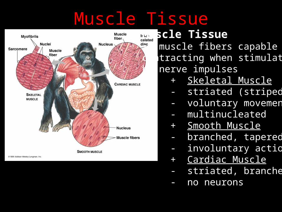
Muscle TissueMuscle Tissue• muscle fibers capable of contracting when stimulatedby nerve impulses + Skeletal Muscle
- striated (striped)- voluntary movements- multinucleated
+ Smooth Muscle- branched, tapered- involuntary actions
+ Cardiac Muscle- striated, branched- no neurons

Muscle ContractionSkeletal Muscle• two kinds + fast-twitch (white meat)
- tend to go anaerobic + slow-twitch (dark meat)
- myoglobin-rich• “twitch” + contraction of protein filaments causes muscles to shorten
- thin (actin) and thick (myosin) bands- interleaved with each other
+ myosin grabs actin and pulls - sliding filament theory of muscle contraction

Muscle ContractionSliding Filament Theory• relaxed muscle + length of each sarcomere is greater
- Z-line to Z-line• Contracting Muscle + actin/myosin slide past each other
- shortening the sarcomere• Contracted Muscle (maximum) + actin filaments overlap each other
- sarcomere is very short

Nervous Tissue

Organ Systems• Digestive System: mouth … anus
• Circulatory System: heart, blood vessels, blood
• Respiratory System: lungs, trachea, other breathing tubes
• Immune and Lymphatic System: marrow, lymph nodes, spleen, WBC
• Excretory System: kidneys, ureters, bladder, urethra
• Endocrine System: hormone-secreting glands
• Reproductive System: ovaries, testes, etc.
• Nervous System: brain, spinal cord, nerves
• Integumentary System: skin and its derivatives
• Skeletal System: bones, tendons, ligaments, cartilage
• Muscular System: skeletal muscles

BioenergeticsOverview• animals derive chemical energy from the environment in food• digestion breaks down food into nutrient molecules + some energy returns to environment as feces• nutrient molecules enter body cells + convert to useful form (ATP)• use ATP for cellular work and biosynthesis + some energy lost as heat• metabolic rate + amount of energy an animal uses in a unit of time + BMR

Energy Content of FoodCalorimeter• instrument used to measure the amount of energy in a food sample + food sample is burned, and the heat produced is measured• calorie + unit commonly used in measuring energy content of food
- amount of heat that is needed to raise the temperature of 1 gram of water 1ºC
+ 1 calorie = 4.2 joules + 1 Calorie = kilocalorie (1000 calories)

Regulating the Internal Environment
Homeostasis• the process of controlled and regulating the internal environment + interstitial fluid

HomeostasisFeedback Mechanisms• negative/positive + receptors detect a change in a variable
- response depends on the type of change
• thermometer (receptor)• variable (room temperature)• response (heat produced)

Human Digestive SystemAlimentary Canal• “tube-within-a-tube” + mouth + pharynx + esophagus + stomach + small intestine + large intestine + rectum + anus• 27 feet long!!! + Take the tour!• accessory organs + salivary glands + liver + pancreas

Mouth and PharynxMechanical Digestion• teeth and tongueChemical Digestion• salivary glands + two types
- thin, watery- thick, mucous
+ salivary amylase - digests starchSwallowing Reflex• tongue pushes bolus to pharynx• epiglottis closes off trachea

EsophagusPeristalsis• alternate waves of relaxation and contraction + moves food through alimentary canal• sphincter + ring of muscle
- cardiac sphincter- pyloric sphincter

The StomachThick-walled, muscular sac• bolus is stored temporarily + 2+ liters of food/liquid + 20 minutes or less• mechanical breakdown + churning and contracting• chemical digestion of proteins + gastric juices
- hydrochloric acid (ulcers)- pepsin
• bolus changed into chyme + thin, soup liquid

The Small IntestineSmall Intestine• 6.5 meters long, 2.5 centimeters diameter + coiled, folded + lined with villi• three regions + duodenum, jejunum, ileum• site of chemical digestion and absorption + pancreatic juice
- amylase, protease, trypsin, lipase
+ bile + intestinal juice
- peptidase, maltase

How does our body know when to secrete these enzymes?
Feedback Mechanisms
Stomach Secretions
Intestinal Secretions

Large IntestineLarge Intestine• 1.5 meters long, 6 centimeters diameter• four regions + ascending, transverse, descending, and sigmoid colon• three functions + reabsorption of water
- diarrhea/constipation + absorption of vitamins from bacteria
- E. coli bacteria produce vit. K + elimination of feces• appendix + vestigial structure with no function

Circulation and Gas ExchangeTransport Systems • functionally connect the organs of exchange with the body cells + diffusion alone is not adequate + circulatory system solves this problem
- chemicals are transported b/n the blood
and interstitial fluid + O2/CO2,
nutrients/waste

Open and Closed Circulatory Systems + animals having many layers of cells
- open: no distinction b/n blood and interstitial fluid + hemolymph- closed: blood is confined to vessels and is distinct from interstitial fluid

Vertebrate Circulatory SystemCardiovascular System• heart, blood vessels, and blood + heart
- atria (1 or 2) and (1 or 2) ventricles
+ blood vessels- arteries/arterioles:
carry blood away from the heart- veins/venules:
carry blood towards the heart- capillaries:
microscopic vessels with very thin, porous walls
+ capillary beds: networks of capillaries infiltrating tissue

Single circuit flow in fish: blood must pass through two capillary beds
Double circuit flow in amphibian: blood is pumped through two circuits
Double circuit flow in mammals: O2-rich blood segregated from O2-poor

The Mammalian HeartAtria• thin-walled collection chamber
Ventricles• thick-walled pumping chamber
Heart Valves• atrioventricular (AV) valve + b/n each atria/ventricle
- tricuspid/bicuspid• semilunar valves + at the each exit of heartThe LEFT side of the heart services O2-rich blood only
• pumps blood through systemic circulationThe RIGHT side of the heart services O2-poor blood only• pumps blood through pulmonary circulation

(1) Right Ventricle + semilunar valve(2) Pulmonary arteries + right/left(3) Lungs (Pulmonary Circulation) + pulmonary veins(4) Left Atrium + bicuspid valve(5) Left Ventricle + semilunar valve(6) Aorta(7) Systemic Circulation + head/forelimbs(8) Systemic Circulation + abdominal organs/legs(9) Anterior Vena Cava(10) Posterior Vena Cava(11) Right Atrium
+ tricuspid valve

The Cardiac Cycle“Lub-dup”• caused by the closing of valves + lub: AV valves + dup: semilunar valves
- heart murmurPulse• rhythmic stretching of the arteries caused by the pressure of blood driven by heart’s contractions + heart rate
- cardiac cycle + systole/diastole

The Cardiac Cycle (con’t)
Sinoatrial (SA) node• specialized muscle tissue that sets the rate at which all cardiacmuscle cells contract + atrioventricular (AV) node
- relay point: delayed for about 0.1 sec. + electrocardiogram (ECG/EKG)

Structure of Blood VesselsArteries and Veins and Capillaries• arteries: thick layer of smooth muscle and elastic connective tissue + carry blood AWAY from the heart• veins: thin layer of smooth muscle + valves promote unidirectional flow of blood
- varicose veins• capillaries + site of exchange; only epithelial tissue - thin-walled; 1 RBC pass through at a time• blood pressure + blood travels 1000x faster in the aorta than the capillaries
- law of continuity: if a pipe’s diameter changes over it’s length, a fluid will stream through narrower segments of the pipe faster than the wider segments + but, with so many capillaries…

Blood Pressure• Systolic/diastolic + measured in mm of mercury
- sphygmomanometer + systolic pressure created when ventricles contract + diastolic pressure is the background pressure
- blood is constantly under some pressure in a closed circulatory system

Lymphatic System
Lymph• plasma leaks out of capillaries + some of it reabsorbed by blood vessels (veins)
- body’s drainage system + lead back to vena cava through network similar to veins + WBC fight infection + filaria infection

BloodComponents of Blood• Plasma (55%) + water (91%) + dissolved ions,hormones, proteins, nutrients
- fibrinogen, albumin, prothrombin, globulin
• RBC + erythrocytes (no nucleus)
- transport O2/CO2
• WBC + leukocytes
- defense/immunity• Platelets + thrombocytes
- blood clotting

Stem Cells• bone marrow or embryo + stem cell research + luekemia

Blood Clotting

Red Blood CellsHemoglobin• 2.8 x 10⁸ per RBC + each hemoglobin can carry 4 O2
• uptake of O2 affected by pH + CO2 combines with H2O in plasma
- carbonic acid (H2CO3) + muscles produce a lot of CO2
- pH around muscles is low (acid) + lower pH causes hemoglobin to let go of O2
Why do we need to carry these gases?

Respiratory SystemGas Exchange• acquire O2, dispose CO2
+ cell respiration- uses O2, produces CO2
• respiratory medium + source of O2
- water vs. air
• respiratory surface + where O2/CO2 exchange
takes place- must be moist
+ earthworm uses skin - high ratio of surface
area to volume

Gills• outfoldings of the body surface specialized for gas exchange for aquatic animals• countercurrent exchange + blood flows in opposite direction of water
- creates diffusion gradient

Tracheal Systems
Tracheal Systems• respiratory adaptations forterrestrial animals + air tubes that branch through- out the body
- extend to the surface of nearly every cell

LungsRespiratory System• nasal passages + filtered by hair • through pharynx to trachea + rings of cartilage + larynx (voice box)• trachea branches into two bronchi• bronchi branch into many bronchioles• bronchioles branch into bronchial tubes• bronchial tubes end in alveoli + clusters of air sacs
- surrounded by blood vessels- moist, thin-walled

Ventilating the LungsBreathing• alternate inhalation andexhalation of air + negative pressure
- suction pump• inhalation + diaphragm pulls downward + rib cage and muscles pull upward
- increases lung volume- decrease pressure in lungs- fresh air rushes in
• exhalation + diaphragm/rib cage and muscles relax - decreases lung volume
- air rushes out

Automatic Control of BreathingBreathing Control Centers• medulla oblongata and pons + sets basic breathing rhythm• monitors CO2 levels in blood + pH of blood
- drop in pH, increase the rate/depth
• O2 sensors in aorta and carotidarteries + sense severely depressed levels of O2 (high altitude)• hyperventilating + depletes CO2 and fools the brain/body into thinking it does not need to breath

Transport of GasesRespiratory Pigments• hemoglobin + four polypeptide subunits each with a cofactor called a heme group
- iron atom at center of group + binds to gases reversibly
- most O2 carried by hemoglobin + loading and unloading dependent on partial pressure of gases
- gases diffuse from a region of higher partial pressure to a region of lower partial pressure

Carbon Dioxide TransportCO2 Transport 1. CO2 produced by tissues 2. CO2 diffuses into interstitial fluid and plasma 3. >90% enters RBC 4. 23% binds to hemoglobin 5. Most reacts with H2O to form H2CO3
6. H2CO3 dissociates into HCO3
- + H+
7. Hemoglobin takes away H+
8. HCO3- diffuses into plasma
9. Carried to lungs 10-13. Opposite happens

The Body’s Defenses
Nonspecific defense mechanisms• not selective in their response + fight off everything the same waySpecific Defense Mechanisms• Immune System + generates efficient and selective response

Nonspecific Defenses Against Infection
1st Line of Defense• physical and chemical barriers + skin and mucous membrane + acid and lysozyme2nd Line of Defense• phagocytosis + ingestion of invading organisms by certain types of WBC
- neutrophils and monocytes- eosinophils and natural killer (NK) cells
• antimicrobial proteins + complement system
- serum proteins help to destroy (lyse) microbial cells + interferons
- secreted by virus-infected cells + limit cell-to-cell spread of viral infection
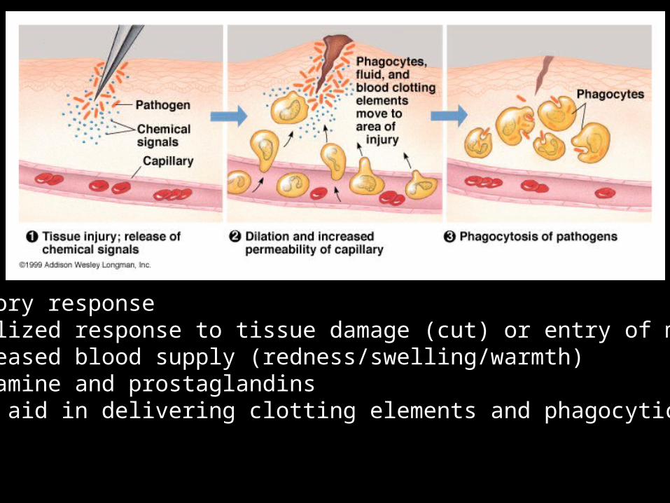
• inflammatory response + localized response to tissue damage (cut) or entry of microorganism
- increased blood supply (redness/swelling/warmth)- histamine and prostaglandins + aid in delivering clotting elements and phagocytic cells

Specific ImmunityImmune System Response• lymphocytes + B cells and T cells
- come from stem cells in bone marrow + mature in different locations before moving on to lymphoid tissue (lymph nodes, spleen, blood, lymph) - respond to specific antigens
+ clonal selection- effector cells and memory cells
+ primary and secondary immune response• self vs. non-self + autoimmune diseases
- Type I diabetes, Multiple sclerosis
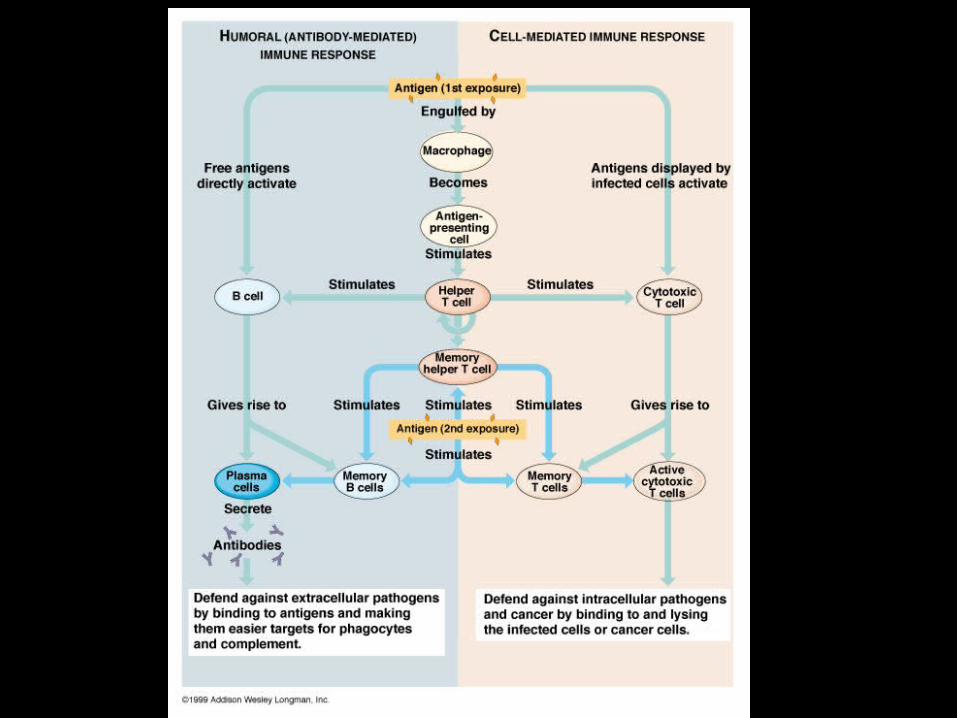
Cell-Mediated Immune ResponseT cells• kill cells that have been infected, or parasites• response initiated through contact with cell or macrophage + divides into four cell lines
- T memory cells- cytotoxic “killer” T cells- T4 helper cells + core of immune system; infected by HIV + “messenger”- T suppressor cells + protect our own tissues

Humoral Immune ResponseB cells• fights infections of plasma (generally bacteria)• “antibody-mediated response” + antibody is quaternary protein (multiple polypeptide chains)
- Y-shaped- can bind to variety of antigens
+ response to foreign antigen- divides into two different cell lines + memory cells + plasma cell - antibody factory - makes antigens easier to locate by phagocytes