united states patent (19) 11) 4,446,122 (45) may 1, 1984 molecular structure but differing in amino...
TRANSCRIPT

United States Patent (19) Chu et al.
11) 4,446,122 (45) May 1, 1984
54 PURIFIED HUMAN PROSTATE ANTIGEN
75 Inventors: Tsann M. Chu; Ming C. Wang, both of Williamsville; Lawrence Papsidero, Lackawanna, all of N.Y.
73 Assignee: Research Corporation, New York,
(21) Appl. No.: 316,954
22, PCT Filed: Dec. 23, 1980
86) PCT No.: S 371 Date: S 102(e) Date:
PCT/US80/01708
Aug. 28, 1981 Aug. 28, 1981
87, PCT Pub. No.: WO81/01849 PCT Pub. Date: Jul. 9, 1981
Related U.S. Application Data (63) Continuation-in-part of Ser. No. 108,217, Dec. 28,
1979, abandoned.
51) int. Cl...................... G01N 33/56; G01N 33/54; C12N 5/00; A61K 37/02
52 U.S. Cl. ......................................... 424/1.1; 435/4; 435/7; 435/240; 436/516; 436/518; 436/536;
436/539; 436/542; 436/543; 436/547; 436/548 58) Field of Search .......... 436/503, 64, 804, 543-548,
436/518, 536-542,813, 516; 424/1, 1.5, 1.1; 435/240, 241, 4, 7
56) References Cited U.S. PATENT DOCUMENTS
4,331,647 5/1982 Goldenberg ............................ 424/1
OTHER PUBLICATIONS Papsidero, L. D. et al., Progress in Clinical and Biologi cal Research, vol. 75A, pp. 435-443 (1981). Wang, M. C. et al., The Prostate, vol. 2, pp. 89-96 (1981). Shulman, A. et al., Proc. Society for Experimental Biol ogy and Medicine, vol. 137 (1) pp. 97-100 (1971). Papsidero, L. D. et al., J. National Cancer Institute, vol. 66 (1), pp. 37-42 (1981). Kuriyama, T. et al., Cancer Research, vol. 41 (10), pp. 3874-3876 (1981). Clarke, S. M. et al., Medical and Pediatric Oncology, vol. 9 (1), p. 94 (1981). Wang et al., Investigative Urology, vol. 17, No. 2, pp. 159-163 (1979). Primary Examiner-Benjamin R. Padgett Assistant Examiner-M. Moskowitz Attorney, Agent, or Firm-Scully, Scott, Murphy & Presser
57 ABSTRACT A prostate antigen distinct from prostatic acid phospha tase has been detected in normal, benign hypertrophic and malignant prostatic tissues, but not in other human tissues. The prostate antigen was purified to homogene ity from prostatic tissues by ammonium sulfate precipi tation, DEAE-BioGel A anion exchange chromatogra phy, molecular sievings on Sephadex G-100 and Sepha dex G-75, and preparative polyacrylamide gel electro phoresis. The purified prostate antigen shows a single protein band on analytical polyacrylamide gel electro phoresis and isoelectric focusing. The molecular weight of purified antigen was estimated by Sephadex G-75 gel filtration to be 33,000 and by sodium dodecyl sulfate polyacrylamide gel electrophoresis to be 34,000 with no subunit. The prostate antigen had an isoelectric point of 6.9.
17 Claims, No Drawings
4,446,122 1
PURFED HUMAN PROSTATE ANTIGEN
This invention was supported in part by Grants No. CA-15126 and CA-15437 from the National Cancer Institute, U.S. Public Health Service.
DESCRIPTION OF THE INVENTION This application is a continuation-in-part of copend
ing, commonly assigned U.S. patent application Ser. No. 108,217 now abandoned, filed Dec. 28, 1979, the contents of which are incorporated by reference herein. TECHNICAL FIELD OF THE INVENTION
This invention relates to a diagnostic reagent and method for the immunochemical detection of a human prostate antigen which is distinct from prostatic acid phosphatase. More particularly, this invention relates to a novel purified human prostate antigen and antibodies specific thereto which are suitable for use in prostatic cancer detection by laboratory methods.
BACKGROUND ART
Prostate cancer is very prevalent in old age, with approximately one half of all males over age 70 having been shown to develop prostatic cancer. This high inci dence of prostate malignancy has led to the search for markers which may be used for its detection. The eleva tion of serum acid phosphatase activity in patients hav ing metastasized prostate carcinoma was first reported by Gutman et al. in J. Clin. Invest. 17: 473 (1938). In cancer of the prostate, prostatic acid phosphatase is released from the cancer tissue into the blood stream with the result that the total serum acid phosphatase level greatly increases above normal values. Numerous studies of this enzyme and its relation to prostatic can cer have been made since that time, e.g., see the review by Yam in Amer. J. Med. 56: 604 (1974). However, the measurement of serum. acid phosphatase by conven tional spectrophotometric methods often fails to detect prostatic cancer in its early stages. In general, the activ ity of serum acid phosphatase is elevated in about 65-90 percent of patients having carcinoma of the prostate with bone metastatis; in about 30 percent of patients without roentgenological evidence of bone metastasis; and in about only 5-10 percent of patients lacking clini cally demonstrable metastasis.
Prior art attempts to develop a specific test for pros tatic acid phosphatase have met with only limited suc cess because techniques which rely on enzyme activity on a so-called "specific' substrate cannot take into ac count other biochemical and immunochemical differ ences among the many acid phosphatases which are unrelated to enzyme activity of prostate origin. In the case of isoenzymes, i.e. genetically defined enzymes having the same characteristic enzyme activity and a similar molecular structure but differing in amino acid sequences and/or content and therefore immunochemi cally distinguishable, it would appear inherently impos sible to distinguish different isoenzyme forms merely by the choice of a particular substrate. It is therefore not surprising that none of these prior art methods is highly specific for the direct determination of prostatic acid phosphatase activity; e.g. see Cancer 5: 236 (1952); J. Lab. Clin. Med. 82: 486 (1973); Clin. Chem. Acta. 44; 21 (1973); and J. Physiol. Chem. 356: 1775 (1975).
In addition to the aforementioned problems of non specificity which appear to be inherent in many of the
10
15
25
30
35
45
50
55
65
2 prior art reagents employed for the detection of pros tate acid phosphatase, there have been reports of ele vated serum acid phosphatase associated with other diseases, which further complicates the problem of obtaining an accurate clinical diagnosis of prostatic cancer. For example, Tuchman et al. in Am. J. Med. 27: 959 (1959) have noted that serum acid phosphatase levels appear to be elevated in patients with Gaucher's disease. Due to the inherent difficulties in developing a "spe
cific' substrate for prostrate acid phosphatase, several researchers have developed immunochemical methods for the detection of prostate acid phosphatase. How ever, the previously reported immunochemical meth ods have drawbacks of their own which have precluded their widespread acceptance. For example, Shulman et al., in Immunology 93: 474 (1964) described an immuno diffusion test for the detection of human prostate acid phosphatase. Using antisera prepared from a prostatic fluid antigen obtained by rectal massage from patients with prostatic disease, no cross-reactivity precipitin line was observed in the double diffusion technique against extracts of normal kidney, testicle, liver and lung. How ever, this method has the disadvantages of limited sensi tivity, even with the large amounts of antigen em ployed, and of employing antisera which may cross react with other, antigenically unrelated serum protein components present in prostatic fluid. Chu et al. in International Patent Application Publi
cation No. WO 79/00475, the contents of which are incorporated by reference herein, describe a new method for the detection of prostatic acid phosphatase isoenzyme patterns associated with prostatic cancer which obviates many of the above drawbacks. How ever, practical problems are posed by the need for a source of cancerous prostate tissue from which the diagnostically relevant prostatic acid phosphatase iso enzyme patterns associated with prostatic cancer are extracted for the preparation of antibodies thereto.
In recent years considerable effort has been spent to identify enzyme or antigen markers for various types of malignancies with the view towards developing specific diagnostic reagents. The ideal tumor marker would exhibit, among other characteristics, tissue or cell-type specificity, and would be released into the circulation or other biological milieu which is easily obtained from individuals. Previous investigators have demonstrated the occurrence of human prostate tissue-specific anti gens. For example, R. H. Flocks et al., I.J. Urol. 84: 134
(1960) immunized rabbits with an extract of BPH (be nign prostatic hypertrophy) prostate tissue and showed the presence of tissue specific antiprostatic antibodies by a gel diffusion technique. However, they presented no data at all to indicate the nature of the reactive anti gen. The precipitation lines formed by antiserum and prostate extract appear due to the reaction of prostatic acid phosphatase and its antibodies.
R. J. Ablin et al., J. Immunol. 104: 1329 (1970) and R. J. Ablin, Cancer 29: 1570 (1972) have also demonstrated the occurrence of human prostate tissue-specific anti gens. By using antiserum obtained from immunizing rabbits with extracts of normal prostate, Ablin et al. showed two antigenic components in human prostate. One of these was identified as prostatic acid phospha tase, while the specificity of other was shown to be a non-prostatic tissue antigen. The xenoantibodies reac tive to the second antigen could not be abolished by
4,446,122 3
treating the antiserum with human prostatic fluid. Fur thermore, this antigen was shown to be deficient in benign and malignant prostatic tissues. In contradistinc tion, the prostate antigen of the present invention is present in all prostate tissue, (normal, benign or malig nant) in almost equal amounts. Further, it is detectable in prostatic fluid and cultured human prostatic malig nant cells and its medium as well. The Ablin articles describe absorption of antisera to the antigens described therein with prostatic fluid, after which a precipitin line was still detected. Absorption of antibodies against the present antigen with prostatic fluid gives no precipitin line, indicating that the present antigen is present in prostatic fluid while that of Ablin et al. is not.
C. W. Moncure et al., Cancer Chemother. Rep. 59: 105, (1975) also demonstrated the occurrence of a human prostate tissue-specific antigen preparation which does not bind to DEAE sepharose at pH 8.0, as does the antigen of the present invention. This charac teristic most probably indicates a significant structural difference between the present protein and that of Mon 'cure et al.
Thus, there is still a need for simple, reliable, sensitive and specific reagents and techniques to detect prostatic cancer with acceptable diagnostic accuracy and with out the aforementioned difficulties of the prior art. The present invention fills such needs.
DISCLOSURE OF THE INVENTION
It is a general object of the present invention to pro vide a purified human prostate antigen useful in prepar ing an improved diagnostic reagent suitable for the immunochemically specific detection of circulating human prostate antigen in blood, urine or other body fluids. Another object of the present invention is to provide
rapid and simple, yet highly specific and sensitive, im munochemical techniques and reagents useful in the early detection of prostatic cancer. A further object of this invention is to provide a new
marker for monitoring prostatic cancer and the effec tiveness of curative therapy therefor. An additional object of this invention is to provide
useful monoclonal antibodies to human prostate anti gen. Upon study of the specification and appended claims,
further objects, features and advantages of the present invention will become more fully apparent to those skilled in the art to which this invention pertains.
BEST MODE FOR CARRYING OUT THE INVENTION
Briefly, the above and other objects, features and advantages of the present invention are attained in one aspect thereof by providing a purified human prostate antigen which is distinct from prostatic acid phospha tase.
In a second aspect, the present invention provides antisera which are highly specific to a purified human prostate antigen and which do not immunochemically cross-react with prostatic acid phosphatases or with acid phosphatases originating from other tissues.
In a third aspect of the present invention, there is provided an immunochemical method for the detection of prostatic cancer which exhibits high sensitivity, good specificity and substantially no false positive results for tumor other than the prostate.
10
15
20
25
30
35
45
50
55
65
4. In a fourth aspect, the present invention provides
specific marker antibodies against human prostate anti gen which are useful, e.g., as carriers for in vivo radi oimmunodetection of prostate cancer and immunos pecific chemotherapy of prostatic cancer. According to the present invention, antigenic prepa
rations from either normal or cancerous human prostate tissue, prostatic fluid, cultured human prostatic malig nant cells or their media are purified to obtain a prepara tion consisting essentially of a human prostate antigen free of prostatic acid phosphatase. These antigenic preparations are employed for immunological vaccina tion and diagnostic procedures, particularly for im munoprecipitin testing.
Since immunological studies indicate that this antigen is present in normal, benign hypertrophic and neoplastic prostate, a pool of various prostate tissues can be used as the starting material for subsequent purification. Briefly, the prostate tissue is first extracted in aqueous media at 4 C. Although EDTA-PBS solution is conve niently used, saline, 3 M KCl or 0.01 percent (v/v) Tween 80 (but not 1M perchloric acid) also can be used for extraction of the prostate antigen. The antigen is thus also distinguished from carcinoembryonic antigens by its sensitivity to perchloric acid.
After clarification of the homogenate by centrifuga tion and filtration, the supernatant from the crude tissue extract is subjected to ammonium sulfate fractionation. Ammonium sulfate concentration at 20-80 percent satu ration almost quantitatively recovers the prostatic anti gen from EDTA-PBS extracts, with the highest yield at 45-50 percent saturation. In order to avoid contamina tion by other proteins as much as possible, the precipi tate is preferably collected between 35-55 percent satu ration of ammonium sulfate, which contains approxi mately 70 percent of the total prostate antigen in the crude extract. Human seminal plasma has been found to contain an
antigen, reacting with rabbit anti-PA serum in double immunodiffusion, which forms a fused immunoprecipi tin line with that of crude extract of prostatic tissue. This result revealed the presence of a protein, immuno logically identical to the human prostate antigen (PA), in seminal plasma. Further study showed that all semi nal plasma specimens examined contained a substantial amount of PA, ranged from 0.4 to 1.8 mg/ml, (n=9, mean-standard deviation=0.71:0.42). It is estimated that PA content in 20 to 30 ml of seminal plasma was equivalent to that in 100 g of prostatic tissue. The elu tion profile of PA in chromatographies is similar to that using prostatic tissue as source of the antigen. PA puri fied from seminal plasma and from prostatic tissue pos sesses an identical mobility and isoelectric point (p 6.87-009) as shown by polyacrylamide gel electro phoresis and isoelectric focusing, respectively. Both purified PA preparations exhibit a molecular weight of 33-34,000 as shown by Sephadex G-75 gel filtration. In addition, a line of identity was obtained in immunodiffu sion when purified PA preparations reacted with anti PA serum.
Since seminal plasma is more readily available than prostatic tissues and contains abundance of PA, it ap pears an ideal source for isolating PA. Using seminal plasma as the source of PA isolation also appears to have an advantage over the use of prostatic tissue. Firstly, the initial extraction step, which requires at least 4 hours, is eliminated. Secondly, at the initial stage of purification, handling a large volume of the tissue ex
4,446,122 5
tract is avoided since 20 to 30 ml of seminal plasma is equivalent to 100 g of tissue in terms of PA content. Thirdly, seminal plasma contains less contaminating proteins and makes purification easier. For instance, hemoglobin in the tissue extract is precipitated concur rently with PA by ammonium sulfate at concentration greater than 50 percent saturation, and the removal of this hemoglobin in later steps of purification results in a reduction of the yield of final purified PA. Since semi nal plasma contains less contaminating proteins, it is possible during fractional precipitation steps to recover a greater amount of PA by increasing the upper cut-off
10
point of ammonium sulfate concentration to 75 percent saturation. A better recovery of PA in purified form is also achieved from seminal plasma. DEAE-BioGel A anion exchange column effectively
retains the prostate antigen and washing the column with tris-HCl buffer at pH 8.0 does not dissociate the antigen from the column. Elution of the antigen can be achieved with 18-78 mM of NaCl in the same buffer, followed by further purification by gel chromatogra phy. The bulk of contaminating proteins in the PA preparation eluted from the DEAE column have a molecular weight greater than 45,000 and can accord ingly be separated from PA (molecular weight: 33,000 to 34,000) by gel filtration on a Sephadex G-100 col
An additional step can be carried out with anion exchange chromatography on a DEAE column using a pH gradient solution as the eluant. Two protein peaks were detected between fractions in the pH range of 7.6 to 6.7, which were shown to contain PA. Upon poly acrylamide gel electrophoresis, protein heterogeneity was seen in the first peak fractions, while the second peak fractions gave a single proteinband. Therefore, for obtaining homogeneous PA, only second peak fractions were collected. At the end of theis elution, approxi mately 20 percent of total PA subjected to chromatog raphy was still bound to the DEAE column. This can be recovered quantitatively by elution with 0.08 M NaCl, but protein heterogeneity in the eluted PA prepa ration was observed. An interesting observation has been made in the course of the above PA purification; the first peak fractions contained PA which possessed a pI different from that of the purified PA from the sec ond peak fractions. Also, the PA retained by the DEAE column at the end of pH gradient elution exhibited a different pI. Similar observations have also been made in the purification of PA from seminal plasma, and treat ment with neuraminidase has increased the pI of thse PA "isomers' to a higher pH range. It thus appears that the human prostate antigen of the present invention may exist in a number of different isomeric forms.
Preparative polyacrylamide gel electrophoresis of the partially purified antigen is effective in achieving a pure antigen preparation. This purified antigen is shown to be homogeneous by polyacrylamide gel electropho resis with and without sodium dodecyl sulfate. It has a molecular weight of 33,000-34,000 with no subunit, and exhibits a single p of 6.9. Using immunoprecipitation and immunocytochemi
cal techniques, PA has been shown to be a prostate gland epithelial marker protein. PA is localized in the epithelial lining of prostatic glands and ducts as well as in prostatic secretions and concretions, but not in epi thelia of periurethral glands, seminal vesicles, vas defer ens, urinary bladder transitional epithelium, prostatic urethra, glandular lining of vonBrunn's nests, or in tes
15
20
25
30
35
45
50
55
65
6 tes. These observations suggest that the PA in the semi nal plasma is of prostatic origin. Data are now available to indicate the potential clini
cal application of PA in prostatic cancer. Quantitation of circulating PA, with a sensitivity of 0.1 ng/ml, has been achieved by an enzyme-immunoassay. PA is not detected in sera from normal females or female cancer patients, while sera from male patients with non-pros tate cancer contain a similar range of PA as that of normal males. Patients with prostatic diseases have been shown to have elevated levels of circulating PA. Al though no quantitative difference in PA levels is found between patients with benign prostatic hypertrophy and stage A of prostatic cancer, patients with other stages of prostatic cancer demonstrate significantly elevated PA levels, both quantitatively and qualitatively. With im munocyto- chemical procedures, all primary and sec ondary prostatic tumors examined reacted positively with anti-PA serum, whereas the tumors of non-pro static origin did not react. These results, therefore, sug gest an additional means for diagnosis of prostate cancer and for monitoring the efficacy of its treatment. An other possible application is the use of specific anti-PA antibodies in in vivo radioimmunodetection of prostate cancer, e.g. according to the method described by D. Pressman in Cancer Research 40: 2960 (1980), particu larly the micro-metastasis which is so critical in staging and treatment. Immune-specific chemotherapy also is a potential area where much work can now be initiated with the availability of PA immunologic reagents, e.g. see T. Ghose et al. in J. Nat. Cancer Inst. 61: 657 (1978). Furthermore, the physiology of the prostate can be studied with the aid of this new marker for prostate gland epithelium.
In order to produce antisera which can be used to detect antigens in prostate tissues and fluids other than acid phosphatase, female rabbits have been injected with a purified prostate antigen isolated from prostate tissue. Sera is collected, heat inactivated and stored at -20 C. until use. After treatment with insolubilized normal human plasma proteins (antibodies to normal plasma constituents are removed by treatment of the antiserum with glutaraldehyde-insolubilized normal plasma obtained from normal male and female adults), the antiserum reacts specifically with prostate tissue extracts (43/43) using the techniques of double im munodifussion and rocket immunoelectro-phoresis. No immunological reactivity was observed against a bat tery of extracts prepared from tissues other than pros tate. The prostate antigen from prostate tissue extracts was characterized by gel filtration chromatography (m.w. 30-40,000), isoelectric focusing (p. 6.9) and aga rose electrophoresis (MR 0.2 relative to bovine serum albumin). The concentration of the prostate antigen was not significantly different among extracts prepared from normal, benign hypertrophic and malignant pros tatic tissues and the antigen exhibited no acid phospha tase enzyme activity as determined by histochemical staining procedures. Furthermore, its approximate mo lecular weight (30-40,000) differs significantly from that of prostatic acid phosphatase (100,000). Although the antigen detected in serum had a higher
apparent molecular weight (80-100,000), antigen mix ing and peak enhancement experiments indicate that prostate antigen in circulation is immunologically iden tical to the human prostate antigen in prostate tissue and prostatic fluid. The serum-borne antigen may be bound to a plasma protein. Such plasma protein binding of a
4,446,122 7
variety of antigens and hormonal substances is well known and has been reported by T. Peters, Jr. "Serum Albumin' In: F. W. Putman (ed.). The Plasma Proteins. Vol. 1: pp. 133-153, New York, N.Y. Academic Press, 1975. However, SPD-PAGE revealed a molecular weight of 36,000, similar to that in prostate tissue and semifil plasma.
Alternatively to the conventional techniques for pre paring antibodies in laboratory and farm animals, mono clonal antibodies against PA can be prepared using known hybridoma cell culture techniques. In general, this method involves preparing an antibody-producing fused cell line, e.g. of primary spleen cells fused with a compatible continuous line of myeloma cells, and grow ing the fused cells either in mass culture or in an animal species from which the myeloma cell line used was derived or is compatible. Such antibodies offer many advantages in comparison to those produced by innocu lation into animals, as they are highly specific and sensi tive and relatively "pure' immunochemically. The full potential of this prostatic antigen in clinical
applications is not known at present, as it does not ap pear to exhibit any tumor specificity or pathological association. However, as in the case of acid phosphatase which is present in all prostate tissues, this prostate antigen can provide useful clinical information upon serum monitoring. Recent experiments in our labora tory have shown that the prostate antigen is detected, by rocket immunoelectrophoresis and enzyme-linked immunoassay techniques, in the sera of some prostate cancer patients but not in the sera of normal healthy persons or of patients with other cancers. Further ex periments have shown that PA is histologically detecti ble in both prostate tissue and established continuous tissue culture cell lines of prostate origin, and that it is released by prostate tumor cells, both in vivo and in -vitro. In addition, it has been found that the PA level in serum of patients suffering from prostatic cancer is unrelated to the serum levels of prostatic acid phospha tase, so that simultaneous determination of circulating PA and circulating prostatic acid phosphatase may well provide an improved means of diagnosing prostatic cancer. The human prostate antigen, although an eu topic component of the prostate, can thus play a major role in the detection of prostate cancer. For the preparation of immunogens suitable for pre
paring diagnostic antibodies against the human prostate antigen, conventional vaccine preparation techniques can be used. Preferably a non-antigenic adjuvant, e.g. alum, Freund's complete adjuvant, saponin, a quater nary ammonium surfactant, an alkyl amine, etc. is ad mixed with the purified prostate antigen in a suitable immunologically acceptable, non-antigenic carrier and the resultant mixture can be sterilized, e.g. by filtration. The vaccine can be administered parenterally follow
ing regimens already known for immunization with other proteins to stimulate the formation of immuno precipitating antibodies, with the primary inoculation being preferably followed up by at least one additional injection one to ten weeks later. Good results have been obtained in rabbits using four booster injections at two week intervals one month after the primary immuniza tion. The protein content per injection in rabbits, goats and other mammals can be varied, but is generally about 50 micrograms of protein per kg. of body weight. The antibodies can be collected and worked up using meth ods well known to those skilled in the art of immuno chemistry, and provide a useful reagent for the immuno
10
15
3 logical detection of prostate specific antigen in a variety of immunochemical procedures, e.g., immunoprecipi tin, fluorescent antibody, serum neutralization, etc. Such antibodies are useful as a control reagent in the diagnostic test for prostatic cancer described more par ticularly below. The simplest immunoprecipitin test involves capillary
tube precipitin testing, wherein separate antibody and antigen solutions are allowed to react at a common interface in a capillary tube and a positive reaction is indicated by the formation of a precipitate at the inter face. This method is relatively insensitive and inaccu rate due, inter alia, to unavoidable diffusion of the two solutions across the interface, and furthermore the final test results cannot be preserved. Agar gel diffusion is the simplest method which
avoids these drawbacks. A solution of the antigen (or
20
25
30
35
45
50
55
65
serum sample) is placed in a central well punched in a continuous agar gel and appropriate dilutions of the serum containing antibodies (or, correspondingly, the antigen) thereto are placed in wells concentrically sur rounding the center well. A positive reaction is noted by the formation of the precipitin line between one or more of the concentric wells and the central well. This method is relatively insensitive and fairly slow, requir ing 1 to 4 days to read the test results.
Radioimmunoassay (RIA), e.g. radioimmunoprecipi tin tests, are extremely sensitive (by several orders of magnitude over older methods) but take several days to perform and require sophisticated equipment and highly trained personnel not always widely available.
Countercurrent immunoelectrophoresis (CIEP) is a widely used immunoprecipitin method which takes only about an hour to perform and which is considera bly more sensitive than agar gel diffusion. Reactive components are placed in opposing wells cut into an agar gel and a small electrical current applied thereto, causing both the antigen and the antibody to migrate towards each other. A positive reaction is indicated by the formation of a precipitate at the antigen-antibody interface. Since this test method is reasonably reliable, readily available and inexpensive, it represents a pre ferred embodiment of this aspect of the present inven tion. For purposes of immunoelectrophoresis testing, the
diagnostic antibody preparation of the present inven tion when used without purification is generally diluted with phosphate buffered saline in a volume ratio of 1:10 to 1:500, depending on the antibody titer thereof. The limiting factor at the lower end of the range is the de gree of distinction achieved in the precipitin lines, which is a function of the antibody content in the total protein present. Purified antibody preparations can of course have lower total protein concentrations, and the protein content of even the unpurified preparations can be varied to suit the particular immunochemical test to be employed, the optimal amounts being determined, e.g. by testing simple serial dilutions.
In a preferred, further aspect of the present invention, circulating human prostate antigen can now be detected by immunochemical techniques, preferably by protein staining of the antibody-antigen precipitin complex. Using the aforementioned specific antisera and coupling this with a conventional means for detecting the anti gen-antibody complex, it is now possible to immuno chemically separate the specific human prostate antigen from a serum sample.
4,446,122 The antigen-antibody complex can be stained by a
number of known histochemical staining techniques, e.g. fluorescent antibody, etc., to increase the sensitivity of this method. Alternatively, one can use radioactive antibody for the assay, which not only provides a better quantitative value but may also further increase the sensitivity of the assay. If desired, an enzyme, e.g. £3 galactosidase or peroxidase, can be coupled with puri fied antibodies for use in an enzyme-linked immunoas say, e.g. using techniques analogous to those described by Kato et al. in J. Immunol. 116: 1554 (1976), the con tents of which are incorporated by reference herein. Especially preferred is the method described by M. Kuriyama et al. in Cancer Res. 40: 4658 (1980), the contents of which are incorporated by reference herein.
It is preferable to bind these antibodies onto a water insoluble support for use in the enzyme assay. Many suitable such supports and techniques for binding prote ins thereto are well known in the art and include inor ganic as well as organic supports. Presently preferred are those water-insoluble supports which can be acti vated with a cyanogen halide, preferably cyanogen bromide, prior to the covalent bonding of the antibodies thereto, e.g. as taught by Axen et al. in U.S. Pat. No. 3,645,852, the contents of which are incorporated by reference herein. Such supports are commercially avail able, e.g. the Enzymobeads available from, Bio-Rad Laboratories. Without further elaboration, it is believed that one
skilled in the art can, using the preceding description, utilize the present invention to its fullest extent. The following preferred specific embodiments are, there fore, to be construed as merely illustrative and not limi tative of the remainder of the disclosure in any way whatsoever, In the following Examples, the tempera tures are set forth uncorrected in degrees Celsius; unless otherwise indicated, all parts and percentages are by weight.
Protein concentration was determined by the method of Lowry et al. described in J. Biol. Chem. 193: 265 (1951), using bovine serum albumin as the standard. Acid phosphatase activity was measured by the method of Babson and Phillips using a-naphthyl phosphate as the substrate.
Immunoprecipitation techniques were modified from B. Weeke, Scand. J. Immunol. (Supplement 1) 2:37-46 (1973). Rocket-IEP was performed on cellulose acetate membranes using 0.83 percent agarose (Sigma low elec tro-endo-osmosis) in 0.08M tris-0.024M tricine-0.024M sodium barbital containing 1.3 mM calcium lactate and 0.02 percent sodium azide (TTB buffer). Antiserum at various final concentrations (0.5-2.0 percent) was incor porated into the agarose at 55° C. prior to plating. Rocket-IEP samples were applied to circular wells (5.0 mm) and electrophoresed at 5 volts/cm overnight at 4 C. using TTB running buffer. Crossed-immunoelectrofocusing was performed in
the second dimension as for rocket-IEP. Isofocused acrylamide gels (5X 100 mm) were molded into the antibody-agarose prior to immunoelectrophoresis modi fied from J. Soderholm et al., Scand. J. Immunol. (Sup plement 2) 4: 107-113 (1975). The first dimension isofocusing was done in 7.5 percent polyacrylamide containing 2 percent ampholytes, pH range 4-8 at 200 volts overnight at 4 C. For some experiments, isofo cused gels were laid on cellulose acetate membrances and covered with 1 percent agrose. Troughs were cut adjacent to the embedded acrylamide gel, the antiserum
10
15
20
25
30
35
45
50
55
65
10 placed in the parallel troughs was allowed to diffuse for 24-48 hours at 25 C. and the resulting immunoprecipi tin lines were recorded.
Crossed-immunoelectrophoresis (cross-IEP) was per formed in the second dimension as for rocket-IEP. The first dimension electrophoresis was performed with 1 percent agarose, and samples (75 ul in rectangular wells) were electrophoresed at 15 volts/cm for 2 hours at 4 C. Migration was monitored with Bromphenol Blue dye. The abbreviations used are: PB-NaCl, 0.05 M. phos
phate buffer containing 0.15 M NaCl, pH 7.2; IEP, immunoelectrophoresis; TTB, Tris-Tricine-Barbital (0.08M, 0.042M, 0.024M) buffer containing 0.3 mM calcium lactate and 0.02 percent sodium azide, pH 8.8; MR, relative immunoelectrophoretic mobility; p, isoe lectric point; m.w.., molecular weight.
EXAMPLE 1 Extraction of Tissues
Human prostatic tissues (normal, benign hyper trophic, malignant) and other human tissues were ob tained during autopsy or surgery. The histology of each issue was confirmed by pathologists. Approximately 10 g of tissues were minced and washed three times with 30 ml of physiological saline, then mixed with 30 ml of 0.02 percent (w/v) disodium ethylenediamine tetraace tate (EDTA)-0.1M phosphate buffered saline (PBS), pH 6.8. The mixture was placed in an ice-water-chilled blending chamber of a Sorvall Omnimixer and sub jected to three 5-minute blendings at a blade speed of 25,000 rpm with intermittent cooling time of 3 minutes. The homogenate was stirred overnight at 4 C., then centrifuged at 25,000 g for 30 minutes. The resultant supernatant constituted the crude tissue extract.
EXAMPLE 2
Ammonium Sulfate Fractionation
Pooled prostate tissues, 100 g, were extracted with 300 ml of EDTA-PBS as described above. Ammonium sulfate (60 g) was added to 285 ml of crude extract (35 percent saturation), mixed for 30 minutes and centri fuged (26,000 g, 30 minutes). 38g of ammonium sulfate were added to 295 ml of the resulting supernatant, (55 percent saturation), mixed for 30 minutes and centri fuged. The precipitate was dispersed in 200 ml of 55 percent saturated ammonium sulfate solution and cen trifuged. Washing of the precipitate was repeated twice, then the precipitate was dissolved in 20 ml of 0.01M tris-HCl buffer, pH 8.0. The residual ammonium sulfate was removed by dialysis against 4 liters of the tris-HCl buffer for 48 hours with one change of buffer during dialysis. The dialyzed solution was centrifuged at 46,000 g for 30 minutes to remove any precipitate formed during dialysis.
EXAMPLE 3
Ion Exchange Chromography of Prostate Antigen The supernatant from Example 2, 28 ml, was applied
onto a DEAE-BioGel A column (2.5X93 cm) pre equilibrated with 0.01M tris-HCl buffer, pH 8.0. The column was first washed with 420 ml of buffer, then eluted with 0-0.2M NaCl gradient (1 liter 0.01M tris HCl buffer, pH 8.0 in mixer and 1 liter of 0.2M NaCl 0.01M tris-HCl, pH 8.0 in the reservoir) at a flow rate of 6.5 ml/hr/cm2. The effluent was collected (10 ml/frac
4,446,122 11
tion) and monitored for the prostate antigen. Gel diffu sion according to the method of T. M. Chu et al. in Cancer Treat. Reports 61: 193 (1977) was used to mea sure the presence of prostate antigen in various chro matographic fractions. The entire procedure was car ried out at 4 C. The prostate antigen-containing frac tions were pooled and concentrated with an Amicon concentrator fitted with a UM2 ultrafiltration mem brane to 4.5 ml.
EXAMPLE 4
Gel Filtration of Prostate Antigen A portion (4.3 ml) of the concentrated solution was
applied onto a Sephadex G-100 column (2.5X110 cm) pre-equilibrated with 0.01M tris-HCl buffer, pH 8.0, which was eluted with same buffer at a flow rate of 3.5 mi/hr/cm2. The pooled DEAE-BioGel A fractions 100-150, containing the prostate antigen upon Sephadex G-100 chromatography, were resolved into several protein peaks. The prostate antigen was found to be in fractions 46-56. Again, the fractions exhibiting prostate antigen reactivity were pooled and concentrated to 3.8 ml.
EXAMPLE 5
Further Purification of Prostate Antigen The above fractions containing prostate antigen were
applied onto a Sephadex G-75 column (2.5X 113 cm) pre-equilibrated with tris-HCl buffer; the column was eluted at a flow rate of 5.5 ml/hr/cm2. This resulted in one major symmetrical protein peak associated with the prostate antigen reactivity and two minor protein peaks. When the fractions containing the prostate antigen were re-chromatographed on the same column, an identical symmetrical protein peak was obtained and the minor protein peaks were eliminated. Acid phosphatase activ ity was measured colorimetrically and histochemically as described by Babson et al. in Clin. Chim. Acta 13: 264-265 (1966) using a-naphthyl phosphate as the sub strate. No prostatic acid phosphatase was detected in the chromatographic fractions of this protein peak.
EXAMPLE 6 Analytical Polyacrylamide Electrophoresis
Acrylamide gel (7.5 percent) columns (50x60mm) were made according to the company's instruction manual (Shandon Southern Instruments, Ltd., Camber ley, England), and 50 pull of sample (10-40 ug protein in 25 percent sucrose solution) were applied onto each gel column. Ten percent sucrose solution was then layered carefully above the sample solution, followed by layer ing 0.05 M tris-glycine buffer, pH 8.5 to the top of the gel-containing tube. Tris-glycine buffer was used in preparing the sucrose solutions and used also as the electrolyte. After electrophoresis with a constant cur rent of 5 mA per tube for 40 minutes, the gels were stained for protein by Coomassie brilliant blue G-250 perchloric acid solution.
EXAMPLE 7
Sodium Dodecyl Sulfate (SDS) Polyacrylamide Gel Electrophoresis
The method was essentially the procedure or Weber and Osborn described in J. Biol. Chem. 244: 4406 (1969). A sample (10–20 g protein) in 50 pull of 0.05M tris-glycine buffer, pH 8.5, containing 250 pull SDS and 2-mercaptoethanol each, was incubated at 37° C. for 2
10
15
20
25
30
35
45
50
55
65
12 hours. After incubation, the sample was mixed with an equal volume of 50 percent sucrose, and 50 pull were subjected to the polyacrylamide gel (containing 0.1 percent SDS) electrophoresis as described before. After electrophoresis, the gels were stained for protein with 0.5 percent (w/v) Coomassie brilliant blue G-250 in ethanol-acetic acid-water (45:10:45, v/v).
EXAMPLE 8
Preparative Polyacrylamide Gel Electrophoresis Although Sephadex G-75 chromatography produced
a symmetrical protein peak with exhibited immunologi cal reactivity of the prostate antigen, the analytical polyacrylamide gel electrophoresis of Examples 6 and 7 revealed several protein components in this prepara tion. Therefore, a preparative polyacrylamide gel elec trophoresis (PAGE) was further used in the purification of prostate antigen. One ml of specimen (4.5 mg protein) was mixed with
1 ml of 50 percent (w/v) sucrose solution and applied onto an annular. 7.5 percent gel column (cross-sectional gel area, 4.4. cm; height, 9.2 cm), followed by succes sive layerings of 2 ml of 10 percent sucrose and 0.05M tris-glycine buffer, pH 8.5. A constant current of 30 mA was first applied for 1 hour, and 80 mA thereafter. The tris-glycine buffer was continuously pumped at a flow rate of 14-15 ml/hour into the elution plate located at the bottom of the column to carry materials emerged from the column into a fractional collector. Fractions containing the prostate antigen, representing the final purified preparation, were pooled and concentrated to 0.5 ml. The prostate antigen was found in fractions 28-62 (eluates obtained between 4-9 hours after electro phoresis). When these fractions were pooled and sub jected to analytical polyacrylamide gel electrophoresis, multiple protein bands still existed. However, by pool ing fractions 56-62 (between 8.1-9.2 hours of electro phoresis) only a single protein band was obtained upon analytical polyacrylamide gel electrophoresis and so dium dodecyl sulfate polyacrylamide gel electrophore SS.
That the final prostate antigen preparation was of high purity was indicated by a single protein band with out any contaminating components in polyacrylamide gel electrophoresis in the absence and the presence of sodium dodecyl sulfate. The purified prostate antigen was shown to have a molecular weight of 33,000 by gel filtration on Sephadex G-75, and 34,000 by sodium do decyl sulfate polyacrylamide gel electrophoresis with no subunit component. Isoelectric focusing revealed a single p of 6.9.
EXAMPLE 9
Simplified Purification of Antigen from Tissue The procedure for the purification of PA reported in
the preceding Examples takes at least two weeks and yields less than 0.5 mg of purified PA per 100 g of pros tatic tissue. Furthermore, for the final step of purifica tion, a painstaking construction of the polyacrylamide gel column as well as a special electrophoretic appara tus are necessary, and problems such as clogging of the elution channel during operation often developed. In this simplified procedure, in addition to the elimination of Sephadex G-75 chromatography and the application of short DEAE columns, the tedious preparative gel electrophoresis has been replaced by a simple pH gradi
4,446,122 13
ent elution of the DEAE column. The yield of purified PA has increased to 1 to 2 mg per 100 g of prostatic tissue and the entire procedure has been shortened to 8 to 9 days.
Benign hypertrophic prostatic tissues (approximately 100 g) were extracted with disodium tetraacetate-phos phate buffered slaine as described in Example 1 except that the homogenate was stirred for only 2 hours rather than overnight. The crude extract was then subjected to fractional precipitation by ammonium sulfate (30 to 50 percent saturation). The precipitate was dissolved in 10 to 15 ml of 0.01M tris-HCl buffer, pH 7.8, and dialyzed against 4 ml of the same buffer for 48 hours with one change of buffer. The dialyzed materials were centri fuged (46,000 g, 30 min) and the supernatant (12 to 18 ml) was applied onto a 2.5x15 cm DEAE-BioGel. A column pre-equilibrated with 0.01M tris-HCl buffer, pH 7.8. The column was washed with ca. 300 ml of the same buffer, followed by elution with 0.08 M. NaCl 0.01M tris-HCl buffer, pH 7.8. Fractions containing PA were pooled, concentrated (5 to 5.5 ml), and applied to a 2.5x100 cm Sephadex G-100 column pre-equilibrated with 0.01M tris-HCl buffer, pH 7.8, which was then eluted with the same buffer at a flow rate of 5 ml per hour per cm. Fractions covering a molecular weight region of 26,000 to 37,000 which contained PA were pooled, concentrated (5 ml) and applied to a 2.5X20 cm DEAE column pre-equilibrated with 0.01M tris-HCl buffer, pH 7.8. The column was washed with 10 ml of 30 the same buffer, followed by elution with pH gradient solution (mixer: 0.01M tris-HCl, pH 7.0, 300 ml; reser voir: 0.01M tris-HCl, pH 6.0, 500 ml) at a flow rate of 5 ml per hour per cm2. Second peak fractions which con tained PA were pooled and concentrated. Table 1 summarizes the results of a typical purifica
tion of PA from prostatic tissue by this simplified proce dure. Although modification at the salt precipitation step led to lesser recovery of PA, a large quantity of 4 contaminating proteins such as hemoglobin were re moved, thereby facilitating further purification of PA. Radial immunodiffusion used in this study was the tech nique of Mancini et al described in Immunochem. 2: 235-254 (1965), with modifications. Anti-PA serum (100 ul) was mixed with 10 ml of 1 percent agarose solution (in 0.154MNaCl-0.017M solium phosphate, pH 7.0) at 55 C. and poured into a Petridish (diameter: 8.3 cm). Wells (diameter: 2 mm) were then made on the agarose gel and to each well, 10ul of sample was added. For monitoring PA in the chromatographic fractions, overnight diffusion was sufficient to reveal precipitin; for quantitation of PA, the diffusion was allowed to continue for 48 hours, followed by washing of the gel for 2 days with 0.154M saline and subsequent staining with Coomassie Brilliant Blue G-250.
TABLE 1
Purification of Prostate Antigen from
O
15
20
25
35
45
50
55
Human Prostatic Tissue 60 Recov
Total Total Total Folds ery volume protein PA purifi (per
Steps (ml) - (mg) (mg) cation cent) Crude extract 2S8.0 T54.4 20.6 100 65 Ammonium sulfate 13.8 278.8 10.0 3. 48.5 (30-50 percent) First DEAE 5.5 80.9 7.9 8.3 38.3 Sephadex G-100 5.0 9.3 4.3 39.4 20.9
14 TABLE 1-continued
Purification of Prostate Antigen from Human Prostatic Tissue
Recov Total Total Total Folds ery volume protein PA purifi (per
Steps (ml) (mg) (mg) cation cent) Second DEAE S.O 15 1.5 85.2 7.3
From 118 g of benign hypertrophic prostate tissues.
EXAMPLE 10
Purification of Antigen from Fluid PA was also purified from 20 ml of seminal plasma
according to the procedure of Example 9 except that the extraction step was omitted and that, in the second step, PA was precipitated by ammonium sulfate at a concentration of 30 to 75 percent saturation. The results are shown in Table 2.
TABLE 2 Purification of Prostate Antigen from
Human Seminal Plasma Total Total Total Folds Recov volume protein PA purific- ery
Steps (ml) (mg) (mg) cation (percent) Seminal plasma 16.2 595.2 8.8 1 100 Ammonium sulfate 20,0 266.2 7.4 1.9 84.1 (30-75 percent) First DEAE 5.3 6.0 5.7 6.3 64.8 Sephadex G-100 6.4 12.8 4.5 23.8 51. Second DEAE 4.5 1.5 1.5 67.6 17.0
EXAMPLE 11
Molecular Weight Determination In order to obtain an approximate molecular weight
of the prostate antigen present in crude prostate tissue extracts and in antigen-positive patients' sera, these samples were subjected to a Sephadex G-200 gel filtra tion chromatography. Serum samples (0.5 ml) previ ously shown to be prostate antigen-positive by rocket IEP, or prostate tissue extracts (0.5 ml containing 8 mg protein) were applied to a column (0.9X60 cm) packed with Sephadex G-200 gel in PB-NaCl. Eluted samples were analyzed for absorbance at 280 nm and prostate antigen level was determined by rocket-IEP using a sample size: 0.5 ml; equilibration buffer: PB-NaCl (pH 7.2); fraction size; 0.8 ml; elution rate: 10 ml per hour. Peak antigen activity as exhibited in prostate tissue extracts eluted between 30-40,000 m.w. Molecular weight reference markers included human immuno globulin G (160,000), bovine serum albumin (68,000), ovalbumin (43,000), chymotrpysinogen A (25,000) and ribonuclease A (13,700). In antigen-positive patient sera examined by gel filtration, prostate antigen eluted as a single symmetrical peak between 80-100,000 m.w.
EXAMPLE 12
Preparation of Antisera Female rabbits were immunized as described previ
ously by T. M. Chu et al. in Investigative Urology 15: 319-323 (1978) with the crude extract of normal human prostatic tissue (for antiserum Ps), or with a purified prostate antigen (for antiserum P17) obtained at the Sephadex G-75 step described above. Sera were col lected, heat inactivated and stored at -20° C. until use. Absorption of the antiserum with normal female serum
4,446,122 S
(NFS) or tissue extracts (10 mg protein/ml) was carried out as described by T. M. Chu et al. in Cancer Treat ment Reports 61: 193-200 (1977).
EXAMPLE 13
Specificity of Antiserum Raised Against Crude Prostate Antigen
Immunoelectrophoresis was performed on a 9.5X 10.2 cm agarose (0.65 percent, w/v) plate. Barbital buffer, (pH 8.2, ionic strength 0.04) was used as the electrolyte and a constant voltage of 90 V. was applied for one hour. After electrophoresis and gel diffusion (20 hours), the plate was washed with 0.154M saline for 2 days and stained first for acid phosphatase with a solu tion of a-naphthyl phosphate-fast garnet GBC salt in 0.1M ammonium acetate, pH 5.0, and then for protein with Coomassie brilliant blue G-250-perchloric acid solution.
Immunoelectrophoresis of the crude extract prepared from normal prostatic tissues and antiserum P8, an anti serum raised against the crude extract of normal pros tate, resulted in three precipitin arcs. One of these arcs was formed by a normal human serum component, as it
10
15
20
disappeared after the antiserum was absorbed with nor mal female serum (NFS-P8). Absorption of the antise rum NFS-P8 with various normal human tissue extracts (urethra, bladder, heart, lung, pancrease, bone, kidney, intestine, liver and spleen, 10-20 mg each per ml) failed to eliminate the two remaining precipitin arcs, one of which was identified as prostatic acid phosphatase since it was stained with the a-naphthyl phosphate-fast garnet GBC salt solution. The other precipitin arc was shown to be a prostate tissue-specific antigen, identified as a protein, not stainable for acid phosphatase activity, which migrated with 3-mobility upon IEP analysis. Absorption with an extract of normal human prostate removed the reactive antibodies from the antiserum NFS-P8 and abolished both precipitin arcs. Further more, this antigen was demonstrated in all 20 of 20 normal prostates and identical results were obtained with the extracts of benign hypertrophic (15/15) and cancerous prostatic (8/8) tissues. These data demon strate that normal, benign hypertrophic and malignant prostate contain a prostatic tissue-specific antigen in addition to prostatic acid phosphatase.
EXAMPLE 14
Specificity of Antiserum Raised Against Purified Prostate Antigen
Following the procedure of Example 11, additional confirmation was provided with the use of antiserum P17 raised against a purified preparation of the prostate antigen, which was devoid of prostatic acid phospha tase. Both gel diffusion and immunoelectrophoresis employing the normal adult male and female serum absorbed antiserum P17 resulted in a single immuno precipitation of the prostate antigen. The extracts pre pared from tissues other than the prostate did not react with the normal female serum-absorbed antiserum P17.
EXAMPLE 15
Immunological Identity of Tissue and Serum Prostate Antigen
In order to determine if the serum-borne antigen shared immunological or biochemical characteristics with the antigen detected in prostate tissue extracts, the following experiments were performed. Samples of sera
25
30
35
45
50
55
60
65
6 and prostate tissue extracts were subjected to rocket IEP both individually and immediately after combina tion of both antigen sources. In each experiment, the samples were adjusted to the same final protein concen tration by the use of buffer dilutions. A representative experiment employed two sample wells containing pa tient sera, sample wells containing prostate tissue ex tract and two sample wells containing a mixture of patient's serum and tissue extract to demonstrate peak enhancement. Sera and tissue extracts were at the same final protein concentration in each well by the use of appropriate volumes of diluting buffer (PB-NaCl). All samples were 25 pul; one percent antibody in 0.83 per cent agarose; 5 V per cm, 20 hours at 4 C. All samples produced a single immuno- precipitate when assayed individually. Mixing samples, in peak enhancement experiments, produced a single reaction whose height was greater than that of individual samples. When anti gen-positive serum samples and prostate tissue extracts were mixed and immediately subjected to crossed-IEP, a fused immunoprecipitation peak resulted. Both meth ods of quantitative immunoelectrophoresis, showing peak enhancement and immunoprecipitate fusion, con firmed the immunological identity of the prostate anti gen as it occurs in tissue and serum according to the method of N. H. Axelson et al. in Scand. J. Immunol. (Supplement 1) 2: 91-94 (1973).
It should be noted that results regarding prostate antigen, as detected in tissue, apply to extracts prepared from normal, benign hypertrophic and malignant pros tate specimens; no physical differences in prostate anti gen (i.e., MR, pI or m.w.) were observed among these antigen sources, and extracts prepared from benign and malignant prostate specimens exhibited immuno precipitation lines of identity with normal prostate when tested against anti-prostate antigen antiserum by double immunodiffusion. In addition, the relative level of prostate antigen was compared among extracts pre pared from normal, benign hypertrophic and malignant prostate tissues. As determined by rocket-IEP (Table 3) no statistically significant difference of antigen level was observed between these antigen sources and a wide variation in antigen level (indicated by the large stan dard deviations calculated) was also noted to occur for each category of tissue extract examined.
TABLE 3 PROSTATE ANTIGEN LEVELS IN PROSTATE
TISSUE EXTRACTS Tissue Pathology (Number) Prostate Antigen Level - S.D. Normal (4) 14.3 units (+ 8.7) Benign hypertrophic (8) 18.4 units (; 21.3) Primary carcinoma (8) 13.0 units (+ 13.8) Each tissue extract was adjusted to 1 mg protein per ml prior to analysis for state antigen by rocket immunoelectrophoresis. All tissue specimens were histopathologically confirmed. For this experiment, one unit was arbitrarily chosen to represent one percent of the antigen level of a reference prostate extract. The reference extract was asssyed in a dilution series and run on the same plates as experimental extracts. As analyzed using Student's t-test, no statistically significant difference was found among these groups.
EXAMPLE 1.7
Immunological Identity of Fluid and Serum Prostate Antigen
Since prostatic acid phosphatase has been reported in both prostatic tissue and seminal fluid, we wished to determine whether PA also exists in the seminal fluid. Anti-PA serum, raised against PA isolated from pros
4,446,122 17
tatic tissue, reacted in immunodiffusion with crude ex tract of prostatic tissue, as well as with seminal plasma, and formed a fused line, indicating immunologic iden tify of the PA in prostatic tissue with that in seminal plasma. By the previously described radial immunodif fusion procedure, the PA concentration in seminal plasma was found to be 0.4 to 1.8 mg/ml, while 1 g of prostatic tissue contained 0.15 to 0.45 mg of PA extract able with disodium ethylenediamine tetraacetate-phos phate buffered saline (EDTA-PBS).
EXAMPLE 18
Histological Localization of Prostate Antigen Localization of PA in the prostate gland as well as
specificity of PA have been probed with the im munoperoxidase staining technique of Heyderman and Neville reported in J. Clin. Path. 30: 138-143 (1977). Sections of freshly fixed prostatic tissue were deparrafi nized in two changes of xylol, rehydrated thru a graded series of ethanol, and washed with distilled water. Fol lowing the inhibition of endogeneous peroxidase activ ity by 7.5 percent H2O2, the reactivity of anti-PA was assessed with rabbit anti-PA serum. After incubation and subsequent washing, the tissue sections were fur ther incubated with peroxidase-labelled goat anti-rabbit y-globulin. Excessive enzyme-labelled second antibody was then removed by washing, and the tissue sections were stained for peroxidase activity. As controls, the substrate alone, substrate plus conjugate, pre-immune rabbit serum, and anti-PA preabsorbed with specific antigen were used.
It was observed that the staining was restricted to epithelial cells comprising the prostatic ductal elements. An intense staining was shown in the apical cytoplasma of these cells, but no staining was seen in the nuclei. Within several ductal elements, positively staining se cretory material was observed in the section examined. Specific staining was not observed for other cellular elements, including stromal and vascular elements. Using the same technique, PA could not be detected in sections derived from other organ tissues, including the pancreas, colon, stomach, liver, seminal vesicles, and testes. Employing similar immunohistochemical tech niques, PA has been demonstrated in all primary and metastatic prostatic tumors tested, but not in non-pro static cancer tissues.
EXAMPLE 19
Detection of Prostate Antigen in Established Tissue Culture Cell Lines
To determine if malignant cells in long-term culture retain the expression of PA, three established strains of prostate cells (LNCaP, PC-3, and Du-145) were exam ined. Extracts prepared from these cells and other cul tured cells of nonprostate origins were assayed for the presence of PA by the previously mentioned Enzyme Immunoassay (EIA) procedure capable of detecting 1 ng PA/ml.
Immobilized anti-PA was prepared using CNBr activated Sepharose 1B (Pharmacia Fine Chemicals). The reaction mixture consisted of 5 g of CNBr activated Sepharose and 130 mg of IgG (anti-PA) in 0.1M borate buffer (pH 8.5) containing 0.5M NaCl. After incubation at 4 C. for 18 hours, the beads were washed with borate buffer and post-treated with a 1-M ethanolamine solution (pH 9.0) to block unreacted
10
15
20
25
30
35
45
50
55
65
18 groups. Covalently coupled beads were further washed and stored in PBS at 4' C.
For the EIA, 100 ul of an antigen sample was mixed with 300 pil of a fifty fold-diluted immobilized antibody and incubated for 3 hours at room temperature. After the addition of 1 ml of assay buffer (PBS containing 1 percent BSA), the mixture was centrifuged at 1,000 g to wash the beads. This procedure was repeated twice. To the washed beads was added 100 ul of peroxidase-IgG (anti-PA) conjugate in assay buffer. After a further incubation for 18 hours at room temperature, the beads were again washed as described above and assayed for the amount of bound peroxidase activity present. The reaction mixture contained 0.08 percent dianisidine and 0.003 percent H2O2 in 0.01 M sodium phosphate buffer (pH 6.0) and was allowed to react with the beads for 90 minutes. The enzyme reaction was then stopped with 100 pil of 1 NHCl, and the absorbance at 403 mn was determined. Each run included standards of known prostate antigen concentration. Using this assay proce dure, linearity of of the dose-response curve was achieved between 1 and 20 ng PA/ml. Before reexami nation, samples containing higher levels of antigen were serially diluted with assay buffer. The results indicated that two lines of prostate cancer cells, LNCaP and PC-3, contained significant levels of PA (50-700 ng/ml) as compared with those found in Du-145 and other cell lines examined (less than 4 ng/ml). PA was also present in conditioned "spent" media derived from prostate cultures producing the antigen.
EXAMPLE 20
Immunological Reactivity of Cultured Prostate Cells with Immunoglobulin Antiserum Fragments
To further assess antibody specificity, we examined cultured cells for their ability to specifically accrete radiolabeled antibody fragments. Included in these ex periments were cells derived from the prostate gland (LNCaP), colon (HT-29), and breast (MCF-7). For the preparation of immunoglobulin fragments, samples of IgG, anti-PA and rabbit pre-immune serum were dis solved in 0.1M sodium acetate buffer (pH 4.0). For every 50 mg of IgG, 1 mg of crystallized pepsin was added and the reaction mixture was incubated over night at 37° C. F(ab')2 fragments were separated from unreacted IgG and from smaller peptides by gel filtra tion over Sephacryl S-300 (Pharmacia Fine Chemicals) equilibrated in PBS buffer. The column (2.6X70 cm) was calibrated by the chromatography of molecular weight standards, including IgG, BSA, and egg albu min. Isolated F(ab')2 fragments were concentrated by ultrafiltration with the use of an Amicon PM-10 mem brane and positive pressure. Trace labeling of F(ab')2 fragments was accomplished
with the use of solid-phase lactoperoxidase-glucose oxidase (Enzymobeads; Bio-Rad Laboratories). To 1 mg of F(ab')2 were added 50 pull of Enzymobeads, 25 pull of 1 percent 6-D-glucose, 200 pull of 0.2M sodium phos phate (pH 7.0), and 1 mCi of carrier-free 131I or 125I. The reaction was allowed to proceed for 30 minutes at room temperature. Unreacted iodide was separated from labeled protein by gel filtration over Sephadex G-25. Specific activities of trace-labeled F(ab')2 prepa rations ranged between 0.6 and 0.8 uCi/ug.
Cells grown in 24-well culture dishes with each well containing 1.5X10⁶ cells were incubated with a paired radiolabeled mixture of 125I-labeled F(ab')2 (anti-PA)
4,446,122 19
and 131-labeled F(ab')2 (preimmune) in fresh media. This mixture contained 1.5x10 cpm for each radioac tive nuclide and was allowed to react with cultured adherent cells overnight under standard conditions. To determine radioactive uptake, cells were scraped from the wells and washed three times in PBS before count ing for each radioactive nuclide in a Packard automatic y-scintillation counter. Preferential uptake of a specific radioactive nuclide was calculated as a localization ratio: (125 cell bound 131I cell bound): (125I added 131I added). A specific localization ratio was calculated for each cell type, and the results indicated a significantly higher antibody uptake by the LNCaP cells of prostate origin than for the HT-29 or MCF-7 cells.
EXAMPLE 21
Release of Prostate Antigen by in vivo Tumor Cells When established as tumor cell xenografts in nude
mice, LNCaP cells released detectable levels of PA into the circulation of these animals. Congenitally athymic nude mice homozygous for the nu/nu allele were bred at Roswell Park Memorial Institute from matings of
10
5
20
BABL/c nu/nu homozygous males and BALB/c --/nu . heterozygous females. Human tumors were established subcutaneously in the nude mice before sera were ob tained for PA analysis by the injection of cultured cell suspensions. For this, the cells were washed in PBS, counted for viability, and adjusted to the desired con centration in sterile 0.9 percent NaCl solutions. Subse quent to nodule formation, nude mouse serum was col lected by severing the retro-orbital plexus and stored at -20 C. Cell cultures used for the establishment of human tumor grafts included LNCaP (adenocarcinoma of the prostate gland), RT-4 (transitional cell carcinoma of bladder), Palarmo (malignant melanoma), and AsPC 1 (pancreatic carcinoma). Of the human tumors examined, only the LNCaP
prostate line released PA into the circulation, which correlates with results obtained from the assay of cul tured cells. The control mice given preimplants of RT-4 cells, Palarmo cells, or AsPC-1 cells showed no detect able PA in their circulation. It is not presently appreci ated if serum antigen levels are commensurate with the tumor load, a phenomenon previously reported by E.J. Pesce et al. in Cancer Res. 37: 1998-2003 (1977) for the lactate dehydrogenasde enzyme released by human xenografts. If this is so, then the human prostate tumor nude mouse system, coupled with detection of PA, can provide a valuable clinical model to monitor the effects of antitumor modalities and the effects of biologic re sponse modifiers for human prostate cancer.
EXAMPLE 22
Antiserum Specificity The optimal concentration of adsorbed antiserum
required in the rocket-IEP procedure was determined by examining the migration of prostate antigen in gels containing varying concentrations of anti-prostate anti gen antiserum. The reactivity of various tissues was examined by rocket-IEP. As shown in Table 4, all ex tracts prepared from normal prostate, benign hyper trophic and naalignant prostate tissues showed reactiv ity, producing a single immunoprecipitin reaction. Ex tracts prepared from the non-prostate tissues, whether normal or neoplastic in nature, gave no immunologic reactivity.
25
30
35
45
SO
55
65
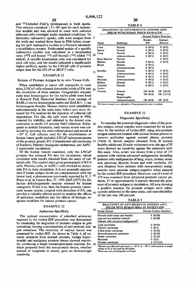
TABLE 4 REACTIVITY OF ANT-PROSTATE ANTIGENANT
SERUM WITH HUMAN ISSUE EXTRACTS Percent Positive Reaction Rocket- Immuno
Tissue Pathology EP difussion
Liver Normal 0 (0/2) 0 (0/1) Spleen Normal 0 (0/1) 0 (0/1) Lung Normal 0 (0/1) 0 (0/1)
Adenocarcinoma 0 (0/1) 0 (0/1) Bone Marrow Normal 0 (0/1) Bladder Normal 0 (0/1) 0 (0/1) Breast Normal 0 (0/1)
Adenocarcinoma 0 (0/5) Intestine Normal 0 (0/1) 0 (0/1)
Adenocarcinoma 0 (0/1) 0 (0/1) Heart Normal 0 (0/1) 0 (0/1) Pancreas Adenocarcinoma 0 (0/2) 0 (0/1) Kidney Normal 0 (0/1) 0 (0/1) Cerebral Normal 0 (0/1) 0 (0/1) Cortex Prostate Normal 100 (4/4) 100 (20/20)
Benign 100 (8/8) 100 (15/15) Hypertrophic Adenocarcinoma 100 (8/8) 100 (8/8)
All tissue extracts were adjusted to 10 mg protein/ml prior to analysis
EXAMPLE 23
Diagnostic Specificity To examine the potential diagnostic value of the pros
tate antigen, serum samples were examined for its pres ence by the method of rocket-IEP, using anti-prostate antigen antiserum treated with normal human plasma to remove antibodies against normal plasma proteins (Table 5). Serum samples obtained from 20 normal healthy adults and 20 male volunteers over the age of 55 years showed no reactivity against the antiserum with this assay. Also, serum was drawn from a total of 175 patients with various advanced malignancies, including patients with malignancies of lung, colon, rectum, stom ach, pancreas, thyroid, breast and with myeloma. All sera obtained from patients with non-prostatic malig nancies were prostate antigen-negative when assayed by the rocket-IEP procedure. However, out of a total of 219 sera examined from advanced prostatic cancer pa tients, 17 or approximately 8 percent showed the pres ence of prostate antigen in circulation. All sera showing a positive reaction for prostate antigen were subse quently subjected to the same assay, and reproducibility of the test was 100 percent.
TABLE 5 REACTIVITY OF ANTI-PROSTATE ANTIGEN ANT SERUM WITH HUMAN SERA BY ROCKET-EP
Serum Donors Percent Positive Reaction
Normal adults (male and female) 0 (0/20) Age-and sex-matched controls 0 (0/20) Patients with advanced malignancies' Lung carcinoma O (O/83) Thyroid carcinoma 0 (0/1) Colon-rectal carcinoma 0 (0/22) Stomach-pancreas carcinoma 0 (0/34) Breast carcinoma 0 (0/33) Myeloma 0 (0/2) Prostate carcinoma 8 (17/219) All sera were aliquoted and stored at -20 C. or -70 C. until required. A sample volume of 25 pil was used for all studies. Each case was pathologically confirmed.
4,446,122 21
EXAMPLE 24
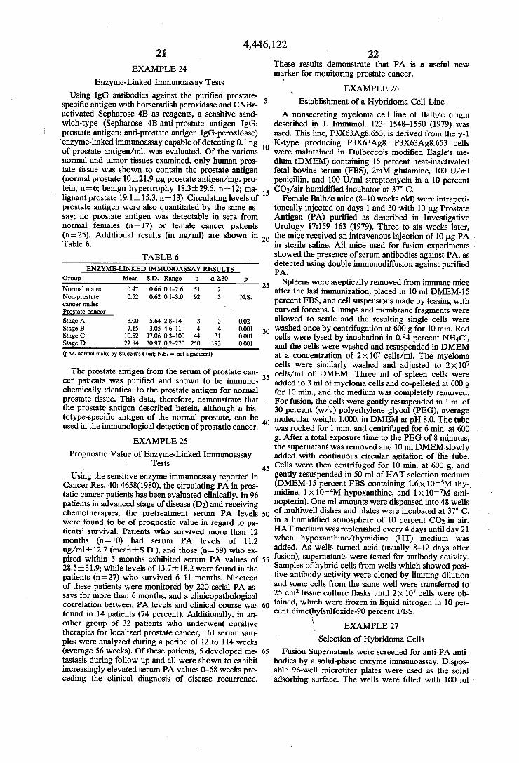
Enzyme-Linked Immunoassay Tests Using IgG antibodies against the purified prostate
specific antigen with horseradish peroxidase and CNBr activated Sepharose 4B as reagents, a sensitive sand wich-type (Sepharose 4B-anti-prostate antigen IgG: prostate antigen:anti-prostate antigen IgG-peroxidase) enzyme-linked immunoassay capable of detecting 0.1 ng of prostate antigen/ml. was evaluated. Of the various normal and tumor tissues examined, only human pros tate tissue was shown to contain the prostate antigen (normal prostate 10-21.9 ug prostate antigen/mg. pro tein, n=6; benign hypertrophy 18.3.29.5, n=12; ma lignant prostate 19.1-15.3, n=13). Circulating levels of prostate antigen were also quantitated by the same as say; no prostate antigen was detectable in sera from normal females (n=17) or female cancer patients (n=25). Additional results (in ng/ml) are shown in Table 6.
TABLE 6 ENZYME-LINKED IMMUNOASSAY RESULTS
Group Mean S.D. Range in or 2.30 p Normal males 0.47 0.66 0.1-2.6 51 2 Non-prostate 0.52 0.62 0.1-3.0 92 3 N.S. cancer males Prostate cancer Stage A 8.00 5.64. 2.8-14 3 3 0.02 Stage B 7.15 3.0S 4.6-11 4. 4 0.001 Stage C 10.52 17.06 0.3-100 44 31 0.001 Stage D 22.84 30.97 0.2-270 250 193 0.001 (p vs. normal males by Student's t test; N.S. = not significant)
The prostate antigen from the serum of prostate can cer patients was purified and shown to be immuno chemically identical to the prostate antigen for normal prostate tissue. This data, therefore, demonstrate that the prostate antigen described herein, although a his totype-specific antigen of the normal prostate, can be used in the immunological detection of prostatic cancer.
EXAMPLE 25
Prognostic Value of Enzyme-Linked Immunoassay - Tests
Using the sensitive enzyme immunoassay reported in Cancer Res. 40: 4658(1980), the circulating PA in pros tatic cancer patients has been evaluated clinically. In 96 patients in advanced stage of disease (D2) and receiving chemotherapies, the pretreatment serum PA levels were found to be of prognostic value in regard to pa tients' survival. Patients who survived more than 12 months (n=10) had serum PA levels of 11.2 ng/ml. 12.7 (mean-S.D.), and those (n=59) who ex pired within 5 months exhibited serum PA values of 28.5-31.9; while levels of 13.7-h18.2 were found in the patients (n=27) who survived 6-11 months. Nineteen of these patients were monitored by 220 serial PA as says for more than 6 months, and a clinicopathological correlation between PA levels and clinical course was found in 14 patients (74 percent). Additionally, in an other group of 32 patients who underwent curative therapies for localized prostate cancer, 161 serum sam ples were analyzed during a period of 12 to 114 weeks (average 56 weeks). Of these patients, 5 developed me tastasis during follow-up and all were shown to exhibit increasingly elevated serum PA values 0-68 weeks pre ceding the clinical diagnosis of disease recurrence.
10
15
20
25
30
35
45
50
55
65
22 These results demonstrate that PA is a useful new marker for monitoring prostate cancer.
' EXAMPLE 26
Establishment of a Hybridoma Cell Line A nonsecreting myeloma cell line of Balb/c origin
described in J. Immunol. 123: 1548–1550 (1979) was used. This line, P3X63Ag8.653, is derived from the y-1 K-type producing P3X63Ag8. P3X63Ag8.653 cells were maintained in Dulbecco's modified Eagle's me dium (DMEM) containing 15 percent heat-inactivated fetal bovine serum (FBS), 2mM glutamine, 100 U/ml penicillin, and 100 U/ml streptomycin in a 10 percent CO2/air humidified incubator at 37 C. Female Balb/c mice (8-10 weeks old) were intraperi
toneally injected on days 1 and 30 with 10 pug Prostate Antigen (PA) purified as described in Investigative Urology 17:159-163 (1979). Three to six weeks later, the mice received an intravenous injection of 10 pug PA in sterile saline. All mice used for fusion experiments . showed the presence of serum antibodies against PA, as detected using double immunodiffusion against purified PA.
Spleens were aseptically removed from immune mice after the last immunization, placed in 10 ml DMEM-15 percent FBS, and cell suspensions made by teasing with curved forceps. Clumps and membrane fragments were allowed to settle and the resulting single cells were washed once by centrifugation at 600 g for 10 min. Red cells were lysed by incubation in 0.84 percent NH4Cl, and the cells were washed and resuspended in DMEM at a concentration of 2X10⁷ cells/ml. The myeloma cells were similarly washed and adjusted to 2X 10⁷ cells/ml of DMEM. Three ml of spleen cells were added to 3 ml of myeloma cells and co-pelleted at 600 g for 10 min, and the medium was completely removed. For fusion, the cells were gently resuspended in 1 ml of 30 percent (w/v) polyethylene glycol (PEG), average molecular weight 1,000, in DMEM at pH 8.0. The tube was rocked for 1 min. and centrifuged for 6 min. at 600 g. After a total exposure time to the PEG of 8 minutes, the supernatant was removed and 10 ml DMEM slowly added with continuous circular agitation of the tube. Cells were then centrifuged for 10 min. at 600 g, and gently resuspended in 50 ml of HAT selection medium (DMEM-15 percent FBS containing 1.6X 10.5M thy midine, 1X 10-M hypoxanthine, and 1X 10⁻⁷M ami nopterin). One ml amounts were dispensed into 48 wells of multiwell dishes and plates were incubated at 37 C. in a humidified atmosphere of 10 percent CO2 in air. HAT medium was replenished every 4 days until day 21 when hypoxanthine/thymidine (HT) medium was added. As wells turned acid (usually 8-12 days after fusion), supernatants were tested for antibody activity. Samples of hybrid cells from wells which showed posi tive antibody activity were cloned by limiting dilution and some cells from the same well were transferred to 25 cm2 tissue culture flasks until 2X 10⁷ cells were ob tained, which were frozen in liquid nitrogen in 10 per cent dimethylsulfoxide-90 percent FBS.
s EXAMPLE 27 Selection of Hybridoma Cells
Fusion Supernatants were screened for anti-PA anti bodies by a solid-phase enzyme immunoassay. Dispos able 96-well microtiter plates were used as the solid adsorbing surface. The wells were filled with 100 ml
4,446,122 23
poly-L-lysine succinate (0.25 mg/ml H2O) and incu bated at 23° C. for 15 min. Following 3 washes with PB-NaCl pH 7.2 (50 mM sodium phosphate:140 mM sodium chloride), 100 ml of purified PA were added at a concentration of 50 ug/ml 10 mM carbonate buffer, pH 9.6. After incubation for 24hr. at 37° C., 100 ml of 1 percent (w/v) bovine serum albumin solution in PB NaCl buffer were added for a further 3 hrs. at 37 C.
Prior to performing the antibody screening assay, the microtiter wells were washed 3 times using PB-NaCl buffer (200 ul). Culture fluids (100 pul) to be tested were applied to individual wells and allowed to incubate at 37° C. for 3 hrs., followed by 3 washes with PB-NaCl buffer. Subsequently, 100 ul of peroxidase-conjugated antiserum to mouse Ig prepared according to the proce dure described in J. Immunol. Methods 15: 305-310 (1977) were added to each well and incubated at 37° C. for 3 hrs. After 3 washes with PB-NaCl buffer, enzyme activity was revealed using substrate solution contain ing 0.08 percent o-dianisidine: 0.003 percent H2O2: 0.01 M sodium phosphate, pH 6.0. The enzyme reaction was stopped after 60 min. at 23° C. using 25 ul of 1 NHCl.
In each assay, positive control samples were included and consisted of serially diluted hyperimmune mouse serum raised against purified PA. Tissue culture super natants from P3X63Ag8 cultures (y 1-Kappa) and from the parental nonsecreting myeloma line were used as negative controls. This screening assay detects immuno globulins of the IgG, IgM and IgA classes.
EXAMPLE 28
Cloning of Hybridoma Cells Desired myeloma hybrids were cloned by limiting
dilution in the presence of peritoneal macrophages to increase cloning efficiency. Hybrid cells were plated in 96-well culture dishes at a density of 0.5 cells/well in DMEM-15 percent FBS complete medium. Vigorous growth was observed after 8-12 days, at which time supernatants were tested from wells showing single colonies. Cultures exhibiting antibody activity were re-cloned by this procedure to ensure population homo geneity.
Peritoneal macrophages were prepared by flushing the peritoneal cavity of Balb/c mice with 5 ml of ice cold, sterile 0.34 M sucrose. To attain high yields of macrophages (5-15X106/mouse), animals received an intraperitoneal injection of 0.5 ml sterile thioglycolate medium 4 days prior to harvesting the cells. Macro phages were washed once by centrifugation at 600 g for 5 min. and were resuspended at 106/ml in tissue culture medium. Each tissue culture well received 100 ul of
... macrophage suspension. The hybridoma described herein has been deposited with the American Type Culture Collection, Rockville, Md. 20850, U.S.A. and is designated ATCC No. HB 8051.
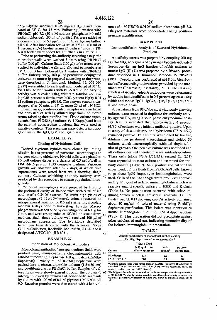
EXAMPLE 29 Purification of Monoclonal Antibodies
Monoclonal antibodies from spent culture fluids were purified using immuno-affinity chromatography with rabbit-antimouse Ig: Sepharose 4B gel matrix (RaMIg Sepharose). Twenty ml of R-aMIg:Sepharose were packed into a chromatographic column (1.5X30 cm) and equilibrated with PB-NaCl buffer. Samples of cul ture fluids were slowly passed through the column (5 ml/hr), followed by removal of non-specific reactants by elution with 100 ml of 0.1 M glycine: 1 M NaCl, pH 9.0. Reactive proteins were then eluted with 3 bed vol
10
15
20
25
30
35
45
50
55
65
24 umes of 4 M KSCN: 0.01 M sodium phosphate, pH 7.2. Dialyzed materials were concentrated using positive pressure ultrafiltration.
EXAMPLE 30
Immunodiffusion Analysis of Secreted Hybridoma Products
An affinity matrix was prepared by coupling 200 mg Ig (R-aMIg) to 5 grams of cyanogen bromide-activated Sepharose 4B; an IgG fraction of rabbit antisera to mouse IgG (H--L) was prepared by a Rivanol proce dure described in J. immunol. Methods 15: 305-310 (1977). Coupling was performed at pH 8.0 in bicarbon ate buffer according to directions provided by the man ufacturer (Pharmacia; Piscataway, N.J.). The class and subclass of isolated anti-PA antibodies were determined by double immunodiffusion in 0.6 percent agarose using rabbit anti-mouse IgG1, IgG2a, Ig2b, IgG3, IgM, anti K and anti-N chain. Supernatants from 96 of the most vigorously growing
cultures were screened in duplicate for antibody activ ity against PA, using a solid phase enzyme-immunoas say. Results indicated that approximately 4 percent (4/96) of cultures were initially antibody-positive. Upon re-assay of these cultures, one hybridoma (F5-A-1/22) remained positive. This culture was cloned by limiting dilution over peritoneal macrophages and yielded 30 cultures which macroscopically exhibited single colo nies of growth. One positive culture was re-cloned and all cultures derived therefrom were antibody-positive. These cells (clone F5-A-1/22.8.13, termed Cl. 8.13) were expanded to mass culture and examined for anti body content (Table 7). As a positive control for this experiment, culture fluids from P3X63Ag8 cells, known to produce IgG1 kappa-type immunoglobulen, were used. Cells of the P3X63Ag8 strain produced approxi mately 12 pg/ml of isolated immunoglobulin which was reactive against specific antisera to IGG1 and K-chain (Table 8). No precipitation occurred with other im munoglobulen subclass antiserum reagents. Culture fluids from Cl. 8.13 showing anti-PA activity contained about 10 pg/ml of isolated material using R-aMIg: Sepharose purification. This isolate was identified as mouse immunoglobulin of the IgM K-type subclass (Table 8). This preparation did not precipitate against other subclass of antisera, indicating monoclonality of the isolated immunoglobulin preparation.
TABLE 7 Affinity purification of monoclonal antibodies using
R-aMIg: Sepharose 4B chromatography." Culture Fluid (ml) applied to Yield uglg/ml
Culture affinity adsorbent Ig(mg) culture fluid P3X63Ag8 125 1.6 13 F5-A-1/22.8.13 300 3.1 10
Clarified culture fluids were passed through R-aMIg: Sepharose 4B adsorbent as described. The gel was washed with 1M NaCl pH 9.0 until absorbance at 280 nm reached baseline (less than 0.0200.D.units). R-aMIg-reactive substances were eluted under chaotropic dissociating conditions
of 4M KSCN. Yield of Ig isolated was determined by optical density measurements where a 1 percent (w/v) solution of mouse IgG shows an extinction coefficient of 14.
4,446,122 25
TABLE 8. Immunodiffusion analysis of affinity purified immonoglobulins from spent culture fluids.
Affinity isolates from cultures P3X63Ag8 F5-a-1/228,3
R-a IgG1 R-a IgG2a R-a IgG2b R-a IgG3 R-cit k chain R-a. A chain R-a, IgM
The preceding examples can be repeated with similar success by substituting the generically or specifically described reactants and/or operating conditions of this invention for those specifically used in the examples. From the foregoing description, one skilled in the art to which this invention pertains can easily ascertain the essential characteristics thereof and, without departing from the spirit and scope of the present invention, can make various changes and modifications to adapt it to various usages and conditions.
NDUSTRIAL APPLICABILITY
As can be seen from the present specification and examples, the present invention is industrially useful in several respects. Although this prostate antigen is an eutopic product, it appears to be a useful tumor marker such as other differentiated cell products, e.g., prostatic acid phosphatase and thyrocalcitonin. The specificity of prostate antigen antiserum can allow for the identifica tion of neoplastic cells in isolated metastases with un known primary origin. Anti-prostate antigen antiserum can also be used as a vector to localize and/or carry cytotoxic substances to neoplastic prostatic tissue. Fur ther, the study of variations in the expression of normal differentiation between antigens of prostatic tissue dur ing development of the gland and in prostatic lesions
5
O
5
20
25
30
35
can provide insight into the phenomenon of growth 40 regulation and metastatic ability of prostate cells. What is claimed is: 1. An in vivo composition of matter comprising an
immunochemically effective concentration and amount of a purified human prostate specific antigen occurring in normal and cancerous prostatic tissue and purified to show a single protein band on analytical polyacryl amide gel electrophoresis and isoelectric focusing, said antigen having a molecular weight as determined by Sephadex G-75 gel offiltration of about 33,000 and by a molecular weight as determined by sodium dodecyl sulfate polyacrylamide gel electrophoresis of about 34,000 with no subunit and having an isoelectric point pI of 6.9, said antigen further being insoluble in perchlo ric acid and substantially free of normal serum protein components, water-insoluble cellular material and of prostatic acid phosphatases.
2. An in vitro composition of matter comprising an immunochemically effective concentration and amount of antibodies against a human prostate specific antigen occurring in normal and cancerous prostatic tissue and purified to show a single protein band on analytical polyacrylamide gel electrophoresis and isoelectric fo cusing, said antigen having a molecular weight by Se phadex G-75 gel filtration of about 33,000 and by so dium dodecyl sulfate polyacrylamide gel electrophore sis of about 34,000 with no subunit and having an isoe lectric point pI of 6.9, said antigen further being insolu
45
50
55
65
26 ble in perchloric acid and said composition being free of antibodies against human prostatic acid phosphatases.
3. A composition according to claim 2, wherein said antibodies are monoclonal antibodies.
4. A composition according to claim 2, wherein said antibodies are immunoprecipitating antibodies.
5. A composition according to claim 2, wherein said antibodies are labeled for radioimmunoassay or en zyme-linked immunoassay.
6. A composition according to claim 2, wherein said antibodies are covalently bonded to a water-insoluble support.
7. A process for preparing immunoprecipitating anti bodies to antigens associated with prostatic tissue and fluid, which comprises:
(a) extracting an antigen according to claim 1 from prostatic tissue or separating said antigen from prostatic fluid;
(b) separating said antigen from extraneous antigenic proteins to obtain a composition consisting essen- . tially of said antigen;
(c) immunizing animals with the resultant purified antigen to form antibodies specific thereto; and
(d) recovering immunoprecipitating antibodies against said antigen from said animals.
8. A process according to claim 7, wherein said anti gen is separated from extraneous antigenic protein ma terial by salting out.
9. A process according to claim 7, wherein said anti gen is obtained from seminal fluid.
10. A process preparing antibodies to antigens associ ated with prostatic tissue and fluid, which comprises:
(a) cloning hybridoma cells capable of secreting said antibodies;
(b) extracting antibodies according to claim 2 from said secretions;
(c) separating said antibodies from extraneous anti genic proteins to obtain a composition consisting essentially of said antibodies; and
(d) recovering an immunologically effective concen tration and amount of said antibodies from said cells.
11. A process according to claim 10, wherein said hybridoma cells are prepared by fusing a nonsecretory myeloma cell line with primary mouse spleen cells.
12. A method for diagnosing carcinoma of the pros tate, which comprises:
(a) forming an immunoprecipitin complex between (i) an an antigen consisting essentially of the human prostate specific antigen of claim 1 associated with both normal and cancerous prostatic tissue which is distinct from prostatic acid phosphatase and (ii) immunoprecipitating antibodies thereto which are free of cross-reactivity against prostatic acid phos phatases and againstantigens associated with other carcinomas; and
(b) detecting the presence of said complex. 13. A method according to claim 12, wherein the
complex is formed by countercurrent immunoelectro phoresis.
14. A method according to claim 12, wherein the complex is formed by immunologically reacting said antigen with antibodies thereto covalently bound to a water-insoluble carrier.
15. A method according to claim 12, wherein said complex is detected by radioimmunoassay or by en zyme-linked immunoassay.
4,446,122 27 28
16. A continuous murine cell line capable of produc- nous population of immunoglobulin in the fused ing monoclonal antibodies of the IgM isotype to the hybridoma. specific antigen of claim under nutrient growth condi- s tions, consisting essentially of a fused cell hybrid of: 17. A cell line according to claim 16, wherein the
(a) primed antibody-producing cells with 5 fused cell hybrid is ATCC HB 8051. (b) myeloma cells capable of producing a homoge- & is a
10
15
20
25
30
35
45
50
55
60
65