universidade de são paulo biblioteca digital da produção ... · 1departamento de imunologia,...
TRANSCRIPT
Universidade de São Paulo
2012
Regulatory T Cells Accumulate in the Lung
Allergic Inflammation and Efficiently Suppress
T-Cell Proliferation but Not Th2 Cytokine
Production CLINICAL & DEVELOPMENTAL IMMUNOLOGY, NEW YORK, v. 66, n. 1, supl., Part 3, pp. 1-12,
JAN, 2012http://www.producao.usp.br/handle/BDPI/43106
Downloaded from: Biblioteca Digital da Produção Intelectual - BDPI, Universidade de São Paulo
Biblioteca Digital da Produção Intelectual - BDPI
Departamento de Imunologia - ICB/BMI Artigos e Materiais de Revistas Científicas - ICB/BMI

Hindawi Publishing CorporationClinical and Developmental ImmunologyVolume 2012, Article ID 721817, 13 pagesdoi:10.1155/2012/721817
Research Article
Regulatory T Cells Accumulate inthe Lung Allergic Inflammation and Efficiently Suppress T-CellProliferation but Not Th2 Cytokine Production
Lucas Faustino,1 Daniel Mucida,2 Alexandre Castro Keller,3 Jocelyne Demengeot,4
Karina Bortoluci,5 Luiz Roberto Sardinha,6 Maisa Carla Takenaka,3
Alexandre Salgado Basso,3 Ana Maria Caetano Faria,7 and Momtchilo Russo1
1 Departamento de Imunologia, Instituto de Ciencias Biomedicas, Universidade de Sao Paulo, 05508-000 Sao Paulo, SP, Brazil2 Laboratory of Mucosal Immunology, The Rockefeller University, New York, NY 10065, USA3 Departamento de Microbiologia, Imunologia e Parasitologia, Universidade Federal de Sao Paulo, 04023-900 Sao Paulo, SP, Brazil4 Instituto Gulbenkian de Ciencia, 2780-901 Oeiras, Portugal5 Departamento de Ciencias Biologicas, Campus Diadema e Centro de Terapia Celular e Molecular (CTCMol),Universidade Federal de Sao Paulo, 04044-010 Sao Paulo, SP, Brazil
6 Instituto Israelita de Ensino e Pesquisa Albert Einstein, 05652-900 Sao Paulo, SP, Brazil7 Departamento de Bioquımica e Imunologia, Universidade Federal de Minas Gerais, 31270-901 Belo Horizonte, MG, Brazil
Correspondence should be addressed to Momtchilo Russo, [email protected]
Received 13 May 2011; Accepted 31 August 2011
Academic Editor: Valerie Verhasselt
Copyright © 2012 Lucas Faustino et al. This is an open access article distributed under the Creative Commons Attribution License,which permits unrestricted use, distribution, and reproduction in any medium, provided the original work is properly cited.
Foxp3+CD25+CD4+ regulatory T cells are vital for peripheral tolerance and control of tissue inflammation. In this study, wecharacterized the phenotype and monitored the migration and activity of regulatory T cells present in the airways of allergic ortolerant mice after allergen challenge. To induce lung allergic inflammation, mice were sensitized twice with ovalbumin/aluminumhydroxide gel and challenged twice with intranasal ovalbumin. Tolerance was induced by oral administration of ovalbumin for 5consecutive days prior to OVA sensitization and challenge. We detected regulatory T cells (Foxp3+CD25+CD4+ T cells) in theairways of allergic and tolerant mice; however, the number of regulatory T cells was more than 40-fold higher in allergic mice thanin tolerant mice. Lung regulatory T cells expressed an effector/memory phenotype (CCR4highCD62LlowCD44highCD54highCD69+)that distinguished them from naive regulatory T cells (CCR4intCD62LhighCD44intCD54intCD69−). These regulatory T cellsefficiently suppressed pulmonary T-cell proliferation but not Th2 cytokine production.
1. Introduction
Regulatory T (Treg) cells have been implicated in the mech-anisms that govern peripheral dominant tolerance. Fromautoimmunity, transplantation, and cancer to mucosal tol-erance, the presence of functional Treg cells, either thymus-derived naturally occurring or peripherally-induced adaptiveTreg cells have been associated with the control of inflamma-tion [1].
Allergic asthma is a chronic inflammatory disease char-acterized by airway eosinophilia, airway hyperreactivity(AHR), mucous hypersecretion, and high titers of IgE [2].In asthmatic patients, CD4+ T lymphocytes upon allergen
challenge secrete type-2 cytokines such as IL-4, IL-5, IL-9,and IL-13 that in turn mediate the Th2-associated inflamma-tory network and IgE production [3]. It has been suggestedthat insufficient immune regulation by Treg cells mightlead to aberrant Th2 response [4–7]. Conversely, mucosalexposure to nonpathogenic antigens results in a stateof hyporesponsiveness, known as mucosal tolerance thatefficiently inhibit pulmonary and systemic Th2-mediatedresponse [8–12].
Different subtypes of regulatory T cells or suppressivecytokines have increasingly been defined as important inmediating T-cell unresponsiveness by mucosal tolerance[9, 13–15]. For instance, TGF-β-producing Th3 cells and

2 Clinical and Developmental Immunology
IL-10-producing Tr1 cells were proposed to mediate oraland nasal tolerances, respectively [9, 16, 17]. Other Tregcells involved in mucosal tolerance have been character-ized as CD4+CD25+CD45RBlow T cells that also expressglucocorticoid-induced TNF receptor (GITR), CTLA-4, andFoxp3 [13, 14, 18–23].
The involvement of Treg cells in the control of allergicresponses was clearly established in double T/B transgenicmice [7], a mice that harbor monoclonal CD4+ T-cellpopulation specific to OVA and monoclonal B cells specificto hemagglutinin A (HA). These animals when devoid ofnatural Treg cells develop hyper-IgE response upon OVA-HA sensitization and challenge [7]. Previously, we haveshown that oral tolerance induced by OVA feeding preventedthe development of hyper-IgE production and asthma-likeresponses in these animals [24]. We found that oral OVAexposure induced the development of adaptive OVA-specificTreg cells that displayed suppressive activity in vivo andin vitro in a TGFβ-dependent manner [24] indicating thatTregs are quite efficient in preventing priming of naiveT cells.
Natural or adaptive Treg cells can be further charac-terized as naive or effector Treg cells by the expression ofchemokine receptors and adhesion molecules responsible fortheir preferential localization in lymph nodes or in inflamedtissues [25]. The suppressive effect of Treg cells in lymphnodes is well documented, whereas their role at sites ofallergen challenge is still elusive. It has been reported thatthe resolution of allergic airway disease induced by long-termallergen challenge (inhalational tolerance) is associated withlocal accumulation of Treg cells [26]. Previous studies thatemployed oral or nasal tolerance to suppress OVA-inducedallergic lung disease did not investigate the migration of Tregcells to the lung [23, 24].
In the present work, using the murine OVA model ofasthma-like responses, we investigated whether Treg cellsmigrate to the site of allergen challenge in allergic mice orin mice made tolerant by OVA feeding before sensitization(oral tolerance). Because we found that Foxp3+ Treg cellsas well as Th2 inflammatory cells and high levels ofsuppressive cytokines accumulated in the airways of allergicbut not in tolerant mice, we further characterized thephenotype of these Treg cells. Upon allergen challenge,Treg cells accumulated into airways of allergic mice andshowed upregulation of the chemokine receptor CCR4 andsubstantially downregulation L-selectin. These two surfacemarkers could, at least, distinguish Treg cells present inthe airways (CCR4highCD62Llow) from those present in thedraining lymph nodes (CCR4intCD62Lhigh). In addition,airway Treg cells also upregulated molecules associated witheffector/memory T cells such as CD54, CD44, and others[27, 28]. Interestingly, the increased frequency of Foxp3+
Treg cells in the allergic lung expressed CD69, whereasthe majority of lung Treg cells from tolerant mice wereFoxp3+CD69-negative. Finally, airway CD4+CD25+ Treg-like cells from allergic mice exhibited strong and efficientantiproliferative activity on lung CD4+CD25− T cells butwere unable to suppress type 2 cytokine production. Indeed,experiments with highly purified green fluorescent Foxp3
Treg cells confirmed the inability of these cells to suppresscytokine production by Th2 cells.
2. Materials and Methods
2.1. Mice. Female BALB/c and C57BL/6 mice at 8–12-weekold, housed under specific pathogen-free conditions at theDepartment of Immunology, Biomedical Science Institute,University of Sao Paulo, Brazil, were used throughoutthe experiments. Foxp3-green fluorescence protein knockin(Foxp3gfp.KI) mice were already described elsewhere [29];these animals were kindly provided by Howard L. Weiner(Center for Neurologic Diseases, Brigham and Women’sHospital, Harvard Medical School) and were bred at theDepartment of Microbiology, Immunology and Parasitologyof Federal University of Sao Paulo. Mice were treatedaccording to Animal Welfare guidelines of the BiomedicalScience Institute (ICB-USP).
2.2. OVA Sensitization and Airway Challenge. Mice weresensitized and boosted by subcutaneous route with 4 μgchicken OVA/1.6 mg of aluminum hydroxide gel in 0.2 mLof sterile PBS at days 0 and 7. For the induction of airwayinflammation, mice receive two intranasal (i.n.) challengeswith 10 μg OVA in 40 μL of sterile PBS at days 14 and 21.Experiments were performed 24 h after the last i.n. OVAchallenge (day 22).
2.3. Oral Tolerance Induction. Oral tolerance to OVA wasinduced by spontaneous intake of 1% OVA (grade V, Sigma-Aldrich, St. Louis, Mo USA) solution dissolved in steriledrinking water for 5 consecutive days before sensitization aspreviously described [24].
2.4. Bronchoalveolar Lavage (BAL). Mice were deeply anes-thetized, trachea was cannulated, and lungs were rinsed with1.0 mL of cold PBS. Total and differential cell counts ofBAL fluid were determined by hemocytometer and cytospinpreparation stained with Instant-Prov (Newprov, Brazil).
2.5. Determination of Respiratory Pattern. Respiratory pat-tern was determined before and after increasing doses ofinhaled methacholine (3, 6, 12, and 25 mg/mL) in con-scious unrestrained mice using whole-body plethysmograph(Buxco Electronics Inc. Wilmington, NC, USA) as previ-ously described [12, 30]. The enhanced pause (Penh), adimensionless value that takes into account box pressurerecorded during inspiration and expiration and the timingcomparison of early and late expiration was used to definethe respiratory pattern.
2.6. Flow Cytometry Analysis. Single cell suspensions werepreincubated with FcBlock for 10 min at room temperature(BD PharMingen, San Diego, Calif, USA). Cells were thenincubated in staining buffer (PBS containing 2% fetalcalf serum and 0.1% NaN3) for 30 min at 4◦C with theantibody cocktails. Samples were analyzed in FACSCaliburor FACSCanto II instruments (Becton Dickinson, San Diego,
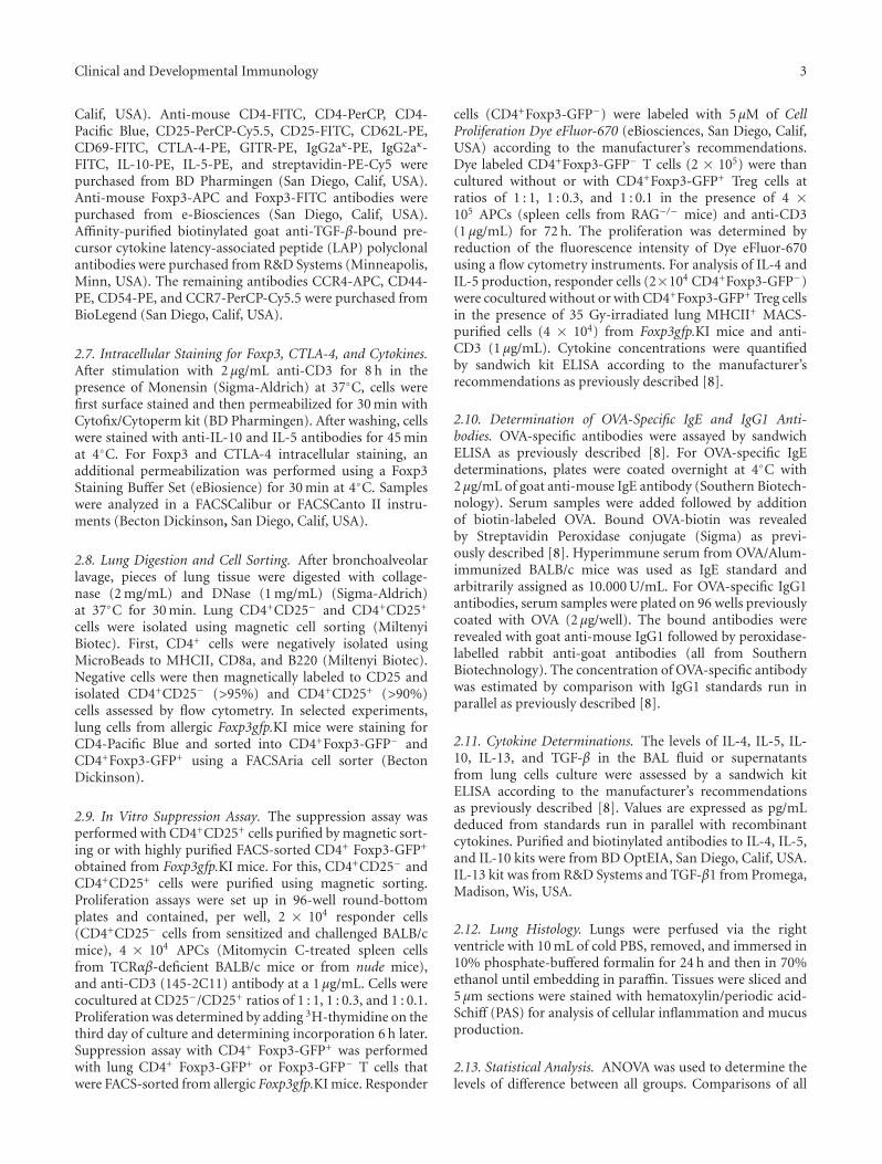
Clinical and Developmental Immunology 3
Calif, USA). Anti-mouse CD4-FITC, CD4-PerCP, CD4-Pacific Blue, CD25-PerCP-Cy5.5, CD25-FITC, CD62L-PE,CD69-FITC, CTLA-4-PE, GITR-PE, IgG2aκ-PE, IgG2aκ-FITC, IL-10-PE, IL-5-PE, and streptavidin-PE-Cy5 werepurchased from BD Pharmingen (San Diego, Calif, USA).Anti-mouse Foxp3-APC and Foxp3-FITC antibodies werepurchased from e-Biosciences (San Diego, Calif, USA).Affinity-purified biotinylated goat anti-TGF-β-bound pre-cursor cytokine latency-associated peptide (LAP) polyclonalantibodies were purchased from R&D Systems (Minneapolis,Minn, USA). The remaining antibodies CCR4-APC, CD44-PE, CD54-PE, and CCR7-PerCP-Cy5.5 were purchased fromBioLegend (San Diego, Calif, USA).
2.7. Intracellular Staining for Foxp3, CTLA-4, and Cytokines.After stimulation with 2 μg/mL anti-CD3 for 8 h in thepresence of Monensin (Sigma-Aldrich) at 37◦C, cells werefirst surface stained and then permeabilized for 30 min withCytofix/Cytoperm kit (BD Pharmingen). After washing, cellswere stained with anti-IL-10 and IL-5 antibodies for 45 minat 4◦C. For Foxp3 and CTLA-4 intracellular staining, anadditional permeabilization was performed using a Foxp3Staining Buffer Set (eBiosience) for 30 min at 4◦C. Sampleswere analyzed in a FACSCalibur or FACSCanto II instru-ments (Becton Dickinson, San Diego, Calif, USA).
2.8. Lung Digestion and Cell Sorting. After bronchoalveolarlavage, pieces of lung tissue were digested with collage-nase (2 mg/mL) and DNase (1 mg/mL) (Sigma-Aldrich)at 37◦C for 30 min. Lung CD4+CD25− and CD4+CD25+
cells were isolated using magnetic cell sorting (MiltenyiBiotec). First, CD4+ cells were negatively isolated usingMicroBeads to MHCII, CD8a, and B220 (Miltenyi Biotec).Negative cells were then magnetically labeled to CD25 andisolated CD4+CD25− (>95%) and CD4+CD25+ (>90%)cells assessed by flow cytometry. In selected experiments,lung cells from allergic Foxp3gfp.KI mice were staining forCD4-Pacific Blue and sorted into CD4+Foxp3-GFP− andCD4+Foxp3-GFP+ using a FACSAria cell sorter (BectonDickinson).
2.9. In Vitro Suppression Assay. The suppression assay wasperformed with CD4+CD25+ cells purified by magnetic sort-ing or with highly purified FACS-sorted CD4+ Foxp3-GFP+
obtained from Foxp3gfp.KI mice. For this, CD4+CD25− andCD4+CD25+ cells were purified using magnetic sorting.Proliferation assays were set up in 96-well round-bottomplates and contained, per well, 2 × 10⁴ responder cells(CD4+CD25− cells from sensitized and challenged BALB/cmice), 4 × 10⁴ APCs (Mitomycin C-treated spleen cellsfrom TCRαβ-deficient BALB/c mice or from nude mice),and anti-CD3 (145-2C11) antibody at a 1 μg/mL. Cells werecocultured at CD25−/CD25+ ratios of 1 : 1, 1 : 0.3, and 1 : 0.1.Proliferation was determined by adding 3H-thymidine on thethird day of culture and determining incorporation 6 h later.Suppression assay with CD4+ Foxp3-GFP+ was performedwith lung CD4+ Foxp3-GFP+ or Foxp3-GFP− T cells thatwere FACS-sorted from allergic Foxp3gfp.KI mice. Responder
cells (CD4+Foxp3-GFP−) were labeled with 5 μM of CellProliferation Dye eFluor-670 (eBiosciences, San Diego, Calif,USA) according to the manufacturer’s recommendations.Dye labeled CD4+Foxp3-GFP− T cells (2 × 10⁵) were thancultured without or with CD4+Foxp3-GFP+ Treg cells atratios of 1 : 1, 1 : 0.3, and 1 : 0.1 in the presence of 4 ×10⁵ APCs (spleen cells from RAG−/− mice) and anti-CD3(1 μg/mL) for 72 h. The proliferation was determined byreduction of the fluorescence intensity of Dye eFluor-670using a flow cytometry instruments. For analysis of IL-4 andIL-5 production, responder cells (2×10⁴ CD4+Foxp3-GFP−)were cocultured without or with CD4+Foxp3-GFP+ Treg cellsin the presence of 35 Gy-irradiated lung MHCII+ MACS-purified cells (4 × 10⁴) from Foxp3gfp.KI mice and anti-CD3 (1 μg/mL). Cytokine concentrations were quantifiedby sandwich kit ELISA according to the manufacturer’srecommendations as previously described [8].
2.10. Determination of OVA-Specific IgE and IgG1 Anti-bodies. OVA-specific antibodies were assayed by sandwichELISA as previously described [8]. For OVA-specific IgEdeterminations, plates were coated overnight at 4◦C with2 μg/mL of goat anti-mouse IgE antibody (Southern Biotech-nology). Serum samples were added followed by additionof biotin-labeled OVA. Bound OVA-biotin was revealedby Streptavidin Peroxidase conjugate (Sigma) as previ-ously described [8]. Hyperimmune serum from OVA/Alum-immunized BALB/c mice was used as IgE standard andarbitrarily assigned as 10.000 U/mL. For OVA-specific IgG1antibodies, serum samples were plated on 96 wells previouslycoated with OVA (2 μg/well). The bound antibodies wererevealed with goat anti-mouse IgG1 followed by peroxidase-labelled rabbit anti-goat antibodies (all from SouthernBiotechnology). The concentration of OVA-specific antibodywas estimated by comparison with IgG1 standards run inparallel as previously described [8].
2.11. Cytokine Determinations. The levels of IL-4, IL-5, IL-10, IL-13, and TGF-β in the BAL fluid or supernatantsfrom lung cells culture were assessed by a sandwich kitELISA according to the manufacturer’s recommendationsas previously described [8]. Values are expressed as pg/mLdeduced from standards run in parallel with recombinantcytokines. Purified and biotinylated antibodies to IL-4, IL-5,and IL-10 kits were from BD OptEIA, San Diego, Calif, USA.IL-13 kit was from R&D Systems and TGF-β1 from Promega,Madison, Wis, USA.
2.12. Lung Histology. Lungs were perfused via the rightventricle with 10 mL of cold PBS, removed, and immersed in10% phosphate-buffered formalin for 24 h and then in 70%ethanol until embedding in paraffin. Tissues were sliced and5 μm sections were stained with hematoxylin/periodic acid-Schiff (PAS) for analysis of cellular inflammation and mucusproduction.
2.13. Statistical Analysis. ANOVA was used to determine thelevels of difference between all groups. Comparisons of all

4 Clinical and Developmental Immunology
0 10 20 300
2
4
6
8
10
AllergicTolerant
Control
∗∗
MCh (mg/mL)
Pen
h
(a)
Control Allergic Tolerant0
10
20
30
Mononuclear cellsEosinophilsNeutrophils
∗∗∗∗∗
∗Cel
ls(×
105)
(b)
IL-5
0
100
200
300
400
500 ∗∗∗∗∗∗
pg/m
L
ControlAllergicTolerant
(c)
IL-13
0
50
100
150
200
250 ∗∗∗∗∗∗
pg/m
L
ControlAllergicTolerant
(d)
α-OVA IgE
0
200
400
600
800
1000
1200
1400
∗∗∗∗∗∗
(a.u
.)
ControlAllergicTolerant
(e)
α-OVA IgG1
0
20
40
60
80
100
120
140
∗∗∗∗∗∗
ControlAllergicTolerant
μg/
mL
(f)
Control Allergic Tolerant
(g)
Figure 1: Oral tolerance prevents airway allergic disease. (a) Respiratory pattern to increasing dose of methacholine (MCh) in control,allergic, or tolerant BALB/c mice 24 h after the last OVA challenge. (b) BAL differential cell counts. Quantification by ELISA of (c) IL-5, (d)IL-13 in the BAL fluid, and (e) anti-OVA IgE, (f) IgG1 in the serum. (g) Histology of lung sections at 100x. Lung parenchyma inflammationand mucus production by goblet cells are shown in representative lung sections stained with hematoxylin/PAS. Values represent the means±SEM for groups of five mice and are representative of more than three experiments. Significant differences ∗P < 0.05, ∗∗P < 0.01, and∗∗∗P < 0.001 are shown.
pairs were performed by Tukey-Kramer honestly significantdifference test. Values for all measurements are expressed asmean ± SEMs, and the P values for significance were set to0.05.
3. Results
3.1. Oral Tolerance Prevents the Development of Asthma-LikeResponses. OVA-sensitized and -challenged mice (Allergic)developed an enhanced ventilation as revealed by Penh
values to increasing doses of methacholine (MCh) com-pared to untreated mice (Control). Conversely, prior oraladministration of OVA (Tolerant) prevented the increase inventilation (Figure 1(a)). Differential cell counts showed anincreased number of mononuclear cells, neutrophils, andmainly eosinophils in allergic mice compared to controlmice. In tolerant mice, the influx of inflammatory cells wasalmost completely absent (Figure 1(b)). The levels of type-2cytokines IL-5 and IL-13 in the BAL (Figures 1(c) and 1(d))and the serum levels of OVA-specific IgE and IgG1 antibodies
Clinical and Developmental Immunology 5
0 1 2 3 4 5 6 7 8 9789
101112131415161718
OVA/Alum OVA/Alum
∗
Days after sensitization
Tolerant
Allergic
Spleen
%Fo
xp3+
CD
4+T
cells
(a)
cLN
0 1 2 3 4 5 6 7 8 9789
101112131415161718
OVA/AlumOVA/Alum
Days after sensitization
Tolerant
Allergic%
Foxp
3+C
D4+
Tce
lls
(b)
mesLN
0 1 2 3 4 5 6 7 8 9789
101112131415161718
OVA/AlumOVA/Alum
Tolerant
Allergic
Days after sensitization
%Fo
xp3+
CD
4+T
cells
(c)
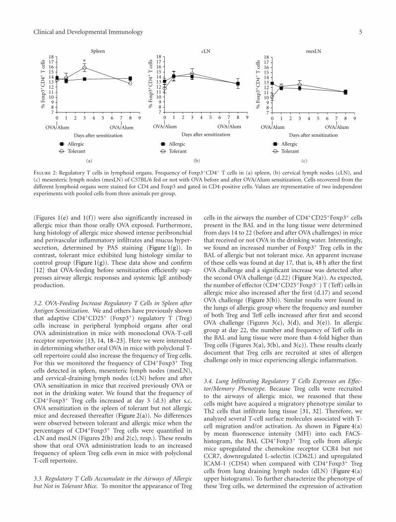
Figure 2: Regulatory T cells in lymphoid organs. Frequency of Foxp3+CD4+ T cells in (a) spleen, (b) cervical lymph nodes (cLN), and(c) mesenteric lymph nodes (mesLN) of C57BL/6 fed or not with OVA before and after OVA/Alum sensitization. Cells recovered from thedifferent lymphoid organs were stained for CD4 and Foxp3 and gated in CD4-positive cells. Values are representative of two independentexperiments with pooled cells from three animals per group.
(Figures 1(e) and 1(f)) were also significantly increased inallergic mice than those orally OVA exposed. Furthermore,lung histology of allergic mice showed intense peribronchialand perivascular inflammatory infiltrates and mucus hyper-secretion, determined by PAS staining (Figure 1(g)). Incontrast, tolerant mice exhibited lung histology similar tocontrol group (Figure 1(g)). These data show and confirm[12] that OVA-feeding before sensitization efficiently sup-presses airway allergic responses and systemic IgE antibodyproduction.
3.2. OVA-Feeding Increase Regulatory T Cells in Spleen afterAntigen Sensitization. We and others have previously shownthat adaptive CD4+CD25+ (Foxp3+) regulatory T (Treg)cells increase in peripheral lymphoid organs after oralOVA administration in mice with monoclonal OVA-T-cellreceptor repertoire [13, 14, 18–23]. Here we were interestedin determining whether oral OVA in mice with polyclonal T-cell repertoire could also increase the frequency of Treg cells.For this we monitored the frequency of CD4+Foxp3+ Tregcells detected in spleen, mesenteric lymph nodes (mesLN),and cervical-draining lymph nodes (cLN) before and afterOVA sensitization in mice that received previously OVA ornot in the drinking water. We found that the frequency ofCD4+Foxp3+ Treg cells increased at day 3 (d.3) after s.c.OVA sensitization in the spleen of tolerant but not allergicmice and decreased thereafter (Figure 2(a)). No differenceswere observed between tolerant and allergic mice when thepercentages of CD4+Foxp3+ Treg cells were quantified incLN and mesLN (Figures 2(b) and 2(c), resp.). These resultsshow that oral OVA administration leads to an increasedfrequency of spleen Treg cells even in mice with polyclonalT-cell repertoire.
3.3. Regulatory T Cells Accumulate in the Airways of Allergicbut Not in Tolerant Mice. To monitor the appearance of Treg
cells in the airways the number of CD4+CD25+Foxp3+ cellspresent in the BAL and in the lung tissue were determinedfrom days 14 to 22 (before and after OVA challenges) in micethat received or not OVA in the drinking water. Interestingly,we found an increased number of Foxp3+ Treg cells in theBAL of allergic but not tolerant mice. An apparent increaseof these cells was found at day 17, that is, 48 h after the firstOVA challenge and a significant increase was detected afterthe second OVA challenge (d.22) (Figure 3(a)). As expected,the number of effector (CD4+CD25+Foxp3−) T (Teff) cells inallergic mice also increased after the first (d.17) and secondOVA challenge (Figure 3(b)). Similar results were found inthe lungs of allergic group where the frequency and numberof both Treg and Teff cells increased after first and secondOVA challenge (Figures 3(c), 3(d), and 3(e)). In allergicgroup at day 22, the number and frequency of Teff cells inthe BAL and lung tissue were more than 4-fold higher thanTreg cells (Figures 3(a), 3(b), and 3(c)). These results clearlydocument that Treg cells are recruited at sites of allergenchallenge only in mice experiencing allergic inflammation.
3.4. Lung Infiltrating Regulatory T Cells Expresses an Effec-tor/Memory Phenotype. Because Treg cells were recruitedto the airways of allergic mice, we reasoned that thesecells might have acquired a migratory phenotype similar toTh2 cells that infiltrate lung tissue [31, 32]. Therefore, weanalyzed several T-cell surface molecules associated with T-cell migration and/or activation. As shown in Figure 4(a)by mean fluorescence intensity (MFI) into each FACS-histogram, the BAL CD4+Foxp3+ Treg cells from allergicmice upregulated the chemokine receptor CCR4 but notCCR7, downregulated L-selectin (CD62L) and upregulatedICAM-1 (CD54) when compared with CD4+Foxp3+ Tregcells from lung draining lymph nodes (dLN) (Figure 4(a)upper histograms). To further characterize the phenotype ofthese Treg cells, we determined the expression of activation

6 Clinical and Developmental Immunology
Treg
d.14 d.15 d.17 d.220
2
4
6
8
10
∗∗
OVA
AllergicTolerant
OVA
Foxp
3+C
D25
+C
D4+
Tce
lls(×
103)
(a)
Teff
d.14 d.15 d.17 d.220
2
4
6
8
10
20
30
40 ∗∗∗
OVA
AllergicTolerant
OVA
Foxp
3−C
D25
+C
D4+
Tce
lls(×
103)
(b)
CD25
2.07
Foxp
3
5.46d.14
87.47
d.15
5
d.171.53
d.225.01
Tolerant
Allergic
Lung
7.1886.28 84.78 7.46
4.852.91 2.89 5.94
9.5381.64
2.11 5.07
7.0285.8 77.24
2.43 6.73
13.59 74.66
4.5 7.4
13.44 64.86 23.23
8.942.97
(c)
Treg
d.14 d.15 d.17 d.220
5
10
15
20
25
OVA OVA
Foxp
3+C
D25
+C
D4+
Tce
lls(×
103)
Allergic
Tolerant
(d)
Teff
d.14 d.15 d.17 d.220
10
20
30
40
50
60
OVA OVA
AllergicTolerant
Foxp
3−C
D25
+C
D4+
Tce
lls(×
103)
(e)
Figure 3: Regulatory T cells accumulate in the airways of allergic but not tolerant mice. Time course of (a) CD4+CD25+Foxp3+ (Treg) and(b) CD4+CD25+Foxp3− (Teff) cells number in the BAL of allergic and tolerant mice. (c) Frequency and (d) number of CD4+CD25+ lungcells expressing or not Foxp3. Pooled cells from three mice recovered from BAL and lung were stained for CD4, CD25, and Foxp3 and gatedin CD4-positive cells. Values in (a) and (b) represent the means ± SEM for groups of three mice and are representative of two experiments.The data in (c) show a representative experiment of two. Significant differences ∗∗P < 0.01, ∗∗∗P < 0.001 are related to tolerant group.

Clinical and Developmental Immunology 7
CCR4
1346
CCR7
3349
CD62L
1285
2030
CD54
2231
CD44
634
CTLA-4
1527
4220
GITR CD25
4188
dLN
BAL
7127419920
2984134505
252708
(a)
d.14
(b)
d.15
(c)
d.17
88.14
d.225.87
Tolerant
Foxp3
0.47
CD
69
5.52
Allergic
d.22
84.35
BALLung
9.81 0.84
4.99 83.82
10.25 1.02
4.91 76.55
16.59 1.91
4.95 78.19
13.62 1.91
6.28
85.01
8.6 0.8
5.59 74.38
17.06 2.84
5.72 58.05
29.83 5.75
6.37 33.2
58.79 4.61
3.4 45.13
41.87 6.85
6.14
Figure 4: Airway regulatory T cells from allergic mice express a memory/effector phenotype. (a) FACS-Histograms of CD4+Foxp3+ cellsfrom allergic mice expressing CCR4, CCR7, CD62L, CD54, CD44, CTLA-4, GITR, CD25 in BAL (red line), or mediastinal draining lymphnodes (dLN) (blue line). The numbers into each histogram represent the mean fluorescence intensity (MFI). Kinetic of lung CD4+CD69+
cells frequency expressing or not Foxp3. (c) Percentage of BAL CD4+CD69+ cells expressing or not Foxp3. Pooled cells from four micerecovered from lung or BAL were stained for CD4, CD69, and Foxp3 and gated in CD4-positive cells. The results are representative of twoexperiments with four mice per group.
markers. We found that BAL CD4+Foxp3+ Treg cells fromallergic mice also upregulated CD44, CTLA-4, GITR, andCD25 (Figure 4(a) lower histograms). Moreover, in lungtissue the frequency of CD4+Foxp3+ Treg cells expressingCD69 molecule increased substantially after OVA challengein allergic mice compared to tolerant mice (Figure 4(b)). Asexpected, the frequency of Foxp3-negative CD69+ T helper(Teff) cells was drastically enhanced in allergic but not intolerant group after OVA challenges (Figure 4(b)). Similarresults were obtained with T cells present in BAL at day 22(Figure 4(c)). Notably, the frequency of CD69+ Treg cells inthe lung and BAL of allergic mice was higher than CD69−
Treg cells, whereas in tolerant mice we found an inverserelation (Figure 4(c)). Taken together, our findings clearlyindicate that infiltrating Foxp3+ Treg cells from allergic mice
acquire an effector/memory phenotype distinguishing themfrom Treg cells present in lung-draining lymph nodes andfrom those present in the airways of tolerant mice.
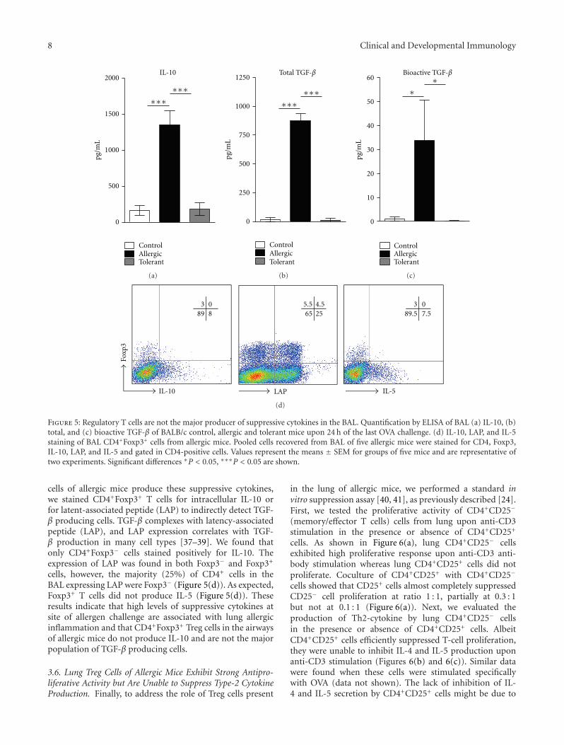
3.5. Regulatory T Cells Recruited to the Airways of AllergicMice Are Not the Principal Producers of Suppressive Cytokines.Interleukin-10 (IL-10) and transforming growth factor-β(TGF-β) have been implicated in suppression of inflamma-tion by Treg cells [33–36]. Therefore, we investigated whetherairway infiltrating Treg cells from allergic mice produce thesecytokines. We first determined the levels of IL-10 and TGF-β in BAL fluid. We found that high levels of IL-10, total,and bioactive TGF-β were significantly increased in the BALof allergic mice compared to control or tolerant groups(Figures 5(a), 5(b), and 5(c)). To ascertain whether Treg
8 Clinical and Developmental Immunology
IL-10
0
500
1000
1500
2000
∗∗∗∗∗∗
pg/m
L
ControlAllergicTolerant
(a)
Total TGF-β
0
250
500
750
1000
1250
∗∗∗∗∗∗
pg/m
L
ControlAllergicTolerant
(b)
Bioactive TGF-β
0
10
20
30
40
50
60
∗∗
ControlAllergicTolerant
pg/m
L
(c)
0389.5 7.5
IL-5
4.55.565 25
LAP
0389 8
IL-10
Foxp
3
(d)
Figure 5: Regulatory T cells are not the major producer of suppressive cytokines in the BAL. Quantification by ELISA of BAL (a) IL-10, (b)total, and (c) bioactive TGF-β of BALB/c control, allergic and tolerant mice upon 24 h of the last OVA challenge. (d) IL-10, LAP, and IL-5staining of BAL CD4+Foxp3+ cells from allergic mice. Pooled cells recovered from BAL of five allergic mice were stained for CD4, Foxp3,IL-10, LAP, and IL-5 and gated in CD4-positive cells. Values represent the means ± SEM for groups of five mice and are representative oftwo experiments. Significant differences ∗P < 0.05, ∗∗∗P < 0.05 are shown.
cells of allergic mice produce these suppressive cytokines,we stained CD4+Foxp3+ T cells for intracellular IL-10 orfor latent-associated peptide (LAP) to indirectly detect TGF-β producing cells. TGF-β complexes with latency-associatedpeptide (LAP), and LAP expression correlates with TGF-β production in many cell types [37–39]. We found thatonly CD4+Foxp3− cells stained positively for IL-10. Theexpression of LAP was found in both Foxp3− and Foxp3+
cells, however, the majority (25%) of CD4+ cells in theBAL expressing LAP were Foxp3− (Figure 5(d)). As expected,Foxp3+ T cells did not produce IL-5 (Figure 5(d)). Theseresults indicate that high levels of suppressive cytokines atsite of allergen challenge are associated with lung allergicinflammation and that CD4+Foxp3+ Treg cells in the airwaysof allergic mice do not produce IL-10 and are not the majorpopulation of TGF-β producing cells.
3.6. Lung Treg Cells of Allergic Mice Exhibit Strong Antipro-liferative Activity but Are Unable to Suppress Type-2 CytokineProduction. Finally, to address the role of Treg cells present
in the lung of allergic mice, we performed a standard invitro suppression assay [40, 41], as previously described [24].First, we tested the proliferative activity of CD4+CD25−
(memory/effector T cells) cells from lung upon anti-CD3stimulation in the presence or absence of CD4+CD25+
cells. As shown in Figure 6(a), lung CD4+CD25− cellsexhibited high proliferative response upon anti-CD3 anti-body stimulation whereas lung CD4+CD25+ cells did notproliferate. Coculture of CD4+CD25+ with CD4+CD25−
cells showed that CD25+ cells almost completely suppressedCD25− cell proliferation at ratio 1 : 1, partially at 0.3 : 1but not at 0.1 : 1 (Figure 6(a)). Next, we evaluated theproduction of Th2-cytokine by lung CD4+CD25− cellsin the presence or absence of CD4+CD25+ cells. AlbeitCD4+CD25+ cells efficiently suppressed T-cell proliferation,they were unable to inhibit IL-4 and IL-5 production uponanti-CD3 stimulation (Figures 6(b) and 6(c)). Similar datawere found when these cells were stimulated specificallywith OVA (data not shown). The lack of inhibition of IL-4 and IL-5 secretion by CD4+CD25+ cells might be due to

Clinical and Developmental Immunology 9
0
1000
2000
3000
4000 ∗∗∗
CD25−/CD25+
∗∗∗∗∗∗
∗∗
[3H
]-T
dR(c
pm)
CD25− CD25+ 1 : 0.1 1 : 0.3 1 : 1
(a)
0
100
200
300
400 ∗
CD25−/CD25+
∗∗
∗
IL-4
(pg/
mL
)
CD25− CD25+ 1 : 0.1 1 : 0.3 1 : 1
(b)
0
200
400
600
800
1000
1200
∗
CD25−/CD25+
IL-5
(pg/
mL
)
CD25− CD25+ 1 : 0.1 1 : 0.3 1 : 1
(c)
Figure 6: Lung CD4+CD25+ T cells from allergic mice suppress T-cell proliferation but not Th2 cytokine production. (a) Proliferation ofMACS-purified lung CD4+CD25− or CD4+CD25+ cells from BALB/c allergic mice alone or cocultured at different CD25−/CD25+ ratiosdetermined by 3H-thymidine (3H-TdR) incorporation after anti-CD3 stimulation. ELISA assays for (b) IL-4 and (c) IL-5 in the culturesupernatants showed in (a). Values represent the means ± SEM of triplicate wells. The results are representative of two experiments.Significant differences ∗P < 0.05, ∗∗P < 0.01, and ∗∗∗P < 0.001 are shown.
the fact that this cell population also contains effector T cells.Indeed, CD4+CD25+ cells produced significant amountsof type-2 cytokines (Figures 6(b) and 6(c)). In order tocircumvent this problem and address more directly whetherTreg cells affect type-2 cytokine production, we performedexperiments in Foxp3gfp.KI mice that harbor fluorescentTreg cells [29]. Therefore, we induced airway allergic diseasein Foxp3gfp.KI mice and sorted CD4+ T cells expressingFoxp3-GFP+ Treg cells and CD4+GFP− T cells (Foxp3−)present in the lungs. We found that only Foxp3− T cellsproduced significant amounts of type 2 cytokines uponanti-CD3 stimulation (Figures 7(a) and 7(b)). Notably, ahighly purified (>98%) lung population of Foxp3-GFP+ Tregcells could not suppress efficiently Th2 cytokine productionby CD4+Foxp3-GFP− T cells upon anti-CD3 stimulation(Figures 7(a) and 7(b)). Finally, through using purifiedlung Foxp3-GFP+ Treg cells, we confirmed the suppressionassay obtained with CD4+CD25+ T-cell by showing thatthey efficiently suppressed T effector (CD4+GFP−) cellsproliferation at ratio 1 : 1 and 0.3 : 1 but not at 0.1 : 1 asevidenced by Dye eFluor-670 staining (Figure 7(c)). Weconclude that lung Treg cells with regulatory phenotypepresent in the airways of allergic mice exhibit a strongantiproliferative activity but are unable to efficiently suppresstype-2 cytokine production.
4. Discussion
A critical issue in immune regulation is where Treg cellsexert their suppressive function. Their presence on lymphoidtissue appears to be required for efficient suppression of naiveT-cell activation. Conversely, some data indicate that Tregcells are recruited to effector site in order to suppress theaction of inflammatory T cells [25, 42, 43]. Previous reportsshowed a relationship between suppression of asthma-likeresponses by mucosal tolerance and the emergence of Tregcells in lymphoid organs [17, 21, 24]. We have previouslyshown in T/B receptors transgenic mice (T-Bmc) devoid ofnatural regulatory T cells that soon after mucosal antigenexposure, Foxp3-expressing Treg cells are generated in dLN
and in spleen [24]. This early induction of Treg cells by priororal antigen exposure appears to inhibit the developmentof polarized Th2 inflammatory cells in a TGF-β-dependentmanner [24]. Indeed, using the T-Bmc model, we found thatTreg cells are able to suppress early T-cell activation, 48 hafter immunization with the cognate antigen [24]. However,after establishment of tolerance they became dispensable forits maintenance in situ. In the present study, we used awell-established model of mucosal tolerance to allergic lunginflammation [8, 12, 24, 44] to monitor the appearanceof Treg cells in the airways after OVA challenge in micewith polyclonal repertoire. We found that only in OVA-fed mice, the frequency of spleen Treg cells increased atday 3 after OVA sensitization, a result resembles the T-Bmc model. However, here we were particularly interested indetermining whether Treg cells migrate to airways of allergicor tolerant mice after administration of OVA. We found thatallergic but not tolerant mice showed a striking increase inthe number of Treg cells in the BAL compared to tolerantmice. Also, high levels of IL-10 and TGF-β were detected inthe airways of allergic mice. Notably, we found that amongCD4+ T cells recruited to allergic inflammation only Foxp3-negative, but not Foxp3-positive T cells stained positivelyfor IL-10. Moreover, the majority of LAP+ cells were Foxp3-negative T cells. Our results are line with data obtained withT-cell infiltrates in Shcistosoma mansoni egg-induced Th2-mediated inflammation [45]. In concert with our findings,migration of Treg cells was also reported in a model ofparasite egg antigens-induced inflammation [46], or otherpathological conditions, such as arthritis, type 1 diabetes,sarcoidosis and transplants [25, 43, 47–52]. Therefore, itis plausible that the allergic inflammatory milieu triggersthe migration of Treg cells into the airways. Accordingly, ithas been shown that recruitment of Foxp3-expressing Tregcells to the site of allergic inflammation is dependent onchemokine receptors such as CCR4 [52] and CCR8 [53],where their ligands CCL17, CCL22, and CCL1 are highexpressed during allergic lung inflammation [54, 55]. Ourdata demonstrated that the majority of Foxp3-expressingTreg cells present in the airways upregulated CCR4, CD44

10 Clinical and Developmental Immunology
Dye
(a)
(b)
Teff
:Tre
gce
lls
(c)
61.5
56.5
44
26.8
Foxp3− Foxp3+0
20
40
60
80 ∗∗
Foxp3−/Foxp3+
IL-4
(pg/
mL
)
Foxp3− Foxp3+0
20
40
60
80
100
120 ∗∗∗
Foxp3−/Foxp3+
IL-5
(pg/
mL
)
1 : 0.1 1 : 0.3 1 : 1
1 : 0.1 1 : 0.3 1 : 1
1 : 0
1 : 0.1
1 : 0.3
1 : 1
Figure 7: Lung Foxp3+ Treg cells from allergic mice suppress T-cell proliferation but not type 2 cytokine production. (a) IL-4 and (b) IL-5levels in the supernatants of FACS-sorted lung CD4+Foxp3-GFP+ (Foxp3+) or CD4+Foxp3-GFP− (Foxp3−) cells from Foxp3gfp.KI allergicmice alone or cocultured at different Foxp3−/Foxp3+ ratios. (c) Proliferation of Foxp3-GFP− cells cocultured at different Foxp3−/Foxp3+
ratios was determined by flow cytometry. Sorted lung CD4+Foxp3-GFP− were labeled with Dye eFluor-670 and the proliferation wasdetermined by reduction of the fluorescence intensity of Dye. Representative FACS-histograms, which indicate the frequency of T-cellproliferation, are shown. Values represent the means ± SEM of triplicate wells. The results are representative of two experiments. Significantdifferences ∗∗P < 0.01, ∗∗∗P < 0.001 are shown.
and CD54 and drastically downregulated CD62L, a pheno-type that resembles effector/memory T cells. Noteworthy,this phenotype could distinguish Treg cells present in theairways from those present in the lung-draining lymphnodes (dLN). In addition, we showed that Treg cells thataccumulated in the airways of allergic mice also acquiredactivated phenotype, as revealed by increased expression ofCTLA-4, GITR, and CD25 contrasting with Treg cells presentin the dLN. Moreover, CD69, a marker of cell activation, washighly expressed in Treg cells present in the lung and BAL ofallergic mice but not in tolerant group. These data suggesta functionally important activation step that accompaniesTreg cell migration. The loss of CD62L and the increased ofCD54 expression by Treg cells could also contribute to theirmigration to the lung [32]. A picture that emerges from ourfindings is that Treg cells get activated and are recruited tosites of allergic inflammation probably because at these sitesCCR4-specific ligands are expressed at high levels [28, 56].
It was recently reported that the loss of CCR4 severelyinhibited the accumulation of CD4+CD25+ T cells in the
lung and skin [57]. CCR4 knockout mice also fail to developallograft tolerance after administration of anti-CD154 withdonor spleen cells, which is associated with a decreased ofFoxp3+ T cells in the graft [43]. Previous data indicateda division of labor between naive and activated Treg cells[58]. For instance, naive-like Treg cells use the chemokinereceptor CCR7 for recirculation through lymph nodes wherethey control the priming phase of an immune responsewhereas CCR7 is dispensable in effector/memory-like Tregcells for their accumulation in inflamed sites and in factCCR7-deficiency enhance Treg cells-mediated suppressionof inflammation [58]. In our model, the role of CCR7could not be established because activated lung Treg cellsexpressed similar levels of CCR7 when compared to naivedLN Treg cells [25]. Using an islet allograft model it wasdemonstrated that Treg cells first migrate from blood to theallograft where they become activated, and then they migrateto the dLN in a CCR7 fashion. This movement was essentialfor optimal suppression allograft rejection [25]. A similarsituation was found by Graca et al. that found regulatory

Clinical and Developmental Immunology 11
T cells in skin allografts suggesting that T-cell suppression ofgraft rejection is an active process that involves the presenceof regulatory T cells at the site of the tolerated transplant[59]. This scenario does not appear to operate in our modelbecause we did not find Treg cells in dLN with an activatedphenotype.
We first studied the suppressive activity of airwayCD4+CD25+ T cells, putative Treg cells, in order to determinetheir role in lung inflammation. We clearly showed thatCD4+CD25+ T cells containing activated Foxp3+ Treg cellsefficiently suppressed the proliferation of lung CD4+CD25−
memory/effector T cells. Strikingly, these CD4+CD25+ Tcells did not suppress the secretion of IL-4 and IL-5 byanti-CD3 or OVA-activated CD4+CD25− T cells. BecauseCD4+CD25+ T cells contain also effector Foxp3-negativeT cells, it is likely that these cells were the source of thetype 2 cytokines detected in the cultures. To circumvent thiswe purified lung fluorescent Foxp3 Treg cells from allergicFoxp3gfp.KI mice and tested their suppressive activity ontype 2 cytokine production by Foxp3-negative CD4+ T cells.In this situation, Foxp3-positive Treg cells did not secretetype 2 cytokines and did not suppress significantly type 2cytokine production by Foxp3-negative CD4+ T cells butdid suppress CD4+ T-cell proliferation. These results couldexplain why, despite the large infiltration of Treg cells, allergicmice still show Th2-associated pathological responses. Ourresults are in line with previous finding showing that inallergic patients, CD4+CD25+ T cells did not suppressthe release of Th2 cytokines [6]. The inefficiency of Tregcell in suppressing inflammatory cytokines in establishedpathological conditions was also reported in sarcoid gran-ulomas, in which Treg cells suppressed T-cell proliferationbut were unable to inhibit TNF-α secretion [51]. Notably,in a model of autoimmune encephalomyelitis, Treg cells alsoexpand after exposure to myelin antigens and infiltrate thecentral nervous system, but these infiltrating Treg cells wereunable to suppress proliferation and inflammatory cytokineproduction of effector T cells from target tissue [60]. Basedon our results and previous reports, it appears that theinflammatory milieu impairs Treg-cell functions.
In summary, we showed that oral tolerance was not asso-ciated with an increased number of Treg cells or suppressivecytokines in the airways. Conversely, allergic inflammationtriggers the infiltration of Treg cells into the airways thatefficiently suppress T-cell proliferation but not Th2 cytokineproduction. Our findings suggest that allergic inflammationrenders the suppressive activity of Treg cells less stringentthat, in turn, allows the manifestations allergic reactionsmediated by type 2 cytokines.
Acknowledgments
This study was supported by Fundacao de Amparo a Pesquisado Estado de Sao Paulo (FAPESP) and Conselho Nacionalde Desenvolvimento Cientıfico e Tecnologico (CNPq). Theauthors thank Paulo Albe for expert technical assistance inhistological preparations and Erica Borducchi for technicalassistance.
Author Contribution
Lucas Faustino and Daniel Mucida contributed equally tothis work.
References
[1] M. A. Curotto de Lafaille and J. J. Lafaille, “CD4+ regulatoryT cells in autoimmunity and allergy,” Current Opinion inImmunology, vol. 14, no. 6, pp. 771–778, 2002.
[2] D. S. Mucida, A. de Castro Keller, E. C. Fernvik, and M. Russo,“Unconventional strategies for the suppression of allergicasthma,” Current Drug Targets: Inflammation and Allergy, vol.2, no. 2, pp. 187–195, 2003.
[3] P. S. Foster, A. W. Mould, M. Yang et al., “Elemental signalsregulating eosinophil accumulation in the lung,” Immunologi-cal Reviews, vol. 179, pp. 173–181, 2001.
[4] H. Z. Shi and X. J. Qin, “CD4+CD25+ regulatory T lympho-cytes in allergy and asthma,” Allergy, vol. 60, no. 8, pp. 986–995, 2005.
[5] E. M. Ling, T. Smith, X. D. Nguyen et al., “Relation ofCD4+CD25+ regulatory T-cell suppression of allergen-drivenT-cell activation to atopic status and expression of allergicdisease,” Lancet, vol. 363, no. 9409, pp. 608–615, 2004.
[6] H. Grindebacke, K. Wing, A. C. Andersson, E. Suri-Payer, S.Rak, and A. Rudin, “Defective suppression of Th2 cytokines byCD4+CD25+ regulatory T cells in birch allergies during birchpollen season,” Clinical and Experimental Allergy, vol. 34, no.9, pp. 1364–1372, 2004.
[7] M. A. Curotto de Lafaille, S. Muriglan, M. J. Sunshine et al.,“Hyper immunoglobulin E response in mice with monoclonalpopulations of B and T lymphocytes,” Journal of ExperimentalMedicine, vol. 194, no. 9, pp. 1349–1359, 2001.
[8] M. Russo, M. A. Nahori, J. Lefort et al., “Suppression ofasthma-like responses in different mouse strains by oraltolerance,” American Journal of Respiratory Cell and MolecularBiology, vol. 24, no. 5, pp. 518–526, 2001.
[9] O. Akbari, R. H. DeKruyff, and D. T. Umetsu, “Pulmonarydendritic cells producing IL-10 mediate tolerance induced byrespiratory exposure to antigen,” Nature Immunology, vol. 2,no. 8, pp. 725–731, 2001.
[10] D. S. Mucida, D. Rodrıguez, A. Castro Keller et al., “Decreasednasal tolerance to allergic asthma in mice fed an amino acid-based protein-free diet,” Annals of the New York Academy ofSciences, vol. 1029, pp. 361–365, 2004.
[11] J. Bousquet and F. B. Michel, “International consensus reporton diagnosis and management of asthma,” Allergy, vol. 47, no.2, pp. 129–132, 1992.
[12] A. C. Keller, D. Mucida, E. Gomes et al., “Hierarchicalsuppression of asthma-like responses by mucosal tolerance,”Journal of Allergy and Clinical Immunology, vol. 117, no. 2, pp.283–290, 2006.
[13] X. Zhang, L. Izikson, L. Liu, and H. L. Weiner, “Activationof CD25+CD4+ regulatory T cells by oral antigen administra-tion,” Journal of Immunology, vol. 167, no. 8, pp. 4245–4253,2001.
[14] F. Hauet-Broere, W. W. J. Unger, J. Garssen, M. A. Hoijer,G. Kraal, and J. N. Samsom, “Functional CD25− and CD25+
mucosal regulatory T cells are induced in gut-draininglymphoid tissue within 48 h after oral antigen application,”European Journal of Immunology, vol. 33, no. 10, pp. 2801–2810, 2003.

12 Clinical and Developmental Immunology
[15] P. Stock, O. Akbari, G. Berry, G. J. Freeman, R. H. DeKruyff,and D. T. Umetsu, “Induction of T helper type 1-likeregulatory cells that express Foxp3 and protect against airwayhyper-reactivity,” Nature Immunology, vol. 5, no. 11, pp. 1149–1156, 2004.
[16] Y. Chen, J. I. Inobe, R. Marks, P. Gonnella, V. K. Kuchroo, andH. L. Weiner, “Peripheral deletion of antigen-reactive T cells inoral tolerance,” Nature, vol. 376, no. 6536, pp. 177–180, 1995.
[17] O. Akbari, G. J. Freeman, E. H. Meyer et al., “Antigen-specificregulatory T cells develop via the ICOS-ICOS-ligand pathwayand inhibit allergen-induced airway hyperreactivity,” NatureMedicine, vol. 8, no. 9, pp. 1024–1032, 2002.
[18] Y. Chen, Y. Ma, and Y. Chen, “Roles of cytotoxic T-lymphocyte-associated antigen-4 in the inductive phase of oraltolerance,” Immunology, vol. 105, no. 2, pp. 171–180, 2002.
[19] S. Fowler and F. Powrie, “CTLA-4 expression on antigen-specific cells but not IL-10 secretion is required for oraltolerance,” European Journal of Immunology, vol. 32, no. 10,pp. 2997–3006, 2002.
[20] K. M. Thorstenson and A. Khoruts, “Generation of anergicand potentially immunoregulatory CD25+CD4 T cells in vivoafter induction of peripheral tolerance with intravenous ororal antigen,” Journal of Immunology, vol. 167, no. 1, pp. 188–195, 2001.
[21] W. W. Unger, F. Hauet-Broere, W. Jansen, L. A. Van Berkel,G. Kraal, and J. N. Samsom, “Early Events in PeripheralRegulatory T Cell Induction via the Nasal Mucosa,” Journal ofImmunology, vol. 171, no. 9, pp. 4592–4603, 2003.
[22] B. Dubois, L. Chapat, A. Goubier, M. Papiernik, J. F. Nicolas,and D. Kaiserlian, “Innate CD4+CD25+ regulatory T cells arerequired for oral tolerance and inhibition of CD8+ T cellsmediating skin inflammation,” Blood, vol. 102, no. 9, pp.3295–3301, 2003.
[23] M. Ostroukhova, C. Seguin-Devaux, T. B. Oriss et al.,“Tolerance induced by inhaled antigen involves CD4+ T cellsexpressing membrane-bound TGF-β and FOXP3,” Journal ofClinical Investigation, vol. 114, no. 1, pp. 28–38, 2004.
[24] D. Mucida, N. Kutchukhidze, A. Erazo, M. Russo, J. J. Lafaille,and M. A. Curotto De Lafaille, “Oral tolerance in the absenceof naturally occurring Tregs,” Journal of Clinical Investigation,vol. 115, no. 7, pp. 1923–1933, 2005.
[25] N. Zhang, B. Schroppel, G. Lal et al., “Regulatory T cellssequentially migrate from inflamed tissues to draining lymphnodes to suppress the alloimmune response,” Immunity, vol.30, no. 3, pp. 458–469, 2009.
[26] W. F. T. Carson, L. A. Guernsey, A. Singh, A. T. Vella, C. M.Schramm, and R. S. Thrall, “Accumulation of regulatory Tcells in local draining lymph nodes of the lung correlates withspontaneous resolution of chronic asthma in a murine model,”International Archives of Allergy and Immunology, vol. 145, no.3, pp. 231–243, 2008.
[27] F. Sallusto, C. R. Mackay, and A. Lanzavecchia, “The roleof chemokine receptors in primary, effector, and memoryimmune responses,” Annual Review of Immunology, vol. 18,pp. 593–620, 2000.
[28] B. D. Medoff, S. Y. Thomas, and A. D. Luster, “T cell traffickingin allergic asthma: the ins and outs,” Annual Review ofImmunology, vol. 26, pp. 205–232, 2008.
[29] E. Bettelli, Y. Carrier, W. Gao et al., “Reciprocal developmentalpathways for the generation of pathogenic effector TH17 andregulatory T cells,” Nature, vol. 441, no. 7090, pp. 235–238,2006.
[30] D. Rodrıguez, A. C. Keller, E. L. Faquim-Mauro et al., “Bac-terial lipopolysaccharide signaling through Toll-like receptor4 suppresses asthma-like responses via nitric oxide synthase2 activity,” Journal of Immunology, vol. 171, no. 2, pp. 1001–1008, 2003.
[31] E. Yurchenko, M. Tritt, V. Hay, E. M. Shevach, Y. Belkaid,and C. A. Piccirillo, “CCR5-dependent homing of naturallyoccurring CD4+ regulatory T cells to sites of Leishmania majorinfection favors pathogen persistence,” Journal of ExperimentalMedicine, vol. 203, no. 11, pp. 2451–2460, 2006.
[32] J. Huehn, K. Siegmund, J. C. U. Lehmann et al., “Develop-mental stage, phenotype, and migration distinguish naive-and effector/memory-like CD4+ regulatory T cells,” Journal ofExperimental Medicine, vol. 199, no. 3, pp. 303–313, 2004.
[33] H. L. Weiner, “Induction and mechanism of action oftransforming growth factor-β-secreting Th3 regulatory cells,”Immunological Reviews, vol. 182, pp. 207–214, 2001.
[34] C. Asseman, S. Mauze, M. W. Leach, R. L. Coffman, and F.Powrie, “An essential role for interleukin 10 in the function ofregulatory T cells that inhibit intestinal inflammation,” Journalof Experimental Medicine, vol. 190, no. 7, pp. 995–1004, 1999.
[35] F. Powrie, J. Carlino, M. W. Leach, S. Mauze, and R. L.Coffman, “A critical role for transforming growth factor-βbut not interleukin 4 in the suppression of T helper type1-mediated colitis by CD45RBlow CD4+ T cells,” Journal ofExperimental Medicine, vol. 183, no. 6, pp. 2669–2674, 1996.
[36] D. H. Strickland, P. A. Stumbles, G. R. Zosky et al., “Reversalof airway hyperresponsiveness by induction of airway mucosalCD4+CD25+ regulatory T cells,” Journal of ExperimentalMedicine, vol. 203, no. 12, pp. 2649–2660, 2006.
[37] H. Ochi, M. Abraham, H. Ishikawa et al., “Oral CD3−
specific antibody suppresses autoimmune encephalomyelitisby inducing CD4+CD25−LAP+ T cells,” Nature Medicine, vol.12, no. 6, pp. 627–635, 2006.
[38] P. Berger, P. O. Girodet, H. Begueret et al., “Tryptase-stimulated human airway smooth muscle cells induce cytokinesynthesis and mast cell chemotaxis,” The FASEB Journal, vol.17, no. 14, pp. 2139–2141, 2003.
[39] R. Gandhi, D. E. Anderson, and H. L. Weiner, “Cutting edge:immature human dendritic cells express latency-associatedpeptide and inhibit T cell activation in a TGF-β-dependentmanner,” Journal of Immunology, vol. 178, no. 7, pp. 4017–4021, 2007.
[40] A. M. Thornton and E. M. Shevach, “CD4+CD25+
immunoregulatory T cells suppress polyclonal T cell activationin vitro by inhibiting interleukin 2 production,” Journal ofExperimental Medicine, vol. 188, no. 2, pp. 287–296, 1998.
[41] T. Takahashi, Y. Kuniyasu, M. Toda et al., “Immunologicself-tolerance maintained by CD25+CD4+ naturally anergicand suppressive T cells: induction of autoimmune diseaseby breaking their anergic/suppressive state,” InternationalImmunology, vol. 10, no. 12, pp. 1969–1980, 1998.
[42] Y. Belkaid, C. A. Piccirillo, S. Mendez, E. M. Shevach, and D.L. Sacks, “CD4+CD25+ regulatory T cells control Leishmaniamajor persistence and immunity,” Nature, vol. 420, no. 6915,pp. 502–507, 2002.
[43] I. Lee, L. Wang, A. D. Wells, M. E. Dorf, E. Ozkaynak,and W. W. Hancock, “Recruitment of Foxp3+ T regulatorycells mediating allograft tolerance depends on the CCR4chemokine receptor,” Journal of Experimental Medicine, vol.201, no. 7, pp. 1037–1044, 2005.
[44] M. Russo, S. Jancar, A. L. Pereira De Siqueira et al., “Pre-vention of lung eosinophilic inflammation by oral tolerance,”Immunology Letters, vol. 61, no. 1, pp. 15–23, 1998.

Clinical and Developmental Immunology 13
[45] M. Baumgart, F. Tompkins, J. Leng, and M. Hesse, “Naturallyoccurring CD4+Foxp3+ regulatory T cells are an essential, IL-10-independent part of the immunoregulatory network inSchistosoma mansoni egg-induced inflammation,” Journal ofImmunology, vol. 176, no. 9, pp. 5374–5387, 2006.
[46] J. J. Taylor, M. Mohrs, and E. J. Pearce, “Regulatory T cellresponses develop in parallel to Th responses and controlthe magnitude and phenotype of the Th effector population,”Journal of Immunology, vol. 176, no. 10, pp. 5839–5847, 2006.
[47] D. Cao, V. Malmstrom, C. Baecher-Allan, D. Hafler, L.Klareskog, and C. Trollmo, “Isolation and functional charac-terization of regulatory CD25bright CD4+ T cells from the targetorgan of patients with rheumatoid arthritis,” European Journalof Immunology, vol. 33, no. 1, pp. 215–223, 2003.
[48] A. E. Herman, G. J. Freeman, D. Mathis, and C. Benoist,“CD4+CD25+ T regulatory cells dependent on ICOS promoteregulation of effector cells in the prediabetic lesion,” Journal ofExperimental Medicine, vol. 199, no. 11, pp. 1479–1489, 2004.
[49] Y. Peng, Y. Laouar, M. O. Li, E. A. Green, and R. A. Flavell,“TGF-β regulates in vivo expansion of Foxp3-expressingCD4+CD25+ regulatory T cells responsible for protectionagainst diabetes,” Proceedings of the National Academy ofSciences of the United States of America, vol. 101, no. 13, pp.4572–4577, 2004.
[50] C. R. Ruprecht, M. Gattorno, F. Ferlito et al., “Coexpressionof CD25 and CD27 identifies FoxP3+ regulatory T cells ininflamed synovia,” Journal of Experimental Medicine, vol. 201,no. 11, pp. 1793–1803, 2005.
[51] M. Miyara, Z. Amoura, C. Parizot et al., “The immuneparadox of sarcoidosis and regulatory T cells,” Journal ofExperimental Medicine, vol. 203, no. 2, pp. 359–370, 2006.
[52] K. Saito, M. Torii, N. Ma et al., “Differential regulatory func-tion of resting and preactivated allergen-specific CD4+CD25+
regulatory T cells in Th2-type airway inflammation,” Journalof Immunology, vol. 181, no. 10, pp. 6889–6897, 2008.
[53] A. Iellem, M. Mariani, R. Lang et al., “Unique chemotacticresponse profile and specific expression of chemokine recep-tors CCR4 and CCR8 by CD4+CD25+ regulatory T cells,”Journal of Experimental Medicine, vol. 194, no. 6, pp. 847–853,2001.
[54] I. H. Heijink and A. J. van Oosterhout, “Targeting T cells forasthma,” Current Opinion in Pharmacology, vol. 5, no. 3, pp.227–231, 2005.
[55] A. Mathew, J. A. MacLean, E. DeHaan, A. M. Tager, F. H.Y. Green, and A. D. Luster, “Signal transducer and activatorof transcription 6 controls chemokine production and Thelper cell type 2 cell trafficking in allergic pulmonaryinflammation,” Journal of Experimental Medicine, vol. 193, no.9, pp. 1087–1096, 2001.
[56] T. Sekiya, M. Miyamasu, M. Imanishi et al., “Inducible expres-sion of a Th2-type CC chemokine thymus- and activation-regulated chemokine by human bronchial epithelial cells,”Journal of Immunology, vol. 165, no. 4, pp. 2205–2213, 2000.
[57] B. D. Sather, P. Treuting, N. Perdue et al., “Altering the dis-tribution of Foxp3+ regulatory T cells results in tissue-specificinflammatory disease,” Journal of Experimental Medicine, vol.204, no. 6, pp. 1335–1347, 2007.
[58] A. Menning, U. E. Hopken, K. Siegmund, M. Lipp, A.Hamann, and J. Huenn, “Distinctive role of CCR7 in migra-tion and functional activity of naive- and effector/memory-like Treg subsets,” European Journal of Immunology, vol. 37, no.6, pp. 1575–1583, 2007.
[59] L. Graca, S. P. Cobbold, and H. Waldmann, “Identificationof regulatory T cells in tolerated allografts,” Journal ofExperimental Medicine, vol. 195, no. 12, pp. 1641–1646, 2002.
[60] T. Korn, J. Reddy, W. Gao et al., “Myelin-specific regulatory Tcells accumulate in the CNS but fail to control autoimmuneinflammation,” Nature Medicine, vol. 13, no. 4, pp. 423–431,2007.