universidade do algarve - core.ac.uk · doutoramento em ciências do mar, especialidade de ecologia...
TRANSCRIPT

UNIVERSIDADE DO ALGARVE FACULDADE DE CIÊNCIAS DO MAR E DO AMBIENTE
DECAPOD CRUSTACEAN LARVAE DYNAMICS IN THE WEST
CONTINENTAL COAST OF PORTUGAL: THE REGION
ADJACENT TO AVEIRO COASTAL LAGOON AS MODEL
CÁTIA ALEXANDRA VIEIRA BARTILOTTI
Doutoramento em Ciências do Mar, especialidade de Ecologia Marinha
FARO
(2010)
UNIVERSIDADE DO ALGARVE FACULDADE DE CIÊNCIAS DO MAR E DO AMBIENTE
DECAPOD CRUSTACEAN LARVAE DYNAMICS IN THE WEST
CONTINENTAL COAST OF PORTUGAL: THE
REGION ADJACENT TO AVEIRO COASTAL LAGOON AS MODEL
CÁTIA ALEXANDRA VIEIRA BARTILOTTI
Doutoramento em Ciências do Mar, especialidade de Ecologia Marinha
Tese orientada por:
Investigadora Doutora Antonina dos Santos
Professora Doutora Margarida Castro
FARO
(2010)

ii

iii
“Porque sou do tamanho do que vejo
E não do tamanho da minha altura”
Bernardo Soares, in: O Livro do Desassossego

iv

v
ACKNOWLEDGEMENTS
I would like to express my gratitude to all of those that gave me support through these years
of work, even knowing that the words are not enough to recognize all the received
assistance. First of all I must thank to FCT for the financial support (SFRH/ BD/ 16695/
2004), to IPIMAR that gave me the conditions to fulfill my PhD working plan, and to my
supervisors Dr. Margarida Castro and Dr. Antonina dos Santos for all the guidance and
support. To Dr. Margarida Castro, thank you for all the help at the University, for the
statistical analysis and for all the nice talks about such varied themes; to Dr. Antonina dos
Santos, thank you for these eight years of lessons, friendship and confidence… As I always
say, Gurney is a visionary, so if he had a time machine (as you always say), I am sure that
he would like to discuss everything about decapod larvae with a generalist like you!
To Dr Miguel Santos for the constant care. To the “ProRecruit- Shelf processes controlling
recruitment to littoral populations in an eastern oceanic boundary: using barnacles and
crabs as models” (POCTI/1999/BSE/36663) staff, in particular, Dr. Henrique Queiroga, Dr.
Álvaro Peliz and Dr. Patrícia Lourenço.
To the Crustacea friends: Dr. Andrew Rhyne, thank you for all the Lysmata larvae that you
gave me (I believe that they will keep me far from Alzheimer); Dr. Juan Ignacio González-
Gordillo for the Ilia nucleus larval series and all the confidence deposited in my work; and
last but not least, to Dr. Ricardo Calado that is an excellent person but also an excellent
scientist always with a new idea to discuss and develop. I also express thanks to all the co-
authors of my papers for all the suggestions made that greatly improved the quality of my
work and taught me so much.

vi
To those that shared IPIMAR with me: Fátima Quintela, my friend, always looking for me
and taking care of me, thank you for your good mood and thank you for your nice smile;
Dr. Sofia Palma and Dr. Alexandra Silva, friends of “marmita” lunch time, living at the
next door lab, were the perfect companionship for the larval description pauses; Dr. Susana
Garrido, the “perfumed” friend, always looking for digested phyto- and zooplankton in
sardines stomachs, shared the smell but also her friendship; Dr. Juan Zwolinski, the tea
time friend, who shared the activities of the weekend and also the GAM’s and other
statistical methodologies.
To my friends, that always believed: Carla, Daniela, Eugénio, Irina, Joana, Marta, Miguel,
Sara, Rui, Vanessa, thank you for your love, your understanding, your support. You make
my life happier, simpler and funnier. To all those that I don’t refer but in some way were
important to me, thank you!
To my Super-heroes family! Thank you! Aqueles que são meus e que estão comigo sempre,
que me amam e protegem incondicionalmente, a eles dedicam o meu trabalho! À Avó,
Mãe, Luis e Nino, obrigada por serem os meus anjos! Ao Pai que considero um super-
homem a cada dia que passa. Aos tios, primos e bebés, por me mimarem sempre. A todos
estes que me ajudaram a percorrer o caminho, muito obrigada! Foi um fim difícil… Aos
que não me acompanharam, também agradeço, porque me mostraram que os nossos limites
são sempre ultrapassáveis…

vii
NOME: Cátia Alexandra Vieira Bartilotti
FACULDADE: Ciências do Mar e do Ambiente
DATA: 12 de Fevereiro de 2010
TÍTULO DA TESE: Dinâmica larvar de crustáceos decápodes na costa ocidental de
Portugal Continental: a região adjacente à Ria de Aveiro como modelo.
RESUMO O Filo Crustacea é um dos maiores taxa do Reino Animal, e a Ordem Decapoda é a maior com cerca de 50000 espécies, um número que continua a aumentar. A maioria dos decápodes distribui-se no oceano e nas zonas estuarinas adjacentes, no entanto algumas espécies invadiram a água doce, e um pequeno número de espécies ocupou os habitats terrestres. A diversidade de espécies de decápodes reflecte a diversidade de estilos de vida, e sendo na sua maioria bentónicos, estes animais podem viver nos habitats tropicais, mangais, recifes de coral, regiões polares, zonas do oceano profundo ou fontes hidrotermais. Os crustáceos decápodes são bastante importantes pois constituem um dos grupos biológicos com pesca dirigida e são muitas vezes elos da cadeia alimentar de inúmeros recursos pesqueiros da nossa costa. Uma estratégia adequada de gestão pesqueira para uma espécie com valor económico requer o conhecimento do seu ciclo de vida. Assim, para os crustáceos decápodes os estudos de plâncton dão informação acerca da distribuição e abundância das espécies durante a fase larvar planctónica. A fase de recrutamento é particularmente crítica nestes organismos, uma vez que implica a articulação de duas fases do ciclo de vida separadas espacialmente: a fase larvar planctónica e a fase adulta bentónica, ou seja, o “stock” explorável depende da sobrevivência dos estádios larvares. A fase larvar constitui assim um período vital no ciclo de vida das várias espécies de decápodes e tem o papel fundamental nos processos de intercâmbio genético, de recrutamento e consequente renovação de populações e “stocks”. As larvas de crustáceos decápodes são nadadoras activas, capazes de regular a sua posição vertical na coluna de água, controlando a extensão e direcção da dispersão horizontal, mantendo a posição favorável ao transporte necessário e adequado à fase do ciclo de vida em que se encontram. Os processos de transporte são decisivos para o mecanismo de fornecimento larvar aos habitats onde o assentamento e o desenvolvimento juvenil irão ocorrer, separando a ontogenia no tempo e no espaço, expondo as larvas a diferentes factores de mortalidade. A compreensão dos padrões dos movimentos temporais e espaciais larvares é fundamental para estudar a ecologia dos decápodes, permitindo o design de estratégias efectivas de conservação e gestão dos recursos. As migrações verticais são a estratégia adoptada pelas larvas de crustáceos decápodes para o transporte adequado e necessário à fase do ciclo de vida em que se encontram, e normalmente são separadas em dois grandes grupos: as migrações verticais diárias (DVM) e as migrações ontogénicas. A fase larvar pelágica apresenta uma morfologia diferente da do adulto. O desenvolvimento larvar dos crustáceos decápodes é descrito como uma sequência de estádios larvares

viii
distintos. Existem três fases de desenvolvimento, separadas por uma metamorfose: nauplius, zoé e megalopa (formas com natação cefálica, torácica e abdominal, respectivamente). As larvas de crustáceos decápodes podem ter diferentes formas mas usualmente apresentam padrões de desenvolvimento estáveis. Na análise dos processos bio-ecológicos envolvidos nesta fase do ciclo de vida, deverão considerar-se além dos factores hidrológicos, a duração do desenvolvimento larvar, e o número de estádios larvares que cada ciclo larvar planctónico comporta, bem como as suas características morfológicas como estratégias adaptativas aos ecossistemas costeiros, sendo que a forma hidrodinâmica das diferentes larvas poderá corresponder a diferentes padrões de dinâmica larvar, ou seja, as características morfológicas podem ser encaradas como estratégias adaptativas aos ecossistemas. Tendo em conta o universo considerado (larvas de crustáceos decápodes), no que diz respeito à forma, podemos agrupá-las nas seguintes formas tipo: tipo A (carapaça e abdómen achatados lateralmente; telson de forma triangular com invaginação central; camarões sensu lato); tipo B (corpo fortemente achatado dorso-ventralmente; apêndices muito longos, projectados lateralmente; infraordem Palinura); tipo C (carapaça achatada lateralmente mas ligeiramente arredondada; abdómen de forma cilíndrica; telson de forma triangular ou bifurcado; infraordens Astacidea e Anomura); tipo D (carapaça quase esférica; usualmente apresenta espinhos laterais, dorsais e um rostro ventralmente direccionado; abdómen de forma cilíndrica e telson em forma de furca; caranguejos). Destas vamos estudar apenas as formas tipo camarão carídeo (tipo A), caranguejo eremita (tipo C) e caranguejo (tipo D). Esta tese pretende descrever os padrões de distribuição horizontal e vertical (ontogénica) das larvas de crustáceos decápodes no sistema de afloramento costeiro português. Pretende também fazer a descrição morfológica externa do desenvolvimento larvar de espécies de cada uma das formas tipo escolhidas: os camarões carídeos, os caranguejos eremita e os caranguejos. Para completar os objectivos ecológicos e morfológicos deste trabalho, desenvolveram-se 5 capítulos, os dois primeiros relativos à ecologia e os três últimos relativos à taxonomia e morfologia dos estádios larvares de decápodes. Nos capítulos 2 e 3 descrevem-se os padrões de distribuição horizontal e vertical (ontogénica) das larvas de crustáceos decápodes no sistema de afloramento português adjacente à Ria de Aveiro. O capítulo 2 pretende testar a hipótese de que a distribuição horizontal larvar de todos os crustáceos decápodes na área estudada reflecte um padrão de retenção. As larvas deverão estar distribuídas em bandas, paralelas à costa, concordantes com os intervalos das distribuições dos adultos. Demonstra-se que as migrações verticais diárias (DVM) anteriormente descritas por dos Santos et al. (2008) são consistentes ao longo do desenvolvimento, de quase todos os taxa estudados, e a estratégia de retenção larvar suposta pelos autores existe. As larvas de decápodes estiveram distribuídas ao longo de bandas meridionais alongadas, paralelas à costa, concordantes com as respectivas origens: as espécies da plataforma interna e Ria de Aveiro distribuíram-se junto à costa, as espécies da plataforma distribuíram-se ao longo da zona média da plataforma, e as espécies da vertente distribuíram-se junto ao bordo da plataforma. Assim, um dos resultados mais importantes do presente trabalho é que a distribuição larvar reflecte a distribuição dos adultos. Definem-se três grupos: espécies da "Inner shelf” distribuídas junto à costa com os máximos de abundância a 8 km de distância à costa (ex: Diogenes pugilator); espécies da “Shelf”: distribuídas na plataforma média com os máximos de abundância entre os 20 e os 60 km de distância à costa (ex: Polybius henslowii); e espécies da “Slope”: distribuídas

ix
sobre a vertente da plataforma com os máximos de abundância a 60 km de distância à costa (ex: Solenocera membranacea). O “Self recruitment” parece existir para as espécies costeiras e da plataforma. As espécies da vertente deverão ter vantagens em ter parte do seu ciclo de vida na plataforma. No que diz respeito ás distribuições em relação aos factores ambientais verificou-se que as espécies da Ria de Aveiro e da plataforma interna estiveram associadas a temperaturas mais elevadas reflexo do relaxamento do afloramento ocorrido durante a amostragem, e por contrário as espécies da vertente estiveram associadas a temperaturas mais baixas reflexo do afloramento ocorrido antes da amostragem. Os zoés de camarões estiveram associados à lente de água menos salina WIBP que estava a ser advectada para o largo, e os megalopas de caranguejo parecem evitar esta massa de água. O capítulo 3 pretende descrever as migrações verticais ontogénicas num ponto fixo, e também pretende descrever as respostas comportamentais dos estádios larvares das espécies seleccionadas aos factores ambientais. Em geral o primeiro estádio de zoé tem uma posição mais superficial que o segundo estádio, e o último zoé teve sempre uma distribuição mais profunda. O megalopa teve uma profundidade média de distribuição semelhante à calculada para o ultimo estádio de zoé, mas teve em geral muitas vezes concentrado na camada de neuston durante a noite, reflectindo provavelmente um mecanismo de transporte. Durante a amostragem a distribuição dos campos hidrológicos foi complexa. Nos capítulos 4 a 6 apresentam-se as descrições morfológicas externas dos desenvolvimentos larvares de crustáceos decápodes obtidos no laboratório das três formas tipo seleccionadas: os camarões carídeos de duas espécies próximas de Lysmata, L. galapagensis e L. moorei, os caranguejos eremitas Clibanarius aequabilis e C. erythropus, e o desenvolvimento larvar do caranguejo Ilia nucleus. O capítulo 4 teve como objectivos descrever os estádios larvares disponíveis de duas espécies próximas L. galapagensis e L. moorei, comparar estas descrições com as da única espécie descrita no “Cosmopolitan Clade” a L. seticaudata, e com o “género larvar” Eretmocaris dadas as semelhanças existentes entre L. galapagensis e E. corniger, e finalmente rever e discutir os caracteres do género. Ambas as espécies apresentaram um desenvolvimento larvar homogéneo: no primeiro zoé os olhos estão fundidos, a carapaça tem um espinho pterigostomiano seguido de 4-5 dentículos marginais, o escafocerito tem 5 segmentos, o 5º segmento abdominal tem um par de espinhos dorso-laterais, os rudimentos dos pereiópodes 1 e 5 estão presentes; no segundo zoé os olhos são pedunculados, os espinhos antenar e supraorbital estão presentes, um espinho pós-rostral, o pereiópodes 1 e 5 estão funcionais; no terceiro zoé o pedúnculo antenar tem dois segmentos e os dois flagelos estão já presentes, o sexto segmento abdominal está separado do telson, o rudimento do pereiópode 2 está presente, os urópodes estão presentes com um endópode rudimentar; no zoé IV o escafocerito não apresenta qualquer segmento, o segundo pereiópode está funcional, e os urópodes são tão longos quanto o telson; o zoé V tem os rudimentos birramosos dos pereiópodes 3 e 4, e os urópodes são tão longos quanto o telson; zoé VI tem os pereiópodes 3 e 4 funcionais e apresenta já uns pequenos rudimentos dos pleópodes no abdómen; no penúltimo estádio (ZVII ou ZVIII) o flagelo da antena é tão longo quanto o escafocerito e tem 8 segmentos, os pleópodes são já birramosos e os endópodes e exópodes têm pequenas sedas apicais; último zoe (ZVIII ou ZIX) tem já o flagelo da antena mais longo que a escama e com mais de 8 segmentos, os pleópodes apresentam o apêndice interno, o telson muito semelhante ao de um adulto. Ambas as espécies eclodem com uma forma semelhante à de L. seticaudata com os rudimentos dos pleópodes 1 e 5. Como o presente trabalho demonstra o estudo das larvas de Lysmata será certamente uma ajuda

x
preciosa para os investigadores que estudam a filogenia deste género, uma vez que ao analisar as descrições larvares disponíveis para o género podemos supor pelo menos dois padrões de desenvolvimento larvar para estas espécies: o primeiro semelhante a L. galapagensis, L. moorei e L. seticaudata em que as larvas eclodem com os primeiro e quinto pereiópodes como rudimentos que estarão funcionais no estádio seguinte e que completarão o seu desenvolvimento com 8 ou 9 estádios de zoe, e um segundo mais longo com as larvas a eclodirem sem qualquer rudimento dos pereiópodes. O primeiro grupo de espécies pertence ao “Cosmopolitan Clade”/ “Lysmata Clade” (Baeza et al. 2009 e Fiedler et al. submetido), o que nos leva a supor que a presença dos rudimentos do 1º e 5º pereiópodes nas larvas recém eclodidas é uma característica partilhada por todas as espécies deste clade. A presença de um espinho anteriormente curvo na região dorsal do 3º segmento abdominal nos estádios mais velhos de L. galapagensis torna esta espécie muito característica. Esta espécie é muito semelhante aos exemplares descritos por Gopalakrishnan & Laurs (1971), portanto demonstramos que as larvas descritas pelos autores como o E. corniger do Pacífico Este são de facto L. galapagensis. Gurney (1937) descreve Sp. A. V, Eretmocaris corniger, do Oceano Atlântico, e esta larva apresenta a mesma forma geral que L. galapagensis. Tendo em conta a proximidade filogenética sugerida pelos estudos moleculares de Baeza et al. (2009) e Fiedler et al. (submetido) entre a L. galapagensis do Pacífico Este e a L. moorei do Atlântico Oeste, o carácter larvar mais evidente que ambas partilham é a presença de um rostro semelhante, razão que nos leva a concluir que provavelmente existe uma espécie de Lysmata desconhecida provavelmente filogeneticamente mais próxima de L. galapagensis que de L. moorei no Atlântico Este. Tendo em conta que L. galapagensis ocorre no Pacífico Este e a larva mais semelhante conhecida até à data é o Eretmocaris corniger colhido no Atlântico Este, questiona-se a biogeografia deste complexo de espécies trans-istmo do Panamá. O capítulo 5 apresenta o estudo do desenvolvimento larvar dos caranguejos eremita do género Clibanarius. Descreve-se C. aequabilis e re-descreve-se C. erythropus. Os estádios larvares das duas espécies de Clibanarius do nordeste Atlântico são muito semelhantes, e não são fáceis de distinguir. Uma comparação exaustiva das características morfológicas larvares das espécies do género descritas até à presente data e das descritas no presente trabalho demonstram que este género é muito homogéneo. Apesar da homogeneidade no género Clibanarius verificam-se pequenas variações entre espécies, particularmente no que respeita à formula do telson e à morfologia dos seus processos depois do segundo estádio de zoé. Também o número de estádios larvares pode variar. Sugere-se que se considere como característica geral dos estádios de zoé do género Clibanarius a transformação do 4º processo plumoso do telson num espinho fundido, reduzido ou desenvolvido, do Segundo para o terceiro estádio de desenvolvimento. Finalmente o capítulo 6 pretende descrever com detalhe os quatro estádios de zoé e o megalopa do caranguejo Ilia nucleus. As características gerais dos 4 zoés e do megalopa destaespécie correspondem às propostas anteriormente por Rice (1980) para os estádios de zoé e por Quintana (1986) para o megalopa da família Leucosiidae. Os estádios larvares descritos no presente estudo são muito semelhantes aos anteriormente descritos. Ng et al. (2008) propuseram recentemente uma nova classificação da família Leucosiidae, com apenas 3 subfamílias: Cryptocneminae, Ebaliinae e Leucosiinae. Os autores afirmaram que a actual subfamília Ebaliinae é um grupo muito heterogéneo. Considerando as características larvares estudadas no presente trabalho, verifica-se que a subfamília Ebaliinae inclui as espécies mais ancestrais e também as mais derivadas nos Leucosiidae, o

xi
que leva à conclusão que esta subfamília é um grupo heterogéneo. Verificou-se também que para a correcta identificação do estádio de zoé além do número de sedas dos exopoditos dos maxilípedes deve também utilizar-se o desenvolvimento da antenula e dos pereiópodes. Os objectives morfológicos desta tese demonstram a importância da taxonomia no estudo das larvas dos crustáceos decápodes. As espécies descritas e as questões levantadas nos três capítulos podem servir de base a novas hipóteses de trabalho. Palavras-chave: larvas de crustáceos decápodes, retenção larvar, distribuição vertical ontogénica, desenvolvimento larvar de Lysmata, desenvolvimento larvar de Clibanarius, desenvolvimento larvar de Ilia.

xii
DECAPOD CRUSTACEAN LARVAL DYNAMICS IN THE WEST CONTINENTAL COAST OF PORTUGAL: THE ADJACENT REGION TO AVEIRO COASTAL LAGOON AS MODEL ABSTRACT Present thesis pretends to describe the horizontal and vertical distribution patterns of decapod crustacean larvae in the Portuguese upwelling ecosystem. The studied taxa were retained close to their parental populations; so, species from the inner shelf had their larvae concentrated close to the shore, species from the shelf had their larvae over the shelf, and the slope species had their larvae along the shelf break. The ontogenetic vertical distribution was also analysed, and the average depth of distribution of the larval stages varied through the larval development. The majority of the studied species had their early zoeal stages more close to the surface and the last zoeal stages in a more deep position. The morphological larval descriptions from laboratory cultured material of the three selected larval forms of decapod larvae are presented: the larval development of two closely related species of Lysmata, L. galapagensis e L. moorei (caridean shrimps), the complete larval developments of Clibanarius aequabilis and C. erythropus (hermit crabs), and the complete larval development of the crab Ilia nucleus.
Key-words: decapod crustacean larvae, larval retention, ontogenetic vertical migration, Lysmata larval development, Clibanarius larval development, Ilia larval development.

TABLE OF CONTENTS
Page
TITLE………………………………………………………………….…………………. i
ACKNOWLEDGEMENTS……………..……………………………………………….. v
RESUMO……………………………………………………………………………….. vii
ABSTRACT………………………………………………………………………...…... xii
TABLE OF CONTENTS……………………………………………………………… xiii
INDEX OF FIGURES………………………………………………………………….xvii
INDEX OF TABLES…………………………………………………………………xxvi
LIST OF ABBREVIATIONS………………………………………………………xxviii
CHAPTER 1- General introduction……………………………………………………… 1
1.1- The importance of decapod crustaceans………………..……………..…………….. 2
1.2- Decapod crustaceans life cycle: ecology………………………………..……...…… 3
1.3- Decapod crustaceans life cycle: morphology……………………………..………… 5
1.4- Objectives…………………………………………………………………………… 8
1.5- Structure of this Dissertation..................................................................................... 10
1.6- Study area.................................................................................................................. 12
1.7- References.................................................................................................................. 13
CHAPTER 2- Circulation patterns and decapod larvae distribution in a coastal upwelling
ecosystem.................................................................................................. 19
2.1- Abstract...................................................................................................................... 20
2.2- Introduction................................................................................................................ 21
2.3- Material and Methods................................................................................................ 23
2.3.1- Field study................................................................................................... 23

xiv
2.3.2- Sample processing...................................................................................... 25
2.3.3- Analysis of decapod larval distribution in relation to the physical
environment............................................................................................................25
2.4- Results.........................................................................................................................29
2.4.1- Oceanographic conditions............................................................................29
2.4.2- Decapod larvae distribution........................................................................ 32
2.4.2.1- Horizontal and vertical distributions of the Inner Shelf species......... 35
2.4.2.2- Horizontal and vertical distributions of the Shelf Species.................. 37
2.4.2.2- Horizontal and vertical distributions of the Slope Species................. 47
2.5- Discusson................................................................................................................... 50
2.6- References.................................................................................................................. 56
CHAPTER 3- Ontogenetic vertical migration behaviour of decapod larvae in the
Portuguese upwelling ecosystem............................................................... 62
3.1- Abstract...................................................................................................................... 63
3.2- Introduction................................................................................................................ 64
3.3- Material and Methods................................................................................................ 66
3.3.1- Field study.............................................................................................. 66
3.3.2- Sample processing.................................................................................. 68
3.3.3- Analysis of decapod vertical distribution in relation to the physical
environment.......................................................................................... 68
3.4- Results........................................................................................................................ 70
3.4.1- Oceanographic conditions...................................................................... 70
3.4.2- Decapod larval stages vertical distribution............................................ 72
3.4.2.1- Vertical distribution of the caridean shrimps larvae.................... 76

xv
3.4.2.2- Vertical distribution of the anomuran crabs larvae...................... 80
3.4.2.2- Vertical distribution of the brachyuran crabs larvae.................... 83
3.5- Discussion.................................................................................................................. 86
3.6- References.................................................................................................................. 91
CHAPTER 4- Shedding light over the larval genus Eretmocaris- morphological larval
features of two closely related trans-isthmian Lysmata species using
laboratory cultured material...................................................................... 96
4.1- Abstract..................................................................................................................... 97
4.2- Introduction............................................................................................................... 98
4.3- Material and Methods.............................................................................................. 100
4.3.1- Larval culture techniques.......................................................................... 100
4.3.2- Larval drawings and measurements.......................................................... 100
4.4- Results...................................................................................................................... 102
4.5- Discussion................................................................................................................ 136
4.5.1- Morphological comparisons of the zoeal stages…………...…………… 136
4.5.2- Biogeographical considerations on Eretmocaris corniger....................... 141
4.5.3- Biodiversity and conservation issues........................................................ 143
4.5.4- Conclusions............................................................................................... 144
4.6- References................................................................................................................ 145
CHAPTER 5- Complete larval development of the hermit crabs Clibanarius aequabilis
and Clibanarius erythropus (Decapoda: Anomura: Diogenidae), under
laboratory conditions, with a revision of the larval features of genus
Clibanarius.............................................................................................. 150
xvi
5.1- Abstract.................................................................................................................... 151
5.2- Introduction.............................................................................................................. 151
5.3- Material and Methods.............................................................................................. 152
5.4- Results...................................................................................................................... 154
5.5- Discussion................................................................................................................ 177
5.6- References................................................................................................................ 186
CHAPTER 6- Complete larval development of the crab Ilia nucleus (Linnaeus, 1758)
(Decapoda, Brachyura, Leucosiidae) reared under laboratory
conditions................................................................................................ 190
6.1- Abstract................................................................................................................... 191
6.2- Introduction............................................................................................................. 191
6.3- Material and Methods.............................................................................................. 193
6.4- Results...................................................................................................................... 194
6.5- Discussion................................................................................................................ 207
6.6- References................................................................................................................ 212
CHAPTER 7- General Discussion and Conclusions…………………….……..……... 215

xvii
INDEX OF FIGURES
Page
Chapter 1
Figure 1. A: Zoea I of Callianassa tyrrhena; B: Zoea II of Palinurus elephas; C: Zoea I of Nephrops
norvegicus; D: Zoea I of Carcinus maenas. In: dos Santos (1999). 7
Chapter 2
Figure 1- Map of the northwest coast of Portugal showing sampling positions (CTD: conductivity,
temperature and depth profiler; and LHPR: Longhurst Hardy plankton recorder) collected aboard the RV
Noruega, during the ProRecruit project survey. Transects are identified as T1, T2, T3 and T4; the squares
represent the stations sampled with the Pro-LHPR. The 30, 100 and 200 m bathymetric lines are also
presented. 24
Figure 2- Horizontal (at 20 m depth) and vertical distributions of salinity measured with a CTD. T1, T2, T3
and T4 represent the four sampled transects (respectively 40.9º N, 40.7º N, 40.5º N and 40.3ºN). The 30,
100 and 200 m bathymetric lines are also identified. The horizontal and vertical distributions of salinity are
represented by the solid lines with respective labels. 30
Figure 3- Horizontal (at 20 m depth) and vertical distributions of temperature (º C) measured with a CTD.
T1, T2, T3 and T4 represent the four sampled transects (respectively 40.9º N, 40.7º N, 40.5º N and 40.3ºN).
The 30, 100 and 200 m bathymetric lines are also identified. The horizontal and vertical distributions of
temperature are represented by the grey areas with respective scale. 31
Figure 4- Horizontal distributions of Diogenes pugilator and Necora puber. The 30, 100 and 200 m
bathymetric lines are identified. The abundances are represented in Ln (X+1) with X in ind.10 m-3. ZN
corresponds to the early zoeal stages, ZO to later zoeal stages, and M to megalopa. 36
Figure 5- Vertical distributions of Diogenes pugilator and Necora puber. T1, T2, T3 and T4 represent the
four sampled transects (respectively 40.9º N, 40.7º N, 40.5º N and 40.3ºN). The vertical distributions of
temperature are represented by the grey areas (scales in Fig. 3). The night and day periods are represented

xviii
by the moon and sun symbols, and the hours of the day are also represented above the night-day symbols.
The abundances are represented in Ln (X+1) with X in ind.10 m-3. ZN corresponds to the early zoeal stages,
ZO to later zoeal stages, and M to megalopa. 37
Figure 6- Horizontal distributions of Upogebia spp. and Anapagurus spp.. The 30, 100 and 200 m
bathymetric lines are identified. The abundances are represented in Ln (X+1) with X in ind.10 m-3. ZN
corresponds to the early zoeal stages, ZO to later zoeal stages, and M to megalopa. 38
Figure 7- Vertical distributions of Upogebia spp. and Anapagurus spp.. T1, T2, T3 and T4 represent the
four sampled transects (respectively 40.9º N, 40.7º N, 40.5º N and 40.3ºN). The vertical distributions of
temperature are represented by the grey areas (scales in Fig. 3). The night and day periods are represented
by the moon and sun symbols, and the hours of the day are also represented above the night-day symbols.
The abundances are represented in Ln (X+1) with X in ind.10 m-3. ZN corresponds to the early zoeal stages,
ZO to later zoeal stages, and M to megalopa. 39
Figure 8- Horizontal distributions of Processa nouveli and Callianassa subterranea. The 30, 100 and 200
m bathymetric lines are identified. The abundances are represented in Ln (X+1) with X in ind.10 m-3. ZN
corresponds to the early zoeal stages, ZO to later zoeal stages, and M to megalopa. 40
Figure 9- Vertical distributions of Processa nouveli and Callianassa subterranea. T1, T2, T3 and T4
represent the four sampled transects (respectively 40.9º N, 40.7º N, 40.5º N and 40.3ºN). The vertical
distributions of temperature are represented by the grey areas (scales in Fig. 3). The night and day periods
are represented by the moon and sun symbols, and the hours of the day are also represented above the
night-day symbols. The abundances are represented in Ln (X+1) with X in ind.10 m-3. ZN corresponds to
the early zoeal stages, ZO to later zoeal stages, and M to megalopa. 41
Figure 10- Horizontal distributions of Eualus occultus, Pandalina brevirostris, Philocheras bispinosus,
Atelecyclus rotundatus, Liocarcinus spp. and Polybius henslowii. The 30, 100 and 200 m bathymetric lines
are identified. The abundances are represented in Ln (X+1) with X in ind.10 m-3. ZN corresponds to the
early zoeal stages, ZO to later zoeal stages, and M to megalopa. 43

xix
Figure 11- Vertical distributions of Eualus occultus and Pandalina brevirostris. T1, T2, T3 and T4
represent the four sampled transects (respectively 40.9º N, 40.7º N, 40.5º N and 40.3ºN). The vertical
distributions of temperature are represented by the grey areas (scales in Fig. 3). The night and day periods
are represented by the moon and sun symbols, and the hours of the day are also represented above the
night-day symbols. The abundances are represented in Ln (X+1) with X in ind.10 m-3. ZN corresponds to
the early zoeal stages, ZO to later zoeal stages, and M to megalopa. 44
Figure 12- Vertical distributions of Philocheras bispinosus and Atelecyclus rotundatus. T1, T2, T3 and T4
represent the four sampled transects (respectively 40.9º N, 40.7º N, 40.5º N and 40.3ºN). The vertical
distributions of temperature are represented by the grey areas (scales in Fig. 3). The night and day periods
are represented by the moon and sun symbols, and the hours of the day are also represented above the
night-day symbols. The abundances are represented in Ln (X+1) with X in ind.10 m-3. ZN corresponds to
the early zoeal stages, ZO to later zoeal stages, and M to megalopa. 45
Figure 13- Vertical distributions of Liocarcinus spp. and Polybius henslowii. T1, T2, T3 and T4 represent
the four sampled transects (respectively 40.9º N, 40.7º N, 40.5º N and 40.3ºN). The vertical distributions of
temperature are represented by the grey areas (scales in Fig. 3). The night and day periods are represented
by the moon and sun symbols, and the hours of the day are also represented above the night-day symbols.
The abundances are represented in Ln (X+1) with X in ind.10 m-3. ZN corresponds to the early zoeal stages,
ZO to later zoeal stages, and M to megalopa. 46
Figure 14- Horizontal distributions of Solenocera membranacea, Parthenope spp. and Goneplax
rhomboides. The 30, 100 and 200 m bathymetric lines are identified. The abundances are represented in Ln
(X+1) with X in ind.10 m-3. ZN corresponds to the early zoeal stages, ZO to later zoeal stages, and M to
megalopa. 47
Figure 15- Vertical distributions of S. membranacea, Parthenope spp. and G. rhomboides. T1, T2, T3 and
T4 represent the four sampled transects (respectively 40.9º N, 40.7º N, 40.5º N and 40.3ºN). The vertical
distributions of temperature are represented by the grey areas (scales in Fig. 3). The night and day periods

xx
are represented by the moon and sun symbols, and the hours of the day are also represented above the
night-day symbols. The abundances are represented in Ln (X+1) with X in ind.10 m-3. ZN corresponds to
the early zoeal stages, ZO to later zoeal stages, and M to megalopa. 48
Chapter 3
Figure 1- Map of the northwest coast of Portugal: sampling position of the fixed station collected aboard
the RV Noruega, from 15-17 May 2002, during ProRecruit project. The 30, 100 and 200 m bathymetric
lines are also identified. 67
Figure 2- Sequence of CTD measurements of: (a) salinity and (b) density (σt, kg.m-3), during sampling at
the 69 h fixed station, 18-21 May 2002. X axis represents the sampling date; Y axis the depth (m). 71
Figure 3- Temperature (º C) measurement during sampling at the 69 h fixed station, 18-21 May 2002. X
axis represents the sampling date; Y axis the depth (m). 72
Figure 4- Average depth of distribution of Processa nouveli (ind. m-3) zoeal stages: ZI (first zoea) to ZIX
(ninth and last zoea), and M (megalopa). The grey areas represent the night period. 77
Figure 5- Average depth of distribution of Pandalina brevirostris (ind. m-3) zoeal stages: ZI (first zoea) to
ZVII (seventh and last zoea), and M (megalopa). The grey areas represent the night period. 78
Figure 6- Average depth of distribution of Philocheras bispinosus (ind. m-3) zoeal stages: ZI (first zoea) to
ZV (fifth and last zoea), and M (megalopa). The grey areas represent the night period. 79
Figure 7- Average depth of distribution of Anapagurus spp. (ind. m-3) zoeal stages: ZI (first zoea) to ZIV
(fourth and last zoea), and M (megalopa). The grey areas represent the night period. 81
Figure 8- Average depth of distribution of Pagurus bernhardus (ind. m-3) zoeal stages: ZI (first zoea) to
ZV (fifth and last zoea), and M (megalopa). The grey areas represent the night period. 82

xxi
Figure 9- Average depth of distribution of Pisidia longicornis (ind. m-3) zoeal stages: ZI (first zoea) to ZII
(second and last zoea), and M (megalopa). The grey areas represent the night period. 83
Figure 10- Average depth of distribution of Atelecyclus rotundatus (ind. m-3) zoeal stages: ZI (first zoea) to
ZV (fifth and last zoea), and M (megalopa). The grey areas represent the night period. 83
Figure 11- Average depth of distribution of Liocarcinus spp. (ind. m-3) zoeal stages: ZI (first zoea) to ZV
(fifth and last zoea), and M (megalopa). The grey areas represent the night period. 84
Figure 12- Average depth of distribution of Necora puber (ind. m-3) zoeal stages: ZI (first zoea) to ZV
(fifth and last zoea), and M (megalopa). The grey areas represent the night period. 85
Chapter 4
Fig. 1- Lysmata galapagensis. First zoea: A, total animal, lateral view; B, antennule; C, antenna; D,
mandibles; E, maxillule; F, maxilla; G, first maxilliped; H, second maxilliped; I, third maxilliped; J, first
pereiopod; K, fifth pereiopod; L, 5th pleomere and telson; L’, detail of the 5th pleomere. Scale bars: 0.5
mm (A); 0.1 mm (B-L’). 104
Fig. 2- Lysmata galapagensis. Second zoea: A, total animal, lateral view; A’, detail of 3rd pleomere, lateral
view; A’’, detail of 5th pleomere, dorsal view; B, carapace, dorsal view; C, antennule; D, antenna; E, first
pereiopod; E’, detail of first pereiopod dactylus setae; E’’, detail of basis of pereiopods; F, fifth pereiopod;
F’, detail of fifth pereiopod propodus lateral spine; F’’, detail of fifth pereiopod end of propodus and
dactylus; G, 5th pleomere and telson. Third zoea: H, detail of antennule flagella; I, detail of scaphocerite
distal segments; J, second pereiopod; K, fifth pereiopod; K’, detail of fifth pereiopod propodus and
dactylus; L, detail of 3rd pleomere, lateral view; M, telson and uropods; M’, endopod of uropods; M’’,
exopod of uropods. Scale bars: 0.5 mm (A, B, G, K); 0.1 mm (A’-A’’, C-F’’, H-J, K’-M’’). 108
Fig. 3- Lysmata galapagensis. Fourth zoea: A, total animal, dorsal view; A’, third pleomere, lateral view of
procurved spine; A’’, fifth pleomere, lateral view of dorso-lateral spines; A’’’, sixth pleomere, lateral view
of dorsal- and ventral-lateral spines; B, antennule; C, antenna; D, mandibles; E, first pereiopod; F, second

xxii
pereiopod; G, third pereiopod; H, fifth pereiopod, end of propodus and dactylus; H’, detail of fifth
pereiopod end of propodus and dactylus; I, telson and uropods. Scale bars: 0.5 mm (A-A’’’, D-F, H, I); 0.1
mm (B-C, G, H’). 111
Fig. 4- Lysmata galapagensis. Fifth zoea: A, carapace, dorsal view; B, maxillule; C, maxilla; D, third
maxilliped; D’, third maxilliped, detail of propodus and dactylus; E, third pereiopod; F, fourth pereiopod;
G, telson and uropods. Sixth zoea: H, total animal, lateral view; I, mandibles; J, first maxilliped; K, second
maxilliped; L, first pereiopod; M, second pereiopod; N, third pereiopod; O, fourth pereiopod. Scale bars: 1
mm (H); 0.5 mm (A, D, E-G, L-O); 0.1 mm (B-C, D’, I-K). 115
Fig. 5- Lysmata galapagensis. Seventh zoea: A, total animal, lateral view; B, carapace, dorsal view; C,
maxillule; D, maxilla; E, first maxilliped; F, second maxilliped; G, third maxilliped; H, first pereiopod; I,
third pereiopod; J, fourth pereiopod; K, fifth pereiopod, propodus and dactylus; L, first pleopod; M,
second pleopod; N, third pleopod; O, fourth pleopod; P, fifth pleopod; Q, telson and uropods. Scale bars: 1
mm (A); 0.5 mm (B, J); 0.1 mm (C-I, K-Q). 119
Fig. 6- Lysmata moorei. First zoea: A, total animal, lateral view; B, antennule; B’, antennule, detail of short
aesthetasc; C, antenna; D, mandibles; E, maxillule; F, maxilla; G, first maxilliped; H, second maxilliped; I,
third maxilliped; J, first pereiopod; K, fifth pereiopod; L, 5th pleomere and telson; L’, detail of the 5th
pleomere. Scale bars: 0.5 mm (A, L); 0.1 mm (B-K, L’). 123
Fig. 7- Lysmata moorei. Second zoea: A, total animal, lateral view; B, antennule; C, antenna; D,
mandibles; E, first pereiopod; E’, detail of basis of pereiopods; F, second pereiopod; G, fifth pereiopod.
Third zoea: H, carapace, dorsal view; I, detail of antennule flagella; J, detail of scaphocerite distal
segments; K, second pereiopod; L, fifth pereiopod; L’, detail of fifth pereiopod propodus and dactylus; M,
telson and uropods; M’, detail of endopod and exopod of uropods. Scale bars: 0.5 mm (A, H); 0.1 mm (B-
G, I-M’). 127
Fig. 8- Lysmata moorei. Fourth zoea: A, total animal, lateral view; B, carapace, dorsal view; C, antennule;
D, antenna; E, mandibles; F, first pereiopod; G, second pereiopod; H, third pereiopod; I, fifth pereiopod,

xxiii
propodus and dactylus; I’, detail of fifth pereiopod propodus and dactylus; J, pleon, lateral view; K, telson
and uropods. Scale bars: 0.5 mm (A-B, I); 0.1 mm (C-H, I’-J). 130
Fig. 9- Lysmata moorei. Last zoea: A, total animal, lateral view; B, carapace, dorsal view; C, mandibles; D,
maxillule; E, maxilla; F, first maxilliped; G, second maxilliped; H, third maxilliped, propodus and
dactylus; I, first pereiopod, propodus and dactylus; J, second pereiopod; K, third pereiopod, propodus and
dactylus; L, fourth pereiopod; M, fifth pereiopod, propodus and dactylus; N, first pleopod; N’, first
pleopods, detail of spines; O, second pleopod; O’, second pleopod, detail of appendix interna; P, third
pleopod; Q, fourth pleopod; R, fifth pleopod; S, telson and uropods. Scale bars: 1 mm (A); 0.5 mm (B, H,
J, L, S); 0.1 mm (C-G, I, K, M-R). 133
Chapter 5
Fig. 1- Clibanarius aequabilis. First zoea: a, total animal, dorsal view; a’, detail of rostrum, lateral view; b,
antennule; c, antenna; d, mandible; e, maxillule; f, maxilla; g, first maxilliped; h, second maxilliped; i, third
maxilliped; j, telson; j’, detail of posterior margin of telson. Scale bars: 0.1 mm. 155
Fig. 2- Clibanarius aequabilis. Second zoea: a, detail of rostrum, lateral view; b, antennule; c, mandible; d,
first maxilliped; e, third maxilliped; f, first to third pereiopod. Third zoea: g, total animal, dorsal view; h,
antennule; i, antenna; j, maxilla; k, second maxilliped; l, third pereiopod; m, fourth pereiopod; n, fifth
pereiopod; o, telson and uropods; o’, detail of endopod of uropods. Scale bars: 1.0 mm (g); 0.1 mm (b-f, h-
o’). 158
Fig. 3- Clibanarius aequabilis. Fourth zoea: a, total animal, lateral view; b, antennule; c, antenna; d,
mandible; e, maxillule; f, maxilla; g, first maxilliped; h, third maxilliped; i, first pereiopod; j, second
pereiopod; k, third pereiopod; l, fourth pereiopod; m, fifth pereiopod; n- q, pleopods of abdominal somites
2-5; r, telson and uropods. Scale bars: 1.0 mm (a); 0.1 mm (b-r). 161
Fig. 4- Clibanarius aequabilis. Megalopa: a, total animal, dorsal view; a’, detail of rostrum, lateral view; b,
antennule; c, antenna; d, mandibles; e, maxillule; f, maxilla; g, first maxilliped; h, second maxilliped; i,
third maxilliped. Scale bars: 0.1 mm. 165

xxiv
Fig. 5- Clibanarius aequabilis. Megalopa: a, first pereiopod; a’, detail of chela of first pereiopod; b, second
pereiopod; b’, detail of dactylus of second pereiopod; c, third pereiopod; d, fourth pereiopod; d’, detail of
dactylus of fourth pereiopod; e, fifth pereiopod; e’, detail of propodus and dactylus of fifth pereiopod; f- i,
pleopods of abdominal somites 2-5; j, telson and uropods. Scale bars: 0.1 mm (a-e, f-j); 0.05 mm (e’). 167
Fig. 6- Clibanarius erythropus. First zoea: a, total animal, lateral view; a’, detail of rostrum, dorsal view; b,
antennule; c, antenna; d, mandible; e, maxillule; f, maxilla; g, first maxilliped; h, second maxilliped; i, third
maxilliped; j, telson; j’, detail of posterior margin of telson. Scale bars: 1.0 mm (a’); 0.1 mm (a-j’). 169
Chapter 6
Fig. 1- Ilia nucleus. First zoea: A, general aspect, frontal view; A’, detail of setae on carapace; B, antennule
and antenna; C, mandibles; D, maxillule; E, maxilla; F, first maxilliped; G, second maxilliped; G’, detail of
second maxilliped endopod; H, dorsal view of abdomen and telson; H’, detail of furcal spine; H’’, detail of
furcal setae. Scale bars: 0.1 mm. 196
Fig. 2- Ilia nucleus. Second zoea: A, general aspect, lateral view; B, antennule and antenna; C, mandibles;
D, maxillule; E, maxilla; F, first maxilliped; G, second maxilliped; H, third maxilliped; I, pereiopods; J,
dorsal view of abdomen and telson. Scale bars: 0.5 mm (A); 0.1 mm (B-J). 198
Fig. 3- Ilia nucleus. Third zoea: A, general aspect, frontal view; B, antennule and antenna; B’, detail of
terminal aesthetascs and seta; C, mandibles; D, maxillule; E, maxilla; F, first maxilliped; G, second
maxilliped; H, third maxilliped; I, pereiopods; J, dorsal view of abdomen and telson; J’, pleopods. Scale
bars: 0.1 mm. 200
Fig. 4- Ilia nucleus. Fourth zoea: A, general aspect, lateral view; B, antennule and antenna; C, mandibles;
D, maxillule; E, maxilla; F, first maxilliped; G, second maxilliped; H, third maxilliped; I, pereiopods; J,
dorsal view of abdomen and telson; K, pleopods. Scale bars: 0.5 mm (A, J); 0.1 mm (B-I, K). 202

xxv
Fig. 5- Ilia nucleus. Megalopa: A, general aspect, dorsal view; A’, detail of frontal view of the rostrum;
A’’, general aspect, lateral view; B, antennule; C, antenna; D, mandibles; E, maxillule; F, maxilla; G, first
maxilliped; H, second maxilliped; I, third maxilliped. Scale bars: 1.0 mm (A’’); 0.1 mm (A-I). 205
Fig. 6- Ilia nucleus. Megalopa: A, first pereiopod; B, second pereiopod; B’, detail of dactylus of second
pereiopod; C, third pereiopod; D, fourth pereiopod; E, fifth pereiopod; F, sternum; G, dorsal view of
abdomen and telson; H, telson and uropods; I, pleopods. Scale bars: 0.5 mm (A-E, G); 0.1 mm (B’, F, H-
I). 206

xxvi
INDEX OF TABLES
Page
Chapter 2
Table 1- List of the selected taxa, respective decapod crustacean group, distribution range when adult, and
the grouping of the larval stages. 26
Table 2- Coefficients of significant explanatory variables of the two steps model (presence or absence
predicted using a Logistic Model in the first step, values of abundance modelled with a Generalized
Addictive Model in the second step) describing the distribution of the decapod larvae selected taxa
collected with the LHPR net in the horizontal grid of stations. Levels of significance are represented as
***p<0.001, **p<0.01, *p<0.05; n.s. not significant. 33-34
Chapter 3
Table 1- List of the selected taxa, respective decapod crustacean group, and larval series. 69
Table 2- Coefficients of significant explanatory variables of the two steps model (presence or absence
predicted using a Logistic Model in the first step, values of abundance modelled with a Generalized
Addictive Model in the second step) describing the larval vertical distribution of the Caridea selected
species collected with the Neuston and LHPR nets in the fixed station. Levels of significance: ***p<0.001,
**p<0.01,*p<0.05; n.s. not significant. 73
Table 3- Coefficients of significant explanatory variables of the two steps model (presence or absence
predicted using a Logistic Model in the first step, values of abundance modelled with a Generalized
Addictive Model in the second step) describing the larval vertical distribution of the Anomura selected
species collected with the Neuston and LHPR nets in the fixed station. Levels of significance: ***p<0.001,
**p<0.01,*p<0.05; n.s. not significant. 74
xxvii
Table 4- Coefficients of significant explanatory variables of the two steps model (presence or absence
predicted using a Logistic Model in the first step, values of abundance modelled with a Generalized
Addictive Model in the second step) describing the larval vertical distribution of the Brachyura selected
species collected with the Neuston and LHPR nets in the fixed station. Levels of significance: ***p<0.001,
**p<0.01,*p<0.05; n.s. not significant. 75
Chapter 4
Table 1 Comparison of relevant morphological characters of L. seticaudata, L. galapagensis and L. moorei
zoeal stages. 137
Table 2 Staging of the “Cosmopolitan Clade”/ “Lysmata Clade” larval morphological characters based on
the three studied species. 139
Chapter 5
Table 1 Comparison of relevant larval characters of Clibanarius species. 179-182

xxviii
LIST OF ABBREVIATIONS
Bir.- biramous
Bottom- depth of the station
CL- carapace length
CTD- conductivity-temperature-depth recorders
CW- carapace width
CWls- carapace width with lateral spines
D- dactylus
Decr. post.- decreasing in size posteriorly
DEPTH_CAT – upper limit of the depth stratum
DIST_COAST - distance to the coast
DL- dactylus length
DVM- Diel vertical migration
GAM- Generalized Addictive Model
Hour1- night and day
Hour2- night, dawn, day, dusk
HOUR_WAVE - hour of the day
ISWM – Intermediate Salinity Water Mass
IPC – Iberian Poleward Current
LHPR- Longhurst Hardy plankton recorder
LM- Logistic Model
M- megalopa or decapodid
MAX_TOW_DEPTH – Maximum depth towed in the station
MID_DEPTH – Midpoint of depth stratum
NA- not available

xxix
ND- not developed
n.s.- not significant
P- propodus
p.-page
Part.- partially
pc- pseudochaetae
PL- propodus length
RDL- rostro-dorsal length
REAL_TIME – Hour of the day
RL (or R)- rostrum length
SAL – salinity
Seg.- segmented
Sub-ac.pr- sub-acute processes
T- transect
TEMP – temperature
TL- total length
Unir.- uniramous
Unseg.- unsegmented
WIBP - Western Iberia Buoyant Plume
WMD- Weighted Mean Depth
Z- zoea
ZN- early zoeal stages
ZO- old zoeal stages

CHAPTER 1
__________________________________________________
General Introduction

General Introduction
- 2 -
1.1- The importance of decapod crustaceans
Tropical and temperate coastal zones are inhabited by 110000 species of benthic
invertebrates, of which approximately 80% have complex life cycles with a pelagic larval
phase, a vital period for gene-flow, recruitment and consequent renovation of populations
(Thorson 1964, McConaugha 1992). The Phylum Crustacea is one of the largest taxa in the
animal kingdom only exceeded by insects and gastropods, and the Decapoda Order is the
largest one with around 50000 species (Tudge 2000), a number that stills increasing (e.g. dos
Santos et al. 2008a). Most of the decapods are found in the sea or adjacent brackish waters,
some species invaded the freshwater, and a small number of species occupied the terrestrial
habitats (e.g. Anger 2001). The diversity of decapod species reflects the diversity in their life
styles, even knowing that most decapods are benthic they can live in the floors of the oceans
from the high latitudes to the tropical environments, in mangroves, in coral reefs, polar and
deep seas or hydrothermal vents. They are also a relevant part of the food web in the marine
ecosystems.
A high number of species of decapods are commercially exploited, and in Portugal they
constitute important fisheries resources (e.g. dos Santos 1999). An adequate fisheries
management strategy for a species with economical value requires the knowledge of the
species life cycle, so in the case of decapods the plankton studies give information about the
distribution and abundance during their pelagic larval phase (e.g. dos Santos 1998). The
exploited stock depends mainly on the larval stages survival (e.g. Roughgarden et al. 1988,
Queiroga et al. 1994, dos Santos 1999), so the larval phase is a vital period in the life cycle of
most decapod species and it is crucial for genetic flux, recruitment and consequent
populations and stocks preservation (Paula 1993).

General Introduction
- 3 -
1.2- Decapod crustaceans life cycle: ecology
The decapod crustacean larvae are active swimmers, that can actively regulate their vertical
position in the water column, controlling the extent range and direction of their horizontal
dispersal (Queiroga & Blanton 2005), maintaining a favourable position to the adequate and
necessary transport (e.g. Forward et al. 1997, Christy & Morgan 1998). The transport
processes are the decisive components of the supply mechanism of larvae to habitats where
settlement and juvenile development will occur (e.g. Botsford 1986), separating the ontogeny
in time and in space (different environments), exposing the larvae to different mortality
factors (Queiroga & Blanton 2005). The understanding of temporal and spatial larval
movement patterns is fundamental to study decapods ecology leading to the design of
effective conservation and resource management strategies (e.g. Pittman & McAlpine 2001,
Mace & Morgan 2006). The larval vertical migrations are the strategy adopted by decapod
larvae for the adequate and necessary transport at a life cycle phase (e.g. Oishi & Saigusa
1997, Christy & Morgan 1998, Paula 1998, Queiroga 1998).
The Diel Vertical Migration (DVM) behaviour seems to be the rule for decapod larvae, but it
is rarely described in detail (see Queiroga & Blanton 2005 for examples), being most of the
studies obtained by the neuston and/or discrete depth levels sampling (e.g. Shanks 1985,
Jamieson & Phillips 1988, Abelló & Guerao 1999). Dos Santos et al. (2007, 2008b) showed
that decapod zoeae and megalopae, as well as Chthamalus stellatus cyprids, displayed the
DVM in the upwelling ecosystem adjacent to the Ria de Aveiro lagoon system. Pineda et al.
(2007) concluded that the vertical swimming behaviour, changes in buoyancy, and
ontogenetic changes in vertical position in the water column influenced the horizontal larval
movements, and recently, dos Santos et al. (2008b) verified the diel vertical migration in

General Introduction
- 4 -
decapod crustacean larvae over the Portuguese upwelling ecosystem, corroborating the
hypothesis presented by the models developed for the study area (Marta-Almeida et al. 2006,
Peliz et al. 2007).
The ontogenetic migrations defined as the change in the average depth of distribution during
the larval life period, is obligatory for benthic crustacean that hatch close to the bottom, feed
in the surface, and must return to the adult habitat (Queiroga & Blanton 2005). Most of the
times, the studies describing the ontogenetic migrations (see Queiroga & Blanton 2005 for
details and references), considered brachyuran species, and in the study area, Queiroga (1996)
described the ontogenetic variations in the vertical distribution of Carcinus maenas.
For the complete understanding of the larval transport and settlement locations is essential to
describe the local hidrography and the three-dimensional current fields, as well as the growth,
survival and behavioural responses of larvae to their environment (e.g. Botsford et al. 1994).
The northwest coast of Portugal is characterised by complex mesoscale variability and a
strong seasonality (Peliz et al. 2002, Relvas et al. 2007). Dos Santos et al. (2008b) studied the
decapod larvae and discussed their results with the modelling studies for the area (Marta-
Almeida et al. 2006, Peliz et al. 2007), confirming that the larval dispersal in the inner and
middle shelf is made alongshore and the observed retention greatly depends on the diel
vertical migration behaviour. Moreover, the authors supposed that the horizontal distribution
patterns of several species retained over the shelf were associated with their presumable
settlement areas. The apparent retention of decapod larvae close to parental populations was
recently discussed by Morgan et al. (2009) and seems to be more frequent than considered
until present date.

General Introduction
- 5 -
1.3- Decapod crustaceans life cycle: morphology
As referred before, the majority of decapods have complex life cycles, with an indirect
development, and after the embryonic development within the egg the larvae are release,
floating in the water column (e.g. Anger 2001). These pelagic larvae usually differ entirely in
their morphology and habits from juvenile and adults, showing their own evolutionary
adaptations. The larval development of crustaceans is described as the sequence of
morphologically distinct stages.
To avoid misunderstandings and due to the particular importance of the concepts, according
to Anger (2001):
- phase: is defined as the sequence of morphologically equal developmental stages, e.g.
all naupliar, zoeal or megalopal stages combined as the “larval phase”;
- instar: is defined as a numerical stage, e.g. the appearance of a new instar may not be
associated with morphological changes;
- stage: is defined as a distinguishable morphologically different instar, e.g. the roman
numbers to identify successive stages in a given phase zoea I, zoea II, etc.;
- metamorphosis: is defined as the sudden and dramatic changes in the morphology of
two subsequent stages, e.g. the transition from nauplius to zoea, from zoea to the
megalopa, from the megalopa to juvenile.
During the larval development of decapod crustacean there are three possible phases
separated by one metamorphosis: the nauplius, the zoea and the decapodid or megalopa
(Williamson1969), depending on the appendages used by the larva for swimming (cephalic,
thoracic and abdominal, respectively).

General Introduction
- 6 -
The nauplius is considered the most ancestral type of larva in the Crustacea, and in decapods,
it is only present in the Suborder Dendrobranchiata. All the other decapods have this phase in
the egg, during the embryonic development. The nauplius swims with the three pairs of
cephalic appendages: antennules, antennae and mandibules. In a subsequent stage of the
naupliar phase, the metanauplius, can develop other appendages however those are
nonfunctional; the nauplius has a rudimentary small median eye.
The Suborder Pleocyemata has an embryonic development longer than the Dendrobranchiata
because the nauplius develops within the egg, that when hatches releases a zoea. This phase is
the most common in the plankton collections, and as a consequence it is the most studied (e.g.
Paula 1993). The zoea presents as locomotion appendages the biramous thoracic appendages.
It has compound eyes, in general sessile in the first zoea and mobile in subsequent stages. The
number of stages in the zoeal phase is greatly variable, varying between groups (from two
zoeal stages in Majidae crabs, to 15 or more in the Palinura lobsters) but also within a species
(e.g. Anger 2001).
The megalopa or decapodid is characterized by the presence of functional pleopods as
swimming appendages. Generally this phase has only one stage, which is the last larval stage
that will metamorphose to a juvenile. It is a stage more similar to the adult (e.g. Paula 1993,
dos Santos 1999), and presents a more or less uniform shape among the various taxonomical
groups.
The decapod crustacean larvae may have different forms, but usually present stable
developmental patterns (Paula 1993). The analysis of bio-ecological processes involved in the
larval phase of the life cycle, the hydrological factors, the duration of the larval development,
General Introduction
- 7 -
and the number of larval stages in each larval series, as well as their morphological characters
should be assigned as adaptive strategies to the coastal ecosystems. It can be supposed that the
larval hydrodynamic form may correspond to different dynamic patterns, so having in mind
the decapod crustacean larvae we grouped them in four different types:
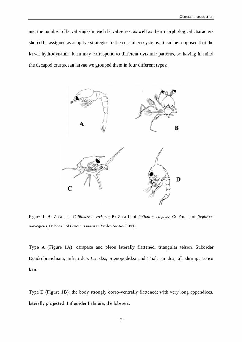
Figure 1. A: Zoea I of Callianassa tyrrhena; B: Zoea II of Palinurus elephas; C: Zoea I of Nephrops
norvegicus; D: Zoea I of Carcinus maenas. In: dos Santos (1999).
Type A (Figure 1A): carapace and pleon laterally flattened; triangular telson. Suborder
Dendrobranchiata, Infraorders Caridea, Stenopodidea and Thalassinidea, all shrimps sensu
lato.
Type B (Figure 1B): the body strongly dorso-ventrally flattened; with very long appendices,
laterally projected. Infraorder Palinura, the lobsters.

General Introduction
- 8 -
Type C (Figure 1C): with a laterally flattened but slightly rounded carapace; a cylindrical
pleon (or abdomen); a bifurcated or triangular telson. Infraorders Astacidea and Anomura, the
Norway lobster and the hermit crabs.
Type D (Figure 1D): a carapace almost spherical; usually with dorsal and lateral spines, and a
rostrum ventrally projected; cylindrical telson or as a furca. Infraorder Brachyura, the crabs.
1.4- Objectives
The aim of present thesis is to describe the horizontal and vertical distribution patterns of
decapod crustacean larvae in the Portuguese upwelling ecosystem, as well as to present the
morphological larval descriptions from laboratory cultured material of the three selected
larval forms of decapod larvae: the caridean shrimps, the hermit crabs, and the crabs. In the
working plan of present thesis five specific objectives related with the larval ecology and
morphology of decapod crustacean larvae were delineated: (1) to verify if the larval vertical
distribution in the water column is related with the day cycle variation and (2) to describe the
vertical migration rhythms of the considered larval forms (larval form and stage), (3) to
establish an average depth of distribution through the larval development and (4) to verify the
existence of ontogenetic vertical migration in the water column. The last objective is to make
the morphological description of the larval development of three selected taxa of the three
selected larval forms (a caridean shrimp, a hermit crab and a crab). To fulfil the ecological
and morphological aims of present dissertation, five different studies were conducted:
- dos Santos et al. (2008b) studied the decapod larvae and hypothesised that the
horizontal distribution patterns of several decapod species retained over the shelf were

General Introduction
- 9 -
related to their presumable settlement areas. So, in Chapter 2 our objective was to test
the hypothesis that the decapod crustacean larval distribution, in the studied coastal
upwelling ecosystem, clearly shows a retention strategy being coincident with the
adults’ distributional ranges. As a result, we also pretend to demonstrate that the
larvae concentrated very close to the shore will be the inner shelf species larvae; those
concentrated somewhere in the middle shelf, will include the shelf species larval
stages; and those that will appear over the continental shelf break, will concentrate the
shelf slope species. To verify our hypothesis we analyse the relationships between the
larval distributions of 15 selected taxa with the physical environment in the Western
Iberia upwelling ecosystem.
- dos Santos et al. (2008b) described the diel vertical migration of decapod larvae.
Knowing this, in Chapter 3 we pretend to analyse in detail the vertical distribution
patterns of the larval stages of 9 species, in order to determine their ontogenetic
vertical migration and also to describe the behavioural responses of each stage to the
environment.
- present work in Chapter 4 pretends to describe the available zoeal characters of two
closely related trans-isthmian caridean shrimp species, Lysmata moorei (Rathbun,
1901) and L. galapagensis Schmitt 1924, using laboratory cultured material. Both
descriptions are compared with the larval features previously described for the only
species in the “Cosmopolitan Clade” with their larvae known, L. seticaudata (Calado
et al. 2004), and with the Eretmocaris larval genus (Bate 1888) given the evident
resemblances between L. galapagensis larvae and E. corniger from the Tropical
Eastern Pacific.
General Introduction
- 10 -
- the hermit crabs Clibanarius aequabilis and C. erythropus are both present in the
eastern Atlantic, and present work pretends to describe and compare the complete
larval series of Clibanarius aequabilis with the previously described larval stages of
other species of the genus, in particularly with those of C. erythropus. These two
species do not co-occur in the same area, but a careful comparison was required, so
Chapter 5 also presents a redescription of C. erythropus larval morphology according
to modern standards.
- finally, the Chapter 6 describes in detail the four zoeal stages and the megalopa of the
crab I. nucleus from laboratory reared material.
1.5- Structure of this Dissertation
As the objectives list indicates, this dissertation is organized in seven chapters where the
ecological and morphological aspects of decapod crustacean larvae are studied. Chapters two
and three present the ecological results, while chapters four to six present the morphological
results. Present chapter, the “Introduction”, includes a general introduction to the study of
decapod crustacean larvae, with the state of the art, the presentation of the objectives of this
thesis, and a brief description of the study area were the research cruise was carried.
The second chapter, the “Circulation patterns and decapod larvae distribution in a coastal
upwelling ecosystem”, analyses the spatial distribution of decapod larvae off the northwest
Portuguese shelf. This chapter is in preparation for publication.

General Introduction
- 11 -
The third chapter, the “Ontogenetic vertical migration behaviour of decapod larvae in the
Portuguese upwelling ecosystem”, describes the ontogenetic vertical migration behaviour of
decapod larvae on the western Iberia upwelling ecosystem. This chapter is also in preparation
for publication.
The fourth chapter, “Shedding light over the larval genus Eretmocaris – morphological larval
features of two closely related trans-isthmian Lysmata species using laboratory cultured
material”, the morphological larval description of the zoeal stages of the caridean shrimps of
the genus Lysmata was made. This chapter was submitted to “Systematics and Biodiversity”
for publication.
The fifth chapter, “Complete larval development of the hermit crabs Clibanarius aequabilis
and Clibanarius erythropus (Decapoda: Anomura: Diogenidae), under laboratory conditions,
with a revision of the larval features of genus Clibanarius”, presents the complete
morphological larval description of the hermit crabs of the genus Clibanarius. This chapter
was published in Helgoland Marine Research.
The sixth chapter, “Complete larval development of the crab Ilia nucleus (Linnaeus, 1758)
(Decapoda, Brachyura, Leucosiidae) reared under laboratory conditions”, describes the
complete morphological larval development of the brachyuran crab of the species Ilia
nucleus. This chapter was published in Scientia Marina.
Finally in the seventh and last chapter the general discussion and conclusions are listed.

General Introduction
- 12 -
1.6- Study area
In the Portuguese coast, until the beginning of the 21st century, Queiroga (1996) and dos
Santos (1999) were the only authors studying the decapods larval distribution over the
continental shelf, so most of the studies considering the spatio-temporal distribution of
planktonic larval stages of decapod crustaceans were carried in estuaries and nearshore zones
(e.g. Flores et al. 2002, Almeida & Queiroga 2003) and generally had as objective the
brachyuran larval stages being the exceptions Paula’s works with Mira estuarine species
(Paula 1987, 1989, 1993, 1998), dos Santos description of decapods occurring in the
Portuguese coastal area (dos Santos 1999) and Pereira et al. (2000) description of the
decapods larval fluxes at Aveiro coastal lagoon.
Queiroga (1996) studied the distribution and drift of the crab larvae of Carcinus maenas over
the continental shelf off northern Portugal, demonstrating that the first zoea was clearly
associated with the estuarine inlets, the older stages were dispersed progressively offshore,
and the megalopal stage experienced an onshore transport. Sustained on these results a survey
was conducted in May 2002 in the adjacent area to the Aveiro coastal lagoon, and several
studies addressing the planktonic larval stages distribution resulted from the collected data
(the most recent: dos Santos et al. 2008b, Moreno et al. 2009, Garrido et al. 2009).
The area adjacent to Aveiro coastal lagoon, the northwest coast of Portugal, is characterised
by complex mesoscale variability and a strong seasonality (Peliz et al. 2002, Relvas et al.
2007). The Western Iberia Shelf is a seasonal upwelling system where the northerlies
(upwelling favourable) are observed in summer (June to September), developing the typical
upwelling phenomena like coastal jets and long filaments (e.g. Peliz et al., 2002). the presence

General Introduction
- 13 -
of a poleward current along the coast of northwestern Iberia, generally described as a slender
poleward flow along the upper slope/shelf break zone, carrying warm and salty waters in the
upper 200- 300 m during autumn and winter was reported (Frouin et al. 1990, Haynes &
Barton 1990). Peliz et al. (2003, 2005) showed that the Iberian Poleward Current (IPC)
circulates along mid-latitude western and northern Iberia continental margins, driven by
density forcing associated with larger-scale meridional thermal gradients, and is intensified
during the winter. A large portion of land runoff from Iberian Peninsula is directed to the
north of Lisbon shelf zone contributing to a fresh water input to the shelf, and generating a
low salinity lens termed the Western Iberia Buoyant Plume (WIBP, Peliz et al. 2002, Santos
et al. 2004). The joint effect of both these features, the Western Iberia Buoyant Plume
(WIBP) and the Iberian Poleward Current (IPC) together with the alternating downwelling
and upwelling episodes, produces extremely variable circulation patterns at short time scales
in which the plankton distributes.
1.7- References
Almeida M.J. & Queiroga H. (2003). Physical forcing of onshore transport of crab megalopae
in the northern Portuguese upwelling system. Estuarine Coastal and Shelf Science 57,
1091-1102.
Abelló P. & Guerao G. (1999) Temporal variability in the vertical and mesoscale spatial
distribution of crab megalopae (Crustacea: Decapoda) in the Northwestern
Mediterranean. Estuarine. Coastal and Shelf Science 49, 129-139.
Anger K. (2001). The Biology of Decapod Crustacean Larvae. Crustacean Issues 14, 1- 419.
Bate C.S. (1888). Crustacea Macrura. Challenger Reports, Zool., XXIV.

General Introduction
- 14 -
Botsford L.W. (1986). Effects of environmental forcing on age-structured populations.
Northern California Dungeness crab (Cancer magister) as an example. Canadian
Journal of Fisheries and Aquatic Sciences 43, 2345–2352.
Botsford L.W., Moloney C.L., Hastings A., Largier J.L., Powell T.M., Higgins K., Quinn J.F.
(1994) The influence of spatially and temporally varying oceanographic conditions on
meroplanktonic metapopulations. Deep-Sea Research II 41, 107-145.
Calado R., Bartilotti C., Narciso L., dos Santos A. (2004). Redescription of the larval stages
of Lysmata seticaudata (Risso, 1816) (Crustacea, Decapoda, Hippolytidae) reared under
laboratory conditions. Jopurnal of Plankton Research 26, 737-752.
Christy J.H. & Morgan S.G. (1998) Estuarine immigration by crab postlarvae: mechanisms,
reliability and adaptive significance. Marine Ecology Progress Series 174, 51-65.
dos Santos A. (1998). On the occurrence of larvae of Parapenaeus longirostris (Crustacea:
Decapoda: Penaeoidea) off the Portuguese coast. Journal of Natural History 32, 1519–
1523
dos Santos A. (1999) Larvas de Crustáceos Decápodes ao Largo da Costa Portuguesa. Tese de
Doutoramento, Universidade de Lisboa, Lisboa, Portugal.
dos Santos A., Santos, A. M. P. & Conway, D. V. P. (2007) Horizontal and vertical
distribution of cirripede cyprid larvae in an upwelling system off the Portuguese coast.
Marine Ecology Progress Series 3269, 145-155.
dos Santos A., Calado R., Araújo R. (2008a) First record of the genus Periclimenaeus
Borradaile, 1815 (Decapoda: Palaemoniidae: Pontoniinae) in the northeastern Atlantic,
with the description of a new species, Periclimenaeus aurae. Journal of Crustacean
Biology 28, 156-166.
dos Santos A., Santos A.M.P., Conway D.V.P., Bartilotti C., Lourenço P., Queiroga H.
(2008b) Diel vertical migration of decapod larvae in the portuguese coastal upwelling

General Introduction
- 15 -
ecosystem: implications for offshore transport. Marine Ecology Progress Series 359,
171-183.
Flores A.A.V., Cruz J., Paula J. (2002). Temporal and spatial patterns of settlement of
brachyuran crab megalopae at a rocky coast in Central Portugal. Marine Ecology
Progress Series 229, 207-220.
Forward R.B.Jr., Swanson J., Tankersley R.A., Welch J.M. (1997). Endogenous swimming
rhythms of blue crab, Callinectes sapidus, megalopae: effects of offshore and estuarine
cues. Marine Biology 127, 621-628.
Frouin R., Fiúza A.F.G., Ambar I., Boyd T.J. (1990). Observations of a Poleward Surface
Current off the Coasts of Portugal and Spain During Winter. Jurnal of Geophysical
Research 95, 679-691.
Garrido S., Santos A.M.P., Ré P. (2009). Spatial distribution and vertical migration of fish
larvae communities off Northwestern Iberia: comparison of LHPR and Bongo nets.
Estuarine Coastal and Shelf Science 84, 463-475.
Haynes R. & Barton E.D. (1990). A poleward current along the Atlantic coast of the Iberian
Peninsula. Journal of Geophysical Research 95, 11425-11441.
Jamieson G.S & Phillips A.C. (1988). Occurrence of Cancer crab (C. magister and C.
oregonensis) megalopae off the west coast of Vancouver Island, British Columbia.
Fishery Bulletin 86, 525-542.
Mace A.J. & Morgan S.G. (2006). Larval accumulation in the lee of a small headland:
implications for the design of marine reserves. Marine Ecology Progress Series 318, 19-
29.
Marta-Almeida M., Dubert J., Peliz A., Queiroga H. (2006) Influence of vertical migration
pattern on retention of crab larvae in a seasonal upwelling system. Marine Ecology
Progress Series 307, 1-19.

General Introduction
- 16 -
McConaugha J.R. (1992). Decapod Larvae: Dispersal, Mortality, and Ecology. A Working
Hypothesis. Amererican Zoologist 32, 512-523.
Moreno A., dos Santos A., Piatkowski U., Santos A.M.P., Cabral H. (2009). Distribution of
cephalopod paralarvae in relation to the regional oceanography of the western Iberia.
Journal of Plankton Research 31, 73-91.
Morgan S.G., Fisher J.L., Mace1 A.J., Akins L., Slaughter A.M., Bollens S.M. (2009). Cross-
shelf distributions and recruitment of crab postlarvae in a region of strong upwelling.
Marine Ecology Progress Series 380, 173-185.
Oishi K. & Saigusa M. (1997). Night time emergence patterns of planktonic and benthic
crustaceans in a shallow subtidal environment. Journal of Oceanography 53, 611-621.
Paula J. (1987). Seasonal distribution of Crustacea Decapoda larvae in S. Torpes bay, South-
western Portugal. Investigación Pesquera 51 (Supl. 1), 267-275.
Paula J. (1989). Rhythms of larval release of decapod crustaceans in the Mira Estuary,
Portugal. Marine Biology 100, 309-312.
Paula J. (1993). Ecologia da Fase Larvar e Recrutamento de Crustáceos Decápodes do
Estuário do Rio Mira. PhD thesis, University of Lisbon, Lisbon, Portugal.
Paula J. (1998). Larval retention and dynamics of the prawns Palaemon longirostris H. Milne
Edwards and Crangon crangon Linnaeus (Decapoda, Caridea) in the Mira estuary,
Portugal. Invertebrate Reproduction and Development 33, 221-228.
Peliz A., Rosa T., Santos A.M.P., Pissarra J. (2002). Fronts, jets and counter flows in the
Western Iberia upwelling system. Journal of Marine Systems 35, 61-77.
Peliz A., Dubert J., Haidvogel D.B., Le Cann B. (2003). Generation and unstable evolution of
a density-driven Eastern Poleward Current: the Iberia poleward current. Journal of
Geophysical Research 108 (C8), 3268 (doi: 10.1029/2002 JC 001443).

General Introduction
- 17 -
Peliz A., Dubert J., Santos A.M.P., Oliveira P.B., Le Cann B. (2005). Winter upper ocean
circulation in the Western Iberian Basin- Fronts, Eddies and Poleward Flows: an
overview. Deep-Sea Research I 52, 621-646.
Peliz A., Marchesiello P., Dubert J., Marta-Almeida M., Roy C., Queiroga H. (2007) A study
of crab larvae dispersal on the western Iberian shelf: physical processes. Journal of
Marine Systems 68, 215–236.
Pereira F., Pereira R., Queiroga H. (2000). Flux of decapod larvae and juveniles at a station in
the lower Canal de Mira (Ria de Aveiro, Portugal) during one lunar month. Invertebrate
Reproduction and Development 38, 183-206.
Pineda J., Hare J.A., Sponaugle S. (2007) Larval transport and dispersal in the coastal ocean
and consequences for population connectivity. Oceanography 20, 22-39.
Pittman S.J. & McAlpine C.A. (2001). Movements of marine fish and decapod Crustaceans:
process, theory and application. Advances in Marine Biology 44, 205-294.
Queiroga,H. (1996) Distribution and drift of the crab Carcinus maenas (L.) (Decapoda,
Portunidae) larvae over the continental shelf of northern Portugal in April 1991. J.
Plank. Res., 18 (11), 1981-2000.
Queiroga H. (1996). Distribution and drift of the crab Carcinus maenas (L.) (Decapoda,
Portunidae) larvae over the continental shelf off northern Portugal in April 1991. Journal
of Plankton Research 18, 1981–2000.
Queiroga H. (1998). Vertical migration and selective tidal stream transport in the megalopa of
the crab Carcinus maenas. Hydrobiologia, 375/376, 137-149.
Queiroga H., Costlow J.D., Moreira M.H. (1994) Larval abundance patterns of Carcinus
maenas (Decapoda, Brachyura) in Canal de Mira (Ria de Aveiro, Portugal). Marine
Ecology Progress Series 111, 63-72.

General Introduction
- 18 -
Queiroga H. & Blanton J. (2005). Interactions between behaviour and physical forcing in the
control of horizontal transport of decapod crustacean larvae. Advances in Marine
Biology 47, 107-214.
Relvas P., Barton E.D., Dubert J., Oliveira P.B., Peliz A., da Silva J.C.B., Santos A.M.P.
(2007). Physical oceanography of the western Iberia ecosystem: latest views and
challenges. Progress in Oceanography 74, 149-173.
Roughgarden J., Gaines S., Possingham H. (1988). Recruitment dynamics in complex life
cycles. Science 241, 1460-1466.
Santos A.M.P., Peliz A., Dubert J., Oliveira P.B., Angélico M.M., Ré P. (2004). Impact of a
winter upwelling event on the distribution and transport of sardine (Sardina pilchardus)
eggs and larvae off western Iberia: a retention mechanisms. Continental Shelf Research
24, 149-165.
Shanks A.L. (1985). The behavioral basis of internal wave induced shoreward transport of the
megalopae of Pachygrapsus crassipes. Marine Ecology Progress Series 24, 289-295.
Thorson G. (1964). Light as an ecological factor in the dispersal and settlement of larvae of
marine bottom invertebrates. Ophelia 1, 167-208.
Williamson D.I. (1969). Names of larvae in the Decapoda and Euphausiacea. Crustaceana,
16, 210-213.

CHAPTER 2
__________________________________________________
Circulation patterns and decapod larvae distribution
in a coastal upwelling ecosystem*
*Bartilotti C., Castro M., Santos A. M. P., Peliz Á., Queiroga H., dos Santos A. (in prep.)
Circulation patterns and decapod larvae distribution in a coastal upwelling ecosystem.

Circulation patterns and decapod larvae distribution
- 20 -
2.1- Abstract
Spatial distribution of decapod larvae off the northwest Portuguese shelf was analysed. Two
grid samplings were performed: a first one to characterise the hydrology and circulation of the
area and a second one addressing physical and biological parameters. Decapod larvae were
associated with upwelled waters and with a deeper intrusion of warmer offshore waters and
were separated in three groups. The “Inner Shelf Species” larvae, Diogenes pugilator and
Necora puber, were concentrated near the shore along a band 8 km off the coastline. The
“Shelf Species” larvae, Eualus occultus, Processa nouveli, Pandalina brevirostris,
Philocheras bispinosus, Callianassa subterranea, Upogebia pusilla, Anapagurus spp.,
Atelecyclus rotundatus, Liocarcinus spp. and Polybius henslowii , were concentrated along
meridionally elongated patches parallel to the coast inshore of the 200 m isobath 8- to 40 km
far from the coastline. Finally the “Slope Species” larvae, Solenocera membranacea,
Parthenope spp. and Goneplax rhomboides were concentrated along the 200 m isobath, in the
vicinity of the shelf break, 56 km far from the coastline. These results confirm previous
findings, that decapod larvae are retained in the Portuguese continental shelf and the self
recruitment for parental populations is the most probable scenario even under upwelling
conditions. The decapod larval distribution was modelled in a two steps approach, having 4
explanatory variables. The Inner Shelf species always had their larval distributions positively
related with the distance to the coast and with the temperature. The Shelf species due to their
wide distribution over the shelf showed more variable responses that were in accordance with
the distribution ranges of the adults. The Slope species distributions were negatively related
with distance to the coast and with the temperature, and never were affected by the Western
Iberia Buoyant Plume (WIBP).

Circulation patterns and decapod larvae distribution
- 21 -
2.2- Introduction
Many marine organisms have complex life cycles with a planktonic larval phase. For most of
these the larval phase is the dominant dispersal stage (e.g. Thorson 1950). It has been
assumed that during this phase, the larval stages were transported to great distances and were
widely dispersed (e.g. Roughgarden et al. 1988). As a consequence, much of the past studies
considering larval dispersal supposed that the coastal benthic populations were open and
highly “connected” through larval transport (e.g. Swearer et al. 2002). The idea of
mechanisms leading to closed populations, in which “self-seeding” occurs at local spatial
scales is quite recent (e.g. Swearer et al. 2002, Levin 2006).
The population connectivity is a bio-physical phenomenon since it results of the combination
of the physical transport and dispersion, with the biological processes such as the larval
behaviour (e.g. Gawarkiewicz et al. 2007, Cowen & Sponaugle 2009). Decapod crustacean
larvae are active swimmers that can dynamically modify their vertical position in the water
column, controlling the extent range and direction of their dispersal (Queiroga & Blanton
2005). The understanding of temporal and spatial larval movement patterns is fundamental to
study decapods ecology leading to the design of effective conservation and resource
management strategies (e.g. Pittman & McAlpine 2001, Mace & Morgan 2006).
It is known that the transport processes are the critical components of the supply mechanisms
of larvae to habitats for settlement and juvenile development (e.g. Queiroga et al. 2006,
2007), separating the larval development in time (development through stages) and in space
(transport in a three-dimensional fluid habitat), exposing the larvae to different mortality
factors (Queiroga & Blanton 2005). To completely understand the larval transport and

Circulation patterns and decapod larvae distribution
- 22 -
settlement locations it is fundamental to have a good description of the local hidrography and
of the three-dimensional current fields, as well as to describe the growth, survival and
behavioural responses of larvae to their environment (e.g. Botsford et al. 1994). The study
area, in front of Aveiro coastal lagoon, northwest coast of Portugal, is characterised by
complex mesoscale variability and a strong seasonality (Peliz et al. 2002, Relvas et al. 2007).
Frouin et al. (1990) and Haynes & Barton (1990) reported the presence of a poleward current
along the coast of northwestern Iberia, generally described as a slender poleward flow along
the upper slope/shelf break zone, carrying warm and salty waters in the upper 200- 300 m
during autumn and winter. Peliz et al. (2003, 2005) showed that the Iberian Poleward Current
(IPC) circulates along mid-latitude western and northern Iberia continental margins, driven by
density forcing associated with larger-scale meridional thermal gradients, and is intensified
during winter. The river runoff is significant to the northern region of Lisbon contributing to a
fresh water input to the shelf, generating a low salinity lens named Western Iberia Buoyant
Plume (WIBP) (Peliz et al. 2002, Santos et al. 2004).
Dos Santos et al. (2008) studied the decapod larvae three-dimensional distribution and
discussed their results with the previous modelling studies for the area (Marta-Almeida et al.
2006, Peliz et al. 2007), confirming the assumptions made by both models that larval
dispersal in the inner and middle shelf is mainly alongshore and that their retention greatly
depends on their diel vertical migration behaviour. Furthermore, they hypothesised that the
horizontal distribution patterns of several decapod species retained over the shelf were related
to their presumable settlement areas. The apparent retention of decapod larvae close to
parental populations was recently discussed by Morgan et al. (2009) and is now almost an
evidence for invertebrate larvae.

Circulation patterns and decapod larvae distribution
- 23 -
We pretend to test the hypothesis that all decapod crustacean larvae distribution, in the
studied coastal upwelling ecosystem, clearly shows a retention strategy in accordance with the
adults’ distributional ranges. We believe that the larvae will be distributed along meridional
patches of larvae according to their origins: one, very close to the shore, will include the inner
shelf species larvae; another, somewhere in the middle shelf, will concentrate the shelf species
larval stages; and a last one, will appear over the continental shelf break, and will concentrate
the shelf slope species. To verify our hypothesis we analysed the relationships between the
larval distributions of 15 selected taxa with the physical environment in the Western Iberia
upwelling ecosystem.
2.3- Material and Methods
2.3.1- Field study
An oceanographic survey was carried out off the northwest coast of Portugal, aboard the RV
‘Noruega’, from 9-22 May 2002 (Fig. 1).
In the beginning of the survey a mooring array with 3 current meters (Aanderaa RCM 9) and
2 conductivity-temperature-depth (CTD) recorders (Sea-Bird Electronics MicroCats) was
deployed on the Portuguese continental shelf (40º 45.9’ N 08º 59.0’ W), approximately 21 km
offshore and at 60 m bottom depth (Fig. 1). In order to obtain a quasi-synoptic view of the
mesoscale circulation patterns before the biological sampling, a first grid of CTD stations was
also carried out, along 4 transects perpendicular to the coast from 11 to 14 May 2002 (Fig. 1).
Temperature, salinity and chlorophyll a (chl a) concentration were registered in each station
with a CTD SBE 9plus (S/N 19860-0530) with a Seapoint fluorometer attached.

Circulation patterns and decapod larvae distribution
- 24 -
Figure 1- Map of the northwest coast of Portugal showing sampling positions (CTD: conductivity, temperature
and depth profiler; and LHPR: Longhurst Hardy plankton recorder) collected aboard the RV Noruega, during the
ProRecruit project survey. Transects are identified as T1, T2, T3 and T4; the squares represent the stations
sampled with the Pro-LHPR. The 30, 100 and 200 m bathymetric lines are also presented.
From 15-17 May, in a total of 60 h, along the same 4 transects perpendicular to the coast
(Fig.1), the plankton sampling was carried out in a 16 stations grid, with a Pro-LHPR system,
a commercially updated version of the Longhurst Hardy Plankton Recorder (Williams et al.
1983). Each transect (T1 to T4) had 4 stations, separated by approximately 16 km, being the
first station in each transect 8 km far from the shore (Fig. 1). Transect T1 was the most
northern, and T4 the most southern located one. The Pro-LHPR net had a mouth aperture of
0.42 m, and collected plankton between two rolls of filtering gauze (280 µm mesh size) that
are advanced at intervals inside a cod end box to give a series of consecutive samples. It was
towed at 3-4 knots on oblique hauls from the surface to a maximum of 200 m depth. Stratified
samples of Pro-LHPR were collected at 5 m depth intervals in the first 25 m, at 10 m depth
T1
T2
T3
T4

Circulation patterns and decapod larvae distribution
- 25 -
intervals from 25 m deep until 65 m, and for the deeper stations points, at 20 m depth
intervals from 65 m until near the bottom.
2.3.2- Sample processing
Plankton samples were preserved in 4% borax-buffered formaldehyde, prepared with
seawater. Samples were sorted for decapod larvae, that were identified to species level and
developmental stage whenever possible using dos Santos & Lindley (2001), González-
Gordillo et al. (2001), and dos Santos & González-Gordillo (2004). Data were standardized
for ind.m-3.
2.3.3- Analysis of decapod larval distribution in relation to the physical environment
The 15 studied taxa were selected having as criterions, the most abundant in the samples and,
whenever possible, the presence of the complete larval series; within the most abundant we
chose species belonging to different decapod groups, Dendrobranchiata shrimps, caridean
shrimps, scavengers, hermit crabs and crabs and the distributional range of the adults,
inhabitants of Inner Shelf, Shelf and Slope, according to Zariquiey-Álvarez (1968) and
d’Udekem d’Acoz (1999) (Table 1).
Diogenes pugilator and Necora puber were considered as Inner Shelf species since both are
coastal species inhabiting the intertidal area when adults. According to Zariquiey-Álvarez
(1968) and d’Udekem d’Acoz (1999) the first species is distributed up to 35 to 40 m depth
and the second has their higher abundances up to 15 m depth. Most of the selected taxa were
considered Shelf species. When adults, Eualus occultus is distributed to a maximum of 150
m, Processa nouveli is distributed between 10 and 330 m, Pandalina brevirostris up to 180 m
and Philocheras bispinosus up to 130 m. Callianassa subterranea is usually present between
35 and 500 m, and Upogebia spp. is distributed to a maximum of 65 m. Most of the

Circulation patterns and decapod larvae distribution
- 26 -
Anapagurus spp. species in the Portuguese west coast are distributed to a maximum of 200 m
depth, Atelecyclus rotundatus is distributed between 9 and 100 m depth, Liocarcinus spp. is
normally distributed in the 50 to 100 m depth band but can reach the upper slope, and finally,
Polybius henslowii a pelagic crab found from 2 to 518 m (Zariquiey-Álvarez 1968, d’Udekem
d’Acoz 1999). The Slope species included Solenocera membranacea, Parthenope spp. and
Goneplax rhomboides, species that inhabit the continental shelf but also the continental shelf
break or slope, and are common beyond the 400 m depths (Zariquiey-Álvarez 1968,
d’Udekem d’Acoz 1999).
Table 1- List of the selected taxa, respective decapod crustacean group, distribution range when adult, and the
grouping of the larval stages.
Taxa Decapod group Distribution range Larval group
ZN ZO M
Solenocera membranacea Shrimps Slope Z II- III Z IV- VI -
Eualus occultus Caridean shrimps Shelf Z I- IV Z V- IX M
Processa nouveli Caridean shrimps Shelf Z I- IV Z V- IX M
Pandalina brevirostris Caridean shrimps Shelf Z I- III Z IV- VII M
Philocheras bispinosus Caridean shrimps Shelf Z I- II Z III- V M
Callianassa subterranea Scavengers Shelf Z I- II Z III- IV -
Upogebia spp. Scavengers Shelf Z I- II Z III- IV -
Diogenes pugilator Hermit crabs Inner Shelf Z I- II Z III- IV M
Anapagurus spp. Hermit crabs Shelf Z I- II Z III- IV M
Parthenope spp. Crabs Slope Z I- II Z III- IV -
Atelecyclus rotundatus Crabs Shelf Z I- II Z III- V M
Liocarcinus spp. Crabs Shelf Z I- II Z III- V M
Necora puber Crabs Inner Shelf Z I- II Z III- IV M
Polybius henslowii Crabs Shelf Z I- II Z III- V M
Goneplax rhomboides Crabs Slope Z I- II Z III- V M
Zoea (Z) and Megalopa (M); New (N) and Old (O).

Circulation patterns and decapod larvae distribution
- 27 -
The larval stages were grouped in accordance with their ontogeny for each taxa, in order to
reduce the zero values, improving the statistics. We considered three groups, the early zoeal
stages or zoeae new (ZN), the later zoeal stages or zoeae old (ZO) and the megalopa or
decapodid (M). Table 1 presents the chosen taxa, respective decapod crustacean group and
distributional range of the adults, as well as the larval stages that were grouped.
Two response variables were considered: abundance (ind.m-3) was log transformed, and
presence/ absence (1/ 0) of a given taxa and larval group.
With respect to the explanatory variables, in a first phase of the exploratory data analysis, all
environmental variables that were available and potentially useful to explain presence and/or
abundance of larvae were investigated. They were:
BOTTOM – depth of the station, continuous variable, in meters;
DEPTH_CAT – Upper limit of the depth stratum, discrete variable, in meters;
MAX_TOW_DEPTH – Maximum depth towed in the station, continuous variable, in
meters;
MID_DEPTH – Midpoint of depth stratum, discrete variable, in meters;
DIST_COAST - distance to the coast, continuous variable, in km;
REAL_TIME – Hour of the day, continuous variable, 0 to 24;
HOUR1 – night and day, categorical variables with two levels, night (18:00-05:59)
and day (06:00-17:59);
HOUR2 – night, dawn, day and dusk, categorical variables with four levels, night
(20:00-05:59), dawn (06:00-07:59), day (08:00-17:59) and dusk (18:00-19:59);
Circulation patterns and decapod larvae distribution
- 28 -
HOUR_WAVE - hour of the day, continuous variable, defined as a circular function
of the hour of the day, sunrise=0, noon=1, sunset=0 and midnight=-1, generated by the
function:
ݏ ݎݑܪ)] ×ߨ224) +
ߨ32 ]
where “Hour” is the hour of the day (0-24);
WIBP - Western Iberia Buoyant Plume, binomial variable, representing the presence
of a less saline continental water mass lens with salinities less than 35.75 (Peliz et al.
2002 for details);
ISWM – Intermediate Salinity Water Mass, binomial variable, representing the
presence of a water mass with salinity within the range [35.75,35.90[;
IPC – Iberian Poleward Current, representing the presence of a high salinity water
mass, with salinity greater than 35.9 (Peliz et al. 2003 for details);
SAL – Salinity, continuous variable, no units;
TEMP - temperature, continuous variable, in ºC.
Most of these variables were not significant in any of the studied cases (species and larval
group) and were dropped from the models. The variables remaining and used in the
construction of a reduced model were: the distance to the coast (DIST_COAST), the hour of
the day (HOUR_WAVE), the temperature (TEMP) and the Western Iberia Buoyant Plume
(WIBP).
The decapod larval distribution, in relation to environmental parameters, was analysed in two
steps, for each species and larval group separately. The two steps approach is used to deal
with the problem of zero inflated data (see Wenger & Freeman 2008). In the first step, the
presence or absence was predicted using a Logistic Model (LM) with the following

Circulation patterns and decapod larvae distribution
- 29 -
explanatory variables: distance to the coast (DIST_COAST), hour of the day
(HOUR_WAVE), temperature (TEMP) and Western Iberia Buoyant Plume (WIBP). The
results from this model were used in the second step for situations where at least one of the
variables was significant and for data points where “presence” was predicted. For these data
points, measured values of abundance were modelled with a Generalized Addictive Model
(GAM), using linear predictors for the distance to the coast (DIST_COAST), the hour of the
day (HOUR_WAVE), the temperature (TEMP) and the Western Iberia Buoyant Plume
(WIBP) and a spline for temperature (TEMP, with 2 degrees of freedom). Statistical analysis
was made using SAS 9.2 (SAS Institute, 2008) using PROC LOGIT for fitting the logistic
model (stepwise option) and PROC GAM for the generalized additive models.
2.4- Results
2.4.1- Oceanographic conditions
For the complete and detailed description of the oceanographic conditions during the survey
see dos Santos et al. (2007). Before the plankton sampling the study area was under coastal
upwelling favourable winds (northerlies in this region). As a consequence, the Western Iberia
Buoyant Plume (WIBP), present in the first 25-30 m depth as a fresher water lens with
salinities inferior to 35.75 (Fig. 2), had a considerably offshore displacement. However during
the plankton sampling there was a relaxation of the upwelling due to the reversal of the wind
direction. This change leads to coastal convergence conditions and to the appearance of a
poleward current over the shelf.
Furthermore, the Iberian Poleward Current (IPC) was represented by the slope waters (Fig. 3)
with salinities larger than 35.90 that invaded the shelf (Fig. 2). The appearance of a thermal

Circulation patterns and decapod larvae distribution
- 30 -
front separating the two northern transects (T1 and T2) from the two southern ones (T3 and
T4) was also observed during the plankton sampling (see Fig. 2 representing the horizontal
distribution of salinity).
Figure 2- Horizontal (at 20 m depth) and vertical distributions of salinity measured with a CTD. T1, T2, T3 and
T4 represent the four sampled transects (respectively 40.9º N, 40.7º N, 40.5º N and 40.3ºN). The 30, 100 and 200
m bathymetric lines are also identified. The horizontal and vertical distributions of salinity are represented by the
solid lines with respective labels.
-9.4 -9.2 -9 -8.8 -8.6
40.2
40.4
40.6
40.8
41
41.2
41.4
200 m 100 m 30 m
Latit
ude
(°N)
Longitude (°W)
35.75
35.45
35.55
35.65
35.75
35.85
60 40 20 0
Distance from coast (km)
140
120
100
80
60
40
20
0
Dep
th (m
)
T1
60 40 20 0
Distance from coast (km)
T2
60 40 20 0
Distance from coast (km)
T3
35.75
60 40 20 0
Distance from coast (km)
T4

Circulation patterns and decapod larvae distribution
- 31 -
Figure 3- Horizontal (at 20 m depth) and vertical distributions of temperature (º C) measured with a CTD. T1,
T2, T3 and T4 represent the four sampled transects (respectively 40.9º N, 40.7º N, 40.5º N and 40.3ºN). The 30,
100 and 200 m bathymetric lines are also identified. The horizontal and vertical distributions of temperature are
represented by the grey areas with respective scale.
Surface salinity varied between 35.44 and 35.81 (Fig. 2). In the water column the minimum
registered was 35.43 in the first transect (Fig. 2, T1), and the maximum registered was 35.96
in the fourth transect (Fig. 2, T4). The deeper intrusion of the more saline waters of the
Iberian Poleward Current (IPC) is clear in the fourth transect (Fig. 2, T4) in the more deep
sampled stations. Surface temperature varied between 14.08 and 16.26 ºC (Fig. 3) and
-9.4 -9.2 -9 -8.8 -8.6
40.2
40.4
40.6
40.8
41
41.2
41.4
200 m 100 m 30 m
Latit
ude
(°N)
Longitude (°W)
13.5
13.7
13.9
14.1
14.3
14.5
14.7
14.9
15.1
60 40 20 0
Distance from coast (km)
140
120
100
80
60
40
20
0
Dep
th (m
)
T1
60 40 20 0
Distance from coast (km)
T2
60 40 20 0
Distance from coast (km)
T3
60 40 20 0
Distance from coast (km)
T4

Circulation patterns and decapod larvae distribution
- 32 -
vertically the minimum registered was 12.63 ºC in the first transect (Fig. 3, T1) and the
maximum registered was 16.29 ºC in the fourth transect (Fig. 3, T4).
2.4.2- Decapod larvae distribution
The zoeal stages were more abundant than the megalopae, and were more concentrated over
the middle shelf (dos Santos et al. 2008). The horizontal and vertical larval distributions of the
15 selected taxa were analysed in detail. The selected planktonic larval stages were
concentrated, as expected, in three different horizontal layers of the sampled area. Most of the
species had their larvae concentrated along the middle shelf (Eualus occultus, Processa
nouveli, Pandalina brevirostris, Philocheras bispinosus, Callianassa subterranea, Upogebia
spp., Anapagurus spp., Atelecyclus rotundatus, Liocarcinus spp., Polybius henslowii),
although there were also species with their higher abundances at the most exterior stations,
and at the most coastal stations.
The two steps approach used for the decapod larval distribution modelling in relation to the
environmental variables (Table 2) showed that the distance to coast (DIST_COAST) was the
variable that explained the larval distribution of the majority of the studied larval stages,
being negatively related with most of the zoeal presences of the Slope species, and positively
related with the zoeal presences of the Inner Shelf species.
The Western Iberia Buoyant Plume (WIBP) was positively related with the presences of the
early zoeal stages (ZN) and negatively related when explaining the presence of the megalopae
(M) of some Shelf species.

Circulation patterns and decapod larvae distribution
- 33 -
Table 2- Coefficients of significant explanatory variables of the two steps model (presence or absence predicted
using a Logistic Model in the first step, values of abundance modelled with a Generalized Addictive Model in
the second step) describing the distribution of the decapod larvae selected taxa collected with the LHPR net in
the horizontal grid of stations. Levels of significance are represented as ***p<0.001, **p<0.01, *p<0.05; n.s. not
significant.
Continued
nSolenocera Zoea New presence 148 n.s. 0.641 * -0.041 ** -1.601 *** -membranacea abundance 34 n.s. 0.130 * n.s. n.s. n.s.
Zoea Old presence 148 n.s. n.s. -0.051 ** n.s. -abundance - - - - - -
Eualus Zoea New presence 148 0.394 * n.s. n.s. -1.290 *** -occultus abundance 20 n.s. n.s. n.s. n.s. n.s.
Zoea Old presence 148 n.s. n.s. n.s. -0.837 ** -abundance 34 n.s. n.s. n.s. n.s. n.s.
Megalopa presence 148 n.s. n.s. 0.102 ** 1.717 ** -abundance - - - - - -
Processa Zoea New presence 148 0.646 ** 0.823 ** n.s. n.s. -nouveli abundance - - - - - -
Zoea Old presence 148 0.564 ** 1.535 *** n.s. n.s. -abundance 17 n.s. 0.621 ** 0.008 * -0.025 * n.s.
Megalopa presence 148 n.s. n.s. n.s. n.s. -abundance - - - - - -
Pandalina Zoea New presence 148 0.573 * n.s. n.s. n.s. -brevirostris abundance - - - - - -
Zoea Old presence 148 n.s. n.s. n.s. -1.110 *** -abundance 41 0.348 ** n.s. n.s. n.s. n.s.
Megalopa presence 148 n.s. -2.253 ** n.s. 1.379 ** -abundance - - - - - -
Philocheras Zoea New presence 148 n.s. n.s. n.s. n.s. -bispinosus abundance - - - - - -
-Zoea Old presence 148 n.s. n.s. n.s. -1.493 *** -
abundance - - - - - -
Megalopa presence 148 n.s. -1.931 * 0.110 *** n.s. -abundance 12 n.s. 4.497 * n.s. -0.024 * n.s.
Callianassa Zoea New presence 148 0.850 *** n.s. 0.033 * 1.602 *** -subterranea abundance 45 n.s. n.s. n.s. n.s. n.s.
Zoea Old presence 148 n.s. n.s. n.s. n.s. -abundance - - - - - -
Upogebia spp. Zoea New presence 148 n.s. n.s. n.s. n.s. -abundance - - - - - -
Zoea Old presence 148 -0.740 * n.s. n.s. n.s. -abundance - - - - - -
Cofficients of significant explanatory variablesSpecies Larval group Model phases
WIBP HOUR_WAVE TEMP (spline)TEMP (linear) DIST_COAST

Circulation patterns and decapod larvae distribution
- 34 -
Table 2 Continued
nDiogenes Zoea New presence 148 n.s. n.s. 0.289 *** n.s. -pugilator abundance 18 n.s. n.s. n.s. 0.044 * n.s.
Zoea Old presence 148 n.s. n.s. 0.069 ** 1.517 ** -abundance - - - - - -
Megalopa presence 148 n.s. n.s. n.s. n.s. -abundance - - - - - -
Anapagurus spp. Zoea New presence 148 n.s. -1.466 * 0.057 ** n.s. -abundance - - - - - -
Zoea Old presence 148 n.s. n.s. n.s. -0.627 * -abundance 19 n.s. n.s. n.s. n.s. n.s.
Megalopa presence 148 n.s. -1.466 * 0.057 ** n.s. -abundance 12 n.s. 4.001 ** n.s. -0.017 ** 0.007
Parthenope sp. Zoea New presence 148 n.s. n.s. -0.137 * n.s. -abundance - - - - - -
Zoea Old presence 148 n.s. n.s. n.s. n.s. -abundance - - - - - -
Atelecyclus Zoea New presence 148 n.s. -1.064 * n.s. n.s. -rotundatus abundance - - - - - -
Zoea Old presence 148 n.s. 0.829 *** n.s. -0.857 ** -abundance 85 n.s. n.s. -0.0039 * n.s. n.s.
Megalopa presence 148 -0.781 *** n.s. -0.049 *** -1.557 *** -abundance 36 n.s. n.s. n.s. n.s. 0.044
Liocarcinus spp. Zoea New presence 148 n.s. n.s. n.s. n.s. -abundance - - - - - -
Zoea Old presence 148 n.s. n.s. n.s. n.s. -abundance - - - - - -
Megalopa presence 148 -0.517 ** -0.560 * n.s. n.s. -abundance 25 n.s. 0.604 * n.s. n.s. n.s.
Necora Zoea New presence 148 n.s. n.s. 0.079 *** 1.287 * -puber abundance - - - - - -
Zoea Old presence 148 n.s. n.s. n.s. 1.470 ** -abundance - - - - - -
Megalopa presence 148 n.s. n.s. n.s. n.s. -abundance - - - - - -
Polybius Zoea New presence 148 n.s. -1.017 *** 0.025 * n.s. -henslowii abundance 12 n.s. n.s. n.s. n.s. n.s.
Zoea Old presence 148 n.s. n.s. -0.050 *** n.s. -abundance 126 n.s. n.s. 0.008 * n.s. n.s.
Megalopa presence 148 -0.591 ** -1.149 *** -0.072 *** n.s. -abundance 68 -0.086 ** -0.070 *** n.s. n.s. n.s.
Goneplax Zoea New presence 148 n.s. n.s. n.s. n.s. -rhomboides abundance - - - - - -
Zoea Old presence 148 n.s. n.s. -0.065 *** -1.536 *** -abundance 22 n.s. 0.191 * n.s. n.s. n.s.
Megalopa presence 148 n.s. -1.154 * n.s. -3.505 *** -abundance - - - - - -
Species Larval group Model phasesCofficients of significant explanatory variables
WIBP HOUR_WAVE DIST_COAST TEMP (linear) TEMP (spline)

Circulation patterns and decapod larvae distribution
- 35 -
The temperature (TEMP) was also an important factor for the presence of the Shelf species,
and it was always negatively related with the larval stages distributions (ZN, ZO and M)
except for P. brevirostris megalopae (M) and C. subterranea early zoeal stages (ZN). The
Slope species, except Parthenope sp. that did not showed any relation with this predictor
(n.s.), also had their presence negatively related with the temperature; the Inner Shelf species
zoeal stages presences were positively related with this predictor.
Finally, the effect of the hour of the day (HOUR_WAVE) reflected the vertical migration
behaviour previously described by dos Santos et al. (2008). This effect was significantly
related with S. membranacea (ZN, presence and abundance), P. nouveli (ZN presence, ZO
presence and abundance), P. brevirostris (M, presence), P. bispinosus (M, presence and
abundance), Anapagurrus spp. (ZN, presence and M, presence and abundance), A. rotundatus
(ZN and ZO, presences), Liocarcinus spp. (M, presence and abundance), P. henslowii (ZN
presence and M, presence and abundance), and G. rhomboides (ZO, abundance and M,
presence) larval distributions.
2.4.2.1- Horizontal and vertical distributions of the Inner Shelf species
The modelling results (Table 2) showed that both species early zoeal stages presences (ZN)
and D. pugilator old zoea (ZO) were significantly and positively related with the distance to
the coast (DIST_COAST). The temperature (TEMP) was always positively related with both
species zoeal stages distribution, the early zoeae (ZN) abundance and old zoeae presence
(ZO) of D. pugilator and the presence of Necora puber zoeal stages (ZN and ZO). The
megalopa (M) distribution of both species was not significantly explained by any of the
selected predictors.

Circulation patterns and decapod larvae distribution
- 36 -
Figure 4- Horizontal distributions of Diogenes pugilator and Necora puber. The 30, 100 and 200 m bathymetric
lines are identified. The abundances are represented in Ln (X+1) with X in ind.10 m-3. ZN corresponds to the
early zoeal stages, ZO to later zoeal stages, and M to megalopa.
Horizontally (Fig. 4), all the larval groups (ZN, ZO and M) occurred with higher larval
abundances in the northern transects (T1 and T2), above the 30 m isobath, approximately 8
km far from the shore (Fig. 4).
Vertically, both species larval stages were distributed in the first 40 m of the water column
(Fig. 5), concentrated in the buoyant plume characterized by a higher temperature and a lower
salinity. The vertical migration behaviour was not evident, and none of the species had their
distribution related with the hour of the day.
-9.4 -9.2 -9 -8.8 -8.6
40.2
40.4
40.6
40.8
41
41.2
41.4
200 m 100 m 30 m
Latit
ude
(°N)
Longitude (°W)
Diogenes pugilatorNumber in Ln (X+1)
0,2230,445
ZNZOM
-9.4 -9.2 -9 -8.8 -8.6200 m 100 m 30 m
Longitude (°W)
Necora puberNumber in Ln (X+1)
0,1040,209
ZNZOM

Circulation patterns and decapod larvae distribution
- 37 -
Figure 5- Vertical distributions of Diogenes pugilator and Necora puber. T1, T2, T3 and T4 represent the four
sampled transects (respectively 40.9º N, 40.7º N, 40.5º N and 40.3ºN). The vertical distributions of temperature
are represented by the grey areas (scales in Fig. 3). The night and day periods are represented by the moon and
sun symbols, and the hours of the day are also represented above the night-day symbols. The abundances are
represented in Ln (X+1) with X in ind.10 m-3. ZN corresponds to the early zoeal stages, ZO to later zoeal stages,
and M to megalopa.
2.4.2.2- Horizontal and vertical distributions of the Shelf Species
The larval distribution of the Shelf species reflected at least three different patterns.
140
120
100
80
60
40
20
0
Dep
th (m
)
03:55 08:50 11:55 14:55
T1
Diogenes pugilator
02:50 00:05 21:50 18:55
T2
Diogenes pugilator
T3
13:30 16:45 19:50 22:20
Diogenes pugilator
10:00 07:15 04:10 01:45
1,6070,804
Number in Ln (X+1)ZNZOM
T4
Diogenes pugilator
60 40 20 0
Distance from coast (km)
140
120
100
80
60
40
20
0
Dep
th (m
)
Necora puber60 40 20 0
Distance from coast (km)
T2
Necora puber60 40 20 0
Distance from coast (km)
T3
Necora puber60 40 20 0
Distance from coast (km)
1,3330,667
Number in Ln (X+1)ZNZOM
T4
Necora puber
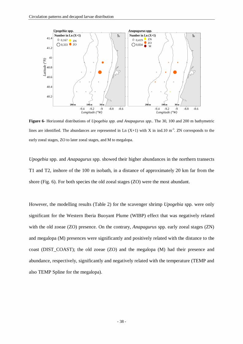
Circulation patterns and decapod larvae distribution
- 38 -
Figure 6- Horizontal distributions of Upogebia spp. and Anapagurus spp.. The 30, 100 and 200 m bathymetric
lines are identified. The abundances are represented in Ln (X+1) with X in ind.10 m-3. ZN corresponds to the
early zoeal stages, ZO to later zoeal stages, and M to megalopa.
Upogebia spp. and Anapagurus spp. showed their higher abundances in the northern transects
T1 and T2, inshore of the 100 m isobath, in a distance of approximately 20 km far from the
shore (Fig. 6). For both species the old zoeal stages (ZO) were the most abundant.
However, the modelling results (Table 2) for the scavenger shrimp Upogebia spp. were only
significant for the Western Iberia Buoyant Plume (WIBP) effect that was negatively related
with the old zoeae (ZO) presence. On the contrary, Anapagurus spp. early zoeal stages (ZN)
and megalopa (M) presences were significantly and positively related with the distance to the
coast (DIST_COAST); the old zoeae (ZO) and the megalopa (M) had their presence and
abundance, respectively, significantly and negatively related with the temperature (TEMP and
also TEMP Spline for the megalopa).
-9.4 -9.2 -9 -8.8 -8.6
40.2
40.4
40.6
40.8
41
41.2
41.4
200 m 100 m 30 m
Latit
ude
(°N)
Longitude (°W)
Number in Ln (X+1)0,1670,333
ZNZO
Upogebia spp.
-9.4 -9.2 -9 -8.8 -8.6200 m 100 m 30 m
Longitude (°W)
Number in Ln (X+1)0,4190,834
ZNZOM
Anapagurus spp.
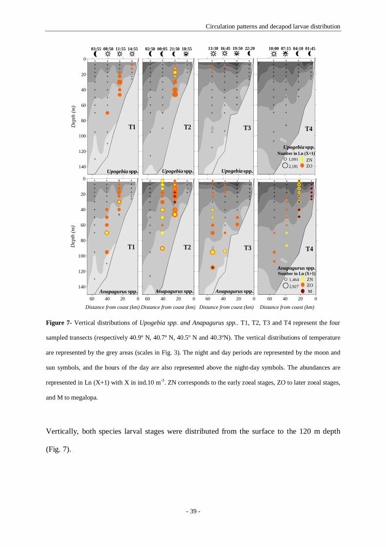
Circulation patterns and decapod larvae distribution
- 39 -
Figure 7- Vertical distributions of Upogebia spp. and Anapagurus spp.. T1, T2, T3 and T4 represent the four
sampled transects (respectively 40.9º N, 40.7º N, 40.5º N and 40.3ºN). The vertical distributions of temperature
are represented by the grey areas (scales in Fig. 3). The night and day periods are represented by the moon and
sun symbols, and the hours of the day are also represented above the night-day symbols. The abundances are
represented in Ln (X+1) with X in ind.10 m-3. ZN corresponds to the early zoeal stages, ZO to later zoeal stages,
and M to megalopa.
Vertically, both species larval stages were distributed from the surface to the 120 m depth
(Fig. 7).
140
120
100
80
60
40
20
0
Dep
th (m
)
03:55 08:50 11:55 14:55
T1
Upogebia spp.
02:50 00:05 21:50 18:55
T2
Upogebia spp.
T3
13:30 16:45 19:50 22:20
Upogebia spp.
10:00 07:15 04:10 01:45
2,181
1,091Number in Ln (X+1)
ZNZO
T4
Upogebia spp.
60 40 20 0
Distance from coast (km)
140
120
100
80
60
40
20
0
Dep
th (m
)
T1
Anapagurus spp.60 40 20 0
Distance from coast (km)
T2
Anapagurus spp.60 40 20 0
Distance from coast (km)
T3
Anapagurus spp.60 40 20 0
Distance from coast (km)
2,9271,464
Number in Ln (X+1)ZNZOM
T4
Anapagurus spp.

Circulation patterns and decapod larvae distribution
- 40 -
The Processa nouveli and Callianassa subterranea had their higher abundances in the
southern transects T3 and T4, inshore of the 100 m isobath, in a distance of approximately 20
km far from the shore (Fig. 8). For both species the early zoeal stages (ZN) were the most
abundant.
Figure 8- Horizontal distributions of Processa nouveli and Callianassa subterranea. The 30, 100 and 200 m
bathymetric lines are identified. The abundances are represented in Ln (X+1) with X in ind.10 m-3. ZN
corresponds to the early zoeal stages, ZO to later zoeal stages, and M to megalopa.
The modelling results (Table 2) showed that both species zoeal stages distributions were
significantly and positively related with the Western Iberia Buoyant Plume (WIBP, P. nouveli
early and old zoeae and C. subterranea early zoeae presences), and with the distance to the
coast (DIST_COAST, P. nouveli old zoeae abundance and C. subterranea early zoeae
presence). The temperature (TEMP) influenced negatively the P. nouveli old zoeae (ZO)
abundance, and positively the C. subterranea early zoeae (ZN) presence.
Vertically, both species larval stages were distributed from the surface to the 120 m depth
(Fig. 9). P. nouveli megalopa (M) was exclusively sampled in the most southern transect (T4),
-9.4 -9.2 -9 -8.8 -8.6
40.2
40.4
40.6
40.8
41
41.2
41.4
200 m 100 m 30 m
Latit
ude
(°N)
Longitude (°W)
Number in Ln (X+1)0,1520,305
ZNZOM
Processa nouveli
-9.4 -9.2 -9 -8.8 -8.6200 m 100 m 30 m
Longitude (°W)
Number in Ln (X+1)0,3270,653
ZNZO
Callianassa subterranea

Circulation patterns and decapod larvae distribution
- 41 -
and during the night (at 04:10) it was more close to the surface than it was at the sunrise (at
07:15). C. subterranea was not collected in the northern transect T1, and the old zoeae (ZO)
were only sampled in the most southern transect T4.
Figure 9- Vertical distributions of Processa nouveli and Callianassa subterranea. T1, T2, T3 and T4 represent
the four sampled transects (respectively 40.9º N, 40.7º N, 40.5º N and 40.3ºN). The vertical distributions of
temperature are represented by the grey areas (scales in Fig. 3). The night and day periods are represented by the
moon and sun symbols, and the hours of the day are also represented above the night-day symbols. The
abundances are represented in Ln (X+1) with X in ind.10 m-3. ZN corresponds to the early zoeal stages, ZO to
later zoeal stages, and M to megalopa.
140
120
100
80
60
40
20
0
Dep
th (m
)
03:55 08:50 11:55 14:55
T1
Processa nouveli
02:50 00:05 21:50 18:55
T2
Processa nouveli
T3
13:30 16:45 19:50 22:20
Processa nouveli
35.75
10:00 07:15 04:10 01:45
1,880
0,940Number in Ln (X+1)
ZNZOM
T4
Processa nouveli
60 40 20 0
Distance from coast (km)
140
120
100
80
60
40
20
0
Dep
th (m
)
T1
Callianassa subterranea60 40 20 0
Distance from coast (km)
T2
Callianassa subterranea
60 40 20 0
Distance from coast (km)
T3
Callianassa subterranea60 40 20 0
Distance from coast (km)
2,2201,110
Number in Ln (X+1)ZNZO
T4
Callianassa subterranea

Circulation patterns and decapod larvae distribution
- 42 -
The remaining 6 species (Eualus occultus, Pandalina brevirostris, Philocheras bispinosus,
Atelecyclus rotundatus, Liocarcinus spp. and Polybius henslowii) presented a more or less
extended distribution in the shelf (Fig. 10). For the brachyuran taxa the megalopal presences
were always negatively related with the WIBP, whenever it explained this stage distribution.
All the six taxa had their higher larval abundances along a band, inshore of the 200 m isobath,
from 8 to 60 km far from the coast (Fig. 10), and the old zoeal stages (ZO) were the most
abundant.
The modelling results for E. occultus and P. brevirostris (Table 2) were quite similar. Both
species had their zoeal stages significantly and positively related with the Western Iberia
Buoyant Plume (WIBP, E. occultus and P. brevirostris early zoeae presence, P. brevirostris
old zoeae abundance). The temperature was significantly and negatively related with both
species zoeal stages presence (E. occultus early and old zoeae, P. brevirostris old zoeae), and
was significantly and positively related with both species megalopae presence. The distance
to the coast (DIST_COAST) only explained the E. occultus megalopal presence. Vertically,
both species larval stages were distributed from the surface to the 120 m depth (Fig. 11).
For P. bispinosus and A. rotundatus the temperature (TEMP) was significantly and negatively
related with the old zoeae (ZO) presences and with the megalopal presence (A. rotundatus)
and abundance (P. bispinosus, Table 2). The distance to the coast (DIST_COAST) and the
hour of the day (HOUR_WAVE) were also explaining both species distribution. Vertically,
both species larval stages were distributed from the surface to the 120 m depth (Fig. 12). The
bathymetrical range distribution of the adults goes to a maximum of 100 m depth.
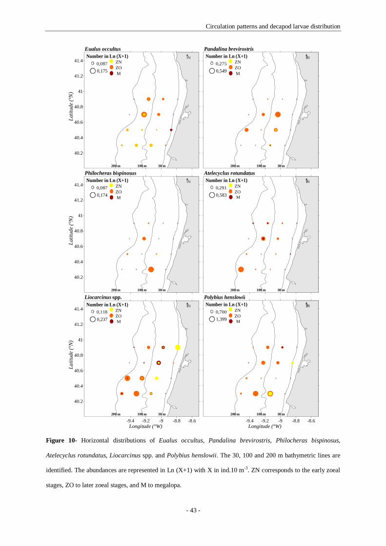
Circulation patterns and decapod larvae distribution
- 43 -
Figure 10- Horizontal distributions of Eualus occultus, Pandalina brevirostris, Philocheras bispinosus,
Atelecyclus rotundatus, Liocarcinus spp. and Polybius henslowii. The 30, 100 and 200 m bathymetric lines are
identified. The abundances are represented in Ln (X+1) with X in ind.10 m-3. ZN corresponds to the early zoeal
stages, ZO to later zoeal stages, and M to megalopa.
40.2
40.4
40.6
40.8
41
41.2
41.4
200 m 100 m 30 m
Latit
ude
(°N
)
Number in Ln (X+1)0,0870,175
ZNZOM
Eualus occultus
200 m 100 m 30 m
Number in Ln (X+1)0,2750,549
ZNZOM
Pandalina brevirostris
40.2
40.4
40.6
40.8
41
41.2
41.4
200 m 100 m 30 m
Latit
ude
(°N
)
Number in Ln (X+1)0,0870,174
ZNZOM
Philocheras bispinosus
200 m 100 m 30 m
Number in Ln (X+1)0,2910,583
ZNZOM
Atelecyclus rotundatus
-9.4 -9.2 -9 -8.8 -8.6
40.2
40.4
40.6
40.8
41
41.2
41.4
200 m 100 m 30 m
Latit
ude
(°N)
Longitude (°W)
Number in Ln (X+1)0,1180,237
ZNZOM
Liocarcinus spp.
-9.4 -9.2 -9 -8.8 -8.6200 m 100 m 30 m
Longitude (°W)
Number in Ln (X+1)0,7001,399
ZNZOM
Polybius henslowii
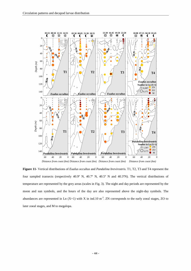
Circulation patterns and decapod larvae distribution
- 44 -
Figure 11- Vertical distributions of Eualus occultus and Pandalina brevirostris. T1, T2, T3 and T4 represent the
four sampled transects (respectively 40.9º N, 40.7º N, 40.5º N and 40.3ºN). The vertical distributions of
temperature are represented by the grey areas (scales in Fig. 3). The night and day periods are represented by the
moon and sun symbols, and the hours of the day are also represented above the night-day symbols. The
abundances are represented in Ln (X+1) with X in ind.10 m-3. ZN corresponds to the early zoeal stages, ZO to
later zoeal stages, and M to megalopa.
140
120
100
80
60
40
20
0D
epth
(m)
03:55 08:50 11:55 14:55
T1
Eualus occultus
02:50 00:05 21:50 18:55
T2
Eualus occultus
T3
13:30 16:45 19:50 22:20
Eualus occultus
10:00 07:15 04:10 01:45
0,1750,087
Number in Ln (X+1)ZNZOM
T4
Eualus occultus
60 40 20 0
Distance from coast (km)
140
120
100
80
60
40
20
0
Dep
th (m
)
T1
Pandalina brevirostris60 40 20 0
Distance from coast (km)
T2
Pandalina brevirostris60 40 20 0
Distance from coast (km)
T3
Pandalina brevirostris60 40 20 0
Distance from coast (km)
3,1751,587
Number in Ln (X+1)ZNZOM
T4
Pandalina brevirostris

Circulation patterns and decapod larvae distribution
- 45 -
Figure 12- Vertical distributions of Philocheras bispinosus and Atelecyclus rotundatus. T1, T2, T3 and T4
represent the four sampled transects (respectively 40.9º N, 40.7º N, 40.5º N and 40.3ºN). The vertical
distributions of temperature are represented by the grey areas (scales in Fig. 3). The night and day periods are
represented by the moon and sun symbols, and the hours of the day are also represented above the night-day
symbols. The abundances are represented in Ln (X+1) with X in ind.10 m-3. ZN corresponds to the early zoeal
stages, ZO to later zoeal stages, and M to megalopa.
For Liocarcinus spp. and Polybius henslowii the temperature (TEMP) was non significant
probably because these larval stages were ubiquitous. P. henslowii zoeal and megalopal
distributions were significantly related with the distance to the coast (DIST_COAST). This
environmental predictor was not significant for Liocarcinus spp.. Finally the hour of the day
140
120
100
80
60
40
20
0
Dep
th (m
)
03:55 08:50 11:55 14:55
T1
Philocheras bispinosus
02:50 00:05 21:50 18:55
T2
Philocheras bispinosus
T3
13:30 16:45 19:50 22:20
Philocheras bispinosus
10:00 07:15 04:10 01:45
1,761
0,880Number in Ln (X+1)
ZNZOM
T4
Philocheras bispinosus
60 40 20 0
Distance from coast (km)
140
120
100
80
60
40
20
0
Dep
th (m
)
T1
Atelecyclus rotundatus60 40 20 0
Distance from coast (km)
T2
Atelecyclus rotundatus60 40 20 0
Distance from coast (km)
T3
Atelecyclus rotundatus60 40 20 0
Distance from coast (km)
2,8451,422
Number in Ln (X+1)ZNZOM
T4
Atelecyclus rotundatus

Circulation patterns and decapod larvae distribution
- 46 -
(HOUR_WAVE) was significantly related with the megalopal presence and abundance of
both species (see Table 2 with the coefficients of significant explanatory variables values and
signals). Both taxa larval stages were sampled in the more deep stations of the survey (Fig.
13).
Figure 13- Vertical distributions of Liocarcinus spp. and Polybius henslowii. T1, T2, T3 and T4 represent the
four sampled transects (respectively 40.9º N, 40.7º N, 40.5º N and 40.3ºN). The vertical distributions of
temperature are represented by the grey areas (scales in Fig. 3). The night and day periods are represented by the
moon and sun symbols, and the hours of the day are also represented above the night-day symbols. The
abundances are represented in Ln (X+1) with X in ind.10 m-3. ZN corresponds to the early zoeal stages, ZO to
later zoeal stages, and M to megalopa.
140
120
100
80
60
40
20
0
Dep
th (m
)
03:55 08:50 11:55 14:55
T1
Liocarcinus spp.
02:50 00:05 21:50 18:55
T2
Liocarcinus spp.
T3
13:30 16:45 19:50 22:20
Liocarcinus spp.
10:00 07:15 04:10 01:45
3,781
1,890Number in Ln (X+1)
ZNZOM
T4
Liocarcinus spp.
60 40 20 0
Distance from coast (km)
140
120
100
80
60
40
20
0
Dep
th (m
)
T1
Polybius henslowii60 40 20 0
Distance from coast (km)
T2
Polybius henslowii
60 40 20 0
Distance from coast (km)
T3
Polybius henslowii60 40 20 0
Distance from coast (km)
4,5602,280
Number in Ln (X+1)ZNZOM
T4
Polybius henslowii

Circulation patterns and decapod larvae distribution
- 47 -
2.4.2.3- Horizontal and vertical distributions of the Slope species
Horizontally (Fig. 14), S. membranacea, Parthenope spp. and G. rhomboides had their higher
larval abundances registered in the southern transects (T3 and T4), offshore of the 100 m
isobath, 40- 56 km far from the shore.
Figure 14- Horizontal distributions of Solenocera membranacea, Parthenope spp. and Goneplax rhomboides.
The 30, 100 and 200 m bathymetric lines are identified. The abundances are represented in Ln (X+1) with X in
ind.10 m-3. ZN corresponds to the early zoeal stages, ZO to later zoeal stages, and M to megalopa.
-9.4 -9.2 -9 -8.8 -8.6
40.2
40.4
40.6
40.8
41
41.2
41.4
200 m 100 m 30 m
Latit
ude
(°N)
Longitude (°W)
Number in Ln (X+1)ZNZO
0.1110.221
Solenocera membranacea
-9.4 -9.2 -9 -8.8 -8.6200 m 100 m 30 m
Longitude (°W)
Number in Ln (X+1)0,0040,009
ZNZO
Parthenope spp.
-9.4 -9.2 -9 -8.8 -8.6
40.2
40.4
40.6
40.8
41
41.2
41.4
200 m 100 m 30 m
Latit
ude
(°N)
Longitude (°W)
Number in Ln (X+1)0,1090,218
ZNZOM
Goneplax rhomboides

Circulation patterns and decapod larvae distribution
- 48 -
Figure 15- Vertical distributions of S. membranacea, Parthenope spp. and G. rhomboides. T1, T2, T3 and T4
represent the four sampled transects (respectively 40.9º N, 40.7º N, 40.5º N and 40.3ºN). The vertical
distributions of temperature are represented by the grey areas (scales in Fig. 3). The night and day periods are
represented by the moon and sun symbols, and the hours of the day are also represented above the night-day
symbols. The abundances are represented in Ln (X+1) with X in ind.10 m-3. ZN corresponds to the early zoeal
stages, ZO to later zoeal stages, and M to megalopa.
140
120
100
80
60
40
20
0
Dep
th (m
)
03:55 08:50 11:55 14:55
T1
Solenocera membranacea
02:50 00:05 21:50 18:55
T2
Solenocera membranacea
T3
13:30 16:45 19:50 22:20
Solenocera membranacea
10:00 07:15 04:10 01:45
2,2861,143
Number in Ln (X+1)ZNZO
T4
Solenocera membranacea
140
120
100
80
60
40
20
0
Dep
th (m
)
T1
Parthenope spp.
T2
Parthenope spp.
T3
Parthenope spp. 0,6450,321
Number in Ln (X+1)ZNZO
T4
Parthenope spp.
60 40 20 0
Distance from coast (km)
140
120
100
80
60
40
20
0
Dep
th (m
)
T1
Goneplax rhomboides60 40 20 0
Distance from coast (km)
T2
Goneplax rhomboides60 40 20 0
Distance from coast (km)
T3
Goneplax rhomboides60 40 20 0
Distance from coast (km)
2,5211,260
Number in Ln (X+1)ZNZOM
T4
Goneplax rhomboides

Circulation patterns and decapod larvae distribution
- 49 -
The early zoeal stages (ZN) had their higher abundances in the southern transect T4 where an
intrusion of slope waters was happening. The S. membranacea older zoeal stages (ZO) were
also sampled in the northern transect, probably because of the larval advection in the
upwelled water mass to northeast.
The modelling results (Table 2) showed that the three species zoeal stages presences (early
zoeae for S. membranacea and Parthenope spp., and old zoeae for S. membranacea and G.
rhomboides) were significantly and negatively related with the distance to the coast
(DIST_COAST). As expected these species never had their distributions explained by the
Western Iberia Buoyant Plume, once that they were distributed along the shelf break where
the WIBP is not present. The three species zoeal stages were negatively related with the
distance to the coast and with the temperature.
The Slope species larval stages presence was always negatively related with the temperature
(TEMP). This environmental variable explained the early zoeae (ZN) of S. membranacea and
Parthenope spp. and the old zoea (ZO) and megalopal stage (M) of G. rhomboides (the only
species in the group with this stage). The Western Iberia Buoyant Plume never explained
these species larval distributions.
Vertically, all the three species larval stages were distributed from the surface to close to the
bottom (Fig. 15). The vertical migration behaviour is once again clear, although the hour of
the day was only significantly related with S. membranacea early zoeal stages presence and
abundance (both positive) and with G. rhomboides old zoeal stages abundance (positive) and
megalopa presence (negative).

Circulation patterns and decapod larvae distribution
- 50 -
2.5- Discussion
Present results demonstrate that the decapod crustacean larvae distribution in the Portuguese
continental shelf, clearly showed a retention strategy in accordance with the adults’
distributional ranges as already hypothesized by dos Santos et al. (2008). During the survey,
the larvae were distributed along meridional patches in accordance to their origins: the inner
shelf species were distributed very close to the shore, the shelf species were distributed in the
middle shelf, and the slope species appeared over the continental shelf break, thus, we can
suppose that the self recruitment for parental populations is a probable scenario for these
larvae even under upwelling conditions. The first evidence to support our hypothesis is the
occurrence of the complete larval series for most of the studied species: when the early zoeal
stages were found, the older zoeal stages and the megalopa were also sampled in the same
area, many times in the same station. Present results disagree with Queiroga (1996) that
described a different scenario for the distribution of Carcinus maenas larval stages.
According to the author, the horizontal distribution of C. maenas reflected an ontogenetic
pattern: the first zoea was associated with the estuarine inlets while the older zoeal stages
were dispersed progressively offshore. As dos Santos et al. (2008) affirmed present results as
well as archive data analysed by the authors positioned the C. maenas larvae inshore to the
middle shelf position described before, and show the mixture of all larval stages in the same
sampling stations. We agree with dos Santos et al. (2008) that pointed as justification for such
differences a particular oceanographic situation that lead to an exceptional sampling period.
Another evidence to support our theory is that the several larval forms studied (the shrimps
sensu lato, the scavenger shrimps, the hermit crabs and the crabs) and their respective
developmental stages (early and old zoeae, and megalopa) had similar behavioural responses
to the selected environmental variables, and their distribution was along meridionally

Circulation patterns and decapod larvae distribution
- 51 -
elongated patches parallel to the coast inshore of the 200 m isobath as previously described in
the study area for decapod larvae (dos Santos et al. 2008) and for cirripede cyprids (dos
Santos et al. 2007). Present work agree well with Peliz et al. (2007) model results, therefore
the larval transport in the study area is mainly alongshore and there is an aggregation along
band-like meridional patches parallel to the coast usually inshore of the 100 m of the larvae.
We decided to model the distribution of the selected taxa in relation to four environmental
predictors: the Western Iberia Buoyant Plume, the hour of the day, the distance to the coast
and the temperature. The distance to the coast predictor was the one that explained most times
the decapods larval distribution in the studied area. It was always positively related with the
Inner Shelf species, and on the contrary, was always negatively related with the Slope species
distribution. We can conclude that this predictor confirmed that the larvae are distributed in
accordance to the adults: the species that were close to the coast were positively related with
the distance to the coast and those that were far from the coastline were negatively related
with it.
It is known that the temperature effect is important in decapod larval development, abundance
and diversity (e.g. Anger, 2001). It was always positively related with the Inner Shelf species
zoeal distribution, and when explaining the Slope species distributions was negatively related,
probably because the Inner Shelf species were more coastal and were distributed in higher
temperatures than the Slope species.
The effect of salinity, in particular the Western Iberia Buoyant Plume (WIBP) presence,
contrary to what we were expecting never explained the Inner Shelf species distribution, and
was only related with the Shelf species distribution, probably because during the survey the
Circulation patterns and decapod larvae distribution
- 52 -
WIBP was considerably displaced to offshore. The WIBP is a different water mass in the
study area that brings stratification and stability to the surface layers, and as a consequence
can constitute a retention mechanism that leads to a higher survival of fish larvae (Santos et
al. 2006). We can assume that E. occultus, P. nouveli, P. brevirostris and C. subterranea
zoeal distributions were always positively related with this less saline water lens because this
is a more productive area and also a less turbulent water mass (Santos et al. 2006).
Contrasting to the zoeal stages distributions, the crab megalopae (A. rotundatus, Liocarcinus
spp. and P. henslowii) were negatively related with the WIBP, reflecting the avoidance of this
less saline water lens by this stage.
Finally, the hour of the day gave information about the existence of diel vertical migrations. It
was shown that in the absence of diel vertical migration the Carcinus maenas larval stages
would be highly dispersed off the Western Iberia (Marta-Almeida et al. 2006), and that the
larvae in the study area were retained over the shelf due to the presence of this mechanism
(dos Santos et al. 2008). Present study also demonstrates that the diel vertical migration
behaviour is clear for most of the sampled species: P. nouveli, P. brevirostris, P. bispinosus,
Anapagurus spp., Liocarcinus spp., and P. henslowii megalopal stage, Parthenope spp. and G.
rhomboides early zoeae, and A. rotundatus early zoeae and megalopa. Therefore, the larval
retention mechanism in meridionally elongated bands parallel to the shore, more or less
coincident with the adults distribution is in fact the result of the synchronized diel vertical
migration behaviour of the larvae. This will maintain the larval patch close to their parental
population, minimizing the horizontal larval excursion, enhancing the species recruitment
(DiBacco et al. 2001).

Circulation patterns and decapod larvae distribution
- 53 -
As referred before, the very recent study of dos Santos et al. (2008) discussed the larval
retention possibility for decapod larvae off the northwest Portuguese coast, affirming that
larvae belonging to coastal species, as Carcinus maenas were mainly restricted to a 10 km-
wide band along the coast, over bottom depths less than 30 m. Present study confirms that the
Inner Shelf species were mainly restricted to a 10 km wide band along the coast, over bottom
depths less than 30 m, even under upwelling events. These results also corroborate Shanks et
al. (2002), Wing et al. (2003), Shanks & Eckert (2005). D. pugilator and N. puber zoeal
distributions were positively related with the temperature predictor, probably because they
were distributed close to the shore, above the depth of the thermocline, where the higher
temperatures were registered.
The retention mechanisms in the coastal environments have been well described for estuarine
species (e.g. Epifanio & Garvine 2001), although for shelf species it is almost unknown.
Marta-Almeida et al. (2006), Peliz et al. (2007) and dos Santos et al. (2008) demonstrated that
the retention strategies for the species more widely distributed in the continental shelf resulted
of the diel vertical migration between water masses moving in different directions at different
depths. Present study demonstrates that the Shelf Species are retained close to their parental
populations, over the shelf near their release sites. Crossing the larval distribution results with
the adults’ spatial distribution (Zariquiey-Álvarez 1968, d’Udekem d’Acoz 1999), the larval
presences reflected the place where the adults’ populations were. During this study the Shelf
species larvae showed three different distributional patterns: some species were more
abundant in the north, others were more abundant in the south, and the third group includes
those species that were widely distributed over the shelf.

Circulation patterns and decapod larvae distribution
- 54 -
The first two groups reflect the larval distribution response to the environmental variables, in
particular to the thermal front and to the WIBP displacement. Upogebia spp. and Anapagurus
spp. were concentrated in the northern transects, approximately 25 km off the shore, inshore
of the 100 m isobaths and never had their distributions explained by the WIBP, while P.
nouveli and C. subterranea the two more abundant in the southern transects, approximately
25 km off the shore, inshore of the 100 m isobath, were positively related with the WIBP.
Probably this less saline water mass is the responsible for the larval retention over the shelf. In
the WIBP is well-developed, the transport in the surface layers would be mainly offshore
during upwelling, but the presence of the the IPC in the deeper stations of the southern
transects creates a blocking effect to the offshore transport, generating a mechanism of larval
retention (Santos et al. 2004).
All the other six species were widely distributed over the shelf and concentrated their higher
abundances along the middle shelf. E. occultus and P. brevirostris abundances were mainly
registered in a band 20 to 60 km off the coastline. Both species had their zoeal stages
positively related with the WIBP and negatively related with the temperature that was
positively related with both species megalopae. This difference in the temperature
significance signal for the zoeal and megalopal stages may be reflecting different ontogenetic
responses to the environment. Most likely, the distribution of the adults to a maximum of 180
m depth may be the reason why these species larval stages have such similar responses to the
environmental predictors. Probably both species hatched in the same environment and migrate
vertically in the shelf water column with similar buoyancy and stratification andas a
consequence, the surface Ekman layer transport is similar for both species.

Circulation patterns and decapod larvae distribution
- 55 -
Also, P. bispinosus and A. rotundatus when adults are distributed to 100 m depth, had their
higher larval abundances along the 20 to 60 km off band, and the temperature affected them in
a similar way. Finally, Liocarcinus spp. and Polybius henslowii have higher distribution
bathymetrical ranges when adults, and had their larvae widely distributed in the area from 8 to
60 km to the shore, with the megalopae similarly affected by the WIBP. The temperature did
not affected these species distribution, probably when adults these species are almost
everywhere (Zariquiey-Álvarez 1968, d’Udekem d’Acoz 1999), and as a consequence their
larvae are ubiquitous and can hatch or settle in any place of the continental shelf.
We suppose that S. membranacea, Parthenope spp. and G. rhomboides were advected to the
shelf, probably because here their survival will be enhanced due to higher food availability
and certainly to favour their retention (e.g. Levin 2006). The absence of the megalopal stage
may be because they were not present in the plankton for this time of year, or they were not in
the shelf anymore once that they already started the return to the parental population where
they will metamorphose and settle.
The larval horizontal transport will be determined by the interactive effect of the position of
the larvae in the water column (vertical distribution) and the hydrodynamics (e.g. Sameoto &
Metaxas 2008). The particular oceanographic conditions during the survey, lead to the
appearance of a frontal structure. This frontal structure was perpendicular to the coastline and
separated the two northern transects (T1 and T2) from the two southern ones (T3 and T4),
concentrating the decapod larvae along the shelf (dos Santos et al. 2008). Analysing the
general distribution of the considered taxa, it is possible to state that the Shelf species larval
distribution reflects the transport in the upwelled water mass, which was advected for the
northern transects (a higher presence of old zoeae in transects T1 and T2). Probably, during

Circulation patterns and decapod larvae distribution
- 56 -
the south-north dislocation of this water mass, the early zoeal stages were transported
continuing to developed, and completing their ontogenetic cycle. As a result, these larvae
reached a northern position older. The examples of species that might be experiencing this
transport are: Solenocera membranacea, Eualus occultus, Pandalina brevirostris and
Anapagurus spp.. Based on dos Santos et al. (2008) data, as well as on the behavioural
reactions described in the literature and described in present study, we agree with Queiroga et
al. (2007) proving that in general decapod zoeae and megalopae are not trapped at frontal
convergence zones as described before (e.g. Shanks et al. 2000). Decapod larval diel vertical
migration behaviour conjunct with the local wind-driven circulation and the coastline
orientation, are capable of keep their distribution along meridionally elongated bands
coincident with the adults bathymetric distribution.
2.6- References
Anger K. (2001). The Biology of Decapod Crustacean Larvae. Crustacean Issues 14, 1- 419.
Botsford L.W., Moloney C.L., Hastings A., Largier J.L., Powell T.M., Higgins K., Quinn J.F.
(1994). The influence of spatially and temporally varying oceanographic conditions on
meroplanktonic metapopulations. Deep-Sea Research II 41, 107-145.
Cowen R.K. & Sponaugle S. (2009). Larval Dispersal and Marine Population Connectivity.
Annual Review of Marine Science 1, 443-466.
DiBacco C., Sutton D., McConnico L. (2001). Vertical migration behavior and horizontal
distribution of brachyuran larvae in a low inflow estuary: implications for bay-ocean
exchange. Marine Ecology Progress Series 217, 191-206.

Circulation patterns and decapod larvae distribution
- 57 -
dos Santos A. & Lindley J.A. (2001). Crustacea, Decapoda: Larvae, II. Dendrobranchiata
(Aristeidae, Benthesicymidae, Penaeidae, Solenoceridae, Sicyonidae, Sergestidae and
Luciferidae). ICES Identification Leaflets for Plankton 186, 1-9.
dos Santos A. & González-Gordillo J.I. (2004). Illustrated keys for the identification of the
Pleocyemata (Crustacea, Decapoda) zoeal stages, from the coastal region of south-
western Europe. Journal of the Marine Biological Association of the United Kingdom 84,
205-227.
dos Santos A., Santos A.M.P., Conway D.V.P. (2007). Horizontal and vertical distribution of
cirripede cyprid larvae in an upwelling system off the Portuguese coast. Marine Ecology
Progress Series 329, 145-155.
dos Santos A., Santos A.M.P., Conway D.V.P., Bartilotti C., Lourenço P., Queiroga H.
(2008). Diel vertical migration of decapod larvae in the portuguese coastal upwelling
ecosystem: implications for offshore transport. Marine Ecology Progress Series 359,
171-183.
d’Udekem d’Acoz C. (1999). Inventaire et distribution des crustacés décapodes de
l’Atlantique nord-oriental, de la Méditerranée et des eaux continentales adjacentes au
nord de 25º N. Collection Patrimoines Naturels (M.N.H.N./S.P.N.) 40, 1-383.
Epifanio C.E. & Garvine R.W. (2001). Larval transport on the Atlantic continental shelf of
North America: a review. Estuarine Coastal and Shelf Science 52, 51–77.
Frouin R., Fiúza A.F.G., Ambar I., Boyd T.J. (1990). Observations of a Poleward Surface
Current off the Coasts of Portugal and Spain During Winter. Jurnal of Geophysical
Research 95, 679-691.
Gawarkiewicz G., Monismith S., Largier J. (2007) Observing larval transport processes
affecting population connectivity- progress and challenges. Oceanography 20, 40-53.

Circulation patterns and decapod larvae distribution
- 58 -
González-Gordillo J.I., dos Santos A., Rodríguez A. (2001). Checklist and annotated
bibliography of decapod Crustacea larvae from the southwestern European coast
(Gibraltar Strait area). Scientia Marina 65, 275-305.
Haynes R. & Barton E.D. (1990). A poleward current along the Atlantic coast of the Iberian
Peninsula. Journal of Geophysical Research 95, 11425-11441.
Levin L. (2006). Recent progress in understanding larval dispersal: new directions and
digressions. Integrative and Comparative Biology 46, 282-297.
Mace A.J. & Morgan S.G. (2006). Larval accumulation in the lee of a small headland:
implications for the design of marine reserves. Marine Ecology Progress Series 318, 19-
29.
Marta-Almeida M., Dubert J., Peliz A., Queiroga H. (2006) Influence of vertical migration
pattern on retention of crab larvae in a seasonal upwelling system. Marine Ecology
Progress Series 307, 1-19.
Morgan S.G., Fisher J.L., Mace1 A.J., Akins L., Slaughter A.M., Bollens S.M. (2009). Cross-
shelf distributions and recruitment of crab postlarvae in a region of strong upwelling.
Marine Ecology Progress Series 380, 173-185.
Peliz A., Rosa T., Santos A.M.P., Pissarra J. (2002). Fronts, jets and counter flows in the
Western Iberia upwelling system. Journal of Marine Systems 35, 61-77.
Peliz A., Dubert J., Haidvogel D.B., Le Cann B. (2003). Generation and unstable evolution of
a density-driven Eastern Poleward Current: the Iberia poleward current. Journal of
Geophysical Research 108 (C8), 3268 (doi: 10.1029/2002 JC 001443).
Peliz A., Dubert J., Santos A.M.P., Oliveira P.B., Le Cann B. (2005). Winter upper ocean
circulation in the Western Iberian Basin- Fronts, Eddies and Poleward Flows: an
overview. Deep-Sea Research I 52, 621-646.

Circulation patterns and decapod larvae distribution
- 59 -
Peliz A., Marchesiello P., Dubert J., Marta-Almeida M., Roy C., Queiroga H. (2007). A study
of crab larvae dispersal on the Western Iberian Shelf: physical processes. Journal of
Marine Systems 68, 215-236.
Pittman S.J. & McAlpine C.A. (2001). Movements of marine fish and decapod Crustaceans:
process, theory and application. Advances in Marine Biology 44, 205-294.
Queiroga H. (1996). Distribution and drift of the crab Carcinus maenas (L.) (Decapoda,
Portunidae) larvae over the continental shelf off northern Portugal in April 1991. Journal
of Plankton Research 18, 1981–2000.
Queiroga H. & Blanton J. (2005). Interactions between behaviour and physical forcing in the
control of horizontal transport of decapod crustacean larvae. Advances in Marine Biology
47, 107-214.
Queiroga H., Almeida M.J., Alpuim T., Flores A.A.V., Francisco S., González-Gordillo I.,
Miranda A.I., Silva I., Paula J. (2006). Tide and wind control of megalopal supply to
estuarine crab populations on the Portuguese west coast. Marine Ecology Progress
Series 307, 21-36.
Queiroga H., Cruz T., dos Santos A., Dubert J., González-Gordillo J.I., Paula J., Peliz A.,
Santos A.M.P. (2007). Oceanographic and behavioural processes affecting invertebrate
larval dispersal and supply in the western Iberia upwelling ecosystem. Progress in
Oceanography 74, 174-191.
Relvas P., Barton E.D., Dubert J., Oliveira P.B., Peliz A., da Silva J.C.B., Santos A.M.P.
(2007). Physical oceanography of the western Iberia ecosystem: latest views and
challenges. Progress in Oceanography 74, 149-173.
Roughgarden J., Gaines S., Possingham H. (1988). Recruitment dynamics in complex life
cycles. Science 241, 1460-1466.

Circulation patterns and decapod larvae distribution
- 60 -
Sameoto J.A. & Metaxas A. (2008). Interactive effects of haloclines and food patches on the
vertical distribution of 3 species of temperate invertebrate larvae. Journal of
Experimental Marine Biology and Ecology 367, 131-141.
Santos A.M.P., Peliz A., Dubert J., Oliveira P.B., Angélico M.M., Ré P. (2004). Impact of a
winter upwelling event on the distribution and transport of sardine (Sardina pilchardus)
eggs and larvae off western Iberia: a retention mechanisms. Continental Shelf Research
24, 149-165.
Santos AMP, Ré P, Dos Santos A, Peliz A (2006) Vertical distribution of the European
sardine (Sardina pilchardus) larvae and its implications for their survival. Journal of
Plankton Research 28, 523–532.
SAS Institute (2008) SAS/Stat User’s Guide. Version 9.2. Cary, NC, USA.
Shanks A.L. & Eckert G.L. (2005). Population persistence of California current fishes and
benthic crustaceans: a marine drift paradox. Ecological Monographs 75, 505-524.
Shanks A.L., Largier J., Brink L. (2000). Demonstration of the onshore transport of larval
invertebrates by the shoreward movement of an upwelling front. Limnology and
Oceanography 45, 230-236.
Shanks A.L., Largier J., Brink L., Brubaker J., Hooff R. (2002). Observations on the
distribution of meroplankton during a downwelling event and associated intrusion of the
Chesapeake Bay estuarine plume. Journal of Plankton Research 24, 391–416.
Swearer S.E., Shima J.S., Hellberg M.E., Thorrold S.R., Jones G.P., Robertson D.R., Morgan
S.G., Selkoe K.A., Ruiz G.M., Warner R.R. (2002). Evidence of self-recruitment in
demersal marine populations. Bulletin of Marine Science 70, 251–271.
Thorson G. (1950). Reproductive and larval ecology of marine bottom invertebrates.
Biological Review 25, 1–45.

Circulation patterns and decapod larvae distribution
- 61 -
Wenger S.J. & Freeman M.C. (2008). Estimating species occurrence, abundance, and
detection probability using zero-inflated distributions. Ecology 89, 2953-2959.
Williams R., Collins N.R., Conway D.V.P. (1983). The double LHPR system, a high-speed
micro- and macroplankton sampler. Deep-Sea Research 30, 331-342.
Wing S.R., Botsford L.W., Morgan L.E., Diehl J.M., Lundquist C.J. (2003). Inter-annual
variability in larval supply to populations of three invertebrate taxa in the northern
California Current. Estuarine Coastal and Shelf Science 57, 859-872.
Zariquiey-Alvarez R. (1968). Crustáceos Decápodos Ibéricos. Investigacion Pesquera 32, 1-
510.
CHAPTER 3
__________________________________________________
Ontogenetic vertical migration behaviour of decapod
larvae in the Portuguese upwelling ecosystem*
*Bartilotti C., Castro M., Santos A. M. P., Queiroga H., dos Santos A. (in prep.) Ontogenetic
vertical migration behaviour of decapod larvae in the Portuguese upwelling ecosystem.

Ontogenetic vertical migration of decapod larvae
- 63 -
3.1- Abstract
The ontogenetic vertical migration behaviour of decapod larvae was analysed on the western
Iberia upwelling ecosystem. An oceanographic cruise conducted in May 2002 sampled
vertically a fixed station point during 69 h, approximately 21 km off the coast. Plankton
samples were collected every 2 hours at the surface with a Neuston net and through the water
column with a Longhurst Hardy Plankton Recorder (Pro-LHPR), in order to obtain a detailed
resolution of larval vertical distribution. Temperature and salinity were registered hourly. The
vertical migration behaviour of nine different species of decapod crustacean, three caridean
shrimps, three hermit crabs, and three crabs were analysed in detail, by stage. The results
showed that all the studied species larval stages had diel vertical migration (DVM) behaviour,
and the average depth of distribution of the studied larval stages varied through the
ontogenetic development. The majority of the studied species had their early zoeal stages
more close to the surface and the last zoeal stages in a more deep position. We also analysed
the larval distributions in relation with the hour of the day which indicated the DVM and as
expected was the predictor that explained most times the larval distributions; the midpoint of
depth stratum reflected the positioning of the larvae in a certain favourable depth giving
information about the average depth of distribution. Finally the temperature and the salinity
gave information about the larval distributions and the probable transport mechanisms in the
water column in the sampling area. During sampling, a less saline water lens appeared in the
second day, and, we suppose that it transported newly hatched larvae to the fixed station
point.
Ontogenetic vertical migration of decapod larvae
- 64 -
3.2- Introduction
Decapod crustacean larvae can dynamically regulate their vertical position in the water
column, controlling the range and direction of their horizontal dispersal (Queiroga & Blanton
2005), maintaining a favourable position to the adequate and necessary transport (e.g.
Forward et al. 1997, Christy & Morgan 1998). The Diel Vertical Migration (DVM) behaviour
seems to be the rule for decapod larvae, although, it is a pattern rarely described in detail (see
Queiroga & Blanton 2005 for examples). Most of the studies present results obtained by
neuston and/or discrete depth levels sampling (e.g. Shanks 1985, Jamieson & Phillips 1988,
Abelló & Guerao 1999), being the studies with a high vertical discrimination rare (Lindley
1986, Lindley et al. 1994, dos Santos et al. 2008). The recent results for the upwelling
ecosystem adjacent to the Ria de Aveiro lagoon system showed that decapod zoeae and
megalopae, as well as Chthamalus stellatus cyprids, displayed the DVM (dos Santos et al.
2007, 2008).
Most decapod crustaceans present DVM ascending to the surface water layer during the night
(Queiroga & Blanton 2005). Observing inert larvae Pineda et al. (2007) stated that vertical
swimming behaviour, changes in buoyancy, and ontogenetic changes in vertical position in
the water column were influencing the horizontal larval movements. Recently, dos Santos et
al. (2008) concluded that the diel vertical migration is the mechanism responsible by the
larval retention in the Portuguese upwelling ecosystem, corroborating the hypothesis
presented by the models developed for the study area (Marta-Almeida et al. 2006, Peliz et al.
2007). The authors concluded that the horizontal larval distribution of the studied species in
the inner and middle shelf was mainly alongshore clearly related to the presumed settlement
Ontogenetic vertical migration of decapod larvae
- 65 -
areas, and the capacity to perform diel vertical migrations was the crucial factor for the
established distributional larval patterns (dos Santos et al. 2008).
The larval vertical distribution behaviour may reflect environmental responses to light (e.g.
Sulkin 1975), salinity (e.g. Latz & Forward 1977) and temperature (e.g. Dawirs 1985). In the
laboratory, small variations of salinity or temperature lead to changes in the larval behaviour
(e.g. Forward 1989), although in the study area, these small variations reflected the water
mixing, which did not modified the larval behaviour (dos Santos et al. 2008). Salinity is one
of the environmental factors with a higher selective pressure for decapod crustacean larvae
(Anger 2003), and the freshwater input can enhance vertical mixing affecting the distribution
and transport of planktonic larvae (e.g. Garrison 1999). Also the temperature takes to various
metabolic responses by crustacean larvae reflecting their tolerance (Agard 1999), and it is
known that a shallow thermocline can create vertically sheared environments that can limit
the larval transport for species with DVM (Pineda et al. 2007). Other important factors for the
establishment of the larval vertical position are the predators’ avoidance (e.g. Hobbs &
Botsford 1992, Forward & Rittschof 2000) or the food availability (e.g. Lindley et al. 1994,
Pearre 2003).
Another category of vertical migrations is the ontogenetic migration, defined as a change in
the average depth of distribution during the larval life period, a process obligatory for benthic
crustacean that hatch close to the bottom, feed in the surface, and when reaching the end of
their larval life must return to the adult habitat (Queiroga & Blanton 2005). Several studies
described these ontogenetic migrations (see Queiroga & Blanton 2005 for details and
references), and most of them considered brachyuran species. In the study area, Queiroga
(1996) demonstrated these ontogenetic differences in the vertical distribution of Carcinus

Ontogenetic vertical migration of decapod larvae
- 66 -
maenas: the first and second zoeal stages were closer to the surface and a gradual
displacement to deeper waters was observed from then on, for the older stages; the megalopa
was equally distributed at the surface and at a higher depth of the water column.
Knowing that the decapod crustacean larvae in the study area perform vertical migrations (dos
Santos et al. 2008), present work pretends to analyse in detail the vertical distribution patterns
of the larval stages of the 9 selected taxa, in order to determine their ontogenetic vertical
migration in the Portuguese continental shelf, and also to describe the behavioural responses
of each larval stage to the environment (depth, salinity, temperature and hour of the day).
3.3- Material and Methods
3.3.1- Field study
An oceanographic survey was carried out off the northwest coast of Portugal (project
ProRecruit’2002-02050502), aboard the RV ‘Noruega’, from 9-22 May 2002 (Fig. 1).
From 11 to 14 May a mooring with 3 current meters (Aandera RCM 9) and 2 conductivity-
temperature-depth (CTD) profilers (Sea-Bird Electronics MicroCats) was carried out on the
Portuguese continental shelf (40º45.9’ N 08º59.0’ W), approximately 21 km offshore,
positioned at a 60 m bottom depth, in an area adjacent to Aveiro coastal lagoon (Fig. 1).
From 18 to 21 May 2002, the fixed station was sampled continuously in two hours intervals
for 69 hours. Environmental sampling was carried (see dos Santos et al. 2007 for details).
Temperature and salinity were measured every hour with a Seabird SBE 9 plus CTD
(conductivity/ temperature/ depth). The depth stratified samples were collected with the Pro-

Ontogenetic vertical migration of decapod larvae
- 67 -
LHPR system (a commercially updated version of the Longhurst Hardy Plankton Recorder,
Williams et al. 1983). The Pro-LHPR had a mouth aperture of 0.42 m and collects plankton
between two rolls of filtering gauze with 280 µm mesh size that are advanced at intervals
inside a cod end box to give a series of consecutive samples. It was towed at 3-4 knots on
oblique hauls from the surface to near the seabed for 30 min intervals. Stratified samples of
Pro-LHPR were collected at 5 m depth intervals in the first 25 m, and 10 m depth intervals
from 25 m deep until near the bottom. The first 20 centimetres of the water column were also
sampled every 2 hours with a Neuston net. It had a rectangular mouth aperture of 0.2× 1.0 m
with a flowmeter mounted and a 335 µm mesh size. It was towed horizontally, for a 3 minutes
period, at a velocity of 1.5 knots.
Figure 1- Map of the northwest coast of Portugal: sampling position of the fixed station collected aboard the RV
Noruega, from 15-17 May 2002, during ProRecruit project. The 30, 100 and 200 m bathymetric lines are also
identified.

Ontogenetic vertical migration of decapod larvae
- 68 -
3.3.2- Sample processing
Plankton samples were preserved in 4% borax-buffered formaldehyde, prepared with
seawater. Samples were sorted for decapod larvae, that were identified to species level and
developmental stage whenever possible using dos Santos & Lindley (2001), González-
Gordillo et al. (2001), and dos Santos & González-Gordillo (2004). Data were standardized
for ind.m-3 using the flow information.
3.3.3- Analysis of decapod vertical distribution in relation to the physical environment
The average depth was calculated according to Pearre (2003) for each species, by stage, as the
Weighted Mean Depth (WMD, in m). It consisted of the weighted abundance per species, of
each stage collected in each sampling stratum z, at each sampling time t:
푊푀퐷푡 =∑ (퐴푧푡 × 퐷푧푡)
∑ 퐴푧푡
where A is the abundance (ind. m-3) and D is the midpoint of depth stratum (m).
Present study considered nine of the more abundant species of the three most represented
decapod crustacean groups, the caridean shrimps, hermit crabs and crabs, previously studied
by dos Santos et al. (2008). Each species larval series was analysed stage by stage. Table 1
presents the list of chosen taxa, the respective decapod crustacean group and the constitution
of the larval series with the number of zoeal stages (Z) and the megalopa or decapodid stage
(M).

Ontogenetic vertical migration of decapod larvae
- 69 -
Table 1- List of the selected taxa, respective decapod crustacean group, and larval series.
Taxa Decapod group Larval series
Processa nouveli
Pandalina brevirostris
Philocheras bispinosus
Caridean shrimps
Caridean shrimps
Caridean shrimps
Z I to IX and M
Z I to VII and M
Z I to V and M
Anapagurus spp.
Pagurus bernhardus
Pisidia longicornis
Hermit crabs
Hermit crabs
Porcelain crabs
Z I to IV and M
Z Ito IV and M
Z Ito II and M
Atelecyclus rotundatus
Liocarcinus spp.
Necora puber
Crabs
Crabs
Crabs
Z I to V and M
Z I to V and M
Z I to V and M
The statistical procedure was similar to the one used in Chapter 2. Two response variables
were considered, the abundance expressed as number of individuals per cubic meter of
filtered water (ind.m-3) that was log transformed to homogenise the variance, and the
presence/absence of a given taxa and larval stage, binomial variable equal to one if abundance
greater than zero, and zero otherwise.
Having in mind that our objective was to determine the ontogenetic larval vertical migration
behaviour in a fixed point, four explanatory variables were established and used in the
construction of the model. These were:
MID_DEPTH – Midpoint of depth stratum, discrete variable, in meters;
HOUR_WAVE - hour of the day, continuous variable, defined as a circular function
of the hour of the day, sunrise=0, noon=1, sunset=0 and midnight=-1, generated by the
function:
Ontogenetic vertical migration of decapod larvae
- 70 -
푠푖푛 [(퐻표푢푟 ×2휋24) +
3휋2 ]
where Hour is the hour of the day (0-24);
SAL – Salinity, continuous variable, no units;
TEMP - temperature, continuous variable, in ºC.
The decapod larval vertical distribution, in relation to these 4 explanatory variables, was
analysed in two steps, for each species and larval stage separately. As referred in the anterior
chapter, the two steps procedure is used to deal with the problem of zero inflated data
(Wenger & Freeman 2008). In the first step, the presence or absence was predicted using a
Logistic Model (LM) with explanatory variables the midpoint of depth stratum
(MID_DEPTH), the hour of the day (HOUR_WAVE), the salinity (SAL) and the temperature
(TEMP). The results from this model were used in the second step for situations where at
least one of the variables was significant and for data points where “presence” was predicted.
For these data points, measured values of abundance were modelled with a Generalized
Addictive Model (GAM), using linear predictors for the midpoint of depth stratum
(MID_DEPTH), the hour of the day (HOUR_WAVE), the salinity (SAL) and the temperature
(TEMP) and a spline for temperature (TEMP, with 2 degrees of freedom). Statistical analysis
was made using SAS 9.2 (SAS Institute, 2008) using PROC LOGIT for fitting the logistic
model (stepwise option) and PROC GAM for the generalized additive models.
3.4- Results
3.4.1- Oceanographic conditions
For the complete description of the oceanographic conditions during the survey please see dos
Santos et al. (2007). The vertical distribution of the hydrological fields showed the arrival of a

Ontogenetic vertical migration of decapod larvae
- 71 -
less saline water lens at the surface from the 12 h of the second day (19th May), consequence
of the change in the oceanographic conditions in the sampling area (Fig. 2a). At the end of the
sampling this less saline water lens was distributed in the first 15 to 20 m of the water
column. Considering the hydrological conditions during sampling, probably this lens was
advected from the north near coast. It was registered a second salinity minimum between the
15 and 35 m depth, flanked by two pycnoclines (Fig. 2b). Salinity maximum was 35.71
registered at 10h of the third sampling day, at 47 m depth; and the minimum was 35.06
registered at 16h of the third day sampling, at 5 m depth (Fig. 2a).
Figure 2- Sequence of CTD measurements of: (a) salinity and (b) density (σt, kg.m-3), during sampling at the 69
h fixed station, 18-21 May 2002. X axis represents the sampling date; Y axis the depth (m).
A thermocline was recorded between the 10 and 20 m depth (Fig. 3). During the sampling the
temperature varied between the 13.14 ºC at 22h of the third sampling day at 53 m depth, and
the 16.04 ºC at 18h of the same day at 2.5 m depth.
Time (Hour) Time (Hour)
(a) (b)
May 18 May 20 May 21May 19 May 18 May 19 May 20 May 21
Dep
th (m
)
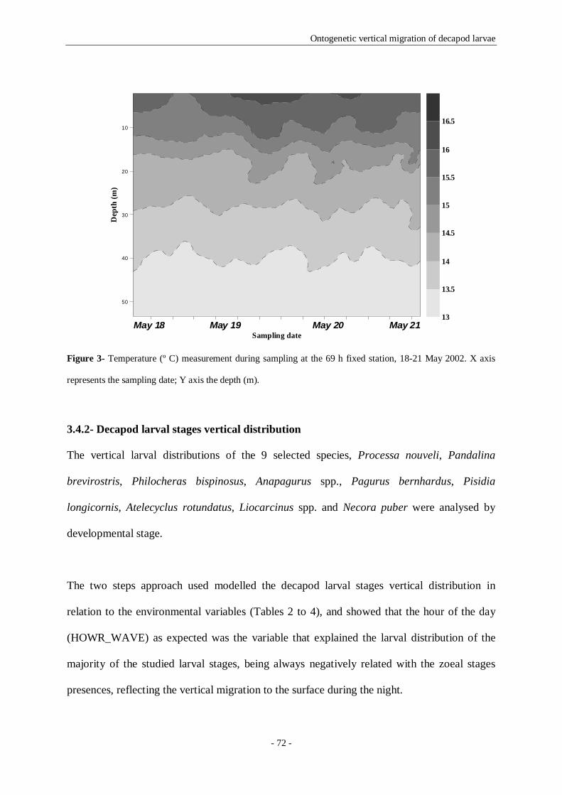
Ontogenetic vertical migration of decapod larvae
- 72 -
Figure 3- Temperature (º C) measurement during sampling at the 69 h fixed station, 18-21 May 2002. X axis
represents the sampling date; Y axis the depth (m).
3.4.2- Decapod larval stages vertical distribution
The vertical larval distributions of the 9 selected species, Processa nouveli, Pandalina
brevirostris, Philocheras bispinosus, Anapagurus spp., Pagurus bernhardus, Pisidia
longicornis, Atelecyclus rotundatus, Liocarcinus spp. and Necora puber were analysed by
developmental stage.
The two steps approach used modelled the decapod larval stages vertical distribution in
relation to the environmental variables (Tables 2 to 4), and showed that the hour of the day
(HOWR_WAVE) as expected was the variable that explained the larval distribution of the
majority of the studied larval stages, being always negatively related with the zoeal stages
presences, reflecting the vertical migration to the surface during the night.
20 25 30 35 40 45 50 55 60 65 70 75 80
Sampling date
50
40
30
20
10
Dep
th (m
)
13
13.5
14
14.5
15
15.5
16
16.5
May 18 May 19 May 20 May 21

Ontogenetic vertical migration of decapod larvae
- 73 -
Table 2- Coefficients of significant explanatory variables of the two steps model (presence or absence predicted
using a Logistic Model in the first step, values of abundance modelled with a Generalized Addictive Model in
the second step) describing the larval vertical distribution of the Caridea selected species collected with the
Neuston and LHPR nets in the fixed station. Levels of significance: ***p<0.001, **p<0.01,*p<0.05; n.s. not
significant.
n SALProcessa ZI presence 297 n.s. -0.788 *** 4.205 * n.s. -nouveli abundance - - - - - -
ZII presence 297 n.s. -0.566 ** n.s. -0.375 ** -abundance 58 n.s. n.s. n.s. n.s. n.s .
ZIII presence 297 n.s. -1.329 *** n.s. n.s. -abundance - - - - - -
ZIV presence 297 n.s. -0.796 * n.s. n.s. -abundance - - - - - -
ZV presence 297 n.s. -1.313 ** n.s. n.s. -abundance - - - - - -
ZVI presence 297 n.s. -1.069 ** n.s. n.s. -abundance - - - - - -
ZVII presence 297 - - - - -abundance - - - - - -
ZVIII presence 297 - - - - -abundance - - - - - -
ZIX presence 297 0.060 *** 0.934 *** -7.759 * n.s. -abundance 31 n.s. n.s. n.s. n.s. n.s .
M presence 297 - - - - -abundance - - - - - -
Pandalina ZI presence 297 0.044 * -1.294 * n.s. n.s. -brevirostris abundance - - - - - -
ZII presence 297 0.059 *** n.s. n.s. n.s. -abundance - - - - - -
ZIII presence 297 0.043 *** -0.812 *** n.s. n.s. -abundance 9 - n.s. n.s. n.s. n.s .
ZIV presence 297 n.s. -0.881 *** 11.320 *** n.s. -abundance 43 n.s. n.s. n.s. n.s. n.s .
ZV presence 297 n.s. -1.331 *** 7.208 ** n.s. -abundance 27 n.s. n.s. n.s. n.s. n.s .
ZVI presence 297 n.s. -1.253 *** 8.498 *** n.s. -abundance 85 n.s. n.s. n.s. 0.067 *** n.s .
ZVII presence 297 0.031 * -1.534 *** n.s. n.s. -abundance 119 n.s. n.s. 0.758 * n.s. 0.046
M presence 297 n.s. -3.357 *** 9.876 ** n.s. -abundance 8 n.s. - n.s. n.s. n.s .
Philocheras ZI presence 297 n.s. -0.909 *** n.s. n.s. -bispinosus abundance - - - - - -
ZII presence 297 n.s. -0.957 *** n.s. n.s. -abundance - - - - - -
ZIII presence 297 n.s. -1.204 *** 6.532 * n.s. -abundance - - - - - -
ZIV presence 297 n.s. -2.140 *** n.s. n.s. -abundance - - - - - -
ZV presence 297 n.s. -1.802 *** n.s. n.s. -abundance 81 0.003 ** n.s. n.s. 0.070 ** 0.035
M presence 297 n.s. -4.566 *** n.s. n.s. -abundance - - - - - -
Species Stage Model phasesCofficients of significant explanatory variables
MID_DEPTH HOUR_WAVE TEMP (linear) TEMP (spline)

Ontogenetic vertical migration of decapod larvae
- 74 -
Table 3- Coefficients of significant explanatory variables of the two steps model (presence or absence predicted
using a Logistic Model in the first step, values of abundance modelled with a Generalized Addictive Model in
the second step) describing the larval vertical distribution of the Anomura selected species collected with the
Neuston and LHPR nets in the fixed station. Levels of significance: ***p<0.001, **p<0.01,*p<0.05; n.s. not
significant.
The midpoint of depth stratum (MID_DEPTH) reflected the positioning of the larvae in a
certain water stratum, and it was important for almost all the studied taxa. It was always
positively related with the caridean shrimps (Table 2) and hermit crabs (Table 3) larval stages
distribution, with the exception of P. longicornis megalopa whose presence was negatively
related with this predictor. Brachyuran crabs larvae (Table 4) were always negatively related
with the midpoint of depth stratum, except A. rotundatus third zoeae presence that was
positively related. The last zoeal stages of the caridean shrimps, ninth, seventh and fifth zoeae
n SALAnapagurus spp. ZI presence 297 n.s. -1.504 *** -7.537 ** n.s . -
abundance - - - - - -
ZII presence 297 n.s. n.s. n.s. n.s . -abundance - - - - - -
ZIII presence 297 n.s. -2.026 *** n.s. n.s . -abundance - - - - - -
ZIV presence 297 n.s. -1.730 *** n.s. -0.926 *** -abundance 206 n.s. -0.163 *** 4.509 *** 0.444 ***
M presence 297 n.s. -3.588 *** n.s. n.s . -abundance 126 n.s. n.s. 3.649 *** 0.288 ** n.s.
Pagurus ZI presence 297 n.s. -3.419 *** n.s. n.s . -bernhardus abundance - - - - - -
ZII presence 297 n.s. -2.089 * n.s. n.s . -abundance - - - - - -
ZIII presence 297 0.059 *** -1.113 *** n.s. n.s . -abundance 17 n.s. n.s. n.s. 0.799 * n.s.
ZIV presence 297 0.059 *** -1.318 *** n.s. n.s . -abundance 81 0.010 * 0.267 *** n.s. n.s . n.s.
ZV presence 297 n.s. n.s. n.s. -1.194 *** -abundance - - - - - -
M presence 297 0.043 *** -1.246 *** n.s. n.s . -abundance 9 - n.s. n.s. n.s . n.s.
Pisidia ZI presence 297 n.s. -1.112 *** -10.101 *** n.s . -longicornis abundance 18 n.s. n.s. n.s. n.s . n.s.
ZII presence 297 n.s. -1.064 *** -7.726 * -0.847 *** -abundance 181 0.007 ** -0.067 * n.s. 0.179 ** -
M presence 297 -0.033 *** -3.671 *** n.s. n.s . -abundance 111 n.s. n.s. n.s. n.s . n.s.
Species Stage Model phases Cofficients of significant explanatory variablesMID_DEPTH HOUR_WAVE TEMP (linear) TEMP (spline)

Ontogenetic vertical migration of decapod larvae
- 75 -
for P. nouveli, P. brevirostris and P. bispinosus respectively, were always related with the
midpoint of depth stratum (Table 2).
Table 4- Coefficients of significant explanatory variables of the two steps model (presence or absence predicted
using a Logistic Model in the first step, values of abundance modelled with a Generalized Addictive Model in
the second step) describing the larval vertical distribution of the Brachyura selected species collected with the
Neuston and LHPR nets in the fixed station. Levels of significance: ***p<0.001, **p<0.01,*p<0.05; n.s. not
significant.
n SALAtelecyclus ZI presence 297 n.s. -1.665 *** -13.319 *** n.s. -rotundatus abundance 11 n.s. n.s. n.s. n.s. 0.0326
ZII presence 297 n.s. -1.493 *** n.s. 0.896 *** -abundance 58 n.s. n.s. - n.s. n.s .
ZIII presence 297 0.078 * -0.589 ** 16.630 *** 2.310 *** -abundance 10 n.s. n.s. - n.s. n.s .
ZIV presence 297 n.s. -0.647 *** n.s. 0.288 * -abundance 58 n.s. n.s. 0.829 * n.s. n.s .
ZV presence 297 n.s. -0.445 ** n.s. n.s. -abundance 180 n.s. -0.062 * 1.118 *** 0.142 ** n.s .
M presence 297 n.s. -2.819 *** n.s. n.s. -abundance 27 n.s. - - n.s. n.s .
Liocarcinus spp. ZI presence 297 -0.036 ** -1.026 *** -8.051 ** n.s. -abundance 97 -0.094 *** -0.537 ** n.s. n.s. n.s .
ZII presence 297 -0.042 *** -1.093 *** -5.619 * n.s. -abundance 145 -0.074 *** -0.463 *** n.s. n.s. n.s .
ZIII presence 297 -0.077 *** n.s. 6.485 * n.s. -abundance 19 n.s. -0.532 ** - n.s. n.s .
ZIV presence 297 n.s. -0.863 *** n.s. n.s. -abundance - - - - - -
ZV presence 297 n.s. n.s. -10.754 *** n.s. -abundance 293 n.s. n.s. -1.027 *** 0.157 *** n.s .
M presence 297 n.s. n.s. -18.498 *** -0.810 ** -abundance 286 -0.005 * -0.064 *** n.s. n.s. n.s .
Necora ZI presence 297 n.s. -1.138 ** n.s. 0.524 * -puber abundance - - - - - -
ZII presence 297 -0.093 *** -1.454 *** 15.598 ** n.s. -abundance - - - - - -
ZIII presence 297 -0.100 * n.s. n.s. n.s. -abundance - - - - - -
ZIV presence 297 -0.120 * n.s. n.s. n.s. -abundance - - - - - -
ZV presence 297 -1.303 ** n.s. n.s. n.s. -abundance - - - - - -
M presence 297 n.s. -0.562 ** n.s. 0.567 *** -abundance - - - - - -
Species Stage Model phases Cofficients of significant explanatory variablesMID_DEPTH HOUR_WAVE TEMP (linear) TEMP (spline)

Ontogenetic vertical migration of decapod larvae
- 76 -
Salinity (SAL) was always explaining some larval stages distribution, except for P.
bernhardus, and was in general related with the early and the late zoeae, as well as with the
megalopal presences and/or abundances.
Finally, the temperature (TEMP) influenced some larval stages of all species, and was in
general significantly related with the last zoeal stages and the megalopa distribution.
3.4.2.1- Vertical distribution of the caridean shrimps larvae
The modelling results (Table 2) confirmed that the three selected species had almost all the
zoeal and megalopal stages presences negatively related with the hour of the day
(HOUR_WAVE) (dos Santos et al. 2008). The three selected species had their last zoeal stage
presence positively related with the midpoint of depth stratum (MID_DEPTH). The first and
last zoeal stages presences of P. nouveli were significantly related with salinity (SAL),
although with different signals (positive for zoeae I and negative for zoeae IX). P. brevirostris
older zoeal stages (zoeae IV to VII) were always positively related with the salinity (SAL). P.
brevirostris and P. bispinosus had their late zoeal stages (sixth zoeae and fifth zoeae
respectively) positively related with the temperature (TEMP). For the three selected species
we present the average depth of distribution of all larval stages (Figs. 4-6). All showed similar
patterns of diel vertical distribution, being in a deeper position during the day, migrating to
the surface layers of the water column during the night, and returning back to the deeper
layers of the water column around the dawn.

Ontogenetic vertical migration of decapod larvae
- 77 -
Figure 4- Average depth of distribution of Processa nouveli (ind. m-3) zoeal stages: ZI (first zoea) to ZIX (ninth
and last zoea), and M (megalopa). The grey areas represent the night period.
P. nouveli second zoeal stage was the more abundant (0.460 ind.m-3), and the megalopa was
almost absent from the water column appearing only in the deeper strata, beyond the 25 m, in
the second night. The larval vertical migration with the upward movement to near the surface
during the night was clear (Fig. 4), for almost all the larval stages, so the higher abundances in
the water column were registered in the night period. The larval migration excursion was not
evident for the eighth and the megalopal larval stages, although, the first to seventh and the
last zoeae had their vertical distribution negatively related with the hour of the day,
confirming the migration to the deeper layers of the water column. These species zoeal stages
were not common in the neuston layer, and particularly the megalopa was sampled only in the
three deeper strata. As referred before, zoeae IX had his presence positively related with the
midpoint of depth stratum because of the higher larval concentrations beyond the 25 m during
all the sampling period (60% of the sampled larvae were distributed beyond the 25 m depth).
Taking into account the ontogenetic development, the first zoea was in a less profound depth
relatively to the second in the first night, but beyond here it was not possible to observe a
0
10
20
30
40
50
6016 20 0 4 8 12 16 20 0 4 8 12 16 20 0 4 8
Aver
age d
epth
of
dist
ribut
ion
(m)
Time of day (h)
ZVI
ZVII
ZVIII
ZIX
M
0
10
20
30
40
50
60
Aver
age d
epth
of
distr
ibut
ion
(m)
ZI
ZII
ZIII
ZIV
ZV

Ontogenetic vertical migration of decapod larvae
- 78 -
distributional pattern for the early zoeal stages. The last zoea was distributed at higher depths
during almost all the sampling period.
Figure 5- Average depth of distribution of Pandalina brevirostris (ind. m-3) zoeal stages: ZI (first zoea) to ZVII
(seventh and last zoea), and M (megalopa). The grey areas represent the night period.
The sixth zoeal stage was the more abundant (1.366 ind.m-3), and the first zoea was almost
absent from the water column. The megalopa never ascended to the neuston layer, and it was
sampled immediately after the sunset in the water column, being absent during the day
(Fig.5). The vertical migration was evident for these species larval stages whose presence in
the first 15 m of the water column was exclusively nocturnal. The first to third and the last
zoeae had their presences positively related with the midpoint of depth stratum because they
were concentrated in the deeper strata of the water column (over 70 % of each of the referred
larval stages were sampled in the strata above the 35 m). The fourth to sixth zoeae and the
megalopa had their presences positively related with the salinity, as well as the last zoea
abundance. Temperature only had a signicant effect in the sixth zoeal stage abundance.
0
10
20
30
40
50
6016 20 0 4 8 12 16 20 0 4 8 12 16 20 0 4 8
Aver
age d
epth
of
dist
ribut
ion
(m)
Time of day (h)
ZV
ZVI
ZVII
M
0
10
20
30
40
50
60
Aver
age d
epth
of
distr
ibut
ion
(m)
ZI
ZII
ZIII
ZIV
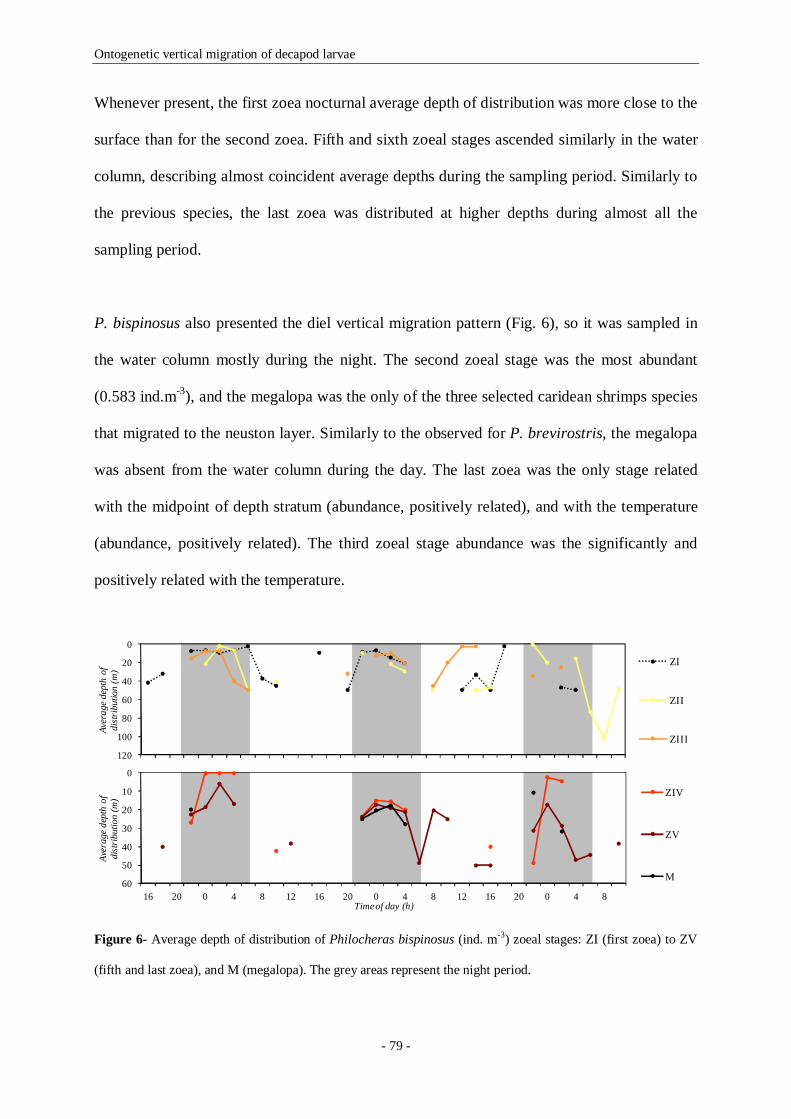
Ontogenetic vertical migration of decapod larvae
- 79 -
Whenever present, the first zoea nocturnal average depth of distribution was more close to the
surface than for the second zoea. Fifth and sixth zoeal stages ascended similarly in the water
column, describing almost coincident average depths during the sampling period. Similarly to
the previous species, the last zoea was distributed at higher depths during almost all the
sampling period.
P. bispinosus also presented the diel vertical migration pattern (Fig. 6), so it was sampled in
the water column mostly during the night. The second zoeal stage was the most abundant
(0.583 ind.m-3), and the megalopa was the only of the three selected caridean shrimps species
that migrated to the neuston layer. Similarly to the observed for P. brevirostris, the megalopa
was absent from the water column during the day. The last zoea was the only stage related
with the midpoint of depth stratum (abundance, positively related), and with the temperature
(abundance, positively related). The third zoeal stage abundance was the significantly and
positively related with the temperature.
Figure 6- Average depth of distribution of Philocheras bispinosus (ind. m-3) zoeal stages: ZI (first zoea) to ZV
(fifth and last zoea), and M (megalopa). The grey areas represent the night period.
0
10
20
30
40
50
6016 20 0 4 8 12 16 20 0 4 8 12 16 20 0 4 8
Aver
age d
epth
of
dist
ribut
ion
(m)
Time of day (h)
ZIV
ZV
M
0
20
40
60
80
100
120
Aver
age d
epth
of
distr
ibut
ion
(m)
ZI
ZII
ZIII

Ontogenetic vertical migration of decapod larvae
- 80 -
Similarly to the described for previous species, the last zoeal stage was distributed in a more
deep position during almost all the sampling period.
3.4.2.2- Vertical distribution of the anomuran crabs larvae
Anapagurus spp. (Table 3) distribution did not showed any relation with the midpoint of
depth stratum (MID_DEPTH), but this predictor was significantly related with the other two
species older larval stages (zoeae three to five and megalopa of P. bernhardus, zoea two and
megalopa of P. longicornis). Salinity was important for the distribution of the first, last and
megalopal stages of Anapagurus spp. with contrary signals: it was positively related with the
first zoea presence and negatively related with last zoea and megalopal presence and
abundance respectively; P. bernhardus did not showed any relation with salinity; P.
longicornis zoeal stages were both negatively related with this predictor. The three species
larval distributions were also influenced by the temperature, having their presences negatively
related with this predictor (Anapagurus spp. fourth zoeal stage, P. bernhardus fifth zoeal
stage and P. longicornis second zoeal stage) and the abundances positively related
(Anapagurus spp. fourth zoeal stage, P. bernhardus third zoeal stage and P. longicornis
second zoeal stage). Figures 7 to 9 show the average depth of distribution of all larval stages
of the three species. The three species had similar patterns of diel vertical distribution, being
almost absent from the water column during the daytime (presences negatively related with
the hour of the day).
Anapagurus spp. was the anomuran most abundant species, being the last zoeal stage the
more represented during the sampling period (7.285 ind.m-3). The megalopa was almost
absent from the water column during the day and when present it was beyond the 35 m depth;
the first to third zoeae appeared in the more superficial strata during the night, having, as well

Ontogenetic vertical migration of decapod larvae
- 81 -
as the megalopa some of their average depth of distribution points in the neuston layer (Fig.
5). The last zoea was always in a more profound distribution relatively to the other zoeal
stages, and during the day it was consistently present in the 30 to 50 m depth layer. The first
zoeal stage appeared with higher abundances in the second and third nights, probably because
it was transported by the less saline water lens advected from nearshore, arrived to the fixed
point during the second day of sampling (19th May at 12h). The first zoeae presence in this
less saline water lens was indicated by the salinity effect (ZI presence negatively related with
the salinity).
Figure 7- Average depth of distribution of Anapagurus spp. (ind. m-3) zoeal stages: ZI (first zoea) to ZIV (fourth
and last zoea), and M (megalopa). The grey areas represent the night period.
P. bernhardus was more abundant in the deeper strata than at the surface, and migrated to less
deep strata during the night (Fig. 8). The nocturnal migration from close to the bottom to a
less deep stratum of the water column, but rarely to the neuston layer, was evident. The larval
stages which were related with the midpoint of depth stratum were concentrated in a more
deep position (Fig. 8), being 74% of the third zoea and 62% of the fourth distributed beyond
the 35 m, and 58 % of the megalopae sampled were distributed deeper than 35 m. Zoeae four
and five, as well as the megalopa were distributed at higher depths during the day. The
temperature also explained the larval distribution of third and fifth zoeal stages, probably
because these were concentrated deeper in the water column, above the thermocline.
0
10
20
30
40
50
6016 20 0 4 8 12 16 20 0 4 8 12 16 20 0 4 8
Aver
age d
epth
of
dist
ribut
ion
(m)
Time of day (h)
ZI
ZII
ZIII
ZIV
M
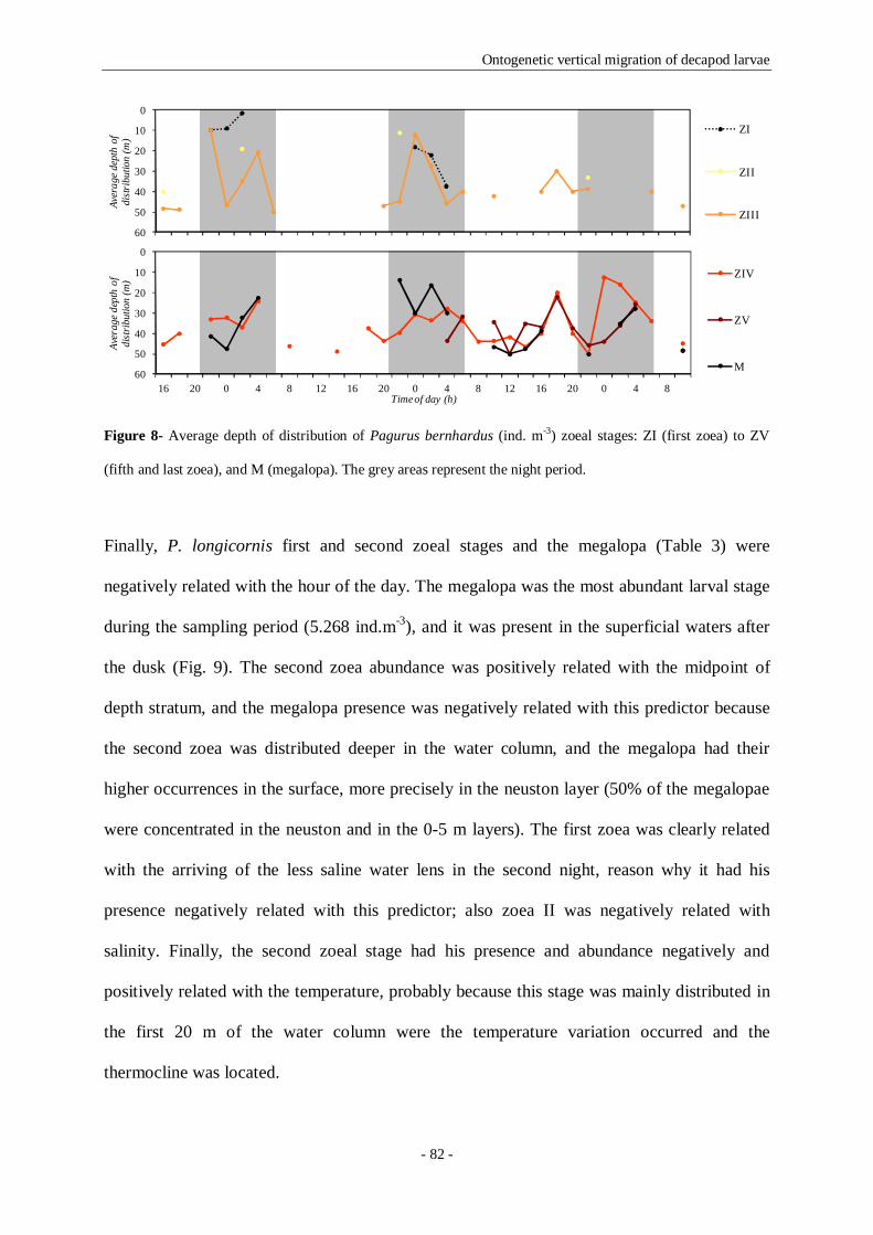
Ontogenetic vertical migration of decapod larvae
- 82 -
Figure 8- Average depth of distribution of Pagurus bernhardus (ind. m-3) zoeal stages: ZI (first zoea) to ZV
(fifth and last zoea), and M (megalopa). The grey areas represent the night period.
Finally, P. longicornis first and second zoeal stages and the megalopa (Table 3) were
negatively related with the hour of the day. The megalopa was the most abundant larval stage
during the sampling period (5.268 ind.m-3), and it was present in the superficial waters after
the dusk (Fig. 9). The second zoea abundance was positively related with the midpoint of
depth stratum, and the megalopa presence was negatively related with this predictor because
the second zoea was distributed deeper in the water column, and the megalopa had their
higher occurrences in the surface, more precisely in the neuston layer (50% of the megalopae
were concentrated in the neuston and in the 0-5 m layers). The first zoea was clearly related
with the arriving of the less saline water lens in the second night, reason why it had his
presence negatively related with this predictor; also zoea II was negatively related with
salinity. Finally, the second zoeal stage had his presence and abundance negatively and
positively related with the temperature, probably because this stage was mainly distributed in
the first 20 m of the water column were the temperature variation occurred and the
thermocline was located.
0
10
20
30
40
50
6016 20 0 4 8 12 16 20 0 4 8 12 16 20 0 4 8
Aver
age d
epth
of
dist
ribut
ion
(m)
Time of day (h)
ZIV
ZV
M
0
10
20
30
40
50
60
Aver
age d
epth
of
distr
ibut
ion
(m)
ZI
ZII
ZIII

Ontogenetic vertical migration of decapod larvae
- 83 -
Figure 9- Average depth of distribution of Pisidia longicornis (ind. m-3) zoeal stages: ZI (first zoea) to ZII
(second and last zoea), and M (megalopa). The grey areas represent the night period.
3.4.2.3- Vertical distribution of the brachyuran crabs larvae
The midpoint of depth stratum was an important predictor in the explanation of brachyuran
larval stages distribution, probably because the three selected species were very represented in
the neuston layer during the night (Table 4). Salinity explained the distribution of most of the
sampled stages of Liocarcinus spp. The effect of temperature was important for the second to
fifth zoeal stage of A. rotundatus, last zoea and megalopa of Liocarcinus spp., and the first
zoea and megalopa of N. puber.
Figure 10- Average depth of distribution of Atelecyclus rotundatus (ind. m-3) zoeal stages: ZI (first zoea) to ZV
(fifth and last zoea), and M (megalopa). The grey areas represent the night period.
0
10
20
30
40
5016 20 0 4 8 12 16 20 0 4 8 12 16 20 0 4 8
Aver
age d
epth
of
dist
ribut
ion
(m)
Time of day (h)
ZI
ZII
M
0
10
20
30
40
50
6016 20 0 4 8 12 16 20 0 4 8 12 16 20 0 4 8
Aver
age
dept
h of
di
strib
utio
n (m
)
Time of day (h)
ZIV
ZV
M
0
10
20
30
40
50
60
Aver
age d
epth
of
distr
ibut
ion
(m)
ZI
ZII
ZIII
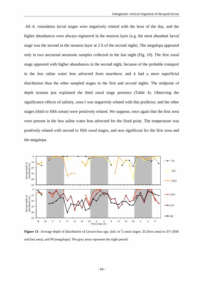
Ontogenetic vertical migration of decapod larvae
- 84 -
All A. rotundatus larval stages were negatively related with the hour of the day, and the
higher abundances were always registered in the neuston layer (e.g. the most abundant larval
stage was the second in the neuston layer at 2 h of the second night). The megalopa appeared
only in two nocturnal neustonic samples collected in the last night (Fig. 10). The first zoeal
stage appeared with higher abundances in the second night, because of the probable transport
in the less saline water lens advected from nearshore, and it had a more superficial
distribution than the other sampled stages in the first and second nights. The midpoint of
depth stratum just explained the third zoeal stage presence (Table 4). Observing the
significance effects of salinity, zoea I was negatively related with this predictor, and the other
stages (third to fifth zoeae) were positively related. We suppose, once again that the first zoea
were present in the less saline water lens advected for the fixed point. The temperature was
positively related with second to fifth zoeal stages, and non significant for the first zoea and
the megalopa.
Figure 11- Average depth of distribution of Liocarcinus spp. (ind. m-3) zoeal stages: ZI (first zoea) to ZV (fifth
and last zoea), and M (megalopa). The grey areas represent the night period.
0
10
20
30
40
5016 20 0 4 8 12 16 20 0 4 8 12 16 20 0 4 8
Aver
age d
epth
of
dist
ribut
ion
(m)
Time of day (h)
ZIV
ZV
M
0
10
20
30
40
50
Aver
age d
epth
of
distr
ibut
ion
(m)
ZI
ZII
ZIII

Ontogenetic vertical migration of decapod larvae
- 85 -
Liocarcinus spp. was the most abundant species during the fixed point sampling. The first
zoea was the most abundant during the sampling period (99.581 ind.m-3, at 0h of the second
night, in the neuston layer). All the larval stages were negatively related with the hour of the
day with the exception of the fifth zoea, that did not presented a clear vertical migration
pattern even having some neustonic presences during the night (Fig. 11). The midpoint of
depth stratum was always negatively related with the first to third zoeae and with the
megalopal distributions, because these stages were concentrated more close to the surface.
The first zoea in the first and second nights was mainly neustonic, and fifth zoea was more
deeply distributed in the water column. The salinity was negatively related with the first,
second, last and megalopal stages presences, though for the third zoea presence it was
positively related. Once again we suppose that the salinity effect reflected the presence of the
less saline water lens that probably transported the newly hatched zoeae and for some reason
advected the third zoea that in the second night of sampling was almost absent from the water
column. The last zoeal stage and the megalopa had their abundance and presence positively
and negatively related with the temperature.
Figure 12- Average depth of distribution of Necora puber (ind. m-3) zoeal stages: ZI (first zoea) to ZV (fifth and
last zoea), and M (megalopa). The grey areas represent the night period.
0
10
20
30
40
50
6016 20 0 4 8 12 16 20 0 4 8 12 16 20 0 4 8
Aver
age d
epth
of
dist
ribut
ion
(m)
Time of day (h)
ZIV
ZV
M
0
10
20
30
40
50
Aver
age d
epth
of
distr
ibut
ion
(m)
ZI
ZII
ZIII

Ontogenetic vertical migration of decapod larvae
- 86 -
N. puber second zoeal stage was the most abundant during the fixed point sampling (58.903
ind.m-3, at 2h of the first night, in the neuston layer). The second to fifth zoeal stages were
negatively related with the midpoint of depth stratum, because these stages had their larvae
concentrated at the surface (more than 84% of these stages were in the upper 10 m of the
water column). The second zoea presence was positively related with the salinity, probably
because of the arrived of the less saline water lens that advected it away from the fixed point
station. At last, the temperature was positively related with the first zoea and the megalopa,
both stages having their maximum in the neuston layer, above the thermocline.
3.5- Discussion
Dos Santos et al. (2008) described the diel vertical migrations of the decapod larvae in the
Portuguese upwelling coast, demonstrating that this behaviour was present in the caridean
shrimps, the anomuran and the brachyuran crabs. Present work verified the existence of the
diel vertical migration (DVM) in almost all the larval stages of the studied species, showing
that all the larval stages were capable of swim through the water column, mitigating the
seaward transport alternating between the surface and the deeper or bottom layers. According
to Peterson (1998) this is the movement responsible for the avoidance of the larvae seaward
dispersal. Marta-Almeida et al. (2006) model showed that if crab larvae perform DVM as
described, then upwelling may enhance retention of larvae in the inner shelf. Dos Santos et al.
(2008) confirmed that the DVM is the mechanism that permits to decapods in general the
larval retention in the inner shelf and also over the shelf. Present results demonstrated that the
9 selected taxa had most of the times their newly hatched zoeae distributed in a more

Ontogenetic vertical migration of decapod larvae
- 87 -
superficial position, and their last zoeae were almost always occupying the deeper strata of the
water column.
Knowing that the light (e.g. Sulkin 1975), the salinity (e.g. Latz e Forward 1977) and the
temperature (e.g. Dawirs 1985) are determinant factors in the vertical positioning of decapod
crustacean larvae, we verified that almost all the selected species larval stages had their
presences negatively related with the hour of the day, as expected, indicating that the studied
zoeal stages and the megalopa migrated vertically in the water column in response to the diel
cycle, and had their higher larval concentrations in the water column during the night (results
previously obtained by dos Santos et al. 2008, and studied by stage in present work). Of the
four selected environmental cues, the diel cycle was the most important, explaining almost all
the larval stages distributions, being the exceptions: the seventh, eighth, and megalopal stages
of P. nouveli; the second zoea of P. brevirostris; the second zoea of Anapagurus spp.; the
fifth zoea of P. bernhardus; the fifth zoea of Liocarcinus spp.; and the third to fifth zoeae of
N. puber. It should be noticed that the hour of the day was many times non significant for the
late zoeal stages distributions, probably because these zoeae started to be synchronized with a
different environmental cycle. Queiroga & Blanton (2005) affirmed that there are two main
types of vertical migration: the cyclic migration and the ontogenetic migration, being the first
characterized by the larvae moving up and down in the water column in synchrony with one
or more environmental cycles.
The midpoint of depth stratum predictor reflected the positioning of the larvae in a certain
depth. All the three caridean shrimps and the anomuran hermit crabs had their last zoeae
positively related with this factor, because they were distributed in the deeper layers of the
water column, as the average depth of distribution graphs demonstrated. There are at least two
Ontogenetic vertical migration of decapod larvae
- 88 -
explanantions for the larval positioning in the deeper layers of the water column: it may result
of the ontogenetic vertical migration with a change of the average depth of distribution
through development (e.g. Queiroga et al. 2005), or it may be describing a strategy to enhance
the larval retention over the shelf (e.g. Peterson 1998). It is known that some larvae avoid the
surface layer where currents tend to be faster, residing deeper in the water column, retarding
the transport, and as a consequence favouring the retention (e.g. Shanks and Brink 2005,
Shanks and Eckert, 2005, Morgan et al. 2009). Contrary to these, P. longicornis megalopa,
Liocarcinus spp. zoeal and megalopal stages, and N. puber zoeal stages were negatively
related with the midpoint of depth stratum, probably because these larvae had more than 40%
of the sampled larvae concentrated at the surface, in the neuston layer, during the night. All
these three taxa when adults are distributed over the shelf but also inshore (Zariquiey-Álvarez
1968, d’Udekem d’Acoz 1999), so we can suppose that at least for the megalopa this
superficial distribution may reflect the transport mechanism to the settlement areas (e.g.
Shanks 1985, Tapia et al. 2004). The larval concentrations in the neuston had a peak during
the night, and decreased rapidly before sunrise. According to dos Santos et al. (2008) the
diurnal distribution of the larvae suggested that both the zoeal and megalopal stages moved to
a narrow layer between the 55 m and the bottom at 63 m.
In the present study sometimes the ontogenetic vertical migrations were not so evident,
probably because during sampling the hydrological conditions were complex, and the arrival
of the less saline water lens to the fixed point lead to the rearrangement of the larval vertical
distribution as a response to the hydrological changes. Although, similarly to what have been
described (see Lindley et al. 1994 for examples), the first zoeal stage seems to reach the
neuston layer more times than the last zoeal stage, and was many times distributed more close
to the surface than the second zoeal stage; also the megalopa had many times their maximum

Ontogenetic vertical migration of decapod larvae
- 89 -
abundances at the neuston layer, but the average depth of distribution was many times more
or less coincident with the last zoea distribution, in a more deep position during the day. The
megalopal higher abundances were registered in the neuston layer, and as referred by previous
authors this position can reflect the transport mechanism. As dos Santos et al. (2008)
discussed, this megalopal presence at the surface water layer did not reflected the internal
waves transport (Shanks 1985) or the daily sea breezes transport (Tapia et al. 2004). Of the 9
studied species, Anapagurus spp., P. longicornis, Atelecyclus rotundatus, Liocarcinus spp.
and N. puber had their megalopae concentrated in the neuston during the night.
Changes during ontogeny in the osmorregulation of decapod crustacean larvae exist, and are
described as an ontogenetic increase in the regulatory capability with the early larval stages in
general showing weaker regulatory capabilities than the conspecific juveniles or adults, and
so are less tolerant to salinity changes in the environment (Anger 2001). In the literature, the
salinity effect is more noticed in the vertical migration excursion than the temperature, and
the haloclines involving differences as small as 1 may stop the ascending movement to the
surface (Hughes, 1969; Roberts, 1971), inhibiting, rather than promoting, larvae to ascend to
surface convergent flows. The effect of salinity was important for the analysed larval stages
once that it reflected the presence of the less saline water mass beyond the 12h of the second
day. A negative effect of the salinity in the first zoeal stage distribution of Anapagurus spp.,
P. longicornis, A. rotundatus and Liocarcinus spp. probably is reflecting the transport of the
newly hatched larvae in the advected water lens from the inner shelf. Whenever the effect was
positive, then we had larvae with a deeper distribution in the water column. Also this relation
reflected the larval distribution response to the arrival of the less saline water lens, once that
the water mass reached the fixed point from the surface in the second day of sampling, it was
advected, and through time it showed a wedge form, occupying the first 15-20 m depth of the
Ontogenetic vertical migration of decapod larvae
- 90 -
water column at the end of the sampling period. As a consequence, the water mass here
present in the first day was moved to a more offshore position, and probably the larvae therein
sinked in the water column or were advected to a different place.
The temperature effect reflected the distribution of the larvae in relation to the thermocline. It
had most of the times a positive effect probably because these larvae had a bimodal
distribution showing their maximum abundances at the surface during the night, and
remaining in the deeper strata or close to the bottom during the day. Observing the larval
distributions of the taxa that had their distributions negatively related with the temperature,
we see that Anapagurus spp. fourth zoea was more abundant in the 5-20 m depth, P.
bernhardus fifth zoea was distributed beyond the 20 m, and P. longicornis second zoea was
more abundant from the surface to the 20 m depth hey have their higher larval concentrations
above or below the thermocline.
Taking into account all the assumptions made, we are in accordance with dos Santos et al.
(2008). Present study demonstrates that decapod crustacean larvae have their vertical
migration movements’, ontogenetic and diel, and with this mechanisms are capable of
regulate their transport. In fact not only the existence of diel vertical migration permits the
larval retention over the shelf (dos Santos et al. 2008), but also the existence of the
ontogenetic vertical migration which permits the sinking behaviour in the water column
probably to enhance larval survival during the return to the adult habitat, and settlement when
competent. The idea of decapod larvae trapped at the frontal convergence zones (e.g. Pineda
1999, Shanks et al. 2000) does not fit in the studied taxa, even considering the larval stages
separately. We believe that the larval retention over the shelf results of a constant response to

Ontogenetic vertical migration of decapod larvae
- 91 -
the environmental conditions. These responses are most of the times revealed as the vertical
distribution of the larvae.
3.6- References
Abelló P. & Guerao G. (1999) Temporal variability in the vertical and mesoscale spatial
distribution of crab megalopae (Crustacea: Decapoda) in the Northwestern
Mediterranean. Estuarine. Coastal and Shelf Science 49, 129-139.
Agard J.B.R. (1999) A four-dimensional response surface analysis of the ontogeny of
physiological adaptation to salinity and temperature in larvae of the palaemonid shrimp
Macrobrachium rosenbergii (de Man). Journal of Experimental Marine Biology and
Ecology 236, 209-233.
Christy J.H. & Morgan S.G. (1998) Estuarine immigration by crab postlarvae: mechanisms,
reliability and adaptive significance. Marine Ecology Progress Series 174, 51-65.
Dawirs R.R. (1985) Temperature and larval development of Carcinus maenas (Decapoda) in
the laboratory; predictions of larval dynamics in the sea. Marine Ecology Progress Series
24, 297-302.
dos Santos A. & Lindley J.A. (2001) Crustacea, Decapoda: Larvae, II. Dendrobranchiata
(Aristeidae, Benthesicymidae, Penaeidae, Solenoceridae, Sicyonidae, Sergestidae and
Luciferidae). ICES Identification Leaflets for Plankton 186, 1-9.
dos Santos A. & González-Gordillo J.I. (2004) Illustrated keys for the identification of the
Pleocyemata (Crustacea, Decapoda) zoeal stages, from the coastal region of south-
western Europe. Journal of the Marine Biological Association of the UK 84, 205-227.
Ontogenetic vertical migration of decapod larvae
- 92 -
dos Santos A., Santos, A. M. P. & Conway, D. V. P. (2007) Horizontal and vertical
distribution of cirripede cyprid larvae in an upwelling system off the Portuguese coast.
Marine Ecology Progress Series 3269, 145-155.
dos Santos A., Santos A.M.P., Conway D.V.P., Bartilotti C., Lourenço P., Queiroga H. (2008)
Diel vertical migration of decapod larvae in the portuguese coastal upwelling ecosystem:
implications for offshore transport. Marine Ecology Progress Series 359, 171-183.
d’Udekem d’Acoz C. (1999). Inventaire et distribution des crustacés décapodes de
l’Atlantique nord-oriental, de la Méditerranée et des eaux continentales adjacentes au
nord de 25º N. Collection Patrimoines Naturels (M.N.H.N./S.P.N.) 40, 1-383.
Forward R.B. Jr. (1989) Behavioral responses of crustacean larvae to rates of salinity change.
Biological Bulletin 176, 229-238.
Forward R.B. Jr. & Rittschof D. (2000) Alteration of photoresponses involved in diel vertical
migration of a crab larva by fish mucus and degradation products of
mucopolysaccharides. Journal of the Experimental Marine Biology and Ecology 245,
277-292.
Forward R.B. Jr., Swanson J., Tankersely R.A., Welch J.M. (1997) Endogenous swimming
rhythms of blue crab, Callinectes sapidus, megalopae: effects of offshore and estuarine
cues. Marine Biology 127, 621-628.
Garrison L.P. (1999) Vertical migration behavior and larval transport in brachyuran crabs.
Marine Ecology Progress Series 176, 103-113.
González-Gordillo J.I., dos Santos A., Rodríguez A. (2001) Checklist and annotated
bibliography of decapod Crustacea larvae from the southwestern European coast
(Gibraltar Strait area). Scientia Marina 65, 275-305.
Hobbs R.C. & Botsford L.W. (1992) Diel vertical migration and timing of metamorphosis of
larvae of the Dungeness crab Cancer magister. Marine Biology 112, 417–428.
Ontogenetic vertical migration of decapod larvae
- 93 -
Hughes D.A. (1969) Responses to salinity changes as a tidal transport mechanism of pink
shrimp Penaeus duodarum. Biological Bulletin 136, 43–53.
Jamieson G.S & Phillips A.C. (1988) Occurrence of Cancer crab (C. magister and C.
oregonensis) megalopae off the west coast of Vancouver Island, British Columbia.
Fishery Bulletin 86, 525-542.
Latz M.I. & Forward R.B. Jr. (1977) The effect of salinity upon phototaxis and geotaxis in a
larval crustacean. Biological Bulletin 153, 163-179. Lindley J.A. (1986) Vertical distributions of decapod crustacean larvae and pelagic post-
larvae over Great Sole Bank (Celtic Sea) in June 1983. Marine Biology 90, 545-549.
Lindley J.A., Williams R., Conway D.V.P. (1994) Variability in dry weight and vertical
distributions of decapod larvae in the Irish Sea and North Sea during the sea. Marine
Biology 120, 385-395.
Marta-Almeida M., Dubert J., Peliz A., Queiroga H. (2006) Influence of vertical migration
pattern on retention of crab larvae in a seasonal upwelling system. Mar. Ecol. Prog. Ser.
307, 1-19.
Morgan S.G., Fisher J.L., Mace1 A.J., Akins L., Slaughter A.M., Bollens S.M. (2009). Cross-
shelf distributions and recruitment of crab postlarvae in a region of strong upwelling.
Marine Ecology Progress Series 380, 173-185.
Pearre S. (2003) Eat and run? The hunger/ satiation hypothesis in vertical migration: history,
evidence and consequences. Biological Reviews of the Cambridge Philosophical Society
78, 1–79.
Peliz A., Marchesiello P., Dubert J., Marta-Almeida M., Roy C., Queiroga H. (2007) A study
of crab larvae dispersal on the western Iberian shelf: physical processes. Journal of
Marine Systems 68, 215–236.
Ontogenetic vertical migration of decapod larvae
- 94 -
Peterson W. (1998) Life cycle strategies of copepods in coastal upwelling zones. J. Mar. Syst.
15, 313-326.
Pineda J. (1999) Circulation and larval distribution in internal tidal bore warm fronts.
Limnology and Oceanography 44, 1400–1414.
Pineda J., Hare J.A., Sponaugle S. (2007) Larval transport and dispersal in the coastal ocean
and consequences for population connectivity. Oceanography 20, 22-39.
Queiroga H. (1996). Distribution and drift of the crab Carcinus maenas (L.) (Decapoda,
Portunidae) larvae over the continental shelf off northern Portugal in April 1991. Journal
of Plankton Research 18, 1981–2000.
Queiroga H. & Blanton J. (2005). Interactions between behaviour and physical forcing in the
control of horizontal transport of decapod crustacean larvae. Adv. Mar. Biol. 47, 107-214.
Roberts Jr. M.H. (1971) Larval development of Pagurus longicarpus Say reared in the
laboratory. III. Behavioural responses to salinity discontinuities. Biological Bulletin 140,
489–501.
SAS Institute (2008) SAS/Stat User’s Guide. Version 9.2. Cary, NC, USA.
Shanks A.L. (1985) The behavioral basis of internal wave induced shoreward transport of the
megalopae of Pachygrapsus crassipes. Marine Ecology Progress Series 24, 289-295.
Shanks A.L., Largier J., Brink L. (2000). Demonstration of the onshore transport of larval
invertebrates by the shoreward movement of an upwelling front. Limnology and
Oceanography 45, 230–236.
Shanks A.L. & Brink L. (2005) Upwelling, downwelling, and cross-shelf transport of bivalve
larvae: test of a hypothesis. Marine Ecology Progress Series 302, 1–12.
Shanks A.L. & Eckert G.L. (2005) Population persistence of California current fishes and
benthic crustaceans: a marine drift paradox. Ecological Monographs 75, 505–524.

Ontogenetic vertical migration of decapod larvae
- 95 -
Sulkin S.D. (1975) The influence of light in the depth regulation of crab larvae. Biological
Bulletin 148, 333-343.
Tapia F.J., Pineda J., Ocampo-Torres F.J., Fuchs H.L., Parnell P.E., Montero P., Ramos S.
(2004) High-frenquency observations of wind-forced onshore transport at a coastal site in
Baja California. Continental Shelf Research 24, 1573–1585.
Wenger S.J. & Freeman M.C. (2008). Estimating species occurrence, abundance, and
detection probability using zero-inflated distributions. Ecology 89, 2953-2959.
Williams R., Collins N.R., Conway D.V.P. (1983) The double LHPR system, a high speed
micro- and macroplankton sampler. Deep-Sea Research 30, 331-342.
Zariquiey-Alvarez R. (1968). Crustáceos Decápodos Ibéricos. Investigacion Pesquera 32, 1-
510.

CHAPTER 4
_______________________________________________
Shedding light over the larval genus Eretmocaris –
morphological larval features of two closely related
trans-isthmian Lysmata species using
laboratory cultured material*
*Bartilotti C., Calado R., Rhyne A., dos Santos A. (Submitted) Shedding light over the
larval genus Eretmocaris – morphological larval features of two closely related trans-
isthmian Lysmata species using laboratory cultured material. Helgoland Marine
Research.

Zoeal development of the shrimps of genus Lysmata
- 97 -
4.1- Abstract
The knowledge of morphological changes during larval development is known to be of vital
importance for the understanding of phylogenetic relationships among related species. Of the
37 Lysmata valid species worldwide, only three have their complete larval series known from
laboratorial studies. Present work analyses the larval development of two closely related
trans-isthmian species of Lysmata using laboratory cultured material. The morphological
larval features of the first four zoeal stages of both species, fifth to seventh stages of L.
galapagensis (Eastern Pacific Ocean), and the last stage of L. moorei (Southwestern Atlantic
Ocean) are described and compared with the larval descriptions currently available for the
genus. Morphological characters showed a very homogeneous development in both species
that hatch in a similar developmental form as L. seticaudata, displaying their first and fifth
pereiopods as buds. The existing correspondence between the zoeal characters and the
phylogenetic results for Lysmata genus is very interesting: L. galapagensis, L. moorei and L.
seticaudata belong to the “Cosmopolitan Clade” / “Lysmata Clade” (Baeza et al. 2009,
Fiedler et al. submitted). We hypothesize that all the species within this clade will hatch with
the first and fifth pereiopods as buds, and will present a maximum of nine zoeal stages. The
resemblance between the zoeal characters described in the present work and those previously
described for the composite larval genus Eretmocaris are discussed, with emphasis to
Eretmocaris corniger. The unique larval features of E. corniger, namely an extremely long
rostrum and a spine in the dorsal surface of third pleomere, are also recorded in L.
galapagensis enabling us to match the larval forms earlier identified as the Tropical Eastern
Pacific E. corniger to a known adult Lysmata species. The possible identity of E. corniger
larvae recorded over one century ago from the Tropical Eastern Atlantic is also discussed.

Zoeal development of the shrimps of genus Lysmata
- 98 -
4.2- Introduction
In recent years, caridean shrimp in the genus Lysmata have become extremely popular in the
marine aquarium industry and have reached high market prices, sometimes ranging over 40 €
per specimen for brightly colored species (Calado et al. 2003a, Calado 2008). Presently there
are 37 valid species in the genus worldwide (Anker et al. 2009), in habitats ranging from
tropical coral reefs to rocky shores in warm-temperate waters (Debelius 2001).
Lysmata culture efforts have been an excellent opportunity to increase the knowledge on its
ecology and reproductive biology. Given the key-role played by larviculture for the successful
production of Lysmata in captivity, mainly to supply the marine aquarium trade, it is
surprising to record how little information has been published on their larval development and
morphology. Apart from the recent work by Calado et al. (2004) with the redescription of the
larval stages of L. seticaudata, the complete larval morphology and development patterns of
Lysmata are only known for two more species in the genus, L. ensirostris (Pillai 1974) and
Kurata’s L. wurdemanni (whose adult species identity is not possible to determine) (Kurata
1970). The knowledge of the morphological changes occurring during larval development are
essential for the understanding of phylogenetic relationships (e.g. Clark 2009), as well as
species geographical differentiation (e.g. Schubart et al. 2005), and ecology (e.g. Levin 2006).
In the “Report of the Scientific Results of the Voyage of H.M.S. Challenger during the years
1873-76” Bate (1888) founded a new genus, Eretmocaris, on which he included four larval
types, E. remipes, E. longicaulis, E. stylorostris and E. corniger. All these larvae shared a
common and remarkable morphological feature: a long and slender ocular peduncle. Caroli

Zoeal development of the shrimps of genus Lysmata
- 99 -
(1918), when studying the larvae of L. seticaudata (Risso, 1816), acknowledged the similarity
of those larval stages with Eretmocaris larvae and hypothesized that they could actually be
the larval forms of two or more caridean shrimp genera, including Lysmata Risso, 1816.
Several years later, Gurney (1937) effectively demonstrated that the great elongation of the
eyestalks, as well as the precocious development of leg 5 before the appearance of legs 3 and
4 into an enormously long appendage with a paddle-like enlargement of the propodus in
Eretmocaris specimens, were in fact larval features displayed by hippolytid shrimp in the
genus Lysmata and allied genus such as Exhippolysmata Stebbing, 1915 and Lysmatella
Borradaile, 1915. Although Eretmocaris has never been validated, the name has been
continually used by several researchers to classify larval forms collected from the plankton
presenting the characters described above (e.g. Lindley et al. 2002, Brightdoom et al. 2006).
One of the most remarkable Eretmocaris is certainly E. corniger. This larval form displays
unique morphological characters among known Eretmocaris, namely a prominent spine on
dorsal surface of the third pleomere and a remarkably long rostrum with several dorsal teeth
(Gurney 1937). Gopalakrishnan & Laurs (1971) reported and described as E. corniger several
specimens from the Eastern Tropical Pacific and suggested a relationship between the
occurrences of these larvae and the eastern ocean tropical islands. The adult form of Lysmata
corresponding to E. corniger larvae remained unknown, both from the Atlantic as from the
Pacific oceans, and so far no Lysmata species has ever been reported to occur simultaneously
in both of these oceans.
The very recent work of Baeza et al. (2009) presents the molecular phylogeny of Lysmata
shrimps based on a segment of the 16S RNA mitochondrial gene analysis. Their results define
a phylogenetic tree with three main clades: the “Tropical American Clade”, the “Cleaner
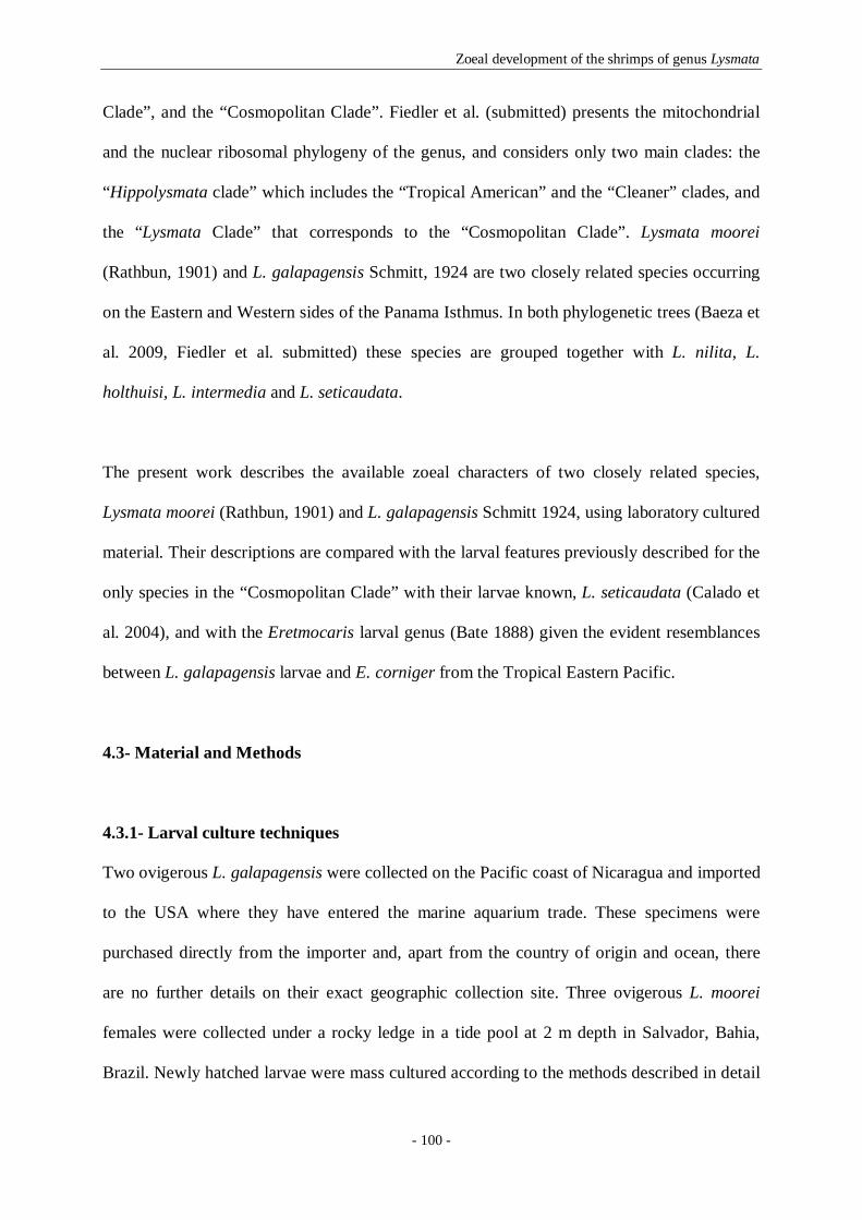
Zoeal development of the shrimps of genus Lysmata
- 100 -
Clade”, and the “Cosmopolitan Clade”. Fiedler et al. (submitted) presents the mitochondrial
and the nuclear ribosomal phylogeny of the genus, and considers only two main clades: the
“Hippolysmata clade” which includes the “Tropical American” and the “Cleaner” clades, and
the “Lysmata Clade” that corresponds to the “Cosmopolitan Clade”. Lysmata moorei
(Rathbun, 1901) and L. galapagensis Schmitt, 1924 are two closely related species occurring
on the Eastern and Western sides of the Panama Isthmus. In both phylogenetic trees (Baeza et
al. 2009, Fiedler et al. submitted) these species are grouped together with L. nilita, L.
holthuisi, L. intermedia and L. seticaudata.
The present work describes the available zoeal characters of two closely related species,
Lysmata moorei (Rathbun, 1901) and L. galapagensis Schmitt 1924, using laboratory cultured
material. Their descriptions are compared with the larval features previously described for the
only species in the “Cosmopolitan Clade” with their larvae known, L. seticaudata (Calado et
al. 2004), and with the Eretmocaris larval genus (Bate 1888) given the evident resemblances
between L. galapagensis larvae and E. corniger from the Tropical Eastern Pacific.
4.3- Material and Methods
4.3.1- Larval culture techniques
Two ovigerous L. galapagensis were collected on the Pacific coast of Nicaragua and imported
to the USA where they have entered the marine aquarium trade. These specimens were
purchased directly from the importer and, apart from the country of origin and ocean, there
are no further details on their exact geographic collection site. Three ovigerous L. moorei
females were collected under a rocky ledge in a tide pool at 2 m depth in Salvador, Bahia,
Brazil. Newly hatched larvae were mass cultured according to the methods described in detail

Zoeal development of the shrimps of genus Lysmata
- 101 -
by Calado et al. (2003b). The Nicaragua material was reared in the U.S. and the Brazilian
material in Brazil. For each species the larvae were haphazardly sampled every day from the
culture tanks in order to determine their zoeal stage. Whenever possible, at least ten
individuals were sampled for each species (exceptions: L. galapagensis fifth and seventh
zoeae; L. moorei first, second and third zoeae). The sampled larvae were fixed in 4%
formaldehyde for posterior morphological analysis.
4.3.2- Larval drawings and measurements
Drawings and measurements were made with the aid of a camera lucida on a binocular Wild
M8 and on a Zeiss microscope. The preparation of slides was temporary. Larval description
followed the method proposed by Clark et al. (1998). Setal terminology is according to Garm
(2004), while aesthetascs are classified according to Gurney (1937). Whenever necessary,
aesthetascs were drawn truncated to facilitate the illustration of the antennule. Similarly, the
long plumose setae on maxilliped exopods and on the pleopods exopods were also drawn
truncated and setules from setae were omitted whenever necessary. The exopods of
pereiopods were also cut to facilitate preparation.
The following larval measurements were taken: total length (TL) - measured from the tip of
the rostrum to posterior end of the telson; carapace length (CL) - measured from the tip of the
rostrum to posterior margin of the carapace; and rostrum length (RL) - measured from the tip
of the rostrum to the eye socket (except in zoea I where it was measured from the tip of the
rostrum to an imaginary line crossing the orbital margin).
Identity of taxa sampled is available from 16S mtDNA sequence data for spent females
catalogued in GenBank; L. moorei (000000,000000,000000) L. galapagensis

Zoeal development of the shrimps of genus Lysmata
- 102 -
(000000,000000). Both larval series have been deposited in the Instituto Nacional de
Recursos Biológicos- IPIMAR in Lisbon, Portugal (IPIMAR/H/Ll/11/2006 and
IPIMAR/H/Lb/11/2007).
4.4- Results
Under laboratory conditions L. galapagensis and L. moorei larvae hatch as typical caridean
zoeae. Due to laboratory constraints, it was not possible to obtain the complete larval series
for both species.
Concerning L. galapagensis, the larval series zoea I to zoea VII was obtained, lacking the last
zoeae and the decapodid stages. The first and the seventh zoeal stages are described in detail,
while for the second to fifth stages only differences of previous ones are described.
For L. moorei, zoea I to IV and the last zoeal stage were obtained. The first and the last zoeal
stages are described in detail, and as in L. galapagensis, for the second to fourth zoeae only
the differences of previous stages are assigned.
Lysmata galapagensis Schmitt, 1924
Figures 1-6
First zoea (Figure 1)
Dimension: TL = 2.46–2.89 mm; CL = 0.92–1.00 mm.

Zoeal development of the shrimps of genus Lysmata
- 103 -
Carapace (Figure 1A): rostrum slender and pointed, longer than the antennular
peduncle, eyes compound and sessile; 1 pterigostomial spine and 4 denticles along anterior-
ventral margin.
Antennule (Figure 1B): peduncle unsegmented, bears terminally 1 long plumose seta
and a small process; short outer flagellum with 1 plumose seta, 3 long aesthetascs and 1 short
and stout aesthetasc that ends in a spoon shaped membrane with thickened midrib on distal
margin.
Antenna (Figure 1C): protopod unsegmented; endopod apically with 1 long plumose
seta and 1 short spine; scaphocerite 5-segmented, 4 short segments distally, with 9 plumose
setae on inner margin, 2 plumose setae on outer margin, and a simple small seta on apex.
Mandibles (Figure 1D): slightly asymmetrical, palp absent, armature of incisor and
molar processes as illustrated.
Maxillule (Figure 1E): coxal endite with 2 serrulate, 2 papposerrate and 2 simple
setae, basial endite with 3 cuspidate and 2 simple setae; endopod with 2 strong subterminal
and 3 strong terminal papposerrate setae.
Maxilla (Figure 1F): coxal endite bilobed with 7-8+4 serrulate and papposerrate setae,
basial endite bilobed with 3-4+4 papposerrate setae; endopod unsegmented bearing 3+2+1+3
papposerrate and simple setae; scaphognathite with 5 marginal plumose setae.
First maxilliped (Figure 1G): coxa with 4-5 papposerrate setae; basis with 10-12
papposerrate setae; endopod 4-segmented with 3, 1, 2, 3 papposerrate and 1 simple setae;
exopod unsegmented, bearing subapically 1 shorter seta on lateral margin and 3 long plumose
setae terminally.
Second maxilliped (Figure 1H): basis with 1+1+2 papposerrate setae; endopod 4-
segmented with 3, 1, 2, 5 papposerrate and 1 simple setae terminally; exopod 4-segmented,
bearing 2, 2, 2, 3 plumose setae.

Zoeal development of the shrimps of genus Lysmata
- 104 -
Fig. 1- Lysmata galapagensis. First zoea: A, total animal, lateral view; B, antennule; C, antenna; D, mandibles;
E, maxillule; F, maxilla; G, first maxilliped; H, second maxilliped; I, third maxilliped; J, first pereiopod; K,
fifth pereiopod; L, 5th pleomere and telson; L’, detail of the 5th pleomere. Scale bars: 0.5 mm (A); 0.1 mm (B-
L’).

Zoeal development of the shrimps of genus Lysmata
- 105 -
Third maxilliped (Figure 1I): basis with 2-3 papposerrate setae; endopod 4-segmented,
with 2+1, 0, 2+4, 2 papposerrate and 1 simple setae; exopod 4-segmented, bearing 2, 2, 2, 3
plumose setae.
First pereiopod (Figure 1J): biramous bud.
Second to fourth pereiopods: absent.
Fifth pereiopod (Figure 1K): uniramous bud.
Pleon (Figures 1A, L, L’): 5 pleomeres; 2 dorso-lateral spines and 3-4 very small
dorsal spines on posterior margin of the 5th pleomere.
Pleopods: absent.
Uropods: absent.
Telson (Figure 1L): triangular, broader posteriorly; indented medially with 7+7 setae
(inner 5 plumose, outer 2 plumose on proximal axis only); minute spines between and around
setae.
Second zoea (Figures 2A–G)
Dimension: TL = 2.81–3.34 mm; CL = 0.89–1.19 mm.
Carapace (Figures 2A, B): eyes stalked, with peduncle shorter than antennal peduncle;
rostrum long and slender, almost as long as carapace, with 2-3 dorsal teeth and 1 post rostral
spine; with 1 pair of supraorbital, 1 pair of antennal and 1 pair of pterigostomial spines
followed by 4-5 denticles on anterior ventral margin.
Antennule (Figures 2B, C): peduncle unsegmented bearing sub distally 3-4 short and
distally 2 short+1 long plumose setae; outer flagellum with 4 long+1 short spoon shaped
membranous aesthetascs and 1 simple seta.

Zoeal development of the shrimps of genus Lysmata
- 106 -
Antenna (Figures 2B, D): protopod unsegmented; endopod small pointed conical
shaped; scaphocerite 4-segmented, 3 short segments distally, with 11-12 plumose setae on
inner margin, 2 plumose setae on outer margin and 1 simple small seta on apex.
Mandibles: unchanged.
Maxillule: coxal endite with 6-7 serrulate, papposerrate and simple setae; basial endite
with 6-7 strong cuspidate and simple setae; endopod with 2 papposerrate and 1 short simple
subterminal setae and 3 strong terminal papposerrate setae.
Maxilla: coxal endite bilobed with 7-9+4 serrulate and papposerrate setae;
scaphognathite with 8 marginal plumose setae. Otherwise unchanged.
First maxilliped: coxa with 5 papposerrate setae; exopod with 1 subapical shorter seta
on lateral margin and 4 long plumose setae terminally. Otherwise unchanged.
Second maxilliped: basis with 1+1+2-3 papposerrate setae; exopod 4-segmented,
bearing 2, 2, 2, 4 plumose setae. Otherwise unchanged.
Third maxilliped: basis with 3 papposerrate setae; exopod 4-segmented, bearing 2, 2,
2, 4 plumose setae. Otherwise unchanged.
First pereiopod (Figures 2E, E’): functional; biramous; basis with 1 simple seta;
endopod 4-segmented, with 2+1, 0, 4, 2 papposerrate and 1 simple setae; exopod 3-segmented
with 2, 2, 4 plumose setae.
Second pereiopod (Figures 2E’’, F): present as a very small biramous bud.
Third and fourth pereiopods: absent.
Fifth pereiopod (Figures 2F, F’, F’’): functional; uniramous; almost as long as the total
larva; basis with 1 simple seta; ischium naked; merus with one spinous process and 2 strong
simple setae terminally; carpus with 2 simple+ 4 simple terminal setae; propodus measuring
approximately one third of whole pereiopod, flattened, and paddle-like enlarged with margins
serrated (17-20 and 19-22 spines on each margin), 2-4 small pappose setae along the surface,

Zoeal development of the shrimps of genus Lysmata
- 107 -
and bearing 2 strong simple setae distally; dactylus small with 1 strong spine, 1 simple
subterminal and 1 strong simple terminal setae.
Pleon (Figures 2A, A’, A’’): third pleomere dorsally with a small protuberance and 2
pairs of simple setae; fifth pleomere with 2 dorso-lateral spines and 0-2 very small dorsal
spines on the posterior margin.
Pleopods: absent.
Uropods: absent.
Telson (Figure 2G): 8+8 plumose setae, the outer 2 plumose on proximal axis only.
Third zoea (Figures 2H-M’’)
Dimension: TL = 3.50–3.77 mm; CL = 1.29–1.56 mm.
Carapace: ocular peduncle as long as antennal peduncle, rostrum as long as carapace,
with 3-4 dorsal teeth and 1 post rostral spine; with 1 pair of supraorbital, 1 pair of antennal
and 1 pair of pterigostomial spines followed by 3-4 denticles on ventral margin.
Antennule (Figure 2H): peduncle 2-segmented; first segment with 2 small plumose
setae proximally, 2-3 small plumose setae positioned at four fifths of the length of the
segment, and 5 small plumose setae distally; distal segment with 5-6 plumose+ 1 simple
setae; inner flagellum with 1 short plumose seta; outer flagellum with 1 small simple+ 1 long
plumose setae+ 2 long aesthetascs.
Antenna (Figure 2I): scaphocerite 3-segmented; proximal segment with 8 plumose
setae on inner margin, 1 small simple seta proximally and 1 strong spine followed by a
plumose seta distally; second segment with 1 plumose seta on inner margin; distal segment
with 4 long plumose setae and 1 strong spine on apex. Otherwise unchanged.
Mandibles: unchanged besides size.

Zoeal development of the shrimps of genus Lysmata
- 108 -
Fig. 2- Lysmata galapagensis. Second zoea: A, total animal, lateral view; A’, detail of 3rd pleomere, lateral
view; A’’, detail of 5th pleomere, dorsal view; B, carapace, dorsal view; C, antennule; D, antenna; E, first
pereiopod; E’, detail of first pereiopod dactylus setae; E’’, detail of basis of pereiopods; F, fifth pereiopod; F’,
detail of fifth pereiopod propodus lateral spine; F’’, detail of fifth pereiopod end of propodus and dactylus; G,
5th pleomere and telson. Third zoea: H, detail of antennule flagella; I, detail of scaphocerite distal segments; J,
second pereiopod; K, fifth pereiopod; K’, detail of fifth pereiopod propodus and dactylus; L, detail of 3rd
pleomere, lateral view; M, telson and uropods; M’, endopod of uropods; M’’, exopod of uropods. Scale bars: 0.5
mm (A, B, G, K); 0.1 mm (A’-A’’, C-F’’, H-J, K’-M’’).

Zoeal development of the shrimps of genus Lysmata
- 109 -
Maxillule: coxal endite with 7-8 serrulate, papposerrate and simple setae; basial endite
with 7 strong cuspidate and simple setae. Otherwise unchanged.
Maxilla: coxal endite bilobed with 8-9+4 serrulate and papposerrate setae;
scaphognathite with 8-9 marginal plumose setae. Otherwise unchanged.
First maxilliped: coxa with 5 papposerrate setae; basis with 11-12 papposerrate setae.
Otherwise unchanged.
Second maxilliped: basis with 1+1+3 papposerrate setae. Otherwise unchanged.
Third maxilliped: basis with 2 papposerrate setae. Otherwise unchanged.
First pereiopod: endopod 4-segmented, with 2+1, 1, 4, 2 papposerrate and 1 simple
setae; exopod 4-segmented with 2, 2, 2, 4 plumose setae.
Second pereiopod (Figure 2J): present as a biramous bud.
Third and fourth pereiopods: absent.
Fifth pereiopod (Figures 2K, K’): longer than the whole larva; basis without any seta;
ischium with 0-2 simple setae; merus with 3-4 simple setae and one spinous process+ 2 strong
simple setae terminally; carpus with 14-17 simple setae; propodus measuring one third of
whole pereiopod, flattened and paddle-like enlarged with margins serrated (34-43 and 36-47
spines on each margin), 5-8 small pappose setae along the surface, and 2 simple setae distally;
dactylus small with 2-3 strong spines, 1 simple subterminal and 1 strong simple terminal
setae.
Pleon (Figure 2L): third pleomere dorsally with a small anteriorly curved spine and 2
pairs of simple setae; fifth pleomere with 2 dorso-lateral spines; sixth pleomere separated
from telson, with one pair of dorso-lateral spines.
Pleopods: absent.

Zoeal development of the shrimps of genus Lysmata
- 110 -
Uropods (Figures 2M, M’, M’’): biramous; endopod small with 2 short plumose setae
apically; exopod well developed reaching the end of telson, with 13-14 marginal plumose
setae and 2 plumose setae on dorsal margin.
Telson (Figure 2M): separated of the sixth pleomere, with one pair of lateral spines
followed by 7+7 plumose setae.
Fourth zoea (Figure 3)
Dimension: TL = 4.14–4.63 mm; CL = 1.61–2.15 mm.
Carapace (Figures 3A): eye with two thirds of antennal length, rostrum one and a half
times longer than carapace, with 5-6 dorsal teeth and 1 post rostral spine; with 1 pair of
supraorbital, 1 pair of antennal and 1 pair of pterigostomial spines followed by 3-4 denticles
on ventral margin.
Antennule (Figures 3A, B): peduncle 2-segmented; first segment with 4 plumose setae
distributed along inner margin, 5-7 small plumose setae on stylocerite, 4-5 small plumose
setae positioned at four fifths of the length of the segment, and 4-5 small plumose setae
distally; distal segment with 4-6 plumose+ 1 simple setae; inner flagellum with 1 plumose+ 2
simple setae; outer flagellum with 2 subterminal aesthetascs, and 1 small simple+ 1 long
plumose setae terminally.
Antenna (Figures 3A, C): endopod short and triangular shaped; scaphocerite
unsegmented; inner margin with 16-17 plumose setae; 1 small simple seta proximally on
outer margin; a very strong spine on apex.
Mandibles (Figure 3D): incisor and molar processes as illustrated.
Maxillule: unchanged besides size.
Maxilla: scaphognathite with 12 marginal plumose setae. Otherwise unchanged.

Zoeal development of the shrimps of genus Lysmata
- 111 -
Fig. 3- Lysmata galapagensis. Fourth zoea: A, total animal, dorsal view; A’, third pleomere, lateral view of
procurved spine; A’’, fifth pleomere, lateral view of dorso-lateral spines; A’’’, sixth pleomere, lateral view of
dorsal- and ventral-lateral spines; B, antennule; C, antenna; D, mandibles; E, first pereiopod; F, second
pereiopod; G, third pereiopod; H, fifth pereiopod, end of propodus and dactylus; H’, detail of fifth pereiopod end
of propodus and dactylus; I, telson and uropods. Scale bars: 0.5 mm (A-A’’’, D-F, H, I); 0.1 mm (B-C, G, H’).

Zoeal development of the shrimps of genus Lysmata
- 112 -
First maxilliped: basis with 12 papposerrate setae. Otherwise unchanged.
Second maxilliped: unchanged besides size.
Third maxilliped: exopod 5-segmented, bearing 2, 2, 2, 2, 4 plumose setae. Otherwise
unchanged.
First pereiopod (Figure 3E): endopod 4-segmented, with 2+1, 1, 1+4, 2 papposerrate
and 1 simple setae; exopod 5-segmented with 2, 2, 2, 2, 4 plumose setae.
Second pereiopod (Figure 3F): functional; biramous; basis with 1 simple seta;
endopod 4-segmented, with 2+2, 0-1, 4, 2 papposerrate and 1 simple setae; exopod 4-
segmented with 2, 2, 2, 4 plumose setae.
Third pereiopod (Figure 3G): present as a very small biramous bud.
Fourth pereiopod: absent.
Fifth pereiopod (Figures 3H, H’): one and a half times longer than the whole larva;
basis with 0-1 simple seta; ischium with 1-2 simple setae; merus with 6-10 simple setae and
one spinous process; carpus with 18-22 simple setae and one spinous process; propodus
measuring two fifhths of the whole pereiopod, flattened and paddle-like enlarged with
margins serrated (52-59 and 53-59 spines on each margin), 11-14 small pappose setae along
the surface, and 2 strong simple setae distally; dactylus small with 2-3 strong spines, 1 simple
subterminal and 1 strong simple terminal setae.
Pleon (Figures 3A, A’, A’’, A’’’): third pleomere dorsally with an anteriorly curved
spine and 2 pairs of simple setae; fifth pleomere with 2 dorso-lateral spines; sixth pleomere
separated from telson, with one pair of dorso-lateral spines and one pair of small ventral-
lateral spines.
Pleopods: absent.
Uropods (Figures 3A, I): protopod without setae, with a spine; endopod with 1-2 short
plumose setae proximally on outer margin and 10-12 plumose setae along distal and inner

Zoeal development of the shrimps of genus Lysmata
- 113 -
margins; exopod as long as telson, with 1 short plumose seta proximally on outer margin, 1
spine on apex followed by 16-18 marginal plumose setae along distal and inner margins, and
2-3 short plumose setae on dorsal margin.
Telson (Figures 3A, I): almost rectangular shaped, with one pair of lateral spines, 1
pair of outer spines and 6 pairs of plumose setae on the posterior end.
Fifth zoea (Figures 4 A-G)
Dimension: TL = 6.08–6.40 mm; CL = 2.88–3.12 mm.
Carapace (Figure 4A): rostrum now measuring twice over the carapace, with 6 dorsal
teeth, 1 post rostral spine; with 1 pair of supraorbital, 1 pair of antennal and 1 pair of
pterigostomial spines. No denticles on ventral margin.
Antennule (Figure 4A): peduncle 2-segmented; first segment with 4-5 plumose setae
distributed along inner margin, 6 small plumose setae on stylocerite, 5 small plumose setae
positioned at two thirds the length of the segment, and 5 small plumose setae distally; distal
segment with 5 plumose+ 1 simple setae. Inner flagellum 2-segmented, proximal segment
naked, distal segment with 2 small simple+ 1 plumose setae. Outer flagellum 2-segmented,
proximal segment naked, distal segment with 3 subterminal aesthetascs+ 2 small simple and 1
plumose terminal setae.
Antenna (Figure 4A): inner margin with 20-22 plumose setae; 1 small simple seta
proximally on outer margin; a very strong spine on apex.
Mandibles: unchanged besides size.
Maxillule (Figure 4B): coxal endite with 8-9 serrulate, papposerrate and simple setae;
basial endite with 8 strong cuspidate and simple setae. Otherwise unchanged.

Zoeal development of the shrimps of genus Lysmata
- 114 -
Maxilla (Figure 4C): coxal endite bilobed with 9+4 serrulate and papposerrate setae;
scaphognathite with 16-19 marginal plumose setae. Otherwise unchanged.
First maxilliped: basis with 12-13 papposerrate setae; epipod bud present. Otherwise
unchanged.
Second maxilliped: unchanged besides size.
Third maxilliped (Figures 4D, D’): basis with 3 papposerrate setae; endopod 4-
segmented, with 2+1, 1, 1+2+4, 2 papposerrate and 1 simple setae; exopod 7-segmented,
bearing 2, 2, 2, 2, 2, 2, 4 plumose setae. Otherwise unchanged.
First pereiopod: exopod 7-segmented, each segment with 2 and the last one with 4
plumose setae. Otherwise unchanged.
Second pereiopod: exopod 7-segmented, each segment with 2 and the last one with 4
plumose setae. Otherwise unchanged.
Third pereiopod (Figure 4E): functional; biramous; basis with 1 simple seta; endopod
4-segmented, with 1+2, 2, 1+4, 2 papposerrate and 1 simple setae; exopod 5-segmented with
2, 2, 2, 2, 4 plumose setae.
Fourth pereiopod (Figure 4F): present as a biramous bud.
Fifth pereiopod: five thirds longer than the whole larva; carpus with 22-23 simple
setae and one spinous process; propodus flattened and paddle-like enlarged with margins
serrated (54-56 and 56-67 spines on each margin), 10-14 small pappose and 3-4 short strong
plumose setae along the surface, and 2 strong simple setae distally; dactylus small with 3
strong spines, 1 simple subterminal and 1 strong simple terminal setae. Otherwise unchanged.
Pleon: anteriorly curved spine in the third pleomere increased in size; fifth pleomere
now without spines; sixth pleomere with one pair of dorso-lateral spines and one pair of small
ventral-lateral spines.
Pleopods: absent.

Zoeal development of the shrimps of genus Lysmata
- 115 -
Fig. 4- Lysmata galapagensis. Fifth zoea: A, carapace, dorsal view; B, maxillule; C, maxilla; D, third
maxilliped; D’, third maxilliped, detail of propodus and dactylus; E, third pereiopod; F, fourth pereiopod; G,
telson and uropods. Sixth zoea: H, total animal, lateral view; I, mandibles; J, first maxilliped; K, second
maxilliped; L, first pereiopod; M, second pereiopod; N, third pereiopod; O, fourth pereiopod. Scale bars: 1 mm
(H); 0.5 mm (A, D, E-G, L-O); 0.1 mm (B-C, D’, I-K).

Zoeal development of the shrimps of genus Lysmata
- 116 -
Uropods (Figure 4G): endopod longer than telson, endopod with 3 short plumose setae
proximally on outer margin and 20-22 plumose setae along distal and inner margins; exopod
with 5 short plumose setae proximally on outer margin, 1 spine on apex followed by 24-26
marginal plumose setae along distal and inner margins, and 4-6 short plumose setae on dorsal
margin.
Telson (Figure 4G): margins laterally parallel, slightly narrower posteriorly, with two
pairs of lateral spines, and 6 pairs of short plumose setae on the posterior end.
Sixth zoea (Figures 4 H-O)
Dimension: TL = 6.64–7.68 mm; CL = 3.28–4.00 mm.
Carapace (Figure 4H): eyes still with two thirds of antennal length, presence of one
pair of cervical carinae, rostrum measuring twice over the carapace with 6-8 dorsal teeth, 1
post rostral spine; with 1 pair of supraorbital, 1 pair of antennal and 1 pair of pterigostomial
spines.
Antennule: peduncle 2-segmented, first segment with 6-7 plumose setae distributed
along inner margin, 7-9 small plumose setae on stylocerite, 5-6 small plumose setae
positioned at two thirds of the length of the segment, and 4-5 small plumose setae distally;
distal segment with 5-6 plumose+1 simple setae. Inner flagellum 4-segmented, proximal
segment with 1-2 small simple setae, second segment with 1-2 small simple setae, third
segment with 1-3 small simple setae, and distal segment with 3-4 short simple+1 plumose
setae. Outer flagellum 5-segmented, proximal segment with 0-1 seta, second segment with 1-
2 short simple setae and 3 aesthetascs, third segment with 0-1 simple seta and 4 aesthetascs,
fourth segment with 2-3 short simple setae, and distal segment with 3 short simple+1 plumose
setae.
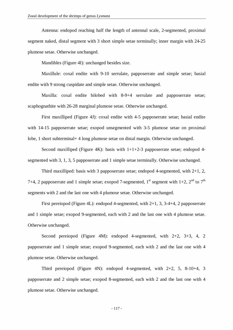
Zoeal development of the shrimps of genus Lysmata
- 117 -
Antenna: endopod reaching half the length of antennal scale, 2-segmented, proximal
segment naked, distal segment with 3 short simple setae terminally; inner margin with 24-25
plumose setae. Otherwise unchanged.
Mandibles (Figure 4I): unchanged besides size.
Maxillule: coxal endite with 9-10 serrulate, papposerrate and simple setae; basial
endite with 9 strong cuspidate and simple setae. Otherwise unchanged.
Maxilla: coxal endite bilobed with 8-9+4 serrulate and papposerrate setae;
scaphognathite with 26-28 marginal plumose setae. Otherwise unchanged.
First maxilliped (Figure 4J): coxal endite with 4-5 papposerrate setae; basial endite
with 14-15 papposerrate setae; exopod unsegmented with 3-5 plumose setae on proximal
lobe, 1 short subterminal+ 4 long plumose setae on distal margin. Otherwise unchanged.
Second maxilliped (Figure 4K): basis with 1+1+2-3 papposerrate setae; endopod 4-
segmented with 3, 1, 3, 5 papposerrate and 1 simple setae terminally. Otherwise unchanged.
Third maxilliped: basis with 3 papposerrate setae; endopod 4-segmented, with 2+1, 2,
7+4, 2 papposerrate and 1 simple setae; exopod 7-segmented, 1st segment with 1+2, 2nd to 7th
segments with 2 and the last one with 4 plumose setae. Otherwise unchanged.
First pereiopod (Figure 4L): endopod 4-segmented, with 2+1, 3, 3-4+4, 2 papposerrate
and 1 simple setae; exopod 9-segmented, each with 2 and the last one with 4 plumose setae.
Otherwise unchanged.
Second pereiopod (Figure 4M): endopod 4-segmented, with 2+2, 3+3, 4, 2
papposerrate and 1 simple setae; exopod 9-segmented, each with 2 and the last one with 4
plumose setae. Otherwise unchanged.
Third pereiopod (Figure 4N): endopod 4-segmented, with 2+2, 5, 8-10+4, 3
papposerrate and 2 simple setae; exopod 8-segmented, each with 2 and the last one with 4
plumose setae. Otherwise unchanged.

Zoeal development of the shrimps of genus Lysmata
- 118 -
Fourth pereiopod (Figure 4O): functional; biramous; basis with 1 simple seta; endopod
4-segmented, with 2+1, 1+3, 5-6+4, 2 papposerrate and 1 simple setae; exopod 6-segmented,
each with 2 and the last one with 4 plumose setae.
Fifth pereiopod (Figure 4H): five thirds longer than the whole larva; merus with 10-12
simple setae and one spinous process; carpus with 21-27 simple setae and one spinous
process; propodus length remains as two fifths of the whole pereiopod, flattened and paddle-
like enlarged with margins serrated (56-62 and 58-73 spines on each margin), 7-10 small
pappose setae along the surface, and 2 strong simple setae distally; dactylus small with 2-3
strong spines, 2 simple subterminal and 1 strong simple terminal setae. Otherwise unchanged.
Pleon (Figure 4H): anteriorly curved spine in the third pleomere increased in size.
Otherwise unchanged.
Pleopods (Figure 4H): presence of small protuberances in pleomeres.
Uropods: endopod with 3 short plumose setae proximally on outer margin and 24-27
plumose setae along distal and inner margins; exopod with 3-5 short plumose setae
proximally on outer margin, 1 spine on apex followed by 29-31 marginal plumose setae along
distal and inner margins, and 5-6 short plumose setae on dorsal margin.
Telson: narrowing towards the posterior end, with one pair of lateral spines followed
by 1 pair of outer spines and 6 pairs of short setae of which the inner 4 pairs are plumose and
the outer 2 pairs are simple.
Seventh zoea (Figure 5)
Dimension: TL = 8.24–8.48 mm; CL = 4.08–4.32 mm.
Carapace (Figure 5A, B): rostrum measuring more than twice over the carapace with 7
dorsal teeth; otherwise unchanged besides size.

Zoeal development of the shrimps of genus Lysmata
- 119 -
Fig. 5- Lysmata galapagensis. Seventh zoea: A, total animal, lateral view; B, carapace, dorsal view; C,
maxillule; D, maxilla; E, first maxilliped; F, second maxilliped; G, third maxilliped; H, first pereiopod; I, third
pereiopod; J, fourth pereiopod; K, fifth pereiopod, propodus and dactylus; L, first pleopod; M, second pleopod;
N, third pleopod; O, fourth pleopod; P, fifth pleopod; Q, telson and uropods. Scale bars: 1 mm (A); 0.5 mm (B,
J); 0.1 mm (C-I, K-Q).

Zoeal development of the shrimps of genus Lysmata
- 120 -
Antennule (Figure 5B): peduncle 2-segmented, first segment with 6 plumose setae
distributed along inner margin, 9 small plumose setae on stylocerite, 6 small plumose setae at
two thirds of the length of the segment, and 6 small plumose setae distally; distal segment
with 5 plumose+2 simple setae. Inner flagellum 8-segmented, proximal and second segments
with 4 small simple setae each, third segment with 3 simple+1 plumose short setae, fourth and
fifth segments with 3 and 4 small simple setae respectively, sixth segment with 4 simple+1
plumose short setae, seventh segment with 4 short simple setae, and distal segment with 2
short+2 longer simple+1 plumose setae. Outer flagellum 8-segmented, proximal segment with
2 simple setae and 3 aesthetascs, second segment with 3 aesthetascs in a middle position+3
short simple setae and 3 aesthetascs terminally, third segment with 3 simple setae and 4
aesthetascs, fourth to sixth segments with 4, 5 and 3 short simple setae respectively, and distal
segment with 3 short simple+1 plumose setae.
Antenna (Figure 5B): flagellum 8-segmented, shorter than scaphocerite, with 2 smaller
segments proximally, 3rd segment the longer one followed by 5 short segments distally, all
bearing 3-5 simple setae; scaphocerite now with 28 plumose setae.
Mandibles: enlarged in size.
Maxillule (Figure 5C): coxal endite with 9-11 serrulate, papposerrate and simple setae;
basial endite with 10-11 strong cuspidate and simple setae. Otherwise unchanged.
Maxilla (Figure 5D): coxal endite bilobed with 10-11+4 serrulate and papposerrate
setae; basial endite with 7+6 papposerrate setae; scaphognathite with 36 marginal plumose
setae. Otherwise unchanged.
First maxilliped (Figure 5E): coxal endite with 5 papposerrate setae; basial endite with
15-17 papposerrate setae; exopod unsegmented with 7-9 plumose setae on proximal lobe, 1
short subterminal+ 4 long plumose setae on distal margin; epipod bilobed. Otherwise
unchanged.

Zoeal development of the shrimps of genus Lysmata
- 121 -
Second maxilliped (Figure 5F): basis with 1+1+3 papposerrate setae; epipod bud
present. Otherwise unchanged.
Third maxilliped (Figure 5G): endopod 4-segmented, with 4, 3, 15-16, 2 papposerrate
and 2 simple setae; exopod 9-segmented, each segment with 2 and the last one with 4
plumose setae. Otherwise unchanged.
First pereiopod (Figure 5H): endopod subchelate, with internal distal margin of
propodus produced forward to about one third of dactylus, 4-segmented, with 2+1, 1+3, 13, 7
papposerrate and simple setae; exopod 10-segmented, each segment with 2 and the last one
with 4 plumose setae. Otherwise unchanged.
Second pereiopod: endopod 4-segmented, with 4, 7, 5, 5 papposerrate and simple
setae; exopod 10-segmented, each segment with 2 and the last one with 4 plumose setae.
Otherwise unchanged.
Third pereiopod (Figure 5I): endopod 4-segmented, with 5, 5, 21, 6 papposerrate and
simple setae; exopod 10-segmented, each segment with 2 and the last one with 4 plumose
setae. Otherwise unchanged.
Fourth pereiopod (Figure 5J): basis with 2 simple seta; endopod 4-segmented, with
2+1, 6, 18, 5 papposerrate and simple setae; exopod 8-segmented, each segment with 2 and
the last one with 4 plumose setae.
Fifth pereiopod (Figure 5K): propodus flattened and paddle-like enlarged with margins
serrated (60 and 80 spines on each margin), 7 small pappose setae along the surface, and 7
simple setae distally; dactylus small with 3 simple subterminal and 1 strong simple terminal
setae. Otherwise unchanged.
Pleon (Figure 5A): anteriorly curved spine in the third pleomere increased in size.
Otherwise unchanged.

Zoeal development of the shrimps of genus Lysmata
- 122 -
Pleopods (Figure 5L-P): endopod rudimentary, bud-like; exopod with 2-4 apical
simple setae.
Uropods (Figure 5Q): endopod with 6 short plumose setae proximally on outer margin
and 32 plumose setae along distal and inner margins; exopod with 4 short plumose setae
proximally on outer margin, 1 spine on apex followed by 33 marginal plumose setae along
distal and inner margins, and 5 short plumose setae on dorsal margin.
Telson (Figure 5Q): narrowing towards the posterior end, bears one pair of lateral
spines followed by 1 pair of plumose setae and 6 pairs of strong short setae.
Lysmata moorei (Rathbun, 1901)
Figures 6-9
First zoea (Figure 6)
Dimension: TL = 2.77–2.81 mm; CL = 0.92–0.96 mm.
Carapace (Figure 6A): rostrum slender and pointed, longer than the antennular
peduncle, eyes compound and sessile; 1 pterigostomial spine and 4-5 denticles along anterior
ventral margin.
Antennule (Figures 6B, B’): peduncle unsegmented, with 1 long plumose seta and a
small process terminally; short outer flagellum with 1 plumose seta, 3 long aesthetascs and 1
short and stout aesthetasc that ends in a spoon shaped membrane with thickened midrib at
distal margin.
Antenna (Figure 6C): protopod unsegmented; endopod apically with 1 long plumose
seta and 1 short spine; scaphocerite 5-segmented, 4 short segments distally, with 9 plumose
setae on inner margin, 2 plumose setae on outer margin, and a simple small seta on apex.

Zoeal development of the shrimps of genus Lysmata
- 123 -
Fig. 6- Lysmata moorei. First zoea: A, total animal, lateral view; B, antennule; B’, antennule, detail of short
aesthetasc; C, antenna; D, mandibles; E, maxillule; F, maxilla; G, first maxilliped; H, second maxilliped; I, third
maxilliped; J, first pereiopod; K, fifth pereiopod; L, 5th pleomere and telson; L’, detail of the 5th pleomere.
Scale bars: 0.5 mm (A, L); 0.1 mm (B-K, L’).

Zoeal development of the shrimps of genus Lysmata
- 124 -
Mandibles (Figure 6D): asymmetrical, palp absent, armature of incisor and molar
processes as illustrated.
Maxillule (Figure 6E): coxal endite with 2 serrulate, 3 papposerrate and 2 simple
setae, basial endite with 3 cuspidate and 2 simple setae; endopod with 2 strong subterminal
and 3 strong terminal papposerrate setae.
Maxilla (Figure 6F): coxal endite bilobed with 8+4 serrulate and papposerrate setae,
basial endite bilobed with 4+4 papposerrate setae; endopod unsegmented bearing 3 + 2 + 1 +
3 papposerrate and simple setae; scaphognathite with 5 marginal plumose setae.
First maxilliped (Figure 6G): coxa with 5 papposerrate setae; basis with 12
papposerrate setae; endopod 4-segmented with 3, 1, 2, 3 papposerrate and 1 simple setae;
exopod unsegmented, bearing subapically 1 shorter seta on lateral margin and 3 long plumose
setae terminally.
Second maxilliped (Figure 6H): basis with 1+1+2-3 papposerrate setae; endopod 4-
segmented with 3, 1, 2, 5 papposerrate and 1 simple setae terminally; exopod 4-segmented,
bearing 2, 2, 2, 3 plumose setae.
Third maxilliped (Figure 6I): basis with 2 papposerrate setae; endopod 4-segmented,
with 2+1, 0, 2+4, 2 papposerrate and 1 simple setae; exopod 4-segmented, bearing 2, 2, 2, 3
plumose setae.
First pereiopod (Figure 6J): biramous bud.
Second to fourth pereiopods: absent.
Fifth pereiopod (Figure 6K): uniramous bud.
Pleon (Figures 6L, L’): 5 pleomeres; 2 dorso-lateral spines and 3-4 very small dorsal
spines on posterior margin of the 5th pleomere.
Pleopods: absent.
Uropods: absent.

Zoeal development of the shrimps of genus Lysmata
- 125 -
Telson (Figure 6L): triangular, broader posteriorly; indented medially with 7+7 setae
(inner 5 plumose, outer 2 plumose on proximal axis only); minute spines between and around
setae.
Second zoea (Figures 7A-G’)
Dimension: TL = 3.08–3.23 mm; CL = 1.00–1.04 mm.
Carapace (Figure 7A): eyes stalked, with peduncle longer than antennal peduncle;
rostrum long and slender, as long as carapace, with 2-3 dorsal teeth and 1 post rostral spine;
with 1 pair of supraorbital, 1 pair of antennal and 1 pair of pterigostomial spines followed by
3-4 denticles on anterior ventral margin.
Antennule (Figure 7B): peduncle unsegmented bearing proximally 1 short, followed
by 4-5 short, and 2 short+1 long plumose setae distally; outer flagellum with 1 very small
subterminal spine, 1 short simple seta, and 1 short+ 4 long aesthetascs.
Antenna (Figure 7C): protopod unsegmented; endopod small pointed conical shaped;
scaphocerite 4-segmented, 3 short segments distally, with 12 plumose setae on inner margin,
2 plumose setae on outer margin and 1 simple small seta on apex.
Mandibles (Figure 7D): unchanged.
Maxillule: basial endite with 6-7 strong cuspidate and simple setae. Otherwise
unchanged.
Maxilla: scaphognathite with 8 marginal plumose setae. Otherwise unchanged.
First maxilliped: exopod with 1 subapical shorter seta on lateral margin and 4 long
plumose setae terminally. Otherwise unchanged.
Second maxilliped: exopod 4-segmented, bearing 2, 2, 2, 4 plumose setae. Otherwise
unchanged.

Zoeal development of the shrimps of genus Lysmata
- 126 -
Third maxilliped: exopod 4-segmented, bearing 2, 2, 2, 4 plumose setae. Otherwise
unchanged.
First pereiopod (Figures 7E): functional; biramous; basis with 1 simple seta; endopod
4-segmented, with 2+1, 1, 4, 2 papposerrate and 1 simple setae; exopod 3-segmented with 2,
2, 3 plumose setae.
Second pereiopod (Figures 7E’, F): present as a very small biramous bud.
Third and fourth pereiopods: absent.
Fifth pereiopod (Figures 7G): functional; uniramous; longer than the whole larva;
basis without seta; ischium with 0-1 seta; merus with one spinous process and 1-3 simple
setae; carpus with 7 simple setae; propodus measuring approximately one third of whole
pereiopod, flattened and paddle-like enlarged with margins serrated (14-16 and 16-19 spines
on each margin), and 5-6 small pappose setae along the surface; dactylus small with 1 simple
subterminal and 1 strong simple terminal setae.
Pleon (Figure 7A): fifth pleomere with 2 dorso-lateral spines and 1-2 very small dorsal
spines on the posterior margin.
Pleopods: absent.
Uropods: absent.
Telson (Figure 7A): 8+8 plumose setae, the outer ones plumose on proximal axis only.
Third zoea (Figures 7H-M’)
Dimension: TL = 3.23–3.44 mm; CL = 1.02–1.24 mm.
Carapace (Figure 7H): eyes with two thirds of antennal length; rostrum a little shorter
than carapace. Otherwise unchanged.

Zoeal development of the shrimps of genus Lysmata
- 127 -
Fig. 7- Lysmata moorei. Second zoea: A, total animal, lateral view; B, antennule; C, antenna; D, mandibles; E,
first pereiopod; E’, detail of basis of pereiopods; F, second pereiopod; G, fifth pereiopod. Third zoea: H,
carapace, dorsal view; I, detail of antennule flagella; J, detail of scaphocerite distal segments; K, second
pereiopod; L, fifth pereiopod; L’, detail of fifth pereiopod propodus and dactylus; M, telson and uropods; M’,
detail of endopod and exopod of uropods. Scale bars: 0.5 mm (A, H); 0.1 mm (B-G, I-M’).
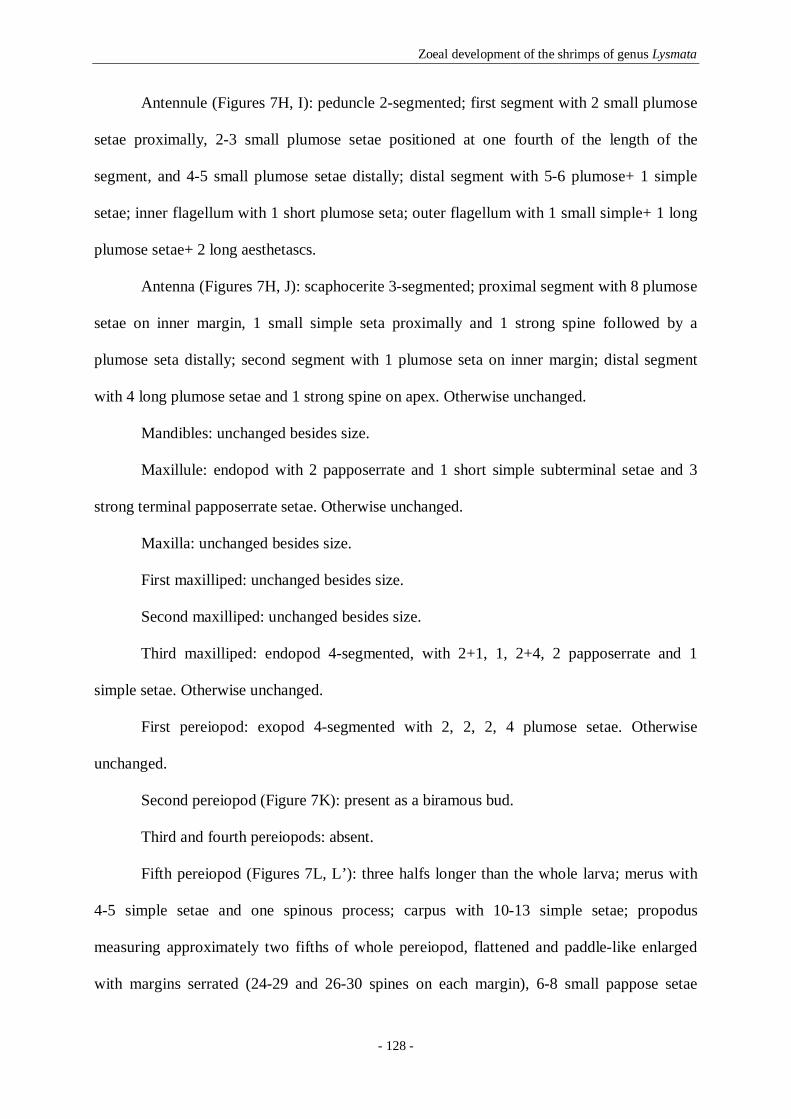
Zoeal development of the shrimps of genus Lysmata
- 128 -
Antennule (Figures 7H, I): peduncle 2-segmented; first segment with 2 small plumose
setae proximally, 2-3 small plumose setae positioned at one fourth of the length of the
segment, and 4-5 small plumose setae distally; distal segment with 5-6 plumose+ 1 simple
setae; inner flagellum with 1 short plumose seta; outer flagellum with 1 small simple+ 1 long
plumose setae+ 2 long aesthetascs.
Antenna (Figures 7H, J): scaphocerite 3-segmented; proximal segment with 8 plumose
setae on inner margin, 1 small simple seta proximally and 1 strong spine followed by a
plumose seta distally; second segment with 1 plumose seta on inner margin; distal segment
with 4 long plumose setae and 1 strong spine on apex. Otherwise unchanged.
Mandibles: unchanged besides size.
Maxillule: endopod with 2 papposerrate and 1 short simple subterminal setae and 3
strong terminal papposerrate setae. Otherwise unchanged.
Maxilla: unchanged besides size.
First maxilliped: unchanged besides size.
Second maxilliped: unchanged besides size.
Third maxilliped: endopod 4-segmented, with 2+1, 1, 2+4, 2 papposerrate and 1
simple setae. Otherwise unchanged.
First pereiopod: exopod 4-segmented with 2, 2, 2, 4 plumose setae. Otherwise
unchanged.
Second pereiopod (Figure 7K): present as a biramous bud.
Third and fourth pereiopods: absent.
Fifth pereiopod (Figures 7L, L’): three halfs longer than the whole larva; merus with
4-5 simple setae and one spinous process; carpus with 10-13 simple setae; propodus
measuring approximately two fifths of whole pereiopod, flattened and paddle-like enlarged
with margins serrated (24-29 and 26-30 spines on each margin), 6-8 small pappose setae

Zoeal development of the shrimps of genus Lysmata
- 129 -
along the surface; dactylus small with 2 strong spines, 1 simple subterminal and 1 strong
simple terminal setae.
Pleon: third pleomere with one pair of dorsal simple setae; fifth pleomere with 2 small
dorso-lateral spines; sixth pleomere separated from telson, with one pair of dorso-lateral
spines and one pair of small ventral-lateral spines.
Pleopods: absent.
Uropods (Figures 7M, M’): biramous; endopod small with 2 short plumose setae
apically; exopod well developed reaching the end of telson, with 12-13 marginal plumose
setae and 2 plumose setae on dorsal margin.
Telson (Figure 7M): separated of the sixth pleomere, with one pair of lateral spines
and 7 + 7 plumose setae.
Fourth zoea (Figure 8)
Dimension: TL = 3.44–3.66 mm; CL = 1.13–1.29 mm.
Carapace (Figures 8A, B): eyes with two thirds of antennal length, presence of one
pair of cervical carinae, rostrum as long as the carapace, pair of pterigostomial spines,
followed by 2-3 denticles on ventral margin. Otherwise unchanged.
Antennule (Figures 8A, C): peduncle 2-segmented; first segment with 4 plumose setae
distributed along inner margin, 4-5 small plumose setae on stylocerite, 2-3 small plumose
setae positioned at four fifths of the length of the segment, and 3-4 small plumose setae
distally; distal segment with 5-6 plumose+ 1 simple setae; inner flagellum with 1 plumose
seta; outer flagellum with 2 subterminal aesthetascs, and 1 small simple+ 1 long plumose
setae terminally.

Zoeal development of the shrimps of genus Lysmata
- 130 -
Fig. 8- Lysmata moorei. Fourth zoea: A, total animal, lateral view; B, carapace, dorsal view; C, antennule; D,
antenna; E, mandibles; F, first pereiopod; G, second pereiopod; H, third pereiopod; I, fifth pereiopod, propodus
and dactylus; I’, detail of fifth pereiopod propodus and dactylus; J, pleon, lateral view; K, telson and uropods.
Scale bars: 0.5 mm (A-B, I); 0.1 mm (C-H, I’-J).

Zoeal development of the shrimps of genus Lysmata
- 131 -
Antenna (Figures 8A, D): scaphocerite unsegmented, with 14 plumose setae on inner
margin, followed by 1 very small simple, 1 short plumose setae, and a very strong spine on
apex.
Mandibles (Figure 8E): incisor and molar processes as illustrated.
Maxillule: coxal endite with 7-8 serrulate, papposerrate and simple setae, basial endite
with 7-8 cuspidate and simple setae. Otherwise unchanged.
Maxilla: coxal endite bilobed with 8-9+4 serrulate and papposerrate setae;
scaphognathite with 9-10 marginal plumose setae. Otherwise unchanged.
First maxilliped: unchanged besides size.
Second maxilliped: unchanged besides size.
Third maxilliped: exopod 5-segmented, bearing 2, 2, 2, 2, 4 plumose setae. Otherwise
unchanged.
First pereiopod (Figure 8F): endopod 4-segmented, with 2+2, 1, 1+4, 2 papposerrate
and 1 simple setae; exopod 5-segmented with 2, 2, 2, 2, 4 plumose setae.
Second pereiopod (Figure 8G): functional; biramous; basis with 1 simple seta;
endopod 4-segmented, with 2+1, 0, 4, 2 papposerrate and 1 simple setae; exopod 4-segmented
with 2, 2, 2, 4 plumose setae.
Third pereiopod (Figure 8H): present as a very small biramous bud.
Fourth pereiopod: absent.
Fifth pereiopod (Figures 8I, I’): two times longer than the whole larva; merus with 8-
10 simple setae and one spinous process; carpus with 20-28 simple setae and one spinous
process; propodus length is one third of the whole pereiopod, flattened and paddle-like
enlarged with margins serrated (31-38 and 32-39 spines on each margin), 11-14 small
pappose setae along the surface, and 2 strong simple setae distally. Otherwise unchanged.
Pleon (Figures 8A, J): unchanged besides size.

Zoeal development of the shrimps of genus Lysmata
- 132 -
Pleopods: absent.
Uropods (Figures 8A, K): protopod without setae, with a spine; endopod with 8-9
plumose setae along distal and inner margins and 4-5 short plumose setae on dorsal margin;
exopod longer than telson, with 1 spine on apex followed by 15-16 marginal plumose setae
along distal and inner margins, and 3 short plumose setae on dorsal margin.
Telson (Figures 8A, K): almost rectangular shaped, with one pair of lateral spines
followed by 2 pairs of outer spines and 5 pairs of plumose setae on the posterior end.
Last zoea (Figure 9)
Dimension: TL = 7.81–9.35 mm; CL = 3.85–4.84 mm.
Carapace (Figures 9A, B): eyes with four fifths of antennal length; presence of one
pair of cervical carinae; rostrum measuring almost twice over the carapace, with 7-9 dorsal
teeth, 1 post rostral spine; with 1 pair of supraorbital, 1 pair of antennal and 1 pair of
pterigostomial spines.
Antennule (Figure 9B): peduncle 2-segmented, proximal segment with 6-7 plumose
setae distributed along inner margin, 8-11 small plumose setae on stylocerite, 6-9 small
plumose setae positioned at four fifths of the length of the segment, and 5-7 plumose+1
simple small setae distally; distal segment with 5-6 plumose+3-4 simple small setae. Inner
flagellum 12-segmented, proximal to eleventh segments with 2-6 small simple setae, and
distal segment with 4-5 short simple+1 plumose setae. Outer flagellum 10-segmented,
proximal segment with 5 aesthetascs, second and third segments with 4 aesthetascs each,
fourth segment with 2-4 simple setae and 5 aesthetascs, fifth segment with 3-4 short simple
setae and 2 aesthetascs, sixth to ninth segments with 2-4 short simple setae, and distal
segment with 3 short simple+2 plumose setae.
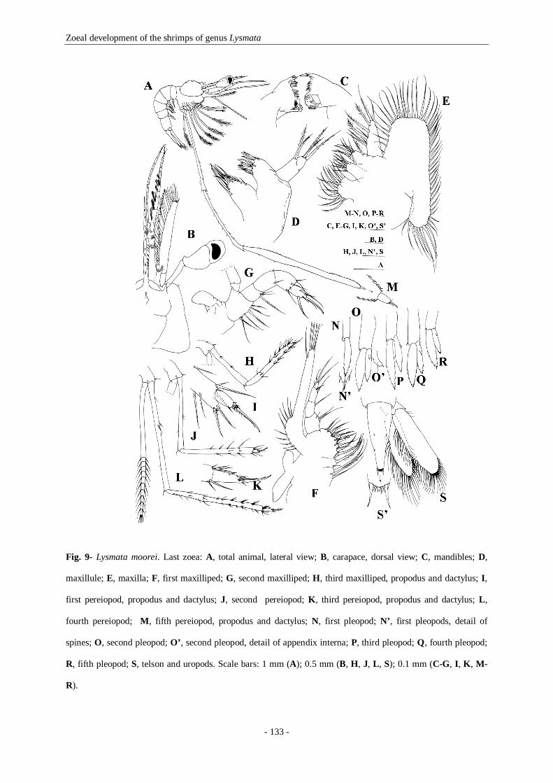
Zoeal development of the shrimps of genus Lysmata
- 133 -
Fig. 9- Lysmata moorei. Last zoea: A, total animal, lateral view; B, carapace, dorsal view; C, mandibles; D,
maxillule; E, maxilla; F, first maxilliped; G, second maxilliped; H, third maxilliped, propodus and dactylus; I,
first pereiopod, propodus and dactylus; J, second pereiopod; K, third pereiopod, propodus and dactylus; L,
fourth pereiopod; M, fifth pereiopod, propodus and dactylus; N, first pleopod; N’, first pleopods, detail of
spines; O, second pleopod; O’, second pleopod, detail of appendix interna; P, third pleopod; Q, fourth pleopod;
R, fifth pleopod; S, telson and uropods. Scale bars: 1 mm (A); 0.5 mm (B, H, J, L, S); 0.1 mm (C-G, I, K, M-
R).

Zoeal development of the shrimps of genus Lysmata
- 134 -
Antenna (Figure 9B): flagellum long and slender, with 10-segments bearing 2-5 short
simple setae each; scaphocerite now with 30-32 plumose setae. Otherwise unchanged.
Mandibles (Figure 9C): armature of incisor and molar processes as illustrated
Maxillule (Figure 9D): coxal endite with 13-16 serrulate, papposerrate and simple
setae; basial endite with 12-14 strong cuspidate and simple setae. Otherwise unchanged.
Maxilla (Figure 9E): coxal endite bilobed with 11-13+4 serrulate and papposerrate
setae; basial endite with 8-10+7-10 papposerrate setae; scaphognathite with 47-51 marginal
plumose setae. Otherwise unchanged.
First maxilliped (Figure 9F): coxal endite with 5 papposerrate setae; basial endite with
17-19 papposerrate setae; exopod unsegmented with 6-10 plumose setae on proximal lobe, 1
short subterminal+ 4 long plumose setae on distal margin; epipod bilobed. Otherwise
unchanged.
Second maxilliped (Figure 9G): basis with 3+1+2 papposerrate setae; endopod 4-
segmented with 3, 2, 4, 5-6 papposerrate and simple setae terminally. Otherwise unchanged.
Third maxilliped (Figure 9H): endopod 4-segmented, with 4-5, 3, 19-22, 5
papposerrate and simple setae; exopod 10-segmented, each segment with 2 and the last one
with 4 plumose setae. Otherwise unchanged.
First pereiopod (Figure 9I): endopod subchelate, with internal distal margin of
propodus produced forward to about a half of dactylus; 4-segmented, with 4-5, 4-5, 12-14, 11-
13 papposerrate and simple setae; exopod 11-segmented, 1st segment with 1+2, 2nd to 10th
segments with 2 and the last one with 4 plumose setae. Otherwise unchanged.
Second pereiopod (Figure 9J): endopod 4-segmented, with 4-5, 9-14, 5-9, 7-12
papposerrate and simple setae; exopod 11-segmented, 1st segment with 1+2, 2nd to 10th
segments with 2 and the last one with 4 plumose setae. Otherwise unchanged.
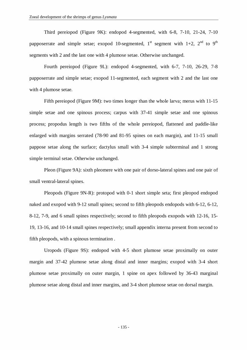
Zoeal development of the shrimps of genus Lysmata
- 135 -
Third pereiopod (Figure 9K): endopod 4-segmented, with 6-8, 7-10, 21-24, 7-10
papposerrate and simple setae; exopod 10-segmented, 1st segment with 1+2, 2nd to 9th
segments with 2 and the last one with 4 plumose setae. Otherwise unchanged.
Fourth pereiopod (Figure 9L): endopod 4-segmented, with 6-7, 7-10, 26-29, 7-8
papposerrate and simple setae; exopod 11-segmented, each segment with 2 and the last one
with 4 plumose setae.
Fifth pereiopod (Figure 9M): two times longer than the whole larva; merus with 11-15
simple setae and one spinous process; carpus with 37-41 simple setae and one spinous
process; propodus length is two fifths of the whole pereiopod, flattened and paddle-like
enlarged with margins serrated (78-90 and 81-95 spines on each margin), and 11-15 small
pappose setae along the surface; dactylus small with 3-4 simple subterminal and 1 strong
simple terminal setae. Otherwise unchanged.
Pleon (Figure 9A): sixth pleomere with one pair of dorso-lateral spines and one pair of
small ventral-lateral spines.
Pleopods (Figure 9N-R): protopod with 0-1 short simple seta; first pleopod endopod
naked and exopod with 9-12 small spines; second to fifth pleopods endopods with 6-12, 6-12,
8-12, 7-9, and 6 small spines respectively; second to fifth pleopods exopods with 12-16, 15-
19, 13-16, and 10-14 small spines respectively; small appendix interna present from second to
fifth pleopods, with a spinous termination .
Uropods (Figure 9S): endopod with 4-5 short plumose setae proximally on outer
margin and 37-42 plumose setae along distal and inner margins; exopod with 3-4 short
plumose setae proximally on outer margin, 1 spine on apex followed by 36-43 marginal
plumose setae along distal and inner margins, and 3-4 short plumose setae on dorsal margin.

Zoeal development of the shrimps of genus Lysmata
- 136 -
Telson (Figure 9S, S’): rather triangular, narrower distally than proximally, bears one
pair of dorso-lateral spines followed by 1 pair of lateral spines and 5 pairs of strong short
setae.
4.5- Discussion
4.5.1- Morphological comparisons of the zoeal stages
Beyond the first zoeal stage, the morphological features displayed by L. galapagensis and L.
moorei larvae are those considered to be diagnosing of the genus Lysmata: eyes stalked with a
long peduncle; 5th pereiopod developing before the 2nd, 3rd and 4th pereiopods and
exhibiting an enlarged, flattened and paddle-like propodus with serrated margins. The first
zoeal stage of both species is similar to that of L. seticaudata, as they hatch with the first and
fifth pereiopods as biramous and uniramous buds, respectively (Calado et al. 2004, p. 739,
figures 1a, 1j-k). A comparison of the most relevant larval features of the first, fourth, seventh
and last zoeal stages of L. galapagensis and L. moorei with those of L. seticaudata is
presented in Table 1.
The studied species present a very homogeneous development, and even in the presence of
incomplete larval series, we expect that both will have a maximum number of nine zoeal
stages as in L. seticaudata larval series. L. galapagensis and L. moorei hatch with the first and
fifth pereiopods as buds which become functional in the second zoeal stage; in the third zoeal
stage all the three species have the second pereiopod as a biramous bud that becomes
functional in the fourth zoeal stage where fourth pereiopod is present as a biramous bud.
When comparing the seventh zoea of L. galapagensis with the same stage of L. seticaudata,
the first one clearly presents more advanced larval characters, such as the antennal flagellum

Zoeal development of the shrimps of genus Lysmata
- 137 -
Table 1 Comparison of relevant morphological characters of L. seticaudata, L. galapagensis and L. moorei zoeal
stages.
Features L. galapagensis Present study (1);
Gopalakrishnan & Laurs, 1971 (2)
L. moorei Present study
L. seticaudata Calado et al. 2004
Distribution area E Pacific SW Atlantic NE Atlantic
Number of zoeal stages 8- 9 8- 9 9
ZI, total length, mm 2.46-2.89 (1) 2.77-2.81 3.09-3.24 ZI, carapace length, mm 0.92-1.00 (1) 0.92-0.96 0.98-1.03 ZI, rostrum length, mm 0.35-0.42 (1) 0.42 0.46 ZIV, total length, mm 4.14-4.63 (1) 3.44-3.66 3.12-3.22 ZIV, carapace length, mm 1.61-2.15 (1) 1.13-1.29 1.04-1.08 ZIV, rostrum length, mm 0.92-1.35 (1) 0.54-0.70 0.31 ZVII, total length, mm 8.24-8.48 (1) NA 4.47-4.79 ZVII, carapace length, mm 4.08-4.32 (1) NA 1.40-1.56 ZVII, rostrum length, mm 2.80-3.04 (1) NA 0.43-0.54 Last Z, total length, mm 16.10-20 (2) 7.81-9.35 6.40-6.80 Last Z, carapace length, mm 2.12-3.40 (2) 3.85-4.84 2.00-2.20 Last Z, rostrum length, mm 6.13-7.00 (2) 2.31-3.19 0.72-0.96
CARAPACE ZI, marginal denticles
ZIV, marginal denticles ZVII, marginal denticles Last, marginal denticles
4 (1) 3-4 (1) 0 (1) 0 (2)
4-5 2-3 NA 0
4 3 0 0
ROSTRUM ZIV, number of spines ZVII, number of spines Last, number of spines
5-6 (1) 7 (1)
7-8 (2)
0 NA 7-9
0 0
1-2 ANTENNA
Scaphocerite, absence of segments ZIV (1) ZIV ZIV FIRST PEREIOPOD
Biramous bud/ Functional ZI (1)/ ZII (1) ZI/ ZII ZI/ ZII SECOND PEREIOPOD Very small biramous bud
Biramous bud Functional
ZII (1) ZIII (1) ZIV (1)
ZII ZIII ZIV
ND ZIII ZIV
THIRD PEREIOPOD Very small biramous bud
Biramous bud Functional
ZIV (1) ZV (1) ZVI (1)
ZIV NA NA
ND ZV ZVI
FOURTH PEREIOPOD Very small biramous bud
Biramous bud Functional
- ZV (1) ZVI (1)
NA NA NA
ND ZV ZVI
FIFTH PEREIOPOD Uniramous bud/ Functional ZI (1)/ ZII (1) ZI/ ZII ZI/ ZII
PLEON 3rd pleomere: dorsal anteriorly curved spine 5th pleomere: one pair of dorso-lateral spines 5th pleomere: small dorsal spines
ZIV-ZVII (1) ZI-ZV (1)
ZI: 3-4; ZII: 0-2; ZIII: 0 (1)
Absent ZI-ZIV
ZI: 3-4; ZII: 1-2; ZIII: 0
Absent ZI-ZII Absent
PLEOPODS Small buds
Biramous buds Endopod as bud, exopod with setae
With appendix interna
ZVI (1) -
ZVII (1) ZVIII (2)
NA NA NA
Last (ZVIII or IX)
ZVI ZVII ZVIII ZIX
Z: zoea. NA: not available in the larval series. ND: not developed.

Zoeal development of the shrimps of genus Lysmata
- 138 -
as long as scaphocerite with 8 segments, biramous pleopods with endopods as buds and
exopods with apical setae, and the telson narrower at the posterior end, typical of a
penultimate stage (Calado et al. 2004, p. 746, figures 6a-k), leading us to believe that this
species larval series will need just one more stage to complete their zoeal development.
Accordingly, is also expected that L. moorei completes the zoeal development with eight or
nine stages since their larvae hatch with the first and fifth pereiopods as buds, and in the
fourth zoeal stage all the morphological structures are at the same level of development as in
the other two species (Table 1). When summarizing the larval characters of genus Lysmata,
Gurney (1937) affirmed that probably it would present 9 zoeal stages. Our results confirm his
prediction for L. galapagensis and L. moorei, as it has been previously found for L.
seticaudata (Calado et al. 2004). The most evident variation between the zoeal developmental
series of L. seticaudata and L. galapagensis occurs during the pleopods development that in
the first species takes 4 stages to complete the development and in the second seems to take
only 3 stages. Knowlton & Alavi (1995) stated that the most evident differences in the
Lysmata larval series were the appearance and development of pereiopods and pleopods.
The study of larval Lysmata will certainly be of valuable help for researchers addressing
phylogeny subjects. Considering the available larval descriptions in the genus, is possible to
hypothesize at least two probable scenarios for the developmental pattern of Lysmata species:
the first one will be similar to L. galapagensis, L. moorei and L. seticaudata where the larvae
hatch with the first and fifth pereiopods as buds becoming functional in the second zoeal stage
and with a complete larval development of 8 to 9 zoeae. The second pattern will be similar to
L. amboinensis (Wunsch 1996), L. anchisteus (Knowlton & Alavi 1996), L. vittata (Yang
1999) and Kurata’s L. wurdemanni (Kurata 1970) where the larvae hatch without buds, and
consequently will have a longer zoeal development with a maximum of 11 zoeal stages. The

Zoeal development of the shrimps of genus Lysmata
- 139 -
existing correspondence between the larval duration and the phylogenetic results for Lysmata
genus is very interesting: the first group of species belongs to the “Cosmopolitan Clade”/
“Lysmata Clade”, while L. amboinensis (Wunsch 1996), L. anchisteus and L. wurdemanni
(Kurata 1970), the only species studied by Baeza et al. (2009) and Fiedler et al. (submitted),
were included in the other obtained clades. Table 2 presents the more relevant morphological
characters for staging the larvae of Lysmata species belonging to the “Cosmopolitan Clade”/
“Lysmata Clade”. It is commonly accepted that marine organisms such as decapod
crustaceans having a pelagic mode of larval development with several larval stages
corresponds to a phylogenetically ancestral state, while shorter larval series are considered a
derived character (Strathmann 1978, Anger 2001). In this way, the information provided by
the zoeal developmental series of Lysmata will certainly help researchers to validate the
current phylogeny of the genus.
Table 2 Staging of the “Cosmopolitan Clade”/ “Lysmata Clade” larval morphological characters based on the
three studied species.
Larval stage General morphological characters First zoea Eyes fused; one pterigostomial spine followed by 4-5 marginal denticles; rostrum long, slender
and pointed. Scaphocerite 5-segmented. 5th pleomere with one pair of dorso-lateral spines. Pereiopods 1 and 5 as buds. Telson with 7+7 plumose setae.
Second zoea Eyes stalked; antennal and supraorbital spines present; pterigostomial spine followed by marginal denticles; rostrum long and slender with one post rostral spine. Pereiopods 1 and 5 developed and functional. Telson with 8+8 plumose setae.
Third zoea Antennular peduncle 2-segmented, inner and outer flagella present. Scaphocerite 4-segmented. 6th pleomere separated from telson. Pereiopod 2 present as a biramous bud. Uropods present, with a rudimentary endopod.
Fourth zoea Scaphocerite unsegmented. Pereiopod 2 functional. Uropods exopods as long as telson.
Fifth zoea Biramous buds of pereiopods 3 and 4. Uropods exopods longer than telson margin. Telson narrower at his posterior end.
Sixth zoea Pereiopods 1 to 5 developed and functional. Pleopods present as small buds.
Penultimate Zoea (Seventh or Eighth)
Antennal flagellum as long as scaphocerite with 8 segments. Biramous pleopods with endopods as buds and exopods with apical setae.
Last Zoea (Eighth or Ninth)
Antennal flagellum longer than scale with more than 8 segments. Pleopods with appendix interna. Telson triangular with the migration of the first and second pairs of dorso-lateral spines almost concluded (telson form almost similar to an adult).

Zoeal development of the shrimps of genus Lysmata
- 140 -
The second zoeal stage of L. galapagensis presents a small protuberance in the dorsal region
of the third pleomere, which in later zoea differentiates to a well defined anteriorly curved
spine. This character makes this species easily distinguishable from all Lysmata larvae that
have been studied. The similarities recorded between the late zoeal stages of L. galapagensis
and the material described by Gopalakrishnan & Laurs (1971), namely the presence of an
extremely long rostrum with several dorsal teeth and the strong anteriorly curved spine in the
dorsal surface of the third pleomere, allow us to suggest that the larvae described as E.
corniger for the Eastern Tropical Pacific probably corresponds to L. galapagensis. Gurney
(1937) Species A. V, Eretmocaris corniger, from the Atlantic Ocean, presents the same
general form of L. galapagensis, however Gopalakrishnan & Laurs (1971) gave a list of
differences between both species, concluding that the Atlantic specimens were much smaller
and presented less one spine on rostrum, three inner lobes on maxilla endopod, less six setae
on basal enlargement of first maxilliped exopod, and first and second pereiopods not chelate.
L. galapagensis penultimate stage described in present work is found to be much smaller (TL
= 8.24-8.48 mm) than the last stage (TL = 16.10-20.00 mm) described by Gopalakrishnan &
Laurs (1971), especially when considering that they differ from one stage only. We suppose
that L. galapagensis larvae when in the last zoeal stage will have several instars which will
allow them to grow without undergoing the morphological changes to decapodid. Lysmata
larvae are well known for their ability to delay metamorphosis (Calado 2008) by performing
mark-time molting, as defined by Gore (1985). In certain laboratory culture trials, Lysmata
have remained in last zoeal stage for over 80 days (Palmtag & Holt 2007), and during this
period these larvae have molted several times, but displayed little or no morphological
differences from one instar to the next apart from a significant increase in size. In this way,
the last zoea described by Gopalakrishnan & Laurs (1971) could be bigger simply because it
was delaying metamorphosis. Gurney (1942) describes these as giant larvae, which after

Zoeal development of the shrimps of genus Lysmata
- 141 -
reaching the last zoeal stage, at which metamorphosis normally occurs, continue to grow
without important changes in structure and without advancing to the juvenile form.
Concerning L. moorei larvae, and despite their similarity to L. galapagensis, these specimens
lack the unique morphological feature displayed by L. galapagensis on the 3rd pleomere.
Regardless of the close phylogenetic relationship suggested by the molecular studies of Baeza
et al. (2009) and Fiedler et al. (submitted) between the Eastern Pacific L. galapagensis and the
Western Atlantic L. moorei, the most evident larval character shared by both species is the
presence of a very long rostrum with several teeth in late zoeal stages. Therefore it seems
evident that E. corniger larval forms found in the Eastern Atlantic are not L. moorei. We
suppose that they belong to an unknown Lysmata species that probably is phylogenetically
more close to L. galapagensis than to L. moorei. Nowadays researchers have a golden
opportunity to validate the identification of Lysmata larvae previously collected from the
plankton and after over one century finally start to correlate the forms of the puzzling
Eretmocaris to present valid species.
4.5.2- Biogeographical considerations on Eretmocaris corniger
L. galapagensis occurs in the eastern Pacific and, considering its larval series, the most
similar larvae known so far are those of Eretmocaris corniger collected in the eastern
Atlantic. The occurrence of such similar larvae in two different oceans, separated by the
Panama Isthmus and the Atlantic Ocean, lead us to suppose that we are in the presence of two
distinct species that, together with L. moorei could constitute a species complex.
Gopalakrishnan & Laurs (1971) had described E. corniger larvae from plankton collected
material in the eastern tropical Pacific Ocean and considered that their specimens could
belong to a Lysmata species associated with the eastern Ocean tropical islands. The rationale
Zoeal development of the shrimps of genus Lysmata
- 142 -
for their assumption was the occurrence of these larvae in the Galapagos (eastern Pacific) and
in Cape Verde islands (eastern Atlantic). The Panama Isthmus is a geographic barrier formed
three million years ago during the Pliocene, opened in 1914 prior to the record of L.
galapagensis species in the eastern tropical Pacific by Schmitt (1924). We do not believe that
the opening of the Panama Channel was the responsible for the colonization of this species in
both Atlantic and the Pacific Oceans. The present work matches the larvae of L. galapagensis
to the E. corniger Pacific type, and Gopalakrishnan & Laurs (1971) presents consistent
differences between this and the E. corniger Atlantic type, refuting the remote hypothesis of
the Atlantic type have dispersed to the Pacific after the opening of the Panama Channel. It is
assumed that present populations of some coastal decapod crustaceans from the eastern
Atlantic, western Atlantic and eastern Pacific are genetically isolated from each other (e.g.
Schubart et al., 2005). Knowlton et al. (1993) suggested that some of the studied species of
the snapping shrimp Alpheus may have diverged before the final closure of the Panama
seaway, in a staggered model. Also, Schubart et al. (2005) stated that the Atlantic and Pacific
species of Pachygrapsus transversus complex diverged prior to the closure of the Isthmus of
Panama, recognizing that the eastern Pacific population was in fact a valid species, P. socius.
The tropical shrimp larvae are capable of long-distance dispersal (Goy 1990), and this is the
explanation for the circumtropical distribution of many shrimp species, implying that the
main way of creating new species is by sympatric speciation. Also, the very recent work of
Rhyne et al. (2009) proved that these species potential for larval dispersal is unrealized, and a
strong reproductive isolation is observed. As a result, the circumtropical and consequently the
trans-isthmian species are not typical in the genus Lysmata, reason why no species in this
genus were simultaneously recorded in the Pacific and Atlantic Oceans. In fact, the only
species once thought to occupy both of these oceans, has been split in two: L. grabhami is an
amphi-Atlantic species, and L. amboinensis known to occur from the Western Tropical Indian

Zoeal development of the shrimps of genus Lysmata
- 143 -
Ocean to the Hawaiian Islands but absent from the Eastern Tropical Pacific (Debelius 2001).
Having in mind that both, L. galapagensis and E. corniger Atlantic type, share the same
feature on the 3rd pleomere, that is absent in L. moorei, the most plausible scenario should be
the possibility that the eastern Atlantic E. corniger corresponds to the larval form of a
Lysmata species, eventually still undescribed, which used to have an amphi-Atlantic
distribution and formed a trans-isthmian complex with L. galapagensis (or with their common
ancestral) and is now restricted to the Eastern Atlantic. Phylogenetically, this undescribed
species might be more close to L. galapagensis than L. moorei, what lead to the hypothesis
that there is a missing link between these two species, which is probably already extinct.
Future efforts to identify the adult form of Atlantic E. corniger should focus on the larval
culture of Lysmata species which are morphologically and/or molecularly close to L.
galapagensis and are known to occur in the Eastern Atlantic, with emphasis to the oceanic
islands.
4.5.3- Biodiversity and conservation issues
The current fishing pressure on ornamental shrimp from the genus Lysmata, namely on coral
reef species displaying dazzling colorations and/or fish cleaning behaviour, has lead
researchers and policy makers to question the sustainability of this practice and urged them to
start monitoring potential ecological impacts of this profitable commercial activity (e.g.
Calado 2008). The correct identification of different taxa in their larval form is essential to
perform studies related to population estimates, connectivity, recruitment and spatiotemporal
distributions (e.g. Levin 2006, Rhyne et al. 2009). This highlights the need of not only
matching known Eretmocaris with their adult forms, but also to describe the complete larval
series of Lysmata species.

Zoeal development of the shrimps of genus Lysmata
- 144 -
Understanding larval dispersal is vital to successfully manage any commercial fishery, either
for human consumption, biotechnological applications or the marine aquarium trade (e.g. dos
Santos 1998, Calado 2008, Marta-Almeida et al. 2008), as well as to establish marine natural
reserves and/or no take areas (e.g. Almany et al. 2009, Planes et al. 2009). These studies
always require a solid knowledge on the larval stages of target species, which commonly is
either incomplete or unavailable, even in heavily studied regions (e.g. the North-Eastern
Atlantic, González-Gordillo et al. 2001). Although several advances have been achieved in
the identification of larval stages using molecular tools (e.g. Pan et al. 2008, Jones et al.
2009), classical larval morphological descriptions are far from being obsolete. In fact, small
intraspecific variations in larval morphology induced by environmental gradients (Schubart et
al. 2005) will certainly only be detected when employing classical approaches. Managing the
collection of Lysmata has the additional challenge posed by the occurrence of cryptic species
occurring in sympatry (Rhyne & Lin 2006), which apparently can be more common than
initially assumed (Baeza et al. 2009). Under this scenario, the knowledge on the larval stages
of these cryptic species can be vital for the establishment of an accurate monitoring and
management program for the commercial capture of these highly valuable marine organisms.
4.5.4- Conclusions
The present work allows us to conclude that Eretmocaris corniger larval type from the eastern
Pacific is in fact Lysmata galapagensis. According to Baeza et al. (2009) and Fiedler et al.
(submitted) L. moorei is phylogenetically the closest known species to L. galapagensis, and
both of these are included in the same clade of L. seticaudata. All these three species hatched
with the first and fifth pereiopods as buds, and present a similar first to fourth zoeal
development. L. galapagensis and L. seticaudata will have a maximum of 9 zoeal stages, so,

Zoeal development of the shrimps of genus Lysmata
- 145 -
we can suppose that all the species from the “Cosmopolitan Clade”/ “Lysmata Clade” will
also present a maximum of nine zoeal stages.
The similarity between L. galapagensis larvae and Eretmocaris corniger larval type from the
Eastern Atlantic, hypothesize the existence of a third, probably still undescribed species,
distributed along the Eastern Tropical Atlantic. Focusing our research efforts on the
“Cosmopolitan Clade”/ “Lysmata Clade” species, it will not be certainly necessary to wait
another century to finally find the adult form of the puzzling Atlantic E. corniger. We believe
that the almost unexplored Western African Coast holds most of the answers to the questions
raised concerning the identity of the adult forms of several Eretmocaris larvae.
4.6- References
Almany G.R., Connolly S.R., Heath D.D., Hogan J.D., Jones G.P., McCook L.J., Mills M.,
Pressey R.L., Williamson D.H. (2009). Connectivity, biodiversity conservation and the
design of marine reserve networks for coral reefs. Coral Reefs 28, 339-351.
Anger K. (2001). The Biology of Decapod Crustacean Larvae. Crustacean Issues 14, 1-419.
Anker A., Baeza J.A., De Grave S. (2009). A new species of Lysmata (Crustacea, Decapoda,
Hippolytidae) from the Pacific coast of Panama, with observations of its reproductive
biology. Zoological Studies 48, 682-692.
Baeza J.A., Schubart C.D., Zillner P., Fuentes S., Bauer R.T. (2009). Molecular phylogeny of
shrimps from the genus Lysmata (Caridea: Hippolytidae): the evolutionary origins of
protandric simultaneous hermaphroditism and social monogamy. Biological Journal of
the Linnean Society 96, 415-424.
Bate C.S. (1888). Crustacea Macrura. Challenger Reports, Zool., XXIV.

Zoeal development of the shrimps of genus Lysmata
- 146 -
Brightdoom M., Marín B., Zoppi E., Moreno C. (2006). Zooplancton del Golfo Cariaco.
Boletim del Instituto Oceanografico de Venezuela Universidad de Oriente 45, 61-78.
Calado R. (2008). Marine Ornamental Shrimp – Biology, Aquaculture and Conservation.
Wiley-Blackwell, Oxford, United Kingdom.
Calado R., Lin J., Rhyne A.L., Araújo R., Narciso L. (2003a). Marine ornamental decapods-
popular, pricey, and poorly studied. Journal of Crustacean Biology 23, 963-973.
Calado R., Narciso L., Morais S., Rhyne A.L., Lin, J. (2003b). A rearing system for the
culture of ornamental decapod crustacean larvae. Aquaculture 218, 329-339.
Calado R., Bartilotti C., Narciso L., dos Santos A. (2004). Redescription of the larval stages
of Lysmata seticaudata (Risso, 1816) (Crustacea, Decapoda, Hippolytidae) reared
under laboratory conditions. Jopurnal of Plankton Research 26, 737-752.
Caroli E. (1918). Miersia clavigera Chun, stadio misidiforme di Lysmata seticaudata Risso.
Pubblicazione de la Stazione Zoologica di Napoli, II, 177-189.
Clark P.F., Calazans D.K., Pohle G.W. (1998). Accuracy and standardization of brachyuran
larval descriptions. Invertebrate Reproduction & Development 33, 127-144.
Clark P.F. (2009). The bearing of larval morphology on brachyuran phylogeny. Crustacean
Issues 18, 221-241.
Debelius H. (2001). Crustacea Guide of the World. IKAN – Unterwasserarchive, Frankfurt,
Germany.
dos Santos A. (1998). On the occurrence of larvae of Parapenaeus longirostris (Crustacea:
Decapoda: Penaeoidea) off the Portuguese coast. Journal of Natural History 32, 1519–
1523.
Garm A. (2004). Mechanical functions of setae from the mouth apparatus of seven species of
decapod crustaceans. Journal Morphology 260, 85-100.

Zoeal development of the shrimps of genus Lysmata
- 147 -
Gopalakrishnan K. & Laurs R.M. (1971). Eretmocaris corniger Bate larvae from the Eastern
Tropical Pacific Ocean (Caridea, Hippolytidae). Crustaceana 20, 9-18.
González-Gordillo J.I., dos Santos A., Rodríguez A. (2001). Checklist and annotated
bibliography of decapod crustacean larvae from the Southwestern European coast
(Gibraltar Strait area). Scientia Marina 65, 275-305.
Gore R.H. (1985). Molting and growth in decapod larvae. Crustacean Issues 2, 1-65.
Goy J.W. (1990). Components of reproductive effort and delay of larval metamorphosis in
tropical marine shrimp (Crustacea: Decapoda: Caridea and Stenopodidae). PhD
Thesis, Texas A&M University, Texas, USA.
Gurney R. (1937). Larvae of decapod crustacea. Part IV. Hippolytidae. Discovery Reports 14,
351-404.
Gurney R. (1942). Larvae of Decapod Crustacea. The Ray Society of London, London,
United Kingdom.
Jones G.P., Almany G.R., Russ G.R., Sale P.F., Steneck R.S., Van Oppen M.J.H., Willis B.L.
(2009). Larval retention and connectivity among populations of corals and reef fishes:
history, advances and challenges. Coral Reefs 28, 307-325.
Knowlton R.E. & Alavi M.R. (1995). The larval morphology of Lysmata anchisteus Chace
(Crustacea: Decapoada) compared with other Lysmata spp. Caribbean Journal of
Science 31, 289-310.
Knowlton N., Weigt L.A., Solórzano L.A., Mills D.K., Bermingham E. (1993). Divergence in
proteins, mitochondrial DNA, and reproductive compatibility across the Isthmus of
Panama. Science 260, 1629-1632.
Kurata H. (1970). Studies on the Life Histories of Decapod Crustacea of Georgia. PhD thesis,
University of Georgia, Sapelo Island, Georgia, USA.

Zoeal development of the shrimps of genus Lysmata
- 148 -
Levin L.A. (2006). Recent progress in understanding larval dispersal: new directions and
digressions. Integrative and Comparative Biology 46, 282-297.
Lindley J.A., Hernández F., Tejera E., Jiménez S. (2002). An unusual pinnotherid zoea
attributed to Afropinnotheres monody Manning, 1993 (Decapoda: Brachyura:
Pinnotheridae) from the Selvagens Islands (Eastern Atlantic Ocean). Bocagiana 205,
1-5.
Marta-Almeida M., Dubert J., Peliz Á., dos Santos A., Queiroga H. (2008). A modelling study
of Norway lobster (Nephrops norvegicus) larval dispersal in southern Portugal:
predictions of larval wastage and self-recruitment in the Algarve stock. Canadian
Journal of Fisheries and Aquatic Sciences 65, 2253–2268.
Palmtag M.R. & Holt G.J. (2007). Experimental studies to evaluate larval survival of the fire
shrimp, Lysmata debelius, to the juvenile stage. Journal of the World Aquaculture
Society 38, 102-113.
Pan M., McBeath A.J.A., Hay S. J., Pierce G.J., Cunningham C.O. (2008). Real-time PCR
assay for detection and relative quantification of Liocarcinus depurator larvae from
plankton samples. Marine Biology 153, 859–870.
Pillai S.V. (1974). Laboratory reared larval forms of Hippolysmata (Exhippolysmata)
ensirostris Kemp (Decapoda: Hippolytidae). Journal of the Marine Biological
Association of India 16, 594-608.
Planes S., Jones G.P., Thorrold S.R. (2009). Larval dispersal connects fish populations in a
network of marine protected areas. Proceedings of the National Academy of Sciences
of the United States of America 106, 5693-5697.
Rhyne A.L. & Lin J. (2006). A western Atlantic peppermint shrimp complex: redescription of
Lysmata wurdemanni, description of four new species, and remarks on Lysmata
Zoeal development of the shrimps of genus Lysmata
- 149 -
rathbunae (Crustacea: Decapoda: Hippolytidae). Bulletin of Marine Science 79, 165-
204.
Rhyne A.L., Zhang D., Lin J., Schizas N.V. (2009). Not any two will do: DNA divergence
and interpopulation reproductive compatibility in a simultaneous hermaphroditic
shrimp Lysmata wurdemanni. Marine Ecology Progress Series 388, 185-195.
Schmitt W.L. (1924). Bijdragen tot de kennis der fauna van Curaçao. Resultaten eener reis
van Dr. C. J. van der Horst in 1920. The macruran, anomuran and stomatopod
Crustacea. Bijdragen tot de Dierkunde 23, 61-81.
Schubart C.D., Cuesta J.A., Felder D.L. (2005). Phylogeography of Pachygrapsus transversus
(Gibbes, 1950), the effect of the American continent and the Atlantic Ocean as gene
flow barriers and recognition of Pachygrapsus socius Stimpson 1871 as a valid
species. Nauplius 13, 99-113.
Strathmann R.R. (1978). The evolution and loss of feeding larval stages of marine
invertebrates. Evolution 32, 894-906.
Wunsch M. (1996). Larval development of Lysmata amboinensis (De Man, 1888) (Decapoda:
Hippolytidae) reared in the laboratory with a note on L. debelius (Bruce, 1983). MSc
thesis, University of Wales, Bangor, United Kingdom.
Yang H.J. (1999). Larval development of eight species of alpheoid shrimps (Decapoda,
Caridea, Alpheoidea) reared in the laboratory. PhD thesis, Pusan National University,
Pusan, Korea.

CHAPTER 5
__________________________________________________
Complete larval development of the hermit crabs
Clibanarius aequabilis and Clibanarius erythropus
(Decapoda: Anomura: Diogenidae), under laboratory
conditions, with a revision of the larval features
of the genus Clibanarius*
*Bartilotti C., Calado R., dos Santos A. (2007) Complete larval development of the hermit
crabs Clibanarius aequabilis and Clibanarius erythropus (Decapoda: Anomura:
Diogenidae), under laboratory conditions, with a revision of the larval features of genus
Clibanarius. Helgoland Marine Research 62, 103- 121.

Complete larval development of the hermit crabs Clibanarius aequabilis and Clibanarius erythropus
- 151 -
5.1- Abstract
The complete larval development (four zoeae and one megalopa) of Clibanarius aequabilis
and C. erythropus, reared under laboratory conditions, is described and illustrated. The larval
stages of the two northeastern Atlantic Clibanarius species cannot be easily differentiated.
Their morphological characters are compared with those of other known Clibanarius larvae.
The genus Clibanarius is very homogeneous with respect to larval characters. All Clibanarius
zoeae display a broad and blunt rostrum, smooth abdominal segments and an antennal scale
without a terminal spine. Beyond the second zoeal stage, the fourth telson process is present
as a fused spine, and the uropods are biramous. In the fourth larval stage all species display a
mandibular palp. The Clibanarius megalopa presents weakly developed or no ocular scales,
symmetrical chelipeds, apically curved corneous dactylus in the second and third pereiopods,
and 5-11 setae on the posterior margin of the telson. Apart from the number of zoeal stages,
Clibanarius species may be separated, beyond the second zoeal stage, by the telson formula
and the morphology of the fourth telson process.
5.2- Introduction
The larval development of Clibanarius species has deserved special attention and has been
described for over 13 species. Brossi-Garcia (1987) compiled the larval morphological
characters of most Clibanarius species described, and highlighted the conservative
developmental pattern of the genus. Siddiqui et al. (1991, 1993) confirmed the existence of
this developmental pattern among Clibanarius species, but also noticed the occurrence of
small intraspecific variations.

Complete larval development of the hermit crabs Clibanarius aequabilis and Clibanarius erythropus
- 152 -
The hermit crabs Clibanarius aequabilis Dana, 1851 and Clibanarius erythropus (Latreille,
1818) are both present in the eastern Atlantic. The first occurs around Madeira and the Canary
and Cape Verde islands; the second occurs on European Atlantic shores, in the Mediterranean
Sea and at the Azores (Udekem d’Acoz 1999, González-Gordillo et al. 2001). In the present
paper we describe the complete larval series of C. aequabilis and compare it with previously
described larval stages of other species of the genus, particularly with those of C. erythropus.
Despite the fact that these two eastern Atlantic species do not co-occur in the same area, a
careful comparison seemed to be required. In this way, the larval morphology of C.
erythropus was redescribed following modern standards. The general features of Clibanarius
larvae are revised and discussed.
5.3- Material and Methods
Five ovigerous Clibanarius aequabilis females were hand-collected during March 2003 in the
intertidal region at Reis Magos (32º38.40’ N, 16º50.70’ W), Madeira Island, Portugal, and
transported to the laboratory. Until hatching of their larvae, females were kept in darkness, in
a 180 l tank connected to a larval collector, at a salinity of 34 ± 1 and a temperature of 24 ± 1º
C. The most active larvae (those displaying pronounced positive phototactic responses) from
five different ovigerous hermit crabs were randomly selected and stocked at a density of 20
larvae l–1 in four 10 l small research scale-rearing tanks described by Calado et al. (2003).
Seawater was UV-sterilized and 1 µm-filtered, had a salinity of 34 ± 1, and the temperature
was kept stable at 24 ± 1º C through a heating/cooling system. The tanks were illuminated
from above with fluorescent light, with a photoperiod of LD 14:10. Ammonia and nitrite were
monitored daily and maintained below detectable levels. Nitrate and pH showed average
values (± standard deviation) of 4 (±1) and 8.1 (±0.1) mg l–1, respectively. Artemia cysts

Complete larval development of the hermit crabs Clibanarius aequabilis and Clibanarius erythropus
- 153 -
(Unibest® 020730) were hatched according to the procedures described by Sorgeloos et al.
(1986). All larval stages were provided with live Artemia nauplii as prey at a density of 5,000
items l–1. Ten haphazardly selected larvae were sampled daily and staged to determine stage
duration. The sampled larvae were fixed in 4% formaldehyde.
For an adequate comparison, C. erythropus larvae were also cultured. Five ovigerous females
were hand-collected during July 2005 in Ria do Alvor (37º08’ N; 08º37’ W), Algarve,
Portugal, and transported to the laboratory. Clibanarius erythropus larval culture followed the
same methods as described for C. aequabilis.
Drawings and measurements were made with the aid of a camera lucida on a binocular Wild
M8. Setal observations and drawings were made using a Zeiss microscope with camera
lucida. Slides with appendages were prepared with Faure’s liquid (Reyne 1949). Larval
description followed the method proposed by Clark et al. (1998), and setal terminology is
according to Ingle (1993) and Garm (2004). The long plumose setae on distal exopod
segments were drawn truncated. Setal counts refer from proximal to distal sequence. The
measurements taken were: the distance between the tip of the rostrum and the posterior end of
the telson (TL), the carapace length from the tip of the rostrum to the posterior margin of the
carapace (CL), and the rostrum length from the tip of the rostrum to the eye socket (R).
The spent females and complete larval series have been deposited in the Instituto Nacional de
Investigação Agrária e Pescas (IPIMAR) in Lisbon, Portugal (numbers
IPIMAR/D/Ca/12/2003 and IPIMAR/D/Ce/09/2005).

Complete larval development of the hermit crabs Clibanarius aequabilis and Clibanarius erythropus
- 154 -
5.4- Results
Under laboratory conditions, C. aequabilis hatches as zoea and then passes through four zoeal
stages and one megalopa. The latter is reached after 8 days. The first and final larval stages
are described in detail, while for the other larval stages only deviating characters are
described.
Under laboratorial conditions, Clibanarius erythropus has a larval development similar to that
of C. aequabilis. A detailed description of the first zoeal stage, following modern standards, is
presented.
Clibanarius aequabilis Dana, 1851
Figures 1–5
First zoea (Figure 1)
Dimension: TL = 2.39– 2.62 mm; CL = 1.04– 1.12 mm; R = 0.46– 0.54 mm
Carapace (Fig. 1a, a’): without processes or spines, with the postero-lateral border
smoothly rounded; rostrum broad, rather blunt with triangular pointed tip extending beyond
antennal scale, rostrum about 0.89 ± 0.06 times the carapace; eyes compound and sessile.
Antennule (Fig. 1b): peduncle unsegmented, with 1 long plumose seta sub-terminally,
3 aesthetascs and 3 unequal simple setae terminally.
Antenna (Fig. 1c): protopod unsegmented with 1 small strong cuspidate seta; endopod
extending beyond more than half the scaphocerite, with 3 long plumose setae on the posterior
end; scaphocerite unsegmented, with 11 plumose setae.

Complete larval development of the hermit crabs Clibanarius aequabilis and Clibanarius erythropus
- 155 -
Fig. 1- Clibanarius aequabilis. First zoea: a, total animal, dorsal view; a’, detail of rostrum, lateral view; b,
antennule; c, antenna; d, mandible; e, maxillule; f, maxilla; g, first maxilliped; h, second maxilliped; i, third
maxilliped; j, telson; j’, detail of posterior margin of telson. Scale bars: 0.1 mm.

Complete larval development of the hermit crabs Clibanarius aequabilis and Clibanarius erythropus
- 156 -
Mandible (Fig. 1d): asymmetrical; palp absent.
Maxillule (Fig. 1e): coxal endite with 3 serrulate, 2 pappose and 2 simple setae; basial
endite with 2 cuspidate and 2–3 simple setae; endopod with 1 + 1 + 2 simple setae.
Maxilla (Fig. 1f): coxal endite bilobed with 7 + 4 serrulate and papposerrate setae,
basial endite bilobed with 5 + 4 papposerrate and simple setae; endopod unsegmented with 3
+ 2 long simple setae and microtricha on outer margin; scaphognathite with 5 marginal
plumose setae.
First maxilliped (Fig. 1g): coxa unarmed; basis with 1 + 1 + 1 + 3 + 3 papposerrate
setae; endopod 5-segmented with 2, 2, 2, 2 and 1 + 4 papposerrate setae, microtricha on outer
margin of second and third segments; exopod unsegmented, bearing 4 long plumose setae
terminally.
Second maxilliped (Fig. 1h): coxa unarmed; basis with 1 + 2 papposerrate setae;
endopod four-segmented with 2, 2, 2 and 1 + 4 papposerrate setae terminally. Exopod
unsegmented, bearing 4 long plumose setae terminally.
Third maxilliped (Fig. 1i): long uniramous bud.
Pereiopods: absent.
Abdomen (Fig. 1a): five somites with margins rounded; with a pair of dorso-lateral
marginal minute setae on somites 2–5; sixth somite feebly segmented.
Pleopods and uropods: absent.
Telson (Fig. 1a, j, j’): triangular, broader posteriorly with deep median cleft; with 7 +
7 processes; first process is a very short blunt spine, second process an anomuran hair, third to
seven processes plumose setae, with the fourth being the longest.

Complete larval development of the hermit crabs Clibanarius aequabilis and Clibanarius erythropus
- 157 -
Second zoea (Figures 2a–f)
Dimension: TL = 3.00– 3.23 mm; CL = 1.31– 1.39 mm; R = 0.58– 0.65 mm
Carapace (Fig. 2a): eyes stalked; otherwise unchanged.
Antennule (Fig. 2a, b): peduncle unsegmented with 2 long plumose and 2 short simple
setae terminally, 1 long plumose seta at site of future endopod; exopod bud clearly delineated,
with 3 simple setae and 3 aesthetascs.
Antenna (Fig. 2a): unchanged except for size.
Mandible (Fig. 2c): molar process with several small denticles and without teeth;
otherwise unchanged.
Maxillule: basial endite with 4 cuspidate and 2 simple setae; otherwise unchanged
except for size.
Maxilla: scaphognathite with 7 marginal plumose setae; otherwise unchanged except
for size.
First maxilliped (Fig. 2d): endopod five-segmented with 1 + 2, 1 + 2, 1 + 1, 2
papposerrate and 1 + 4 papposerrate setae; exopod bearing six long plumose setae terminally;
otherwise unchanged.
Second maxilliped: endopod four-segmented with 2, 1 + 2, 1 + 2 papposerrate and 1 +
4 papposerrate setae terminally. Exopod bearing 6 long plumose setae terminally; otherwise
unchanged.
Third maxilliped (Fig. 2e): biramous; endopod represented by a small bud; exopod
unsegmented, bearing 6 plumose setae.
First to third pereiopods (Fig. 2f): present as small uniramous buds.
Abdomen: unchanged except for size.
Pleopods and uropods: absent.
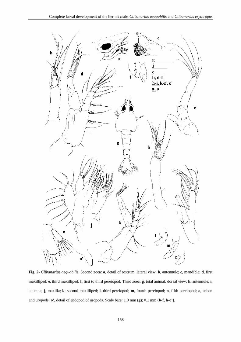
Complete larval development of the hermit crabs Clibanarius aequabilis and Clibanarius erythropus
- 158 -
Fig. 2- Clibanarius aequabilis. Second zoea: a, detail of rostrum, lateral view; b, antennule; c, mandible; d, first
maxilliped; e, third maxilliped; f, first to third pereiopod. Third zoea: g, total animal, dorsal view; h, antennule; i,
antenna; j, maxilla; k, second maxilliped; l, third pereiopod; m, fourth pereiopod; n, fifth pereiopod; o, telson
and uropods; o’, detail of endopod of uropods. Scale bars: 1.0 mm (g); 0.1 mm (b-f, h-o’).

Complete larval development of the hermit crabs Clibanarius aequabilis and Clibanarius erythropus
- 159 -
Telson: 8 + 8 processes, with an additional inner pair of telson plumose setae;
otherwise unchanged.
Third zoea (Figures 2g–o’)
Dimension: TL = 3.44– 3.77 mm; CL = 1.61– 1.72 mm; R = 0.75– 0.81 mm
Carapace (Fig. 2g): unchanged except for size.
Antennule (Fig. 2h): peduncle unsegmented with 1 basial simple seta, 4 long plumose
setae and 4 short simple setae terminally; inner flagellum small, bud-like; outer flagellum
with 3 aesthetascs, 1 long plumose seta and 3 shorter simple setae.
Antenna (Fig. 2i): endopod as long as the scale, with 1 short simple seta in apical
region; scaphocerite with 12 plumose setae. Otherwise unchanged.
Mandible and maxillule: unchanged except for size.
Maxilla (Fig. 2j): coxal endite with 9 + 4 serrulate and papposerrate setae;
scaphognathite with 10–11 marginal plumose setae; otherwise unchanged except for size.
First, second (Fig. 2k) and third maxillipeds: unchanged except for size.
First pereiopod: uniramous bud showing its chelate nature.
Second and third (Fig. 2l) pereiopods: unchanged.
Fourth (Fig. 2m) and fifth (Fig. 2n) pereiopods: uniramous buds.
Abdomen (Fig. 2g): abdominal somite 6 fully separated from telson, bearing a pair of
minute dorso-lateral setae; otherwise unchanged.
Pleopods: absent.
Uropods (Figs. 2o, o’): biramous; exopods well developed, not reaching the end of
telson, with 7–8 plumose setae; endopod small bud with 2 minute projections at its tip.

Complete larval development of the hermit crabs Clibanarius aequabilis and Clibanarius erythropus
- 160 -
Telson (Fig. 2o): triangular, broader posteriorly with median cleft less pronounced;
number of telson processes: 9–10 + 9–10 (first process as a very short blunt spine, second as
an anomuran hair, third as a plumose seta, fourth as a very reduced fused spine and fifth to
ninth or tenth processes as plumose setae). One individual in seven observed with 9 + 9
processes (additional inner pair of plumose telson setae, without reduced fused spine).
Fourth zoea (Figure 3)
Dimension: TL = 4.08– 4.40 mm; CL = 1.92– 2.08 mm; R = 0.88– 0.96 mm
Carapace (Fig. 3a): unchanged except for size.
Antennule (Fig. 3b): peduncle unsegmented with 2 + 1 simple setae along lateral
margin, 4 short simple and 4 long plumose setae distally; inner flagellum unsegmented,
enlarged in size; outer flagellum with 2 + 2 + 2 + 1 aesthetascs, 2 long and 2 short plumose
setae.
Antenna (Fig. 3c): endopod longer than antennal scale, two-segmented: first segment
unarmed, second segment with a short simple seta and a pointed tip; scaphocerite with 12–13
plumose setae. Otherwise unchanged.
Mandible (Fig. 3d): differs from the previous stage by the presence of an unsegmented
palp.
Maxillule (Fig. 3e): coxal endite with 3 papposerrate, 2 pappose and 3 simple setae;
otherwise unchanged.
Maxilla (Fig. 3f): coxal endite with 11–12 + 4 serrulate and papposerrate setae; basial
endite with 7 + 6 papposerrate and simple setae; endopod unsegmented, bilobed, terminally
bearing 3 + 2 long simple setae; scaphognathite with 15–17 marginal plumose setae, inferior
lobe without setae.

Complete larval development of the hermit crabs Clibanarius aequabilis and Clibanarius erythropus
- 161 -
Fig. 3- Clibanarius aequabilis. Fourth zoea: a, total animal, lateral view; b, antennule; c, antenna; d,
mandible; e, maxillule; f, maxilla; g, first maxilliped; h, third maxilliped; i, first pereiopod; j, second
pereiopod; k, third pereiopod; l, fourth pereiopod; m, fifth pereiopod; n- q, pleopods of abdominal
somites 2-5; r, telson and uropods. Scale bars: 1.0 mm (a); 0.1 mm (b-r).

Complete larval development of the hermit crabs Clibanarius aequabilis and Clibanarius erythropus
- 162 -
First (Fig. 3g) and second maxillipeds: unchanged.
Third maxilliped (Fig. 3h): endopod two-segmented, incomplete 2–5 segments with
two-minute projections at its tip; otherwise unchanged.
First pereiopod (Fig. 3i): cheliped with chela enlarged, dactyl distinct, coxa, and merus
clearly delineated, with tubercles on surface.
Second and third pereiopods (Fig. 3j, k): long, the first six segments clearly delineated,
coxa and merus with some tubercles on surface.
Fourth and fifth pereiopods (Fig. 3l, m): short, with some segments clearly delineated.
Abdomen (Fig. 3a): unchanged except for size.
Pleopods (Fig. 3n–q): present as biramous buds on segments 2–5.
Uropods (Fig. 3r): total individualization of protopod, exopod and endopod; exopod
with 8–9 plumose setae on apical region and 3–4 papposerrate setae on ventral surface;
endopod smaller with 5 plumose setae on apical region and 1 pappose seta on ventral surface.
Telson (Fig. 3r): almost rectangular in shape; number of telson processes: 9 + 9–10
(first process as a very short blunt spine, second as an anomuran hair, third as a plumose seta,
fourth as a very reduced fused spine and fifth to ninth or tenth processes as plumose setae);
presence of two pairs of pappose setae on dorsal surface.
Megalopa (Figures 4, 5)
Dimension: TL = 2.85– 3.04 mm; CL = 1.08– 1.15 mm; R = 0.15– 0.19 mm
Carapace (Fig. 4a, a’): smooth, slightly broader than long; rostrum broad and blunt;
ocular peduncles stout reaching the end of the antennular peduncle; ocular acicles not
developed; surface and margins with proboscate setae distributed as figured.

Complete larval development of the hermit crabs Clibanarius aequabilis and Clibanarius erythropus
- 163 -
Antennule (Fig. 4b): peduncle three-segmented, proximal segment with 12–13
papposerrate and simple setae; second segment with 1 long and 3–4 short simple setae and 1
papposerrate seta; distal segment with 6 simple setae; outer flagellum five-segmented,
proximal segment unarmed, second segment with 5 aesthetascs, third segment with 5
aesthetascs and 3 simple setae, fourth segment with fourth aesthetascs and 2–3 simple setae
terminally, distal segment with three simple setae; inner flagellum three segmented with two,
four, and seven simple setae.
Antenna (Fig. 4c): basis with 3–4 simple setae, peduncle three-segmented with 1, 2, 3–
4 simple setae; flagellum composed of 10 segments, each with 0–6 simple setae on distal
margin, arranged as figured, except for the distal segment which has 9 simple setae on distal
margin; acicle with 1 spine-like projection on lateral margin, 4 simple setae and bifid spinous
tip.
Mandibles (Fig. 4d): reduced and simplified, lower extremities of the incisor part with
processes as illustrated; palp two-segmented, with 4–5 simple setae and 4 serrulate setae on
the distal segment.
Maxillule (Fig. 4e): coxa with 18–21 papposerrate and simple setae; basis with 2
simple setae on the lateral inner margin and 5 cuspidate, 4 serrulate, 5–7 papposerrate, 3–4
simple and 1 long plumose setae on the distal margin; endopod unsegmented with 1 + 1
simple setae; 1 plumose seta present on the inner margin of basis.
Maxilla (Fig. 4f): coxa bilobed with 25–32 + 7–9 papposerrate and simple setae; basis
bilobed with 8–10 + 11–13 papposerrate and simple setae; endopod unsegmented without
seta; scaphognathite with 50–58 plumose setae on the distal margin and 1 simple seta on the
dorsal surface of the upper lobe.
First maxilliped (Fig. 4g): coxa with 3–4 simple, 3 pappose and 1 papposerrate setae;
basis with 4 simple, 3 serrulate and 11–14 papposerrate setae; endopod unsegmented without

Complete larval development of the hermit crabs Clibanarius aequabilis and Clibanarius erythropus
- 164 -
seta; exopod unsegmented with 7–9 plumose setae on the outer margin.
Second maxilliped (Fig. 4h): coxa with 1 simple seta; basis with 3 pappose setae;
endopod four-segmented, proximal segment with 1 + 1 simple seta, second segment with 3
simple setae, third segment with 8 simple and papposerrate setae, and 7–8 papposerrate setae
on the distal segment; exopod two-segmented, proximal segment with 2 simple setae and the
distal segment with 6–8 plumose setae terminally.
Third maxilliped (Fig. 4i): basis with 6–7 simple setae and 3–4 sub-acute processes;
endopod five-segmented, ischium with 8–10 simple seta and 5–6 sub-acute processes on
crista dentata, merus with 4–6 simple setae, carpus with 9–10 simple setae, propodus with 5
simple, 2 cuspidate, and 13–14 papposerrate setae, and dactyl with 4 serrate, 7–8 simple, 2
cuspidate and 6 papposerrate setae; exopod 2- segmented with 1 simple seta on proximal
segment and 6–8 (2 + 4 or 2 + 2 + 4) plumose setae terminally on distal segment.
First pereiopod (Fig. 5a, a’): chelae equal, coxa with 6–8 simple setae; basis with 4–5
simple setae; ischium with 6–7 simple setae; merus, longest segment, with 14–15 simple
setae; carpus with 6–8 simple setae; propodus with 31–35 simple setae, distal upper extremity
corneous; dactyl length about half palm + propodal prolongation, with 24–26 simple setae and
distal extremity corneous.
Second pereiopod (Fig. 5b, b’): coxa with 7–8 simple setae; basis smaller with 2–3
simple setae; ischium with 8–9 simple setae; merus, longest segment, with 14–16 simple
setae; carpus less than half the ischium length, with 11–12 simple setae; propodus longer than
carpus, with 23–25 simple setae and 1 stout cuspidate seta; dactylus very stout, apically
curved and corneous, with 28–30 simple setae and 3 spines.
Third pereiopod (Fig. 5c): coxa with 6–7 simple setae; basis with 3 simple setae and 1
tooth; ischium with 9–11 simple setae; merus longer than ischium and with 14–15 simple
setae; carpus with 12–13 simple setae; propodus longer than carpus, with 23–24 simple setae

Complete larval development of the hermit crabs Clibanarius aequabilis and Clibanarius erythropus
- 165 -
Fig. 4- Clibanarius aequabilis. Megalopa: a, total animal, dorsal view; a’, detail of rostrum, lateral view; b,
antennule; c, antenna; d, mandibles; e, maxillule; f, maxilla; g, first maxilliped; h, second maxilliped; i, third
maxilliped. Scale bars: 0.1 mm.

Complete larval development of the hermit crabs Clibanarius aequabilis and Clibanarius erythropus
- 166 -
and 1 stout cuspidate seta; dactylus very stout, apically curved and corneous, with 24–26
simple setae and 3 spines.
Fourth pereiopod (Fig. 5d, d’): basis with 5–7 simple setae; ischium with 5–8 simple
setae; merus with 8–9 simple setae; carpus with 5 simple setae; propodus not chelate with 9
simple setae and 14–16 pseudochaetae as figured; dactyl with 1 long papposerrate seta, 8–9
simple setae and 2 teeth on the distal extremity.
Fifth pereiopod (Fig. 5e, e’): coxa with 6–7 simple setae; basis with 3 simple setae;
ischium with 2–3 simple setae; merus with 2 long papposerrate setae and 6–7 small simple
setae; carpus with 3 papposerrate and 5–6 simple setae; propodus not chelate with 3 long
papposerrate, 16–18 simple setae and 17–18 pseudochaetae as figured; dactyl with 9–11
simple setae and 3–4 spatulate-shaped pseudochaetae distributed as figured.
Abdomen (Fig. 4a): somites with postero-lateral angles rounded; somite 1 with 2 pairs
of dorsal simple setae, somites 2–3 each with 4 pairs of dorsal simple setae and 3 pairs of
simple setae on dorso-lateral margin, somites 4–5 each with 5 pairs of dorsal simple setae and
4–5 pairs of simple setae on dorso-lateral margin, somite 6 with 5 pairs of dorsal simple setae
and 7 pairs of simple setae on dorsolateral margin.
Pleopods (Fig. 5f–i): well developed, biramous, decreasing in size posteriorly; outer
ramus with 8–9 plumose setae; inner ramus shorter, with 2 small apical hooks.
Uropods (Fig. 5j): right pair slightly longer than left pair; protopod with 1 simple seta
on outer margin; exopod with 4–5 simple setae dorsally, 3–4 simple setae on dorsal margin,
17–19 plumose setae and 10–13 pseudochaetae marginally; endopod with 4–5 simple setae
dorsally, 4 simple setae on dorsal margin, 10–11 plumose setae and 7–8 pseudochaetae
marginally.
Telson (Fig. 5j): telson with terminal margin rounded, 10–11 pairs of simple setae on
the dorsal surface, 2 pairs of pappose setae on the lateral margin and 5–6 pairs of long

Complete larval development of the hermit crabs Clibanarius aequabilis and Clibanarius erythropus
- 167 -
Fig. 5- Clibanarius aequabilis. Megalopa: a, first pereiopod; a’, detail of chela of first pereiopod; b, second
pereiopod; b’, detail of dactylus of second pereiopod; c, third pereiopod; d, fourth pereiopod; d’, detail of
dactylus of fourth pereiopod; e, fifth pereiopod; e’, detail of propodus and dactylus of fifth pereiopod; f- i,
pleopods of abdominal somites 2-5; j, telson and uropods. Scale bars: 0.1 mm (a-e, f-j); 0.05 mm (e’).

Complete larval development of the hermit crabs Clibanarius aequabilis and Clibanarius erythropus
- 168 -
plumose setae on the distal margin, distributed as illustrated. One of the observed individuals
presented an unpaired number (11) of plumose setae.
Clibanarius erythropus (Latreille, 1818)
Figure 6
Larval references: Pike and Williamson, 1960: p. 503, Fig. 2 (zoea I, zoea II and zoea III); Le
Roux, 1966: p. 227, Fig. 1 (zoea IV); Dechancé and Forest, 1958: p. 277, Fig. 2; p. 283, Figs.
16–23; p. 284, Figs. 24–29 (megalopa)
First zoea (Figure 6)
Dimension: TL = 1.83– 1.94 mm; CL = 0.81– 0.91 mm; R = 0.27– 0.38 mm
Carapace (Figs. 6a, a’): without processes or spines, with the postero-lateral border
smoothly rounded; rostrum broad, rather blunt, with triangular pointed tip extending beyond
antennal scale, rostrum about 0.66 ± 0.08 times the carapace; eyes compound and sessile.
Antennule (Fig. 6b): peduncle unsegmented, with 1 long plumose seta sub-terminally,
3 aesthetascs and 3 unequal simple setae terminally.
Antenna (Fig. 6c): protopod unsegmented with 1 small strong cuspidate seta; endopod
with more than half-length the scale, with 3 long plumose seta apically; scaphocerite
unsegmented, with 11 plumose setae.
Mandible (Fig. 6d): asymmetrical; palp absent.
Maxillule (Fig. 6e): coxal endite with 3 serrulate, 2–3 pappose and 2 simple setae,
basial endite with 2 cuspidate setae and 2 simple setae; endopod with 1 + 1 + 2 simple setae.

Complete larval development of the hermit crabs Clibanarius aequabilis and Clibanarius erythropus
- 169 -
Fig. 6- Clibanarius erythropus. First zoea: a, total animal, lateral view; a’, detail of rostrum, dorsal view; b,
antennule; c, antenna; d, mandible; e, maxillule; f, maxilla; g, first maxilliped; h, second maxilliped; i, third
maxilliped; j, telson; j’, detail of posterior margin of telson. Scale bars: 1.0 mm (a’); 0.1 mm (a-j’).

Complete larval development of the hermit crabs Clibanarius aequabilis and Clibanarius erythropus
- 170 -
Maxilla (Fig. 6f): coxal endite bilobed with 7–8 + 4 serrulate and papposerrate setae,
basial endite bilobed with 5 + 4–5 papposerrate and simple setae; endopod unsegmented
bilobed terminally, bearing 3+2 long simple setae and microtricha on outer margin;
scaphognathite with 5 marginal plumose setae.
First maxilliped (Fig. 6g): coxa unarmed; basis with 1 + 1 + 1 + 3 + 3 papposerrate
setae; endopod 5-segmented with 2, 2, 1, 2 papposerrate and 1+4 papposerrate setae,
microtricha on outer margin of second and third segments; exopod unsegmented, with 4 long
plumose setae terminally.
Second maxilliped (Fig. 6h): coxa unarmed; basis with 1 + 2 papposerrate setae;
endopod 4-segmented with 2, 2, 2 papposerrate and 1 + 4 papposerrate setae terminally.
Exopod unsegmented, with 4 long plumose setae terminally.
Third maxilliped (Fig. 6i): long uniramous bud.
Pereiopods: absent.
Abdomen (Fig. 6a): somites with margins rounded, with a pair of dorso-lateral
marginal minute setae on somites 2– 5; sixth somite feebly segmented.
Pleopods and uropods: absent.
Telson (Fig. 6j, j’): triangular, broader posteriorly with deep median cleft; with 7 + 7
processes, the first process is a very short blunt spine, the second process an anomuran hair,
the third to the seventh processes plumose setae, being the fourth the longest.
Second zoea
Dimension: TL = 2.10– 2.26 mm; CL = 1.02– 1.13 mm; R = 0.43– 0.48 mm
Carapace: eyes stalked; otherwise unchanged.
Antennule: peduncle unsegmented with 2 long plumose setae sub-terminally and 2

Complete larval development of the hermit crabs Clibanarius aequabilis and Clibanarius erythropus
- 171 -
short simple setae terminally, 1 long plumose seta at site of future endopod; exopod bud
clearly delineated, with 3 simple setae and 3 aesthetascs.
Antenna: unchanged except for size.
Mandible: molar process with several small denticles and without teeth; otherwise
unchanged.
Maxillule: basial endite with 4 cuspidate setae and 2 simple setae; otherwise
unchanged except for size.
Maxilla: basial endite bilobed with 5 + 4 papposerrate and simple setae;
scaphognathite with 7 marginal plumose setae; otherwise unchanged except for size.
First maxilliped: endopod 5-segmented with 1 + 2, 2–3, 2, 2 papposerrate and 1 + 4
papposerrate setae. Exopod bearing 6 long plumose setae terminally; otherwise unchanged.
Second maxilliped: endopod four-segmented with 2, 2 + 1, 1 + 2 papposerrate and 1 +
4 papposerrate setae terminally. Exopod bearing 6 long plumose setae terminally; otherwise
unchanged.
Third maxilliped: biramous; endopod represented by a small bud basally situated;
exopod unsegmented, bearing 5 plumose setae.
First to third pereiopods: present as small uniramous buds.
Abdomen: unchanged except for size.
Pleopods and uropods: absent.
Telson: 8 + 8 processes, with an additional inner pair of telson plumose setae; broader
posteriorly with the median cleft less pronounced; otherwise unchanged.
Third zoea
Dimension: TL = 2.74– 2.85 mm; CL = 1.35– 1.51 mm; R = 0.48– 0.59 mm

Complete larval development of the hermit crabs Clibanarius aequabilis and Clibanarius erythropus
- 172 -
Carapace: unchanged except for size.
Antennule: peduncle unsegmented with 1 small basial simple seta, 4 long plumose
setae and 4 short simple setae distally; the outer flagellum with 3 aesthetascs, 1 long plumose
seta and 3 shorter simple setae; the inner flagellum small, bud-like.
Antenna: endopod as long as the scale, with 1 short plumose seta in apical region;
scaphocerite with 11–12 plumose setae. Otherwise unchanged.
Mandible and maxillule: unchanged except for size.
Maxilla: coxal endite bilobed with 8 + 4 serrulate and papposerrate setae;
scaphognathite with 9–10 marginal plumose setae; otherwise unchanged except for size.
First and second maxillipeds: unchanged except for size.
Third maxilliped: endopod bud enlarged in size; exopod unsegmented, bearing 6
plumose setae.
First pereiopod: uniramous bud showing its chelate nature.
Second and third pereiopods: unchanged except for size.
Fourth and fifth pereiopods: small uniramous buds.
Abdomen: abdominal somite 6 fully separated from telson, bearing a pair of dorso-
lateral marginal minute setae on somites 2–5; otherwise unchanged.
Pleopods: absent.
Uropods: biramous; exopod well developed, not reaching the end of telson, with 7–8
plumose setae; endopod a small bud with 2 minute projections at its tip.
Telson: triangular, broader posteriorly with the median cleft less pronounced; the
number of telson processes: 8–9 + 9 (the first process as a very short blunt spine, the second
as an anomuran hair, the third as a plumose seta, the fourth as a very reduced fused spine and
the fifth to eighth or ninth processes as plumose setae).

Complete larval development of the hermit crabs Clibanarius aequabilis and Clibanarius erythropus
- 173 -
Fourth zoea
Dimension: TL = 3.01– 3.17 mm; CL = 1.51– 1.61 mm; R = 0.48– 0.59 mm
Carapace: unchanged except for size.
Antennule: peduncle unsegmented with 2 simple setae on lateral margin, 4 short
simple and 4 long plumose setae distally; outer flagellum with 2 + 2 + 2 + 1 aesthetascs, 2
long and 2 short plumose setae; inner flagellum unsegmented, enlarged in size.
Antenna: endopod longer than antennal scale, two-segmented: first segment unarmed,
second segment with a short plumose seta and a pointed tip terminally; scaphocerite with 11–
13 plumose setae. Otherwise unchanged.
Mandible: presence of an unsegmented palp.
Maxillule: unchanged except for size.
Maxilla: coxal endite bilobed with 9–12 + 4 serrulate and papposerrate setae; basial
endite with 6–7 + 4–5 papposerrate and simple setae; endopod unsegmented, bilobed,
terminally bearing 3 + 2 long simple setae; scaphognathite with 13–16 marginal plumose
setae.
First and second maxillipeds: unchanged.
Third maxilliped: endopod two-segmented, incomplete 2 to 5 segments with two-
minute projection at its tip; otherwise unchanged.
First pereiopod: cheliped with chela enlarged, dactyl distinct, coxa and merus clearly
delineated, with tubercles on surface.
Second and third pereiopods: long, six segments clearly delineated, coxa and merus
with some tubercles on surface.
Fourth and fifth pereiopods: short, with some segments clearly delineated.
Abdomen: unchanged except for size.

Complete larval development of the hermit crabs Clibanarius aequabilis and Clibanarius erythropus
- 174 -
Pleopods: present as biramous buds on segments 2 to 5.
Uropods: total individualization of protopod, exopod and endopod; exopod with 9–10
plumose setae on apical region and 2–3 papposerrate setae on ventral surface; endopod
smaller, with 4–5 plumose setae on apical region and 1 pappose seta on ventral surface.
Telson: almost rectangular in shape; number of telson processes: 9 + 9 (first process as
a very short blunt spine, second as an anomuran hair, third as a plumose seta, fourth as a very
reduced fused spine and fifth to ninth processes as plumose setae).
Megalopa
Dimension: TL = 2.80– 2.96 mm; CL = 1.08– 1.24 mm; R = 0.16– 0.22 mm
Carapace: smooth, slightly broader than long; rostrum broad and blunt; ocular
peduncles stout, reaching the end of the antennular peduncle; ocular acicles not developed;
surface and margins with proboscate setae distributed as figured.
Antennule: peduncle three-segmented, proximal segment with 2 papposerrate, 4–6
simple and 5 papposerrate setae; second segment with 1 long and 3–5 short simple setae and 1
papposerrate seta; distal segment with 5–6 simple setae; outer flagellum five-segmented,
proximal segment unarmed, second segment with 5 aesthetascs, third segment with 5
aesthetascs and 4–5 simple setae, fourth segment with 3 aesthetascs and 2–3 simple setae
terminally, distal segment with 3–4 simple setae; inner flagellum three-segmented with 2, 2–3
and 6–8 simple setae.
Antenna: basis with 3 simple setae, peduncle 3-segmented with 1–2, 2, 4–5 simple
setae; flagellum composed of 9–10 segments, each with 0–6 simple setae on distal margin,
except for the distal segment which has 8–9 simple setae on distal margin; acicle with 1
spinelike projection on lateral margin, 4 simple setae and bifid spinous tip.

Complete larval development of the hermit crabs Clibanarius aequabilis and Clibanarius erythropus
- 175 -
Mandibles: reduced and simplified, lower extremities of the incisor part with processes
as illustrated; palp three-segmented, proximal segment with 1 strong simple seta, second
segment unarmed, distal segment with 4–5 simple setae and 4–5 serrulate setae terminally.
Maxillule: coxa with 18–20 papposerrate and simple setae; basis with 2 simple setae
on lateral inner margin and 5 cuspidate, 5 serrulate, 6–7 papposerrate, 3 simple and 1 long
plumose setae on distal margin; endopod unsegmented with 1 + 1 simple setae; 1 plumose
seta present on inner margin of basis.
Maxilla: coxa bilobed with 27–29 + 7–8 papposerrate and simple setae; basis bilobed
with 9–12 + 10–13 papposerrate and simple setae; endopod unsegmented without seta;
scaphognathite with 47–52 plumose setae on distal margin and 1 simple seta on dorsal surface
of the upper lobe.
First maxilliped: coxa with 2–3 simple, 2–3 pappose and 3 papposerrate setae; basis
with 3–4 simple, 3 serrulate and 13–14 papposerrate setae; unsegmented endopod without
seta; exopod unsegmented with 6–8 plumose setae on outer margin.
Second maxilliped: coxa without any seta or with 1 simple seta; basis with 2–3
pappose setae; endopod four-segmented, proximal segment with 1 + 1 simple setae, second
segment with 2–3 simple setae, third segment with 7–9 simple and papposerrate setae, and 6–
7 papposerrate setae on distal segment; exopod two-segmented with 2 simple setae on
proximal segment and 8 plumose setae terminally on distal segment.
Third maxilliped: basis with 5–7 simple setae and 3–4 sub-acute processes; endopod
five-segmented, ischium with 7–9 simple seta and 4–5 sub-acute processes on crista dentata,
merus with 4–6 simple setae, carpus with 9–10 simple setae, propodus with 3–4 simple, 2
cuspidate and 13–15 papposerrate setae, and dactyl with 5 serrate, 6–7 simple, 3 cuspidate
and 5–6 papposerrate setae; exopod 2-segmented with 1 simple seta on proximal segment and
8 plumose setae terminally on distal segment.

Complete larval development of the hermit crabs Clibanarius aequabilis and Clibanarius erythropus
- 176 -
First pereiopod: chelae equal, with segments smooth; coxa with 9–10 simple setae;
basis with 4 simple setae; ischium with 8–9 simple setae; merus, longest segment, with 14–16
simple setae; carpus with 6–7 simple setae; propodus with 38–40 simple setae, distal upper
extremity corneous; dactyl length about half palm + propodal prolongation, with 23–26
simple setae and distal extremity corneous.
Second pereiopod: coxa with 7–8 simple setae; basis smaller with 2 simple setae;
ischium with 7–8 simple setae; merus, longest segment, with 14–16 simple setae; carpus less
than half the ischium length, with 12–14 simple setae; propodus longer than carpus, with 22–
25 simple setae and 1 stout cuspidate seta; dactylus very stout, apically curved and corneous,
with 27–29 simple setae and 3–4 spines.
Third pereiopod: coxa with 7–8 simple setae; basis with 4–5 simple setae; ischium
with 10–11 simple setae; merus longer than ischium and with 14–16 simple setae; carpus with
11–12 simple setae; propodus longer than carpus, with 24–27 simple setae and 1 stout
cuspidate seta; dactylus very stout, apically curved and corneous, with 25–28 simple setae and
3–4 spines.
Fourth pereiopod: basis with 7 simple setae; ischium with 8–10 simple setae; merus
with 9–10 simple setae; carpus with 4–5 simple setae; propodus not chelate, with 8–9 simple
setae and 10–14 pseudochaetae; dactyl with 1 long papposerrate seta, 7–9 simple setae and 2
teeth on distal extremity.
Fifth pereiopod: coxa with 5–6 simple setae; basis with 3 simple setae; ischium with
2–3 simple setae; merus with 2 long papposerrate setae and 6–7 small simple setae; carpus
with 3 papposerrate and 5–6 simple setae; propodus not chelate, with 5–6 long papposerrate,
16–18 simple setae and 11–13 pseudochaetae; dactyl with 5–7 simple setae and 3 spatulate-
shaped pseudochaetae.

Complete larval development of the hermit crabs Clibanarius aequabilis and Clibanarius erythropus
- 177 -
Abdomen: somites with postero-lateral angles rounded; somite 1 with 2 pairs of dorsal
simple setae, somites 2–3 each with 4–5 pairs of dorsal simple setae and 5 pairs of simple
setae on dorsolateral margin, somites 4–5 each with 5 pairs of dorsal simple setae and 4–5
pairs of simple setae on dorsolateral margin, somite 6 with 5 pairs of dorsal simple setae and 6
pairs of simple setae on dorsolateral margin.
Pleopods: well developed, biramous, decreasing in size posteriorly; outer ramus with
8–9 plumose setae; inner ramus shorter, with 2 small apical hooks.
Uropods: right one slightly longer than left one; protopod with 4 simple setae on outer
margin; exopod with 3 simple setae dorsally, 4 simple setae on dorsal margin, 16–18 plumose
setae and 9–11 pseudochaetae marginally; endopod with 3–5 simple setae dorsally, 4–5
simple setae on dorsal margin, 8–9 plumose setae and 5–8 pseudochaetae marginally.
Telson: telson with terminal margin rounded, 8–9 pairs of simple setae on dorsal
surface, 2 pairs of pappose setae on lateral margin and 5 pairs of long plumose setae on the
distal margin.
5.5- Discussion
Larval stages of C. erythropus cultured in the present study displayed only minor differences
to those described previously by Pike & Williamson (1960), Le Roux (1966) and Dechancé &
Forest (1958). In our study the first zoeal stage had longer TL and CL than described by Pike
& Williamson (1960) and Le Roux (1966), while the megalopa was smaller than recorded by
Dechancé & Forest (1958) (Table 1). In the first zoeal stage, the endopod of the maxillule was
found unsegmented, whereas in previous studies it had been described as a two-segmented
structure. Although Pike & Williamson (1960) and Le Roux (1966) reported the absence of
pereiopods in the second zoeal stage, in our study this zoeal stage already
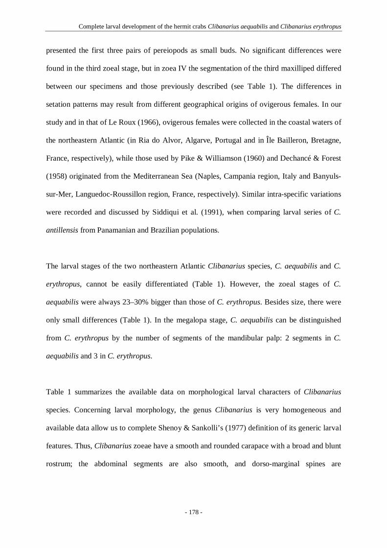
Complete larval development of the hermit crabs Clibanarius aequabilis and Clibanarius erythropus
- 178 -
presented the first three pairs of pereiopods as small buds. No significant differences were
found in the third zoeal stage, but in zoea IV the segmentation of the third maxilliped differed
between our specimens and those previously described (see Table 1). The differences in
setation patterns may result from different geographical origins of ovigerous females. In our
study and in that of Le Roux (1966), ovigerous females were collected in the coastal waters of
the northeastern Atlantic (in Ria do Alvor, Algarve, Portugal and in Île Bailleron, Bretagne,
France, respectively), while those used by Pike & Williamson (1960) and Dechancé & Forest
(1958) originated from the Mediterranean Sea (Naples, Campania region, Italy and Banyuls-
sur-Mer, Languedoc-Roussillon region, France, respectively). Similar intra-specific variations
were recorded and discussed by Siddiqui et al. (1991), when comparing larval series of C.
antillensis from Panamanian and Brazilian populations.
The larval stages of the two northeastern Atlantic Clibanarius species, C. aequabilis and C.
erythropus, cannot be easily differentiated (Table 1). However, the zoeal stages of C.
aequabilis were always 23–30% bigger than those of C. erythropus. Besides size, there were
only small differences (Table 1). In the megalopa stage, C. aequabilis can be distinguished
from C. erythropus by the number of segments of the mandibular palp: 2 segments in C.
aequabilis and 3 in C. erythropus.
Table 1 summarizes the available data on morphological larval characters of Clibanarius
species. Concerning larval morphology, the genus Clibanarius is very homogeneous and
available data allow us to complete Shenoy & Sankolli’s (1977) definition of its generic larval
features. Thus, Clibanarius zoeae have a smooth and rounded carapace with a broad and blunt
rostrum; the abdominal segments are also smooth, and dorso-marginal spines are

Tab
le 1
Com
par
ison
of
rele
vant
lar
val
char
acte
rs o
f C
liba
na
riu
s sp
ecie
s.
F
eatu
res
C
. aeq
ua
bil
is
(pre
sent
stu
dy)
C
. er
ythr
opus
(p
rese
nt s
tud
y)
C
. ery
thro
pus
(1),
(2)
, (3
)
C
. a
nti
llen
sis
(4)
C
. vit
tatu
s (5
)
C
. scl
op
eta
riu
s(6
)
C
. a
lbid
igit
us
(7)
C
. si
gn
atu
s (8
)
C
. vi
resc
ens
(8)
C
. in
fra
spin
atu
s(9
)
C
. mer
guie
nsis
(10)
C
. pad
aven
sis
(11)
C
. cl
iba
na
riu
s(1
2)
C
. lo
ng
ita
rsu
s(1
3)
C
. o
liva
ceus
(1
4)
Fem
ales
co
llec
tio
n ar
ea
Sta
ges:
zoe
a (Z
)+m
egal
opa
(M)
NE
Atl
anti
c
4Z+
1M
NE
Atl
anti
c
4Z+
1M
NE
Atl
anti
c
4Z+
1M
Car
ibbe
an S
ea
4Z+
1M
NW
Atl
anti
c
5Z+
1M
SW
Atl
anti
c
5Z+
1M
Cen
tral
E P
acif
ic
4Z+
1M
Ara
bian
Sea
4Z+
1M
Ara
bian
Sea
4Z+
1M
Ara
bian
Sea
4Z+
1M
Ara
bian
Sea
4Z+
1M
Ara
bian
Sea
4Z+
1M
Bay
of
Ben
gal
4Z+
1M
Bay
of
Ben
gal
4Z
+1M
Bay
of
Ben
gal
5Z
+1M
Fir
st Z
oea
Tot
al l
engt
h, m
m
2.39
-2.6
2 1.
83-1
.94
1.
70-1
.80
1.20
1.
90-2
.40
- -
1.60
1.
80
- -
- 1.
90
1.52
1.
57
Car
apac
e le
ngt
h, m
m
1.04
-1-1
2 0.
81-0
.91
0.80
0.
80
0.90
-1.1
0 -
0.63
-0.7
6 -
- 1.
00
1.00
0.
90
0.85
0.
85
0.90
CA
R A
PA
C E
R
ostr
um/
cara
pace
0.
89±
0.06
0.
66±
0.08
-
- -
- -
- -
- -
- -
- -
AN
TE
N N
U L
E
Ped
un
cle:
seg
men
ts/
se
tae
Un
seg.
/ 1+
(3)+
3 U
nse
g./
1+(3
)+3
Un
seg.
/ -
Un
seg.
/ 1+
(3)+
3 U
nse
g./
1+(3
)+2
-3
Un
seg.
/ 1+
(2)+
2 U
nse
g./
1+(2
-4)+
2-3
Un
seg.
/ 1+
(4)+
2 U
nse
g./
1+(4
)+1
Un
seg.
/ 1+
(3)+
3 U
nse
g./
1+(2
)+2
Un
seg.
/ 1+
(2)+
3 U
nse
g./
1+(2
)+3
Un
seg.
/ 1+
- U
nse
g./
1+(3
)+3
A
N T
E N
NA
P
roto
pod:
seg
men
ts/
seta
e E
ndop
od:
segm
ents
/ se
tae
Sca
phoc
erit
e
Un
seg.
/1
Un
seg.
/3
11
Un
seg.
/1
Un
seg.
/3
11
Un
seg.
/1
Un
seg.
/3
11
Un
seg.
/1
Un
seg.
/3
10-1
1
Un
seg.
/1
Un
seg.
/3
9-11
Un
seg.
/1
Un
seg.
/3
11
Un
seg.
/1
Uns
eg./
2-3
11
Un
seg.
/1
Un
seg.
/3
11
Un
seg.
/1
Un
seg.
/3
11
Un
seg.
/1
Un
seg.
/3
11
Un
seg.
/1
Un
seg.
/3
11
Un
seg.
/1
Un
seg.
/3
10-1
1
Un
seg.
/1
Un
seg.
/3
11
- - -
Un
seg.
/1
Un
seg.
/3
11
M A
X I L
L U
L E
C
ox
al e
nd
ite
Bas
ial
endi
te
End
opod
: se
gmen
ts/
seta
e
7 5 U
nse
g./4
7-8 4
Un
seg.
/4
- -
2/1
,2
7 4 U
nse
g./3
6 3-4
Un
seg.
/3
5 3 U
nse
g./3
7 4 U
nse
g./4
7 4 2
/1,2
5 2 2
/1,2
7 3 -
6 3 U
nse
g./3
5 3 U
nse
g./3
7 3 U
nse
g./3
- 3 U
nseg
./-
6 3 U
nse
g./3
M A
X IL
LA
C
ox
al e
nd
ite
Bas
ial
endi
te
End
opod
: se
gmen
ts/
seta
e S
caph
ogn
athi
te
7+4
5+4
Uns
eg./
3+2
5
7-8+
4 5+
4-5
Uns
eg./
3+2
5
-
-
-
-
7+3
4-5+
3 U
nseg
./2+
2 5-
6
6+4
5+3-
4 U
nseg
./2+
2 4-
6
3+2-
3 5-
6+2-
3 U
nse
g./4
5
7+4-
5 5-
6+4
Uns
eg./
2+2
5
6+3
5+4
Un
seg.
/4
5
5+3
4+4
Un
seg.
/4
5
7+4
5+3
Uns
eg./
2+2
5
6+4
4+4
Uns
eg./
2+2
5
7+3
4+3
Uns
eg./
2+2
5
6+3
2+4
Uns
eg./
2+2
5
- - U
nseg
./-
5
6+3
4+4
Uns
eg./
2+2
5 F
IRS
T M
AX
ILL
IPE
D
B
asia
l en
dite
E
ndop
od:
segm
ents
/ se
tae
Ex
opod
: se
gmen
ts/
seta
e
9 5
/2,2
,2,2
,5
Un
seg.
/4
9 5
/2,2
,1,2
,5
Un
seg.
/4
-
-
Un
seg.
/4
6 5
/1,3
,2,3
,5
2/0
,4
7-9
5/2
,2,1
,3,4
U
nse
g./4
7 5
/1,1
,1,3
,3
-
7-8
5/2
,2,1
,2,5
2
/0,4
6 5
/2,2
,2,3
,4
Un
seg.
/4
9 - -
7 5
/2,2
,1,2
,5
Un
seg.
/4
8 5
/1,2
,1,2
,5
2/0
,4
6-7
5/2
,2,1
,2,5
U
nse
g./4
- 5
/2,2
,2,2
,5
Un
seg.
/4
6 5/-
Un
seg.
/4
6 5
/2,2
,2,3
,4
Un
seg.
/4
SE
CO
ND
MA
XIL
LIP
ED
Bas
ial
endi
te
End
opod
: se
gmen
ts/
seta
e E
xop
od:
segm
ents
/ se
tae
3 4
/2,2
,2,5
U
nse
g./4
3 4
/2,2
,2,5
U
nse
g./4
-
-
Un
seg.
/4
3 4
/2,3
,3,5
2
/0,4
3 4
/2,2
,2,5
U
nse
g./4
3-6
4/2
,2,2
,3
-
3 4
/2,2
,2,5
2
/0,4
3 4
/2,2
,2,5
U
nse
g./4
3 4
/2,2
,2,5
U
nse
g./4
3 4
/2,2
,2,5
U
nse
g./4
4 4
/2,2
,2,5
2
/0,4
3 4
/2,2
,1,5
U
nse
g./4
3 4
/2,2
,2,5
U
nse
g./4
- 4/-
Un
seg.
/4
2 4
/2,2
,3,4
U
nse
g./4
T
HIR
D M
AX
ILL
IPE
D
D
evel
opm
ent
Seg
men
ts
Uni
r. b
ud
Uns
eg.
Uni
r. b
ud
Uns
eg.
Uni
r. b
ud
Uns
eg.
Uni
r. b
ud
2 U
nir.
bud
-
Uni
r. b
ud
- U
nir.
bud
U
nseg
. U
nir.
bud
2
Uni
r. b
ud
2 U
nir.
bud
-
Uni
r. b
ud
- U
nir.
bud
-
Uni
r. b
ud
Uns
eg.
Uni
r. b
ud
Uns
eg.
Uni
r. b
ud
Uns
eg.
T
EL
SO
N
Po
ster
ior
mar
gin
pro
cess
es
7+
7 7+
7 7+
7 7+
7 7+
7 7+
7 7+
7 7+
7 7+
7 7+
7 7+
7 7+
7 7+
7 7+
7 7+
7
Complete larval development of the hermit crabs Clibanarius aequabilis and Clibanarius erythropus
- 179 -
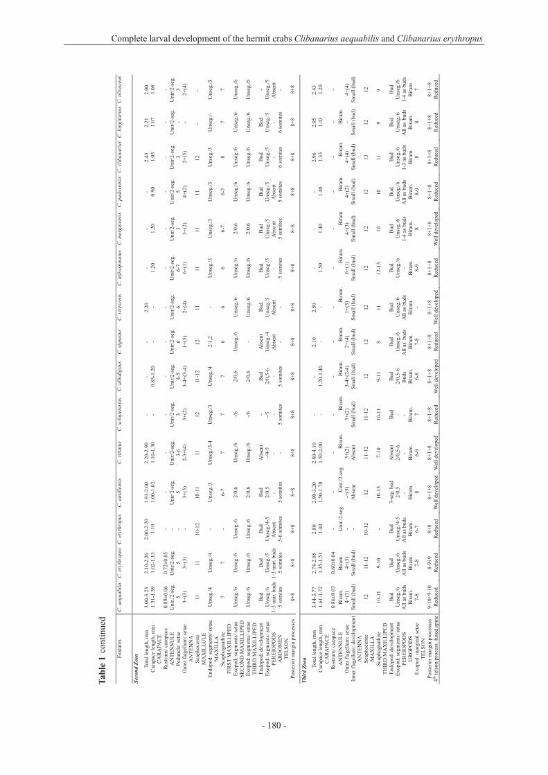
Tab
le 1
co
nti
nu
ed
F
eatu
res
C. a
equ
ab
ilis
C
. er
yth
rop
us
C.
eryt
hro
pu
sC
. an
till
ensi
s C
. vit
tatu
s C
. sc
lop
eta
riu
sC
. a
lbid
igit
us
C. s
ign
atu
s C
. vir
esce
ns
C.
infr
asp
ina
tus
C. m
ergu
iens
is
C.
pada
vens
isC
. cl
iba
na
riu
sC
. lo
ng
ita
rsu
sC
. o
liva
ceus
Sec
ond
Zoe
a
Tot
al l
engt
h, m
m
3.00
-3.2
3 2.
10-2
.26
2.00
-2.2
0 1.
91-2
.00
2.20
-2.9
0 -
- -
2.20
-
- -
2.43
2.
21
2.00
C
arap
ace
len
gth,
mm
1.
31-1
.39
1.02
-1.1
3 1.
10
1.00
-1.0
2 1.
10-1
.30
- 0.
85-1
.20
- -
1.20
1.
20
0.90
1.
05
1.07
1.
08
CA
R A
PA C
E
Ros
trum
/ ca
rapa
ce
0.89
±0.
06
0.72
±0.
05
- -
- -
- -
- -
- -
- -
- A
NT
EN
NU
LE
U
nir
./2
-seg
.
Un
ir/2
-seg
. -
Un
ir/2
-seg
.
Un
ir/2
-seg
.
Un
ir/2
-seg
. U
nir
/2-s
eg.
U
nir
/2-s
eg.
Un
ir/2
-seg
. U
nir
/2-s
eg.
Un
ir/2
-seg
.
Un
ir/2
-seg
.
Un
ir/2
-seg
. U
nir
/2-s
eg.
U
nir
/2-s
eg.
Ped
uncl
e: s
etae
O
ute
r fl
agel
lum
: se
tae
5 3+
(3)
5 3+
(3)
-
-
5 3+
(5)
3-6
2-3+
(4)
3 3+
(2)
4-5
3-4+
(3-4
) 6
1+(5
) 6
2+(4
) 6-
7 6+
(1)
3 3+
(2)
5 4+
(2)
3 2+
(5)
- - 3
2+(4
) A
NT
EN
N A
Sca
phoc
erit
e 11
11
10
-12
10-1
1 11
12
11
-12
12
11
11
11
11
12
- -
MA
XIL
LU
L E
E
ndop
od:
segm
ents
/ se
tae
Uns
eg./
4 U
nseg
./4
- U
nseg
./3
Uns
eg./
3-4
Uns
eg./
3 U
nseg
./4
2/1,
2 -
Uns
eg./
3 U
nseg
./3
Uns
eg./
3 U
nseg
./3
Uns
eg./
- U
nseg
./3
MA
XIL
L A
Sca
phog
nat
hite
7
7 -
6-7
7 7
7 6
6 6
6-7
6-7
8 7
7 F
IRS
T M
AX
IL L
I P
ED
E
xo
po
d:
segm
ents
/ se
tae
Un
seg.
/6
Un
seg.
/6
Un
seg.
/6
2/0
,6
Un
seg.
/6
-/6
2/0
,6
Un
seg.
/6
Un
seg.
/6
Un
seg.
/6
2/0
,6
Un
seg.
/6
Un
seg.
/6
Un
seg.
/6
Un
seg.
/6
SE
CO
ND
MA
X I L
L
IPE
D
Ex
op
od
: se
gmen
ts/
seta
e U
nse
g./6
U
nse
g./6
U
nse
g./6
2
/0,6
U
nse
g./6
-/
6 2
/0,6
-
Un
seg.
/6
Un
seg.
/6
2/0
,6
Un
seg.
/6
Un
seg.
/6
Un
seg.
/6
Un
seg.
/6
TH
IRD
MA
XIL
L I
PE
D
End
opod
: de
velo
pmen
t E
xop
od:
segm
ents
/ se
tae
Bud
U
nse
g./6
B
ud
Un
seg.
/5
Bud
U
nseg
./4-
5 B
ud
2/0
,5
Abs
ent
-/
4-5
- -/5
Bud
2/
0,5-
6 A
bsen
t U
nse
g./4
B
ud
Un
seg.
/5
Bud
U
nse
g./5
B
ud
Un
seg.
/5
Bud
U
nse
g./5
B
ud
Un
seg.
/5
Bud
U
nse
g./5
-
Un
seg.
/5
PE
RE
IOP
OD
S
1-3
unir
. bu
ds
1-3
unir
. bud
s A
bsen
t -
- -
- A
bsen
t A
bsen
t -
Abs
ent
A
bsen
t -
- A
bsen
t A
BD
OM
EN
5
som
ites
5
som
ites
5
-6 s
om
ites
5
som
ites
-
5 so
mit
es
5 so
mit
es
- -
5 so
mit
es
5 so
mit
es
5 so
mit
es
6 so
mit
es
6 so
mit
es
- T
EL
SO
N
Pos
teri
or m
argi
n pr
oces
ses
8+8
8+8
8+8
8+8
8+8
8+8
8+8
8+8
8+8
8+8
8+8
8+8
8+8
8+8
8+8
Th
ird
Zo
ea
Tot
al l
engt
h, m
m
3.44
-3.7
7 2.
74-2
.85
2.80
2.
90-3
.20
2.80
-4.1
0 -
- 2.
10
2.50
-
- -
2.96
2.
95
2.43
C
arap
ace
len
gth,
mm
1.
61-1
.72
1.35
-1.5
1 1.
40
1.50
-1.7
0 1.
50-2
.00
- 1.
20-1
.40
- -
1.50
1.
40
1.40
1.
33
1.43
1.
20
CA
RA
PA C
E
Ros
trum
/ ca
rapa
ce
0.86
±0.
03
0.60
±0.
04
- -
- -
- -
- -
- -
- -
-
AN
TE
NN
UL
E
Bir
am.
Bir
am.
U-
nir
./2
-seg
. U
nir
./2
-seg
. B
iram
. B
iram
. B
iram
. B
iram
. B
iram
. B
iram
. B
iram
B
iram
. B
iram
. B
iram
. O
ute
r fl
agel
lum
: se
tae
Inn
er f
lage
llum
: de
velo
pmen
t4+
(3)
Sm
all
(bu
d)
4+(3
) S
mal
l (b
ud
) - -
-+(5
) A
bsen
t 5+
(2)
Abs
ent
3+
(2)
Sm
all
(bu
d)
3-4+
(2-4
) S
mal
l (b
ud
) 2+
(4)
Sm
all
(bu
d)
1+(5
) S
mal
l (b
ud
) 6+
(1)
Sm
all
(bu
d)
4+(3
) S
mal
l (b
ud
) 4+
(2)
Sm
all
(bu
d)
4+(4
) S
mal
l (b
ud
) -
Sm
all
(bu
d)
4+(4
) S
mal
l (b
ud
) A
NT
EN
N A
Sca
phoc
erit
e 12
11
-12
10-1
2 12
11
-12
11-1
2 12
12
12
12
12
12
13
12
12
M
AX
ILL
A
S
caph
ogn
athi
te
10-1
1 9-
10
- 10
-13
7-10
10
-11
9-11
8
11
12-1
3 10
10
11
9
9 T
HIR
D M
AX
IL L
I P
ED
E
ndop
od:
deve
lopm
ent
Ex
opod
: se
gmen
ts/
seta
e B
ud
Un
seg.
/6
Bud
U
nse
g./6
B
ud
Uns
eg./
4-5
3-se
g. b
ud
2/0
,5
Abs
ent
2/0,
5-6
Bud
-
Bud
2/
0,5-
6 B
ud
Un
seg.
/6
Bud
U
nse
g./6
B
ud
Un
seg.
/6
Bud
U
nse
g./6
B
ud
Un
seg.
/6
Bud
U
nse
g./6
B
ud
Un
seg.
/6
Bud
U
nse
g./6
P
ER
EIO
PO
DS
A
ll a
s bu
ds
All
as
buds
A
ll a
s bu
ds
- -
- B
uds
All
as
bud
s A
ll a
s bu
ds
- 1-
4 as
bud
s A
ll a
s bu
ds
1-3
as b
uds
All
as
buds
1-
4 as
bud
s U
RO
PO
DS
B
iram
. B
iram
. B
iram
. B
iram
. B
iram
. B
iram
. B
iram
. B
iram
. B
iram
. B
iram
. B
iram
. B
iram
. B
iram
. B
iram
. B
iram
. E
xop
od:
mar
gina
l se
tae
7-8
7-8
6-7
8 6-
9 7
6-8
7-8
- 8-
9 8
8-9
8 8
7 T
EL
SO
N
Po
ster
ior
mar
gin
pro
cess
es
4th t
elso
n pr
oces
s: f
used
spi
ne9-
10+
9-10
R
educ
ed
8-9+
9 R
educ
ed
8+8
Red
uced
8+
1+8
Wel
l de
velo
ped
8+1+
8 W
ell
deve
lope
d 8+
1+8
Red
uced
8+
1+8
Wel
l de
velo
ped
8+1+
8 R
educ
ed
8+1+
8 W
ell
deve
lope
d 8+
1+8
Red
uced
8+
1+8
Wel
l de
velo
ped
8+1+
8 R
educ
ed
8+1+
8 R
educ
ed
8+1+
8 R
educ
ed
8+1+
8 R
educ
ed
Complete larval development of the hermit crabs Clibanarius aequabilis and Clibanarius erythropus
- 180 -

Tab
le 1
co
nti
nu
ed
F
eatu
res
C. a
equ
ab
ilis
C
. ery
thro
pu
s C
. ery
thro
pu
sC
. a
nti
llen
sis
C.
vitt
atu
s C
. scl
op
eta
riu
s C
. a
lbid
igit
us
C. s
ign
atu
s C
. vir
esce
ns
C.
infr
asp
ina
tus
C. m
ergu
iens
isC
. pa
dave
nsis
C.
clib
an
ari
us
C. l
on
git
ars
us
C.
oli
vace
us
Fo
urt
h Z
oea
Tot
al l
engt
h, m
m
4.08
-4.4
0 3.
01-3
.17
2.80
3.
10-3
.30
3.60
-5.2
0 -
- -
3.80
-
- -
3.77
3.
58
3.19
C
arap
ace
len
gth,
mm
1.
92-2
.08
1.51
-1.6
1 1.
40
1.60
-1.7
0 1.
70-2
.60
- 1.
50-1
.70
- -
1.60
1.
70
1.60
1.
72
1.63
1.
73
CA
RA
PAC
E
R
ostr
um/
cara
pace
0.
86±
0.08
0.
54±
0.04
-
- -
- -
- -
- -
- -
- -
AN
TE
NN
UL
E
Bir
am.
Bir
am.
Bir
am.
Bir
am.
Bir
am.
Bir
am.
Bir
am.
Bir
am.
Bir
am.
Bir
am.
Bir
am.
Bir
am.
Bir
am.
Bir
am.
Bir
am.
Ou
ter
flag
ellu
m:
segm
ents
/set
aeIn
ner
fla
gel
lum
: d
evel
op
men
t U
nseg
./4+
(7)
Un
seg.
/0
Uns
eg./
4+(7
) U
nse
g./0
U
nseg
./-
Uns
eg./
- U
nseg
./4+
(9)
Un
seg.
/0
Uns
eg./
4+(4
-5)
Bud
U
nseg
./4-
5+(4
-5)
Un
seg.
/0
Uns
eg./
3-4+
(7)
Un
seg.
/0
Uns
eg./
2+(6
) U
nse
g./1
U
nseg
./1+
(7)
Un
seg.
/0
Uns
eg./
6+(1
) U
nse
g./0
U
nseg
./4+
(3)
Un
seg.
/0
Uns
eg./
3+(3
)U
nse
g./2
U
nseg
./9+
(-)
Un
seg.
/0
Uns
eg./
- U
nse
g./0
U
nseg
./4+
(4)
Un
seg.
/0
AN
TE
NN
A
S
caph
ocer
ite
12-1
3 11
-13
10-1
2 13
11
-14
12-1
3 11
-12
10
12
- 13
13
-14
14
13
13
MA
ND
IBL
E
Pal
p p
rese
nt
Pal
p p
rese
nt
- P
alp
pre
sen
t P
alp
pre
sen
t/A
bse
nt
Pal
p p
rese
nt
Pal
p p
rese
nt
Pal
p p
rese
nt
Pal
p p
rese
nt
Pal
p p
rese
nt
Pal
p p
rese
nt
Pal
p p
rese
nt
Pal
p p
rese
nt
Pal
p p
rese
nt
Pal
p p
rese
nt
MA
XIL
L A
End
opod
: se
gmen
ts/
seta
e
Sca
phog
nat
hite
U
nseg
./3+
2 15
-17
Uns
eg./
3+2
13-1
6 - -
Uns
eg./
2+2
12-1
6 U
nseg
./2+
2 12
-14
Uns
eg./
2+2
12
Uns
eg./
2+2
12-1
8 U
nse
g./4
9
Un
seg.
/4
16
- - U
nseg
./2+
2 14
U
nseg
./2+
2 35
-38
Uns
eg./
2+2
13
Uns
eg./
- 13
U
nseg
./2+
2 16
F
IRS
T M
AX
IL L
I P E
D
Ex
op
od
: se
gmen
ts/
seta
e U
nse
g./6
U
nse
g./6
U
nse
g./6
2
/0,6
U
nse
g./6
-/
7 U
nse
g./6
-7
Un
seg.
/6
Un
seg.
/6
Un
seg.
/6
2/0
,6
Un
seg.
/7
Un
seg.
/6
Uns
eg./
6
- S
EC
ON
D M
AX
I L L
I P E
D
Ex
opod
: se
gmen
ts/
seta
e U
nseg
./6
Uns
eg./
6 U
nseg
./6
2/0,
6 U
nseg
./6-
7 -/
7 U
nseg
./7-
8 U
nseg
./6
Uns
eg./
6 U
nseg
./6
2/0,
6 U
nseg
./7
Un
seg.
/6
Uns
eg./
6 -
TH
IRD
MA
XIL
L I P
E D
E
ndop
od:
deve
lopm
ent
Ex
opod
: se
gmen
ts/
seta
e 2-
seg
Un
seg.
/6
2-se
g.
Un
seg.
/6
Inc.
5-s
eg.
Un
seg.
/6
Inc.
5-s
eg.
2/0
,5
Abs
ent
2/0,
5-6
Uns
eg.
-/6
Inc.
3-5
seg
. 2/
0,5-
6 - -
Uns
eg.
Un
seg.
/6
- U
nse
g./6
3-
seg.
U
nse
g./6
U
nseg
. U
nse
g./7
U
nseg
. U
nse
g./6
U
nseg
. U
nse
g./6
U
nseg
. U
nse
g./6
P
ER
EIO
PO
DS
A
ll a
s bu
ds
All
as
buds
A
ll a
s bu
ds
All
as
buds
A
ll a
s b
uds
- A
ll a
s bu
ds
All
as
buds
A
ll a
s bu
ds
- A
ll a
s bu
ds
Al
l as
bu
ds
All
as
bu
ds
All
as
bu
ds
All
as
bu
ds
PL
EO
PO
DS
B
iram
. bu
ds
Bir
am.
buds
B
iram
. bud
s B
iram
. bud
s A
bsen
t-sm
all
buds
B
iram
. bud
s B
iram
. bud
s B
iram
. bu
ds
Bir
am.
buds
B
iram
. bud
s B
iram
. bud
s B
iram
. bud
s B
iram
. bud
s B
iram
. bud
A
bsen
t U
RO
PO
D S
Uro
pod
endo
pod:
mar
gin
al s
etae
Uro
pod
exop
od:
mar
gina
l se
tae
5
8-9
4-
5
9-10
3-
5 8-
9 5-
6
10
3-6
6-9
4-
5
9-11
4-
6
8-10
4
9
4
9
- -
4-6
9-
10
5-6
11
-12
5
12
5
11
5
11
T
EL
SO
N
Po
ster
ior
mar
gin
pro
cess
es
4th t
elso
n pr
oces
s: f
used
spi
ne
9+9-
10
Red
uced
9+
9 R
educ
ed
9+9
Red
uced
8+
1+8
Wel
l de
velo
ped
8+1+
8/ 9
+9
Wel
l d
evel
oped
8+
1+8
Red
uced
8+
1+8
W
ell
deve
lope
d8+
1+7
Red
uced
8+
1+8
Wel
l de
velo
ped
8+1+
8 R
educ
ed
8+1+
8 W
ell
deve
lope
d8+
1+8
Red
uced
8+
1+8
Red
uced
8+
1+8
R
educ
ed
8+1+
8 R
educ
ed
Fif
th Z
oea
Tot
al l
engt
h, m
m
- -
- -
4.30
-5.3
0 -
- -
- -
- -
- -
3.76
C
arap
ace
len
gth,
mm
-
- -
- 2.
30-2
.70
- -
- -
- -
- -
- 2.
19
AN
TE
NN
U L
E
Inn
er f
lag
ellu
m:
deve
lopm
ent
- -
- -
Uns
eg./
0-1
U
nseg
./0
- -
- -
- -
- -
Uns
eg./
1 A
NT
EN
N A
End
opod
: se
gmen
ts/
seta
e -
- -
- 2/
0,1
3/0,
0,1
- -
- -
- -
- -
- M
AN
DIB
LE
-
- -
- P
alp
pre
sent
2-
seg.
pal
p -
- -
- -
- -
- U
nseg
. pa
lp
SE
CO
ND
MA
XI L
L I P
E D
E
xop
od:
segm
ents
/ se
tae
- -
- -
Uns
eg./
8 -/
7 -
- -
- -
- -
- -
TH
IRD
MA
XIL
L I P
E D
E
ndop
od:
deve
lopm
ent
Ex
opod
: se
gmen
ts/
seta
e - -
- - - -
- - B
ud
2/0
,6
Uns
eg.
-/6
- - - -
- - - -
- - - -
- - - -
Uns
eg.
Un
seg.
/7
PE
RE
IOP
OD
S
- -
- -
All
as
buds
-
- -
- -
- -
- -
All
as
buds
P
LE
OP
OD
S
- -
- -
Uni
r. b
uds
Bir
am.
buds
-
- -
- -
- -
- B
iram
. bud
s T
EL
SO
N
Pos
teri
or m
argi
n pr
oces
ses
- -
- -
9+9
- -
- -
- -
- -
- 8+
1+8
Complete larval development of the hermit crabs Clibanarius aequabilis and Clibanarius erythropus
- 181 -

-
-U R O
PO
DS
-
Tab
le 1
co
nti
nu
ed
Fea
ture
s C
. aeq
ua
bil
is
C.
eryt
hro
pu
s C
. er
yth
rop
us
C. a
nti
llen
sis
C.
vitt
atu
s C
. sc
lop
eta
riu
sC
. ali
dig
itu
s C
. sig
na
tus
C. v
ires
cens
C.
infr
asp
ina
tus
C.
mer
guie
nsis
C
. pa
dave
nsis
C
. cl
iba
na
riu
sC
. lo
ng
ita
rsu
sC
. oli
vace
us
Meg
alop
a
Tot
al l
engt
h, m
m
2.85
-3.0
4 2.
80-2
.96
3.
10-3
.30
2.70
-2.9
0 3.
20-4
.10
- -
2.50
3.
20
- -
- 2.
72
3.14
3.
40
Car
apac
e le
ngt
h, m
m
1.08
-1.1
5
1.08
-1.2
4
0.90
-1.0
0 0.
60-0
.80
1.10
-1.5
0 -
0.80
-1.0
0 -
- 1.
40
1.20
1.
20
1.12
1.
09
1.34
C
A R
A P
AC
E
Ro
stru
m
R
ostr
um/c
arap
ace
Ocu
lar
scal
es
Bro
ad,
b
lun
t 0
.17
±0
.02
Abs
ent
Bro
ad,
b
lun
t 0
.18
±0
.03
Abs
ent
Bro
ad,
b
lun
t -
Abs
ent
Tri
angu
lar
- -
Sm
all,
b
lun
t -
Abs
ent
Sm
all,
b
lun
t - -
Bro
ad,
b
lun
t -
Wea
kly
de
velo
ped
Tri
angu
lar
- A
bsen
t
Tru
ncat
ed
- A
bsen
t
- - W
eakl
y
deve
lope
d
Bro
ad
- W
eakl
y
deve
lope
d
Tri
angu
lar
- W
eakl
y
deve
lope
d
Bro
ad,
ro
unde
d -
Wea
kly
de
velo
ped
Bro
ad,
ro
unde
d - -
Bro
ad,
rou
nded
-
Wea
kly
de
velo
ped
A N
T E
N N
U L
E
P
edun
cle:
seg
men
ts/
seta
e O
ute
r fl
agel
lum
: se
gmen
ts/
aest
heta
scs
Inn
er f
lag
ellu
m:
segm
ents
/set
ae
3/12
-13,
4-5,
6 5
/ 0,
(5),
(5),
(4),
0 3
/2,4
,7
3/11
-13,
5-7,
5-6
5/
0,(5
),(5
),(3
),0
3/2,
2-3,
6-8
3/-
5/ - 3/-
3/7,
1-2,
1-2
5/
0,(6
),(5
),(3
),0
3/1
,3,5
- 5/
0,(4
),(4
),(3
),0
3/2,
5-6
3/9-
13,-
,-
5/
0,(6
),(4
),(2
),0
3/-
3/7-
10,3
-5,3
-5
6/
0,(6
),(4
-5),
(3),
0,0
3/2-
4,2-
3,4-
5
3/-
5/ -
2-3/
-
3/-
5/ - 3/-
3/-,
-,4
5/
(14)
3/
-,-,
4
3/-
5/
0,(5
-6),
(4),
(2),
0 -
3/-
4/ -
3/1
,1,4
3/-
5/ - 3/-
3/-
5/ - 3/-
3/-
5/
0,(7
-9),
0 3/
-,-,
4
AN
T E
NN
A
Fla
gell
um:
segm
ents
10
7-
10
7-9
10
11
10-1
1 10
8
11
11
9 10
13
13
12
M
AN
DIB
LE
2-
seg.
pal
p 3-
seg.
pal
p 3-
seg.
pal
p 2-
seg.
pal
p 3-
seg.
pal
p 3-
seg.
pal
p 2-
/3-s
eg.
palp
3-
seg.
3-
seg.
P
art.
seg
. 3-
seg.
3-
seg.
P
art.
3-s
eg.
Par
t. S
eg.
3-se
g.
M
A X
I LL
U L
E
C
ox
al e
nd
ite
Bas
ial
endi
te
End
opod
: se
gmen
ts/s
etae
18-2
1 21
-24
1+1
18-2
0 23
-24
1+1
- - -
- - 1
18-2
0 19
1
22
20
1
- - 1
18
19
1+2
- - -
- - 1+
2-3
19
17-1
9 1+
1-2
- - -
- - 1
- - 1
- - -
MA
XIL
LA
C
ox
al e
nd
ite
Bas
ial
endi
te
End
opod
: se
gmen
ts/s
etae
S
caph
ogn
athi
te
25-3
2+7-
9 8-
10+
11-1
3 0
50-5
8+1
27-2
9+7-
8 9-
12+
10-1
3 0
47-5
2+1
- - - -
- - 0 49
-55
27+
6 8+
15
0 60
27-3
0+6-
7 10
-12+
10-1
5 0 52
- - 0 45
-60
19
14
0 45
- - - -
- - 0 -
- - - 50
- - 0 -
- - 0 -
- - - -
- - 0 54
-56
FIR
ST
MA
XIL
LIP
ED
Bas
ial
endi
te
End
opod
: se
gmen
ts/s
etae
E
xop
od:
segm
ents
/set
ae
18-2
1 U
nse
g./0
U
nseg
./7-
9
19-2
1 U
nse
g./0
U
nseg
./6-
8
- U
nse
g./0
7
- U
nse
g./0
U
nse
g./9
20
Un
seg.
/0
Uns
eg./
8-10
15-1
6 U
nse
g./0
U
nse
g./9
- U
nse
g./0
U
nseg
./9-
10
15
Uns
eg./
3+6
2/5
,6
- U
nse
g./0
2/
10,-
- 5/
-,-,
-,-,
4 U
nseg
./8-
12
- - -
- U
nse
g./1
U
nse
g./1
1
- U
nseg
./-
Uns
eg./
9+2
- U
nseg
./-
Uns
eg./
9+2
- - U
nse
g./1
0 S
EC
ON
D M
AX
ILL
IPE
D
B
asia
l en
dite
E
ndop
od:
segm
ents
/set
ae
Ex
opod
: se
gmen
ts/s
etae
3 4/
2,3,
8,7-
8 2/
2, 6
-8
2-3
4/2,
2-3,
7-9,
6-7
2
/2,8
- - -
2 4/
1-2,
2,-,
- 2/
1-2,
7-8
3 4/
-,-,
4,6
Uns
eg./
6-8
0 or
2-3
4/
2,2-
3,4-
6,7
2/-,
7
- 4/
1-3,
1-3,
1-3,
- 2/
1-2,
6-7
- 5
/4,2
,3,5
,6
2/1
,6
- - 2/
2,-
- 5(
part
.7)/
- -/
6
- - -
- 5/-
2/-,
4-6
- 5/-
2/-,
6
- 5/-
2/-
- 5/-
2/0
,10
TH
IRD
MA
XIL
LIP
ED
Bas
ial
endi
te
End
opod
: se
gmen
ts
En
do
po
d:
cris
ta d
enta
ta
Ex
opod
: se
gmen
ts/s
etae
6-7+
3-4s
ub-a
c.pr
5
P
rese
nt
2/1,
6-8
5-7+
3-4
sub-
ac.p
r5
Pre
sent
2
/1,8
- - P
rese
nt
-
4+-
5 P
rese
nt
2/1
,8
2-3 5 -
Uns
eg./
2-4,
6
4+1-
2sub
-ac.
pr5
Pre
sen
t
2/-
-+2-
4sub
-ac.
pr
5 P
rese
nt
2/0
,6
2sub
-ac.
pr
5 P
rese
nt
2/-,
6
- - P
rese
nt
-
-+4s
ub-a
c.pr
7
Abs
ent
3/-,
-,6
3+-
5/2+
2,2+
1,4+
1,-,
--
Un
seg.
/6
- 6/- - -
- 5/- -
2/-,
6
- 5/- -
2/-,
6
- - - -
P E
R E
IO P
O D
S
F
irst
: ch
elip
eds
Sec
ond/
thi
rd:
dact
yl s
pine
s
Fo
urt
h/
fift
h:
pro
po
dal
pc
Equ
al
3 14
-16/
17-1
8
Equ
al
3-4
10-1
4/11
-15
Equ
al
- -
Equ
al
3 -
Equ
al
- -
Equ
al
3-4/
8-11
-
Equ
al
3 -
Equ
al
- -
Equ
al
- -
Sub
-equ
al
3 -
Sub
-equ
al
2-3/
4-6
-
Equ
al/s
ub-e
qual
4 -
Sub
-equ
al
- -
Sub
-equ
al
- -
Sub
-equ
al
- - P
LE
OP
OD
S
Dec
r. p
ost.
D
ecr.
pos
t.
Dec
r. p
ost.
D
ecr.
pos
t.
Equ
al
Equ
al
Dec
r. p
ost.
E
qual
-
Dec
r. p
ost.
-
End
opod
: se
tae
E
xop
od:
seta
e 10
-11+
7-8p
c 17
-19+
10-1
3pc
8-9+
7-8p
c 16
-18+
9-11
pc
7-8+
6-8p
c -+
10pc
11
-14+
5-7p
c 19
-21+
8-12
pc12
-20+
6-7p
c8-
15+
- 8-
10+
5-6p
c 11
-14+
5-6p
c 9-
13+
8-10
pc
16-1
9+11
-13p
c 9+
-
18+
- -
- - - -
- -.
7+-
14
+-
12+
-
19+
-
10+
4-6p
c 16
+5-
6pc
TE
L S
ON
S
etae
: m
argi
nal
/lat
eral
/dis
tal
10
-11
/2 p
airs
/10
-12
8-9
/2 p
airs
/10
-/-/
10
10
/1-2
pai
rs/9
-/-/
8-9
-/
-/9
-10
-/-/
9 -
- 6
/2 p
airs
/9
6/2
pai
rs/9
5/
-/9
-/2
pair
s/9
- -/
-/9
Un
seg
. un
segm
ente
d;
un
ir.
un
iram
ou
s; s
eg.
seg
men
ted
; b
ir.
bir
amo
us;
pa
rt.
part
iall
y; s
ub
-ac.
pr
sub
-acu
te p
roce
sses
; p
c ps
eudo
chae
tae;
dec
r. p
ost
. de
crea
sing
in
size
p
oste
rior
ly.
Sou
rces
: (1
) P
ike
and
Wil
liam
son
(19
60);
(2)
Le
Rou
x (1
966
); (
3) D
echa
ncé
and
For
est
(195
8);
(4)
Sid
diq
ui e
t al
. (1
991)
; (5
) L
ang
an
d Y
oung
(19
77);
(6)
B
ross
i-G
arci
a (1
987)
; (7
) S
iddi
qui
et a
l. (
1993
); (
8) T
irm
izi
and
Sid
diq
ui (
1979
); (
9) S
heno
y an
d S
anko
lli
(197
7);
(10)
Nay
ak (
1984
); (
11)
She
noy
and
San
koll
i (1
975
);
(12
) A
jmal
Kh
an e
t al
. (1
98
1);
(1
3)
Ajm
al K
han
and
Nat
araj
an (
198
1a)
; (1
4)
Ajm
al K
han
and
Nat
araj
an (
198
1b)
. N
um
ber
s in
par
enth
esis
: n
um
ber
of
aest
het
ascs
. If
no
t o
ther
wis
e in
dic
ated
, th
e g
iven
nu
mb
ers
rep
rese
nt
the
resp
ecti
ve
seta
tio
n fo
rmu
la.
Complete larval development of the hermit crabs Clibanarius aequabilis and Clibanarius erythropus
- 182 -

Complete larval development of the hermit crabs Clibanarius aequabilis and Clibanarius erythropus
- 183 -
missing. The antennal scale lacks a terminal spine; the eyes are stalked in the second larval
stage, with rounded eyes at the top of a short ocular peduncle. From the third zoeal stage on,
the formula of the telson processes is usually 8 + 1 + 8, but also 9 + 9 or 9 + 9–10; the
outermost process is a very short blunt spine, the second an anomuran hair, the third an
articulated plumose seta, the fourth a fused spine (reduced or well developed), and the fifth to
ninth or tenth processes are plumose setae. Biramous uropods are already present in the third
stage, and in the fourth stage the mandibular palp is present. Clibanarius megalopa displays a
carapace without processes or spines, with the postero-lateral border smoothly rounded, and a
broad and rather blunt rostrum; the abdominal somites have their postero-lateral angles
rounded; the telson has a rounded posterior margin, with 5–11 setae. Usually, the megalopa
has symmetrical chelipeds, and an apically curved and corneous dactylus in the second and
third pereiopods. Although Shenoy & Sankolli (1977) reported the presence of ocular acicles
as a generic character for the megalopa, these structures can be reduced or even absent in
some species (see Table 1).
Regardless of the general larval morphology homogeneity in Clibanarius, there is some
variation among congeneric species. These relate to the telson formula and to the morphology
of the telson processes beyond the second larval stage, as well as to the number of zoeal
stages.
Shenoy & Sankolli (1977) stated that the telson of the fourth zoeal stage always bears 8 + 1 +
8 processes, except for C. erythropus where it bears 9 + 9 processes. However, the available
data indicate that the latter telson formula can also be displayed by another two species: C.
aequabilis with 10–9 + 9–10 processes in zoea III and 9 + 9–10 processes in zoea IV, and C.
vittatus with 8 + 1 + 8 or 9 + 9 processes in zoea III and 9+9 processes in zoea IV (Table 1).

Complete larval development of the hermit crabs Clibanarius aequabilis and Clibanarius erythropus
- 184 -
All these three species are from the North Atlantic Ocean (C. aequabilis and C. erythropus
from the northeastern coast and C. vittatus from the northwestern coast). Thus, the telson
formula may be related to geographic distribution, since only the above three species acquire
an inner pair of plumose setae in the third zoeal stage and not only a single median plumose
seta. Clibanarius vittatus can present both types of telson formula and that C. aequabilis can
also present an uneven number of telson processes on the terminal margin of the telson.
Therefore, telson development in Clibanarius apparently shows some intraspecific plasticity,
a feature already highlighted by other authors (e.g. Barria et al. 2006).
Concerning the fourth telson process as a fused spine, beyond the second larval stage, this
fused spine can be either well developed (C. albidigitus, C. antillensis, C. merguiensis, C.
virescens and C. vittatus), or reduced (C.aequabilis, C. erythropus, C. clibanarius, C.
infraspinatus, C. longitarsus, C. olivaceus, C. padavensis, C. sclopetarius and C. signatus).
Shenoy & Sankolli (1977) considered this reduced fused spine a generic feature of
Clibanarius larvae. However, Nayak (1984) later showed that in C. merguiensis the fourth
telson process is a well developed used spine. Therefore, we suggest that it is the
transformation of the plumose fourth telson process into a fused spine (either well developed
or reduced) in the transition from zoea II to zoea III that should be considered a generic
character of Clibanarius zoeal stages.
Studies on anomuran larvae generally consider telson processes a very important diagnostic
character (e.g. McLaughlin et al. 1992, 1993). Based on the different formation of the fourth
telson process beyond the second zoeal stage, four groups of species can be differentiated
within the Diogenidae: group 1 (genera Calcinus, Dardanus, Petrochirus and Trizopagurus)
with the fourth telson process as a well developed fused spine; group 2 (genus Clibanarius)
Complete larval development of the hermit crabs Clibanarius aequabilis and Clibanarius erythropus
- 185 -
with the fourth telson process either as a well develop or a reduced fused spine; group 3
(genus Diogenes) with the fourth telson process as a strongly reduced spine which disappears
during larval development; and finally group 4 (genus Paguristes) where in all larval stages
telson processes are represented by setae. These findings do not give any indication of the
polarity of the respective evolutionary changes. In pagurids and diogenids early larval stages,
the fourth telson process is articulated and later becomes fused, while the opposite sequence
has never been recorded so far. McLaughlin et al. (2004) highlighted the fact that telson
processes frequently become fused in paguroids, particularly in members of the family
Paguridae. The same evolutionary pattern may therefore be suggested for the Diogenidae
(with the exception of the genus Paguristes), as indicated by our study.
Finally, the occurrence of a fifth zoeal stage as in C. olivaceus, C. sclopetarius and C. vittatus,
does not seem to be the rule among Clibanarius species. Siddiqui et al. (1991) recorded the
occurrence of four or five zoeal stages in Clibanarius and the need of 5 zoeal stages to
complete the larval sequence in C. vittatus. From the larval sequences of the three species
with five zoeal stages (Table 1), we can conclude that in C. olivaceus and C. vittatus a fifth
zoea is absolutely necessary to acquire all the features of a final zoeal stage, while a fifth zoea
might be unnecessary in C. sclopetarius. Lang & Young (1977) also concluded that the most
common developmental series in C. vittatus involves five zoeal stages, with stage IV being
separated from stage V by the lack of mandibular palp. The larval sequence in C. sclopetarius
commonly includes four zoeal stages, and when a fifth zoeal stage appears, zoea III already
exhibits a biramous antennule (Brossi-Garcia 1987). Due to the morphological similarity
displayed by the fourth and fifth zoeal stages of C. sclopetarius, it seems that the occurrence
of a fifth zoeal stage might result from a laboratorial artefact. Gore (1985) reported how
unsuitable rearing conditions may favour the appearance of extra and intermediate larval
Complete larval development of the hermit crabs Clibanarius aequabilis and Clibanarius erythropus
- 186 -
stages. Another explanation for the occurrence of a fifth zoeal stage may be interspecific
variation (Ajmal Khan & Natarajan 1981b). Furthermore, since Clibanarius larvae can easily
delay metamorphosis (Harms 1992, Harvey 1996), the absence of adequate settlement cues
might be another factor favouring the occurrence of extra zoeal stages in laboratory cultures.
5.6- References
Ajmal Khan S. & Natarajan R. (1981a). Laboratory rearing of larval stages of the estuarine
hermit crab Clibanarius longitarsus (De Haan) (Decapoda: Anomura). Indian Journal of
Marine Science 10, 74-81.
Ajmal Khan S. & Natarajan R. (1981b). Metamorphosis of an estuarine hermit crab
Clibanarius olivaceus Henderson in the laboratory (Crustacea: Decapoda: Anomura).
Mahasagar: Bulletin of the National Institute of Oceanography 14, 265-276.
Ajmal Khan S., Sundaramoorthy S., Thomas M., Kannupandi T., Natarajan R. (1981).
Laboratory reared larval stages of the marine hermit crab Clibanarius clibanarius
(Herbst) (Decapoda: Anomura). Proceedings of the Indian Academy of Sciences (Animal
Scinces) 90, 225-236.
Barria E.M., DaForno E.E., Jara C.G. (2006). Larval development of the hermit crab Pagurus
edwardsii (Decapoda: Anomura: Paguridae) under laboratory conditions. Journal of
Crustacean Biology 26, 154-167.
Brossi-Garcia A.L. (1987). Morphology of the larval stages of Clibanarius sclopetarius
(Herbst, 1796) (Decapoda, Diogenidae) reared in the laboratory. Crustaceana 52, 251-
275.
Calado R., Narciso L., Morais S., Rhyne A.L., Lin J. (2003). A rearing system for the culture
of ornamental decapod crustacean larvae. Aquaculture 218, 329-339.

Complete larval development of the hermit crabs Clibanarius aequabilis and Clibanarius erythropus
- 187 -
Clark P.F., Calazans D.K., Pohle G.W. (1998). Accuracy and standardization of brachyuran
larval descriptions. Invertebrate Reproduction and Development 33, 127-144.
Dechancé M. & Forest J. (1958). Les glaucothoés de Catapaguroides timidus (Roux) et de
Clibanarius erythropus (Latreille). Remarques sur le stade post-larvaire des pagurides.
Bulletin de la Société Zoologique de France 83, 274-293.
Garm A. (2004). Mechanical functions of setae from the mouth apparatus of seven species of
decapod crustaceans. Journal of Morphology 260, 85-100.
González-Gordillo J.I., dos Santos A., Rodríguez A. (2001). Checklist and annotated
bibliography of decapod crustacean larvae from the Southwestern Europe coast
(Gibraltar Strait area). Scientia Marina 65, 275-305.
Gore R. (1985). Molting and growth in decapod larvae. Crustacean Issues 2, 1-65.
Harms J. (1992). Larval development and delayed metamorphosis in the hermit crab
Clibanarius erythropus (Latreille) (Crustacea, Diogenidae). Journal of Experimental
Marine Biology and Ecology 156, 151-160.
Harvey A.W. (1996). Delayed metamorphosis in Florida hermit crabs: multiple cues and
constraints (Crustacea: Decapoda: Paguridae and Diogenidae). Marine Ecology
Progress Series 141, 27-36.
Ingle R.W. (1993). Hermit crabs of the Northeastern Atlantic Ocean and the Mediterranean
Sea. Chapman and Hall, London.
Lang W.H. & Young A.M. (1977) The larval development of Clibanarius vittatus (Bosc)
(Crustacea: Decapoda: Diogenidae) reared in the laboratory. Biological Bulletin 152,
84-104.
Le Roux A. (1966). Contribution a l’étude du développement larvaire de Clibanarius
erythropus (Latreille) (Crustacé Décapode Anomoure Diogénidé). Cahiers de Biologie
Marine 7, 225-230.

Complete larval development of the hermit crabs Clibanarius aequabilis and Clibanarius erythropus
- 188 -
McLaughlin P.A., Crain J.A., Gore R.H. (1992). Studies on the provenzanoi and other pagurid
groups: VI. Larval and early juvenile stages of Pagurus ochotensis Brandt (Decapoda;
Anomura; Paguridae) from a northeastern Pacific population, reared under laboratory
conditions. Journal of Natural History 26, 507-531.
McLaughlin P.A., Siddiqui F.A., Crain J.A. (1993). Larval and early juvenile development in
Pagurus stevensae Hart, 1971 (Decapoda: Anomura: Paguridae) reared in the
laboratory. Journal of Crustacean Biology 13, 322-342.
McLaughlin P.A., Lemaitre R., Tudge C.C. (2004). Carcinization in the Anomura- fact or
fiction? II. Evidence from larval, megalopal and early juvenile morphology.
Contributions to Zoology 73, 165-205.
Nayak V.N. (1984). Larval culture of the hermit crab Clibanarius aequabilis var. merguiensis
de Man (Decapoda, Anomura, Diogenidae) reared in the laboratory. Journal of Bombay
Natural History Society 81, 29-41.
Pike R.B. & Williamson D.I. (1960). Larvae of Decapod Crustacea of the Families
Diogenidae and Paguridae from the Bay of Naples. Pubblicazioni della Stazioni
Zoologica di Napoli 31, 493-552.
Reyne A. (1949). Faure’s vloeistof als Insluitmiddel voor Microscopische Prepataten van
klein Insecten. Entomologische Berichten 13, 37-42.
Shenoy S. & Sankolli K.N. (1975). Metamorphosis of an estuarine hermit crab, Clibanarius
padavensis de Man, in the laboratory (Decapoda, Anomura). Bulletin of the Department
of Marine Sciences, University of Cochin VII, 671-683.
Shenoy S. & Sankolli K.N. (1977). Laboratory culture of the hermit crab Clibanarius
infraspinatus Hilgendorf (Crustacea, Decapoda, Anomura). Proceedings of the
Symposium of the Warm Water Zooplankton, Special Publication of the National
Institute of Oceanography, Goa, India, pp 660-670.
Complete larval development of the hermit crabs Clibanarius aequabilis and Clibanarius erythropus
- 189 -
Siddiqui F.A., McLaughlin P.A., Crain J.A. (1991). Larval development of Clibanarius
antillensis (Crustacea: Anomura: Diogenidae) reared under laboratory conditions: a
comparison between Panamanian and Brazilian populations. Journal of Natural History
25, 917-932.
Siddiqui F.A., McLaughlin P.A., Crain J.A. (1993). Larval development of the hermit crab
Clibanarius albidigitus (Crustacea: Anomura: Diogenidae) reared under laboratory
conditions. Marine Biology 116, 603-613.
Sorgeloos P., Lavens P., Léger P., Tackaert W., Versichele D. (1986). Manual for the Culture
of Brine Shrimp Artemia in Aquaculture. University of Ghent, Ghent, Belgium.
Tirmizi N.M. & Siddiqui F.A. (1979). The larval development of Clibanarius signatus Helle
and C. virescens (Krauss) (Decapoda: Diogenidae) under laboratory conditions.
Pakistan Journal of Zoology 11, 239-261.
Udekem d’Acoz C. d’ (1999). Inventaire et distribution des crustacés décapodes de
l’Atlantique nordoriental, de la Méditerranée et des eaux continentales adjacentes au
nord de 25º N. Collection Patrimoines Naturels (M.N.H.N./S.P.N.) 40, 1-383.
CHAPTER 6
__________________________________________________
Complete larval development of the crab Ilia nucleus
(Linnaeus, 1758) (Decapoda, Brachyura, Leucosiidae)
reared under laboratory conditions*
*Bartilotti C., González-Gordillo J.I., dos Santos A. (2009) Complete larval development of
the crab Ilia nucleus (Linnaeus, 1758) (Decapoda, Brachyura, Leucosiidae) reared under
laboratory conditions. Scientia Marina 73, 551-562.

Complete larval development of the crab Ilia nucleus
- 191 -
6.1- Abstract
The complete larval development of Ilia nucleus (Linnaeus, 1758) reared under laboratory
conditions was obtained. The four zoeal stages and the megalopa are described and illustrated
in detail. The larval features observed in I. nucleus fit into the characteristics of the family
proposed by Rice (1980) for zoeal stages and by Quintana (1986) for megalopa. The
morphological characters of larval stages of I. nucleus are compared with previous
descriptions, and with those of other known larvae of Leucosiidae (only for subfamilies
Ebaliinae and Leucosiinae). Present work supports the hypothesis that subfamily Ebaliinae is
a heterogeneous group. For the correct identification of a zoeal stage of a leucosiid crab
besides counting the number of setae on maxilliped exopods, the antennule as well as the
pereiopods development should be used as additional characters.
6.2- Introduction
Family Leucosiidae Samouelle, 1819 is represented in northeastern Atlantic and
Mediterranean by 17 species (d’Udekem d’Acoz 1999), but the larval development is only
known for six of these (González-Gordillo et al. 2001). Ilia nucleus (Linnaeus, 1758) sensu
lato is a leucosiid crab, occurring in the Atlantic Ocean from southeast Spain to Cape Verde
islands, and in the Mediterranean Sea (d’Udekem d’Acoz 1999). Previous larval descriptions
for this species are known from plankton collected material and laboratory reared larval stages
(Cano 1891, Boraschi 1921, Bourdillon-Casanova 1960, Heegard 1963), although these do
not make a complete larval development description according to modern standards.
Complete larval development of the crab Ilia nucleus
- 192 -
Cano (1891) described the first and second zoeal stages as well as the megalopa of I. nucleus
from laboratory reared material (females collected in the Gulf of Napoli), but the first two
zoeal stages were ascribed to Nautilograpsus. Later, Boraschi (1921) presented a telson of the
first zoea of Nautilograpsus minutus collected from plankton along the Italian coast
(collection sites: Quarto dei Mille in Genova, Ligurian Sea, and Palermo in Sicily, Tyrrhenian
Sea) citing Cano’s description (1891), and keeping the name assigned by the previous author.
In the same year, Caroli (1921) perceived Cano’s mistake. He captured some late zoeae of
what he thought to be Planes minutus (as Nautilograpsus minutus), and reared these in the
laboratory. After the moult, a small brachyuran crab with all characters of I. nucleus was
obtained, results that lead him to suppose that Cano (1891) described both species, I. nucleus
and N. minutus, but during the plates preparation presented I. nucleus zoeal description in the
place of N. minutus, with the correct figure for the megalopa, and vice versa. Bourdillon-
Casanova (1960) described the first three zoeal stages of I. nucleus from plankton captured
larvae and discussed her results with previous larval descriptions of this species. She
concluded that Boraschi (1921) ignored a correction made by Cano in 1893 to his own
mistake. Bourdillon-Casanova (1960) made the same assumptions about Cano (1891) zoeal
description: the megalopa attributed to I. nucleus was correctly identified; however, the zoeal
stages description attributed to I. nucleus belong to N. minutus. Later, Heegard (1963)
presented a description of the first zoea of I. nucleus, from laboratory reared material.
Present study describes in detail the four zoeal stages and the megalopa of I. nucleus from
laboratory reared material. The morphological characters of larval stages of I. nucleus are
compared with previous descriptions, and with those of other known larvae of Leucosiidae
(only for subfamilies Ebaliinae and Leucosiinae).

Complete larval development of the crab Ilia nucleus
- 193 -
6.3- Material and Methods
Three ovigerous females of Ilia nucleus were caught on March 2003 with a benthic trawl at 6
m depth in Valdelagrana Beach, Cádiz Bay, SW Spain (36º34.24'N, 06º14.19'W). The three
specimens were maintained in a 2 l glass beaker, containing well-aerated filtered natural
seawater (36) until hatching. No food was added. Females released larvae 72 h after their
collection in several pulses for 24 h, in a total amount of approximately 1600 larvae. After
hatching, the most actively swimming larvae were gathered in only one pool. The larvae
contained in the pool were then transferred to 1 l glass bottles (500 larvae per litre) with
aeration at constant temperature (20º C±1) and fed with Artemia nauplii. The water was
changed daily, and larvae were checked for evidence of moulting. Each time the water was
renewed, 3- 4 larvae were preserved in 4% formalin. Rearing was terminated when larvae
moulted to the megalopal stage.
Drawings and measurements were made with the aid of a camera lucida on a binocular Wild
M8. Setal observations and drawings were made using a Zeiss microscope with camera
lucida. The preparation of slides with appendages was temporary. Larval description followed
the method proposed by Clark et al. (1998) and setal terminology is according to Ingle (1992).
The aesthetascs of figures 3B’ and 5B were drawn truncated to facilitate the illustration of the
antennule. Similarly, the long plumose setae on maxilliped exopods in figures 4F-G and on
the pleopods exopods in figures 6H-I are drawn truncated. Finally, the megalopal pereiopods
are also drawn truncated in figures 5A, 5A’, and 6F. The setules from setae were omitted
from drawings when necessary.
Complete larval development of the crab Ilia nucleus
- 194 -
The sizes are given as the arithmetic mean ± 95% confidence intervals. Measurements taken
in zoeal stages were: rostro-dorsal length (RDL) measured from tip of rostral spine to tip of
dorsal spine; carapace length (CL) measured from the base of the rostrum (between the eyes)
to posterolateral carapace margin; carapace width (CW) the greatest distance across the
carapace measured between the bases of carapace spines and, carapace width with lateral
spines (CWls) measured between the tips of lateral spines. In megalopa, total length (TL) is
the distance from tip of rostrum to posterio-median margin of telson; carapace length (CL) is
the distance between the tip of rostrum and the posterior margin of the carapace; carapace
width (CW) is the carapace maximum width; propodus length (PL) from an imaginary line
across the base of propodus to the distal margin, and dactylus length (DL) from an imaginary
line across the base of dactylus to its distal end. The DL and PL measurements proportion of
the first pereiopod of the megalopa is presented, once that in the future this might be useful
for phylogenetic and systematic purposes.
The complete larval series has been deposited in the Instituto Nacional de Recursos
Biológicos- IPIMAR in Lisbon, Portugal (number IPIMAR/L/In/02/2007).
6.4- Results
Under laboratory conditions, the complete larval development of I. nucleus from hatching to
megalopa took 32- 38 days at 20º C. Four zoeal stages and one megalopa were obtained. The
first zoeal stage and megalopa are fully described, while for second to fourth zoeae only
differences of previous stages are described in detail.

Complete larval development of the crab Ilia nucleus
- 195 -
Ilia nucleus (Linnaeus, 1758)
Figures 1-6
First zoea (Figure 1)
Dimensions: RDL= 1.436± 0.041 mm; CL= 0.548± 0.013 mm; CW= 0.400± 0.010 mm;
CWls= 0.856± 0.056 mm.
Carapace (Figs. 1A, A’): globose and smooth. Dorsal spine present, long, stout and
straight; rostral spine present inward curving, almost as long as dorsal spine; well developed
lateral spines, with half the length of rostrum, backwards curving. One pair of dorsolateral
setae present. Ventral margin of carapace without any seta. Eyes sessile.
Antennule (Fig. 1B): uniramous, unsegmented, conical shaped. With 4 aesthetascs and
1 short simple seta terminally.
Antenna (Fig. 1B): present as a very small bud.
Mandible (Fig. 1C): asymmetrical, palp absent. Incisor and molar processes
differentiated.
Maxillule (Fig. 1D): coxal endite with 5-6 setae (1 simple and 4-5 papposerrate setae);
basial endite with 4 strong (3 papposerrate and 1 serrulate) and 1 simple seta. Endopod
unsegmented, with 4 terminal setae. Microtrichia as illustrated. Exopod seta absent.
Maxilla (Fig. 1E): coxal endite bilobed with 3+2 sparsely papposerrate setae, basial
endite bilobed with 4+4 sparsely papposerrate setae. Endopod unsegmented with 2+2 pappose
setae and microtrichia on inner and outer margin. Scaphognathite with 4 marginal plumose
setae and a long setose posterior process. Microtrichia arranged as figured.
First maxilliped (Fig. 1F): coxa with 1 seta; basis with 8 medial setae arranged
2+2+2+2 on the ventral side; endopod 5-segmented with 2, 2, 1, 2, 5 (one subterminal and

Complete larval development of the crab Ilia nucleus
- 196 -
Fig. 1- Ilia nucleus. First zoea: A, general aspect, frontal view; A’, detail of setae on carapace; B, antennule and
antenna; C, mandibles; D, maxillule; E, maxilla; F, first maxilliped; G, second maxilliped; G’, detail of second
maxilliped endopod; H, dorsal view of abdomen and telson; H’, detail of furcal spine; H’’, detail of furcal setae.
Scale bars: 0.1 mm.

Complete larval development of the crab Ilia nucleus
- 197 -
Exopod unsegmented, bearing 4 long plumose natatory setae terminally.
Second maxilliped (Figs. 1G, G’): coxa without setae; basis with 4 medial setae
arranged 1+1+1+1 on the inner side; endopod unsegmented with 3 (one subterminal and two
terminal) setae. Exopod unsegmented, bearing 4 long plumose natatory setae terminally.
Third maxilliped and pereiopods: absent.
Abdomen (Fig. 1H): 5 somites. A pair of dorsolateral processes on the posterior
margin of somite 2, and another pair in the median portion of somite 3. Somites 2-5 with a
pair of pappose dorso-marginal setae each.
Pleopods: absent.
Telson (Figs. 1H, H’, H’’): subtriangular, posterio-external angles each with one small
furcal spine and with 6 setae on posterior margin, being the central ones the longest.
Second zoea (Figure 2)
Dimensions: RDL= 2.440± 0.160 mm; CL= 0.818± 0.055 mm; CW= 0.608± 0.059 mm;
CWls= 1.598± 0.188 mm.
Carapace (Fig. 2A): dorsal spine as long as rostral spine and lateral spines unchanged.
Eyes stalked.
Antennule (Fig. 2B): unchanged besides size.
Antenna (Fig. 2B): unsegmented rounded bud.
Mandible (Fig. 2C): unchanged besides size.
Maxillule (Fig. 2D): coxal endite with 7 setae (1 simple and 6 papposerrate setae);
basial endite with 8 strong (7 papposerrate and 1 serrulate) and 1 simple seta. Exopod pappose
seta present on outer margin.

Complete larval development of the crab Ilia nucleus
- 198 -
Fig. 2- Ilia nucleus. Second zoea: A, general aspect, lateral view; B, antennule and antenna; C, mandibles; D,
maxillule; E, maxilla; F, first maxilliped; G, second maxilliped; H, third maxilliped; I, pereiopods; J, dorsal
view of abdomen and telson. Scale bars: 0.5 mm (A); 0.1 mm (B-J).

Complete larval development of the crab Ilia nucleus
- 199 -
Maxilla (Fig. 2E): coxal endite bilobed with 3+3 sparsely papposerrate setae, basial
endite bilobed with 5+4 sparsely papposerrate setae. Scaphognathite with 20-21 marginal
plumose setae.
First maxilliped (Fig. 2F): exopod unsegmented, bearing 6 long plumose natatory
setae terminally.
Second maxilliped (Fig. 2G): exopod unsegmented, bearing 6 long plumose natatory
setae terminally.
Third maxilliped (Fig. 2H): present as bud.
Pereiopods (Fig. 2I): present as undifferentiated buds.
Abdomen (Fig. 2J): first somite with 4-5 pappose setae as illustrated.
Pleopods: absent.
Telson (Fig. 2J): unchanged.
Third zoea (Figure 3)
Dimensions: RDL= 3.154± 0.092 mm; CL= 1.081± 0.025 mm; CW= 0.842± 0.035 mm;
CWls= 2.131± 0.080 mm.
Carapace (Fig. 3A): rostral spine measuring approximately two thirds of dorsal spine
length; lateral spines almost as long as rostrum.
Antennule (Figs. 3B, B’): endopod present as a small bud; exopod with 6 aesthetascs
arranged 1 subterminal and 5 terminal, and 1 simple seta terminally.
Antenna (Fig. 3B): endopod bud present.
Mandible (Fig. 3C): unchanged besides size.
Maxillule (Fig. 3D): coxal endite with 6-7 setae (1 simple and 5-6 papposerrate setae);
basial endite with 9-11 strong (7-9 papposerrate and 2 serrulate) and 1 simple seta.

Complete larval development of the crab Ilia nucleus
- 200 -
Fig. 3- Ilia nucleus. Third zoea: A, general aspect, frontal view; B, antennule and antenna; B’, detail of terminal
aesthetascs and seta; C, mandibles; D, maxillule; E, maxilla; F, first maxilliped; G, second maxilliped; H, third
maxilliped; I, pereiopods; J, dorsal view of abdomen and telson; J’, pleopods. Scale bars: 0.1 mm.

Complete larval development of the crab Ilia nucleus
- 201 -
Maxilla (Fig. 3E): basial endite bilobed with 5+4-5 sparsely papposerrate setae.
Scaphognathite with 30-33 marginal plumose setae.
First maxilliped (Fig. 3F): exopod unsegmented, bearing 8 long plumose natatory
setae terminally.
Second maxilliped (Fig. 3G): exopod unsegmented, bearing 8 long plumose natatory
setae terminally.
Third maxilliped (Fig. 3H): biramous unsegmented bud. Epipodite present as a very
small bud.
Pereiopods (Fig. 3I): elongated. Chelipeds bilobed and pereiopods unsegmented.
Abdomen (Fig. 3J): first somite with 6 dorsal pappose setae; somites 2-5 with a pair of
posterodorsal setae each.
Pleopods (Fig. 3J’): second to fifth pleopods present as small biramous buds; sixth
pleopod very small, hidden at the base of the telson.
Telson (Fig. 3J): unchanged besides size.
Fourth zoea (Figure 4)
Dimensions: RDL= 3.589± 0.182 mm; CL= 1.243± 0.035 mm; CW= 1.065± 0.042 mm;
CWls= 2.647± 0.185 mm.
Antennule (Fig. 4B): endopod bud enlarged; exopod with 10 aesthetascs arranged 2 +
4 subterminal and 4 terminal and 1 simple seta terminally.
Antenna (Fig. 4B): enlarged in size.
Mandible (Fig. 4C): palp bud present.
Carapace (Fig. 4A): unchanged besides size.
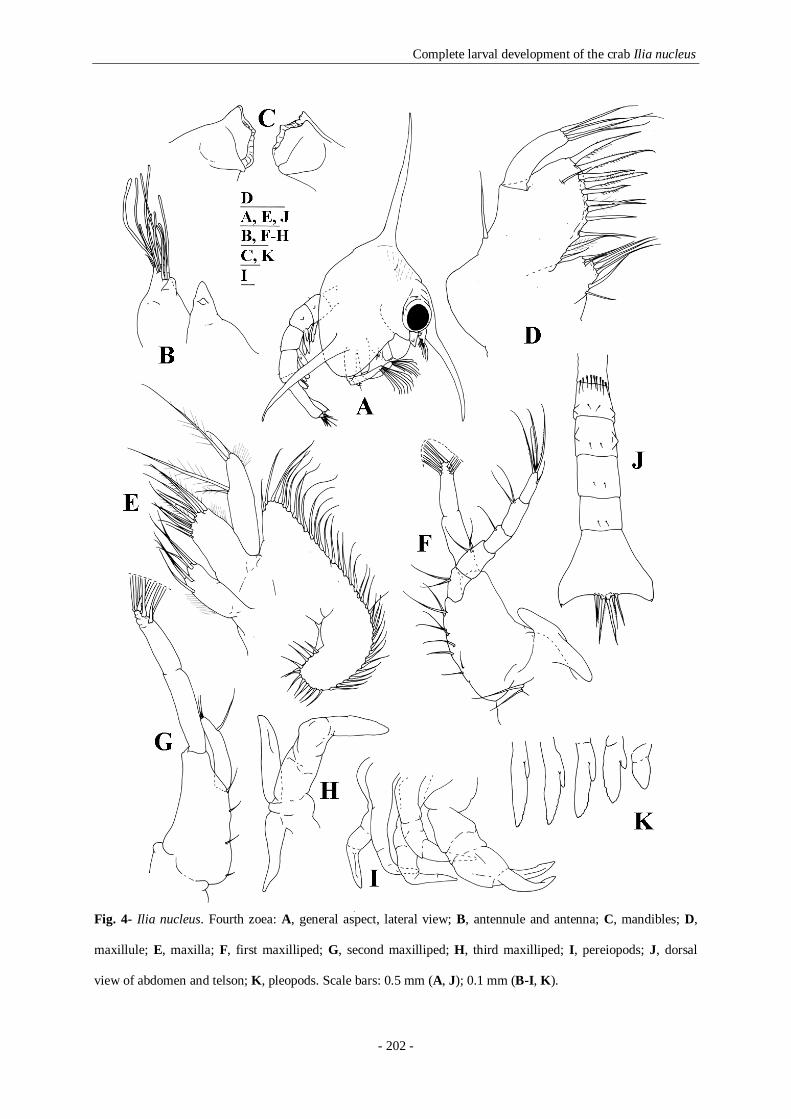
Complete larval development of the crab Ilia nucleus
- 202 -
Fig. 4- Ilia nucleus. Fourth zoea: A, general aspect, lateral view; B, antennule and antenna; C, mandibles; D,
maxillule; E, maxilla; F, first maxilliped; G, second maxilliped; H, third maxilliped; I, pereiopods; J, dorsal
view of abdomen and telson; K, pleopods. Scale bars: 0.5 mm (A, J); 0.1 mm (B-I, K).

Complete larval development of the crab Ilia nucleus
- 203 -
Maxillule (Fig. 4D): coxal endite with 7-8 setae (1 simple and 6-7 papposerrate setae);
basial endite with 11-12 strong (8-9 papposerrate and 3 serrulate) and 1 simple seta.
Maxilla (Fig. 4E): basial endite with 6-5+6-5 sparsely papposerrate setae.
Scaphognathite with 40-46 marginal plumose setae. Microtrichia as figured.
First maxilliped (Fig. 4F): coxa with 2 setae. Exopod unsegmented, bearing 8 long
plumose natatory setae terminally.
Second maxilliped (Fig. 4G): exopod unsegmented, bearing 8 long plumose natatory
setae terminally.
Third maxilliped (Fig. 4H): lobes of epipod, endopod and exopod enlarged.
Pereiopods (Fig. 4I): chelipeds and pereiopods 2-5 more developed and feebly
segmented.
Abdomen (Fig. 4J): first somite with 9 dorsal pappose setae.
Pleopods (Fig. 4K): pleopod buds more developed in somites 2-5. Sixth pleopod bud
uniramous.
Telson (Fig. 4J): unchanged besides size.
Megalopa (Figures 5, 6)
Dimensions: TL= 2.781± 0.077 mm; CL= 1.754± 0.057 mm; CW= 1.369± 0.075 mm.
Carapace (Figs. 5A, 5A’, 5A’’): rectangular, convex on lateral margins. Rostrum
directed obliquely downward, very small; frontal region broad. Submedian lobes hardly
developed; hepatic region swollen; a pair of epibranchial- mesobranchial carinae; a cardiac
swelling and an intestinal tubercle developed. Setae distributed as illustrated.
Antennule (Fig. 5B): peduncle 3-segmented, basal segment with 4, middle segment
with 1 and distal segment with 2-3 setae respectively. Endopod margin with 1 subterminal

Complete larval development of the crab Ilia nucleus
- 204 -
and 4 terminal (arranged 1+3) simple setae. Exopod 5-segmented with 1 seta, 1 seta+ 6
aesthetascs, 1 seta+ 7 aesthetascs, 4 aesthetascs, and 1 subterminal aesthetasc + 1 terminal
seta.
Antenna (Fig. 5C): peduncle unsegmented with two simple setae and with the
shortened exopod process; flagellum 4-segmented, segments first to fourth progressing
proximally to distally, each with 0, 0, 0, 3 terminal (one shorter and two longer) simple setae.
Mandibles (Fig. 5D): asymmetric, with a broad plate-shaped incisor process with an
acute inner margin; palp 2-segmented, first segment with 1 simple seta, distal segment with
13-14 papposerrate setae.
Maxillule (Fig. 5E): coxal endite with 8-10 papposerrate setae; basial endite with 20-
24 papposerrate setae. Endopod unsegmented without any seta.
Maxilla (Fig. 5F): coxal endite with one seta; basial endite with 5+4-5 setae. Endopod
unsegmented without any seta. Scaphognathite with 75-82 marginal plumose setae and blade
with 2-3 simple setae.
First maxilliped (Fig. 5G): coxa with 7-9 setae; basis with 27-30 setae. Endopod
unsegmented with 5 setae. Exopod 2-segmented with 2 plumose setae on proximal segment,
and 2 setae on distal segment. Epipod with 4-5 setae.
Second maxilliped (Fig. 5H): coxa with 1-2 setae; basis with 1-3 setae. Endopod 5-
segmented with 1, 0, 2, 5-6 and 6-8 papposerrate setae, respectively. Exopod 2-segmented,
first segment without setae, distal segment with 2 plumose setae.
Third maxilliped (Fig. 5I): arthrobranch and podobranch gill buds present; coxa with
10-12 setae; basis not clearly differentiated with 2 setae. Endopod 5-segmented, with 28-31,
8-10, 5-6, 8-10 and 10-12 setae, respectively. Exopod reduced to a single segment with 12 or
14 setae. Epipod elongated with 7-8 setae.

Complete larval development of the crab Ilia nucleus
- 205 -
Fig. 5- Ilia nucleus. Megalopa: A, general aspect, dorsal view; A’, detail of frontal view of the rostrum; A’’,
general aspect, lateral view; B, antennule; C, antenna; D, mandibles; E, maxillule; F, maxilla; G, first
maxilliped; H, second maxilliped; I, third maxilliped. Scale bars: 1.0 mm (A’’); 0.1 mm (A-I).

Complete larval development of the crab Ilia nucleus
- 206 -
Fig. 6- Ilia nucleus. Megalopa: A, first pereiopod; B, second pereiopod; B’, detail of dactylus of second
pereiopod; C, third pereiopod; D, fourth pereiopod; E, fifth pereiopod; F, sternum; G, dorsal view of abdomen
and telson; H, telson and uropods; I, pleopods. Scale bars: 0.5 mm (A-E, G); 0.1 mm (B’, F, H-I).
Complete larval development of the crab Ilia nucleus
- 207 -
Pereiopods (Figs. 6A-E): all segments well differentiated, with setae as figured.
Chelipeds equal, with a slender dactylus longer than the palm (D/ P= 0.54- 0.68). Pereiopods
2-5 long, with sharp dactyls.
Sternum (Fig. 6F): maxillipeds and cheliped sternites fused with 10-11 pairs of setae
and one medial small process; sternites of pereiopods 2-5 with 5-6, 3-5, 2 and 1 setae and 2
broad protuberance each which progressively become smaller anteriorly.
Abdomen (Figs. 5A, 5A’’, 6G): six somites, broader than long, with well developed
tergites. Somites 1-6 proximally to distally with 4, 10, 10, 10, 10 and 8 simple setae dorsally
and laterally distributed.
Pleopods (Figs. 6H, I): biramous, well developed decreasing in size from 1st to 4th.
Endopod unsegmented with 5-6, 4-6, 4-6 and 4-5 distal cincinuli on outer margin
respectively. Exopod unsegmented with 19-20, 19-20, 18-19 and 16-18 marginal plumose
natatory setae respectively. Uropods lacking endopod, 2-segmented, proximal segment
(protopod) without setae, and distal segment (exopod) with 11-12 marginal plumose natatory
setae.
Telson (Figs. 6G, H): broad, rounded posterior margin, with 2 pairs of simple setae on
dorsal surface.
6.5- Discussion
The morphological features of the zoeal stages of Ilia nucleus fit in the characters proposed
by Rice (1980) for the family Leucosiidae: 4 zoeal stages; a globose carapace that may have
dorsal, lateral and rostral spines, as in genera Persephona, Arcania, Myra and Ilia, or all
spines absent as in the Ebalia species, or almost all the variations between these for the other
genera; antenna reduced to a bud without setae; maxillule endopod unsegmented bearing 4

Complete larval development of the crab Ilia nucleus
- 208 -
terminal setae and maxilla endopod with 4 setae; first maxilliped endopod with 2, 2, 1, 2 and
5 setae; second maxilliped basis with 4 setae and endopod unsegmented with 3 setae;
abdomen with 5 somites in all stages, somites 2 and 3 with small dorso-lateral knobs, and
pleopods appearing in the third stage; subtriangular telson with posterio-external angles each
with one small furcal spine. Correspondingly, the larval stages of I. nucleus described in the
present study are very similar to the described by previous authors (Cano 1891, Boraschi
1921, Bourdillon-Casanova 1960 and Heegard 1963). The features observed coincide well
with Heegard’s description (1963), showing only minor differences. However some features
pointed out by Heegard, such as a bifurcated antenna and the lack of one seta on the endopod
of the maxilla (the author describes 2+1 setae instead of 2+2), differ from those described in
the present work. This fact can be explained by differences in specimen preparation or by the
precision of the optical observations.
In Northeastern Atlantic and Mediterranean Sea it has been considered that the family
Leucosiidae is represented by subfamilies Iliinae, Leucosiinae, and Ebaliinae (see Zariquiey-
Alvarez 1968), with Ilia nucleus included in the subfamily Iliinae. However, based on adult
morphology, Ng et al. (2008) recently proposed a new classification of Leucosiidae family.
The family Leucosiidae, is represented by subfamilies Cryptocneminae, Ebaliinae and
Leucosiinae (until now no larvae have been described for the first subfamily). The authors
have discussed the general consensus that Leucosiidae has 4 or 5 subfamilies:
Cryptocneminae, Leucosiinae, Ebaliinae, Philyrinae and perhaps Iliinae (being the last the
only one with a wholly Atlantic distribution). However considering that Ebalinae, Philyrinae
and Iliinae were not well defined, they have decided to include all genera from these three
groups in the subfamily Ebaliinae due to the seniority of this name until a complete generic
reappraisal could be conducted.

Complete larval development of the crab Ilia nucleus
- 209 -
Nevertheless, Ko (2000) considered I. nucleus within subfamily Philyrinae and separated this
subfamily in 4 different groups, taking as distinctive characters the presence of dorsal, lateral
and rostral spines of the carapace, the number of setae on the endopod of maxilla, and the
number of spines on posterolateral telson margin. The 4 groups considered by the author were
formed by Persephona, Arcania and Myra genera in the first group, the single genus Ilia in
the second group, and the species included in the Philyra genus were separated in the third
and fourth groups. The assignment of the genus Ilia to the Ko’s second group, was based on
Heegard’s (1963) description, that considered the presence of 2+1 setae on the endopod of
maxilla. However, present study demonstrates that I. nucleus larvae have 2+2 setae on the
endopod of the maxilla, character that would include this species in the first group. In fact, I.
nucleus zoeal stages are very similar to those defined by Ko (2000) for the first group of
Philyrinae, having all carapace spines; 4 setae on maxillule endopod, 4 and 5-6 setae on
maxillule basial and coxal endites, respectively; 2+2 setae on maxilla endopod; and one spine
on posterolateral telson margin. This first group of larvae was considered by Ko (2000) the
most ancestral group in Leucosiidae family, and providing the same group of characters I.
nucleus would also be classified as an ancestral species in the family.
Also, the megalopa shows the typical characteristics of a leucosiid crab according to Quintana
(1986): a smooth carapace with dorsal regions slightly swollen, a rudimentary rostrum, and 2
broad protuberances on sternites 2-4. Even though the significance of antennular armature
seems to be unclear, Quintana (1986) stated that Leucosiidae megalopae could be divided in
three subfamilies (Ebaliinae, Philyrinae and Leucosiinae) regarding the segmentation and
armature of outer flagellum of antennule, a consistent character at the subfamily level. The
megalopa of I. nucleus, presents a 5-segmented antennular outer flagellum, aesthetascs in the
terminal segment in a subterminal position, and the higher number of aesthetascs observed in

Complete larval development of the crab Ilia nucleus
- 210 -
the family (a total of 18), which according to Quintana (1986) place it in the Ebaliinae, and
which also agrees with Ng et al. (2008) recent classification. Taking into account the number
of aesthetascs, I. nucleus is more closely related with the Arcania, Myra and Philyra group,
identified as Philyrinae by Quintana (1986), since these exhibit 16-17 aesthetascs in the
antennular flagellum. These results are also coherent with Ng et al. (2008), once that
Ebaliinae subfamily was synonymised with the Philyrinae. We can state that the megalopa of
Ebaliinae presents a 3- to 5-segmented antennular flagellum, the aesthetascs on distal segment
can be absent or present, and the total number of aesthetascs varies among 5-18.
Larval morphological characters are useful for species to infer phylogenetic relationships.
Hence, Quintana (1986) assumed that characters exhibited by megalopae belonging to Ebalia
and Nucia were plesiomorphic, being family Ebaliinae the most primitive leucosiid group,
and consequently the Leucosia species (subfamily Leucosiinae) should be the apomorphic
group. Despite that, the inferences about the phylogenetic position considering the megalopal
stage will also define I. nucleus as the most primitive among the Ebaliinae. As Ng et al.
(2008) affirmed, the actual subfamily Ebaliinae is a very heterogeneous group and
considering the larval characters, it includes the most ancestral species in the Leucosiidae and
also the most derived ones.
Finally, I. nucleus shows eight natatory setae on exopod of the maxillipeds in third and fourth
zoeal stages. Nevertheless, the typical sequence of the number of natatory plumose setae on
exopods of maxillipeds in Brachyura larval series with four zoeal stages is: four setae in first
zoea, six in second, eight in third and ten in fourth. As in I. nucleus, Jorgensen (1925)
described analogous results for Ebalia sp., presenting the sequence of zoea II to IV with 6
natatory plumose setae on maxilliped exopods. Afterwards, Lebour (1928) described E.

Complete larval development of the crab Ilia nucleus
- 211 -
tuberosa with six natatory plumose setae on maxilliped exopods in the second and third
zoeae, and eight in fourth zoea. Lebour (1928) concluded that eight setae on maxilliped
exopods in the fourth zoeal stage was a particular feature of Ebalia, and Salman (1982)
concluded the same when studying the larval development of this genus. Other authors, e.g.
Krishnan and Kannupandi (1990) described Philyra globosa as having 4, 6 and 6 plumose
natatory setae on maxilliped exopods in first to third zoeae; Ko (2000 and 2001) described P.
platychira and P. kanekoi respectively with 4, 6 and 6 setae through zoeal stages; and Ghory
and Siddiqui (2008) described Leucosia aff. biannulata and Philyra aff. platychira as having
6 setae on maxilliped exopods in third zoeal stage. However, Negreiros-Fransozo et al. (1989)
described the normal sequence (4, 6, 8 and 10 setae) for Persephona mediterranea zoeal
stages. Consequently in leucosiid crab zoeal stages a first zoea always have 4 plumose
natatory setae on maxilliped exopods, a second zoea always have 6, a third zoea can have 6 or
8, and a fourth zoeal stage would present 8 or 10 plumose natatory exopod setae. As a result,
we suggest that for the correct identification of a zoeal stage of a leucosiid besides counting
the number of setae on maxilliped exopods, the antennule development as well as the
pereiopods development should be used as additional characters.
Taking into account that Leucosiinae and Ebaliinae are the only subfamilies with their larvae
known, and identifying the existent heterogeneity in Ebaliinae, we are of the opinion that the
present larval morphology conclusions do not give many clarifications to the current Code,
and maybe a complete generic reappraisal is still needed. More larval descriptions of
Leucosiidae genera and species are needed. Present work describes the 16th leucosiid
megalopae, and in the last 20 years Negreiros-Fransozo et al. (1989) and Ko (2000) were the
only publications that presented the complete larval development of this Family species. As
Rice (1980) and Quintana (1986) stated, the development of the Leucosiidae is still very

Complete larval development of the crab Ilia nucleus
- 212 -
poorly known and the information of their zoeal and megalopal morphology would be
significantly modified as further information becomes available.
6.6- References
Boraschi L. (1921) Osservazione sulle larve dei Crostacei Decapodi: brachyuri e anomuri.
Mem. R. Com. Talassogr. Ital. 87, 1-32.
Bourdillon-Casanova L. (1960) Le meroplancton du Golfe de Marseille: les larves de
crustacés décapodes. Rec. Trav. Stat. Mar. Endoume 30, 1-286.
Cano G. (1891) Sviluppo postembrionale dei Dorippidei, Leucosiadi, Corystoidei e Grapsidi.
Memorie della Società Italiana di Scienze Naturali 8, 1-14.
Caroli E. (1921) Sullo svillupo larvale dei Crostacei Decapodi. Rend. Union. Zool. Ital.
Trieste 1921, 16-18. In: Monitore Zoologico Italiano Firenze, 32, 12.
Clark P.F., Calazans D.K., Pohle G.W. (1998) Accuracy and standardization of brachyuran
larval descriptions. Invertebrate Reproduction and Development 33, 127-144.
d’Udekem d’Acoz C. (1999) Inventaire et distribution des crustacés décapodes de
l’Atlantique nord-oriental, de la Méditerranée et des eaux continentales adjacentes
au nord de 25º N. Collection Patrimoines Naturels (M.N.H.N./S.P.N.) 40, 1-383.
Ghory F.S. & Siddiqui F.A.. (2008). Description of Leucosiidae (Crustacea: Brachyura) larval
stages collected from the Manora Channel, Pakistan, during 1993-1995. Pakistan
Journal of Zoology 40, 353-363.
González-Gordillo J.I., dos Santos A., Rodríguez A. (2001) Checklist and annotated
bibliography of decapod crustacean larvae from the Southwestern Europe coast
(Gibraltar Strait area). Scientia Marina 65, 275-305.

Complete larval development of the crab Ilia nucleus
- 213 -
Heegard P. (1963) Decapod larvae from the Gulf of Napoli hatched in captivity.
Videnskabelige Meddelelser fra Dansk Naturhistorik Forening 125, 449-493.
Ingle R.W. (1992) Larval stages of Northeastern Atlantic crabs. An illustrated key. Chapman
and Hall, London, 363 pp.
Jorgensen O.M. (1925) Some Crustacean Larvae from the Northumberland Plankton.
Transactions of the Natural History Society of Northumberland 6, 157-166.
Ko H.S. (2000) Larval development of Philyra platychira (Decapoda: Leucosiidae) reared in
the laboratory. Journal of Crustacean Biology 20, 309-319.
Ko H.S. (2001) Zoeal stages of Philyra kanekoi Sakai, 1934 (Crustacea: Decapoda:
Leucosiidae) reared in the laboratory. Korean Journal of Biological Science 5,
275-281.
Krishnan T. & Kannupandi T. (1990) Larval and post-larval development of the purse crab
Philyra globosa (Fabricius, 1888) (Decapoda: Brachyura: Leucosiidae) reared in
the laboratory. Hydrobiologia 190, 171-182.
Lebour M.V. (1928) Studies of the Plymouth Brachyura. II. The larval stages of Ebalia and
Pinnotheres. Journal of the Marine Biological Association of the United Kingdom
15, 109-123.
Negreiros-Fransozo M.L., Fransozo A., Hebling N.J. (1989) Larval development of
Persephona mediterranea (Herbst, 1794) (Brachyura, Leucosiidae) under
laboratory conditions. Crustaceana 57, 177-193.
Ng P.K.L., D. Guinot, Davie P.J.F. (2008). Systema Brachyurorum: Part I. An annotated
checklist of extant Brachyuran crabs of the world. The Raffles Bulletin of Zoology 17,
1-286.
Quintana R. (1986). The megalopal stage in the Leucosiidae (Decapoda, Brachyura).
Zoological Science 3, 533-542.

Complete larval development of the crab Ilia nucleus
- 214 -
Rice A.L. (1980) Crab zoeal morphology and its bearing on the classification of the
Brachyura. Trans. Zool. Soc. London 35, 271-424.
Salman S.D. (1982) Observations on the larvae of the North European crabs of the genus
Ebalia (Brachyura, Leucosiidae). Crustaceana 42, 256-269.
Zariquiey-Alvarez R. (1968). Crustáceos Decápodos Ibéricos. Investigacion Pesquera 32, 1-
510.

CHAPTER 7
__________________________________________________
General Discussion and Conclusions
General Discussion and Conclusions
- 216 -
This thesis has provided detailed information about the horizontal and vertical distribution
patterns of decapod crustacean larvae in the Portuguese upwelling ecosystem. In the first two
chapters, the four specific objectives related with the larval ecology were fulfilled.
Present work showed that the diel vertical migration behaviour previously described by dos
Santos et al. (2008) is consistent through the larval development, of almost all the studied
taxa, and demonstrated that the larval retention strategy hypothesized by the authors exists.
The decapod larvae were distributed along meridionally elongated patches concordant with
their origins: the inner shelf species were sampled very close to the shore, the shelf species
were distributed in the middle shelf, and the slope species appeared over the continental shelf
break. Thus, one of the most important conclusions of present study is that the self
recruitment for parental populations is a probable scenario for the decapods larvae in the
sampling area, even under upwelling conditions. The presence of the complete larval series
for most of the studied species in the same area, many times in the same station for the several
larval forms studied support our supposition.
It is also demonstrated that the caridean shrimps, the anomuran and the brachyuran crabs had
a similar larval distribution in the water column, responding in a similar way to the
environment. The ontogenetic vertical migration in the water column when observable
showed the first zoeal stage in a more superficial position relatively to the second zoea, and
the last stage was always more deeply distributed. The megalopa had an average depth of
distribution similar to the one recorded for the last zoea, but it was many times concentrated
in the neuston layer during the night, position that can reflect a transport mechanism strategy.
During the sampling the hydrological conditions were complex, and the arrival of the less
saline water lens to the fixed point could be the responsible for the less evident ontogenetic

General Discussion and Conclusions
- 217 -
vertical migration behaviour, once that a probable larval rearrangement as a response to the
hydrological changes could have happened.
It is demonstrated that the vertical migration movements’, ontogenetical and diel, permit to
decapod larvae to regulate their transport, and even considering the larval stages separately,
the larval retention over the shelf was the result of a constant response to the environmental
conditions, response that is most of the times reflected as the vertical larval position. This
seems to be true for the three studied larval forms: the caridean shrimps, the hermit crabs and
the crabs.
The last specific objective was related with the larval morphology of those three selected
larval forms. The morphological aims of present thesis were also fulfilled in the third to sixth
chapters.
Concerning the caridean shrimps larval development, in Chapter 4 we studied two closely
related trans-isthmian Lysmata species, L. galapagensis and L. moorei. Both had a
homogeneous development, and even in the presence of incomplete larval series, we believe
that both will complete their development with a maximum of nine zoeal stages. Both species
hatch with the first and fifth pereiopods as biramous and uniramous buds, respectively,
similarly to L. seticaudata (Calado et al. 2004). As present thesis demonstrates, the study of
larval Lysmata will certainly be of valuable help for researchers addressing phylogeny
subjects, once that analyzing the available descriptions for the genus is possible to
hypothesize at least two scenarios for the developmental pattern for these species: the first one
similar to L. galapagensis, L. moorei and L. seticaudata where the larvae hatch with the first
and fifth pereiopods as buds, that will complete their development with 8 or 9 zoeae, and the
second one, longer, with the larvae hatching without buds. The first group of species belongs

General Discussion and Conclusions
- 218 -
to the “Cosmopolitan Clade”/ “Lysmata Clade” (Baeza et al. 2009 and Fiedler et al.
submitted), so we suppose that the presence of the first and fifth pereiopod buds in the newly
hatched larvae is a character shared by all the species within this clade. The presence of a
small protuberance in the dorsal region of the third pleomere in L. galapagensis second zoeal
stage, which in later zoea differentiates to a well defined anteriorly curved spine makes it
easily distinguishable from all Lysmata larvae studied. This species late zoea is very similar to
the material described by Gopalakrishnan & Laurs (1971), so, we believe that the larvae
described as E. corniger for the Eastern Tropical Pacific are in fact L. galapagensis. Gurney
(1937) Species A. V, Eretmocaris corniger, from the Atlantic Ocean, presents the same
general form of L. galapagensis. Regardless of the close phylogenetic relationship suggested
by the molecular studies of Baeza et al. (2009) and Fiedler et al. (submitted) between the
Eastern Pacific L. galapagensis and the Western Atlantic L. moorei, the most evident larval
character shared by both species is the presence of a similar rostrum, therefore we conclude
that there is an unknown Lysmata species, probably phylogenetically more close to L.
galapagensis than to L. moorei in the Eastern Atlantic. Having in mind that L. galapagensis
occurs in the eastern Pacific and the most similar larvae known so far are those of
Eretmocaris corniger collected in the eastern Atlantic, some pertinent questions were raised.
Due to the occurrences of these three species, we believe that we are in the presence of an
unknown species complex.
For the hermit crabs larval development we decided to describe the larval stages of
Clibanarius species. We made a redescription according to modern standards of C. erythropus
and a description of C. aequabilis. The larval stages of the two northeastern Atlantic
Clibanarius species, were not easily differentiated. An exhaustive comparison of the
morphological larval characters of the species described herein and those described by other
General Discussion and Conclusions
- 219 -
authors lead us to the conclusion that the genus is very homogeneous. Regardless of the
general larval morphology homogeneity in Clibanarius, there is some variation among
congeneric species, particularly in the telson formula and in the morphology of the telson
processes beyond the second larval stage, as well as in the number of zoeal stages. We
suggest that as a generic character of Clibanarius zoeal stages the transformation of the
plumose fourth telson process into a fused spine (either well developed or reduced) from the
second to the third zoea.
Finally, the complete larval development of the crab Ilia nucleus was studied. The
morphological features of the four zoeal stages and the megalopa of this species fit in the
characters proposed by Rice (1980) for the zoeal stages and by Quintana (1986) for the
megalopa of the family Leucosiidae. The larval stages described in the present study are very
similar to those described by previous authors. Ng et al. (2008) recently proposed a new
classification of Leucosiidae family, with only three subfamilies the Cryptocneminae,
Ebaliinae and Leucosiinae, and affirmed that the actual subfamily Ebaliinae is a very
heterogeneous group. Considering the larval characters studied we verified that the Ebaliinae
include the most ancestral species in the Leucosiidae and also the most derived ones, so we
must conclude that this subfamily is a heterogeneous group. We also verified that for the
correct identification of a zoeal stage of a leucosiid besides the number of setae on
maxillipeds exopods, the development of the antennule and pereiopods must be used. Due to
the lack of knowledge about this family larval development, our conclusions were not capable
of clarify present code, so we believe that all the assumptions made about the leucosiid zoeal
and megalopal morphology would be significantly modified as further information becomes
available.

General Discussion and Conclusions
- 220 -
The morphological aims of present thesis demonstrate the importance of taxonomy in the
study of decapods crustacean larvae. The species described and the questions raised in the
previous chapters can create new working hypothesis, such as the study of the adults’
phylogeny, the phylogeography, or the aquaculture.
References
dos Santos A., Santos A.M.P., Conway D.V.P., Bartilotti C., Lourenço P., Queiroga H. (2008)
Diel vertical migration of decapod larvae in the portuguese coastal upwelling ecosystem:
implications for offshore transport. Marine Ecology Progress Series 359, 171-183.
Baeza J.A., Schubart C.D., Zillner P., Fuentes S., Bauer R.T. (2009). Molecular phylogeny of
shrimps from the genus Lysmata (Caridea: Hippolytidae): the evolutionary origins of
protandric simultaneous hermaphroditism and social monogamy. Biological Journal of
the Linnean Society 96, 415-424.
Calado R., Bartilotti C., Narciso L., dos Santos A. (2004). Redescription of the larval stages
of Lysmata seticaudata (Risso, 1816) (Crustacea, Decapoda, Hippolytidae) reared
under laboratory conditions. Jopurnal of Plankton Research 26, 737-752.
Gopalakrishnan K. & Laurs R.M. (1971). Eretmocaris corniger Bate larvae from the Eastern
Tropical Pacific Ocean (Caridea, Hippolytidae). Crustaceana 20, 9-18.
Gurney R. (1937). Larvae of decapod crustacea. Part IV. Hippolytidae. Discovery Reports 14,
351-404.
Ng P.K.L., D. Guinot, Davie P.J.F. (2008). Systema Brachyurorum: Part I. An annotated
checklist of extant Brachyuran crabs of the world. The Raffles Bulletin of Zoology 17,
1-286.
Quintana R. (1986). The megalopal stage in the Leucosiidae (Decapoda, Brachyura).
Zoological Science 3, 533-542.

General Discussion and Conclusions
- 221 -
Rice A.L. (1980). Crab zoeal morphology and its bearing on the classification of the
Brachyura. Transactions of the Zoological Society of London 35, 271-424.