university of groningen a cytogenetic study of male germ
TRANSCRIPT

University of Groningen
A cytogenetic study of male germ cell tumorsEchten-Arends, Jantien van
IMPORTANT NOTE: You are advised to consult the publisher's version (publisher's PDF) if you wish to cite fromit. Please check the document version below.
Document VersionPublisher's PDF, also known as Version of record
Publication date:1996
Link to publication in University of Groningen/UMCG research database
Citation for published version (APA):Echten-Arends, J. V. (1996). A cytogenetic study of male germ cell tumors. [S.n.].
CopyrightOther than for strictly personal use, it is not permitted to download or to forward/distribute the text or part of it without the consent of theauthor(s) and/or copyright holder(s), unless the work is under an open content license (like Creative Commons).
The publication may also be distributed here under the terms of Article 25fa of the Dutch Copyright Act, indicated by the “Taverne” license.More information can be found on the University of Groningen website: https://www.rug.nl/library/open-access/self-archiving-pure/taverne-amendment.
Take-down policyIf you believe that this document breaches copyright please contact us providing details, and we will remove access to the work immediatelyand investigate your claim.
Downloaded from the University of Groningen/UMCG research database (Pure): http://www.rug.nl/research/portal. For technical reasons thenumber of authors shown on this cover page is limited to 10 maximum.
Download date: 31-12-2021

A CYTOGENETIC STUDY OF MALE GERM
CELL TUMORS
Jannie van Echten-Arends

A CYTOGENETIC STUDY OF MALE GERM CELL TUMORS

STELLING EN
I. Het lijkt onwaarschijnlijk <lat verlies van heterozygotie van 12q zo'n belangrijke rol
in de oncogenese van testiculaire kiemcel tumoren speelt als wordt gesteld door Murty et al. (1992, Proc Natl Acad Sci USA, 89:11006-11010).
2. Volledig gedifferentieerd weefsel in testiculaire kiemceltumoren en (behandelde)
metastasen is niet het equivalent van goedaardig weefsel.
3. Een sarcomateuze component in een kiemceltumor ontstaat niet altijd door maligne
transformatie van teratoom, maar kan zich ook uit een dooierzaktumor component
ontwikkelen.
4. Het oncogenetische stappen proces van kiemceltumoren maakt geen onderscheid
tussen mannen en vrouwen.
5. De preventie van testiculaire kiemceltumoren is gebaat bij het slankheidsideaal van
vrouwen.
6. Het chromosomenpatroon van een maligne ovariele kiemceltumor (immatuur
teratoom) beschreven door Rodriguez et al. (1995, Cancer Genet Cytogenet 82:62-66)
geeft, in tegenstelling tot wat beweerd wordt, geen duidelijke aanwijzing voor een
overeenkomstige ontstaanswijze van deze tumor en testiculaire kiemceltumoren.
7. Het is onwaarschijnlijk <lat een lapjeskater de schrik van de buurt zal zijn.
8. Als men moeilijk de slaap kan vatten is het verstandiger om de TV aan te zetten dan een boek te pakken.
9. Klezmer-muziek is een muzikaal allegaartje
10. Kijken is de cytogenetische kunst.
Jannie van Echten Groningen, 19 juni 1996

RIJKSUNIVERSITEIT GRONINGEN
A CYTOGENETIC STUDY OF MALE GERM CELL TUMORS
Proefschrift
ter verkri j ging van het doctoraat in de Medische Wetenschappen aan de Rijksuniversiteit Groningen
op gezag van de Rector Magnificus Dr F. van der Woude
in het openbaar te verdedigen op woensdag 19 juni 1996
des namiddags te 4.00 uur
door
Jantien van Echten-Arends
geboren op 22 februari 1963 te Vries

Promotores: Prof. Dr. B. de Jong Prof. Dr. J.W. Oosterhuis Prof. Dr. D.Th. Sleijfer
aan mijn ouders en Erik

Promotiecommissie:
STICHTING DRUKKERIJ
Prof. Dr. H. Schraffordt Koops Prof. Dr. A. Geurts van Kessel Prof. Dr. S.M.M.J. Castedo
Financial support for the printing of this thesis was kindly provided by the "Stichting voor Erfelijkheidsvoorlichting te Groningen".

CONTENTS
Voorwoord
Abbreviations
Chapter 1
Chapter 2
Chapter 3
General introduction
1.1 1.2 1.3 1.4 1.5 1.6
Genomic alterations and cancer Germ cell tumors; an introduction Testicular germ cell tumors of adults and adolescents Residual mature teratoma. Extragonadal germ cell tumors of the adult male Aim and outline of this thesis
References
Invasive and non-invasive testicular germ cell tumors of adults and adolescents
2.1
2.2
2.3
No recurrent structural abnormalities apart from i(l2p) in primary germ cell tumors of the adult testis Genes Chromosom Cancer 14:133-144 (1995) Cytogenetic evidence that carcinoma in situ is the precursor lesion for invasive testicular germ cell tumors Cancer Genet Cytogenet 85:133-137 (1995) Mixed testicular germ cell tumors: Monoclonal or polyclonal Mod Pathol 9;(4):371-374 (1996)
References
Residual mature teratoma
3.1 Comparison of the chromosomal pattern of primary testicular nonseminomas and residual mature teratomas after chemotherapy submitted
9
11
13
13 14 14 20
22
23
25
31
31
45
51
56
59
59

Chapter 4
Chapter 5
Samenvatting
3.2 Cytogenetics of primary testicular nonseminoma, residual mature teratoma and growing teratoma lesion in individual patients submitted
References
Definition of a new entity of malignant extragonadal germ cell tumors
Genes Chromosom Cancer 12: 8-15 (199 5)
References
Summary, general discussion and perspectives
5.1
5.2
5.3
5.4
5.5
Testicular and extragonadal germ cell tumors of adults and adolescents Which genomic changes play a role in the oncogenesis and/or tumor progression of TGCTs? What is the pathogenetic relationship between CIS, SEs, and NSs? Which chromosomal changes play a role in tumor progression and which are the mechanisms of therapy related differentiation? What is the pathogenetic relationship between different subtypes of extragonadal GCTs and between extragonadal and testicular GCTs of adult males?
References
Curriculum vitae
Other publications
71
78
81
90
93
93
95
96
98
99
101
103
107
109

VOORWOORD
'Promoveren? Ik? ...... ' Dat was zo ongeveer mijn reaktie op Bauke's vraag: 'Jannie, wil jij een promotie-onderzoek doen op testiculaire kiemceltumoren?' Nu, zo'n 5 jaar later, ben ik het voorwoord voor 'mijn boekje' aan het schrijven. Ik zou echter nooit zover gekomen zijn zonder de hulp, steun en aanmoediging van velen in de afgelopen jaren.
Ten eerste zijn dat mijn drie promotores, Prof. Dr. Bauke de Jong, Prof. Dr. J. Wolter Oosterhuis en Prof. Dr. Dirk Th. Sleijfer. Zij hebben de afgelopen tien jaar velen enthousiast weten te maken voor het onderzoek van (testiculaire) kiemceltumoren. Zo ook mij. Ik wil jullie alle drie bedanken voor de begeleiding van het onderzoek en het schrijven van dit proefschrift.
Bauke, ik wil jou bedanken dat ik als analist de mogelijkheid heb gekregen een promotie-onderzoek te doen. De afgelopen jaren heb ik veel van je geleerd en wist je me altijd te motiveren. Dat het laatste uiltje bijna in de rij kan aansluiten is ook jouw verdienste. Wolter, jou wil ik bedanken voor je altijd kritische kijk op het onderzoek en voor je waardevolle correcties van de manuscripten op zowel inhoudelijk als stilistisch vlak. En Dirk, bedankt voor de prettige en vlotte wijze waarop je de manuscripten hebt beoordeeld.
De leden van de promotiecommissie, Prof. Dr. H. Schraffordt Koops, Prof. Dr. A. Geurts van Kessel en Prof. Dr. S.M.M.J. Castedo wil ik hartelijk danken voor hun bereidheid om op korte termijn dit proefschrift te beoordelen.
Prof. Dr. H. Schraffordt Koops (Chirurgische Oncologie van het AZG) wil ik bedanken voor zijn aandeel - gedurende al vele jaren - aan het kiemceltumor onderzoek.
Met Ad Geurts van Kessel en zijn onderzoeksgroep (KU Nijmegen) hebben wij al jaren een prettige samenwerking. Dit heeft al menige 'mooie plaatjes' en diverse gezamenlijke publikaties opgeleverd. Bedankt hiervoor.
With Sergio Castedo I have had many fruitful discussions and nice talks. Dear Sergio, thank you very much for all your help and support.
Het in dit proefschrift beschreven onderzoek was zeker niet mogelijk geweest zonder de - nog steeds bestaande - nauwe samenwerking met de pathologie van het AZG. Iedereen die hieraan een bijdrage heeft geleverd, bedankt. Eveneens bedank ik de medewerkers van LEPO (Dr. Daniel den Hoed Kliniek, Rotterdam) voor de prettige samenwerking.
M'n collega's wil ik bedanken ik voor de prettige werksfeer, waardoor ik al 11 jaar (meestal) met plezier naar het werk ga. Een aantal mensen wil ik in het bijzonder bedanken: Menke, Harry en Ronald voor het snelle en prima fotowerk, Jenny voor haar hulp bij het literatuurbestand, Gerard voor de statistiek en Erik voor het oplossen van computerprobleempjes. Eva, Beike, Bauke, Rolf, Boudewijn, Mirjam, Vines, Anlce, Conny en Trijnie wil ik bedanken voor de leerzame en leuke maandagmiddag praatjes/discussies. Trijnie en Eva, bedankt voor jullie hulp en aanmoediging tijdens mijn onderzoek. Trijnie wil ik eveneens bedanken voor haar kritische blik op 'mijn' karyotypen.
9

Dank natuurlijk voor mijn paranimfen, Kiena ten Brink en Beike Leegte, voor hun uitstekende hulp en goede zorgen. Kiena, onze hechte zusjes-band betekent veel voor mij. Beike, hoewel onze werkzaamheden slechts gedeeltelijk overlappen, waardeer ik onze samenwerking zeer.
Familie en vrienden wil ik bedanken voor hun belangstelling en voor de zo belangrijke dingen naast het werk. In dit kader bedank ik Umcha voor het nodige muziekplezier. In het bijzonder wil ik mijn ouders, Jan en Johanna Arends, bedanken dat zij mij altijd gesteund en gemotiveerd hebben.
Tot slot, lieve Erik, jij bedankt voor je aanmoediging, ondersteuning en begrip. Promoveren doe je niet alleen, daar weet jij nu alles van.
10

AFP
CH CIS CT(s) DI DNA EC FISH GCT(s) GTE HCG i(12p) IGCNU IT LOH MCN MT NS(s) PGC(s) RMT(s) SE(s) TGCT(s) WHO YS
alpha-feto-protein choriocarcinoma carcinoma in situ combined tumor( s) DNA index
ABBREVIATIONS
deoxyribonucleic acid embryonal carcinoma fluorescence in situ hybridization germ cell tumor( s) growing teratoma beta-human chorionic gonadotrophin isochromosome of the short arm of chromosome 12 intratubular germ cell neoplasia of the unclassified type immature teratoma loss of heterozygosity modal chromosome number mature teratoma nonseminomatous germ cell tumor(s) primordial germ cell( s) residual mature teratoma(s) seminoma( s) testicular germ cell tumor(s) World Health Organisation yolk sac tumor
11

CHAPTER 1
GENERAL INTRODUCTION
1.1 GENOMIC ALTERATIONS AND CANCER
Cancer is a genetic disease of cells and tissues. When the regulation of normal cell growth is altered, uncontrolled cell growth is initiated and a tumor may develop. The alteration or transformation of a normal cell in a malignant one is caused by changes in its genome. Some of these genetic changes may be detectable at the microscopic level as chromosomal abnormalities, others are too small and can only be detected by molecular analysis.
In essence, two distinct types of genes can cause cancer; oncogenes and tumor suppressor genes. Normal cell growth and proliferation is stimulated by the products of proto-oncogenes and is under the negative control of tumor suppressor genes. Alteration or overexpression of an oncogene can cause uncontrolled cell growth. Most oncogenes originate from single mutations of proto-oncogenes ( structural mutations at gene or chromosome level, or numerical alterations) and their action is dominant. The action of tumor suppressor genes is recessive; inactivating mutations or loss of both alleles leads to tumor formation [l]. In addition, alterations in genes which are involved in DNA (mismatch) repair, DNA replication, and chromosome segregation may lead to growth deregulation and malignancy by unrepaired mutations or genome destabilisation [2,3]. However, malignant transformation is not achieved by a single event; oncogenesis is a multi-step process [1]. Accumulation of several oncogenetic changes is necessary to bring about a disturbance of the balanced processes of growth regulation.
Most cancers are monoclonal in origin [ 4]. Despite the clonal nature of neoplasms, malignant tumors are genetically heterogeneous. They consist of a society of multiple interacting subpopulations of cancer cells, that differ in behavioral properties, such as growth rate, ability to metastasize, and sensitivity to treatment [ 5]. Progression of a tumor is the result of clonal evolution of a tumor cell population, generally characterized by increasing genetic instability of tumor cells, decreasing capacity of differentiation, increasing proliferative potential, and higher malignant potential, e.g., capacity to invade and metastasize. Of new clones that emerge during tumor progression, some may have selective growth advantage and may overgrow their predecessors [ 4,6]. Tumor progression is paralleled by karyotype evolution [ 6]. Only certain subpopulations of tumor cells present in the primary tumor, have the capacity to form metastatic lesions [7,8]. Invasion and metastasis require genetic changes additional to those which are required for unrestrained growth alone. Thus tumorigenicity and metastatic potential have both overlapping and separate features [9]. Tumor metastasis is a nonrandom process [7].
In solid tumors a variety of numerical and structural chromosomal abnormalities have been described ([1 O], for review). The different types of chromosomal rearrangements have different oncogenetic effects. Net gain of chromosomal material results in a simple dose effect of oncogenes. In some types of neoplasia, amplification of a smaller chromosomal segment is known to be pathogenetically important (e.g., N-myc
13

General Introduction
amplification in neuroblastomas). Deletions and unbalanced translocations, resulting in net loss of chromosomal material, or loss of entire chromosomes, are oncogenetic by loss of tumor suppressor genes. Additional, translocations, insertions, inversions, or deletions may cause relocation of DNA sequences, through which genes may be destroyed, new fusion genes may be created, or the regulatory control of genes may be interfered with [ 1 1]. Some chromosomal abnormalities are associated with a particular tumor type (e.g., t(X; 18) in synovial sarcoma and t( 12; 16) in myxoid liposarcoma). These specific chromosomal abnormalities are important for tumor diagnosis. Other chromosomal abnormalities are indicative for tumor progression and important for prognosis. Some non-clonal chromosomal abnormalities which are often found in solid tumors, are referred as cytogenetic noise [ 1 1].
Chromosomal analysis of solid tumors may shed light on tumorigenesis. Furthermore, it can be helpful in determining tumor progression steps and in clarifying the pathogenetic relationship of different subtypes of a tumor. Tumorcytogenetics has proven to be of clinical usefulness, since it can reveal information relevant for the diagnosis and/or prognosis of a malignancy.
1.2 GERM CELL TUMORS; AN INTRODUCTION
Human germ cell tumors (GCTs) are a heterogeneous group of neoplasms, located in the testis, the ovary, and in extragonadal sites. Their pathogenesis, histological composition, cytogenetics, ploidy, and degree of malignancy differ, depending on the anatomical site of the tumor and the patient's sex and age [ 12].
This thesis deals with testicular germ cell tumors (TGCTs) and extragonadal GCTs of adult men. GCTs of elderly men have the histology of spermatocytic seminoma. Because of its different pathogenesis, these tumors are not included [ 13, 14].
1.3 TESTICULAR GERM CELL TUMORS OF ADULTS AND ADOLESCENTS
Epidemiology and histology
Primary testicular germ cell tumors (TGCTs) of adults and adolescents are rare neoplasms, counting for about 1 to 2% of all cancers in males. However, it is a highly frequent malignancy in young men, constituting about 25% of cancers in males aged 20 to 34 years [ 15, 16].
TGCTs can be divided clinically and morphologically into two distinct entities, seminomas (SEs) (about 50%) and nonseminomatous germ cell tumors (NSs) (about 40%) [ 17]. SEs are tumors reflecting germ cell differentiation, and NSs reflecting somatic and extraembryonal differentiation [ 18]. Unlike SEs, NSs are histologically heterogeneous. Embryonal carcinoma (EC) cells are the stem cells for all NS derivatives; choriocarcinoma (CH) and yolk sac carcinoma (YS) (both extraembryonic differentiation) and immature teratoma (IT) and mature teratoma (MT)) (both embryonic differentiation) [ 19]. Most NSs have a mixed histology, with the different histological components intermingled or truly
14

Chapter 1
separated. In approximately 10 to 20% of all TGCTs a SE and NS component coexist, classified as combined tumors (CTs) in the British classification [20] and as NSs in the
WHO classification [17]. Both SEs and NSs are derived from carcinoma in situ (CIS) [21]. CIS is often
observed in the seminif�rous tubules of the adult testis adjacent to invasive cancer [22,23]. Seminiferous tubules containing CIS cells are comprised of a single row of atypical germ cells between normal Sertoli cells and the basement membrane [24]. In general, CIS of the testis is a diffuse process [24,25]. Other terms used for CIS are gonocytoma in situ (GIS), intratubular germ cell neoplasia (ITGCN), intratubular germ cell neoplasia of the unclassified type (IGCNU), intratubular malignant germ cells (ITMGC), or testicular intraepithelial neoplasia (TIN).
The current incidence of TGCTs is 2 to 8 per 100.000 men per year in white populations. In most non-white populations the incidence is much lower. In Europe higher rates are recorded in Scandinavian countries (however the incidence is low in Finland) than in Mediterranean and Eastern areas [16]. Over the last 40 to 50 years the incidence of TGCTs has been increased 3 to 4 fold worldwide [26]. This may be related to increased oestrogen exposure in utero [16,27]. TGCTs have a characteristic age distribution, which clearly differs from most other solid cancers. In general, the incidence of solid cancers increases with increasing age, most probably by accumulation of exposure to carcinogenic events [15]. TGCTs show a small peak of incidence before the age of 2 years. A maximum incidence is reached at about 25 to 34 years, and then declines to become uncommon in men over the age of 55 years [16,28]. NSs show a maximum incidence at 25 to 29 years, SEs at 35 to 39 years, and for CTs it is in between [29]. The incidence of TGCTs in cohorts born during the second world war suggests that the inducing agent acts early in life, probably before birth [15].
An important risk factor for TGCTs is undescended testis or cryptorchidism, with approximately a 3 to 10 times higher risk than in the general male population. However, it is not yet clear whether the effect of cryptorchidism ( e.g. raised temperature) leads to malignancy or that an underlying (prenatal) cause of cryptorchidism also induces TGCTs [16].
Clinical behaviour
TGCTs have a characteristic metastasizing pattern. Lymphatic spread is common for all types of TGCTs, with the exception of choriocarcinoma, which is notorious for direct hematogenous dissemination. Lung metastases develop hematogenously, and precede or coincide with dissemination to brain and probably liver and bone [30]. Metastases of TGCTs usually have the same histological composition as the primary tumor. If there is a difference, it usually reflects the loss of one or more of the components present in the primary tumor [30].
In general, SEs are less aggressive than NSs, although the aggressiveness of the latter depends on the histological components. In about 50 to 80% of patients with SEs the tumor is confined to the testis. In contrast, about 25 to 30% of the patients with NSs are free of metastases. Over 50% of patients with NSs have metastases beyond the regional
15

General Introduction
lymph nodes at the time of diagnosis [30]. Of NSs, teratoma is slow to metastasize and choriocarcinoma has the highest metastatic potential. Embryonal carcinoma and yolk sac tumor take a position in between [30].
Presently about 85% of all patients with TGCTs are cured [31]. However, survival varies strongly according to the histology and the stage of the tumor. SEs are cured by orchidectomy alone or by orchidectomy and adjuvant radiotherapy. Advanced SEs are treated by cisplatin-based chemotherapy [32]. Disseminated NSs are cured by orchidectomy followed by cisplatin-based chemotherapy. In case of the presence of residual masses following chemotherapy, surgical resection of these masses is performed [31]. Histological examination of the resected residual masses reveals either necrosis and fibrosis (40%), mature teratoma (50%), or persistent viable tumor cells (10%) [33]. The prognosis after resection is generally favourable, with 5-year relapse-free survival over 85% after resection of necrosis/fibrosis or mature teratoma, and between 50 to 80% after resection of cancer followed by additional chemotherapy. Incomplete resected patients have a poor prognosis [34,35].
Genomic aberrations
TGCTs are characterized by a peritriploid chromosome number; SEs hypertriploid and NSs hypotriploid ([36], for review). This is in accordance with DNA-flow cytometry studies on TGCTs [29,37-41].
The consistent structural abnormality of TGCTs is the isochromosome of the short arm of chromosome 12, the i(l2p), first described by Atkin and Baker [42] in 1982. It is present in about 80% of all TGCTs [36]. Cytogenetic studies on i(12p)-negative TGCTs have shown that other aberrations of chromosome 12, resulting in amplification of 12p, often occur [43-45]. A significantly higher number of breakpoints in 12p13 was found iri i(12p)-negative TGCTs compared to i(12p)-positive TGCTs [46]. Suijkerbuijk et al. [47,48] and Rodriguez et al. [46] have shown by fluorescence in situ hybridization (FISH) that 12p abnormalities are also common in i(l2p)-negative TGCTs. This consistent overrepresentation of 12p sequences points to a common oncogenetic mechanism in i( 12p )positive and -negative TGCTs, and indicates that genes on 12p play an important role in the oncogenesis of TGCTs. Suijkerbuijk et al. [ 49] reported on amplification of 12p 11.2-p 12.1 in a metastasis of a seminoma. They suggest that this particular region may harbor gene( s) relevant in TGCT progression.
In TGCTs specific loss and gain of chromosomes is observed [36,50,51]. Both in SEs and NSs chromosomes 7, 8, 12, and X were overrepresented and chromosomes 11, 13, 18, and Y were underrepresented. It was suggested that these chromosomes might harbor genes important for the oncogenesis of TGCTs, e.g., oncogenes on the overrepresented chromosomes and tumor suppressor genes on the underrepresented chromosomes [36]. The modal copy numbers of chromosome 15 and 22 were significantly higher in SEs compared with NSs [36]. These chromosomes probably contain genes important for tumor progression or for the direction of differentiation to SE or NS [36]. In their cytogenetic study of 24 primary and metastatic testicular and extragonadal GCTs and a review of the cytogenetic data of others, Samaniego et al. [52] concluded that
16

Chapter 1
chromosomes 1, 7, 9, 12, 17, 21, 22, and X were nonrandomly gained. Besides the i( 12p) chromosome, which was present in 90% of the tumors, they found del(12)(q13➔q22) in 44% of NSs and mixed GCTs. They suggest that loss of a tumor suppressor gene in the region 12q13-q22 is important in the oncogenesis of TGCTs [53,54]. Loss of heterozygosity (LOH) on 12q could not be confirmed by others [55-58]. In SEs, Samaniego et al. [52] described rearrangements predominated in the regions lpl 1, 12pll , 12q24 and in chromosome 17 leading to i(l 7q), and in NSs in the regions lp32-p36, l qllq21, 7pll -p22, 12q13-q22 and 17q25. Rodriguez et al. [54] found chromosomal rearrangements associated with certain histologies, e.g., l p32-p36 and 7qll in teratomas and lp22 in yolk sac tumor.
In TGCTs LOH has been reported on Ip [59], lq [59,60], 3p [61], 5p [62], 5q [63,64], I Ip [56,61,63-67], 12q [62], 13q [64,68], 16p [63], 17p [62,64] and, 18q [64,69]. In TGCTs ( especially SE and embryonal carcinoma) the expression of the retinoblastoma (RB 1) gene was decreased, although no gross alteration of the gene was found at the DNA level [70]. In TGCTs no mutations in p53 were found [64,68,71-74], however overexpression of the p53 protein was reported [75]. The oncogene KRAS is located on the short arm of chromosome 12 and therefore may be important in the oncogenesis of TGCTs. Olie et al. [76] and Moul et al. [77] found that N- or KRAS mutation are rare in TGCTs. Considering this, it is unlikely that KRAS mutations are essential in the oncogenesis of TGCTs. In addition, Suijkerbuijk et al. [ 49] have reported that KRAS is located outside the amplificated segment 12pl 1.2-pl2.l , which they have found in a metastasis of a SE. The expression of the c-kit oncogene was found high in SEs compared with NSs [78-80]. In NSs a higher expression of the oncogene hst-l was found than in SEs [79,81]. The c-mos oncogene is expressed at a high incidence in embryonal carcinoma and SEs [78]. N-myc is expressed in embryonal carcinoma and in SE, but not in the relatively more differentiated teratoma [78,80]. In contrast, c-myc is expressed low in SE and high in immature teratoma [80]. One of the tyrosine kinase growth factor receptor proto-oncogenes, c-erbB-1, was expressed commonly at high levels in immature teratomas [78]. The parathyroid hormone-related protein was expressed in all TGCTs containing SE and CH elements [81].
Muller et al. [82] performed DNA flow measurement on testicular CIS cells in infertile men. They found DNA index (DI) values of about 1.5 to 2.5 (mean about 2). In CIS adjacent to invasive cancer, El Naggar et al. [37] and de Graaff et al. [83] found that the DI of CIS and SEs were not significantly different and significantly higher than the DI of NSs. However, Hittmair et al. [84] found the DI of CIS comparable with the DI of the invasive cancer. In this study different subtypes of CIS are suggested.
Cytogenetic data of CIS of the testis is limited to three cases [85]. All three cases were strongly aneuploid and two cases lacked i(12p). The total chromosome number was in the peritriploid range. As compared with the corresponding invasive tumor, the karyotypes of CIS showed few structural chromosomal abnormalities.
17

General Introduction
Histogenesis and Pathogenesis
It is now generally accepted that all TGCTs, with the possible exception of spermatocytic seminoma, are derived from carcinoma in situ (CIS) [21]. This assumption is supported by the frequent observation of CIS in the testicular parenchyma surrounding invasive cancer [23], the morphological similarities of CIS and SE [22], as well as by the development of invasive TGCT in patients where CIS had been diagnosed previously [25,86]. Additionally, morphological, ultrastructural, and immunohistochemical studies have shown similarities between CIS and primordial germ cells [87-89]. This strongly suggest that TGCTs are of germ cell origin.
Carcinogenesis probably starts during intrauterine life [21,28]. An early event might be polyploidization of a dysplastic germ cell precursor, resulting in CIS with a hypertriploid to peritetraploid DNA content [29,37,82-84]. SEs have significantly higher ploidy values than NSs [29,37,38,40,83,84].
Another early event is the formation of i(l 2p ). This characteristic chromosomal abnormality of TGCTs is present in about 80% of these tumors. Geurts van Kessel et al. [55] have shown that i(12p) formation is preceded by polyploidization using RFLP analysis. Sinke et al. [90] have proven the uniparental origin of i(12p). It is most likely that the i( 12p) chromosome originates from a misdivision of the centromere [90] rather than from a translocation or a nonsister chromatid exchange [46]. In i(12p)-negative TGCTs, additional chromosome 12 segments also are of uniparental origin [90].
CIS C IS
� l SE EC SE
� YS C H IT MT l
EC
� YS C H IT MT
A. B . *: n ot by defin ition cl l n l cal ly manifest
Figure 1 . Different pathogenetic models for TGCTs of adults
18

Chapter 1
Although there is general agreement that TGCTs are derived form CIS [2 1], the developmental relationship between SEs and NSs has been subject of controversy for many years. Historically two different views about the pathogenetic relationship between SEs and NSs exist ( [9 1], for review). In the first model, the histogenesis of SE diverges from that of the other TGCTs at an early stage. The neoplastic germ cell either may differentiate along the germ cell lineage, resulting in SE, or to embryonic and/or extraembryonic tissues, resulting in NS. The neoplastic pathway of SEs and NSs is distinct, with no or only limited crossover. Each of them leads to a different end point (Figure IA). In the second model, SEs and NSs have a common origin with a single neoplastic pathway. SE may be an end stage in differentiation, as well as an intermediate stage in the development of NS. As a consequence, SEs and NSs may show a strong relationship (Figure lB) [29,9 1,92] .
� .s � Q
� (I) E
2-----�------------------------
1 , 5 . . . . . . . . . . ' ' . - - . . - - - -, - . - - - . . . - - - - - ,-
- - - - - ♦ ' - �
1
0 UI
.a c.> "iii al � a. ·= E
... al 0
E E 0 .a
c.> al al C � E E .E UI 0 0 Cl) al C C UI c.. ·c:; .E � �
UI
� � � >, Cl) "C UI
0
C "C .E Cl)
Cl) C
'l' :c C E 0 0 C c.>
25 30
p olyp lo i - in i tia l fu rther
cu
E 0 C ·e UI
35
dization net loss net loss approx. age of cl in ical presentati on
of DNA of DNA
young to adolescent age adult age
Figure 2. Tumor progression model of TGCTs of adults proposed by Oosterhuis et al. [29]
19

General Introduction
When SEs and NSs are unrelated tumors according to the first histogenetic model, one would expect to find differences in chromosomal pattern between both entities. However, chromosomal studies by Castedo et al. [50,51] and de Jong et al. [36] have shown that SEs and NSs have related karyotypes, suggesting a common karyotypic evolution. They found both in SEs and NSs specific chromosomes overrepresented ( e.g. 7, 8, 12, and X) and other chromosomes underrepresented (e.g. 11, 13, 18, and Y). The total chromosome numbers are significantly higher in SEs (hypertriploid) than in NSs (hypotriploid) [36,50,51]. The copy number of chromosome 15 and 22 were significantly higher in SEs compared to NSs [36]. Based on these cytogenetic data, together with data on ploidy, Oosterhuis et al. [29] proposed a pathogenetic model of TGCTs, according to the hypothesis that SEs and NSs have a common neoplastic pathway (Figure 2). An early event is polyploidization of a dysplastic germ cell precursor, resulting in CIS with a DI of about 2. Initial net loss of DNA (chromosomes or parts of chromosomes), leads to invasive SE, a tumor type through which all other types progress. Rapid progression through the SE stage by further loss of DNA ( e.g. loss of chromosome 15 and 22) leads to NS. In terms of tumor evolution a NS represents a more advanced cancer than a SE. Slower progression through the SE stage may result in combined tumor. SEs, being the least aggressive, become clinically manifest at older age than the more aggressive NSs. CTs have an age of clinical presentation in between SEs and NSs [29].
The progression of CIS or SE to NS could also be termed reprogramming. The CIS cells or SE cells change their germ cell lineage programme into the programme of the (pluripotent) EC cells [18]. EC cells may stay nullipotent (NS composed exclusively of EC), or may develop pluripotency, with the formation of one or more of the elements CH, YS, IT or MT. The factors controlling the direction of differentiation of NSs are largely unknown. The finding that different histological components of mixed NSs show similarities in ploidy [29] and chromosomal pattern [93,94] suggests that epigenetic factors may be involved in the direction of differentiation in NSs. Oosterhuis and Looijenga [18] have suggested that genomic imprinting might play a role. Paternal imprinting would favor extraembryonic differentiation and maternal imprinting on the other hand may lead to embryonic differentiation.
1.4 RESIDUAL MATURE TERATOMA
Histology
When metastases of primary NSs are treated with chemotherapy, in about 50% of all cases residual mature teratoma (RMT) is left [33]. RMT is composed of fully differentiated mature somatic tissue. Untreated metastases of primary NSs rarely consist exclusively of mature somatic tissue, they usually retain the histology of the primary tumor [30].
20
Chapter 1
Clinical behaviour
Surgical resection of RMT following chemotherapy is necessary because of the risk of recurrence [30] . RMTs may give rise to growing teratoma syndrome [95] and secondary non germ cell malignancies [96,97]. When RMT is resected, a relatively good prognosis is reported [30,98] .
Growing teratoma lesions usually occur at metastatic sites involved at presentation . It may become apparent during chemotherapy or after a 'disease free' interval. For long term survival complete surgical resection of the lesion is necessary [99] .
The formation of malignant non-germ cell components probably occurs e ither by partial differentiation of the pluripotential germ cell (EC) component of the tumor, with concomitant malignant transformation, or malignant transformation ( dedifferentiation) of pre-existing teratoma [97] . The types of non-germ cell malignancies are various sarcomas ( e .g . embryonal rhabdomyosarcoma), adenocarcinoma, nephroblastoma, and neuroblastoma [96,97] . The majority of patients with these chemotherapy resistant neoplasms are curable with complete surgical resection [96] .
Effects of chemotherapy on metastases of NSs
Residual metastases after chemotherapy often contain only differentiated teratoma . Apparently after chemotherapy there is a shift towards higher degrees of differentiation . This can be achieved by three possible mechanisms : (a) Selective destruction of components other than mature teratoma; (b) Direct induction of differentiation of malignant cells; and (c) Spontaneous differentiation of the malignant cells made possible or facilitated by chemotherapy [ 100, 101] .
Two mechanisms, a and c, are essentially similar and based on selection; there is selection of already existing mature teratoma in a and of cells with an inherent capacity of spontaneous somatic differentiation in c. Thus , actually only two basically d ifferent mechanisms remain to be considered : induction of differentiation or selection . These two mechanisms are not mutually exclusive [ 100, 101] .
Oosterhuis et al . [ I 00] hypothesized that RMT is caused by selective destruction of cells other than MT cells or cells with an inherent capacity of differentiation . Mature components were found significantly more often in metastases derived from primary tumors containing mature components as well . The same holds for metastases not treated with chemotherapy. This means that the chemotherapy fails to cause differentiation in those cases where the metastatic cells lacked an inherent capacity of spontaneous differentiation. There does not seem to be de nova induction of differentiation .
Genomic aberrations
RMTs are highly aneuploid and may contain an i( 12p) chromosome [98] . Apparently , mature teratoma, in primary lesions and in residual lesions, has a highly abnormal karyotype, despite its benign looking phenotype [98, 101]. Castedo et al. [ 10 1] found that
2 1
General Introduction
both RMTs and primary NSs have hypotriploid chromosome numbers. However, they found in RMTs a higher degree of underrepresentation of chromosome Y than in primary NSs, whereas the underrepresentation of chromosome 9 and 1 1, as well as the overrepresentation of 12 and X, was lower in RMTs than in primary NSs. A lower average number of i( 12p) was found in RMTs ( 1.6 ; n=14) than in NSs (2.3; n= l5). In addition, they found less structural abnormalities in RMTs compared to NSs . They concluded that RMTs are the result of selection of clones with a less abnormal karyotype and possibly the right balance of genes allowing differentiation [ 101].
1.5 EXTRAGONADAL GERM CELL TUMORS OF THE ADULT MALE
Epidemiology, histology and clinical behaviour
Extragonadal GCTs are rare neoplasms. They occur in the midline of the body (sacral area, retroperitoneum, anterior and posterior mediastinum, hypothalamic region, and pineal region), but also away form the midline ( e.g. orbit, neck, stomach). The histological composition and clinical behaviour of extragonadal GCTs are remarkably different depending on their anatomical localization and the patient's sex and age [ 12].
Extragonadal GCTs of the adult and adolescent male most often are located intracranially and in the retroperitoneum and mediastinum. In adult males one has to exclude the possibility of a metastasis of a TGCT before assuming that the tumor is an extragonadal GCT, particularly in the case of retroperitoneal tumors [ 102]. Histologically, extragonadal GCTs mimic their testicular counterparts and both SEs and NSs are described [ 103] . As in TGCTs there is a correlation between the histological composition of the extragonadal GCT and the prognosis of survival. The survival rate for SEs is much higher than for choriocarcinoma [30] .
Germinomas (extragonadal gonocytomas) occur only in the mediastinum and in the midline of the brain, particular in the pineal gland and hypothalamus (germinoma is the most frequent GCT of the brain) [ 12] . Pure primary intracranial germinomas have a very good prognosis with a 5 to 10 year survival of more than 80% [ 104]. Patients with mediastinal or retroperitoneal GCTs also have a good prognosis, with a 4 year survival rate of more than 60% and 70% respectively [ 105]. Patients with Klinefelter's syndrome have and increased risk of mediastinal GCTs [ 103]. Mediastinal GCTs are associated with hematologic malignancies [ 106]. Sarcomatous elements may occur in mediastinal GCTs, which is a poor prognostic sign [ 107].
Lungs, liver, central nervous system, and bone are the most common sites of metastases. As is the case with metastases of TGCTs, metastases of extragonadal GCTs very rarely contain components other than those of the primary tumor [30].
Genomic aberrations
Several cytogenetic studies on extragonadal GCTs of adults were reported [52,54, 104, 108- 1 19] . GCTs in the mediastinum and the midline of the brain have either
22

Chapter 1
diploid or peritriploid chromosome numbers [ 12, 120]. An i( l2p) chromosome, present in about 80% of all TGCTs, may be present. An i( 12p)-chromosome also has been found in leukemias after the diagnosis of mediastinal GCTs [ 109, 1 18 , 12 1, 122]. In two cases similar clonal abnormalities were found in a mediastinal GCT and the leukemia [ 109, 1 18] .
Histogenesis and pathogenesis
Essentially, there are two groups of hypotheses about the origin of extragonadal GCTs; (a)
the primordial germ cell hypothesis, and ( b) several non-germinal hypotheses ( embryonic and extraembryonic stem cells, included twins) . Extragonadal GCTs might originate from different cell types, and they may have a different pathogenesis, depending on the anatomical site [ 123] . GCTs of the mediastinum and the brain , which have the same histology as gonadal GCTs (including germinomas) , may arise from diploid primordial germ cells (gonocytes) which have migrated during embryo genesis from the yolk sac along the midline of the body to sites other than the gonadal blastoma. However , the other extragonadal GCTs, which consistently lack germinoma, or germinoma-components, probably originate from pluripotent embryonic or extraembryonic stem cells [ 12].
The chromosomal similarities between the mediastinal GCT and the hematologic malignancy indicate that both malignancies are clonally related and probably originate from a common precursor [ 109 , 1 18].
1.6 AIM AND OUTLINE OF THIS THESIS
In the present study we have investigated the chromosomal patterns of invasive and noninvasive TGCTs, of residual mature teratomas, and growing teratoma lesions, and of male extragonadal GCTs, in order to shed light on the following questions :
Which genomic changes play a role in the oncogenesis and/or tumor progression of TGCTs?
What is the pathogenetic relationship between CIS, SEs, and NSs?
Which chromosomal changes play a role in tumor progression and which are the mechanisms of therapy related differentiation?
What is the pathogenetic relationship between different subtypes of extragonadal GCTs and between extragonadal and testicular GCTs of adult males?
In Chapter 2 the cytogenetic analysis of CIS and TGCTs are reported with special emphasis on the progression of CIS to invasive tumor, and on the oncogenesis and tumor progression of TGCTs . Cancer is a genetic disease of cells and tissues. Cancer is caused by genomic alterations and is a multistep process. Polyploidization, 12p-amplification, and
23
General Introduction
loss and/or gain of specific chromosomes are thought to be important steps in the oncogenesis of TGCTs. Besides i(12p), little is known about the role of other structural chromosomal abnormalities in the pathogenesis of TGCTs.
Furthermore, these cytogenetic data may provide more information about the pathogenetic relationship between CIS, SEs and NSs. Whether SEs and NSs have an independent origin with SE as end stage in differentiation, or whether they are closely related where SE may either be an end stage in differentiation or may progress to a NS, is still a matter of debate. Of particular interest in this context are TGCTs which contain both a SE and a NS component, the combined tumors.
Tumor progression is due to or accompanied by karyotype evolution. Tumorigenicity and metastatic potential have both overlapping and separate features. From metastases of NSs treated with chemotherapy, often RMT is left behind. RMT is composed of fully differentiated, mature somatic tissue, while their cells have a strongly abnormal chromosomal pattern. Apparently, after chemotherapy there is a shift toward higher degrees of differentiation, in spite of the highly abnormal chromosomal pattern. In Chapter 3 a cytogenetic comparison of primary NSs, RMTs, and growing teratoma lesions was made to shed light on metastasizing and on the mechanism(s) of therapy-related differentiation.
In Chapter 4 the chromosomal pattern of two male extragonadal GCTs are reported. The karyotypes of the two cases are compared with cytogenetic data of extragonadal and testicular GCTs of adult males, in order to investigate whether all GCTs of adult males have a common neoplastic pathway.
In Chapter 5 the results and conclusions obtained in the different studies are summarized and discussed according to the aforementioned four questions.
24

Chapter 1
REFERENCES
I . Thompson MW, Mcinnes RR, Willard HF ( 1991 ): Genetics of cancer. In: Thompson and Thompson: Genetics in medicine, 5th Ed., W.B. Saunders Co., Philadelphia, pp. 365-38 1 .
2. Nowell PC ( 1991 ): Commentary : How many human cancer genes? J Natl Cancer Inst 83( 1 5): 1 06 1 - 1064.
3 . Loeb LA ( 1994): Microsatellite instability: Marker of a mutator phenotype in cancer. Cancer Res 54:5059-5063.
4. Kerbel RS, Waghome C, Korczak B, Lagarde A, Breitman ML ( 1988): Clonal dominance of primary tumours by metastatic cel ls: Genetic analysis and biological implications. Cancer Surv 7:597-629.
5 . Poste G, Doll J, Fidler IJ ( 198 1 ): Interactions among clonal subpopulations affect stability of the metastatic phenotype in polyclonal populations of B 16 melanoma celis. Proc Natl Acad Sci USA I 0:6226-6230.
6. Nowell PC (1 986): Mechanisms of tumor progression. Cancer Res 46:2203-2207. 7. Fidler IJ, Hart IR ( 1 982): Biological diversity . in metastatic neoplasms: Origins and implications.
Science 2 1 7:998- 1 003. 8. Poste G, Fidler IJ ( 1980): The pathogenesis of cancer metastasis. Nature 283 : 1 39- 146. 9. Liotta LA, Stetler-Stevenson WG ( 199 1 ): Tumor invasion and metastasis: An imbalance of positive
and negative regulation. Cancer Res 5 1 :5054s-5059s. 1 0. Mitelman F ( 1 994): Catalog of chromosome aberrations in cancer. Wiley-Liss, Inc., New York. 1 1 . Heim S, Mitelman F ( 1995): Nonrandom chromosome abnormalities in cancer. An overview. In:
Cancer Cytogenetics, 2nd Ed. Wiley-Liss, Inc., New York, pp. 1 9-32. 12. Oosterhuis JW, Castedo SMMJ, de Jong B ( 1 990): Cytogenetics, ploidy and differentiation of
human testicular, ovarian and extragonadal germ cel l tumours. Cancer Surv 9:32 1 -332. 1 3 . Eble JN (1 994): Spermatocytic seminoma. Hum Pathol 25: 1 035- 1042. 14 . Brodsky GL ( 199 1 ): Pathology of testicular germ cel l tumors. Hematol Oncol Clin North Am
5(6): 1 095- 1 126. 1 5 . M01ler H ( 1 993): Clues to the aetiology of testicular germ cell tumours form descriptive
epidemiology. Eur Urol 23:8- 15 . 1 6. Swerdlow AJ ( 1994): The epidemiology of testicular cancer. Eur Urol 23 (suppl 2):35-38. 17 . Mostofi FK, Sobin LH (1 977): International histological classification of testicular tumors (No. I 6).
In: International Histologic Classification of Tumors. Geneva: World Health Organization. 1 8 . Oosterhuis JW, Looijenga LHJ ( 1993): The biology of human germ cell tumours: retrospective
speculations and new prospectives. Eur Urol 23 :245-250. 1 9. Damjanov I ( 1 991 ): Pathobiology of human germ cell neoplasia. Recent Results Cancer Res
123 : 1 - 19 . 20. Pugh RCB (1 976): Combined tumours. In: Pathology of the testis. RCB Pugh, ed. Blackwell,
Oxford, pp. 245-248. 2 1 . Skakkebrek NE, Berthelsen JG, Giwercman A, Miiller J ( 1 987): Carcinoma-in-situ o f the testis:
Possible origin from gonocytes, and precursor of all types of germ cell tumors except spermatocytoma. Int J Androl 1 0: 1 9-28.
22. J0rgensen N, Millier J, Giwercman A, Skakkebrek NE ( 1990): Clinical and biological significance of carcinoma in situ of the testis. Cancer Surv 9:287-302.
23 . Jacobsen GK, Hendriksen OB, von der Maase H ( 198 1 ): Carcinoma in situ of testicular tissue adjacent to malignant germ cell tumors: A study of 10 cases. Cancer 47:2660-2662.
24. Nagler HM, Kaufman DG, O'Toole KM, Sawczuk IS ( 1 990): Carcinoma in situ of the testis: Diagnosis by aspiration flow cytometry. J Urol 143 :359-36 1 .
25. Giwercman A, von der Maase H, Skakkebrek NE ( 1 993): Epidemiological and clinical aspects of carcinoma in situ of the testis. Eur Urol 23 : 1 04- 1 I 4.
26. Giwercman A, Carlsen E, Keiding N, Skakkebrek NE ( 1993): Evidence for increasing incidence of abnormalities of the human testis: A review. Environ Health Perspect I O I (suppl 2):65-7 1 .
25
General Introduction
27. Sharpe RM, Skakkebrek NE ( 1993): Are oestrogens involved in falling sperm counts and disorders of the male reproductive tract? Lancet 341 : 1 392- 1 395.
28. Forman D, M0ller H ( 1 994): Testicular cancer. Cancer Surv 1 9/20:323-341 . 29. Oosterhuis JW, Castedo SMMJ, de Jong B, Cornelisse CJ, Dam A, Sleijfer DT, Schraffordt Koops
H ( 1989): Ploidy of primary germ cell tumors of the testis. Pathogenetic and clinical relevance. Lab Invest 60: 14-20.
30. Oosterhuis JW ( 1 983): The metastasis of human teratomas. In: The Human Teratomas, I Damjanov, B Knowles, D Solter, eds. The Humana Press, Clifton New Jersey, pp. 1 37- 1 7 1 .
3 1 . Einhorn LH ( 1990): Treatment of testicular cancer: an new and improved model. J Clin Oneal 8 : 1 777- 1 78 1 .
32. Horwich A, Deamaley DP ( 1992): Treatment of seminoma. Sem Oncol 19: 1 7 1 - 1 80. 33 . Nativ 0, Shajrawi I, Leibovitch I, Moskovitz B ( 1 994): The malignant potential of
postchemotherapy residual mature teratoma for disseminated nonseminomatous testicular tumors. Eur Ural 26:2 1 6-2 1 8.
34. Steyerberg EW, Keizer HJ, Stater G, Habbema JDF ( 1 994): Predictors of residual mass histology following chemotherapy for metastatic non-seminomatous testicular cancer: a quantitative overview of 996 resections. Eur J Cancer 30A: 123 1 - 1239.
35. Fox EP, Weathers TD, Williams SD, Loehrer PJ, Ulbright TM, Donohue JP, Einhorn LH ( 1 993): Outcome analysis for patients with persistent nonteratomatous germ cell tumor in postchemotherapy retroperitoneal lymph node dissections. J Clin On col 1 1 : 1 294- 1299.
36. de Jong B, Oosterhuis JW, Castedo SMMJ, Vos AM, te Meerman GJ ( 1 990): Pathogenesis of adult testicular germ cel l tumors. A cytogenetic model. Cancer Genet Cytogenet 48: 143- 1 67.
37. El-Naggar AK, Ro N, McLemore D, Ayala AG, Batsakis JG ( 1 992): DNA ploidy in testicular germ cell neoplasms. Histogenetic and clinical implications. Am J Surg Pathol 1 6:6 1 1 -6 1 8 .
38. Fossa SD, Nesland JM, Pettersen EO, Amellem 0, Wrehre H, Heimdal K ( 1 99 1 ): DNA ploidy in primary testicular cancer. Br J Cancer 64:948-952.
39. Fossa SD, Nesland JM, Wrehre H, Amellem 0, Pettersen EO ( 1 99 1 ): DNA ploidy in the primary tumor from patients with nonseminomatous testicular germ cell tumors clinical stage I . Cancer 67: 1 874- 1 877.
40. Fischer CG, Weidner W, Schroeder-Printzen I, Kallerhof M, Ringert RH ( 1 994): Difference in DNA index of seminomas and nonseminomas. Andrologia 26:337-34 1 .
4 1 . Hittmair A , Rogatsch H, Feichtinger H , Hobisch A , Mikuz G ( 1 995): Testicular seminomas are aneuploid tumors. Lab Invest 72:70-74.
42. Atkin NB, Baker MC ( 1 982): Specific chromosome change, i( 1 2p), in testicular tumours? Lancet 2: 1 349.
43. Castedo SMMJ, de Jong B, Oosterhuis JW, Seruca R, Idenburg VJ, Buist J, Sleijfer OT (1 988): i(12p)-negative testicular germ cell tumors. A different group? Cancer Genet Cytogenet 35 : 1 7 1 - 1 78.
44. Atkin NB, Fox MF, Baker MC, Jackson Z ( 1 993): Chromosome 12-containing markers, including two dicentrics, in three i{l2p)-negative testicular germ cel l tumors. Genes Chromosom Cancer 6:2 1 8-22 1 .
45 . Melani AM, Berger C, Dobbs R, White R, Sandberg AA ( 199 1) : Characterization of unusual marker chromosomes in testicular tumors by fluorescence in situ hybridization. Cancer Genet Cytogenet 56:97.
46. Rodriguez E, Houldsworth J, Reuter VE, Meltzer P, Zhang J, Trent JM, Bosl GJ, Chaganti RSK ( 1 993): Molecular cytogenetic analysis of i( 12p)-negative human male germ cell tumors. Genes Chromosom Cancer 8 :230-236.
47. Suijkerbuijk RF, Sinke RJ, Melani AM, Parrington JM, van Echten J, de Jong B, Oosterhuis JW, Sandberg AA, Geurts van Kessel A ( 1 993): Overrepresentation of chromosome 12p sequences and karyotypic evolution in i{l2p)-negative testicular germ-cel l tumors revealed by fluorescence in situ hybridization. Cancer Genet Cytogenet 70:85-93 .
48. Suijkerbuijk RF, Looijenga L, de Jong B, Oosterhuis JW, Cassiman JJ, Geurts van Kessel A ( 1992):
26
Verification of isochromosome 12p and identification of other chromosome 12 aberrations in gonadal and extragonadal human germ cel l tumors by bicolor double fluorescence in situ

Chapter 1
hybridization. Cancer Genet Cytogenet 63 : 8-16. 49. Suijkerbuijk RF, Sinke RJ, Olde Weghuis DEM, Roque L, Forus A, Stellink F, Siepman A, van de
Kaa C, Soares J, Geurts van Kessel A ( 1994): Amplification of chromosome subregion 12p l l .2-p l2. 1 in a metastasis of an i(l 2p)-negative seminoma: Relationship to tumor progression? Cancer Genet Cytogenet 78: 145- 1 52.
50. Castedo SMMJ, de Jong B, Oosterhuis JW, Seruca R, te Meerman GJ, Dam A, Schraffordt Koops H ( 1 989): Cytogenetic analysis of ten human seminomas. Cancer Res 49:439-443.
5 1 . Castedo SMMJ, de Jong B , Oosterhuis JW, Seruca R, Idenburg VJS, Dam A , te Meerman GJ, Schraffordt Koops H, Sleijfer DT (1 989): Chromosomal changes in human primary testicular nonseminomatous germ cell tumors. Cancer Res 49:5696-570 1 .
52. Samaniego F, Rodriguez E, Houldsworth J, Murty VVVS, Ladanyi M, Lele KP, Chen Q, Dmitrovsky E, Geller NL, Reuter V, Jhanwar SC, Bosl GJ, Chaganti RSK ( 1990): Cytogenetic and molecular analysis of human male germ cell tumors: chromosome I 2 abnormalities and gene amplification. Genes Chromosom Cancer I :289-300.
53 . Murty VVVS, Houldsworth J, Baldwin S, Reuter V, Hunziker W, Besmer P, Bosl GJ, Chaganti RSK ( 1992): Allelic deletions in the long arm of chromosome 12 identify sites of candidate tumor suppressor genes in male germ cell tumors. Proc Natl Acad Sci USA 89: 1 1 006- 1 1 0 1 0.
54. Rodriguez E, Mathew S, Reuter V, Ilson DH, Bos! GJ, Chaganti RSK ( 1992): Cytogenetic analysis of 124 prospectively ascertained male germ cel l tumors. Cancer Res 52:2285-229 1 .
55 . Geurts van Kessel A, van Drunen E, de Jong B, Oosterhuis JW, Langeveld A, Mulder MP ( 1989): Chromosome 12q heterozygosity is retained in i( l 2p)-positive testicular germ cell tumor cells. Cancer Genet Cytogenet 40: 1 29- 1 34.
56. Radice P, Pierotti MA, Lacerenza S, Mondini P, Radice MT, Pilotti S, Della Porta G ( 1 989): Loss of heterozygosity in human germinal tumors. Cytogenet Cell Genet 52:72-76.
57. Peltomaki P, Halme A, de la Chapelle A ( 1 990): Human testicular cancer. Changes in autosomal dosage. Cancer Genet Cytogenet 48: 1 - 12.
58. Mukherjee AB, Murty VVVS, Rodriguez E, Reuter VE, Bos! GJ, Chaganti RSK ( 199 1): Detection and analysis of origin of i( l 2p), a diagnostic marker of human male germ cell tumors, by fluorescence in situ hybridization. Genes Chromosom Cancer 3 :300-307.
59. Mathew S, Murty VVVS, Bos! GJ, Chaganti RSK ( 1994): Loss of heterozygosity identifies multiple sites of allelic deletions on chromosome 1 in human male germ cell tumors. Cancer Res 54:6265-6269.
60. Murty VVVS, Li RG, Mathew S, Reuter VE, Bronson DL, Bosl GJ, Chaganti RSK ( 1 994): Replication error-type genetic instability at 1 q42-43 in human male germ cell tumors. Cancer Res 54:3983-3985.
6 1 . Lothe RA, Fossa SD, Stenwig AE, Nakamura Y , White R, B0rresen AL, Brngger A ( 1989): Loss of 3p or I Ip alleles is associated with testicular cancer tumors. Genomics 5: 134- 138.
62. Murty VVVS, Bosl GJ, Houldsworth J, Meyers M, Mukherjee AB, Reuter V, Chaganti RSK ( 1994): Allel ic loss and somatic differentiation in human male germ cell tumors. Oncogene 9:2245-225 1 .
63 . Al-Jehani RMA, Povey S, Delhanty JDA, Parrington JM ( 1995): Loss of heterozygosity on chromosome arms 5q, 1 1 p, 1 1 q, l 3q, and l 6p in human testicular germ cell tumors. Genes Chromosom Cancer 1 3 :249-256.
64. Peng HQ, Bailey D, Bronson D, Goss PE, Hogg D ( 1995): Loss of heterozygosity of tumor suppressor genes in testis cancer. Cancer Res 55 :287 1 -2875.
65. Lothe RA, Hastie N, Heimdal K, Fossa SD, Stenwig AE, B0rresen AL (1 993): Frequent loss of l l p l 3 and 1 l p l 5 loci in male germ cell tumours. Genes Chromosom Cancer 7:96- 1 0 1 .
66. Looijenga LHJ, Abraham M, Gillis AJM, Saunders GF, Oosterhuis JW ( 1994): Testicular germ cell tumors of adults show deletions of chromosomal bands l l p l 3 and 1 l p l 5.5, but no abnormalities within the zinc-finger regions and exons 2 and 6 of the wilms' tumor I gene. Genes Chromosom Cancer 9: 1 53- 1 60.
67. Smith RC, Rukstalis DB ( 1995): Frequent loss of heterozygosity at 1 lp loci in testicular cancer. J Urol 1 53 : 1 684- 1687.
68. Strohmeyer TG, Slamon DJ ( 1 994): Proto-oncogenes and tumor suppressor genes in human
27

General Introduction
urological malignancies. J Urol 1 5 1 : 14 79- 1497. 69. Murty VVVS, Li RG, Houldsworth J, Bronson DL, Reuter VE, Bosl GJ, Chaganti RSK (1 994):
Frequent allelic deletions and loss of expression characterize the DCC gene in male germ cell tumors. Oncogene 9:3227-323 1 .
70. Strohmeyer T, Reissmann P, Cordon-Cardo C, Hartmann M, Ackermann R, Slamon D ( 199 1 ): Correlation between retinoblastoma gene expression and differentiation in human testicular tumors. Proc Natl Acad Sci USA 88:6662-6666.
7 1 . Fleischhacker M , Strohmeyer T, Imai Y , Slamon DJ, Koeffler H P (1 994): Mutations of the p53 gene are not detectable in human testicular tumors. Mod Pathol 7:435-439.
72. Peng HQ, Hogg D, Malkin D, Bailey D, Galliel BL, Bulbul M, Jewett M, Buchanan J, Goss PE ( 1993): Mutations of the p53 gene do not occur in testis cancer. Cancer Res 53 :3574-3578.
73. Schenkman NS, Sesterhehn IA, Washington L, Tong YA, Weghorst CM, Buzard GS, Srivastava S, Moul JW ( 1995): Increased p53 protein does not correlate to p53 gene mutations in microdissected human testicular germ cel l tumors. J Urol 1 54:617-62 1 .
74. Lothe RA, Peltomaki P, Tommerup N, Foss� SD, Stenwig AE, B0rresen AL, Nesland JM (1 995): Molecular genetic changes in human male germ cel l tumors. Lab Invest 73 :606-6 14.
75. Riou G, Barrois M, Prost S, Terrier MJ, Theodore C, Levine AJ ( 1 995): The p53 and mdm-2 genes in human testicular germ-cell tumors. Mol Carcinogen 12 : 124- 1 3 1 .
76. Olie RA, Looijenga LHJ, Boerrigter L, Top B, Rodenhuis S, Langeveld A, Mulder MP, Oosterhuis JW ( 1 995): N- and KRAS mutations in primary testicular germ cell tumors: Incidence and possible biological implications. Genes Chromosom Cancer 12 : 1 1 0- 1 1 6.
77. Moul JW, Theune SM, Chang EH (1 992): Detection of RAS mutations in archival testicular germ cell tumors by polymerase chain reaction and oligonucleotide hybridization. Genes Chromosom Cancer 5 : 1 09- 1 1 8.
78. Shuin T, Misaki H, Kubota Y, Yao M, Hosaka M (1 994): Differential expression of protooncogenes in human germ cell tumors of the testis. Cancer 73 : 1 72 1 - 1 727.
79. Strohmeyer T, Peter S, Hartmann M, Munemitsu S, Ackermann R, Ullrich A, Slamon DJ ( 1 99 1 ): Expression of the hst-1 and c-kit protooncogenes in human testicular germ cell tumors. Cancer Res 5 1 : 1 8 1 1 - 1 8 1 6.
80. Izquierdo MA, van der Valk P, van Ark-Otte J, Rubio G, Germa-Lluch JR, Ueda R, Scheper RJ, Takahashi T, Giacconet G ( 1 995): Differential expression of the c-kit proto-oncogene in germ cel l tumours. J Pathol 177:253-258.
8 1 . Shimogaki H , Kitazawa S , Maeda S , Kamidono S ( 1 993): Variable expression o f hst- 1 , int-2, and parathyroid hormone-related protein in different histological types of human testicular germ cel l tumors. Cancer J 6 :81-85.
82. MUiier J, Skakkebrek NE ( 198 1 ) : Microspectrophotometric DNA measurements of carcinoma-in-situ germ cells in the testis. Int J Androl suppl 4:2 1 1 -22 1 .
83 . de Graaff WE, Oosterhuis JW, de Jong B, Dam A, van Putten WLJ, Castedo SMMJ, Sleijfer DT, Schraffordt Koops H (1 992): Ploidy of testicular carcinoma in situ. Lab Invest 66: 1 66-168.
84. Hittmair A, Rogatsch H, Feichtinger H, Hobisch A, Mikuz G (1 994): Carcinoma in situ of the testis detected by DNA flow cytometry of testicular fine-needle aspirates. Cytometry 1 7:327-33 1 .
85. Vos AM, Oosterhuis JW, de Jong B, Buist J, Schraffordt Koops H (1 990): Cytogenetics of carcinoma in situ of the testis. Cancer Genet Cytogenet 46:75-8 1 .
86. Giwercman A, Skakkebrek NE ( 1993): Carcinoma in situ of the testis: biology screening and management. Eur Urol 23 (suppl 2): 1 9-2 1 .
87. Gondos B ( 1 993): Ultrastructure of developing and malignant germ cells. Eur Urol 23 :68-75 . 88. Giwercman A, Andrews PW, forgensen N, MUiier J, Grrem N, Skakkebrek NE (1 993):
Immunohistochemical expression of embryonal marker TRA- 1 -60 in carcinoma in situ and germ cell tumors of the testis. Cancer 72: 1 308- 1 3 14.
89. Giwercman A, MUiier J, Skakkebrek NE ( 199 1 ): Carcinoma in situ of the testis: Possible origin, clinical significance, and diagnostic methods. Recent Results in Cancer Research 123 :2 1 -36.
90. Sinke RJ, Suijkerbuijk RF, de Jong B, Oosterhuis JW, Geurts van Kessel A ( 1 993): Uniparental origin of i( 12p) in human germ cell tumors. Genes Chromosom Cancer 6: 1 6 1 - 1 65.
28

Chapter 1
9 1 . Damjanov I (1 989): Editorial. I s seminoma a relative or a precursor o f embryonal carcinoma? Lab Invest 60: 1 -3 .
92. Ulbright TM (1 993): Germ cell neoplasms of the testis. Am J Surg Pathol 1 7: 1 075- 109 1 . 93. de Graaff WE, Oosterhuis JW, de Jong B, van Echten J, Wiersema-Buist J, Schraffordt Koops H,
Sleijfer DT ( 1992): Cytogenetic analysis of the mature teratoma and the choriocarcinoma component of a testicular mixed nonseminomatous germ cel l tumor. Cancer Genet Cytogenet 6 1 :67-73.
94. de Graaff WE, de Jong B, Oosterhuis JW, van Echten J, Wiersema-Buist J, Schraffordt Koops H, Sleijfer DT ( 1991 ): Cytogenetic analysis of the mature and immature teratoma components of a metastatic testicular nonseminomatous germ cel l tumor. Cancer Genet Cytogenet 57:59-68.
95. Logothetis CJ, Samuels ML, Trindade A, Johnson DE ( 1982): The growing teratoma syndrome. Cancer 50: 1 629-1635 .
96. Little JS, Foster RS, Ulbright TM, Donohue JP ( 1994): Unusual neoplasms detected in testis cancer patients undergoing post-chemotherapy retroperitoneal Iymphadenectomy. J Urol 1 52: 1 144- 1 149.
97. Ulbright TM, Loehrer PJ, Roth LM, Einhorn LH, Wil liams SD, Clark SA ( 1984): The development of non-germ cel l malignancies within germ cell tumors. Cancer 54: 1 824- 1833.
98. Oosterhuis JW, de Jong B, Comelisse CJ, Molep.aar IM, Meiring A, ldenburg V, Schraffordt Koops H, Sleijfer DT (1 986): Karyotyping and DNA flow cytometry of mature residual teratoma after intensive chemotherapy of disseminated nonseminomatous germ cell tumor of the testis: A report of two cases. Cancer Genet Cytogenet 22: 149- 1 57.
99. Simmonds PD, Mead GM, Whitehouse JMA ( 1995): A complicated case of metastatic teratoma. Growing teratoma syndrome and cerebral metastasis. Ann On col 6: 1 8 1 - 1 85 .
100. Oosterhuis JW, Suurmeyer AJH, Sleijfer DT, Schraffordt Koops H, Oldhoff J, Fleuren G ( 1 983): Effects of multiple-drug chemotherapy (Cis-diammine-dichloroplatinum, bleomycin, and vinblastine) on the maturation of retroperitoneal lymph node metastases of nonseminomatous germ cell tumors of the testis. No evidence for de novo induction of differentiation. Cancer 5 1 :408-4 16.
1 0 1 . Castedo SMMJ, de Jong B , Oosterhuis JW, ldenburg VJS, Seruca R , Buist J , t e Meerman GJ, Schraffordt Koops H, Sleijfer DT ( 1989): Chromosomal changes in mature residual teratomas following polychemotherapy. Cancer Res 49:672-676.
102. Daugaard G, von der Maase H, Olsen J, R.0rth M, Skakkebrek NE (1 987): Carcinoma-in-situ testis in patients with assumed extragonadal germ-cell tumours. Lancet 5 :528-530.
103 . Mead GM (1 992): Currents issues in Cancer: Testicular cancer and related neoplasms. BMJ 304: 1426- 1429.
104. Albrecht S, Armstrong DL, Mahoney DH, Cheek WR, Cooley LD ( 1993): Cytogenetic demonstration of gene amplification in a primary intracranial germ cel l tumor. Genes Chromosom Cancer 6:6 1 -63 .
105. Bukowski RM, Wolf M, Kulander BG, Montie J, Crawford ED, Blumenstein B ( 1 993): Alternating combination chemotherapy in patients with extragonadal germ cell tumors. Cancer 7 1 :263 1 -2638.
1 06. Ladanyi M, Samaniego F, Reuter VE, Motzer RJ, Jhanwar SC, Bosl GJ, Chaganti RSK ( 1990): Cytogenetic and immunohistochemical evidence for the germ cel l origin of a subset of acute leukemias associated with mediastinal germ cell tumors. J Natl Cancer Inst 82(3):22 1 -227.
107. Gonzales-Vela JL, Savage PD, Manivel JC, Torkelson JL, Kennedy BJ ( 1990): Poor prognosis of mediastinal germ cell cancers containing sarcomatous components. Cancer 66: 1 1 14-1 1 1 6.
108. Dal Cin P, Drochmans A, Moerman P, van den Berghe H ( 1989): lsochromosome 12p in mediastinal germ cell tumor. Cancer Genet Cytogenet 42:243-25 1 .
109. Chaganti RSK, Ladanyi M, Samaniego F, Offit K, Reuter VE, Jhanwar SC, Bosl GJ ( 1989): Leukemic differentiation of a mediastinal germ cell tumor. Genes Chromosom Cancer 1 :83-87.
1 1 0. Mann BD, Sparkes RS, Kem DH, Morton DL ( 1983): Chromosomal abnormalities of a mediastinal embryonal cel l carcinoma in a patient with 47,XXY klinefelter syndrome: Evidence for the premeiotic origin of a germ cell tumor. Cancer Genet Cytogenet 8 : 1 9 1 - 1 96.
1 1 1 . Oosterhuis JW, van den Berg E, de Jong B, Timens W, Castedo SMMJ, Rammeloo RHU, Sleijfer DT ( 1991 ) : Mediastinal germ cel l tumor with secondary nongerm cel l malignancy, and extensive hematopoietic activity. Pathology, DNA-ploidy, and karyotyping. Cancer Genet Cytogenet 54: 1 83- 195 .
29

General Introduction
1 12. Oosterhuis JW, de Jong B, van Dalen I, van der Meer I, Visser M, de Leij L, Mesander G, Collard JG, Schraffordt Koops H, Sleijfer DT ( 1 985): Identical chromosome translocations involving the region of the c-myb oncogene in four metastases of a mediastinal teratocarcinoma. Cancer Genet Cytogenet 1 5 :99- 1 07.
1 1 3 . Shen V , Chaparro M, Byung HC, Young R , Bernstein R (1 990): Absence of isochromosome 12p in a pineal region malignant germ cell tumor. Cancer Genet Cytogenet 50: 1 53- 1 60.
1 14. Kaplan CG, Askin FB, Benirschke K ( 1979): Cytogenetics of extragonadal tumors. Teratology 1 9 :26 1 -266.
1 1 5 . Casalone R, Righi R, Granata P, Portentoso P, Minelli E , Meroni E, Solera CL, Allegranza A ( 1 994): Cerebral germ cell tumor and XXY karyotype. Cancer Genet Cytogenet 74:25-29.
1 1 6. de Bruin TWA, Slater RM, Defferrari R, Geurts van Kessel A, Suijkerbuijk RF, Jansen G, de Jong B, Oosterhuis JW ( 1 994): Isochromosome 1 2p-positive pineal germ cell tumor. Cancer Res 54: 1 542- 1 544.
1 1 7. Aly MS, Dal Cin P, Jiskoot P, Deneffe G, Marynen P, van den Berghe H (1994): Competitive in situ hybridization in a mediastinal germ cell tumor. Cancer Genet Cytogenet 73:53-56.
1 1 8. Woodruff K, Wang N, May W, Adrone E, Denny C, Feig SA ( 1 995): The clonal nature of mediastinal germ cell tumors and acute myelogenous leukemia. Cancer Genet Cytogenet 79:25-3 1 .
1 19. Yu IT, Griffin A, Phillips PC, Strauss LC, Perlman EJ ( 1 995): Numerical sex chromosomal abnormalities in pineal teratomas by cytogenetic analysis and fluorescence in situ hybridization. Lab Invest 4:4 1 9-423.
120. Oosterhuis JW, Rarnmeloo RHU, Comelisse CJ, de Jong B, Dam A, Sleijfer DT ( 1 990): Ploidy of malignant mediastinal germ-cell tumors. Hum Pathol 21 :729-732.
1 2 1 . Sandberg AA, Abe S, Kowalczyk JR, Zedgenidze A, Takeuchi J, Kakati S ( 1982): Chromosomes and causation of human cancer and leukemia. I. Cytogenetics of leukemias complicating other diseases. Cancer Genet Cytogenet 7:95- 1 36.
1 22. Sole F, Bosch F, Woessner S, Perez-Losada A, Cervantes F, Montserrat E, Florensa L, Rozman C (1 994): Refractory anemiea with excess of blasts and isochromosome 12p in a patient with primary mediastinal germ-cell tumor. Cancer Genet Cytogenet 77: 1 1 1 - 1 1 3 .
123 . Gonzales-Crussi F ( 1982): Extragonadal Teratomas. In : Atlas of Tumor Pathology, 2nd series, fascicle 1 8 . Armed Forces Institute of Pathology, Washington DC.
30

CHAPTER 2
INVASIVE AND NON-INVASIVE TESTICULAR GERM CELL TUMORS OF ADULTS AND ADOLESCENTS
2.1 NO RECURRENT STRUCTURAL ABNORMALITIES APART FROM i(12p) IN PRIMARY GERM CELL TUMORS OF THE ADULT TESTIS
Jannie van Echten 1, J. Wolter Oosterhuis5, Leendert H.J. Looijenga5, Mirjam van de Pol5, Janneke Wiersema2, Gerard J. te Meerman 1, Heimen Schraffordt Koops3 , Dirk Th. Sleijfer4, and Bauke de Jong 1
Departments of 1Medical Genetics, 2Pathology, 3Surgical Oncology and 4Medical Oncology, University of Groningen, and 5Laboratory of Experimental Patho-Oncology, Dr. Daniel den Hoed Cancer Center, Academic Hospital Rotterdam, The Netherlands
Abstract
Malignant transformation may be caused by gene deregulation resulting from specific chromosomal rearrangements, by amplification, by mutations in proto-oncogenes, by loss of tumor suppressor genes, or a combination of these. We investigated the role of numerical and structural chromosomal abnormalities in 102 cytogenetically abnormal cases of primary testicular germ cell tumors of adolescents and adults (TGCTs) [32 seminomas (SEs) and 70 nonseminomatous germ cell tumors (NSs)]. We confirmed that an isochromosome for l 2p, i(l 2p), is the only consistent structural chromosomal abnormality in TGCTs, present in about 70% of our cases. Both the .frequency and the number of copies of i(l 2p) are higher in NSs than in SEs. This may suggest that i(l 2p) is involved in tumor progression. Besides i(l 2p), several clonal structural chromosomal abnormalities were found, but none appeared to be specific. SEs and NSs had chromosome numbers in the triploid range, with significantly higher numbers in SEs than in NSs ( average modal chromosome number of 73. 4 in SEs and 65. 0 in NSs). Both in SEs and NSs, some chromosomes were significantly underrepresented (e. g., 11, 13, 18, and J? and others overrepresented (e. g. , 7, 8, 12, 21, and X). In SEs, a significantly higher copy number of chromosomes 7, 15, 19, and 22 was found and a significantly lower number of chromosome 17, compared with NSs. These chromosomes may play an important role in the differentiation of TGCTs. Genes Chromosom Cancer 14: 1 33- 144 ( 1 995). © 1 995 Wiley-Liss, Inc.
Introduction
Primary testicular germ cell tumors of adolescents and adults (TGCTs) can be divided clinically and morphologically into two distinct entities, seminomas (SEs) and nonseminomatous germ cell tumors (NSs) [1]. Both SEs and NSs are derived from carcinoma in situ (CIS) [2]. It has been suggested that CIS cells are the neoplastic
3 1
Invasive and non-invasive testicular germ cell tumors of adults and adolescents
counterparts of gonocytes [2]. The neoplastic germ cells may give rise to SE, reflecting differentiation along the germ cell lineage, or to NS, reflecting somatic differentiation [3]. Embryonal carcinoma (EC) cells are the stem cells of all NS derivatives : choriocarcinoma (CH) and yolk sac tumor (YS) ( extra embryonic differentiation) and teratoma (TE) ( embryonic differentiation) [ 4].
Important steps in the oncogenesis of TGCTs are supposed to be polyploidization, i( l2p) formation, the characteristic chromosomal abnormality of TGCTs, or 12p amplification and specific loss or gain of chromosomes [ 5- 12] . Besides i( l 2p ), little is known about the role of other structural chromosomal abnormalities in the pathogenesis of TGCTs [6 ,7, 13, 14]. Samaniego et al. [15] and Murty et al. [ 16] found that rearrangements , mostly deletions, in 12q 13-22 were recurrent. They concluded that loss of a tumor suppressor gene in this chromosomal region is an important step in the oncogenesis of TGCTs.
We here report the analysis of 1 02 cytogenetically abnormal primary TGCTs with special emphasis on the structural chromosomal abnormalities and the loss or gain of specific (parts of) chromosomes.
Materials and methods
A cytogenetic analysis of 1 02 primary TGCTs (32 SEs and 70 NSs) was carried out. The full karyotype description of cases 1 to 1 0 of the SEs [ 17] and cases 1 to 14 of the NSs [ 18] have been published. Culturing and harvesting of the tumors was performed as described [ 17, 18]. The chromosomes were GTG or GPG banded (G-banding using 0.25% trypsin or 0. 1 % pancreatin and Giemsa). For each tumor, a modal composite karyotype was described according to the ISCN [19], but compared with the triploid level since this enables a better visualization of an important feature of the chromosomal pattern of TGCTs, specific over- and underrepresentation of chromosomes [6]. Because of the consistently high DNA index (DI) of TGCTs [5,8- 1 0,20] , only tumors with an abnormal chromosomal pattern were incorporated in this study.
The modal number of short and long arms was determined for each tumor and chromosome. Parts of chromosomal arms involved in structural abnormalities were registered as whole arms if they represented 50% or more of the total arm length. The modal number of short arms plus long arms divided by 2 revealed the average modal number of chromosomes. The average number of sex chromosomes for each tumor was multiplied by 2 to allow comparison with the autosomes [ 6].
Statistical analysis of the data was done by the Mann-Whitney U-test or, when appropriate , by the chi-square test . If necessary, Bonferroni 's correction for multiple testing was performed.
Results
Table 1 shows the cytogenetic and histological data on the 1 02 chromosomally abnormal cases of primary TGCTs. In the 32 SEs, the modal chromosome number ranged from 58
32

Chapter 2
TABLE I . Description of the Modal Composite Karyotype, the Modal Chromosome Number, and the Histological Components of 32 SEs and 70 NSs
Casea Description of modal composite karyotype SE
1 05- I 1 7,XXY, +add(X)( q28), + Y, +add( I )(p22),+add(I )( q I I ), +de!( I )(p 1 1 ), +del(I )( q 1 1 ), +der(I )t( I ; l 5)(p 1 1 ;q 1 1 ),+2,+dic(2;5)(q37;p l 5),del(3)(q24),+dic(3 ; l2)(p25;q24),+4,+6,+6, +7,+7,+der(7)t(5 ;7)(q1 3 ;q36)x2,+8,+9,+ I O,+ I O,+ 10,+ 1 2,+ 12,+ 12,+ l2,+i(l2)(p I O)x4,-l 3,+ 1 4, + 14,+ 14,+add(l4)(q24),+ 1 5,+ 16,+ 16,+ 1 8,+ 19,+ 19,+20,+20,+21 ,+2 1 ,+22,+22,+3mar[cp3]
2 67-69,XXY,del(l )(q3 1 ),+dic(2; 1 6)(q33;p 1 3),-5,+7,-9,dic(9;20)(p23;p 12),-l 1 ,+i(l2)(p 1 O)x2, -1 3,- 16,-1 7,-1 8,- 1 9,+20,+2 1 ,+2 1 ,+add(22)(q 13)[cp3)
3 I 00-1 1 1 ,XXY,+X,+X,+i(X)(p lO)x2,+Y,+1 ,+add(l )(q32),+i( l )(q lO),+i(2)(ql0),+3,+3,+6,+7, +add(7)(q22),+8,+8,+i(8)(ql 0),- 1 1 ,+ 12,+ 12,+add(l 2)(p 12),+i( 12)(pl O)x2,-l 3,-l 3,+ 14,+ 1 5, +1 5,+ 15,+ 16,+1 6,-1 7,+1 9,+20,+21 ,+2 1 ,+22,+22,+22,+22,+3mar[cp8)
4 103-1 14,XX,+ X,-Y,+ 1 ,+add(l )(p 1 1 ),+2,+2,+i(2)(q 1 O),+add(3)(p23),+4,+6,+6,+7,+7,+7,+8, +8,+8,+9,+9,+ 12,+ 12,+del(l 2)(q24.2),+ 14,+ 1 5,+ 1 5,+add(l 5)(q22)x2,+ 16,+ 1 8,+ 1 9,+20,+20, �1�21�21�21�2���+n�1�
5 70-73,XXY,+Y,+3,-4,-5,+7,+8,+8,add(9)(q l3),add(l l )(q l l),der(l l )t( l ; l l)(q21 ;q22), +add(l 2)(q24),+i( l2)(p 1 0),-1 3,+ 1 5,add(l 6)( q24),-l 7,i(l 7)(q 10),-1 8,- 1 8,-21 ,-22,+der(?) t(?;l l )(?;p l l ),+mar[cp l O)
6 66-73,XXY,+Y,-4,-5,-6,+7,+8,-I0,-1 1 ,+add(l2)(p l2),+add(l2)(q 1 1 ),+i(l2)(p lO)x3,-1 3,+ 1 4, - 17,-1 8,- 1 8,+21 ,+2I [cp l O)
7 62-72,XXY,-2,der(4)t(l ;4)(q2I ;q3 1 ),-5,+7,+8,-l l ,+add(l2)(pl 3),- 13,+ 1 5,-1 8,+21 ,+22, +22[cp7)
8 64-66,XY ,add(X)(p2 l ),add( I )(p 1 1 ),add( 1 )(p 1 3 ),-2,-3 ,-4, -5, +add(7)( q22),-8,-9 ,dup( 1 1 ) (q l 3q23)x2,+ I 2,+der(12)t( 12; I 5)(pl 1 ;ql l ),+i(l2)(p 10),-1 3,dic(l 3; 14)(p 1 1 ;p 1 1 ),-14,-1 5, - 1 6,add(l 7)(q25),- 18,-1 8,+add(l 9)(q l3.4),+add(20)(p 12),add(2 l )(p 1 1 ),-22,der(22)t(l ;22) (p l l ;pl 1 ),+3mar[cp5]
9 56-64,XXY,-3,add(3)(p2 1 ),-4,-5,+7,+8,-9,-l0,-I l ,der(l l )t(l 1 ; 1 4)(pl3;q1 3),+i(l2)(p l0), -1 3,- 13,- 1 4,+ 1 5,-16,-18,+2 I [cp7]
10 72-73,XXY, +add( 1 )(p 1 1 ),+del(3 )( q 1 1 ),add( 4 )( q35),der( 4 )t( 4;7)( q21 ;q 1 1 . 1 ),-5,-6,add(7) ( q21 ),+der(7)t( 4;7)(q 12;p 1 5),+add(8)(q 1 1 ),+i(8)(q l0),+add(9)(q 12),- 10,-1 1 ,add(l l )( q23), der(12)t(5; l 2)(q33;q24),+i(l2)(p I 0),-1 3,- 13 ,+ 1 5,+ 1 7,-1 8,- 1 8,+ l 9,-20,-2 1 ,add(2 l )(pl 1 ), -22,+4mar[cp2]
1 1 66-69,XXY,+X,+Y,-4,-5,-6,+7,-9,add(9)(p24),-l0,del( l l )(q21q23),der(l l)t(5 ; 1 1 )(q 1 3 ;q23), +i( l2)(p I 0),-1 3,+ 14,+ l 5,-1 8,+22[cp5]
12 71-73,XXY,dic(l ;?;9)( 1 pter➔ 1 q42::?: :9q34➔9pter),del( 4)( q2?3),-5,+7,+i(8)( q 1 0),-9,add(9) (q34),-1 l ,inv(l 1 )(q l 3q25),+add(l2)(q24),-1 3,+ 14,+ 1 5,+ 1 5,- 16,-16,-1 7,- 1 8,+2 1 ,+2 1 ,+der(21 ) t(9;2 1 )(q l l ;p l l ),+2mar[cp7]
13 63-66,XXY,add(l )(q44),-2,+3,-4,-5,+8,del(9)(q22),- I O,-l l ,add(I I )(q l4),add(12)(p 1 3), +i( 12)(p10)x2,- 1 3,- 1 3,- 14,i(l4)(ql0),+ l5 ,der(l 7)t(7; 1 7}(pl l ;pl l ),-1 8,+mar[cp4]
14 6 1 -69,XXY,-2,-2,-4,-5,+del(6)(q l 5},+der(7)t(2;7)(q2 1 ;q36),+dic(7;?; 1 2)(7pter➔ 7q32::?: : 1 2p 13➔ 12qter),-9,-1 1 ,- 1 3,+ 14,-17,-1 7,-1 8,+2 1 ,+22,+mar[cp7]
1 5 68-70,X,del(X)( q22),-Y,add(l )(p22),-5,+add(7)(q32),+8,-l l ,der(l l )t(6; 1 1 )(q 1 1 ; q l 4),der(l2} t( 12; l 7)(pl 1 ;q 1 1 ),+i(12)(p 10),- 1 3,- 14,- 16,- 16,-1 8,+20,+2 1 ,+2 1 ,+22,+der(?)t(?; 1 4) (?;q l l )[cp5]
16 60-75,XY,-X,+Y,+add(I )(p 12),add(3)(p l 1 ),-4,-5,+add(7)(q22),+trp(8)(q 1 3q21 .2},+add(I 0) (p 12),- 1 1 ,+add( I 2)(q24),+i( l 2)(p 1 O)x2,-1 3,add(l 4)(q32},+ 1 5,add( 16)(q24),-17,-1 7,- 18,- 1 8, + 1 9,+der(l 9)t(9; 1 9)(q 12;q 13.4),+2 1 ,+2 1 ,+der(?)t(?; 14)(?;q l l }add(14)(q32},+mar[cp 1 O]
17 74-89,XXY,+ 1 ,+2,+del(3}(p2 1 )x2,der(3)i(3)(p I O)dic(3;6}(p25;q27),+7,+7,+8,+8,+ 12,-1 3, + 14,+1 5,+1 5,+19,+2 1 ,+mar[cpl O]
1 8 60-63,XX,-Y,-3,-4,-5,+ 7,+8,-9,-10,-1 1 ,-1 3,-1 3,+ l 5,-l 6,+20,-22[cp8] 1 9 75-1 13 ,XXY,+Y,+ l ,+l ,+der(l )t(l ; 14)(pl l ;q l 1 ),add(2)(p23),+3,+add(4)(q34),-5,+6,del(6)
(q2 l q23)x2,+7,+7,+7,+add(7)(q32),+8,i(9)(q 1 O),+ 12,+ 12,+ 12,- 1 3,- 14,+ 1 5,+ 1 5,+add(l6) (q 1 1 ),+add(l 6)(q 13),-1 8,+ 19,+ 19,+20,+20,+21 ,+8-16mar[cp6)
20 55-65,X,add(X)( q27),-Y,der( I )t( I ; 1 7)(p 1 O;q I O),+idic(I )(p I 3),-3,add(3)( q24)x2,-4,-4,-5, add(7)(q 1 1 ),+der(7)t(3 ;7)(ql I ;p22),+8,+8,-9,-9,-l l ,- I I ,+i(l2)(p I O}x2,-1 3,der(1 3)t( 13 ; 1 5) (q22;q 1 1 ),der{l4)t(3 ;?; I 4)(q I O;?;q I 0),-1 5,add(l 5)(pl I ),add(l 6)(q21 ),-1 7,-l 8,+ 1 9,-20,-2 1 , der(2 l )t( 1 2;2 I )(p 1 O;p 13),-22,-22,+6-7mar[ cp 1 O]
Modal number
1 12
68
l 09
1 06
7 1
72
7 1
65
63
73
68
7 1
65
66
68
73
82
62 l 05
63
3 3

Invasive and non-invasive testicular germ cell tumors of adults and adolescents
TABLE 1 . (continued)
Casea Description of modal composite karyotype 2 1 63-68,XY,-X,+Y,dic(l ;?; 14)(pl 1 ;?;q32),-2,-4,-5,add(6)(p23),+add(7)(q3 l ),+8,+8,+der(8)
t(7;8)(p 14;p22),-9,der(9)t(9;9)(p24;q I 0),- 1 1 ,- l l ,+ 12,+i(l2)(p l0),-1 3,-14,- 14,add(l 5)(p 13), - 1 7,-1 8,+der(l 9)t(l 7; 1 9)(q2 1 ;ql3),+idic(l 9)(p l I ),-22,-22,+4mar[cp lOJ
22 72-77,XXY,+Y,add(l )(ql l ),+add(2)(q l 3),add(4)(q34),+der(4)t(l ;?;4)(q23;?;q2 1)x2,-5, add(6)(q25),+7,+der(7)t(7;?;7)(pter➔ql 1 .2: :? : :p 1 3➔pter),+8,+8,+ 10,-1 1 ,add(l l )(p 1 1 ), der(I I )t( l ;? ; l l )(q23;?;q23),+ 1 2,+ 12,-1 3,-14,+ 1 5,+ 1 5,add(l 7)(q25),-21 ,+4mar[cp 1 OJ
23 58-64,XXY,-l ,-2,-4,-5,+6,der(6)t(2;?;6)(q24;?;ql2)x2,+7,+8,-9,- 10,add(l l )(pl 5),del(I I ) (q23),+i(l2)(p l0),- 1 3,- 1 5,- l 7,-1 8,+2-6mar[cp6J
24 62-67,XXY,add(I )(q32),add(l )(p 1 1 ),-4,add(7)(q22),add(7)(q2 1 ),+8,- 10,-I I ,-I I ,i( l 2)(p 1 0), - 1 3,- 15 ,-16,-1 7,-1 8,-20,+3-9mar[cpl 2J
25 57-65,XX,-Y,add(l )(p l3),+dic(I ;4)(p34;p l6),add(3)(pl 1 ),-4,-4,-5,add(6)(pl I . l ),add(7) (q2 1 ),der(8)t(7;?;8)(p 1 3;?;q 1 3),-9,der(I I )add( l 1 )(q25)dup(l l )(q I 3q23),+ 12,+add(l2)(q 1 1 ), - 1 3,- 16,-1 8,-20,+2 1 ,-22[cpl 0J
26 44-67,XXY ,der( I )t( I ; 1 2)(p I I ;q I 3),add(2)(p24), +add(3 )( q2 l ),-4,-5, +del( 6)( q I 3 ),+del(7) (q3 1 ),+del(7)(q3 1 ),-8,-9,-10,-1 1 ,del(l l )(q23),+der(12)t(l 1 ; 12)(q 12;q23),-1 3,-14,-1 5,-1 6,- 1 7, - 1 8,add(l 9)(q I l ),-2 1 ,-22,+der(?)t(?;7)(?;p 1 1 ),+2-10mar[cp7J
27 68-76,XXY,add(l )(q 12),+dic(I ; 1 2)(p I 3;q22),-5,+del(6)(q2 lq23),+7,+8,+8,-1 3,+ 14,+ I 5, - 1 8,+21 ,-22,+1 -7mar[cp l I J
28 62-73,XXY,+add(l )(p 1 3),-3,-4,-5,+add(7)(q3 l ),der(9)t(9; l l )(p 1 3;p 1 1 ),- 1 1 ,-1 1 ,+ 12,+i(12) (p 1 0)x2,-1 3,- 14,i(l 5)(q I 0),-16,add(l 6)(q 1 3),-1 7,- 1 8,+ I 9,+2 1 ,+ l -2mar[cp6J
29 57-59,X,der(X)del(X)(p22)t(X; 1 2)( q23;q 12),-Y,add(I )(p 13 ),add(I )( q24 ),-2,+dic(3;22) (p 1 1 ;p l 1 ),i(3)(q 1 0)x2,-4,dic(4; 1 3)(q35;p 1 2),-5,-7,+8,-9,-10,-1 1 ,add(l2)(q24. 1 ),+i(l2)(p 10), - 13 ,- 1 3,dic( 13 ; 1 9)(p 1 1 ;q 1 1 ),-1 5,der(l 5)t(l 5; 1 S)(p 1 3 ;q 1 1 ),- 16,-1 7,-1 8,der(20)(20pter➔ 20q 12 : :? : : 1 q25➔ I q44: :2q32➔2qter),-2 l ,add(2 l )(p I 2),der(2 I )t(2 I ;2 I )(p l 2;q I 1 ),der(22) t(7 ;22)( q I I ;p 12), +4-6mar[ cp8J
30 69-74,XXY,der(I )t( I ; l 6)(p I 2;p I I .2),add(3)(q24),-4,-5,+del(7)(p 12),+der(8)t(8; 1 2) (q lO;p 1 1 )x2,-9,add(9)(q24),der(9)t(3;9)(q 1 3 ;p24),-l 1 ,+ 12,-1 3,-1 5,-1 7,add(I 7)(p 1 1 .2),-1 8, add(I 8)(q22),add(l 9)(p 1 3.2),+2 I ,+21 ,+2 I ,+22,+der(?)t(?; 12)(?;p I I ),+ 1 -2mar[cp lOJ
3 I 7 1 -82,XXY,+ l ,+2,i(4)(q 10),-5,+6,+7,+8,+add(l2)(pl l),- 13,+ 14,+ 1 5,+ 16,+ 19,-20,+2 1 ,+2 1 , +22,+ 1 -7mar[cp9J
32 55-69,XXY,+add( I )(p22),add(2)(q37),add(3 )(p24),der(3)t(3;9)(p24;q I 2),add( 4)(q32),der( 4) i( 4)(q 1 O)add( 4)(q3 5),+i(4)(q 1 0),-5,der(6)add(6)(q l 6)del{6)(p23),+add(6)( q27),+add(7)(p 1 1 ), -9,- 1 0,- 1 1 ,+ 12,+add(12)(p 1 1 ),- 1 3,+ 1 4,+ 1 5,add(l 6)(q2 1 ),-1 7,- 1 8,- 1 8,-20,+2 1 ,+add(2 I )(p 12), + I -Smar[ cp 1 OJ
Caseb Description of modal composite karyotype NS
54-67,XXY,-2,-4,add(6)(p25),+7,+8,-9,-10,-1 1 ,+ 1 2,+i(12)(p 1 O)x2,-1 3,-1 4,-1 6, -1 8,- 1 9,+2 1 ,-22,+der(?)t(?; l )(?;q 1 1 ),+2mar[cp 12J
2 55-59,XY,-X,-1 ,der(2)t(l ;2)(p32;q35),der(3)t(3;?4)(p I l ;p l l ),-4,-5,+7,add(8) (q24),-9,- 1 0,-1 1 ,add(l l )(q25),-1 3,- 14,- 1 5,-16,add(l 7)(p 1 1 ),-1 8,- 1 9,-20,der(2 1 ) t( 1 ;2 l )(q l2;p l l ),-22[cp9J
3 54-57,XXY,-3,-4,-5,-6,+del(7)(q32),-8,-9,-1 0,- 1 1 ,+i{l2)(pl0)x2,der( l3 ; 14) (q I O;q I O)del(l4)(q3 l ),- 14,- 14,-1 5,-1 6,del(l 7)(p 1 1 ),-1 8,-19,-20,-20,-22, +2mar[cp7]
4 63-67,XXY,+X,der(l )t( l ; l l )(p34;q 1 3),-5,-9,-1 0,-1 1 ,der(l l )t( l ; l l )(ql2;q23), +i( 1 2)(p 1 O)x3,-l 3,- 1 5,-I 8,-l 9,-20,-22,+der(?)t(?;7)(?;p 1 1 )[cp 1 5]
5 61 -67,XXY,+X,+X,del(l)(p35)x2,-4,-5,add(8)(q24),-9,-10,-l l ,+i(l2)(p l0)x3,- 13 , - 14,-1 5,add(l S)(pl I ),-1 8,-1 9,-20,+2 l [cp7]
6 56-59,XY,inv(X)(p l l .2p22. l ),+ Y,- 1 ,-2,-4,-5,-7,add(8)(q24),-9,-1 O,+i( 1 2)(p 1 O)x3, - 13 ,- 14,- 1 5,-1 8,- 1 9,-20,-21 ,-22,+der(?)t(?; 1 )(?;q 1 1 )[cp5]
34
Modal number
65
55
57
65
64
59
Modal number
66
75
61
65
62
64
74
71
58
72
8 1
68
Histologyc
YS;CH;EC;MT
EC;IT;MT
YS;EC
MT
YS;EC;IT;MT;SE
YS;EC;MT
Chapter 2
TABLE I . (continued)
Modal Caseb Description of modal composite karyotype number Histologyc
7 97-107,XXY,+ X,+ X,+ Y,+ I ,+add( I )(p 1 3 ),+2,+3,+ 3,+del(3)(p23),+4,+add(5)(q3 1 ), 1 02 YS;EC;IT;MT +del( 6)( q2 1 ),der(7)t(7 ;7)(p 1 5 ;q 1 1 ), +inv(7)(p 1 5p22),+8, +8, +del(8)(p 12),+del(9) (p l l ),del( I O)(p 1 3),add( l l )(q25),+der(l l )t(l 1 ; 1 4)(q 14;q l l ),+ 1 2,+del( l 2) (q 1 5q24)x2,+i( 12)(p I O)x4,+ 1 3,+ 14,+ 1 5,+ 1 8,+20,+20,+20,+2 1 ,i(22)(q IO)x2, +der(?)t(?;7)(?;p I O)[cp24]
8 57-59,XXY,+ Y,del(I )(p2 1 ),+der(I )t( I ; I )(p22;q 12),-3,-4,-5,-7,-9,-10,-1 1 ,+i{ l 2) 57 MT (p I O)x3,- 13 ,- 14,- 16,- 18,-1 9,-20,-2 1 ,-22[cp9]
9 53-6 1 ,XXY,+ Y,add(I )(p 1 1 ),+der(I )del(I )(p34 )del(I )(q42),del(2)( q3 1 ),-3,-4,-6, 59 EC -9,-1 O,- I I ,-1 1 ,+i{l2)(p I O)x3,-13,+add(14)(q24),-1 5,- 16,+add( l 7)(q25),-1 8,- 19, -20,-2 1 ,-22[ cp9]
10 58-62,XX,-Y,-1 ,-2,add(2)(p23),-4,dup(4)(q l2q2 1 ),-5,-6,+8,-9,- 10,-1 1 ,der(I I ) 60 EC;MT t(I ; 1 l )(q I l ;q21 ),+i( l2)(pl 0),-1 3,- 1 5,- 16,-1 8,-1 9,+20,+21 ,+21 [cpl O]
1 1 5 1 -53,X,add(X)(p 1 1 ),-Y,add(I )(p35-36),del(I )(q 12),+der(I )add( I )(p35-36)del( I ) 53 EC;MT;SE (q32),del(2)(q34),del(3)(q2 1 ),der(3)t(3;7;?3)(3qter➔3p21 : :7q36➔ 7q 1 1 : :3q23 ➔3dte?dupp�(q21 q27�,-4,del(5)(r l2),-6,-6,dic(7; 1 7dW I ;� 1 3}-9,-1 0,-1 1 ,- 1 3, - 14, e 1 4)t( 2, 14)(q l3 ,p l l ),-1 5,- 7,- 1 8,-1 8,- 1 9,del( (q i ),- l ,-2 1 ,der(2 1 ) t(2 1 ;22)(p 1 1 ;q 1 1 ),-22,-22,+mar[cp9]
12 54-126,XXY,+ X,+ Y,+i( I )(p I O)x2,inv(I )(p32q2 1 ),+der(2)t( I ;2)( q 12;q37)x2,+ 3, I O I EC;SE + 3,+6,+7,+del(7)(p22)x2,+del(7)(q22),+der(8)t(8;9)(p23;q l 2)x2,+ I O,+ 1 1 ,+ 1 2, + 1 2,+i( l2)(p I O)x4,+ 13,+ 14,add(l 5)(p l I ),+ 1 7,+ 1 7,+ 1 9,+20,+20,+2 1 ,+2 1 , +2mar[cp7]
1 3 57-63,XXY,add( I )(q2 1 ),-2,-4,-5,-6,-9,-1 O,-I I ,+i( l 2)(p I O)x4,-1 3,- 14,-1 5,-16,-1 8, 62 YS;EC;IT;MT - 19,del(22)(q l2)[cpl I ]
14 1 07-1 1 3,XXY,+X,+X,+X,+Y,+l ,+dic( l ;20)(20qter➔20pl 3 : : l q44➔1q l2:: l q21 1 1 3 YS;EC;IT;MT ➔ I pter?,+2,+2,+ 3,+5,+6,+6,+7,+7,+add(7d(q 1 1 ),+8,+8,+add(8)Jp2 l ),+9,+ 1 0, +add(l )(q23),+ 1 2,+ 12,+ 12,+i( l2)(p1 0)x ,+ 13 ,+1 3,+ 1 4,+ 14,+ er( l4)t(7; 14) (q2 1 ;p 12),+ 1 5,+ 1 6,+ 17,+ 1 7,+ 18,+ I 9,+der(l 9)t(7; 1 9)(q2 1 ;p 13),+20,+21 ,+2 1 , +2 1 ,+22[cp l3]
15 6 1 -65,XXY,-4,-5,+del(6)(q2 1 q22),-9,-1 0,-1 1 ,+del(l2)(q 13),+i(l 2)(p I O)x3, 63 YS;CH;EC;IT;MT -1 3,- 14,-1 6,- 1 7,- 1 8,- 1 9,+2 1 ,-22[cp 1 1 ]
1 6 56-60,XXY,+Y,+der(2)t(2;9)(q23;q 1 1 ),-4,-6,-9,del(9)(q 1 1 ),-10,- l l ,+i(l2)(p 10), 59 MT;SE add(1 3)(p 1 1 ),-1 4,-1 5,-1 8,-1 9,-20,-22[cp7]
1 7 54-56,XX,-Y,del(l )(p21 ),-4,-5,-6,+8,-9,- l 0,-l l ,add( l l )(q23),del( l2)(ql 3), 55 EC;IT;MT +dic( l 2; l 5)(pl 3;p 1 3),+i{ l 2)(p10)x2,-1 3,-14,-1 5,- l 5,dic(l 5;20)(q26;p 13),- l 6,- l 8, -1 9,add(l 9)(q l 3),-20,-20,add(2 1 )(q22),add(2 l )(q22),der(2 l )t(l ;2l )(p22;p 1 3),-22, +der(?)t(?;5)(?;q 13)[ cp9]
1 8 59-63,XXY,+ X,-1 ,-2,-4,-5,+ 7,der(8)t( I ;8)(q 12;p23),- I I ,+i( 12)(p I O)x2,-l 3,- l 4, 61 YS;EC;IT;MT - 1 5,- 1 6,- 1 8,- 1 8,-l 9,-20,der(20)t(6;20)(p2 1 ;p 1 3),-2 l ,+2mar[cp 1 OJ
19 56-62,XXY,+ Y,-l ,-2,-4,-5,-6,add(8)(p21 ),-1 0,-1 1 ,+i(l 2)(p I O)x2,-l 3,der( l 3 ) 60 YS;EC t( 1 3 ;2 l )(pl 1 ;q l l ),-1 5,- 16,-1 8,-1 9,-21 ,-22,+mar[cp l O]
20 59-62,XX,-Y,-2,-4,-5,-6,-7,+8,+8,-9,add(9)(q22),+ 1 1 ,der(l l )t( l ; l l )(q2 l ;p l 5)x2, 60 YS +add( l2)(p l 3),+add(l2)(q 1 1 ),-13,- 14,-1 5,-16,- l6,-1 7,-l 8,+ 19,-2 1 ,-22, +2mar[cpl 0]
21 56-60,XXY,add( l )(q2 l ),der( l )t(l ;9)(q32;q 1 1 ),dic(2;?; l 5)(pl 1 ;?;p 1 3),+dup(2) 58 EC;MT (q3 l q32),-4,dic(5;22)(p l 5.3;q 13),+del(6)(q 1 3),der(8)t(l ;8)(ql 1 ;p23),-9,t(9; 1 3) (q 1 1 ;q34),-l O,add(I O)(p l 5),-l l ,der( l2)t( I ; 1 2)(q2 l ;q24),+i(l 2)(p 1 0),- 1 3,- 13 , del( l 4)(q23q2?4),-1 5,-1 5,-1 8,-1 9,+del(20)(q l 3),-2 1 ,-2 1 ,-22,-22,+mar[cp 10]
22 63-68,XXY,+del( l )(p 13),+der(I )t( l ; 1 1 )(p36;q 1 3),-4,+del(7)(q3 1 ),-9,-l 0,- 1 1 ,- 13, 65 YS;EC;IT;MT;SE - 14,- 1 8,- 1 9,der(l 9)t(? l2; l 9)(p 1 1 ;q 1 1 ),+add(20)(p 1 3)x2,+der(20)t(9;20)(q 1 1 ;p 13), -2 1 ,-22,+mar[cp I O]
23 60-63,XXY,+add( I )(p22),-4,-5,-7,+8,-9,-9,-1 O,-l l ,- l 3,der(l 6)t(9; 1 6)( q 12;q22), 63 YS;CH;EC;MT - 18,+mar[cp 1 O]
24 63-68,XXY,+ Y,-4,-5,+7,+der(8)t(8;9)(p23;q 12),-9,-9,- 10,- 1 1 ,+i( l2)(p 1 O)x2,-1 3, 66 YS;EC;IT;MT;SE -1 6,- 1 8,-1 9,-20,+21 ,+2 1 ,+mar[cp I O]
25 56-66,XXY,+Y,del( I )(p3 1 ),-4,-5,-I I ,der(l2)t(2; 1 2)(p 13 ;p l 3),+i(12)(p I 0),-13 , 63 EC;IT;MT - 14,- 1 5.- 1 8,-20,+2 1 ,+mar[cp 1 O]
35
Invasive and non-invasive testicular germ cell tumors of adults and adolescents
TABLE 1 . (continued) Modal
Caseh> Description of modal composite karyotype number Histology<
26 62-68,XXY,+der(l )t(l ;9)(p34;q 1 1 ),+7,+8,-9,-10,-1 I ,dup(l2)(q 13q2 l ),+i( l2) 66 EC;IT;MT (p l O)x2,-1 3,-I 8,+ I 9,+2 I ,-22,-22[cp8]
27 60-66,XXY,add( 1 )( q43 ), +del( I )( q4 I ),add(2)(p 1 1 ),-4,-7,der(9)t(7 ;9)(p 1 I ;p 1 1 ), 6 1 YS;EC;IT;MT - 10,-1 l ,+i(l 2)(p I O)x2,-14,add(l4)(p 1 1 ),-1 5,-1 6,-1 8,- 1 9,+2 1 ,+2 1 ,-22,+mar[cp8]
28 1 0 1 - 106,XXY,+X,+1 ,+add(l )(pl I ),+2,+3,+der(4)t(4;?6)(p 16;p2 1 ),+5,+6,+6,+7, 105 YS;IT;MT;SE +der(7)t(7; I 4)(q34;q22),+8,+8,+der(9)t(9; 12)(p 13 ;q I 5),+ I 0,+ 1 I ,+ 1 2,+add(12)) (p l l),+i( l 2)(p I O)x3,der(l 3)t(2; 1 3)(p 1 1 ;p I 2),+der(l 3)t(5; 1 3)(p 1 3 ;p 12),+ 14,+ 14, + 1 5,+der(1 5)t(3; 1 5)(p l3;p 12),+ I 7,+add( l 7)(p l 1 ),+ 1 8,+ 19,+ l 9,+20,+add(20) ( q I 3),+2 l ,+22,+22,+2mar[cp7]
29 67-7 1 ,XY,der(X)t(X;2)(p22;q32),+8,-9,+der(I O)t( l O; 1 2)( q26;q 1 3 ),+i(l 2)(p 1 O)x2, 69 YS;CH;EC;IT;MT - 13,+der(l 4)t(9; 14)(q 1 1 ;p l I ),-1 8,der(22)t( I 7;22)(q I 1 ;p l I )[cp I O]
30 6 l -66,XXY,add(3)(p l 3),der(3 )t(2;3)(p l 3;p26),-4,-5,+ 7,-10,-1 l ,+i(12)(p 1 O)x2, 65 YS;CH;EC;IT;MT dup(l 3)( q l 3q 14 ),-14,- 1 6,-1 8,- 1 9,+2 1 ,+der(?)t(?; 1 1 )(? :p 1 O)[cp 1 O]
3 1 68-75,XXY,-6,add(6)(p25),+7,+7,+8,+ 1 2,+i(l2)(pl O)x2,-l 3,+ l 7,- 1 8,-20,i(22) 74 YS;EC;MT;SE (ql O)[cp9]
32 52-56,XXY,+ Y,del(l )(p34 ),-2,-4,-5,i(6)(p 10),-7,-9,- 1 O,add(l 0)( q26),add(l 2) 54 YS;EC;IT;MT (p 1 3 ),- 13 , - 14,-1 5,-16,- 1 8,-20,-20,-22,+mar[cp8]
33 54-55,XXY,-I ,-2,-3,add(3)(pl I ),-4,-5,der(5)t(5; l I )(q35;q I 3),-6,+der(7)t(7; I I ) 55 YS;EC (q22;q 1 3),-9,-1 0,-1 I ,del(I l )(q2 l q24),-1 3,-1 4,-16,-1 8,add(l 8)(q23),-I 9,-20,-2 1 , -22,+3mar[cp6]
34 52-60,XXY,+add(I )(p36),-2,-4,-5,-7,+8,add(9)(p24 ),add(9)(p24),- I I ,+ I 2,+i( I 2) 59 YS;EC;IT;MT;SE (p I O)x2,- l 3,- I 4,- 15 ,- I 5,i(I 5)(q I 0),-1 8,-20,-2 I ,-22,+mar[ cp6]
35 I I 0-1 I 6,XXY,+ X,+ Y,+Y,+ l ,+ I ,+2,+2,+3,+3,+4,+5,+6,+7,+7,+add(7)(q 1 I ), I I I EC;IT;MT;SE +8,+8,+8,der(9)t(6;9)(ql I ;p l l ),inv(9)(pl Iq l 3),+ IO,+ 1 1 ,+ 12,+ 12,+ 12,+i( I2) (p I O)x2,+ 1 3 ,+ 14,+ 14,+ I 5,+ I 5,+ I 6,+ I 7,+ I 8,+ I 9,+ I 9,+20,+20,+20,+2 I ,+2 I ,+2 I , +22,+mar[cp4]
36 54-62,XXY,der( l )t(l ;9)(p34;q I I ),-2,-4,-5,-6,-9,- 10,-1 I ,+i(l2)(p 1 O)x2,- 13,- I 5, 61 EC;IT;MT - 1 8,-19,der(22)t(6;22)(pl l ;pl l ) [cp5]
37 57-60,XX,+ X,-Y,+ l ,der(l )t(I ;I )(lqter➔ I q23 : : I p34. I➔ l q23 :)x2,del(2)(q 1 I ),-4, 58 YS;EC;MT -5,+del(6)(q I 5),-7,-9,-1 O,+i(12)(p 1 O)x2,-I 3,-14,-1 5,-1 6,-1 8,- 1 9,-20,-22[cp I O]
38 55-62,XX, +dic(X;?;?9)(Xpter➔Xq28: :? : :9p2 l ➔9qter),-Y,-4,-5,-8,-9,-9,-1 I , 58 YS;IT der(l 1 )t(9; 1 1 )(q 12;q25),+add(1 2)(p13),- 13 ,der(1 3)t( l 3 ;2 1 )(p 1 3 ;q 1 1 ),-1 5,- 1 8,-2 1 , -2 I ,dic(?8;?8;2 1 )(8pter➔8q21 : :8q 1 3➔8q24.3 : :2 1 q22.3➔21 pter),-22,+2mar[cp8]
39 6 1 -65,XXY,+ Y,+der(2)t(2;8)( q22;q22),-4,add( 4)(p I 5.3),-5,-1 0,- 1 1 ,+i(l 2)(p I O)x3, 63 YS;EC;IT;MT;SE -1 3,- I 4,der(l 5)t(8; 1 5)( q I 3;q I 5),-I 6,-1 8,- I 9,-20,-22[ cp 1 O]
40 57-64,XXY,-4,-5,-6,-8,-9,der(lO)t(9; IO)(q 1 1 ;q26),+ 12,+i(l 2)(p I O)x2,-l 3,- 14,- 1 5, 60 YS;EC;IT;MT -1 6,- 1 8,der(l 9)t(3; 1 9)(q 1 1 :q 13 .4),-22[cp4]
4 1 60-63,XXY,add(l )(p I I ),+add(I )(q32),der(2)t(2;?;9)(2pter➔2q24: :?: :9q I 1 62 YS;CH;EC;IT;MT ➔9tr),-4,-5,der(6j1<6;14)(f2;q2 IJ,+7,-9,- I O,- I I ,add(l2)(f l 1 ),+i(I 2)(�10), +tas 1 2; 1 3 )(pl3 ;pl ),- 1 3,-1 ,-1 4,-1 ,-1 6,-1 7,-1 8,del(l 8)(q2 ),-1 9,add(2 )(qi I ), +2mar[cp7]
42 59-68,XXY,-4,-5,-9,-1 O,- l l ,+i(l 2)(p I O)x2,-l 4,-1 8,- 1 9,+21 ,-22[ cp I O] 63 EC;MT 43 62-72,XXY,-4,-5,+7,+add(7)(q3 l ),+8,-9,-1 O,- l l ,+i( l 2)(p 10)x2,- 1 3,+ 14,inv(l 7) 68 YS;EC;MT
(p l l .2p 12),- 1 8,+2 1 ,+2 1 ,-22,+mar[cp 10] 44 72-78,XXY,+6,+7,+8,+ 12,+i(I2)(p I O)x2,+ 1 5,+ I 7,-1 8,-20,+2 1 [cp5] 74 YS;EC;IT;MT 45 57-63,XXY,add(l )(p I 3),-3,add(3)( q26),-4,-5,-7,der(7)t(7;7; I 8),(7qter➔ 7p22: 59 IT;MT
:7q I I ➔ 7q34: : 1 8q 1 I ➔ 1 8qter),+add(8)(p23),-9,- 10,-l 1 ,-1 3,add(l4)(q32),-I 5,-1 8, -I 8,add(l 8)(p l 1 ),-20,-22,+3mar[cp 10]
46 59-63,XXY,+ X,del(l )(p34 ),add(2)(p23),der( 4 )t( 4;8)( q2 l ;q 1 3),-5,-9,- I 0,-1 1 , 62 YS;EC +i(l 2)(p I O)x2,- 14,- 1 8,-19,-20,-22,+mar[cp9]
47 4 7-50,XX,-Y,- I ,-2,dic(3; l 5)(p2 l ;p I I ),-4,-5,i(6)(p 1 O),+der(6)t(6;9)(p22;q I 3 ),-7, 50 MT -9,-9,-1 0,- 1 0,-1 l ,der(l l )t(l I ; 1 2)(q24;q2 l ),-1 3,-14,- 1 5,-1 5,der(I 6)t(l ; I 6)(q l2;q2 l ) add( I )( q42),- I 8,- 1 9,-20,-2 I ,-22,-22,+der(?)t(?; l 4)(?;q I 1 ),+2mar[cp6]
36

TABLE I . (continued)
Caseb Description of modal composite karyotype 48 55-60,X,-X,-Y,add{l )(p 13),-2,-3,dic(3;7)(3pter➔3q23::7q 1 1 .2➔ 7q3 l : :7q3 l
➔7pter),-4,-5,-6,-8,add(8)(pl l ),-9,-10,-l l ,add(l2)(q24),+i( l2)(pl0)x2,inv( l2) (q l 5q24. l ),- 1 3,- 14,-1 5,- 16,-1 8,-1 9,-20,-22,add(22)(q 1 3),+3marLcp f O]
49 59-63,XXY,add(l )(p 1 1 ),der(2)add(2)(p22)del(2)( q2 l ),-3,-4,-5,-6,add(6)( q 16), +add(7)( q32),-9,der(9)t( 6;9)( q 1 1 ;q2 I ),der( I O)t(9; I 0)( q2 I ;q2 I ),- 1 1 , +i( I 2)(p I O)x2, -1 3,-1 5,- 1 6,- 1 7,- 1 8,- 1 9,add(l 9)(p 1 3),-22,+mar[cp9]
50 50-56,XXY,del( l )(p35),-2,-3,-4,-5,der(6)t(6; 1 5)(q 1 1 ;q 1 1 ),del(7)(q 1 1 ),-8,-9,-10, -I l ,- 12,i(l 2)(pl O)x2,-13 ,dic(l 3;2l )(p 1 1 ;p 1 3),-14,-1 5,- 1 8,- 19,-20,add(20)(q 1 3), -2 1 ,-22,+mar[cp5]
5 1 57-67,XXY,-5,del(7)(q34),-l l ,+i(l2)(pl O)x2,-1 3,add(l6)(q24),-1 8,- 19,-22, +mar[cp lO]
52 49-60,XXY,-l ,der( l )t(l ; I )(p36;q25),del(2)(q I ?3),-4,-5,del(6)(q 13),-9,- 10,-1 1 , +i(l2)(p I O)x2,-1 3,-l 4,-l 5,-16,- 1 7,-1 8,- 19,-20,-22,+der(?)t(?; l 7)(?;q 1 1 ), +mar[cp lO]
53 47-6 I ,XXY,der( l )t{l ; I 7)(p34;q2 1 ),add(2)(q 14),-4,-5,add(6)(q l 5),+der(6)t(6;9) ( q I 2;q l 2),add(7)( q2 l ),del(8)( q24 ),-9,-9,-10,- 10,-1 1 , +i( l2)(p I O)x2,-1 3,-1 5,-l 6, - 1 8,- 1 9,-22,+der(?)t(?; I O)(?;q l l .2)[cp I O]
54 57-62,XXY,add(l )(q 1 1 ),-4,-5,-6,-10,-l l ,+i(l2)(p I O)x2,-1 3,-l 5,-1 8,-1 9,-20, -22[cp I O]
55 67-74,XXY,+Y,+7,+8,-l l ,+i(l2)(p I O)x2,- 1 3,+ 14,+ 1 5,-1 8,der(20)t(3;20) (p24;p l2),+21 [cp lO]
56 56-59,XXY,dic(1 ;6)(pl l ;q27),dic( l ;3)(p l3 ;q l2),add(2)(q33),der(2)add(2)(pl 6) ins(2;?)(q3 1 ;?),-3,-4,-4,-5,add(6)(q 1 5),+add(7)(q l l .2),-9,-9,- I O,-l l ,- l l ,der{l l ) t(l 1 ; 1 1 ;9)(1 l pter➔l l q25 : : 1 1 q25➔1 1q l3 : :9q 12➔9qter),add(l2)(pl2),+add(l2) ( q 1 1 ),- 13 ,- 13 ,t( l 4; l 5)(q 1 O;q I 0),-1 5,-1 5,-1 5,-1 6,-1 8,- 1 8,-20,add(22)( q 13),+der(?) t(?;3)(?;p 14),+4-l 2mar(cp9]
57 50-52,XXY,add(I )(q 1 1 ),der( I )t(l ;9)(p 1 1 ;p 1 2),+der(l )t(l ;7)(p 1 1 ;p l3),der(2)t(2;9) (p24;q 1 1 ),-3,-4,-5,-6,-7,-8,-9,-9,-10,-1 1 ,add(l l )( q25),+i{l 2)(p I 0),- 1 3,-14,- 1 5,-1 7, - 1 8,- 1 9,-2 1 ,-2 1 ,-22[cp IO]
58 56-59,XXY,der( I )t( I ; I )( q I O;q 1 O),add(3)(p 12),-4,-5,add(6)(p2 l ),del(6)(q 1 3),-8, -9,-1 O,-l l ,+i(l 2)(p I O)x2,- 1 3,- 14,-1 5,der(l 7)t(5; 1 7)(q 1 3;p 13),-1 8,-1 9,del(2 1 ) (q22),-22[cp I O]
59 99-1 1 2,XXY,+Y,+1 ,+add(2)(p l3),+der(2)t(2;8)(q23;q l3),+3,+4,+5,+der(5)t(l ;5) (q l2;q l l . l ),+6,+6,+add(7)(ql l .2),+del(7)(q l 1 .2),+der(7)t(7;8)(pl l . l ;q l 1 . 1 ),+8, +8,+9,+ I O,+ 1 1 ,+ 12,+ 12,+i(12)(p I O)x2,+ 1 3,+ 14,+ 1 5,+ 1 5,+1 6,+17,+der{l 7) t(l 7; 1 8)(p l 1 .?2;q l I ),+ 1 9,+20,add(20)(pl 1 .2)x2,+2 1 ,+22,+3mar[cpI O]
60 47-63,XX,-Y,add(l )(p35),+der(l )t(l ; 1 2)(p 1 1 ;q 1 3),add(2)(p24),-4,-5,+del(7)(q3 1 ), +del(7)(q3 l ),-9,- I O,-l l ,del(l l )(q23),-12,der(l2)t(l l ; 12)(q 12;q23),-l 5,- l 8,add(l 9) (q 1 1 ),-2 1 ,-2 1 ,+rl ,+r2,+der(?)t(?;7)(?;p l l ),+der(?)t(9;?;2 l )(q 12;?;q l l)[cp 16]
6 1 57-67,XXY,+der(X)t(X;3)(q23;p 14),add(I )(q 1 1 ),-4,-5,+7,+8,-9,-I O,-l l ,+i( l2) (p I 0),- 13 ,- 1 3,-14,der(l 6)t(l 3; 1 6)( q I O;q24),-l 8,-l 9,+mar[cp I O]
62 52-56,XXY,add(l )(q l l ),+t(l ;2)(qlO;p l0),-2,add(3)(pl2),-4,-5,-6,i(7)(pl0),-9, del(9)(ql l ),-IO,- l l ,der(l l )t(l l ; l l ;9)(1 1pter➔l l q25 : : l l q25➔1 lq l3 : :9ql3 ➔9qter),- l 3,add(l 3)( q2 l ),+add(l 4)(p 12),-1 5,-16,-1 8,-19,-20,-21 ,-21 ,-21 ,-22, -22, + 3-9mar[ cp I O]
63 68-74,XXY,tas( l ;9)(p36;q34),del(6)(q 13q 1 4),+8,-9,tas(9; l 7)(q34;q25),+i( l2) (p lO)x2,+ 14,- 1 7,-1 8,add(l 8)(pl l .3),-22(cp l0]
64 67-70,XXY,t(3; 14)(p25;q 1 3),del(6)(q 13q 1 5),+8,-9,-l I ,+i(l2)(pl O)x3,t(1 3 ; 1 5) (q I O;q 10),-16,-1 8,+2 1 ,-22,+mar[cp 1 O]
65 52-58,XXY,-l ,-2,-4,-5,-6,dic(7;?; l 3)(p22;?;p 12),+8,-9,-10,- 1 I ,i(l2)(p10),-13,- 13 , - 16,- 1 8,-l 9,-20,-22[cp 16]
66 63-7 1 ,XXY,+Y,-5,+8,-9,-10,-1 1 ,+i(12)(p 1 O)x3,-1 3,+ 14,-1 8,-20,+mar[cpI O] 67 6 1 - 123,XX,-Y,-4,-9,- 10,-l l ,add(l l)(q 1 3),+i( 12)(pIO)x2,-13 ,-14,-1 8,+ 19,add(l 9)
(q 1 3 )x2,-22,+mar[ cp 1 0] 68 6 1 -9 1 ,XXY,+Y,-5,-6,- I0,- 1 1 ,+i( l2)(p I O)x2,- 13,-1 8,+21 ,+22[cp I O]
Chapter 2
Modal number Histology0
57 YS;EC
61 YS;CH;EC;IT; MT;SE
56 EC
65 EC;IT;MT
58 CH;EC;MT
59 EC;IT;MT
59 YS;EC;IT;MT
73 EC
58 YS;SE
51 CH;EC;MT;SE
58 MT;SEdl
102 EC;MT
62 EC;SE•>
65 YS;IT;MT
53 YS;MT
69 YS;EC;IT;MT
69 MT
55 YS;MT
69 YS;IT;MT 64 EC;IT
65 YS;EC;IT;MT
37

Invasive and non-invasive testicular germ cell tumors of adults and adolescents
TABLE 1 . (continued)
Caseb Description of modal composite karyotype 69 64-66,XX,-Y,add(l )(p32),add(l)(q3 l ),+add(3)(p 1 4),-5,+7,-10,-l l ,+add(l2)(q 1 1 ),
+i(l2)(p 1 0)x2,-1 3,-14,add(l 5)(q24),-19,-20,-2 1 ,-22,+ l -6mar[cp4] 70 53-6 1 ,XY,add(X)(q 1 1 ),add(l )(p36),-2,add(2)(q34),add(3)(p2 l ),-4,-5,-6,add(9)
(q2 l ),-1 0,-l l ,+i(l 2)(p 1 0),- 13 ,- 1 5,- 1 8,- 1 9,-2 1 ,-22,-22,+der(?)t(7;?; 12) (pl l ;?;pl l ),+5-14mar[cp8]
• cases 1 to 10 of the SEs have been described previously [ 17]. b cases 1 to 14 of the NSs have been described previously [1 8].
Modal number Histologyc
65 EC;MT
59 YS;EC;IT;MT
c YS = yolk sac tumor; CH = choriocarcinoma; EC = embryonal carcinoma; MT = mature teratoma;IT = immature teratoma;SE = seminoma.
d The SE and NS (MT) components have been karyotyped separately; for karyotype description of the SE, see SE case 25.
• The SE and NS (EC) components have been karyotyped separately;for karyotype description of the SE, see SE case 26.
to 112 [average, 73.4, standard deviation (SD), 14.3] and in the 70 NSs, from 50 to 113 (average, 65.0, SD, 13.5) (P<0.001). Figure 1 shows the average modal number of short and long arms for each chromosome in the SEs and NSs. The average number of copies of the different chromosomes was highly similar in SEs and NSs (Spearman rank correlation: 0.812, P<0.001). Chromosomes 7, 8, 12, 21, and X were overrepresented and chromosomes 11, 13, 18, and Y underrepresented in both tumor types. In SEs and NSs, overrepresentation of chromosome 12 was due mainly to gain of 12p. There was a significantly higher copy number of chromosomes 7, 15, 19, and 22 in SEs than in NSs (P<0.001) and a significantly lower copy number of chromosome 17 in SEs than in NSs (P<0.001).
We found i(12p) in 56% of the SEs and in 83% of the NSs. The average number of copies of i(12p) in the SEs was 0.9 (SD, 1.0) and in the NSs 1.7 (SD, 1.0)(P<0.005). When only the i(12p)-positive tumors were considered; the average copy number of i(12p) was 1.6 [SD, 0.8, number of cases (n)=l8] in SEs and 2.1 (SD, 0.7, n=58) in NSs (P<0.005). Chromosome arm 12p was clearly overrepresented, mainly due to i(12p). in SEs and NSs (Fig. 2). In both SEs and NSs the smallest overrepresented region of overlap was 12p 11.1. Other regions frequently lost or gained may be deduced from Figure 2.
In the SEs, a total number of 328 breakpoints (average, 10.3, SD, 6.1) involving 155 different chromosomal bands were found, and in the NSs, 598 breakpoints (average, 8.5; SD, 5.0) in 199 bands (Fig. 3). A clustering of breakpoints was found to chromosomes 11 and 12 for the SEs and to chromosomes 1 and 12 for the NSs. Breakpoints in region lp31-pter were more common in NSs than in SEs (P<0.001) (Fig. 3).
38

Chapter 2
- SE (n=32) CZ:J NS (n=70)
7
6 □, ......
5
.0 4 E :I C ® C , .... : 3 E
2
0 --+--....----r--,----,---.----.----,-------...---,----.----,----.----,----,---,---,---,---,r---.--.---.--.---,
2 3 4 5 6 7 8 9 1 0 1 1 1 2 1 3 1 4 1 5 1 6 1 7 1 8 1 9 20 21 22 X Y
chromosome number
Figure 1. Average modal number per chromosome in a group of 32 SEs (thin line) and 70 NSs (dark line) (see Materials and Methods for the calculation of modal numbers). The average number of sex chromosomes for each case was multiplied by 2 to allow comparison with the autosomes. In addition, the average number of short and long arms of chromosome 12 is indicated separately.
Figure 2 (next pages). Over- and underrepresentation of (parts ot) chromosomes in 32 SEs (A) and 70 NSs (B). The autosomes are calculated on the basis of a triploid DNA content (expected number is three), while the sex chromosomes are calculated on the basis of a diploid DNA content (expected number is one). The relative overrepresented regions are indicated per tumor on the right side of the chromosomes, while the underrepresented regions are indicated on the left side. The copy number of 12p, due to i( l 2p), is indicated inside a rectangle.
39
Invasive and non-invasive testicular germ cell tumors of adults and adolescents
I I
l
1 3 14
1 7 1 9 20
1 B
X
y
A . L _ _ _ _ _ _ _ _ _ _ _ _ _ J
40
I I I I
B .
Chapter 2
1 6
� - - - - - - - - - - - - - - - - 7
I I
y I
I I
L _ _ _ _ _ _ _ _ _ _ _ _ _ _ _ _ J
4 1

Invasive and non-invasive testicular germ cell tumors of adults and adolescents
Discussion
It is assumed that polyploidization, i(12p) formation or 12p amplification, and loss or gain of specific chromosomes or parts of chromosomes are important steps in the oncogenesis of TGCTs [6,21]. Polyploidization is supposed to be an early event [5,6]. For both SEs and NSs, we found chromosome numbers in the triploid range but with a significantly higher number of chromosomes in SEs than in NSs. These data are in agreement with ploidy studies of SEs and NSs showing, on average, a higher DI in SEs than in NSs [5,8,10]. Together with our finding of a very similar distribution pattern of the different chromosomes in SEs and NSs as well as the resemblance in distribution of breakpoints in SEs and NSs, these data fit a pathogenetic model of TGCTs suggesting that SEs and NSs have a common origin with a single neoplastic pathway, with SE constituting an intermediate stage in NS development [5,22]. This does not mean that all NSs pass through a clinically manifest SE precursor phase. NS may evolve directly from CIS when the appropriate genomic alterations take place.
i(12p) is the characteristic chromosomal abnormality of TGCTs [6]. We observed an i(12p) chromosome in 56% of SEs and in 83% of NSs. The number of i(12p) copies was higher in NSs than in SEs, suggesting that an increase in i( 12p) copy numbers may be related to tumor progression from SEs to NSs. Rodriguez et al. [14] found i(12p) in 71 % of the SEs and in, respectively, 89%, 83% and 100% of TE, EC and YS. In i(12p)negative TGCTs, other structural abnormalities of chromosome 12, often resulting in increased copy number of (parts of) the short arm, have been described [23-25]. Recent fluorescence in situ hybridization (FISH) studies by Suijkerbuijk et al. [26,27] and Rodriguez et al. [28] revealed an increased copy number of 12p in i(12p)-negative tumors. 12p material (12pll➔p13) was present in rearrangements of chromosome 12 and in unidentified marker chromosomes. In addition, Rodriguez et al. [28] found a significantly higher number of breakpoints in 12p13 in i(12p)-negative tumors (35%) compared to i(12p)-positive tumors (3%). In our series of i(12p)-negative TGCTs, structural rearrangements of chromosome 12 identifiable by karyotyping are present in 10/14 SEs and in 7/12 NSs. Chromosome 12 rearrangements resulting in overrepresentation of 12p material occured in 8/14 (57%) SEs and in 6/12 (50%) NSs, and breakpoints in 12p13 were present in 2/14 (14%) SEs and in 3/12 (25%) NSs. With regard to the overrepresentation of 12p, 12p 11.1 was the smallest region of overrepresentation in our entire material.
The significant excess of breakpoints (in chromosomes 11 and 12 in SEs and in chromosomes 1 and 12 in NSs) and the significant difference in the number of breakpoints in chromosome arm 1 p between SEs and NSs, might indicate the localization of genes important in the oncogenesis of TGCTs. The autosomes 7, 8, 12, and 21 are relatively overrepresented in TGCTs. These chromosomes may harbor genes deregulated by amplification. The autosomes 11, 13, and 18 are relatively underrepresented in human TGCTs. These chromosomes are likely candidates for the location of tumor suppressor genes. Loss of heterozygosity (LOH) has been described on chromosome 11 [29-33] and on chromosome 18 [33,34].
In comparison with NSs, SEs showed a higher copy number of chromosomes 7, 15, 19, and 22 and a lower copy number of chromosome 17. These chromosomes might
42
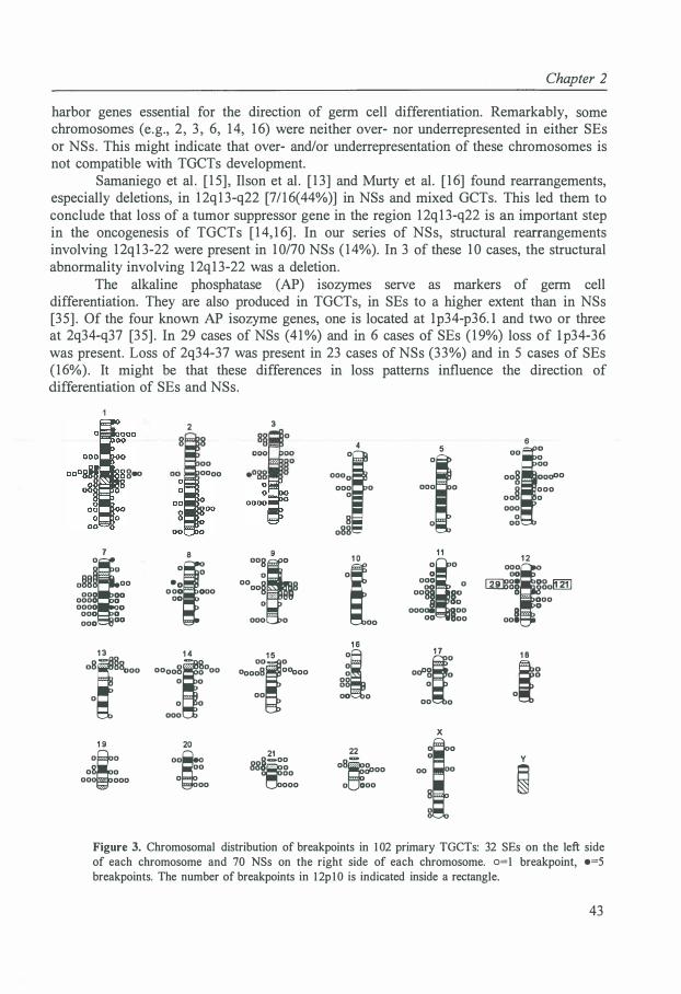
Chapter 2
harbor genes essential for the direction of germ cell differentiation. Remarkably, some chromosomes (e.g., 2, 3, 6, 14, 16) were neither over- nor underrepresented in either SEs or NSs. This might indicate that over- and/or underrepresentation of these chromosomes is not compatible with TGCTs development.
Samaniego et al. [15], Ilson et al. [13] and Murty et al. [16] found rearrangements, especially deletions, in 12q13-q22 [7/16(44%)] in NSs and mixed GCTs. This led them to conclude that loss of a tumor suppressor gene in the region 12q13-q22 is an important step in the oncogenesis of TGCTs [14,16]. In our series of NSs, structural rearrangements involving 12q13-22 were present in 10/70 NSs (14%). In 3 of these 10 cases, the structural abnormality involving 12q13-22 was a deletion.
The alkaline phosphatase (AP) isozymes serve as markers of germ cell differentiation. They are also produced in TGCTs, in SEs to a higher extent than in NSs [35]. Of the four known AP isozyme genes, one is located at lp34-p36.l and two or three at 2q34-q37 [35]. In 29 cases of NSs (41%) and in 6 cases of SEs (19%) loss of l p34-36 was present. Loss of 2q34-37 was present in 23 cases of NSs (33%) and in 5 cases of SEs (16% ). It might be that these differences in loss patterns influence the direction of differentiation of SEs and NSs.
g.,
7
�,. 000 00
0000 0 0000 0
000 0 000 °
0
1 3
o8f8 000
0
1 9
;· 0 0 00 ·
:· 000
2
8 00 0000
8
"f •o 0 0 0 000
00
1 4 8(" 0000 · 0 00
0 0
0 0
000
20
�r 00
0 00
3
0
9
""Bf 0
0000
§ 8
00: ,, . ,,, 0
1 5
l 00008 · · ·· 00ooo
00
21
§-00 88
�0 00
000
4
=f
000 0 0
8 088
1 0
"i. 1 6
gl 00 0
22
08�
�00
0 000
5
·l 1 1
"( 0
ogg o oos
9 §�
00009 §00 00 00
1 7
·f 00 0
X
00 0
1·
6
·i 0 00
oo§ . . . ooo0°
§
000 00 0
000 0
00
1 2
.., 00
l:fil8§ ... ogolIBJ 000 0
8 - - · ooe
1 8
.,g
y
�
Figure 3. Chromosomal distribution of breakpoints in 1 02 primary TGCTs: 32 SEs on the left side of each chromosome and 70 NSs on the right side of each chromosome. o=l breakpoint, •=5 breakpoints. The number of breakpoints in 1 2pl0 is indicated inside a rectangle.
43


Chapter 2
2.2 CYTOGENETIC EVIDENCE THAT CARCINOMA IN SITU IS THE PRECURSOR LESION FOR INVASIVE TESTICULAR GERM CELL TUMORS
Jannie van Echten 1, Ruud J.H.L.M. van Gurp2
, Marian Stoepker1, Leendert H.J. Looijenga2, Bauke de Jong 1
and J. Wolter Oosterhuis2
1Department of Medical Genetics, University of Groningen, and 2Laboratory of Experimental PathoOncology, Dr. Daniel den Hoed Cancer Center, Academic Hospital Rotterdam, The Netherlands
Abstract
A cytogenetic study of two cases of carcinoma in situ of the testis (CJS) and their adjacent invasive tumors, one a nonseminomatous germ cell tumor (NS) and one a seminoma (SE), revealed similarities in chromosomal pattern between the CJS and the invasive lesion in the same patient. These findings present for the first time cytogenetic evidence that CIS of the testis and its adjacent germ cell tumor are clonally related, which suggests that the CJS is indeed the precursor lesion of the invasive tumor. Cancer Genet Cytogenet 85 : 1 33- 137 ( 1 995). © Elsevier Science Inc., 1 995
Introduction
Testicular germ cell tumors (TGCTs) are thought to be derived from dysplastic germ cell precursors (gonocytes), which progress to carcinoma in situ (CIS) [2]. This assumption is supported by the frequent observation of CIS in the testicular parenchyma surrounding invasive cancer [36], as well as by the development of invasive TGCT in patients where CIS had been diagnosed previously [37,38]. Thus CIS is considered the precursor for all subtypes of TGCTs of adolescents and adults, with the possible exception of spermatocytic seminoma [2].
CIS cells have a characteristic morphology ([39] for review) and their ploidy varies from peritriploid to pentaploid [10, 11,40-42]. Cytogenetic data of CIS are limited to only three cases, reported by us [43]. Karyotyping of CIS is troublesome, because tissue culturing has not been successful thus far, and direct harvesting is difficult because of the small number of tumor cells that can be obtained from the seminiferous tubules. Cytogenetics of CIS is important to understand the progression of CIS to invasive tumor and to shed light on the pathogenesis of TGCTs.
In two cases, one seminoma (SE) and one nonseminomatous TGCT (NS), we succeeded in karyotyping both the invasive tumor and the CIS in the surrounding parenchyma.
45

Invasive and non-invasive testicular germ cell tumors of adults and adolescents
Case reports
Case I A 40-year-old patient presented with a mass in his right testis. Histological
examination of the orchiectomy specimen showed a NS with the following components: embryonal carcinoma, immature teratoma, mature teratoma and yolk sac tumor. The parenchyma adjacent to the tumor contained CIS. Remarkably, in one of the seminiferous tubules a trophoblastic giant cell was found in continuity with the CIS.
Case II A 30-year-old patient presented with a mass in his right testis. Histological
examination of the orchiectomy specimen showed a SE with scattered trophoblastic giant cells. The parenchyma was largely atrophic. Within the seminiferous tubules there was extensive CIS.
Materials and methods
In both cases, after frozen section diagnosis material from the tumor and the parenchyma was separately processed for karyotyping.
Short-term culturing and harvesting of the invasive NS and direct harvesting of the invasive SE were performed as described [17,18]. To obtain CIS cells the method for direct harvesting of SE was also applied to the remaining parenchyma, surrounding the invasive tumor, which on light microscopy showed CIS.
Chromosomes were GPG banded (G-banding using Pancreatin [Sigma, P3292, 0.1 % in Hanks' solution] and Giemsa). A modal composite karyotype has been created according to ISCN 1991 [19]. However, the karyotypical descriptions are expressed in relation to the triploid level, to make specific over- and underrepresentation of chromosomes, an important feature of the chromosomal pattern of TGCTs, better visible and comparable [ 6].
Results
From Case I three abnormal metaphases could be analyzed from the CIS component and 10 from the invasive tumor (NS). The representative karyotypes of the CIS and its invasive tumor, respectively are shown in Figures la and lb.
From the CIS component of Case II two metaphases of substandard quality were found. From its invasive tumor (SE) six metaphases were analyzed. Figures 2a and 2b show, respectively, one of the two karyotypes of the CIS, and a representative karyotype of the invasive tumor.
Table 1 shows the modal composite karyotypes of CIS of Case I and the invasive tumors of both cases. Because of the substandard quality of the metaphases of the CIS of Case II no modal composite karyotype is presented. Only the clonal structural abnormalities in the two metaphases are noted.
46
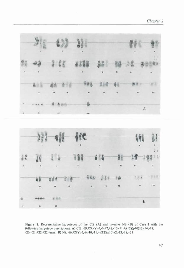
13 14
• "f" ·• --
---�.
13 ,..
21 22
,,,,_
22
Chapter 2
11 12
15 111 17 18 111 20
: ---------�------------------1
10 11 12
j . ... ..__ ___ . , · -- --, ,.,,_
1S 17 18 20
Figure 1 . Representative karyotypes of the CIS (A) and invasive NS (B) of Case I with the following karyotype descriptions. A) CIS, 69,XX,-Y,-5,-6,+7,+8,- 1 0,- 1 1 ,+i( l2)(p10)x2,- 14,- 1 8, -20,+2 1 ,+22,+22,+mar; B) NS, 66,XXY,-5,-6,- 10,- l l ,+i( l2)(p 10)x2,- 1 3,- 1 8,+2 1
47
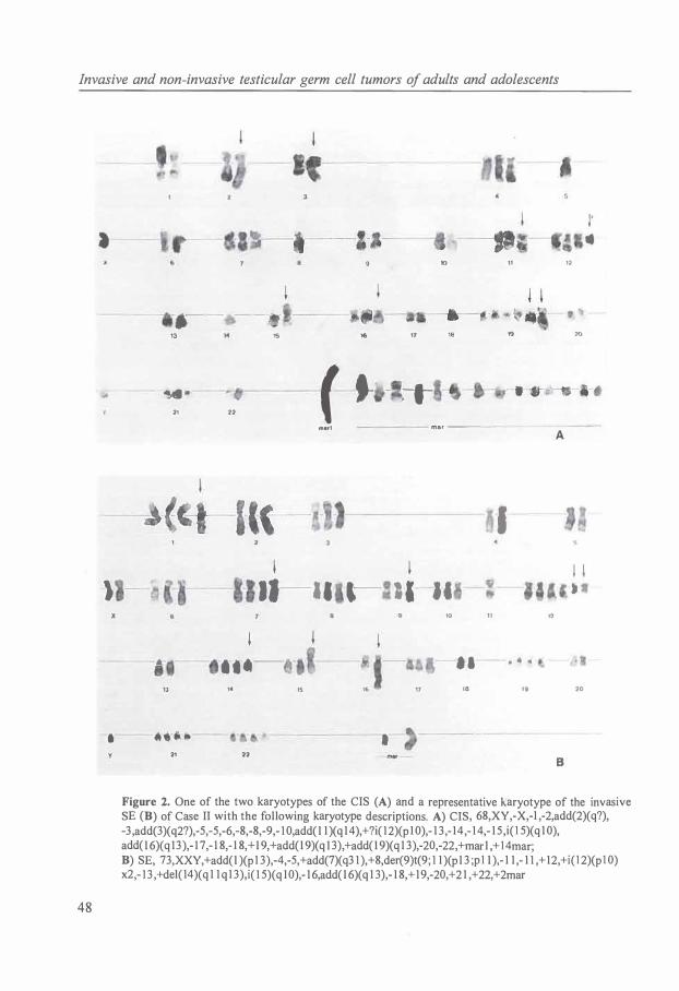
Invasive and non-invasive testicular germ cell tumors of adults and adolescents
48
•
) X
-,--- - -13
-�-21 22
IO
'i5 16 17 18
f i - � mer1 mar ---
\ l
·11-11 · ·,-. ll- It 10 11
11
19
-·
I
12
12
20
1 1 -
I' ..
---•- --'
-.-.-·- ---·• ·- • - 4
13 ,. 15 16 17 18
.--·· .. --- .- ----- -21 22
19 20
Figure 2. One of the two karyotypes of the CIS (A) and a representative karyotype of the invasive SE (B) of Case II with the following karyotype descriptions. A) CIS, 68,XY,-X,-1 ,-2,add(2)(q?), -3 ,add(3)(q2?),-5,-5,-6,-8,-8,-9,- 1 0,add( l 1 )(q 14),+?i( 12)(p 1 0),- 1 3 ,- 14,- 14,- 1 5,i( l 5)(q 1 0), add( l 6)(q 13),- 1 7,- 1 8,- 1 8,+ l 9,+add(l 9)(q I 3),+add( l 9)(q 1 3),-20,-22,+mar I ,+ l 4mar; B) SE, 73,XXY,+add(l )(p 1 3),-4,-5,+add(7)(q3 1 ),+8,der(9)t(9; 1 1 )(p l 3 ;p l l ),- 1 1 ,- 1 1 ,+12,+i( l 2)(p l 0) x2,- 1 3 ,+del( 14)(q 1 1 q 1 3),i( 1 5)( q 10),- 1 6,add( l 6)(q 1 3),- 1 8,+ 19,-20,+2 1 ,+22,+2mar

Chapter 2
Both cases of CIS revealed a peritriploid chromosome pattern: Case I (n = 68) and Case II (n � 65; it was impossible to determine the exact chromosome number of the CIS component of Case II). The karyotypes of CIS and the invasive tumor of Case I show an identical modal number of copies of the chromosomes X, 1 , 2, 3, 4, 5, 6, 7, 9, 1 0, 1 1 , 12, 14, 1 5, 16, 1 7, 18, 19, 2 1 , and 22. Moreover, they have an i(l2p) as common structural abnormality. In Case II an i( 15q) is present both in the CIS component and its invasive SE. An i( 12p) chromosome, present in the invasive SE, might be present in one of the metaphases of the CIS (Fig. 2a)
TABLE I . Modal composite karyotypes of CIS and their invasive TGCTs
Case
II
Discussion
Modal Comp<:>site Karyotype
Carcinoma in situ
67-69,XXY,-5,-6,+8,- 1 0,- l l ,+i( l2) (p l 0)x2,- l 8,-20,+2 1 ,+22[cp3]
Clonal structural abnormalities add(2)(q?) i( 1 5)(q 1 0) add(l 9)(q 1 3) marl
Invasive tumor
6 1 -9 1 ,XXY,+Y,-5,-6,- l 0,- l l ,+i(12) (p l0)x2,- 1 3,- 1 8,+2 1 ,+22[cpl 0]
62-73,XXY,+add( l )(p 1 3),-3,-4,-5, +add(7)(q3 l ),der(9)t(9; 1 l )(p l 3 ;p l 1 ), -l l ,- l l ,+12,+i( l2)(p 1 0)x2,- 1 3 , i( 1 5)(ql 0),- 1 6,add(l6)(q 13),- 1 7,- 1 8, + l 9,+2 l ,+2-6mar[ cp6]
Carcinogenesis of TGCTs starts early in life, probably in utero [2,44]. Based on immunohistochemical and ultrastructural studies, CIS, derived from premalignant gonocytes, [ 45-4 7] is supposed to be the precursor for all TGCTs of adolescents and adults except spermatocytic seminoma [2]. Cytogenetic investigations of CIS may support this view and are important to shed light on the pathogenesis of TGCTs and to understand the progression of CIS to invasive tumor.
In the two cases in which we succeeded in karyotyping both the invasive tumor and its adjacent CIS, we observed karyotypical similarities. In Case I, in CIS and its invasive NS, an identical number of modal copies of the chromosomes X, 1 , 2, 3, 4, 5, 6, 7, 9, 10, 1 1 , 12, 14, 1 5, 1 6, 1 7, 18, 19, 2 1 , and 22, and i( 12p) was found. Despite of the substandard quality of the two metaphases of CIS in Case II, we found an i(l 5q) both in the CIS and SE. An i(l 2p) chromosome might also be present in both components. The karyotypical similarities between CIS and the invasive tumor in both cases demonstrate their clonal relationship and strongly suggest that we karyotyped the CIS that preceded the invasive cancer. As the testicular parenchyma adjacent to the tumor was free of invasive cancer, the abnormal metaphases harvested from the parenchyma must be derived from the
49
Invasive and non-invasive testicular germ cell tumors of adults and adolescents
CIS which was present in the seminiferous tubules. In our previous cytogenetic comparison of CIS with their invasive tumors (all NSs) [43], no clear similarity was observed, except for two copies of i(12p) in both components in one case. It is conceivable that in these cases we failed to karyotype the CIS that was clonally related to the invasive tumor. CIS of the testis in general is very extensive [38] and sometimes heterogeneous [48].
In the present study the karyotypes of the two cases of CIS revealed a peritriploid chromosome pattern. This finding is in keeping with our previous cytogenetic studies [43], as well as with ploidy studies [10,11,40,41]. Polyploidization of a dysplastic germ cell precursor resulting in CIS is supposed to be an early event in the carcinogenesis of TGCTs ([5] for review). Noteworthy is the finding of an i(12p) in the CIS component of the three i(12p) positive invasive tumors (both cases in this study and Case 3 in our previous study [43]). This confirms that i(l2p) formation is an early event in the oncogenesis of TGCTs (for review [6,49]), although most likely preceded by polyploidization [50].
In conclusion, our results present for the first time cytogenetic evidence that CIS is clonally related to and is the precursor for invasive TGCTs.
50

Chapter 2
2.3 MIXED TESTICULAR GERM CELL TUMORS: MONOCLONAL OR POLYCLONAL
Jannie van Echten 1 , J. Wolter Oosterhuis5, Leendert H.J. Looijenga5, Anke Dam2, Dirk Th. Sleijfer3 , Heimen
Schraffordt Koops4 and Bauke de Jong 1
Department of 1Medical Genetics, 2Pathology, 3Medical Oncology and 4Surgical Oncology, University of Groningen, and 5Laboratory of Experimental Patho-Oncology, Dr. Daniel den Hoed Cancer Center, Academic Hospital Rotterdam, The Netherlands
Abstract
Testicular germ cell tumors (TGCTs) of adolescents and adults are, for clinical and pathological reasons, divided in seminomas (SEs) and nonseminomatous germ cell tumors (NSs). Whether and to what degree these two entities are pathogenetically related is still controversial and a matter of debate. TGCTs may contain both SE and NS components. Cytogenetic studies of the SE and NS components of these mixed TGCTs might shed light on the pathogenetic relationship between both components. Separate cytogenetic analysis was performed on both components of three cases of mixed TGCTs with both SE and NS components. The karyotypes of both components were compared with each other. In one case, the SE and NS component share eight different structural chromosomal abnormalities, indicating that the SE and NS component are pathogenetically closely related and have a common neoplastic pathway for a considerable length. Both compon�nts of the other two cases have, respectively, i(l 2p) and no structural chromosomal abnormalities in common. Our results, together with data from the literature, indicate that in mixed TGCTs with SE and NS components, both components may have either a monoclonal or a polyclonal origin. Mod Pathol 9;(4):37 1 -374 ( 1 996). © 1 996 United States and Canadian Academy of Pathology, Inc.
Introduction
Testicular germ cell tumors (TGCTs) of adolescents and adults can be divided both clinically and morphologically in two distinct entities, seminomas (SEs) and nonseminomatous (NSs) TGCTs [ l ]. Intratubular germ cell neoplasia of the unclassified type (IGCNU) is generally accepted as the noninvasive precursor of these tumors [2]. In essence, two models exist regarding the pathogenetic relationship between SEs and NSs [22].
In the first model, the histogenesis of SEs is assumed to diverge from that of the other TGCTs at an early stage. The neoplastic germ cell either may give rise to SE, reflecting germ cell differentiation, or may differentiate to embryonic and/or extraembryonic tissues resulting in NS. In this model, the neoplastic pathway of SEs and NSs is different, with no or only limited crossover, each of them leading to a different end
51

Invasive and non-invasive testicular germ cell tumors of adults and adolescents
point. The second model suggests that SEs and NSs have a common origin with a single
neoplastic pathway on which SE may be an intermediate stage in development of NS. According to this view, SE may not only be an end stage in differentiation, but SE cells may progress to a nonseminomatous phenotype. As a consequence, SEs and NSs may show a strong relationship [5,22,51].
Approximately 10 to 20% of all TGCTs have both SE and NS components. Cytogenetic studies of the SE and NS component of these mixed TGCTs may shed light on the pathogenesis of TGCTs and the still controversial pathogenetic relationship between SEs and NSs.
Materials and methods
Tumor material
Three orchidectomy specimens of TGCTs revealed, on histological examination, both a SE and a NS component. The tumors were classified on the basis of hematoxylin-and-eosinstained paraffin-embedded tissue section, and additional immunohistochemical stainings as described [ 5]. The NS component of Case 1 consists of embryonal carcinoma; Case 2 of yolk sac tumor, immature teratoma, and mature teratoma; and Case 3 of mature teratoma.
Cytogenetic analysis
Direct harvesting of the SE component and short-term culturing and harvesting of the NS component of each tumor were performed as described [17,18]. Chromosomes were GPG banded [G-banding using Pancreatin (Sigma, P3292, 0.1 % in Hanks' solution, Sigma, Germany) and Giemsa]. A modal composite karyotype has been created according to ISCN 1991 Guidelines for Cancer Cytogenetics [19]. However, the karyotypic descriptions are expressed in relation to the triploid level, to make an important feature of the chromosomal pattern of TGCTs better visible and comparable to previous reports [6].
Results
From Case 1, 7 metaphases were analysed from the SE component and 16 from the NS component with the following karyotype descriptions:
SE component, 44-67,XXY,der(l)t(1; 12)(pll ;q13),add(2)(p24),+add(3)(q21 ),-4, -5,+del(6)( q 13), +del(7)( q31.1),+del(7)( 432),-8,-9,-10,-1 1 ,del(ll) ( q23),+der(l2)t(11 ;12)( q12;q23),-13,-14,-15,-16,-17,-18,add(l9)( qll),-21, -22,+der(?)t(?;7)(? ;pll),+2-10mar[cp7]; NS component, 47-63,XX, -Y,add(l)(p36),+der(l)t(1;12)(pll ;q13),add(2)(p24),-4,-5,+del(7)(q31.1), +del(7)( q32),-9,-10,-11,del(ll)( q23),-12,der(12)t(ll ; 12)( ql2;q23),-15,-18,add(19) ( qll),-21,-21, +rl , +r2,+der(?)t(?;7)(?;pll), +der(?)t(9;?;21 )( q 12;?;q 11 )[ cp 16]
52
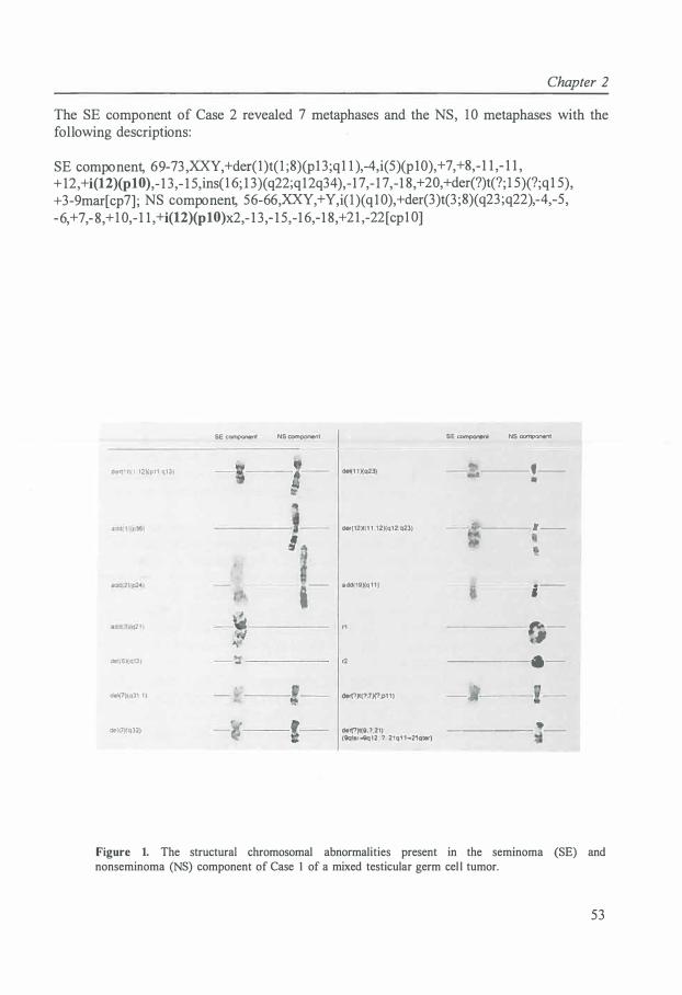
Chapter 2
The SE component of Case 2 revealed 7 metaphases and the NS, I O metaphases with the following descriptions :
SE component, 69-73 ,XXY,+der( l)t( l ;8)(p 13;q l 1),-4,i(5)(p 10),+7,+8,-1 1 ,- 1 1 , + 12 ,+i(12)(p10),- 13,- 15,ins( l 6; 13)(q22;q 12q34),-17,- 17,-18 ,+20,+der(?)t(?; l 5)(?;q 15) , +3-9mar [cp7] ; NS component, 56-66,XXY,+Y,i( l)(q l O),+der(3)t(3;8)(q23;q22),-4,-5, -6,+ 7,-8 ,+ I 0,- 1 1 ,+i(l2)(p10)x2,- 13,- 15,-16,-18 ,+2 1,-22 [ cp l O]
der(1 ll, I l2)ipl1 ql3)
def1G)(q13)
�1(7)(q31 1 )
dtl(7)1q32)
SE component NS component
I
del(1 1 )(q23)
der(12)1(1 1 ,121(q12,q23)
1_ acld(19)(q 1 1 )
r1
r2
dof(?)l(?,7)('1,p1 1)
dert?)l(ll,?,21) (9q!er..Qc:i12. ? 21q1 1-21qter)
SE ccmpont<1I NS ocmponenl
· ~�----f--
,•r•---1'--
,-,:-.,
---•-
______ .. _
Figure 1. The structural chromosomal abnormalities present in the seminoma (SE) and nonseminoma (NS) component of Case 1 of a mixed testicular germ cel l tumor.
53
Invasive and non-invasive testicular germ cell tumors of adults and adolescents
From both the SE and the NS component of Case 3, 10 metaphases were analysed with the following descriptions:
SE component, 57-65,:XX,-Y,add(l)(p13),+dic(l ;4)(p34;p16),add(3)(pl 1),-4,-4, -5,add(6)(pl 1.1),add(7)(q21),der(8)t(7;?;8)(pl3;?;q13),-9,der(l l)add(l l) (q25)dup(l l)(ql3q23),+ 12,+add(l2)(ql l),-13,-16,-18,-20,+21,-22[cp10]; NS component, 56-59,:XXY,i(l )( q 1 0),add(3)(p 12),-4,-5,add(6)(p21 ),del(6)( q 13),-8,-9,-10, del(l l)(q14q21),+i(l2)(pl 0)x2,-13,-14,-15,der(l 7)t(5;17)(q 13;p13),-18,-19,del(21)(q22), -22[cp10]
Common structural abnormalities in the SE and the NS component are indicated in bold; e.g., eight in Case 1 [der(l)t(l; 12)(pll ;q13), add(2)(p24), del(7)(q3 l.l), del(7)(q32), del(ll)(q23), der(l2)t(l 1;12)(q12;q23), add(19)(qll), and der(?)t(?;7)(?;pl 1)]; one in Case 2 [i(l2)(pl0)]; and none in Case 3. Figure 1 shows the comparison of the structural abnormalities present in the SE and NS component of Case 1.
Discussion
According to the pathogenetic relationship between SEs and NSs, in essence, two different views exist. One view states that SEs and NSs have an independent origin in which SE is an end stage in differentiation. The other view holds that SEs and NSs are pathogenetically closely related and that SE may either be an end stage in differentiation or may progress to a NS [3,5,6,8,10,22,51-58].
SEs and NSs show strong similarities in over- and under-representation of chromosomes [6], as well as in the distribution of chromosomal breakpoints [59]. Ploidy studies of TGCTs show a DNA index of SEs and IGCNU adjacent to invasive cancer in between that of preinvasive IGCNU and NSs [5,10,11,40-42,56]. SEs, in general, have higher chromosome numbers than NSs. Patients with primary SEs may have NS metastases [60,61]. In SEs, small NS foci have been observed [55,62]. These data suggest that SEs and NSs are closely related and point to a linear progression from IGCNU via SE to NS by net loss of chromosomes. SE may be an end stage, as well as an intermediate stage, in the development of NS.
Our results show that the SE and NS component of Case 1 have eight different structural abnormalities in common: a der(l)t(l;l2)(pl l;ql3), add(2)(p24), del(7)(q31.1), del(7)(q32), del(l l)(q23), der(l2)t(l 1;12)(q12;q23), add(19)(ql 1), and a der(?)t(?;7)(?;pl 1) (Fig 1. ). Common chromosomal abnormalities in the SE and NS component of mixed TGCTs is found by Walt et al. [63], Haddad et al. [64] and Rodriguez et al. [28]. These findings obviously point to a close relationship between the SE and NS component of these TGCTs and support the idea of the linear progression model. In Case 2, we found an i(12p) chromosome as the only structural abnormality present in both components. Castedo et al. [65] and Rodriguez et al. [14] both described i(l2p) as the common chromosomal abnormality in the SE and NS component of a mixed TGCT. For these cases, it is difficult to derive a conclusion regarding a common neoplastic pathway, because i(l2p) is present in about 70% of all TGCTs [59]. Case 3 revealed no common chromosomal abnormalities
54

Chapter 2
in both components, although it might be conceivable that the abnormal chromosomes 3 and 6 in both components are related abnormalities.
We do not expect to find in all cases of mixed TGCTs with a SE and NS component a (strong) relationship between both components. Carcinogenesis of TGCTs probably starts during intrauterine life [2]. The neoplastic counterparts of gonocytes, the IGCNU cells, remain silent until puberty and then progress to invasive TGCT, most often before the age of 40. The mixed TGCTs with common chromosomal abnormalities in the SE and NS component point to a monoclonal origin and pathogenetic relationship of the two components. Those mixed TGCTs with no or weak cytogenetic similarity between the SE and NS component, are probably tumors in which both components have an independent origin, or in which development of both components might have diverged at an early stage in their development. The possibility of a monoclonal and polyclonal origin for the SE and NS components of mixed TGCTs is also described by Gillis et al. [ 48].
In conclusion, one of our three cases of mixed TGCTs with a SE and NS component, and three cases of the literature [28,63,64], show clear cytogenetic evidence for a common origin and strong pathogenetic relationship of the SE and NS component. These cytogenetic findings, of course, do not prove that a SE may progress to a NS but at least they indicate that SEs and NSs are pathogenetically closely related and may have a common neoplastic pathway for a considerable length.
55

Invasive and non-invasive testicular germ cell tumors of adults and adolescents
REFERENCES
I . Mostofi FK, Sobin LH (1977): International histological classification of testicular tumors (No. 16). In: International Histologic Classification of Tumors. Geneva: World Health Organization.
2. Skakkebrek NE, Berthelsen JG, Giwercman A, Milller J ( 1 987): Carcinoma-in-situ of the testis: Possible origin from gonocytes, and precursor of all types of germ cell tumors except spermatocytoma. Int J Androl 10 : 1 9-28.
3 . Oosterhuis JW, Looijenga LHJ ( 1 993): The biology of human germ cell tumours: retrospective speculations and new prospectives. Eur Urol 23 :245-250.
4. Damjanov I ( 1 99 1 ): Pathobiology of human germ cel l neoplasia. Recent Results Cancer Res 123 : 1 - 1 9.
5 . Oosterhuis JW, Castedo SMMJ, de Jong B, Cornelisse CJ, Dam A, Sleijfer OT, Schraffordt Koops H ( 1 989): Ploidy of primary germ cell tumors of the testis. Pathogenetic and clinical relevance. Lab Invest 60: 14-20.
6. de Jong B, Oosterhuis JW, Castedo SMMJ, Vos AM, te Meerman GJ ( 1 990): Pathogenesis of adult testicular germ cell tumors. A cytogenetic model. Cancer Genet Cytogenet 48: 143- 1 67.
7 . Sandberg AA (1 990): The chromosomes in human cancer and leukemia, 2nd Ed. Elsevier, New York.
8. Fossa SD, Nesland JM, Pettersen EO, Amellem 0, Wrehre H, Heimdal K ( 199 1 ): DNA ploidy in primary testicular cancer. Br J Cancer 64:948-952.
9. Fossa SD, Nesland JM, Wrehre H, Amellem 0, Pettersen EO ( 1 99 1 ): DNA ploidy in the primary tumor from patients with nonseminomatous testicular germ cell tumors clinical stage I. Cancer 67: 1 874- 1 877.
10 . EI-Naggar AK, Ro JY, McLemore D, Ayala AG, Batsakis JG ( 1 992): DNA ploidy in testicular germ cell neoplasms. Histogenetic and clinical implications. Am J Surg Pathol 16 :6 1 1 -6 1 8.
1 1 . de Graaff WE, Oosterhuis JW, de Jong B, Dam A, van Putten WLJ, Castedo SMMJ, Sleijfer OT, Schraffordt Koops H (1 992): Ploidy of testicular carcinoma in situ. Lab Invest 66: 1 66- 168.
12. Looijenga LHJ, Gillis AJM, van Putten WLJ, Oosterhuis JW (1 993): In situ numeric analysis of centromeric regions of chromosome 1, 12, and 1 5 of seminomas, nonseminomatous germ cel l tumors, and carcinoma in situ of human testis. Lab Invest 68:2 1 1 -2 19 .
13 . Ilson DH, Bosl GJ, Motzer R, Dmitrovsky E, Chaganti RSK ( 199 1 ): Genetic analysis of germ cell tumors: Current progress and future prospects. Hematol Oncol Clin North Am 5 : 127 1 - 1283 .
14 . Rodriguez E, Mathew S, Reuter V, Ilson DH, Bosl GJ, Chaganti RSK ( 1 992): Cytogenetic analysis of 1 24 prospectively ascertained male germ cel l tumors. Cancer Res 52:2285-2291 .
1 5 . Samaniego F , Rodriguez E, Houldsworth J , Murty VVVS, Ladanyi M , Lele KP, Chen Q, Dmitrovsky E, Geller NL, Reuter V, Jhanwar SC, Bosl GJ, Chaganti RSK (1 990): Cytogenetic and molecular analysis of human male germ cel l tumors: chromosome 12 abnormalities and gene amplification. Genes Chromosom Cancer I :289-300.
1 6. Murty VVVS, Houldsworth J, Baldwin S, Reuter V, Hunziker W, Besmer P, Bosl GJ, Chaganti RSK (1992): Allelic deletions in the long arm of chromosome 12 identify sites of candidate tumor suppressor genes in male germ cell tumors. Proc Natl Acad Sci USA 89: 1 1 006- 1 1 0 10.
1 7 . Castedo SMMJ, de Jong B, Oosterhuis JW, Seruca R, te Meerman GJ, Dam A, Schraffordt Koops H ( 1 989): Cytogenetic analysis of ten human seminomas. Cancer Res 49:439-443.
1 8 . Castedo SMMJ, de Jong B, Oosterhuis JW, Seruca R, ldenburg VJS, Dam A, te Meerman GJ, Schraffordt Koops H, Sleijfer DT (1989): Chromosomal changes in human primary testicular nonseminomatous germ cell tumors. Cancer Res 49:5696-570 1 .
1 9 . ISCN(l99 1 ): Guidelines for Cancer Cytogenetics, Supplement to an International System for Human Cytogenetic Nomenclature. Mitelman F, ed. S. Karger, Basel.
20. Hittmair A, Rogatsch H, Feichtinger H, Habisch A, Mikuz G ( 1 995): Testicular seminomas are aneuploid tumors. Lab Invest 72:70-74.
2 1 . Oosterhuis JW, Castedo SMMJ, de Jong B ( 1 990): Cytogenetics, ploidy and differentiation of human testicular, ovarian and extragonadal germ cell tumours. Cancer Surv 9:32 1 -332.
56
Chapter 2
22. Damjanov I ( 1 989): Editorial. Is seminoma a relative or a precursor of embryonal carcinoma? Lab Invest 60: 1 -3 .
23 . Castedo SMMJ, de Jong B, Oosterhuis JW, Seruca R, Idenburg VJ, Buist J , Sleijfer DT ( 1 988): i( l 2p)-negative testicular germ cell tumors. A different group? Cancer Genet Cytogenet 35 : 1 7 1 - 1 78.
24. Meloni AM, Berger C, Dobbs R, White R, Sandberg AA (I 991 ) : Characteriz.ation of unusual marker chromosomes in testicular tumors by fluorescence in situ hybridiz.ation. Cancer Genet Cytogenet 56:97.
25. Atkin NB, Fox MF, Baker MC, Jackson Z ( 1 993): Chromosome 12-containing markers, including two dicentrics, in three i(12p)-negative testicular germ cell tumors. Genes Chromosom Cancer 6:2 1 8-22 1 .
26. Suijkerbuijk RF, Looijenga L, de Jong B, Oosterhuis JW, Cassiman JJ, Geurts van Kessel A ( 1 992): Verification of isochromosome 12p and identification of other chromosome 12 aberrations in gonadal and extragonadal human germ cell tumors by bicolor double fluorescence in situ hybridization. Cancer Genet Cytogenet 63 : 8- 16 .
27. Suijkerbuijk RF, Sinke RJ, Meloni AM, Parrington JM, van Echten J, de Jong B, Oosterhuis JW, Sandberg AA, Geurts van Kessel A ( 1993 ): Overrepresentation of chromosome 12p sequences and karyotypic evolution in i( 12p)-negative testicular germ-cell tumors revealed by fluorescence in situ hybridization. Cancer Genet Cytogenet 70:85-93 .
28. Rodriguez E, Houldsworth J, Reuter VE, Meltzer P, Zhang J, Trent JM, Bos! GJ, Chaganti RSK ( 1 993): Molecular cytogenetic analysis of i(12p)-negative human male germ cel l tumors. Genes Chromosom Cancer 8:230-236.
29. Lothe RA, Fossa SD, Stenwig AE, Nakamura Y, White R, B0rresen AL, Brngger A (1 989): Loss of 3p or l l p alleles is associated with testicular cancer tumors. Genomics 5 : 1 34- 138.
30. Radice P, Pierotti MA, Lacerenz.a S, Mondini P, Radice MT, Pilotti S, Della Porta G ( 1 989): Loss of heterozygosity in human germinal tumors. Cytogenet Cell Genet 52:72-76.
3 1 . Lothe RA, Hastie N, Heimdal K, Fossa SD, Stenwig AE, B0rresen AL ( 1993): Frequent loss of 1 l p 1 3 and l l p 1 5 loci in male germ cell tumours. Genes Chromosom Cancer 7:96- 1 0 1 .
32. Looijenga LHJ, Abraham M, Gill is AJM, Saunders GF, Oosterhuis JW ( 1 994): Testicular germ cell tumors of adults show deletions of chromosomal bands l l p 1 3 and l l p 1 5.5, but no abnormalities within the zinc-finger regions and exons 2 and 6 of the wilms' tumor I gene. Genes Chromosom Cancer 9: 1 53- 160.
33 . Murty VVVS, Bos! GJ, Houldsworth J , Meyers M, Mukherjee AB, Reuter V, Chaganti RSK ( 1994): Allelic loss and somatic differentiation in human male germ cell tumors. Oncogene 9:2245-225 1 .
34. Murty VVVS, Li RG, Houldsworth J, Bronson DL, Reuter VE, Bos! GJ, Chaganti RSK ( 1 994): Frequent allelic deletions and loss of expression characterize the DCC gene in male germ cell tumors. Oncogene 9:3227-323 1 .
35 . Hofmann MC, Millan JL ( 1993): Developmental expression of alkaline phosphatase genes; Reexpression in germ cell tumours and in vitro immortalized germ cells. Eur Urol 23:38-45 .
36. Jacobsen GK, Hendriksen OB, von der Maase H ( 198 1): Carcinoma in situ of testicular tissue adjacent to malignant germ cell tumors: A study of 10 cases. Cancer 47:2660-2662.
37. Giwercman A, Skakkebrek NE ( 1993): Carcinoma in situ of the testis: biology screening and management. Eur Urol 23 (suppl 2): 1 9-2 1 .
38 . Giwercman A, von der Maase H, Skakkebrek NE (1 993): Epidemiological and clinical aspects of carcinoma in situ of the testis. Eur Urol 23: 1 04- 1 14.
39. Soosay GN, Bobrow L, Happerfield L, Parkinson MC ( 199 1 ): Morphology and immunohistochemistry of carcinoma in situ adjacent to testicular germ cell tumors in adults and children: Implications for histogenesis. Histopathology 1 9:537-544.
40. Nagler HM, Kaufman DG, O'Toole KM, Sawczuk IS ( 1 990): Carcinoma in situ of the testis: Diagnosis by aspiration flow cytometry. J Urol 143 :359-361 .
4 1 . Nistal M , Codesal J, Paniagua R ( 1989): Carcinoma i n situ o f the testis i n infertil e men. A histological, immunocytochemical, and cytomorphometric study of DNA content. J Pathol 1 59:205-2 10.
42. Millier J, Skakkebrek NE ( 198 1 ): Microspectrophotometric DNA measurements of carcinoma-in-situ
57

Invasive and non-invasive testicular germ cell tumors of adults and adolescents
germ cells in the testis. Int J Androl suppl 4:2 1 1 -22 1 . 43. Vos AM, Oosterhuis JW, de Jong B, Buist J, Schraffordt Koops H ( 1 990): Cytogenetics of
carcinoma in situ of the testis. Cancer Genet Cytogenet 46:75-8 1 . 44. Forman D, M0ller H ( 1994): Testicular cancer. Cancer Surv 19/20:323-34 1 . 45. Gondos B ( 1 993): Ultrastructure of developing and malignant germ cells. Eur Urol 23 :68-75. 46. Giwercman A, Andrews PW, J0rgensen N, Millier J, Grrem N, Skakkebrek NE (1 993):
Immunohistochemical expression of embryonal marker TRA- 1 -60 in carcinoma in situ and germ cell tumors of the testis. Cancer 72: 1 308- 13 14.
47. Giwercman A, Millier J, Skakkebrek NE (1991 ): Carcinoma in situ of the testis: Possible origin, clinical significance, and diagnostic methods. Recent Results Cancer Res 123 :2 1 -36.
48. Gillis AJM, Looijenga LHJ, de Jong B, Oosterhuis JW ( 1 994): Clonality of combined testicular germ cell tumors of adults. Lab Invest 7 1 : 874-878.
49. Bosl GJ, Dmitrovsky E, Reuter VE, Samaniego F, Rodriguez E, Geller NL, Chaganti RSK (1 989): Isochromosome of chromosome 12 : clinically useful marker for male germ cell tumors. J Natl Cancer Inst 8 1 : 1 874- 1 878.
50. Geurts van Kessel A, van Drunen E, de Jong B, Oosterhuis JW, Langeveld A, Mulder MP (1 989): Chromosome 12q heterozygosity is retained in i(12p)-positive testicular germ cell tumor cells. Cancer Genet Cytogenet 40: 129- 1 34.
5 1 . Ulbright TM (1 993): Germ cell neoplasms of the testis. Am J Surg Pathol 17 : 1 075- 1 09 1 . 52. Damjanov I ( 1993): Pathogenesis of testicular germ cell tumours. Eur Urol 23 :2-7. 53. Abu-Jawdeh GM, Oyasu R ( 199 1 ) : Testicular germ cell tumors. An update on clinical pathologic
correlation. Acta Pathol Jpn 41 :83-93 . 54. Bailey D, Marks A, Stratis M, Baumal R ( 1991 ): Immunohistochemical staining of germ cell tumors
and intratubular malignant germ cells of the testis using antibody to placental alkaline phosphatase and a monoclonal anti-seminoma antibody. Mod Pathol 4: 1 67- 1 7 1 .
55. Manivel JC (1 992): Transformation of seminoma to yolk sac tumor. Am J Clio Pathol 97:463-465 . 56. Oliver RTD ( 1 987): HLA phenotype and clinicopathological behaviour of germ cell tumours:
possible evidence for clonal evolution from seminomas to nonseminomas. Int J Androl 1 0:85-93 . 57. Oliver RTD (1991): Testicular cancer. Curr Opin Oncol 3 :559-564. 58. Sesterhenn IA ( 1985): The role of intratubular malignant germ cells in the histogenesis of germ cell
tumours. In: Germ cell tumours II, WG Jones, AM Ward, CK Anderson, eds. Pergamon press, Oxford, pp. 25-35 .
59. van Echten J, Oosterhuis JW, Looijenga LHJ, van de Pol M, Wiersema J, te Meerman GJ, Schraffordt Koops H, Sleijfer DT, de Jong B ( 1995): No recurrent structural abnormalities apart from i( 12p) in primary germ cell tumors of the adult testis. Genes Chromosom Cancer 1 4: 1 33- 144.
60. Loehrer PJ, Wil liams SD, Einhorn LH (1 988): Testicular cancer: The quest continues. J Natl Cancer Inst 80: 1373- 1 382.
6 1 . Ulbright TM, Roth LM ( 1987): Recent developments i n the pathology of germ cel l tumors. Semin Diagn Pathol 4:304-3 19.
62. Czaja JT, Ulbright TM ( 1 992): Evidence for the transformation of seminoma to yolk sac tumor, with histogenetic considerations. Am J Clio Pathol 97:468-477.
63. Walt H, Arrenbrecht S, Delozier-Blanchet CD, Keller PJ, Nauer R, Hedinger CE (1 986): A human testicular germ cell tumor with borderline histology between seminoma and embryonal carcinoma secreted beta-human chorionic gonadotropin and alpha-fetoprotein only as a xenograft. Cancer 58: 1 39- 146.
64. Haddad FS, Sorini PM, Somsin AA, Nathan MH, Dobbs RM, Berger CS, Sandberg AA ( 1 988): Familial double testicular tumors: identical chromosome changes in seminoma and embryonal carcinoma of the same testis. J Urol 139:748-750.
65. Castedo SMMJ, de Jong B, Oosterhuis JW, Seruca R, Buist J, Schraffordt Koops H ( 1 988): Cytogenetic study of a combined germ cell tumor of the testis. Cancer Genet Cytogenet 35 : 1 59- 165 .
58

CHAPTER 3
RESIDUAL MATURE TERATOMA
3.1 COMPARISON OF THE CHROMOSOMAL PATTERN OF PRIMARY TESTICULAR NONSEMINOMAS AND RESIDUAL MATURE TERATOMAS AFTER CHEMOTHERAPY
Jannie van Echten l , Wanda S. van der Vloedt 1 , Mirjam van de Pol5, Anlce Dam2, Gerard J. te Meennan' , Heimen Schraffordt Koops3 , Dirk Th. Sleijfer4, J. Wolter Oosterhuis5 and Bauke de Jong' Departments of 'Medical Genetics, 2Pathology, 3Surgical Oncology and 4Medical Oncology, University of Groningen, Groningen, and 5Laboratory of Experimental Patho-Oncology, Dr. Daniel den Hoed Cancer Center, Academic Hospital Rotterdam, Rotterdam, The Netherlands
Abstract
About 70 to 75% of patients with nonseminomatous testicular germ cell tumors (NSs) present with metastases. When these metastases are treated with chemotherapy, often residual mature teratoma (RMT) is left. RMT is composed of fully differentiated somatic tissue. Untreated metastases of NSs rarely consist exclusively of mature somatic tissue. Apparently, after chemotherapy treatment there is a shift towards higher degrees of differentiation. Investigating tumor progression and the mechanism(s) involved in therapy related differentiation, we compared the cytogenetically abnormal karyotypes of a series of 70 NSs with those of 31 RMTs. In NSs and RMTs the modal total chromosome number does not differ and is in the triploid range. Both the .frequency and the average copy number of i(l 2p) is not different, and the pattern of chromosomal over- and underrepresentation and distribution of breakpoints do not differ significantly in both series. We obtained no evidence that specific chromosomal alterations are related with the development of primary NSs to metastasis and with therapy related differentiation of the metastasis. The cytogenetic data suggest that both induction of differentiation of (selected) cells or selection of cells with capacity to differentiate are possible mechanisms for the therapy related differentiation of RMTs. Earlier histologic, and in vitro and in vivo studies suggest that RMT is the result of selective destruction of the non-teratomatous elements by the chemotherapy. (submitted).
Introduction
Primary nonseminomatous testicular germ cell tumors (NSs) of adults are in general tumors with mixed histology. They can be composed of embryonal carcinoma (EC), yolk sac tumor (YS), choriocarcinoma (CH), immature teratoma (IT) and mature teratoma (MT) [1,2]. A seminoma (SE) component may be present. Pure NSs, with one histological
59

Residual mature teratoma
component , are rare [3]. At presentation , about 70 to 75% of patients with NSs have lymphatic and/or hematogenous metastases. However , NSs are highly curable solid tumors. The patients are treated by orchidectomy, in case of metastatic disease, followed by cisplatin containing chemotherapy and additional surgical resection of residual mass [ 4].
From metastases of primary NSs treated with chemotherapy, often residual mature teratoma (RMT) is left. RMT is composed of fully differentiated, mature somatic tissue. As is the case in primary tumors, untreated metastases of primary NSs rarely consist exclusively of mature somatic tissue, they usually retain the histology of the primary tumor [5]. Apparently, after chemotherapy there is a shift towards higher degrees of differentiation. This effect of chemotherapy might be due to the induction of differentiation of malignant cells to more differentiated cells , to selective destruction of cells other than MT cells, or to selection of cells with an inherent capacity of (therapy related) differentiation. The mechanisms are not mutually exclusive [6].
We compared the cytogenetically abnormal karyotypes of a series of 70 NSs with those of 3 1 RMTs, in order to study which chromosomal changes play a role in tumor progression (e .g . metastasis) and/or which mechanism(s) are involved in therapy related differentiation. However , distinction between both events can not be made because we can only cytogenetically investigate metastases of NSs after chemotherapy treatment.
Materials and methods
A cytogenetic comparison of 70 NSs with 3 1 RMTs was carried out. Culturing and harvesting of the tumors was performed using standard cytogenetic techniques [7,8]. For each tumor, a modal composite karyotype description was made according to the ISCN 1995 [9]. However , all karyotype descriptions are based on the triploid level, since this makes over- and underrepresentation of specific chromosomes, an important feature of testicular germ cell tumors [8 , 10] , better visible and comparable. Only NSs and RMTs with an abnormal karyotype are included in this study, because NSs and RMTs show a consistent high DNA index (DI) [ 1 1 - 15].
For each tumor and chromosome, the average number of short and long arms was determined. Parts of chromosomal arms involved in structural abnormalities were registered as whole arms if they represented 50% or more of the total arm length. The modal number of short and long arms divided by 2, revealed the average modal number of chromosomes. The average number of sex chromosomes for each tumor was multiplied by 2, to allow comparison with the autosomes [8 , 10].
Statistical analysis and comparison of the cytogenetic data of the NSs and RMTs was performed using the Mann-Whitney U or chi-square test with Bonferroni 's correction for multiple testing, when necessary.
60

Chapter 3
TABLE 1 . Description of the Modal Composite Karyotype and the Modal Chromosome Number of 3 1 RMTs
Modal Case• Description of modal composite karyotype number 1 61 ~ 79,XX,-Y,+ 1 ,+6,+8,+9,+ 1 O,+i( 12)(p 1O),+1 3,+ 17,-1 8,+2 1 ,+mar[cp7] 78 2 42~60,XXY,- 1 ,-2,-3,-4,-5,-9,- 1 O,+i( l 2)(p I O)x2,- 1 3,- 14,- 15,-1 8,-1 9,-20, +mar[cp 18] 60 3 62~63,XXY,-8,i(l 2)(p I O),+i(12)(p 1O)x2,-1 3,- 14,- l 8,-20,del(22)( q 12)[cp2] 62,5 4 56~59,XX,-Y,add(] )(p36),der(2)t(2;8)(q32;q23),-3,-4,-5,-9,- l O,- l l ,+i(] 2)(p 1 O)x2,- l 3, 58
dic( l 3; l 7)(p 1 1 ;q22),- 1 4,-1 5,-1 6,- 17,-1 8,-1 9,-20,add(22)(q 1 1 ),+2mar[cp 1 3] 5 59~62,XXY,der( l )t(l ;3)(p32;p2 l ),-2,-4,-5,der(7)t(5;7)(q l 3;q22),-l O,-l l ,+i(l 2)(p 1 O)x3, 62
- 13 ,- 14,- 1 5,add(l 7)(q25),-1 8,- l 9,+2 1 ,-22[cp3] 6 63~65,XXY,+del(l )(q4 l ),-4,-5,+7,+8,add(9)(p 13 ),-10,-1 O,- l l ,+i(l2)(p l 0),- 13 ,- 14,- 1 5, 63
del( l 6)(p 1 3),-1 8,-l 9,+mar[cp4] 7 49~54,XX,-Y,del(l )(p34 ),-2,-3,-4,-5,der(5)t(3 ;5)(q2 l ;pl 5),-6,+add(7)( q22),-9,-10, 53
add( 10)( q26),- 1 1 ,- 1 3,- 14,-1 5,-1 6,del( I 7)(p 1 1 ),- 1 8,-1 8,-1 9,-20,-2 1 ,-22,+der(?)t(?; 1 8) (?;q i 1 ),+mar[cp 14]
8 54~58,XXY,-2,-3,-4,-9,-9,- 10,- 10,-1 l ,+i(l2)(p l O)x2,- 13,- 14,-1 5,- l 6,-1 8,del( l 8)(p 1 1 ),- 1 9, 57 -2 l ,-22,+der(?)t(?;9)(?;q l l ),+2mar[cp6]
9 52~56,XXY,del( ] )(p3?5),-2,-3,-4,-5,+6,-9,-l 0,-1 l ,+add(l 2)(p 13),+i(l 2)(p l O)x2,- l 3,- l 4, 52 -1 5,- 16,-1 8,- 1 9,-20,-2 1 ,-22[cp9]
1 0 56~58,XXY,add(] )(p 1 1 ),add( l )(p34),-2,-3,-4,-5,-8,del(8)(p22),-9,-10,-1 1 ,+i(l 2)(p 1 O)x2,-1 3, 57 - 14,- 14,- 1 5,- 16,- 1 8,- 1 9,-20,+2 1 ,-22,+2mar[cp9]
1 1 78~88,XXY,+ l ,+2,+ 3,+ 3,+add(5)(q3 1 ),-6,add(7)(p 1 1 ),der(7)t(7;7)(p22;q 1 1 ),+inv(7) 85
(p 1 5p22),+8,+9,+del( 1O)(p 1 3),add(l 1 )(q25),+ 12,+del(12)(q2 1q24),+i(12)(p 1 O)x3,+ 13, + 14,+ 1 7,+20,+2 1 ,-22,-22,i(22)(q 1 O),+der(?)t(?;7)(?;p 1O),+mar[cp1 8]
1 2 57~66,XXY,+Y,-l ,-4,-5,-6,+7,+7,+del(7)(q3 1 ),-8,- 10,-1 1 ,+i( l2)(p 10),-1 3,- 14,-1 7,-1 8,- 19, 62 + 2 l ,-22,+der(?)t(?;5)(?;q 13),+mar[ cp9]
1 3 55~57,XY,-X,add(l )(p36),-4,-6,-9,-1 O,-l l ,+i(] 2)(p l O)x2,- 13 ,-14,-1 5,-1 8,- 1 9,-20,-2 l ,-22[cp9] 57 14 62~66,XY,+Y,del(X)(p2 l ),+add(l )(p36),der(2)t( l ;2)(q2 l ;q37),-4,+7,-9,-1 O,-l l ,+i( l2) 64
(p 10)x2,add(13)(p l l ),-14,-1 8,-1 9,-20,-22[cp9] 1 5 56~59,XXY,+X,dic( l ;20)del(1 )(20qter➔20pl 3 : : l q44➔lq 12 : : l q2 l➔lp34),-2,-3,-4, 58
del(4)(p l 5),-5,add(7)(q 1 1 ),-9,-1 0,-1 1 ,add(] 1 )(q23),add(l2)(q24),+i(l2)(p 10)x2, - 13,- 14,-1 5,- 1 6,- 1 8,- 1 9,-20,-2 1 ,der(22)t(7;22)(q 1 1 ;q 1 3)[cp7]
1 6 57~62,XXY,+Y,+der(] )t(l ;6)(p34;p2 l ),-2,-4,-9,-10,- 1 1 ,+i(12)(p 10),-1 3,-14,-1 5,-1 8,-1 9, 60 add(20)(p 1 3),+add(20)(p 1 3),-22[ cp9]
1 7 4 7~53,XX,-Y,del(] )(p21 ),del(l )(p35),-2,-4,-5,-6,-9,-l 0,-1 1 ,add(l l )(q23),del(l 2)(q 1 3), 52 +die( 12; 1 5)(p 13 ;p 13),+i(] 2)(p 10),- 13,-14,-1 5,- 1 5,dic(] 5;20)( q26;p 1 3),-1 6,- 1 8,- 1 9, add(l 9)(q 1 3),-20,-20,-2 l ,add(2 l )(q22),der(2 1 )t(l ;2 1 )(p3 l ;p 1 3),-22,add(22)(p 1 3),+der(?) t(?;5)(?;q l 3)[cpl l ]
1 8 54~63,XX,-Y,add(l )(p32),+add(l )(q2 l ),+del(l )(p22),del(2)( q33),dic(2;?6)(p25;q2 1 ), 6 1 -4,+del(8)(p 12),+der(8)t( l ;8)(p22;p 1 1 ),-9,-1 O,+i(l 2)(p 1 O)x2,-l 3,- 1 5,-1 6,-1 8,- 1 8, i(l 8)( q 1 0),-1 9,-2 l ,-22,+2mar[cp l O]
1 9 6 1 ~66,XXY ,add( 1 )(p36)x2, +del( l )( q 1 1 ), +del(2 )(p24 ),-4,-5, +add( 6)(p22), +8,-9 ,- 1 0, 64 +i( l 2)(p 1 O)x2,-1 3,dic(l 3 ; l 3)(p l 2;p l 2),-l 4,-l 5,add(1 5)(p 12),- 1 6,- 1 8,-l 9,del(20)(p 12), -22,+mar[cp 10]
20 60~65,XXY,+Y,del(l )(p34),der(1 )t(l ;5)(q23;q 1 3),-4,-5,+der(6)t(6;7)(q 1 1 ;p 1 1 ), 63 +der(8)t(8;9)(p2 l ;q 1 1 ),-9,-1 O,- l l ,+i(l2)(plO)x2,-l 3,- 14,-1 5,- 16,-1 8,-1 9,+20,+2 l , -22,+mar[cpl l ]
2 1 56~60,XXY,der(l )t(l ;4)(p 1 1 ;q l l),-2,add(2)(p25),-4,-5,-6,-6,dup(7)(q l l .2q2 l ),+8,-9, 59 - 10,-l l ,add(l l )(q24),- 13 ,- 1 6,-1 7,-1 8,+mar[cp6]
22 57~59,XY,-X,add( l )(p36),-2,del(3)(p2 l ),-4,-7,+i(8)(q I 0),-9,-10,-1 l ,+i(l2)(p l 0),- 1 3,- 13 , 58
- l 4,- l 5,del(l 6)(p 1 3),der( l 6)t(7; l 6)(q 1 1 ;q24),-1 8,- l 9,-20,+der(?)t(?; l 3)(?;q l 2)[cp7] 23 54~64,XXY,del(l )(pl l ),-4,-5,dic(5;5)(p 15.3 ;p l 5.3),+der(7)t(7;7)(q3 I ;pl 1 .2),- 1 1 , 6 1
+i( l 2)(p I O)x2,- l 4,add(14 )(p 1 3),-1 5,-1 6,-1 8,- l 9,add(20)(p l 2),+21 ,-22[cp I O] 24 47~59,XXY,+Y,add( l )(p36),der(2)t(2;9)(p23;q l l ),-4,-5,-8,-9,-9,-1 0,-l l ,- 1 3,add(l4)(pl 3), 58
-1 5,add(l6)(q22),- 1 8,- 1 9,-20,+mar[cp 10] 25 57~64,XXY,+ X,add(l )(p36),-2,-4,-5,+add(7)(q2 l ),der(8)t(8;2 l )(p 1 1 ;q2 l ),-9,-10,-1 1 , 60
+i( l 2)(p I O)x2,- I 3,-14,- I 5,-16,- I 8,- 19,+2 1 ,-22,+2mar[cp IO]
6 1

Residual mature teratoma
TABLE 1 . (continued)
Case•> Description of modal composite karyotype
26 4 7~58,XXY,add(l )(p36),-2,del(3)(p23),-4,-5,-6,+add(7)( q22),add(8)(p23),-9,-l O,add(l 0) (q26),-l l ,+add(l2)(p 1 3)x2,-1 3,-14,-l 5,-1 6,del( l 7)(p 1 1 ),-1 8,-20,der(20)t(6;20)(p 1 1 ;p 1 3), -22[cp4]
27 57-62,XXY,add(l )(p l 3),add(2)(q 1 1 ),-3,add(3)( q26),-4,add(5)( q35),add(7)(p22),der(7) t(7;7; l 8)(7qter➔7p22: :7q 1 1 ➔7q34: : l 8q 1 1 ➔ 1 8qter),+add(8)(p23),-9,-l 0,- 1 3 , add(l 4)(q32),- 1 5,-1 8,- l 8,der(l 8)t(2; l 8)(q l 4;p 1 1 ),add(l 9)( q 12),-20,-22,+ 3mar[cp 1 O]
28 50~61 ,XXY,add(l )(q32),add(l )(p36),-2,-4,-5,-6,+8,-9,- 10,- l l ,+i( l2)(pl0),- 1 3 ,- 1 5, - l 6,i(l 7)(q 1 0),-1 8,- 1 9,- l 9,add(l 9)(q 1 3),-20,+der(?)t(?; 1 2)(?;p l 1 ),+mar[cplO]
29 56-62,XXY,+Y,add( l )(p l l ),add(2)(q l3),-3,-4,-5,-6,add(6)(q l6),+add(7)(q32),-9,der(9) t(6;9)(q 1 1 ;q2 l ),der(l O)t(9; 1 O)(q2 l ;q2 l )- l l ,+i(l2)(p 1 O)x2,-1 3,- 1 5,-1 7,-1 8,- 1 9,add(l 9)(p 13), -22,+2mar[cp I O]
30 56~82,XXY,add(l )(p34),add(2)(p 13),-4,-5,-9,-l 0,- 1 l ,+i(l2)(p 1 O)x2,-13 ,-1 4,-1 5,- 1 8, -l 9,-20,-22,+2mar[10]
3 1 59-62,XXY,del(l )(p35),-2,add(3)(p 12),-4,-5,+del(8)(p 1 1 ),-9,-10,add(1 0)(q22),-1 1 ,- 13, - 14,-15,- 1 8,-1 9,der(20)t(l 2;20)(p l 1 ;p 1 1 ),+21 ,-22,+2mar[cp 10]
•l Case 1 to 1 3 have been described previously [16] .
... cu
7
6
5
i4 :::, C C = 3 ---t"'�llllllllllla:
E
2
I -NS (n = TO) - RMT (n = 31 I
□1:zp-nnt
Modal number
56
58
58
60
59
6 1
2 3 4 5 6 7 B 9 1 0 1 1 1 2 1 3 1 4 1 5 1 6 1 7 1 B 1 9 2 0 2 1 2 2 X Y
62
chromosome number
Figure 1. Average modal number per chromosome in a group of 70 NSs (dark line) and 3 1 RMTs (thin line) (see Materials and Methods for the calculation of modal numbers). The average number of the sex chromosomes for each case was multiplied by 2 to allow comparison with the autosomes. In addition, the average number of short and long arms of chromosome 12 is indicated separately.

Chapter 3
TABLE 2. Histological Components of 70 NSs
Case Histology Case Histology Case Histology Case Histology
1 YS;CH;EC;MT 2 1 EC;MT 4 1 YS;CH;EC;IT;MT 6 1 YS;IT;MT
2 EC;IT;MT 22 YS;EC;IT;MT;SE 42 EC;MT 62 YS;MT
3 YS;EC 23 YS;CH;EC;MT 43 YS;EC;MT 63 YS;EC;IT;MT
4 MT 24 YS;EC;IT;MT;SE 44 YS;EC;IT;MT 64 MT
5 YS;EC;IT;MT;SE 25 EC;IT;MT 45 IT;MT 65 YS;MT
6 YS;EC;MT 26 EC;IT;MT 46 YS;EC 66 YS;IT;MT
7 YS;EC;IT;MT 27 YS;EC;IT;MT 47 MT 67 EC;IT
8 MT 28 YS;IT;MT;SE 48 YS;EC 68 YS;EC;IT;MT
9 EC 29 YS;CH;EC;IT;MT 49 YS;CH;EC;IT;MT;SE 69 EC;MT
1 0 EC;MT 30 YS;CH;EC;IT;MT 50 EC 70 YS;EC;IT;MT
1 1 EC;MT;SE 3 1 YS;EC;MT;SE 5 1 EC;IT;MT
1 2 EC;SE 32 YS;EC;IT;MT 52 CH;EC;MT
1 3 YS;EC;IT;MT 33 YS;EC 53 EC;IT;MT
14 YS;EC;IT;MT 34 YS;EC;IT;MT;SE 54 YS;EC;IT;MT
1 5 YS;CH;EC;IT;MT 35 EC;IT;MT;SE 55 EC
1 6 MT;SE 36 EC;IT;MT 56 YS;SE
1 7 EC;IT;MT 37 YS;EC;MT 57 CH;EC;MT;SE
1 8 YS;EC;IT;MT 38 YS;IT 58 MT;SE
1 9 YS;EC 39 YS;EC;IT;MT;SE 59 EC;MT
20 YS 40 YS;EC;IT;MT 60 EC;SE
YS = yolk sac tumor; CH = choriocarcinoma; EC = embryonal carcinoma; MT = mature teratoma; IT = immature teratoma; SE = seminoma.
Figure 2 (next pages). Over- and underrepresentation of (parts ot) chromosomes in 70 NSs (A)[8] and 3 1 RMTs (B). The autosomes are calculated on the basis of a triploid DNA content ( expected number is three), while the sex chromosomes are calculated on the basis of a diploid DNA content (expected number is one). The relative overrepresented regions are indicated per tumor on the right side of the chromosomes, while the underrepresented regions are indicated on the left side. The copy number of 12p, due to i(12p), is indicated inside a rectangle.
63

Residual mature teratoma
1 B
A.
64
1 6
1 9
, i I X
I
L _ _ _ _ _ _ _ _ _ _ _ _ _ _ _ _ J
Chapter 3
I I . . . .
I
11111n11
11111
1�11 1r1111111ii1�1 11�1�11i11�11�1�i 111r 1111111111,11
lllll'lllllllll"llllllll'l'llllllli ll1l1ll1lllllll1llllllllllllll�
1111·111llllllllll�III
, : - - - - - - - - - - - - 1
1· .11111�1111111111 111111111111111111111111� ! ;1111111111111111ii111111
11111�111�1 !
L - - - - -I _ _ _ _ _ _ ] 65

Residual mature teratoma
Results
Karyotypes
The modal composite karyotypes and the modal chromosome numbers for the 3 1 chromosomally abnormal cases of RMTs are given in Table 1 . Cases 1 to 1 3 have been published [ 16] , as well as the 70 chromosomally abnormal NSs [8]. Table 2 shows the histological components of the 70 NSs
Statistical analysis and comparison of NSs and RMTs
The Mann-Whitney U test showed no significant difference between the modal total
chromosome number in NSs (average, 65.0; standard deviation [SD], 1 3 .5 ; n = 70) and RMTs (average, 60.5 ; SD, 6.5; n = 3 1 ) (P>0.053). Figure 1 clearly shows that the average number of copies of the different chromosomes are highly similar in the NSs and RMTs (Spearman rank correlation: 0.9 1 8, P<0.001 ). In RMTs and NSs a similar pattern of overrepresentation (e.g. chromosomes 7, 8, 1 2, 2 1 , and X) and underrepresentation (e.g. 1 1 , 1 3 , 1 8, and Y) is present. Additionally, Figure 2 shows this similar pattern of overand underrepresentation of (parts of) chromosomes (Fig. 2A has been published before (8]). Chromosome arm 1 2p was clearly overrepresented, mainly due to i(l2p), in NSs and
RMTs. No significant difference in number of copies of the different chromosomes was observed when comparing the NSs without a teratoma component (n = 12), the NSs with a teratoma component (n = 58), and the RMTs (n = 3 1 ) (P>0.05).
Both the frequency of i(12p) (83% in NSs and 81 % in the RMTs) as well as the average copy number of i( 12p) ( 1 .7; SD, 1 .0 in the NSs and 1 .5; SD, 0.9 in the RMTs) did not differ significantly between NSs and RMTs (P>0.05).
Figure 3 shows the number and location of breakpoints in each chromosome in the 70 NSs and 3 1 RMTs. The distribution of breakpoints in both groups does not differ
significantly (P<0.00 1). Both in the NSs and RMTs a clustering of breakpoints was found in chromosome 1 and 12 (P<0.001 ), for chromosome 12 mainly due to i(l2p).
Discussion
Cytogenetic comparison of primary tumors and metastases may indicate chromosomal changes playing a role in tumor progression. Tumor progression is the result of clonal evolution of a tumor population. It is generally characterized by increasing genetic instability of tumor cells, decreasing capacity of differentiation, increasing proliferative potential, and higher malignant potential, e.g. , capacity to invade and metastasize. Tumor progression is paralleled by karyotype evolution [ 1 7]. Due to clonal evolution and selection, malignant tumors are genetically heterogeneous and contain multiple subpopulation of cancer cells. Only certain subpopulations of tumor cells have the capacity to form metastatic lesions [ 1 8].
We are not able to investigate the chromosomal pattern of untreated metastases of NSs. It is only possible to study RMT lesions, which are often left behind from metastases
66

7 0001
c os ...
0
00 00 00
00 0 0 0
8 80
O 000 88,
§8
1 8
0li00
2 3
8 8
000 0 0:,
0
:
fs
0)s0
15
8
r
o 00 000 0
0 0
20
21 0
t
o .... oo 0 00 �80
00 000
Chapter 3
14 00
1
5 18 80
o .. ooo00 o coo o goo
10
0100
18
0
1 ... .
0
22 0--
o
e
8ooo
000
1 1
I° 0 §0
0 §��
1 7
.. ,: 0 0
X
i:
12
000
1 G[)o
08ornJ 000
O 0
0
Figure 3. Chromosomal distribution of breakpoints in 70 NSs (right side of each chromosome) and 3 1 RMTs (left side of each chromosome). o = 1 breakpoint; • = 5 breakpoints. The number of breakpoints in 1 2p 10 is indicated inside a rectangle.
of NSs treated with chemotherapy. These RMT lesions are composed of fully differentiated tissue [ 5]. This higher degree of differentiation after chemotherapy treatment might be due to direct induction of differentiation of tumor cells to fully differentiated cells, to selective destruction of cells other than MT cells, or to selection of cells with an inherent capacity of differentiation or capacity of therapy related differentiation [6].
A cytogenetic comparison between NSs and RMTs may shed light on which chromosomal changes play a role in tumor progression and on the mechanism(s) of therapy related differentiation, although a distinction between both events can not be made.
67

Residual mature teratoma
Our comparison between the series of 70 NSs and 3 1 RMTs revealed no significant chromosomal differences . This may be explained in different ways.
First by clonal dominance. This means that during progression a primary tumor gradually becomes overgrown by the progeny of a metastatic clone. This primary tumor almost exclusively consists of cells of this dominant metastatic clone and is biologically equivalent to the metastasis [19]. The cells of the primary tumor and the metastasis will show identical or very similar karyotypes. Under the influence of therapy the metastatic cells differentiate irrespective their highly abnormal karyotype .
Second, the observed chromosomal similarities between NSs and RMTs might be due to in vitro selection during culture. The histology of primary NSs in general is heterogeneous. RMT most often is found when the primary tumor contains MT [ 6]. It might be that this MT component populates the RMT and is selected in the culture of the primary NS. However, no significant difference in number of copies of the different chromosomes was observed when comparing the NSs without a teratoma component, the NSs with a teratoma component and the RMTs .
Third, one would not expect to find chromosomal differences between primary NSs and RMTs, when metastasis is not paralleled by visible chromosomal alterations and when RMTs are the result of therapy-related induction of differentiation of cells, irrespective their chromosomal pattern and inherent capacity of differentiation [16].
However, if RMTs are the result of differentiation of selected cells with an abnormal chromosomal pattern, but with a proper balanced chromosomal constitution allowing differentiation, one might only expect specific chromosomal differences between the primary NSs and RMTs [ 16], when different directions or degree of differentiation are brought about by differences in chromosomal pattern. However, in a NS and a metastatic NS, respectively, we observed comparable karyotypes in the different pure histological components, which were karyotyped separately [20,2 1]. Furthermore, in our series of NSs with pure histology [8], although small, we have no indications that the different histological components have different specific chromosomal constitutions. These data suggest that differences in direction of differentiation are not accompanied by gross chromosomal changes . Therefore, when RMTs are the result of selection of specific cells, and metastasizing is not caused by visible chromosomal alterations, one also may observe common karyotypes between the primary NSs and RMTs.
Oosterhuis et al. [6] hypothesized that RMT is caused by selective destruction of cells other than MT cells or cells with an inherent capacity of differentiation . In metastases of NSs treated with chemotherapy and in untreated metastases of NSs, they observed MT components significantly more often when the primary NSs contained MT components as well. This means that the chemotherapy fails to cause differentiation in those cases where the metastatic cells lacked an inherent capacity of spontaneous differentiation. In an in vitro study, Oosterhuis et al. [22] found that EC cell lines were more sensitive to the cytotoxic effect of chemotherapy than cell lines with a more differentiated phenotype . In both studies no arguments for induction of differentiation were found [ 1 1,22].
In a previous cytogenetic comparison of a series of 14 NSs and 13 RMTs, we observed some differences between NSs and RMTs ( e.g. smaller over- and underrepresentation of specific chromosomes and less i( 12p )-copies and breakpoints in RMTs than in NSs). These findings lead us to conclude that RMTs are the result of
68
Chapter 3
selection of clones with a less abnormal karyotype and possibly the right balance of genes allowing differentiation [ 16]. In our present, much larger series of NSs and RMTs we found no evidence for the selection of clones with a less abnormal karyotype. However, selection of cells is still a possible mechanism.
Murty et al. [23] found that well differentiated teratomas exhibited a s ignificantly higher level of allelic loss compared to the less differentiated embryonal carcinomas. Their results led them to suggest that nonrandom loss or inactivation of certain genes may be associated with tumor development and that loss or inactivation of other genes may be associated with somatic differentiation. Cytogenetically we did not find a s ignificant difference in loss of specific chromosomal parts in RMTs compared to primary NSs with different histologies .
In conclusion, primary NSs and RMTs have comparable karyotypes. We found no chromosomal evidence for specific chromosomal alterations to be related with the progression of primary NSs to metastasis and/or therapy related differentiation. It might be that genetic changes, not detectable at the chromosomal level, or epigenetic factors play a role in this stage of tumor progression and/or in the therapy related differentiation of these tumors. It might be that most chromosomal changes related with tumor progress ion in NSs take place very early during tumor development, probably when the non-invasive precursor , carcinoma in situ, develops to invasive tumor. Both induction of differentiation of ( selected) cells or selection of cells with capacity to differentiate are possible mechanisms for the therapy related differentiation of RMTs. The fully abnormal karyotype justify the complete surgical removal of RMT despite its benign histological appearance [ 15,24].
69

Chapter 3
3.2 CYTOGENETICS OF PRIMARY TESTICULAR NONSEMINOMA, RESIDUAL MATURE TERATOMA AND GROWING TERATOMA LESION IN INDIVIDUAL PATIENTS
Jannie van Echten 1, Dirk Th. Sleijfer, Janneke Wiersema3, Heimen Schraffordt Koops4, J. Wolter Oosterhuis5
and Bauke de Jong' Departments of 'Medical Genetics, 2Medical Oncology, 3Pathology and 4Surgical Oncology, University of Groningen, Groningen, and 5Laboratory of Experimental Patho-Oncology, Dr. Daniel den Hoed Cancer Center, Academic Hospital Rotterdam, Rotterdam, The Netherlands
Abstract
Residual mature teratoma (RMT) is often left behind when metastases of primary nonseminomatous germ cell tumors (NSs) are treated with chemotherapy. RMT is composed of fully differentiated somatic tissue. A growing teratoma (GTE) lesion may occur after (incomplete) resection of RMT. To shed light on karyotype evolution paralleling tumor progression and/or the mechaninsm(s) of therapy related differentiation we investigated the chromosomal pattern of the primary NSs and RMTs in twelve patients, of the primary NS, RMT and GTE lesion in one patient, and of the RMT and GTE lesion in two patients. Although several chromosomal differences are observed between the RMT and NSs and between the GTE and RMTs in the same patient, we obtained no evidence that specific chromosomal alteration(s) play a role in metastasis and/or differentiation. (Submitted).
Introduction
About 70 to 75% of patients with primary testicular nonseminomatous germ cell tumors (NSs) have metastases at time of diagnosis [5]. Despite these extensive metastases, NSs are highly curable solid tumors [4]. The metastases of NSs usually have the same histological composition as the primary tumor. Embryonal carcinoma (EC), choriocarcinoma (CH), yolk sac tumor (YS), immature teratoma (IT), mature teratoma (MT), and/or seminoma (SE) may be present [1-3].
When metastases of primary NSs are treated with chemotherapy, often residual mature teratoma (RMT) is left [25]. RMT is composed of fully differentiated mature somatic tissue [5]. Probably, chemotherapy causes a selective destruction of other cells than MT cells or selects for cells with an inherent capacity of differentiation, or the Ghemotherapy induces differentiation of EC or other cells to more differentiated cells [ 6,22,26]. Although RMT has a benign looking histology, resection is indicated because of the risk of recurrence. The chromosomal pattern of RMTs is highly abnormal [ 15,16]. Furthermore, RMT may give rise to growing teratoma {GTE) syndrome [27] and to secundary non germ cell malignancies [28,29]. When RMT is resected, a relatively good
71
Residual mature teratoma
prognosis is reported [5, 15] . Growing teratoma (GTE) lesions usually occur at metastatic sites involved at presentation . It may become apparent during chemotherapy or after a 'disease free ' interval. For long term survival complete surgical resection of the lesion is necessary [30] .
In general, tumor progression is the result of clonal evolution of a tumor population and is due to or accompanied by karyotype evolution [ 1 7] . Due to clonal evolution and selection, multiple subpopulations of cancer cells will arise in a malignant tumor. Only a part of these subclones have the right properties to metastasize [ 18,3 1].
Comparison of the chromosomal pattern of the primary NSs, the metastasis and recurrent lesion within the same patient may indicate chromosomal changes playing a role in tumor progression and/or may shed light on the mechanism(s) of therapy related differentiation [26] . Because metastases of NSs are resected after chemotherapy treatment, only investigation of RMTs following chemotherapy is possible. Therefore, it is difficult to make a distinction between the mechanism( s) of tumor progression and therapy related differentiation.
In a recent cytogenetic study we found no chromosomal differences between a series of 70 NSs and 3 1 RMTs [26]. We obtained no evidence that metastasis and/or differentiation is not accompanied by specific chromosomal changes .
We here report on a series of 15 cases of which we compared the chromosomal patterns of the primary NS, the RMT and/or the GTE lesion in individual patients.
Materials and methods
We were able to karyotype the primary NS and the RMT after chemotherapy of twelve patients . Of two patients we have karyotyped the RMT lesion and the GTE lesion, which developed several years after resection of the RMT. Of one patient we karyotyped the primary NS, the RMT and the GTE lesion. Tissue culturing and harvesting was performed using standard cytogenetic techniques [7,8] . The karyotype descriptions are made according to ISCN 1995 [9], although the descriptions are based on the triploid level, as described before [8] .
For each patient we compared the chromosomal pattern of the RMT with that of the NS and/or of the GTE with that of the RMT. In case 1 1, a primary NS consisting of two different clones, we compared the karyotype of the RMT ( chromosome number of 56~59) with the karyotype of the clone of the NS with 57~59 chromosomes .
Results
Table 1 shows the cytogenetic and histological data of all patients and the 'disease free ' interval between GTE and RMT in three patients . The chromosomal differences between RMT and NS and between GTE and RMT are indicated in bold . All cases show clonal relationship between NS, RMT and/or GTE. In case 4 an obvious difference in total chromosome number between the NS and RMT is present . The NS of case 1 1 consists of
72

Chapter 3
TABLE 1 . Modal composite karyotypes and modal chromosome numbers of NS, RMT and GTE, the histology of the primary NS and the 'disease-free' interval between GTE and RMT
Case ' Modal composite karyotype description and modal chromosome number2
Histology3
NS-45 57-63,XXY,add(l )(pl3),-3,add(3)(q26),-4,-5,-7,der(7)t(7;7; 1 8), IT;MT (7qter➔7p22::7q l 1 ➔7q34:: 1 8q l 1➔1 8qter),+add(8)(p23),-9,-10, -l l,- 13 ,add(l4)(q32),-1 5,-1 8,-1 8,add(l8)(pl l),-20, -22,+3mar[cp 10] (MCN;59)
RMT-27 57-62,XXY,add(l )(p l 3),add(2)(ql l),-3,add(3)(q26),-4,add(5) (q35),add(7)(p22),der(7)t(7;7; 1 8)(7qter➔ 7p22::7q 1 1 ➔ 7q34: : l 8q 1 1 ➔ l 8qter),+add(8)(p23),-9,-l 0,- 1 3,add(l 4)(q32),-1 5,- l 8,-l 8, der(18)t(2;18)(q14;pll),add(19)(q12),-20,-22,+3mar[cp l0] (MCN;58)
'disease-free' interval
2 NS-49 59-63,XXY,add(l )(pl l ),der(2)add(2)(p22)del(2)(q21),-3,-4,-5, YS;CH;EC;IT;MT;SE -6,add(6)(q 16),+add(7)(q32),-9,der(9)t(6;9)(q 1 1 ;q2 1 ),der(l0) t(9; 1 0)(q2 l ;q2 l ),-l l ,+i(l2)(p10)x2,-1 3,-1 5,-16,-1 7,-l 8, -l 9,add(l 9)(p 1 3),-22,+mar[cp9] (MCN;61 )
RMT-29 56-62,XXY,+Y,add(l )(p l l ),add(2)(q13),-3,-4,-5,-6,add(6)(q l6), +add(7)(q32),-9,der(9)t(6;9)(q 1 1 ;q2 l ),der(I 0)t(9�1 0)(q2 l ;q2 l ), -l l ,+i(12)(p 1 0)x2,-1 3,-1 5,-1 7,-1 8,-l 9,add(l9)(p 1 3),-22,+2mar [cp lO] (MCN;60)
3 NS-46 59-63,XXY,+X,del(l )(p34),add(2)(p23),der(4)t(4;8)(q21;ql3),-5, YS;EC -9,- 10,-l l ,+i( l2)(p I O)x2,- 14,-1 8,-19,-20,-22,+mar[cp9] (MCN;62)
RMT-30 56-82,XXY,add(l )(p34),add(2)(p13),-4,-5,-9,-10,- l l ,+i(l2) (p l 0)x2,-13,- 14,-15,-1 8,-1 9,-20,-22,+2mar[l 0] (MCN;59)
4 NS-7 97-107,XXY,+X,+X,+Y,+ l ,+add(l)(p13),+2,+3,+3,+del(3)(p23), YS;EC;IT;MT +4,+add(5)(q3 l ),+del(6)(q21),der(7)t(7;7)(pl5;q l l ),+inv(7) (p 1 5p22),+8,+8,+del(8)(pl2),+del(9)(pl l),del(l 0)(p 1 3),add(l l ) ( q25),+der(l l)t(1 1; 14)(q14;q1 l),+ l 2,+del(l2)(ql5q24)x2,+i(l 2) (p 1 0)x4,+ 13 ,+ 14,+ 15,+ 18,+20,+20,+20,+21 ,i(22)(ql0)x2,+der(?) t(?;7)(?;p 1 0)[cp24] (MCN; 102)
RMT-1 1 78-88,XXY,+ l ,+2,+3,+3,+add(5)(q3 1 ),-6,add(7)(pll),der(7)t(7;7) (p22;q 1 1 ),+inv(7)(p l 5p22),+8,+9,+del(IO)(p 1 3),add(l l )(q25), + l 2,+del( l 2)(q2lq24),+i(l2)(p 1 0)x3,+ 13 ,+ 1 4,+17,+20,+21 ,-22, -22,i(22)(q l0),+der(?)t(?;7)(?;pl0),+mar[cpl 8] (MCN;85)
5 NS- 13 57-63,XXY,add(l)(q21),-2,-4,-5,-6,-9,-10,- l l ,+i(l2)(pl0)x4, YS;EC;IT;MT -13, 14,-15,-16,- 1 8,-1 9,del(22)(ql2)[cpl l] (MCN;62)
RMT-14 62-66,XY,+Y,del(X)(p21),+add(l)(p36),der(2)t(l ;2)(q21;q37), -4,+7,-9,-1 0,- l l ,+i(l2)(p 1 0)x2,add(13)(pl l),- 14,-1 8,- 1 9,-20, -22[cp9] (MCN;64)
6 NS 53-122,XY,add(X)(q22),+add(X)(q22),-3,-4,-5,add(6)(qll), EC;IT;MT +add(6)(q13),+add(7)(pl3),+del(8)(ql3q22),-9,- l 0,-1 1 , +i(l 2)(p 1 0)x2,-l 3,dic(l 3; l 3)(p l 2;p 12),-1 4,-1 5,-1 6,-17,-1 8,-l 9 , -20,der(2l)t(21;21)(q22;qll),-22,-22,+l -4mar[cp8] (MCN;60)
RMT-19 6 1-66,XXY,add(l)(p36)x2,+del(l)(ql1),+del(2)(p24),-4, -5,+add(6)(p22),+8,-9,-l 0,+i(l2)(p 1 0)x2,- l 3,dic( l3 ; l 3)(p 12;p 12), - 14,-1 5,add(l5)(pl2),-16,-1 8,-19,del(20)(pl2),-22,+mar[cp 1 O] (MCN; 64)
7 NS 66-70,XXY,+X,dic(l;2)(pl l ;q37),-2,-4,+add(7)(ql l ),del(8)(p21), EC;MT - 10,- l l ,+i( l2)(pl0)x2,-18,-21 [cp3] (MCN;69)
RMT 68,XXY,+X,+Y,+dic(l ;2)(pl l ;q37),-2,-2,-4,-5,+add(7)(q l l )x2, -1 0,- l l ,+i(l2)(p 1 0),-13,-14,-15,-19,-20,+3mar[cp2] (MCN;68)
8 NS-43 62-72,XXY,-4,-5,+7,+add(7)(q3 1 ),+8,-9,- 10,-l l ,+i( l2)(p l0)x2, YS;EC;MT -13,+ 14,inv(l 7)(pl l.2p12),-1 8,+21,+21,-22,+mar[cp 1 0] (MCN;68)
RMT 6 1-64,XXY,i(l)(q10),-4,+del(7)(pl5),+add(7)(q3 1 ),+8, add(9)(q34),- l 0,- l l ,+i(l 2)(p 1 0),+ 15,-16,-1 8,-19,-19,-20[ cp2] (MCN;62) (continued)
73

Residual mature teratoma
9 NS-16 56~60,XXY,+Y,+der(2)t(2;9)(q23;qll),-4,-6,-9,del(9)(qll),- 10, MT;SE -1 1 ,+i(12)(p 10),add(l3)(pll),-1 4,- 1 5,-I 8,- 19,-20,-22[cp7] (MCN;59)
RMT- 16 57~62,XXY,+Y,+der(l)t(l;6)(p34;p21),-2,-4,-9,- 10,-l l ,+i( l2) (p 1 0),-13,-14,- 1 5,- 1 8,- 1 9,add(20)(pl3),+add(20)(pl3),-22[cp9] (MCN;60)
I O NS-17 54~56,XX,-Y,del( l )(p2 1 ),-4,-5,-6,+8,-9,-1 0,-1 1 ,add(l l )(q23), EC;IT;MT del{l2)( q 1 3),+dic(12; 1 5)(p l 3;p 1 3),+i(12)(p 1 O)x2,-1 3,- l 4,-1 5, - 1 5,dic(l 5;20)(q26;p 1 3),-1 6,- 1 8,-1 9,add(l 9)(q 1 3),-20,-20, add{21 )( q22),add(2l)(q22),der(2 1 )t(l ;21 )(p22;p 1 3 ),-22, +der(?)t(?;5)(?;q 1 3)[cp9] (MCN;55)
RMT- 17 47~53,XX,-Y,del( l )(p21 ),del(l)(p35),-2,-4,-5,-6,-9,- 10,- I I, add{l l )( q23),del{l 2)( q 1 3 ),+dic(l 2; l 5)(p l 3;p 1 3),+i(l 2)(p 1 0),- 13 , -14,- 1 5,-1 5,dic(l 5;20)( q26;p 1 3),- 1 6,-1 8,- 1 9,add(l 9)( q 1 3),-20,-20, -21,add{2 l )(q22),der(2 l )t(l ;2 1 )(p31 ;p 1 3),-22,add(22)(p13), +der(?)t(?;5)(?;q 1 3)[cpl l] (MCN;52)
1 1 NS-14 107~ 1 1 3,XXY,+X,+X,+X,+Y,+l ,+dic(l ;20)(20qter➔20p13 : YS;EC;IT;MT 36 months : l q44➔l ql2 : : l q2 1➔1pter),+2,+2,+3,+5,+6,+6,+7,+7,+add(7)
12
1 3
1 4
1 5
(ql 1 ),+8,+8,+add(8)(p2 1 ),+9,+ 10,+add(l l )(q23),+ 12,+ 12,+ 12, +i( 12)(p 1 O)x2,+ 13 ,+ 1 3,+ 14,+ 14,+der(l4)t(7; 14)(q21 ;p 1 2),+ 1 5,+ 1 6, + 1 7,+ 17,+ 1 8,+ 1 9,+der(l 9)t(7; l 9)(q2 1 ;p l 3),+20,+2 l ,+2 1 , +2 1 ,+22[cp13] (MCN; l 1 3)/57~59,XXY,dic{l ;20)(20qter➔20pl 3 : : l q44➔ 1 q 12: : l q2 1 ➔lpter),-3,-4,add(8)(p21),-9,-l l ,add{l l )(q23), i(l 2)(p 1 O),-l 3,der(l4)t(7; 14)(q21;pl2),-1 5,-16,-1 8,der(l9)t(7; 19) (q21;p13),-20,+21,-22,-22[cp4] (MCN;58)
RMT- 1 5 56~59,XXY,+X,dic{l ;20)del( l )(20qter➔20p 13 : : 1 q44➔ lq 12 : : 1 q2 l ➔ 1 p34),-2,-3,-4,del(4)(pl5),-5,add(7)(ql 1),-9,-10,-1 1 , add{l l )( q23),add(l2)(q24),+i(l 2)(p 1 O)x2,- 13,-14,- 1 5,- 1 6,-1 8, -19,-20,-21,der(22)t(7;22)(qll ;q13)[cp7] (MCN;58)
GTE 54~1 1 5,XXY,+X,dic( 1 ;20)del(l )(20qter➔20pl3 : : l q44➔ lq l2:
NS-24
RMT-20
NS-32
RMT-26
RMT-7
GTE
RMT-4
GTE
: 1 q2 1 ➔ 1 p34 ),der(l )t(l ; 1 )(pter➔q44: :q44➔q2 l : ),der(2)t(2;7) (q 11 ;pl3),-3,-4,add( 4 )(p 1 5),-7,add(8)(q23),-9,-9,- l 0,- 1 1 , add(l 1 )( q23),add(l 2)(q24),+i(l 2)(p 1 O)x2,add(l3)(q32),-14,-l 5, del(l6)(q21),+add(l 7)(p 13),- 1 8,- 19,-20,-2 1 ,der(22)t(7;22) (ql l ;q l 3),+der(?)t(?;6)(?;ql4)[cp1 5] (MCN;60) 63~68,XXY,+ Y,-4,-5,+7,+der(8)t(8;9)(p23;ql2),-9,-9,- l 0,- 1 1 , YS;EC;IT;MT;SE +i(l 2)(p 1 O)x2,- 13 ,- 16,- 1 8,-1 9,-20,+2 1 ,+21,+mar[cp 1 OJ (MCN;66) 60~65,XXY, + Y,del(l )(p34),der(l )t(l ;5)( q23;q 13),-4,-5,+der(6) t(6;7)(ql l;pl l),+der(8)t(8;9)(p2l;ql 1),-9,-l O,-l l ,+i(l2)(p 1 O)x2, - 13 ,-14,-15,- 16,- 1 8,-1 9,+20,+2 1 ,-22,+mar[cp l l] (MCN;63) 52~56,XXY,+ Y,del(l)(p34),-2,-4,-5,i(6)(p10),-7,-9,-1 O,add(l 0)( q26), YS;EC;IT;MT add(l 2)(p 1 3),- 1 3,- 14,-1 5,-16,-1 8,-20,-20,-22,+mar[cp8] (MCN;54) 47~58,XXY,add(l)(p36),-2,del(3)(p23),-4,-5,-6,+add(7)(q22), add(8)(p23),-9,-l O,add(l O)(q26),-l l,+add(l 2)(p 1 3)x2,- l 3,-1 4,- 1 5, - l 6,del(l 7)(pl l),- l 8,-20,der(20)t(6;20)(p 1 1 ;p 13),-22[cp4] (MCN;56) 49~54,XX,-Y,del(l )(p34),-2,-3,-4,-5,der(5)t(3 ;5)(q21 ;p 1 5),-6, (EC) 76 months +add(7)(q22),-9,-10,add(I O)(q26),- l l ,- 1 3,- 14,-l 5,- 16,del(l 7)(p 1 1 ), - 1 8,-1 8,-1 9,-20,-2 1 ,-22,+der(?)t(?; 18)(?;ql l),+mar[ cp 1 4] (MCN;53) 47~54,XX,-Y,add(l)(p31),-3,-4,-5,der(5)t(3;5)( q13;p 1 5),-6, +add(7)( q31),+8,-9,-10,add(l O)(q26),- l l ,- 1 3,-1 4,- 1 5,- 16, del(l 7)(pl l ),-1 8,-1 8,-1 9,-20,-2 1 ,-22,+mar[cp lO] (MCN;5 l) 56~59,XX,-Y,add( I )(p36),der(2)t(2;8)(q32;q23),-3,-4,-5,-9,-10, (EC; YS;IT;MT) 80 months - l l ,+i(12)(pl O)x2,-1 3,dic( 13 ; 1 7)(pl 1 ;q22),- 14,-1 5,- 16,-1 7,-1 8, - 1 9,-20,add(22)(ql 1 ),+2mar[cp 13] (MCN;58) 46~104,XX,-Y,add(l)(p36),+del(l)(p35),der(2)t(2;8)(q32;q23),-3, -4,-5,+8,-9,- 10,-1 l ,+i(l2)(p 1 0),-1 3,dic(1 3; 1 7)(p 1 1 ;q22),-14,-1 5, - 16,- 1 7,- 1 8,-1 9,-20,add(22)(ql l ),+3mar[cp8] (MCN;57)
1) The numbers written after NS or RMT correspond with case numbers in previous cytogenetic studies; [8] for NSs and [26] for RMTs
2) MCN = modal chromosome number 3) YS=yolk sac tumor, CH=choriocarcinoma, EC=embryonal carcinoma, IT=immature teratoma, MT=mature teratoma,
SE= seminoma
74

Chapter 3
TABLE 2. Net chromosomal changes that appeared during progression from primary NS to RMT and between RMT and GTE
-2ql l➔ql4, +5pter➔q35, +7p22➔qter, +1 1 , -l 9q l 2➔qter
2 +2p22➔pter, -2q l 3➔q2 1 , +16, +Y
3 -2p l 3➔p23, -4pter➔q21 , -8q1 3➔qter, - 13 , - 15, -X
4 - lp l3➔qter, -3p23➔qter, -4, -6, -6pter➔q21 , -7p22➔pter, -7pl l ➔pl5, -8, -8p l2➔qter, + 9pl l➔pter, +10, - 1 l pter➔ql4, - 12ql5-;q24, -i( l 2p), - 14ql l➔qter, - 15, + 17, - 1 8, -20, -20, -22,
-22qlO➔qterx2, -X, -X
, -Y
5 +lp36➔qter, +lq2l➔qterx2, +2pter➔q37, +3,
+5, +6, +7, -8, -i(l 2p)x2, +13pl l➔qter, +15, +16, -20, +2 lpter➔q22, -22pter➔ql2, -Xp2 l➔pter,
+Y 6 - lp36➔pter, +lp36➔ql I, +2p24➔qter, +3,
+6ql 3➔qter, +6p22➔qter, -7pl 3➔qter, +8q13➔q22, + 1 1 , - 1 5p l2➔pter, + 17, + 17, +20pl 2➔qter, -2lq l l➔qter, -Xpter➔q22, +Xq22➔qter
7 + I , -2,-5, +7pter➔ql I, +8p2l➔pter, -i(l 2p)x2, -13 , - 14, - 1 5, + 1 8, - 19, -20, +2 1 , +Y
8 - lp lO➔pter, + lq lO➔qter, +5, -7p l5➔pter, -8, +9pter➔q34, -i( l2p), + 13, - 14, - 16, - 17, - 19,
-20, -2 1 , -2 1
9 + lp34➔qter, -2, -2pter➔q23, +6, +6p2l➔pter, - 13pl l➔qter, +20p1 3➔qterx2
10 - lp22➔p3 1 , - lp35➔pter, -2, -8, -i( l2p),
-2 1 pter➔q22, -22p 1 3➔pter
1 1 - lp34➔pter, -2, -4pl 5➔pter, -5, -7q2l➔qterx2,
+8p2 l➔pter, - IO, - 12q24➔qter, +i( l2p), - 14p l2➔qter,- 19p l3➔qter, -2 1 , -2 1 , +22, +22pter➔ql3, +X
12 -l p34➔pter, - lq23➔qter, +5q 1 3➔qter, +6pter➔ql I , -7p l l➔qter, -9, - 14, - 15, +20, +20, -2 1 , -22
1 3 +l p34➔p36, -3p23➔pter, -6plO➔pter, +7,
+ 7pter➔q22, -8p23➔pter, -1 1 , + 1 2, + 12pl3➔qter, - 1 7p l l➔pter, +20p l 3➔qter, -Y
14
15
GTE
+lq2l➔q44, +2pter➔ql 1, +5, +6ql4➔qter, -7pl 3➔ql I, -8q23➔qter, -9, + 13pter➔q32, + l 6pter➔q2 l , + l 7p l 3➔qter
- Ip3 l➔p34, +2, -3q l3➔q21 , -7q22➔q3 1 , +8, - 18q l l➔qter
+ lp35➔qter, +8, -i(l2p)
75

Residual mature teratoma
two clones, one with a range of 107-113 chromosomes, and one with 57-59 chromosomes. A derivative of the latter clone populates the RMT and GTE of this patient.
In Table 2 the chromosomal changes that appeared during the progression from primary NS to RMT and between RMT and GTE, are indicated as net losses or gains of chromosomes or chromosomal abnormalities. These net chromosomal losses and gains, are indicated in Figure 1. In this figure, the losses and gains between GTE and RMT are indicated in dotted lines, and multiple identical chromosomal losses or gains in one patient are indicated in bold print. Losses and/or gains of terminal chromosomal bands and of the short arms of the acrocentric chromosomes are discarded.
The modal chromosome number (MCN) in the NS compared with the RMT was higher in 8 patients, lower in 4 patients, and in one patient a similar MCN was observed in NS and RMT. Two times a lower and one time a higher MCN was observed in GTE compared with RMT. In between the progression of primary NS to RMT 63 times a gain and 99 times a loss of chromosomal material, and between RMT and GTE 11 times chromosomal gain and 8 times chromosomal loss was observed. · Some chromosomes ( e.g., 1, 2, 6, 7, 8, 12, and 20) are more often involved in (partial) losses or gains than others (e.g., 3, 4, 9, 10, 11, 16, and 18). Chromosome 4, 14, and 19 show only loss. We observed loss of one or two copies of i(12p) in five patients comparing RMT with NS, and in one patient comparing GTE with RMT. In one patient gain of i(l2p) was present between RMT versus NS.
Discussion
When metastases of primary NSs are treated with chemotherapy, often RMT is left [25]. RMT may be the result of selection of differentiated cells or of selection of cells with the capacity to differentiate or may result from induction of differentiation of cells [26]. Surgical resection of RMT is necessary because of the risk of recurrence and growing teratoma (GTE) syndrome [27]. Incomplete resection of a RMT lesion may lead to GTE, even after a relatively long 'disease free' period.
Cytogenetic comparison of a primary NS, the RMT and GTE lesion within the same patient may clarify steps which are important in tumor progression of NS. In general, tumor progression is the result of clonal evolution of a tumor population and is due to or accompanied by karyotype evolution [17]. Due to clonal evolution and selection, multiple subpopulations of cancer cells will arise in a malignant tumor. Only a part of these subclones have the right properties to metastasize [18,31].
Because it is only possible to study metastases of NSs after chemotherapy treatment (the RMTs), the cytogenetic comparison of NS, RMT and GTE may furthermore shed light on the mechanism(s) of therapy related differentiation. However, a distinction between this event and steps important in tumor progression can not be made.
Several chromosomal differences are present between the RMT and NS and between the GTE and RMT in the same patient. No consistent specific chromosomal changes were observed.
76
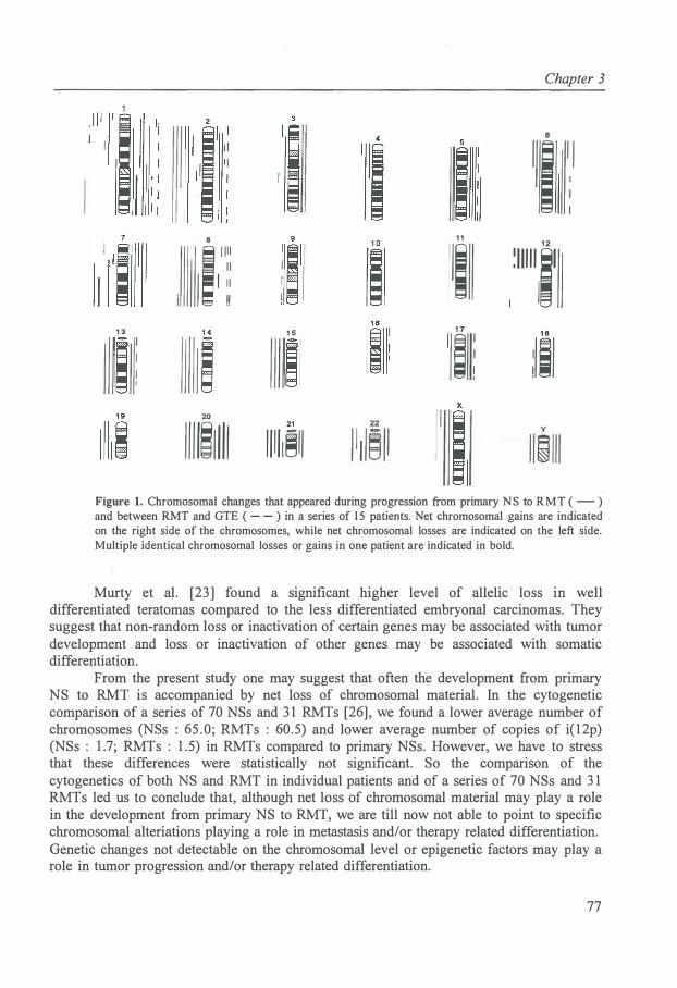
Chapter 3
2
6
1 ; l j
i i : 8 1 1 1 1 1
I I
I I 1 1
!!
1 1
1; 1 2
� 1 1 1 1 1 i' I ll
1 3 1 4 1 5 1 7 l � i :Iii
20
1 1 1 1!1 1 1 1 21
1 1 1 1 ,�1 1
Figure 1. Chromosomal changes that appeared during progression from primary NS to RMT ( - ) and between RMT and GTE ( - - ) in a series of 1 5 patients. Net chromosomal gains are indicated on the right side of the chromosomes, while net chromosomal losses are indicated on the left side. Multiple identical chromosomal losses or gains in one patient are indicated in bold.
Murty et al. [23] found a significant higher level of allelic loss in well differentiated teratomas compared to the less differentiated embryonal carcinomas. They suggest that non-random loss or inactivation of certain genes may be associated with tumor development and loss or inactivation of other genes may be associated with somatic differentiation.
From the present study one may suggest that often the development from primary NS to RMT is accompanied by net loss of chromosomal material. In the cytogenetic comparison of a series of 70 NSs and 31 RMTs [26], we found a lower average number of chromosomes (NSs : 65.0; RMTs : 60.5) and lower average number of copies of i(12p) (NSs : 1.7; RMTs : 1.5) in RMTs compared to primary NSs. However, we have to stress that these differences were statistically not significant. So the comparison of the cytogenetics of both NS and RMT in individual patients and of a series of 70 NSs and 31 RMTs led us to conclude that, although net loss of chromosomal material may play a role in the development from primary NS to RMT, we are till now not able to point to specific chromosomal alteriations playing a role in metastasis and/or therapy related differentiation. Genetic changes not detectable on the chromosomal level or epigenetic factors may play a role in tumor progression and/or therapy related differentiation.
77

Residual mature teratoma
REFERENCES
I . Mostofi FK, Sesterhenn IA, Davis CJ ( 1987): Immunopathology of germ cell tumors of the testis. Semin Diagn Pathol 4 :320-34 1 .
2. Damjanov I ( 1 99 1 ) : Pathobiology of human germ cell neoplasia. Recent Results Cancer Res 123 : 1 - 1 9.
3 . Ulbright TM ( 1 993): Germ cell neoplasms of the testis. Am J Surg Pathol 1 7 : 1 075- 1 09 1 . 4. Einhorn LH ( 1 990): Treatment of testicular cancer: an new and improved model. J Clin Oncol
8 : 1 777- 1 78 1 . 5. Oosterhuis JW ( 1983): The metastasis of human teratomas. In: The Human Teratomas, I Damjanov,
B Knowles, D Solter, eds. The Humana Press, Clifton New Jersey, pp. 137- 1 7 1 . 6. Oosterhuis JW, Suurmeyer AJH, Sleijfer OT, Schraffordt Koops H, Oldhoff J, Fleuren G ( 1 983):
Effects of multiple-drug chemotherapy (Cis-diammine-dichloroplatinum, bleomycin, and vinblastine) on the maturation of retroperitoneal lymph node metastases of nonseminomatous germ cel l tumors of the testis. No evidence for de novo induction of differentiation. Cancer 51 :408-41 6.
7 . Castedo SMMJ, de Jong B, Oosterhuis JW, Seruca R, Idenburg VJS, Dam A, te Meerman GJ, Schraffordt Koops H, Sleijfer OT ( 1 989): Chromosomal changes in human primary testicular nonseminomatous germ cell tumors. Cancer Res 49:5696-570 1 .
8. van Echten J, Oosterhuis JW, Looijenga LHJ, van de Pol M, Wiersema J, te Meerman GJ, Schraffordt Koops H, Sleijfer OT, de Jong B ( 1 995): No recurrent structural abnormalities apart from i( l2p) in primary germ cell tumors of the adult testis. Genes Chromosom Cancer 14: 1 33- 144.
9. ISCN(l 995): Guuidelines for Cancer Cytogenetics, An International System for Human Cytogenetic Nomenclature. Mitelman F, ed. S. Karger, Basel.
1 0. de Jong B, Oosterhuis JW, Castedo SMMJ, Vos AM, te Meerman GJ ( 1 990): Pathogenesis of adult testicular germ cel l tumors. A cytogenetic model. Cancer Genet Cytogenet 48: 143- 1 67.
1 1 . Oosterhuis JW, Castedo SMMJ, de Jong B , Comelisse CJ, Dam A, Sleijfer OT, Schraffordt Koops H ( 1 989): Ploidy of primary germ cell tumors of the testis. Pathogenetic and clinical relevance. Lab Invest 60: 14-20.
12. Fossa SD, Nesland JM, Pettersen EO, Amellem 0, Wrehre H, Heimdal K ( 199 1 ): DNA ploidy in primary testicular cancer. Br J Cancer 64:948-952.
1 3 . Fossa SD, Nesland JM, Wrehre H , Amellem 0, Pettersen EO ( 1 99 1 ): DNA ploidy in the primary tumor from patients with nonseminomatous testicular germ cell tumors clinical stage I. Cancer 67: 1 874- 1 877.
14 . El-Naggar AK, Ro JY, McLemore D, Ayala AG, Batsakis JG ( 1 992): DNA ploidy in testicular germ cell neoplasms. Histogenetic and clinical implications. Am J Surg Pathol 1 6:6 1 1 -6 1 8 .
15 . Oosterhuis JW, de Jong B, Comelisse CJ, Molenaar IM, Meiring A, Idenburg V, Schraffordt Koops H, Sleijfer OT ( 1 986): Karyotyping and DNA flow cytometry of mature residual teratoma after intensive chemotherapy of disseminated nonseminomatous germ cel l tumor of the testis: A report of two cases. Cancer Genet Cytogenet 22: 149- 1 57.
1 6. Castedo SMMJ, de Jong B, Oosterhuis JW, Idenburg VJS, Seruca R, Buist J, te Meerman GJ, Schraffordt Koops H, Sleijfer OT ( 1989): Chromosomal changes in mature residual teratomas following polychemotherapy. Cancer Res 49:672-676.
1 7 . Nowell PC ( 1986): Mechanisms of tumor progression. Cancer Res 46:2203-2207. 1 8. Fidler IJ, Hart IR ( 1 982): Biological diversity in metastatic neoplasms: Origins and implications.
Science 2 1 7:998- 1 003 . 1 9 . Kerbel RS, Waghome C, Korczak B, Lagarde A, Breitman ML (1 988): Clonal dominance of
primary tumours by metastatic cells: Genetic analysis and biological implications. Cancer Surv 7:597-629.
20. de Graaff WE, Oosterhuis JW, de Jong B, van Echten J, Wiersema-Buist J, Schraffordt Koops H, Sleijfer OT (1 992): Cytogenetic analysis of the mature teratoma and the choriocarcinoma component of a testicular mixed nonseminomatous germ cell tumor. Cancer Genet Cytogenet 6 1 :67-73 .
2 1 . de Graaff WE, de Jong B, Oosterhuis JW, van Echten J, Wiersema-Buist J, Schraffordt Koops H,
78

Chapter 3
Sleijfer DT ( 199 1 ): Cytogenetic analysis of the mature and immature teratoma components of a metastatic testicular nonseminomatous germ cell tumor. Cancer Genet Cytogenet 57:59-68.
22. Oosterhuis JW, Andrews PW, Knowles BB, Damjanov I (1 984): Effects of Cis-platinum on embryonal carcinoma cell lines in vitro. Int J Cane 34: 1 33-139.
23. Murty VVVS, Bosl GJ, Houldsworth J, Meyers M, Mukherjee AB, Reuter V, Chaganti RSK ( 1 994): Allelic loss and somatic differentiation in human male germ cell tumors. Oncogene 9:2245-225 1 .
24. Sella A, El-Naggar A, Ro JY, Dexeus H, Amato RJ, Lee JS, Finn L, Logothetis CJ ( 1 99 1 ): Evidence of malignant features in histologically mature teratoma. J Urol 146 : 1 025- 1 028.
25. Nativ 0, Shajrawi I, Leibovitch I, Moskovitz B (1 994): The malignant potential of postchemotherapy residual mature teratoma for disseminated nonseminomatous testicular tumors. Eur Ural 26:2 16-2 1 8 .
26. van Echten J, van der Vloedt WS, van de Pol M, Dam A, te Meerman GJ, Schraffordt Koops H, Sleijfer OT, Oosterhuis JW, de Jong B ( 1 996): Comparison of the chromosomal pattern of primary testicular nonseminomas and residual mature teratomas after chemotherapy. (submitted)
27. Logothetis CJ, Samuels ML, Trindade A, Johnson DE (1 982): The growing teratoma syndrome. Cancer 50: 1 629- 1635.
28. Little JS, Foster RS, Ulbright TM, Donohue JP (1 994): Unusual neoplasms detected in testis cancer patients undergoing post-chemotherapy retroperitoneal lymphadenectomy. J Urol 1 52: 1 1 44- 1 149.
29. Ulbright TM, Loehrer PJ, Roth LM, Einhorn LH, Williams SD, Clark SA ( 1 984): The development of non-germ cell malignancies within germ cell tumors. Cancer 54: 1 824- 1 833 .
30. Simmonds PD, Mead GM, Whitehouse JMA ( 1995): A complicated case of metastatic teratoma. Growing teratoma syndrome and cerebral metastasis. Ann Oneal 6: 1 8 1 - 1 85.
3 1 . Paste G , Fidler IJ ( 1 980): The pathogenesis of cancer metastasis. Nature 283 : 1 39- 146.
79

CHAPTER 4
DEFINITION OF A NEW ENTITY OF MALIGNANT EXTRAGONADAL GERM CELL TUMORS
Jannie van Echten 1 , Bauke de Jong 1 , Richard J. Sinke3 , Daniel Olde Weghuis3, Dirk Th. Sleijfer, J. Wolter
Oosterhuis4
Departments of 1Medical Genetics and 2Medical Oncology of the University of Groningen; 3Department of Human Genetics of the University ofNijmegen; and 4Department of Experimental Pathology of the Dr. Daniel den Hoed Cancer Center, Rotterdam, The Netherlands
Abstract
Two malignant extragonadal germ cell tumors are reported, histologically classified as immature teratomas, having pseudo-diploid karyotypes with complex structural rearrangements but lacking isochromosome 12p or other rearrangements involving 12p. The absence of 12p material in structural rearrangements was confirmed by chromosome painting. In the two tumors the following common chromosomal breakpoints were found: 6p21, 6p22, 6q23 and llq13. Exactly the same chromosomal regions 6p22::6q23 and 6p21::llq13 were involved in fusions. The two tumors belong to a new entity of extragonadal immature teratomas of adults which may be located in the retroperitoneum and posterior mediastinum and are prone to blood borne metastasis. Genes Chromosom Cancer 12 :8- 1 5 ( 1 995). © 1 995 Wiley-Liss, Inc.
Introduction
Human germ cell tumors (GCTs) are a heterogeneous group of neoplasms located in the testis, the ovary and in extragonadal sites. Their pathogenesis, histological composition, cytogenetics, ploidy and degree of malignancy differ, depending on the anatomical site of the tumor and the patient's sex and age [ 1 ].
Extragonadal GCTs by themselves are a heterogeneous group of rare neoplasms. They occur in the midline of the body (pineal region, hypothalamic region, anterior and posterior mediastinum, retroperitoneum and sacral area), but also away from the midline (e.g., orbit, neck, stomach, placenta) [1].
In adult males one has to exclude the possibility of a metastasis of a testicular GCT before assuming that the tumor is an extragonadal GCT, particularly in the case of retroperitoneal tumors [2].
We report two pseudo-diploid extragonadal GCTs with common abnormalities of chromosomes 6 and 11. The two tumors seem to belong to a separate entity which is not only defined cytogenetically, but also by its pathological characteristics and clinical behaviour. Our cytogenetic data and published reports allow a cytogenetic classification of malignant extragonadal GCTs. It is possible that the cytogenetically different tumor types originate from
8 1

Definition of a new entity of malignant extragonadal germ cell tumors
different cell types and by different pathogenesis .
Case report
Case 1
A 65-year-old man presented with a soft tissue mass in his left thigh, which he had first noticed 6 weeks before. A biopsy revealed tumor tissue with the histological appearance of immature teratoma composed of mesenchymal, epithelial, and neural embryonal tissues. No other germ cell tumor components, such as embryonal carcinoma, yolk sac tumor, choriocarcinoma, or seminoma could be identified histologically. Clinical staging revealed no other tumor manifestations; in particular, both testes were normal and no retroperitoneal or mediastinal tumors were identified. Serum alpha-feta-protein (AFP) and beta-human chorionic gonadotrophin (HCG) levels were normal . On the basis of the original histological classification of the tumor as a sarcoma an upper thigh amputation was performed . The amputation specimen showed an encapsulated tumor measuring 15 x 8 x 8 cm. The tumor was attached to the periosteum, but did not invade the bone . Its histology was similar to that of the biopsy . Three months later a retroperitoneal tumor became manifest and was resected . The specimen measured 6 x 4.5 x 4.5 cm and was composed of the same components as before. After another year an enlarged inguinal lymph node and retroperitoneal masses were noticed, and a fine needle aspiration confirmed the recurrence of immature teratoma . The serum level of AFP was now slightly raised, HCG was in the normal range. Chemotherapy using carboplatin, etoposide and bleomycin caused regression of the tumor and normalization of the serum AFP level. Further surgery was deemed unfeasible. The patient died 3 months later, with extensive retroperitoneal tumor masses .
Case 2
The patient was a 39-year-old white male with a disseminated extragonadal GCT histologically classified as immature teratoma [3] . The localization of the primary tumor in the posterior mediastinum was established beyond doubt at autopsy . Twelve months elapsed between the patient 's presentation with a subcutaneous metastasis of the trunk and his death due to local and metastatic tumor growth . The first chemotherapy, consisting of a combination of cisplatinum, vinblastine, and bleomycin (PVB), resulted in a partial remission that lasted 5 months . Residual metastatic tumor in the lungs was surgically removed. A relapse of the disease was treated with salvage chemotherapy using VP 16-2 13 and cyclophosphamide, followed by autologous bone marrow transplantation, which again resulted in a partial remission of 5 months .
Following PVB, the residual tumor tissue consisted exclusively of mature teratoma. Apart from that, all the tumor tissue, either surgically removed or found at autopsy, was histologically predominantly composed of immature teratoma with small foci of mature somatic tissue. Serum AFP levels were slightly elevated in the last 2 months. Serum levels for HCG, lactate dehydrogenase (LDH), and CEA were consistently in the normal range.
82
Chapter 4
Minute amounts of AFP were immunohistochemically demonstrated in the immature teratoma component of the tumor.
Materials and methods
Cytogenetic analysis
In case 1 , cytogenetic analysis was carried out on two tumors (from the left thigh and the retroperitoneum) after short term culture (5-7 days) as described previously [4].
The karyotype of the tumor of case 2 has been published before [3]. By using another banding method and elongated chromosomes , a better morphology of the chromosomes was achieved, which allowed us to refine the earlier karyotype description.
In situ hybridization
Bicolor double FISH experiments were carried out basically as described before [5,6] . In short , DNAs from the chromosome 6, 7, 1 1 , and 12 specific plasmid libraries (i .e. , pBS-6 , -7, - 1 1 , and - 12 : [7]) were labeled with biotin- 14-dATP (Life Technologies, Breda) or digoxigenin- 1 1-dUTP (Boehringer Mannheim, Germany) following standard nick-translation . The labeled DNAs of pBS-6 and - 1 1 , and pBS-7 and - 12, respectively, were coprecipitated with 15 x Cot- 1 DNA, preannealed for 15 minutes at 37 °C and, subsequently , used as a hybridization mix on metaphases of tumor cells. Chromosomes and chromosomal segments hybridizing to biotin- or digoxigenin-labeled probes were visualizied using a layer of fluorescein isothiocyanate (FITC)-conjugated avidin (Vector Laboratories, Burlingame, CA) and alternating layers of rabbit anti-FITC and FITC conjugated mouse anti-rabbit (Jackson Immuno-Research, West Grove, PA) or Rhodamin-conjugated sheep anti-digoxigenin antibodies (Boehringer Mannheim) and Texas Red-conjugated donkey anti-sheep antibodies (Jackson Immuno-Research) , respectively . Counterstaining of the chromosomes was performed with the blue DNA-specific dye DAPI (Sigma, St . Louis, MO) . Chromosomes were studied under a Zeiss Axiophot epifluorescence microscope, equipped with appropriate filters for the visualization of FITC, Rhodamin/Texas Red and DAPI fluorescence, as well as the simultaneous visualization of FITC and Texas Red fluorescence (Omega double filter ; Omega Optical, Inc . , Battleboro, VT) . Acquisition of separate digital images (for Texas Red , FITC, and DAPI) was established using a Photometrics high-performance CH250/ A cooled CCDcamera (Photometrics, Tucson, AZ) interfaced onto a Macintosh Quadra 900 computer. The images were superimposed and displayed on red-green-blue pseudocolors on the computerscreen by means of the BOS-Image TM FISH software package (Biological Detection Systems, Inc., Rockville , MD). Photographs were made from the computer screen on Kodak EPP 100 Plus colorslide film using a Polaroid Quickprint.
83

Definition of a new entity of malignant extragonadal germ cell tumors
Results
Karyotyping
In both tumors of case 1 (left thigh and retroperitoneum) a total number of 10 cells was analyzed. All analyzed metaphases from case 1 showed the same karyotype with the description :
46,XY,der(6)t(6; 1 1 )(p2 1 ;q 13)t(6;6)( q23;p22),der(7)t(6;7)(p2 1 ;p22)t(6;6)(p22;q23), del( l l)(q 13) .
Because of improved banding and high resolution techniques we could refine the karyotype description of the earlier published extragonadal GCT with abnormal chromosomes 6 and 1 1 (case 2) [3]. All metaphases analyzed showed the same karyotype with the revised description :
46,XY,der(6)t(6;6)(q2 1 ;q 16),der(6)t(6;6)(p22;q23)t(6; 1 l)(q 1 6;q 13), der( l l)t(6; 1 l)(p2 1;q 13).
The two tumors have the following common chromosomal breakpoints : 6p2 1, 6p22, 6q23, and 1 1 q 13. The same chromosome regions, 6p22 : : 6q23 and 6p2 l : : 1 1 q 13 are involved in fusions in both tumors (Fig. 1). Peripheral blood from both patients showed a normal 46,XY karyotype.
FISH analysis
Bicolor double FISH analysis was carried out on both tumors, using chromosome 7 and 12 or 6 and 1 1 specific paints. In case 2, the chromosomes 7 and 12 appeared to be normal, as expected . Positive signals for the chromosomes 6 and 1 1 were present according to the der(6)t(6;6), the der(6)t(6;6)t(6; 1 1), the der( l l)t(6; 1 1), and one normal copy of chromosome 1 1 . In case 1, two normal copies of chromosome 12 were present. Positive signals for chromosome 6 and 1 1 were present according to one normal copy of chromosome 6, the der(6), the der(7), the del( l l) of the t(6;7; 1 1) and one normal copy of chromosome 1 1. In Figure 2, the normal chromosomes 7 and 12 of case 2 (Fig . 2A) and the t(6;7; 1 l) of case 1 (Fig. 2B) are shown .
Discussion
Cytogenetics
The published, complete karyotypes of seven malignant extragonadal germ cell tumors [8- 14] in addition to the present case and the revised karyotype of our previously reported case [3], are listed in Table 1. The karyotypes of four more mediastinal GCT' s have been reported : two by Samaniego et al . [ 15], and two by Rodriquez et al. [ 16]. Because of the limited available
84

--,
u
A
B
2 ldlr(7)
-�,-ij-13 ..
- -.-,-.--•-,..-21 22
13 14
21 22
15
15
16 17
-i: 6- I ( : dcr s
16 17
l
6 ,-- =
8 -
Chapter 4
ldW(l''
• -:;-10 11 12
�-18 19 20
�·-- ,_n
dor(7) det(11)
.' L
.. -.---10 12
.. . 18 19 20
, ;f �w = 11 • := Ji; '"' .... 8 ' "' [ _ .....
· !
det' S) der(11)
Figure 1 . Karyotype and partial karyotype and schematic representation of the structural ly abnormal chromosomes of case I (A) and case 2 (B). Both tumors show common breakpoints: 6p2 1 , 6p22, 6q23 and l l q13, with the same chromosomal regions, 6p22: :6q23 and 6p2 1 : : l lq 13 , involved in fusions.
85

Definition of a new entity of malignant extragonadal germ cell tumors
Figure 2. FISH analysis of tumors of case 2 (A) and case 1 (B) using chromosome 7 (yellow-green) and 12 (red) or 6 (yellow-green) and 1 1 (red)specific paints. The differently labeled normal and translocation-derived chromosomes are marked. Chromosomes 7 and 1 2 appear to be normal in A ( case 2), as expected (see text), whereas a clear t(6 ;7; 1 l ) is visualized in B (case 1 ).
clinical data, these cases were not included in the table. However, they are considered in the discussion.
Cases 1 and 2 are pseudo-diploid and show complex chromosomal translocations, with common breakpoints in chromosomes 6 and 1 1 . They lack isochromosome l 2p, or other rearrangements involving 12p, as convincingly demonstrated by FISH.
Cases 3 through 9 lack the specific translocations of the cases 1 and 2, and may have an isochrome 12p, the specific chromosomal abnormality frequently found in gonadal GCTs [ 1 7, 1 8] . It is present in cases 3 , 4, and 7, and probably also in case 5 [8]. The tumors lacking i( l2p) may have amplification of 12p in other chromosomal rearrangements similar to the pattern found in "i( l2p)-negative" germ cell tumors of the adult testis. These tumors have extra copies of l 2p hidden in various marker chromosomes, as demonstrated by painting of metaphase cells [5]. The tumors of this second category may be (near)diploid or polyploid and may have non-random numerical abnormalities (e.g., +7, +21 , and - 1 3) similar to those found in testicular GCTs [ 1 8] . In testicular GCTs, the breakpoints of cases 1 and 2 are rare, and the fusions 6p22: :6q23 and 6p21 : : l lq 13 have not been demonstrated in our material of 95 cases (unpublished data).
86
Chapter 4
Cytogenetic classification
The available karyotypic data on malignant extragonodal GCTs reviewed above allow the following cytogenetic classification:
Type 1. Diploid tumors with specific structural chromosomal abnormalities of chromosomes 6 and 11 (case 1 and 2). Type 2. Near-diploid or polyploid tumors which may have i(12p), and multiple non-random numerical abnormalities, similar to the chromosomal abnormalities found in testicular germ cell tumors ( cases 3 through 9).
The four mediastinal GCTs reported by Samaniego et al. [15] and Rodriquez et al. [16] are all type 2 tumors. All four have one or more copies of i(12p), three are polyploid.
Clinical pathological correlation
The two tumors of type 1 occurred in male patients who were old as compared to the four patients with type 2 extragonadal GCTs of the anterior mediastinum. Both presented with blood borne metastasis to the soft tissues (lower extremity and trunk). The primary tumors were localized in the retroperitoneum (case 1) and in the posterior mediastinum (case 2). Both patients developed slight elevation of AFP after multiple recurrences. Other serum tumor markers were negative. Notwithstanding extensive locoregional and systemic treatment, both patients died of their tumors. Primary tumors and untreated metastases of both patients were composed of immature teratoma. Minute amounts of AFP could be demonstrated in the tumor tissue of the second patient shortly before death [3].
Four of the seven listed tumors of type 2 occured in young adults, and were localized in the anterior mediastinum. The tumors in the three youngest patients were localized in the midline of the �rain. The patients presented with complaints from the primary tumors, without manifest metastatic disease. The serum tumor markers, most often AFP and HCG, were elevated at presentation. Six tumors were histologically classified as nonseminomatous GCTs with the same mixed histology which is encountered in testicular GCTs of adults, including the presence of a seminoma/germinoma component in some tumors (cases 3 and 8). Case 9 was histologically classified as a germinoma. One of the patients with GCTs of the midline of the brain is alive without evidence of disease after a combination of radiotherapy and chemotherapy (follow-up 21 months). The four patients with mediastinal tumors died, notwithstanding extensive surgery and chemotherapy, two of them with secondary leukemias, an established risk of extragonadal GCTs of the anterior mediastinum [9].
Histogenesis
The remarkable resemblance in terms of histological composition and chromosomal constitution, between testicular GCTs of adults (and certain ovarian GCTs), and the extragonadal GCTs of type 2, suggests that they have a similar histogenesis, and similar cells
87

Definition of a new entity of malignant extragonadal germ cell tumors
of origin. It is now generally accepted that the common precursor of all GCTs of the adult testis
with the exception of spermatocytic seminoma, is carcinoma in situ [ 19] , composed of tumor cells which are the neoplastic counterparts of primordial germ cells. Type 2 extragonadal GCTs are probably a lso derived from primordial germ cells, which have migrated, by an as yet unclarified mechanism, to the anterior mediastinum/thymus, and the midline of the brain. One could argue that the presence of i( 12p), in type 2 extragonadal GCTs, is the best evidence yet that primordial germ cells do migrate to the midline of the brain and the thymus. The fact that they survive in these anatomical localizations through childhood and adult life suggests that they are more than embryonal vestiges , but have an as yet obscure functional meaning [20].
It is possible that type 1 tumors are also derived from germ cells. Immature teratomas of the ovary which have a very similar histology and may behave as frankly malignant tumors , are derived from germ cells. Ovarian immature teratomas , however , are karyotypically different from type 1 extragonadal GCTs [2 1]. Alternatively type 1 tumors are derived from pluripotent embryonal cells, analogous to what is considered for sacral teratomas and teratomas of the head and neck of infants [22]. Too few of these tumors have been karyotyped to allow a comparison with type 1 extragonadal GCTs [ 1].
Animal models of teratoma/teratocarcinoma and yolk sac tumor in the mouse and the rat could support either hypothesis on the cellular origin of type 1 tumors. Testicular teratomas in 129 mice [23] and ovarian teratomas of the LT mouse strain [24] are derived from germ cells. However, tumors with almost identical histology produced in mice by embryo transplantation [25] and in rats by fetectomy [26] , are derived from pluripotent embryonal cells.
Diagnosis of the new entity
Suspicion of a case of type 1 extragonadal GCT should be raised by the clinical presentation, in particular the relatively high age of the patient, and by the diploid character of the lesion. The only way to prove a case as yet is by karyotyping. Lack of involvement of chromosome 12 in interphase cytogenetics (in situ hybridization for the centromere of #12 [27]) argues against a type 2 and for a type 1 tumor. In the future it may be possible to demonstrate the specific chromosomal fusion regions with molecular techniques [28].
Acknowledgements
Supported by the Dutch Cancer Society Grant GUKC 90- 17. The authors thank J. W. Gray for his generous gift of the chromosome-specific plasmid libraries pBS-6, -7, - 1 1 , and - 12.
88

Chapter 4
TABLE 1 . Cytogenetic Classification of Extragonadal GCTs
Age/ Anatomical Serum Case sex site markers Histology Karyotype Follow-up Reference Type I I 65/M Retro- AFPt TI 46,XY,der(6)t(6;1 l )(p2 1 ;q 1 3)t(6;6) Died after 2 1 This report
peritoneal HCG (q23;p22),der(7)t(6;7)(p2 1 ;p22) months of normal t( 6;6)(p22;q23 ),de!( 1 1 )( q 1 3) metastatic
disease 2 39/M Posterior AFPt TI 46,XY,der(6)t(6;6)(q21 ;q 16),der(6) Died after 12 Oosterhuis
mediastinal HCG t(6;6)(p22;q23)t(6; 1 l )(q1 6;q 1 3), months of et al. [3] normal der(l l )t(6; 1 1 )(p21 ;q 13 ) local and
metastatic disease
Type 2 3 1 8/M Anterior AFPt SE EC TI 47,XY,-13;f2 1 ,+i(l2p) Died after 1 3 Dal Cin et
rnediastinal HCGt TD YS months of al. [8) recurrence
4 19/M Anterior AFPt TI TD YS 48-49,XY,+I ,+6,+i(l 2p) Died after Chaganti et mediastinal HCG 49,XY,+1 ,+2 1 ,+i( l2p) 12 months of al. [9]
normal 49,XY,+21 ,+del(l )( q32),+i( l 2p) secondary ANLL
5 1 8/M Anterior AFPt EC 50,XXYc,+7,+2 1 ,+mar(i(2 1q)?) Died after 10 Mann e t al. mediastinal HCGt months of [ 10]
local and metastatic disease
6 26/M Anterior AFPt TI TD 76,XY,+X,+Y,+l ,+1 ,+2,+3,+3,+5, Died after Oosterhuis mediastinal HCGt +5,+6,+6,+7,+7,+8,+8,+ I O,+ 1 1 , 2 years of et al. [I I ]
+ 12,+ 1 3,+15,+16,+ 1 7,+17,+19,+ 1 9, secondary +20,+2 1 ,+2 1 ,+22,+del(9)(q2 1q22) ANLL
7 16/M Midline AFPt EC CH 64,XY,+ X,+3,+7,+7,+8,+8,+ 12,+ 14, Alive after De Bruin brain HCGt +20,+2 1 ,+2 1 ,-22,del( I ),+der( I ), 22 months et al . [ 12] pineal +del(2), +i( 1 2p ), +i( l 2p ),+der( 17), follow up
+del(20),+der(22),+mar 8 14/M Midline AFPt SE EC TI 78,X,-Y,+X,+X,+2,+2,+3,+3,+4,+5, No follow up Shen et al.
brain HCGt TD +6,+6,+7,+8,+9,+10,+l2,+12,+14, available [ 13] pineal + 15,+ 16,+ 1 7,+ l 9,+20,+20,+21 ,+21 ,
+2 1 ,+21 ,+22,+t( I ;?)(qi l ;?),+t( I ;?), +der( I )t(I ;?)(p 1 1 ;?), +der( I )t( I ;?) (q i 1 ;?),+der(l l )t(8; 1 1 )(q 1 3 ;q23)
9 1 1 /M Midline Not SE 8 1 ,XY,+ X,+Y,+ 1 ,+2,+2,+add(3) No follow up Albrecht et brain reported (p2 1 ),+5,+5,+6,+6, +7,+8,+8,+8, available al. [ 14] pineal +add(9)(p 1 3 ), + 10, +add( 12)(p 1 1 ),
+ 14,+ 14,+ 1 5,+ 1 5,+ 16,+ 1 7,+i(l 7) (q l 0),+18,+ 19,+2 1 ,+2 1 ,+22,+HSR (?),+Smar
AFP=alpha fetoprotein; HCG=human chorionic gonadotropin; CH=choriocarcinoma; EC=embryonal carcinoma; TD=teratoma differentiated; TI=teratoma immature; SE=seminoma; YS=yolk sac tumor; ANLL=acute nonlymphocytic leukemia
89

Definition of a new entity of malignant extragonadal germ cell tumors
REFERENCES
1 . Oosterhuis JW, Castedo SMMJ, de Jong B ( 1990): Cytogenetics, ploidy and differentiation of human testicular, ovarian and extragonadal germ cel l tumours. Cancer Surv 9:32 1 -332.
2 . Daugaard G, von der Maase H, Olsen J, R.0rth M, Skakkebrek NE ( 1987): Carcinoma-in-situ testis in patients with assumed extragonadal germ-cel l tumours. Lancet 528-530.
3 . Oosterhuis JW, de Jong B , van Dalen I , van der Meer I , Visser M , d e Leij L , Mesander G , Collard JG, Schraffordt Koops H, Sleijfer DT (1 985): Identical chromosome translocations involving the region of the c-myb oncogene in four metastases of a mediastinal teratocarcinoma. Cancer Genet Cytogenet 1 5 :99-1 07 .
4 . Castedo SMMJ, de Jong B, Oosterhuis JW, Seruca R, Idenburg VJS, Dam A, te Meerman GJ, Schraffordt Koops H, Sleijfer DT ( 1 989): Chromosomal changes in human primary testicular nonseminomatous germ cell tumors. Cancer Res 49:5696-570 1 .
5 . Suijkerbuijk RF, Looijenga L , de Jong B , Oosterhuis JW, Cassiman JJ, Geurts van Kessel A ( 1 992): Verification of isochromosome l 2p and identification of other chromosome 12 aberrations in gonadal and extragonadal human germ cell tumors by bicolor double fluorescence in situ hybridization. Cancer Genet Cytogenet 63 :8- 16.
6 . O lde Weghuis D, Stoepker MEJ, de Leeuw B, van den Berg E, Suijkerbuijk RF, Molenaar WM, de Jong B, Geurts van Kessel A ( 1994): A synovial sarcoma with a complex t(X; 1 8;5;4) and a break in the ornithine aminotransferase (OA T)LI cluster on Xp 1 1 .2. Genes Chromosom Cancer 9:288-29 1 .
7 . Collins C, Kuo WL, Segraves R, Fuscoe J, Pinke! D, Gray JW ( 199 1 ): Construction and characterization of plasmid libraries enriched in sequences from single human chromosomes. Genomics 1 1 :997- 1 006.
8. Dal Cin P, Drochmans A, Moerman P, van den Berghe H (1 989): Isochromosome 12p in mediastinal germ cell tumor. Cancer Genet Cytogenet 42:243-25 1 .
9. Chaganti RSK, Ladanyi M, Samaniego F, Offit K, Reuter VE, Jhanwar SC, Bosl GJ ( 1 989): Leukemic differentiation of a mediastinal germ cell tumor. Genes Chromosom Cancer 1 : 83-87.
1 0. Mann BD, Sparkes RS, Kern DH, Morton DL ( 1 983): Chromosomal abnormalities of a mediastinal embryonal cell carcinoma in a patient with 47,XXY klinefelter syndrome: Evidence for the premeiotic origin of a germ cel l tumor. Cancer Genet Cytogenet 8 : 1 9 1 - 1 96.
1 1 . Oosterhuis JW, van den Berg E, de Jong B, Timens W, Castedo SMMJ, Rammeloo RHU, Sleijfer DT ( 1 99 1 ): Mediastinal germ cell tumor with secondary nongerm cell malignancy, and extensive hematopoietic activity. Pathology, DNA-ploidy, and karyotyping. Cancer Genet Cytogenet 54: 1 83- 1 95 .
1 2 . de Bruin TWA, Slater RM, Defferrari R, Geurts van Kessel A, Suijkerbuijk RF, Jansen G, de Jong B, Oosterhuis JW ( 1994): Isochromosome 12p-positive pineal germ cell tumor. Cancer Res 54: 1 542- 1 544.
1 3 . Shen V, Chaparro M, Byung HC, Young R, Bernstein R ( 1 990): Absence of isochromosome 12p in a pineal region malignant germ cell tumor. Cancer Genet Cytogenet 50: 1 53- 160.
1 4. Albrecht S, Armstrong DL, Mahoney DH, Cheek WR, Cooley LD ( 1 993) : Cytogenetic demonstration of gene amplification in a primary intracranial germ cell tumor. Genes Chromosom Cancer 6:6 1 -63 .
1 5 . Samaniego F, Rodriguez E, Houldsworth J, Murty VVVS, Ladanyi M, Lele KP, Chen Q, Dmitrovsky E, Geller NL, Reuter V, Jhanwar SC, Bos! GJ, Chaganti RSK ( 1990): Cytogenetic and molecular analysis of human male germ cell tumors: chromosome 12 abnormalities and gene amplification. Genes Chromosom Cancer 1 :289-300.
1 6. Rodriguez E, Mathew S, Reuter V, Ilson DH, Bos! GJ, Chaganti RSK ( 1 992): Cytogenetic analysis of 1 24 prospectively ascertained male germ cell tumors. Cancer Res 52:2285-229 1 .
1 7 . Atkin NB, Baker MC ( 1 982): Specific chromosome change, i( l 2p ), in testicular tumours? Lancet 2 : 1 349 1 8. de Jong B, Oosterhuis JW, Castedo SMMJ, Vos AM, te Meerman GJ ( 1 990): Pathogenesis of
adult testicular germ cell tumors. A cytogenetic model. Cancer Genet Cytogenet 48: 1 43- 1 67. 1 9. Skakkebrek NE, Berthelsen JG, Giwercman A, Millier J ( 1 987): Carcinoma-in-situ of the testis: Possible
origin from gonocytes, and precursor of all types of germ cell tumors except spermatocytoma. Int J Androl 1 0 : 1 9-28.
20. Friedman NB ( 1987): The function of the primordial germ cel l in extragonadal tissues. Int J Androl 1 0:43-49.
2 1 . Surti U, Hoffner L, Chakravarti A, Ferrell RE ( 1 990): Genetics and biology of human ovarian teratomas
90
Chapter 4
I. Cytogenetic analysis and mechanism of origin. Am J Hum Genet 47:635-643 . 22. Gonzales-Crussi F ( 1 982): Extragonadal Teratomas. In: Atlas of Tumor Pathology, 2nd series, fascicle 1 8.
Armed Forces Institute of Pathology, Washington DC. 23. Stevens LC (1 973) : A new inbred subline of mice ( 129/terSv) with a high incidence of spontaneous
congenital testicular teratomas. J Natl Cancer Inst 50:235-242. 24. Stevens LC, Varnum DS (1 974): The development of teratomas from parthenogenetically activated ovarian
mouse eggs. Dev Biol 37:369-380. 25. Damjanov I, Salter D ( 1 974): Experimental teratoma. Curr Top Pathol 59:69- 1 30. 26. Sobis H, Van Hove L, Vandeputte M (1 982): Trophoblastic and mesenchymal structures in rat yolk sac
carcinoma. Int J Cancer 29: 1 8 1 - 1 86. 27. Looijenga LHJ, Smit VTHBM, Wessels JW, Mollevanger P, Oosterhuis JW, Comelisse CJ, Devilee P
(1 990): Localization and polymorphism of a chromosome 12-specific alpha satellite DNA sequence. Cytogenet Cell Genet 53 :2 1 6-2 1 8.
28. Sinke RJ, Olde Weghuis D, Suijkerbuijk RF, Tanigami A, Nakamura Y, Larsson C, Weber G, de Jong B, Oosterhuis JW, Molenaar WM, Geurts van Kessel A ( 1 994): Molecular characterization of a recurring complex chromosomal translocation in two human extragonadal germ cell tumors. Cancer Genet Cytogenet 73 : 1 1 - 1 6.
9 1


CHAPTER 5
SUMMARY, GENERAL DISCUSSION AND PERSPECTIVES
5.1 TESTICULAR AND EXTRAGONADAL GERM CELL TUMORS OF ADULTS AND ADOLESCENTS
Germ cell tumors of adult and adolescent males are rare neoplasms, located in the gonads (testis) and in extragonadal sites (e.g. retroperitoneum, mediastinum, brain). Primary testicular germ cell tumors (TGCTs) can be divided clinically and morphologically into two distinct entities [ l ]. First are seminomas (SEs), reflecting differentiation along the germ cell lineage. The second entity is nonseminomatous TGCTs (NSs), of which pluripotent embryonal carcinoma (EC) cells are the stem cells. EC may differentiate into extraembryonic cell types resulting in choriocarcinoma (CH) and yolk sac tumor (YS) and/or along the lines of embryonic cells and tissues, resulting in immature teratoma (IT) and mature teratoma (MT) [2]. Most NSs have a mixed histology with the different histological elements geographically separated, or truly mixed. A minority of TGCTs contains both a SE and NS component, the combined tumors (CTs).
Most TGCTs are thought to be derived from dysplastic germ cell precursors, which progress to carcinoma in situ (CIS) [3]. It is suggested that the initiation of TGCTs starts early in life, probably before birth [4].
Whether and to what degree SEs and NSs are pathogenetically related is still controversial and a matter of debate. In essence, two models exist about the pathogenetic relationship between SEs and NSs ([5] for review). In the first model, the histogenesis of SEs is assumed to diverge from that of the other TGCTs at an early stage. The neoplastic germ cell either may give rise to SE, reflecting germ cell differentiation or may differentiate to embryonic and/or extraembryonic tissues resulting in NS. The neoplastic pathway of SEs and NSs is different, with no or only limited crossover. The second model suggests that SEs and NSs have a common origin with a single neoplastic pathway, in which SE may be an intermediate stage in development of NS. According to this view, SE may not only be an end stage in differentiation, but SE cells may also progress to a NS phenotype. As a consequence, SEs and NSs may show a strong relationship.
Presently about 85% of all patients with TGCTs are cured, despite the fact that a large amount of patients presents with metastases. When metastases of NSs are treated with chemotherapy, often fully differentiated mature teratoma is left [ 6]. This residual mature teratoma (RMT) may be the result of selection of cells composed of mature teratoma or of cells with an inherent capacity of differentiation to mature teratoma. Another possibility is that the chemotherapy induces differentiation of malignant cells [7]. Surgical resection of RMT is necessary because of the risk of recurrence. RMT may give rise to growing teratoma syndrome [8] and to secondary non germ cell malignancies [9,10].
All histological components which can be found in TGCTs may be present in extragonadal GCTs of the adult and adolescent male as well. It is assumed that not all
93

Summary, general discussion and perspectives
extragonadal GCTs originate from the same stem cell. They may originate from diploid primordial germ cells, which have migrated during embryogenesis from the yolk sac along the midline of the body to sites other than the gonadal blastoma, or from other pluripotent embryonic or extraembryonic stem cells [ 1 1 ].
Knowing that cancer and progression of cancer is caused by genetic changes (e.g. changes at the chromosomal or gene level), we investigated the karyotypes of testicular and extragonadal GCTs of the adult and adolescent male. The chromosomal analysis of these tumors may shed light on oncogenesis, mechanism(s) of tumor progression, therapy related differentiation, and pathogenetic relationship. In the next four paragraphs we summarize and discuss the results and conclusions of the different studies, according to the four questions mentioned in the Introduction of this thesis.
"t>
� c:::i i:::
� Cl) E
94
2 ______ =--------------------------
1 , 5
... 0 � .a t) "iii � ·= Q. t) l1I l1I .. E E ,n 0 0 l1I C: C: ci. ·u ·e ,n ---+ � >, ---+ Q)
"'O ,n
p o l yp lo i - i n i tia l dization
l1I E 0 C:
·e Q) '? C: ---+ g
further
net loss
of DNA
' · · , - � ' - ►.
... l1I 0
E E 0 .a C: "'O ·e Q) Q) C:
'? :c C: E 0 0 C: t)
25 30
l1I E 0 C:
·e Q) ,n
35
approx. age of clinical presen tation
adult age
Figure 1. Tumor progression model of TGCTs of adults proposed by Oosterhuis et al. [ 1 3], supplemented with relevant chromosomal changes observed in the present study.

Chapter 5
5.2 WHICH GENOMIC CHANGES PLAY A ROLE IN THE ONCOGENESIS AND/OR TUMOR PROGRESSION OF TGCTs?
Tumor progression is the result of clonal evolution of a tumor cell population, due to or accompanied by karyotype evolution [ 12] . Our cytogenetic data of TGCTs support the linear progression model in which SE may be an end stage in differentiation as well as an intermediate stage in the development of CIS to NS (see also Chapter 5 .2) . Figure 1 shows a tumor progression model of TGCTs (proposed by Oosterhuis et al. [ 13]) , supplemented with relevant chromosomal changes observed in the present study.
An important and early event in the oncogenesis of TGCTs is polyploidization of a dysplastic germ cell precursor , resulting in CIS. The progression of CIS ➔ SE ➔ NS is accompanied by net loss of chromosomes. The chromosome numbers of CIS and TGCTs are in the triploid range, with significant higher chromosome numbers in SEs than in NSs (average modal number of 73.4 in SEs (n=32, range 58- 1 12) and 65.0 in NSs (n=70, range 50-1 13). The mean total chromosome number of CIS, which we have karyotyped, is 65.6 (n=5, range 55-79) . Because the number of cases of CIS and the number of analyzed metaphases is small, it is rather speculative to derive conclusions regarding the chromosome numbers of CIS (see also Chapter 5.2). The DI of CIS may be similar to its adjacent cancer (SEs and NSs), or CIS may have a higher DI than NSs [ 14- 16] .
i( 12p) is the characteristic chromosomal abnormality of TGCTs, present in about 70% of our material . We found i( l2p) in CIS, the non-invasive precursor lesion of most TGCTs . This suggests that i( l 2p) formation is an important and early event in the oncogenesis of TGCTs, although most likely preceded by polyploidization [ 17]. In NSs, the frequency and the number of copies of i( 12p) is significantly higher than in SEs. This may suggest that i( l2p) is involved in tumor progression. We and others observed aberratio�s of chromosome 12 in i( 12p)-negative TGCTs [18-20]. FISH studies have shown that i( 12p)-negative tumors have amplification of 12p [2 1-23] . Our cytogenetic data extended with our molecular data point to an overrepresentation of the region 12p l 1. 1-p 12 . 1 . The consistent overrepresentation of 12p sequences , by i( l 2p )-formation or other aberrations of chromosome 12, indicates that genes on 12p play an important role in the oncogenesis of TGCTs . However , which gene(s) might be involved is still not known.
The gain and loss of chromosomes during the progression of TGCTs, resulting in net loss of chromosomal material , is nonrandom; specific chromosomes are overrepresented (e.g. 7, 8, 12 , 2 1 , and X), and others are underrepresented (e .g. 1 1 , 13, 18 , and Y) . Although our series of CIS is small , we have indications that loss and gain of chromosomes already occurs in CIS. The autosomes which are overrepresented may harbor genes deregulated by amplification and tumor suppressor genes may be located on the underrepresented chromosomes. Remarkably, some chromosomes (e .g . 2 , 3, 6, 14 , and 16) were neither over- nor underrepresented in either SEs or NSs. This might indicate that over- and/or underrepresentation of these chromosomes is not compatible with TGCTs qevelopment . In SEs , a significantly higher copy number of chromosomes 7, 15, 19, and 22 was found and a significantly lower copy number of chromosome 17, compared with NSs. We observed a significant excess of breakpoints in chromosomes 1 1 and 12 in SEs and in chromosomes 1 and 12 in NSs, and a significant difference in the number of breakpoints in chromosome arm 1 p between SEs and NSs. Loss of the chromosomal
95

Summary, general discussion and perspectives
regions lp34-36 and 2q34-37 was more often observed in NSs than in SEs. In these regions the alkaline phosphatase (AP) isozymes are located. AP isozymes serve as markers of germ cell differentiation, and are also produced in TGCTs, in SEs to a higher extent than in NSs [24]. The observed chromosomal differences between SEs and NSs may play a role in tumor progression and/or in the direction of differentiation of SE and NS.
In conclusion, important steps in the oncogenesis and/or progression of TGCTs are polyploidization, 12p amplification, overrepresentation of some specific chromosomes and underrepresentation or retention of others, resulting in net loss of (parts) of chromosomes. i(12p)-formation and over- and underrepresentation of chromosomes already occur in CIS. This may suggest that karyotype evolution/tumor progression in TGCTs takes place in an early stage of tumor evolution. It would be of importance to obtain more data of CIS adjacent to invasive tumor and of CIS before it has progressed to invasiveness. Comparison of the different stages of CIS with invasive tumor may shed light on the processes of tumor evolution in TGCTs.
5.3 WHAT IS THE PATHOGENETIC RELATIONSHIP BETWEEN CIS, SEs, AND NSs?
Because we observed highly similar chromosomal patterns in SEs and NSs (Chapter 2.1 ), we concluded that SEs and NSs have a common origin with a single neoplastic pathway, in which SE may be an intermediate stage in NS development (the linear progression model). A schematic representation of a linear progression model of TGCTs is shown in Figure 2.
96
PGC
l CIS
I in trau terin e life
pub erty
25
30
35
T reprogramming 7, 1 5, 1 9, 22, 1 p34-36, 2q34-37 + 1 7, 1(1 2p) f,
* : not b y definition cl ln lcally manifest
Figure 2. Schematic representation of a linear progression model of TGCTs

Chapter 5
Carcinogenesis of TGCTs starts early in life, probably in utero. CIS is supposed to be the precursor for all TGCTs of adolescents and adults, except spermatocytic seminoma [3]. In Chapter 2.2 we described the karyotypical similarities between two cases of TGCTs and its adjacent CIS. These results are the first cytogenetic evidence that CIS is clonally related to and is the precursor for invasive TGCTs. In a previous cytogenetic study of TGCTs and adjacent CIS no clear similarities were observed. Probably in these cases we failed to karyotype the CIS that was clonally related to the invasive tumor. CIS of the testis in general is a diffuse process and probably sometimes heterogeneous.
The progression of CIS to NS takes about ten year less than the progression of CIS to SE. When CIS develops to NS, probably through a not clinically manifest SE stage, it requires that CIS cells or SE cells change their germ cell lineage programme of differentiation into the programme of the (pluripotent) embryonal carcinoma (EC) cell [25]. This switch towards a different differentiation lineage is accompanied by chromosomal changes (e.g. loss of chromosome 7, 15, 19, and 22 and chromosomal regions lp34-36 and 2q34-37, and gain of chromosome 17 and copies of i(12p)). When CIS develops to SE, the germ cell lineage of differentiation is kept, and less chromosomal changes take place. Looijenga et al. [26] found evidence that CIS which progresses to NS, is already changing its chromosomal pattern towards that of NS ( e.g. lower copy number of chromosome 15 in CIS adjacent to NS than in CIS adjacent to SE).
Although in the majority of the cases of TGCTs the reprogramming of CIS cells or SE cells takes place while in situ, invasive SE may also undergo reprogramming. This might be the case in CTs, which comprise about 10 to 20% of all TGCTs. One of our cases of CTs and three cases of the literature [23,27,28] show clear cytogenetic evidence for a common origin and strong pathogenetic relationship of the SE and NS component (Chapter 2.3). The CTs with no or weak cytogenetic similarity between the SE and NS component, are probably tumors in which both components have an independent origin, or where the development of both components might have diverged at an early stage in their development. The possibility of a monoclonal and polyclonal origin for the SE and NS component of CTs is also described by Gillis et al. [29]. In CIS adjacent to CT (SE and NS component) they found copy number of chromosome 15 high (SE-pattern), low (NSpattern), or high in CIS adjacent to the SE component, and low in CIS adjacent to the NS component.
When CIS cells or SE cells are reprogrammed to EC, the EC cells may stay nullipotent (NS composed exclusively of EC), or may develop pluripotency, with the formation of one or more of the elements CH, YS, IT, or MT. The factors controlling the direction of differentiation of NS are largely unknown. There is no convincing evidence that changes at the chromosomal level play a role in the direction of differentiation in NS [30,31].
In conclusion, results from cytogenetic and interphase cytogenetic studies and from DNA-flow cytometry, strongly suggest that karyotype evolution already occurs in CIS. fyfost TGCTs are thought to derive from CIS, the neoplastic counterpart of primordial germ cells (PGC). In the period before CIS becomes invasive, CIS cells may spread throughout the testis. When CIS keeps the features of the germ cell lineage of differentiation a SE may develop. On the other hand when the germ cell lineage programme of differentiation of CIS cells or SE cells is changed, (pluripotent) EC will
97

Summary, general discussion and perspectives
develop. Loss of (parts of) chromosome 1, 2, 7, 15, 19, and 22 and gain of chromosome 1 7 and copies of i( 12p) may play a role in the switch of the programme of differentiation. In addition, these or other chromosomal changes may play a role in controlling whether EC cells stay nullipotent or develop pluripotency. CIS may already develop NS characteristics. CTs may develop by reprogramming of invasive SE or from different CIS precursors.
5.4 WHICH CHROMOSOMAL CHANGES PLAY A ROLE IN TUMOR PROGRESSION AND WHICH ARE THE MECHANISMS OF THERAPY RELATED DIFFERENTIATION?
Our comparison of a group of 70 primary NSs with a group of 31 RMTs revealed no chromosomal differences between the two groups. The same holds for the cytogenetic comparison of the primary NS and RMT in the same patient. In addition, chromosomal analysis of growing teratoma (GTE) lesions several years after resection of RMT revealed comparable karyotypes with the resected RMT. These observed chromosomal similarities between primary NSs, RMTs and GTE may be explained in different ways.
First by clonal dominance. This means that during progression a primary tumor gradually becomes overgrown by the progeny of a metastatic clone. This primary tumor almost exclusively consists of cells of this dominant metastatic clone and is biologically equivalent to the metastasis [32]. Both the cells of the primary tumor and the metastasis will show similar or almost similar karyotypes. The cells of the metastases differentiate under the influence of the therapy irrespective their chromosomal pattern.
A second explanation of the high degree of similarity of karyotypes of primary NSs and RMTs may be in vitro selection during culture. Primary NSs in general have a heterogeneous histology. RMTs after therapy are associated with primary tumors containing teratoma as one of the components [7]. It might be that this MT component populates the RMT and is selected in the culture of primary NS cells.
Third, one would expect no chromosomal differences between primary NSs and RMTs when progression from primary NS to metastases is not paralleled by visible chromosomal changes and when RMTs are the result of therapy-related induction of differentiation of tumor cells, irrespective their chromosomal pattern and inherent capacity of differentiation [33]. On the other hand, if RMTs are the result of therapy related selection of differentiated cells or of cells with the capacity to differentiate, it depends whether differentiation is accompanied by chromosomal alteration, whether one would expect to observe chromosomal differences between NSs and RMTs. In previous cytogenetic studies we found comparable karyotypes in different histological components of a NS and of a metastatic NS [30,31]. Furthermore, in our series of NSs (Chapter 2.1) we found no indications that the NSs with different pure histologies have different specific chromosomal constitutions. These data suggest that differentiation is not accompanied or caused by gross chromosomal alterations. Thus, if RMTs are the result of therapy related selection of specific tumor cell populations, and when these different tumor cell populations have comparable karyotypes, one will not find chromosomal differences between primary NSs and RMTs. Oosterhuis et al [7] hypothesized that RMT is caused by
98
Chapter 5
selective destruction of cells other than MT cells or cells with an inherent capacity of differentiation. They found no evidence for a real induction of differentiation.
In conclusion, the comparison of the chromosomal pattern of CIS, primary NSs, RMTs, and GTE strongly suggests that tumor progression and karyotype evolution already take place for a large part during the progression from CIS to invasive tumor. The chromosomal changes appearing during progression largely define the destiny of the (pre) malignant cells and their programme of differentiation in an early stage of development. Once this programme is fixed, the malignant cells can afford only very few changes in their highly abnormal chromosomal pattern in order to survive, either as malignant tumor cell or as differentiated RMT cell. Comparison of the chromosomal pattern of CIS with metastases of TGCTs and of primary NSs with and without metastases might reveal steps which are important in the metastatic process of TGCTs. In a previous cytogenetic study, we observed no chromosomal differences between stage I (without metastases) and higher stage (with metastases) primary NSs ( 15 and 22 cases respectively) [34] .
Furthermore, the cytogenetic comparison of NSs and RMTs suggests that the higher degrees of differentiation in RMTs is not brought about by gross chromosomal changes. Other genetic changes, not detectable on the chromosomal level or epigenetic changes might be involved in the development of NSs to metastases and in the differentiation of these metastases after therapy.
5.5 WHAT IS THE PATHOGENETIC RELATIONSHIP BETWEEN DIFFERENT SUBTYPES OF EXTRAGONADAL GCTs AND BETWEEN EXTRAGONADAL AND TESTICULAR GCTs OF ADULT MALES?
Based on the cytogenetic results, extragonadal GCTs of adult males can be divided in two subtypes (Chapter 4. 1 ). The karyotypes and histological composition of the first subtype of extragonadal GCTs is rather different from TGCTs. They show diploid karyotypes with specific chromosomal abnormalities of chromosomes 6 and 1 1 , and are composed of immature teratoma. These extragonadal GCTs may be derived from primordial germ cells like ovarian immature teratomas. However the karyotypes of ovarian immature teratomas are different [35] . Alternatively, these extragonadal GCTs may be derived from pluripotent embryonal cells as is suggested for sacral teratomas and teratomas of the head and neck of infants [36]. Only few of these tumors have been karyotyped which makes a comparison with this subtype of extragonadal GCTs rather speculative [ 1 1 ].
The two extragonadal GCTs of the first subtype showed karyotypes with a diploid chromosomal pattern and specific chromosomal translocations. In general, translocations are oncogenetic because of deregulation of genes, located at the chromosomal breakpoints, which are involved in the normal differentiation of the cells of that tissue. So identification of those genes, located near the breakpoint of the translocation, might reveal the normal [:unction and destiny of the stem cells of this subtype of extragonadal GCTs.
The second subtype of extragonadal GCTs show similarities in histological composition and chromosomal constitution with TGCTs. They may be composed of SE and all histological subtypes of NS. The tumors are diploid or polyploid and may have i( 12p). Over- and underrepresentation of chromosomes, as is present in TGCTs may be
99

Summary, general discussion and perspectives
present. This suggests that these extragonadal GCTs and TGCTs have a similar histogenesis, and a common cell of origin. Most TGCTs are derived from CIS, the neoplastic counterpart of primordial germ cells [3]. The extragonadal GCTs of this subtype are probably also derived from primordial germ cells, which have migrated, by an as yet unclarified mechanism, to the anterior mediastinum/thymus, and the midline of the brain [37].
In conclusion, extragonadal GCTs of adult males may be derived from a similar precursor cell and may have a similar histogenesis as TGCTs. However, a rare subtype of extragonadal GCTs of males, has a different histology and chromosomal constitution as TGCTs, which suggests that these tumors have a different histogenesis. Of interest is to obtain more (cytogenetic) data of gonadal and extragonadal GCTs of adults and children (male and female). Comparison of these different types of GCTs might clarify their pathogenetic relationship.
100
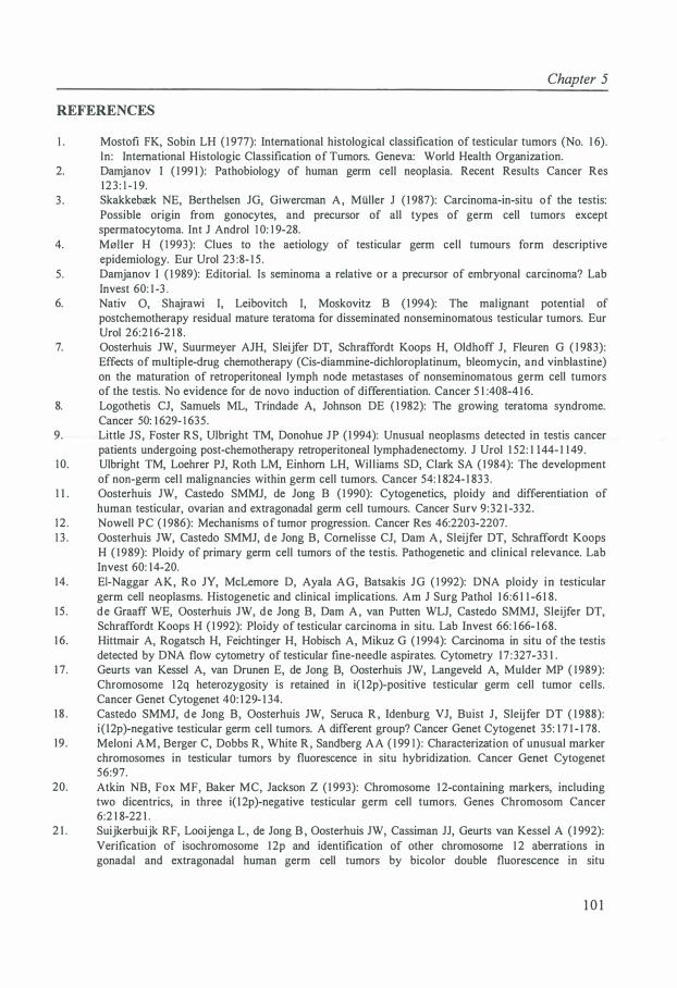
Chapter 5
REFERENCES
1 . Mostofi FK, Sobin LH ( 1 977): International histological classification of testicular tumors (No. 1 6). In: International Histologic Classification of Tumors. Geneva: World Health Organization.
2. Damjanov I ( 1 99 1 ): Pathobiology of human germ cell neoplasia. Recent Results Cancer Res 123 : 1 - 1 9.
3 . Skakkebrek NE, Berthelsen JG, Giwercman A, Millier J ( 1 987): Carcinoma-in-situ of the testis: Possible origin from gonocytes, and precursor of all types of germ cell tumors except spermatocytoma. Int J Androl 1 0: 1 9-28.
4. M01Ier H ( 1 993): Clues to the aetiology of testicular germ cell tumours form descriptive epidemiology. Eur Urol 23 :8- 1 5 .
5. Damjanov I ( 1 989): Editorial. Is seminoma a relative or a precursor of embryonal carcinoma? Lab Invest 60: 1 -3 .
6. Nativ 0, Shajrawi I, Leibovitch I, Moskovitz B ( 1 994): The malignant potential of postchemotherapy residual mature teratoma for disseminated nonseminomatous testicular tumors. Eur Urol 26:2 16-2 1 8.
7. Oosterhuis JW, Suurmeyer AJH, Sleijfer DT, Schraffordt Koops H, Oldhoff J, Fleuren G ( 1 983): Effects of multiple-drug chemotherapy (Cis-diammine-dichloroplatinum, bleomycin, and vinblastine) on the maturation of retroperitoneal lymph node metastases of nonseminomatous germ cell tumors of the testis. No evidence for de novo induction of differentiation. Cancer 5 1 :408-416.
8. Logothetis CJ, Samuels ML, Trindade A, Johnson DE ( 1 982): The growing teratoma syndrome. Cancer 50: 1 629- 1 635.
9. Little JS, Foster RS, Ulbright TM, Donohue JP ( 1 994): Unusual neoplasms detected in testis cancer patients undergoing post-chemotherapy retroperitoneal lymphadenectomy. J Urol 1 52: 1 144- 1 149.
10. Ulbright TM, Loehrer PJ, Roth LM, Einhorn LH, Wil liams SD, Clark SA ( 1984): The development of non-germ cel l malignancies within germ cell tumors. Cancer 54: 1 824- 1 833 .
1 1 . Oosterhuis JW, Castedo SMMJ, de Jong B ( 1 990): Cytogenetics, ploidy and differentiation of human testicular, ovarian and extragonadal germ cell tumours. Cancer Surv 9:32 1 -332.
12 . Nowell PC ( 1986): Mechanisms of tumor progression. Cancer Res 46:2203-2207. 1 3 . Oosterhuis JW, Castedo SMMJ, de Jong B, Cornelisse CJ, Dam A , Sleijfer DT, Schraffordt Koops
H ( 1989): Ploidy of primary germ cell tumors of the testis. Pathogenetic and clinical relevance. Lab Invest 60: 14-20.
14. EI-Naggar AK, Ro JY, McLemore D, Ayala AG, Batsakis JG ( 1 992): DNA ploidy in testicular germ cell neoplasms. Histogenetic and clinical implications. Am J Surg Pathol 1 6:6 1 1 -6 1 8.
15 . de Graaff WE, Oosterhuis JW, de Jong B, Dam A, van Putten WLJ, Castedo SMMJ, Sleijfer OT, Schraffordt Koops H ( 1 992): Ploidy of testicular carcinoma in situ. Lab Invest 66: 1 66- 1 68.
1 6. Hittmair A, Rogatsch H, Feichtinger H, Hobisch A, Mikuz G ( 1994): Carcinoma in situ of the testis detected by DNA flow cytometry of testicular fine-needle aspirates. Cytometry 17 :327-33 1 .
1 7. Geurts van Kessel A, van Drunen E, de Jong B, Oosterhuis JW, Langeveld A, Mulder MP ( 1989): Chromosome 1 2q heterozygosity is retained in i( l 2p)-positive testicular germ cell tumor cells. Cancer Genet Cytogenet 40: 129- 1 34.
18 . Castedo SMMJ, de Jong B, Oosterhuis JW, Seruca R, Idenburg VJ, Buist J, Sleijfer DT ( 1 988): i( l2p)-negative testicular germ cell tumors. A different group? Cancer Genet Cytogenet 35: 1 7 1 - 1 78.
19 . Meloni AM, Berger C, Dobbs R, White R, Sandberg AA ( 1 99 1 ) : Characterization of unusual marker chromosomes in testicular tumors by fluorescence in situ hybridization. Cancer Genet Cytogenet 56:97.
20. Atkin NB, Fox MF, Baker MC, Jackson Z ( 1 993): Chromosome 12-containing markers, including two dicentrics, in three i(l 2p)-negative testicular germ cell tumors. Genes Chromosom Cancer 6:2 1 8-22 1 .
2 1 . Suijkerbuijk RF, Looijenga L , de Jong B , Oosterhuis JW, Cassiman JJ, Geurts van Kessel A ( 1 992): Verification of isochromosome 12p and identification of other chromosome 1 2 aberrations in gonadal and extragonadal human germ cell tumors by bicolor double fluorescence in situ
101

Summary, general discussion and perspectives
hybridization. Cancer Genet Cytogenet 63 :8- 1 6. 22. Suijkerbuijk RF, Sinke RJ, Meloni AM, Parrington JM, van Echten J, de Jong B, Oosterhuis JW,
Sandberg AA, Geurts van Kessel A (1 993 ): Overrepresentation of chromosome I 2p sequences and karyotypic evolution in i(l2p)-negative testicular germ-cell tumors revealed by fluorescence in situ hybridization. Cancer Genet Cytogenet 70:85-93 .
23 . Rodriguez E, Houldsworth J, Reuter VE, Meltzer P, Zhang J, Trent JM, Bosl GJ, Chaganti RSK ( 1 993): Molecular cytogenetic analysis of i( l 2p)-negative human male germ cel l tumors. Genes Chromosom Cancer 8 :230-236.
24. Hofmann MC, Millan JL ( 1993): Developmental expression of alkaline phosphatase genes; Reexpression in germ cell tumours and in vitro immortalized germ cells. Eur Urol 23 :38-45.
25. Oosterhuis JW, Looijenga LHJ ( 1993): The biology of human germ cell tumours: retrospective speculations and new prospectives. Eur Urol 23 :245-250.
26. Looijenga LHJ, Gillis AJM, van Putten WLJ, Oosterhuis JW ( 1 993): In situ numeric analysis of centromeric regions of chromosome 1, 1 2, and 1 5 of seminomas, nonseminomatous germ cell tumors, and carcinoma in situ of human testis. Lab Invest 68:2 1 1 -2 1 9 .
27. Walt H, Arrenbrecht S, Delozier-Blanchet CD, Keller PJ, Nauer R, Hedinger CE ( 1 986): A human testicular germ cell tumor with borderline histology between seminoma and embryonal carcinoma secreted beta-human chorionic gonadotropin and alpha-fetoprotein only as a xenograft. Cancer 58: 1 39- 146.
28. Haddad FS, Sorini PM, Somsin AA, Nathan MH, Dobbs RM, Berger CS, Sandberg AA ( 1 988): Famil ial double testicular tumors: identical chromosome changes in seminoma and embryonal carcinoma of the same testis. J Urol 139:748-750.
29. Gil l is AJM, Looijenga LHJ, de Jong B, Oosterhuis JW ( 1994): Clonality of combined testicular germ cell tumors of adults. Lab Invest 7 1 :874-878.
30. de Graaff WE, Oosterhuis JW, de Jong B, van Echten J, Wiersema-Buist J, Schraffordt Koops H, Sleijfer OT (1 992): Cytogenetic analysis of the mature teratoma and the choriocarcinoma component of a testicular mixed nonseminomatous germ cell tumor. Cancer Genet Cytogenet 61 :67-73 .
3 1 . de Graaff WE, de Jong B , Oosterhuis JW, van Echten J, Wiersema-Buist J, Schraffordt Koops H, Sleijfer OT ( 1991 ): Cytogenetic analysis of the mature and immature teratoma components of a metastatic testicular nonseminomatous germ cell tumor. Cancer Genet Cytogenet 57:59-68.
32. Kerbel RS, Waghorne C, Korczak B, Lagarde A, Breitman ML (1 988): Clonal dominance of primary tumours by metastatic cel ls: Genetic analysis and biological implications. Cancer Surv 7 :597-629.
33 . Castedo SMMJ, de Jong B, Oosterhuis JW, ldenburg VJS, Seruca R, Buist J , te Meerman GJ, Schraffordt Koops H, Sleijfer DT ( 1 989): Chromosomal changes in mature residual teratomas following polychemotherapy. Cancer Res 49:672-676.
34. de Graaff WE, van Echten J, Oosterhuis JW, de Jong B, te Meerman GJ, Wiersema-Buist J, Sleijfer OT, Schraffordt Koops H ( 1993): Cytogenetic abnormalities and clinical stage in testicular nonseminomatous germ cell tumors. Cancer Genet Cytogenet 70: 1 2- 1 6.
35. Surti U, Hoffner L, Chakravarti A, Ferrell RE ( 1 990): Genetics and biology of human ovarian teratomas I. Cytogenetic analysis and mechanism of origin. Am J Hum Genet 47:635-643 .
36. Gonzales-Crussi F ( 1 982): Extragonadal Teratomas. In: Atlas of Tumor Pathology, 2nd series, fascicle 1 8. Armed Forces Institute of Pathology, Washington DC.
37. Friedman NB (1 987): The function of the primordial germ cell in extragonadal tissues. Int J Androl 1 0 :43-49.
102
SAMENV ATTING
Het menselijke lichaam ontstaat uit een enkele eel, een door een zaadcel bevruchte eicel . Door deling na deling ontstaan uit die eerste eel de circa honderd biljoen cellen waaruit een volwassen individu bestaat . Naast celdeling treedt specialisatie op, waarbij cellen een specifieke vorm en functie krijgen (differentiatie). Door veroudering of beschadiging sterven per dag een paar honderd milj ard cell en, die over het algemeen allemaal weer worden vervangen. Deze celvernieuwing ontstaat door celdeling . De processen van celdeling, celvemieuwing, differentiatie en celsterfte zijn nauwkeurig gereguleerd .
Bij kanker is er een storing in deze regulatie opgetreden . Hierdoor ontstaat overmatige celdeling en verandert de specifieke vorm en functie van cellen in meer of mindere mate . Dit resulteert in de vorming van een gezwel (tumor) . De kankercellen kunnen doordringen in het omliggende weefsel en kunnen losraken van de oorspronkelijke tumor en via lymfe en/of bloed in het lichaam worden verspreid . Deze kankercellen kunnen op een andere plaats in het lichaam weer verder uitgroeien, de zogenaamde uitzaaiingen (metastasen) . De mate van ongeremde celdeling en het vermogen van kankercellen om te metastaseren bepaalt hoe kwaadaardig (maligne) een tumor is .
In de kem van elke lichaamscel bevindt zich het erfelijke (genetische) materiaal (DNA), dat bij celdeling zichtbaar wordt als staafvormige structuren (chromosomen) . Een normale lichaamscel heeft 46 chromosomen; 22 chromosomen die elk in tweevoud aanwezig zijn ( chromosomen 1 tot en met 22), plus nog twee geslachtsbepalende chromosomen (twee X-chromosomen bij de vrouw, en een X- en Y-chromosoom bij de man). De (genetische) informatie die een eel nodig heeft om zijn functies uit te oefenen, zoals deling, groei en differentiatie, wordt gecodeerd door genen (bepaalde gedeeltes van het DNA) .
Kanker wordt ook wel een genetische ziekte van cellen en weefsels genoemd . Kanker ontstaat namelijk door veranderingen in genen die in normale gezonde cellen deling, groei en differentiatie reguleren. Deze veranderingen kunnen voorkomen in genen die celgroei stimuleren (proto-oncogenen), die celgroei onderdrukken (tumor suppressorgenen) en daarnaast bijvoorbeeld in genen die betrokken zijn bij herstel van DNA schade . Kanker wordt echter niet veroorzaakt door een enkele genetische verandering; het is een meerstapsproces .
Door additionele veranderingen in bovengenoemde genen, of doordat in een antler type genen veranderingen optreden, kunnen kankercellen van kwaad tot erger (ver)worden . Dit proces wordt tumorprogressie genoemd . Deze verdere veranderingen in genen vinden willekeurig in verschillende kankercellen plaats, die ze weer overdragen aan hun dochtercellen. Daardoor bestaat een tumor over het algemeen uit verschillende families van cellen, met verschillende genetische eigenschappen, een verschillende graad van kwaadaardigheid en een verschillend biologisch gedrag . Op het moment dat een kanker klinisch herkend kan worden, bestaat deze al uit miljarden cellen . Meestal heeft slechts een deel van deze kankercellen het vermogen om uit te zaaien .
Omdat kanker ontstaat door veranderingen in bepaalde genen en tumorprogressie gepaard gaat met verdere veranderingen in genen, kan het proces van oncogenese ( ontstaan van een tumor) en tumorprogressie bestudeerd worden door de dragers van de genen, de
103

Samenvatting
chromosomen, te onderzoeken ( cytogenetisch onderzoek) , en/of door de genen zelf te onderzoeken (moleculair genetisch onderzoek). Verder kan genetisch onderzoek van tumoren bijdragen tot het herkennen van de eel van oorsprong en de mate van kwaadaardigheid van een kanker. Dit laatste kan van belang zijn voor de behandeling van de patient . Tevens kan middels dit onderzoek de plaats en functie van groei(regulerende)en differentiatie genen opgehelderd worden .
Dit proefschrift beschrijft het cytogenetisch onderzoek van kiemceltumoren bij volwassen mannen. Kiemceltumoren ontstaan uit de voorlopercellen van zaadcellen of eicellen (kiemcellen). Bij mannen worden deze tumoren aangetroffen in de zaadbal, bij vrouwen in de eierstok. Zowel bij mannen als bij vrouwen kunnen kiemceltumoren echter ook, zij het zelden, op andere plaatsen in het lichaam dan in deze geslachtsorganen (gonaden) voorkomen, de zogenaamde extragonadale kiemceltumoren.
Kiemceltumoren van de zaadbal (testiculaire kiemceltumoren) zijn zeldzaam. In Nederland is slechts 1 % van alle kwaadaardige tumoren bij de man een testiculaire kiemceltumor . Echter in de leeftijdsgroep van 20 tot 34 jaar is het de meest voorkomende vorm van kanker.
Testiculaire kiemceltumoren worden op basis van celkenmerken onderverdeeld in seminomen en non-seminomen . Een seminoom bestaat uit een type cellen, die nog kenmerken hebben van primitieve kiemcellen. Het is een relatief goedaardige tumor, met pas in een laat stadium uitzaaiingen, die goed kan worden behandeld met bestraling . Het non-seminoom bestaat uit allerlei celtypen , die voorkomen in het embryo en in de vruchtvliezen . Ondanks het agressieve karakter van deze tumor is deze goed te behandelen met chemotherapie. In een kleine groep testiculaire kiemceltumoren komen seminoom en non-seminoom gezamenlijk voor, de zogenaamde gecombineerde testiculaire kiemceltumoren. Seminomen en non-seminomen hebben een identiek voorstadium, het zogenaamde carcinoma in situ.
Hoofdstuk 2 beschrijft welke genetische veranderingen een rol spelen bij het meerstapsproces van de oncogenese en/of tumorprogressie van testiculaire kiemceltumoren. Een belangrijke stap in het ontstaan van deze tumoren is polyploidisatie (vermeerdering van het chromosomenaantal). Een normale lichaamscel heeft 46 chromosomen (diploid). Testiculaire kiemceltumoren hebben ongeveer 69 chromosomen (triploid). Carcinoma in situ en seminoom hebben in het algemeen een aantal chromosomen dat iets hoger ligt dan 69 (hypertriploid). Non-seminoom heeft meestal iets minder chromosomen dan 69 (hypotriploid).
De meeste testiculaire kiemceltumoren hebben een specifiek, afwijkend chromosoom, dat is opgebouwd uit twee korte armen, p-armen, van chromosoom 12, het zogenaamde isochromosoom 12p [i( l2p)] . Als gevolg van deze afwijking is van de p-arm van chromosoom 12 een te groot aantal kopieen aanwezig. Deze kopie-vermeerdering van (soms een deel) van 12p, is eveneens belangrijk in het ontstaansproces van deze tumoren. Paragraaf 2 . 1 beschrijft dat andere specifieke structurele chromosomale afwijkingen dan i( 12p) waarschijnlijk een ondergeschikte rol spelen in het ontstaansproces en/of de progressie van testiculaire kiemceltumoren.
Zoals genoemd hebben cellen van testiculaire kiemceltumoren ongeveer een triploid chromosomen aantal . In triploide cellen komen drie exemplaren van elk chromosoom voor . In testiculaire kiemceltumoren echter, zijn bepaalde chromosomen (7, 8, 12, 2 1 en X) in
104

Samenvatting
significant hogere aantallen aanwezig dan verwacht (oververtegenwoordigd) , andere ( 1 1 , 13, 18 en Y) significant minder vaak aanwezig dan verwacht ( ondervertegenwoordigd). Het is aannemelijk dat de oververtegenwoordigde chromosomen oncogenen bevatten en de ondervertegenwoordigde tumor suppressor genen. Het i( l 2p) en over- en ondervertegenwoordiging van bepaalde chromosomen zijn al in het carcinoma in situ aanwezig. Dit suggereert dat een belangrijk gedeelte van het oncogenetisch proces al in het carcinoma in situ plaatsvindt, dus vroeg in de tumorevolutie van testiculaire kiemceltumoren .
In hoof dstuk 2 wordt eveneens uiteengezet in hoeverre seminomen en nonseminomen aan elkaar verwant zijn. Zowel het seminoom als het non-seminoom ontstaat uit het carcinoma in situ. Omtrent de verwantschap tussen seminomen en non-seminomen bestaan twee theorieen. De eerste theorie stelt dat het carcinoma in situ uitgroeit tot of seminoom of non-seminoom. Het seminoom staat dus in deze theorie los van het nonseminoom. De tweede theorie stelt dat het seminoom een voorloper is van, en kan overgaan in , het non-seminoom . Uit de resultaten van het onderzoek beschreven in paragraaf 2 . 1 blijkt dat seminoom en non-seminoom een sterk overeenkomstig chromosomenpatroon hebben. Deze bevinding steunt de tweede theorie die stelt dat het seminoom en non-seminoom verwante tumoren zijn . De progressie van seminoom naar non-seminoom, gaat gepaard met additioneel verlies van de chromosomen 7, 15, 19 en 22 en winst van i( 12p) en chromosoom 17. Dit is in overeenstemming met het gemiddeld lagere chromosomenaantal van non-seminomen dan seminomen. De zienswijze dat seminomen en non-seminomen verwante tumoren zijn wordt eveneens gesteund door cytogenetisch onderzoek van gecombineerde testiculaire kiemceltumoren, zoals beschreven is in paragraaf 2 .3 . Een overeenkomstig chromosomenpatroon tussen de seminoom en nonseminoom component van zo ' n tumor duidt op verwantschap tussen beide componenten. Een verschillend chromosomenpatroon voor de seminoom en non-seminoom component betekent dat de beide componenten niet uit elkaar zijn ontstaan, of dat de beide componenten al in een vroeg stadium in hun ontstaansproces zijn gesplitst. Paragraaf 2.2 handelt over overeenkomstige chromosomale afwijkingen in carcinoma in situ en de aangrenzende invasieve testiculaire kiemceltumor . Dit onderzoek levert het cytogenetisch bewijs dat carcinoma in situ de voorloper is van deze tumoren.
Ongeveer 85% van alle patienten met een testiculaire kiemceltumor wordt genezen, ondanks het feit dat een grote groep patienten zich met (uitgebreide) metastasen presenteert . De zaadbal met de tumor moet operatief verwijderd worden. V ervolgbehandeling kan bestaan uit bestraling of chemotherapie . Onbehandelde metastasen van het non-seminoom bevatten , net als de primaire tumor, een afwisselend beeld van weefseltypen. Na chemotherapeutische behandeling blijft van deze metastasen in ongeveer de helft van de gevallen een resttumor over , het zogenaamde residuaal matuur teratoom of de mature resttumor . Deze resttumor bestaat uit volledig gedifferentieerd weefsel. Het is belangrijk dat deze resttumoren operatief verwijderd worden, omdat dit weefsel verder kan uitgroeien en omdat een andere niet-kiemcel maligniteit uit dit restweefsel kan ontstaan.
Een mature resttumor zou het gevolg kunnen zijn van het feit dat chemotherapie alle tumorcellen vernietigt, behalve die cellen die volledig gedifferentieerd zijn. Een andere mogelijkheid is dat de chemotherapie het differentiatieproces in de verschillende typen cellen (weer) in gang zet , resulterend in volledig gedifferentieerd weefsel. Ook een
105

Samenvatting
combinatie van beide processen is mogelijk. Hoofdstuk 3 behandelt de cytogenetische vergelijking van non-seminomen met de
mature resttumoren, teneinde inzicht te krij gen in het proces van metastasering en in het tot stand komen van uitsluitend volledig gedifferentieerd weefsel in een resttumor. Er werden geen duidelijke chromosomale verschillen gevonden tussen non-seminomen en resttumoren. Zeer waarschijnlijk gaan metastasering en differentiatie niet gepaard met specifieke zichtbare chromosomale veranderingen. Het is eveneens mogelijk <lat de veranderingen die ertoe leiden <lat een eel kan gaan metastaseren al vroeg in de ontwikkeling van een tumor optreden. Mogelijk <lat een groot gedeelte van het proces van tumorprogressie en karyotype-evolutie al tijdens de ontwikkeling van het carcinoma in situ naar een invasieve testiculaire kiemceltumor plaatsvindt.
In hoofdstuk 4 wordt het chromosomenpatroon van twee extragonadale kiemceltumoren beschreven. In beide tumoren zijn specifieke structurele afwijkingen van de chromosomen 6 en 1 1 gevonden. Omdat het chromosomenpatroon van deze twee tumoren sterk verschilt met <lat van andere extragonadale- en van testiculaire kiemceltumoren, en omdat beide tumoren een typisch weefselbeeld en een uitzonderlijk ziektebeloop vertonen, kan van een nieuw subtype extragonadale kiemceltumor worden gesproken. Mogelijk <lat deze extragonadale kiemceltumoren niet zijn ontstaan uit primitieve kiemcellen maar uit primitieve embryonale cellen. De structurele afwijkingen van de chromosomen 6 en 1 1 , die in beide tumoren zijn gevonden, spelen zeer waarschijnlijk een cruciale rol in het ontstaan van deze tumoren.
1 06

22 februari 1963
10 juni 1981
30 mei 1985
1 juni 1985
16 april 1990
CURRICULUM VITAE
geboren te Vries
RAVO-diploma Openbare Scholengemeenschap "Dag Hammarskjold" te Assen
diploma Hoger Laboratoriumonderwijs, Klinische Chemie Dagschool voor Laboratoriumpersoneel te Groningen Afstudeeropdracht: Het vergelijken van enkele magnesium-inserum bepalingen (klinisch chemisch laboratorium, Wilhelmina Ziekenhuis Assen)
aanstelling als cytogenetisch analist bij de vakgroep Medische Genetica ( destijds Anthropogenetica) van de Rijksuniversiteit Groningen ten laste van de Stichting voor Erfelijkheidsvoorlichting te Groningen
aanstelling als research analist bij tumorcytogenetica bij de vakgroep Medische Genetica van de Rijksuniversiteit Groningen
107


OTHER PUBLICATIONS
de Graaff WE, de Jong B, Oosterhuis JW, van Echten J, Wiersema-Buist J, Schraffordt Koops H, Sleijfer DTh (1991): Cytogenetic analysis of the mature and immature teratoma components of a metastatic testicular nonseminomatous germ cell tumor. Cancer Genet Cytogenet 57:59-68.
Suijkerbuijk RF, van de Veen AY, van Echten J, Buys CHCM, de Jong B, Oosterhuis JW, Warburton DA, Cassiman JJ, Schonk D, Geurts van Kessel A (1991): Demonstration of the genuine iso-l 2p character of the standard marker chromosome of testicular germ cell tumors and identification of further chromosome 12 aberrations by competitive in situ hybridization. Am J Hum Genet 48:269-273.
Hamers A, de Jong B, Suijkerbuijk RF, Geurts van Kessel A, Oosterhuis JW, van Echten J, Evers J, Bosman F (1991) : A 46,XY female with mixed gonadal dysgenesis and a 48,XY,+7,+i(l2p) chromosome pattern in a primary gonadal tumor. Cancer Genet Cytogenet 57:219-224.
de Graaff WE, Oosterhuis JW, de Jong B, van Echten J, Wiersema-Buist J, Schraffordt Koops H, Sleijfer DTh (1992) : Cytogenetic analysis of the mature teratoma and the choriocarcinoma component of a testicular mixed nonseminomatous germ cell tumor. Cancer Genet Cytogenet 61 :67-73.
Von Eyben FE, de Graaff WE, Marrink J, Blaabjerg 0, Sleijfer DTh, Schraffordt Koops H, Oosterhuis JW, Petersen PH, van Echten J, de Jong B (1992) : Serum lactate dehydrogenase isoenzyme 1 activity in patients with testicular germ cell tumors correlates with the total number of copies of the short arm of chromosome 12 in the tumor. Mol Gen Genet 235:140-146.
de Graaff WE, van Echten J, Oosterhuis JW, de Jong B, te Meerman GJ, Wiersema-Buist J, Sleijfer DTh, Schraffordt Koops H (1993) : Cytogenetic abnormalities and clinical stage in testicular nonseminomatous germ cell tumors. Cancer Genet Cytogenet 70:12-16.
Suijkerbuijk RF, Sinke RJ, Meloni AM, Parrington JM, van Echten J, de Jong B, Oosterhuis JW, Sandberg AA, Geurts van Kessel A (1993): Overrepresentation of chromosome 12p sequences and karyotypic evolution in i(l2p)-negative testicular germ-cell tumors revealed by fluorescence in situ hybridization. Cancer Genet Cytogenet 70:85-93.
van den Berg E, Molenaar WM, van Echten J, Dam A, Mensink HJA, de Jong B (1994) : Cytogenetic analysis of a leiomyosarcoma of the kidney. Cancer Genet Cytogenet 72:126-129.
109

Lambrechts AC, Looijenga LHJ, van't Veer MB, van Echten J, Timens W, Oosterhuis JW (1995): Lymphomas with testicular localisation show a consistent BCL-2 expression without a translocation (14;18) : a molecular and immunohistochemical study. Br J Cancer 71:73-77.
van Echten J, van den Berg E, van Baarlen J, van Noort G, Vermey A, Dam A, Molenaar WM (1995): An important role for chromsome 17, band q25, in the histogenesis of alveolar soft part sarcoma. Cancer Genet Cytogenet 82:57-61
Mostert MMC, van de Pol M, van Echten J, Olde Weghuis D, Geurts van Kessel A, Oosterhuis JW, Looijenga LHJ (1996) : Fluorescence in situ hybridization-based approaches for the detection of 12p-overrepresentation, in particular i( 12p ), in cell lines of human testicular germ cell tumors of adults. Cancer Genet Cytogenet 87:95-102
Mostert MMC, van de Pol M, Olde Weghuis D, Suijkerbuijk RE, Geurts van Kessel A, van Echten J, Oosterhuis JW, Looijenga LHJ (1996): Chromosomal imbalances in germ cell tumors of the adult testis detected by conventional karyotyping and comparitive genomic hybridization. Cancer Genet Cytogenet (in press)
110