university of groningen effects of oxygen during long-term
TRANSCRIPT

University of Groningen
Effects of Oxygen During Long-term Hypothermic Machine Perfusion in a Porcine Model ofKidney Donation After Circulatory DeathCOPE consortium; Venema, Leonie H; Brat, Aukje; Moers, Cyril; 't Hart, Nils A; Ploeg, RutgerJ; Hannaert, Patrick; Minor, Thomas; Leuvenink, Henri G DPublished in:Transplantation
DOI:10.1097/TP.0000000000002728
IMPORTANT NOTE: You are advised to consult the publisher's version (publisher's PDF) if you wish to cite fromit. Please check the document version below.
Document VersionPublisher's PDF, also known as Version of record
Publication date:2019
Link to publication in University of Groningen/UMCG research database
Citation for published version (APA):COPE consortium, Venema, L. H., Brat, A., Moers, C., 't Hart, N. A., Ploeg, R. J., Hannaert, P., Minor, T., &Leuvenink, H. G. D. (2019). Effects of Oxygen During Long-term Hypothermic Machine Perfusion in aPorcine Model of Kidney Donation After Circulatory Death. Transplantation, 103(10), 2057-2064.https://doi.org/10.1097/TP.0000000000002728
CopyrightOther than for strictly personal use, it is not permitted to download or to forward/distribute the text or part of it without the consent of theauthor(s) and/or copyright holder(s), unless the work is under an open content license (like Creative Commons).
The publication may also be distributed here under the terms of Article 25fa of the Dutch Copyright Act, indicated by the “Taverne” license.More information can be found on the University of Groningen website: https://www.rug.nl/library/open-access/self-archiving-pure/taverne-amendment.
Take-down policyIf you believe that this document breaches copyright please contact us providing details, and we will remove access to the work immediatelyand investigate your claim.
Downloaded from the University of Groningen/UMCG research database (Pure): http://www.rug.nl/research/portal. For technical reasons thenumber of authors shown on this cover page is limited to 10 maximum.
1
Transplantation Publish Ahead of Print
DOI: 10.1097/TP.0000000000002728
Effects of oxygen during long-term hypothermic machine perfusion in a porcine model
of kidney donation after circulatory death.
Leonie H. Venema1, Aukje Brat
1 MSc, Cyril Moers
1 MD PhD, Nils A. ‘t Hart
2 MD PhD,
Rutger J. Ploeg3 Prof MD,
Patrick Hannaert
4 PhD, Thomas Minor
5 Prof, Henri G.D.
Leuvenink1 Prof
1Dept of Surgery, University Medical Centre Groningen, University of Groningen, The
Netherlands
2Dept of Pathology, University Medical Centre Groningen, University of Groningen, The
Netherlands
3Dept of Surgery, Nuffield Department of Surgical Science, University of Oxford, United
Kingdom
4IRTOMIT, INSERM U1082, Faculté de Médecine et de Pharmacie, Université de Poitiers,
France
5Dept for Surgical Research/General Surgery, University Hospital Essen, Germany
On behalf of the COPE consortium
Correspondence information: Leonie Venema , CMC V Y2144, BA44, Hanzeplein 1, 9713
GZ Groningen, [email protected], +31630117620
Authorship
LHV: Participated in research design, performance of the experiments, participated in data
analysis and wrote the paper.
AB: Participated in research design, performance of the experiments and wrote the paper.
CM: Participated in research design.
ACCEPTED
2
NAT: Participated in research design, contributed in histology analysis.
RJP: Participated in research design.
PH: Participated in research design, participated in data analysis.
TM: Participated in research design
HGDL: Participated in research design, Supervising role.
Disclosure: “The authors declare no conflicts of interest”
Funding: The experiments presented in this paper are funded by the Consortium for Organ
Preservation in Europe (COPE).
Abbreviations
ASAT, aspartate amino transferase
ADP, adenosine monophosphate
ADP, adenosine diphosphate
ATP, adenosine triphosphate
AUC, area under curve
CS, cold storage
DBD, donation after brain death
DCD, donation after circulatory death
DGF, delayed graft function
Hb, hemoglobin
H&E, haemotoxylin and eosin
HMP, hypothermic machine perfusion
LDH, lactate dehydrogenase
NAG, N-Acetyl-β-D-glucosaminidase
NMP, normothermic machine perfusion
ROS, reactive oxygen species
ACCEPTED

3
SCD, standard criteria donor
TBARS, thio barbituric acid reactive substances
UW MPS, university of wisconsin machine perfusion solution
WIT, warm ischemic time
This is an open-access article distributed under the terms of the Creative Commons
Attribution-Non Commercial-No Derivatives License 4.0 (CCBY-NC-ND), where it is
permissible to download and share the work provided it is properly cited. The work cannot be
changed in any way or used commercially without permission from the journal.
ACCEPTED

4
Abstract
Background:
Hypothermic machine perfusion (HMP) has become standard care in many center’s to
preserve kidneys donated after circulatory death (DCD). Despite a significant reduction in
metabolism at low temperatures, remaining cellular activity requires oxygen. Since the role
and safety of oxygen during HMP has not been fully clarified, its supply during HMP is not
standard yet. This study investigates the effect of administering oxygen during HMP on renal
function in a porcine DCD model.
Methods:
After 30 minutes of warm ischemia, porcine slaughterhouse kidneys were preserved for 24
hours by means of cold storage (CS), or HMP with Belzer Machine Perfusion Solution (UW-
MPS) supplemented with no oxygen, 21% or 100% oxygen. Next, kidneys were reperfused
for 4 hours in a normothermic machine perfusion (NMP) setup.
Results:
HMP resulted in significantly better kidney function during NMP. Thiobarbituric acid-
reactive substances (TBARS), markers of oxidative stress, were significantly lower in HMP
preserved kidneys. HMP preserved kidneys showed significantly lower ASAT and LDH
levels compared to kidneys preserved by CS. No differences were found between the HMP
groups subjected to different oxygen concentrations. ATP levels significantly improved
during HMP when active oxygenation was applied.
Conclusion:
This study showed that preservation of DCD kidneys with HMP is superior to CS. Although
the addition of oxygen to HMP did not result in significantly improved renal function,
beneficial effects were found in terms of reduced oxidative stress and energy status. Oxygen
addition proofed to be safe and did not show detrimental effects.
ACCEPTED

5
Introduction
Persistent organ shortage in transplantation results in the use of sub-optimal quality organs. In
addition, the number of donations after brain death (DBD) from donors younger than 50
without comorbidities or so-called standard criteria donors (SCD) is decreasing.1 As a result,
allografts from donors deceased due to a circulatory arrest are increasing in a number of
countries.1 Kidneys retrieved from such donors are more prone to ischemia-reperfusion
injuries and are associated with a significantly higher incidence of delayed graft function
(DGF).2
DGF results in the necessity of dialysis after transplantation until the kidney recovers
its function.3 DGF, therefore, affects the quality of life of the recipient and it also appears to
be a risk factor for acute cellular rejection and poorer long-term outcomes.4
Hypothermic
machine perfusion (HMP) as preservation modality has already been proven to reduce the
incidence and duration of DGF compared to cold storage(CS).2,3,5,6
Both CS and HMP are based on the suppression of metabolism due to hypothermia. Notably,
even at 4 degrees Celsius, approximately 10% of physiological metabolic rate remains. This
suggests that oxygen continues to be consumed and its addition during preservation might be
beneficial to support ongoing metabolism. HMP offers the possibility to provide the kidney
with oxygen during the preservation period. However, oxygen supply during preservation is
not standard of care and kidneys are usually perfused without oxygen.
Next to limited experimental and preclinical evidence, clinical proof of the need of oxygen
during HMP is also still lacking.5,7–9
Therefore, two international, double blinded,
randomized controlled trials are currently ongoing to assess the efficacy of 100% oxygen
addition during HMP of older donation after circulatory death (DCD) kidneys and in
expanded criteria donor (ECD) kidneys.10,11
Furthermore, safety of the addition of oxygen at
whatever level during preservation, remains a matter of concern. In addition, the work that
has been performed until now focused mainly on cold storage versus HMP with 100%
ACCEPTED

6
oxygen or on HMP with different oxygen concentrations only. A study comparing CS with
HMP with different oxygen concentrations using a clinically approved preservation solutions
is still lacking. We, therefore, combined all strategies. Our aim was to provide a
comprehensive evaluation about the effects of different oxygen concentrations during HMP
of porcine kidneys compared to CS preservation. Besides early renal function, we want to
address the safety of different oxygen concentrations by means of the release of reactive
oxygen species (ROS), and the effect of oxygen during HMP on the metabolism of the
kidney. In order to avoid the use of laboratory animals we used a porcine DCD kidney
slaughterhouse model as previously developed in our lab.
Materials and methods
Animal model
Porcine kidneys were obtained from two abattoirs. Pigs were slaughtered by a standardized
procedure of sedative electric shock followed by exsanguination. Immediately, 1 liter of
blood was collected in a container containing 25.000 IU of heparin (LEO Pharma A/S,
Ballerup, Denmark). Since we made use of slaughterhouse waste material as our organ and
blood source, no animal ethics committee approval was needed.
Experimental design
Warm ischemic time (WIT) of thirty minutes was chosen to induce ischemic injury. The four
different preservation techniques used were applied for 24 hours: cold storage (CS), non
oxygenated hypothermic machine perfusion (HMP0%), hypothermic machine perfusion with
21% oxygen (HMP21%) or 100% oxygen addition (HMP100%). All kidneys were subsequently
reperfused in an ex vivo normothermic machine perfusion (NMP) setup for a total duration of
4 hours. Every group contained six kidneys.
ACCEPTED

7
Cold storage and hypothermic machine perfusion
After warm ischemia the kidney was flushed with 180 ml saline at 4° Celsius (Baxter BV,
Utrecht, The Netherlands). A cortical biopsy was taken (Invivo, Best, The Netherlands) and
stored in sonification solution (SONOP containing 0.372 g EDTA in 130 mL H2O and NaOH
(ph 10.9) + 370 mL 96% ethanol) and 4% buffered formaldehyde for further analysis. In the
CS group the kidneys were stored in a bag, submerged in 500 mL University of Wisconsin
solution (Belzers CS, Bridge to life Ltd., London, United Kingdom) and stored on melting
ice. In the HMP groups the kidneys were cannulated to connect the renal artery to the HMP
device (Kidney Assist Transport, Organ Assist, Groningen, The Netherlands). A total of 500
mL University of Wisconsin machine perfusion solution (Belzers MP, Bridge to life Ltd.,
London, United Kingdom) was used as perfusion solution. Preservation was performed at
4°C with a pulsatile pressure-controlled perfusion with a mean arterial pressure of 25 mmHg.
Either, no oxygen, 21% or 100% oxygen was supplied to the oxygenator (Hilite LT 1000,
Medos Medizin technik AG, Stolberg, Germany) with a fixed flow rate of 100 ml/min.
Perfusion solution samples were taken after 15, 60 minutes and 24 hours. Perfusion
parameters, such as pressure, temperature and flow rates were monitored continuously.
Ex vivo normothermic machine perfusion to assess renal function
After 24 hours of preservation, renal function was assessed in an isolated ex vivo
normothermic machine perfusion setup. The renal artery and ureter were cannulated with a 12
and 8 French cannula, respectively. The kidneys were flushed with 50 ml of saline (4°) to
remove remaining preservation solution. Afterwards the kidneys were weighed and another
biopsy was taken and stored as described above.
The kidney was placed in an organ chamber and perfused at 37°C for 4 hours with a pressure-
controlled pulsatile pump at a mean pressure of 75 mmHg (Kidney Assist Transport, Organ
Assist, Groningen, The Netherlands). The perfusate was oxygenated with a mixture of 95%
ACCEPTED

8
O2 and 5% CO2 through the oxygenator (Hilite LT 1000, Medos Medizin technik AG,
Stolberg, Germany) with a fixed flow rate of 500 ml/min. The setup was surrounded by a
heating cabinet with a feedback system, keeping the ambient temperature at 37°C. The
perfusion medium consisted of 500 ml heparinized, leukocyte-depleted autologous whole
blood. Leukocyte-depletion was carried out with a leukocyte filter (Bio R O2 plus, Fresenius
Kabi, Zeist, The Netherlands). The blood was diluted with 300 ml of lactated Ringer’s
(Baxter BV, Utrecht, The Netherlands), containing 6 mg Mannitol (Sigma-Aldrich, St Louis,
USA), 6 mg Dexamethasone (Centrafarm, Etten-Leur, The Netherlands) 10 ml 8,4% sodium
bicarbonate (B Braun Melsungen AG, Melsungen, Germany), 90 mg creatinine (Sigma-
Aldrich, St Louis, USA), 1000mg/200mg Amoxicilline /Clavulanic acid (Sandoz BV,
Almere, The Netherlands), and 100 µl 20 mg/ml sodium nitroprusside (Sigma-Aldrich, St
Louis, USA). Furthermore, a continuous supply of nutrients consisting of 10% Aminoplasmal
(Braun Melsungen AG, Melsungen, Germany), 2.5 ml 8,4% sodium bicarbonate, and 17 IU
Novorapid, (Novo Nordisk, Bagsvaerd, Denmark) was added to the perfusion circuit at a rate
of 20 ml/h. 5% glucose (Baxter BV, Utrecht, The Netherlands) was administered when
glucose levels dropped below 5 mmol/L.
Evaluation of renal function
During the testing period, renal flow rate and urine production were measured every 15
minutes. Blood and urine samples were taken after 15, 60, 120, 180 and 240 minutes. At
these same time points arterial and venous blood samples were taken for blood gas analysis
(ABL90 FLEX, Radiometer, Zoetermeer, The Netherlands).
Concentrations of creatinine and sodium were determined in blood and urine, using routine
procedures at the clinical chemistry lab of the University Medical Center Groningen
(UMCG). Creatinine clearance served as the primary functional endpoint of this study.
Tubular and glomerular integrity were used as secondary functional endpoints and were
ACCEPTED

9
assessed using fractional sodium excretion and urine protein content, respectively. Proteins in
the urine were measured in a standardized manner at the clinical chemistry lab of the UMCG.
Metabolic activity
Calculating the renal oxygen consumption (QO2) approximated metabolic activity of the
kidneys. The difference between the venous and arterial dissolved and bound oxygen was
calculated by using the following formula:
𝑂𝑥𝑦𝑔𝑒𝑛 𝑐𝑜𝑛𝑠𝑢𝑚𝑝𝑡𝑖𝑜𝑛 (
𝑚𝑙𝑂2
𝑚𝑖𝑛100𝑔𝑟
) =
((((𝐻𝑏 ∗ 2,4794) + (𝑝𝑂2𝑎𝑟𝑡𝑒𝑟𝑖𝑎𝑙 ∗ 𝐾)) − ((0,024794 ∗ 𝐻𝑏 ∗ 𝑆𝑂2 𝑣𝑒𝑛𝑜𝑢𝑠 ) + (𝑝𝑂2 𝑣𝑒𝑛𝑜𝑢𝑠 ∗
𝐾))) ∗ 𝑄) /𝑔) ∗ 100
Where Hb is the perfusates hemoglobin content in mmol/L, pO2 is the partial oxygen pressure
arterial or venous in kPa, K is the solubility constant of oxygen in water at 37°C and equals
0.0225 (mL O2 per kPa), SO2 is the saturation in %, Q is the renal blood flow in L/min and g
is the kidney weight in grams.
Adenosine triphosphate (ATP) was analyzed in biopsies that were taken before and after the
preservation period, and at the end of reperfusion. ATP content was determined according to
a standard protocol and expressed in µmol/g protein.12
Metabolic coupling of sodium transport by ATPase in tubular epithelial cells was calculated
by dividing transported sodium (TSodium) with renal oxygen consumption QO2:
𝑇𝑆𝑜𝑑𝑖𝑢𝑚 (
𝑚𝑚𝑜𝑙 𝑆𝑜𝑑𝑖𝑢𝑚𝑚𝑚𝑜𝑙 𝑂2
100𝑔𝑟)
=((𝐶𝑟𝑐𝑙𝑒𝑎𝑟𝑎𝑛𝑐𝑒 ∗ 𝑃𝑙𝑎𝑠𝑚𝑎𝑆𝑜𝑑𝑖𝑢𝑚 ) − (𝑈𝑟𝑖𝑛𝑒 𝑓𝑙𝑜𝑤 ∗ 𝑈𝑟𝑖𝑛𝑒𝑆𝑜𝑑𝑖𝑢𝑚 ))
𝑄𝑂2
Oxidative stress due to active oxygenation
Thiobarbituric acid-reactive substances (TBARS) were measured as indicator of oxidative
stress in the preservation solution, blood and urine at the specified sampling time points. The
ACCEPTED

10
protocol for this analysis has been described in detail previously.12
TBARS concentrations
are expressed in µM.
Kidney injury markers
Enzymatic activities of lactate dehydrogenase (LDH), and aspartate aminotransferase
(ASAT) were determined at the clinical chemistry lab of the UMCG according to standard
procedures. Urinary N-acetyl-beta-D-glucosaminidase(uNAG) was determined following a
protocol described previously by our lab.12,13
Histological examination and morphology scoring
Kidney biopsies were fixed by immersion in 4% buffered formaldehyde, embedded in
paraffin and cut into 4 µm slices. These sections were stained with haematoxylin-eosin
(H&E). Ischemia reperfusion injury was scored on the basis of 3 criteria14
: proximal tubular
cell edema, tubular cell vacuolation and proximal tubular cell necrosis. Every item was given
a score between 1 and 5, as representing no signs of edema, vacuoles or cell necrosis (score
1), minor (score 2), medium (score 3), severe (score 4) or extreme signs (score 5). The
biopsies were randomly assigned to two independent experienced examiners for light
microscopy evaluation.
Statistics
Results are reported as means with standard deviations. Statistical analysis was performed
with IBM SPSS Statistics 23. Area under the curve (AUC) was calculated for renal flow rates
during HMP and NMP, creatinine clearance and oxygen consumption rates. All other markers
were tested for significant differences at every time point. Groups were compared using a
Kruskal-Wallis test followed by a Mann-Whitney U posthoc test. P<0.05 was considered to
indicate statistical significance.
ACCEPTED

11
Results
Hypothermic and normothermic perfusion parameters
All HMP groups showed similar flow patterns in the cold, starting with a steep increase
within the first twenty minutes and a slow increase thereafter until the end of preservation
(Figure 1A). No statistical differences in flow rate during HMP was found.
During the period of NMP, renal blood flow increased during the first 120 minutes in every
group, and slowly decreased thereafter (Figure 1B). The CS group showed a trend towards a
higher mean flow rate (p=0.072)) compared to the HMP groups.
Renal function during normothermic perfusion
In terms of creatinine clearance all HMP groups showed significantly higher clearances at
every time point in comparison to the CS group. The HMP100% group presented the highest
clearance rate. It was, however, not significantly different from the HMP0% and HMP21%
groups (Figure 2A). Proteinuria in CS kidneys was significantly higher than in HMP groups
(Figure 2B). HMP kidneys had reduced urinary levels of protein, but no differences were
observed within the 3 HMP groups. Significant improvement in fractional sodium excretion
levels was found when HMP was applied. Again no differences were found when comparing
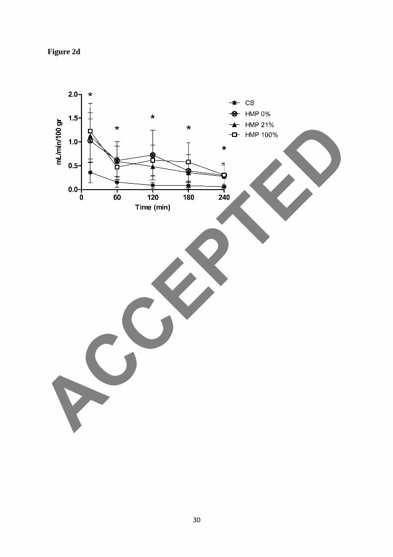
the different oxygen concentrations (Figure 2C). In all groups urine production was the
highest during the first fifteen minutes after reperfusion (Figure 2D).
Metabolic activity during normothermic perfusion
Oxygen consumption rates were significantly higher in all HMP groups compared to the CS
kidneys. Although renal function in the CS kidneys was almost absent (filtration < 0.1
mL/(min.100g; FENa% > 70%), oxygen consumption was still present (Figure 3A).
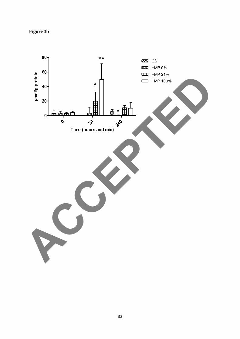
After 30 minutes WIT and before preservation was initiated, ATP was almost completely
depleted in every group (Figure 3B, timepoint 0.5). CS for 24 hours resulted in complete loss
of all ATP. HMP0% resulted in no additional ATP production during HMP. The addition of 21
ACCEPTED

12
or 100% yielded significant higher ATP during 24 hours preservation in these groups (Figure
3B, timepoint 24). 4 hours of NMP resulted in significant lower ATP levels in the HMP0%
group, while the other groups showed similar ATP levels (Figure 3B, timepoint 28).
Metabolic coupling ratio was improved in HMP perfused kidneys and was significantly
improved after 120 and 180 minutes after start reperfusion in all HMP kidneys compared to
CS kidneys (Figure 3C).
Oxidative stress due to active oxygenation
TBARS measured in the preservation solution during HMP were negligible in all groups at
every time point (Figure 4A). TBARS in the perfusate were significantly higher after 120
minutes in the CS kidneys compared to the HMP groups, and slowly decreased in the HMP
groups over the 4 hours NMP period (Figure 4B). The urinary TBARS showed an immediate
increase during the first hour of reperfusion in all HMP groups, but decreased thereafter. The
CS group had a gradual increasing concentration of TBARS in the urine and has a higher
value (not significant) at the end of 4 hours perfusion (Figure 4C).
Kidney injury markers
LDH values remained stable in the HMP groups while the CS kidneys showed a rise in LDH
levels in the (NMP) perfusate over time (Figure S1A, SDC, http://links.lww.com/TP/B723).
At the end of 4 hours reperfusion, LDH levels were significantly higher in the CS group.
Urinary N-acetyl-beta-D-glucosaminidase (uNAG) remained stable during 4 hours in the
HMP groups. The CS kidneys showed a significant increased value from the onset of
reperfusion and also a rise over time (Figure S1B, SDC, http://links.lww.com/TP/B723). All
3 HMP groups have a similar trend for ASAT levels; however, there is a significant benefit
for the 100% oxygen group in comparison with the HMP0% and CS groups. Similar for LDH
and NAG, the CS group showed an increase over a time and significant higher levels of
ASAT compared to all HMP groups (Figure S1C, SDC, http://links.lww.com/TP/B723).
ACCEPTED

13
Histology
During histological examination it became clear that the stage of damage was already beyond
the point of vacuolation and that proximal tubular cell necrosis was already detectable. The
histological scoring resulted in the following mean necrosis scores: 2.17±0.49, 2.60±0.55,
2.67±0.82 for the HMP 0, 21 and 100% groups, respectively. This was significantly lower
than the average necrosis for the CS kidneys (of 4.7±0.49; Figure S2A, SDC,
http://links.lww.com/TP/B723).
The average edema score for CS, HMP 0, 21 and 100 were comparable, with scores of 2.0±0,
2.50±0.84, 2.20±0.84, and 2.5±0.58, respectively (Figure S2B, SDC,
http://links.lww.com/TP/B723).
Discussion
In this study we evaluated the effect of different oxygen concentrations during HMP on renal
function with a clinically approved perfusion solution. We found that HMP is superior to CS
in terms of creatinine clearance and fractional sodium excretion. Active oxygenation during
preservation did not result in significant advantages with regard to renal function in this
porcine DCD model. Only for ASAT and ATP levels, a significant beneficial effect of
oxygen was found. No signs of oxidative stress through the addition of oxygen during
preservation were observed.
Experimental research on the addition of oxygen during HMP is scarce. There are only two
studies that assessed CS versus 100% oxygenated HMP in preclinical porcine (auto)
transplantation models. Both were able to show the significant beneficial effects of
oxygenated HMP in comparison to CS on renal function.5,7
This result corresponds with our
findings. However, non oxygenated HMP was not included and the addition of oxygen can
therefore not be assessed. Furthermore, in another study, short-term effects of oxygen during
HMP was addressed in a reperfusion model.8
This study found beneficial effects of oxygen
ACCEPTED

14
during the preservation of DCD kidneys in terms of function and injury. Unfortunately, we
were not able to reproduce these findings, however, there are some major differences in setup
of these studies. One of these differences is that we used porcine slaughterhouse kidneys. The
downside of using these is that not only the conditions of the experiment are less controlled
but also baseline quality of these kidneys is not controllable since we are not allowed to take
samples at life. Therefore, we cannot elaborate on preexisting injury and the amount of injury
induced by warm ischemia that we have chosen in this study. Animal welfare inspired us to
develop a slaughterhouse model since ethical considerations concerning the use of animals
for scientific research is an important topic in the Netherlands. We believe that
slaughterhouse organs can provide us reliable and translational data and we are not the first
group believing in slaughterhouse organs for machine perfusion research.15–18
Nath et al
already demonstrated similarity in metabolic processes between human and slaughterhouse
pig kidneys, which provides additional confidence in these kidneys for scientific research.16
One prerequisite to make sure that the slaughtering procedure does not negatively influence
the outcome we streamlined the process with our local butchers by explaining them our goals
and procedures such as ischemic times and appropriate handling of blood and organs.
The reason for conducting this study was to assess a clinically approved HMP device in
combination with a clinically approved perfusion solution. Hoyer et al used an experimental
perfusion solution, Histidine-tryptophan-ketoglutarate –N, and their results can therefore not
be directly extrapolated to the current clinical situation.8 In our setup, UW-MP solution was
used, which is currently the only approved clinical machine perfusion solution for
preservation of kidney grafts. The oxygen carrying capacity of UW-MP solution was not
measured in this study but has been measured before. The amount of dissolved oxygen
present in UW-MP is approximately 70 kPa, and 21 kPa with the addition of 100 ml/min
100% and 21% oxygen, respectively.17,19,20
A recent study also showed that oxygen is indeed
ACCEPTED

15
delivered to kidneys cells and supports aerobic metabolism, as reflected by both adenosine
monophosphate (AMP), adenosine diphosphate (ADP) and ATP levels in both the medulla
and cortex of the kidneys when UW-MPS solution is used.17
The only other comparable study on different oxygen concentrations during HMP was
performed in a porcine DCD auto transplantation model.9 This group was able to show that
animals transplanted with HMP0% oxygen had significantly higher peak creatinine levels at
day 5 post transplant in comparison to pigs that were transplanted with HMP100% oxygen
kidneys. The effect of oxygen was still present at the 3-month follow up, shown by
significantly lower serum creatinine levels and a significantly reduced proteinuria. These
significant differences first became apparent at day 5 after transplantation. Long-term
function cannot be assessed with reperfusion duration of only 4 hours and a (auto)
transplantation model is necessary to address chronic injury and long-lasting quality and
function. Another possibility would be longer reperfusion times. However, NMP as
reperfusion modality also has its limits. NMP up to 24 hours are reported but maintaining a
physiological electrolyte content and pH is problematic.21–23
In our study, we only tested
kidney function for four hours. This interval could be too short to find conclusive results
concerning active oxygenation in our model. In the end, the ongoing clinical studies need to
answer the question of oxygen during HMP is beneficiary for the long-term quality of
transplanted kidneys.
Both studies addressed in the prior paragraph, conclude that active oxygenation during HMP
of DCD kidneys is beneficial in terms of renal function. We were not able to show this.
However, we do see a trend in favor of active oxygenation during long-term HMP. Kidney
quality is, however, more than function alone. Therefore, we performed supplementary
analyses to address a broad range of other quality and injury markers. With these we can
answer some oxygen-specific issues that are fundamental in kidney preservation.
ACCEPTED

16
Oxygen consumption during NMP was calculated as indicator of metabolic activity and a
significantly lower consumption was found for CS kidneys. Similar oxygen consumption
rates were found for the different HMP groups and were in-line with renal function. We think
that oxygen consumption could function as a suitable quality marker during NMP. This is
supported by studies comparing subnormothermic machine perfusion with HMP and CS,
where significant improvements in oxygen consumption were found for kidneys that were
better preserved in terms of function.24,25
Two different formulae are described; the first
considering only dissolved oxygen,26
the second also considering hemoglobin-bound
oxygen.24
In our model, variations in hemoglobin (Hb) and venous saturations were present,
which urges us to make use of the more complicated formula.
In addition to oxygen consumption, metabolic coupling was calculated. This provides
information regarding efficient use of oxygen for ATP production and subsequent active
transport of sodium ions over the tubules. We observed a significant improvement in
metabolic coupling at time points t=120 and 180 for all HMP groups. This result is in-line
with the fractional sodium excretion. All HMP kidneys are able to transport sodium in
comparison to the CS kidneys. This active sodium transport requires ATP as energy source.
We found a significantly lower ATP content after reperfusion in the HMP0% group and it is
likely that the total ATP production and usage during NMP is balanced to make active
sodium transport possible causing this lower ATP content after reperfusion in the non
oxygenated HMP group. The required net ATP content in the oxygenated groups after
reperfusion indicates that the addition of oxygen during HMP leads to better mitochondrial
function. ATP levels in the CS group, however, indicate a low usage of ATP since metabolic
coupling is poor in this group. It is also likely that mitochondrial function is disturbed in the
CS group. TBARS levels in the plasma support that there is indeed a distortion in the
mitochondrial respiratory chain that resulted in significantly higher TBARS levels in the CS
ACCEPTED

17
kidneys. Furthermore, mitochondrial damage can also be assessed with ASAT, which is
present in the cytoplasm of mitochondria. In this isolated perfusion system the only source
are kidney mitochondria and, therefore, serves as a valuable marker for mitochondrial
damage. In-line with the TBARS levels, there are the significantly higher ASAT levels,
indicating more mitochondrial damage in the CS compared to the HMP kidneys. In favor of
100% oxygenation during HMP are the significant lower ASAT levels in comparison with
HMP0% and HMP21High oxygen concentrations during HMP resulted in better restoration of
tissue ATP content in this study. This given is also supported by Patel et al, showing not only
increased levels of AMP, ADP and ATP, but also increased lactate and alanine levels,
metabolites of glycolysis, indicating a switch to anaerobic metabolism when insufficient
oxygen was supplied to the cells.17
A short period of oxygenated HMP after CS has been
shown to reestablish cellular respiration, resulting in improved preservation of rat kidneys27
and rat and porcine livers.28,29
Long-term oxygenated HMP seem to result in improved
cellular respiration as well, considering the ATP levels that we and others found.17
It clearly
proves that at low temperatures metabolism is ongoing and should be supported by
oxygenation.
In conclusion, cellular energy status significantly improved when active oxygenation was
applied during long-term HMP in this slaughterhouse reperfusion model. Although a trend
towards preservation benefits was seen during NMP, this never reached a statistical
significance. Long-term HMP on itself, significantly improved renal function, tissue integrity
and in lower injury compared to CS during NMP.
ACCEPTED

18
Acknowledgments
The authors are very grateful to Kroon Vlees and butchery Kuipers for their support in
providing kidneys for this research. Our special thanks go to Sipke Woudstra and Henk
Luinge for making the logistics to perform scientific research possible in a commercial
slaughterhouse. Furthermore, many thanks to Petra Ottens, Janneke Wiersema-Buist and
Jacco Zwaagstra for performing most of the analysis described in this paper.
ACCEPTED

19
References
1. Moers C, Leuvenink HGD, Ploeg RJ. Non-heart beating organ donation: Overview and
future perspectives. Transpl Int. 2007;20(7):567-575.
2. Jochmans I, Moers C, Smits JM, et al. Machine perfusion versus cold storage for the
preservation of kidneys donated after cardiac death: A multicenter, randomized,
controlled trial. Ann Surg. 2010;252(5):756-764.
3. Watson CJE, Wells AC, Roberts RJ, et al. Cold machine perfusion versus static cold
storage of kidneys donated after cardiac death: A UK multicenter randomized
controlled trial. Am J Transplant. 2010;10(9):1991-1999.
4. Yarlagadda SG, Coca SG, Formica RN, et al. Association between delayed graft
function and allograft and patient survival: A systematic review and meta-analysis.
Nephrol Dial Transplant. 2009;24(3):1039-1047.
5. Minor T, Sitzia M, Dombrowski F. Kidney transplantation from non-heart-beating
donors after oxygenated low-flow machine perfusion preservation with histidine-
tryptophan-ketoglutarate solution. Transpl Int. 2005;17(11):707-712.
6. Moers C, Smits JM, Maathuis M-HJ, et al. Machine Perfusion or Cold Storage in
Decreased-Donor Kidney Transplantation. N Engl J Med. 2009;360(1):7-19.
7. Maathuis MHJ, Manekeller S, van der Plaats A, et al. Improved kidney graft function
after preservation using a novel hypothermic machine perfusion device. Ann Surg.
2007;246(6):982-989.
8. Hoyer DP, Gallinat A, Swoboda S, et al. Influence of oxygen concentration during
hypothermic machine perfusion on porcine kidneys from donation after circulatory
death. Transplantation. 2014;98(9):944-950.
ACCEPTED
20
9. Thuillier R, Allain G, Celhay O, et al. Benefits of active oxygenation during
hypothermic machine perfusion of kidneys in a preclinical model of deceased after
cardiac death donors. J Surg Res. 2013;184(2):1174-1181.
10. ISRCTN registry: primary clinical trial registry recognised by WHO and ICMJE. Cold
oxygenated machine preservation of aged renal donation after cardiovascular death
transplants. http://www.isrctn.com/ISRCTN32967929. Published October 24, 2013.
Updated February 11, 2016. Accessed February 11, 2016.
11. ISRCTN registry: primary clinical trial registry recognised by WHO and ICMJE.
COPE-POMP: 'in house' pre-implantation oxygenated hypothermic machine perfusion
reconditioning after cold storage versus cold storage alone in expanded criteria donor
(ECD) kidneys from brain dead donors. http://www.isrctn.com/ISRCTN63852508.
Published February 28, 2014. Updated July 9, 2018. Accessed July 9, 2018.
12. Mahboub P, Ottens P, Seelen M, et al. Gradual rewarming with gradual increase in
pressure during machine perfusion after cold static preservation reduces kidney
ischemia reperfusion injury. PLoS One. 2015;10(12):1-12.
13. Moers C, Varnav OC, van Heurn E, et al. The value of machine perfusion perfusate
biomarkers for predicting kidney transplant outcome. Transplantation.
2010;90(9):966-973.
14. Dittrich S, Groneberg DA, von Loeper J, et al. Influence of cold storage on renal
ischemia reperfusion injury after non-heart-beating donor explantation. Nephron Exp
Nephrol. 2004;96(3):97-102.
15. Grosse-Siestrup C, Unger V, Fehrenberg C, et al. A model of isolated autologously
hemoperfused porcine slaughterhouse kidneys. Nephron. 2002;92:414-421.
ACCEPTED
21
16. Nath J, Guy A, Smith TB, et al. Metabolomic perfusate analysis during kidney
machine perfusion: the pig provides an appropriate model for human studies. PLoS
One. 2014;9(12):e114818.
17. Patel K, Smith TB, Neil DAH, et al. The effects of oxygenation on ex vivo kidneys
undergoing Hypothermic Machine Perfusion. Transplantation. 2019;103(2):314-322.
18. Okamoto T, Chen F, Zhang J, et al. Establishment of an Ex Vivo Lung Perfusion
Model Using Non-Heart-Beating Large Pigs. Transplant Proc. 2010;42(5):1598-1601.
19. Darius T, Gianello P, Vergauwen M, et al. The effect on early renal function of various
dynamic preservation strategies in a preclinical pig ischemia-reperfusion
autotransplant model. Am J Transplant. 2019;19(3):752-762. doi:10.1111/ajt.15100
20. Van Der Plaats A. Hypothermic machine preservation of the liver: A comparison of
miniature oxygenators using Belzers UW solution [PhD thesis]. Groningen, the
Netherlands: University of Groningen; 2005.
21. Weissenbacher A, Lo Faro L, Boubriak O, et al. Twenty-four hour normothermic
perfusion of discarded human kidneys with urine recirculation. Am J Transplant.
2019;19(1):178-192.
22. Kaths JM, Cen JY, Chun YM, et al. Continuous Normothermic Ex Vivo Kidney
Perfusion Is Superior to Brief Normothermic Perfusion Following Static Cold Storage
in Donation After Circulatory Death Pig Kidney Transplantation. Am J Transplant.
2017;17(4):957-969.
23. Kaths JM, Echeverri J, Linares I, et al. Normothermic Ex Vivo Kidney Perfusion
Following Static Cold Storage—Brief, Intermediate, or Prolonged Perfusion for
Optimal Renal Graft Reconditioning? Am J Transplant. 2017;17(10):2580-2590.
ACCEPTED

22
24. Hoyer DP, Gallinat A, Swoboda S, et al. Subnormothermic machine perfusion for
preservation of porcine kidneys in a donation after circulatory death model. Transpl
Int. 2014;27(10):1097-1106.
25. Minor T, Sutschet K, Witzke O, et al. Prediction of renal function upon reperfusion by
ex situ controlled oxygenated rewarming. Eur J Clin Invest. 2016;46(12):1024-1030.
26. Bagul A, Hosgood SA, Kaushik M, et al. Experimental renal preservation by
normothermic resuscitation perfusion with autologous blood. Br J Surg.
2008;95(1):111-118.
27. Kron P, Schlegel A, de Rougemont O, et al. Short, cool, and well oxygenated - HOPE
for kidney transplantation in a rodent model. Ann Surg. 2016;264(5):815-822.
28. Schlegel A, Kron P, Graf R, et al. Warm vs. cold perfusion techniques to rescue rodent
liver grafts. J Hepatol. 2014;61(6):1267-1275.
29. Op den Dries S, Sutton ME, Karimian N, et al. Hypothermic oxygenated machine
perfusion prevents arteriolonecrosis of the peribiliary plexus in pig livers donated after
circulatory death. PLoS One. 2014;9(2):e88521.
ACCEPTED

23
Figure legends
Figure 1. Flow rates during preservation and testing.
Porcine kidneys were treated with hypothermic machine perfusion with the addition of 0, 21
or 100% oxygen or cold storage. (A) Renal flow rates during 24 hours kidney preservation,
(B) Renal flow rates during functionality testing. CS, cold storage; HMP 0%, hypothermic
machine perfusion with no oxygen; HMP 21%, hypothermic machine perfusion oxygenated
with air; HMP 100%, hypothermic machine perfusion with 100% oxygen. The data are
shown as mean±SD.
Figure 2. Parameters of renal function at 15, 60, 120, 180 and 240 minutes after
normothermic machine perfusion.
Porcine kidneys underwent 4 hours of normothermic autologous blood perfusion after 24
hours preservation to test renal function (A) Creatinine clearance, (B) Total protein content
in urine, (C) Fractional sodium excretion, and (D) Urine production. CS, cold storage; HMP
0%, hypothermic machine perfusion with no oxygen; HMP 21%, hypothermic machine
perfusion oxygenated with air; HMP 100%, hypothermic machine perfusion with 100%
oxygen. The data are shown as mean±SD.* p<0.05 significance between CS and all HMP
groups.
Figure 3. Parameters of metabolism.
(A ) Oxygen consumption. * p<0,05 significance between CS and all HMP groups, (B) ATP
content in kidney tissue after thirty minutes warm ischemia (timepoint 0,5), 24 hours
preservation (timepoint 24) and at the end of 240 minutes normothermic machine perfusion
(Timepoint 28). * p<0.05 significance between HMP 100% and all other groups, ** p<0,05
significance between HMP 100% and HMP 21 and 0%, # p<0,05 significance between
HMP0% and all other groups, (C) Metabolic coupling. * p<0,05 significance between CS and
all HMP groups. CS, cold storage; HMP 0%, hypothermic machine perfusion with no
ACCEPTED

24
oxygen; HMP 21%, hypothermic machine perfusion oxygenated with air; HMP 100%,
hypothermic machine perfusion with 100% oxygen. The data are shown as mean±SD.
Figure 4. Parameters of oxidative stress during preservation and testing. Samples were taken
during hypothermic and normothermic machine perfusion for oxidative stress analysis (A)
TBARS concentrations at 15, 60, and 24 hours measured in the UW solution during
hypothermic machine perfusion, (B) TBARS concentrations in plasma at 15, 60, 120, 180
and 240 minutes after normothermic machine perfusion, *p<0,05 significance between CS
and all HMP groups, (C) TBARS concentrations in the urine at 15, 60, 120, 180 and 240
minutes after normothermic machine perfusion, ^ p<0,05 significant difference between CS
and HMP21 and 100%, @ p<0,05 significance between HMP0 and HMP21%, $ p<0,05
significant difference between CS, HMP0% and HMP21%. TBARS, thiobarbituric acid-
reactive substances; CS, cold storage; HMP 0%, hypothermic machine perfusion with no
oxygen; HMP 21%, hypothermic machine perfusion oxygenated with air; HMP 100%,
hypothermic machine perfusion with 100% oxygen. The data are shown as mean±SD.
ACCEPTED

25
Figure 1a
ACCEPTED

26
Figure 1b
ACCEPTED

27
Figure 2a
ACCEPTED

28
Figure 2b
ACCEPTED

29
Figure 2c
ACCEPTED
30
Figure 2d
ACCEPTED

31
Figure 3a
ACCEPTED
32
Figure 3b
ACCEPTED

33
Figure 3c
ACCEPTED

34
Figure 4a
ACCEPTED

35
Figure 4b
ACCEPTED

36
Figure 4c
ACCEPTED