university of liège - vliz · university of liège department of biology, ecology and evolution...
TRANSCRIPT


University of Liège
Department of Biology, Ecology and Evolution
Laboratory of Ecophysiology and Animal Physiology
HISTOLOGICAL RESPONSES OF THE SEA ANEMONE
A. PALLIDA TO BLEACHING INDUCING STRESSES
David Fransolet*
A thesis submitted in fulfillment of the requirements for the degree of
Doctor of Philosophy in Sciences
2013 - 2014
Supervisor: Dr. Jean-Christophe Plumier


v
Abstract
Tropical Coral reefs are among the richest and most important
ecosystem on Earth. This success would not be possible without the
symbiosis established between corals and unicellular algae of the
genus Symbiodinium that provide them with photosynthesis-derived
carbon. Unfortunately, with the climatic upheaval that we witness
today, the long-term survival of coral reefs could be in jeopardy.
Massive loss of symbiotic algae, a phenomenon known as coral
bleaching, becomes indeed more and more frequent throughout the
globe and already urged scientists to study its mechanisms for more
than a decade. Their research highlighted the central role of reactive
oxygen species in the collapse of symbiosis. They also established that
the expulsion of Symbiodinium from its host is mainly operated
through the death of the host cell. The ensuing events, although
determining the eventual survival of the energetically compromised
coral, are however much less detailed. In this work, we decided to
investigate these “post-bleaching” events and focused our efforts on
the evaluation of cell proliferation and mucocyte number, for the role
they may respectively play in regenerative processes and
heterotrophic feeding. For this purpose, we worked with the sea
anemone model A. pallida in which we analyzed the incorporation of a
thymidine analogue (EdU). After preliminary experiments assessing
the general repartition and the circadian variations of cellular
proliferation in healthy specimens, we conducted a series of bleaching
experiments using a variety of stresses. Every treatment, namely cold
and darkness, heat and light or exposition to a photosynthesis
inhibitor, drastically reduced the Symbiodinium density. This reduction
was always accompanied by important histological modifications. In
every case, we highlighted an increase in cellular proliferation in both
the ectodermis and the gastrodermis as well as an increase in

vi
ectodermal mucocyte density. These values returned then to normal
as algae that survived the stress progressively repopulated anemones.
Further experiments showed that, following bleaching, a small
fraction of the newly produced ectodermal cells migrate to the
gastrodermis. Along with new gastrodermal cells, they most probably
operate a regeneration of the wounded tissue, differentiating into
host cells in order to harbor new algae. Another experiment also
indicated that a small but significant part of ectodermal newly
produced cells might differentiate into mucocytes, therefore
explaining their increased density in bleached individuals. We
hypothesize that the higher amount of mucus produced, in addition to
providing protection against various aggravating stresses, would be a
way to efficiently increase the feeding capacity of the bleached
cnidarians. This heterotrophic shift would therefore allow a sufficient
energy income until full restoration of the symbiosis. This work
emphasizes the need to focus more attention on the post-bleaching
period, a critical time in which some modifications might be decisive
for coral and coral reef survival.

vii
Résumé
Les récifs coralliens tropicaux font partie des plus riches et plus
importants écosystèmes sur terre. Ce succès ne serait pas possible
sans la symbiose établie entre les coraux et les algues unicellulaires du
genre Symbiodinium qui fournissent ces derniers en carbone d’origine
photosynthétique. Malheureusement, avec le bouleversement
climatique que nous observons aujourd’hui, la survie à long terme des
récifs coralliens pourrait bien être en péril. La perte massive d’algues
symbiotiques, un phénomène connu sous le nom de blanchissement
corallien, devient en effet de plus en plus fréquente à travers le
monde et a déjà poussé les scientifiques à en étudier les mécanismes
depuis plus d’une décennie. Leurs recherches ont mis en évidence le
rôle central joué par les espèces réactives de l’oxygène dans
l’effondrement de la symbiose. Elles ont aussi établi que l’expulsion de
Symbiodinium s’opère principalement par la mort de la cellule hôte.
Les événements qui s’en suivent, bien que déterminant dans
l’éventuelle survie du corail énergétiquement compromis, sont
cependant beaucoup moins détaillés. Dans ce travail, nous avons
décidé d’investiguer ces événements “post-blanchissement” et avons
alors focalisé nos efforts sur l’évaluation de la prolifération cellulaire
et du nombre de mucocytes, pour les rôles qu’ils pourraient
respectivement jouer dans les processus de régénération et
l’alimentation hétérotrophe. Pour ce faire, nous avons travaillé avec
l’anémone modèle A. pallida chez laquelle nous avons analysé
l’incorporation d’un analogue de la thymidine (EdU). Après quelques
expériences préliminaires évaluant la répartition générale et les
variations circadiennes de la prolifération cellulaire chez des
spécimens sains, nous avons conduit une série d’expériences de
blanchissement en utilisant une variété de stress. Chaque traitement,
à savoir le froid et l’obscurité, le chaud et la lumière ou l’exposition à
un inhibiteur de la photosynthèse, a réduit de manière drastique la

viii
densité en Symbiodinium. Cette réduction a alors toujours été
accompagnée par des modifications histologiques importantes. Dans
chaque cas, nous avons mis en évidence une augmentation de la
prolifération cellulaire tant au sein de l’ectoderme que de
l'endoderme ainsi qu’une augmentation de la densité en mucocytes
ectodermiques. Ces valeurs retournèrent ensuite à la normale alors
que les algues ayant survécu au stress recolonisaient progressivement
l’anémone. Des expériences supplémentaires ont montré que, suite
au blanchissement, une faible fraction des nouvelles cellules
ectodermiques migrent vers le gastroderme. Accompagnées des
nouvelles cellules d'origine endodermique, ces dernières opèrent
probablement une régénération du tissu blessé, se différentiant en
cellules hôtes de manière à abriter de nouvelles algues. Une autre
expérience a également indiqué qu’une faible mais significative partie
des nouvelles cellules ectodermiques se différentieraient en
mucocytes, expliquant dès lors leur densité accrue chez les individus
blanchis. Nous faisons l’hypothèse que la quantité supérieure de
mucus produite, en plus de fournir une protection contre divers stress
aggravants, pourrait être un moyen d’accroitre efficacement la
capacité à se nourrir des cnidaires blanchis. Ce shift hétérotrophique
pourrait dès lors permettre un apport énergétique suffisant jusqu’à la
restauration complète de la symbiose. Ce travail souligne la nécessité
de se concentrer d’avantage sur la période post-blanchissement, un
moment critique durant lequel certaines modifications pourraient être
décisives pour la survie du corail et des récifs coralliens.

ix
Remerciements
Une thèse de doctorat est une expérience toute particulière dont
la difficulté et le ressenti sont différents pour chaque personne. En ce
qui me concerne, ce fut une épreuve qui m’a, à de nombreuses
reprises, confronté à des obstacles, fait douter de moi et mis ma
motivation à l’épreuve. Certains moments furent bien sur très
gratifiants mais ceux-ci furent jalonnés d’obstacles que je n’aurais
certainement pas pu franchir sans l’aide de nombreuses personnes.
Je voudrais en premier tout particulièrement remercier ma mère
Patricia. Je la remercie d’être qui elle est. Je la remercie pour tout ce
qu’elle a fait pour moi tout au long de ma vie. Je l’admire pour son
dévouement et les nombreux sacrifices qu’elle a réalisés, et sans
lesquels je n’en serais pas qui je suis et où j’en suis aujourd’hui. Je lui
serais à jamais reconnaissant ne pourrais jamais assez la remercier.
Maman, tu es tout simplement la meilleure !
Je voudrais également remercier Anaïs, perle rare que cette thèse
m’a providentiellement fait rencontrer il y aura bientôt un an, et qui
depuis illumine mes journées et enchante mes nuits. Je la remercie
tout simplement d’être là à mes côtés et d’avoir partagé avec moi les
moments de joie et d’euphorie comme les moments de doute et de
déception.
Je remercie aussi mon Promoteur Jean-Christophe Plumier pour
m’avoir aussi bien soutenu que supporté durant ces quatre années.
Son positivisme, sa disponibilité ainsi que sa bonne humeur furent
essentiels à la progression de mes recherches.
Je dois également un grand merci à mon collègue, et maintenant
ami, Stéphane. Sans son aide et ses conseils je n’aurais certainement
pas pu finir ce travail dans les temps.

x
Une multitude de remerciements reviennent également à tous
mes amis. En particulier à Sylvain, Nathalie, Gilles, François, Loic, Vik
et Arnaud, Céline et Tony, Lucky, Benji, Geo, Pierre, Thomas et Nico. Je
les remercie pour avoir su supporter les variations de mon humeur
durant ces quatre années, pour les bons moments qui m’ont changé
les idées ainsi que simplement pour leur amitié qui m’a aidé à tenir le
coup durant certains moments critiques.
Enfin, pour leur soutien technique, leur aide pratique ou leur
lumières avisées, je voudrais remercier les membres de mon jury ainsi
qu’Anne-Catherine Herman, le Dr. Célia Joaquim-Justo, le Dr. Patrick
Motte, le Dr. Emmanuelle Javaux, le Dr. Sylvie Gobert, le Dr. Fabrice
Franck, le Dr. Jean-Baptiste Braquenier, le personnel de l’aquarium
Dubuisson et le personnel de la station océanographique Stareso.

xi
List of figures
Figure 1 | Coral reef distribution and number of species. .................... 4
Figure 2 | Classification of cnidarians. .................................................. 7
Figure 3 | Anatomy of the coral polyp and its skeleton. ..................... 11
Figure 4 | Structure and functioning of the cnidocyte. ....................... 13
Figure 5 | Structure and components of the mucine monomer. ........ 16
Figure 6 | Symbiodinium life cycle. ..................................................... 26
Figure 7 | Schematic illustration of Symbiodinium infection of a
cnidarian host cell. ............................................................. 34
Figure 8 | Illustration of a bleached Acropora sp. ............................... 45
Figure 9 | Oxidative stress mechanisms responsible for bleaching. ... 47
Figure 10 |Host-controlled cellular processes involved in
Symbiodinium loss.............................................................. 52
Figure 11 |In toto illustrations of the model anemone Aiptasia pallida.
........................................................................................... 64
Figure 12 | Histological illustration of the model anemone Aiptasia
pallida (H&E staining). ....................................................... 65
Figure 13 | Advantages of Aiptasia over corals as a model organism. 66
Figure 14 | Illustration of the "Click" reaction between the Alexa fluor
488 azide and the ethynyl group of the EdU molecule
incorporated in the DNA. ................................................... 67
Figure 15 | Summary of the cold-induced bleaching experiment. ..... 69

xii
Figure 16 | Diel variations of EdU incorporation. ............................... 72
Figure 17 | Loss of Symbiodinium following cold-shock stress. .......... 73
Figure 18 | Increase of cell proliferation after cold-induced bleaching.
........................................................................................... 74
Figure 19 | Increase of mucocyte density after cold-induced bleaching.
........................................................................................... 75
Figure 20 | Transient reduction of Symbiodinium density following
photic/thermic stress. ........................................................ 88
Figure 21 | Loss of Symbiodinium following photic/thermic stress. ... 89
Figure 22 | EdU and WGA labeling. ..................................................... 90
Figure 23 | Increase of cell proliferation after heat/light-induced
bleaching. ........................................................................... 91
Figure 24 | Increase of mucocyte density after heat/light-induced
bleaching.. .......................................................................... 93
Figure 25 | Experimental treatments and sampling strategy. .......... 103
Figure 26 | The maximal photochemical quantum yield in
Symbiodinium of the sea anemone Aiptasia pallida, before,
during and after treatment with 20 mM DCMU. ............. 107
Figure 27 | Symbiodinium density in control and DCMU-treated sea
anemones Aiptasia pallida. .............................................. 108
Figure 28 | Apoptotic cell death and Symbiodinium density in tentacles
of Aiptasia pallida, before the treatment with 20 mM
DCMU (A), after 2 days (B) and 1 week (C). ..................... 109

xiii
Figure 29 | Cell proliferation in the ectodermis (A) and the
gastrodermis (B) of control and DCMU-treated sea
anemones Aiptasia pallida. .............................................. 110
Figure 30 | Mucocyte density in the ectodermis in control and DCMU-
treated sea anemones Aiptasia pallida. .......................... 111
Figure 31 | In toto labeling of mucocytes and proliferating cells. .... 121
Figure 32 | In toto EdU labeling. ....................................................... 125
Figure 33 |Cell proliferation following photic/thermic stress and HU
incubation in the ectodermis of A. pallida. ...................... 126
Figure 34 |Cell proliferation following photic/thermic stress and HU
incubation in the gastrodermis of A. pallida. .................. 127
Figure 35 | Confocal picture of a WGA+EdU+ cell in the ectodermis of
A. pallida 7 days after bleaching treatment. ................... 128
Figure 36 | Ratio of EdU+ mucocytes 5 and 7 days following
photic/thermic stress in A. pallida. .................................. 129

xiv
Abbreviations
·OH : hydroxyl radical
1O2 : singlet oxygen
ABC : ATP-binding cassette
ABH : adaptative bleaching hypothesis
APX : ascorbate peroxidase
ATP : adenosine-5’-triphosphate
BrdU : 5-bromo-2'-deoxyuridine
BSA : bovine serum albumin
CA : carbonic anhydrase
CHAR : contribution of heterotrophically acquired carbon
to daily animal respiration
Ctrl : control
CZAR : contribution of Symbiodinium-acquired carbon to
animal respiration
DAB : diaminobenzidine
DCMU : 3-(3,4-Dichlorophenyl)-1,1-dimethylurea
DOM : dissolved organic matter
ECM : extracellular calcifying medium

xv
EdU : 5-ethynyl-2′-deoxyuridine
F0 : initial fluorescence level
FM : maximum fluorescence level
FP’s : fluorescent pigments
FV/FM : maximum potential quantum yield
GPCR : G-protein-coupled receptors
GTP : guanosine-5’-triphosphate
H2O2 : oxygen peroxide
H&E : hematoxylin and eosin
HRFs : host release factors
HSP’s : heat-shock proteins
HU : hydroxyurea
MAA’s : mycosporine-like amino acids
NADP+ : oxidized NADPH
NADPH : nicotinamide adenine dinucleotide phosphate
NO : nitric oxide
O2– : superoxide anion
ONOO- : peroxynitrite
PAH : phagosome arrested hypothesis

xvi
PBS : phosphate buffer saline
PCD : programmed cell death
POM : particulate organic matter
PRRs : pattern recognition receptors
PSII : photosystem II
ROS : reactive oxygen species
S.E.M. : standard error of the mean
SOD : superoxide dismutase
SST : sea surface temperature
TdT : terminal deoxynucleotidyl transferase
TGFβ : transforming growth factor beta
UV : ultraviolet
WGA : wheat germ agglutinin
xvii
Table of contents
Abstract ............................................................................... v
Résumé ............................................................................. vii
Remerciements ................................................................... ix
List of figures ...................................................................... xi
Abbreviations ................................................................... xiv
Table of contents ............................................................. xvii
Preamble .......................................................................... xxi
Chapter 1 ................................................................................. 1
General Introduction............................................................ 1
Coral Reefs ............................................................................ 1
Origins ........................................................................................ 1
Repartition ................................................................................. 3
Value .......................................................................................... 4
Cnidarians.............................................................................. 7
Phylogeny .................................................................................. 7
Ontogeny ................................................................................... 8
Anatomy .................................................................................... 9
Histology .................................................................................. 11
Ectodermis .......................................................................... 12
Mucus.................................................................................. 15
Calicodermis ........................................................................ 19
Mesoglea ............................................................................. 21
Gastrodermis ...................................................................... 22
Symbiodinium ...................................................................... 24
Phylogeny ................................................................................ 24
Habitats .................................................................................... 24
Lifecycle ................................................................................... 25
Photosynthesis ......................................................................... 26
Clades ....................................................................................... 27

xviii
Symbiosis ............................................................................. 31
Transmission ............................................................................ 32
Acquisition ............................................................................... 33
Establishment .......................................................................... 39
Exchanges ................................................................................ 41
Regulation ................................................................................ 43
Bleaching ............................................................................. 44
Causes ...................................................................................... 45
Mechanisms ............................................................................. 47
Countermeasures .................................................................... 49
Algal loss .................................................................................. 50
Host-cell persistence ........................................................... 51
Host-cell loss ....................................................................... 52
Adaptative bleaching ............................................................... 54
Recovery .................................................................................. 55
Tissue regeneration ............................................................ 55
Heterotrophy ...................................................................... 56
Objectives ............................................................................ 61
Chapter 2 ............................................................................... 63
Cold Shock Response and circadian rhythmicity of EdU
incorporation in A. pallida. ................................................ 63
Introduction ........................................................................ 63
Material and Methods ........................................................ 68
Biological material ................................................................... 68
Experimental treatment .......................................................... 68
Tissue histology ........................................................................ 70
Countings and statistical analyses ........................................... 71
Results ................................................................................. 71
Diel variations of cell proliferation .......................................... 71
Cold Stress and Symbiodinium density .................................... 72
Cell proliferation in bleached tissues ....................................... 73
Mucocytes................................................................................ 75
Discussion ............................................................................ 75

xix
Chapter 3 ............................................................................... 81
Increased cell proliferation and mucocyte density in the sea
anemone Aiptasia pallida recovering from bleaching. ........ 81
Introduction ........................................................................ 81
Material and Methods ........................................................ 84
Biological material ................................................................... 84
Induction of bleaching by thermal/photic stress ..................... 84
Symbiodinium identification and population density .............. 85
Tissue histology ........................................................................ 86
Analyses and Statistics ............................................................. 87
Results ................................................................................. 88
Population density of Symbiodinium ....................................... 88
Proliferation of cells within ectoderm and gastrodermal tissues.
.......................................................................................................... 90
Mucocytes................................................................................ 92
Discussion ............................................................................ 93
Chapter 4 ............................................................................... 99
Impairment of symbiont photosynthesis increases cell
proliferation in the ectodermis of the sea anemone Aiptasia
pallida. ............................................................................. 99
Introduction ........................................................................ 99
Material and Methods ...................................................... 101
Biological material ................................................................. 101
Experimental treatments ....................................................... 102
Chlorophyll fluorescence measurements .............................. 103
Tissue histology ...................................................................... 103
Counting and statistical analysis ............................................ 105
Results ............................................................................... 106
Treatment with DCMU inhibited the photosynthetic electron
transport through PSII .................................................................... 106
The inhibition of photosynthesis led to bleaching ................. 107
Cells proliferated within ectodermal and gastrodermal tissues
........................................................................................................ 109

xx
Mucocyte density increased after DCMU treatment............. 111
Discussion .......................................................................... 112
Chapter 5 ............................................................................. 117
Trans-tissular migration and mucocyte differentiation of
bleaching-induced proliferating cells. .............................. 117
Introduction ...................................................................... 117
Material and Methods ...................................................... 121
Biological material ................................................................. 121
In toto cell proliferation ......................................................... 122
Trans-tissular cell migration .................................................. 122
WGA and EdU co-labeled cells ............................................... 123
Counting and statistics ........................................................... 124
Results ............................................................................... 125
In toto cell proliferation ......................................................... 125
Trans-tissular cell migration .................................................. 125
EdU localization in WGA+ cells .............................................. 127
Discussion .......................................................................... 129
Chapter 6 ............................................................................. 133
General Discussion ........................................................... 133
Post-bleaching tissular modifications ............................... 134
Cell proliferation and tissue regeneration ........................ 136
Cell proliferation and mucocyte density ........................... 139
Perspectives ...................................................................... 141
Conclusion ......................................................................... 143
References ....................................................................... 145

xxi
Preamble
In the beginning, there was light.
And with light came energy.
And thanks to this energy emerged life in all its exuberance.
From then on, the story is well-known, mostly hard working
plants being eaten and so injecting carbon into the food chain.
Some organisms, though, opted for a more, let’s say… fair trade
policy. These organisms chose to fuse with the vegetable instead of
eating it and yet managed to build some of the most precious
ecosystems of earth: the coral reefs.
Unfortunately, nowadays, light and the precious energy it carries
overwhelm this fragile association. These same sunrays that once gave
birth to coral reefs now threaten them and may one day cause their
demise.
For the sake of those magnificent structures, not only for their
beauty and what they are in essence but also for what they represent
for mankind, we have to try something. We have to grasp any pieces
of knowledge leading to a better understanding of coral reef
ecosystems for, even trivial, they can be paving stones on the road
leading to their salvation.
Such is the purpose of this manuscript, trying humbly to unravel a
few very specific, although important, details of coral histology.
Helping to beat the odds and possibly allowing us to consider a future
in which coral reefs will keep flourishing from the light.


Chapter 1
General Introduction
First of all, it is important to make clear that our work fits into the
framework of a need, currently supported by many scientists, for
using model organisms to progress in our understanding of the coral
biology [396]. Except for its lack of skeleton, the sea anemone model
chosen for this study is unanimously recognized to be very similar to
corals, to which we can therefore extrapolate our experimental
results. This general introduction will thus focus mainly on corals,
detailing their biology and the threat they are facing, while the
characteristics and advantages of our anemone model in the study of
such organisms will be described later in chapter 2.
Coral Reefs
Since Darwin and his description of these shallow water
structures as "oases in the desert of the ocean", coral reefs have kept
raising curiosity amongst people. This could be due to what struck
Darwin the most, something he described as a paradox: the richness
and diversity of such structure even so surrounded by water that
contains hardly any nutrient. How can such richness and biodiversity
sustain in such depleted waters?
Origins
This assertion whets even more ones curiosity considering the
fact that, according to fossil records, coral reefs have a long successful
story: being with other kind of reefs a major source of evolution and
diversity throughout geologic times [199]. Apparition of “modern”

2 | General Introduction
coral reefs as we can admire them today was however a long process
marked by distinct steps.
While the fall of atmospheric level of carbon dioxide and the
consequent saturation of calcium carbonate in the oceans have early
led some marine species to build hard skeleton and premises of reefs,
the first bioconstructed structures that can be seen as coral reefs
came a few million years later. In fact, ancestors of reef building corals
made their first appearance in oceans during the Ordovician (488M
years ago) while terrestrial plants were only taking their faltering
steps. They were then erected by representatives of the orders
Tetracoralla (or Rugosa) and Tabulata, which are now extinct, most
probably due to the Permian Crisis [353]. It's not until the late Middle
Triassic (about 237M years ago) that today's “modern” corals, known
as scleractinians, can be found in fossils records. They have
supposedly evolved from an anemone-like soft-bodied ancestor and
developed calcification under the pressure of changing environmental
geochemical conditions, especially carbonate balance and CO2
concentration. Such corals, however, were not yet the prolific builders
that populate today’s oceans as they were still lacking their most
intriguing and important characteristic: their symbiosis with the
Symbiodinium algae. On the basis of multiple factors such as their size
and shape, their corallite integration, their annual growth bands or
their isotopic composition, some scientists concluded that this
symbiosis appeared relatively quickly. Indeed, many coral seem to
already fulfill symbiotic traits during the Late Triassic. Scleractinians
subsequently survived many environmental perturbations and major
extinction events that shaped their diversity, such as the K/T mass
extinction, and started to progressively colonize the oceans around
the globe during the whole Phanerozoic [353]. Today’s oldest coral
reefs however rarely exceed 10000 years old.

General Introduction | 3
Repartition
The peculiar nature of coral reefs rapidly gathered attention
among the scientific community and the aforementioned reef paradox
didn't remain a mystery for long. Observations made during pioneer
studies quickly revealed their symbiotic nature, determining in the
same manner their ecological needs and geographic repartition.
Indeed, as we will see later, the symbiotic algae, although not the only
symbiotic partners of corals [126, 204, 289], are the main factor
involved in coral reefs unbounded growth and are therefore the key of
their localization. The vast majority of algae-bearing corals, qualified
for the first time of “hermatypic corals” by Wells in 1933 [399], can
only be found in warm waters in which the temperature averages near
the calcification optimum of 25°C to 27°C. They are therefore mostly
restricted between the tropic lines but their repartition can be even
more accurately defined by the 20°C isocrymes (imaginary lines
connecting the same mean coldest temperature). Hermatypic corals
also need clear oligotrophic waters (N < 2 μmol/l, P < 0,2 μmol/l),
which, as well as for their depth limit of about 50 meters, is a
consequence of the algae imperious need for sunlight. This excludes
the presence of coral reefs in the vicinity of major river mouth
carrying lots of sediments and decreasing drastically water clarity.
Upwellings with their cold and nutrient-rich water brought from the
depth also make nearby water unsuitable for coral growth. Coral reefs
are consequently absent from area such as the West coasts of Africa
and South America. Their vast majority is in fact found in the Atlantic
(mostly Caribbean) and the Indo-Pacific region, which includes the Red
Sea, the Indian Ocean, the South-East Asia and the Pacific Ocean.
While the most famous reef is arguably the Great Barrier Reef off of
the coast of Queensland in Australia, hotspots for coral diversity are
located a bit northern, in South-East Asia, mostly in waters bathing the
coast of Malaysia, Indonesia and Philippines (Fig. 1). Structures

4 | General Introduction
adopted by coral reefs in these regions are variables but are
commonly divided in three main categories: the fringing reef, the
barrier reef and the atoll. These three reef types are in fact different
evolution steps of the same reef over geological times: The fringing
reef receding seaward to form a barrier as the island subsides and
finally disappears in the water only leaving the ring of the atoll. Every
other kind of reef that does not fit those three may be referred to as
patch reef but can also be classified into other occasional categories
such as apron reef, bank reef, ribbon reef, table reef, habili,
microatoll, cays or seamount.
Figure 1 | Coral reef distribution and number of species. (from Veron, Corals of the World, 2000)
Value
As mentioned above, coral reefs have always been a major
source of biodiversity. They are still living up to this reputation as they
are shelter to a number of identified species reaching between 100
000 and 150 000. The total number of species is however estimated to
be between 500 000 and 2 millions. This means that we still only have
an overview of coral reef diversity and that the number of species,
which are yet to be identified, could be as high as 80% of the total.
Nevertheless, coral reef value is not limited to this naturalist romantic
view, seeing them for what they represent as natural heritage. They
also have a more tangible and economical value that may be their

General Introduction | 5
main asset into our economy-governed world. To the countries they
border the coasts, coral reefs give many services like coastal
protection during storms, touristic attraction or direct valuable
resources. The number of people living in less than 100 km of a reef is
estimated to be as high as 500 000 000. Their economical function
goes even way beyond as they are also shelter to larvae and juveniles
of over one quarter of all marine species. Therefore, they are not only
an important resource for the 101 countries harboring reefs near their
coastlines but an essential basis for every people and country relying
on the fishing industry. Even though the difficulties of attributing a
financial value to the “goods and services” given by coral reefs, an
estimation made in 1997 by Costanza and colleagues ranged it to over
6000 USD hectare-1 per year [65]. This value becomes head-spinning
once summed by their estimation of the total area covered over the
world, coral reefs being then worth 375 billions USD annually to
mankind.
Those facts talk for themselves, coral reefs deserve and need to
be protected. Today, as global warming scenarios get more and more
precise, the threat on them is getting inevitable, leading some
specialists to unfortunately consider their upcoming extinction [163,
164, 167]. The urge on scientists to help preserving this natural
heritage against human impact has never been so high. Every effort
grasping pieces of knowledge about the functioning of this ecosystem
is worth being made. Especially regarding a better comprehension of
the mechanisms involved in the balance of the coral symbiosis with, in
line of sight, the elucidation of the symbiosis breakdown known as
coral bleaching. This thesis takes interest on the often-unheeded
events that happen following the said bleaching, especially on the
histological level. Questions such as “Are there any modifications
within the host after the loss of its symbiotic algae?” or “What is the
dynamic of symbiosis recovery?” were guidelines during the whole

6 | General Introduction
course of this study. In order to ease the comprehension of themes
that will be discussed in the following chapters, the rest of this
introduction will focus on the cnidarian biology, with a particular
emphasis on their histology. We will then review the current
knowledge concerning the symbiosis establishment, its regulation
and, most of all, its breakdown leading to coral bleaching as well as its
eventual recovery.

General Introduction | 7
Cnidarians
Phylogeny
Figure 2 | Classification of cnidarians. (From http://biophysics.sbg.ac.at)

8 | General Introduction
As said before, coral reef builders, which we commonly call stony
coral due to their hard external skeleton, are members of the order
Scleractinia, sometime called Madreporia. They are, along with
anemones, which belong to the Actinaria order, members of the
Zoantharia class, which is further included into the Anthozoa
subphylum. Anthozoa, together with Medusozoa to which belong
jellyfishes, compose the phylum Cnidaria (Fig. 2). Scleractinians most
probably evolved from soft-bodied anemone-like ancestor. Phylogenic
reconstructions based on their microstructures as well as those based
on fossil records tend to give scleractinians a polyphyletic origin [353].
However, modern genetic analyses tend to classify them into a
monophyletic group further subdivided into two main branches
separating “robust” or massive corals and “complex” or branched
corals [198].
Ontogeny
Of the 1400 known extant species, 60% are colonials and
composed of a high number of polyps, connected to each other and
tightly attached to their underlying skeleton [15]. They can reproduce
either by clonal ways, budding new polyps and therefore extending
the colony, or by sexual means. Corals can be organized into four
groups depending on their way of reproduction. They can either have
two distinct sexes and be gonochoric or be both male and female and
therefore hermaphrodite. Both those types can also be further
subdivided into two modes of fertilization. Fertilization and formation
of the larvae can either occur in the water column after release of
gametes by species that will therefore be called “broadcast spawners”
or into the gastric cavity of the polyp for fewer species called
“brooders” [15, 367]. Broadcaster spawners tend to have a large
colony and reproduce once a year during a short period while
brooders are more likely to form small colonies producing larvae

General Introduction | 9
multiple times each year [367]. Release occurs during the night for
both types of fertilization with a higher settlement and survival rate of
the larvae released by brooders before dawn.
Following fecundation, the newly formed coral zygote follows the
classical stages of development. After the blastula stage and a stage
similar to the nutritive stage of metathozoans in which it segregates
nutrients in an inner nutritive layer, the embryo starts its gastrulation.
This phenomenon, leading to the formation of the endoderm, is
mainly provoked by tissue invagination but also by the action of
individual epithelial cells that lose their morphology and migrate into
the blastocoel. This process ultimately leads to the formation of a
larva called planula. A recent study demonstrated that, during
embryonic development, a part of cnidarian nervous system takes
shape following an axial way and a serially repeated pattern [147].
This trait is similar to bilateralians and was probably inherited from a
common ancestor to which the phylogenetic relation is still detectable
during ontogeny. The separation between cnidarians and bilateralians
becomes even more tenuous in the light of recent findings
undermining the long-lasting concept of cnidarians constituted of only
two tissue layers. These findings show that they may in fact harbor a
third more discrete layer that can be seen as a mesodermal
component [347].
Anatomy
Once completely formed, the planula develops numerous
adhesion-committed cells, called spirocytes, to its aboral region and
attaches itself to the substrate before undergoing its metamorphosis
into a fully functional polyp [282]. The polyp is radially symmetrical
and is composed of a column, usually ranging a few millimeters in
diameter, topped by various numbers of tentacles surrounding the
oral disc which opens in its middle by the mouth. The mouth, which

10 | General Introduction
also serves as anus, is followed by the pharynx and leads to a cavity
called the coelenteron or gastric cavity where the extracellular
digestion occurs. This cavity extends into the tentacles as well as in
the layer of tissue connecting the polyps of the colony (coenosarc).
The water in the coelenteron shows specific physicochemical
proprieties that differ greatly from the surrounding water. Those
parameters may vary following a diel pattern and include O2 and
nutrients concentration, pH and alkalinity [5]. The coelenteron is
divided by mesenteries which are only six in smallest polyps but are
further separated by secondary, tertiary mesenteries, etc, in larger
ones. Mesenteries can reach the pharynx and be complete or have
their edges free in the coelenteron. This edge expends into a trilobed
structure called the mesenterial filament which serves in digestion
and water circulation. Those filaments extend at their basal
extremities into threadlike appendices called acontial filaments which
serve in defense mechanisms as well as in prey capture and extra-oral
digestion processes. Both filaments harbor specialized cells such as
nematocytes, mucocytes or other gland cells that we will describe
later. While the colony grows, a thin layer of tissues called the
coenosarc appears to connect the polyps’ coelenterons to each other,
allowing exchange of nutrients and their homogenous repartition
thanks to a current produced by ciliated cells [290].
Once settled, the young coral quickly starts building its
exoskeleton called corallum. This skeleton is made of aragonite
(CaCO3), a carbonate mineral of the same composition as calcite but
differing in its crystalline structure. The skeleton is composed of two
major parts: the corallites and the coenosteum (Fig. 3). The
coenosteum is the porous heap of aragonite that can be seen as the
body of the skeleton while the corallites house the polyps and present
a more specific structure. Corallites are composed of a basal plate
called sole and a cylindrical wall called theca which protrudes septa

General Introduction | 11
within the mesenteries. As the colony extends, the polyps build higher
floors to their corallites and the coral skeleton thickens and grows in
its wide and characteristic variety of shapes. Skeletal differences
between species range from different levels, from the corallites itself
with some species showing fused polyps, to the whole colony which
can be encrusting, massive, branched or table-like. Individuals of the
same species can even show major structural differences depending
environmental conditions such as current, turbidity or bathymetry
[277].
Figure 3 | Anatomy of the coral polyp and its skeleton. (modified from Veron, 1986)
Histology
As said earlier, corals, as for every cnidarians, are essentially
composed of two tissues, the ectodermis and the gastrodermis. The
mesoglea, a gelatinous matrix populated by only a few cells, separates
these two tissues.

12 | General Introduction
Ectodermis
The ectodermis covers the external surface of the polyp and the
colony as well as the surface in contact with the aragonite skeleton. It
is composed of four principal types of cells: the epitheliomuscular
cells, the neuronal cells, the cnidocytes and the mucocytes.
Most of the surface of the ectodermis is composed of
epitheliomuscular cells that rest against the mesoglea and form a
prismatic epithelium. Unlike in traditional prismatic epithelium,
epitheliomuscular cells extend into two, three or more basal
extensions. Every successive extension in the column and the
tentacles is connected to each other and contains smooth myofibrils
that form a muscular longitudinal layer [24]. This layer allows
contraction of the polyp and retraction in its corallite to take shelter
and avoid predators or harsh environmental conditions. It also
participates in the movements of the tentacles to the mouth during
feeding [174]. These cells can also harbor cilia that create movement
of the mucus layer covering the animal, either to bring food particles
to the mouth or to clean the ectodermis from sediments.
Epitheliomuscular cells are also believed to be the source of the other
types of cells found in the ectodermis, whether during growth or
regeneration processes. Recent observations made on the sea
anemone Nematostela vectensis highlighted the plasticity of these
cells and their ability to dedifferentiate and produce the other cell
phenotypes [134]. Epitheliomuscular cells cannot be seen as stem
cells per-se but are believed to give birth to all other cell lineages
composing the ectodermis [134]. They are therefore supposed to
underlie the regeneration processes occurring in the ectodermis
observed during experimental lesions on N. vectensis [291] or during
regeneration in the solitary Fungiid corals [207].

General Introduction | 13
Figure 4 | Structure and functioning of the cnidocyte. (Copyright B. Cummings, Pearson Education, 2006)
Neuronal cells are represented in the ectodermis by two
different cell types: the sensory cells and the nerve cells. Sensory cells
are especially abundant in tentacles, conferring them their tactile
faculty. Their apical region is elongated and forms either a bristle or a
sphere while their basal pole connects to nerve cells through a
variable number of neuronal processes. Some sensory cells can be
found invaginated within epitheliomuscular cells. Nerve cells run at
the base of the ectodermis, next to the mesoglea. They are similar to
multipolar neurons found in other metazoans. Nerve cells also make
neuroglandular synapses with mucous cells or zymogenic cells
sometimes found in the pharynx and allow a nervous control over
their secretions [402]. Nerve cells also make multiple connections with
different kind of cnidocytes therefore modulating their discharge [400,
401].
Cnidocytes are the most characteristic and exclusive feature of
cnidarians. Those even inherited their evocative name from it, cnide
(κνιδη) meaning nettle in ancient Greek. They are very specialized
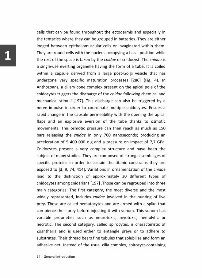
14 | General Introduction
cells that can be found throughout the ectodermis and especially in
the tentacles where they can be grouped in batteries. They are either
lodged between epitheliomuscular cells or invaginated within them.
They are round cells with the nucleus occupying a basal position while
the rest of the space is taken by the cnidae or cnidocyst. The cnidae is
a single-use everting organelle having the form of a tube. It is coiled
within a capsule derived from a large post-Golgi vesicle that has
undergone very specific maturation processes [286] (Fig. 4). In
Anthozoans, a ciliary cone complex present on the apical pole of the
cnidocytes triggers the discharge of the cnidae following chemical and
mechanical stimuli [197]. This discharge can also be triggered by a
nerve impulse in order to coordinate multiple cnidocytes. Ensues a
rapid change in the capsule permeability with the opening the apical
flaps and an explosive eversion of the tube thanks to osmotic
movements. This osmotic pressure can then reach as much as 150
bars releasing the cnidae in only 700 nanoseconds; producing an
acceleration of 5 400 000 x g and a pressure on impact of 7,7 GPa.
Cnidocytes present a very complex structure and have been the
subject of many studies. They are composed of strong assemblages of
specific proteins in order to sustain the titanic constrains they are
exposed to [3, 9, 74, 414]. Variations in ornamentation of the cnidae
lead to the distinction of approximately 30 different types of
cnidocytes among cnidarians [197] .Those can be regrouped into three
main categories. The first category, the most diverse and the most
widely represented, includes cnidae involved in the hunting of live
prey. Those are called nematocytes and are armed with a spike that
can pierce their prey before injecting it with venom. This venom has
variable proprieties such as neurotoxic, myotoxic, hemolytic or
necrotic. The second category, called spirocytes, is characteristic of
Zoantharia and is used either to entangle preys or to adhere to
substrates. Their thread bears fine tubules that solubilize and form an
adhesive net. Instead of the usual cilia complex, spirocyst-containing

General Introduction | 15
cnidocytes bear circlets of microvilli receptors on their apical surface.
The last category called ptychocytes regroups cnidocytes which cnidae
contain simple, spineless, adhesive threads. They are exclusively
present in Ceriantharia, solitary anemones of the Zoantharia class that
use them to build the tube in which they are encased.
Mucocytes represent the fourth type of ectodermal cells. They
are club-shaped cells that can present contractile basal extensions
similarly to the epitheliomuscular cells. They can occupy a major
proportion of the ectodermis, especially on its oral portion, with some
species having mucocytes accounting for as much as 90% of their areal
extent in some areas of tissue [42]. Their size, which ranges from 5 to
10 micrometers wide and up to 30 micrometers high, as well as their
abundance, depends on the species considered and the localization
within the animal [42, 138]. Their nucleus is situated in a basal
position while most of their cytoplasm contains large granular
inclusions that will ultimately be secreted into the water from a
circular aperture situated the apical pole.
Mucus
The observed composition of mucus seems to be very variable.
This could be due in part to the different methods of collection and
analyze, the mucus undergoing quick modification upon release into
the water. Furthermore the mucus could vary in its composition
depending on the stress applied to induce its production [42] or the
species studied [257]. Besides water, which accounts for an important
fraction of mucus weight, the main organic component of the mucus,
is however constant among all species and regardless the conditions.
This component is the mucine, a highly heterogeneous glycoprotein of
which the shape, size and charge determine the mucus viscoelastic
properties (Fig. 5). The mucine is composed of two parts. The core
first, which accounts for 20% the molecule weight (200-500 kDa), is

16 | General Introduction
made of a variable number of tandem repeats of proline, threonine
and serine (PTS-repeats). Variable numbers of PTS-repeats are linked
together by cystein-rich domains and are flanked by multiple “von
Willebrand factor” domains and “cystein knot” domains, thus
completing the molecule core. The remaining 80% of the molecule
consist of carbohydrates either O-linked to the serine and threonine
residues or N-linked to flanking domains. Those sugar side chains are
composed of 2 to 20 monosaccharides and can be linear or branched.
They can be constituted of sugar such as mannose, fucose, arabinose,
galactose, N-acetylgalactosamine and N-acetylglucosamine. The
extremities of the side chains can be sulfated and/or contain sialic acid
terminals. This confers the mucus its characteristic polyionic
proprieties, which are crucial for its hydration. Another major
characteristic of the mucine molecule is the ability of its cystein
residues to form disulfide bonds thus enabling formation of dimers
and subsequent polymerisation [42, 52, 257]. The release of the
mucus on the surface of the coral seems to be under nervous control
[402], corresponds to the holocrine mode of secretion and therefore
leads to the death of the mucocyte. Nevertheless, the dynamic of
their development and their turnover rate is still unclear [42]. This is
contrasting considering the numerous and important roles they play
not only for the coral but also for the whole reef ecosystem.
Figure 5 | Structure and components of the mucine monomer. (modified from [52])

General Introduction | 17
The layer of mucus secreted on its ectodermal surface has
multiple functions for the coral. The most evident one is, as
mentioned earlier, the ability of mucus to trap food particles and bring
them to the mouth thanks to ciliary movement [42]. This function is
especially important for corals devoid of tentacles [342]. The mucus
layer covering the coral also confers it protection against UV
radiations thanks to a variety of UV-absorbing compounds regrouped
as mycosporine-like amino acids (MAA’s) [42]. This is of major
importance in their resistance to light-induced bleaching, a threat of
great concerns as we will see later.
UV protection can also be achieved thanks to UV-absorbing
bacteria living in the mucus layer [309]. In fact, a very diverse bacterial
flora lives within the mucus. Its composition may vary depending
environmental conditions [202] but the major representatives are α-
and γ-proteobacteria [126, 202]. Many of them play crucial roles in
the coral biology and can be seen as symbiotic partners. Coral health
can indeed be tributary to this probiotic bacterial flora as it can
control colonization by opportunistic pathogens implicated in disease
or bleaching event. Recent studies even suggested that this bacterial
flora could stimulate coral immunity [126, 208, 311, 313, 321].
Bacteria in the mucus layer can also be seen as a supplementary
heterotrophic food source accompanying food particles into the
mouth of the coral.
The list of mucus benefices to the coral goes on as it also plays
essential roles in its protections against desiccation during spring
tides, against sediment smothering thanks to ciliary movements,
against pollutant poisoning by limiting their penetration or against
wave damage thanks to its surfactant and lubricant proprieties [42].

18 | General Introduction
Once released into the water the coral mucus keeps playing
important functions on the ecosystem level. Its dispersion from the
mucocytes relies on the current as well as its intrinsic rheological
proprieties, which also controls its exudation, hydration and swelling
[42]. Upon release from the mucocytes, more than half of the mucus
(56% to 80%) immediately dissolves into the water and feeds
planktonic bacteria [404]. The threads and particles released from the
mucus layer into the water carry with them particles of once
suspended organic matter that accumulated onto them while still
attached to the coral. While in the water column, mucus aggregates
keep trapping suspended particles and triple their organic carbon and
nitrogen content [404]. This makes the coral mucus a formidable
energy carrier and a major input source into the trophic system of
coral reefs. This particulate organic matter (POM) released by corals
has already been documented for multiple species and seems to be
constant throughout the year [272, 404]. If macroscopic animals can
ingest some part of this POM, most of it reaches the sediment rather
quickly and therefore relatively closely to its coral source (less than 5
meters)[52]. Once on the permeable calcareous reef sand, aggregates
can either be directly eaten by benthic animals or be degraded by the
microbial fauna living in the sediments providing 10% to 20% of the
total carbon provided to the sedimentary community [404]. The
turnover into nutrients is relatively fast, 7% of the carbon being
transformed in one hour. Those nutrients can then enhance the
primary production of planktonic autotrophic organisms on which can
rely the food chain of the coral reef [52, 105, 404]. Considering these
facts, it appears that any event inducing modifications into mucocyte
number or mucus production by coral would have a strong impact not
only on the coral itself but on the whole reef ecology.

General Introduction | 19
Calicodermis
The description of cells given above concerns the ectodermal
layer of the coral that is in contact with the water column. The
ectodermal layer adjacent to the skeleton and responsible for the
production of its aragonite component is however much different.
This specialized tissue is called calicodermis or calicoblastic
epithelium. It is devoid of cnidocytes and mucocytes and its sole
function resides in the deposition of aragonite crystals through
processes regulated by its dedicated calicoblastic cells. Its surface is
also punctuated by anchoring cells or desmocytes that were formed
on the aboral pole of the planula larvae during its settlement. They
are tightly attached to the coral skeleton by desmosome-like
extensions [71, 137].
The mechanisms by which the calicoblastic cells progressively
construct the mineral skeleton are multifaceted. First of all they
secrete a colloidal gel matrix called organic matrix into the medium
between their apical membrane and the skeleton. This extracellular
calcifying medium (ECM) is therefore filled by an organic framework
that greatly enhances the deposition of calcium carbonate crystals
[71, 136]. The calicoblastic cells also harbor many mitochondria that
not only produce energy but also carbon dioxide which is a source of
carbon for calcification. The important amount of energy produced is
required for the functioning of the multiple pumps and transporters
situated on the apical membrane of calicoblastic cells. They allow the
cells to precisely control the ionic composition and pH of the ECM.
This control is crucial to the growth of the skeleton as the more
alkaline pH measured in the ECM (often 0.2 to 0.5 unit above
surrounding seawater pH) helps producing bicarbonate anions by
shifting the equation of dissociation of carbonate anions to the right
(HCO3- ⇄ CO3
2- + H+). This has the effect of increasing the saturation

20 | General Introduction
state of aragonite (Ωarag) and allows the spontaneous precipitation of
calcium carbonate (CaCO3)[71]. Finally, the calicoblastic cells also
produce carbonic anhydrases, specific enzymes that once liberated
into the ECM play an important role in the interconversion of
inorganic carbon species [395].
As mentioned earlier, the symbiotic algae also play a major if not
essential role in the high rate of calcification observed in reef-building
corals. This was confirmed by studies showing a drastic reduction of
the calcification rate following inhibition of photosynthesis within
these algae [384]. This phenomenon of algal stimulation, known as
light enhanced calcification, boosts CaCO3 production up to tenfold; a
production that therefore outweighs degradation processes such as
wave erosion and allows the reef to expand. This increased
calcification can even produce some sort of positive feedback for the
algae, the structure of the skeleton produced by some corals being
able to increase their light absorption [96]. The way by which
symbiotic algae enhance calcification is however still poorly
understood as both photosynthesis and calcification compete for
available inorganic carbon. A recent hypothesis proposes that such
high rates of calcification and photosynthesis are achievable thanks to
the coral morphology which allows the compartmentalization of these
two processes [185]. Translocation of energetically rich
photosynthates from the algae located in the gastrodermis, as we will
see later, could also fuel mitochondria within the separated
calicoblastic epithelium and enhance their performance. However,
questions still remain concerning the complex movement of ions such
as protons and carbonates which may either diffuse or be actively
transported. Many studies have recently proposed complex ion fluxes
and pathways, especially in the coelenteron and the coenosarc where
the lumen between the two tissue bilayers may play an important
function [8, 41, 185, 270]. There is however still much to understand

General Introduction | 21
before getting a full picture of the mechanisms involved in the
amazing calcifying capacity of corals.
Mesoglea
Beneath the ectodermis lies the mesoglea, an extracellular matrix
mainly composed of collagen and proteoglycans [339]. While it has
always been considered to be barely anhistic and almost deprived of
cells, hence not representing a third type of tissue to the organism,
more and more studies have pointed out the fact that it may be an old
archaic form of mesoderm [347]. Indeed, in most cnidarians,
anthozoans included, muscle cells clearly separated from both
epithelia as well as wandering isolated cells can be observed within
the mesoglea. While muscle cells are organized into bundles and have
obvious contractile functions used for motility, lone cell functions are
much more debated. These cells, called amoebocytes, are often
compared to interstitial cells found in hydrozoans and extensively
studied in Hydra. Whereas interstitial cells stem-like role in growth
and regeneration processes has been clearly indentified [36, 39, 349],
the function or precise identity of amoebocytes is much more vague.
Their most arguable function is their role in immunity and
inflammation following wounding or pathogen infection. In these
cases, amoebocytes may show phagocytic capacity or secrete
connective mesogleal fibers [134, 259]. However, as no unambiguous
description and characterization of amoebocytes has been done to
date, it is impossible to elude the possibility that the term
“amoebocytes” is in fact a catch-all term, referring to wandering cells
from different origins and different degrees of specialization that are
temporarily found within mesoglea. This could include migrating cells
participating in growth and regeneration as well as innate immunity
cells such as granulocytes [287]. This is an even more plausible

22 | General Introduction
explanation considering the fact that amoebocytes have also recently
been observed in both epithelia [134, 380].
Gastrodermis
The gastrodermis is the second tissue layer composing the body
of all cnidarians. Its cellular composition is fairly similar to the
ectodermis but lacks cnidocytes and sensory neuronal cells. The main
cellular components of the gastrodermis are the nutritive-muscle
cells. They are the pendants of the epitheliomuscular cells found in
the ectodermis but differ in some aspects. They are usually ciliated
and their contractile extensions, which are less developed, harbor
contractile fibers orientated perpendicularly to those of the
ectodermis and the axis of the body column. They thus form a circular
muscle that allows sphincter-like movements, especially in the mouth
region to allow its closing. Contraction of those fibers is controlled by
nerve cells underlying the tissue in the same way as in ectodermis but
in a fewer number [24]. Similarly to epitheliomuscular cells, nutritive-
muscle cells show an important plasticity and are therefore believed
to ensure growth and regeneration. They are able to dedifferentiate
and give birth to the other phenotypes present in the gastrodermis
but are also the putative progenitors of cells observed in the
mesoglea. More importantly, they are at the origin of the germ line
precursor cells that will mature into gametes [134]. The last function
of the nutritive-muscle cells is, as their name suggests, to phagocyte
nutritive particles in order to complete their assimilation after their
initial degradation in the coelenteron.
Enzymes liberated by zymogenic glandular cells operate this
extracellular digestion. These cells have a similar appearance to the
mucocytes, also present in the gastrodermis. Their nuclei occupy a
basal position while the apical region of their cytoplasm is filled with
vacuoles containing the zymogen molecules. Both mucocytes and

General Introduction | 23
zymogenic cells harbor an apical cilium surrounded by microvilli [402].
While the secretion mode of gastrodermal mucocytes is likely the
same as in the ectodermis, zymogen liberation has not been clearly
documented yet but could be operated through exocytosis and hence
correspond to the merocrine mode of secretion.
Last but not least, the most characteristic and by far the most
studied cells of scleractinian gastrodermis are the symbiotic host-cells
containing the Symbiodinium algae. These cells are specifically
simplified and harbor usual cytoplasmic components except for one
large symbiotic vacuole called symbiosome. This algae-containing
symbiosome occupies most of the cytoplasm, squeezing the nucleus in
a basal position and giving the cell a round shape that protrudes into
de coelenteron. Whether symbiotic host-cells are readily competent
before algae acquisition has not been established yet but is quite
unlikely. The most plausible explanation is that gastrodermal nutritive-
muscle cells undergo transdifferentiation into symbiotic host-cells
following algae phagocytosis [71]. Not every scleractinian is symbiotic
and therefore possesses this kind of cell. This character has been lost
and/or gained multiple times during the phylogenic history of
scleractinians and is therefore polyphyletic [22]. Today, for the 1314
known species only 48,2% of the genera and 49% of the species
harbor Symbiodinium within their gastrodermis [353].

24 | General Introduction
Symbiodinium
Phylogeny
To understand the mechanisms underlying any symbiosis
establishment and breakdown, it is first necessary to have a good
knowledge of each partner involved. The cnidarian host being amply
described, we shall now bear our attention to the Symbiodinium
algae. Symbiodinium, commonly referred to as zooxanthellae, are
unicellular algae, members of the Dinoflagellata phylum wherein they
belong to the Dinophyceae Class and to the Suessiales Order.
Although zooxanthellae were once all considered as members of a
single pandemic specie, Symbiodinium microadriaticum Freudenthal
(1962), precursor work of Rowan and coworkers using molecular
techniques revealed a much more complex phylogenetic organization
[331, 332]. Recent molecular and genetic analyses, based upon
variation of nuclear ribosomal DNA (18s, ITS and 28s rDNA),
chloroplast 23s rDNA and other specific sequences such as HSP90 or
psbA non-coding region, have revealed that the genus Symbiodinium
is in fact divided into nine large clades (A to I), each comprising
multiple strains or species [60, 173, 216, 220, 303, 322]. Curiously, this
evolutionary radiation seems to be relatively recent compared to the
acquisition of symbiosis by scleractinians. Phylogenetic
reconstructions showed however that their first diversification from a
common ancestor happened only about 50 million years ago during
the Eocene [218, 304].
Habitats
Today, the ability for Symbiodinium to establish symbioses is not
restricted to reef-building corals but also involves a variety of other
cnidarians (octocorals, soft corals, sea anemones and jellyfish) and
some representatives of the Platyhelminthes, Mollusca, Porifera,
General Introduction | 25
Foraminifera and ciliates [53, 354]. Several studies also report the
presence of free-living Symbiodinium in the water column, within
sediments [4, 53, 59, 142, 159, 246, 248, 305, 368, 409] and in
association with macroalgae beds in the vicinity of coral reefs [306].
Nevertheless, the density distribution of free-living Symbiodinium
seems to be highly heterogeneous between and within reefs [246].
While every clade could theoretically be found in the water column
upon release by its host, not all of them seem in fact suited for this
environment. Symbiodinium belonging to clades A and B are the most
commonly found in free-living state while analyzing samples of the
water column and sediments [60, 159, 368] but specimens belonging
to clade C, D, E, F, G and H have also been detected and often reflect
the dominant type found in symbiosis within areas of collection [142,
248, 368, 409]. For some of them the water column would then
represent a transitory niche upon release by its host and before
establishment of a new symbiosis.
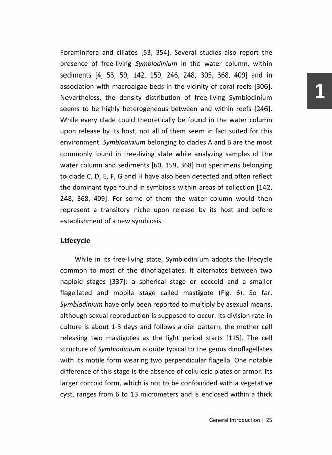
Lifecycle
While in its free-living state, Symbiodinium adopts the lifecycle
common to most of the dinoflagellates. It alternates between two
haploid stages [337]: a spherical stage or coccoid and a smaller
flagellated and mobile stage called mastigote (Fig. 6). So far,
Symbiodinium have only been reported to multiply by asexual means,
although sexual reproduction is supposed to occur. Its division rate in
culture is about 1-3 days and follows a diel pattern, the mother cell
releasing two mastigotes as the light period starts [115]. The cell
structure of Symbiodinium is quite typical to the genus dinoflagellates
with its motile form wearing two perpendicular flagella. One notable
difference of this stage is the absence of cellulosic plates or armor. Its
larger coccoid form, which is not to be confounded with a vegetative
cyst, ranges from 6 to 13 micrometers and is enclosed within a thick

26 | General Introduction
cellulosic cell wall. In this stage, Symbiodinium is also characterized by
a large single, peripheral, reticulated chloroplast bounded by three
membranes [79, 119]. Although Symbiodinium are mixotrophe and
therefore able to feed heterotrophically [180], they rely essentially on
the chloroplast and photosynthesis for their energy incomes.
Figure 6 | Symbiodinium life cycle. Mitosis (1) produces two mastigotes (2) that can return to the coccoid form (3). Tetrads result from meiosis (4) or two successive mitosis (6) and produce four mastigotes. Sexual reproduction is believed to occur during the mastigote stage (5).
Photosynthesis
A brief reminder of the photosynthesis basics seems therefore
adequate. Photosynthesis is divided into two main processes, the light
and dark reactions. In the light dependent reaction, photons are
absorbed by chlorophyll pigments of the antenna to which they
transfer their excitation energy. This energy is transmitted to the
reaction center where it either induces charge separation and
produces an electron or returns to the antenna to be lost as heat or
fluorescence. Thanks to pheophytin and quinone molecules, the newly

General Introduction | 27
produced electron enters then the electron transport chain where it
will ultimately produce NADP+ and NADPH. The chlorophyll molecule
regains its lost electron through the lysis of water molecules which
also produces dioxygen and a proton. This proton, together with
another one produced by the electron transport chain creates a
gradient across the chloroplast membrane that enables production of
ATP by an ATP-synthase enzyme. The dark reactions use the newly
produced ATP and NADPH as well as CO2 to produce C3 sugars. This
process involves the RuBisCO enzyme and a series of reaction called
the Calvin-Benson Cycle.
Clades
Within the nine Symbiodinium clades listed today, all but the
clades E, H and I have been identified within a scleractinian host [60,
303]. Nevertheless, clades A, B, D and especially C are the most
common ones within scleractinians while clades F and G are more
anecdotic and are found only in modest numbers. Clades F, G, H and I
are meanwhile mostly established within foraminifera [60, 354]. The
four major clades living in symbiosis with scleractinians can be found
all around the globe and can be present in different combinations
within the same coral colony. Clade C, however, is the dominant one
across the Indo-Pacific region while clade B is the most encountered in
the Atlantic [217, 218]. The origin of this disparity could be found
within the field of phylogeography with oceans separation and
allopatric speciation events favoring different clades in different
places. Today however, the dominance of some clades or subclades in
some places and the niche partitioning within some habitats is most
probably related to their competitive advantage over others given
their ecological proprieties and environmental conditions [64, 217,
218, 334]. Indeed, while some subtle morphological differences such
as cell size can be observed within clades, those differ mostly by their

28 | General Introduction
biochemical or physiological characteristics such as MAA production,
thylakoids membrane composition or host infectivity [76, 77, 216,
372].
Clade A for example is relatively different from others and has
been described as an opportunistic or even parasitic clade [234, 260,
357, 377]. This can be in some ways related to the ancestral position
of the clade A in Symbiodinium phylogenic tree and its close proximity
with its sister group apicomplexans, well known for their parasitic
lifestyle [234]. Symbiodinium of the clade A rather infect host in
suboptimal health state, releasing less fixed carbon and hence being
less beneficial to them. This ancestral clade has seemingly followed a
different evolutionary trajectory and is probably better adapted to the
free-living state as suggests its ability to outcompete other clades in
cultures [304, 357]. On the other hand, members of the clade A have
been reported to possess an enhanced capacity to use alternative
photosynthetic electron-transport pathways as well as pronounced
ability to dissociate its antenna complex of the photosystem II. They
are therefore less subjected to damages in environment characterized
by high light intensities and could then be beneficial for their hosts in
such environments [314].
In fact, most of the differences between clades rely on their
ability to cope with diverse environmental conditions and abiotic
factors [357]. While some studies have used in vitro cultures of
Symbiodinium to analyze the physiological variations between clades,
especially concerning their photosynthetic apparatus [76, 314, 371],
most observations have been made on symbiotic hosts and
extrapolated to the clades they contain. Early observations of
differential clade distribution among a same colony [329, 330, 381], or
along the depth gradient for example [178, 378], revealed their
discrepancies regarding light tolerance. Most of the recent studies,

General Introduction | 29
however, focus the tolerance of some clades against elevated
temperature and therefore against bleaching. Some pointed out
differences regarding heat resistance between different subclades
within clade C [108, 335] but the majority of the publications are
related to the clade D. Symbiodinium belonging to this clade seems
indeed to have an increased capacity to cope with elevated
temperatures [328]. Studies already highlighted the outcompeting
capacity and the opportunist behavior of these algae, which
sometimes gain prevalence within host that previously suffered from
heat-induced bleaching [219, 375]. They seem also able to confer their
host with an increased resistance to such noxious events [26, 260,
284, 355] or infect aposymbiotic juveniles with greater success when
exposed to high temperature [2]. There are however some drawbacks
with clade D members being almost always suboptimal for their hosts,
causing a decrease in their growth rate and reproductive capacity
compared to conspecific specimen bearing algae of the clade C under
normal conditions [187, 188, 245, 355, 357]. This decrease seems to
be linked to a lower photosynthetic efficiency and a lower lipid store
of Symbiodinium belonging to the clade D. Their tolerance to heat is
indeed likely to be related to a lower maximum relative electron
transport rate as well as a lower chlorophyll-a content [188]. This
explains why the clade D is able to outcompete the clade C during
water temperature anomalies and bleaching events but never achieve
to establish its predominance once environmental conditions returned
to normal.
Clade C and clade B Symbiodinium are, as said earlier, the most
commonly found within scleractinian corals. This could be due to their
relative flexibility regarding to environmental conditions [196] but
more simply, and especially concerning clade C, because they are the
most beneficial for their host’s fitness, releasing the highest amount
of photosynthesis derived carbon [234, 357]. Clade C is particular in

30 | General Introduction
the sense that it seems almost strictly restricted to its symbiotic way
of life with scleractinians. This restriction to a specialized environment
is even more accentuated by the large number of hosts transmitting
clade C Symbiodinium in a vertical manner, directly from the mother
colony to the larvae. This promoted evolution within clade C, that
harbors now a subcladal diversity equivalent to the diversity observed
within other genus of dinoflagelates [354, 357].

General Introduction | 31
Symbiosis1
As we can see, every clade has its specific characteristics and
confers its host with variable fitness. Observations like these,
underlying different outcomes for the host depending on the clade
and the environmental conditions, lead some researchers to propose
the existence of a continuum of relation status between both
partners. This continuum would extend from pure mutualism to plain
parasitism by the algae with a relative position depending on the
environment and the clade but also on the host [234]. Vertical
transmission and obligate symbiosis, as observed with many
representatives of the clade C, favoring mutualism and symbiont
effectiveness for its host while horizontal transmission and facultative
association, as mainly observed for clade A, favoring parasitism. Still,
none of the clades or subclades can be seen as pure mutualists or
parasites but as occupying a subtle position between those two
extremes. Understanding of the relation between coral hosts and algal
symbionts gets even more convoluted as recent studies have even
hypothesized that the host could be itself a parasite. Corals could
indeed be actively farming Symbiodinium and would then choose with
which to establish the most advantageous symbiosis [407]. Such an
active or even exclusive participation of the host in the symbiosis is
supported by studies showing that symbiosis fitness not only relies on
the symbiont and environmental conditions but also on the host itself
[145]. This leads to the idea that the host can operate, at least in part,
a choice over its dominant symbiont composition, relegating other
algal populations, which are almost always present [19, 261, 356], to a
minority fraction. This choice may obviously depend on Symbiodinium
availability but also on the host physiological characteristics in
1 Parts of the following section have been published as a review article in
Journal of Experiment Marine Biology and Ecology. (cf. [118])

32 | General Introduction
response to environmental conditions. This multitude of factors to be
taken into account makes it very tricky to understand the reason why
some clades or subclades are present in some corals within some
locations. Cladal composition could indeed vary between different
geographic sites at global but also local scales, sometimes even
showing some form of endemism [123, 374, 383]. Monitoring showed
that, within these particular locations, the symbiont composition
inside different coral taxa or even colonies seems however to be
stable as long as environmental conditions stay steady [144, 301, 323].
Yet, its reaction to environmental stress and its eventual switch to a
composition dominated by another clade are not unanimously
recognized and may be species specific [18, 143, 374, 375]. This
hypothesis will be addressed later, when we discuss the subject of
coral bleaching.
Transmission
Various degrees of cladal composition specificity exist among
coral species [17]. Such specificity can be observed early during the
life of corals. This is especially true for species that acquire their
symbionts through vertical transmission. In this case, algae are
transmitted from the mother colony to its egg or brooded planula
larvae prior to release [28, 29]. Interestingly, during early stages of
development, algae can be found within both ectodermis and
endodermis [175, 416]. This suggests an initial, and later lost,
competence of ectodermal cells to host algae. The developing larvae
perpetuate then the association present within its species local
population. This mechanism is however a minority, representing only
15% to 20% of hermatypic scleractinian coral species with particular
occurrence within brooding species. The vast majority of corals rely on
horizontal transmission where aposymbiotic larvae recruit new algae
from the surrounding water [15, 99, 318, 326, 345, 367]. Larvae or

General Introduction | 33
juveniles of these coral species are able to initially associate with
either homologous (same as adult colony) or heterologous clades of
Symbiodinium [1, 51, 67, 153, 245, 325, 345, 398]. Such early
association has also been reported in cultured gorgonians and sea
anemones [61, 73]. However during the following months, the initial
distribution of symbionts progressively adjusts to the most common
distribution found in adjacent adult colonies [1, 61, 140, 245]. The
host seems indeed able to finely tune its symbiont composition thanks
to alternative discharge mechanisms [410]. This corroborates results
showing that the pattern of transmission has only little effect on the
Symbiodinium composition of some coral species [382] and suggests
the presence of specific mechanisms to favor the development of an
optimal relationship between the coral and a specific symbiont type.
Acquisition
During this so-called horizontal transmission, many studies
showed that larvae acquire their symbiont during a nutritional process
[153, 160, 325, 326, 345, 346, 398]. Although symbiont infection
through feeding is probably the major acquisition mechanism, it could
also be facilitated by active mechanisms deployed by Symbiodinium.
Indeed, as described earlier, Symbiodinium cells interchange between
a coccoid form, which is always the one encountered when
endosymbiotic, and a motile zoospore. The motility of the flagellated
Symbiodinium is regulated in a diel-cycle and cells are active under
illumination [66, 111, 115, 228, 408]. During that period,
Symbiodinium seems able to swim in an oriented manner, thus
showing phototaxis [169] and/or chemotaxis [110, 292]. However, a
potential host-seeking behavior remains limited by the flow velocity
and the proximity of a host [293]. Finally, the success of Symbiodinium
acquisition strongly depends on environmental conditions. In the
same manner as during bleaching, which will be extensively discussed

34 | General Introduction
later, high temperatures accompanied by elevated light irradiance
have been demonstrated to drastically impair algae uptake.
Symbiodinium concentration in juvenile corals falls from 87% at 28°C
and high light to 8% at 31°C [2].
Figure 7 | Schematic illustration of Symbiodinium infection of a cnidarian host cell. Motile or non-motile (cyst) Symbiodinium enter the gastrodermal cavity of the host. (1) Lectins secreted by the host cell induce motile algae to progress to the cyst stage. (2) Contact and recognition are mediated by other lectins present on the host cell surface and glycoproteins on the surface of the non-motile Symbiodinium. (3) Symbiodinium are phagocyted and oriented to the early endocytic compartment. (4) Healthy Symbiodinium end in a functional symbiosome while damaged Symbiodinium are digested by fusion with lysosome after transiting through late endocytic compartment. Rab5, Rab7 and Rab11 are respectively involved in the early endocytic compartment, late endocytic compartment and endosome recycling. G: Golgi apparatus, L: Lysosome, S: Symbiodinium.
In order to establish the symbiosis, both partners first have to
recognize each other. Recognition mechanisms could be similar to the
winnowing mechanism, which consists in a multi-step process
involving both partners, that was first described by Nyholm and

General Introduction | 35
McFall-Ngai [278] for the symbiotic relationship between the bobtail
squid, Euprymna scolopes, and its bioluminescent bacterium, Vibrio
fischeri. These mechanisms necessarily take place before, during, and
after contact with Symbiodinium and its internalization, leading
ultimately to the formation of a stable mutualistic relationship [71].
Recognition mechanisms between host cells and algae seem to be
necessary, at least at some point, during vertical or horizontal
transmission. Indeed, while it is quite obvious for horizontal
transmission, Marlow and Martindale [251] showed that recognition
events could also be involved during vertical transmission in
Pocillopora meandrina.
According to Weis [394], among others, the onset of the infection
by Symbiodinium involves the same mechanisms as those acting in the
recognition of pathogenic organisms (Fig. 7). For many eukaryotes, the
innate immune system implies the production of pattern recognition
receptors (PRRs) able to recognize and bind to specific conserved
components of microbe cell walls (carbohydrates, proteins, lipids;
[200]). Among those PRRs, lectins (carbohydrate-binding proteins;
[139]) are widely distributed in most classes of living organisms and
are thought to play an important role in various symbiotic associations
(e.g.: [50, 161, 264]). To date, lectins have been described in, at least,
two cnidarians classes (Hydrozoa and Anthozoa) and have been
reported in five scleractinians, including Acropora millepora [213],
Montastrea faveolata, Oculina patagonica [155], Pocillopora
damicornis [385] and Ctenactis echinata [183]. Several studies have
thus examined the implication of lectin/glycan interactions in the
recognition process between cnidarians and Symbiodinium.
Lin et al. [243] identified ten different proteins on the cell surface
of symbiotic dinoflagellates. Among those figured five glycoproteins
with two kinds of terminal sugar residues: mannose-mannose and

36 | General Introduction
galactose-β(1-4)-N-acetylglycosamine. Moreover, cell surface glycan
profiles of cultured Symbiodinium seem to be stable throughout their
life history, indicating that Symbiodinium may maximize the potential
for host recognition by retaining recognition molecules throughout
their vegetative growth [247]. The importance of these membrane-
bound glycoproteins during the infection of the sea anemone Aiptasia
pulchella with Symbiodinium has been demonstrated by masking or
removing cell surface glycans of the dinoflagellate. Indeed,
Symbiodinium incubated with either trypsin, α-amylase, N-
glycosidase, O-glycosidase or with different kinds of lectins, in order to
remove or mask carbohydrate groups, showed a significant decline in
the infection rate of an aposymbiotic host [243]. Similarly,
Symbiodinium treated to digest or mask α-mannose/α-glucose and α-
galactose residues failed to efficiently infect larvae of the fungiid coral
Fungia scutaria [405].
These membrane-bound glycoproteins could play a role in
Symbiodinium recognition by acting as “markers” that could be bound
by various types of lectins. SLL-2 is one of these lectin-type proteins.
This (N-glycosylated) galactose-binding lectin, isolated from the
octocoral Sinularia lochmodes, is preferentially localized not only in
nematocysts but also on the cell surface of Symbiodinium, whether
they are present within a host cell or in the coelenteron [181, 184].
The most surprising propriety of SLL-2 is its ability to induce the switch
of flagellated and motile Symbiodinium cells to a non-motile coccoid
form still able to divide [205]. Such physiological transformations may
favor a condition more suited for the establishment of symbiosis. SLL-
2 action on Symbiodinium seems to present more than a simple
carbohydrate binding property as shown by the lack of algal
modification in the presence of proteins showing similar properties as
SLL-2. Koike et al. [205] suggested that SLL-2 could be stored within
nematocysts and released into the gastrodermal cavity in the

General Introduction | 37
presence of micro-algae. It would then bind to glycolipidic residues
present on their surface [182]. Response of these algae to SLL-2 could
therefore act as a first screening process, modifying algal physiology
to favor further interactions with the host or causing damages to
micro-algae unsuitable for symbiosis. Recently, CecL discovered in
Ctenactis echinata showed effects on cell transformation highly
analogous to those of SLL-2. It seems that CecL lectin also has the
ability to temporarily suppress the rate of cell division without
affecting cell viability and thus, can regulate Symbiodinium density in
the coral gastrodermis, where only a limited number of algae can be
accommodated [183].
Another lectin has been identified in Acropora millepora [213].
Named millectin, this lectin possesses a Ca2+-dependent
carbohydrate-binding site that preferentially binds to mannose and
similar sugars. According to immunohistochemical analysis, millectin is
localized in nematocysts present in the epidermal tissue [214]. This
protein is phylogenetically close to collectins, which play a key role in
vertebrate innate and adaptive immune responses. Millectin
expression is up-regulated in response to lipopolysaccharides and
peptidoglycans. Moreover, it has the ability to bind to both gram +
and gram – bacteria (including Vibrio coralliilyticus, a coral pathogen)
and to various clades of Symbiodinium (C1, C2 and A2) in vitro and in
vivo [213, 214]. When bound to pathogens, millectin prevents their
dispersion into the host and induces opsonisation for phagocytosis
and destruction. Finally, numerous millectin isoforms showing amino
acid substitution sites in close proximity to the binding site have been
identified. This vast diversity of millectins suggests a probable
appearance of some recognition specificity and a role of millectins in
the winnowing process [213].

38 | General Introduction
Recently, the sequence variation of a putative coral immunity
gene, tachylectin-2, has been investigated in the coral Oculina
patagonica [155]. Tachylectin-2 was originally isolated from the
Japanese horseshoe crab (Tachypleus tridentatus) and has been
demonstrated to possess anti-microbial activity [281]. Nevertheless, a
role for Tachylectin-2 in the symbiosis of coral species has not been
confirmed yet.
Lectins also appear to play an important role under stress
conditions. Indeed, the downregulation of the transcriptional
expression levels of two C-type lectins has been observed in
aposymbiotic larvae of A. millepora [324] and nubbins of Pocillopora
damicornis exposed to thermal stress (PdC-lectin; [385]). Conversely,
the same gene has been shown to be up-regulated when exposed to
the pathogen Vibrio coralliilyticus under virulent conditions [386].
These contrasting results underline the complexity of these
lectin/glycan interactions and the tenuous link between the coral
physiological response and the establishment of the symbiosis.
Lectin-type proteins are not the only molecules involved in
mutual recognition between host and symbiont; other proteins have
also been reported to be over-expressed in the presence of
symbionts. These include AtSym-02, a glycosylated membrane protein
identified in Acropora tenuis [412], and Sym32 identified in
Anthopleura elegantissima [315]. AtSym-02 may belong to the
fasciclin-I (fasI) gene family, also known to be involved in cell-cell
recognition mechanisms [412]. Sym32 can be found both in anemone
protein homogenates and bound to membranes. Sym32 is also
expressed more in anemones infected by Symbiodinium than in those
hosting green algae (Zoochlorellae), leading the authors to
hypothesize that the symbiont has the ability to control the
expression of some host genes [315].

General Introduction | 39
Establishment
After the recognition step, symbionts are internalized by a
phagocytic process conducted by host cells, probably through the
formation of a lectin-binding complex. Then, a wide range of other
cellular processes are necessary for the maturation of Symbiodinium-
containing phagosomes into functional symbiosomes able to avoid
fusion with the host endolysosomal system during endosymbiosis [55,
114]. Indeed, while normal phagosomes mature by fusing with
lysosomes, early observations showed that healthy Symbiodinium-
containing symbiosomes did not reach that stage [114], suggesting the
existence of host and/or symbiont-specific molecules involved in
mechanisms maintaining the symbiosis once established. Parts of
these mechanisms, such as modulation of TGFβ synthesis, could be
similar to those employed by some parasites to promote host
tolerance [75, 344], while others could be specific to this symbiosis.
Although the mechanisms of symbiosome membrane formation
are still poorly understood, some evidence in Aiptasia pulchella
indicates that ApARF1 and ApRab genes could be involved in this
particular process. ApARF1 is homologous to ARF1, a member of a
family that regulates intracellular vesicle transport and its gene
expression appeared reduced in symbiotic anemones [56]. However,
further investigation is needed to clarify apARF1 function in the
establishment of symbiosis. The role of ApRab seems more
convincing. ApRab shows similarities with Rab family members, coding
for small GTP binding proteins found in many vertebrates, where they
assume the regulation of vesicular trafficking, membrane fusion and
also the biogenesis and the function of membrane-bound organelles
[171]. To date, four of these ApRab proteins have been shown to play
a role in the establishment of symbiosis.

40 | General Introduction
ApRab7 usually participates in late acidic endocytic and
phagocytic pathways [387]. However, in A. pulchella, ApRab7 is
excluded from symbiosomes containing either resident or newly
internalized Symbiodinium. ApRab7 can also be found on phagosomes
containing heat-killed or PSII-impaired (DCMU-treated) Symbiodinium,
where it probably promotes their maturation and fusion with
lysosomes [55].
ApRab5 appears before ApRab7 and is present in the early
endocytic and phagocytic compartments where it can promote their
fusion [23]. In A. pulchella, ApRab5 localizes on symbiosomes
containing healthy algae and is absent from symbiosomes in which
algae have been damaged by heat or DCMU treatment. These
observations suggest that active retention of ApRab5 by
Symbiodinium participates in their persistence in the host, possibly by
preventing ApRab7 binding. Indeed, these two proteins never co-
localize in A. pulchella vesicles [54].
ApRab11 acts during endosome recycling processes, a necessary
step towards maturation [415]. In A. pulchella ApRab11 can be located
on phagosomes containing damaged symbionts but is absent from
those containing healthy ones [57]. By actively excluding ApRab11, the
authors observed that Symbiodinium interferes with the vesicular
recycling process and thus prevents the maturation of their
symbiosome and its fusion with lysosomes.
ApRab3 is the last of the ApRab family members identified in
cnidarians. It appears to be preferentially localized in the
compartments of the biosynthetic pathway including both the Trans
Golgi Network and a subpopulation of secretory vesicles. In A.
pulchella, symbiosome formation seems to involve interactions with
ApRab3-positive vesicles. However its function in the symbiosis

General Introduction | 41
establishment remains to be determined, as phagosomes containing
either healthy or damaged Symbiodinium or even latex beads
progressively accumulate ApRab3-specific labeling [171].
The differentiation of phagosomes into functional symbiosomes
obviously requires some particular cellular developments in order to
maintain the endosymbiosis and to optimize mutual exchanges.
Indeed, the analysis of symbiosome structure conducted on Aiptasia
pallida, using transmission electron microscopy and immunological
techniques, revealed a membrane complex composed of a single host-
derived outer membrane and a multilayered inner membrane
originating from the algal symbiont [388, 389]. The presence of
symbiont thecal vesicles in situ suggests that this multilayered
membrane could be the result of a continuing delayed in situ ecdysis
cycle [388]. Anyway, the formation of this kind of structure requires
some regulation by the cytoskeleton of the host cell and its symbionts,
allowing normal host cell physiology [295]. Moreover, proteomic
analyses of isolated symbiosome membranes conducted on A. pallida,
highlighted the presence of proteins highly similar to GPCR family
proteins. These receptor-like proteins are distributed to symbiosome
surface and should be involved in various cellular responses and
modulation of host gene expression [295]. The presence of ATP
synthase complexes and ABC transporters on this structure
demonstrates the pivotal process of molecular transport during the
mutualistic association [295].
Exchanges
Indeed, the symbiosis is considered to be optimally functional
when a significant amount of photosynthates generated by
Symbiodinium is transferred to the host cells. Once in symbiosis, the
symbiont provides up to 95% of its photosynthetic products (glycerol,
glucose, amino acids or lipids) to the host [250, 265, 267], thus

42 | General Introduction
contributing massively to its energy demands [128]. In many species,
half of this translocated carbon is used for mucus production [404].
Lipid droplets could play an important role in these transfers as shown
by the light induced rhythmicity observed in their formation and
localization [58, 294]. A significant part of the photosynthetically fixed
carbon is translocated through processes controlled by host release
factors (HRFs) which are yet to be clearly identified [411]. HRFs are
believed to divert surplus of assimilated carbon from the synthesis of
storage compounds to translocated compounds. Interestingly, if the
heterotrophic incomes of a coral host become scarce, this leads its
nitrogen-limited symbionts to allocate more carbon to storage and
therefore to its starving host [72]. Contrastingly, translocation of
photosynthates could also be induced by the symbiont. This
hypothesis, known as phagosome arrested hypothesis (PAH) states
that Symbiodinium stays hidden in its host cell by mimicking a
constant prey digestion by the endosomal machinery through
perpetual release of carbon based nutrients [157]. Regarding the
amount of transferred molecules, especially glycerol, osmolarity
within the host cell is susceptible to many changes; its fine regulation
has recently been pointed out as a key factor in symbiosis regulation.
While increase in osmolarity has been shown to enhance release of
photosynthates from algae [364], its decrease, due to photosynthesis
failure for example, could lead to hyperosmotic stress and ultimately
to symbiosis breakdown [253]. Finally, as said earlier, high O2
concentration due to Symbiodinium photosynthesis also helps in
maintaining the high ATP level needed for the calcification process
[185].
In return for these services, the host ensures protection to
Symbiodinium and provides a source of inorganic nutrients (CO2, NH3
and PO4³-)[411]. Carbon dioxide transfer to the algae seems
particularly crucial considering its low concentration in seawater and

General Introduction | 43
the poor discrimination of dinoflagelates RuBisCO II between O2 and
CO2. The host addresses this issue not only with CO2 production by
respiration processes but also thanks to mechanisms concentrating it
from the surrounding water. These mechanisms mostly involve
various isoforms of the carbonic anhydrase (CA), an enzyme operating
conversions between HCO3- and CO2, which is upregulated in
symbiotic cnidarians [120, 397].
Regulation
Under normal conditions, Symbiodinium densities in symbiotic
corals reach a steady state wherein neither partner outgrows the
other [266]. The maintenance of this dynamic equilibrium suggests
the existence of intrinsic and environmental factors that can
potentially regulate algal density pre- or post-mitotically [168, 269].
This regulation involves a variety of mechanisms, like the limitation of
algal nutrient supply [101], the digestion of algae [195, 269, 376], the
expulsion of excess or dividing Symbiodinium [13, 168, 195], the
accommodation of excess algae by division of host cells [376] and
possibly the production of growth inhibiting factors [352]. This fine
regulation of algal density also occurs on daily [109, 166, 195, 361]
and yearly bases showing, for example, seasonal variations [43, 100,
109, 360]. Expelled algae can then be recruited by gastrodermal cells
or released to the ambient environment. Symbiotic algae that are
released every day to the external environment, either through
density regulation mechanisms [166, 361] or through survival of
corallivorous species digestion [12, 262], may contribute to the
maintenance of a free-living Symbiodinium population.

44 | General Introduction
Bleaching
Under atypical or extreme conditions however, this seemingly
stable equilibrium often suffers severe impairments that ultimately
lead to its collapse. This phenomenon, revealing the coral white
skeleton beneath its algae-depleted tissues, is known as coral
bleaching (Fig. 8). This sadly famous incident is the central element of
a majority of recent coral-related studies, including this one. The main
reason of this infatuation is, as said earlier, the upsurge of scenarios
based on climate change predictions considering a baneful destiny for
coral reefs. The urge for a better understanding of coral bleaching is
even more strengthened by the increased number of field
observations witnessing its occurrence all around the world. The
change is underway and the threat needs to be taken seriously. Since
the first alarming observations of coral bleaching, their subsequent
death and the species disappearance in Panama following a warming
event in the early 80’s [132, 133, 223, 236], reports of bleaching keep
accumulating. Whether they come from the Caribbean region [81, 84,
122, 125, 327], the Pacific Ocean and the neighboring South-East Asia
[30, 47, 130, 186, 279] or the coasts of the Indian Ocean [44, 221, 237,
255, 256], they all give the same feeling of anxiety and alarmism
reporting reduction of coral health, cover and architectural complexity
accompanied by modifications of the benthic community.

General Introduction | 45
Figure 8 | Illustration of a bleached Acropora sp. (From en.wikipedia.org)
Causes
Coral bleaching has been rapidly linked to elevated sea surface
temperatures [141] but the concomitant cellular responses to this
abiotic stress have been slow to be elucidated and are still not totally
understood. Scientists particularly struggle to understand the
respective responsibilities of each partner (coral, algae and bacteria)
in the collapse of the symbiosis. Many fingers are however pointed
toward Symbiodinium and its photosynthetic apparatus. This is
legitimate considering studies demonstrating a link between algal
density and symbiosis susceptibility to light and/or thermal stress in
cultured cell aggregates [275], planula larvae [274] or adult colonies
[68]. Every investigation undertaken to identify these intimate links
between Symbiodinium and bleaching leads to the same culprits:
photosynthesis dysfunction and reactive oxygen species (ROS)
production [83, 179, 298, 351]. Subtle distinction can then be made
concerning the different effects and relative responsibilities of light
and high temperatures here. Sunlight could be considered, with the

46 | General Introduction
energy it carries, as the source of the ROS, the toxic compounds that
lead to bleaching, while elevated temperature is the cause of
impairment within the photosynthesis apparatus that ultimately leads
to this production of ROS. High UV radiations could also induce some
deterioration of photosynthesis components and have been subject of
a few studies in the past [21, 230, 233, 235]. UV could especially
induce reduction in chlorophyll synthesis and exacerbate effects of
visible light and heat on photosynthetic apparatus. This abiotic factor
is however barely affected by climate change, thus relegating its
implication in increasing coral bleaching events as a facilitating factor
but not a main cause such as elevated temperature.
Within chloroplasts machinery of Symbiodinium, photosystem II
(PSII) is the most sensitive component to light and therefore the main
siege of the degradations leading to ROS production. It can suffer from
multiple sorts of damages that often result in a process called
photoinhibition, a drastic reduction of photosynthetic capacity.
Several studies have pointed out the role of the D1 protein during
heat related photoinhibition in Symbiodinium. The D1 protein plays a
central role in PSII functioning but is also its most vulnerable
component: it is very sensible to heat and can be easy destabilized. An
active reparation mechanism fortunately exists but often fails to cope
with the high rate of D1 degradation occurring during high water
temperature events. Some experiments even showed that D1
reparation mechanism could itself suffer from heat. Following D1
degradation, excitation energy starts backing up and finally leads to
photoinhibition. This is usually translated by a drastic reduction of
photosynthesis efficiency, measured by its maximum potential
quantum yield (Fv/Fm) [369, 392, 394]. Similar effects can be
observed with chemicals such as DCMU, an herbicide blocking the PSII
electron transport chain [190]. Elevated temperatures seem also able
to impair the dark reactions of the photosynthesis, altering CO2
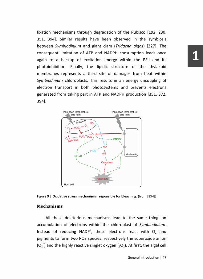
General Introduction | 47
fixation mechanisms through degradation of the Rubisco [192, 230,
351, 394]. Similar results have been observed in the symbiosis
between Symbiodinium and giant clam (Tridacna gigas) [227]. The
consequent limitation of ATP and NADPH consumption leads once
again to a backup of excitation energy within the PSII and its
photoinhibition. Finally, the lipidic structure of the thylakoid
membranes represents a third site of damages from heat within
Symbiodinium chloroplasts. This results in an energy uncoupling of
electron transport in both photosystems and prevents electrons
generated from taking part in ATP and NADPH production [351, 372,
394].
Figure 9 | Oxidative stress mechanisms responsible for bleaching. (from [394])
Mechanisms
All these deleterious mechanisms lead to the same thing: an
accumulation of electrons within the chloroplast of Symbiodinium.
Instead of reducing NADP+, these electrons react with O2 and
pigments to form two ROS species: respectively the superoxide anion
(O2–) and the highly reactive singlet oxygen (1O2). At first, the algal cell

48 | General Introduction
tries to cope with this ROS production, alleviating their effect with the
help of detoxification enzymes such as the superoxide dismutase
(SOD) and ascorbate peroxidase (APX). It is then important to note
that the less damaging H2O2 produced by SOD is likely, in presence of
Fe2+, to transform into the hydroxyl radical (·OH), the most reactive
ROS. These detoxification mechanisms set in place by the algae will
however be eventually exceeded. Accumulation of ROS will then
engage a positive feedback loop, degrading pigments and reacting
with D1 proteins and photosynthetic membranes to further damage
the photosynthetic machinery [192, 231, 372, 394].
ROS produced by the algae eventually diffuse into the cytoplasm
of the gastrodermal host cell. From there, things get a bit more
complicated (Fig. 9). A direct response to the stress from the host cell
itself becomes indeed difficult to discern from its reaction to the
diffusion of ROS from its symbiont. This is particularly true regarding
the ROS produced by the host and their addition to those coming from
the algae. These come from the host mitochondria, which are also
susceptible to damages from the heat [89]. All these ROS elicit the
establishment of an antioxidant response by the host cell. This
consists in the production of different types of SOD, as well as catalase
(CAT), ascorbic acid, carotenoids and mycosporine glycine [14, 238,
258, 317]. Similarly to the algae, this response quickly becomes
overwhelmed by the amount of ROS, still accumulating in the
cytoplasm, and fails to prevent their oxidizing nature to cause
damages to the DNA, membranes and proteins of the host [231, 232,
316]. A recent study even suggests that the impact of ROS could even
go beyond the collapsing cell membrane and diffuse into its
extracellular environment to affect surrounding cells [338].
ROS may however not be the only molecules involved in the
cellular bleaching cascade. Many studies have demonstrated that

General Introduction | 49
nitric oxide (NO) also plays an important role in the induction of the
symbiosis collapse. Origins of this molecule could be multiple. It could,
in part, diffuse from the algae where high temperatures have been
demonstrated to induce its production [40]. NO is also believed to be
synthesized in high concentrations within the host cell in response to
heat stress. This production involves the activation of the transcription
by NF-κB of the gene coding for the inducible nitric oxide synthase
(iNOS)[296, 394]. Nitric oxide would then activate apoptosis pathways
either by direct action over p53 or through damages made to the
mitochondrial membrane by its derivate peroxynitrite (ONOO-), which
forms in reaction with superoxide [154, 231, 296, 394]. Finally, some
studies demonstrated that calcium concentration seems to play a
significant role in bleaching. They showed that heat stress increases its
cytoplasmic concentration and that bleaching could be inhibited using
calcium chelator or ionophore to reduce its extra- and intra-cellular
concentration [102, 176].
Countermeasures
This list of mechanisms, although trying to be as exhaustive as
possible, probably does not represent what exactly happens within
every coral gastrodermal cells during bleaching. Not all of these
mechanisms may occur but different combinations may or may not
cross the threshold that will elicit breakdown of symbiosis. Moreover,
multiple studies have shown that coral response to a defined abiotic
stress may vary depending on the host species [33, 112]. This could be
related to their different intrinsic abilities to cope with cellular stress.
Indeed, the host cells often deploy a large panel of molecules trying to
overcome or alleviate the perturbations happening in its cytoplasm.
The type and concentration of these molecules may vary between
different species, explaining their different susceptibility [14, 112].
Aside from the antioxidant system, another main strategy consists in

50 | General Introduction
the production of fluorescent pigments (FP’s) that dissipate high-
energy light radiations. The different production capacities of these
pigments between coral species are very likely to define, in part, their
variable susceptibilities to bleaching [14, 287]. The aforementioned
MAA’s are also found within the host cytoplasm where they play a
crucial role absorbing UV radiation and dissipating their energy
without formation of ROS. The shikimic acid pathway that leads to the
formation of such molecules excludes their possible production by the
host cell. MAA have thus either to come from heterotrophic feeding
or from translocation from the algae. Even if the algae also benefit
from the highest MAA concentration found in its host cytoplasm,
which partner of the symbiosis control their complement and
distribution is still largely unknown [14, 120]. Finally, a last known
strategy of the host to alleviate the cellular stress and escape
bleaching consists in the upregulation of heat-shock proteins (HSP’s).
These are molecular chaperones that help maintaining proteins
tertiary structures and functions during stressful conditions. Their
increased production seems to be related to the heat-related buildup
of calcium concentration within the host cell [14, 102].
Algal loss
All these procedures may however not be sufficient to prevent
the host cell from crossing the threshold that will engage it into
bleaching. Defining this tipping point and the succession of events that
follows therefore represents important steps in the understanding of
the coral-bleaching phenomenon. Such is not an easy task as many
mechanisms are evoked to explain the disappearance of algal
coloration. While some studies have shown that visual bleaching of
coral can be sometimes related to a diminution in chlorophyll
concentration [370, 394], scientists agree that disappearance of
Symbiodinium from the host gastrodermis is the main cause of coral

General Introduction | 51
bleaching. How this happens has been largely debated and the
mechanisms proposed in the literature can be organized in two
categories: those that do not imply the loss of the host cells and those
that do (Fig. 10).
Host-cell persistence
Mechanisms involving persistence of the host cell might only take
place during mild environmental stress or before damages provoked
by ROS production become too severe. Such mechanisms are most
often related to in situ degradation of the algae as reported by
microscopic examination performed on corals [6, 45] and sea
anemones [88, 93, 116, 362]. This degradation is believed to result
from two possible mechanisms. The first mechanism involves
damages caused by ROS, such that they induce the death of the algae.
This death can occur by means of programmed cell death (PCD) or
apoptosis with the algae showing typical cellular patterns such as a
condensed nucleus, crenated membrane and condensation of
organelles and cytoplasm [88, 93, 116, 362]. ROS damages to the
algae cellular components can be so harsh that the PCD cannot be
established and the death occurs in an uncontrolled manner. The
algae therefore die showing classic necrosis features that are pyknotic
chromatin, vacuolization, breakdown of cellular structures and
rupture of the membranes [88, 92, 93, 116, 362]. The second
mechanism of algal degradation implies active degradation by the
host cell and digestion of the remains. This mechanism could be
spontaneously initiated following the impairment of the ApRab
pathway manipulation by the algae. This one can therefore no longer
prevent maturation of its symbiosome into a phagosome and lysis of
its component by enzymes contained in lysosomes. It could also
involve autophagy, a catabolic pathway responsible for the recycling
of organelles as well as other cellular key functions. In this case, it is

52 | General Introduction
sometimes called symbiophagy and actively participates in algal in situ
degradation by promoting fusion of the phagosome with lysosomes
[85, 91, 152]. Other scenarios sparing the host cell during algal
expulsion have been proposed but are not unanimously accepted.
Those were rarely described in the literature and include algae
exocytosis [358] and the pinching off of a portion of the host cell
cytoplasm [336].
Figure 10 |Host-controlled cellular processes involved in Symbiodinium loss. A. Schematic representation of the coral polyp with its tissular organization. B. Illustration of the mechanisms believed to be involved in the release of Symbiodinium from its host cell. S: Symbiodinium, H: Host cell. (Adapted from [127, 394])
Host-cell loss
Although it could only concern severe cases of bleaching-related
intense stress [363], many studies reported that loss of algae during
bleaching is often accompanied by the loss of their host cells. Two
scenarios are responsible for this loss, both happen consequently to
major disruptions of the host cell homeostasis. Perturbation of the

General Introduction | 53
calcium cytoplasmic equilibrium leads to the collapse of the
cytoskeleton and perturbations of the cell adhesion. The host cell
detachment that follows has often been described as a cause of
Symbiodinium loss [45, 127, 341]. The vast majority of recent studies,
however, agree to say that Symbiodinium loss during bleaching is
largely caused by the death of their former host cells. Cellular swelling
as well as other cellular characteristics of necrosis can sometimes be
observed [223]. Nevertheless, necrosis is likely related to extreme
stress conditions while apoptosis seems to be much more frequent
and is described in recent studies [92, 93, 154]. As evoked earlier,
environmental stresses experienced during bleaching are believed to
induce apoptosis following multiple cascades of events. Heat as well
as ONOO- and ROS can cause damages to host mitochondria and
therefore induce activation of caspases and other pro-apoptotic
factors. Nitric oxide also activates p53, a pro-apoptotic transcription
factor that also activates caspases. Finally, NF-κB, which is
upregulated due to ROS concentration, can have a direct action over
apoptosis activation [317, 394]. The host seems however to operate
some control over this caspase-induced apoptotic pathway. Some
studies showed fine regulation of Bcl-2 protein family members
following bleaching induction of apoptosis [215, 299]. Authors thus
propose that, following caspase activation, coral quickly modulates
this response in order to limit apoptosis and prevent its progression to
other cells to limit the damages and optimize its survival. Happening
prior to bleaching, the suppression of the cell death cascade within
these cells will allow them to serve as basis for later tissue
regeneration [7, 373]. Moreover, apoptosis and autophagy seem to be
interconnected as demonstrated by experiment showing induction of
one while the other is inhibited [91]. A similar “see-saw” mechanism
has already been reported in vertebrates during immune response to
pathogens and parasites.

54 | General Introduction
Altogether, these information combine to form the following
hypothesis of what is happening during bleaching. Exposed to
environmental stressful conditions (high light and temperature), the
cnidarian gastrodermic host cell will experience diverse degrees of
perturbations depending the intensity of the stress, the algal clade it
contains and its cellular equipment, which vary depending its species.
Consequently, the host cell will identify algae, which are already
suffering and could be dying, as the source of the stress and will try to
get rid of it. Depending the scale of the perturbation it has suffered, it
will favor “soft” mechanisms such as autophagy before using
apoptosis. However, if the stress is too strong, the host cell will
ultimately lose control of its homeostasis and either detach or
undergo necrosis.
Adaptative bleaching
After release from their former host cells, Symbiodinium
accumulate within the gastrodermal cavity before being expelled in
the form of aggregates embedded in mucus pellets. It is then
important to notice that multiple studies have observed that most of
these algae are not always condemned and seem to be able to recover
from the stress and multiply in the water column [34, 158, 307, 308].
The expulsion by the host of these stressed but seemingly viable algae
corroborates the adaptative bleaching hypothesis (ABH). According to
this hypothesis, the host could use aforementioned mechanisms such
as apoptosis to eliminated algae unsuited to environmental condition
in order to establish symbiosis with better ones. Such radical way of
proceeding has even been documented in the winnowing mechanism
during symbiosis establishment in coral larvae [94]. The ABH was
initially proposed by Buddemeier and Fautin [49] who considered
bleaching as an opportunity for the host to be repopulated by new,
stress tolerant, algae [103]. Mathematical modelization [391] as well

General Introduction | 55
as evaluation of its basic assumptions [82, 201] further strengthened
this hypothesis. This shift between two dominating Symbiodinium
populations, which usually favors the thermally tolerant clade D, has
since been described on multiple occasions whether following natural
bleaching events [20, 189, 375] or transplantation induced bleaching
[16]. Such adaptative response is also supposed to occur during mild
environmental changes and in absence of bleaching. This more
progressive modification, called symbiont shuffling, would be similar
to bleaching in the mechanisms involved but would operate more
progressively and therefore in absence of visual whitening effect [31,
280]. ABH is now accepted by numbers of scientists but some of them
raise concerns about long terms effects of such new association and
the need for more studies. As mentioned earlier, hosting of thermally
tolerant symbionts lower the symbiosis fitness and therefore reduce
the competitively of such corals if conditions return to normal [203].
Recovery
The ABH illustrates a specific case of algal density recovery from
bleaching. Such recovery has been documented on cnidarians,
whether in the field [113, 156, 377] or during laboratory experimental
studies [32, 240], and can take from a few weeks to several months.
The origin of the new Symbiodinium is however not clearly established
as they could come from the environment or the multiplication of the
remaining ones. Presence of new genotypes could then be explained
by their new acquisition or by their initial low level that did not reach
detection threshold [165].
Tissue regeneration
Recovery from bleaching does however not only depend upon
reinfection by new Symbiodinium. During the weeks or months
leading up to complete reestablishment of the symbiosis, the host will

56 | General Introduction
have to face other hardships in order to survive. First of all, the coral
will have to mend its injured tissues. As we just described, expulsion
of algae is mainly linked to the loss of their host cells and
subsequently involves damages to the host gastrodermis. Moreover,
some authors reported that, during prolonged exposure to high
intensity stress even the ectodermis could suffer some slight damages
[7, 93]. It seems therefore obvious that the host suffers wounds that
need to be treated in order to ease recovery, especially if the
bleached gastrodermis now lacks competent host cells able to engage
symbiosis with new algae. Studies addressing this matter are however
very scarce, with only some studies evaluating re-epithelization after
UV-induced damages [25] or impact of bleaching in wound-healing
processes [252]. The processes reported in these studies involve
successive phases that are similar to those observed in other
invertebrates or vertebrates [288]. Some of them, such as
amoebocyte penetration, proliferation of newly formed cells and their
maturation, could also be involved during bleaching-related healing
and could be relevant in the understanding of this process. Yet of
great interest, this is nevertheless speculative as the evaluation of the
regeneration processes occurring after heat-induced bleaching has
never been addressed so far. The source of the new cells in particular,
although possibly originating from dedifferentiation of
epitheliomuscular and nutritive-muscle cells [134], is still to be
determined.
Heterotrophy
The second, but not least, danger that hermatypic corals have to
face after bleaching is the drastic reduction of their autotrophy-based
carbon incomes. Whereas Symbiodinium was initially providing almost
all of its energy demand, the bleached coral is now threatened by
starvation [149]. Survival to bleaching is therefore logically linked to

General Introduction | 57
its energy reserves, as shown by longer survival rate predicted for
individuals harboring large lipid stores [11], as well as its ability to find
a new source for carbon acquisition. Numerical models of symbiosis
exchanges also suggest that heterotrophy could be crucial for the
survival of the host and could potentially delay the onset of coral
mortality [11, 150].
Multiple studies have already addressed the importance of
heterotrophy for corals as well as the mechanisms implied. Feeding in
coral involves both passive and active mechanisms. Passive
mechanisms refer to simple diffusion of dissolved organic matter
(DOM) from the water column. This essentially concerns
carbohydrates but also includes uptake of ammonium and urea, both
byproducts of animal metabolism that contain nitrogen, a crucial
element for the Symbiodinium [174]. Active feeding mechanisms are
meanwhile used for the capture of food particles and live prey. They
can be elicited by either chemical or tactile stimuli and involves
capture by tentacles, entanglement with mucus net and filaments or
both [242]. A large variety of carbon sources participate in active
feeding by cnidarians. They range from suspended detritic particulate
organic matter [241] to the microbial and macroscopic fractions of the
zooplankton. Active feeding is therefore assumed to be occurring
mainly during the night, when zooplankton density at the depth of the
coral is the highest [174]. Recent studies also showed that coral could
vary its diet and sometimes be herbivore and feed on planktonic
microalgae [224] or neighboring algal turfs [249]. Many morphological
factors affect coral heterotrophic feeding ability. In contrary to what
was initially thought, the size of the polyp has only little importance.
The shape of the colony, branched corals being more efficient, as well
as the size of the tentacles and the type of nematocytes are more
determining factors in prey capture [174]. Some corals even
developed specific morphological features like the multifunctional

58 | General Introduction
digestive filaments described in Mycetophyllia reesi, a species lacking
tentacles [135]. Similar types of acontial filaments are often used to
initiate digestion of large preys before their penetration into the
gastrovascular cavity [403]. Depending the species and the type of
prey, the complete digestion, measured by total disappearance of the
prey DNA, can then take up to ten days [225].
While being essential in aposymbiotic cold-water corals [273],
heterotrophy has furthermore been demonstrated to provide benefits
to symbiotic corals. In addition to its essential nitrogen contribution to
Symbiodinium survival, it can also boost colony growth [285], tissue
thickness [95] and calcification rates [86, 95]. Heterotrophic carbon
incomes have however been demonstrated to reduce the percentage
of photosynthetic carbon translocation from Symbiodinium, reducing
it from 70% to 53% [379]. There also seems to be an even stronger
link between heterotrophy and autotrophy as variations of
heterotrophic capacity in response to environmental conditions have
often been described. Such variations can be observed in relation with
fluctuation of abiotic factors that are within the range of normal
conditions. Heterotrophic capacity has, for example, been
demonstrated to vary in a seasonal manner, being more important
during winter [106]. Nevertheless, variations in heterotrophic feeding
are more pronounced when the coral is exposed to abnormal
conditions. Studies evaluating this have mainly focused on
heterotrophic response to light regime variations. Some showed that
heterotrophy increases following darkness exposure and subsequent
diminution of photosynthetic capacity [172]. Other experimental
results showed that, when exposed to shaded and turbid conditions,
corals could increase their heterotrophic feeding and compensate for
the reduced photosynthesis [10]. Field study of stable isotopes of C
and N supports this and shows that corals are able to switch to
heterotrophy and support both its own and Symbiodinium energy

General Introduction | 59
demands during rainy seasons and low turbidity [366]. This ability
seems however to be very species-specific [174].
Even though these findings suggest that heterotrophy can be a
crucial element in the response of corals to bleaching [14], only few
studies addressed this assumption so far. Most of them decided to
focus on the role of heterotrophy on bleaching resistance and
photosynthesis activity instead of the host energetics. They
demonstrated involvement of heterotrophy in sustaining
photosynthetic activity of PSII as shown by a lower decrease in
maximum potential quantum yield, relative electron transport rate or
oxygen production [37, 107]. This is further confirmed by study
showing that food availability reduces pigmentation loss and increases
coral survival during heat stress [62]. Although not completely
understood yet, this alleviating effect could be related to the high
amount of nitrogen needed for protein repair and synthesis of new
PSII D1 protein [37]. Finally, only one study to date has assessed the
variation in heterotrophic feeding following bleaching in terms of
energetics. It however provides results of major importance [149]. In
this study, multiple coral species were bleached by exposure to high
temperatures. This has the effect of reducing the contribution of
Symbiodinium-acquired carbon to animal respiration (CZAR) from over
100% to approximately 50%. Following this reduction, one of the
species (M. capitata) showed a fivefold increase in its feeding rate and
a subsequent striking augmentation in its contribution of
heterotrophically acquired carbon to daily animal respiration (CHAR).
This capacity, which like other corals initially ranged around 20%, was
then as high as 100% and therefore able to cover all energy demands
of the coral. M. capitata was therefore able to maintain high energy
reserves contrary to other species, which only owe their survival to
the depletion of these reserves. This could allow production of
gametes following bleaching but, above all, could make the difference

60 | General Introduction
during prolonged bleaching events and allow survival of species
capable of such plasticity.
Whereas some coral species seem able to increase their
heterotrophic capacity in response to bleaching, strategies involved in
this process are yet to be identified. These could include
augmentation of polyp preying activity, with a longer lasting
protrusion time, or modification of histophysiological aspects like
nematocyte number and mucus production. So far, information
available on this matter is very limited and only concern mucocyte
numbers and mucus production. Most of the studies evaluating this
subject tend however to confirm the possible role played by mucus in
heterotrophic shift. Generally speaking, they demonstrated an
increase of mucus production [276, 406] or mucocyte number [223]
during bleaching or seasonal elevation of sea surface temperatures
[302]. This phenomenon seems however to be very variable and
species-dependent with some corals showing opposite trends [132].
Despite these results, coral mucous response to bleaching is still
largely debated and, though unlikely, can be seen as an uncontrolled
reaction to stress. Nevertheless, could such a waste of carbon be
possible in an already threatened host, especially considering the
majority of mucus-allocated carbon initially came from Symbiodinium
[42, 276, 379]? Aside heterotrophy, the multiplicity of the functions
that mucus could play following bleaching events further legitimates
the doubt on this eventuality and raises the importance of further
investigations.

General Introduction | 61
Objectives
As we have just reviewed, cellular regeneration and mucocytes
production, although believed to play an important role in coral
recovery, have been poorly addressed so far. The aim of this thesis is
to gather information that will partially complement the lacunar
knowledge concerning the histological modification occurring
subsequently to bleaching. To do so, we decided to use the sea
anemone model A. pallida. The usefulness of this model has been
recognized for a long time now and its utilization participated in many
of the aforementioned results.
The second chapter of this work will therefore explore the
histological reaction of A. pallida to repetitive exposure to cold, a very
potent bleaching inducer. It will also provide information on the
validity of the histological staining methods used as well as first
insights of the host response to algal loss. This chapter will finally
address the circadian variations of DNA replication in healthy A.
pallida in order to give an idea of the impact of light exposure on cell
replication.
Techniques and model utility being tried and tested, the third
chapter will then focus on the histological effects of environmental
bleaching conditions, namely elevated temperatures conjugated with
high solar irradiance. Exposed to such conditions, anemones will be
subjected to evaluations of their cellular proliferation rate and
mucocyte number. These measures, applied on the course of days and
weeks until recovery to normal Symbiodinium densities, will provide
new information on the regenerative process occurring following algal
loss as well as first evidence of a hypothetical mucocytes proliferation
and heterotrophic shift.

62 | General Introduction
In the fourth chapter we will try to further investigate this
heterotrophic shift theory and the links with energetic dependence to
the algae. Bleaching will therefore be induced using chemicals that are
believed to only affect photosynthesis, thus minimizing the impact to
the host. This will be confirmed by the comparison of the cellular
death between treated and untreated anemones. The results
gathered in this chapter will consequently further strengthen the link
between the loss of autotrophic energy incomes and the rate of
proliferation as well as the mucocyte number.
The origin of the proliferating cells and the determination of their
final phenotype will be the center element of the fifth chapter.
Although origin and migration of newly formed cells have been
extensively documented in Hydra, these elements have not yet been
described in A. pallida. We will give first insights to this matter by
evaluating the relative numbers of new cells in both tissues of the
anemone after pulse incubation in proliferation marker and inhibition
of mitosis. Some of the final phenotypes of these new cells as well as
their putative increase following bleaching will then be identified by
co-localization using different staining methods.
The sixth and final chapter of this thesis will serve as a general
discussion of the gathered results. Using these results we will try to
expand the actual knowledge concerning tissue regeneration and
modification following Symbiodinium expulsion. We will also try to
include the newly found information into the heterotrophic shift
hypothesis and highlight the importance of mucus production and
mucocyte number in this framework. We will finally discuss the
questions left unanswered and propose some perspectives for future
research.

Chapter 2
Cold Shock Response and circadian
rhythmicity of EdU incorporation in A.
pallida.
Introduction
The actual threat posed by climate change on coral reefs is
currently urging coral scientists to improve their knowledge on
cnidarian-Symbiodinium symbiosis. Whereas many research fields
deserve interest, there is especially much to learn about symbiosis
and bleaching on the cellular scale. In order to do so, many experts
argued that coral science should learn from the history of biological
science and note that lots of major discoveries have been made
thanks to model organisms [396].
Some tropical sea anemones of the genus Aiptasia (usually A.
pallida or sometimes A. pulchella) are unanimously recognized as ideal
models for the study of the cellular processes involved during
bleaching (Fig. 11). While the only drawbacks of using such models are
due to their lack of skeleton, making them unsuited for studies of
calcification or endoskeletal microbes, their advantages are multiple
(Fig. 13). One of the most important advantages of Aiptasia is the ease
with which it can be cultivated. Its ability to maintain rapid growth
rate in standard aquarium conditions makes it a “pest-species” for
some hobbyists but is essential for laboratory studies. Large clonal
populations can then be quickly obtained [226] and used with multiple
kinds of techniques as illustrated by the abundance of scientific

64 | Cold Stress and Circadian Proliferation
publications using Aiptasia. These techniques include genetic,
molecular or biochemical analyses [124, 210, 297, 365] as well as
different types of microscopic observations [89, 90]. The absence of
skeleton, easing the use of such procedures, is therefore another
major advantage of Aiptasia (Fig. 12). Yet, the main benefit of using
Aiptasia as a model for the study of coral is to be seen in its relative
tolerance to bleaching. Indeed, while many corals often die from
bleaching, Aiptasia can be easily bleached and proves itself to be
remarkably tolerant with a high survival rate of individuals deprived of
their symbiotic algae. Such anemones can then be maintained
bleached for months and be eventually reinfected in order to study
the process occurring during this period [32, 396].
Figure 11 |In toto illustrations of the model anemone Aiptasia pallida. (A) Overall view shows the general distribution of Symbiodinium. (B) Closer view of some tentacles revealing the patchy distribution of brown Symbiodinium within the gastrodermis. (C) The lack of chlorophyll autofluorescence in the ectodermis of a tentacle tip confirms the gastrodermal distribution of Symbiodinium (arrow).

Cold Shock and Circadian Proliferation | 65
Figure 12 | Histological illustration of the model anemone Aiptasia pallida (H&E staining). General view (A) and close-ups of tentacles (B) and symbiotic algae (C).

66 | Cold Stress and Circadian Proliferation
There is still however much to learn about the Aiptasia model. While
many studies are working on deciphering its genome, other
macroscopic traits are somewhat neglected. This particularly concerns
histological aspects that, although being extensively studied in other
cnidarians such as Hydra [39] or the non-symbiotic anemone
Nematostella vectensis [134], are poorly described in Aiptasia with
only some ultrastructural studies [401, 402].
Figure 13 | Advantages of Aiptasia over corals as a model organism. (from [396])
One of the main elements of the histological characterization of
Aiptasia would be the study of its cellular proliferation during normal
and bleaching conditions. The use of 5-ethynyl-2′-deoxyuridine (EdU)
incorporation, a recently developed technique, seems therefore to be
ideal considering its ease of use, its novelty to this field and its proven
effectiveness [48, 333]. EdU has been used as a marker of cell
proliferation in multiple studies and in many different animals such as
annelids [129], reptiles [300] and mammals [97]. This molecule and its
use are very similar to those of BrdU but have some significant
advantages. After incorporation within the cell DNA during the
replication phase, this nucleoside analogue of thymidine can be
detected thanks to a simple and quick “Click” reaction with a
fluorescent azide (Fig. 14). The small size of this marker enables a
good tissue penetration and allows in toto whole individuals staining.
Moreover, the reaction involved (a copper(I)-catalyzed variant of the

Cold Shock and Circadian Proliferation | 67
Huisgen [3+2] cycloaddition between a terminal alkyne and an azide)
allows the conservation of the DNA integrity. These are particularly
appreciable compared to the BrdU revelation process, which involved
antibodies recognition and acid catalyzed hydrolysis of the DNA.
Figure 14 | Illustration of the "Click" reaction between the Alexa fluor 488 azide and the ethynyl group of the EdU molecule incorporated in the DNA. (from http://www.lifetechnologies.com/)
In this chapter we conducted a primary approach of the use of
EdU incorporation in measuring cell proliferation in A. pallida either
during normal conditions or after bleaching. In normal conditions,
particular interest has been given to the evaluation of EdU
incorporation depending the time of the day. The autotrophic
symbiotic nature of hermatypic cnidarians and the possible diel
rhythmicity of photosynthates transfer [58] suggest that cell
proliferation may present a circadian variation. For this preliminary
assessment of the effect of bleaching on cell proliferation we opted
for the use of a cold-shock protocol in order to trigger Symbiodinium
expulsion. This protocol was chosen for its simplicity and its
recognized efficacy [358]. In addition to the evaluation of cell
proliferation, we also conducted mucocyte staining using a mucus-
specific fluorescent probe [302]. The subsequent results would give us
a first insight in mucocyte density variation following loss of

68 | Cold Stress and Circadian Proliferation
Symbiodinium and would bring first clues about a hypothetical
heterotrophic shift.
Material and Methods
Biological material
A few dozen of Aiptasia pallida were initially sampled in the
Dubuisson public aquarium of the University of Liège. Specimens were
then kept during several weeks in artificial seawater (Reef Crystals,
Aquatic systems, France) in order to multiply and form a multi-clonal
population of anemones. During this period, they were exposed to
light following a daily cycle of 12 hours/day (from 7h00 to 19h00) at
an intensity of 30-50 μmol photons m-2 s-1. An electronic system
(Dupla T-Control Delta, Dohse Aquaristik, Germany) was used to
maintain the water at a constant temperature of 26 ± 0.2°C. Artemia
shrimps were fed to the anemones on a weekly basis, except during
experiments.
Experimental treatment
In order to assess cellular proliferation, anemones were
incubated in a solution of the thymidine analogue 5-ethynyl-2´-
deoxyuridine (EdU; Invitrogen, Eugene, Oregon, USA) in artificial
seawater. Preliminary tests were first conducted to assess the optimal
concentration to be used. The ideal concentration having been fixed
at 10 μM, this technique was then utilized for 2 different experiments,
one evaluating the circadian variations of cell proliferation and the
other measuring the effect of bleaching on this proliferation.
To study the circadian variations, anemones were grouped by 5 in
12 beakers and placed in an experimental tank reproducing the same
conditions as in the stock aquarium. Each group was successively

Cold Shock and Circadian Proliferation | 69
incubated in EdU for 2 hours, thus covering a complete 24-hour cycle.
After incubation each specimen was briefly anesthetized in a 1:1
solution of seawater and 0.37 M MgCl2 before fixation in seawater
containing 4% paraformaldehyde.
Figure 15 | Summary of the cold-induced bleaching experiment.
For the bleaching experiment, algae expulsion was induced by a
cold shock treatment [358]. This treatment implied two successive
exposures of anemones to a water at 4°C in the dark. The 2 exposures
were separated by an interval of 20 hours during which the anemones
were incubated in the dark at room temperature (20°C). Following the
second exposure, the anemones were again incubated in the dark at
room temperature for 44 hours. Once returned to stock aquarium
conditions, the anemones were separated into 3 groups and allowed
to recover for 24 hours (see Fig. 15). Anemones of the first group (N =
5) were fixed in a 30% formalin solution and used for evaluation of
their Symbiodinium density. They were dried in absorbent paper and
weighed using an analytical scale. They were then crushed in a glass
potter with a precise quantity of filtered seawater. The solution was
then subjected to several counts under the microscope using a
haematocytometer. Using the weight of the anemones and the
quantity of water used, we calculated the density of Symbiodinium per
mg of fresh tissue. These results were compared with densities
measured in healthy anemones collected from the stock aquarium on
the same day (N = 5). Anemones of the 2 other groups were incubated
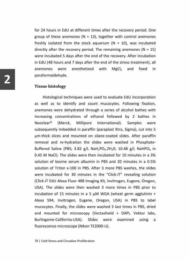
70 | Cold Stress and Circadian Proliferation
for 24 hours in EdU at different times after the recovery period. One
group of these anemones (N = 13), together with control anemones
freshly isolated from the stock aquarium (N = 10), was incubated
directly after the recovery period. The remaining anemones (N = 15)
were incubated 5 days after the end of the recovery. After incubation
in EdU (48 hours and 7 days after the end of the stress treatment), all
anemones were anesthetized with MgCl2 and fixed in
paraformaldehyde.
Tissue histology
Histological techniques were used to evaluate EdU incorporation
as well as to identify and count mucocytes. Following fixation,
anemones were dehydrated through a series of alcohol bathes with
increasing concentrations of ethanol followed by 2 bathes in
Neoclear® (Merck, Millipore International). Samples were
subsequently imbedded in paraffin (paraplast Xtra, Sigma), cut into 5
μm-thick slices and mounted on silane-coated slides. After paraffin
removal and re-hydration the slides were washed in Phosphate-
Buffered Saline (PBS; 3.82 g/L NaH2PO4.2H20; 10.48 g/L NaHPO4 in
0.45 M NaCl). The slides were then incubated for 10 minutes in a 3%
solution of bovine serum albumin in PBS and 20 minutes in a 0.5%
solution of Triton x-100 in PBS. After 3 more PBS washes, the slides
were incubated for 30 minutes in the “Click-iT” revealing solution
(Click-iT EdU Alexa Fluor 488 Imaging Kit, Invitrogen, Eugene, Oregon,
USA). The slides were then washed 3 more times in PBS prior to
incubation of 15 minutes in a 5 μM WGA (wheat germ agglutinin +
Alexa 594, Invitrogen, Eugene, Oregon, USA) in PBS to label
mucocytes. Finally, the slides were washed 3 last times in PBS, dried
and mounted for microscopy (Vectashield + DAPI, Vektor labs,
Burlingame-California-USA). Slides were examined using a
fluorescence microscope (Nikon TE2000-U).

Cold Shock and Circadian Proliferation | 71
Countings and statistical analyses
Mean densities of EdU+ nuclei and mucocytes were evaluated
using Nikon NIS software v3.1. Each value resulted from 5 counts
made in transversal sections of tentacles randomly selected for each
anemone. For the circadian experiment of EdU incorporation, nuclei
counts were reported to the total area of the tentacle. For the
bleaching experiment, these counts were reported to the tissue area
of the ectodermis. The bleaching inducing algal loss and cell death in
the gastrodermis, this method was therefore preferentially chosen to
standardize our results. Diminution of Symbiodinium densities
following bleaching was evaluated using Student t-test. Analyses of
variance (one-way ANOVA) followed by Fisher’s post hoc were used to
compare EdU-positive (EdU+) nuclei and mucocyte densities between
the bleached groups and the control group. Analyses of variance and
Student t-test were both used to evaluate the impact of the time of
the day and the presence of light in the number of EdU+ nuclei. These
analyses were performed using Statistica v10.
Results
Diel variations of cell proliferation
Histological analyses highlighted variations of the number of
EdU+ nuclei within healthy anemone tentacles (Fig. 16). Analyses of
variance revealed that this variation is significantly correlated to the
time of the day (ANOVA F(11,48) = 2.95 and p = 0.005). Exposure to
light seemed to have no significant direct effect on EdU incorporation
(p = 0.13). However, further analyses showed that the introduction of
a 2 hours lag period, shifting the light period of one group in the data,
reveals on significant impact of the light on EdU incorporation (p =
0.0005). The density of EdU+ nuclei rises then significantly at 9:00 and

72 | Cold Stress and Circadian Proliferation
stays high during the day before reaching its maximum at 19:00 and
decreasing.
Figure 16 | Diel variations of EdU incorporation. The density of EdU+ nuclei within A. pallida tentacles increases significantly 2 hours after exposure to the daylight (red area) before returning to lower values during the night.
Cold Stress and Symbiodinium density
The cold stress treatment successfully bleached the anemones, as
shown by their apparent loss of coloration. However, expulsion of
algae only occurred during the 24 hours recovery period, anemones
still showing their characteristic brown coloration after the 48 hours in
the dark concluding the stress procedure. Control anemones showed
a density of 620 ± 8.1 x 10³ (mean ± S.E.M.) algae per mg of fresh
tissue while this value dropped to 70 ± 36 x 103 after the recovery
period (Student t-test, p < 0.0001)(Fig. 17).

Cold Shock and Circadian Proliferation | 73
Figure 17 | Loss of Symbiodinium following cold-shock stress. Symbiodinium density (mean ± S.E.M.) was lower in stressed anemones than in controls 24 hours after the stress. Asterisks represent values significantly different than controls (Student t test; p < 0.05).
Cell proliferation in bleached tissues
As for the study of circadian variations, EdU+ nuclei were
observed in the tentacles of control anemones, within both the
ectodermis and gastrodermis. However, this value differs greatly
between the two tissues, with the ectodermis showing 20 times more
EdU+ nuclei than the gastrodermis. The cold-stress treatment
increased the cell proliferation in both tissues at both time points (Fig.
18A and B). The analyses of variance showed a significant effect of the
stress in the ectodermis (ANOVA: F(2,35) = 7.44 ; p = 0.002) where the
number of EdU+ nuclei rose from 1001 ± 80 nuclei/mm2 before
treatment to 1418 ± 75 after 48 hours of recovery (p = 0,0005). This
value was still higher than controls after 7 days of recovery with 1289
± 91 nuclei/mm2 (p = 0.024). The effect of the cold-stress was even
more pronounced in the gastrodermis (F(2,35) = 10.89 ; p = 0.00001)
where the number of EdU+ nuclei rose from 42 ± 24 nuclei/mm2 in

74 | Cold Stress and Circadian Proliferation
control anemones to 206 ± 22 nuclei/mm2 (p = 0.000013) after 48
hours of recovery and 217 ± 27 nuclei/mm2 (p = 0.000024) after 7 days
of recovery.
Figure 18 | Increase of cell proliferation after cold-induced bleaching. Cell proliferation (mean ± S.E.M.) in the gastrodermis (A) and ectodermis (B) shows a rapid increase following the bleaching procedure. Asterisks represent values significantly different than controls following ANOVA and Fisher’s post hoc test (p < 0.001).

Cold Shock and Circadian Proliferation | 75
Mucocytes
The cold stress had a significant effect on the number of
ectodermal mucocytes found within the tentacles (F(2,30) = 4.83)(Fig.
19). This effect was however not significant until 7 days of recovery
when mucocyte density rose substantially from 133 ± 22 cells/mm2 in
controls to 240 ± 26 cells/mm2 (p = 0.005).
Figure 19 | Increase of mucocyte density after cold-induced bleaching. Mucocyte density (mean ± S.E.M.) in the ectodermis shows a delayed increase following the bleaching procedure. Asterisks represent values significantly different than controls following ANOVA and Fisher’s post hoc test (p = 0.005).
Discussion
Scientists extensively use the thymidine analogues BrdU and EdU.
To date, such molecules have proven their usefulness in tracking
cellular proliferation in many types of organisms [97, 129, 300]. Our
results showed that EdU was successfully incorporated within Aiptasia
cells and attested, for the first time, of the efficacy of this newly
developed technique in studying cnidarians. Being a thymidine
analogue, EdU is incorporated in cell DNA as it replicates during the S-
phase of the cell cycle. Therefore, its detection, although indicating

76 | Cold Stress and Circadian Proliferation
that the labeled cell is mitotically active and proliferating, doesn’t
highlight the mitotic event per se. After DNA replication and EdU
incorporation, the cell will enter the G2 phase before ultimately
dividing. The duration of this G2 phase seems to be quite variable.
While some organisms or cancer cells did not present an apparent G2
phase [151, 244], studies showed that this phase usually lasted only a
few hours as demonstrated in cultured human cells [222]. Concerning
cnidarians, information regarding the duration of the cell cycle phases
are scarce and essentially focused on the Hydra model. Such studies
demonstrated that the G2 phase could last from at least six hours for
the stem cells to 12 hours for the committed precursors and even 24
hours or more for epithelial cells [70, 170]. However, the very specific
cell proliferation dynamics of the Hydra makes it difficult to compare
to Aiptasia. Nonetheless, considering the time necessary for the
completion of the S phase, it seems appropriate to note that while
short EdU incubation times would likely label cells that barely finished
DNA replication, longer incubation are more likely to label cells that
effectively undergo mitosis.
Our study of the circadian variations of EdU incorporation implied
an incubation time of 2 hours. It is therefore very unlikely that these
cells have undertaken mitosis or even finished their DNA replication.
However, these results showed that DNA replication, and hence, the
cycles of host cells were significantly affected by the time of the day as
well as the diel light cycle. Light seemed indeed able to upregulate
host cell DNA synthesis with a significant increase of this activity two
hours after the beginning of light exposure. DNA synthesis stayed high
during the day and progressively fell when the lights were shut off.
The 2-hour latency between the beginning of the light period and the
increase in EdU incorporation could be explained in two different
ways. If the host cell reaction to the light is mediated by cellular
mechanisms initiated in its own cytoplasm, then this lag could be

Cold Shock and Circadian Proliferation | 77
solely explained by the time needed for the progression of the cell
from G1 to the S phase. Such circadian variations in host physiology
have been demonstrated concerning the gene expression of the corals
Acropora millepora [239] and Favia fragum [162] as well as the non-
symbiotic anemone N. vectensis [312]. Although sharing mammalian
rhythm-inducing processes like the heterdimerization of the Clock and
Cycle proteins, the cellular components triggering these light-
dependent, molecular clock mechanism are yet to be described. On
the other hand, the detection of the light could be achieved within the
symbiotic algae. The lag could then represent the time needed by
light-induced mechanisms engaged by the algae as well as the
response of the host cell to such mechanisms. This second hypothesis
seems more likely as many studies reported influence of the diel light
cycle over Symbiodinium algae. The influence of light seems then
particularly important determining the progression of the algae
through their cell cycle. A recent study demonstrated that cultured
Symbiodinium isolated from coral undergo mitosis during the night
and produce two flagellated daughter cells just before the beginning
of the light period [390]. In symbiotic state however, only a small
fraction of algae shows a mitotic activity. Studies performed on the
anemone Aiptasia pulchella and the coral Astrangia poculata showed
that 3 to 5 percent of Symbiodinium multiply within their host [78,
352]. Although most of the newly produced algae seem to be expelled
from the host cell [78, 101], they may also have some influence over
its cell cycle progression. Such link between the mitosis of the host cell
and its algae has been demonstrated in the green Hydra [254]. The
presence of two Symbiodinium cells within the host gastrodermal cells
at the beginning of the light period may therefore be an explanation
for the increased EdU incorporation detected during the following
hours. Moreover, the cell cycle of host cells could be influenced by the
diel fluctuation of the translocation of photosynthates from the algae.
The circadian variations of Symbiodinium derived energetic resources,

78 | Cold Stress and Circadian Proliferation
such as lipid bodies [58], could be a factor facilitating the progression
of host cells from G1 to the S phase. This may be especially true in the
ectodermis where the multiplication of the algae has no direct impact.
Our bleaching experiment showed, similarly to the literature
[358], that successive exposures to a water temperature of 4°C
combined with incubation in darkness evoke rapid expulsion of
Symbiodinium from Aiptasia. After a recovery period of 24 hours
following the end of the treatment, anemones showed a striking loss
of coloration accompanied by tenfold reduction of their algal density.
Moreover, using EdU incorporation we also demonstrated that the
rate of cell proliferation in both tissues was significantly affected by
the bleaching treatment. In the gastrodermis of bleached anemones,
this proliferation could be induced in order to mend the wounded
tissue. Indeed, multiple studies demonstrated that, during bleaching,
Symbiodinium loss is usually linked to the loss of its gastrodermal host
cell, either through apoptosis or necrosis [6, 88, 373]. The relation
between the bleaching treatment and the increased cell proliferation
detected in the ectodermis is however more tricky to determine. This
could be explained by the upregulation of the production of specific
cell types that play a role in bleaching survival. With the breakdown of
the symbiosis, the host loses most of its energy incomes and is
threatened by starvation. The production of cells specialized in
heterotrophic feeding and prey capture could then help the bleached
host facing this menace. Our results also showing an increase in
mucocyte density one week after the end of the stress support this
hypothesis. Mucocytes and mucus production are indeed one of the
key elements of heterotrophic feeding, accumulating particulate
organic matter (POM) and carrying it to the mouth of the animal [42,
174].

Cold Shock and Circadian Proliferation | 79
The duration of this increased cell proliferation, being still
significantly higher compared to control anemones after one week,
casts doubt concerning the causes of such response. While the loss of
algae is the main consequence of the cold stress treatment and is
undoubtedly affecting the host, this treatment could also be directly
affecting the host. These first results of the host histological response
to bleaching are therefore promising but their interpretation is made
difficult by the nature of the stress chosen for this first experiment.
Further analyses using a bleaching inducing stress procedure that
mimics environmental condition seem therefore essential in refining
our interpretations.


Chapter 3
Increased cell proliferation and mucocyte
density in the sea anemone Aiptasia pallida
recovering from bleaching.2
Introduction
High sea surface temperature (SST) accompanied by high levels of
solar irradiance are known to disrupt the symbiosis between
scleractinian corals and endosymbiotic dinoflagellates of the genus
Symbiodinium (aka coral bleaching). Studies have shown that these
environmental factors can act both separately [44, 46, 125, 141, 186,
237] and in combination [131, 167, 232, 235, 394]. Coral bleaching
typically involves an impairment of algal photosynthesis and
eventually loss of the algae from the host tissue [179, 192, 230, 392]
and therefore deprives the host of its main energy source causing a
disruption of symbiosis [265]. Consequently, during the weeks
following this disruption the nutritional state of the coral is
compromised. Depending on the symbiont/host association and the
intensity of the stress, the coral could either die [255, 256, 373] or
survive [46, 80, 113] through a process of recovery that until now is
still poorly characterized.
Cases of coral recovery have highlighted many cellular
modifications occurring in the host tissue of the energetically
2 This chapter has been published in PLOS one. (cf. [117])

82 | Photic/Thermic Stress
compromised coral. A number of studies have come to conflicting
conclusions about the role of mucocytes and mucus secretion in
particular. Some hypothesize that mucus production is dependent on
Symbiodinium for energy supply and carbon input [276, 302]. Indeed a
reduction of Symbiodinium cell density was induced by shading,
eliciting a decrease in mucus production [267, 302]. Other studies
have reported increases in mucus release [6, 223, 276] or mucocyte
number [132, 223] following bleaching. This variation can be partially
explained by inter-specific variation. For example, Lasker et al. [223]
showed that mucocyte number could increase or decrease after
bleaching depending on the coral species involved.
The mucus has critical functions for coral protection and feeding
[42, 138, 342] but also plays a fundamental role of energy carrier in
reef ecology [52, 404]. It contains antimicrobial substances controlling
the associated microbial community [321] and it stimulates planktonic
or benthic microbial activity [104, 404]. Moreover, owing to its
adhesive character, attached and/or secreted coral mucus acts as a
particle trap and accumulates suspended inorganic and organic
particles from the water column, thus supporting the retention and
the recycling of essential nutrients within the reef ([404]; see Bythell
and Wild [52] for review). Therefore, modifications of the mucus
production caused by environmental stress factors related to climate
change could have dramatic consequences in organic matter recycling
and have the potential to affect the coral reef ecosystem [105, 321].
Although observations concerning mucocytes are important,
post-bleaching recovery in hermatypic cnidarians is primarily
characterized by the return of pigmentation due to the symbionts
within the host tissue. While bleaching can be attributed to a decrease
in chlorophyll concentration, it most often implies the loss of algae
from host cells [156]. Many studies have focused on the mechanisms

Photic/Thermic Stress | 83
involved in Symbiodinium loss [82]. Several scenarios like symbiont
digestion via autophagy [85, 91], symbiont expulsion [336, 358] and
host cell detachment [45, 127, 341] have been considered [394].
However, most recent findings argue for mechanisms implying the
death of host cells either by necrosis or apoptosis [88, 93, 215, 299,
373]. These apoptotic pathways induced by bleaching events were
first reported in the zooxanthelate sea anemone Aiptasia pallida [93]
which is often used as a model cnidarian [63, 229, 268].
Several studies have documented the recovery of corals from
bleaching [46, 80, 113], highlighting eventual modifications in the algal
community of the host [17, 20, 46, 113, 156, 191]. Although
understanding of re-infection mechanisms of healed host tissue is
progressing [118], little is known concerning tissular mending [156] or
regeneration processes that occur during this recovery. Most
knowledge on tissue regeneration comes from studies on Hydra. In
that model organism, tissue regeneration requires the cooperation of
three stem cell populations: ectodermal epithelial stem cells,
endodermal epithelial stem cells and interstitial stem cells. The latter
provides cells committed to specific differentiation pathways leading
to one class of somatic cells: neurons, nematocysts and secretory cells
(of which gland cells are only detected in the body column and mucus
cells in the head region), (for review see Bode [36] and Galliot and
Ghila [121]). In Hydra mucus cells are replaced by at least two
mechanisms: 1) proliferation of interstitial stem cells followed by their
differentiation, and 2) transdifferentiation (no cell division) of gland
cells of the body column combined with a translocation to the head
region [36, 121]. Some of these pathways and mechanisms are
generalized as tissue regeneration of sea anemones. However strong
differences exist between these model organisms, among those is the
presence of mucus cells in the tentacles of A. pallida but not in Hydra
[36, 121].

84 | Photic/Thermic Stress
In the present study we try to clarify some of the histological
modifications induced by bleaching stress in cnidarians. We
hypothesize that new host cells rapidly replace cells lost during
bleaching in order to regenerate the damaged gastrodermis. To
address our hypothesis, we investigate the cellular proliferation
following bleaching stress (high temperature combined with high
irradiance) in the zooxanthelate sea anemone Aiptasia pallida. Our
results provide insight in changes occurring in the gastrodermis and
ectodermis as well as mucocyte dynamics following thermal stress and
bleaching, with important potential insights into the response of reef-
building corals to similar challenges.
Material and Methods
Biological material
Aiptasia pallida specimens were collected in the public aquarium
of the University of Liège. Individuals were kept in artificial seawater
(Reef Crystals, Aquatic systems, France) for several weeks providing a
multi-clonal population of anemones. Light was provided on a daily
cycle of 12 hours/day at an intensity of 30-50 μmol photons m-2 s-1.
The temperature in the aquaria was electronically controlled with a
Dupla T-Control Delta (Dohse Aquaristik, Germany) in combination
with a cooling unit (Titan 150, Aqua Medic, Germany) to ensure a
constant temperature of 26 ± 0.2 °C. A. pallida were fed weekly with
frozen Artemia shrimps, except during experiments.
Induction of bleaching by thermal/photic stress
Twenty-four hours before the beginning of the experiment, sea
anemones were placed in Petri dishes in which the water was
constantly renewed by a flow-through mechanism using a peristaltic
pump. Anemones were maintained at control conditions or subjected

Photic/Thermic Stress | 85
to a stress treatment (adapted from Bhagooli and Hidaka [34]). The
stress treatment consisted of a 30 hours exposure to 33°C and
illumination of approximately 1900 μmol photons.m-2.s-1 (measured in
the Petri dishes using a Submersible Spherical Micro Quantum Sensor
(Walz, Germany) connected to a LI-250A Light Meter (Li-Cor, USA))
produced by led bulbs (12W, 6000K, Elix Belgium). Such light intensity
has previously been detected in the field [43] and the combination of
temperature and high irradiance is widely used to induce loss of
symbionts or bleaching in cnidarian hosts [163, 298]. All anemones
were then returned to delimited parts of the same experimental
aquarium and allowed to recover under normal conditions (see
Biological material). The first group of anemones (one day post-stress
group) was incubated immediately after the stress in EdU-containing
seawater for 24 hours, a second group (one week post-stress group)
was incubated for 24 hours in EdU-containing seawater at the 6th day
after the end of the stress, a third group at the 20th day (3 weeks post-
stress group) and the last group at the 55th day (8 weeks post-stress
group). Anemones that were not subjected to stress were sampled
and incubated in EdU-containing seawater for 24 hours at the same
time points as the stressed groups (1 day, 1 week, 3 weeks and 8
weeks post-stress) and served as controls (N = 10-17/time point). In
addition, anemones (pre-stress group) were also sampled and
incubated in EdU-containing seawater before stress conditions
commenced. Sampling was performed at the same time of the day for
each group.
Symbiodinium identification and population density
The dominant Symbiodinium type from our pool of A. pallida was
identified as a clade B1 by denaturing gradient gel electrophoresis
(DGGE) and sequencing of the internal transcribed spacer region 2 of
the ribosomal DNA (ITS2 rDNA). Following DNA extraction with a

86 | Photic/Thermic Stress
DNeasy Plant Mini Kit (Qiagen, Netherlands), the ITS2 rDNA region
was amplified using the forward primer ‘ITSintfor 2’ (5’-
GAATTGCAGAACTCCGTG-3’) and a reversed primer with a GC-clamp
‘ITS 2 clamp’ (5’-CGCCCGCCGCGCCCCGCGCCCGTCCCGCCGCCC
CCGCCCGGGATCCATATGCTTAAGTTCAGCGGGT-3’) producing a
fragment size of 330-360 bp. Amplification products were screened
for polymorphisms using DGGE (Biorad DCode system) and run on
acrylamide gels (30-65 % gradient) following the manufacturer’s
instructions (Biorad Laboratories). Dominant bands were excised, re-
amplified and subsequently sequenced at the Australian Genome
Research Facility University of Queensland, Australia) using an ABI
3730x/ sequencer in combination with BigDye Terminator sequencing
reaction kits. Sequences were then examined using Codoncode
Aligner version 3.5.3. (Codoncode Corporation) and identified by
BLAST comparisons in GenBank.
Bleaching was estimated in each set of experimental groups using
a coral health chart, as usually done on coral reefs to grossly assess
the health of coral colonies during diving. In addition, to confirm that
bleaching resulted from a loss of Symbiodinium cells as previously
described in Aiptasia sp. [93] we evaluated Symbiodinium density in
tentacle sections of the pre-stress and bleached anemones during the
recovery period (N = 3/time point).
Tissue histology
Histological techniques were used to evaluate the cellular
proliferation and the number of mucocytes in control and bleached
anemones isolated at each time point (N = 10-17/time point). Cell
proliferation assays consisted of counting nuclei which incorporated
thymidine analogue during DNA synthesis. To do so, each anemone
was incubated for 24 hours in a solution of 1 μM EdU (5-ethynyl-2’-
deoxyuridine, thymidine analogue, Invitrogen, Eugene-Oregon-USA) in
Photic/Thermic Stress | 87
seawater [291]. Anemones were then anesthetized for 15 minutes in a
1:1 solution of seawater and 0.37 M MgCl2 before fixation in a
solution of 4% paraformaldehyde in seawater. Fixed specimens were
subsequently dehydrated, embedded in paraffin (paraplastXtra,
Sigma), cut into 5 μm-thick slices and finally placed on silane-coated
slides. After dissolution of the paraffin and re-hydration, the slides
were washed 3 times for 5 minutes in Phosphate-Buffered Saline (PBS;
3.82 g/L NaH2PO4.2H2O; 10.48 g/L Na2HPO4 in 0.45 M NaCl). Then the
slides were incubated for 10 minutes in a blocking solution of 3%
Bovine Serum albumin in PBS in order to prevent non-specific
interactions. This was followed by a permeabilization procedure of 20
minutes in a solution of 0.5% Triton x-100 in PBS prior to 3 PBS washes
for 5 minutes and incubation for 30 minutes in the reaction mix made
from the Click-iT EdU kit (Click-iT EdU Alexa Fluor 488 Imaging Kit,
Invitrogen, Eugene, Oregon, USA). After 3 washes for 5 minutes in PBS
the slides were incubated for 15 minutes in a 5 μM solution of WGA
(wheat germ agglutinin + Alexa 594, Invitrogen, Eugene-Oregon-USA)
in order to label the mucocytes [302]. Finally, the slides were washed
3 times for 5 minutes in PBS, dried and mounted for microscopy
(Vectashield + DAPI, Vektor labs, Burlingame CA, USA). Slides were
examined under a fluorescence microscope (Nikon TE2000-U).
Omission of Click-iT solution during the revelation step and detection
of fluorescence in anemones that were not incubated in EdU were
used to verify the specificity of fluorescent signals. Some sections
were also observed following standard hematoxylin/eosin staining
procedures to visualize Symbiodinium cells using transmitted light
microscopy.
Analyses and Statistics
Mean densities of Symbiodinium, EdU+ nuclei and mucocytes
were calculated from 5 counts made in randomly sampled tentacle
88 | Photic/Thermic Stress
sections of each anemone using Nikon NIS software v3.1. The
numbers of Symbiodinium, EdU+ nuclei and mucocytes were reported
to the tissue area [6, 138, 302]. The ectodermal area, rather than the
entire tissue area, was used to standardize counts because bleaching
is known to affect the gastrodermis due to algal loss and cell death
[93]. Counting the total number of ectodermal nuclei was not possible
because nuclei were too tightly packed to distinguish individual nuclei.
At each time point, cell proliferation was obtained by dividing the
density of EdU+ nuclei of stressed anemones by that of control
anemones. Statistical analyses were performed using Statistica v10.
Symbiodinium densities were analyzed at each time point using
Student t-test. Analyses of variance (one-way ANOVA) followed by
Dunnett’s post hoc test were used to compare ratios of cell
proliferation and mucocyte densities after stress to pre-stress ratio.
Results
Population density of Symbiodinium
Figure 20 | Transient reduction of Symbiodinium density following photic/thermic stress. H&E stained transversal sections of tentacles illustrating Symbiodinium (arrows) density in the gastrodermis of anemones before the bleaching procedure (a) and after 1 week (b) and 8 weeks (c) of recovery. After 8 weeks, the gastrodermis has regained its normal appearance.

Photic/Thermic Stress | 89
Figure 21 | Loss of Symbiodinium following photic/thermic stress. Symbiodinium density (mean ± S.E.M.) was lower in stressed anemones than in controls 24 hours and 3 weeks after the stress. No difference between groups was detected 8 weeks after stress. Asterisks represent values significantly different than controls (Student t test; p < 0.05).
The light and temperature treatment successfully bleached
anemones as observed by evaluating the color of anemones before
and after the induced stress using the coral health chart (coral watch)
as reference. Similarly variations of algal densities after stress were
also detected in hematoxylin/eosin stained sections of tentacles (Fig.
20). A quantitative confirmation that bleaching was caused by loss of
algae was obtained by measuring Symbiodinium density before and
after stress (Fig. 21). Control anemones showed a similar density
(about 8 x 10³ algae per mm2 of ectodermal area; mean ± S.E.M.) at
each time point after photic/thermic stress. Twenty four hours after
the stress, anemones of the stress group showed a density (1.1 ± 0.2 x
103 algae.mm-2) significantly lower than controls (7.7 ± 1.0 x 10³ algae.
mm-2; Student t Test; p = 0.0003). Symbiodinium densities were still

90 | Photic/Thermic Stress
lower (p = 0.00001) in stressed anemones 3 weeks after the stress
ended (1.3 ± 0.5 x 10³ and 8.9 ± 0.2 x 10³ algae. mm-2, respectively).
Symbiodinium densities were similar in control and stress groups 8
weeks after the end of the stress (Fig. 2) with a density of 8.5 ± 0.3 x
103 and of 8.8 ± 0.1 x 10³ algae.mm-2, respectively (p = 0.16).
Proliferation of cells within ectoderm and gastrodermal
tissues.
Histological analyses revealed that EdU was detected in tentacle
tissues. Histological controls confirmed that fluorescent signals
corresponded to EdU labeling in tentacle tissues and not to cellular
autofluorescence or methodological artifacts due to the protocol of
EdU revelation.
Figure 22 | EdU and WGA labeling. Transversal section of a tentacle showing histological labeling of EdU
+ nuclei (green) and mucocytes (arrow) stained with WGA
(red). DAPI staining (blue) was used to visualize nuclei. E, endodermis; G, gastrodermis.

Photic/Thermic Stress | 91
Figure 23 | Increase of cell proliferation after heat/light-induced bleaching. Cell proliferation (mean ± S.E.M.; EdU+ cell density in treated anemones divided by EdU+ cell density in controls) in the gastrodermis (a) and ectodermis (b) shows a rapid and transient increase following the bleaching procedure (N = 10-17/time point). Asterisks represent values significantly different than pre-stress values following ANOVA and Dunnett’s post hoc test (p < 0.001).
Under normal conditions (i.e., in anemones of the pre-stress
group) EdU+ cells were observed in both the gastrodermis and the
ectodermis. However, the number of EdU+ cells strongly differed
between these tissues (Fig. 22). The number of EdU+ cells in the
ectodermis of control anemones was about 16-fold higher than in

92 | Photic/Thermic Stress
their gastrodermis. Similarly, in stressed anemones, higher numbers of
EdU+ cells were also observed in the ectodermis compared to the
gastrodermis.
To assess the effects of stress on cell renewal in the gastrodermis
and the ectodermis, we determined the ratio of EdU+ cell densities
between stressed and control tissues at each time point. In the
gastrodermis of bleached anemones, a rapid increase of cell
proliferation ratio was observed following the stress period (Fig. 23a).
During the first day following the bleaching stress, cell proliferation
increased to 905 ± 133 % of controls (mean ± S.E.M.). After 1 week,
the ratio was down to 250 ± 54 % and remained low at 3 weeks and 8
weeks after stress (177 ± 26 and 141 ± 21 % of controls, respectively).
ANOVA (F(4,62) = 21.350) followed by Dunnett’s post hoc test
confirmed that cell proliferation ratio was significantly higher
immediately after stress, at the beginning of the recovery phase than
before stress (p < 0.001). In the ectodermis, we observed the same
trend as in the gastrodermis, that is, a transient increase in cell
proliferation ratios after stress (Fig. 23b). Immediately after the stress
was induced the cell proliferation (317 ± 38 % of controls) was slightly
higher than when measured before stress. One week, 3 weeks and 8
weeks after stress, the ratios (158 ± 16, 158 ± 20 and 112 ± 13 % of
controls, respectively) were close to those measured in anemones of
the pre-stress group. Statistical analyses revealed that cell
proliferation in the 1 day post-stress group was significantly higher
than in the pre-stress group (F(4,62) = 14.628; Dunnett’s post hoc test,
p < 0.0001).
Mucocytes
Although the number of ectodermal mucocytes varied between
batches of anemones used for each experiment (73 ± 27 to 437 ± 154
cells.mm-2), it remained similar in all control anemones (controls of
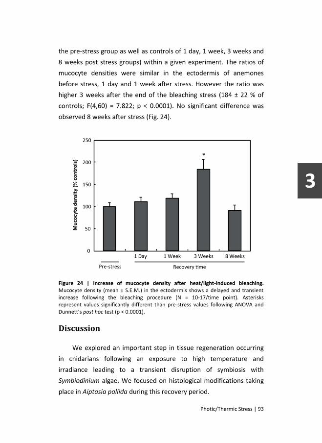
Photic/Thermic Stress | 93
the pre-stress group as well as controls of 1 day, 1 week, 3 weeks and
8 weeks post stress groups) within a given experiment. The ratios of
mucocyte densities were similar in the ectodermis of anemones
before stress, 1 day and 1 week after stress. However the ratio was
higher 3 weeks after the end of the bleaching stress (184 ± 22 % of
controls; F(4,60) = 7.822; p < 0.0001). No significant difference was
observed 8 weeks after stress (Fig. 24).
Figure 24 | Increase of mucocyte density after heat/light-induced bleaching. Mucocyte density (mean ± S.E.M.) in the ectodermis shows a delayed and transient increase following the bleaching procedure (N = 10-17/time point). Asterisks represent values significantly different than pre-stress values following ANOVA and Dunnett’s post hoc test (p < 0.0001).
Discussion
We explored an important step in tissue regeneration occurring
in cnidarians following an exposure to high temperature and
irradiance leading to a transient disruption of symbiosis with
Symbiodinium algae. We focused on histological modifications taking
place in Aiptasia pallida during this recovery period.

94 | Photic/Thermic Stress
Recent studies on mechanisms involved in the loss of symbiotic
dinoflagellates during coral bleaching highlighted that this loss is
potentially related to loss of gastrodermal host cells, leaving the
gastrodermis heavily damaged [127, 341]. Here, we show that cellular
proliferation was significantly enhanced in the gastrodermis following
bleaching in the sea anemone Aiptasia pallida. This suggests that a
massive cellular proliferation rapidly occurs in response to stress. The
exact timing of initiation of this response is uncertain. Cell
proliferation could be triggered at the beginning, during or at the end
of the induced stress, when conditions return to normal. In any case,
cell proliferation returned to normal levels one week after the induced
stress, suggesting that the gastrodermis has recovered from its stress
induced cell loss. Indeed, a large part of the gastrodermis can remain
healthy, despite the bleached state of anemones [93]. Therefore, a
normal level of cell proliferation may suffice to rebuild the
gastrodermal cell layer with time. Studies of cell turnover in the
gastrodermis may clarify this suggestion.
Cell proliferation increased immediately following bleaching in
stressed anemones, most likely in order to regenerate the damaged
tissue and eventually regain the symbiotic state with algae. New
Symbiodinium cells in recovering anemones were sourced from the
proliferation of those that remained in the bleached hosts or were
recently expelled from them as the seawater used in our experiments
was artificial, regularly renewed and thus lacked any live
Symbiodinium. These observations complement results of other
studies reporting on the loss of host cells during bleaching [88, 93,
373] and suggest that gastrodermal regeneration may represent an
important step in the recovery process of bleached cnidarians.
Surprisingly, our results also show an increase in cell proliferation
in the ectodermis of bleached anemones. Although this increase is less

Photic/Thermic Stress | 95
striking than that observed in the gastrodermis, with values 3 times
higher in the ectodermis one day after bleaching stress than in the
control experiment, this observation was unexpected because the
ectodermis of A. pallida is reported to suffer only little damage
following bleaching [93]. Cell proliferation in the ectodermis is
generally relatively high, even under normal conditions, suggesting a
large potential to recover from the relatively limited damage induced
by stress. It is therefore unlikely that our observations are solely
related to regeneration processes. A plausible additional explanation
would be an augmentation in the production of cellular phenotypes
that potentially improve the survival of the bleached host. Here, we
focus on mucocytes, a cell type that has often been reported to be
crucial for the holobiont (host animal and symbionts)[52], but whose
response to bleaching is not yet completely understood and still
subject to debate [132, 302].
Although an increase in mucus release has been well
documented in stressed corals following various environmental
stressors, including heat stress and high irradiance [291] changes in
the population density of mucocytes in bleached organisms remain
unclear. Lasker et al. [223] found an augmentation of epidermal
mucocyte cells in bleached samples of Favia fagrum. Glynn et al. [132]
observed divergent results in bleached corals. Bleached samples of
Pavona clavus presented an increase in mucous secretory cells of the
epidermis but samples of P. gigantea and P. varians had fewer
mucous secretory cells compared to the healthy samples. More
recently Piggot et al. [302] reported that the number of epidermal
mucocytes in Montastraea annularis diminished when shading
increased and, conversely, was higher in samples obtained during a
seasonal increase in sea surface temperature. Both conditions
resulted in a reduction of algal density. All these observations suggest

96 | Photic/Thermic Stress
that factors, such as host species identity and the nature of the stress,
may influence the number of epidermal mucocytes.
We found that mucocyte densities range between 70 to 440
cells.mm-2 in A. pallida. These values seem relatively consistent with
mucocyte density reported in the epidermis of coral species, ranging
from 220 to 3000 cells.mm-2 depending on the host species and light
conditions [138, 302]. The coral Mycetophyllia reesi harbors about
3000 mucocytes.mm-2 of epidermis [138] while Montastraea annularis
showed seasonal variation of mucocytes densities from 220 cells.mm-2
during spring to 1750 cells.mm-2 during summer [302]. We found that
epidermal mucocyte density was slightly low in the sea anemone
Aiptasia pallida in absence of stress. This density may be explained by
intrinsic differences between sea anemones and coral species or by
the low level of light intensity used in culture (30-50 μmol photons m-2
s-1) [302].
The mucocyte density was affected by a combination of
hyperthermia and increased irradiance. We did not observe a
decrease in mucocyte density, which is to be expected if the host is
energetically impaired immediately following stress. It is possible that
such changes occurred during the induced stress and that by sampling
one day after the stress such reduction in mucocyte density was
overlooked if recovery was rapid. However, we observed a significant
and transient augmentation of mucocyte density in the ectodermis 3
weeks after bleaching. The lag period observed between the cell
proliferation peak and mucocyte density peak could account for the
time needed to produce mature mucocytes, that may even have been
produced outside of the tentacle as secretory cell precursors as seen
in Hydra [349]. Another plausible and non-exclusive explanation for
this delay is that differentiation of new cells into mucocytes are only
engaged after a certain threshold (depletion of lipid stores [11] or

Photic/Thermic Stress | 97
other physiological signals [406]) that was not yet reached one week
after bleaching induction.
By producing and secreting mucus, mucocytes contribute to
important roles in the holobiont such as: UV protection, microbial
defense, sediment cleansing, energy carrying and particle trapping
[11, 42, 52, 138, 241, 242, 276, 342]. In the bleached anemone, the
ability of mucus to trap particles and carry them to the hosts’ mouth is
highly profitable. Therefore, even if the host is energetically impaired,
increased production of mucocytes and mucus are in fact a helpful
strategy. Heterotrophic feeding can sustain the hosts’ energy incomes
and compensate for a reduction of algal autotrophic contribution [42,
149, 276]. This idea is corroborated by a recently developed model in
which autotrophy significantly offsets effects of bleaching principally
by restoring lipid stores inside host cells [11]. Augmentation of mucus
production as such, potentially reflects a strategy to limit
photoinhibition in algae and subsequent production of oxidative
radicals [406] or help to protect the bleached, and thus more
susceptible, host against UV radiation or pathogens [42, 276].
Conversely, when stressed anemones have recovered Symbiodinium
densities similar to controls (at 8 weeks after stress), mucocyte
densities in stressed anemones were also similar to controls, arguing
for a relationship between mucocyte and Symbiodinium densities in
Aiptasia.
Mucocytes are not the only cell type derived from the increased
proliferation in the ectodermis. Indeed, the density of EdU+ cells was
several fold higher than the density of mucocytes (see Fig. 3). Among
those EdU+ cells, a small number will differentiate and mature in
mucocytes. Some of those newly produced ectodermal cells may
differentiate into other cell types such as cnidocytes (aka nematocysts
or stinging cells). Increased differentiation into cnidocytes is very likely

98 | Photic/Thermic Stress
in bleached anemones considering their major role in heterotrophic
feeding [174]. Some of the EdU+ cells may also migrate to the
gastrodermis. Indeed, in Hydra, ectodermal cells committed to
secretory cell lineages can migrate through mesoglea to the
gastrodermis [36]. If similar mechanisms are present in A. pallida
some new ectodermal cells may migrate to the gastrodermis to
participate in gastrodermal regeneration after bleaching.
The origin of new cells in the gastrodermis and ectodermis has
yet to be identified. Since a short time of incubation in EdU-containing
seawater suffices to label cells in both tissue types, it seems likely that
cells are produced locally by division of either precursor cells (i.e. cells
committed to a lineage) or multipotent stem cells (i.e. interstitial stem
cells). Such multipotent stem cells located in the mesoglea have
previously been reported during regeneration of damaged tissue [207,
259, 319]. New cells could also differentiate from interstitial stem
cells. These cells are located under the ectodermal surface between
epithelia-muscular cells and are known to be stem cells producing
gametes and other phenotypes, including secretory cells [319]. While
EdU+ cells are most likely produced locally, the origin of mature
mucocytes remains to be solved. Transdifferentiation and migration of
precursor cells from another part of the anemone, such as the
tentacle basis or the oral region, cannot be excluded. Further
investigation with shorter incubation periods conducted during and
directly after the stress treatment will help clarify the origin of
proliferating cells as well as mucocytes.
Finally, additional studies are needed to elucidate the trigger of
cell proliferation (the induced stress or a consequence of a reduction
in Symbiodinium density) as well as the nature of the signal itself. The
latter question applies specifically to the ectodermis, which is not
directly affected by the effects of algal loss.

Chapter 4
Impairment of symbiont photosynthesis
increases cell proliferation in the ectodermis
of the sea anemone Aiptasia pallida.3
Introduction
The ecological success of reef-building corals throughout tropical
oligotrophic waters relies on the symbiosis between cnidarians and
photosynthetic dinoflagellates of the genus Symbiodinium (commonly
referred to as zooxanthellae). This mutualistic relationship is
established following a multi-step process involving both partners (see
[118] for review). Once in symbiosis, the symbiont is intracellularly
located within gastrodermal host cells and provides up to 95% of its
photosynthetic products to the host [265], thus contributing massively
to: respiration, tissue growth, calcification, gamete production and
survival [348]. In return, the host ensures protection of Symbiodinium
from planktonic grazers, provides a source of inorganic nutrients (CO2,
NH3 and PO4³-; [411]) and a fixed position in the water column
allowing an optimal harvesting of light [348]. This mutual relationship
continuously adjusts to subtle changes in the environment to optimize
the survival of the holobiont (the entity including the host and the
symbionts).
Nevertheless, over the last decades, reef-building corals have
faced recurrent large-scale bleaching events compromising their
3 This chapter is submitted for publication in Marine Biology.

100 | Photosynthesis Impairment
survival [167, 177, 283]. Currently, it is widely accepted that high sea
surface temperature accompanied by high levels of solar irradiance is
responsible for the impairment of photosynthesis [392] and the
induction of an oxidative stress that ultimately ends with the
disruption of the symbiosis between cnidarians and Symbiodinium
[394]. Among several proposed mechanisms for coral bleaching, the
most supported one involves the death of the gastrodermal host cells.
This could happen either by the regulated pathway of apoptosis or
more abruptly by cell necrosis [93, 299, 373]. In both cases, the
gastrodermis of the bleached host is left damaged and partly depleted
of symbiont-containing cells.
The loss of symbionts has drastic consequences on the symbiotic
cnidarian, which relies on its photosynthetic partners for energy
supply. Therefore, when photosynthesis is suppressed or lowered
such as during bleaching, the host relies on its own energy reserves
(proteins, carbohydrates and lipids) but also on its ability to modify its
metabolism and feeding habits. It has been found that in such
conditions, several temperate and tropical coral species were able to
increase their feeding effort when preys were available [10, 106, 107,
172]. Moreover, studies conducted on the scleractinian coral
Montipora capitata reported that bleached and recovering specimens
were able to meet their daily metabolic energy requirements by
markedly increasing their feeding rates and their CHAR (per cent
contribution of heterotrophically acquired carbon to daily animal
respiration) from less than 20% to over 100% [148, 149]. Such increase
in heterotrophy permitted M. capitata to maintain its energy income
in absence of Symbiodinium and to survive during prolonged bleaching
events and recovery. These changes in the nutritional balance
necessarily involve significant cellular rearrangements. In a recent
study, Fransolet et al. [117] have demonstrated that, following high
seawater temperature combined with high light irradiance, stressed

Photosynthesis Impairment | 101
sea anemones Aiptasia pallida showed an increase in cell proliferation
in the gastrodermis, which had to recover from the host cell depletion
caused by the stress, but also in the ectodermis. These observations
led the authors to hypothesize that the increase in cell proliferation
observed in the ectodermis could reflect an augmentation of the
holobiont heterotrophic capacity due to the loss of endosymbionts
[117]. Such modifications could be achieved by increasing the number
of ectodermal cells specialized in the nutritional processes, such as
cnidocytes and mucocytes [117, 172, 302].
The comprehension of the cellular mechanisms induced by the
host to maintain its energy balance is therefore critical to understand
how the cnidarians can acclimate to lower autotrophic inputs and
recover from bleaching. In this context, the present study aimed to
investigate the impact of a reduction of energy supply provided by
photosynthetic symbionts on cell proliferation in the tissues of A.
pallida. To do so, A. pallida specimens were treated with 3-(3,4-
dichlorophenyl)-1,1-dimethylurea (DCMU), an herbicide that binds to
the acceptor side of photosystem II (plastoquinone QB) and inhibits
photosynthesis by blocking the electron transport between
photosystems II and I (Hill reaction). Then, Symbiodinium densities,
cell death and cell proliferation in both the ectodermis and
gastrodermis were assessed using histological techniques.
Material and Methods
Biological material
Sea anemones Aiptasia pallida (Verrill) were collected in the
Dubuisson aquarium belonging to the University of Liège. Specimens
were maintained in artificial seawater (Reef Crystals, Aquatic systems,
France) for several weeks, and thus generated a multi-clonal
population of A. pallida. Sea anemones were kept in a 12 h/12 h light

102 | Photosynthesis Impairment
cycle at an intensity of 30-50 μmol photons m-2 s-1. The temperature in
the aquaria was electronically controlled and maintained at 26 ± 0.2°C
using a Dupla T-Control Delta (Dohse Aquaristik, Germany) in
combination with a cooling unit (Titan 150, Aqua Medic, Germany). A.
pallida were fed weekly with frozen Artemia shrimps, except during
experimental treatments.
Experimental treatments
Twenty-four hours before the beginning of the experiment, A.
pallida specimens were placed in individual beakers. Beakers were
placed in a tank where water temperature was controlled with the
same devices as in stock aquaria. Sea anemones were maintained at
control conditions or exposed to seawater containing 20 mM DCMU
(3-(3,4-dichlorophenyl)-1,1-dimethylurea; Sigma-Aldrich) for 1 week.
Control and DCMU-containing seawater was renewed on a daily basis.
During this period, both control and DCMU-treated samples were
exposed to a light intensity of approximately 200 μmol photons m-2 s-1,
provided by 12 W LED bulbs (6000 K, Elix Belgium) and measured in
the bottom of the beaker using a Submersible Spherical Micro
Quantum Sensor (Walz, Germany) connected to a LI-250A Light Meter
(Li-Cor, USA). One week after the beginning of the experimental
treatment, DCMU-treated samples were returned to control seawater
and allowed to recover under normal conditions (recovery period; Fig.
25). Five control and five DCMU-treated A. pallida specimens were
sampled two days, one, two and four weeks after the beginning of the
experimental treatments (Fig. 25).

Photosynthesis Impairment | 103
Figure 25 | Experimental treatments and sampling strategy.
Chlorophyll fluorescence measurements
Chlorophyll fluorescence parameters of Symbiodinium sp. in
hospite of A. pallida were measured using a diving-PAM chlorophyll
fluorometer (Walz GmbH, Germany). Measurements were always
made by placing the fiber-optic of the diving-PAM 1 to 2 centimeters
above the apical part of the sample placed in a 3 mL plastic cuvette.
After a dark adaptation of 20 min, the initial fluorescence level (F0)
was determined by applying weak modulated pulses of red measuring
light. A 1 second saturating pulse of actinic light (>3000 μmol photon
m-2 s-1) was then applied to measure the maximum fluorescence level
(FM). The maximal photochemical quantum yield was calculated as
(FV/FM), where FV = FM-F0. Measures were made on samples of the
DCMU-treatment groups before exposure to DCMU, 1 and 7 days
after the beginning of the treatment, and 1, 7 and 21 days after the
beginning of the recovery period (referred hereafter as 8 days, 2
weeks and 4 weeks, respectively; Fig. 25).
Tissue histology
Histological techniques were used to evaluate Symbiodinium
densities, the density of cells engaged in cell cycle, the number of
mucocytes and the extent of cell death in control and DCMU-treated

104 | Photosynthesis Impairment
sea anemones sampled at each time point. Assessment of mitotic cells
consisted of counting nuclei which incorporated a thymidine
analogue, 5-ethynyl-2´-deoxyuridine (EdU; Invitrogen, Eugene,
Oregon, USA) during DNA synthesis. To do so, 24 hours before each
sampling time point, 5 control and 5 DCMU-treated samples were
incubated in 10 μM EdU-containing seawater [117, 291]. Samples
were then processed for histological analyses as previously described
[117]. Briefly, samples were anesthetized in a 1:1 solution of seawater
and 0.37 M MgCl2 before fixation in seawater containing 4%
paraformaldehyde. Fixed specimens were subsequently dehydrated,
embedded in paraffin (paraplast Xtra, Sigma), cut into 5 μm-thick
slices and finally mounted on silane-coated slides. After paraffin
removal and re-hydration the slides were washed in Phosphate-
Buffered Saline (PBS; 3.82 g/L NaH2PO4.2H20; 10.48 g/L NaHPO4 in
0.45 M NaCl). Then the slides were incubated for 10 minutes in 3%
Bovine Serum Albumin made in PBS followed by 20 minutes in 0.5%
Triton x-100 made in PBS. After PBS washes the slides were incubated
for 30 minutes in the reaction mix made from the Click-iT EdU kit
(Click-iT EdU Alexa Fluor 488 Imaging Kit, Invitrogen, Eugene, Oregon,
USA). After 3 washes for 5 minutes in PBS, the slides were incubated
for 15 minutes in PBS containing 5 μM WGA (wheat germ agglutinin +
Alexa 594, Invitrogen, Eugene, Oregon, USA) in order to label
mucocytes [117, 302]. Finally, the slides were washed 3 times in PBS,
dried and mounted for microscopy (Vectashield + DAPI, Vektor labs,
Burlingame-California-USA). Slides were examined under a
fluorescence microscope (Nikon TE2000-U). Omission of Click-iT
solution during the revelation step and detection of autofluorescence
in A. pallida that were not incubated in EdU were used to verify the
specificity of the fluorescent signal.

Photosynthesis Impairment | 105
Some sections were also observed following standard
hematoxylin/eosin staining procedures to visualize Symbiodinium cells
by transmitted light microscopy and assess their densities.
Another set of slides was used for the assessment of cell death
using the TUNEL assay (ApopTag Plus Peroxidase In Situ Apoptosis
Detection Kit, Millipore, Billerica, Massachusetts-USA) as previously
used on coral [6]. Following paraffin removal and re-hydration, the
slides were incubated for 10 minutes in PBS containing 10 μM
Proteinase-K. They were subsequently washed 3 times in deionized
water, exposed 5 minutes in a solution of 3% H2O2 in order to quench
endogenous peroxidases and washed 2 times in PBS before 30
minutes of incubation in equilibration buffer. Slides were then
incubated for 1 hour at 37°C with the TdT cocktail. Reaction was then
stopped by washing the slides for 10 minutes in the stop/wash
solution and 3 times in PBS. After 30 minutes of incubation in Anti-
Digoxigenin Conjugage and 4 more washes in PBS, revelation was
made using DAB Peroxidase Substrate. The slides were finally washed,
counterstained with 0.5% methyl green and mounted for light
microscopy using successive bathes of N-butanol and xylene.
Counting and statistical analysis
Mean densities of Symbiodinium, EdU+ nuclei and mucocytes
were calculated from 5 counts made in randomly sampled tentacle
sections of each anemone using Nikon NIS software v3.1. For each
tentacle section, the numbers of Symbiodinium, EdU+ nuclei and
mucocytes were reported to the tissue area according to Fransolet et
al. [117]. At each time point, cell proliferation was obtained by
dividing the density of EdU+ nuclei of DCMU-treated sea anemones by
that of control sea anemones.
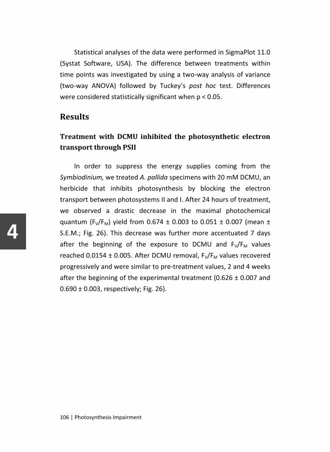
106 | Photosynthesis Impairment
Statistical analyses of the data were performed in SigmaPlot 11.0
(Systat Software, USA). The difference between treatments within
time points was investigated by using a two-way analysis of variance
(two-way ANOVA) followed by Tuckey’s post hoc test. Differences
were considered statistically significant when p < 0.05.
Results
Treatment with DCMU inhibited the photosynthetic electron
transport through PSII
In order to suppress the energy supplies coming from the
Symbiodinium, we treated A. pallida specimens with 20 mM DCMU, an
herbicide that inhibits photosynthesis by blocking the electron
transport between photosystems II and I. After 24 hours of treatment,
we observed a drastic decrease in the maximal photochemical
quantum (FV/FM) yield from 0.674 ± 0.003 to 0.051 ± 0.007 (mean ±
S.E.M.; Fig. 26). This decrease was further more accentuated 7 days
after the beginning of the exposure to DCMU and FV/FM values
reached 0.0154 ± 0.005. After DCMU removal, FV/FM values recovered
progressively and were similar to pre-treatment values, 2 and 4 weeks
after the beginning of the experimental treatment (0.626 ± 0.007 and
0.690 ± 0.003, respectively; Fig. 26).

Photosynthesis Impairment | 107
Figure 26 | The maximal photochemical quantum yield (Mean ± S.E.M.) in Symbiodinium of the sea anemone Aiptasia pallida, before, during and after treatment with 20 mM DCMU. The maximal photochemical quantum yield was calculated as (FV/FM), where FV = FM-F0.
The inhibition of photosynthesis led to bleaching
While Symbiodinium density in control samples was similar
throughout the experiment, and was comprised between 9.32 and
11.45 x 10³ algae per mm² of tissue area, it drastically decreased in A.
pallida specimens treated with DCMU (Fig. 27-28). Indeed, after 1
week of DCMU exposure, Symbiodinium density was 5-fold lower than
controls and reached 1.9 ± 0.6 x 10³ algae mm-2 (two-way ANOVA and
Tuckey’s post hoc test; p < 0.001). Although Symbiodinium density
partially recovered, it was still lower in DCMU-treated sea anemones 2
weeks after the beginning of the DCMU treatment than in controls
(5.1 ± 0.5 x 10³ and 9.9 ± 0.3 x 10³ algae mm-2, respectively; p < 0.001).
Then, it fully recovered to control values at the end of the
experimental treatment (10.7 ± 0.6 x 10³ algae mm-2; Fig. 27).

108 | Photosynthesis Impairment
Figure 27 | Symbiodinium density (Mean ± S.E.M.) in control and DCMU-treated sea anemones Aiptasia pallida. Asterisk indicates statistically significant differences (two-way ANOVA and Tuckey’s post hoc test; p < 0.001).
TUNEL revealed the presence of a few apoptotic nuclei in both
ectodermis and gastrodermis of control and DCMU-treated A. pallida
(Fig. 28); particularly no difference in TUNEL was detected in the
ectodermis 2 days and 1 week after DCMU treatment. Histological
analysis also showed numerous healthy Symbiodinium in the
gastrodermis of control A. pallida (Fig. 28A). After 2 days of incubation
with 20 mM DCMU (Fig. 28B), most Symbiodinium were TUNEL-
positive. After 1 week of DCMU treatment, the gastrodermis was
depleted of most Symbiodinium cells (Fig. 28C), thinning the
gastrodermis cell layer. This observation on tentacle sections
confirmed the Symbiodinium cell density determined tentacle sections
(Fig. 27). It also suggested that the drastic loss of Symbiodinium was
likely related to apoptotic cell death (Fig. 28C).

Photosynthesis Impairment | 109
Figure 28 | Apoptotic cell death and Symbiodinium density in tentacles of Aiptasia pallida, before the treatment with 20 mM DCMU (A), after 2 days (B) and 1 week (C). Apoptotic nuclei, brown; counterstained nuclei, blue; E: epidermis, G: gastrodermis, S: Symbiodinium.
Cells proliferated within ectodermal and gastrodermal tissues
EdU+ cells were observed in both the gastrodermis and the
ectodermis with a strong difference in their relative numbers. The
number of EdU+ cells in the ectodermis of control samples was about
11-fold higher than in the gastrodermis (data not shown). Similarly, in
DCMU-treated A. pallida, higher numbers of EdU+ cells were also
observed in the ectodermis compared to the gastrodermis (data not
shown). To assess the effect of exposure to DCMU on cell proliferation
in both tissues we determined the ratio of EdU+ cell densities
between control and DCMU-treated samples at the different sampling
time points (Fig. 29).

110 | Photosynthesis Impairment
Figure 29 | Cell proliferation (Mean ± S.E.M.) in the ectodermis (A) and the gastrodermis (B) of control and DCMU-treated sea anemones Aiptasia pallida. Asterisk indicates statistically significant differences (two-way ANOVA and Tuckey’s post hoc test; p < 0.001).
In the ectodermis of DCMU-treated samples, EdU+ cell densities
were similar to controls 2 days after the beginning of the DCMU

Photosynthesis Impairment | 111
treatment. It increased significantly and reached 160 ± 5% of controls
after 1 week of exposure (two-way ANOVA and Tuckey’s post hoc test;
p < 0.001). But then, EdU+ cell density rapidly decreased during the
recovery period, reaching values similar to controls after 1 and 4
weeks of treatment. In the gastrodermis of A. pallida exposed to
DCMU, EdU+ cell densities were similar to controls throughout the
experimental treatment, excepted after 4 weeks where EdU+ cell
density increased significantly to 155 ± 15% of controls (two-way
ANOVA and Tuckey’s post hoc test; p < 0.05).
Mucocyte density increased after DCMU treatment
Figure 30 | Mucocyte density (Mean ± S.E.M.) in the ectodermis in control and DCMU-treated sea anemones Aiptasia pallida.
Finally, we assessed the effect of DCMU exposure on the number
of mucocytes in the ectodermis by determining their relative densities
in control and DCMU-treated samples (Fig. 30). We observed that
control values were similar at each time point (about 400 mucocytes
per mm² of ectodermal area; data not shown) and that the mucocyte

112 | Photosynthesis Impairment
density in the ectodermis was higher in DCMU-treated A. pallida than
in controls. However, despite statistical analyses revealing an overall
effect of treatment (two-way ANOVA and Tuckey’s post hoc test; p <
0.05), at no time point a significant difference was observed.
Discussion
When photosynthesis in Symbiodinium cells fails, sea anemones,
like other symbiotic cnidarians, likely implement alternative
mechanisms to compensate for the lack of autotrophic energy income
[149]. For example, sea anemones can change location to find a spot
with a better light irradiance, metabolize stored energy reserves and
modify their feeding strategy to favor heterotrophy. This latter
mechanism takes place in the ectodermis and can involve several
aspects of tissue re-organization involving cell recruitment (i.e.:
migration, de-differentiation), cell maturation (i.e.: increase in mucus
production, differentiation of precursor cells) and/or cell division (i.e.:
stem cell division; [134]). Here, we examined how cell proliferation
participated in the tissue response to a diminution of photosynthesis
efficiency in the sea anemone A. pallida.
Photosynthesis efficiency was reduced by tenfold 1 day after the
beginning of the treatment with 20 mM DCMU and such reduction
persisted for the entire duration of the treatment. One day after the
end of the DCMU treatment, photosynthesis efficiency had already
recovered about 50% of its pretreatment values (Fig. 26). DCMU
treatment also induced a significant drop of Symbiodinium density
(Fig. 27). This loss occurred after several days of DCMU treatment as
illustrated by the same algal density in control sea anemones and
samples treated for 2 days. At that time, although algae were still
present in the gastrodermis, impairment of photosynthesis had
already affected their physiology and many Symbiodinium cells were

Photosynthesis Impairment | 113
committed to cell death as illustrated by TUNEL histology (Fig. 28B).
After 1 week of DCMU treatment, Symbiodinium loss was obvious.
Such bleaching effect was not surprising as coral bleaching has
previously been reported after exposures to PSII herbicides [193, 194].
Transient inhibition of photosynthesis and the subsequent loss of
Symbiodinium, placed host cells in a deficit of algae-derived energy
input, drastically reducing autotrophic energy supply. In such
conditions, bleached and recovering hosts need additional means to
obtain their daily metabolic energy. Among these, are the use of
stored energy supplies and the enhancement of food capture [149,
172].
We observed that cell proliferation was significantly increased in
the ectodermis of DCMU-treated A. pallida (Fig. 29A). One week after
DCMU treatment, when Symbiodinium density and photosynthesis
efficiency were at the lowest, the density of EdU+ cells was maximal in
the ectodermis. Considering that DCMU treatment did not modify the
level of ectodermal cell death, it seems likely that the augmentation of
EdU+ cells reflects a response to the reduction of energy supply rather
than to a direct damage to the ectodermal tissue. We observed a
150% increase of EdU incorporation within the ectodermis at the end
of the stress treatment. This observation contrasts with a previous
study where proliferation of ectodermal cells was detected as early as
24 h after exposure to a combination of high temperature and light
irradiance [117]. However, in that study, one cannot exclude a direct
effect of seawater temperature on the ectodermis such as
modifications of cell metabolism (i.e., [87]). However, in the study of
Fransolet et al. [117], as in the present study, ectodermal cell
proliferation was increased when autotrophic energy supplied by
photosynthesis was at the lowest. Grottoli et al. [149] reported that
when autotrophic energy supply is drastically reduced the coral
Montipora capitata could sufficiently increase the rate of zooplankton

114 | Photosynthesis Impairment
capture and the heterotrophic energy income to prevent depletion of
energy reserves. Conversely, other coral species such as Porites
compressa or Porites lobata were unable to modify the contribution of
heterotrophically acquired carbon [149]. An increase in prey capture
was also observed in Cladocora caespitosa when cultured in darkness
[172]. Surprisingly, changes in heterotrophy efficiency could also occur
in C. caespitosa placed in high light intensity [172]. In that case,
feeding increase mostly served to augment supplies of nitrogen and
phosphorus (to allow tissue growth) rather than carbon. To achieve
these heterotrophic shifts, ectodermal tissue needs to change its
heterotrophic ability through the production of cnidocytes to capture
large preys and/or mucocytes to trap particulate food [42, 126, 174,
321]. Our results support this hypothesis, showing that mucocyte
density is significantly higher in stressed A. pallida following
diminution of the photosynthesis efficiency (Fig. 30). Further analyses
and study of cell population dynamics are still needed to determine
the relative proportion of new cells committed to mucocytes and to
cnidocytes as well as their lifespan.
Cell proliferation also increased in the gastrodermis (Fig. 29B).
Four weeks after DCMU treatment, EdU incorporation within the
gastrodermis was increased by 150%. In this tissue, cell proliferation
likely contributed to tissue regeneration following host cell damage
due to bleaching. Indeed, many nuclei were positively stained for
apoptosis 48 h and especially 1 week after the onset of the DCMU
treatment. Such gastrodermal cell death has been observed
after/during bleaching episodes in corals and other symbiotic
cnidarians [93, 299, 373]. In the present study, Symbiodinium density
partially recovered at 2 weeks, even before gastrodermal cell
proliferation was increased. This suggested that some gastrodermal
cells were able to host new algae at this time. Several possibilities
exist: (1) some cells, although competent, did not host Symbiodinium

Photosynthesis Impairment | 115
cells at the time of the stress and then remained healthy and available
for Symbiodinium symbiosis ; (2) some cells might have expelled
Symbiodinium cells before any injury [394] and remained competent
to host new Symbiodinium; (3) or some cells, like nutritive cells, which
are the main cell phenotype of Anthozoans gastrodermis, were able to
undergo dedifferentiation and transformation into symbiotic host-cell
when phagocyting competent Symbiodinium [71, 134]. Proliferation of
these putative host cells would then depend on their encounter with
new algae, whose presence within the water could only rely on the
release and proliferation of the few that survived the stress. Further
studies should examine the cellular origin of cells able to host new
Symbiodinium cells after stress as well as the fate of new gastrodermal
cells.
In conclusion, sustained decrease in photosynthesis efficiency
leading to bleaching induces tissue remodeling such as cell
proliferation in the ectodermis and gastrodermis of A. pallida.
Ectodermal cell increase may contribute to a heterotrophic shift to
sustain energy demands, which are drastically reduced following the
loss of autotrophic carbon source. Such shift could, in part, rely on an
increased production of cells, such as mucocytes, that play a role in
heterotrophic feeding.


Chapter 5
Trans-tissular migration and mucocyte
differentiation of bleaching-induced
proliferating cells.
Introduction
Bleaching in hermatypic cnidarians does not simply consist in the
expulsion of Symbiodinium from its host. Multiple elements, related
either to the stress causing the bleaching, the mechanisms involved in
the loss of the symbiotic algae or the starvation resulting from the
cessation of autotrophic incomes, profoundly alter and transform the
host. Scientists demonstrated that bleached cnidarians are indeed
heavily affected by the phenomenon, showing multiples signs of
cellular death [93, 299, 373], as well as important modifications of
their feeding habits [10, 149, 172]. In the previous chapters, we
confirmed some of these observations and showed in A. pallida that
the loss of Symbiodinium triggers an increase of cell proliferation in
both tissues as well as an increase of ectodermal mucocyte density
[117]. The origin and the fate of these newly produced mucocytes are
however still to be clearly determined.
Although being extensively studied in cnidarians, cell
proliferation, either during growth or regenerative processes, is poorly
understood in anthozoans. The production of new cells in cnidarians
has, in fact, been essentially studied in hydrozoans and in particular in
Hydra due its spectacular regenerative capacity [36, 39, 349]. These
studies showed the existence of specific stem cells, or I-cells, which

118 | Migration and Differentiation
are able to multiply in order to produce most of, if not every, other
kinds of cells [134, 209, 263]. Anthozoans lack such I-cells but harbor
similar wandering cells called amoebocytes. While amoebocytes have
been identified to play a role in wound healing processes in the
anemone Nematostella vectensis [380], the coral Porites cylindrica
[288] or the gorgonians Plexaurella fusifera [259], this role is likely
related to the innate immune system rather than tissue regeneration
[287]. The capacity of amoebocytes to generate differentiated cell
types is therefore very unlikely. Amoebocytes are most probably an
artificial cellular type regrouping amorphous wandering cells with
differing degrees of specialization [134]. Similarly to Hydra, whose
epithelial cells are self-renewing, production of new cells in
anthozoans seems, in fact, to be related to dedifferentiation of
epithelio-muscular or nutritive-muscle cells [134]. Indeed, a recent
experiment demonstrated in N. vectensis planulae that both
ectodermal and gastrodermal epithelial cells are able to
dedifferentiate and produce neurons [271]. Epithelial cells would
therefore also have a stem-like function, assuming the development
of tissues as well as their renewal and repair through a succession of
mechanisms implying dedifferentiation, multiplication, migration and
specialization [134]. Recent results showed in N. vectensis that this
ability is not restricted to specific zones and is present throughout the
body in both tissues [291].
Following its dedifferentiation into a pluripotent progenitor, the
epithelial cell will most likely undergo mitosis. Regeneration without
mitosis, or morphallaxis, is also possible but, within cnidarians, has
only been described in hydrozoans [121, 134]. Such division of stem
cells is therefore usually organized in two categories depending on the
nature of the daughter cells produced. A symmetrical division
produces two similar cells that will both further differentiate into
specific phenotypes while an asymmetrical division produces one

Migration and Differentiation | 119
committed cell and one stem cell daughter, allowing self-renewal
[121]. Although largely described in Hydra [69], this categorization
may not be suited for anthozoans. Indeed, the notion of asymmetrical
division and self-renewal of stem cell pool seem linked to “classical”
stem cells such as Hydra’s I-cells, which are always undifferentiated
and pluripotent, and not newly dedifferentiated cells. Self-renewal in
the case of anthozoans could therefore be seen as a symmetrical
division followed by the differentiation of one of the daughter cells
back to the original epithelial phenotype. Although likely, this is
however uncertain, just as the belonging of this phenomenon to such
classification.
Migration of these newly produced cells is well described in
cnidarians. However, most observations are once again carried out in
hydrozoans. The long-distance cell migration observed in hydrozoans
is probably linked to the existence of specific proliferative sites [206,
209, 263]. The detection of proliferating cells throughout the body of
some anthozoans [291] suggests that these cells may migrate over
shorter distances. The mesoglea seems to play a crucial role in such
cell migration [343]. Some experiments showed that anthozoan cells
dissociated from sea anemones (including Aiptasia), scleractinians or
alcyonaceans and exposed to media of various compositions would
only migrate in contact with pieces of mesoglea [320]. Further studies
identified many mesogleal components, such as integrin [310],
fibronectin [359] and laminin [340], to be necessary for this migration.
Once at destination, the new cells will finish their differentiation.
While they seem able to adopt many cell phenotypes [209], the
majority of the studies addressing this matter documented the
transformations taking place during the maturation of cnidocytes,
ephemeral cells that present an intensive turnover [74, 197, 206].

120 | Migration and Differentiation
As we outlined here, most of the actual knowledge concerning
cell proliferation, migration and differentiation in cnidarians derives
from studies conducted in hydrozoans. Although providing important
information, its extension to the results gathered on A. pallida
presented in previous chapters is however hypothetical. We therefore
decided to conduct some experiments in order to assess the origin of
the bleaching-induced proliferating cells as well as their possible
differentiation into mucocytes. First of all, we evaluated the cell
proliferation in the whole individual (in toto) to confirm its
homogeneous repartition. We then focused on the possible migration
of cells from the ectodermis to the gastrodermis following heat and
light-induced bleaching. Being previously described in Hydra [350],
this phenomenon could accelerate the regeneration of the wounded
gastrodermis and could also explain, in part, the increased
proliferation in the undamaged ectodermis. To do so, we used an
indirect technique: we counted EdU+ nuclei at different time intervals
following incubation with hydroxyurea (HU), a molecule inhibiting the
synthesis of nucleotides and therefore hindering mitosis [291]. We
then estimated the stability of populations in the ectodermis and the
gastrodermis. The mitosis being inhibited, changes in EdU+ cell
densities within one tissue would therefore reflect a contribution from
the other tissue (a cellular migration). Additionally, we evaluated the
contiguity of mucocytes and proliferating cells 5 and 7 days following
bleaching (Fig. 31). This allowed us to further confirm that the
ectodermal newly produced cells participate in the increased number
of mucocytes that we previously observed following bleaching-
inducing stresses.

Migration and Differentiation | 121
Figure 31 | In toto labeling of mucocytes and proliferating cells. WGA (A), EdU (B) and merged (C) staining. (D) Close-up on mucocytes in a tentacle.
Material and Methods
Biological material
Sea anemones Aiptasia pallida (Verrill), originating from the
aquarium of the University of Liège, were maintained in our
laboratory where they rapidly produced a multi-clonal population. Our
aquariums were filled with artificial seawater (Reef Crystals, Aquatic
systems, France) maintained at 26 ± 0.2°C. A Dupla T-Control Delta
(Dohse Aquaristik, Germany) in combination with a cooling unit (Titan
150, Aqua Medic, Germany) assured the stability of this temperature.
Illumination following a 12 h/12 h light cycle was provided by 12 W
LED bulbs (6000 K, Elix Belgium) producing 30-50 μmol photons m-2 s-1
(measured on the bottom of the aquarium using a Submersible
Spherical Micro Quantum Sensor (Walz, Germany)). Anemones were
122 | Migration and Differentiation
fed every week with frozen Artemia shrimps, except during
experimental treatments.
In toto cell proliferation
Healthy anemones were incubated for 24 hours in a 10 μM
solution of 5-ethynyl-2´-deoxyuridine (EdU; Invitrogen, Eugene,
Oregon, USA) in filtered seawater [117, 291]. They were subsequently
anesthetized for 15 minutes in a 1:1 solution of 0,37 M MgCl2 and
filtered seawater before fixation in a 4% solution of paraformaldehyde
in filtered seawater. Specimens were then washed 3 times for 5
minutes in Phosphate-Buffered Saline (PBS; 3.82 g/L NaH2PO4.2H20;
10.48 g/L NaHPO4 in 0.45 M NaCl) and then incubated during 20
minutes in a 0.5 % solution of Triton x-100 made in PBS. Following 3
washes of 5 minutes in a 3 % solution of Bovine Serum Albumin made
in PBS, specimens were incubated for 30 minutes in the reaction mix
made from the Click-iT EdU kit (Click-iT EdU Alexa Fluor 488 Imaging
Kit, Invitrogen, Eugene, Oregon, USA). Finally, anemones were washed
3 times in PBS and kept in darkness in PBS before microscopic
examination.
Trans-tissular cell migration
We used a thermal/photic stress treatment to bleach our
anemones during this experiment (see chapter 3, [117]). Eighteen
anemones of approximately 2 centimeters in height were isolated
from the stock aquarium and transferred into 100 mL beakers. They
were then exposed during 30 hours to a water temperature of 33°C
and an illumination of approximately 1900 μmol photos.m-2.s-1
(measured in the bottom of the beaker). They were then returned to
normal conditions and allowed to recover for 2 hours before being
incubated for 4 hours in a 10 μM solution of EdU in filtered seawater.
Following this incubation, anemones were divided into 3 groups of 6

Migration and Differentiation | 123
individuals: T0, T1+HU and T1-Ctrl. Anemones of the T0 group were
immediately anesthetized for 15 minutes in a 1:1 solution of 0.37 M
MgCl2 and seawater and fixed in a 4% solution of paraformaldehyde in
seawater. Anemones of the T1+HU group were allowed to recover
during 24 more hours in a 20 mM solution of HU (Sigma) in filtered
seawater, preventing any further cell division, while anemones of the
T1-Ctrl group recovered in normal seawater. During this period,
anemones of the 2 groups were kept under normal conditions of light
and temperature. Another anemone was incubated at the same time
in both EdU and HU in order to attest the efficacy of HU in blocking
mitosis. HU blocks mitosis by inhibiting the ribonucleotid reductase
and therefore the synthesis of deoxyribonucleotides. HU efficiency
decreasing over time, the solutions were renewed 3 times at 8-hour
intervals. After the 24 hours, the anemones were also anesthetized in
MgCl2 and fixed in paraformaldehyde. Following fixation, specimens
were processed for histological analysis. They were dehydrated in a
succession of alcohol solutions and imbedded in paraffin (paraplast
Xtra, Sigma). Specimens were then cut into 5 μm-thick slices and
mounted on silane-coated slides. Following paraffin removal and
rehydration, the slides were washed and treated in order to label the
EdU+ nuclei. To do so, we proceeded in the exact same manner as for
the in toto revelation described above. After the last PBS washes, the
slides were dried and mounted for microscopic observation
(Vectashield + DAPI, Vektor labs, Burlingame-California-USA).
WGA and EdU co-labeled cells
For this experiment, 12 anemones were bleached using the same
protocol of photic/thermic stress as described above. They were then
pooled together and incubated for 24 hours in a 10 μM solution of
EdU in filtered seawater. At the same time, 12 anemones isolated
from the stock aquarium were incubated in the same way and served

124 | Migration and Differentiation
as control group. Following the EdU incubation, the anemones were
washed multiple times in seawater in order to clear out the EdU. On
the fifth day following the stress treatment, 6 anemones of each
group were anesthetized and fixed. The rest of the anemones were
anesthetized and fixed on the seventh day. They were then all treated
for paraffin embedding, cut into 2 μm-thick slices and processed for
EdU revelation following the protocol described above. The slides
were additionally incubated for 15 minutes in a 5 μM solution of WGA
(wheat germ agglutinin + Alexa 594, Invitrogen, Eugene-Orgeon-USA)
in order to label mucocytes and washed 3 times in PBS before being
mounted for microscopy.
Counting and statistics
In toto anemones were observed under a fluorescence binocular
microscope (Olympus SZX-16) and slides were examined under a
fluorescence microscope (Nikon TE2000-U and Confocal Leica SP5).
Mean densities of EdU+ nuclei were evaluated in transversal sections
of tentacles randomly chosen for each anemone using Nikon NIS
software v3.1. Each value was calculated from 5 counts that were
reported to the ectodermal tissue area according to Fransolet et al.
[117]. A cell was considered labeled by WGA and EdU when both
stainings were at least partially overlapping. While this does not
ensure that the new cells indeed matured into mucocytes, the
thickness of the tissue slices and the criteria of analysis make it very
likely. The numbers of WGA+EdU+ cells were reported to the total
number of mucocytes.
Statistical analyses of the data were performed using Statistica
v10. Concerning cell migration, the difference between the three
treatments was investigated by using a one-way analysis of variance
(one-way ANOVA) followed by Fisher’s post hoc test. As to evaluation
of the co-labeling, differences between control and stressed

Migration and Differentiation | 125
anemones were assessed using Student t-test. Differences were
considered statistically significant when p < 0.05.
Results
In toto cell proliferation
Observations of EdU labeling in the whole anemone revealed that
cell proliferation occurs throughout the body, from the pedal disk to
the extremities of the tentacles. Density of EdU+ nuclei seems
however to be a bit higher in the basal part of the column (Fig. 32).
Figure 32 | In toto EdU labeling. Cell proliferation occurs throughout the whole body of the A. pallida with a higher density of dividing cells in the lower part of the column.
Trans-tissular cell migration
The efficacy of hydroxyurea in blocking mitosis was confirmed by
the absence of EdU incorporation in the anemone incubated in both

126 | Migration and Differentiation
solutions (data not shown). Measures performed at T0 revealed that,
although incubation was limited to 4 hours during this experiment, a
large amount of cells incorporated EdU. The number of EdU+ nuclei
was significantly higher in the ectodermis (6010 ± 598 nuclei per mm2,
Mean ± S.E.M.), with 30 times the density observed in the
gastrodermis (184 ± 65 nuclei per mm2).
Figure 33 |Cell proliferation (Mean ± S.E.M.) following photic/thermic stress and HU incubation in the ectodermis of A. pallida. The density of EdU+ nuclei does not differ from T0 values after 24 hours of incubation in HU. This value doubles in anemones not incubated with HU (T1-Ctrl). (ANOVA and Tuckey’s post hoc test; p < 0.001).
After 24h of recovery, anemones treated with HU (T1+HU)
showed no variation in the ectodermal density of EdU+ nuclei
compared to T0. In the untreated group, however, ectodermal density
of EdU+ nuclei increased dramatically to 12303 ± 305 nuclei per mm2
and thus doubling compared to T0 and T1+HU (Fig. 33). ANOVA (F(2,
15) = 32.621) followed by Tuckey’s post hoc test confirmed the
significant difference between T1+HU and the other groups (p =
0.0002).

Migration and Differentiation | 127
Figure 34 |Cell proliferation (Mean ± S.E.M.) following photic/thermic stress and HU incubation in the gastrodermis of A. pallida. Densities of EdU+ nuclei were 4 and 6-fold higher in the T1+HU and T1-Ctrl groups, respectively, compared to the T0 group (ANOVA and Tuckey’s post hoc test; p < 0.001).
In the gastrodermis, the density of EdU+ nuclei increased at T1 in
both groups compared to T0 (Fig. 34). This density reached 800 ± 92
nuclei per mm2 in anemones exposed to HU and 1263 ± 163 nuclei per
mm2 in untreated ones (increases of 400% and 600% respectively).
ANOVA (F(2, 15) = 22.465) followed by Tuckey’s post hoc test
confirmed that the T1+HU group was significantly different from the T0
group (p = 0.005) and that the T1-Ctrl group was significantly different
from the others (p = 0.0002 compared to T0 and p = 0.03 compared to
T1+HU).
EdU localization in WGA+ cells
No significant difference in the number of WGA+EdU+ cells
between control and stressed anemones could be observed five days
following the bleaching treatment. At this time, approximately 40% of
the mucocytes were EdU+ in both groups. A significant increase of
WGA+EdU+ cells appeared in stressed anemones on the seventh day

128 | Migration and Differentiation
following the bleaching treatment (Fig. 35). At this time, while only
38% of the mucocytes in control anemones possessed EdU+ nuclei
(Fig. 36), this value reached 52% in the stressed ones (Student t-test; p
= 0.00004).
Figure 35 | Confocal picture of a WGA+EdU+ cell (arrow) in the ectodermis of A. pallida 7 days after bleaching treatment.

Migration and Differentiation | 129
Figure 36 | Ratio of EdU+ mucocytes (Mean ± S.E.M.) 5 and 7 days following photic/thermic stress in A. pallida. While no difference can be observed between control and stressed anemones after 5 days, the ratio of WGA+ cells possessing EdU+ nuclei in stressed anemones rises to approximately 50% after 7 days.
Discussion
In previous chapters, we demonstrated that, following different
stress treatments, bleached cnidarians show an increase of their cell
proliferation. This increased production in the gastrodermis
corroborates previous studies showing that algal loss is, in part,
operated through the death of the host cells [93, 299, 373]. In those
chapters, we also highlighted an increased density of ectodermal
mucocytes following bleaching and proposed that it could be part of a
strategy engaged by the host to operate a heterotrophic shift. This
shift was described in the literature as a viable option for the host to
compensate the loss of autotrophic incomes [10, 149, 172]. The origin
of the newly produced cells and their differentiation into mucocytes
were, however, still to be explored.

130 | Migration and Differentiation
In toto observations of EdU labeling confirmed that cell
proliferation occurs throughout the whole body of A. pallida. Unlike
hydrozoans, where cell proliferation takes place in specific regions of
the animal and cell migration occurs over long distances [206, 209,
263], new cells produced in A. pallida are likely to migrate over short
distances to reach their final site. Moreover, cell mitosis seems more
active in the basal region of the anemone. This is probably linked to
the asexual mode a reproduction of A. pallida. This phenomenon,
called pedolaceration, consists in the detachment of little fragments
of tissues from the pedal disc and therefore requires active cell
proliferation.
While these observations argue for a limited migration of newly
produced cells, trans-tissular migration was still a possible
phenomenon that had to be investigated. To test this hypothesis, we
used bleached A. pallida that we incubated in EdU and divided into
three groups. Anemones of the first group were directly sacrificed
while those of the second group were incubated for 24 hours in
hydroxyurea, a mitosis inhibitor. Any change in the relative numbers
of EdU+ nuclei in each tissue of these anemones compared to those
sacrificed directly after the EdU incubation would then reflect trans-
tissular migratory events. The third group of anemones was sacrificed
after 24 hours of incubation in regular seawater and served as control,
showing normal increase of EdU+ nuclei.
No change of EdU+ nucleus density was reported in the
ectodermis of the HU-treated anemones. This indicates that, if some
cells migrated from the ectodermis to the gastrodermis, they do not
represent a significant number compared to the total amount of
ectodermal proliferating cells. In the ectodermis of anemones
incubated for 24 hours in seawater, the density of EdU+ nuclei
doubled compared to the T0 group. Compared to the T1-HU group, this
Migration and Differentiation | 131
observation confirms the efficacy of the HU in inhibiting the mitosis.
This massive increase in EdU+ nucleus density is very unlikely related
to a second mitotic event, which would take much more time. It is
most probably caused by the division of the cells that incorporated
EdU during the 4 hours incubation and had not yet time to undergo
mitosis at T0. On the other hand, EdU+ nuclei located in the
gastrodermis of the HU-treated anemones showed a 400% increase
compared to T0. Since no cell could have divided following HU
incubation, this observation has to be related to the migration of cells
from the ectodermis. Even though this density augmentation is
relatively important, the quantity of new cells only represents a tenth
of the cells initially present in the ectodermis at T0. This would
therefore explain why no diminution of EdU+ nucleus density could be
measured in the ectodermis of HU-treated anemones. Finally, a 600%
increase of EdU+ nucleus density was measured in the gastrodermis of
anemones incubated for 24 hours in seawater. This most probably
results from the division of the cells that were EdU+ at T0 combined
with the migration of EdU+ cells from the ectodermis.
These results highlight the existence of trans-tissular migratory
events in bleached A. pallida. Already reported in Hydra [350], this
phenomenon could bring an important amount of new cells to the
bleached and wounded gastrodermis, which is largely depleted of its
symbiotic host-cells and presents a reduced proliferative capacity due
to the sustained stress. Moreover, the much larger quantity of cells in
the ectodermis makes it a very effective production site for new cells.
This mechanism could therefore accelerate the regeneration of the
gastrodermis, allowing a faster replenishment of competent host-cells
and a quick recovery of symbiotic algae.
The experiment assessing the ratio of EdU+ cells in WGA+ cells
brought new elements that complement the results gathered in

132 | Migration and Differentiation
previous chapters. In this experiment, we highlighted a significant
increase in WGA+EdU+ cells 7 days after the thermic/photic stress
treatment. This suggests that, at least, some of the bleaching-induced
proliferating cells transform into mucocytes. These newly produced
mucocytes must have fully matured quite shortly before being
observed since no increase in EdU+ mucocytes was observed 2 days
earlier. So far, most of the studies concerning migration and
differentiation of cells in cnidarians focused on cnidocytes [74, 197,
206, 393]. This experiment is the first to date to assess the
differentiation of cnidarian cells into mucocytes. These observations
further strengthen our previous results showing increase in
ectodermal cellular proliferation and linking it to the higher number of
mucocytes following bleaching in A. pallida.
Altogether, these results provide new elements concerning the
fate of the new ectodermal cells produced following bleaching in A.
pallida. Some of these cells migrate to the gastrodermis where they
most probably participate in the regeneration of the damaged tissue
in order to re-establish symbiosis. On the other hand, another fraction
of the new cells also seems to differentiate into mucocytes. The
increased mucus production that follows is believed to help the
bleached host to survive, via protective properties as well as functions
in prey capture and heterotrophy.

Chapter 6
General Discussion
In this work, we investigated the histological modifications
occurring in cnidarians following a bleaching event. While today’s
research continues to primarily focus on the causes of bleaching, our
knowledge about the events that follow the loss of algae is still very
lacunar. Two major and recent discoveries led us to approach this
overlooked subject from a histological point of view:
Firstly, the mechanisms involved in Symbiodinium expulsion from
the host’s gastrodermal tissue. Many publications demonstrated that
this process implies the loss of the host cell either through cell death
mechanisms [92, 93, 154, 215, 223, 299] or cell detachment [45, 127,
341]. The regeneration of this wounded tissue, although being
essential for the re-establishment of symbiosis, has so far, never been
the subject of studies. We therefore decided to address this matter
using some newly developed methods allowing detection of cell
proliferation.
Secondly, the ability of some coral species to cope with the
drastic reduction of their autotrophic energy incomes. Multiple
studies linked this aptitude to variations of their heterotrophic
capacity, either following bleaching [149] or reduction of
photosynthesis [10, 366]. However, none of them investigated the
mechanisms implied in such a modification. The mucus layer covering
corals playing a key role in heterotrophic feeding [42], we decided to
evaluate the variations of mucocyte density following bleaching. We
chose to perform all our experiments in the well-documented sea
anemone model Aiptasia pallida [396].

134 | General Discussion
Post-bleaching tissular modifications
In all our experiments, whether bleaching was induced by cold
and darkness, elevated temperature and light or a photosynthesis
inhibitor, histological analyses revealed similar results. In every case,
the bleached anemones presented an increase in cellular proliferation
in both their gastrodermis and ectodermis. The respective delays of
these responses varied depending on the stress protocol, which
always occurred during the following days or weeks. Both these
observations seem linked to the loss of Symbiodinium since the
treatment with a photosynthesis inhibitor eluded the possible effect
of the stress on the host.
The higher cell proliferation rates observed in the gastrodermis of
bleached anemones are most probably induced to regenerate the
wounded tissue. As said earlier, the gastrodermis is depleted of most
of its algae-hosting cells during the bleaching and has to recover in
order to host new symbionts. Moreover, damages could have spread
to non-symbiotic gastrodermal cells if the leakage of ROS from
damaged host cells was not stanched in time [338]. The function of
the increased cell proliferation measured in the ectodermis of the
bleached anemones is however less evident. A small fraction of these
new cells could, in part, contribute to the restoration of the
gastrodermis. Indeed, our experiment highlighting the migration of
newly produced ectodermal cells to the gastrodermis suggests that
ectodermal cells contribute to the regeneration of the gastrodermal
tissue. Another fraction of these new ectodermal cells could also be
linked to the higher densities of ectodermal mucocytes that we
measured in bleached anemones, regardless of the stress. Further
experiments revealed that, following bleaching, a higher fraction of
mucocytes incorporated the thymidine analogue EdU. This reinforces
the hypothesis that some of the bleaching-induced ectodermal

General Discussion | 135
proliferating cells differentiate into mucocytes. The higher quantity of
mucus produced, in addition to providing protection against UV and
pathogens, would enhance the heterotrophic feeding capacity of the
host. This modification would last until the regeneration of the
gastrodermis and the reestablishment of the symbiosis allow the
return of autotrophic incomes back to a normal level.
Although having been conducted in the sea anemone Aiptasia
pallida, our results very likely reflect the events occurring during the
bleaching of most hermatypic anthozoans. The value of A. pallida as a
model for the study of coral bleaching has indeed been unanimously
recognized [396]. Moreover, the use of such models is even
recommended since progress moves faster when scientists focus their
work on the same organism. This is due to the fact that model
organisms allow easier comparisons and reproductions of results as
well as development of new tools and genomic databases [396]. This
kind of benefit is particularly relevant for studies, such as those
deciphering the mechanisms of symbiosis, which are conducted on
the cellular-scale. Altogether, the synergic efforts gravitating around
model organisms ultimately enable quicker field applications, an
advantage that reinforces even more their utility for the urgent study
of coral bleaching.
Once extrapolated to corals, the results we gathered with A.
pallida will hopefully help us clarify some aspects of the pandemic
bleaching phenomenon that threatens them [167]. These results
especially invite us to try to understand the cellular events that follow
bleaching and that, along with environmental factors [146], explain at
least in part, the variable surviving rates observed between different
coral species.

136 | General Discussion
Cell proliferation and tissue regeneration
So far, bleaching-related regenerative processes in corals have
never been studied. The only information available comes from
experiments evaluating re-epithelization of the gastrodermis following
damages induced by ultraviolet-C radiations in acroporids [25] or
wound-healing processes engaged after scrape injuries in bleached
Montastraea annularis [252] and lacerating injuries in Porites
cylindrica [288]. These studies suggest that anthozoans share, at least,
some of the tissular plasticity and regenerative capacity of their close
and extensively studied relative Hydra [38]. The first two studies bring
however only little information aside from delays of regeneration and
are not very relevant to the understanding of post-bleaching tissular
modifications. The third study is more pertinent and, although
inflicted wounds are very different from damage induced by
bleaching, provides detailed observations of the healing process. The
successive phases described by the authors are similar to those
observed in other invertebrates and give a first insight on how
regeneration could take place in the bleached gastrodermis. Except
for the plug formation and the inflammation process, which are likely
due to the open nature of the lesion, they also raise the existence of
stem cells that would be stimulated by the injury. These newly
produced cells would then infiltrate the lesion and further multiply to
operate its regeneration [288].
Our results confirm the existence of similar processes during the
regeneration that follows bleaching events. Moreover, we
demonstrated that, contrary to Hydra, the new cells do not originate
from specific niches but, instead, can be observed throughout the
whole body of the anthozoan in both tissues. We also highlighted the
existence of similar migratory events that are, in our case, likely to be
operated on rather short distances. However, the identity of the stem

General Discussion | 137
cells and thus the origin of the new cells still remain to be identified.
In a recently published review, Gold and Jacobs proposed that
production of new cells in anthozoans relies on the dedifferentiation
of ectodermal epitheliomuscular cells and gastrodermal nutritive-
muscle cells [134]. Each of these two types of dedifferentiated cells
could then adopt a variety of phenotypes depending on its origin.
Differentiation into cnidocytes, for example, being restricted to cells
dedifferentiated from epitheliomuscular cells. Our observations
suggest the revision of this hypothesis. Newly produced cells seem
indeed able to migrate, at least, from the ectodermis to the
gastrodermis, and this, most likely to produce new symbiotic host
cells. The dedifferentiated and mitotically active cells found in
anthozoans are then believed to have a wider differentiation capacity,
with less dependency on their original phenotype. This further raises
the question of the differentiation potential of these cells
(pluripotency vs. totipotency) and encourages for a deeper
investigation of this matter in other specific mechanisms such as
pedolaceration.
In the gastrodermis, we observed that the delays between the
peak of cellular proliferation and the complete replenishment of
Symbiodinium density vary between the stress treatments.
Comparison of these delays to other laboratory or environmental
studies is very difficult considering the multitude of varying
parameters such as the species utilized or observed, the type or
duration of the stress and the amount of new Symbiodinium available
for reinfection. Nonetheless, while some laboratory studies actively
reinfected their bleached specimens with new algae [32], we here
showed that replenishment of Symbiodinium density could solely rely
on the few that escaped expulsion from their host. While not being
one of the major results of this work, this observation, although never
put forward in the literature, has important field consequences. Most

138 | General Discussion
of the studies focused on the exogenous origin of new Symbiodinium
following bleaching. They highlighted their presence in multiple
habitats such as the water column and the sediments [4, 53, 59, 142,
159, 246, 248, 305, 368, 409] or the surface of macroalgae [306], and
underlined the similarities between the free-living populations and
the symbiotic ones [248, 368, 409]. While there is no doubt that free-
living Symbiodinium are able to infect a bleached host [240], our
results suggest that the reestablishment of the symbiosis can be
operated without any external input. Furthermore, this corroborates
the hypothesis that post-bleaching changes in Symbiodinium clade
composition may be operated through a shuffle mechanism. This
implies that some clades, initially present in little density within the
host gastrodermis, could become dominant following bleaching. They
owe this to their competitive advantage provided by their relative
better fitness when exposed to stressful conditions [31, 280].
Having demonstrated that, during bleaching, the new
gastrodermal host cells have two different origins (ectodermal and
gastrodermal), the question of their equality before the newly
infecting algae seems legitimate. Considering the multitude and
complexity of the cellular components involved in Symbiodinium
recognition and selection [71, 118], dissimilarities may exist between
the new cells derived from the ectodermis and the gastrodermis.
Although both type of cells being naïve to symbiosis, they could share
epigenetic characteristics of their lineage [35, 413]. The cells
originating from the ectodermis could therefore have a different algal
affinity, especially if their trans-tissular migration is a bleaching
restricted process. With today’s increasing number of observations
depicting Symbiodinium population shifts within corals and the rising
interest they generate [16, 20, 189, 375], this hypothesis seems worth
being investigated.

General Discussion | 139
Cell proliferation and mucocyte density
The second major modification that occurs within corals following
bleaching is to be found on the metabolic and energetic level [11].
Once deprived of their autotrophic energy incomes, some coral
species seem able to cope with the situation by increasing their
heterotrophic capacity. Contrary to other species, which only rely on
their energy reserves, this strategy spares these supplies and allows
for maintained sexual reproduction as well as a better survival rate in
case of prolonged or successive stresses [149]. However, the
mechanisms underlying this heterotrophic shift, whether it happens
during sediment-related shading [10, 366] or bleaching [149] events,
are not yet entirely clarified.
In our work, we propose that an increase in ectodermal mucocyte
density could contribute to this acclimatization. This corroborates
previous studies showing a similar increase in mucus production [276,
406] or mucocyte number [223] following bleaching events in corals.
Our results further expand our limited knowledge about the
development of anthozoan mucocytes [42] and show that their
maturation following cell division takes at least between six and seven
days. Following this period, a higher number of mucocytes would not
only result in a larger amount of mucus in a thicker layer, it would
also, along with increased ciliary movements, enhance the transport
and therefore the quantity of food particles brought to the mouth of
the coral polyps [42]. These particles include detritical POM
(particulate organic matter) as well as live POM, ranging in size from
the pico, nano and microplankton to the larger invertebrates
belonging to the mesoplankton [174].
A major contribution of the carbon participating in mucine
synthesis being initially derived from Symbiodinium photosynthates

140 | General Discussion
[42, 276, 379, 404], increase in its production following bleaching has
to imply some significant metabolic modifications. These may involve
different pathways and could also result in subtle variations of mucine
composition such as a reduction in the amount of oligosaccharide
residues. The C:N ratio in the mucus, a highly variable characteristic as
shown by multiple studies made of healthy corals [42], could
therefore be strongly influenced by the loss of Symbiodinium. Overall,
while investment of its remaining carbon resources in mucus
production by the coral seem to be a gamble, it can ultimately lead to
a net gain as illustrated by the salvaged lipid reserves observed in
heterotrophy-shifting corals [149].
Along with food-particles capture, the thicker mucus layer that
covers the bleached coral could also provide some other significant
benefits. Thanks to the MAA’s it contains, the mucus may confer an
increased protection against UV radiations [42]. This is not negligible
since the damages they would cause to the weakened coral could be
potentially fatal. In a same way, the mucus layer could also protect the
bleached coral against pollutants or other toxics [42]. Moreover, the
mucus having some antibacterial proprieties, it may strengthen the
coral defense against pathogens [42]. This could be critical since many
studies linked the presence of bacteria, such as Vibrio shiloi or Vibrio
coralliilyticus, to bleaching [27, 211, 212]. A reinforced protective
mucus layer would therefore limit the infection probabilities and
increase the chances of coral survival until algae recolonisation.
Having showed that an increase in mucocyte density could be induced
solely by photosynthesis dysfunction and algal expulsion from the
host, heterotrophy is therefore more likely the main purpose of this
increase. The mucus benefits exposed above come thus handy after
bleaching but have however to be considered as collateral
advantages.
General Discussion | 141
Implications of the increased mucus production go beyond the
functions it plays for the bleached coral. The fraction that dissolves
upon release in the water (up to 80% for some acroporids) as well as
the particles and threads that detach from the surface of the coral
play a major role in the coral reef ecosystems [404]. These carbon-rich
molecules feed bacteria and enter the trophic chain where they
generate a significant nutrient release. Mucus aggregates alone can
represent as much as 20% of the organic carbon metabolized by the
community living in the sediments [404]. Relatively speaking, this
means that an increase of mucus production by the bleached corals
populating a reef could significantly increase nutrient input to the
water column. While this could be a benefit for the benthic and
pelagic communities, it could also enhance the growth of macroalgae.
Such phenomenon could therefore be a major drawback of increased
mucus production since macroalgae can be dangerous competitors for
corals, especially after bleaching events [77, 98, 167, 203, 221].
Perspectives
Altogether, the results gathered by this work bring some new
light to the still obscure events that take place in corals after
bleaching. Although bringing conclusions that could be key elements
to the comprehension of those events, they also open new leads for
further research.
Clear identification of the proliferating cells and confirmation of
their epithelial phenotype seem particularly important to complete
the picture that we have just sketched. This could be achieved for
example by using in situ hybridization and co-localizing cell
proliferation staining with mRNA sequences specific of epithelial cells.
As we said above, it could also be interesting to verify if cellular
migration from the ectodermis to the gastrodermis is a constant

142 | General Discussion
phenomenon or if it is only triggered during bleaching. Interesting
results could also come from the analysis of the cellular proliferation
in the column of bleached A. pallida. In this part of the animal, the
gastrodermis harbors very few Symbiodinium but is composed of
numerous secretory cells that operate digestion and are therefore
important in heterotrophic feeding. Increase in gastrodermal
proliferation in the column could therefore be interpreted as another
element corroborating a heterotrophic shift. Multiple other
experiments could also be considered in order to confirm this
hypothesis. The quantity of mucus released by the bleached
anemones could be measured and compared to the healthy ones.
Such comparisons could also be made with the speed of ciliary
movements bringing food to the mouth. Likewise, a major
improvement to the heterotrophic shift hypothesis could come from
the assessment of the respective cnidocyte densities between healthy
and bleached A. pallida. The main function of these cells residing in
prey capture, their increased production would, with no doubt,
indicate a rise in the feeding capacity of the bleached host. However,
analysis of cnidocyte density involves overcoming some difficulties. In
toto analyses seem essential since cnidocytes are often grouped in
batteries and counts made in tentacle sections would therefore not
represent an accurate estimation of their density. This gets even more
complicated considering that the only immunoglobins engineered so
far have been developed to detect Hydra’s cnidocytes and do not
work properly with A. pallida. Other detection methods will therefore
have to be elaborated in order to conduct such an experiment.
Genetic analyses could then be particularly useful, as to further
highlight bleaching influence on cell proliferation and migration.
Contrary to most transcriptomic studies focusing on symbiosis or ROS-
related genes, they could be used to evaluate modifications in the
expression of genes characteristic of heterotrophically active cells or
cell-migration processes. Finally, even acknowledging the efficacy of

General Discussion | 143
A. pallida as a model species, the extension of these experiments to
some coral models would be of great interest. It would then be
noteworthy to see if a similar increase in cellular proliferation can be
observed in the tissues of the coenosarc, in particular in the
calicoblastic epithelium, a tissue lacking in the anemone.
Conclusion
To conclude, even though this thesis brings only preliminary
answers and barely lifts the veil on the events occurring during the
days and weeks that follow bleaching in cnidarians, it reveals a
promising field for future research. Today, as massive coral bleaching
events become more and more frequent and as their intimate causes
seem largely unraveled, it highlights the importance of post-bleaching
mechanisms for the survival and recovery of reefs. It modestly bears
optimism and lets us hope that, one day, a better comprehension of
the regenerative processes and the modifications engaged to resist
starvation will maybe help us to determine the key to bleaching
survival and hence to the perpetuation of coral reef ecosystem.


References| 145
References
[1] D. Abrego, M.J.H. Van Oppen, B.L. Willis, Onset of algal endosymbiont specificity varies among closely related species of Acropora corals during early ontogeny, Molecular Ecology, 18 (2009) 3532-3543.
[2] D. Abrego, B.L. Willis, M.J.H. van Oppen, Impact of Light and Temperature on the Uptake of Algal Symbionts by Coral Juveniles, PLoS One, 7 (2012).
[3] P. Adamczyk, S. Meier, T. Gross, B. Hobmayer, S. Grzesiek, H.P. Bachinger, T.W. Holstein, S. Ozbek, Minicoliagen-15, a novel minicollagen isolated from hydra, forms tubule structures in nematocysts, Journal of Molecular Biology, 376 (2008) 1008-1020.
[4] L.M. Adams, V.R. Cumbo, M. Takabayashi, Exposure to sediment enhances primary acquisition of Symbiodinium by asymbiotic coral larvae, Marine Ecology-Progress Series, 377 (2009) 149-156.
[5] S. Agostini, Y. Suzuki, T. Higuchi, B.E. Casareto, K. Yoshinaga, Y. Nakano, H. Fujimura, Biological and chemical characteristics of the coral gastric cavity, Coral Reefs, 31 (2012) 147-156.
[6] T.D. Ainsworth, O. Hoegh-Guldberg, S.F. Heron, W.J. Skirving, W. Leggat, Early cellular changes are indicators of pre-bleaching thermal stress in the coral host, Journal of Experimental Marine Biology and Ecology, 364 (2008) 63-71.
[7] T.D. Ainsworth, K. Wasmund, L. Ukani, F. Seneca, D. Yellowlees, D. Miller, W. Leggat, Defining the tipping point. A complex cellular life/death balance in corals in response to stress, Scientific Reports, 1 (2011).
[8] D. Allemand, C. Ferrier-Pages, P. Furla, F. Houlbreque, S. Puverel, S. Reynaud, E. Tambutte, S. Tambutte, D. Zoccola, Biomineralisation in reef-building corals: from molecular mechanisms to environmental control, Comptes Rendus Palevol, 3 (2004) 453-467.
[9] H. Amano, O. Koizumi, Y. Kobayakawa, Morphogenesis of the atrichous isorhiza, a type of nematocyst, in Hydra observed with a monoclonal antibody, Development Genes and Evolution, 207 (1997) 413-416.
[10] K.R.N. Anthony, K.E. Fabricius, Shifting roles of heterotrophy and autotrophy in coral energetics under varying turbidity, Journal of Experimental Marine Biology and Ecology, 252 (2000) 221-253.
[11] K.R.N. Anthony, M.O. Hoogenboom, J.A. Maynard, A.G. Grottoli, R. Middlebrook, Energetics approach to predicting mortality risk from environmental stress: a case study of coral bleaching, Functional Ecology, 23 (2009) 539-550.

146| References
[12] S. Bachman, G. Muller-Parker, Viable algae released by the seastar Dermasterias imbricata feeding on the symbiotic sea anemone Anthopleura elegantissima, Marine Biology, 150 (2007) 369-375.
[13] G. Baghdasarian, L. Muscatine, Preferential expulsion of dividing algal cells as a mechanism for regulating algal-cnidarian symbiosis, Biological Bulletin, 199 (2000) 278-286.
[14] A.H. Baird, R. Bhagooli, P.J. Ralph, S. Takahashi, Coral bleaching: the role of the host, Trends in Ecology & Evolution, 24 (2009) 16-20.
[15] A.H. Baird, J.R. Guest, B.L. Willis, Systematic and Biogeographical Patterns in the Reproductive Biology of Scleractinian Corals, Annual Review of Ecology Evolution and Systematics, 40 (2009) 551-571.
[16] A.C. Baker, Ecosystems - Reef corals bleach to survive change, Nature, 411 (2001) 765-766.
[17] A.C. Baker, Flexibility and specificity in coral-algal symbiosis: Diversity, ecology, and biogeography of Symbiodinium, Annual Review of Ecology Evolution and Systematics, 34 (2003) 661-689.
[18] A.C. Baker, T.R. McClanahan, C.J. Starger, R.K. Boonstra, Long-term monitoring of algal symbiont communities in corals reveals stability is taxon dependent and driven by site-specific thermal regime, Marine Ecology Progress Series, 479 (2013) 85-97.
[19] A.C. Baker, A.M. Romanski, Multiple symbiotic partnerships are common in scleractinian corals, but not in octocorals: Comment on Goulet (2006), Marine Ecology-Progress Series, 335 (2007) 237-242.
[20] A.C. Baker, C.J. Starger, T.R. McClanahan, P.W. Glynn, Corals' adaptive response to climate change, Nature, 430 (2004) 741-741.
[21] A.T. Banaszak, R.K. Trench, Effects of ultraviolet (UV) radiation on marine microalgal-invertebrate symbioses .1. Response of the algal symbionts in culture and in hospite, Journal of Experimental Marine Biology and Ecology, 194 (1995) 213-232.
[22] M.S. Barbeitos, S.L. Romano, H.R. Lasker, Repeated loss of coloniality and symbiosis in scleractinian corals, Proceedings of the National Academy of Sciences of the United States of America, 107 (2010) 11877-11882.
[23] M.A. Barbieri, G. Li, M.I. Colombo, P.D. Stahl, Rab5, an early acting endosomal GTPase, supports in vitro endosome fusion without GTP hydrolysis, Journal of Biological Chemistry, 269 (1994) 18720-18722.
[24] R.D. Barnes, Invertebrate Zoology, Holt-Saunders International Editions ed., 1982.

References| 147
[25] D. Basti, I. Bricknell, D. Beane, D. Bouchard, Recovery from a near-lethal exposure to ultraviolet-C radiation in a scleractinian coral, Journal of Invertebrate Pathology, 101 (2009) 43-48.
[26] I.B. Baums, M.E. Johnson, M.K. Devlin-Durante, M.W. Miller, Host population genetic structure and zooxanthellae diversity of two reef-building coral species along the Florida Reef Tract and wider Caribbean, Coral Reefs, 29 (2010) 835-842.
[27] Y. Ben-Haim, F.L. Thompson, C.C. Thompson, M.C. Cnockaert, B. Hoste, J. Swings, E. Rosenberg, Vibrio coralliilyticus sp. nov., a temperature-dependent pathogen of the coral Pocillopora damicornis, International Journal of Systematic and Evolutionary Microbiology, 53 (2003) 309-315.
[28] Y. Benayahu, Y. Achituv, T. Berner, Embryogenesis and acquisition of algal symbionts by planulae of Xenia umbellate (Octocorallia, Alcyonacea), Marine Biology, 100 (1988) 93-101.
[29] Y. Benayahu, M.H. Schleyer, Reproduction in Anthelia glauca (Octocorallia : Xeniidae). II. Transmission of algal symbionts during planular brooding, Marine Biology, 131 (1998) 433-442.
[30] R. Berkelmans, G. De'ath, S. Kininmonth, W.J. Skirving, A comparison of the 1998 and 2002 coral bleaching events on the Great Barrier Reef: spatial correlation, patterns, and predictions, Coral Reefs, 23 (2004) 74-83.
[31] R. Berkelmans, M.J.H. van Oppen, The role of zooxanthellae in the thermal tolerance of corals: a 'nugget of hope' for coral reefs in an era of climate change, Proceedings of the Royal Society B-Biological Sciences, 273 (2006) 2305-2312.
[32] T. Berner, G. Baghdasarian, L. Muscatine, Repopulation of a sea-anemone with symbiotic dinoflagellates - analysis by in-vivo fluorescence, Journal of Experimental Marine Biology and Ecology, 170 (1993) 145-158.
[33] R. Bhagooli, M. Hidaka, Comparison of stress susceptibility of in hospite and isolated zooxanthellae among five coral species, Journal of Experimental Marine Biology and Ecology, 291 (2003) 181-197.
[34] R. Bhagooli, M. Hidaka, Release of zooxanthellae with intact photosynthetic activity by the coral Galaxea fascicularis in response to high temperature stress, Marine Biology, 145 (2004) 329-337.
[35] V.A. Blomen, J. Boonstra, Stable transmission of reversible modifications: Maintenance of epigenetic information through the cell cycle, Cellular and Molecular Life Sciences, 68 (2011) 27-44.
[36] H.R. Bode, The interstitial cell lineage of hydra: A stem cell system that arose early in evolution, Journal of Cell Science, 109 (1996) 1155-1164.

148| References
[37] E.M. Borell, K. Bischof, Feeding sustains photosynthetic quantum yield of a scleractinian coral during thermal stress, Oecologia, 157 (2008) 593-601.
[38] T.C.G. Bosch, Why polyps regenerate and we don't: Towards a cellular and molecular framework for Hydra regeneration, Developmental Biology, 303 (2007) 421-433.
[39] T.C.G. Bosch, Hydra and the evolution of stem cells, BioEssays, 31 (2009) 478-486.
[40] J.N. Bouchard, H. Yamasaki, Implication of nitric oxide in the heat-stress-induced cell death of the symbiotic alga Symbiodinium microadriaticum, Marine Biology, 156 (2009) 2209-2220.
[41] C. Brahmi, I. Domart-Coulon, L. Rougée, D.G. Pyle, J. Stolarski, J.J. Mahoney, R.H. Richmond, G.K. Ostrander, A. Meibom, Pulsed 86Sr-labeling and NanoSIMS imaging to study coral biomineralization at ultra-structural length scales, Coral Reefs, 31 (2012) 741-752.
[42] B.E. Brown, J.C. Bythell, Perspectives on mucus secretion in reef corals, Marine Ecology-Progress Series, 296 (2005) 291-309.
[43] B.E. Brown, R.P. Dunne, I. Ambarsari, M.D.A. Le Tissier, U. Satapoomin, Seasonal fluctuations in environmental factors and variations in symbiotic algae and chlorophyll pigments in four Indo-Pacific coral species, Marine Ecology-Progress Series, 191 (1999) 53-69.
[44] B.E. Brown, R.P. Dunne, H. Chansang, Coral bleaching relative to elevated seawater temperature in the Andaman Sea (Indian Ocean) over the last 50 years, Coral Reefs, 15 (1996) 151-152.
[45] B.E. Brown, M.D.A. Letissier, J.C. Bythell, Mechanisms of bleaching deduced from histological studies of reef corals sampled during a natural bleaching event, Marine Biology, 122 (1995) 655-663.
[46] B.E. Brown, Suharsono, Damage and recovery of coral reefs affected by El-Nino related seawater warming in the Thousand Islands, Indonesia, Coral Reefs, 8 (1990) 163-170.
[47] J.F. Bruno, E.R. Selig, Regional decline of coral cover in the indo-pacific: timing, extent, and subregional comparisons, PLoS One, 2 (2007).
[48] S.B. Buck, J. Bradford, K.R. Gee, B.J. Agnew, S.T. Clarke, A. Salic, Detection of S-phase cell cycle progression using 5-ethynyl-2'-deoxyuridine incorporation with click chemistry, an alternative to using 5-bromo-2'-deoxyuridine antibodies, BioTechniques, 44 (2008) 927-929.

References| 149
[49] R.W. Buddemeier, D.G. Fautin, Coral bleaching as an adaptative mechanism – a testable hypothesis, Bioscience, 43 (1993) 320-326.
[50] S. Bulgheresi, I. Schabussova, T. Chen, N.P. Mullin, R.M. Maizels, J.A. Ott, A new C-type lectin similar to the human immunoreceptor DC-SIGN mediates symbiont acquisition by a marine nematode, Applied and Environmental Microbiology, 72 (2006) 2950-2956.
[51] K.A. Byler, M. Carmi-Veal, M. Fine, T.L. Goulet, Multiple symbiont acquisition strategies as an adaptive mechanism in the coral Stylophora pistillata, PLoS One, 8 (2013).
[52] J.C. Bythell, C. Wild, Biology and ecology of coral mucus release, Journal of Experimental Marine Biology and Ecology, 408 (2011) 88-93.
[53] A.A. Carlos, B.K. Baillie, M. Kawachi, T. Maruyama, Phylogenetic position of Symbiodinium (Dinophyceae) isolates from tridacnids (Bivalvia), cardiids (Bivalvia), a sponge (Porifera), a soft coral (Anthozoa), and a free-living strain, Journal of Phycology, 35 (1999) 1054-1062.
[54] M.C. Chen, Y.M. Cheng, M.C. Hong, L.S. Fang, Molecular cloning of Rab5 (ApRab5) in Aiptasia pulchella and its retention in phagosomes harboring live zooxanthellae, Biochemical and Biophysical Research Communications, 324 (2004) 1024-1033.
[55] M.C. Chen, Y.M. Cheng, P.J. Sung, C.E. Kuo, L.S. Fang, Molecular identification of Rab7 (ApRab7) in Aiptasia pulchella and its exclusion from phagosomes harboring zooxanthellae, Biochemical and Biophysical Research Communications, 308 (2003) 586-595.
[56] M.C. Chen, Y.M. Cheng, L.H. Wang, C.H. Lin, X.Y. Huang, M.C. Liu, P.J. Sung, L.S. Fang, Cloning and characterization of the first cnidarian ADP-ribosylation factor, and its involvement in the Aiptasia-Symbiodinum endosymbiosis, Marine Biotechnology, 6 (2004) 138-147.
[57] M.C. Chen, M.C. Hong, Y.S. Huang, M.C. Liu, Y.M. Cheng, L.S. Fang, ApRab11, a cnidarian homologue of the recycling regulatory protein Rab11, is involved in the establishment and maintenance of the Aiptasia-Symbiodinium endosymbiosis, Biochemical and Biophysical Research Communications, 338 (2005) 1607-1616.
[58] W.N.U. Chen, H.J. Kang, V.M. Weis, A.B. Mayfield, P.L. Jiang, L.S. Fang, C.S. Chen, Diel rhythmicity of lipid-body formation in a coral-Symbiodinium endosymbiosis, Coral Reefs, 31 (2012) 521-534.
[59] M.A. Coffroth, C.F. Lewis, S.R. Santos, J.L. Weaver, Environmental populations of symbiotic dinoflagellates in the genus Symbiodinium can initiate symbioses with reef cnidarians, Current Biology, 16 (2006) R985-R987.

150| References
[60] M.A. Coffroth, S.R. Santos, Genetic diversity of symbiotic dinoflagellates in the genus Symbiodinium, Protist, 156 (2005) 19-34.
[61] M.A. Coffroth, S.R. Santos, T.L. Goulet, Early ontogenetic expression of specificity in a cnidarian-algal symbiosis, Marine Ecology-Progress Series, 222 (2001) 85-96.
[62] S.R. Connolly, M.A. Lopez-Yglesias, K.R.N. Anthony, Food availability promotes rapid recovery from thermal stress in a scleractinian coral, Coral Reefs, 31 (2012) 951-960.
[63] C.B. Cook, S.K. Davy, Are free amino acids responsible for the 'host factor' effects on symbiotic zooxanthellae in extracts of host tissue?, Hydrobiologia, 461 (2001) 71-78.
[64] T.F. Cooper, R. Berkelmans, K.E. Ulstrup, S. Weeks, B. Radford, A.M. Jones, J. Doyle, M. Canto, R.A. O'Leary, M.J.H. van Oppen, Environmental factors controlling the distribution of symbiodinium harboured by the coral Acropora millepora on the great barrier reef, PLoS One, 6 (2011).
[65] R. Costanza, R. D'Arge, R. De Groot, S. Farber, M. Grasso, B. Hannon, K. Limburg, S. Naeem, R.V. O'Neill, J. Paruelo, R.G. Raskin, P. Sutton, M. Van Den Belt, The value of the world's ecosystem services and natural capital, Nature, 387 (1997) 253-260.
[66] C.B. Crafts, J.R. Tuliszewski, Motility rhythms in cultured zooxanthellae isolated from the Scyphomedusa Linuche unguiculata., Bulletin of Marine Science, 56 (1995) 822-825.
[67] V.R. Cumbo, A.H. Baird, M.J.H. van Oppen, The promiscuous larvae: Flexibility in the establishment of symbiosis in corals, Coral Reefs, 32 (2013) 111-120.
[68] R. Cunning, A.C. Baker, Excess algal symbionts increase the susceptibility of reef corals to bleaching, Nature Climate Change, 3 (2013) 259-262.
[69] C.N. David, Interstitial stem cells in Hydra: Multipotency and decision-making, International Journal of Developmental Biology, 56 (2012) 489-497.
[70] C.N. David, R.D. Campbell, Cell cycle kinetics and development of Hydra attenuata. I. Epithelial cells, Journal of Cell Science, 11 (1972) 557-568.
[71] S.K. Davy, D. Allemand, V.M. Weis, Cell biology of cnidarian-dinoflagellate symbiosis, Microbiology and Molecular Biology Reviews, 76 (2012) 229-261.
[72] S.K. Davy, C.B. Cook, The influence of 'host release factor' on carbon release by zooxanthellae isolated from fed and starved Aiptasia pallida (Verrill), Comparative Biochemistry and Physiology a-Molecular & Integrative Physiology, 129 (2001) 487-494.

References| 151
[73] S.K. Davy, I.A.N. Lucas, J.R. Turner, Uptake and persistence of homologous and heterologous zooxanthellae in the temperate sea anemone Cereus pedunculatus (Pennant), Biological Bulletin, 192 (1997) 208-216.
[74] E. Denker, M. Manuel, L. Leclere, H. Le Guyader, N. Rabet, Ordered progression of nematogenesis from stem cells through differentiation stages in the tentacle bulb of Clytia hemisphaerica (Hydrozoa, Cnidaria), Developmental Biology, 315 (2008) 99-113.
[75] O. Detournay, C.E. Schnitzler, A. Poole, V.M. Weis, Regulation of cnidarian-dinoflagellate mutualisms: Evidence that activation of a host TGFbeta innate immune pathway promotes tolerance of the symbiont, Developmental and Comparative Immunology, 38 (2012) 525-537.
[76] E. Diaz-Almeyda, P.E. Thome, M. El Hafidi, R. Iglesias-Prieto, Differential stability of photosynthetic membranes and fatty acid composition at elevated temperature in Symbiodinium, Coral Reefs, 30 (2011) 217-225.
[77] G. Diaz-Pulido, L. McCook, S. Dove, R. Berkelmans, G. Roff, D. Kline, S. Weeks, R. Evans, D. Williamson, O. Hoegh-Guldberg, Doom and boom on a resilient reef: climate change, algal overgrowth and coral recovery., PLoS One, 4 (2009).
[78] J. Dimond, E. Carrington, Symbiosis regulation in a facultatively symbiotic temperate coral: Zooxanthellae division and expulsion, Coral Reefs, 27 (2008) 601-604.
[79] S.L. Domotor, C.F. D'Elia, Cell-size distribution of zooxanthellae in culture and symbiosis, Biological Bulletin, 170 (1986) 519-525.
[80] T.J. Done, L.M. DeVantier, E. Turak, D.A. Fisk, M. Wakeford, R. van Woesik, Coral growth on three reefs: development of recovery benchmarks using a space for time approach, Coral Reefs, 29 (2010) 815-833.
[81] S.D. Donner, T.R. Knutson, M. Oppenheimer, Model-based assessment of the role of human-induced climate change in the 2005 Caribbean coral bleaching event, Proceedings of the National Academy of Sciences of the United States of America, 104 (2007) 5483-5488.
[82] A.E. Douglas, Coral bleaching - how and why?, Marine Pollution Bulletin, 46 (2003) 385-392.
[83] C.A. Downs, J.E. Fauth, J.C. Halas, P. Dustan, J. Bemiss, C.M. Woodley, Oxidative stress and seasonal coral bleaching, Free Radical Biology and Medicine, 33 (2002) 533-543.
[84] C.A. Downs, J.E. Fauth, C.E. Robinson, R. Curry, B. Lanzendorf, J.C. Halas, J. Halas, C.M. Woodley, Cellular diagnostics and coral health: Declining coral health in the Florida Keys, Marine Pollution Bulletin, 51 (2005) 558-569.

152| References
[85] C.A. Downs, E. Kramarsky-Winter, J. Martinez, A. Kushmaro, C.M. Woodley, Y. Loya, G.K. Ostrander, Symbiophagy as a cellular mechanism for coral bleaching, Autophagy, 5 (2009) 211-216.
[86] E.J. Drenkard, A.L. Cohen, D.C. McCorkle, S.J. de Putron, V.R. Starczak, A.E. Zicht, Calcification by juvenile corals under heterotrophy and elevated CO2, Coral Reefs, 32 (2013) 727-735.
[87] P. Ducommun, P.A. Ruffieux, A. Kadouri, U. Von Stockar, I.W. Marison, Monitoring of temperature effects on animal cell metabolism in a packed bed process, Biotechnology and Bioengineering, 77 (2002) 838-842.
[88] S.R. Dunn, J.C. Bythell, M.D.A. Le Tissier, W.J. Burnett, J.C. Thomason, Programmed cell death and cell necrosis activity during hyperthermic stress-induced bleaching of the symbiotic sea anemone Aiptasia sp., Journal of Experimental Marine Biology and Ecology, 272 (2002) 29-53.
[89] S.R. Dunn, M. Pernice, K. Green, O. Hoegh-Guldberg, S.G. Dove, Thermal stress promotes host mitochondrial degradation in symbiotic cnidarians: Are the batteries of the reef going to run out?, PLoS One, 7 (2012).
[90] S.R. Dunn, W.S. Phillips, D.R. Green, V.M. Weis, Knockdown of actin and caspase gene expression by RNA interference in the symbiotic anemone Aiptasia pallida, Biological Bulletin, 212 (2007) 250-258.
[91] S.R. Dunn, C.E. Schnitzler, V.M. Weis, Apoptosis and autophagy as mechanisms of dinoflagellate symbiont release during cnidarian bleaching: every which way you lose, Proceedings of the Royal Society B-Biological Sciences, 274 (2007) 3079-3085.
[92] S.R. Dunn, J.C. Thomason, M.D.A. Le Tissier, J.C. Bythell, Different forms of cell death activity during bleaching of the symbiotic sea anemone Aiptasia sp., Proceedings of the 9th International Coral Reef Symposium, A4 (2000).
[93] S.R. Dunn, J.C. Thomason, M.D.A. Le Tissier, J.C. Bythell, Heat stress induces different forms of cell death in sea anemones and their endosymbiotic algae depending on temperature and duration, Cell Death and Differentiation, 11 (2004) 1213-1222.
[94] S.R. Dunn, V.M. Weis, Apoptosis as a post-phagocytic winnowing mechanism in a coral-dinoflagellate mutualism, Environmental Microbiology, 11 (2009) 268-276.
[95] P.J. Edmunds, Zooplanktivory ameliorates the effects of ocean acidification on the reef coral Porites spp., Limnology and Oceanography, 56 (2011) 2402-2410.
[96] S. Enriquez, E.R. Mendez, R. Iglesias-Prieto, Multiple scattering on coral skeletons enhances light absorption by symbiotic algae, Limnology and Oceanography, 50 (2005) 1025-1032.
References| 153
[97] D.M. Erasso, E.M. Camporesi, D. Mangar, S. Saporta, Effects of isoflurane or propofol on postnatal hippocampal neurogenesis in young and aged rats, Brain Research, 1530 (2013) 1-12.
[98] K.E. Fabricius, Effects of terrestrial runoff on the ecology of corals and coral reefs: review and synthesis, Marine Pollution Bulletin, 50 (2005) 125-146.
[99] K.E. Fabricius, J.C. Mieog, P.L. Colin, D. Idip, M.J.H. Van Oppen, Identity and diversity of coral endosymbionts (zooxanthellae) from three Palauan reefs with contrasting bleaching, temperature and shading histories, Molecular Ecology, 13 (2004) 2445-2458.
[100] I. Fagoonee, H.B. Wilson, M.P. Hassell, J.R. Turner, The dynamics of zooxanthellae populations: A long-term study in the field, Science, 283 (1999) 843-845.
[101] P.G. Falkowski, Z. Dubinsky, L. Muscatine, L. McCloskey, Population control in symbiotic corals - Ammonium ions and organic materials maintain the density of zooxanthellae, Bioscience, 43 (1993) 606-611.
[102] L.S. Fang, S.P. Huang, K.L. Lin, High temperature induces the synthesis of heat-shock proteins and the elevation of intracellular calcium in the coral Acropora grandis, Coral Reefs, 16 (1997) 127-131.
[103] D.G. Fautin, R.W. Buddemeier, Adaptive bleaching: a general phenomenon, Hydrobiologia, 530 (2004) 459-467.
[104] C. Ferrier-Pagès, J.P. Gattuso, S. Dallot, J. Jaubert, Effect of nutrient enrichment on growth and photosynthesis of the zooxanthellate coral Stylophora pistillata, Coral Reefs, 19 (2000) 103-113.
[105] C. Ferrier-Pagès, N. Leclercq, J. Jaubert, S.P. Pelegri, Enhancement of pico- and nanoplankton growth by coral exudates, Aquatic Microbial Ecology, 21 (2000) 203-209.
[106] C. Ferrier-Pagès, A. Peirano, M. Abbate, S. Cocito, A. Negri, C. Rottier, P. Riera, R. Rodolfo-Metalpa, S. Reynaud, Summer autotrophy and winter heterotrophy in the temperate symbiotic coral Cladocora caespitosa, Limnology and Oceanography, 56 (2011) 10.
[107] C. Ferrier-Pagès, C. Rottier, E. Beraud, O. Levy, Experimental assessment of the feeding effort of three scleractinian coral species during a thermal stress: Effect on the rates of photosynthesis, Journal of Experimental Marine Biology and Ecology, 390 (2010) 118-124.

154| References
[108] P.L. Fisher, M.K. Malme, S. Dove, The effect of temperature stress on coral-Symbiodinium associations containing distinct symbiont types, Coral Reefs, 31 (2012) 473-485.
[109] W. Fitt, Cellular growth of host and symbiont in a cnidarian-zooxanthellar symbiosis, Biological Bulletin, 198 (2000) 110-120.
[110] W.K. Fitt, The role of chemosensory behavior of Symbiodinium microadriaticum, intermediate hosts, and host behavior in the infection of coelenterates and molluscs with zooxanthellae, Marine Biology, 81 (1984) 9-17.
[111] W.K. Fitt, S.S. Chang, R.K. Trench, Motility Patterns of Different Strains of the Symbiotic Dinoflagellate Symbiodinium (=Gymnodinium) microadriaticum (Freudenthal) in Culture., Bulletin of Marine Science, 31 (1981).
[112] W.K. Fitt, R.D. Gates, O. Hoegh-Guldberg, J.C. Bythell, A. Jatkar, A.G. Grottoli, M. Gomez, P. Fisher, T.C. Lajuenesse, O. Pantos, R. Iglesias-Prieto, D.J. Franklin, L.J. Rodrigues, J.M. Torregiani, R. van Woesik, M.P. Lesser, Response of two species of Indo-Pacific corals, Porites cylindrica and Stylophora pistillata, to short-term thermal stress: The host does matter in determining the tolerance of corals to bleaching, Journal of Experimental Marine Biology and Ecology, 373 (2009) 102-110.
[113] W.K. Fitt, H.J. Spero, J. Halas, M.W. White, J.W. Porter, Recovery of the coral Montastrea annularis in the Florida Keys after the 1987 Caribbean bleaching event, Coral Reefs, 12 (1993) 57-64.
[114] W.K. Fitt, R.K. Trench, Endocytosis of the symbiotic dinoflagellate Symbiodinium microadriaticum Freudenthal by endodermal cells of the scyphistomae of Cassiopeia xamachana and resistance of the algae to host digestion, Journal of Cell Science, 64 (1983) 195-212.
[115] W.K. Fitt, R.K. Trench, The relation of diel patterns of cell division to diel patterns of motility in the symbiotic dinoflagellate Symbiodinium microadriaticum Freudenthal in culture, New Phycologist, 94 (1983) 421-432.
[116] D.J. Franklin, P. Hoegh-Guldberg, R.J. Jones, J.A. Berges, Cell death and degeneration in the symbiotic dinoflagellates of the coral Stylophora pistillata during bleaching, Marine Ecology-Progress Series, 272 (2004) 117-130.
[117] D. Fransolet, S. Roberty, A.C. Herman, L. Tonk, O. Hoegh-Guldberg, J.C. Plumier, Increased Cell Proliferation and Mucocyte Density in the Sea Anemone Aiptasia pallida Recovering from Bleaching, PLoS One, 8 (2013).
[118] D. Fransolet, S. Roberty, J.C. Plumier, Establishment of endosymbiosis: The case of cnidarians and Symbiodinium, Journal of Experimental Marine Biology and Ecology, 420-421 (2012) 1-7.

References| 155
[119] H.D. Freudenthal, Symbiodinium gen. nov. and Symbiodinium microadriaticum sp. nov., a Zooxanthella: Taxonomy, Life Cycle, and Morphology., Journal of Protozoology, 9 (1962) 45-52.
[120] P. Furla, D. Allemand, J.M. Shick, C. Ferrier-Pages, S. Richier, The symbiotic anthozoan: A physiological chimera between alga and animal, Integrative and Comparative Biology, 45 (2005) 595-604.
[121] B. Galliot, L. Ghila, Cell plasticity in homeostasis and regeneration, Molecular Reproduction and Development, 77 (2010) 837-855.
[122] T.A. Gardner, I.M. Cote, J.A. Gill, A. Grant, A.R. Watkinson, Long-term region-wide declines in Caribbean corals, Science, 301 (2003) 958-960.
[123] M. Garren, S.M. Walsh, A. Caccone, N. Knowlton, Patterns of association between Symbiodinium and members of the Montastraea annularis species complex on spatial scales ranging from within colonies to between geographic regions, Coral Reefs, 25 (2006) 503-512.
[124] T.A. Garrett, J.L. Schmeitzel, J.A. Klein, J.J. Hwang, J.A. Schwarz, Comparative lipid profiling of the cnidarian Aiptasia pallida and Its dinoflagellate symbiont, PLoS One, 8 (2013).
[125] R.D. Gates, Seawater temperature and sublethal coral bleaching in Jamaica, Coral Reefs, 8 (1990) 193-197.
[126] R.D. Gates, T.D. Ainsworth, The nature and taxonomic composition of coral symbiomes as drivers of performance limits in scleractinian corals, Journal of Experimental Marine Biology and Ecology, 408 (2011) 94-101.
[127] R.D. Gates, G. Baghdasarian, L. Muscatine, Temperature stress causes host-cell detachment in symbiotic Cnidarians – Implications for coral bleaching., Biological Bulletin, 182 (1992) 324-332.
[128] J.P. Gattuso, D. Allemand, M. Frankignoulle, Photosynthesis and calcification at cellular, organismal and community levels in coral reefs: a review on interactions and control by carbonate chemistry, American Zoologist, 39 (1999) 160-183.
[129] E. Gazave, J. Béhague, L. Laplane, A. Guillou, L. Préau, A. Demilly, G. Balavoine, M. Vervoort, Posterior elongation in the annelid Platynereis dumerilii involves stem cells molecularly related to primordial germ cells, Developmental Biology, 382 (2013) 246-267.
[130] M.G. Gleason, Effects of Disturbance on Coral Communities - Bleaching in Moorea, French-Polynesia, Coral Reefs, 12 (1993) 193-201.

156| References
[131] P.W. Glynn, Coral-Reef Bleaching - Ecological Perspectives, Coral Reefs, 12 (1993) 1-17.
[132] P.W. Glynn, E.C. Peters, L. Muscatine, Coral tissue microstructure and necrosis: relation to catastrophic coral mortality in Panama, Diseases of Aquatic Organisms, 1 (1985) 29-37.
[133] P.W. Glynn, D.E.W. WH, Elimination of Two Reef-Building Hydrocorals Following the 1982-83 El Nino Warming Event, Science, 253 (1991) 69-71.
[134] D.A. Gold, D.K. Jacobs, Stem cell dynamics in Cnidaria: Are there unifying principles?, Development Genes and Evolution, 223 (2013) 53-66.
[135] W. Goldberg, Gastrodermal structure and feeding responses in the scleractinian Mycetophyllia reesi, a coral with novel digestive filaments, Tissue Cell, 34 (2002) 246-261.
[136] W.M. Goldberg, Acid polysaccharides in the skeletal matrix and calicoblastic epithelium of the stony coral Mycetophyllia reesi, Tissue Cell, 33 (2001) 376-387.
[137] W.M. Goldberg, Desmocytes in the calicoblastic epithelium of the stony coral Mycetophyllia reesi and their attachment to the skeleton, Tissue Cell, 33 (2001) 388-394.
[138] W.M. Goldberg, Feeding behavior, epidermal structure and mucus cytochemistry of the scleractinian Mycetophyllia reesi, a coral without tentacles, Tissue Cell, 34 (2002) 232-245.
[139] I.J. Goldstein, R.C. Hughes, M. Monsigny, T. Osawa, N. Sharon, What should be called a lectin?, Nature, 285 (1980) 66.
[140] M.D. Gomez-Cabrera, J.C. Ortiz, W.K.W. Loh, S. Ward, O. Hoegh-Guldberg, Acquisition of symbiotic dinoflagellates (Symbiodinium) by juveniles of the coral Acropora longicyathus, Coral Reefs, 27 (2008) 219-226.
[141] T.J. Goreau, R.L. Hayes, Coral bleaching and ocean "hot spots", Ambio, 23 (1994) 176-180.
[142] W.L. Gou, J. Sun, X.Q. Li, Y. Zhen, Z.Y. Xin, Z.G. Yu, R.X. Li, Phylogenetic analysis of a free-living strain of Symbiodinium isolated from Jiaozhou Bay, PR China, Journal of Experimental Marine Biology and Ecology, 296 (2003) 135-144.
[143] T.L. Goulet, Most corals may not change their symbionts, Marine Ecology-Progress Series, 321 (2006) 1-7.
[144] T.L. Goulet, M.A. Coffroth, Stability of an octocoral-algal symbiosis over time and space, Marine Ecology-Progress Series, 250 (2003) 117-124.

References| 157
[145] T.L. Goulet, C.B. Cook, D. Goulet, Effect of short-term exposure to elevated temperatures and light levels on photosynthesis of different host-symbiont combinations in the Aiptasia pallida Symbiodinium symbiosis, Limnology and Oceanography, 50 (2005) 1490-1498.
[146] N.A.J. Graham, K.L. Nash, J.T. Kool, Coral reef recovery dynamics in a changing world, Coral Reefs, 30 (2011) 283-294.
[147] H. Gröger, V. Schmid, Larval development in Cnidaria: a connection to Bilateria?, Genesis, 29 (2001) 110-114.
[148] A.G. Grottoli, L.J. Rodrigues, C. Juarez, Lipids and stable carbon isotopes in two species of Hawaiian corals, Porites compressa and Montipora verrucosa, following a bleaching event, Marine Biology, 145 (2004) 621-631.
[149] A.G. Grottoli, L.J. Rodrigues, J.E. Palardy, Heterotrophic plasticity and resilience in bleached corals, Nature, 440 (2006) 1186-1189.
[150] M.S.M. Gustafsson, M.E. Baird, P.J. Ralph, The interchangeability of autotrophic and heterotrophic nitrogen sources in Scleractinian coral symbiotic relationships: A numerical study, Ecological Modelling, 250 (2013) 183-194.
[151] S. Handeli, H. Weintraub, The ts41 mutation in Chinese hamster cells leads to successive S phases in the absence of intervening G2, M, and G1, Cell, 71 (1992) 599-611.
[152] S.D. Hanes, S.C. Kempf, Host autophagic degradation and associated symbiont loss in response to heat stress in the symbiotic anemone, Aiptasia pallida, Invertebrate Biology, 132 (2013) 95-107.
[153] S. Harii, N. Yasuda, M. Rodriguez-Lanetty, T. Irie, M. Hidaka, Onset of symbiosis and distribution patterns of symbiotic dinoflagellates in the larvae of scleractinian corals, Marine Biology, 156 (2009) 1203-1212.
[154] T.D. Hawkins, B.J. Bradley, S.K. Davy, Nitric oxide mediates coral bleaching through an apoptotic-like cell death pathway: evidence from a model sea anemone-dinoflagellate symbiosis, Faseb Journal, (2013).
[155] M.L. Hayes, R.I. Eytan, M.E. Hellberg, High amino acid diversity and positive selection at a putative coral immunity gene (tachylectin-2), BMC Evolutionary Biology, 10 (2010).
[156] R.L. Hayes, P.G. Bush, Microscopic observations of recovery in the reef-building Scleractinian coral, Montastrea annularis, after bleaching on a Cayman reef, Coral Reefs, 8 (1990) 203-209.

158| References
[157] M. Hill, A. Hill, The magnesium inhibition and arrested phagosome hypotheses: new perspectives on the evolution and ecology of Symbiodinium symbioses, Biological Reviews, 87 (2012) 804-821.
[158] R. Hill, P.J. Ralph, Post-bleaching viability of expelled zooxanthellae from the scleractinian coral Pocillopora damicornis, Marine Ecology-Progress Series, 352 (2007) 137-144.
[159] M. Hirose, J.D. Reimer, M. Hidaka, S. Suda, Phylogenetic analyses of potentially free-living Symbiodinium spp. isolated from coral reef sand in Okinawa, Japan, Marine Biology, 155 (2008) 105-112.
[160] M. Hirose, H. Yamamoto, M. Nonaka, Metamorphosis and acquisition of symbiotic algae in planula larvae and primary polyps of Acropora spp, Coral Reefs, 27 (2008) 247-254.
[161] A.M. Hirsch, Role of lectins (and rhizobial exopolysaccharides) in legume nodulation, Current Opinion in Plant Biology, 2 (1999) 320-326.
[162] K.D. Hoadley, A.M. Szmant, S.J. Pyott, Circadian clock gene expression in the coral Favia fragum over diel and lunar reproductive cycles, PLoS One, 6 (2011).
[163] O. Hoegh-Guldberg, Climate change, coral bleaching and the future of the world's coral reefs, Marine and Freshwater Research, 50 (1999) 839-866.
[164] O. Hoegh-Guldberg, Coral reef ecosystems and anthropogenic climate change, Regional Environmental Change, 11 (2011) 215-227.
[165] O. Hoegh-Guldberg, R.J. Jones, S. Ward, W.K. Loh, Ecology - Is coral bleaching really adaptive?, Nature, 415 (2002) 601-602.
[166] O. Hoegh-Guldberg, L.R. McCloskey, L. Muscatine, Expulsion of zooxanthellae by symbiotic Cnidarians from the Red-Sea, Coral Reefs, 5 (1987) 201-204.
[167] O. Hoegh-Guldberg, P.J. Mumby, A.J. Hooten, R.S. Steneck, P. Greenfield, E. Gomez, C.D. Harvell, P.F. Sale, A.J. Edwards, K. Caldeira, N. Knowlton, C.M. Eakin, R. Iglesias-Prieto, N. Muthiga, R.H. Bradbury, A. Dubi, M.E. Hatziolos, Coral reefs under rapid climate change and ocean acidification, Science, 318 (2007) 1737-1742.
[168] O. Hoegh-Guldberg, G.J. Smith, Influence of the population-density of zooxanthellae and supply of ammonium on the biomass and metabolic characteristics of the reef corals Seriatopora hystrix and Stylophora pistillata, Marine Ecology-Progress Series, 57 (1989) 173-186.
[169] L.L. Hollingsworth, R.A. Kinzie, T.D. Lewis, D.A. Krupp, J.A.C. Leong, Phototaxis of motile zooxanthellae to green light may facilitate symbiont capture by coral larvae, Coral Reefs, 24 (2005) 523-523.

References| 159
[170] T.W. Holstein, C.N. David, Cell cycle length, cell size, and proliferation rate in hydra stem cells, Developmental Biology, 142 (1990) 392-400.
[171] M.C. Hong, Y.S. Huang, W.W. Lin, L.S. Fang, M.C. Chen, ApRab3, a biosynthetic Rab protein, accumulates on the maturing phagosomes and symbiosomes in the tropical sea anemone, Aiptasia pulchella, Comparative Biochemistry and Physiology B-Biochemistry & Molecular Biology, 152 (2009) 249-259.
[172] M. Hoogenboom, R. Rodolfo-Metalpa, C. Ferrier-Pagès, Co-variation between autotrophy and heterotrophy in the Mediterranean coral Cladocora caespitosa, Journal of Experimental Biology, 213 (2010) 2399-2409.
[173] M. Hoppenrath, B.S. Leander, Dinoflagellate Phylogeny as Inferred from Heat Shock Protein 90 and Ribosomal Gene Sequences, PLoS One, 5 (2010).
[174] F. Houlbreque, C. Ferrier-Pages, Heterotrophy in Tropical Scleractinian Corals, Biological Reviews, 84 (2009) 1-17.
[175] H.J. Huang, L.H. Wang, W.N.U. Chen, L.S. Fang, C.S. Chen, Developmentally regulated localization of endosymbiotic dinoflagellates in different tissue layers of coral larvae, Coral Reefs, 27 (2008) 365-372.
[176] S.P. Huang, K.L. Lin, L.S. Fang, The involvement of calcium in heat-induced coral bleaching, Zoological Studies, 37 (1998) 89-94.
[177] T.P. Hughes, A.H. Baird, D.R. Bellwood, M. Card, S.R. Connolly, C. Folke, R. Grosberg, O. Hoegh-Guldberg, J.B.C. Jackson, J. Kleypas, J.M. Lough, P. Marshall, M. Nystrom, S.R. Palumbi, J.M. Pandolfi, B. Rosen, J. Roughgarden, Climate change, human impacts, and the resilience of coral reefs, Science, 301 (2003) 929-933.
[178] R. Iglesias-Prieto, V.H. Beltran, T.C. LaJeunesse, H. Reyes-Bonilla, P.E. Thome, Different algal symbionts explain the vertical distribution of dominant reef corals in the eastern Pacific, Proceedings of the Royal Society B-Biological Sciences, 271 (2004) 1757-1763.
[179] R. Iglesias-Prieto, J.L. Matta, W.A. Robins, R.K. Trench, Photosynthetic response to elevated-temperature in the symbiotic Dinoflagellate Symbiodinium microadriaticum in culture, Proceedings of the National Academy of Sciences of the United States of America, 89 (1992) 10302-10305.
[180] H.J. Jeong, Y.D. Yoo, N.S. Kang, A.S. Lim, K.A. Seong, S.Y. Lee, M.J. Lee, K.H. Lee, H.S. Kim, W. Shin, S.W. Nam, W. Yih, K. Lee, Heterotrophic feeding as a newly identified survival strategy of the dinoflagellate Symbiodinium, Proceedings of the National Academy of Sciences of the United States of America, 109 (2012) 12604-12609.

160| References
[181] M. Jimbo, K. Koike, R. Sakai, K. Muramoto, H. Kamiya, Cloning and characterization of a lectin from the octocoral Sinularia lochmodes, Biochemical and Biophysical Research Communications, 330 (2005) 157-162.
[182] M. Jimbo, Y. Suda, K. Koike, S. Nakamura-Tsuruta, J. Kominami, M. Kamei, J. Hirabayashi, R. Sakai, H. Kamiya, Possible involvement of glycolipids in lectin-mediated cellular transformation of symbiotic microalgae in corals, Journal of Experimental Marine Biology and Ecology, 439 (2012) 129-135.
[183] M. Jimbo, H. Yamashita, K. Koike, R. Sakai, H. Kamiya, Effects of lectin in the scleractinian coral Ctenactis echinata on symbiotic zooxanthellae, Fisheries Science, 76 (2010) 355-363.
[184] M. Jimbo, T. Yanohara, K. Koike, R. Sakai, K. Muramoto, H. Kamiya, The D-galactose-binding lectin of the octocoral Sinularia lochmodes: characterization and possible relationship to the symbiotic dinoflagellates, Comparative Biochemistry and Physiology B-Biochemistry & Molecular Biology, 125 (2000) 227-236.
[185] P.L. Jokiel, The reef coral two compartment proton flux model: A new approach relating tissue-level physiological processes to gross corallum morphology, Journal of Experimental Marine Biology and Ecology, 409 (2011) 1-12.
[186] P.L. Jokiel, S.L. Coles, Response of Hawaiian and other Indo-Pacific reef corals to elevated-temperature, Coral Reefs, 8 (1990) 155-162.
[187] A. Jones, R. Berkelmans, Tradeoffs to Thermal Acclimation: Energetics and Reproduction of a Reef Coral with Heat Tolerant Symbiodinium Type-D, Journal of Marine Biology, 2011 (2011).
[188] A. Jones, R. Berkelmans, The photokinetics of thermo-tolerance in Symbiodinium, Marine Ecology, 33 (2012) 490-498.
[189] A.M. Jones, R. Berkelmans, M.J.H. van Oppen, J.C. Mieog, W. Sinclair, A community change in the algal endosymbionts of a scleractinian coral following a natural bleaching event: field evidence of acclimatization, Proceedings of the Royal Society B-Biological Sciences, 275 (2008) 1359-1365.
[190] R.J. Jones, Testing the 'photoinhibition' model of coral bleaching using chemical inhibitors, Marine Ecology Progress Series, 284 (2004) 133-145.
[191] R.J. Jones, Coral bleaching, bleaching-induced mortality, and the adaptive significance of the bleaching response, Marine Biology, 154 (2008) 65-80.
[192] R.J. Jones, O. Hoegh-Guldberg, A.W.D. Larkum, U. Schreiber, Temperature-induced bleaching of corals begins with impairment of the CO2 fixation mechanism in zooxanthellae, Plant, Cell and Environment, 21 (1998) 1219-1230.

References| 161
[193] R.J. Jones, A.P. Kerswell, Phytotoxicity of Photosystem II (PSII) herbicides to coral, Marine Ecology Progress Series, 261 (2003) 149-159.
[194] R.J. Jones, J. Muller, D. Haynes, U. Schreiber, Effects of herbicides diuron and atrazine on corals of the Great Barrier Reef, Australia, Marine Ecology Progress Series, 251 (2003) 153-167.
[195] R.J. Jones, D. Yellowlees, Regulation and control of intracellular algae (equals zooxanthellae) in hard corals, Philosophical Transactions of the Royal Society of London Series B-Biological Sciences, 352 (1997) 457-468.
[196] S. Karako-Lampert, D.J. Katcoff, Y. Achituv, Z. Dubinsky, N. Stambler, Responses of Symbiodinium microadriaticum clade B to different environmental conditions, Journal of Experimental Marine Biology and Ecology, 318 (2005) 11-20.
[197] G. Kass-Simon, A.A. Scappaticci, The behavioral and developmental physiology of nematocysts, Canadian Journal of Zoology, 80 (2002) 1772-1794.
[198] A.M. Kerr, Molecular and morphological supertree of stony corals (Anthozoa: Scleractinia) using matrix representation parsimony, Biological Reviews of the Cambridge Philosophical Society, 80 (2005) 543-558.
[199] W. Kiessling, C. Simpson, M. Foote, Reefs as cradles of evolution and sources of biodiversity in the phanerozoic, Science, 327 (2010) 196-198.
[200] D.C. Kilpatrick, Animal lectins: a historical introduction and overview, Biochimica Et Biophysica Acta-General Subjects, 1572 (2002) 187-197.
[201] R.A. Kinzie, M. Takayama, S.R. Santos, M.A. Coffroth, The adaptive bleaching hypothesis: Experimental tests of critical assumptions, Biological Bulletin, 200 (2001) 51-58.
[202] J.S. Klaus, I. Janse, J.M. Heikoop, R.A. Sanford, B.W. Fouke, Coral microbial communities, zooxanthellae and mucus along gradients of seawater depth and coastal pollution, Environmental Microbiology, 9 (2007) 1291-1305.
[203] N. Knowlton, The future of coral reefs, Proceedings of the National Academy of Sciences of the United States of America, 98 (2001) 5419-5425.
[204] N. Knowlton, F. Rohwer, Multispecies microbial mutualisms on coral reefs: The host as a habitat, American Naturalist, 162 (2003) S51-S62.
[205] K. Koike, M. Jimbo, R. Sakai, M. Kaeriyama, K. Muramoto, T. Ogata, T. Maruyama, H. Kamiya, Octocoral chemical signaling selects and controls dinoflagellate symbionts, Biological Bulletin, 207 (2004) 80-86.

162| References
[206] I.A. Kosevich, Cell migration during growth and morphogenesis in thecate hydroids, Marine Ecology, 34 (2013) 83-95.
[207] E. Kramarsky-Winter, Y. Loya, Regeneration versus budding in fungiid corals: A trade-off, Marine Ecology Progress Series, 134 (1996) 179-185.
[208] C.J. Krediet, K.B. Ritchie, V.J. Paul, M. Teplitski, Coral-associated micro-organisms and their roles in promoting coral health and thwarting diseases, Proceedings of the Royal Society B-Biological Sciences, 280 (2013).
[209] T. Kunzel, R. Heiermann, U. Frank, W. Muller, W. Tilmann, M. Bause, A. Nonn, M. Helling, R.S. Schwarz, G. Plickert, Migration and differentiation potential of stem cells in the cnidarian Hydractinia analysed in eGFP-transgenic animals and chimeras, Developmental Biology, 348 (2010) 120-129.
[210] J. Kuo, M.C. Chen, C.H. Lin, L.S. Fang, Comparative gene expression in the symbiotic and aposymbiotic Aiptasia pulchella by expressed sequence tag analysis, Biochemical and Biophysical Research Communications, 318 (2004) 176-186.
[211] A. Kushmaro, Y. Loya, M. Fine, E. Rosenberg, Bacterial infection and coral bleaching, Nature, 380 (1996) 396-401.
[212] A. Kushmaro, E. Rosenberg, M. Fine, Y. Loya, Bleaching of the coral Oculina patagonica by Vibrio AK-1, Marine Ecology Progress Series, 147 (1997) 159-165.
[213] E.C.E. Kvennefors, W. Leggat, O. Hoegh-Guldberg, B.M. Degnan, A.C. Barnes, An ancient and variable mannose-binding lectin from the coral Acropora millepora binds both pathogens and symbionts, Developmental and Comparative Immunology, 32 (2008) 1582-1592.
[214] E.C.E. Kvennefors, W. Leggat, C.C. Kerr, T.D. Ainsworth, O. Hoegh-Guldberg, A.C. Barnes, Analysis of evolutionarily conserved innate immune components in coral links immunity and symbiosis, Developmental and Comparative Immunology, 34 (2010) 1219-1229.
[215] H. Kvitt, H. Rosenfeld, K. Zandbank, D. Tchernov, Regulation of apoptotic pathways by Stylophora pistillata (anthozoa, pocilloporidae) to survive thermal stress and bleaching, PLoS One, 6 (2011).
[216] T.C. LaJeunesse, Investigating the biodiversity, ecology, and phylogeny of endosymbiotic dinoflagellates in the genus Symbiodinium using the its region: In search of a "species" level marker, Journal of Phycology, 37 (2001) 866-880.
[217] T.C. LaJeunesse, Diversity and community structure of symbiotic dinoflagellates from Caribbean coral reefs, Marine Biology, 141 (2002) 387-400.

References| 163
[218] T.C. LaJeunesse, "Species" radiations of symbiotic dinoflagellates in the Atlantic and Indo-Pacific since the Miocene-Pliocene transition, Molecular Biology and Evolution, 22 (2005) 570-581.
[219] T.C. LaJeunesse, R.T. Smith, J. Finney, H. Oxenford, Outbreak and persistence of opportunistic symbiotic dinoflagellates during the 2005 Caribbean mass coral 'bleaching' event, Proceedings of the Royal Society of London. Series B, Biological Sciences, 276 (2009) 4139-4148.
[220] T.C. LaJeunesse, D.J. Thornhill, Improved resolution of reef-coral endosymbiont (Symbiodinium) species diversity, ecology, and evolution through psbA non-coding region genotyping, PLoS One, 6 (2011).
[221] A.L. Lambo, R.F.G. Ormond, Continued post-bleaching decline and changed benthic community of a Kenyan coral reef, Marine Pollution Bulletin, 52 (2006) 1617-1624.
[222] S. Larsson, T. Ryden, U. Holst, S. Oredsson, M. Johansson, Estimating the distribution of the G2 phase duration from flow cytometric histograms, Mathematical Biosciences, 211 (2008) 1-17.
[223] H.R. Lasker, E.C. Peters, M.A. Coffroth, Bleaching of reef coelenterates in the San Blas Islands, Panama, Coral Reefs, 3 (1984) 183-190.
[224] M.C. Leal, C. Ferrier-Pagès, R. Calado, M.E. Thompson, M.E. Frischer, J.C. Nejstgaard, Coral feeding on microalgae assessed with molecular trophic markers, Molecular Ecology, 22 (2013).
[225] M.C. Leal, J.C. Nejstgaard, R. Calado, M.E. Thompson, M.E. Frischer, Molecular assessment of heterotrophy and prey digestion in zooxanthellate, Molecular Ecology, 22 (2013).
[226] M.C. Leal, C. Nunes, S. Engrola, M.T. Dinis, R. Calado, Optimization of monoclonal production of the glass anemone Aiptasia pallida (Agassiz in Verrill, 1864), Aquaculture, 354-355 (2012) 91-96.
[227] W. Leggat, S. Whitney, D. Yellowlees, Is coral bleaching due to the instability of the zooxanthellae dark reactions?, Symbiosis, 37 (2004) 137-153.
[228] K.A. Lerch, C.B. Cook, Some effects of photoperiod on the motility rhythm of cultured zooxanthellae, Bulletin of Marine Science, 34 (1984).
[229] M.P. Lesser, Photobiology of natural populations of zooxanthellae from the sea anemone Aiptasia pallida: assessment of the host's role in protection against ultraviolet radiation, Cytometry, 10 (1989) 653-658.

164| References
[230] M.P. Lesser, Elevated temperatures and ultraviolet radiation cause oxidative stress and inhibit photosynthesis in symbiotic dinoflagellates, Limnology and Oceanography, 41 (1996) 271-283.
[231] M.P. Lesser, Oxidative stress in marine environments: Biochemistry and physiological ecology, Annual Review of Physiology, 68 (2006) 253-278.
[232] M.P. Lesser, J.H. Farrell, Exposure to solar radiation increases damage to both host tissues and algal symbionts of corals during thermal stress, Coral Reefs, 23 (2004) 367-377.
[233] M.P. Lesser, J.M. Shick, Effects of irradiance and ultraviolet radiation on photoadaptation in the zooxanthellae of Aiptasia pallida: primary production, photoinhibition, and enzymic defenses against oxygen toxicity, Marine Biology, 102 (1989) 243-255.
[234] M.P. Lesser, M. Stat, R.D. Gates, The endosymbiotic dinoflagellates (Symbiodinium sp.) of corals are parasites and mutualists, Coral Reefs, 32 (2013) 1-9.
[235] M.P. Lesser, W.R. Stochaj, D.W. Tapley, J.M. Shick, Bleaching in coral reef anthozoans: effects of irradiance, ultraviolet radiation, and temperature on the activities of protective enzymes against active oxygen, Coral Reefs, 8 (1990) 225-232.
[236] H.A. Lessios, P.W. Glynn, D.R. Robertson, Mass mortalities of coral reef organisms, Science, 222 (1983) 715.
[237] M.D.A. LeTissier, B.E. Brown, Dynamics of solar bleaching in the intertidal reef coral Goniastrea aspera at Ko Phuket, Thailand, Marine Ecology-Progress Series, 136 (1996) 235-244.
[238] O. Levy, Y. Achituv, Y.Z. Yacobi, N. Stambler, Z. Dubinsky, The impact of spectral composition and light periodicity on the activity of two antioxidant enzymes (SOD and CAT) in the coral Favia favus, Journal of Experimental Marine Biology and Ecology, 328 (2006) 35-46.
[239] O. Levy, L. Appelbaum, W. Leggat, Y. Gothlif, D.C. Hayward, D.J. Miller, O. Hoegh-Guldberg, Light-responsive cryptochromes from a simple multicellular animal, the coral Acropora millepora, Science, 318 (2007) 467-470.
[240] C. Lewis, M. Coffroth, The acquisition of exogenous algal symbionts by an octocoral after bleaching, Science, 304 (2004) 1490-1492.
[241] J.B. Lewis, Suspension feeding in Atlantic reef corals and the importance of suspended particulate matter as a food source, Proceedings of the 3rd International Coral Reef Symposium, 1 (1977) 405-408.

References| 165
[242] J.B. Lewis, W.S. Price, Feeding mechanisms and feeding strategies of Atlantic reef corals, Journal of Zoology, 176 (1975) 527-544.
[243] K.L. Lin, J.T. Wang, L.S. Fang, Participation of glycoproteins on zooxanthellal cell walls in the establishment of a symbiotic relationship with the sea anemone, Aiptasia pulchella, Zoological Studies, 39 (2000) 172-178.
[244] R.M. Liskay, Absence of a measurable G2 phase in two Chinese hamster cell lines, Proceedings of the National Academy of Sciences of the United States of America, 74 (1977) 1622-1625.
[245] A.F. Little, M.J.H. van Oppen, B.L. Willis, Flexibility in algal endosymbioses shapes growth in reef corals, Science, 304 (2004) 1492-1494.
[246] R.A. Littman, M.J.H. van Oppen, B.L. Willis, Methods for sampling free-living Symbiodinium (zooxanthellae) and their distribution and abundance at Lizard Island (Great Barrier Reef), Journal of Experimental Marine Biology and Ecology, 364 (2008) 48-53.
[247] D.D.K. Logan, A.C. LaFlamme, V.M. Weis, S.K. Davy, Flow-cytometric characterization of the cell-surface glycans of symbiotic dinoflagellates (Symbiodinium spp.), Journal of Phycology, 46 (2010) 525-533.
[248] M.M. Manning, R.D. Gates, Diversity in populations of free-living Symbiodinium from a Caribbean and Pacific reef, Limnology and Oceanography, 53 (2008) 1853-1861.
[249] K.L. Marhaver, Bleaching corals of two species appear to feed from neighboring algal turfs, Coral Reefs, 30 (2011) 651.
[250] D.A. Markell, R.K. Trench, Macromolecules exuded by symbiotic Dinoflagellates in culture – Amino-acid and sugar composition., Journal of Phycology, 29 (1993) 64-68.
[251] H.Q. Marlow, M.Q. Martindale, Embryonic development in two species of scleractinian coral embryos: Symbiodinium localization and mode of gastrulation, Evolution & Development, 9 (2007) 355-367.
[252] P.E. Mascarelli, L. Bunkley-Williams, An experimental field evaluation of healing in damaged, unbleached and artificially bleached star coral, Montastraea annularis, Bulletin of Marine Science, 65 (1999) 577-586.
[253] A.B. Mayfield, R.D. Gates, Osmoregulation in anthozoan-dinoflagellate symbiosis, Comparative Biochemistry and Physiology a-Molecular & Integrative Physiology, 147 (2007) 1-10.

166| References
[254] P. McAuley, Temporal relationships of host cell and algal mitosis in the green hydra symbiosis, Journal of Cell Science, 58 (1982) 423-431.
[255] T.R. McClanahan, The relationship between bleaching and mortality of common corals, Marine Biology, 144 (2004) 1239-1245.
[256] T.R. McClanahan, A.H. Baird, P.A. Marshall, M.A. Toscano, Comparing bleaching and mortality responses of hard corals between southern Kenya and the Great Barrier Reef, Australia, Marine Pollution Bulletin, 48 (2004) 327-335.
[257] P. Meikle, G.N. Richards, D. Yellowlees, Structural investigations on the mucus from six species of coral, Marine Biology, 99 (1988) 187-193.
[258] P.L. Merle, C. Sabourault, S. Richier, D. Allemand, P. Furla, Catalase characterization and implication in bleaching of a symbiotic sea anemone, Free Radical Biology and Medicine, 42 (2007) 236-246.
[259] A. Meszaros, C. Bigger, Qualitative and quantitative study of wound healing processes in the coelenterate, Plexaurella fusifera: Spatial, temporal, and environmental (light attenuation) influences, Journal of Invertebrate Pathology, 73 (1999) 321-331.
[260] J. Mieog, J. Olsen, R. Berkelmans, S. Bleuler-Martinez, B. Willis, M. van Oppen, The roles and interactions of symbiont, host and environment in defining coral fitness., PLoS One, 4 (2009) e6364.
[261] J.C. Mieog, M.J.H. van Oppen, N.E. Cantin, W.T. Stam, J.L. Olsen, Real-time PCR reveals a high incidence of Symbiodinium clade D at low levels in four scleractinian corals across the Great Barrier Reef: implications for symbiont shuffling, Coral Reefs, 26 (2007) 449-457.
[262] G. Muller-Parker, Dispersal of zooxanthellae on coral reefs by predators on cnidarians, Biological Bulletin, 167 (1984) 159-167.
[263] W.A. Muller, R. Teo, U. Frank, Totipotent migratory stem cells in a hydroid, Developmental Biology, 275 (2004) 215-224.
[264] W.E. Muller, R.K. Zahn, B. Kurelec, C. Lucu, I. Muller, G. Uhlenbruck, Lectin, a possible basis for symbiosis between bacteria and sponges, Journal of Bacteriology, 145 (1981) 548-558.
[265] L. Muscatine, The role of symbiotic algae in carbon and energy flux in reef corals, in: Z. Dubinsky (Ed.) Coral Reefs Ecosystems of the World, Elsevier, Amsterdam, 1990, pp. 75-87.

References| 167
[266] L. Muscatine, P.G. Falkowski, Z. Dubinsky, P.A. Cook, L.R. McCloskey, The effect of external nutrient resources on the population-dynamics of zooxanthellae in a reef coral., Proceedings of the Royal Society B-Biological Sciences, 236 (1989) 311-324.
[267] L. Muscatine, P.G. Falkowski, J.W. Porter, Z. Dubinsky, Fate of Photosynthetic Fixed Carbon in light- and shade-adapted colonies of the symbiotic coral Stylophora pistillata., Proceedings of the Royal Society B-Biological Sciences, 222 (1984) 181-202.
[268] L. Muscatine, D. Grossman, J. Doino, Release of symbiotic algae by tropical sea-anemones and corals after cold shock, Marine Ecology Progress Series, 77 (1991) 233-243.
[269] L. Muscatine, R.R. Pool, Regulation of numbers of intracellular algae, Proceedings of the Royal Society of London - Biological Sciences, 204 (1979) 131-139.
[270] T. Nakamura, K. Nadaoka, A. Watanabe, A coral polyp model of photosynthesis, respiration and calcification incorporating a transcellular ion transport mechanism, Coral Reefs, 32 (2013) 779-794.
[271] N. Nakanishi, E. Renfer, U. Technau, F. Rentzsch, Nervous systems of the sea anemone Nematostella vectensis are generated by ectoderm and endoderm and shaped by distinct mechanisms, Development, 139 (2012) 347-357.
[272] M.S. Naumann, A. Haas, U. Struck, C. Mayr, M. El-Zibdah, C. Wild, Organic matter release by dominant hermatypic corals of the Northern Red Sea, Coral Reefs, 29 (2010) 649-659.
[273] M.S. Naumann, C. Orejas, C. Wild, C. Ferrier-Pagès, First evidence for zooplankton feeding sustaining key physiological processes in a scleractinian cold-water coral, Journal of Experimental Biology, 214 (2011) 3570-3576.
[274] B. Nesa, A.H. Baird, S. Harii, I. Yakovleva, M. Hidaka, Algal symbionts increase DNA damage in coral planulae exposed to sunlight, Zoological Studies, 51 (2012) 12-17.
[275] B. Nesa, M. Hidaka, High zooxanthella density shortens the survival time of coral cell aggregates under thermal stress, Journal of Experimental Marine Biology and Ecology, 368 (2009) 81-87.
[276] W. Niggl, M. Glas, C. Laforsch, C. Mayr, C. Wild, First evidence of coral bleaching stimulating organic matter release by reef corals, Proceedings of the 11th International Coral Reef Symposium, 19 (2008).
[277] O. Nir, D.F. Gruber, S. Einbinder, S. Kark, D. Tchernov, Changes in scleractinian coral Seriatopora hystrix morphology and its endocellular Symbiodinium characteristics along a bathymetric gradient from shallow to mesophotic reef, Coral Reefs, 30 (2011) 1089-1100.

168| References
[278] S.V. Nyholm, M.J. McFall-Ngai, The winnowing: Establishing the squid-Vibrio symbiosis, Nature Reviews Microbiology, 2 (2004) 632-642.
[279] D. Obura, S. Mangubhai, Coral mortality associated with thermal fluctuations in the Phoenix Islands, 2002-2005, Coral Reefs, 30 (2011) 607-619.
[280] D.O. Obura, Reef corals bleach to resist stress, Marine Pollution Bulletin, 58 (2009) 206-212.
[281] N. Okino, S.I. Kawabata, T. Saito, M. Hirata, T. Takagi, S. Iwanaga, Purification, characterization, and cDNA cloning of a 27-kDa lectin (L10) from horseshoe crab Hemocytes, Journal of Biological Chemistry, 270 (1995) 31008-31015.
[282] N. Okubo, T. Motokawa, Embryogenesis in the reef-building coral Acropora spp, Zoological Science, 24 (2007) 1169-1175.
[283] J.K. Oliver, R. Berkelmans, C.M. Eakin, Coral Bleaching in Space and Time, in: S.B. Heidelberg (Ed.) Coral Bleaching - Patterns, Processes, Causes and Consequences, vol. 205, 2009.
[284] T.A. Oliver, S.R. Palumbi, Many corals host thermally resistant symbionts in high-temperature habitat, Coral Reefs, 30 (2011) 241-250.
[285] R. Osinga, M. Schutter, B. Griffioen, R.H. Wijffels, J.A.J. Verreth, S. Shafir, S. Henard, M. Taruffi, C. Gili, S. Lavorano, The Biology and Economics of Coral Growth, Marine Biotechnology, 13 (2011) 658-671.
[286] S. Ozbek, P.G. Balasubramanian, T.W. Holstein, Cnidocyst structure and the biomechanics of discharge, Toxicon, 54 (2009) 1038-1045.
[287] C.V. Palmer, J.C. Bythell, B.L. Willis, Levels of immunity parameters underpin bleaching and disease susceptibility of reef corals, Faseb Journal, 24 (2010) 1935-1946.
[288] C.V. Palmer, N.G. Traylor-Knowles, B.L. Willis, J.C. Bythell, Corals use similar immune cells and wound-healing processes as those of higher organisms, PLoS One, 6 (2011).
[289] O. Pantos, J.C. Bythell, A novel reef coral symbiosis, Coral Reefs, 29 (2010) 761-770.
[290] A.P. Parrin, S.E. Netherton, L.S. Bross, C.S. McFadden, N.W. Blackstone, Circulation of Fluids in the Gastrovascular System of a Stoloniferan Octocoral, Biological Bulletin, 219 (2010) 112-121.

References| 169
[291] Y.J. Passamaneck, M.Q. Martindale, Cell proliferation is necessary for the regeneration of oral structures in the anthozoan cnidarian Nematostella vectensis, BMC Developmental Biology, 12 (2012) 12-34.
[292] Z. Pasternak, A. Bachar, A. Abelson, Y. Achituv, Initiation of symbiosis between the soft coral Heteroxenia fuscescens and its zooxanthellae, Marine Ecology-Progress Series, 279 (2004) 113-116.
[293] Z. Pasternak, B. Blasius, A. Abelson, Y. Achituv, Host-finding behaviour and navigation capabilities of symbiotic zooxanthellae, Coral Reefs, 25 (2006) 201-207.
[294] J.S. Patton, S. Abraham, A.A. Benson, Lipogenesis in the intact coral Pocillopora capitata and its isolated zooxanthellae: Evidence for a light-driven carbon cycle between symbiont and host, Marine Biology, 44 (1977) 235-247.
[295] S.E. Peng, Y.B. Wang, L.H. Wang, W.N. Chen, C.Y. Lu, L.S. Fang, C.S. Chen, Proteomic analysis of symbiosome membranes in Cnidaria-dinoflagellate endosymbiosis, Proteomics, 10 (2010) 1002-1016.
[296] S. Perez, V. Weis, Nitric oxide and cnidarian bleaching: an eviction notice mediates breakdown of a symbiosis, Journal of Experimental Biology, 209 (2006) 2804-2810.
[297] S. Perez, V. Weis, Cyclophilin and the regulation of symbiosis in Aiptasia pallida, Biological Bulletin, 215 (2008) 63-72.
[298] S.F. Perez, C.B. Cook, W.R. Brooks, The role of symbiotic dinoflagellates in the temperature-induced bleaching response of the subtropical sea anemone Aiptasia pallida, Journal of Experimental Marine Biology and Ecology, 256 (2001) 1-14.
[299] M. Pernice, S.R. Dunn, T. Miard, S. Dufour, S. Dove, O. Hoegh-Guldberg, Regulation of apoptotic mediators reveals dynamic responses to thermal stress in the reef building coral Acropora millepora, PLoS One, 6 (2011).
[300] K.J. Perry, A.G. Thomas, J.J. Henry, Expression of pluripotency factors in larval epithelia of the frog Xenopus: Evidence for the presence of cornea epithelial stem cells, Developmental Biology, 374 (2013) 281-294.
[301] D.T. Pettay, D.C. Wham, J.H. Pinzon, T.C. Lajeunesse, Genotypic diversity and spatial-temporal distribution of Symbiodinium clones in an abundant reef coral, Molecular Ecology, 20 (2011) 5197-5212.
[302] A.M. Piggot, B.W. Fouke, M. Sivaguru, R.A. Sanford, H.R. Gaskins, Change in zooxanthellae and mucocyte tissue density as an adaptive response to environmental stress by the coral, Montastraea annularis, Marine Biology, 156 (2009) 2379-2389.

170| References
[303] X. Pochon, R.D. Gates, A new Symbiodinium clade (Dinophyceae) from soritid foraminifera in Hawai'i, Molecular Phylogenetics and Evolution, 56 (2010) 492-497.
[304] X. Pochon, J.I. Montoya-Burgos, B. Stadelmann, J. Pawlowski, Molecular phylogeny, evolutionary rates, and divergence timing of the symbiotic dinoflagellate genus Symbiodinium, Molecular Phylogenetics and Evolution, 38 (2006) 20-30.
[305] X. Pochon, M. Stat, M. Takabayashi, L. Chasqui, L.J. Chauka, D.D.K. Logan, R.D. Gates, Comparison of endosymbiotic and free-living Symbiodinium (Dinophyceae) diversity in a Hawaiian reef environment., Journal of Phycology, 46 (2010) 53-65.
[306] I. Porto, C. Granados, J. Restrepo, J. Sánchez, Macroalgal-associated dinoflagellates belonging to the genus Symbiodinium in Caribbean reefs., PLoS One, 3 (2008).
[307] P.J. Ralph, R. Gademann, A.W.D. Larkum, Zooxanthellae expelled from bleached corals at 33 degrees C are photosynthetically competent, Marine Ecology-Progress Series, 220 (2001) 163-168.
[308] P.J. Ralph, A.W.D. Larkum, M. Kuhl, Temporal patterns in effective quantum yield of individual zooxanthellae expelled during bleaching, Journal of Experimental Marine Biology and Ecology, 316 (2005) 17-28.
[309] J. Ravindran, E. Kannapiran, B. Manikandan, K. Francis, S. Arora, E. Karunya, A. Kumar, S.K. Singh, J. Jose, UV-absorbing bacteria in coral mucus and their response to simulated temperature elevations, Coral Reefs, 32 (2013) 1043-1050.
[310] S. Reber-Muller, R. Studer, P. Muller, N. Yanze, V. Schmid, Integrin and talin in the jellyfish Podocoryne carnea, Cell Biology International, 25 (2001) 753-769.
[311] K.C. Reed, E.M. Muller, R. van Woesik, Coral immunology and resistance to disease, Diseases of Aquatic Organisms, 90 (2010) 85-92.
[312] A.M. Reitzel, L. Behrendt, A.M. Tarrant, Light entrained rhythmic gene expression in the sea anemone Nematostella vectensis: The evolution of the animal circadian clock, PLoS One, 5 (2010).
[313] L. Reshef, O. Koren, Y. Loya, I. Zilber-Rosenberg, E. Rosenberg, The Coral Probiotic Hypothesis, Environmental Microbiology, 8 (2006) 2068-2073.
[314] J.M. Reynolds, B.U. Bruns, W.K. Fitt, G.W. Schmidt, Enhanced photoprotection pathways in symbiotic dinoflagellates of shallow-water corals and other cnidarians, Proceedings of the National Academy of Sciences of the United States of America, 105 (2008) 13674-13678.
[315] W.S. Reynolds, J.A. Schwarz, V.M. Weis, Symbiosis-enhanced gene expression in cnidarian-algal associations: cloning and characterization of a cDNA, sym32, encoding

References| 171
a possible cell adhesion protein, Comparative Biochemistry and Physiology a-Molecular & Integrative Physiology, 126 (2000) 33-44.
[316] S. Richier, P. Furla, A. Plantivaux, P.L. Merle, D. Allemand, Symbiosis-induced adaptation to oxidative stress, Journal of Experimental Biology, 208 (2005) 277-285.
[317] S. Richier, C. Sabourault, J. Courtiade, N. Zucchini, D. Allemand, P. Furla, Oxidative stress and apoptotic events during thermal stress in the symbiotic sea anemone, Anemonia viridis, FEBS Journal, 273 (2006) 4186-4198.
[318] R.H. Richmond, Reproduction and recruitment in corals: critical link in the persistence of reefs, in: C. Birkeland (Ed.) Life and Death of Coral Reefs, 1997.
[319] B. Rinkevich, Do reproduction and regeneration in damaged corals compete for energy allocation?, Marine Ecology Progress Series, 143 (1996) 297-302.
[320] B. Rinkevich, Scyphozoan jellyfish's mesoglea supports attachment, spreading and migration of anthozoans' cells in vitro, Cell Biology International, 23 (1999) 307-311.
[321] K.B. Ritchie, Regulation of microbial populations by coral surface mucus and mucus-associated bacteria, Marine Ecology Progress Series, 322 (2006) 1-14.
[322] M. Rodriguez-Lanetty, Evolving lineages of Symbiodinium-like dinoflagellates based on ITS1 rDNA, Molecular Phylogenetics and Evolution, 28 (2003) 152-168.
[323] M. Rodriguez-Lanetty, S.J. Chang, J.I. Song, Specificity of two temperate dinoflagellate-anthozoan associations from the north-western Pacific Ocean, Marine Biology, 143 (2003) 1193-1199.
[324] M. Rodriguez-Lanetty, S. Harii, O. Hoegh-Guldberg, Early molecular responses of coral larvae to hyperthermal stress, Molecular Ecology, 18 (2009) 5101-5114.
[325] M. Rodriguez-Lanetty, D.A. Krupp, V.M. Weis, Distinct ITS types of Symbiodinium in Clade C correlate with cnidarian/dinoflagellate specificity during onset of symbiosis, Marine Ecology-Progress Series, 275 (2004) 97-102.
[326] M. Rodriguez-Lanetty, E.M. Wood-Charlson, L.L. Hollingsworth, D.A. Krupp, V.M. Weis, Temporal and spatial infection dynamics indicate recognition events in the early hours of a dinoflagellate/coral symbiosis, Marine Biology, 149 (2006) 713-719.
[327] C.S. Rogers, E.M. Muller, Bleaching, disease and recovery in the threatened scleractinian coral Acropora palmata in St. John, US Virgin Islands: 2003-2010, Coral Reefs, 31 (2012) 807-819.
[328] R. Rowan, Coral bleaching - Thermal adaptation in reef coral symbionts, Nature, 430 (2004) 742-742.

172| References
[329] R. Rowan, N. Knowlton, Intraspecific diversity and ecological zonation in coral algal symbiosis, Proceedings of the National Academy of Sciences of the United States of America, 92 (1995) 2850-2853.
[330] R. Rowan, N. Knowlton, A. Baker, J. Jara, Landscape ecology of algal symbionts creates variation in episodes of coral bleaching, Nature, 388 (1997) 265-269.
[331] R. Rowan, D.A. Powers, A molecular genetic classification of zooxanthellae and the evolution of animal-algal symbioses., Science, 251 (1991) 1348-1351.
[332] R. Rowan, D.A. Powers, Molecular genetic identification of symbiotic Dinoflagellates (Zooxanthellae). Marine Ecology-Progress Series, 71 (1991) 65-73.
[333] A. Salic, T.J. Mitchison, A chemical method for fast and sensitive detection of DNA synthesis in vivo, Proceedings of the National Academy of Sciences of the United States of America, 105 (2008) 2415-2420.
[334] E.M. Sampayo, L. Franceschinis, O. Hoegh-Guldberg, S. Dove, Niche partitioning of closely related symbiotic dinoflagellates, Molecular Ecology, 16 (2007) 4648-4648.
[335] E.M. Sampayo, T. Ridgway, P. Bongaerts, O. Hoegh-Guldberg, Bleaching susceptibility and mortality of corals are determined by fine-scale differences in symbiont type, Proceedings of the National Academy of Sciences of the United States of America, 105 (2008) 10444-10449.
[336] Sandeman, Fragmentation of the gastrodermis and detachment of zooxanthellae in symbiotic cnidarians: a role for hydrogen peroxide and Ca2+ in coral bleaching and algal density control, Revista de Biología Tropical, 54 (2006) 79-96.
[337] S.R. Santos, M.A. Coffroth, Molecular genetic evidence that dinoflagellates belonging to the genus Symbiodinium freudenthal are haploid, Biological Bulletin, 204 (2003) 10-20.
[338] E. Saragosti, D. Tchernov, A. Katsir, Y. Shaked, Extracellular production and degradation of superoxide in the coral Stylophora pistillata and cultured Symbiodinium, PLoS One, 5 (2010).
[339] M.P. Sarras Jr, M.E. Madden, X. Zhang, S. Gunwar, J.K. Huff, B.G. Hudson, Extracellular matrix (mesoglea) of Hydra vulgaris: I. Isolation and characterization, Developmental Biology, 148 (1991) 481-494.
[340] M.P. Sarras Jr, L. Yan, A. Grens, X. Zhang, A. Agbas, J.K. Huff, P.L. St John, D.R. Abrahamson, Cloning and biological function of laminin in Hydra vulgaris, Developmental Biology, 164 (1994) 312-324.

References| 173
[341] S.J. Sawyer, L. Muscatine, Cellular mechanisms underlying temperature-induced bleaching in the tropical sea anemone Aiptasia pulchella, Journal of Experimental Biology, 204 (2001) 3443-3456.
[342] D. Schlichter, H. Brendelberger, Plasticity of the scleractinian body plan: Functional morphology and trophic specialization of Mycedium elephantotus (Pallas, 1766), Facies, 39 (1998) 227-241.
[343] V. Schmid, S.I. Ono, S. Reber-Muller, Cell-substrate interactions in cnidaria, Microscopy Research and Technique, 44 (1999) 254-268.
[344] J.A. Schwarz, Understanting the intracellular niche in cnidarian-symbiodinium symbioses: parasites lead the way, Vie et Milieu, 58 (2008) 141-151.
[345] J.A. Schwarz, D.A. Krupp, V.M. Weis, Late larval development and onset of symbiosis in the scleractinian coral Fungia scutaria, Biological Bulletin, 196 (1999) 70-79.
[346] J.A. Schwarz, V.M. Weis, D.C. Potts, Feeding behavior and acquisition of zooxanthellae by planula larvae of the sea anemone Anthopleura elegantissima, Marine Biology, 140 (2002) 471-478.
[347] K. Seipel, V. Schmid, Mesodermal anatomies in cnidarian polyps and medusae, International Journal of Developmental Biology, 50 (2006) 589-599.
[348] C. Sheppart, S.K. Davy, G.M. Pilling, The biology of coral reefs, New York, 2009.
[349] S. Siebert, F. Anton-Erxleben, T.C.G. Bosch, Cell type complexity in the basal metazoan Hydra is maintained by both stem cell based mechanisms and transdifferentiation, Developmental Biology, 313 (2008) 13-24.
[350] I. Smid, P. Tardent, Migration of I-cells from ectoderm to endoderm in Hydra attenuata pall (Cnidaria, Hydrozoa) and their subsequent differentiation, Developmental Biology, 106 (1984) 469-477.
[351] D.J. Smith, D.J. Suggett, N.R. Baker, Is photoinhibition of zooxanthellae photosynthesis the primary cause of thermal bleaching in corals?, Global Change Biology, 11 (2005) 1-11.
[352] G.J. Smith, L. Muscatine, Cell cycle of symbiotic dinoflagellates: variation in G(1) phase-duration with anemone nutritional status and macronutrient supply in the Aiptasia pulchella-Symbiodinium pulchrorum symbiosis, Marine Biology, 134 (1999) 405-418.
[353] G.D. Stanley Jr, The evolution of modern corals and their early history, Earth-Science Reviews, 60 (2003) 195-225.

174| References
[354] M. Stat, D. Carter, O. Hoegh-Guldberg, The evolutionary history of Symbiodinium and scleractinian hosts - Symbiosis, diversity, and the effect of climate change, Perspectives in Plant Ecology Evolution and Systematics, 8 (2006) 23-43.
[355] M. Stat, R.D. Gates, Clade D Symbiodinium in Scleractinian Corals: A “Nugget” of Hope, a Selfish Opportunist, an Ominous Sign, or All of the Above?, Journal of Marine Biology, 2011 (2011).
[356] M. Stat, W.K.W. Loh, T.C. LaJeunesse, O. Hoegh-Guldberg, D.A. Carter, Stability of coral-endosymbiont associations during and after a thermal stress event in the southern Great Barrier Reef, Coral Reefs, 28 (2009) 709-713.
[357] M. Stat, E. Morris, R.D. Gates, Functional diversity in coral-dinoflagellate symbiosis, Proceedings of the National Academy of Sciences of the United States of America, 105 (2008) 9256-9261.
[358] R.G. Steen, L. Muscatine, Low-temperature evokes rapid exocytosis of symbiotic algae by a sea-anemone, Biological Bulletin, 172 (1987) 246-263.
[359] R.P. Stidwill, M. Christen, Alteration of fibronectin affinity during differentiation modulates the in vitro migration velocities of Hydra nematocytes, Cell Motility and the Cytoskeleton, 41 (1998) 68-73.
[360] J. Stimson, The annual cycle of density of zooxanthellae in the tissues of field and laboratory-held Pocillopora damicornis (Linnaeus), Journal of Experimental Marine Biology and Ecology, 214 (1997) 35-48.
[361] J. Stimson, R.A. Kinzie, The temporal pattern and rate of release of zooxanthellae from the reef coral Pocillopora damicornis (Linnaeus) under nitrogen-enrichment and control conditions, Journal of Experimental Marine Biology and Ecology, 153 (1991) 63-74.
[362] K.B. Strychar, M. Coates, P.W. Sammarco, T.J. Piva, Bleaching as a pathogenic response in scleractinian corals, evidenced by high concentrations of apoptotic and necrotic zooxanthellae, Journal of Experimental Marine Biology and Ecology, 304 (2004) 99-121.
[363] K.B. Strychar, P.W. Sammarco, Exaptation in corals to high seawater temperatures: Low concentrations of apoptotic and necrotic cells in host coral tissue under bleaching conditions, Journal of Experimental Marine Biology and Ecology, 369 (2009) 31-42.
[364] L.P. Suescin-Bolivar, R. Iglesias-Prieto, P.E. Thomé, Induction of Glycerol Synthesis and Release in Cultured Symbiodinium, PLoS One, 7 (2012).
[365] S. Sunagawa, E.C. Wilson, M. Thaler, M.L. Smith, C. Caruso, J.R. Pringle, V.M. Weis, M. Medina, J.A. Schwarz, Generation and analysis of transcriptomic resources

References| 175
for a model system on the rise: the sea anemone Aiptasia pallida and its dinoflagellate endosymbiont, BMC Genomics, 10 (2009).
[366] H.A. Susanto, M. Komoda, M. Yoneda, A. Kano, M. Tokeshi, K. Koike, A stable isotope study of the relationship between coral tissues and zooxanthellae in a seasonal tropical environment of East Kalimantan, Indonesia, International Journal of Marine Science, 3 (2013) 285-294.
[367] A.M. Szmant, Reproductive ecology of Caribbean reef corals, Coral Reefs, 5 (1986) 43-53.
[368] M. Takabayashi, L.M. Adams, X. Pochon, R.D. Gates, Genetic diversity of free-living Symbiodinium in surface water and sediment of Hawai'i and Florida, Coral Reefs, 31 (2012) 157-167.
[369] S. Takahashi, T. Nakamura, M. Sakamizu, R. Van Woesik, H. Yamasaki, Repair machinery of symbiotic photosynthesis as the primary target of heat stress for reef-building corals, Plant and Cell Physiology, 45 (2004) 251-255.
[370] S. Takahashi, S. Whitney, S. Itoh, T. Maruyama, M. Badger, Heat stress causes inhibition of the de novo synthesis of antenna proteins and photobleaching in cultured Symbiodinium, Proceedings of the National Academy of Sciences of the United States of America, 105 (2008) 4203-4208.
[371] S. Takahashi, S.M. Whitney, M.R. Badger, Different thermal sensitivity of the repair of photodamaged photosynthetic machinery in cultured Symbiodinium species, Proceedings of the National Academy of Sciences of the United States of America, 106 (2009) 3237-3242.
[372] D. Tchernov, M.Y. Gorbunov, C. de Vargas, S.N. Yadav, A.J. Milligan, M. Haggblom, P.G. Falkowski, Membrane lipids of symbiotic algae are diagnostic of sensitivity to thermal bleaching in corals, Proceedings of the National Academy of Sciences of the United States of America, 101 (2004) 13531-13535.
[373] D. Tchernov, H. Kvitt, L. Haramaty, T.S. Bibbyd, M.Y. Gorbunov, H. Rosenfeld, P.G. Falkowski, Apoptosis and the selective survival of host animals following thermal bleaching in zooxanthellate corals, Proceedings of the National Academy of Sciences of the United States of America, 108 (2011) 9905-9909.
[374] D. Thornhill, Y. Xiang, W. Fitt, S. Santos, Reef endemism, host specificity and temporal stability in populations of symbiotic dinoflagellates from two ecologically dominant Caribbean corals, PLoS One, 4 (2009) e6262.
[375] D.J. Thornhill, T.C. LaJeunesse, D.W. Kemp, W.K. Fitt, G.W. Schmidt, Multi-year, seasonal genotypic surveys of coral-algal symbioses reveal prevalent stability or post-bleaching reversion, Marine Biology, 148 (2006) 711-722.

176| References
[376] E.A. Titlyanov, T.V. Titlyanova, V.A. Leletkin, J. Tsukahara, R. vanWoesik, K. Yamazato, Degradation of zooxanthellae and regulation of their density in hermatypic corals, Marine Ecology-Progress Series, 139 (1996) 167-178.
[377] W. Toller, R. Rowan, N. Knowlton, Repopulation of Zooxanthellae in the Caribbean corals Montastraea annularis and M. faveolata following experimental and disease-associated bleaching, Biological Bulletin, 201 (2001) 360-373.
[378] W.W. Toller, R. Rowan, N. Knowlton, Zooxanthellae of the Montastraea annularis species complex: Patterns of distribution of four taxa of Symbiodinium on different reefs and across depths, Biological Bulletin, 201 (2001) 348-359.
[379] P. Tremblay, C. Ferrier-Pagès, J.F. Maguer, C. Rottier, L. Legendre, R. Grover, Controlling effects of irradiance and heterotrophy on carbon translocation in the temperate coral Cladocora caespitosa, PLoS One, 7 (2012).
[380] R.P. Tucker, B. Shibata, T.N. Blankenship, Ultrastructure of the mesoglea of the sea anemone Nematostella vectensis (Edwardsiidae), Invertebrate Biology, 130 (2011) 11-24.
[381] K.E. Ulstrup, M.J.H. Van Oppen, Geographic and habitat partitioning of genetically distinct zooxanthellae (Symbiodinium) in Acropora corals on the Great Barrier Reef, Molecular Ecology, 12 (2003) 3477-3484.
[382] M.J.H. van Oppen, Mode of zooxanthella transmission does not affect zooxanthella diversity in acroporid corals, Marine Biology, 144 (2004) 1-7.
[383] M.J.H. van Oppen, F.P. Palstra, A.M.T. Piquet, D.J. Miller, Patterns of coral-dinoflagellate associations in Acropora: significance of local availability and physiology of Symbiodinium strains and host-symbiont selectivity, Proceedings of the Royal Society B-Biological Sciences, 268 (2001) 1759-1767.
[384] J.H. Vandermeulen, N.D. Davis, L. Muscatine, The effect of inhibitors of photosynthesis on zooxanthellae in corals and other marine invertebrates, Marine Biology, 16 (1972) 185-191.
[385] J. Vidal-Dupiol, M. Adjeroud, E. Roger, L. Foure, D. Duval, Y. Mone, C. Ferrier-Pages, E. Tambutte, S. Tambutte, D. Zoccola, D. Allemand, G. Mitta, Coral bleaching under thermal stress: putative involvement of host/symbiont recognition mechanisms., BMC Physiology, 9 (2009).
[386] J. Vidal-Dupiol, O. Ladrière, A.L. Meistertzheim, L. Fouré, M. Adjeroud, G. Mitta, Physiological responses of the scleractinian coral Pocillopora damicornis to bacterial stress from Vibrio coralliilyticus, Journal of Experimental Biology, 214 (2011) 1533-1545.

References| 177
[387] R. Vitelli, M. Santillo, D. Lattero, M. Chiariello, M. Bifulco, C.B. Bruni, C. Bucci, Role of the small GTPase RAB7 in the late endocytic pathway, Journal of Biological Chemistry, 272 (1997) 4391-4397.
[388] T.S. Wakefield, M.A. Farmer, S.C. Kempf, Revised description of the fine structure of in situ "Zooxanthellae" genus Symbiodinium, Biological Bulletin, 199 (2000) 76-84.
[389] T.S. Wakefield, S.C. Kempf, Development of host- and symbiont-specific monoclonal antibodies and confirmation of the origin of the symbiosome membrane in a cnidarian-dinoflagellate symbiosis, Biological Bulletin, 200 (2001) 127-143.
[390] L.H. Wang, Y.H. Liu, Y.M. Ju, Y.Y. Hsiao, L.S. Fang, C.S. Chen, Cell cycle propagation is driven by light-dark stimulation in a cultured symbiotic dinoflagellate isolated from corals, Coral Reefs, 27 (2008) 823-835.
[391] J.R. Ware, D.G. Fautin, R.W. Buddemeier, Patterns of coral bleaching: Modeling the adaptive bleaching hypothesis, Ecological Modelling, 84 (1996) 199-214.
[392] M.E. Warner, W.K. Fitt, G.W. Schmidt, Damage to photosystem II in symbiotic dinoflagellates: A determinant of coral bleaching, Proceedings of the National Academy of Sciences of the United States of America, 96 (1999) 8007-8012.
[393] J. Weber, Novel tools for the study of development, migration and turnover of nematocytes (cnidarian stinging cells), Journal of Cell Science, 108 (1995) 403-412.
[394] V.M. Weis, Cellular mechanisms of Cnidarian bleaching: stress causes the collapse of symbiosis, Journal of Experimental Biology, 211 (2008) 3059-3066.
[395] V.M. Weis, D. Allemand, What Determines Coral Health?, Science, 324 (2009) 1153-1155.
[396] V.M. Weis, S.K. Davy, O. Hoegh-Guldberg, M. Rodriguez-Lanetty, J.R. Pringle, Cell biology in model systems as the key to understanding corals, Trends in Ecology and Evolution, 23 (2008) 369-376.
[397] V.M. Weis, W.S. Reynolds, Carbonic anhydrase expression and synthesis in the sea anemone Anthopleura elegantissima are enhanced by the presence of dinoflagellate symbionts, Physiological and Biochemical Zoology, 72 (1999) 307-316.
[398] V.M. Weis, W.S. Reynolds, M.D. deBoer, D.A. Krupp, Host-symbiont specificity during onset of symbiosis between the dinoflagellates Symbodinium spp and planula larvae of the scleractinian coral Fungia scutaria, Coral Reefs, 20 (2001) 301-308.
[399] J.W. Wells, Corals of the Cretaceous of the Atlantic and Gulf coastal plains and western interior of the United States, Bulletins of American Paleontology, 18 (1933) 285-288.

178| References
[400] J.A. Westfall, Neural pathways and innervation of cnidocytes in tentacles of sea anemones, Hydrobiologia, 530-531 (2004) 117-121.
[401] J.A. Westfall, D.D. Landers, J.D. McCallum, Ultrastructure of neuro-spirocyte synapses in the sea anemone Aiptasia pallida (Cnidaria, Anthozoa, Zoantharia), Journal of Morphology, 241 (1999) 165-173.
[402] J.A. Westfall, J.D. McCallum, R.W. Carlin, Neuroglandular synapses in the pharynx of the sea anemone Aiptasia pallida (Cnidaria, Anthozoa), Journal of Morphology, 247 (2001) 134-141.
[403] T. Wijgerde, R. Diantari, M.W. Lewaru, J.A.J. Verreth, R. Osinga, Extracoelenteric zooplankton feeding is a key mechanism of nutrient acquisition for the scleractinian coral Galaxea fascicularis, Journal of Experimental Biology, 214 (2011) 3351-3357.
[404] C. Wild, M. Huettel, A. Klueter, S.G. Kremb, M.Y.M. Rasheed, B.B. Jorgensen, Coral mucus functions as an energy carrier and particle trap in the reef ecosystem, Nature, 428 (2004) 66-70.
[405] E.M. Wood-Charlson, L.L. Hollingsworth, D.A. Krupp, V.M. Weis, Lectin/glycan interactions play a role in recognition in a coral/dinoflagellate symbiosis, Cellular Microbiology, 8 (2006) 1985-1993.
[406] S.A. Wooldridge, A new conceptual model for the enhanced release of mucus in symbiotic reef corals during 'bleaching' conditions, Marine Ecology-Progress Series, 396 (2009) 145-152.
[407] S.A. Wooldridge, Is the coral-algae symbiosis really "mutually beneficial" for the partners?, BioEssays, 32 (2010) 615-625.
[408] T. Yacobovitch, Y. Benayahu, V.M. Weis, Motility of zooxanthellae isolated from the Red Sea soft coral Heteroxenia fuscescens (Cnidaria), Journal of Experimental Marine Biology and Ecology, 298 (2004) 35-48.
[409] H. Yamashita, K. Koike, Genetic identity of free-living Symbiodinium obtained over a broad latitudinal range in the Japanese coast, Phycological Research, 61 (2012) 68-80.
[410] H. Yamashita, G. Suzuki, T. Hayashibara, K. Koike, Do corals select zooxanthellae by alternative discharge?, Marine Biology, 158 (2011) 87-100.
[411] D. Yellowlees, T.A.V. Rees, W. Leggat, Metabolic interactions between algal symbionts and invertebrate hosts, Plant Cell and Environment, 31 (2008) 679-694.
[412] I. Yuyama, H. Hayakawa, H. Endo, K. Iwao, H. Takeyama, T. Maruyama, T. Watanabe, Identification of symbiotically expressed coral mRNAs using a model

References| 179
infection system, Biochemical and Biophysical Research Communications, 336 (2005) 793-798.
[413] X. Zeng, S. Chen, H. Huang, Phosphorylation of EZH2 by CDK1 and CDK2: A possible regulatory mechanism of transmission of the H3K27me3 epigenetic mark through cell divisions, Cell Cycle, 10 (2011) 579-583.
[414] C. Zenkert, T. Takahashi, M.-O. Diesner, S. Oezbek, Morphological and Molecular Analysis of the Nematostella vectensis Cnidom, PLoS One, 6 (2011).
[415] M. Zerial, H. McBride, Rab proteins as membrane organizers, Nature Reviews Molecular Cell Biology, 2 (2001) 107-117.
[416] D. Zurel, O. Shaham, I. Brickner, Y. Benayahu, DAPI-based vital staining reveals entry of heterologous zooxanthellae into primary polyps of a vertically-transmitting soft coral, Symbiosis, 46 (2008) 145-151.

180| References

References| 181
NOTES

182| References

References| 183