university of southampton research repository eprints...
TRANSCRIPT

University of Southampton Research Repository
ePrints Soton
Copyright © and Moral Rights for this thesis are retained by the author and/or other copyright owners. A copy can be downloaded for personal non-commercial research or study, without prior permission or charge. This thesis cannot be reproduced or quoted extensively from without first obtaining permission in writing from the copyright holder/s. The content must not be changed in any way or sold commercially in any format or medium without the formal permission of the copyright holders.
When referring to this work, full bibliographic details including the author, title, awarding institution and date of the thesis must be given e.g.
AUTHOR (year of submission) "Full thesis title", University of Southampton, name of the University School or Department, PhD Thesis, pagination
http://eprints.soton.ac.uk

UNIVERSITY OF SOUTHAMPTON
FACULTY OF NATURAL AND ENVIRONMENTAL SCIENCES
School of Ocean and Earth Science
Describing the Fate of Diazotroph-derived
New Nitrogen
by
Elizabeth Colby Sargent
Thesis for the degree of Doctor of Philosophy
February 2014

UNIVERSITY OF SOUTHAMPTON
ABSTRACT
FACULTY OF NATURAL AND ENVIRONMENTAL SCIENCES
Ocean and Earth Sciences
Thesis for the degree of Doctor of Philosophy
DESCRIBING THE FATE OF DIAZOTROPH-DERIVED NEW NITROGEN
Elizabeth C. Sargent
Marine diazotrophs play an important role in marine biogeochemical cycles by
fixing N2
into bioavailable forms, thus sustaining oceanic productivity over
broad timescales through maintenance of bioavailable nitrogen stores.
However, as assessments of diazotrophic organisms are traditionally
constrained to the upper ocean, the fate of diazotroph-derived new nitrogen is
not clear. Many previous assessments of the fate of diazotrophs has assumed
that the majority of new nitrogen produced in these organisms is recycled in
the upper ocean through the microbial loop and that diazotroph contribution
to export is minimal except following blooms of diazotrophic diatom
associations (DDAs). In this study, a combination of light microscopy,
transmission electron microscopy, and qPCR of sinking particulate material
from the subtropical and tropical Atlantic Ocean and Gulf of Mexico has
revealed that filamentous, heterocystous and unicellular cyanobacterial
diazotrophs are present below 100 m, and provides some of the first evidence
that this appears to be a widespread occurrence. Herein we identify the
mechanisms by which diazotrophs are exiting the mixed layer via passive
sedimentation, aggregation, and incorporation in faecal material. Diazotrophs
also appear to be contributing to the export of particulate organic nitrogen
with Trichodesmium composing up to 3% of PON standing stock and 1 – 17.5%
of PON flux at 10 m below the mixed layer in the (sub-)tropical Atlantic Ocean.
The likelihood that the subsequent remineralisation of diazotroph-derived
material at depth is contributing to the N* anomaly observed in the
thermocline in the North Atlantic sub-tropical gyre is also discussed. This work
provides some of the first descriptions of mechanisms by which diazotrophs
contribute to these anomalous nutrient distributions, such as through
remineralisation of diazotroph biomass following cellular lysis. These results
aid in the elucidation of the extent to which Trichodesmium and other

diazotrophs are contributing to the biogeochemistry of deeper waters and
provides novel insight into the cycling of fixed nitrogen in the oligotrophic
ocean.

i
Contents
ABSTRACT ................................................................................................................................ i
Contents ................................................................................................................................... i
List of tables ......................................................................................................................... v
List of figures .................................................................................................................... vii
DECLARATION OF AUTHORSHIP ........................................................................... xvii
Acknowledgements ....................................................................................................... xix
Definitions and Abbreviations ............................................................................... xxi
1. Introduction ................................................................................................................ 1
1.1 Marine Particulate Organic Matter ....................................................... 1
1.2 Biological Carbon Pump (BCP) ............................................................. 2
1.2.1 Mechanisms of Export .................................................................. 3
1.2.2 Remineralisation .......................................................................... 3
1.3 The Marine Nitrogen Cycle .................................................................. 4
1.4 Nitrogen Fixation ................................................................................ 9
1.4.1 Importance of Nitrogen Fixation ................................................. 10
1.5 Marine Diazotrophs .......................................................................... 11
1.5.1 Filamentous diazotrophs ............................................................ 12
1.5.2 Heterocystous diazotrophs ......................................................... 13
1.5.3 Unicellular diazotrophs .............................................................. 15
1.5.4 Heterotrophic Diazotrophs ......................................................... 16
1.6 Ecology and Physiology of Marine Diazotrophs ................................. 16
1.6.1 Controls on Diazotrophy ............................................................ 16
1.6.2 Grazing ...................................................................................... 18
1.6.3 Nutrient Transfer ........................................................................ 18
1.6.4 Cell death ................................................................................... 20
1.7 Fate of Diazotrophic N ...................................................................... 20
1.7.1 Export of filamentous diazotrophs ............................................. 22
1.7.2 Export of heterocystous diazotrophs .......................................... 22
1.7.3 Export of unicellular diazotrophs ............................................... 23
1.8 Guide to this thesis ........................................................................... 24
1.8.1 Ancillary data ............................................................................. 24
1.8.2 Aims and objectives ................................................................... 24
1.8.3 Hypotheses ................................................................................ 25

ii
1.8.4 Thesis structure .......................................................................... 28
2. Novel molecular insights into the fate of N2
fixed by
diazotrophs ........................................................................................................................ 29
2.1 Abstract ............................................................................................ 29
2.2 Introduction ...................................................................................... 30
2.3 Methods ........................................................................................... 32
2.3.1 Sample Collection ....................................................................... 32
2.3.2 Sample Extraction and DNA Quantification ................................. 36
2.3.3 nifH Quantification of in situ samples ......................................... 36
2.3.4 Statistical Assessments ............................................................... 37
2.4 Results .............................................................................................. 39
2.4.1 nifH abundance of in situ samples .............................................. 39
2.4.2 Dominance and distribution all phylotypes ................................. 41
2.4.3 nifH dominance and distribution of the four main diazotroph
phylotypes ............................................................................................... 46
2.5 Discussion ........................................................................................ 49
2.6 Conclusion ........................................................................................ 52
3. Describing the presence of Trichodesmium spp. in sinking
material ................................................................................................................................. 55
3.1 Abstract ............................................................................................ 55
3.2 Introduction ...................................................................................... 56
3.3 Methods ........................................................................................... 59
3.3.1 Sample Collection ....................................................................... 59
3.3.2 Marine Snow Catcher Deployment ............................................... 60
3.3.3 Particle Collection ....................................................................... 61
3.3.4 Sinking Rates .............................................................................. 62
3.3.5 SAPS Deployment ....................................................................... 63
3.3.6 Trichodesmium Enumeration and Biomass Estimation ................. 63
3.3.7 Nitrogen Fixation Rates .............................................................. 65
3.3.8 234
Th-derived PON Fluxes............................................................. 65
3.4 Results .............................................................................................. 66
3.4.1 Spatial Distribution ..................................................................... 66
3.4.2 Aggregates ................................................................................. 70
3.4.3 Trichodesmium Biomass and Contribution to Fluxes ................... 73
3.5 Discussion ........................................................................................ 75
3.5.1 Trichodesmium-derived Fluxes of PON ........................................ 75
3.5.2 Mechanisms of Export of Trichodesmium ................................... 77
3.5.3 Fate of Trichodesmium-derived Nitrogen .................................... 84
iii
3.6 Conclusion ....................................................................................... 87
4. An exploration of the significance of polyploidy in
Trichodesmium ................................................................................................................. 89
4.1 Abstract............................................................................................ 89
4.2 Introduction ..................................................................................... 90
4.3 Methods ........................................................................................... 92
4.3.1 Investigation of ploidy in Trichodesmium ................................... 92
4.3.2 Genome Copy Determination ...................................................... 93
4.3.3 DAPI staining and Imaging ......................................................... 93
4.4 Results ............................................................................................. 94
4.4.1 Discrepancy between cell counts and gene copies ...................... 94
4.4.2 Ploidy assessment ...................................................................... 97
4.4.3 Intracellular DNA distribution ..................................................... 97
4.4.4 Chlorophyll ................................................................................ 99
4.5 Discussion ...................................................................................... 100
4.6 Conclusion ..................................................................................... 108
5. Ultrastructure of negatively buoyant Trichodesmium and
implications for cell death pathways ............................................................... 111
5.1 Abstract.......................................................................................... 111
5.2 Introduction ................................................................................... 112
5.3 Methods ......................................................................................... 115
5.3.1 Colony collection and culture conditions .................................. 115
5.3.2 TEM processing and imaging .................................................... 116
5.4 Results ........................................................................................... 120
5.4.1 Gas vesicles ............................................................................. 120
5.4.2 Storage products ...................................................................... 120
5.4.3 Cellular integrity ...................................................................... 124
5.4.4 Epibionts .................................................................................. 124
5.4.5 Other cellular inclusions ........................................................... 126
5.5 Discussion ...................................................................................... 127
5.6 Conclusion ..................................................................................... 131
6. Conclusions and Future Directions ........................................................ 133
6.1 Summary and main findings ........................................................... 133
6.1.1 Implications of polyploidy in Trichodesmium ............................ 134
6.1.2 Refining diazotroph contribution to N* and Trichodesmium
contribution to PON export ................................................................... 136
6.1.3 New mechanisms of diazotroph contribution to export ............ 140

iv
6.2 Applications of this work ................................................................ 141
6.3 Revisiting Hypotheses ..................................................................... 142
6.3.1 Hypotheses .............................................................................. 142
6.4 Final remarks .................................................................................. 144
Appendices ....................................................................................................................... 145
Appendix 1 ............................................................................................... 147
List of References ......................................................................................................... 149
v
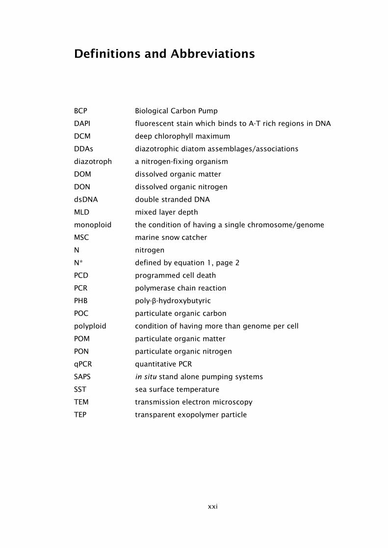
List of tables
Table 1-1. Summary of biologically mediated N transformation processes in the
marine environment (Modified from Gruber 2008). ................... 6
Table 2-1. Collection details and phylotype presence/absence ...................... 34
Table 2-2. qPCR primers and TaqMan probes used in this study ................... 38
Table 3-1. Estimated Trichodesmium-derived PON contribution and fluxes .... 74
Table 4-1. Estimated genome copies in T. erythraeum IMS 101 under varying
phosphorus conditions calculated by division of gene copies/L
by cells/L as described by Pecoraro et al. 2011). ..................... 97
Table 4-2. Experimentally determined ploidy levels of marine and aquatic
cyanobacterial species (Amended from Griese et al. 2011) .... 102
Table 4-3. Estimated Trichodesmium spp. contribution to total in situ
chlorophyll based on cell counts and gene copies. ................ 105
Table 5-1 Species specific storage products and qualitative observations of the
ultrastructure of colonies collected in situ. ............................ 118


vii
List of figures
Figure 1-1. Simplified schematic representation of the marine nitrogen cycle
and its coupling to the oxygen, phosphorus, and carbon cycling
in the marine environment. (reproduced from(Gruber 2008) ..... 7
Figure 1-2. Global distribution of N* along the σθ=26.8 surface corresponding
to the median of the range of densities composing the SAMW
(Sarmiento et al. 2004) illustrating excess nitrate accumulation
in the thermocline of the subtropical North Atlantic. Nutrient
data from Key et al. (2004). Black dots represent areas sampled
in this study. ............................................................................. 8
Figure 1-3. Phylogeny of nifH amino acid sequences from various nifH
containing organisms. Note that nifH diversity within the
cyanobacterial clade is high enough for targeted assessments of
individual cyanobacterial species using qPCR of the nifH gene.
Abbreviations of note include Csp: Cyanothece sp. strain ATCC
51142 a unicellular diazotroph, and Tsp: Trichodesmium sp.
strain IMS101. Reproduced from Choo et al. (2003). ............... 10
Figure 1-4. Trichodesmium colony morphology. Various colonial morphologies
include (A) radial puffs, (B) twisted tufts, (C) rafts of trichomes
arranged parallel to each other, and (D) flat, matted nests.
Trichodesmium colonies typically consist of 100-200 trichomes
(Carpenter et al. 2004). Scale bars represent 1 mm. ................ 12
Figure 1-5. A diazotrophic diatom association (DDA). Richelia intracellularis
associations with host diatoms. (A) Richelia-Rhizosolenia where
R. intracellularis is confined to the apices of the host diatom
(arrows), and (B) Richelia-Hemiaulus where cyanobionts are
obscured by the cytoplasm unless they are (C) viewed under
excitation of the phycoerythrin pigment which is not present in
the host diatoms. Scale bars represent 100 µm. ...................... 14
Figure 1-6. Schematic of some of the previously theorised export mechanisms
for Trichodesmium and other diazotrophs including export of

viii
diazotroph biomass following incorporation into faecal material
and secondary export following breaking down and recycling of
diazotroph derived N, which is then available for incorporation
into the biomass of other organisms (modified from Mulholland
2007). ..................................................................................... 21
Figure 2-1. Stations sampled for nifH abundance on the D361. Total nifH
abundance (gene copies m-3
) in surface samples reveals the peak
of detectable diazotrophic nifH was located between the equator
and 10°N. Note that nifH was not detected in the surface at the
southerly most station. ........................................................... 33
Figure 2-2. (A) Total nifH detected in each sample at each station (latitudinal
color bar) along the north-south transect from 2 – 500 m
illustrating rapid attenuation of nifH with depth. Note log scale.
(B) Individual phylotype nifH detected at depth alongside water
column nitrogen fixation rates. (C) Latitudinal section of
temperature (°C) along the north-south cruise transects
demonstrating nearly all SAPS samples collected below 100 m
were outside of the mixed layer. ............................................. 40
Figure 2-3. Standard curves for qPCR assessments of serially diluted standards
for all eight phylotypes. R2
= 0.999 for the filamentous standard
(Fil, blue asterisks); R2
= 0.991 for the Richelia-Rhizosolenia
standard (Het1, orange circles); R2
= 0.999 for the Richelia-
Hemiaulus standard (Het2, pink rectangles); R2
= 0.999 for the
Group A cyanobacteria standard (UCYN-A, blue crosses); R2
=
0.998 for the Group B cyanobacteria standard (UCYN-B, green
triangles); R2
= 0.984 for the Group C cyanobacteria standard
(UCYN-C, red squares); R2
= 0.998 for the Gamma A standard
(purple crosses); R2
= 0.997 for the Cluster III standard (blue
diamonds). .............................................................................. 42
Figure 2-4. Latitudinal sections of absolute nifH phylotype abundance (gene
copies m-3
) along the D361 transect demonstrating unicellular
affinity for higher latitudes and cooler temperatures,
filamentous nifH localisation just north of the equator, and the

ix
ubiquity of the non-cyanobacterial Gamma A nifH in the surface
layer. Note varying scales........................................................ 43
Figure 2-5. Bray Curtis dissimilarity dendrogram of nifH diversity normalised to
total nifH in each sample. Relative proportions of nifH
phylotypes within each sample are indicated by colour
distributions in each bar: Trichodesmium-attributable nifH
(purple), Group A Cyanobacteria-attributable nifH (green),
Hemiaulus-Richelia-attributable nifH (red), and Rhizosolenia-
Richelia-attributable nifH (blue); bar height represents absolute
nifH abundance in each sample. Starred samples represent no
nifH detection or absolute nifH abundance below 1000 copies
m-3
. Sample labels are displayed at the station number followed
by the depth of the sample. .................................................... 48
Figure 3-1. Sampling locations. (A) D361 cruise track overlain on N* (µmol kg-1
)
along the 26.8σθ and (B) SST (degC). Data from Key et al. (2004).
(C) CH0711 stations overlain on SST (degC) from shipboard
measurements. ....................................................................... 60
Figure 3-2. The 100L Marine Snow Catcher (A) and the particle collection
technique (B): each particle was transferred from the aquarium
to an individual well in a welled plate using a glass Pasteur
pipette. ................................................................................... 61
Figure 3-3. Forms of Trichodesmium collected. Colonial puff (A) and colonial
tuft (D), as was quantified in the Gulf of Mexico collections, and
free trichomes (B, C), which vary in length, as were quantified in
the Atlantic. Scale bars represent 100μm. ............................... 64
Figure 3-4. Trichodesmium abundance in the (sub-)tropical Atlantic. Nitrogen
fixation rates and surface colonial morphology of
Trichodesmium (A). Percentages of Trichodesmium surface
abundance at depth; shaded area represents depths over which
remineralisation is assumed to occur (B). ................................ 68
Figure 3-5. Sinking speeds of Trichodesmium colonies in the Gulf of Mexico
and in the Atlantic collected between 100 and 250 m. Solid
black lines represent he average sinking speed for each depth,

x
which ranged from 19 to 100 m day-1
. Replicates for each depth
are as noted above. ................................................................. 69
Figure 3-6. Plate of photomicrographs of Trichodesmium in aggregates. (A, B,
C, E) Trichomes in conjunction with radiolarians. (D) Active
phycoerythrin fluorescence of the trichome from panel E. Scale
bars represent 100µm. ............................................................ 71
Figure 3-7. Plate of photomicrographs of Trichodesmium in faecal pellets. (A)
Partially degraded trichomes incorporated in (B) faecal pellets
from CH0711; (C) Confocal micrograph of seemingly intact
trichomes from (D) faecal pellet of unknown origin; (E) pteropod
food web from same collection as pellet with intact trichomes
from D361. Scale bars represent 100µm. ................................ 72
Figure 3-8. Schematic of possible export mechanisms for Trichodesmium
(modified from Mulholland 2007). ........................................... 77
Figure 3-9. Longitudinal section of Phosphate (µM) along a pelagic transect
during CHO711 (A) and a latitudinal section of phosphate (µM)
along the north-south transect of D361 (B) illustrating enhanced
phosphate concentrations within the depth range of potential
nutrient mining (dashed boxes). .............................................. 79
Figure 3-10. Light micrographs of Trichodesmium spp. in association with
aggregates within the hour following collection showing
apoptosis of trichomes in progress. Scale bars represent 200µm
as indicated. ........................................................................... 82
Figure 3-11. Latitudinal section of oxygen along the south-north transect on
D361 showing decreased water column oxygen in the depth
range where remineralisation of Trichodesmium is assumed
(100-250 m), and alignment with the density surfaces within
which remineralisation of diazotroph derived material could
contribute to N*. Oxygen saturation was calculated from the
method described by Garcia and Gordon (1992) and AOU was
subsequently derived from the equation provided by Sarmiento
and Gruber (2006). ................................................................. 86

xi
Figure 4-1. Raw Discrepancy between Trichodesmium cell counts and nifH
gene copies. (A) in situ data from surface samples along the
D361 transect (black), R2
=0.87, and AMT17 transect (blue), R2
=
0.83, and (B) Trichodesmium erythraeum IMS101 culture
samples extracted from Durapore (red), R2
=0.78, and
polycarbonate (green), R2
=0.88, filters. Gene copies consistently
exceed cell counts by one to two orders of magnitude (p<0.01),
except at and below the limit of detection for the qPCR assay.
Theoretical genome copies per cell derived from this
discrepancy (gene copies/L divided by cells/L; Pecoraro et al.
2011) range from 1-119 for in situ data and from 159-698 for
Trichodesmium erythraeum IMS 101. The solid black lines
represent a 1:1 ratio between cell counts and gene copies. ..... 95
Figure 4-2. A combined assessment of log transformed cell count and gene
copy abundances to illustrate the sustained discrepancy across
all samples, except at and below the limit of detection for the
qPCR assay. ............................................................................ 96
Figure 4-3. Multiple sections of 3 series (out of 11) of DAPI staining
assessments in Trichodesmium IMS101 showcasing the DNA
distribution within each cell is not uniform along the z plane
(sections). The distance between each panel is approximately
55nm. Scale bars represent 5 µm. ........................................... 98
Figure 4-4. DNA distribution in Trichodesmium erythraeum IMS 101. (A) DAPI
(blue) and autofluorescence (orange) illustrate marked
segregation of DNA staining within the cell. (B) Transmission
electron micrograph illustrating that ultrastructure alone doesn’t
account for DAPI staining patchiness as intracellular organic
matter is not segregated in the same manner as is observed by
DAPI fluorescence. (C) Such DNA segregation is not as readily
visible as lower resolutions. Scale bars represent 2µm. ........... 99
Figure 4-5. Theoretical chlorophyll content of individual Trichodesmium IMS
101 cells derived from cell counts (green) and from qPCR of the
nifH gene in extracts from Durapore (blue) and polycarbonate
(red) filters over the four cultures of varying age and

xii
phosphorus source. The femtogram scale of chla per cell derived
from qPCR abundances implies gene copies are not an accurate
representation of cell number as cellular chlorophyll content
derived from these abundances results in 3 orders of magnitude
less chlorophyll per cell than is reported in the literature.
Literature values for Trichodesmium cellular chlorophyll content
are represented by the yellow box, 1.1-1.5 pg chla cell-1
(La
Roche and Breitbarth 2005), and the blue box, 1.56-3.9 pg chla
cell-1
(Carpenter et al. 2004); it should be noted that these
cellular chlorophyll content values are derived from field
samples, which may contain less chlorophyll per cell than
observed in the phosphate culture. ....................................... 100
Figure 4-6. Genomic contribution to total cellular phosphorus in
Trichodesmium. Diagonal lines indicate the ranges within which
genomic P accounts for 10, 25, 50, and 100% of total cellular P.
The red box incorporates the observed range of genome copies
per cell in Trichodesmium spp. from D361 and AMT17 and the
range of cellular P content for Trichodesmium spp. in situ
(Tovar-Sanchez et al. 2011; Neuster et al. 2012; this study)
following conversion from colonial P to cellular P assuming 200
trichomes per colony and 100 cells per trichome (Carpenter et
al. 2004). The blue box incorporates the range of observed
genome copies per cell (this study) and the range of cellular P
content for T. erythraeum IMS101 (Barcelos e Ramos et al.
2007). ................................................................................... 107
Figure 5-1. Schematic representation of over ballasting and Trichodesmium
PON quality. .......................................................................... 114
Figure 5-2. (A) Sectioning of resin block using a glass knife resulting in a
ribbon of sections including gold-coloured sections which are
the suitable thickness for TEM imaging. (B) Ultramicrotome
section colour reference chart (Drukker international). .......... 119
Figure 5-3. Transmission electron micrographs of Trichodesmium erythraeum
IMS 101cells showing various ultrastructural components and
variable integrity: vacuoles (V), intercellular space (IS), outer

xiii
membrane (OM), plasma-membrane (PM), nitrogen storage
vacuoles known as cyanophycin granules (CG), carboxysomes
(C), gas vesicles (GV), and vacuolisation (VS), which were used
for reference in subsequent identification of ultrastructural
components of in situ samples. Scale bars represent 1 µm in
panels A and D, 100 nm in panel B, and 500 nm in panel C. . 119
Figure 5-4. Gas vacuole integrity. Transmission electron micrographs of
positively (A, B) and negatively (C-F) buoyant Trichodesmium
colonies collected in the western Sargasso Sea showcasing
varying degrees of gas vesicle integrity (red arrows). Orange
lines mark cellular borders. (A) View of intact gas vesicles
indicated by parallel distribution of electron dense fibres
interspaced by electron transparent regions. (B) Intact gas
vesicles in a cell from a positively buoyant surface colony
indicated by normal, regular arrangement of the electron dense
fibres within the gas vacuole and the clearly defined gas vesicle
with electron transparent center regions. (C) Partially collapsed
gas vesicles in a negatively buoyant colony indicated by the
uneven distribution of electron dense fibres within the gas
vacuole and reduction in marked circular vesicle walls. (D, E)
Intact gas vesicles in a negatively buoyant colony are indicated
as above. (F) Collapsed gas vesicles in a negatively buoyant
colony indicated by a complete lack of structural regularity
within the gas vacuoles, as well as a lack of electron transparent
regions. Scale bars represent 500 nm in panels A, C, E and F,
200 nm in panel B and 1000 nm in panel D. ......................... 122
Figure 5-5. Intracellular storage components. Transmission electron
micrographs of positively (A, B) and negatively (C - F) buoyant
Trichodesmium colonies collected in the western Sargasso Sea.
Orange lines mark cellular borders. Various storage components
are indicated by red and orange arrows, such as (A, B, C)
cyanophycin granules known to be involved in nitrogen storage,
(D) scroll-like bodies associated with toxin production, (E) poly-β-
hydroxybutyric acid granules theorised to be involved in
carbohydrate ballasting, (B, F, orange arrow) vacuole-like
xiv
structures suspected to store photosynthetic metabolites, and
(F, blue arrows) medium electron dense granules assumed to
serve a short-term storage role and are important in nitrogen
metabolism function. Scale bars represent 5000 nm in panel A,
3000 nm in panel B, 2000 nm in panels C and F, 500 nm in
panel D, and 1000 nm in panel E........................................... 123
Figure 5-6. Intracellular vacuolisation. Transmission electron micrographs of
Postively (A, B) and negatively (C - F) buoyant Trichodesmium
colonies collected in the western Sargasso Sea illustrating
variation in cellular density. (A) Healthy, intact cells. (B, C, D, E,
F) moderate to extensive intracellular vacuolisation and cellular
shrinkage compared to panel A. Orange lines mark cellular
borders. Reductions in intracellular density may be indicative of
senescence. The shaded, electron dense regions of the cell are
gas vacuoles (C - F, orange arrows). Some storage bodies are
still present despite vacuolisation, such as PHB granules (E, red
arrows) and cyanophycin granules (F, red arrow). Scale bars
represent 1000 nm in panel A, 5000 nm in panel B, and 2000
nm in panels C – F. ................................................................ 125
Figure 5-7. Epibionts. Transmission electron micrographs of cells from
negatively buoyant Trichodesmium colonies collected in the
western Sargasso Sea showcasing extensive colonisation of the
mucilaginous sheath by epibionts indicated by red arrows.
Orange lines mark cellular borders. Epibiont distribution
appears to span the entire perimeter of the Trichodesmium cell
(A, B, C). Individual epibionts are tightly packed, irregularly
arranged along the perimeter, and are occasionally overlapping
(D). Scale bars represent 2 µm in panels A and B, 5 µm in panel
C, and 1 µm in panel C. Epibionts were not observed around
cells from positively buoyant surface colonies (Figure 5-4A, 5-
5A, 5-6A). ............................................................................. 126
Figure 5-8. Other inclusions of note. Transmission electron micrographs of
cells from negatively buoyant Trichodesmium colonies collected
in the western Sargasso Sea. Orange lines mark cellular borders.

xv
Various other inclusions encountered in the samples are
indicated by red arrows, such as (A) thylakoid lamellae, (B) the
extracellular matrix, (C) polyglucoside granules, which are
theorised to be involved in ballasting, and (D) extracellular
bodies, which may indicate the presence of compromised cells
within the colony although we cannot rule out that they may
simply be from associated organisms living in/on the colony.
Scale bars represent 500 nm in panels A, B, and C, and 1000 nm
in panel D. ............................................................................ 127
Figure 6-1. Raw (left) and amended (right) nifH gene abundances for
Trichodesmium following reduction of Trichodesmium-specific
nifH in accordance with the theoretical upper threshold of
polyploidy observed in situ (120 genome copies) compared to
raw Richelia-Hemiaulus nifH. Color bar represents section
latitude. ................................................................................ 135
Figure 6-2. Log nifH abundance and N* with potential density (σθ) contours
highlighting enhanced N* within the σθ=26.6-27.0 range along
the transect corresponding to increased nifH abundance in the
same depth range. Note slight difference in scale of the y axis
for each panel. ...................................................................... 137
Figure 6-3. Schematic representation of potential passive sedimentation and
remineralisation pathways for Trichodesmium colonies. The
orange colony border represents intact cell membranes and
yellow colony interior represents non-vacuolated cells. Exudation
of intracellular material and subsequent remineralisation are
represented by dashed arrows as indicated above. ................ 138
Figure 6-4. Micrographs of an aggregate using (A) standard light microscopy,
and (B) excitation of the phycoerythrin pigment using
epifluorescent microscopy showcasing that Trichodesmium
presence can sometimes only be revealed via the latter
technique. This aggregate was collected at 175 m in the Gulf of
Mexico. Scale bars represent 50µm. ...................................... 140

xvi

xvii
DECLARATION OF AUTHORSHIP
I, Elizabeth C. Sargent, declare that the thesis entitled DESCRIBING THE FATE
OF DIAZOTROPH-DERIVED NEW NITROGEN, and the work presented in the
thesis are both my own, and have been generated by me as the result of my
own original research. I confirm that:
this work was done wholly or mainly while in candidature for a research
degree at this University;
where any part of this thesis has previously been submitted for a degree or
any other qualification at this University or any other institution, this has
been clearly stated;
where I have consulted the published work of others, this is always clearly
attributed;
where I have quoted from the work of others, the source is always given.
With the exception of such quotations, this thesis is entirely my own work;
I have acknowledged all main sources of help;
where the thesis is based on work done by myself jointly with others, I have
made clear exactly what was done by others and what I have contributed
myself;
none of this work has been published before submission
Signed:
Date: 20 Feb 2014


xix
Acknowledgements
First and foremost, I would like to thank my supervisors, Alex Poulton,
Mark Moore, and Tom Bibby. Your support and feedback has been invaluable in
my growth as a scientist, and I truly appreciate the time and effort you have
each contributed to my progress over the last 3 years. I must also express my
gratitude to Mike Zubkov and Alan Kemp for their helpful participation in
discussions about the scope of this work, and for their guidance as this project
has developed.
I also wish to extend my appreciation to all those that have aided in the
collection and processing of samples discussed herein, as well as to those who
contributed their data for use in my analyses. Specifically, thank you Rebecca
Langlois for the copious time contributed to training me and for subsequent
data contributions. My sincerest thanks are also extended to the staff of the
Biomedical Imaging Unit at Southampton General Hospital for the time they
devoted to my training and for their continued interest in, and support of, my
analyses. In addition, thank you to David Honey and Katsia Pabortsava for
their help with deployments and for data contributions and to Andreas
Johansson for taking time away from the lab to accompany me on a research
expedition. Likewise, many thanks to Joe Snow for help with deployments and
sample processing, and for countless discussions that have aided in data
interpretation. I am also grateful to Eric Achterberg, Tracy Villareal, and Pia
Moisander for providing me with space on their research cruises and to the
crew and officers of the RRS Discovery, R/V Cape Hatteras, and R/V Atlantic
Explorer for facilitating each of those expeditions. I am also indebted to
GSNOCS for providing me with the funding to pursue a PhD at the University of
Southampton.
Though many friends are deserving of mention, I will here limit
acknowledgement Dave, Helen and Harriet. We have shared in an arduous
journey, and I will always remember how much we laughed through the good
and the bad. Dave, I will forever be appreciative of our echoed effort to keep
one foot firmly grounded outside of academia, and for the entertainment
provided by your policing of my vocabulary. Helen, thank you for continually
offering a fresh perspective, for your support in and out of the lab, and for
always knowing when it is time for wine. Harriet, you have been an amazing

xx
friend and teacher, and I attribute my sustained sanity nearly completely to
your never being too busy for a coffee break. We share an enthusiasm for
communicating science that I find refreshing and fulfilling, and I look forward
to our continued progress in science and our friendship throughout it.
I am also grateful to my parents for continued encouragement and
support of my educational pursuits. Your advice and guidance has been
integral to my successes, and I am continually appreciative of your tolerance to
my geographic flexibility. I also extend my familial appreciation to Jana. You
have made me better person both personally and professionally, and I credit
you in all of my achievements.
Finally, I express my gratitude to Geordie, my partner, and my best
friend. You have been remarkably patient and encouraging through the most
difficult stages of this PhD, and your sustained belief in me has helped to
ensure that I never doubted myself. I count myself lucky to have your support
in all that I do. Thank you.
xxi
Definitions and Abbreviations
BCP Biological Carbon Pump
DAPI fluorescent stain which binds to A-T rich regions in DNA
DCM deep chlorophyll maximum
DDAs diazotrophic diatom assemblages/associations
diazotroph a nitrogen-fixing organism
DOM dissolved organic matter
DON dissolved organic nitrogen
dsDNA double stranded DNA
MLD mixed layer depth
monoploid the condition of having a single chromosome/genome
MSC marine snow catcher
N nitrogen
N* defined by equation 1, page 2
PCD programmed cell death
PCR polymerase chain reaction
PHB poly-β-hydroxybutyric
POC particulate organic carbon
polyploid condition of having more than genome per cell
POM particulate organic matter
PON particulate organic nitrogen
qPCR quantitative PCR
SAPS in situ stand alone pumping systems
SST sea surface temperature
TEM transmission electron microscopy
TEP transparent exopolymer particle

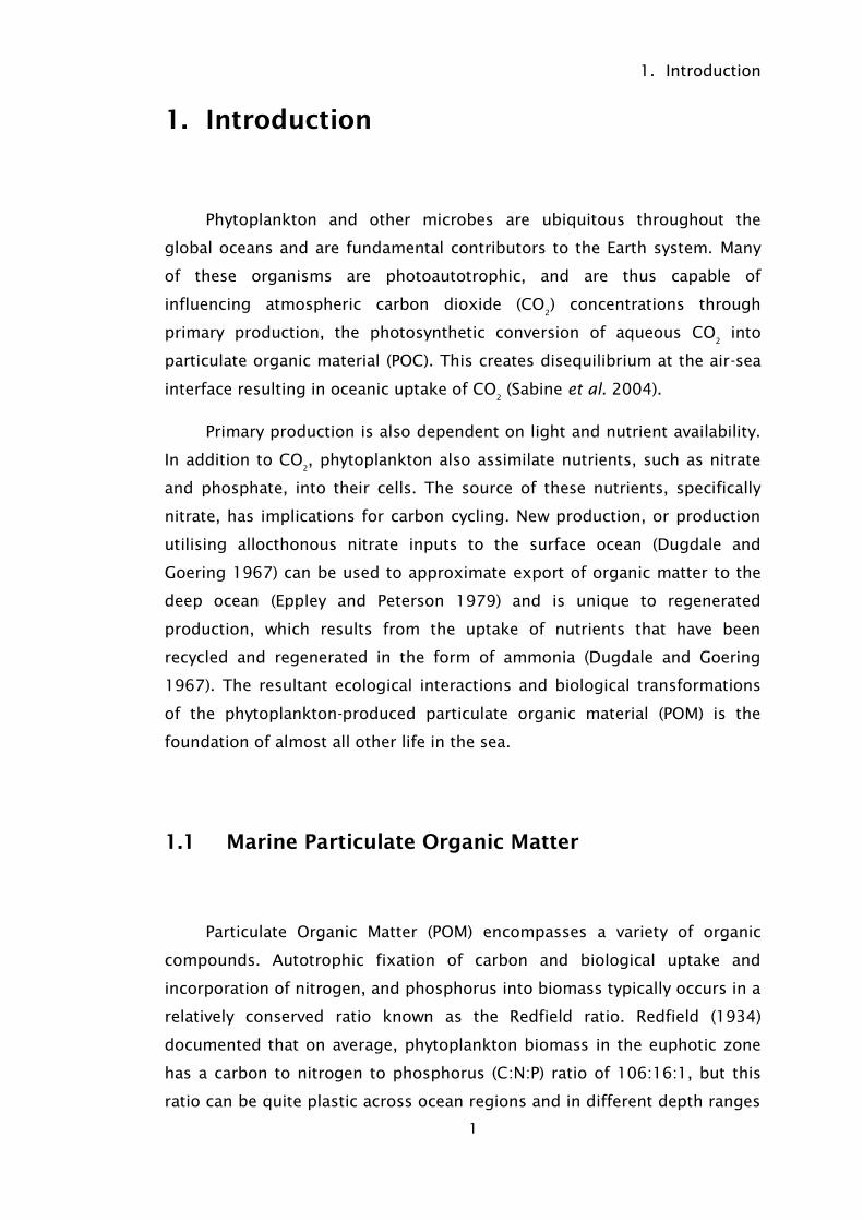
1. Introduction
1
1. Introduction
Phytoplankton and other microbes are ubiquitous throughout the
global oceans and are fundamental contributors to the Earth system. Many
of these organisms are photoautotrophic, and are thus capable of
influencing atmospheric carbon dioxide (CO2
) concentrations through
primary production, the photosynthetic conversion of aqueous CO2
into
particulate organic material (POC). This creates disequilibrium at the air-sea
interface resulting in oceanic uptake of CO2
(Sabine et al. 2004).
Primary production is also dependent on light and nutrient availability.
In addition to CO2
, phytoplankton also assimilate nutrients, such as nitrate
and phosphate, into their cells. The source of these nutrients, specifically
nitrate, has implications for carbon cycling. New production, or production
utilising allocthonous nitrate inputs to the surface ocean (Dugdale and
Goering 1967) can be used to approximate export of organic matter to the
deep ocean (Eppley and Peterson 1979) and is unique to regenerated
production, which results from the uptake of nutrients that have been
recycled and regenerated in the form of ammonia (Dugdale and Goering
1967). The resultant ecological interactions and biological transformations
of the phytoplankton-produced particulate organic material (POM) is the
foundation of almost all other life in the sea.
1.1 Marine Particulate Organic Matter
Particulate Organic Matter (POM) encompasses a variety of organic
compounds. Autotrophic fixation of carbon and biological uptake and
incorporation of nitrogen, and phosphorus into biomass typically occurs in a
relatively conserved ratio known as the Redfield ratio. Redfield (1934)
documented that on average, phytoplankton biomass in the euphotic zone
has a carbon to nitrogen to phosphorus (C:N:P) ratio of 106:16:1, but this
ratio can be quite plastic across ocean regions and in different depth ranges

E. Sargent
2
(Arrigo 2005). Regardless, an excess or deficit in the availability of any of
these elements can promote or limit the growth and productivity of different
phytoplankton and thus the fate of their biomass, which has implications for
biogeochemical cycles of carbon, nitrogen and phosphorus.
1.2 Biological Carbon Pump (BCP)
The biological carbon pump (BCP) is the downward export, or transfer of
carbon from the surface to the ocean’s interior, of biogenic material (Volk
and Hoffert 1985; Falkowski et al. 1998; Boyd and Trull 2007). This POM
includes phytoplankton biomass, zooplankton faecal material, and resultant
detritus, which can be subsequently colonised by heterotrophic bacteria,
often resulting in further breakdown, remineralisation and eventual mixing
and recycling of the resultant nutrients and dissolved carbon into the upper
ocean. However, some of that POM continues to settle through the water
column with eventual sequestration of the material to the deep ocean where
it remains isolated from the atmosphere. This loss of material from the
surface layer results in a continual disequilibrium of ocean-atmosphere CO2
concentrations driving continued uptake of CO2
by the oceans (Volk and
Hoffert, 1985). The ocean is a major global reservoirs of carbon, is crucial in
the global carbon cycle and hence in the regulation of climate. In the
~12,000 years prior to the industrial era, and excluding the earth’s crust
and sediments, the deep ocean held an order of magnitude more carbon
than all other carbon reservoirs combined (Sigman and Boyle 2000).
Following industrial revolution, anthropogenic activities have resulted in an
unprecedented increase in atmospheric CO2
to 390.5 ppm as of 2011
compared to 278.9 ppm in 1750 (Hartmann et al. In press). The ocean has
been integral in buffering this increase as without continued oceanic uptake,
we would currently be facing atmospheric CO2
concentrations in excess of
450 ppm (Doney et al. 2009).

1. Introduction
3
1.2.1 Mechanisms of Export
There are a variety of mechanisms by which export of POM occurs in
the oceans, such as aggregation and sinking of marine snow, grazing and
faecal pellet production, passive sedimentation, and active transport of POM
by eddies. Marine snow consists of large (>0.5 mm) aggregate particles that
are typically composed of a mixture of phytodetritus, refractory material,
biominerals, and mucoid matrices or other polymeric materials (Alldredge
and Silver 1998). The size, sinking speed, and composition of marine snow
varies widely (Alldredge and Gotschalk 1988), although ballasting of
biominerals is thought to increase sinking rates and aid in direct transfer of
POM to depth (Armstrong et al. 2002; Sanders et al. 2010; Riley et al. 2012).
Faecal material is also a large contributor to POM export as faecal pellets are
dense and often have enhanced sinking speeds (Alldredge and Silver 1988;
Turner 2002; Wilson et al. 2008). Other particles such as dead zooplankton
or large phytoplankton may sink passively through the water column. These,
and breakdown products of marine snow aggregates and faecal material,
compose the slow sinking pool of POM, and may be more susceptible to
remineralisation (Riley et al. 2012), although the relationship between
particle size, sinking speed, and susceptibility to remineralisation is still in
debate (De La Rocha and Passow 2007). Further breakdown products of
these particles can become part of the suspended pool of particulate
material. Active transport of all of these pools of material by eddies has also
been shown to occur with both enhanced POM production and export within
eddies compared to the adjacent waters (Bidigare et al. 2003; Guidi et al.
2012).
1.2.2 Remineralisation
As this POM sinks through the mesopelagic layer (or twilight zone
defined as the depths between the base of the euphotic zone ~200 m and
1000 m (Buesseler and Boyd 2009)) up to 90% of the POM can be
remineralised back into inorganic constituents by heterotrophic bacteria

E. Sargent
4
and/or zooplankton (Martin et al. 1987), resulting in a release of nutrients
into the water column where it is then available for reincorporation into
biomass either at depth or in the surface layer following vertical mixing
(Robinson et al. 2010). Remineralisation of POM can also occur in the
surface ocean and subsequent incorporation of the derived dissolved
organic matter (DOM) into the biomass of the microbes, which is then
available for grazing/breakdown and cycling of that material through
various trophic levels and eventually back to the POM and dissolved organic
matter DOM pools is known as the “microbial loop” (Azam et al. 1993). The
attenuation of POM between the surface and depth, and thus the ability or
inability of material to remain resilient to remineralisation as it sinks
through the mesopelagic, can be assessed by calculating the attenuation
coefficient of a power law function, b (Martin et al. 1987).
However, remineralisation of the multiple components of the POM pool
does not occur at the same rate. Anderson and Sarmiento (1994) proposed
an amendment to the Redfield ratio to 117:16:1 for sinking particles and
regenerated nutrients between 400 and 4000 m, which was consistent with
the C:N:P ratio of samples collected at all depths during cruises of the World
Ocean Circulation Experiment (WOCE) in the Atlantic, Pacific, and Indian
Oceans (Gruber 2008). This increase in carbon relative to nitrogen and
phosphorus in this depth range may be due to the differences in the
remineralisation rates of these elements, i.e. PON and phosphorus are
preferentially remineralised relative to POC (Anderson and Sarmiento 1994;
Christian et al. 1997; Shaffer et al. 1999; Schneider et al. 2003; Monteiro
and Follows 2012).
1.3 The Marine Nitrogen Cycle
Nitrogen is a major component of both nucleotides (the components of
DNA and RNA) and amino acids (the building blocks for proteins), and it is
thus required by all living organisms. These essential requirements
potentially make nitrogen a crucial limiting factor in biological processes,

1. Introduction
5
such as throughout most of the (sub-)tropical surface ocean globally (Moore
et al. 2013). In the marine environment, nitrogen exists in six stable forms:
dinitrogen (N2
), nitrous oxide (N2
O), nitrite (NO2
-
), nitrate (NO3
-
), ammonium
(NH4
+
), as well as dissolved organic nitrogen (DON). The nitrogen cycle is the
complex series of reactions that control the abundance of each of these
nitrogen species. Each reaction is biologically mediated, including the
assimilation of NO2
-
, NO3
-
, and NH4
+
by phytoplankton, the nitrification of NO2
-
and NH4
+
by oxidizing organisms, ammonification by bacteria and
zooplankton, anaerobic oxidation of ammonium (Anammox) and
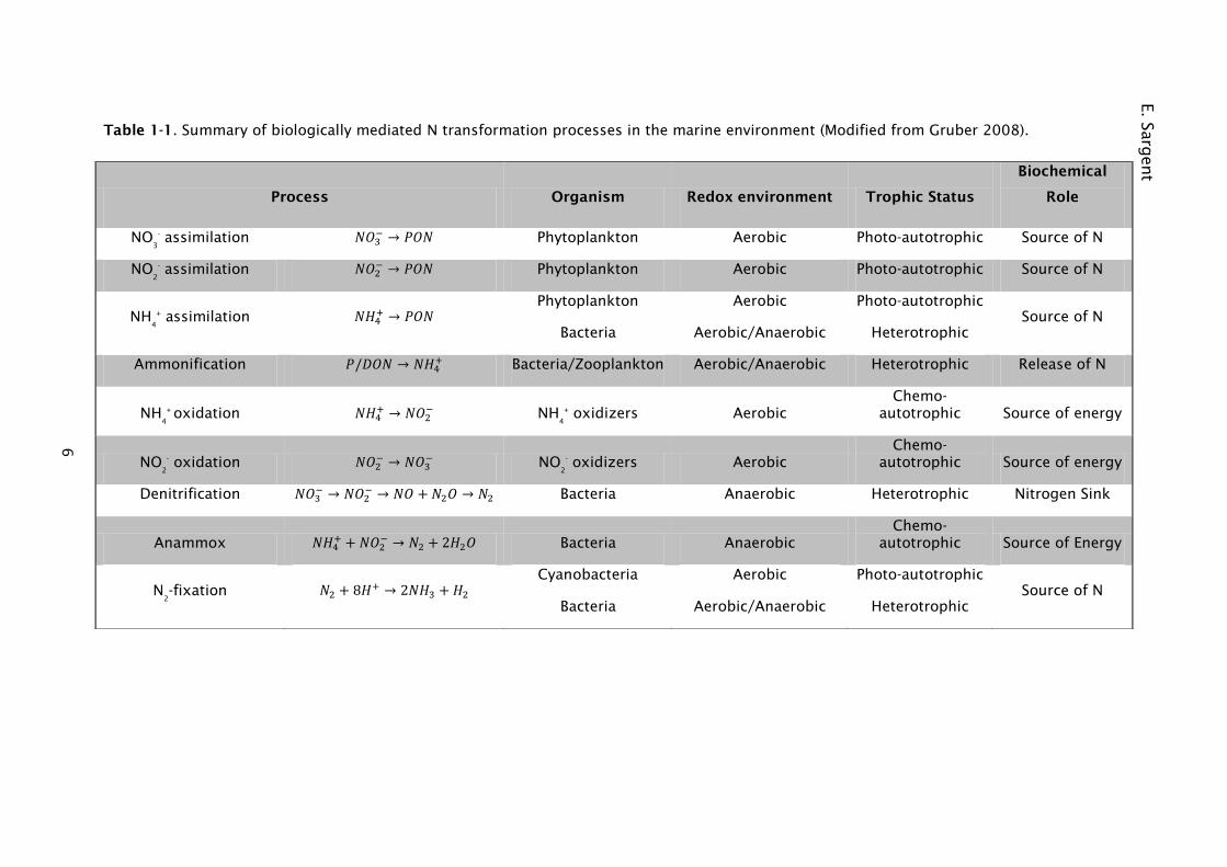
denitrification by bacteria, and nitrogen fixation by cyanobacteria (Table 1-
1).
The microbes involved in the various transformations of N are also
pivotal in the regulation and cycling of phosphorus in the oceans. In trying
to determine the primary limiting nutrient in marine environments, Redfield
(1934) theorized phosphorus must be limiting because any excess
phosphorus that decreased the N:P ratio could be ‘corrected’ back to 16:1
by biological nitrogen fixation. Conversely, an increased N:P ratio due to
excess nitrate could be corrected back to 16:1 through biological
denitrification (and/or Anammox) (Gruber 2008). Thus the nitrogen cycle is
closely linked to the phosphorus (and carbon) cycles, and perturbations in
any of the three can have a result on, or result from changes in, the other
two (Figure 1.1) (Gruber 2008).
E. Sargent
6
Table 1-1. Summary of biologically mediated N transformation processes in the marine environment (Modified from Gruber 2008).
Process Organism Redox environment Trophic Status
Biochemical
Role
NO3
-
assimilation Phytoplankton Aerobic Photo-autotrophic Source of N
NO2
-
assimilation Phytoplankton Aerobic Photo-autotrophic Source of N
NH4
+
assimilation
Phytoplankton
Bacteria
Aerobic
Aerobic/Anaerobic
Photo-autotrophic
Heterotrophic
Source of N
Ammonification Bacteria/Zooplankton Aerobic/Anaerobic Heterotrophic Release of N
NH4
+
oxidation
NH
4
+
oxidizers Aerobic
Chemo-
autotrophic Source of energy
NO2
-
oxidation
NO
2
-
oxidizers Aerobic
Chemo-
autotrophic Source of energy
Denitrification
Bacteria Anaerobic Heterotrophic Nitrogen Sink
Anammox
Bacteria Anaerobic
Chemo-
autotrophic Source of Energy
N2
-fixation
Cyanobacteria
Bacteria
Aerobic
Aerobic/Anaerobic
Photo-autotrophic
Heterotrophic
Source of N

1. Introduction
7
Figure 1-1. Simplified schematic representation of the marine nitrogen cycle and its
coupling to the oxygen, phosphorus, and carbon cycling in the marine environment.
(reproduced from(Gruber 2008)
The relative abundances of nitrate and phosphate are typically described
in terms of N*, which measures the departure from the Redfield ratio,
herein, we define N* by defined by equation 1, which is similar to the base
DINxs
equation; Other groups have previously added a constants to the
equation to force the global average of N* to zero (Gruber and Sarmiento
1997; Deutsch et al. 2001; Hansell et al. 2004, 2007; Moore et al. 2009):
(eq. 1)
Throughout the majority of the global ocean N* is negative within the
thermocline and in deeper waters (Gruber and Sarmiento 1997; Moore et al.
2009), but north of the equator in the Atlantic Ocean there is a positive N*
anomaly in the subtropical thermocline to ~1100 m. In this region nitrate is

E. Sargent
8
accumulating in excess at a rate of 7.8 ± 1.7x10¹¹
mol N yr-1
(Hansell et al.
2007) along the σθ=26.8 density surface, which corresponds to the median
of the range of densities composing the Subantarctic Mode Water (SAMW)
(Sarmiento et al. 2004) (Figure 1-2).
Figure 1-2. Global distribution of N* along the σθ=26.8 surface corresponding to the
median of the range of densities composing the SAMW (Sarmiento et al. 2004)
illustrating excess nitrate accumulation in the thermocline of the subtropical North
Atlantic. Nutrient data from Key et al. (2004). Black dots represent areas sampled in
this study.
Gradients of increasing N* can hence be used to infer a net gain of
nitrogen, which is tantamount to the high dissolved N:P ratios, and in turn
appears to result in phosphate becoming severely depleted in surface waters
of the North Atlantic subtropical gyre (Gruber and Sarmiento 1997; Wu et al.
2000; Moore et al. 2009). Formation of enhanced N* are thought to come
from a variety of sources, such as atmospheric deposition of high N:P
nutrients (Gruber and Sarmiento 1997) and advection of high N:P DOM into
the main thermocline (Michaels et al. 1996). Nitrogen fixation is also
thought to influence the excess N accumulations, such as through the
remineralisation of the high N:P biomass of nitrogen fixing organisms
(Anderson and Sarmiento 1994; Hansell et al. 2004, 2007). Mechanisms by

1. Introduction
9
which nitrogen fixation can contribute to the N* anomaly will be discussed
further in Chapters 2, 3, 5, and 6.
1.4 Nitrogen Fixation
Nitrogen fixation is the conversion of inert N2
to bioavailable ammonia
(NH3
). The reaction (detailed in Table 1-1) is catalyzed by the nitrogenase
enzyme. This enzyme has two subunits, an Fe-binding protein and an MoFe-
binding protein. The Fe-binding subunit of the nitrogenase enzyme is coded
for by the nifH gene (Young 1992). Diversity within this gene region allows
delineation between various groups of nifH-containing organisms as
indicated by Figure 1-3. Advances in molecular technologies have allowed
for the identification of unique nifH phylotypes (taxon-neutral phylogenetic
groups) and development of primers and probes for use in quantitative PCR
(qPCR) assays (Zehr et al. 1997; 1998; 2003; Church et al. 2005a; 2005b,
Foster et al. 2009, Moisander et al. 2010). This will be discussed in more
detail in Chapter 2.
The nitrogenase enzyme is oxygen sensitive, so the nitrogen fixation
reaction must be carried out anaerobically (Peters et al. 1995). The necessity
for oxygen segregation from nitrogenase in diazotrophic cyanobacteria is
enhanced due to the photosynthetic capability of these organisms resulting
in oxygen production within their cells. Diazotrophic cyanobacteria have
thus developed adaptive ways for segregating the nitrogen fixation process
from oxygen exposure (Newton 2007). Some diazotrophic cyanobacteria
overcome this segregation challenge spatially, such as through formation of
a specialized cell known as a heterocyst where nitrogen fixation but not
photosynthesis occurs (Fay 1992; Kumar et al. 2010), some separate
nitrogen fixation and photosynthesis temporally (Toepel et al. 2008), while
others do both (Bergman et al. 2013).

E. Sargent
10
Figure 1-3. Phylogeny of nifH amino acid sequences from various nifH containing
organisms. Note that nifH diversity within the cyanobacterial clade is high enough
for targeted assessments of individual cyanobacterial species using qPCR of the nifH
gene. Abbreviations of note include Csp: Cyanothece sp. strain ATCC 51142 a
unicellular diazotroph, and Tsp: Trichodesmium sp. strain IMS101. Reproduced
from Choo et al. (2003).
1.4.1 Importance of Nitrogen Fixation
The upper boundary of growth and productivity of marine photoautotrophs
is constrained by nitrogen availability globally as many ocean regions are
nitrogen limited (Falkowski 1997; Gruber and Galloway 2008; Moore et al.
2013). As nitrogen can be a limiting factor for growth, it is not surprising
1. Introduction
11
that nitrogen fixing organisms are found in nearly all marine environments
as they are not limited by nitrogen availability. Nitrogen fixing organism are
present in the open ocean, deep sea, benthos/sediments, coral reefs,
seagrass beds, intertidal areas, estuaries, other coastal areas (Carpenter and
Capone 2008), and potentially even inside the guts of zooplankton (Braun et
al. 1999). These organisms are often responsible for supporting the rest of
the phytoplankton community via both the release of recently fixed nitrogen
(discussed below) and through supply and recycling of that new nitrogen in
the ocean. Gruber and Sarmiento (1997) postulated that the majority of
fixed nitrogen in the surface oceans is diazotroph-derived, and thus these
organisms play an integral role in ocean ecosystems. Fixed-nitrogen
availability in the ocean is also coupled to the sequestration of atmospheric
carbon and thus is not only an integral part of the nitrogen cycle, but also
plays a role in the regulation of global biogeochemical cycles (Falkowski
1997). This is especially apparent over even longer timescales where
changes in the fixed nitrogen inventory due to a variety of factors (such as
changes in ocean circulation) are thought to cause glacial/interglacial
changes in atmospheric carbon dioxide concentrations (Falkowski 1997;
Broecker and Henderson 1998; Karl et al. 2002; Gruber and Galloway 2008;
Straub et al. 2013). Although the full extent of the effect of continued
global-scale anthropogenic perturbations of ocean biogeochemistry is
currently unknown, it has been suggested that the geographical range of
nitrogen fixers will expand in a future, warmer ocean (Boyd and Doney
2002), and that nitrogen fixation rates and carbon fixation by nitrogen
fixing organisms are likely to increase as pCO2
increases during the next
century (Hutchins et al. 2007).
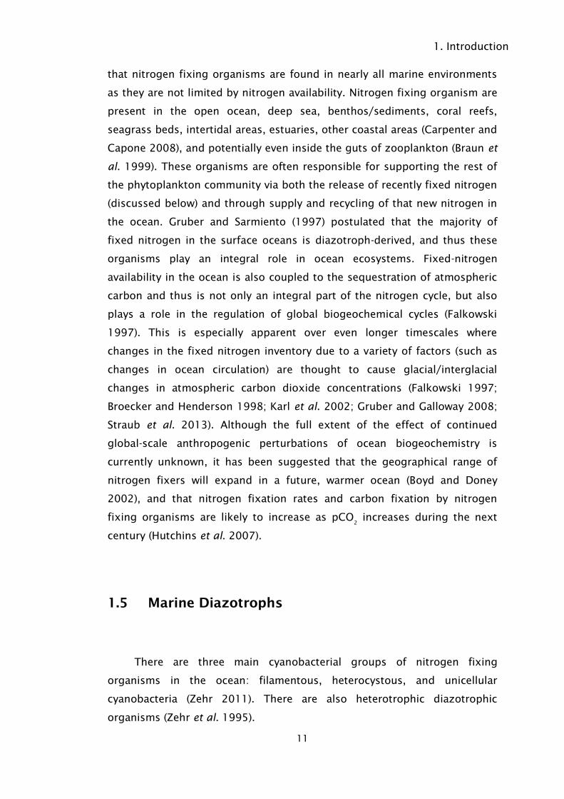
1.5 Marine Diazotrophs
There are three main cyanobacterial groups of nitrogen fixing
organisms in the ocean: filamentous, heterocystous, and unicellular
cyanobacteria (Zehr 2011). There are also heterotrophic diazotrophic
organisms (Zehr et al. 1995).
E. Sargent
12
1.5.1 Filamentous diazotrophs
The most widely studied marine diazotroph is the filamentous, non-
heterocystous cyanobacterial genus Trichodesmium, members of which are
considered to be the most abundant oceanic nitrogen fixers (Zehr 2011).
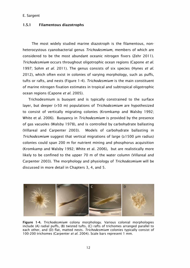
Trichodesmium occurs throughout oligotrophic ocean regions (Capone et al.
1997; Sohm et al. 2011). The genus consists of six species (Hynes et al.
2012), which often exist in colonies of varying morphology, such as puffs,
tufts or rafts, and nests (Figure 1-4). Trichodesmium is the main constituent
of marine nitrogen fixation estimates in tropical and subtropical oligotrophic
ocean regions (Capone et al. 2005).
Trichodesmium is buoyant and is typically constrained to the surface
layer, but deeper (>50 m) populations of Trichodesmium are hypothesized
to consist of vertically migrating colonies (Kromkamp and Walsby 1992;
White et al. 2006). Buoyancy in Trichodesmium is provided by the presence
of gas vacuoles (Walsby 1978), and is controlled by carbohydrate ballasting
(Villareal and Carpenter 2003). Models of carbohydrate ballasting in
Trichodesmium suggest that vertical migrations of large (≥1500 µm radius)
colonies could span 200 m for nutrient mining and phosphorus acquisition
(Kromkamp and Walsby 1992; White et al. 2006), but are realistically more
likely to be confined to the upper 70 m of the water column (Villareal and
Carpenter 2003). The morphology and physiology of Trichodesmium will be
discussed in more detail in Chapters 3, 4, and 5.
Figure 1-4. Trichodesmium colony morphology. Various colonial morphologies
include (A) radial puffs, (B) twisted tufts, (C) rafts of trichomes arranged parallel to
each other, and (D) flat, matted nests. Trichodesmium colonies typically consist of
100-200 trichomes (Carpenter et al. 2004). Scale bars represent 1 mm.

1. Introduction
13
One of the six described Trichodesmium species falls under the
antiquated genus Katagnymene. Members of this genus lack heterocysts
and, in contrast to other Trichodesmium species, do not form colonies
(Lundgren et al. 2001). Katagnymene spp. is present in warm, oligotrophic
ocean regions globally, and has been successfully isolated into culture
allowing for more thorough physiological investigations. Like other
Trichodesmium species, Katagnymene only possess the nitrogenase enzyme
in confined zones of cells along the trichome (Lundgren et al. 2001) now
known as diazocytes (Berman-Frank et al. 2001). These diazocyte regions
aide in the segregation of the nitrogenase enzyme from oxygen allowing for
nitrogen fixation to occur during the light period when oxygen is also being
produced via photosynthesis (Berman-Frank et al. 2001; Sandh et al. 2012).
Although Katagnymene spp. is morphologically distinct from other
Trichodesmium species, it is phylogenetically similar. The nifH gene region
in both genera is nearly indistinguishable (Lundgren et al. 2001), and high
similarity is also found within the internal transcribed spacer (ITS) region
between the 16S and 23S rRNA genes (Orcutt et al. 2002) and the hetR gene
regions (Lundgren et al. 2005) prompting a call for reclassification and
combination of Katagnymene into the Trichodesmium genus (Hynes et al.
2012).
1.5.2 Heterocystous diazotrophs
Heterocystous cyanobacteria possess specialized cells where nitrogen
fixation, but not photosynthesis, occurs (Fay 1992), and compose the
second group of marine diazotrophs, which include cyanobionts.
Cyanobionts are cyanobacteria that are involved in symbiotic relationships
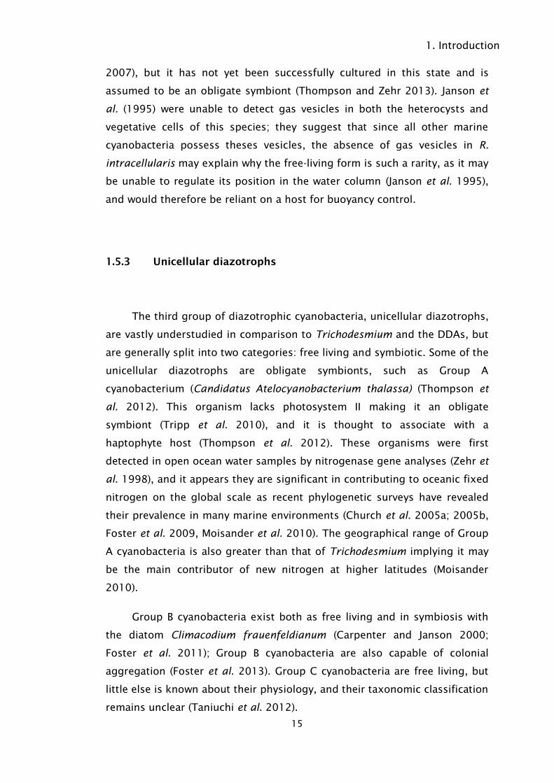
with a eukaryotic host. In diazotrophic diatom associations (DDAs), the
cyanobionts are nitrogen-fixing cyanobacteria, such as Richelia
intracellularis and Calothrix rhizosoleniae. R. intracellularis was first
described in an endosymbiotic relationship with the diatom Rhizosolenia
clevei (Ostenfeld and Schmidt 1901) (Figure 1-5A); R. intracellularis has
since been documented in similar consortia with the diatom species

E. Sargent
14
Hemiaulus spp. (Heinbokel 1986; Villareal 1991; Kimor et al. 1992; Villareal
1994; Janson et al. 1999; Scharek et al. 1999; Foster and Zehr 2006; Bar-
Zeev et al. 2008; Foster et al. 2009; Goebel et al. 2010; Padmakumar et al.
2010) (Figure 1-5B, C) and Guinardia cylindrus (a synonym of Rhizosolenia
cylindrus) (Venrick 1974; Villareal 1992; Scharek et al. 1999).
Figure 1-5. A diazotrophic diatom association (DDA). Richelia intracellularis
associations with host diatoms. (A) Richelia-Rhizosolenia where R. intracellularis is
confined to the apices of the host diatom (arrows), and (B) Richelia-Hemiaulus where
cyanobionts are obscured by the cytoplasm unless they are (C) viewed under
excitation of the phycoerythrin pigment which is not present in the host diatoms.
Scale bars represent 100 µm.
R. intracellularis possesses a single terminal heterocyst and up to ten
vegetative cells (Ostenfeld and Schmidt 1901); the heterocyst of this species
is noticeably larger than the vegetative cells, which are uniform in size
(Janson et al. 1999). C. rhizosoleniae was originally described as an epiphyte
of the diatom Chaetoceros compressum (Lemmerman 1905). This
cyanobacterium also consists of a single terminal heterocyst and multiple
vegetative cells, but unlike R. intracellularis, the heterocyst of C.
rhizosoleniae is smaller than the vegetative cells. Additionally, the vegetative
cells are not uniform in size, and extend in a slightly tapered filament
(Janson et al. 1999). Calothrix has been successfully cultured independent
of a host diatom (Foster and Zehr 2006). Infrequently, R. intracellularis is
reported as free-living (Weare et al. 1974; Goméz et al. 2005; White et al.
1. Introduction
15
2007), but it has not yet been successfully cultured in this state and is
assumed to be an obligate symbiont (Thompson and Zehr 2013). Janson et
al. (1995) were unable to detect gas vesicles in both the heterocysts and
vegetative cells of this species; they suggest that since all other marine
cyanobacteria possess theses vesicles, the absence of gas vesicles in R.
intracellularis may explain why the free-living form is such a rarity, as it may
be unable to regulate its position in the water column (Janson et al. 1995),
and would therefore be reliant on a host for buoyancy control.
1.5.3 Unicellular diazotrophs
The third group of diazotrophic cyanobacteria, unicellular diazotrophs,
are vastly understudied in comparison to Trichodesmium and the DDAs, but
are generally split into two categories: free living and symbiotic. Some of the
unicellular diazotrophs are obligate symbionts, such as Group A
cyanobacterium (Candidatus Atelocyanobacterium thalassa) (Thompson et
al. 2012). This organism lacks photosystem II making it an obligate
symbiont (Tripp et al. 2010), and it is thought to associate with a
haptophyte host (Thompson et al. 2012). These organisms were first
detected in open ocean water samples by nitrogenase gene analyses (Zehr et
al. 1998), and it appears they are significant in contributing to oceanic fixed
nitrogen on the global scale as recent phylogenetic surveys have revealed
their prevalence in many marine environments (Church et al. 2005a; 2005b,
Foster et al. 2009, Moisander et al. 2010). The geographical range of Group
A cyanobacteria is also greater than that of Trichodesmium implying it may
be the main contributor of new nitrogen at higher latitudes (Moisander
2010).
Group B cyanobacteria exist both as free living and in symbiosis with
the diatom Climacodium frauenfeldianum (Carpenter and Janson 2000;
Foster et al. 2011); Group B cyanobacteria are also capable of colonial
aggregation (Foster et al. 2013). Group C cyanobacteria are free living, but
little else is known about their physiology, and their taxonomic classification
remains unclear (Taniuchi et al. 2012).

E. Sargent
16
1.5.4 Heterotrophic Diazotrophs
Heterotrophic diazotrophs are distributed throughout the world’s oceans,
and unlike cyanobacterial diazotrophs, heterotrophic diazotrophs use
organic carbon as an energy source. However, photoheterotrophy is also
possible as is suggested for the Group A cyanobacterium, which lacks
photosystem II (Zehr et al. 1998; Tripp et al. 2010). Heterotrophic bacteria
were some of the first diazotrophic organisms described (Waksman et al.
1933), and non-cyanobacterial nifH is now known to compose the majority
nifH community structure globally (Farnelid et al. 2011). Heterotrohpic
diazotrophs also dominate nitrogen fixation in the South Pacific Gyre (Halm
et al. 2012). There are multiple nifH clades of heterotrophic diazotrophs
including proteobacteria and sulphate-reducing bacteria, which can exist
free living, in association with microbial mats, and even in hydrothermal
vent systems (Eckford et al. 2002; Steppe and Paerl 2002; Mehta et al. 2003;
Halm et al. 2011). Active pelagic heterotrophic diazotrophs have also been
detected via nifH assessments at and below 500 m (Hewson et al. 2007;
Hamersley et al. 2011).
1.6 Ecology and Physiology of Marine Diazotrophs
1.6.1 Controls on Diazotrophy
The distribution of some diazotrophs appears to be restrained by
temperature. Trichodesmium growth is most active above 20 °C (Carpenter
1983; Breitbarth et al. 2007), while the unicellular cyanobacteria display a
tolerance for cooler water temperatures in temperate zones (Holl et al.
2007; Needoba et al. 2007; Langlois et al. 2008). Turbulence has also been
hypothesised to adversely affect nitrogenase activity in Trichodesmium due
to increased oxygen exposure (Carpenter and Price 1976), but the effect of
turbulence on the nitrogen fixing capabilities of other diazotrophs is poorly

1. Introduction
17
understood (Howarth et al. 1993). Nitrogen fixing organisms are not well
described in higher latitudes, but nifH-containing organisms have been
detected in the Arctic (Farnelid et al. 2011). As cyanobacterial nitrogen
fixation has been observed in terrestrial and marine polar environments
near 0 °C (Vincent 2002; Stall et al. 2003; Blais et al. 2012), and as actively
growing populations of Trichodesmium and other diazotrophs are often
observed in turbulent areas (Carpenter et al. 2004), it does not appear that
activity of the nitrogenase enzyme itself is restricted by temperature and
turbulence, but rather the physiology of individual diazotrophs limits their
tolerances to these factors.
Trace metals, specifically iron, can be a primary limiting nutrient for
diazotrophs as the nitrogenase enzyme requires both iron and
molybdenum, and thus the iron requirements of diazotrophs can be an
order of magnitude greater than the iron requirements of strict
photoautotrophs (Berman-Frank et al. 2001; Kustka et al. 2003; Sañudo-
Wilhelmy et al. 2001). This is especially apparent in the subtropical South
Atlantic were phosphorus concentrations are in excess of the requirements
for diazotrophic growth, but diazotrophy is minimal (Moore et al. 2009). In
contrast, diazotrophy thrives in the subtropical North Atlantic where there is
also an input of iron from atmospheric aerosols (Michaels et al. 1996; Wu et
al. 2000; Mahaffey et al. 2003; Tyrrell et al. 2003; Moore et al. 2009). Iron
has been observed to stimulate growth and nitrogen fixation in
Trichodesmium both in culture and in situ (Rueter 1988; Paerl et al. 1994;
Berman-Frank et al. 2001). As atmospheric dust deposition is low in many
oligotrophic ocean regions where diazotrophs should otherwise flourish,
iron limitation of nitrogen fixation has been proposed as a near global
occurrence (Falkowski 1997; Berman-Frank et al. 2001; Moore et al. 2002).
Other nutrients, such as phosphorus can also limit diazotrophy, and co
limitation of diazotrophy by iron and phosphorus is likely occurring in the
subtropical North Atlantic (Mills et al. 2004; Ward et al. 2013). However,
some diazotrophs, such as Trichodesmium are able to access organic pools
of phosphorus allowing for continued growth even when phosphate is
depleted (Mulholland et al. 2002; Dyhrman et al. 2006).

E. Sargent
18
1.6.2 Grazing
Grazing of diazotrophs is not widely reported, but grazing deterrence
by diazotrophs appears to be widespread. Depressed growth rates, egg
production and biomass of zooplankton have been reported in the presence
of cyanobacterial blooms (Heerkloss et al. 1984; Sellner et al. 1997), and
some diazotrophs, such as Trichodesmium, are known to possess grazer-
deterring toxins (Siddiqui 1992; Janson et al. 1995; O’Neil 1999; Kerbrat et
al. 2010). However, modest grazing of Trichodesmium biomass has been
reported in harpacticoid copepods (O’Neil and Roman 1994; O’Neil 1998;
1999), and isotopic assessments (see below) suggest that the transfer of
diazotroph-derived new nitrogen to higher trophic levels does occur (O’Neil
et al. 1996; Montoya et al. 2002; Loick-Wilde et al. 2012). Fish have also
been observed to graze on Trichodesmium (Carpenter and Capone 2008 and
references therein). Additionally, salp grazing on Trichodesmium has been
theorised to occur following observations of negatively correlated salp and
Trichodesmium abundances in situ (Carpenter and Capone 2008). Some
coccoid cyanobacteria, as well as heterotrophic bacteria, are also directly
grazed (Caron et al. 1991).
1.6.3 Nutrient Transfer
In heterocystous cyanobacteria the transfer of nutrients is essential,
both along the trichome between the differentiated specialised cells and
from symbiont to host. Only vegetative cells are able to perform
photosynthesis, and therefore must supply the heterocyst(s) with the
necessary sugars to support the energy demanding process of nitrogen
fixation; in return, the heterocyst supplies the vegetative cells with fixed
nitrogen (Golden and Yoon 2003; Foster et al. 2011). The mechanism for
this transfer is described by Mullineaux et al. (2008), who showed diffusion
of fluorescent molecules from a heterocyst cytoplasm to a vegetative cell
cytoplasm, and vice versa, through intercellular channels, or
microplasmodesmata, in the cyanobacterium Anabaena cylindrical.

1. Introduction
19
Crucially, they showed that the exchange rate of fluorescent dye between
vegetative cells significantly increases after 72 hours of nitrate deprivation,
which suggests an increase in the number of connections between these
cells after heterocyst differentiation thus facilitating a more efficient
metabolite exchange pathway (Mullineaux et al. 2008). This also suggests
that although nitrogen fixation takes place in the heterocysts, the vegetative
cells of R. intracellularis could be responsible for nitrogen transfer from
cyanobiont to host (Janson et al. 1995). Villareal (1990) cultured symbiotic
Rhizosolenia-Richelia and asymbiotic Rhizosolenia in both nitrogen deplete
and nitrogen replete medium, and found that both diatoms grew at
comparable rates under replete conditions; conversely, under nitrogen
deplete conditions Rhizosolenia-Richelia proliferated at a significantly higher
rate, highlighting the benefits provided by this symbiotic association.
The dynamics of nutrient transfer and N release in Trichodesmium is
slightly more complicated as Trichodesmium is free living and does not need
to release N to support a host. However, release of DON and ammonia by
Trichodesmium has been observed both in situ and in culture (Capone et al.
1994; Glibert and Bronk 1994; Mulholland et al. 2004). Potentially,
Trichodesmium uses N release as a mechanism to ensure all cells have
access to fixed nitrogen as only specialised cells along the trichome are able
to fix nitrogen (Mulholland and Capone 2000; Berman-Frank 2001). Another
potential explanation for new nitrogen exudation in Trichodesmium is
support of associated organisms, such as epibionts, which may serve in a
mutually beneficial partnership as these epibionts may aid in increasing
Trichodesmium’s acquisition of phosphorus (Van Mooy et al. 2012).
The physiology and nutrient transfer dynamics of the unicellular
cyanobacteria is less clear, but as with DDAs, it likely that the transfer of
newly fixed N from the unicellular cyanobiont to host occurs. The unicellular
Crocosphaera watsonii has been observed in close association with a diatom
host, Climacodium frauenfeldianum lending some evidence to this theory
(Foster et al. 2011).

E. Sargent
20
1.6.4 Cell death
Cell death among diazotrophs can be a complicated, multi-pathway
process. Programmed cell death (PCD), a self-initiated cell death mechanism,
is common among eukaryotes (Jiménez et al. 2009), and has recently been
described as a prominent cell death pathway among cyanobacteria (Sigee et
al. 2007; Zheng et al. 2013), and appears to be the dominate cell death
pathway in Trichodesmium (Bar-Zeev et al. 2013). Various cell death
pathways in Trichodesmium have been previously assessed, and PCD
appears to be a common pathway by which Trichodesmium dies (Berman-
Frank et al. 2004; Bar-Zeev et al. 2013). PCD can occur with ageing or
following exposure to a variety of physiological stressors, such as high
irradiance, nutrient starvation, and temperature shock (Berman-Frank et al.
2004; Jiménez et al. 2009). Other cell death pathways among the
cyanobacteria include autophagy, necrosis and autolysis (Zheng et al. 2013).
The cell death pathways of Trichodesmium will be discussed further in
Chapter 5.
1.7 Fate of Diazotrophic N
Nitrogen fixed by diazotrophs eventually becomes available to the
wider phytoplankton community through the previously mentioned
exudation of fixation products, grazing, or breakdown of diazotroph
biomass (Capone et al. 1994; Glibert and Bronk 1994; O’Neil et al. 1996;
Mulholland and Capone 2001; Berman-Frank et al. 2004; Mulholland et al.
2004; Hewson et al. 2004). This transfer of new nitrogen to other organisms
can support their growth and productivity, and potentially facilitate
enhanced export of other organisms (Capone et al. 1998; Carpenter et al.
1999; Ohlendieck et al. 2000). Mullholland (2007) theorised that
diazotroph-derived new nitrogen, specifically that which is derived from
Trichodesmium, is likely constrained to continuous recycling in the euphotic

1. Introduction
21
zone through the microbial loop, where it may eventually contribute to
export indirectly via the settling/sinking of biomass of other organisms.
However Mulholland (2007) also postulated that there is the potential for
direct export of diazotroph biomass following grazing (Figure 1-6).
More recently, species specific physiologies have been taken into
account when considering organismal fate. Many now believe that
programmed cell death plays a role in the demise of Trichodesmium blooms
(Bar-Zeev et al. 2013; Chapter 5). The ever increasing nifH dataset has also
provided new insights into species-specific vertical distributions (Church et
al. 2005), and in situ camera systems have revealed diazotrophs, such as
Trichodesmium can be evenly distributed as deep at 120m ( the deepest
depth assessed) (Davis and McGillicuddy 2006).
Figure 1-6. Schematic of some of the previously theorised export mechanisms for
Trichodesmium and other diazotrophs including export of diazotroph biomass
following incorporation into faecal material and secondary export following
breaking down and recycling of diazotroph derived N, which is then available for
incorporation into the biomass of other organisms (modified from Mulholland
2007).
E. Sargent
22
1.7.1 Export of filamentous diazotrophs
Guidi et al. (2012) suggested that active export of Trichodesmium
could be possible in association with downwelling eddies in the North Pacific
Subtropical Gyre. Sweeping mechanisms where Trichodesmium would be
caught up in the export of other organisms, such as diatoms, have also
been used as an explanation for Trichodesmium presence at depth (Scharek
et al. 1999b). Such occurrences are rarely observed, but this potenitally due
to undersampling. The general consensus among the nitrogen fixation
research community is that there is currently no evidence to suggest that
Trichodesmium sinks below the euphotic zone unaided, and a recent review
by Thompson and Zehr (2013) concluded that Trichodesmium is not likely to
directly contribute to export of particulate organic matter (POM).
“At this time there is no evidence that Crocosphaera or
Trichodesmium cells sink beyond the euphotic zone, thus an oceanic
regime dominated by these cell types would have patterns of C and
N export very different from an ocean dominated by the symbiotic
cyanobacterial N2 fixers and their hosts.” –Thompson and Zehr
(2013)
1.7.2 Export of heterocystous diazotrophs
Goldman (1993) hypothesized that diatom blooms resulting from new
nitrogen inputs into the euphotic zone would be coupled with an export
flux, specifically near the deep chlorophyll maximum (DCM). More recent
data suggests larger diatoms, such as Hemiaulus, a known host of the
diazotrophic Richelia intracellularis, are exported from the mixed layer, not
the DCM (Scharek et al. 1999). Diatoms are historically important
constituents of export events (Boyd and Newton 1995; Buesseler 1998; Boyd
and Newton 1999; Kemp et al. 2000), and Hemiaulus, and other large
autotrophs, can act as direct exporters of nitrogen, silica, and carbon to the
deep sea (Scharek et al. 1999).
1. Introduction
23
For direct export of new nitrogen out of the euphotic zone to occur,
the cyanobionts, and any hosts, would need to remain intact, and their
sinking rate would need to be rapid relative to the rate of remineralisation.
Alldredge and Silver (1988) confirmed that diatoms are capable of reaching
the sea floor intact following a bloom, but only when settling in aggregates
0.5mm or larger (Alldredge and Silver 1988). Alldredge and Gotschalk
(1988) showed that the flocculation process was not only plausible, but
highly likely; during diatom blooms they observed all visible diatoms
aggregated into 0.1-1cm sized flocs between ten and 25m, and they
additionally predicted that significant flocculation can occur in under 24
hours (Alldredge and Gotschalk 1988). Karl and others (2012) recently
described an occurrence of DDAs constituting and causing the annual
summer export pulse in the North Pacific Ocean over a 13- year study period
(Karl et al. 2012), indicating that, at least in this ocean region, DDAs are
integrally involved in the export of carbon and nitrogen into the oceans
interior. Additionally, assessments of diazotrophic community structure and
export in the Amazon River plume have revealed an association between
DDA drawdown of CO2
and increased export of POM in mesohaline
environments (Subramaniam et al. 2008). Further, assessments of the fossil
record suggest that historic diatom export was supported by diazotrophic
cyanobacteria, and thus the potential for such associations is likely in a
highly-stratified future ocean (Kemp and Villareal 2013).
1.7.3 Export of unicellular diazotrophs
There is no evidence that the free living unicellular diazotrophs
contribute to the export of POM. However, as is seen in DDAs, the
association with a host organism may increase the likelihood that these
organisms contribute to export, such as following aggregation or grazing of
the host.

E. Sargent
24
1.8 Guide to this thesis
1.8.1 Ancillary data
The data discussed herein was collected and analysed by the author
unless otherwise noted. Worthy of specific mention are the contributions of
surface nifH abundances (Chapters 2, 4) carried out by Dr. Rebecca Langlois
currently of Dalhousie University, nitrogen fixation rate measurements
(Chapter 3) carried out by Dr. David Honey previously of the University of
Southampton/National Oceanography Centre, Southampton, and SAPS
deployment/234
Th measurements (Chapter 3) carried out by doctoral
candidate Katsia Pabortsava of the University of Southampton/National
Oceanography Centre, Southampton. Chlorophyll measurements and
Trichodesmium cell counts for AMT17 and AMT 21 were provided by Dr.
Mark Moore, and doctoral candidate Joe Snow respectively (Chapter 4).
1.8.2 Aims and objectives
Herein the role of these three groups of diazotrophs in export will be
more thoroughly discussed with aims to elucidate species specific fates. In
the next chapter molecular assessments are used to (1) determine if
Trichodesmium is constrained to the upper ocean due to its innate
buoyancy, (2) determine if unicellular diazotrophs are constrained to the
euphotic zone due to their small size, and (3) determine if DDAs are
escaping the mixed layer in the (sub-)tropical Atlantic and thus are
contributing to export as is seen in the Pacific (Karl et al. 2012) and in river
plumes (Subramaniam et al. 2008).
In Chapter 3 optical assessments are used to (1) determine whether
Trichodesmium is contributing to the export flux of PON, (2) determine
whether mechanisms of Trichodesmium export are restricted to
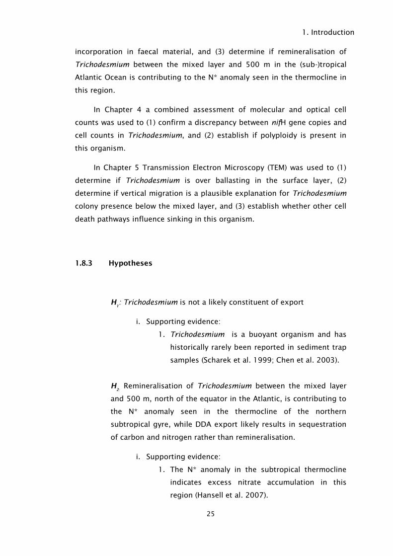
1. Introduction
25
incorporation in faecal material, and (3) determine if remineralisation of
Trichodesmium between the mixed layer and 500 m in the (sub-)tropical
Atlantic Ocean is contributing to the N* anomaly seen in the thermocline in
this region.
In Chapter 4 a combined assessment of molecular and optical cell
counts was used to (1) confirm a discrepancy between nifH gene copies and
cell counts in Trichodesmium, and (2) establish if polyploidy is present in
this organism.
In Chapter 5 Transmission Electron Microscopy (TEM) was used to (1)
determine if Trichodesmium is over ballasting in the surface layer, (2)
determine if vertical migration is a plausible explanation for Trichodesmium
colony presence below the mixed layer, and (3) establish whether other cell
death pathways influence sinking in this organism.
1.8.3 Hypotheses
H1
: Trichodesmium is not a likely constituent of export
i. Supporting evidence:
1. Trichodesmium is a buoyant organism and has
historically rarely been reported in sediment trap
samples (Scharek et al. 1999; Chen et al. 2003).
H2:
Remineralisation of Trichodesmium between the mixed layer
and 500 m, north of the equator in the Atlantic, is contributing to
the N* anomaly seen in the thermocline of the northern
subtropical gyre, while DDA export likely results in sequestration
of carbon and nitrogen rather than remineralisation.
i. Supporting evidence:
1. The N* anomaly in the subtropical thermocline
indicates excess nitrate accumulation in this
region (Hansell et al. 2007).

E. Sargent
26
2. Nitrogen fixation influence is assumed, but at
current no mechanism is described for the
transfer of diazotroph-derived new nitrogen to
depth (Hansell et al. 2004).
3. DDAs have been seen intact in sediment traps up
to 4000 m previously (Scharek et al. 1999; Karl et
al. 2012).
H3
: There are multiple mechanisms by which diazotroph export
occurs
i. Supporting evidence:
1. Trichodesmium has been documented in
association with a copepod that was observed to
graze minimally upon it (Eberl and Carpenter
2007).
2. Carbohydrate ballasting in Trichodesmium has
been documented and vertical migration has
been proposed as a mechanism by which the
organism can access enhanced phosphate
concentrations in deeper waters for nutrient
mining (Villareal and Carpenter 2003); therefore,
passive sedimentation is a potential export
mechanism for this organism.
3. Trichodesmium has been documented in
sediment traps following diatom blooms (Scharek
et al. 1999), which indicates a sweeping
mechanism following incorporation in aggregates
is a plausible mechanism of export.
H4
: Over ballasting of Trichodesmium in the surface layer is
causing it to sink past phosphocline and terminating the ability for
the colony to return to the surface.
i. Supporting evidence:

1. Introduction
27
1. Models predict that ballasting for nutrient mining
is feasible, but colonies could only reach depths
of 150 m and maintain the ability to return to the
surface if they were large (>1500um radius)
(Kromkamp and Walsby 1992; White et al. 2006).
H5
: If over ballasting is the mechanism by which Trichodesmium is
exported, the intracellular POC/PON is high quality.
i. Supporting evidence:
1. Senescent Trichodesmium would not possess the
ability to migrate (Villareal and Carpenter 2003).
H6
: Gas vacuole collapse in colonies collected between 100 and
300 m guarantees sinking Trichodesmium at these depths lacks
the ability to return to the surface.
i. Supporting evidence:
1. Depending on the species, Trichodesmium gas
vessicles collapse between 100 and 300 m
(Walsby 1978).
H7
: Quantification of the nifH gene via qPCR can be directly related
to cell number
i. Supporting evidence:
1. Amplification of nifH genes now frequently
replaces optical cell counts as the method for
diazotroph quantification on research cruises
(Zehr et al. 1998; Langlois et al. 2008; Foster et
al. 2009; Goebel et al. 2010). Luo et al (2012)
acknowledged a discrepancy of an order of
magnitude between gene copies and cell counts
(Luo et al. 2012), but it was not pursued as 1:1
ratios of copies to counts have previously been
observed in Crocosphaera watsonii (Langlois pers.
comm.).

E. Sargent
28
1.8.4 Thesis structure
The format of this thesis involves the presentation of chapters 2-5 as
standalone pieces of work intended for submission as manuscripts. Chapter
6 is a synthesis of chapters 2-5 and discusses limitations of this study and
recommendations for future work.

2. Molecular insights into fate of new N
29
2. Novel molecular insights into the fate of
N2
fixed by diazotrophs
2.1 Abstract
The fate of diazotroph-derived new nitrogen and its role in marine
biogeochemical cycles is not well understood. Sporadically, optical
assessments of sinking particulate material have revealed the presence of
two diazotrophic organisms, Trichodesmium spp. and Richelia
intracellularis, outside of the mixed layer. However, assessments of
diazotrophic organisms are traditionally constrained to the upper ocean;
hence, the extent of diazotroph contribution to export is not clear. In an
attempt to classify this occurrence, real-time quantitative PCR analysis with
TaqMan probes was carried out on extracts of sinking particulate samples to
500 m to assess the presence of the nifH gene, which encodes the iron-
binding subunit of the nitrogenase enzyme complex. Eight nifH phylotypes
were assessed: a single filamentous cyanobacterial probe specific to
Trichodesmium, two heterocystous cyanobacterial probes specific to
Richelia-Rhizosolenia and Richelia-Hemiaulus (diazotrophic diatom
associations), three unicellular cyanobacterial probes specific to
Crocosphaera watsonii and the uncultured Group A and Group C
cyanobacteria, and two non-cyanobacterial probes specific to the uncultured
gammaproteobacterium Gamma A and the uncultured Cluster III. All but the
Cluster III phylotype were detected in sinking particles below the mixed
layer, which indicates previous assessments of the vertical distributions of
these organisms may have overlooked the presence of multiple groups of
diazotrophs at depth. Contrary to previous expectations, all three groups of
marine diazotrophs were constituents of sinking material exported out of
the euphotic zone in the subtropical and tropical Atlantic Ocean. This novel
finding allows for comparisons of the distribution of nifH phylotypes in the
surface and at depth, which aids in elucidation of species-specific fates.
Quantifying the extent to which these groups are exporting provides new
insight into the cycling of fixed nitrogen in the oligotrophic ocean.

E. Sargent
30
2.2 Introduction
Marine diazotrophs play an important role in marine biogeochemical
cycles by fixing N2
into bioavailable forms, thus sustaining oceanic
productivity over broad timescales through maintenance of bioavailable
nitrogen stores. Historically, the two conspicuous groups of diazotrophs,
non-heterocystous, filamentous cyanobacteria such as Trichodesmium and
the heterocystous endosymbiotic cyanobacteria of diatoms (DDAs), were
quantified optically, but the advent of molecular techniques, such as PCR,
has resulted in a shift towards gene-based abundance assessments. It was
not until this method became available that the unicellular nitrogen fixing
Group A cyanobacterium was even detected (Zehr et al. 2001).
The ease of collection coupled with the large number of samples that
can be analysed at once has resulted in a massive dataset of global
diazotroph abundances, biomass, and nitrogen fixation rates, which
continues to expand (Luo et al. 2012). Amplification of nifH, a highly
conserved gene which codes for the iron binding component of the
nitrogenase gene (Young 1992), allows detection of diverse groups of
diazotrophs, including non-cyanobacterial nifH containing organisms, such
as the gammaproteobacteria and green sulphur bacteria. These organisms
were previously inaccessible via optical techniques, and their contribution to
global nitrogen fixation remains unknown (Riemann et al., 2010). New nifH
sequences are continuing to be detected (Langlois et al. 2005, Thompson et
al. 2012)
Until recently, it was widely believed that Trichodesmium did not
passively sediment out of the mixed layer, as it is buoyant, and typically
occupies the surface layer in oligotrophic ocean regions (Carpenter and Price
1977, Carpenter et al. 2004). At least three mechanisms by which
Trichodesmium contributes to export will be discussed in Chapter 3: free
sinking, incorporation in faecal material, and incorporation in aggregates.
The dominant mechanism appears to be free sinking followed by
remineralisation in the upper twilight zone, which may contribute to the N*
anomaly (Chapter 3) or following incorporation into the microbial loop

2. Molecular insights into fate of new N
31
(Sellner 1992; Mulholland 2007). Additionally, physical mechanisms, such as
eddies (Guidi et al. 2012) and particle sweeping (Scharek et al. 1999b), as
well as physiological mechanisms, such as programmed cell death (PCD)
(Bar-Zeev et al. 2013), have been described to drive and enhance
Trichodesmium export. Any or all of these export mechanisms could
potentially apply to all pelagic diazotrophic organisms (Mulholland 2007).
Many diatoms, including DDAs, are capable of contributing to export
(Scharek et al. 1999a; 1999b, Karl et al. 2012). Richelia hosts Rhizosolenia
and Hemiaulus flocculate when in dense concentrations (Alldredge and
Gotschalk 1988, Villareal et al. 2011), and subsequently can directly export
in fast sinking aggregations as Karl and others (2012) highlighted in the
annual summer export pulse in the North Pacific Ocean. Karl et al. (2012)
also speculated this occurrence may be specific to that region. What has yet
to be assessed is what contribution DDAs and other diazotrophs are making
to the direct export of carbon and nitrogen in other ocean regions.
Recent phylogenetic surveys of nitrogen fixers have revealed that
previously undetected diazotrophs, unicellular nitrogen fixing
cyanobacteria, are dominant in many marine environments (Church et al.
2005a; 2005b, Foster et al. 2009, Moisander et al. 2010). One such
unicellular diazotroph, the Group A cyanobacterium (Candidatus
Atelocyanobacterium thalassa) (Thompson et al. 2012), is an obligate
symbiont, as it lacks photosystem II (Tripp et al. 2010). However, little is
currently known about the physiology, ecology and fate of this Group A
cyanobacterium. It displays similar nifH expression patterns to
Trichodesmium and heterocystous cyanobacteria implying it is also an
important contributor to newly fixed nitrogen into the upper ocean (Church
et al. 2005b). Describing the presence or absence of Group A cyanobacteria
in sinking material will provide vital insight into the cycling and fate of new
nitrogen in this and other unicellular cyanobacterial groups.
To date, most studies of these three main groups of N2
-fixing
organisms in the ocean have generally been limited to assessment of their
presence and activity in the euphotic zone, while the role these organisms
have in the export of material to the ocean's interior has seldom been
addressed. In an attempt to track the fate of these nitrogen fixing

E. Sargent
32
organisms we have quantified 4 nifH phylotypes in the surface and in
sinking particles as deep as 500 m in the (sub-)tropical Atlantic Ocean
attributable to the main diazotrophs: Trichodesmium, two groups of DDAs
and the unicellular Group A cyanobacterium. We also amplified nifH from
two other groups of unicellular cyanobacteria, Group B and Group C
cyanobacteria, as well as two non-cyanobacterial nifH phylotypes, Cluster III
and Gamma A, in this region.
These findings highlight that previous assessments of the vertical
extent of marine diazotrophs has been under estimated, and that the fate of
diazotrophic new nitrogen is not constrained to the surface layer. On the
contrary, we propose that marine diazotrophs actively contribute to the
export particulate organic material (POM) in the sub-tropical Atlantic Ocean.
These observations of increased nitrate relative to phosphate beneath areas
of high diazotroph abundance supports the potential for substantial
remineralisation of diazotroph-derived organic material in the upper twilight
zone along the northern part of the transect contributing to enhanced N*
within the thermocline. This provides novel insight into the cycling and fate
of fixed nitrogen in this region, and potentially throughout the global ocean.
2.3 Methods
2.3.1 Sample Collection
Samples for this study were collected in the (sub-)tropical Atlantic
Ocean (Figure 2-1) on RRS Discovery D361 cruise (February and March
2011). All samples were collected using a conductivity, temperature, depth
(CTD) rosette or in situ Stand Alone Pumping Systems (SAPS). See Table 2-1
for collection details.

2. Molecular insights into fate of new N
33
During two-hour deployment, SAPS samples were collected on
consecutively stacked 53 and 1 µm Nitex meshes in an attempt to separate
the sinking portion of POM from suspended material. Samples were
collected from depths ranging from 2 to 500 m. Upon retrieval, SAPS filters
were carefully rinsed with 1L of filtered seawater to resuspend all collected
material. Equal splits were the obtained using a folsom splitter. 25 mL of
SAPS resuspension, representative of 47.9-85.5 L depending on total volume
filtered (Table 2-1; Table A1), was filtered onto 0.7µm GF/F (Whatman)
filters under low (2 mbar) vacuum pressure. To obtain surface reference
samples, 2 L of CTD water was filtered onto 0.22 µm Durapore (Millipore)
filters. After filtration, filters were flash frozen in liquid nitrogen and stored
at -80°C until DNA extraction.
Figure 2-1. Stations sampled for nifH abundance on the D361. Total nifH
abundance (gene copies m-3
) in surface samples reveals the peak of detectable
diazotrophic nifH was located between the equator and 10°N. Note that nifH was not
detected in the surface at the southerly most station.

E. Sargent
34
Table 2-1. Collection details and phylotype presence/absence
Station Lat Lon Collection
Depth
(m)
Size
fraction
(µm)
Extracted
Volume
(L)
Presence (+) / Absence (-)
Fil Het1 Het2
Group
A
Group
B
Group
C
Cluster
III
Gamma
A
10 07°13 S 25°00 W CTD 3.5 0.2 2 - - - - - - - +
10 07°13 S 25°00 W SAPS 70 53 51.7 + - - - - - - +
10 07°13 S 25°00 W SAPS 70 1 51.7 + - + - - - - +
10 07°13 S 25°00 W SAPS 170 53 58.6 - - - - - - - -
10 07°13 S 25°00 W SAPS 170 1 58.6 - - - - - + - -
10 07°13 S 25°00 W SAPS 500 53 54.4 - + + - + - - -
13 01°09 N 26°02 W CTD 21.5 0.2 2 + + - + - + - +
13 01°09 N 26°02 W SAPS 70 53 47.9 + + + + - + - +
13 01°09 N 26°02 W SAPS 70 1 47.9 - + + - - + - +
13 01°09 N 26°02 W SAPS 170 53 51 + + + - - - - -
13 01°09 N 26°02 W SAPS 170 1 51 - + - - - + - -
13 01°09 N 26°02 W SAPS 500 53 85.5 - - - - - - - -
16 08°20 N 28°20 W CTD 3 0.2 2 + + + - + + - +
16 08°20 N 28°20 W SAPS 70 53 50.6 + + + + + + - +
16 08°20 N 28°20 W SAPS 70 1 50.6 - + + - - + - +
16 08°20 N 28°20 W SAPS 170 53 51.3 + - - - - - - -
16 08°20 N 28°20 W SAPS 170 1 51.3 + - + - - + - -
16 08°20 N 28°20 W SAPS 500 53 53.7 + - - - - - - -
19 15°30 N 28°23 W CTD 1.2 0.2 2 + + + + + + - +
19 15°30 N 28°23 W SAPS 85 53 50.6 + + + + + + - +
19 15°30 N 28°23 W SAPS 85 1 50.6 - + + + + + - +
19 15°30 N 28°23 W SAPS 170 53 58.6 + - - - - - - -

35
2
. M
olecular insights in
to fate of new
N
Table 2-1 continued
Station Lat Lon Collection Depth
(m)
Size
fraction
(µm)
Extracted
Volume
(L)
Fil Het1 Het2 Group A Group B Group C Cluster III Gamma A
19 15°30 N 28°23 W SAPS 170 1 58.6 + - - - - + - -
19 15°30 N 28°23 W SAPS 500 53 52.9 + - - + - - - +
21 19°10 N 28°07 W CTD 6.2 0.2 2 + + + + + - - +
21 19°10 N 28°07 W SAPS 140 53 50.3 + + - + - + - +
21 19°10 N 28°07 W SAPS 140 1 50.3 + - - + + + - -
21 19°10 N 28°07 W SAPS 250 53 59.2 + - - + NA NA NA NA
21 19°10 N 28°07 W SAPS 250 1 59.2 - - - + NA NA NA NA
21 19°10 N 28°07 W SAPS 500 53 53.7 + - - - NA NA NA NA

E. Sargent
36
2.3.2 Sample Extraction and DNA Quantification
Frozen filters were manually crushed using a plastic pestle, and then
samples were extracted using the Qiagen DNeasy mini plant kit according to
the manufacturer’s protocol. Following extraction, DNA concentrations were
determined using the RediPlate 96 dsDNA Quantitation Kit (Molecular
Probes). DNA concentrations were read on a Fluoroscan Ascent microplate
reader.
2.3.3 nifH Quantification of in situ samples
Previously described primers and probes were chosen for
amplification of 8 nifH phylotypes (Table 2-2). The qPCRs were run on an ABI
Prism 7000 (Applied Biosystems) using cycling conditions of 2 minutes at
50°C, 10 minutes at 95°C, and 40 cycles of 95°C for 15 seconds, followed by
1 minute at 60°C. Each 25 µL qPCR reaction contained 10 pmol/µL each of
the forward and reverse primers, 100 pmol/µL of the corresponding probe,
1 µL of bovine serum albumin, 5.25 µL of PCR water, 12.5 µL of TaqMan mix
(Applied Biosystems), and 5 µL of template DNA from either a standard or an
environmental sample (Langlois et al. 2008). The template DNA was
equivalent to 1 to 5ng of DNA per reaction. Samples were run in duplicate
and were quantified against serially diluted standards (107
to 101
gene
copies). We considered Ct
values, the cycle at which the amplification curve
crosses the threshold of detection, above 37 to be detectable but not
quantifiable (DNQ). Due to the different filter types used during collection
and different volumes sampled (Table 2-1), we assume differences in
collection and extraction efficiency were introduced between CTD and SAPS
collected samples. Therefore, we consider this in situ data to be a qualitative
assessment of nifH phylotype distribution in the study region and note that
the SAPS-derived nifH values are likely underestimates of actual nifH
abundance as a bias may have been introduced towards larger diazotrophs

2. Molecular insights into fate of new N
37
due to prefiltration and loss during the rinsing and reconstituting of
samples.
However, as our SAPS extractions are representative of much larger
volumes of water than is traditionally assessed, our analyses allow for a
lower limit of detection than is conventionally accepted. Gene based
abundances are typically carried out on extracts derived from volumes
ranging from 0.5-5 L (Langlois et al. 2008; Goebel et al. 2010; Moisander et
al. 2010; Kong et al. 2011). When following a standard Qiagen DNeasy mini
plant kit DNA extraction protocol, the final extract volume is typically 80 µL.
5µL of this extract is then typically added to a qPCR reaction for
quantification of nifH. Although the assay is theoretically capable of
detecting a single gene copy in the 5µL DNA template loaded, the actually
volume represented would be 31.25-312.5 mL. Thus, in practice the
detection limit when working with an example volume of 2L would be ≥8
copies per litre. These large volume SAPS extractions afford us a unique
sensitivity of detection. The limit of detection for this study in SAPS samples
representative of 47.9-85.5 L therefore ranges from being able to detect a
single nifH copy in 3-5 L.
2.3.4 Statistical Assessments
Statistical assessments of nifH community structure were carried out in
PRIMER v6 (PRIMER-E Ltd). Raw nifH phylotype data was normalised to
percent of total nifH each sample to ensure assessments were reflective of
similarity between community structure and that statistical groupings were
not biased towards differences in absolute abundance. Bray-Curtis
dissimilarity matrices were produced to assess the differences between
samples and allow for dendrogram construction. An analysis of similarities
(ANOSIM) was then performed within PRIMER to test for statistically
significant difference between samples.

E. Sargent
38
Table 2-2. qPCR primers and TaqMan probes used in this study
Target
Primer Probe
Forward Reverse
Filamentous TGGCCGTGGTATTATTACTGCTATC (165-189) GCAAATCCACCGCAAACAAC (275-256) AAGGAGCTTATACAGATCTA
(206-225)
Het-1 CGGTTTCCGTGGTGTACGTT (105–124) AATACCACGACCCGCACAAC (158-177) TCCGGTGGTCCTGAGCCTGGTGT
(133-155)
Het-2 TGGTTACCGTGATGTACGTT (106-124) AATGCCGCGACCAGCACAAC (158-177) TCTGGTGGTCCTGAGCCTGGTGT
(133-155)
UCYN-A TAGCTGCAGAAAGAGGAACTGTAGAAG (50–76) TCAGGACCACCGGACTCAAC (146–127) TAATTCCTGGCTATAACAAC
(98-117)
UCYN-B TGCTGAAATGGGTTCTGTTGAA (54–75) TCAGGACCACCAGATTCTACACACT (146–
122)
CGAAGACGTAATGCTC
(87–102)
UCYN-C TCTACCCGTTTGATGCTACACACTAA (1–26) GGTATCCTTCAAGTAGTACTTCGTCTAGCT
(112–83)
AAACTACCATTCTTCACTTAGCAG
(32–55)
Gamma A TTATGATGTTCTAGGTGATGTG (240–266) AACAATGTAGATTTCCTGAGCCTTATTC
(321–294)
TTGCAATGCCTATTCG
(275–290)
Cluster III ACCTCGATCAACATGCTCGAA (175–195) GCAGACCACGTCACCCAGTAC (267–247) CCTGGACTACGCGTTC
(225–240)
All sequences are 5' to 3' and relate to the 324-base nifH segment, and come from Langlois et al. 2008 and Foster et al. 2007

2. Molecular insights into fate of new N
39
2.4 Results
2.4.1 nifH abundance of in situ samples
Apart from station 10 (Figure 2-2A-B, purple), where nifH was rarely
detectable and surface abundance consisted solely of the non-cyanobacterial
nifH phylotype Gamma A, total nifH detected below the mixed layer was
consistently lower than surface nifH by at least an order of magnitude, and
abundance generally decreased with depth at stations north of the equator.
As SAPS collected samples were originally prefiltered during collection
onto 53 µm and 1 µm filters, thus excluding anything smaller than 1 µm for
samples collected at 10 and 100 m below the mixed layer and anything
smaller than 53µm for samples collected at 500 m, we can assume all SAPS
collected samples are underestimations of actual nifH abundance, and that
we have under sampled the unicellular cyanobacterial and non-
cyanobacterial nifH. However, as the >53µm size fraction is considered to
represent sinking material, we are confident that these samples have
captured an accurate representation of the nifH associated with diazotrophs
within this larger size fraction of POM, such as Trichodesmium and the
DDAs.
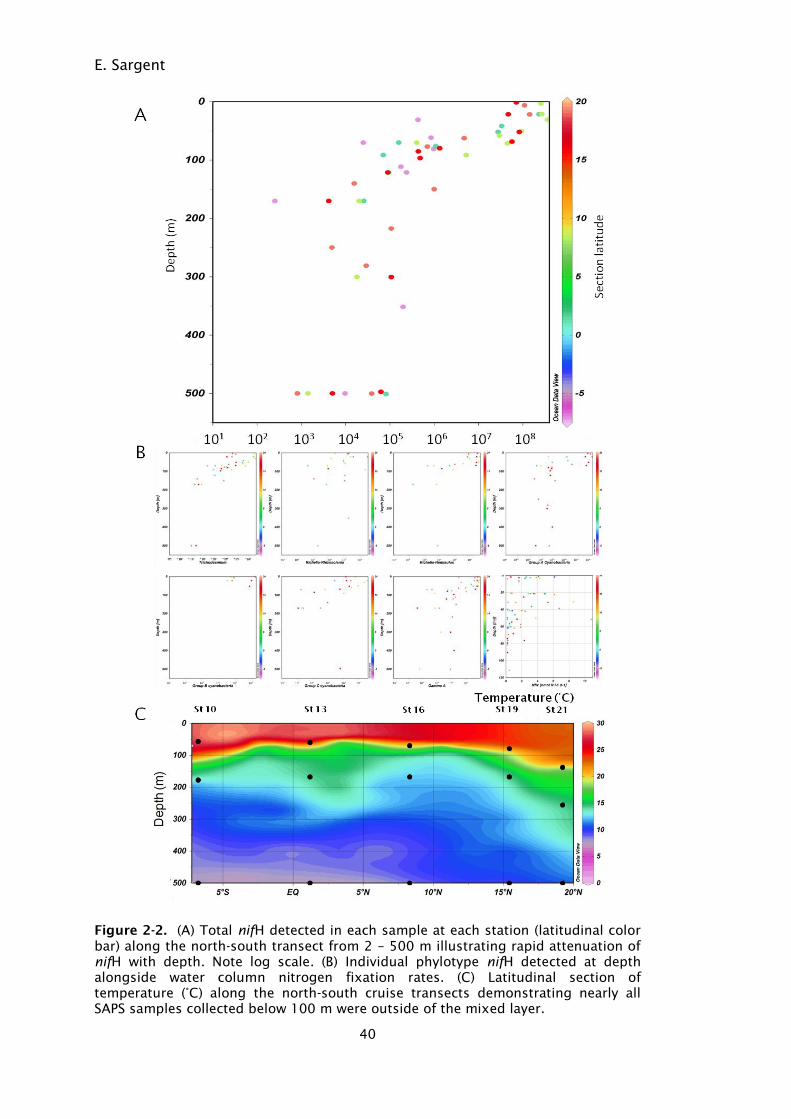
E. Sargent
40
40
Figure 2-2. (A) Total nifH detected in each sample at each station (latitudinal color
bar) along the north-south transect from 2 – 500 m illustrating rapid attenuation of
nifH with depth. Note log scale. (B) Individual phylotype nifH detected at depth
alongside water column nitrogen fixation rates. (C) Latitudinal section of
temperature (°C) along the north-south cruise transects demonstrating nearly all
SAPS samples collected below 100 m were outside of the mixed layer.

2. Molecular insights into fate of new N
41
2.4.2 Dominance and distribution all phylotypes
Gamma A non-cyanobacterial nifH and Trichodesmium-attributable nifH
were ubiquitous in this study region with detectable nifH for these
phylotypes at every station (Figure 2-4). Group B and C cyanobacteria nifH
abundances were maximal at stations 19 (15°30 N) and 16 (8°20 N)
respectively, but Group A and Trichodesmium-associated nifH dominated
total nifH community structure at these stations (Figure 2-4).
The non-cyanobacterial nifH phylotype Cluster III was undetectable in
all samples. As the standard curves were normal for this phylotype (Figure 2-
3), we assume it was not present in this study region. All other phylotypes
assessed were found outside of the mixed layer (60 - 130 m) at at least one
station along the cruise transect. Excluding zero values, the abundance of
total nifH detected in each sample varied from 253 gene copies m-3
in the
170 m sample at the southern most station, 7°13 S (station 10), to 25.7 x
107
gene copies m-3
in the surface sample at 8°20 N (station 16) (Figure 2-4).

42
E. Sargent
Figure 2-3. Standard curves for qPCR assessments of serially diluted standards for all eight phylotypes. R2
= 0.999 for the filamentous standard
(Fil, blue asterisks); R2
= 0.991 for the Richelia-Rhizosolenia standard (Het1, orange circles); R2
= 0.999 for the Richelia-Hemiaulus standard
(Het2, pink rectangles); R2
= 0.999 for the Group A cyanobacteria standard (UCYN-A, blue crosses); R2
= 0.998 for the Group B cyanobacteria
standard (UCYN-B, green triangles); R2
= 0.984 for the Group C cyanobacteria standard (UCYN-C, red squares); R2
= 0.998 for the Gamma A
standard (purple crosses); R2
= 0.997 for the Cluster III standard (blue diamonds).
1.00E+00
1.00E+01
1.00E+02
1.00E+03
1.00E+04
1.00E+05
1.00E+06
1.00E+07
1.00E+08
10 15 20 25 30 35 40
Lo
g tem
plate ab
uan
dan
ce (co
pies L-1
)
Cycle number
Fil
Het 1
Het 2
UCYN-A
UCYN-B
UCYN-C
Gamma A
Cluster III

2. Molecular insights into fate of new N
43
Figure 2-4. Latitudinal sections of absolute nifH phylotype abundance (gene copies
m-3
) along the D361 transect demonstrating unicellular affinity for higher latitudes
and cooler temperatures, filamentous nifH localisation just north of the equator,
and the ubiquity of the non-cyanobacterial Gamma A nifH in the surface layer. Note
varying scales.

E. Sargent
44
E. Sargent
Figure 2-4 Continued. Latitudinal sections of absolute nifH phylotype abundance
(gene copies m-3
) along the D361 transect demonstrating unicellular affinity for
higher latitudes and cooler temperatures, filamentous nifH localisation just north of
the equator, and the ubiquity of the non-cyanobacterial Gamma A nifH in the
surface layer. Note varying scales.

2. Molecular insights into fate of new N
45
Figure 2-4 Continued. Latitudinal sections of absolute nifH phylotype abundance
(gene copies m-3
) along the D361 transect demonstrating unicellular affinity for
higher latitudes and cooler temperatures, filamentous nifH localisation just north of
the equator, and the ubiquity of the non-cyanobacterial Gamma A nifH in the
surface layer. Note varying scales.
E. Sargent
46
E. Sargent
Figure 2-4 Continued. Latitudinal sections of absolute nifH phylotype abundance
(gene copies m-3
) along the D361 transect demonstrating unicellular affinity for
higher latitudes and cooler temperatures, filamentous nifH localisation just north of
the equator, and the ubiquity of the non-cyanobacterial Gamma A nifH in the
surface layer. Note varying scales.
2.4.3 nifH dominance and distribution of the four main diazotroph
phylotypes
I was unable to detect nifH from any of the four main groups of
cyanobacterial diazotrophs (Group A unicellular, filamentous, and both
heterocystous phylotypes) in the 500 m sample at station 13 (1°09N) and in
the 2 m and 170 m samples at the southerly most station (station 10, 7°13
S). All other samples had diazotrophic community structure which could be
characterised by dominance by a single nifH phylotype.
Unicellular Group A cyanobacterial-attributable nifH was dominant in
the surface samples at the northerly most stations 19 (15°30 N) and 21

2. Molecular insights into fate of new N
47
(19°10 N), while Trichodesmium-attributable nifH dominated the surface
samples between the equator and 10° N. This relationship was not
consistent in deeper samples, such as the northerly most station, station 21,
where Trichodesmium-attributable nifH was the sole component of the 500
m sample (Table 2-1). Among the four main groups of cyanobacterial
diazotrophs, Bray Curtis dissimilarity and ANOSIM assessments did not
reveal any statistically significant grouping of nifH phylotype dominance
with latitude (Figure 2-5). At all stations except for the southerly most
station, station 10 (7°13 S), nifH phylotypes that were present in samples
outside of the mixed layer were also present in the surface sample.
However, the dominant nifH phylotype in a surface sample was not always
the dominant nifH phylotype in the deeper samples at the same station,
such as at stations 19 and 21 where Group A was dominant at the surface,
while Trichodesmium was dominant below the mixed layer (Figure 2-5), and
total nifH abundance was highly variable.
Trichodesmium-attributable nifH constituted the majority of detectable
nifH in the surface layer between the equator and 10°N, where it composed
98.1 and 99.6% of total detectable nifH at stations 13 (1°09 N) and 16 (8°20
N), respectively. Trichodesmium nifH dominance was also echoed in deeper
samples at these stations at 10 m below the mixed layer composing 66.2%
of diazotroph community structure at station 13 and 94.7% at station 16.
Trichodesmium’s surface dominance was replaced by unicellular diazotrophs
to the north, where Group A cyanobacteria contributed 89.5 and 93.0% to
total detectable nifH in the surface samples at stations 19 and 21 (Figure 2-
5). Trichodesmium nifH was dominant in deeper samples at these stations at
10 m below the mixed layer composing 80.6% of nifH community structure
at station 19 and 43.5% at station 21.

48
E. Sargent
Figure 2-5. Bray Curtis dissimilarity dendrogram of nifH diversity normalised to total nifH in each sample. Relative proportions of nifH
phylotypes within each sample are indicated by colour distributions in each bar: Trichodesmium-attributable nifH (purple), Group A
Cyanobacteria-attributable nifH (green), Hemiaulus-Richelia-attributable nifH (red), and Rhizosolenia-Richelia-attributable nifH (blue); bar height
represents absolute nifH abundance in each sample. Starred samples represent no nifH detection or absolute nifH abundance below 1000
copies m-3
. Sample labels are displayed at the station number followed by the depth of the sample.

2. Molecular insights into fate of new N
49
2.5 Discussion
These results indicate that filamentous, heterocystous, and unicellular
diazotrophs are escaping the surface layer and are all potentially contributing
to particulate export in this study region. The mechanisms by which this is
occurring are not well defined, but we speculate a cumulative influence of
passive sedimentation, aggregation, and grazing, as is described for
Trichodesmium via optical observations in Chapter 3. Guidi et al. (2012) have
also suggested active export of Trichodesmium would be possible in
association with eddies in the North Pacific Subtropical Gyre. This may also be
important in this study region where enhancement of POC export by eddies
has been previously observed (Sweeney et al. 2003, Davis and McGillicuddy
2006). We assume enhancement of diazotroph export by eddies would not be
restricted to Trichodesmium and would likely influence the fate of all
diazotroph-derived new nitrogen. Additionally, other mechanisms exist
whereby diazotroph-derived N could contribute to export fluxes secondarily,
following incorporation into other material, such as following viral lysis of the
cells (Hewson et al. 2004) or extracellular release of fixation products (Glibert
and Bronk 1994, Foster et al. 2011).
The symbiotic association between the Group A unicellular
cyanobacterium, A. thalassa, and its haptophyte host (Thompson et al. 2012)
implies there is also the potential for this group to contribute to export via a
variety of mechanisms, such as ‘export by association’ if the host is apt to
aggregation or is grazed upon. The host of A. thalassa is closely related to the
heavily calcified coccolithophore genus Braarudosphaera, which is well
documented in the fossil record (Kelly et al. 2003) suggesting its relatives may
also be susceptible to sinking and thus contributing to export. This data
revealed Group A specific nifH was present at 500 m in the open ocean, and
nifH specific to Group A unicellular cyanobacteria has previously been
amplified in coastal sediment samples (Thompson and Zehr 2013) suggesting
the loss of new nitrogen within Group A from the euphotic zone is potentially a
widespread occurrence. Similarly, some Group B unicellular cyanobacteria are
known to associate with the diatom Climacodium frauenfeldianum (Carpenter

E. Sargent
50
and Janson 2000; Foster et al. 2011), and may also be susceptible to export by
association as is seen with the DDAs Richelia-Rhizosolenia and Richelia-
Hemiaulus (Karl et al. 2012).
Historically, Trichodesmium is thought to be the dominant nitrogen fixing
organism in this study area (Tyrrell et al. 2003, Moore et al. 2009, Zehr 2011,
Groβkopf et al. 2012), and is potentially capable of accounting for the majority
of the geochemically derived rates of nitrogen fixation inferred from the N*
distributions in the North Atlantic (Gruber and Sarmiento 1997, Capone et al.
2005, Mahaffey et al. 2005, Hansell et al. 2004, 2007). These results indicate
Trichodesmium’s dominance is restricted to the surface layer between the
equator and 10°N. Farther to the north unicellular diazotrophs compose the
majority of nifH community structure. This was not consistently echoed in
deeper samples, which implies that Trichodesmium may be more likely to sink
out of the mixed layer than Group A cyanobacterium. As only a >53 µm size
fraction sample was taken at 500 m depth and the association between Group
A and its host is known to be very weak (Tripp et al. 2010), the 500 m
collections may have under sampled Group A, specifically at particle-light
station 21, where turbulence from the pump could cause disassociation of the
cyanobacterium from its host and loss of the former through the large mesh
size. The presence of Group A in the 500m sample at station 19 highlights the
potential for Group A to reach this depth and thus it’s potential to transport
new nitrogen out of the upper ocean. The 500 m sample at station 19 had
nearly double the POC as the station 21 sample at the same depth (0.44 vs.
0.26 µgC L-1
) implying that even if Group A became detached from its host
during collection, there may have been retention of the cyanobacterium on the
filter by all of the other particulate material present. Future investigation of the
diversity and abundance of nifH containing organisms at depth using a qPCR
approach will require optimisation of the sampling procedures to ensure that
larger volume collections are representative of an unbiased sample.
South of the equator at station 10, diazotroph abundance was minimal,
and nifH was undetectable at the surface (Figure 2-4); this is likely to be due to
the lack of the iron-rich aerosol deposition in this region, which is known to
support diazotrophic growth north of the equator (Moore et al. 2009). At 10 m
below the mixed layer, nifH from Trichodesmium and Richelia-Hemiaulus
contributed 92.7% and 7.3% to diazotrophic community structure respectively.

2. Molecular insights into fate of new N
51
The presence of these diazotrophs at depth, but not at the surface implies a
temporal component to these findings; it is likely that in the days prior to the
sampling these organisms were present in the surface, but sank or were
otherwise transported to depth before we encountered them. In the 500 m
sample at station 10 Trichodesmium-attributable nifH was undetectable, but
Richelia-Hemiaulus and Richelia-Rhizosolenia attributable nifH, was present and
composed 78.7% and 21.3% of diazotrophic community structure respectively.
The complete absence of the Cluster III nifH phylotype in this study
region is unexpected. The Cluster III nifH phylotype may be particularly
important in the oligotrophic North Pacific Ocean where it has been seen to
dominate nifH communities in the lower photic zone (Church et al. 2005a) or
in the western subtropical Atlantic Ocean where it has been observed
previously (Langlois et al. 2008). However, previous reports of its distribution
have documented an affinity for cooler temperatures than were observed in
this study (Farnelid et al. 2011). Langlois et al. (2008) observed this phylotype
to be associated with mean water temperatures of 18°C, which is 5°C cooler
than the lowest temperature encountered on this transect. Cluster III specific
nifH has also been found to dominate nifH community structure in the Arctic
(Farnelid et al. 2011).
Furthermore, Cluster III anaerobes have previously been described to
associate with zooplankton (Braun et al. 1999). The lack of Cluster III detection
may be due to methodology as “swimmers” (zooplankton) were picked from
SAPS filters following collection and were not subsequently included in DNA
extraction. This zooplankton associated nifH is not well defined. If Cluster III
nifH is expressed and nitrogen fixation is occurring inside zooplankton, this
highlights an opportunity to explore whether faecal pellets contain new
nitrogen, thus further contributing to export.
The majority of qPCR nifH assessments to date have come from small
volume samples in the upper ocean, which may highlight an under-sampling
bias both in vertical extent and sample volume. Church et al. (2005a) assessed
nifH phylotype diversity in 1L samples as deep as 200 m, and found that Group
B and Cluster III nifH was still detectable in the deepest samples while Group A
and Trichodesmium nifH was not. Potentially these phylotypes were present,
but under the detection limit, and would have been revealed with larger

E. Sargent
52
volume sampling. This idea is supported by optical observations of
Trichodesmium in the 150 to 200 m depth range and below in this (Chapter 3)
and other studies (Carpenter and Price 1977; Scharek et al. 1999b).
Additionally, the detection of Trichodesmium and Group A associated nifH in
larger volume samples in this study provides further support for the idea that
previous assessments have under-evaluated nifH presence at depth.
2.6 Conclusion
These results suggest that filamentous, heterocystous, and unicellular
diazotrophs are all being removed from the mixed layer and are likely to be
involved in the export of PON through a variety of potential mechanisms.
Previously Trichodesmium and R. intracellularis have only been observed to
contribute to export following incorporation in fecal pellets or following
surface blooms (Scharek et al. 1999a; 1999b, Mulholland 2007), and it has
only recently been suggested that Group A unicellular cyanobacteria could
contribute to export by association with their host (Thompson and Zehr 2013).
Further investigation of the mechanisms contributing to diazotroph presence
in sinking material will give a clearer idea of the fate of the nitrogen fixed by
these organisms and hence expand the definition of their biogeochemical
roles.
Quantitative PCR of the nifH gene is clearly a fundamental tool in the
study of marine diazotrophs and comparison of the distributions of various
nifH phylotypes in the surface and at depth will aid in the elucidation of
species specific fates. This work represents some of the first comprehensive
qPCR analyses of nifH in marine snow and other sinking particulate material
and thus provides vital insight into the contribution nifH containing organisms
are making to export.
Quantifying the extent to which these groups are exporting will provide
novel insight into the cycling of fixed nitrogen in the oligotrophic ocean and
determining the fate of newly fixed nitrogen by nitrogen fixers will provide
insight to the nitrogen budget through delineation of utilised, remineralised,

2. Molecular insights into fate of new N
53
and deep-ocean-sequestered fixation products. Contrary to previous
expectations, results suggest that all three groups of marine diazotrophs are
constituents of sinking material and are exported out of the euphotic zone in
the subtropical and tropical Atlantic Ocean. Diazotrophs play a vital role in
global biogeochemical cycles by providing significant amounts of newly fixed
nitrogen to surface ocean globally. As we assume for Trichodesmium (Chapter
3), some of that new nitrogen could potentially be injected into the water
column below the mixed layer following remineralisation, which in turn could
contribute to the N* anomaly.

E. Sargent
54

3. Trichodesmium in sinking material
55
3. Describing the presence of
Trichodesmium spp. in sinking material
3.1 Abstract
Trichodesmium plays a major role in ocean biogeochemical cycling through
supplying fixed nitrogen to the warm surface ocean. However, the
subsequent fate of the nitrogen fixed by Trichodesmium and its role in the
biogeochemistry of deeper waters has yet to be established. We examined
the loss of Trichodesmium derived organic carbon and nitrogen from the
surface ocean to the ocean interior through sinking/settling. Sinking
particles were collected and assessed for Trichodesmium presence in the
subtropical and tropical Atlantic Ocean. Trichodesmium was commonly
present below 100 m in three forms: tufted colonies, free filaments, and free
filaments associated with aggregations of other organisms and faecal
matter. We collected negatively buoyant Trichodesmium colonies sinking at
12 - 120 m d-1
between depths of 80-250 m in all regions where
Trichodesmium was a significant component of the surface population. Free
filaments were also observed in low concentrations as deep as 500 m.
Contrary to the previous belief that Trichodesmium was constrained to the
surface layer and did directly contribute to export, these results suggest
that despite its buoyancy during active growth, Trichodesmium can be an
important constituent of sinking material with an average of 8.8% of the
surface Trichodesmium standing stock leaving the mixed layer each day.
Biomass estimates suggest that Trichodesmium contributes 0.4-3% of the
total particulate organic nitrogen (PON) standing stock directly beyond the
mixed layer and an average of 5.6% of the total PON flux leaving the mixed
layer along a latitudinal transect in the Atlantic Ocean. These results provide
some of the first characterizations of the mechanisms by which
Trichodesmium (and diazotrophs in general) could contribute to the N*
anomaly observed in the thermocline in the North Atlantic sub-tropical gyre.
E. Sargent
56
3.2 Introduction
Dinitrogen fixation by marine diazotrophs constitutes the leading source
of external nitrogen to the ocean (Zehr and Kudela 2011). However, with a
few notable exceptions (Karl et al. 2012), we lack knowledge of the short
term fate of the newly fixed nitrogen contributed to the surface ocean
microbial community by these organisms, which limits our understanding of
their integrated biogeochemical role. Trichodesmium is a buoyant,
filamentous cyanobacterium that is found in warm oligotrophic oceans in
colonial aggregations and as free trichomes (Capone et al. 1997), and is the
most conspicuous marine diazotroph. The highest abundances of
Trichodesmium in the Atlantic have historically been observed to correlate
with shallow mixed layer depth and areas estimated to have high iron
deposition (i.e. between 0 and 15°N) (Tyrrell et al. 2003; this study),
although this relationship appears to breakdown north of this latitudinal
range where Trichodesmium abundance has been observed to correlate
more closely with temperature (Agawin et al. 2013).
Actively growing Trichodesmium populations are predominantly
observed in the upper portion (0 to ~50 m) of the water column (Carpenter
and Price 1977; Carpenter et al. 2004). Recent work has documented that
Trichodesmium colonies can also be well distributed down to ~120 m in
some regions (Davis and McGillicuddy Jr. 2006). Deeper (>50 m) populations
of Trichodesmium have frequently been hypothesized to consist of vertically
migrating colonies (Kromkamp and Walsby 1992; White et al. 2006).
Buoyancy in Trichodesmium is provided by the presence of gas vacuoles
(Walsby 1978), and is controlled by carbohydrate ballasting (Villareal and
Carpenter 2003). Models of carbohydrate ballasting in Trichodesmium
suggest that vertical migrations of large (≥1500 µm radius) colonies could
span 200 m for nutrient mining (e.g. phosphorus acquisition) (Kromkamp
and Walsby 1992; White et al. 2006), but are realistically more likely
confined to the upper 70 m of the water column (Villareal and Carpenter
2003).
3. Trichodesmium in sinking material
57
Unlike diazotrophic diatom assemblages (DDAs), which have been
observed to predominate sediment trap collections in regions where both
DDAs and Trichodesmium are present in the surface layer (Scharek et al.
1999b; Karl et al. 2012), Trichodesmium has rarely been reported to reach
the deep ocean (>1 km) intact. Consequently, Trichodesmium is not thought
be an important component of direct export.
The relative abundances of the macronutrients nitrate and phosphate can
be used to infer inputs and loss of nitrogen through the biological processes
of nitrogen fixation and denitrification respectively (Gruber and Sarmiento
1997). The derived N* tracer (Chapter 1, equation 1) (Gruber and Sarmiento
1997; Hansell et al. 2004, 2007; Moore et al. 2009), indicates deviation of
the local nitrate and phosphate pools from the N:P ratio of typical
particulate organic matter (POM), assumed to have a canonical value of 16:1
(Redfield 1934). Gradients of increasing N* can hence be used to infer a net
gain of nitrogen through nitrogen fixation or other sources (Gruber and
Sarmiento 1997; Hansell et al. 2004, 2007), which are discussed in more
detail in Chapter 1. Throughout the majority of the global ocean, N* is
negative within the thermocline and in deeper waters (Gruber and Sarmiento
1997; Moore et al. 2009). However, north of the equator in the Atlantic
there is a positive N* anomaly in the subtropical thermocline to ~1100 m
(Hansell et al. 2007) (Figure 3-1). Moreover the isotopic signature of the
nitrate pool in the subtropical North Atlantic thermocline indicates that
newly fixed nitrogen is contributing to this N* anomaly (Gruber and
Sarmiento 1997; Karl et al. 2002; Knapp et al. 2008).
Various studies have inferred nitrogen fixation rates in the Atlantic on
the basis of both the N* anomaly (Gruber and Sarmiento 1997; Hansell et al.
2004, 2007) and the isotopic anomaly of the nitrate pool (Knapp et al.
2008), with the range of recent estimates spanning 0.4 – 2 Tmol N year -1
(Gruber and Sarmiento 1997; Hansell et al. 2004, 2007; Knapp et al. 2008).
Trichodesmium appears to be the dominant nitrogen fixing organism in the
tropical and sub-tropical North Atlantic (Tyrrell et al. 2003; Moore et al.
2009; Groβkopf et al. 2012) potentially fixing around 1.6 Tmol N year-1
(Capone et al. 2005).

E. Sargent
58
Consequently this organism may contribute a significant, if not the
dominant fraction of the geochemically derived estimates of nitrogen
fixation in the region (Capone et al. 2005; Mahaffey et al. 2005).
For nitrogen fixation to be capable of explaining some of the anomaly in
N* within the North Atlantic thermocline, a mechanism is required for newly
fixed nitrogen to be transferred to depth (Hansell et al. 2004, 2007), such
as remineralisation of diazotroph biomass and advection of resultant
nutrients (Yoshikawa et al. 2013) or active transport of diazotroph biomass
to depth via eddies (Guidi et al. 2012). If nitrogen fixation by
Trichodesmium is to be capable of contributing significantly to the anomaly
in N* within the North Atlantic thermocline and consequential phosphate
depletion in surface waters of the gyre (Wu et al. 2000; Moore et al. 2009),
such mechanisms require further investigation. Mulholland (2007)
suggested Trichodesmium-derived material is likely recycled in the euphotic
zone through the microbial loop, but could also be exported in faecal
material following grazing with release of Trichodesmium-derived nutrients
into the water column during sloppy feeding or microbial break down of
faecal material (Sellner 1992). Other potential fates of Trichodesmium-
derived newly-fixed nitrogen could follow cellular lysis, autocatalytic
programmed cell death (PCD), or extracellular release of fixation products,
but all previous assessment of these processes have occurred in near-
surface collected or cultured samples (Capone et al., 1994; Glibert and
Bronk, 1994; Hewson et al., 2004; Mulholland et al., 2004, 2007; Bar-Zeev
et al. 2013). This highlights an unexplored area of the fate Trichodesmium
in deeper waters.
The current study focuses on tracing the fate of Trichodesmium-derived
new nitrogen through the assessment of multiple export mechanisms along
a transect in the (sub-)tropical Atlantic. Determining the fate of newly fixed
nitrogen by diazotrophs provides insight into nitrogen cycling through
delineation of shallow, remineralised, and deep-ocean-sequestered fixation
products. We document evidence of grazing on Trichodesmium and detail
the presence of Trichodesmium outside of the euphotic zone as both free
trichomes incorporated in sinking aggregates and unincorporated sinking
colonies. When combined with 234
Th derived PON fluxes, observations of
sinking Trichodesmium allow us to estimate this organism’s contribution to

3. Trichodesmium in sinking material
59
export fluxes. Overall, this data suggests substantial remineralisation of
Trichodesmium-derived organic material between 100 and 500 m, which we
suggest contributes to the N* anomaly observed in the thermocline of the
sub-tropical gyre further to the north.
3.3 Methods
3.3.1 Sample Collection
Sampling was carried out in February-March 2011 (D361; 03 Feb 2011
– 19 March 2011) on the RRS Discovery during a transect spanning the (sub-
)tropical Atlantic Ocean (Figure 3-1). The Marine Snow Catcher (MSC) and in
situ Stand Alone Pumping Systems (SAPS) were used to collect sinking
particles from below the Deep Chlorophyll Maximum (DCM) at the base of
the euphotic zone. Hydrographic data and the depth of the DCM were
determined using a CTD interfaced with a Chelsea MKIII Aquatracka
fluorometer. Water column oxygen profiles were determined using a CTD
mounted Sea-Bird 43 oxygen sensor. Additional measurements of
Trichodesmium colony sinking speeds were undertaken in July 2011
(CH0711; 04 July 2011 – 27 July 2011) on the RV Cape Hatteras in the Gulf
of Mexico.
A B

E. Sargent
60
Figure 3-1. Sampling locations. (A) D361 cruise track overlain on N* (µmol kg-1
)
along the 26.8σθ and (B) SST (degC). Data from Key et al. (2004). (C) CH0711
stations overlain on SST (degC) from shipboard measurements.
3.3.2 Marine Snow Catcher Deployment
The MSC (Figure 3-2A), a messenger-operated PVC closing water bottle,
samples 100 L of seawater during deployment, and allows for collection of
sinking particles with minimal turbulent agitation (Lampitt et al. 1993). The
MSC allows for the separation of fast and slow sinking particulate material
(Riley et al. 2012). After each of 9 deployments to 10 m below the DCM on
D361 (35-100 m), the MSC was recovered and lashed upright on deck for 2-
3 hours to allow particles to settle into a 7 L ‘aquarium’ chamber at the
bottom of the sample column. The top 93 L of water was then drained
through a 20 µm mesh and the concentrate was resuspended and preserved
in 2% acidic Lugol’s iodine to allow for Trichodesmium trichome and total
cell enumeration. Once the top column was empty, it was detached from the
C

3. Trichodesmium in sinking material
61
aquarium and any particles which had sunk to the bottom were collected
from the base.
Figure 3-2. The 100L Marine Snow Catcher (A) and the particle collection technique
(B): each particle was transferred from the aquarium to an individual well in a welled
plate using a glass Pasteur pipette.
3.3.3 Particle Collection
Individual aggregates, faecal pellets, and whole colonies of
Trichodesmium were picked out of the base unit using a glass Pasteur
pipette and were transferred to welled plates for observation and imaging
under an optical microscope and/or for use in sinking rate experiments
(Figure 3-2B). Each aggregate was picked from the aquarium chamber and
was immediately transferred into an individual well of a 24-well culture plate

E. Sargent
62
containing 1mL of 0.2µm filtered seawater. This was done in an attempt to
ensure artificial aggregation would not occur between free filaments and
aggregates during storage and transfer as trichomes were observed
unentangled in MSC collections in both the top column and in the aquarium
chamber following the two hour settling period. Trichodesmium colony
length was measured on a millimetre-scale ocular micrometer under a
dissecting microscope at 10x magnification, with measurements were
documented to the nearest 0.5 mm. Particles that were not mounted for
shipboard microscope observations were preserved in either 2% acidic
Lugol’s iodine or 5% buffered formalin for post cruise image analysis.
Handling and transfer of particles was kept to a minimum as morphological
alteration of particles through physical manipulation could affect sinking
rates and imaging. The water remaining in the aquarium after all visible
particles had been picked out was drained through a 20 µm mesh and the
concentrate was resuspended and preserved in acidic Lugol’s iodine to allow
for trichome and total Trichodesmium cell enumeration.
3.3.4 Sinking Rates
Sinking rate data was combined from the two cruises. During D361, 4
colonies collected from below 100m were used in sinking rate experiments.
During the separate cruise in July 2011 (Gulf of Mexico), sinking rates for 24
additional Trichodesmium colonies collected between 100 and 250 m were
calculated in a 2 L graduated cylinder filled with surface seawater at ambient
temperature. Colonies were transferred into the measuring cylinder using a
Pasteur pipette, or inoculation loop depending on Trichodesmium colony
size. Colonies were placed just below the water surface in the settling
column (Martin et al. 2010), then monitored settling over the 0.5m length of
the column for 60 minutes, with observations performed at 5, 6, 7.5 and 10
minutes from the start of the experiment, and every 5 minutes after that.
Sinking rates were then calculated based on the maximum time interval at
which individual colonies reached the bottom of the cylinder. Duplicate
measurements of sinking rates were made for 17 colonies, the remaining 11

3. Trichodesmium in sinking material
63
colonies disassociated into free filaments between the first and second
measurement.
3.3.5 SAPS Deployment
During deployment on D361 the SAPS pumped between 1000-2000 L
of seawater through size fractionated (53 µm and 1 µm) Nitex meshes at 10
m, 100 m and 500 m below the base of the mixed layer (determined from
temperature profiles) for sinking particle collection (collection details:
Chapter 2, Table 3-1). Once recovered, particles were rinsed off the meshes
with 1 L of filtered seawater and the resuspension was split into four equal
parts with a Folsom splitter, one of which was preserved in 2% acidic Lugol’s
iodine for microscope counts. Another split was filtered onto pre-combusted
(450°C for 12 hours) 25 mm diameter, 0.7 µm pore size Whatman GF/F for
particulate organic carbon and nitrogen (POC/PON) analysis post-cruise.
POC/PON samples were fumed with 37% HCl for 24hrs, dried, pelleted in
pre-combusted aluminium capsules and analyzed for organic carbon and
nitrogen with a CHN analyzer (Thermo Finnigan EA 1112 Series Flash
Elemental Analyzer).
3.3.6 Trichodesmium Enumeration and Biomass Estimation
Colony and trichome counts were performed on preserved samples.
Cell abundance was directly measured for free trichomes as the number of
cells per trichome was easily quantifiable, while the lengths of free
trichomes were variable (Figure 3-3). When Trichodesmium was present in
aggregates and faecal pellets, cell counts were performed for visible
material only. Surface abundances of Trichodesmium were determined by
gravity filtering a 20 L CTD surface bottle through a 47 mm diameter 10 µm
pore size, polycarbonate filter. The resultant concentrate was rinsed and

E. Sargent
64
resuspsended in 30-45 mL filtered seawater and preserved in 2% acidic
Lugol’s iodine.
To facilitate comparison against measured standing stock of POC/PON
of SAPS collected material, Trichodesmium biomass was estimated assuming
166 pgC per cell, which was determined assuming 10 µm x 10 µm cell size
applied to the protocol of Kovala and Larrence (1966). The estimated carbon
content per cell, 166 pgC cell-1
, was then converted to PON using the
average C:N ratio of Trichodesmium biomass, 6.3 (Mulholland 2007). The
calculated carbon content per Trichodesmium cell falls within colonial
carbon content observed by Carpenter et al. (2004) and within the range for
the dominant Trichodesmium species in the Atlantic, T. thiebautii (O’Neil
1998; Carpenter et al. 2004; Capone et al. 2005). Luo et al.(2012) reported
a carbon content of 120 ± 57 pgC per cell for T. thiebautii, which was
calculated from biovolume data derived from Hynes et al. (2012) applied to
a carbon content model described by Verity (1992); the 166 pgC per cell
assumption was outside of the range determined by Luo et al. (2012) for T.
erythraeum (65 ± 32 pgC per cell), which is another common Atlantic
Trichodesmium species (O’Neil 1998; Carpenter et al. 2004; Capone et al.
2005).
Figure 3-3. Forms of Trichodesmium collected. Colonial puff (A) and colonial tuft
(D), as was quantified in the Gulf of Mexico collections, and free trichomes (B, C),
which vary in length, as were quantified in the Atlantic. Scale bars represent 100μm.

3. Trichodesmium in sinking material
65
3.3.7 Nitrogen Fixation Rates
N2
fixation rate data for the D361 cruise was provided by Dr. David
Honey. It was determined from incorporation of 15
N2
gas assimilation as
previously described (Montoya et al. 1996; Moore et al. 2009). Briefly, 4.5 L
polycarbonate bottles with screw cap seals and silicon septa were
completely filled with CTD captured seawater so that no air bubbles
remained. Each bottle was then injected with 4 mL 15
N2
gas using the method
described by Montoya et al. (1996). The spiked bottles were then placed in
deck incubators cooled to sea surface temperature using running seawater
and at light levels shaded to approximate in situ conditions using blue and
natural density filters. After 24 hours the contents of each bottle were
filtered onto pre-ashed glass-fibre filters (Whatman GF/F or Fisherbrand
MF300), which were subsequently folded into eppendorf tubes and dried for
24 hours at 40°C. After drying samples were stored in a cool, dry
environment until analysis by isotope ratio mass spectrometry on a Thermo
Finnigan EA 1112 Series Flash Elemental Analyzer. Use of the bubble
injection technique potentially led to systematic underestimation of actual
rates by up to 50% (Groβkopf et al. 2012; Wilson et al. 2012). Rates were
integrated over the upper 20 to 50 m of the water column at each station.
3.3.8 234
Th-derived PON Fluxes
The thorium-derived PON flux data for D361 was provided by Katsia
Pabortsava. To determine total 234
Th activity in seawater, the ‘small volume’
technique (Pike et al. 2005) was used. Within ~1 hour of collection, seawater
samples were acidified to a pH of 1-2 with concentrated nitric acid at
1.5ml/L to separate 234
Th from parental 238
Uranium and shaken vigorously.
50µl (0.35Bq/g) of 230
Th yield tracer was then added. The samples were
vigorously shaken again and left to equilibrate for 6-8hrs. After
equilibration, 7-8ml of ammonium hydroxide was added per sample to bring
the pH to 8.0-8.1. To form suspension of manganese oxide, 50µl (7.5mg/L)
E. Sargent
66
of potassium permanganate and 50µl (7.5mg/L) of manganese chloride were
subsequently added to seawater samples. The manganese oxide precipitate
was then allowed to scavenge 234
Th for 6-8hrs. The contents were then
filtered onto ashed 25mm QMA (Whatman) filters. Filter precipitates were
dried for 12-24hrs at 60°C and then mounted onto Risø beta-counter filter
holder under a layer of Mylar film and aluminium foil in order to shield
alpha-particles and low energy beta-emitters. 234
Th was quantified by
counting daughter 234m
Protactinium (t1/2
=1.2 min) on a low-level Argon gas-
flow 5-sample GM-25 beta-counter manufactured by Risø National
Laboratories (Roskilde, Denmark). The unit was completely shielded by lead
bricks to reduce background count rates. To assess efficiency of beta-
counting, deep water (from >1000 m depth) was collected and analyzed for
234
Th activity. The activity of parental 238
U was calculated from salinity
according to Chen et al. (1986). Initial counting was followed by a final
background radiation count after >7 half-lives of 238
Th decay (~6 months).
3.4 Results
3.4.1 Spatial Distribution
Sampling spanning depths of 2-500 m in the subtropical and tropical
Atlantic Ocean, demonstrated that Trichodesmium was a major component
of surface community structure relative to other diastrophic organisms
(Chapter 2). Trichodesmium was also present below 100 m in three forms:
tufted colonies, free filaments (Figure 3-3), and free filaments included in
aggregations with other organisms/faecal matter. Intact colonies of
Trichodesmium were observed as deep as 300 m, although free filaments
dominated the collections below 150 m, and were observed as deep as 500
m within SAPS deployments (the maximum depth assessed).

3. Trichodesmium in sinking material
67
Surface Trichodesmium abundance varied along a latitudinal transect in
the Atlantic ranging from 48 x 103
– 19 x 106
cells m-3
under varying colonial
morphology (Figure 3-4A) Total cell abundance consistently decreased with
depth below the mixed layer (Figure 3-4B), ranging from 4.5-74% of the
surface abundance at 10 m below the DCM, <1-63% 100 m below the mixed
layer, and 0-0.8% of the surface abundance at 500 m respectively. Colony
morphology was mixed at the surface with similar ratios of tufts and puffs
at all stations (Figure 3-4A).
In contrast, all the negatively buoyant colonies collected below the
DCM using the MSC were of the tuft morphology (Figure 3-3D). Negatively
buoyant colonies were consistently small, and total colony length did not
exceed 2000 µm in any of the colonies collected below the DCM. However,
larger colonies were observed near the surface. Sinking rates of negatively
buoyant colonies between 100 and 250 m averaged 62.1 ± 39.7 m d-1
,
ranging from 12-120 m d-1
(Figure 3-5).

E. Sargent
68
Figure 3-4. Trichodesmium abundance in the (sub-)tropical Atlantic. Nitrogen
fixation rates and surface colonial morphology of Trichodesmium (A). Percentages
of Trichodesmium surface abundance at depth; shaded area represents depths over
which remineralisation is assumed to occur (B).
B
A

3. Trichodesmium in sinking material
69
Figure 3-5. Sinking speeds of Trichodesmium colonies in the Gulf of Mexico and in
the Atlantic collected between 100 and 250 m. Solid black lines represent he
average sinking speed for each depth, which ranged from 19 to 100 m day-1
.
Replicates for each depth are as noted above.
E. Sargent
70
3.4.2 Aggregates
Free trichomes of Trichodesmium were present at all depths assessed
except the deepest sample at the most northerly station. Trichomes varied
in length from 15-177 cells. Aggregate isolation from MSC collections
revealed trichomes were consistently present in aggregations with
radiolarians in varying abundances (Figure 3-6). Trichomes incorporated in
aggregates were intact, and both phycoerythrin (Figure 3-6C) and
chlorophyll fluorescence were consistently observed under epifluorescence
microscopy immediately following collection and isolation. Infrequently (at
one station each), both intact (Figure 3-7C) and degraded trichomes (Figure
3-6A) were observed incorporated into faecal pellets (Figure 3-7B, D) in
material collected in MSC deployments as deep as 250 m.
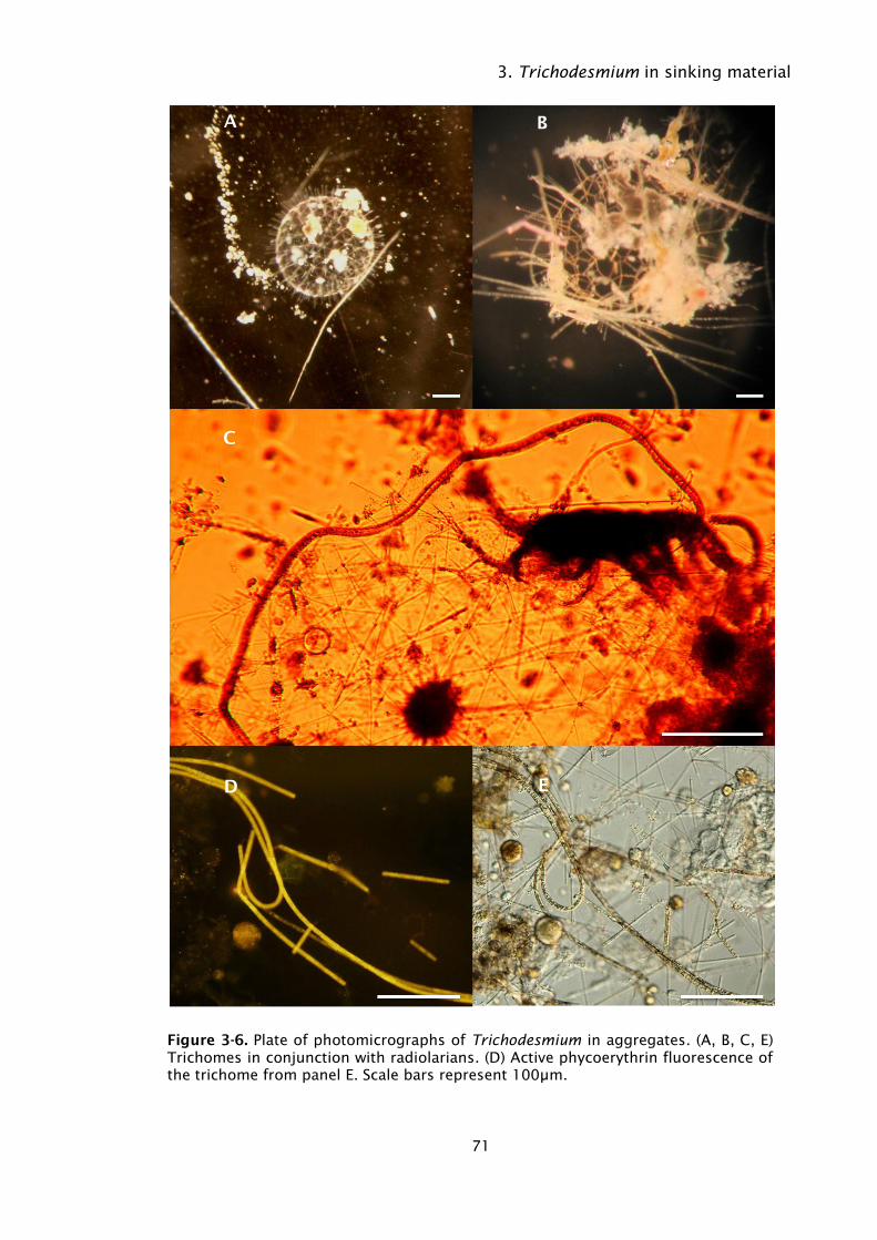
3. Trichodesmium in sinking material
71
Figure 3-6. Plate of photomicrographs of Trichodesmium in aggregates. (A, B, C, E)
Trichomes in conjunction with radiolarians. (D) Active phycoerythrin fluorescence of
the trichome from panel E. Scale bars represent 100µm.
A B
E D
C

E. Sargent
72
Figure 3-7. Plate of photomicrographs of Trichodesmium in faecal pellets. (A)
Partially degraded trichomes incorporated in (B) faecal pellets from CH0711; (C)
Confocal micrograph of seemingly intact trichomes from (D) faecal pellet of
unknown origin; (E) pteropod food web from same collection as pellet with intact
trichomes from D361. Scale bars represent 100µm.

3. Trichodesmium in sinking material
73
3.4.3 Trichodesmium Biomass and Contribution to Fluxes
The proportion of PON collected by SAPS deployments which could be
directly attributed to Trichodesmium-derived PON calculated from visible
Trichodesmium cells in SAPS collections ranged from 0.4-3% at 10 m below
the mixed layer, 0.03-1% at 100 m below the mixed layer, and from 0-0.4%
at 500 m (Table 3-1).
When combined with sinking rates measured for colonies (Figure 3-5),
the estimated visible Trichodesmium-attributable PON flux leaving the
mixed layer averaged 32.9 (5.0 – 99.6) µmol m-2
d-1
, which accounts for an
average of 5.6% (1– 17.5%), of the total PON flux independently derived from
234
Th (Table 3-1).
Measured N2
fixation rates varied along the transect, ranging from 26-
228 µmol m-2
d-1
(Figure 3-4A). Consequently, when compared to surface
nitrogen fixation rates, exported (visible) Trichodesmium-derived PON
accounted for an average of 23% of the daily nitrogen fixation in the upper
water column (Table 3-1). If all the newly fixed nitrogen in the surface were
contributing to organic nitrogen export, estimated surface-N2
-fixation rates
could have supported 20% of the 234
Th-derived PON flux at 10 m below the
mixed layer (Table 3-1).
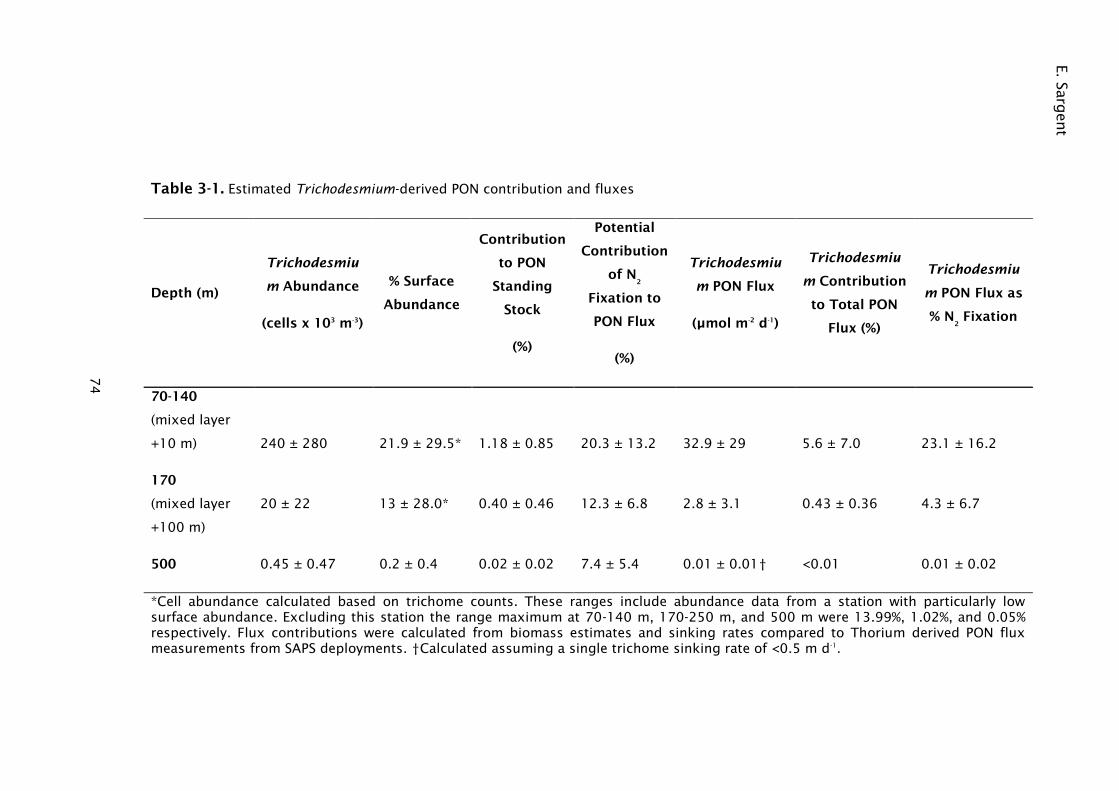
74
E. Sargent
Table 3-1. Estimated Trichodesmium-derived PON contribution and fluxes
Depth (m)
Trichodesmiu
m Abundance
(cells x 103
m-3
)
% Surface
Abundance
Contribution
to PON
Standing
Stock
(%)
Potential
Contribution
of N2
Fixation to
PON Flux
(%)
Trichodesmiu
m PON Flux
(µmol m-2
d-1
)
Trichodesmiu
m Contribution
to Total PON
Flux (%)
Trichodesmiu
m PON Flux as
% N2
Fixation
70-140
(mixed layer
+10 m) 240 ± 280 21.9 ± 29.5* 1.18 ± 0.85 20.3 ± 13.2 32.9 ± 29 5.6 ± 7.0 23.1 ± 16.2
170
(mixed layer
+100 m)
20 ± 22 13 ± 28.0* 0.40 ± 0.46 12.3 ± 6.8 2.8 ± 3.1 0.43 ± 0.36 4.3 ± 6.7
500 0.45 ± 0.47 0.2 ± 0.4 0.02 ± 0.02 7.4 ± 5.4 0.01 ± 0.01† <0.01 0.01 ± 0.02
*Cell abundance calculated based on trichome counts. These ranges include abundance data from a station with particularly low
surface abundance. Excluding this station the range maximum at 70-140 m, 170-250 m, and 500 m were 13.99%, 1.02%, and 0.05%
respectively. Flux contributions were calculated from biomass estimates and sinking rates compared to Thorium derived PON flux
measurements from SAPS deployments. †Calculated assuming a single trichome sinking rate of <0.5 m d-1
.
3. Trichodesmium in sinking material
75
3.5 Discussion
3.5.1 Trichodesmium-derived Fluxes of PON
The current study presents some of the first evidence that Trichodesmium
contributes to PON export through passive sedimentation. Previously, only
active transport (Guidi 2012) and sweeping mechanisms (Scharek et al. 1999b),
or aggregation, have been suggested to account for Trichodesmium presence
at depth. Guidi et al. (2012) suggested that active export of Trichodesmium
could be possible through downwelling associated with eddies in the North
Pacific Subtropical Gyre. Such enhancement of POC export by eddies has also
been observed in the Sargasso Sea (Sweeney et al. 2003; Davis and
McGillicuddy 2006).
Based on the biomass estimates and sinking rates, up to 74% of surface
Trichodesmium standing stock could leave the mixed layer each day. Moreover,
visible Trichodesmium cells likely composed up to 3% of the standing stock of
PON at depth (Table 3-1). When Trichodesmium biomass estimates were
combined with colonial sinking rates, the visible Trichodesmium-attributable
PON flux composed an average of 5.6% of the 234
Th derived PON flux at 10 m
below the mixed layer (Table 3-1). This indicates that intact Trichodesmium
cells are making a contribution to PON leaving the mixed layer, potentially
through a combination of relatively high sinking speeds and enhanced nitrogen
content compared to the other sinking particulate organic material. The
maximum contribution to the PON flux was observed below the maximum
surface abundance of Trichodesmium encountered along the transect (8.3°N,
28.3°W). At this station Trichodesmium contributed an estimated 17.5% of the
exported PON flux. Such a contribution is comparable to, although still less
than, previous estimates of the proportional contribution of Trichodesmium to
total new production in regions of high Trichodesmium abundance (Capone et
al. 2005). Observations under regions of high Trichodesmium surface
abundance, where standing stocks and nitrogen
fixation rates can be an order
of magnitude higher than we observed (Carpenter et al. 2004; Capone et al.
E. Sargent
76
2005) would be required to examine whether the potential for increased
Trichodesmium contribution to PON flux continues to scale with increasing
surface biomass. A recent study of an artificial bloom of Trichodesmium IMS
101 suggests this is a plausible scenario, as the collapse of the artificial bloom
resulted in enhanced PON export (Bar-Zeev et al. 2013).
The estimated contribution of Trichodesmium PON export herein are
likely conservative as we could only attribute the flux associated with intact,
microscopically visable cells. Although the intact nature of the cells may not be
preserved, and therefore not easily quantified, there remains the potential for
the packaging of Trichodesmium into faecal material beyond that observed
visibly, which could increase the Trichodesmium-attributable PON flux
estimates. Additionally, other mechanisms exist whereby Trichodesmium-
derived N could contribute to export fluxes secondarily, following
incorporation into other material, such as following viral lysis of cells (Hewson
et al. 2004), extracellular release of fixation products (Glibert and Bronk 1994),
or following PCD (Bar-Zeev et al. 2013).
N2
-fixation, which appeared to be dominated by Trichodesmium between
the equator and 10°N (Chapter 2), could potentially have supported 20% of the
234
Th-derived PON flux at 10 m below the mixed layer (Table 3-1). There is the
potential for a much larger diazotroph contribution to PON export yet to be
described, which could contribute to the transfer of new nitrogen to depth. If
consistent with these observations for sinking Trichodesmium, this PON could
then be available for remineralisation and integration into the thermocline. The
possibility that the measured surface rates are systematic underestimates
(Wilson 2012; Groβkopf et al. 2012) also requires consideration. An average
doubling of surface rates (Wilson 2012; Groβkopf et al. 2012) would mean that
surface N2
fixation had the potential to contribute to 40% of the total PON flux,
while visible Trichodesmium-derived PON flux would be reduced to around 12%
of the surface N2
fixation.

3. Trichodesmium in sinking material
77
3.5.2 Mechanisms of Export of Trichodesmium
Preliminary results suggest that despite its archetypal surface distribution
and buoyant properties, Trichodesmium is a standard constituent of sinking
material and is directly involved in the export of POC/PON through a variety of
mechanisms (Figure 3-8). Deep colonial distributions of Trichodesmium are
largely unreported, and reports of its presence in sediment traps are a rarity
(Scharek et al. 1999b; Chen et al. 2003). Below the euphotic zone,
Trichodesmium cannot photosynthesize, and is unlikely to be actively fixing N2
.
This was confirmed by nifH expression collected on this cruise (Langlois 2011
pers. comm.). This study thus provides evidence that while not diazotrophically
active, Trichodesmium is likely a regular constituent of sinking material, and
previous assessments have overlooked or underestimated Trichodesmium’s
presence at depth.
Figure 3-8. Schematic of possible export mechanisms for Trichodesmium (modified
from Mulholland 2007).
There are number of potential mechanisms that could explain
Trichodesmium’s presence below the depth where it can photosynthetically fix
carbon and nitrogen. The vertical migration of colonies provides a potential
mechanism for transport of Trichodesmium to depth. This theory assumes that
E. Sargent
78
after fixing carbon near the surface, colonies become laden down with
carbohydrates, overcoming their positive, gas vesicle supplied buoyancy, which
causes them to sink. As they do so they respire allowing them to ‘burn-off’ the
excess ballast, which decreases the colony sinking rate (Villareal and Carpenter
2003). In an optimal system, once a colony reaches the phosphocline, it would
be neutrally buoyant, and would be able to absorb nutrients for a few hours
before regaining positive buoyancy and returning to the surface (Villareal and
Carpenter 2003). This nutrient mining hypothesis is a potential explanation for
the location of the colonies collected at depth. The colonies collected between
120 and 175 m had much slower, and less variable, sinking rates than colonies
distributed above and below these depths (Figure 3-5). These decreased
sinking rates observed between 120 and 175 m correspond with the depth
range of enhanced phosphorus concentrations along both of the cruise tracks
(Figure 3-9) indicating these colonies could be in the process of reversing
buoyancy in an effort to return to the surface following nutrient acquisition.

3. Trichodesmium in sinking material
79
Figure 3-9. Longitudinal section of Phosphate (µM) along a pelagic transect during
CHO711 (A) and a latitudinal section of phosphate (µM) along the north-south transect
of D361 (B) illustrating enhanced phosphate concentrations within the depth range of
potential nutrient mining (dashed boxes).
However, colony size and sinking rate did not appear to be correlated in
this study (R2
<0.1) (CH0711, n = 24; D361, n=4). Previous models considering
cell size, gas vacuole content and carbohydrate ballast potential suggest that
only large (>1500 µm radius) colonies would possess the capability to carry out
vertical migrations beyond 100 m (Kromkamp and Walsby 1992; White et al.
2006), suggesting that such a mechanism is insufficient to account for
colonies collected in this study, which were consistently less than 1000 µm in
length. However, these models did not incorporate any cellular components
other than carbohydrates into cellular density analyses, and also did not
include physical influences on colony transport in the water column. Therefore,
colony size cannot be used as a strict determinant for colony ability to return
to the surface from depth.
E. Sargent
80
Colony collections included cells which were morphologically variable, but
phylogenetic analyses are necessary to determine whether multiple species of
Trichodesmium are depth distributed as interspecific cryptic diversity can make
morphological identification difficult (Hynes et al. 2012). Determining species
type would give insight into species-specific physiologies, which could
influence the assumptions of gas vacuole dynamics. The dominant
Trichodesmium species in this study region, T. thiebautii (O’Neil 1998;
Carpenter et al. 2004; Capone et al. 2005), have previously been shown to
suffer only a 10% gas vacuole loss through collapse at 200 m depth, with 100%
of gas vacuoles collapsing by 350 m (Walsby 1978). The other common
Trichodesmium species in the Atlantic, T. erythraeum has been observed to
suffer 100% gas vacuole collapse at a much shallower depth, between 105 and
120 m. If sinking colonies consist of T. erythraeum, their fate is likely locked
below 100 m, but if the species is T. thiebautii, as is suggested by
ultrastructural assessments of sinking colonies in a different study region
(Chapter 5), we must consider the potential for these colonies to reverse
buoyancy. Regardless of species type, small, fast sinking colonies present
below 100 m would be locked in a persistent and irreversible downward
trajectory. With maximum sinking rates of 120 m d-1
it is plausible that these
colonies would reach depths potentially complete gas vacuole collapse (>300
m) within 1-2 days whereby it would be impossible to reverse direction.
There was no correlation between sinking rate and depth in this study.
When gas vacuoles were manually pressure-destroyed colonial sinking rates
varied between 43 and 255 m d-1
(Walsby 1978). Most colonial sinking rates
from both cruises fell within this range (Figure 3-5), but species identifications
were not carried out in this study and sample size below 100 m was relatively
small (n=19). Additional assessments of species-specific sinking rates would be
beneficial to expanding the understanding of the mechanism of sinking in
Trichodesmium colonies.
The deepest colonies were present to 300 m, but a switch from colony to
trichome dominated samples occurred below 150 m. Colonies likely begin to
disassociate into free trichomes with depth as free trichomes should be unable
to sink to these depths unaided by incorporation into large, fast sinking
particles; colonial sinking rates were markedly faster than that of free filaments
as MSC settling indicated free filaments were in low abundance, and were part
3. Trichodesmium in sinking material
81
of the slow sinking/suspended pool of particulate material. It is also possible
that colonial associations become more fragile as they sink, as the
disassociation of some negatively buoyant colonies into filaments during
sinking rate experiments was observed. This observation supports the idea
that the deep free filaments we observed originated in colonies and were
transported to depth in that morphology, but that some other, likely physical
force caused them to disassociate. For the SAPS collected samples, the
filtration process would certainly have disturbed morphological features.
Moreover, partial cell breakage during SAPS filtration could also
contribute to the estimated contribution of Trichodesmium to PON flux being
an underestimate due to the potential loss of visually distinguishable
trichomes. The deepest colony reported here, which represents the deepest
report of a colony to date, was collected at 300 m using the CTD, but free
trichomes were distributed as deep as 500 m along the latitudinal transect in
the Atlantic, albeit sampled using SAPS. Previously Trichodesmium colonies
have been observed to 200 m (Carpenter and Price 1977; Tyrrell et al. 2003)
and free filaments have been reported deeper in the South China Sea in a
sediment trap at 925 m (Chen et al. 2003), and at station ALOHA in the Pacific
in a sediment trap at 2800 m (Scharek et al. 1999b).
Factors causing colonies to disassociate into free filaments at depth
greater than 100 m could greatly decrease the sinking rate of Trichodesmium,
potentially enhancing susceptibility to remineralisation in the twilight zone
(200-1000 m). Additionally, the significance of the observed morphological
bias, where mixed colony morphology at the surface was absent at depth
remains to be investigated. As all negatively buoyant colonies collected below
the mixed layer were in tuft form, enhanced fragility and disassociation of puff
colonies could explain the disproportional morphological sampling below the
mixed layer. Alternatively, it remains to be explored whether physiological or
species specificity of Trichodesmium sinking, such as epibiont communities
unique to different morphologies, plays a role in the morphological
characteristics of sinking Trichodesmium colonies.
The presence of Trichodesmium trichomes in aggregates with other
organisms, such as radiolarians (Figure 3-6), and/or detrital material was
encountered in most collections and increased with depth, denoting alternative

E. Sargent
82
pathways of export for this organism. Due to the method of SAPS collection, a
bias towards entanglement could have been introduced during pumping and
conglomeration of material on the filter, so it is possible that free trichomes
are more common at these depths than observed. Phycoerythrin and
chlorophyll fluorescence of cells at depth indicated that cells were reasonably
un-degraded, but cells were not particularly stable. Multiple observations of
trichome degradation were observed during imaging implying these trichomes
may have been on the verge of apoptosis when collected (Figure 3-10);
potentially the stress of collection and/or imaging may have induced the
observed apoptosis. If this process were to occur while the aggregates were
sinking it would represent a mechanism of direct release of Trichodesmium
intracellular contents into the water column at depth.
Figure 3-10. Light micrographs of Trichodesmium spp. in association with aggregates
within the hour following collection showing apoptosis of trichomes in progress. Scale
bars represent 200µm as indicated.
Additionally, one of the negatively buoyant colonies collected using the
MSC in the Gulf of Mexico was found to still exhibit variable chlorophyll
fluorescence, indicating a retained degree of photosynthetic competence. If
these trichomes remain intact while sinking after being caught in associations
with other organisms, they would likely retain their cell matter and transport it
out of the euphotic zone where it could then be remineralised. Further, as
phycoerythrin has been previously shown to be a nitrogen storage mechanism
in Synechococcus (Kirchman and Alberte 1985), its maintained presence here
represents a concentrated nitrogen pool exported within Trichodesmium.

3. Trichodesmium in sinking material
83
Previous reports have rarely documented grazing on Trichodesmium, so
the observed sporadic presence of this organism in faecal material is
significant. Trichomes of Trichodesmium were observed to be relatively intact
in faecal matter (Figure 3-7C). Trichomes of Trichodesmium were observed in
faecal matter both partially degraded, and seemingly intact. The latter situation
implies accidental feeding by an organism not capable of digesting
Trichodesmium. Faecal pellets containing intact trichomes shared
characteristics with pteropod waste material. As pteropods are non-selective
web feeders that passively capture their prey through filtration into mucous
sheets (Silver and Bruland 1981), Trichodesmium could have become entangled
in the feeding mesh and consumed unintentionally. Such an incidence would
preserve intact trichomes in a compact, fast-sinking pellet that would transport
trichomes to depth much quicker than if they were free-sinking. Intact,
previously ingested organisms in pteropod waste is common (Silver and
Bruland 1981), providing a transport mechanism for all Trichodesmium cell
matter. The fecal pellet containing intact Trichodesmium also shared
characteristics with euphausiid waste material (Yoon et al. 2001). Further
investigation of the occurrence of krill grazing on filamentous cyanobacteria is
warranted to assess whether these organisms may also unintentionally or
incompletely ingest Trichodesmium. The frequency of occurrence of
unintentional digestion is currently unknown, and was only encountered at one
station on D361 where pteropods were noted in the surface samples.
Partial degradation of trichomes in faecal matter suggests digestion is
taking place. In the present study this was only seen once in a pellet of
unknown origin. Trichome abundance was very low in this pellet compared to
the pellet with intact trichomes indicating it may be possible for some feeders
to breakdown and digest Trichodesmium in low quantities. Again, this could
contribute to quick sequestration of cell matter to depth. A more thorough
assessment of faecal material is needed to approximate the frequency of this
occurrence as packaging of fresh trichomes in large faecal pellets could
transport the associated newly fixed nitrogen to depth more efficiently than if
they were free-sinking, and could potentially make a large contribution to total
Trichodesmium export.

E. Sargent
84
3.5.3 Fate of Trichodesmium-derived Nitrogen
When Trichodesmium cell abundance is combined from colonies, free
trichomes, and trichomes incorporated in aggregates, it represents a
significant fraction of PON at depth, and the spatial consistency of the values
indicates the potential for significant episodic export following surface blooms
via the mechanisms we have previously described. Moreover, the occurrence of
Programmed cell death (PCD) during bloom collapse can significantly increase
transparent exopolymer particle (TEP) production, thereby enhancing export of
Trichodesmium-derived POM, such that after 5 days, ~0.08 mg N m-2
was
present at the bottom of the artificial bloom settling column when initial
surface concentrations were ~0.13 mg N m-2
(Bar-Zeev et al. 2013).
These results suggest that despite its archetypal surface distribution and
buoyant properties, Trichodesmium is a standard constituent of sinking
material in these (sub-)tropical waters and is directly involved in the export of
PON through a variety of mechanisms. Previously Trichodesmium was only
believed to export in fecal pellets on a small scale as well as secondarily by
cycling through the microbial loop in the euphotic zone resulting in eventual
loss of Trichodesmium-derived new nitrogen following incorporation into the
biomass of other organism and their subsequent export (Mulholland 2007).
The current study confirms that Trichodesmium export does occur following
grazing and repackaging in fecal material, but also documents two novel
mechanisms by which Trichodesmium exports (i.e. freely sinking and
incorporation in aggregates). Trichodesmium does not appear to be a major
contributor to total PON export under the observed moderate abundance
conditions in the Atlantic (Table 3-1), where surface abundance rarely
exceeded 5 colonies per litre.
However, the contribution to PON export and total new N production in
the surface (Capone et al. 2005; Mourino-Carballido 2011) may be more
significant under conditions of higher abundance of both Trichodesmium
(Carpenter et al. 2004) and other diazotrophs. Scharek and others (1999b)
attributed Trichodesmium presence in sediments traps to be a consequence of
a sweeping mechanism whereby trichomes were caught up in the flux of
larger, stickier diatom aggregates. The sweeping mechanism could also
3. Trichodesmium in sinking material
85
explain the observation of trichome incorporation in aggregates. Continued
observation of trichome incorporation in aggregates and fecal pellets suggests
a previously overlooked involvement in export that remains to be more
comprehensively explored. Further investigation of Trichodesmium’s presence
in sinking material will give a clearer idea of this organism’s contribution to
export fluxes allowing us to better constrain the fate of the nitrogen fixed by
this organism and hence it’s detailed biogeochemical role.
Trichodesmium is the dominant nitrogen fixing organism in this study
area (Tyrrell et al. 2003; Moore et al. 2009; Zehr 2011; Groβkopf et al. 2012),
and is potentially capable of accounting for the majority of the geochemically
derived rates of nitrogen fixation inferred from the N* distributions in the
North Atlantic (Gruber and Sarmiento 1997; Capone et al. 2005; Mahaffey et al.
2005; Hansell et al. 2004, 2007). The spatial consistency and continuous
decrease in cell abundance with depth below the mixed layer along the
transect is indicative of remineralisation between 100 and 500 m (Figure 3-4B).
The water column oxygen minima and region of maximum apparent oxygen
utilization was located between 150 and 750 m within our (sub-)tropical
Atlantic transect (Figure 3-11), suggesting that the majority of the total export
of organic material is also likely remineralised within this same depth. The
depth range of likely remineralisation also partially overlaps with the density
surfaces within which remineralisation of diazotroph-derived material could
contribute to N* (Figure 3-11).

E. Sargent
86
Figure 3-11. Latitudinal section of oxygen along the south-north transect on D361
showing decreased water column oxygen in the depth range where remineralisation of
Trichodesmium is assumed (100-250 m), and alignment with the density surfaces
within which remineralisation of diazotroph derived material could contribute to N*.
Oxygen saturation was calculated from the method described by Garcia and Gordon
(1992) and AOU was subsequently derived from the equation provided by Sarmiento
and Gruber (2006).
Maximum N* anomalies in the North Atlantic gyre are associated with a
potential density interval in the = 26.1-27.1 range (Gruber and Sarmiento
1997; Hansell et al. 2007). Due to the shoaling of the permanent thermocline
at low latitudes near the equator (Sarmiento et al. 2004), this potential density
interval was shallower than 500 m along the Atlantic transect. Consequently,
release of Trichodesmium-derived exported material into the upper twilight
zone remineralisation region (100-500 m), provides a mechanism for
generating at least a portion of the N* anomaly observed within the = 26.1-
27.1 range. Direct export of Trichodesmium thus potentially resolves one of
the pathways by which the North Atlantic N* anomaly develops (Hansell et al.
2007).
3. Trichodesmium in sinking material
87
3.6 Conclusion
Trichodesmium provides a significant amount of newly fixed nitrogen to the
warm surface ocean in many oligotrophic regions thereby indirectly supporting
the growth and export of non-diazotrophic organisms (Carpenter and Capone
2008). Conservative estimates that an average of 8.8% (excluding the southerly
most station, otherwise average of 22%) of the surface standing stock of
Trichodesmium is vertically leaving the mixed layer each day contributing an
average of 5.6% to the total PON flux, provides insight into the biogeochemical
fate of a significant proportion of the nitrogen fixed by this organism. The
decrease in Trichodesmium biomass from 10 m below the mixed layer to 500
m suggests that Trichodesmium is remineralised within the twilight zone and
hence contributes diazotroph-derived new nitrogen to the thermocline of the
(sub-)tropical Atlantic Ocean. When coupled with the potential for enhanced
export following surface blooms, such a previously undescribed mechanism
could export newly fixed nitrogen to depths facilitating accumulation along the
isopycnals associated with the thermocline of the sub-tropical gyre, thus
contributing to the N* anomaly in these waters (Gruber and Sarmiento 1997;
Hansell et al. 2007). Consequently nitrogen fixation by Trichodesmium in the
low latitude Atlantic is not only an important contributor of new nitrogen in the
euphotic zone (Capone et al. 2005), but may also be important in the supply of
nutrients to the thermocline. In turn these nutrients could sustain productivity
on longer time scales, through eventual resupply to the surface ocean, while
also potentially contributing to the deficiency of phosphorus downstream
within the surface waters of the gyre.

E. Sargent
88

4. Exploration of ploidy in Trichodesmium
89
4. An exploration of the significance of
polyploidy in Trichodesmium
4.1 Abstract
Quantitative PCR of the nifH gene is commonly used to assess the
biogeography and relative abundance of diazotrophic species in the global
oceans, and was employed in Chapter 2 to gauge the fate of a variety of
diazotrophic groups. However, discrepancies between nifH-based and
optical abundance data have been previously acknowledged, often citing an
order of magnitude higher nifH-based than cell-count-based abundance for
Trichodesmium indicating the potential for extreme overestimation of
biomass when converting between the two methodologies. As DNA
extraction efficiency is variable between species, and can be less than 100%
efficient, the discrepancy between nifH gene copies and cell counts is
potentially even larger. Similar discrepancies have also been noted for
heterocystous, diazotrophic cyanobacteria. Herein we endeavour to explain
the discrepancy between detectable nifH gene copies and optical cell counts
through an investigation of polyploidy in Trichodesmium. Polyploidy, or the
condition of having more than one genome per cell, is well described in
prokaryotic organisms and it present in many cyanobacterial groups. Results
suggest polyploidy in Trichodesmium could result in inflated detectable nifH
relative to cell abundance. If Trichodesmium is polyploid, this has
implications for the interpretation of qPCR data globally, and thus we
caution against any use of direct conversion from detectable nifH gene
copies to cell numbers, especially in Trichodesmium.

E. Sargent
90
4.2 Introduction
The development of molecular technologies has resulted in a near
universal switch from microscopy (hereafter optical) cells counts to qPCR of
the nifH gene (hereafter gene-based abundances) to understand the
distribution and abundance of diazotrophs in the global ocean. This switch
from optical to gene-based abundance methodologies is striking with 40
studies spanning 49 years resulting in 5191 data points from optical
abundance data, while 19 gene-based abundance studies have resulted in
3201 data points in just 7 years (Luo et al. 2012).
As previously discussed in Chapter 2, the nifH gene is a highly
conserved gene which codes for the iron binding component of the
nitrogenase enzyme (Young 1992). Sequence diversity of nifH is variable
enough between genera of diazotrophs that the technique has been used to
detect and quantify diverse groups of nifH-containing organisms (Zehr et al.
1997), including non-cyanobacterial nifH-containing organisms, such as the
gammaproteobacteria and green sulphur bacteria whose contribution to
global nitrogen fixation remains unknown (Riemann et al., 2010). This influx
of data derived from gene-based abundances has substantially contributed
to the expansion of our knowledge of the diversity and biogeography of
nifH-containing organisms, and use of the procedures has been crucial to
increased understanding of the intricacies of the marine nitrogen cycle (Zehr
2011; Thompson and Zehr 2013).
Without the advent of gene-based abundance technologies we would
essentially know nothing about some of the unicellular nitrogen fixing
cyanobacteria, such as Group A and Group C cyanobacteria. Group A
cyanobacteria have proven to be dominant in many marine environments
(Church et al. 2005a; 2005b, Foster et al. 2009, Moisander et al. 2010) and
may be quite a significant contributor of new nitrogen in these regions
(Church et al. 2005b). Gene-based technologies have also helped to
elucidate the ecological significance of these unicellular cyanobacterial
groups, and have expanded our understanding of the role of unicellular,

4. Exploration of ploidy in Trichodesmium
91
filamentous and heterocystous cyanobacteria in the global oceans
(Thompson and Zehr 2013).
However, there are discrepancies between gene-based and optical
abundance data. Often an order of magnitude higher gene-based abundance
is reported for Trichodesmium than can be accounted for by optically
enumerated cells (Luo et al. 2012). This is troubling when considering
biomass estimations derived from gene-based abundance data, as it would
likely be an overestimation of actual biomass, and could result in an
inaccurate representation of dominant biomass when assessing total nifH
community structure. This of course also has knock on effects when
comparing gene-based abundance data with nitrogen fixation rate
measurements and other parameters.
This study compares gene-based abundance data alongside optical
abundance data for both in situ and culture experiments. Herein, we
endeavour to explain the higher abundance of detectable nifH gene copies
relative to optical cell counts through an investigation of polyploidy in
Trichodesmium. In the case of prokaryotes, polyploidy is the condition of
containing more than one genome per cell. Polyploidy is known to be
present in other cyanobacterial groups such as Synechocystis and Anabaena,
and known polyploid prokaryotes actually outnumber known monoploid
prokaryotes (Griese et al. 2011). This suggests polyploidy may be the norm
for prokaryotic organisms and thus the investigation of this condition in
Trichodesmium is warranted.
Results from comparisons of gene-based and optical abundances,
assessments of the potential contribution Trichodesmium would make to in
situ chlorophyll derived from gene-based abundances if assuming a single
gene copy per cell, and microscopic assessments of intracellular DNA
distribution in Trichodesmium all suggest polyploidy is a reasonable
explanation for the aforementioned discrepancy. Gene-based technologies
are still a useful tool for assessing the diversity and biogeography of nifH-
containing organisms, but we caution against any use of direct conversion
from detectable nifH gene copies to cell numbers, and call for reintroduction
of optical based cell counts when conducting assessments of

E. Sargent
92
Trichodesmium biomass in situ until a suitable conversion metric can be
established, should this even be possible.
4.3 Methods
4.3.1 Investigation of ploidy in Trichodesmium
Surface cell counts for Trichodesmium (Chapter 3) were compared
with surface nifH gene copy abundance using the filamentous probe
(Chapter 2). Following detection of an enhanced gene-based abundance
relative to cell counts, samples were obtained from Trichodesmium
erythraeum IMS 101 cell cultures to conduct a side by side analysis of the
two abundance methodologies. Ten millilitres of four different cultures
grown under varying phosphorus sources were filtered in triplicate onto
0.22 µm Durapore (Millipore) filters under low (2 mbar) vacuum pressure.
Testing of the different phosphorus conditions was opportunistic, but was
later used to determine if differences in gene and optical abundances were
affected by the phosphorus source or ages of cultures. After filtration, filters
were flash frozen in liquid nitrogen and stored at -80°C until DNA extraction
as described previously (Chapter 2.3.2). Primers and probes were chosen for
amplification of the filamentous nifH phylotypes and qPCR was run as
described above (Chapter 2.3.3) by Dr. Rebecca Langlois at the Leibniz
Institute for Marine Sciences, in Kiel, Germany; Dr. Langlois is now located
at Dalhousie University in Halifax, Nova Scotia, Canada.
Cell counts were performed in triplicate on 10 mL sub-samples of the
Trichodesmium erythraeum IMS 101 cultures, which were preserved in 2%
acidic Lugol’s iodine (Throndsen 1978). Cell abundance was directly
measured from free trichomes via light microscopy at 200x magnification. A
10 mL sub-sample was also filtered onto a 25mm GF/F, 0.7μm poresize
filter, which was frozen until acetone extraction and fluorometric chlorophyll
determination.
4. Exploration of ploidy in Trichodesmium
93
4.3.2 Genome Copy Determination
Theoretical genome copies per cell were determined for in situ and
culture samples. Following qPCR, the calculated gene copies per litre were
divided by optically determined cells L-1
to give an upper threshold for
potential genome copies per cell as described by Pecoraro et al. (2011).
4.3.3 DAPI staining and Imaging
Trichodesmium erythraeum IMS 101 was examined under
epifluorescent microscopy following DAPI staining to assess intracellular
DNA distribution. Following gravitational settling and aspiration of five
millilitres of media, Trichodesmium biomass was fixed with 1%
glutaraldehyde, 3% formaldehyde, and 14% sucrose in PIPES buffer. After 20
minutes at 4 °C, samples were filtered onto 0.8µm polycarbonate black
filters, transferred to microscope slides, and were mounted with
VECTASHIELD HardSet Mounting Medium with DAPI at 1.5 μg mL-1
(Vector
Laboratories). Slides were allowed to set overnight at 4 °C. Samples were
imaged on a Leica SP5 confocal microscope under UV and 488nm excitation,
which excites DAPI, chlorophyll a, and phycoerythrin fluorescence. The
method used to process and acquire the transmission electron micrographs
is discussed thoroughly in Chapter 5.3.

E. Sargent
94
4.4 Results
4.4.1 Discrepancy between cell counts and gene copies
A large discrepancy between the abundance of gene copies and cell
numbers is apparent when comparing cell counts and gene copies from in
situ data. Abundances from gene copies consistently exceed cell count
abundances by one to two orders of magnitude except at the lowest end of
abundance where under sampling may have contributed to non-
representative DNA extractions, and gene copies below the detection limit of
the qPCR assay resulted in cells counts exceeding gene copies (Figure 4-1A).
For culture samples, the discrepancy between gene copies and cell counts
consistently span two orders of magnitude (Figure 4-1B). The trend of gene
copies exceeding cell counts is consistent for all data, and the two
abundance methodologies correlate strongly (R2
=0.89) (Figure 4.2).

4. Exploration of ploidy in Trichodesmium
95
Figure 4-1. Raw Discrepancy between Trichodesmium cell counts and nifH gene
copies. (A) in situ data from surface samples along the D361 transect (black),
R2
=0.87, and AMT17 transect (blue), R2
= 0.83, and (B) Trichodesmium erythraeum
IMS101 culture samples extracted from Durapore (red), R2
=0.78, and polycarbonate
(green), R2
=0.88, filters. Gene copies consistently exceed cell counts by one to two
orders of magnitude (p<0.01), except at and below the limit of detection for the
qPCR assay. Theoretical genome copies per cell derived from this discrepancy (gene
copies/L divided by cells/L; Pecoraro et al. 2011) range from 1-119 for in situ data
and from 159-698 for Trichodesmium erythraeum IMS 101. The solid black lines
represent a 1:1 ratio between cell counts and gene copies.

96
E. Sargent
Figure 4-2. A combined assessment of log transformed cell count and gene copy abundances to illustrate the sustained discrepancy
across all samples, except at and below the limit of detection for the qPCR assay.

4. Exploration of ploidy in Trichodesmium
97
4.4.2 Ploidy assessment
In Trichodesmium IMS101, estimated genome copies per cell ranged
from an average of 159 ± 59 to 698 ± 100 (Table 4-1), while the ploidy level
of Trichodesmium spp. collected in situ averaged 12 ± 13 (1 - 50) on
AMT17 and 31 ± 30 (1 - 120) on D361.
Table 4-1. Estimated genome copies in T. erythraeum IMS 101 under varying
phosphorus conditions calculated by division of gene copies/L by cells/L as
described by Pecoraro et al. 2011).
Culture Cell count (cells/L) Potential genome copies
(phosphorus source)
T. erythraeum IMS 101 (PO4 exponential) 213967 ± 59800 639.4 ± 41.3
T. erythraeum IMS 101 (PO4 senescent) 35367 ± 2201 697.5 ± 100.1
T. erythraeum IMS 101 (Glycerol) 1239533 ± 195725 265.9 ± 86.8
T. erythraeum IMS 101 (Methyl) 370000 ± 25662 158.7 ± 59.2
4.4.3 Intracellular DNA distribution
All stained Trichodesmium cells revealed a markedly segregated DAPI
staining throughout the entirety of the cell in which the pattern of
distribution was unique along the z plane (i.e. through the depth of the cell),
implying intracellular DNA distribution in Trichodesmium IMS101 is
scattered and extensive (Figure 4-3). Chlorophyll and phycoerythrin were
predominantly constrained to the thylakoid lamellae (Figure 4-4A).
Transmission electron microscopy (TEM) of the same culture revealed that
ultrastructure alone cannot account for the observed scattering as
intracellular organic matter is not segregated in the same manner as is
observed by DAPI fluorescence (Figure 4-4B).

98
E. Sargent
Figure 4-3. Multiple sections of 3 series (out of 11) of DAPI staining assessments in
Trichodesmium IMS101 showcasing the DNA distribution within each cell is not
uniform along the z plane (sections). The distance between each panel is
approximately 55nm. Scale bars represent 5 µm.

4. Exploration of ploidy in Trichodesmium
99
Figure 4-4. DNA distribution in Trichodesmium erythraeum IMS 101. (A) DAPI (blue)
and autofluorescence (orange) illustrate marked segregation of DNA staining within
the cell. (B) Transmission electron micrograph illustrating that ultrastructure alone
doesn’t account for DAPI staining patchiness as intracellular organic matter is not
segregated in the same manner as is observed by DAPI fluorescence. (C) Such DNA
segregation is not as readily visible as lower resolutions. Scale bars represent 2µm.
4.4.4 Chlorophyll
The chlorophyll content of individual Trichodesmium IMS 101 derived
from cell counts and from qPCR of the nifH gene in extracts was highly
variable. Cellular chlorophyll content derived from cell counts was on the
pictogram scale as is consistent with the literature, while cellular chlorophyll
content derived from assumptions that a single nifH gene copy is equal to a
single cell was on the femtogram scale, three orders of magnitude below
cellular chlorophyll levels reported in the literature (Figure 4-5).
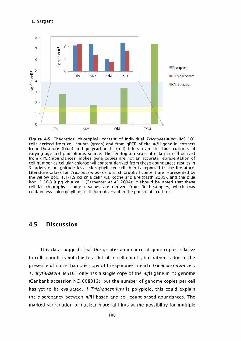
100
E. Sargent
Figure 4-5. Theoretical chlorophyll content of individual Trichodesmium IMS 101
cells derived from cell counts (green) and from qPCR of the nifH gene in extracts
from Durapore (blue) and polycarbonate (red) filters over the four cultures of
varying age and phosphorus source. The femtogram scale of chla per cell derived
from qPCR abundances implies gene copies are not an accurate representation of
cell number as cellular chlorophyll content derived from these abundances results in
3 orders of magnitude less chlorophyll per cell than is reported in the literature.
Literature values for Trichodesmium cellular chlorophyll content are represented by
the yellow box, 1.1-1.5 pg chla cell-1
(La Roche and Breitbarth 2005), and the blue
box, 1.56-3.9 pg chla cell-1
(Carpenter et al. 2004); it should be noted that these
cellular chlorophyll content values are derived from field samples, which may
contain less chlorophyll per cell than observed in the phosphate culture.
4.5 Discussion
This data suggests that the greater abundance of gene copies relative
to cells counts is not due to a deficit in cell counts, but rather is due to the
presence of more than one copy of the genome in each Trichodesmium cell.
T. erythraeum IMS101 only has a single copy of the nifH gene in its genome
(Genbank accession NC_008312), but the number of genome copies per cell
has yet to be evaluated. If Trichodesmium is polyploid, this could explain
the discrepancy between nifH-based and cell count-based abundances. The
marked segregation of nuclear material hints at the possibility for multiple
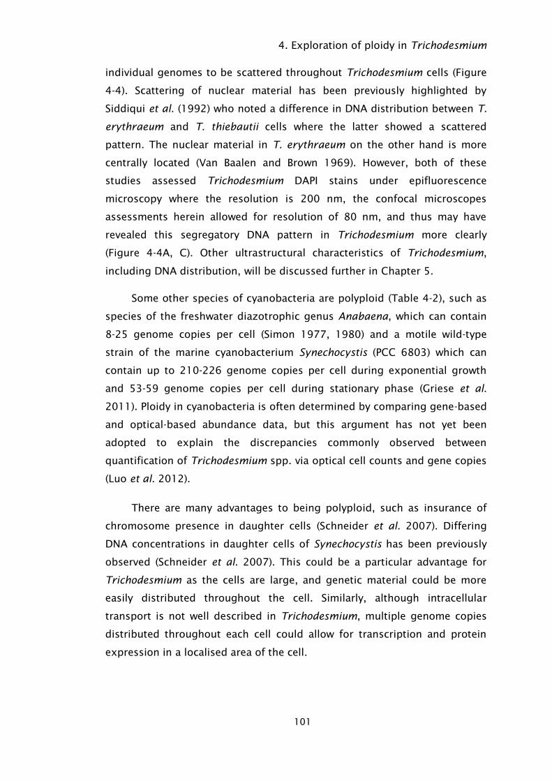
4. Exploration of ploidy in Trichodesmium
101
individual genomes to be scattered throughout Trichodesmium cells (Figure
4-4). Scattering of nuclear material has been previously highlighted by
Siddiqui et al. (1992) who noted a difference in DNA distribution between T.
erythraeum and T. thiebautii cells where the latter showed a scattered
pattern. The nuclear material in T. erythraeum on the other hand is more
centrally located (Van Baalen and Brown 1969). However, both of these
studies assessed Trichodesmium DAPI stains under epifluorescence
microscopy where the resolution is 200 nm, the confocal microscopes
assessments herein allowed for resolution of 80 nm, and thus may have
revealed this segregatory DNA pattern in Trichodesmium more clearly
(Figure 4-4A, C). Other ultrastructural characteristics of Trichodesmium,
including DNA distribution, will be discussed further in Chapter 5.
Some other species of cyanobacteria are polyploid (Table 4-2), such as
species of the freshwater diazotrophic genus Anabaena, which can contain
8-25 genome copies per cell (Simon 1977, 1980) and a motile wild-type
strain of the marine cyanobacterium Synechocystis (PCC 6803) which can
contain up to 210-226 genome copies per cell during exponential growth
and 53-59 genome copies per cell during stationary phase (Griese et al.
2011). Ploidy in cyanobacteria is often determined by comparing gene-based
and optical-based abundance data, but this argument has not yet been
adopted to explain the discrepancies commonly observed between
quantification of Trichodesmium spp. via optical cell counts and gene copies
(Luo et al. 2012).
There are many advantages to being polyploid, such as insurance of
chromosome presence in daughter cells (Schneider et al. 2007). Differing
DNA concentrations in daughter cells of Synechocystis has been previously
observed (Schneider et al. 2007). This could be a particular advantage for
Trichodesmium as the cells are large, and genetic material could be more
easily distributed throughout the cell. Similarly, although intracellular
transport is not well described in Trichodesmium, multiple genome copies
distributed throughout each cell could allow for transcription and protein
expression in a localised area of the cell.

10
2
E. Sargent
Table 4-2. Experimentally determined ploidy levels of marine and aquatic cyanobacterial species (Amended from Griese et al. 2011)
Species Physiology Cell size
(µm) Genome size
(Mbp) Average ploidy level Ploidy Reference
Anabaena cylindrica Aquatic 10 - 25 Polyploid Simon (1977)
Anabaena variabilis Aquatic 10 7.1 5-8 Oligoploid Simon (1980)
10 Microcystis strains Aquatic 5 - 1-10 Oligoploid Kurmayer & Kutzenberger (2003)
Anabaena sp. PCC 7120 Aquatic 10 7.2 8.2 Oligoploid Hu et al. (2007)
Prochlorococcus Marine 1 1.7 - Monoploid Vaulot et al. (1995) Synechococcus elongatus PCC 7942 Marine 0.8-1.5 2.8 3.9/3.3* Oligoploid Griese et al. (2011)
S. elongatus PCC 7942 Marine 0.8-1.5 2.8 3-5 Oligoploid Mori et al. (1996)
Synechococcus sp. PCC 6301 Marine 0.8-1.5 2.7 2-6 Oligoploid Binder & Chisholm (1990)
Synechococcus sp. WH 7803 Marine 0.8-1.5 2.4 3.6 Oligoploid Griese et al. (2011)
Synechococcus sp. WH 7803 Marine 0.8-1.5 2.4 2-4 Oligoploid Binder & Chisholm (1995)
Synechococcus sp. WH 7805 Marine 0.8-1.5 2.6 1 Monoploid Binder & Chisholm (1995)
Synechococcus sp. WH 8101 Marine 0.8-1.5 3.2 1 Monoploid Armbrust et al. (1989)
Synechococcus sp. WH 8103 Marine 0.8-1.5 2.7 1-2 Monoploid Binder & Chisholm (1995) Synechocystis sp. PCC 6803 (motile) Aquatic 2-5 3.6 218/58/58* Polyploid Griese et al. (2011) Synechocystis sp. PCC 6803 (GT) Aquatic 2-5 3.6 142/47/43* Polyploid Griese et al. (2011) Synechocystis sp. PCC 6803 (‘Kazusa’) Aquatic 2-5 3.6 12 Polyploid Labarre et al. (1989) Trichodesmium erythraeum IMS101 Marine 10 7.75 440 Polyploid This study
Trichodesmium spp. Marine 10 - 12, 50 Polyploid This study (AMT17 and D361 respectively)
*Exponential phase/linear phase/stationary phase.

4. Exploration of ploidy in Trichodesmium
103
As the ploidy level of Trichodesmium is previously unconfirmed, and as
we are unable to distinguish between nifH gene copies present inside living
diazotrophs from extracellular nifH gene copies in the water column or in
association with other material following grazing or apoptosis, we must
consider whether the discrepancy between gene-based abundances and cell
counts can be attributable to artefact. Nucleic acids are a well described
component of the dissolved organic matter pool (Karl and Bailiff 1989), which
can bind to particulate material in the water column and in sediments and
resist degradation (Lorenz and Wackernagel 1987; Corinaldesi et al. 2011), but
if extracellular DNA was providing additional template for nifH amplification,
we would have expected to see a larger disproportion of gene copies to cell
counts in the older [culture] samples and/or between filter types. As no
significant difference (p=0.7) in potential genome copies was observed
between active (639 ± 41.3) and senescent (697.5 ± 100.1) cultures (Table 4-1)
we do not feel extracellular DNA could account for the entirety of the gene-
based versus optical-based abundance discrepancy. Additionally, Durapore
filters are used most frequently in qPCR work as they are known not to bind
nucleic acids. If extracellular DNA was contributing to the enhanced nifH
abundances we observed, we would have expected to detect more gene copies
in samples taken onto polycarbonate filters as they may have bound more
extracellular DNA, but there was no significant difference between the nifH
abundances detected from these two filter types (p=0.4).
Another potential contributor of non-cyanobacteria nifH template could
come from cyanophages. Prochlorococcus-specific cyanophages have been
confirmed to contain identical copies of Prochlorococcus PSII genes (Sullivan et
al. 2005). Trichodesmium may also harbour phages (Hewson et al. 2004), but
evidence for a Trichodesmium-specific phage is inconclusive at current (Brown
et al. 2013). Trichodesmium-associated phages appear to be phylogenetically
distinct from the virioplankton community, and some even contained
Trichodesmium–like genes (Brown et al. 2013). It seems likely that a
Trichodesmium-specific phage exists, but it remains to be assessed whether
DNA from said phage carrying identical nifH gene copies could account for
some of the observed discrepancy between gene copies and cell counts.
Nonetheless, we hypothesize that the entirety of the discrepancy cannot be
explained by cyanophage contributions as investigations are currently being

E. Sargent
104
E. Sargent
carried on the hetR, 16S rRNA, and psbA genes in Trichodesmium via qPCR,
and premilinary results suggest that are also inflated relative to cell counts
(Sargent et al. In Prep). It would be unlikely that a phage would harbour
identical copies of all of these diverse genes. Additionally, if the qPCR
determined ratio of nifH to multiple copy number genes is in close agreement
with the ratio predicted from the Trichodesmium genome sequence (Genbank
accession NC_008312) (i.e nifH:hetR:16S rRNA:psbA of 1:1:2:3), this would
provide further evidence against phage contribution to the gene pool. Even if a
Trichodesmium-specific phage contained each of these Trichodesmium genes,
it is very unlikely that it would contain identical gene quantities to the host
across all of these genes, especially as they are distant within the genome
making horizontal acquisition extremely unlikely.
If we dismiss extracellular and viral contributions to nifH template and
assume the discrepancy must be explained in a deficit of optical cell counts
rather than enhanced gene-based abundances, then we would expect other
species-specific physiological parameters to correlate with the gene-based
abundances. However, if we assume each detected nifH gene copy attributable
to Trichodesmium represents a single cell, the estimated chlorophyll-content
per cell is a fraction of what is reported in the literature (Figure 4-5) further
indicating gene-based abundances are not an accurate representation of actual
cell abundance.
Further related evidence for ploidy in Trichodesmium is revealed when
comparing the contribution that Trichodesmium would make to total in situ
chlorophyll from gene-abundances reported in the literature if we assume a
single gene copy represents a single cell and if a single cell contains 1.1 pg chl
(La Roche and Breitbarth 2005). The lowest of the reported Trichodesmium
chlorophyll content per cell was chosen to test whether cellular contribution to
total would sill exceed 100% even if all cells contained a minimal amount of
chlorophyll. As average Trichodesmium contribution to total in situ chlorophyll
equals or exceeds 100% in all gene-based abundance conversions derived from
the literature (Table 4-3), it is clear that nifH abundances cannot be considered
a 1:1 proxy for cell abundance in Trichodesmium. The work discussed here
that is derived from Trichodesmium abundances reported in Chapters 2 and 3
represents the only study for which we have data that assesses in situ optical
and gene-based abundances side by side. Although Trichodesmium
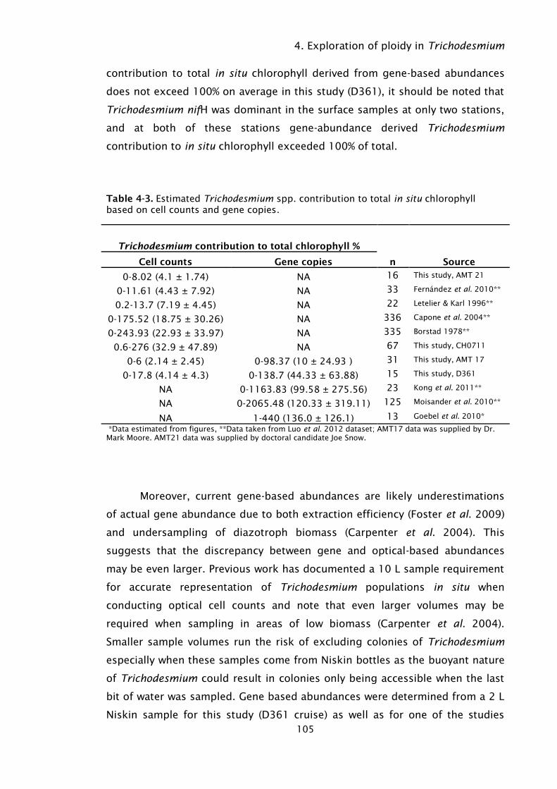
4. Exploration of ploidy in Trichodesmium
105
contribution to total in situ chlorophyll derived from gene-based abundances
does not exceed 100% on average in this study (D361), it should be noted that
Trichodesmium nifH was dominant in the surface samples at only two stations,
and at both of these stations gene-abundance derived Trichodesmium
contribution to in situ chlorophyll exceeded 100% of total.
Table 4-3. Estimated Trichodesmium spp. contribution to total in situ chlorophyll
based on cell counts and gene copies.
Trichodesmium contribution to total chlorophyll %
n Source Cell counts Gene copies
0-8.02 (4.1 ± 1.74) NA 16 This study, AMT 21
0-11.61 (4.43 ± 7.92) NA 33 Fernández et al. 2010**
0.2-13.7 (7.19 ± 4.45) NA 22 Letelier & Karl 1996**
0-175.52 (18.75 ± 30.26) NA 336 Capone et al. 2004**
0-243.93 (22.93 ± 33.97) NA 335 Borstad 1978**
0.6-276 (32.9 ± 47.89) NA 67 This study, CH0711
0-6 (2.14 ± 2.45) 0-98.37 (10 ± 24.93 ) 31 This study, AMT 17
0-17.8 (4.14 ± 4.3) 0-138.7 (44.33 ± 63.88) 15 This study, D361
NA 0-1163.83 (99.58 ± 275.56) 23 Kong et al. 2011**
NA 0-2065.48 (120.33 ± 319.11) 125 Moisander et al. 2010**
NA 1-440 (136.0 ± 126.1) 13 Goebel et al. 2010*
*Data estimated from figures, **Data taken from Luo et al. 2012 dataset; AMT17 data was supplied by Dr.
Mark Moore. AMT21 data was supplied by doctoral candidate Joe Snow.
Moreover, current gene-based abundances are likely underestimations
of actual gene abundance due to both extraction efficiency (Foster et al. 2009)
and undersampling of diazotroph biomass (Carpenter et al. 2004). This
suggests that the discrepancy between gene and optical-based abundances
may be even larger. Previous work has documented a 10 L sample requirement
for accurate representation of Trichodesmium populations in situ when
conducting optical cell counts and note that even larger volumes may be
required when sampling in areas of low biomass (Carpenter et al. 2004).
Smaller sample volumes run the risk of excluding colonies of Trichodesmium
especially when these samples come from Niskin bottles as the buoyant nature
of Trichodesmium could result in colonies only being accessible when the last
bit of water was sampled. Gene based abundances were determined from a 2 L
Niskin sample for this study (D361 cruise) as well as for one of the studies
E. Sargent
106
E. Sargent
documented in Table 4-3 (Goebel et al. 2010). The other aforementioned
studies sampled 4-5 L (Moisander et al. 2010) and 0.5-1 L from Niskin bottles
(Kong et al. 2011). Thus, all are potentially underestimations of actual gene
abundance, and therefore we have potential underestimated the level of ploidy
in in situ population of Trichodesmium spp.
Trichodesmium, like all living organisms, requires phosphorus for growth, and
although cellular phosphorus pools in phytoplankton are often dominated by
RNA (Geider and La Roche 2002), genomic P contribution to cellular P can
exceed 50% in some cyanobacteria, such as Synechococcus and
Prochlorococcus (Bertilsson et al. 2003). To assess whether the ploidy level of
Trichodesmium determined herein is plausible, an evaluation of genomic P
contribution to cellular P was carried out after converting colonial P values
reported in the literature to cellular P concentrations (Figure 4-6) to insure
estimations of genomic P would not exceed 100% of cellular P. A single
Trichodesmium genome was calculated to contain 7.97 x 10⁻¹⁶
g of P, and the
range of cellular P content for Trichodesmium spp. was calculated as 3.10 x 10-
13
– 1.41 x 10⁻¹²
g P in situ (Tovar-Sanchez et al. 2011; Neuster et al. 2012; this
study), and 6.19 x 10⁻¹³
- 2.17 x 10⁻¹²
g P in Trichodesmium IMS 101 in culture
(Barcelos e Ramos et al. 2007). The range of ploidy in the in situ samples from
D361 and AMT17 correspond to a maximal genomic contribution to total P of
just over 25%. The same assessment for Trichodesmium IMS101 spans nearly
the entire range from 6-95% genomic P contribution to cellular P (Figure 4-6).

4. Exploration of ploidy in Trichodesmium
107
Figure 4-6. Genomic contribution to total cellular phosphorus in Trichodesmium.
Diagonal lines indicate the ranges within which genomic P accounts for 10, 25, 50, and
100% of total cellular P. The red box incorporates the observed range of genome
copies per cell in Trichodesmium spp. from D361 and AMT17 and the range of cellular
P content for Trichodesmium spp. in situ (Tovar-Sanchez et al. 2011; Neuster et al.
2012; this study) following conversion from colonial P to cellular P assuming 200
trichomes per colony and 100 cells per trichome (Carpenter et al. 2004). The blue box
incorporates the range of observed genome copies per cell (this study) and the range
of cellular P content for T. erythraeum IMS101 (Barcelos e Ramos et al. 2007).
As the range of genome copies per cell in Trichodesmium is so large, it
is difficult to normalise the contribution genomic P would make to cellular P
based on population assessments where cells may be under different
environmental stressors or in different growth phases. Potentially, the ploidy
level, and thus the genomic P contribution to total cellular P, in Trichodesmium
IMS101 is inflated relative to in situ populations simply because replete
nutrient conditions allow for excess P uptake. Previously Bertilsson et al.
(2003) theorised that decreased nutrient availability may lead to a marked
depletion in RNA and DNA concentrations in Prochlorococcus and
Synechococcus. The same may also be the case for P-stressed Trichodesmium
spp. populations in situ. The remaining cellular P in these populations may
come from polyphosphate, which has been previously reported to compose 8%
- 25% of cellular P in nutrient deplete conditions in the Sargasso Sea (Orchard
et al. 2010), and other P-rich cellular components, such as RNA, phospholipids,

E. Sargent
108
E. Sargent
and ATP (Geider and La Roche 2002). Further evidence for polyploidy in
Trichodesmium relative to cellular P content stems from genomic P
contribution to total cellular P if only one genome was present in the cell. In
this case, genomic P would compose less than 0.5% of total cellular P, which
would force P contributions from other cellular components much higher than
has been previously reported (Bertilsson et al. 2003). Future investigation of
the elemental composition and physiology of ploidy in Trichodesmium IMS101
and in situ populations is essential to better interpret these findings.
4.6 Conclusion
The discrepancy between nifH gene copy abundances and cell count
abundances supports polyploidy in Trichodesmium, as gene-based abundances
consistently exceed optical abundances both in field and culture samples.
Along with reconfirmation of this discrepancy, the additional optical and
molecular assessments strongly suggest that Trichodesmium is polyploid as
the demonstrated method for determining ploidy in other cyanobacteria,
comparison of optical and gene-based abundance reveals the potential for a
high level of ploidy in Trichodesmium. This has serious implications for any
research relying on biomass estimates derived from Trichodesmium nifH-based
abundances as such conversions would almost surely result in largely inflated
values. Additionally, as variance in growth phase results in marked differences
in genome copies per cell in other cyanobacteria dependent (Griese et al.
2011), and since we also observed variations in genome copies in
Trichodesmium under varying conditions, we are unable to suggest a useful
conversion metric for determining accurate cell abundance from nifH gene
copy abundances in Trichodesmium at present.
Apart from the previously described advantages of polyploidy, such as
insurance of chromosome presence in daughter cells, it remains to be
investigated whether Trichodesmium may benefit in other ways from being
polyploid. Such investigations would be integral to increasing our
understanding of the ecophysiology of this organism. Although polyploidy in

4. Exploration of ploidy in Trichodesmium
109
Trichodesmium is anomalous when compared to other marine cyanobacteria
which are mostly monoploid (Table 4-2), Trichodesmium is also anomalous in
its physiological capabilities. It would be interesting to assess whether the level
of ploidy in Trichodesmium varies with growth phase as is seen in other
polyploid organisms or if Trichodesmium may use production and storage of
multiple genome copies as a mechanism for phosphorus storage.
At minimum these results do necessitate that the physiology and ploidy
level of Trichodesmium be more thoroughly investigated before any conversion
from gene copy abundances to biomass are integrated into standard practice.
We suggest that special consideration be taken when only compiling nifH-
based abundances, and that these assessments be limited to biogeography
and nifH community structure diversity at present. This work also provides
motivation for future investigations of polyploidy in other nifH-containing
organisms, such as the heterocystous diazotroph Richelia intracellularis.

E. Sargent
110
E. Sargent

5. Trichodesmium ultrastructure
111
5. Ultrastructure of negatively buoyant
Trichodesmium and implications for cell
death pathways
5.1 Abstract
Previous observations indicate that Trichodesmium is important in
export production through a variety of mechanisms, and that it is likely
remineralised in the upper twilight zone (<500 m), thus contributing to the
N* anomaly in the thermocline of the subtropical North Atlantic (Chapter 3).
However, the cellular integrity and intracellular storage products in sinking
Trichodesmium colonies have yet to be assessed. As earlier estimations of
Trichodesmium contribution to particulate organic nitrogen (PON) standing
stock hinge on assumptions of standard nitrogen content per cell,
investigations of the ultrastructure of negatively buoyant Trichodesmium is
necessary to determine whether these assumptions are accurate. Here,
Transmission Electron Microscopy (TEM) was used to assess the intracellular
integrity and ultrastructural storage components of negatively buoyant
Trichodesmium colonies collected below 100 m in the Sargasso Sea. Results
suggest that due to extensive vacuolisation of negatively buoyant
Trichodesmium cells and associated loss of cellular DON, it appears sinking
colonies do not contain as much PON as previously assumed in Chapter 3.
However, collapsed gas vesicles were observed in these negatively buoyant
cells indicating Trichodesmium’s inability to return to the surface layer, and
other ultrastructural characteristics strongly indicate programmed cell death
(PCD) is responsible for sinking in Trichodesmium. This well characterised
cell death pathway eventually results in apoptosis, thus ensuring any
Trichodesmium biomass that exits the mixed layer during PCD would be
rapidly available for remineralisation at depth following apoptosis (cellular
lysis). This study provides further insight into impending release of fixed N
into the water column, which is essential to understand how much nitrogen
is being exported in this organism, and highlights previously undescribed
mechanisms by which Trichodesmium can contribute to the anomalous
nutrient distributions in the North Atlantic thermocline.
E. Sargent
112
5.2 Introduction
The ultrastructure and intracellular storage components of both in situ
populations of Trichodesmium spp. and of the cultured Trichodesmium IMS
101 are thoroughly described in the literature (van Baalen and Brown 1969;
Haxo et al. 1987; Siddiqui et al. 1992; Janson et al. 1995; Berman-Frank et
al. 2004), but the ultrastructural details of negatively buoyant colonies are
currently unknown. The fate of Trichodesmium blooms is still not fully
defined, but lytic viral destruction of cells (Hewson et al. 2004) and
programmed cell death (PCD) (Bar-Zeev et al. 2013) are assumed to be the
two main pathways by which bloom demise occurs. The fate of
Trichodesmium in non-bloom conditions is even more ambiguous, but
results suggest a variety of mechanisms transport this organism out of the
upper ocean where it is then remineralised (Chapter 3). As enhanced PON
export has been observed following the collapse of an artificial bloom of
Trichodesmium (Bar-Zeev et al. 2013) and colonies have been known lose
their innate buoyancy and sink during senescence (Berman-Frank et al.
2004), investigations of the quality and integrity of sinking Trichodesmium
are crucial to better understand the fate of Trichodesmium-derived nitrogen.
The optical observations of sinking Trichodesmium and subsequent
estimations of Trichodesmium-specific contribution to PON (Chapter 3) rely
on the assumption that colonies are engaging in over-ballasted vertical
migration and that cells are otherwise healthy and intact when they exit the
mixed layer. The link between enhanced N* distributions and nifH
concentrations (Chapter 2) also suggest that remineralisation of nitrogen-
rich diazotrophic material, including Trichodesmium, is occurring between
100 and 500 m. What remains to be assessed is the quality of this sinking
diazotroph biomass, confirmation of the mechanism(s) causing
Trichodesmium colonies to sink, and elucidation of the species involved.
Some ultrastructural assessments aid in taxonomic classification of
Trichodesmium allowing for speculation on the effect species-specific
physiology has on the fate of Trichodesmium. For example, only T.
thiebautii is known to possess intracellular scroll-like bodies, which are

5. Trichodesmium ultrastructure
113
involved in anti-grazing toxin production (Siddiqui 1992; Janson et al.
1995). Using this and other ultrastructural markers to establish the species
type of negatively buoyant Trichodesmium colonies is of particular interest
when considering variability in gas vacuole dynamics between
Trichodesmium species. Trichodesmium erythraeum is seemingly much
more susceptible to gas vacuole collapse than T. thiebautii (Walsby 1978),
which has implications for colonial ability to return to the surface layer
following a nutrient mining attempt as gas vacuole collapse in these cells
eliminates their innate buoyancy (Chapter 3). Determining species type
ultimately aids in expanding our understanding of the fate of
Trichodesmium-derived new nitrogen.
Transmission Electron Microscopy (TEM) can be used to assess
intracellular storage components, such as cyanophycin granules.
Cyanophycin granules are a known nitrogen storage structure in
Trichodesmium (Siddiqui et al. 1992; Romans et al. 1994; Fredriksson and
Bergman 1997; Finzi-Hart et al. 2009). These nitrogen storage vacuoles a
rich in newly fixed nitrogen (Finzi-Hart et al. 2009). They have been
observed to be produced during active nitrogen fixation in the day time and
are subsequently consumed at night (Finzi-Hart 2009). Thus, the presence of
cyanophycin granules within negatively buoyant Trichodesmium cells would
indicate both recent nitrogen fixation activity, and enhanced, concentrated
nitrogen stores available for remineralisation if these colonies are unable to
return to the surface layer.
TEM allows visualisation of other intracellular storage structures, such
as ballasting components, which can shed light on the mechanism behind
sinking in these Trichodesmium colonies. We have yet to confirm whether
over ballasted vertical migration is a plausible explanation for
Trichodesmium’s presence below 100 m (Chapter 3). If vertical migration is
taking place, ballasting components should be present in negatively buoyant
Trichodesmium cells. Suspected ballasting components in Trichodesmium
include poly-β-hydroxybutyric acid (PHB) granules and polyglucoside
granules (Siddiqui et al. 1992; Romans 1994). This is important in
determining the quality of sinking Trichodesmium as over ballasted
Trichodesmium would theoretically transport all of its PON out of the mixed
layer (Figure 5-1).
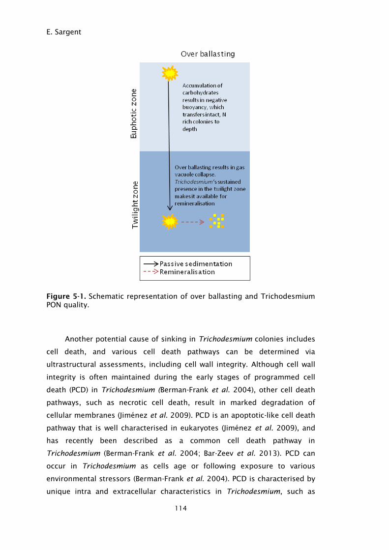
E. Sargent
114
Figure 5-1. Schematic representation of over ballasting and Trichodesmium
PON quality.
Another potential cause of sinking in Trichodesmium colonies includes
cell death, and various cell death pathways can be determined via
ultrastructural assessments, including cell wall integrity. Although cell wall
integrity is often maintained during the early stages of programmed cell
death (PCD) in Trichodesmium (Berman-Frank et al. 2004), other cell death
pathways, such as necrotic cell death, result in marked degradation of
cellular membranes (Jiménez et al. 2009). PCD is an apoptotic-like cell death
pathway that is well characterised in eukaryotes (Jiménez et al. 2009), and
has recently been described as a common cell death pathway in
Trichodesmium (Berman-Frank et al. 2004; Bar-Zeev et al. 2013). PCD can
occur in Trichodesmium as cells age or following exposure to various
environmental stressors (Berman-Frank et al. 2004). PCD is characterised by
unique intra and extracellular characteristics in Trichodesmium, such as

5. Trichodesmium ultrastructure
115
cellular shrinkage and cytoplasmic vacuolisation, which precedes cellular
lysis (Berman-Frank et al. 2004). The presence of these and other
characteristics could indicate PCD as the trigger for sinking of
Trichodesmium colonies.
Results from ultrastructural assessments of Trichodesmium
erythraeum IMS101, positively buoyant surface colonies, and negatively
buoyant colonies collected between 100 and 250 m in the Western Sargasso
Sea suggest that the negatively buoyant Trichodesmium colonies we observe
herein are senescent and may be in the process of PCD. These cells were of
highly vacuolated compared to surface cells, and some also contained
ballasting components. As vacuolisation precedes lysis during PCD in
Trichodesmium (Berman-Frank et al. 2004), the highly vacuolated negatively
buoyant cells we observe imply impending release of fixed N into the water
column. Thus, although these negatively buoyant cells are “element-light”
compared to surface populations due to extensive vacuolisation, any
nitrogen and phosphorus remaining in sinking Trichodesmium, such as the
newly fixed nitrogen in cyanophycin granules, would be available for
remineralisation at depth.
5.3 Methods
5.3.1 Colony collection and culture conditions
Positively buoyant colonies were collected via surface net tows and
negatively buoyant colonies were collected using the Marine Snow Catcher
between 100 and 250 m on the BATS284 research cruise in August 2012 in
the Sargasso Sea. Colonies were picked using a glass Pasteur pipette and
transferred into an individual well of a 24-well culture plate containing 1 mL
of 0.2 µm filtered seawater. Following a sinking rate assessment to confirm
positive/negative buoyancy, colonies were picked from the 24-well culture
plate using an inoculation loop to minimise liquid transfer, and were placed

E. Sargent
116
into 1.5 mL microcentrifuge tubes containing 1mL of 3% glutaraldehyde, 4%
formaldehyde in 14% sucrose 0.1 M PIPES buffer at pH 7.2, hereafter
referred to as ‘main fixative’. Samples were stored at 4 °C until alginate
embedment, postfixation and TEM processing post cruise.
Trichodesmium IMS101 cultures were maintained in YBC-II media at 27
°C on a 12:12 light:dark cycle on an orbital shaker prior to preservation for
TEM. Cells were filtered onto 5 µm polycarbonate filters under low vacuum
pressure. Filters were transferred to 10 mL centrifuge tubes containing
0.5mL of the main fixative. Cells were rinsed off the filter into the main
fixative with 1 mL of the main fixative bringing the sample volume to 1.5
mL. Samples were transferred into 1.5 mL centrifuge tubes and were stored
at 4 °C until alginate embedment, postfixation and TEM processing as
described below.
5.3.2 TEM processing and imaging
The following protocol is an amendment of the official TEM preparation
and processing guidelines provided by the Biomedical Imaging Unit at the
University of Southampton. Trichodesmium samples (colonies and culture
samples) in the main fixative were transferred into 1.5 mL centrifuge tubes
containing 1 drop (approximately 100 µL) of 5% sodium alginate. Each
suspension was then spun at 1250 g for 10 minutes to allow the cells to
embed in the sodium alginate pellet at the bottom of the tube. The
remaining main fixative was decanted and replaced with a 50:50 solution of
6% glutaraldehyde, 8% formaldehyde in 28% sucrose 0.2 M PIPES buffer to
0.1 M calcium chloride. After 10 minutes, the set alginate pellet containing
the Trichodesmium cells was dislodged from the bottom of the
microcentrifuge tube using a Pasteur pipette and was allowed to set for a
further 5 minutes. The pellet and fixative/calcium chloride solution was then
decanted into a TEM processing vial (0.5 mL glass vial), and loaded onto a
rotator. The fixative/calcium chloride solution was aspirated and replaced
with 1 ml of main fixative, and the sample was left to rotate for one hour.
Once the main fixative was aspirated, the pellet was then twice rinsed in 14

5. Trichodesmium ultrastructure
117
% sucrose, 0.1M PIPES buffer, hereafter known as ‘buffer’. The buffer was
then replaced with osmium tetroxide, hereafter known as ‘postfixative’, and
was rotated for one hour. The postfixative was aspirated and the pellet was
then twice rinsed in buffer for ten minutes. The buffer was then replaced by
water, and then immediately replaced by 2% aqueous uranyl acetate. Pellets
were rotated in this solution for 20 minutes before beginning the
dehydration series.
For dehydration, the uranyl acetate was aspirated and replaced with
30% ethanol, which was then replaced by 50% ethanol following a 10 minute
rotation. This was repeated for the transfer from 50% ethanol to 70% ethanol
and the transfer from 70% ethanol to 95% ethanol. The 95% ethanol was
replaced by absolute ethanol and the pellet was rotated for 20 minutes
before refreshing the absolute ethanol and repeating the 20 minute
rotation. The absolute ethanol was then replaced with acetonitrile for a 10
minute rotation before the pellet was left to rotate in a 50:50 solution of
acetonitrile:TAAB resin overnight. The aceonitrile:TAAB resin was then
aspirated and the pellet was rotated in TAAB resin for 6 hours. The pellet
was then embedded in fresh TAAB resin and was polymerised at 60 °C for
20-24 hours.
Polymerised resin blocks were trimmed and mounted into a Reichert
Ultracut E microtome. Ribbons of ~70 nm sections were produced using
glass knives (Figure 5-2), and suitable gold sections were mounted on
copper grids. Grids were then lead stained. As lead stain precipitates in the
presence of CO2
, drops of Reynold’s lead stain were placed on dental wax
inside a petri dish surrounded by pellets of NaOH to minimise CO2
concentrations. Grids were inverted onto the lead stain drops for 5 minutes,
and were then rinsed in distilled water. Grids were then blotted dry and
stored until imaging. Grids were imaged on a Hitachi H7000 Transmission
Electron Microscope and a FEI Technai 12 Transmission Electron Microscope
at the Biomedical Imaging Unit at Southampton General Hospital.
Ultrastructural characteristics were assessed according to the features
described by Siddiqui et al. (1992) and Janson et al. (1995) (Table 5-2), and
were identified in Trichodesmium IMS 101 cultures samples for reference
against in situ samples (Figure 5-3).
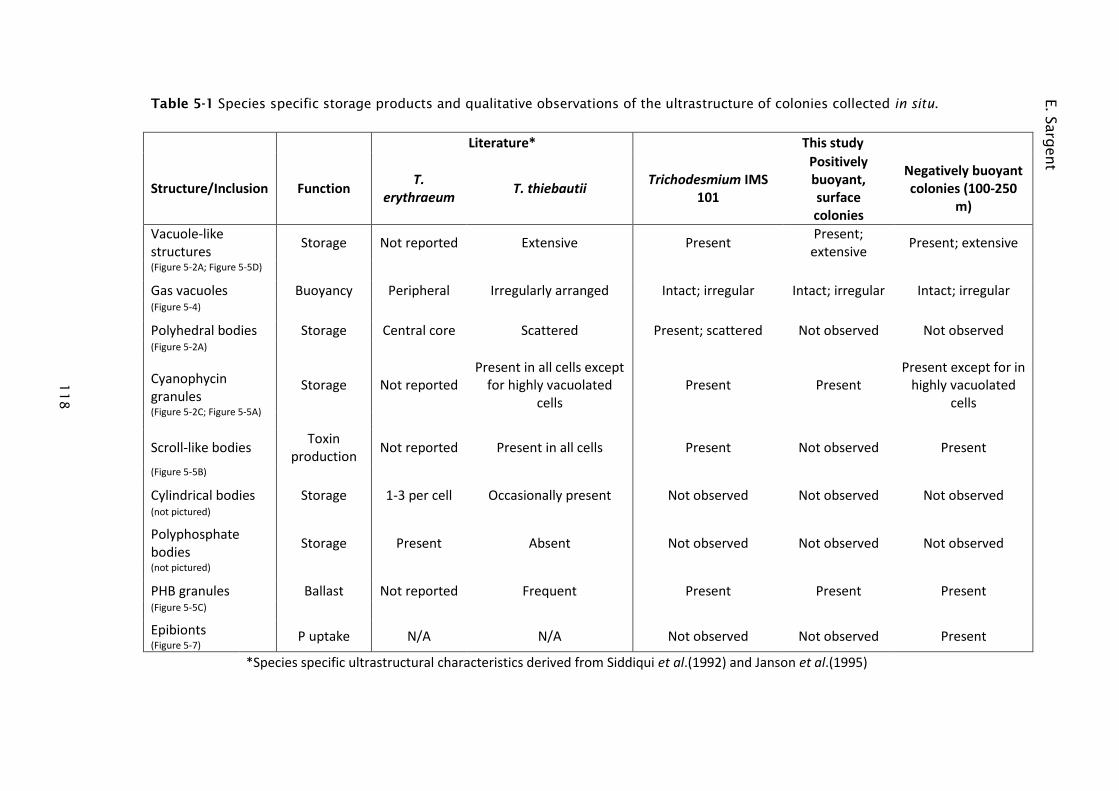
E. Sargent
11
8
Table 5-1 Species specific storage products and qualitative observations of the ultrastructure of colonies collected in situ.
Literature* This study
Structure/Inclusion Function T.
erythraeum T. thiebautii
Trichodesmium IMS 101
Positively buoyant, surface colonies
Negatively buoyant colonies (100-250
m)
Vacuole-like structures
Storage Not reported Extensive Present Present;
extensive Present; extensive
(Figure 5-2A; Figure 5-5D)
Gas vacuoles Buoyancy Peripheral Irregularly arranged Intact; irregular Intact; irregular Intact; irregular (Figure 5-4)
Polyhedral bodies Storage Central core Scattered Present; scattered Not observed Not observed (Figure 5-2A)
Cyanophycin granules (Figure 5-2C; Figure 5-5A)
Storage Not reported Present in all cells except
for highly vacuolated cells
Present Present Present except for in
highly vacuolated cells
Scroll-like bodies Toxin
production Not reported Present in all cells Present Not observed Present
(Figure 5-5B)
Cylindrical bodies Storage 1-3 per cell Occasionally present Not observed Not observed Not observed (not pictured)
Polyphosphate bodies
Storage Present Absent Not observed Not observed Not observed
(not pictured)
PHB granules Ballast Not reported Frequent Present Present Present (Figure 5-5C)
Epibionts (Figure 5-7)
P uptake N/A N/A Not observed Not observed Present
*Species specific ultrastructural characteristics derived from Siddiqui et al.(1992) and Janson et al.(1995)

5. Trichodesmium ultrastructure
119
Figure 5-2. (A) Sectioning of resin block using a glass knife resulting in a ribbon of
sections including gold-coloured sections which are the suitable thickness for TEM
imaging. (B) Ultramicrotome section colour reference chart (Drukker international).
Figure 5-3. Transmission electron micrographs of Trichodesmium erythraeum IMS
101cells showing various ultrastructural components and variable integrity: vacuoles
(V), intercellular space (IS), outer membrane (OM), plasma-membrane (PM), nitrogen
storage vacuoles known as cyanophycin granules (CG), carboxysomes (C), gas vesicles
(GV), and vacuolisation (VS), which were used for reference in subsequent identification
of ultrastructural components of in situ samples. Scale bars represent 1 µm in panels A
and D, 100 nm in panel B, and 500 nm in panel C.

E. Sargent
120
5.4 Results
5.4.1 Gas vesicles
Gas vesicles were present in all cells viewed (n=145), and were intact in
cells from the positively buoyant surface colonies (Figure 5-4A,B). However, gas
vesicles were not fully intact in the cells of the negatively buoyant colonies
(n=113). In negatively buoyant colonies, some cells contained both intact and
degraded gas vesicles (Figure 5-4C-F). ) Intact gas vesicles were indicated by
regular arrangement of the electron dense fibres within the gas vacuole (Figure
5-5A) and the clear definition of the electron transparent center regions of
each gas vesicle (Figure 5-4B). Partially collapsed and degrading gas vesicles
were indicated by uneven distribution of electron dense fibres within the gas
vacuole and reduction in marked circular vesicle walls (Figure 5-5C). Collapsed
gas vesicles were indicated by a complete lack of structural regularity within
the gas vacuoles, as well as a lack of electron transparent regions (Figure 5-5F).
Gas vesicle distribution varied between samples. Gas vesicles were occasionally
distributed around the periphery of the cell as is consistent with the
Trichodesmium species T. erythraeum, but were more often irregularly
arranged within the cell as is consistent with T. thiebautii.
5.4.2 Storage products
Trichodesmium cells contained an assortment of storage products (Figure
5-5). Positively buoyant surface cells (Figure 5-5A, B) and Trichodesmium
IMS101 (Figure 5-3C) contained cyanophycin granules in every cell assessed,
while only 27% of negatively buoyant cells assessed contained these structures,
which known to be involved in nitrogen storage (Finzi-Hart et al. 2009) (Figure
5-5C). 0.9% of negatively buoyant cells contained multimembranous scroll-like
structures known to be involved in toxin production (present in T. thiebautii

5. Trichodesmium ultrastructure
121
only) (Figure 5-5D), which were not present in positively buoyant surface cells
or Trichodesmium IMS101. 33% of negatively buoyant cells contained poly-β-
hydroxybutyric acid (PHB) granules which are a suspected ballast component
(Figure 5-5E). PHB granules were also observed in positively buoyant surface
cells and Trichodesmium IMS101 (not pictured). 5.3% of negatively buoyant
cells contained vacuole-like structures suspected to be involved in the storage
of photosynthetic metabolites (Figure 5-5F), which were also observed in
positively buoyant surface cells (Figure 5-5B) and Trichodesmium IMS101
(Figure 5-3A).
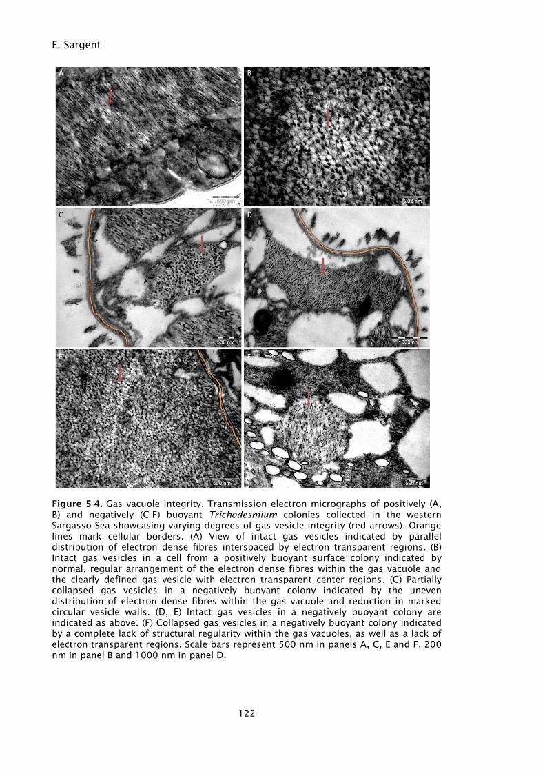
E. Sargent
122
Figure 5-4. Gas vacuole integrity. Transmission electron micrographs of positively (A,
B) and negatively (C-F) buoyant Trichodesmium colonies collected in the western
Sargasso Sea showcasing varying degrees of gas vesicle integrity (red arrows). Orange
lines mark cellular borders. (A) View of intact gas vesicles indicated by parallel
distribution of electron dense fibres interspaced by electron transparent regions. (B)
Intact gas vesicles in a cell from a positively buoyant surface colony indicated by
normal, regular arrangement of the electron dense fibres within the gas vacuole and
the clearly defined gas vesicle with electron transparent center regions. (C) Partially
collapsed gas vesicles in a negatively buoyant colony indicated by the uneven
distribution of electron dense fibres within the gas vacuole and reduction in marked
circular vesicle walls. (D, E) Intact gas vesicles in a negatively buoyant colony are
indicated as above. (F) Collapsed gas vesicles in a negatively buoyant colony indicated
by a complete lack of structural regularity within the gas vacuoles, as well as a lack of
electron transparent regions. Scale bars represent 500 nm in panels A, C, E and F, 200
nm in panel B and 1000 nm in panel D.

5. Trichodesmium ultrastructure
123
Figure 5-5. Intracellular storage components. Transmission electron micrographs of
positively (A, B) and negatively (C - F) buoyant Trichodesmium colonies collected in the
western Sargasso Sea. Orange lines mark cellular borders. Various storage components
are indicated by red and orange arrows, such as (A, B, C) cyanophycin granules known
to be involved in nitrogen storage, (D) scroll-like bodies associated with toxin
production, (E) poly-β-hydroxybutyric acid granules theorised to be involved in
carbohydrate ballasting, (B, F, orange arrow) vacuole-like structures suspected to store
photosynthetic metabolites, and (F, blue arrows) medium electron dense granules
assumed to serve a short-term storage role and are important in nitrogen metabolism
function. Scale bars represent 5000 nm in panel A, 3000 nm in panel B, 2000 nm in
panels C and F, 500 nm in panel D, and 1000 nm in panel E.

E. Sargent
124
5.4.3 Cellular integrity
Trichodesmium IMS 101 samples and positively buoyant surface colonies
contained a mixture of intact and vacuolated cells (Figure 5-3; Figure 5-6A, B).
All negatively buoyant cells were heavily vacuolated (Figure 5-6C - F).
Additionally all cells from negatively buoyant colonies suffered from minimal to
extensive cellular shrinkage indicated by the irregular shape of cellular
perimeters (Figure 5-6D, E). This cellular shrinkage is not an artefact of TEM
preparation as osmolarity was carefully controlled during processing to ensure
hypoosmotic and hyperosmotic conditions were not encountered. Reductions
in intracellular density may be indicative of senescence, but some storage
bodies are still present despite vacuolisation, such as PHB granules (Figure 5-
6E, red arrows) and cyanophycin granules (Figure 5-6F, red arrow).
5.4.4 Epibionts
Epibionts were not observed around cells from Trichodesmium IMS101 cultures
(Figure 5-3) or on positively buoyant surface colonies (Figure 5-4A, 5-5A, 5-6A).
Epibionts extensively colonised the mucilaginous sheath of 2 negatively
buoyant colonies (19% of analysed cells from negatively buoyant colonies)
(Figure 5-7); all of the colonised Trichodesmium cells also contained the
ballasting component PHB granules (Figure 5-5E), and nearly half (11% of total)
contained another ballasting component, polyglucoside granules (Figure 5-8D).
Epibiont distribution spanned the entire perimeter of these Trichodesmium
cells (Figure 5-7A, B, C). Individual epibionts were tightly packed, irregularly
arranged along the perimeter, and were occasionally overlapping (Figure 5-7D).
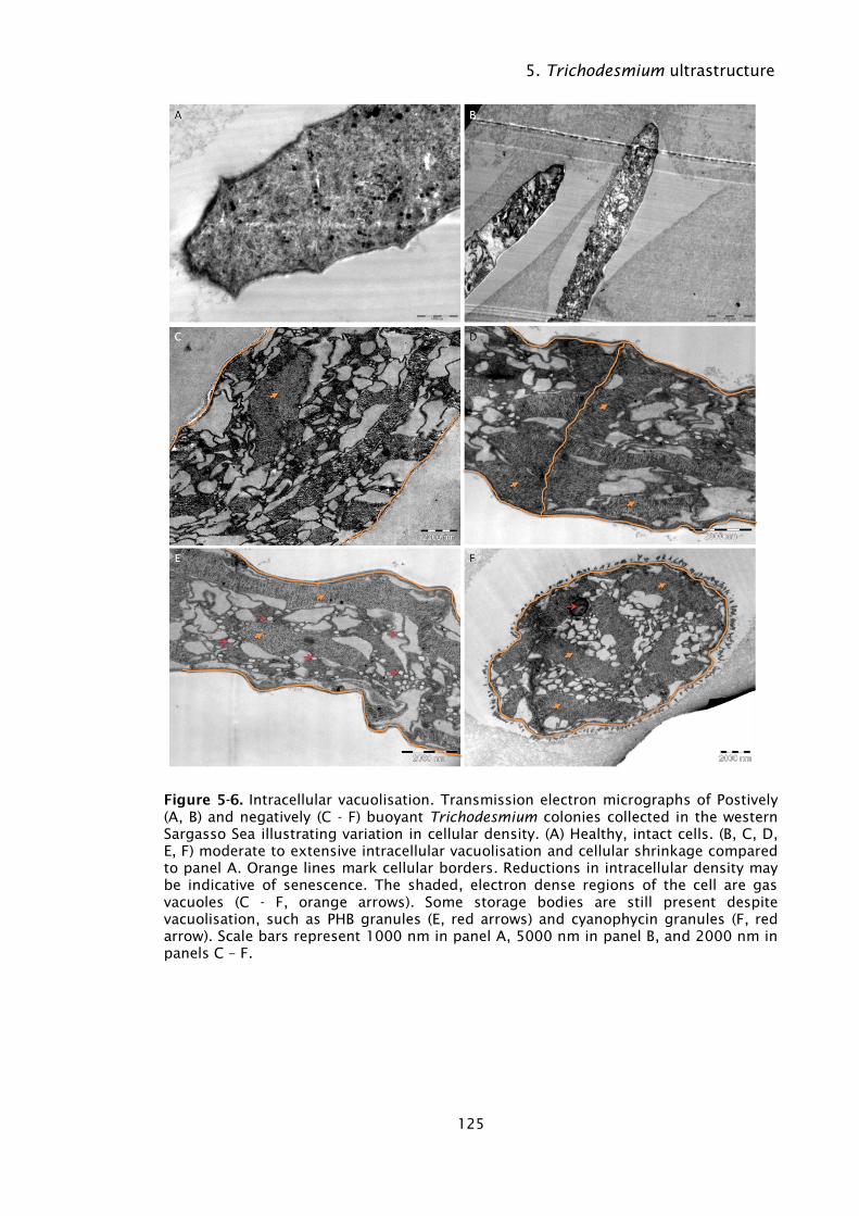
5. Trichodesmium ultrastructure
125
Figure 5-6. Intracellular vacuolisation. Transmission electron micrographs of Postively
(A, B) and negatively (C - F) buoyant Trichodesmium colonies collected in the western
Sargasso Sea illustrating variation in cellular density. (A) Healthy, intact cells. (B, C, D,
E, F) moderate to extensive intracellular vacuolisation and cellular shrinkage compared
to panel A. Orange lines mark cellular borders. Reductions in intracellular density may
be indicative of senescence. The shaded, electron dense regions of the cell are gas
vacuoles (C - F, orange arrows). Some storage bodies are still present despite
vacuolisation, such as PHB granules (E, red arrows) and cyanophycin granules (F, red
arrow). Scale bars represent 1000 nm in panel A, 5000 nm in panel B, and 2000 nm in
panels C – F.

E. Sargent
126
Figure 5-7. Epibionts. Transmission electron micrographs of cells from negatively
buoyant Trichodesmium colonies collected in the western Sargasso Sea showcasing
extensive colonisation of the mucilaginous sheath by epibionts indicated by red
arrows. Orange lines mark cellular borders. Epibiont distribution appears to span the
entire perimeter of the Trichodesmium cell (A, B, C). Individual epibionts are tightly
packed, irregularly arranged along the perimeter, and are occasionally overlapping (D).
Scale bars represent 2 µm in panels A and B, 5 µm in panel C, and 1 µm in panel C.
Epibionts were not observed around cells from positively buoyant surface colonies
(Figure 5-4A, 5-5A, 5-6A).
5.4.5 Other cellular inclusions
One cell in a negatively buoyant colony contained distinct thylakoid
lamellae (Figure 5-8A). All of the cells containing epibionts (19%) were also
surrounded by an extracellular matrix that was not detectable in any of the
other negatively buoyant samples (Figure 5-8B). Polyglucoside granules, which
are present only in T. thiebautii and are theorised to be involved in carbon
storage and ballasting, were also present in 11% of negatively buoyant cells. In
one negatively buoyant colony, multiple extracellular bodies were observed
(Figure 5-8D).

5. Trichodesmium ultrastructure
127
Figure 5-8. Other inclusions of note. Transmission electron micrographs of cells from
negatively buoyant Trichodesmium colonies collected in the western Sargasso Sea.
Orange lines mark cellular borders. Various other inclusions encountered in the
samples are indicated by red arrows, such as (A) thylakoid lamellae, (B) the
extracellular matrix, (C) polyglucoside granules, which are theorised to be involved in
ballasting, and (D) extracellular bodies, which may indicate the presence of
compromised cells within the colony although we cannot rule out that they may simply
be from associated organisms living in/on the colony. Scale bars represent 500 nm in
panels A, B, and C, and 1000 nm in panel D.
5.5 Discussion
Poly-β-hydroxybutyric acid (PHB) and polyglucoside granules may act as
part of the ballast required for vertical migration in Trichodesmium (Romans
1994). The presence of PHB granules in 33% of cells in negatively buoyant
Trichodesmium colonies, as well as the presence of polyglucoside granules in
11% of cells, offers some support to the over ballasting for vertical migration
theory postulated in Chapter 3. However, in order to thoroughly assess
whether vertical migration is a likely mechanism by which Trichodesmium is
escaping the euphotic zone, we must also consider other conditions that could
E. Sargent
128
cause Trichodesmium to sink, such as programmed cell death (PCD) or
necrosis, which could result in passive sedimentation during senescence or
following cell death (Bar-Zeev et al. 2013) or viral infection and eventual viral
lysis, a known contributor to Trichodesmium bloom demise (Hewson et al.
2004; Brown et al. 2013).
PCD can occur with ageing or following prolonged exposure to high
irradiance, oxidative stress, and nutrient starvation in Trichodesmium (Berman-
Frank et al. 2004). Osmotic shock, UV shock, and heat shock, are also known
to stimulate PCD in other organisms (Jiménez et al. 2009). Of these we assume
Trichodesmium wouldn’t be normally exposed to osmotic or heat shock in the
environment, but could plausibly suffer from nutrient starvation and
senescence in a number of situations including during blooms. Additionally
Trichodesmium could experience prolonged exposure to high irradiance
following entrapment at the surface due to uncompensatable buoyancy
(Berman-Frank et al. 2004). This condition is often encountered in buoyancy
models where the organism is unable to produce enough ballast to overcome
the positive buoyancy provided by gas vacuoles (Kromkamp and Walsby 1992;
White et al. 2006). In such situations, long-standing exposure to high
irradiance may then result in prolonged photoinhibition (Lewis et al. 1988;
Kromkamp and Walsby 1992), which could render cells unable to produce the
ballast necessary to escape the surface layer, eventually resulting in sinking
and cell death (Berman-Frank et al. 2004).
As 67% of the cells from negatively buoyant colonies lacked known ballast
components, and as viruses were not observed in these samples, cell death
may be the cause of sinking in these colonies. Since the colonies assessed
were not associated with surface blooms, we assume high irradiance was not a
trigger of PCD in these samples. Instead, we hypothesis that senescence during
the stationary growth phase preceded cell death initiation in these samples. In
Trichodesmium cell death is characterised by unique intra and extracellular
characteristics, such as cellular shrinkage (Figure 5-6), cytoplasmic
vacuolisation (Figure 5-6), and degradation of other intracellular components
such as thylakoids (only observed in one cell from a negatively buoyant
colony), carboxysomes (not observed in the negatively buoyant cells assessed),
gas vesicles (Figure 5-4), and cyanophycin granules (absent in over 70% of
negatively buoyant cells assessed) (Berman-Frank et al. 2004). We observed

5. Trichodesmium ultrastructure
129
extensive vacuolisation in these samples, and with the exception of one cell
that contained thylakoids, all observed cells from negatively buoyant colonies
lacked thylakoids and carboxysomes, and contained degraded/collapsed gas
vesicles. All negatively buoyant cells assessed also displayed signs of cellular
shrinkage. Taken together these characteristics offer support to cell death as
an explanation for sinking in these colonies. There is also evidence that
although compromised cell membrane integrity, sometimes including leakage
of cellular material, occurs during PCD in other organisms (Jiménez et al. 2009;
Zheng et al. 2013), PCD in Trichodesmium may not immediately present these
characteristics as the plasmamembrane can remain intact following extensive
intracellular vacuolisation (Berman-Frank et al. 2004).
If senescence and PCD are responsible for sinking in this study, it remains
to be assessed whether senescence commenced prior to Trichodesmium
exiting the euphotic zone. If so, Trichodesmium would transport much less
new nitrogen to depth if biomass was lost in the upper ocean. Thus, the
intracellular integrity of negatively buoyant Trichodesmium cells within the
upper 100 m requires further investigation. In light of this, Trichodesmium
contribution to PON standing stock may be much less than previously
calculated (Chapter 3). On the other hand, as 33% of the assessed cells from
negatively buoyant colonies contained ballasting materials, these colonies may
have become senescent following their exit from the upper ocean. In these
colonies, there remains the potential for the exudation of intracellular biomass
to occur below 100 m.
The presence of cyanophycin granules in over a quarter of cells from
negatively buoyant colonies (27.4%) also warrants further assessment when
considering new-nitrogen fate and Trichodesmium taxonomy. Cyanophycin
granules are a nitrogen storage structure in cyanobacteria (Siddiqui et al.
1992), and although the cells from negatively buoyant colonies were highly
vacuolated, they were transporting concentrated new nitrogen stores to depth.
Cyanophycin granules were originally thought to be absent in T.
erythraeum (Siddiqui et al. 1992) and in highly vacuolated T. thiebautii cells (Li
and Lee 1990), but have since been described in T. erythraeum (Janson et al.
1995). Due to this duality we cannot use the presence/absence of these
granules as a taxonomic marker, but the absence of cyanophycin granules in

E. Sargent
130
the majority of cells coupled with cellular vacuolisation again hints at a late
growth phase regardless of species type. The loss of cyanophycin granules
during the stationary growth phase in other cyanobacteria has been previously
observed (Allen 1984), and vacuolisation may be representative of the cell
density decrease consistent with cellular senescence in other organisms
(Jiménez et al. 2009). Thus, cellular senescence may play a more important
role than over ballasting in the sinking/settling of these Trichodesmium
colonies.
Other storage products present in the negatively buoyant colonies are
able to provide some taxonomic insight. The multimembranous scroll-like
structures (Figure 5-5B) are known to be involved in toxin production and are
so far described only in T. thiebautii (Siddiqui 1992; Janson et al. 1995). The
vacuole-like structures observed in some of the negatively buoyant colonies
(Figure 5-5D) are unique to the vacuolisation process previously described and
serve a suspected photosynthetic metabolites storage role. Vacuole-like
structures are described as extensive in T. thiebautii, but are not reported for
T. erythraeum in the literature (Siddiqui 1992). Polyphosphate bodies were not
observed in any of these collections, but are only described for T. erythraeum.
Medium electron dense granules were also not observed in the negatively
buoyant Trichodesmium samples. They are thought to serve a nitrogen
metabolism function and are described for T. thiebautii only (Li and Lee 1990).
Taking all of these factors into consideration, we tentatively assume the
majority of these colonies are T. thiebautii which is consistent with
observations that T. thiebautii is the dominant Trichodesmium species in the
Atlantic Ocean (O’Neil 1998; Carpenter et al. 2004; Capone et al. 2005). T.
thiebautii has higher carbon content, and thus higher nitrogen content, per cell
than T. erythraeum (120 ± 57 pgC cell-1
and 65 ± 32 pgC cell-1
respectively)
(Luo et al. 2012), which again supports the potential for substantial
Trichodesmium-attributable N availability at depth even in highly vacuolated
cells.

5. Trichodesmium ultrastructure
131
5.6 Conclusion
When PCD was induced in the laboratory, cellular lysis has been shown to
occur within 24 hours following the onset of decreased intracellular density
(i.e. extensive vacuolisation) (Berman-Frank et al. 2004). Potentially then, with
an average colonial sinking rates of 62.1 ± 39.7 m d-1
(Chapter 3), and
depending on the depth of PCD onset, all negatively buoyant Trichodesmium
observed in this study would have apoptosed within hours. During apoptosis
the transfer of Trichodesmium biomass from the fast sinking pool of POM to
the suspended pool would occur nearly instantaneously. A constant rain of
senescent Trichodesmium colonies destined to burst in the upper twilight zone
would result in the injection of Trichodesmium-attributable PON directly into
the water column thus making it readily available for remineralisation and
contribution to enhanced N*. Further assessments of the ultrastructure and/or
elemental composition of negatively buoyant Trichodesmium colonies would
aid in constraining the estimations of Trichodesmium contribution to PON
standing stock, and would be integral in expanding the understanding of
Trichodesmium contribution to enhanced N* in the thermocline of the sub-
tropical North Atlantic gyre.

E. Sargent
132
6. Future Directions
133
6. Conclusions and Future Directions
6.1 Summary and main findings
The role that diazotrophs play in marine nitrogen dynamics is well
described in the surface ocean, but species-specific fates are still not
completely defined. This work provides novel insights into the potential for
these organisms to contribute to the anomalous nutrient distributions in the
thermocline of the subtropical Atlantic Ocean despite previously reports that
diazotrophic growth fuels export production rather than remineralisation
(Michaels and Silver 1988) and that unicellular and filamentous diazotroph-
derived N is likely constrained to cycling in the surface ocean (Thompson and
Zehr 2013). This work has also revealed that previous assumptions on the
mechanisms by which Trichodesmium exits the euphotic zone being limited to
incorporation in faecal material (Mulholland 2007), active transport (Guidi et al.
2012), or sweeping mechanisms (Scharek et al. 1999b) also require
redefinition. We can confirm that Trichodesmium is incorporated into faecal
material and is involved in sweeping mechanisms by incorporation into
aggregates (Chapter 3), but this organism also exits the euphotic zone via free
sinking of potentially senescent colonies (Chapters 3 and 5). The presence of
nifH from the unicellular and heterocystous diazotrophs below the euphotic
zone (Chapter 2) also implies additional mechanisms of transport exist for
these organisms, but further investigation specifically into these groups would
be beneficial in better defining their fate.
This final chapter is a synthesis of Chapters 2-5 and will discuss the
implications of the findings of each previous chapter in relation to the findings
of the other chapters. Limitations of this study and recommendations for
future work will also be discussed.

E. Sargent
134
6.1.1 Implications of polyploidy in Trichodesmium
The assessments of the nifH community carried out in Chapter 2 revealed
Trichodesmium-specific nifH to be dominant in many of the samples, such as
at the surface between the equator and 10°N on the D361 transect. As we now
know that gene copies exceed cell counts by two orders of magnitude on this
cruise and that Trichodesmium is potentially polyploid (Chapter 4), these
conclusions may not reflect the actual diazotrophic community structure in this
region.
The maximal discrepancy between cell counts and gene copies on D361
resulted in a theoretical ploidy level of 120 genome copies per cell for
Trichodesmium. If this upper threshold of ploidy is applied to the dataset, and
Trichodesmium-specific nifH is reduced by a factor of 120 to bring nifH copies
in the theoretical range of cell numbers, Trichodesmium-specific nifH would no
longer be the largest component of the nifH community structure between the
equator and 10°N. Instead, it would only composes the majority of nifH
community structure at station 16 (8°20 N), and other phylotypes could
increase in contribution, such as the heterocystous Richelia-Hemaulus (Figure
6-1).
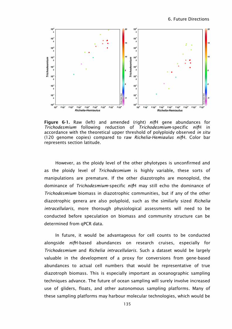
6. Future Directions
135
Figure 6-1. Raw (left) and amended (right) nifH gene abundances for
Trichodesmium following reduction of Trichodesmium-specific nifH in
accordance with the theoretical upper threshold of polyploidy observed in situ
(120 genome copies) compared to raw Richelia-Hemiaulus nifH. Color bar
represents section latitude.
However, as the ploidy level of the other phylotypes is unconfirmed and
as the ploidy level of Trichodesmium is highly variable, these sorts of
manipulations are premature. If the other diazotrophs are monoploid, the
dominance of Trichodesmium-specific nifH may still echo the dominance of
Trichodesmium biomass in diazotrophic communities, but if any of the other
diazotrophic genera are also polyploid, such as the similarly sized Richelia
intracellularis, more thorough physiological assessments will need to be
conducted before speculation on biomass and community structure can be
determined from qPCR data.
In future, it would be advantageous for cell counts to be conducted
alongside nifH-based abundances on research cruises, especially for
Trichodesmium and Richelia intracellularis. Such a dataset would be largely
valuable in the development of a proxy for conversions from gene-based
abundances to actual cell numbers that would be representative of true
diazotroph biomass. This is especially important as oceanographic sampling
techniques advance. The future of ocean sampling will surely involve increased
use of gliders, floats, and other autonomous sampling platforms. Many of
these sampling platforms may harbour molecular technologies, which would be
E. Sargent
136
beneficial in increasing gene-based abundance datasets, but inadequate for
biomass estimates if accurate conversion factors are not available.
6.1.2 Refining diazotroph contribution to N* and Trichodesmium
contribution to PON export
When comparing diazotroph abundance and distribution with N*, there is
a clear trend observed between high nifH abundances in the surface and
enhanced N* at depth, which implies diazotrophs likely influence enhanced
nitrate below the mixed layer (Figure 6-2). This is supported by the isotopic
signature of the nitrate pool in the subtropical North Atlantic thermocline,
which indicates that newly fixed nitrogen is likely contributing to the N*
anomaly (Knapp et al. 2008). Maximum N* anomalies in the North Atlantic gyre
are associated with a potential density interval in the = 26.1-27.1 range
(Gruber and Sarmiento 1997, Hansell et al. 2007), which was located between
100 and 500 m along the transect in this study. We hypothesize that sinking
diazotrophs are being remineralised shortly after their exit from the mixed
layer, relatively rapidly, consequently depositing their N-rich cellular contents
into the water column between 100 and 500 m, which provides a mechanism
for generating some of the N* anomaly observed at depth (Figure 6-2). This is
consistent with the accumulation of the N* signal only occurring in regions
where these isopycnals are shallower than ~400m (Hansell et al. 2004), where
release of diazotroph-derived material between 100 and 500 m could inject
into this density layer following the shoaling of the permanent thermocline at
low latitudes near the equator (Sarmiento et al. 2004). The dominant
diazotrophs north of the equator, Trichodesmium and unicellular Group A
cyanobacteria, may be larger contributors to the N* anomaly than R.
intracellularis, which may not contribute as readily to the N* anomaly as its
host diatoms are more apt to aggregate and sink too quickly for
remineralisation in the upper twilight zone (Alldredge and Gotschalk 1988,
Villareal et al. 2011). However, the contribution individual diazotrophs make to
N* remains unresolved.

6. Future Directions
137
Figure 6-2. Log nifH abundance and N* with potential density (σθ) contours
highlighting enhanced N* within the σθ=26.6-27.0 range along the transect
corresponding to increased nifH abundance in the same depth range. Note slight
difference in scale of the y axis for each panel.
Trichodesmium colonies collected on the D361 cruise were exiting the
mixed layer via passive sedimentation, theorised to be due to over-ballasted
vertical migration attempts. These negatively buoyant colonies were present as
deep as 300 m (Chapter 3), but the cellular integrity of these colonies was
unknown. Ultrastructural assessments of negatively buoyant colonies collected
on the BATS284 cruise between 100 and 250 m revealed the presence of
ballasting components in 33% of cells assessed, thus confirming the potential
for over ballasting to transport some Trichodesmium colonies to depth, but the
majority of negatively buoyant cells (the remaining 77%) did not contain
ballasting components, and were instead highly vacuolated (100% of negatively
buoyant cells assessed), which is indicative of a programmed cell death (PCD)
pathway (Chapter 5). If the PCD pathway is initiated while colonies are still in

E. Sargent
138
the surface layer, then the exudation of intracellular material would result in
the loss of Trichodesmium-derived biomass to the upper ocean. Conversely, if
the PCD pathway is initiated below the mixed layer, the loss of intracellular
material would still be available for remineralisation and contribution to N*.
This would also be the case for intact colonies exiting the mixed layer via the
over ballasting mechanism. These various passive sedimentation and
remineralisation pathways for Trichodesmium colonies are illustrated in Figure
6-3.
Figure 6-3. Schematic representation of potential passive sedimentation and
remineralisation pathways for Trichodesmium colonies. The orange colony border
represents intact cell membranes and yellow colony interior represents non-vacuolated
cells. Exudation of intracellular material and subsequent remineralisation are
represented by dashed arrows as indicated above.
Due to the potential range of fates for Trichodesmium-derived material,
such as surface cycling of Trichodesmium-derived biomass or export and
remineralisation of Trichodesmium biomass below the euphotic zone, it is
difficult to quantify the contribution of Trichodesmium to PON export at the
present stage. If surface initiated PCD is responsible for the presence of some

6. Future Directions
139
colonies at depth, then previous estimates of Trichodesmium contribution to
PON standing stock described in Chapter 3 would decrease due to a
combination of full contribution of biomass by ballasted cells and a decreased
contribution of biomass by the cells involved in the PCD pathway. This would
decrease Trichodesmium contribution to PON standing stock at 10 m below the
mixed layer, which would also decrease Trichodesmium derived export fluxes.
However, over a quarter of the cells assessed from negatively buoyant
colonies contained multiple cyanophycin granules in each cell, which are a
known nitrogen storage structure (Siddiqui et al. 1992). Although these cells
were highly vacuolated, they contained these concentrated new nitrogen
stores. Thus, the amended estimates of Trichodesmium contribution to PON
standing stock should be considered conservative, as cellular nitrogen content
was derived from a 6.3 conversion of C:N in Trichodesmium (Mulholland 2007)
in Chapter 3, and the maintenance of nitrogen stores during cell density loss in
these cells would likely skew that ratio to a higher N content. Additional
assessments of cellular integrity in sinking Trichodesmium, such as cell death
stains during cell counts or caspase activity assays, would aid in a better
definition of Trichodesmium contribution to PON export.
Additionally, as the PCD pathway eventually results in cellular lysis
(Berman-Frank et al. 2004), much more recently apoptosed Trichodesmium-
derived material may be present in the water column below the mixed layer
than is detectable optically. Similarly, the degradation of Trichodesmium-
derived biomass present in aggregates and faecal material may also contribute
more to Trichodesmium-derived PON at depth than we are able to quantify
optically. This is particularly apparent after assessments of aggregates using
standard light microscopy versus epifluorescent microscopy where recently
degraded trichomes are only revealed when the phycoerythrin pigment is
excited (Figure 6-4). Unfortunately, this assessment was not available on the
D361 cruise, so estimated Trichodesmium contribution to PON and PON fluxes
should be considered conservative as biomass may have been missed due to
methodological limitations.
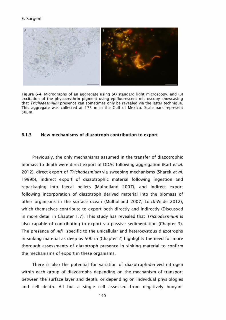
E. Sargent
140
Figure 6-4. Micrographs of an aggregate using (A) standard light microscopy, and (B)
excitation of the phycoerythrin pigment using epifluorescent microscopy showcasing
that Trichodesmium presence can sometimes only be revealed via the latter technique.
This aggregate was collected at 175 m in the Gulf of Mexico. Scale bars represent
50µm.
6.1.3 New mechanisms of diazotroph contribution to export
Previously, the only mechanisms assumed in the transfer of diazotrophic
biomass to depth were direct export of DDAs following aggregation (Karl et al.
2012), direct export of Trichodesmium via sweeping mechanisms (Sharek et al.
1999b), indirect export of diazotrophic material following ingestion and
repackaging into faecal pellets (Mulholland 2007), and indirect export
following incorporation of diazotroph derived material into the biomass of
other organisms in the surface ocean (Mulholland 2007; Loick-Wilde 2012),
which themselves contribute to export both directly and indirectly (Discussed
in more detail in Chapter 1.7). This study has revealed that Trichodesmium is
also capable of contributing to export via passive sedimentation (Chapter 3).
The presence of nifH specific to the unicellular and heterocystous diazotrophs
in sinking material as deep as 500 m (Chapter 2) highlights the need for more
thorough assessments of diazotroph presence in sinking material to confirm
the mechanisms of export in these organisms.
There is also the potential for variation of diazotroph-derived nitrogen
within each group of diazotrophs depending on the mechanism of transport
between the surface layer and depth, or depending on individual physiologies
and cell death. All but a single cell assessed from negatively buoyant
6. Future Directions
141
Trichodesmium colonies seemingly suffering from PCD in this study lacked
thylakoids (Chapter 5), whereas chlorophyll and other pigments were
maintained in sinking Trichodesmium which was incorporated in aggregates
and faecal pellets (Chapter 3; Figure 6-4). Thus, there may also be variation in
the elemental composition of Trichodesmium cells depending on the
mechanism of export. The potential for plasticity in Trichodesmium-derived
POC and PON under these different conditions increases the difficulty of
quantifying their contribution to export fluxes.
6.2 Applications of this work
It was previously assumed that nitrogen fixation was contributing to
enhanced N* in the thermocline of the subtropical North Atlantic Ocean
(Hansell et al. 2004, 2007), but mechanisms to describe the transfer of newly
fixed nitrogen to depth were lacking. This work reveals those mechanisms and
allows for redefinition of the fate of diazotrophic organisms for incorporation
into future biogeochemical models and estimations of their importance and
function in the marine nitrogen cycle. This work additionally provides
motivation for further investigation of the fate of diazotrophs in other ocean
regions.
This work also highlights a physiological trait previously undescribed in
Trichodesmium, polyploidy. This work warrants redefinition of the applications
of the qPCR method for assessing diazotrophic abundance. This method still
proves valuable in assessments of biogeography and nifH community
structure, but the potential for polyploidy in Trichodesmium and other
diazotrophs introduces difficulties in the extension of this method to use in
biomass estimates. These findings also provide a stimulus for ploidy
assessments in the other diazotrophic cyanobacterial groups as well as
continued assessments of the conditions under which the ploidy level of
Trichodesmium varies.

E. Sargent
142
6.3 Revisiting Hypotheses
6.3.1 Hypotheses
H1
: Trichodesmium is not a likely constituent of export
i. Conclusions: H1
partially rejected. Previous assessments of
Trichodesmium have overlooked this organism’s potential
to sink out of the mixed layer, but it’s contribution to PON
flux appears to be minimal.
H2:
Remineralisation of Trichodesmium between the mixed layer and
500 m, north of the equator in the Atlantic, is contributing to the N*
anomaly seen in the thermocline of the northern subtropical gyre,
while DDA export likely results in sequestration of carbon and
nitrogen rather than remineralisation.
i. Conclusions: H2
partially accepted. While significant loss of
Trichodesmium biomass between the mixed layer and 500
m suggests the organism is being remineralised within this
depth range, and thus is contributing to N*, we have yet to
quantify this contribution. Our assessments of DDA fate
are inconclusive as we encountered them far less often
than anticipated.
H3
: There are multiple mechanisms by which diazotroph export
occurs
i. Conclusions: H3
accepted. It appears a variety of
mechanism transport diazotrophs to depth including
sweeping, grazing, free sinking, and active transport.

6. Future Directions
143
H4
: Over ballasting of Trichodesmium in the surface layer is causing
it to sink past phosphocline and terminating the ability for the
colony to return to the surface.
i. Conclusions: H4
inconclusive: although there was evidence
of ballasting components in negatively buoyant
Trichodesmium colonies, it appears PCD may play a larger
role in facilitating the transfer of Trichodesmium biomass
to depth
H5
: If over ballasting is the mechanism by which Trichodesmium is
exported, the intracellular POC/PON is high quality.
i. Conclusions: H5
rejected: negatively buoyant
Trichodesmium colonies contained highly vacuolated cells,
and thus a smaller, more degraded quantity of POC/PON than
would be present in a cell that was sinking solely due to
ballasting.
H6
: Gas vacuole collapse in colonies collected between 100 and 300
m guarantees sinking Trichodesmium at these depths lacks the
ability to return to the surface.
i. Conclusions: H6
partially accepted. Although gas vacuoles
were degraded and collapsed in negatively buoyant colonies,
we cannot be sure whether this is due to pressure or PCD
associated breakdown. Regardless, negative buoyancy would
be irreversible in either case.
H7
: Quantification of the nifH gene via qPCR can be directly related to
cell number.
i. Conclusions: H7 rejected. There is a large discrepancy
between cell counts and gene copy numbers in
Trichodesmium. This discrepancy has yet to be confirmed with
other phylotypes.

E. Sargent
144
6.4 Final remarks
Contrary to previous expectations molecular assessments revealing the
presence of all three groups of diazotrophs below the euphotic zone (Chapter
2) and optical confirmation of the conspicuous diazotrophs present in these
depth ranges (Chapter 3) reveal that previous assessments of the fate of
diazotrophs have underestimated their ability to exit the euphotic zone.
Marine diazotrophs play an important role in marine biogeochemical cycles
by fixing N2
into bioavailable forms. Elucidation of the extent to which these
diazotrophs are contributing to the biogeochemistry of deeper waters provides
novel insight into the cycling of fixed nitrogen in the oligotrophic ocean thus
highlighting the importance of these organisms in both the supply of nutrients
to the thermocline and in the sustenance of ocean productivity on longer time
scales.

Appendices
145
Appendices

E. Sargent
146

Appendix 1
147
Appendix 1
Table A1. SAPS deployment details
Station Lat Lon Depth
(m)
Size
fraction
(µm)
Deployment
time
(minutes)
Total volume
filtered (L)
10 07°13 S 25°00 W 70 53 137 2066
10 07°13 S 25°00 W 70 1 137 2066
10 07°13 S 25°00 W 170 53 140 2343
10 07°13 S 25°00 W 170 1 140 2343
10 07°13 S 25°00 W 500 53 137 2174
13 07°13 S 25°00 W 70 53 138 1915
13 01°09 N 26°02 W 70 1 138 1915
13 01°09 N 26°02 W 170 53 138 2041
13 01°09 N 26°02 W 170 1 138 2041
13 01°09 N 26°02 W 500 53 133 3419
16 08°20 N 28°20 W 70 53 138 2024
16 08°20 N 28°20 W 70 1 138 2024
16 08°20 N 28°20 W 170 53 138 2053
16 08°20 N 28°20 W 170 1 138 2053
16 08°20 N 28°20 W 500 53 142 2148
19 15°30 N 28°23 W 85 53 138 2025
19 15°30 N 28°23 W 85 1 138 2025
19 15°30 N 28°23 W 170 53 138 2342
19 15°30 N 28°23 W 170 1 138 2342
19 15°30 N 28°23 W 500 53 138 2117
21 19°10 N 28°07 W 140 53 138 2012
21 19°10 N 28°07 W 140 1 138 2012
21 19°10 N 28°07 W 250 53 138 2368
21 19°10 N 28°07 W 250 1 138 2368
21 19°10 N 28°07 W 500 53 138 2146

E. Sargent
148

References
149
List of References
Agawin, N. S. R., A. Tovar-Sánchez, et al. (2013). "Variability in the abundance
of Trichodesmium and nitrogen fixation activities in the subtropical NE
Atlantic." Journal of Plankton Research.
Alldredge, A. L. and C. C. Gotschalk (1988). "Direct observations of the mass
flocculation of diatom blooms: characteristics, settling velocities and
formation of diatom aggregates." Deep-Sea Research 36(2): 159-171.
Alldredge, A. L. and M. W. Silver (1988). "Characteristics, Dynamics and
Significance of Marine Snow." Progress in Oceanography 20: 41-82.
Allen, M. M. (1984). "Cyanobacterial Cell Inclusions." Annual Review of
Microbiology 38(1): 1-25.
Anderson, L. A. and J. L. Sarmiento (1994). "Redfield ratios of remineralization
determined by nutrient data analysis." Global Biogeochemical Cycles
8(1): 65-80.
Armbrust, E. V., J. D. Bowen, et al. (1989). "Effect of light on the cell cycle of a
marine Synechococcus strain." Applied and Environmental Microbiology
55: 425-432.
Armstrong, R. A., C. Lee, et al. (2002). "A new, mechanistic model for organic
carbon fluxes in the ocean based on the quantitative association of POC
with ballast minerals." Deep Sea Research Part II: Topical Studies in
Oceanography 49(1–3): 219-236.
Baker, A. R., K. Weston, et al. (2007). "Dry and wet deposition of nutrients from
the tropical Atlantic atmosphere: Links to primary productivity and
nitrogen fixation." Deep Sea Research Part I: Oceanographic Research
Papers 54(10): 1704-1720.
Bar-Zeev, E., I. Avishay, et al. (2013). "Programmed cell death in the marine
cyanobacterium Trichodesmium mediates carbon and nitrogen export."
ISME J.
Bar-Zeev, E., T. Yogev, et al. (2008). "Seasonal dynamics of the endosymbiotic,
nitrogen-fixing cyanobacterium Richelia intracellularis in the eastern
Mediterranean Sea." The ISME Journal 2: 911-923.
Bergman, B., G. Sandh, et al. (2013). "Trichodesmium – a widespread marine
cyanobacterium with unusual nitrogen fixation properties." FEMS
Microbiology Reviews 37(3): 286-302.
Berman-Frank, I., K. D. Bidle, et al. (2004). "The Demise of the Marine
Cyanobacterium, Trichodesmium SPP., via an Autocatalyzed Cell Death
Pathway." Limnology and Oceanography 49(4): 997-1005.

E. Sargent
150
Berman-Frank, I., P. Lundgren, et al. (2001). "Segregation of Nitrogen Fixation
and Oxygenic Photosynthesis in the Marine Cyanobacterium
Trichodesmium." Science 294(5546): 1534-1537.
Bertilsson, S., O. Berglund, et al. (2003). "Elemental composition of marine
Prochlorococcus and Synechococcus: Implications for the ecological
stoichiometry of the sea." Limnology and Oceanography 48(5): 1721-
1731.
Binder, B. J. and S. W. Chisholm (1990). "Relationship between DNA cycle and
growth rate in Synechococcus sp. strain PCC 6301." Journal of
Bacteriology 172(2312-2319).
Binder, B. J. and S. W. Chisholm (1995). "Cell cycle regulation in marine
Synechococcus sp. strains." Applied and Environmental Microbiology 61:
708-717.
Blais, M., J.-É. Tremblay, et al. (2012). "Nitrogen fixation and identification of
potential diazotrophs in the Canadian Arctic." Global Biogeochemical
Cycles 26(3): GB3022.
Borstad, G. A. (1978). Some aspects of the occurrence and biology of
Trichodesmium (Cyanophyta) in the western tropical Atlantic near
Barbados, West Indies. Marine Sciences Centre. Montreal, Quebec,
Canada, McGill University. PhD: 234.
Boyd, P. W. and S. C. Doney (2002). "Modelling regional responses by marine
pelagic ecosystems to global climate change." Geophysical Research
Letters 29(16): 53-51-53-54.
Boyd, P. W. and W. E. Newton (1995). "Evidence of the potential influence of
planktonic community structure on the interannual variability of
particulate carbon flux." Deep-Sea Research I 42: 619-639.
Boyd, P. W. and W. E. Newton (1999). "Does planktonic community structure
determine downward particulate organic carbon flux in different oceanic
provinces." Deep-Sea Research I 46: 61-91.
Brandes, J. A. and A. H. Devol (2002). "A global marine-fixed nitrogen isotopic
budget: Implications for Holocene nitrogen cycling." Global
Biogeochemical Cycles 16(4).
Braun, S. T., L. M. Proctor, et al. (1999). "Molecular evidence for zooplankton-
associated nitrogen-fixing anaerobes based on amplification of the nifH
gene." FEMS Microbiology Ecology 28(3): 273-279.
Breitbarth, E., A. Oschlies, et al. (2007). "Physiological constraints on the global
distribution of Trichodesmium – effect of temperature on
diazotrophy." Biogeosciences 4(1): 53-61.
Broecker, W. S. and G. M. Henderson (1998). "The sequence of events
surrounding Termination II and their implications for the cause of the
glacial-interglacial CO2
changes." Paleooceanography 13: 352-364.

References
151
Brown, J. M., B. A. LaBarre, et al. (2013). "Characterization of Trichodesmium-
associated viral communities in the eastern Gulf of Mexico." FEMS
Microbiology Ecology 84(3): 603-613.
Buesseler, K. O. (1998). "The decoupling of production and particulate export
in the surface ocean." Global Biogeochemical Cycles 12: 297-310.
Capone, D. G., J. A. Burns, et al. (2005). "Nitrogen fixation by Trichodesmium
spp.: An important source of new nitrogen to the tropical and
subtropical North Atlantic Ocean." Global Biogeochemical Cycles 19.
Capone, D. G., M. D. Ferrier, et al. (1994). "Amino Acid Cycling in Colonies of
the Planktonic Marine Cyanobacterium Trichodesmium thiebautii."
Applied and Environmental Microbiology 60(11): 3989-3995.
Capone, D. G., A. Subramaniam, et al. (1998). "An extensive bloom of the N2
-
fixing cyanobacterium Trichodesmium erythraeum in the central
Arabian Sea." Marine Ecology Progess Series 172: 281-292.
Capone, D. G., J. P. Zehr, et al. (1997). "Trichodesmium, a globally significant
marine cyanobacterium." Science 276: 1221-1229.
Caron, D. A., E. L. Lim, et al. (1991). "Grazing and utilization of chroococcoid
cyanobacteria and heterotrohpic bacteria by protozoa in laboratory
cultures and a coastal plankton community." Marine Ecology Progess
Series 76: 205-217.
Carpenter, E. and C. Price (1976). "Marine oscillatoria (Trichodesmium):
explanation for aerobic nitrogen fixation without heterocysts." Science
191(4233): 1278-1280.
Carpenter, E. J. (1983). "Physiology and ecology of marine planktonic
Oscillatoria (Trichodesmium)." Marine Biology Letter 4: 69-85.
Carpenter, E. J. and D. G. Capone (2008). Nitrogen in the marine environment.
Nitrogen in the Marine Environment. D. G. Capone, D. A. Bronk, M. R.
Mulholland and E. J. Carpenter. San Diego, Academic Press: 141-198.
Carpenter, E. J., R. G. Harbison, et al. (1977). "Rhizosolenia Mats." Limnology
and Oceanography 22(4): 739-741.
Carpenter, E. J. and S. Janson (2000). "Intracellular cyanobacterial symbionts in
the marine diatom Climacodium frauenfeldianum (Bacillariophyceae)."
Journal of Phycology 36: 540-544.
Carpenter, E. J., J. P. Montoya, et al. (1999). "Extensive bloom of a N2
-fixing
diatom/cyanobacterial association in the tropical Atlantic Ocean." Marine
Ecology Progess Series 185: 273-283.
Carpenter, E. J. and C. C. I. Price (1977). "Nitrogen fixation, distribution, and
production of Oscillatoria (Trichodesmium) spp. in the western Sargasso
and Caribbean Seas." Limnology and Oceanography 22(1): 60-72.

E. Sargent
152
Carpenter, E. J., A. Subramaniam, et al. (2004). "Biomass and primary
productivity of the cyanobacterium Trichodesmium spp. in the tropical N
Atlantic ocean." Deep Sea Research Part I: Oceanographic Research
Papers 51: 173-203.
Checkley Jr, D. M. and C. A. Miller (1989). "Nitrogen isotope fractionation by
oceanic zooplankton." Deep Sea Research Part A. Oceanographic
Research Papers 36(10): 1449-1456.
Chen, Y.-l. L., H.-Y. Chen, et al. (2003). "Distribution and downward flux of
Trichodesmium in the South China Sea as influenced by the transport
from the Kuroshio Current." Marine Ecology Progess Series 259: 47-57.
Choo, Q.-C., M.-R. Samian, et al. (2003). "Phylogeny and Characterization of
Three nifH-Homologous Genes from Paenibacillus azotofixans." Applied
and Environmental Microbiology 69(6): 3658-3662.
Christian, J. R., M. R. Lewis, et al. (1997). "Vertical fluxes of carbon, nitrogen,
and phosphorus in the North Pacific Subtropical Gyre near Hawaii."
Journal of Geophysical Research: Oceans 102(C7): 15667-15677.
Church, M. J., B. D. Jenkins, et al. (2005). "Vertical distributions of nitrogen-
fixing phylotypes at Stn ALOHA in the oligotrophic North Pacific Ocean."
Aquatic Microbial Ecology 38: 3-14.
Church, M. J., C. M. Short, et al. (2005). "Temporal Patterns of Nitrogenase
Gene (nifH) Expression in the Oligotrophic North Pacific Ocean." Applied
and Environmental Microbiology 71(9): 5362-5370.
Corinaldesi, C., M. Barucca, et al. (2011). "Preservation, origin and genetic
imprint of extracellular DNA in permanently anoxic deep-sea
sediments." Molecular Ecology 20(3): 642-654.
Davis, C. S. and D. J. McGillicuddy Jr. (2006). "Transatlantic abundance of the
N2
-fixing colonial cyanobacterium Trichodesmium." Science 312(9):
1517-1520.
Dugdale, R. C. and J. J. Goering (1967). "Uptake of new and regenerated forms
of nitrogen in primary productivity." Limnology and Oceanography 12:
196-207.
Dyhrman, S. T., E. A. Webb, et al. (2006). "Cell-specific detection of phosphorus
stress in Trichodesmium from the western north Atlantic." Limnology
and Oceanography 47(6): 1832-1836.
Eckford, R., F. D. Cook, et al. (2002). "Free-Living Heterotrophic Nitrogen-Fixing
Bacteria Isolated from Fuel-Contaminated Antarctic Soils." Applied and
Environmental Microbiology 68(10): 5181-5185.
Eppley, R. W. and B. J. Peterson (1979). "Particulate organic matter flux and
planktonic new production in the deep ocean." Nature 282: 677-680.

References
153
Falkowski, P. G. (1997). "Evolution of the nitrogen cycle and its influence on the
biological sequestration of CO2 in the ocean." Nature 387(6630): 272-
275.
Falkowski, P. G., R. T. Barber, et al. (1998). "Biogeochemical Controls and
Feedbacks on Ocean Primary Production." Science 281(5374): 200-206.
Farnelid, H., A. F. Andersson, et al. (2011). "Nitrogenase Gene Amplicons from
Global Marine Surface Waters Are Dominated by Genes of Non-
Cyanobacteria." PLoS ONE 6(4): e19223.
Fay, P. (1992). "Oxygen relations of nitrogen fixation in cyanobacteria."
Microbiology 56: 340-373.
Fernández, A., B. Mouriño-Carballido, et al. (2010). "Latitudinal distribution of
Trichodesmium spp. and N2
fixation in the Atlantic Ocean."
Biogeosciences Discussions 7: 2195-2225.
Finzi-Hart, J. A., J. Pett-Ridge, et al. (2009). "Fixation and fate of C and N in the
cyanobacterium Trichodesmium using nanometer-scale secondary ion
mass spectrometry." Proceedings of the National Academy of Sciences
106(15): 6345-6350.
Foster, R. A., M. M. M. Kuypers, et al. (2011). "Nitrogen fixation and transfer in
open ocean diatom–cyanobacterial symbioses." The ISME Journal.
Foster, R. A., A. Subramaniam, et al. (2007). "Influence of the Amazon River
plume on distributions of free-living and symbiotic cyanobacteria in the
western tropical north Atlantic Ocean." Limnology and Oceanography
52(2): 517-532.
Foster, R. A., A. Subramaniam, et al. (2009). "Distribution and activity of
diazotrophs in the Eastern Equatorial Atlantic." Environmental
Microbiology 11(4): 741-750.
Foster, R. A., S. Sztejrenszus, et al. (2013). "Measuring carbon and N2 fixation
in field populations of colonial and free-living unicellular cyanobacteria
using nanometer-scale secondary ion mass spectrometry1." Journal of
Phycology 49(3): 502-516.
Foster, R. A. and J. P. Zehr (2006). "Characterization of diatom–cyanobacteria
symbioses on the basis of nifH, hetR and 16S rRNA sequences."
Environmental Microbiology 8(11): 1913-1925.
Fredriksson, C. and B. Bergman (1997). "Ultrastructural characterisation of cells
specialised for nitrogen fixation in a non-heterocystous
cyanobacterium,Trichodesmium spp." Protoplasma 197(1-2): 76-85.
Garcia, H. E. and L. I. Gordon (1992). Oxygen solubility in seawater : better
fitting equations. Waco, TX, ETATS-UNIS, American Society of Limnology
and Oceanography.
E. Sargent
154
Geider, R. and J. La Roche (2002). "Redfield revisited: variability of C:N:P in
marine microalgae and its biochemical basis." European Journal of
Phycology 37(1): 1-17.
Glibert, P. M. and D. A. Bronk (1994). "Release of Dissolved Organic Nitrogen
by Marine Diazotrophic Cyanobacteria, Trichodesmium spp." Applied
and Environmental Microbiology 60(11): 3996-4000.
Glibert, P. M. and D. A. Bronk (1994). "Release of dissolved organic nitrogen by
marine diazotrophic cyanobacteria, Trichodesmium spp." Applied and
Environmental Microbiology: 3996-4000.
Goebel, N. L., K. A. Turk, et al. (2010). "Abundance and distribution of major
groups of diazotrophic cyanobacteria and their potential contribution to
N2
fixation in the tropical Atlantic Ocean." Environmental Microbiology.
Golden, J. W. and H. S. Yoon (2003). "Heterocyst development in Anabaena."
Current Opinion in Microbiology 6: 557-563.
Goldman, J. C. (1993). "Potential role of large oceanic diatoms in new primary
production." Deep Sea Research Part I: Oceanographic Research Papers
40(1): 159-168.
Goméz, F., K. Furuya, et al. (2005). "Distribution of the cyanobacterium Richelia
intracellularis as an epiphyte of the diatom Chaetoceros compressus in
the western Pacific Ocean." Journal of Plankton Research 27(4): 323-330.
Griese, M., C. Lange, et al. (2011). "Ploidy in cyanobacteria." FEMS Microbiology
Letters 323: 124-131.
Großkopf, T., W. Mohr, et al. (2012). "Doubling of marine dinitrogen-fixation
rates based on direct measurements." Nature 488(7411): 361-364.
Gruber, N. (2008). The marine nitrogen cycle: overview and challenges.
Nitrogen in the Marine Environment. E. J. Carpenter and D. G. Capone.
Burlington, MA, Elsevier Inc.: 1-50.
Gruber, N. and J. N. Galloway (2008). "An Earth-system perspective of the
global nitrogen cycle." Nature 451(7176): 293-296.
Gruber, N. and J. L. Sarmiento (1997). "Global patterns of marine nitrogen
fixation and denitrification." Global Biogeochemical Cycles 11(2): 235-
266.
Guidi, L., P. H. R. Calil, et al. (2012). "Does eddy-eddy interaction control
surface phytoplankton distribution and carbon export in the North
Pacific Subtropical Gyre?" Journal of Geophysical Research:
Biogeosciences 117(G2): G02024.
Halm, H., P. Lam, et al. (2012). "Heterotrophic organisms dominate nitrogen
fixation in the South Pacific Gyre." ISME J 6(6): 1238-1249.

References
155
Hamersley, M. R., K. A. Turk, et al. (2011). "Nitrogen fixation within the water
column associated with two hypoxic basins within the Southern
California Bight." Aquatic Microbial Ecology.
Hansell, D. A., N. R. Bates, et al. (2004). "Excess nitrate and nitrogen fixation in
the North Atlantic Ocean." Marine Chemistry 84: 243-265.
Hansell, D. A., D. B. Olson, et al. (2007). "Assessment of excess nitrate
development in the subtropical North Atlantic." Marine Chemistry 106:
562-579.
Haxo, F. T., R. A. Lewin, et al. (1987). "Fine structure and pigments of
Oscillatoria (Trichodesmium) aff. thiebautii (Cyanophyta) in culture."
Phycologia 26(4): 443-456.
Heerkloss, R., H. Arndt, et al. (1984). "Consumption and assimilation by
zooplankton related to primary production in the batlic coastal water
inlet Barther Bodden." Limnologica 15(2): 387-394.
Heinbokel, J. F. (1986). "Occurrence of Richelia intracellularis (Cyanophyta)
within the diatoms Hemiaulus Haukii and H. membranaceus off Hawaii."
Journal of Phycology 22(3): 399-399.
Hewson, I., S. R. Govil, et al. (2004). "Evidence of Trichodesmium viral lysis and
protential significance for biogeochemical cycling in the oligotrophic
ocean." Aquatic Microbial Ecology 36: 1-8.
Hewson, I., P. H. Moisander, et al. (2007). "Diazotrophic bacterioplankton in a
coral reef lagoon: phylogeny, diel nitrogenase expression and response
to phosphate enrichment." The ISME Journal 1(1): 78-91.
Hoering, T. C. and H. T. Ford (1960). "The isotope effect in the fixation of
nitrogen by Azotobacter." Journal of the American Chemical Society 82:
376-378.
Holl, C., A. Waite, et al. (2007). "Unicellular diazotrophy as a source of nitrogen
to Leeuwin Current coastal eddies." Deep Sea Research Part II: Topical
Studies in Oceanography 54(8-10): 1045-1054.
Howarth, R. W., T. Butler, et al. (1993). "Turbulence and planktonic nitrogen
fixation: a mesocosm experiment." Limnology and Oceanography 38(8):
1696-1711.
Hu, B., G. Yang, et al. (2007). "MreB is important for cell shape but not for
chromosome segregation of the filamentous cyanobacterium Anabaena
sp. PCC 7120." Molecular Microbiology 63: 1640-1652.
Hutchins, D. A., F.-X. Fu, et al. (2007). "CO2
controls of Trichodesmium N2
fixation, photosynthesis, growth rates, and elemental ratios:
Implications for past, present, and future ocean biogeochemistry."
Limnology and Oceanography 52(4): 1293-1304.
E. Sargent
156
Hynes, A., E. A. Webb, et al. (2012). "Comparison of cultured Trichodesmium
(Cyanophyceae) with species characterized from the field." Journal of
Phycology 48: 196-210.
Janson, S., A. N. Rai, et al. (1995). "Intracellular cyanobiont Richelia
intracellularis: ultrastructure and immno-localisation of phycoerythrin,
nitrogenase, Rubisco, and glutamine synthase." Marine Biology 124.
Janson, S., J. Woulters, et al. (1999). "Host specificity in the Richelia-diatom
symbiosis revealed by hetR gene sequence analysis." Environmental
Microbiology 1(5): 431-438.
Jiménez, C., J. M. Capasso, et al. (2009). "Different ways to die: cell death
modes of the unicellular chlorophyte Dunaliella viridis exposed to
various environmental stresses are mediated by the caspase-like activity
DEVDase." Journal of Experimental Botany 60(3): 815-828.
Karl, D., A. Michaels, et al. (2002). "Dinitrogen fixation in the world's oceans."
Biogeochemistry 57/58: 47-98.
Karl, D. M. (2002). "Nutrient dynamics in the deep blue sea." Trends in
Microbiology 10(9): 410-418.
Karl, D. M. and M. D. Bailiff (1989). "The Measurement and Distribution of
Dissolved Nucleic Acids in Aquatic Environments." Limnology and
Oceanography 34(3): 543-558.
Karl, D. M., M. J. Church, et al. (2012). "Predictable and efficient carbon
sequestration in the North Pacific Ocean supported by symbiotic
nitrogen fixation." Proceedings of the National Academy of Sciences
109(6): 1842-1849.
Kelly, D. C., R. D. Norris, et al. (2003). "Deciphering the paleoceanographic
significance of Early Oligocene Braarudosphaera chalks in the South
Atlantic." Marine Micropaleontology 49(1–2): 49-63.
Kemp, A. E. S., J. Pike, et al. (2000). "The "Fall dump" - a new perspective on the
role of a "shade flora" in the annual cycle of diatom production and
export flux." Deep Sea Research Part II: Topical Studies in Oceanography
47: 2129-2154.
Kemp, A. E. S. and T. A. Villareal (2013). "High diatom production and export in
stratified waters – A potential negative feedback to global warming."
Progress in Oceanography(0).
Kerbrat, A.-S., H. T. Darius, et al. (2010). "Detection of ciguatoxin-like and
paralysing toxins in Trichodesmium spp. from New Caledonia lagoon."
Marine Pollution Bulletin 61(7–12): 360-366.
Key, R. M., A. Kozyr, et al. (2004). "A global ocean carbon climatology: Results
from Global Data Analysis Project (GLODAP)." Global Biogeochemical
Cycles 18(4): n/a-n/a.

References
157
Kimor, B., N. Gordon, et al. (1992). "Symbiotic associations among the
microplankton in oligotrophic marine environments, with specials
reference to the Gulf of Aqaba, Red Sea." Journal of Plankton Research
14(9): 1217-1231.
Kirchman, A. L. and R. S. Alberte (1985). "Phycoerythrin as a nitrogen storage
pool in Synechococcus." EOS, Transactions, American Geophysical Union
66: 1302.
Knapp, A. N., P. J. DiFiore, et al. (2008). "Nitrate isotopic composition between
Bermuda and Puerto Rico: Implications for N2 fixation in the Atlantic
Ocean." Global Biogeochemical Cycles 22(3): n/a-n/a.
Kong, L., H. Jing, et al. (2011). "Phylogenetic diversity and spatio-temporal
distribution of nitrogenase genes (nifH) in the northern South China
Sea." Aquatic Microbial Ecology 65(1): 15-27.
Kovala, P. E. and J. D. Larrence (1966). "Computation of phytoplankton number,
cell volume, cell surface and plasma volume per litre from microscope
counts." Office of Naval Research, Seattle, Washington.
Kromkamp, J. and A. E. Walsby (1992). Buoyancy regulation and vertical
migration of Trichodesmium: a computer model prediction. . Marine
Pelagic Cyanobacteria: Trichodesmium and other Diazotrophs. E. J.
Carpenter, D. G. Capone and J. G. Reuter. Boston, Kluwer Academic
Publishers: 239-248.
Kumar, K., R. A. Mella-Herrera, et al. (2010). "Cyanobacterial heterocysts." Cold
Spring Harbor Perspectives in Biology 2: a000315.
Kurmayer, K. and T. Kutzenberger (2003). "Application of real time PCR for
quantification of microcystin genotypes in a population of the toxic
cyanobacterium Microcystis sp." Applied and Environmental
Microbiology 69: 6723-6730.
Kustka, A., S. Sañudo-Wilhelmy, et al. (2003). "A revised estimate of the iron
use efficiency of nitrogen fixation, with special reference to the marine
cyanobacterium Trichodesmium spp. (Cyanophyta)." Journal of
Phycology 39(1): 12-25.
La Roche, J. and E. Breitbarth (2005). "Importance of the diazotrophs as a
source of new nitrogen in the ocean." Journal of Sea Research 53(1-2):
67-91.
Labarre, J., F. Chauvat, et al. (1989). "Insertional mutagenesis by random
cloning of antibiotic resistance genes into the genome of the
cyanobacterium Synechocystis strain PCC 6803." Journal of Bacteriology
171: 3449-3457.
Lampitt, R. S., K. F. Wishner, et al. (1993). "Marine snow studies in the
Northeast Atlantic Ocean: distribution, composition and role as a food
source for migrating plankton." Marine Biology 116: 689-702.

E. Sargent
158
Langlois, R. J., D. Hummer, et al. (2008). "Abundances and distributions of the
dominant nifH phylotypes in the northern Atlantic Ocean." Applied and
Environmental Microbiology 74(6): 1922-1931.
Langlois, R. J., J. LaRoche, et al. (2005). "Diazotrophic diversity and distribution
in the tropical and substropical Atlantic Ocean." Applied and
Environmental Microbiology 71(12): 7910-7919.
Lemmerman, E. (1905). "Die algenflora der Sandwich-Islen. Ergebnisse einer
Reise nach dem Pacific. H. Schauinsland 1896/97." Botanische
Jahrbu¨cher fu¨r Systematik, Pflanzengeschichte und
Pflanzengeographie 34: 607-663.
Letelier, R. M. and D. M. Karl (1996). "Role of Trichodesmium spp. in the
productivity of the subtropical North Pacific Ocean." Marine Ecology
Progress Series 133: 263-273.
Lewis, R., O. ULLOA, et al. (1988). Photosynthetic action, absorption, and
quantum yield spectra for a natural population of Oscillatoria in the
North Atlantic.
Li, C. and M. Lee (1990). "Cellular differentiation in the trichome of
Trichodesmium thiebautii (Cyanophyta)." Botanica Marina 33(4): 347-
353.
Loick-Wilde, N., J. Dutz, et al. (2012). "Icorporation of nitrogen from N2
fixation
into amino acids of zooplankton." Limnology and Oceanography 57(1):
199-210.
Lorenz, M. G. and W. Wackernagel (1987). "Adsorption of DNA to sand and
variable degradation rates of adsorbed DNA." Applied and
Environmental Microbiology 53(12): 2948-2952.
Lundgren, P., S. Janson, et al. (2005). "Unveiling of Novel Radiations within
Trichodesmium Cluster by hetR Gene Sequence Analysis." Applied and
Environmental Microbiology 71(1): 190-196.
Lundgren, P., E. Söderbäck, et al. (2001). "Katagymene: characterization of a
novel marine diazotroph." Journal of Phycology 37(6): 1052-1062.
Luo, Y.-W., S. C. Doney, et al. (2012). "Database of diazotrophs in global ocean:
abundances, biomass and nitrogen fixation rates." Earth Systems
Science Data Discussions 5: 47-106.
Mahaffey, C., A. F. Michaels, et al. (2005). "The conundrum of marine N2
fixation." American Journal of Science 305: 546-595.
Mahaffey, C., R. Williams, et al. (2003). "Biogeochemical signature of nitrogen
fixation in the eastern North Atlantic." Geophyscial Reserach Letters
30(6): 1300-1303.
Mariotti, A. (1983). "Atmospheric nitrogen is a reliable standard for natural 15
N
abundance measurements." Nature 303.

References
159
Martin, P., J. T. Allen, et al. (2010). "Sedimentation of acantharian cysts in the
Iceland Basin: Strontium as a ballast for deep ocean particle flux, and
implications for acantharian reproductive strategies." Limnology and
Oceanography 55(2): 604-614.
Mehta, M. P., D. A. Butterfield, et al. (2003). "Phylogenetic Diversity of
Nitrogenase (nifH) Genes in Deep-Sea and Hydrothermal Vent
Environments of the Juan de Fuca Ridge." Applied and Environmental
Microbiology 69(2): 960-970.
Michaels, A. F., D. Olson, et al. (1996). "Inputs, losses and transformations of
nitrogen and phosphorus in the pelagic North Atlantic Ocean."
Biogeochemistry 35(1): 181-226.
Michaels, A. F. and M. W. Silver (1988). "Primary production, sinking fluxes and
the microbial food web." Deep-Sea Research 35(4): 473-490.
Mills, M. M., C. Ridame, et al. (2004). "Iron and phosphorus co-limit nitrogen
fixation in the eastern tropical North Atlantic." Nature 429: 292-294.
Moisander, P. H., R. A. Beinart, et al. (2010). "Unicellular Cyanobacterial
Distributions Broaden the Oceanic N2 Fixation Domain." Science
327(5972): 1512-1514.
Montoya, J. P., E. J. Carpenter, et al. (2002). "Nitrogen fixation and nitrogen
isotope abundances in zooplankton of the oligotrophic North Atlantic."
Limnology and Oceanography 47(6): 1617-1628.
Montoya, J. P., M. Voss, et al. (1996). "A simple, high-precision, high-sensitiviy
tracer assay for N2
fixation." Applied and Environmental Microbiology
62(3): 986-993.
Moore, C. M., M. M. Mills, et al. (2009). "Large-scale distribution of Atlantic
nitrogen fixation controlled by iron availability." Nature Geoscience 2:
867-871.
Moore, C. M., M. M. Mills, et al. (2013). "Processes and patterns of oceanic
nutrient limitation." Nature Geosci advance online publication.
Moore, K. J., S. C. Doney, et al. (2002). "Iron cycling and nutrient-limitation
patterns in surface waters of the world ocean." Deep Sea Research Part II
49(1-3): 463-507.
Mori, T., B. Binder, et al. (1996). "Circadian gating of cell division in
cyanobacteria growing with average doubling times of less than 24
hours." Proceedings of the National Academy of Sciences 93: 10183-
10188.
Mourino-Carballido, B., R. Grana, et al. (2011). "Importance of N2 fixation vs.
nitrate eddy diffusion along a latitudinal transect in the Atlantic Ocean."
Limnology and Oceanography 56(3): 999-1007.
Mulholland, M. R. (2007). "The fate of nitrogen fixed by diazotrophs in the
ocean." Biogeosciences 4: 37-51.

E. Sargent
160
Mulholland, M. R. and D. A. Bronk (2004). "Dinitrogen fixation and release of
ammonium and dissolved organic nitrogen by Trichodesmium IMS101."
Aquatic Microbial Ecology 37(1): 85-94.
Mulholland, M. R. and D. G. Capone (2000). "The nitrogen physiology of the
marine N2-fixing cyanobacteria Trichodesmium spp." Trends in Plant
Science 5(4): 148-153.
Mulholland, M. R., S. Floge, et al. (2002). "Phosphorus dynamics in cultures and
natural populations of Trichodesmium spp." Marine Ecology Progress
Series 239: 45-55.
Mullineaux, C. W., V. Mariscal, et al. (2008). "Mechanism of intercellular
molecular exchange in heterocyst-forming cyanobacteria." The EMBO
Journal 27.
Needoba, J. A., R. A. Foster, et al. (2007). "Nitrogen fixation by unicelluar
diazotrophic cyanobacteria in the temperate oligotrophic North Pacific
Ocean." Limnology and Oceanography 52(4): 1317-1327.
Newton, W. E. (2007). Physiology, biochemistry and molecular biology of
nitrogen fixation. Biology of the nitrogen cycle. H. Bothe, S. J. Ferguson
and W. E. Newton. Amsterdam, Netherlands, Elsevier.
Nuester, J., S. Vogt, et al. (2012). "The unique biogeochemical signature of the
marine diazotrophic Trichodesmium." Frontiers in Microbiology 3: 1-15.
O'Neil, J. M. (1998). "The colonial cyanobacterium Trichodesmium as a physical
and nutritional substrate for the harpacticoid copepod Macrostella
gracilis." Journal of Plankton Research 20(1): 43-59.
O'Neil, J. M. (1999). "Grazer interactions with nitrogen-fixing marine
Cyanobacteria: adaptation for N-acquisition?" Bull. Inst. Oceanogr.
Monaco 19: 293-317.
O'Neil, J. M., P. Metzler, et al. (1996). "Ingestion of 15
N2
-labelled Trichodesmium,
and ammonium regeneration by the pelagic harpacticoid copepod
Marcosetella gracilis." Marine Biology 125: 89-96.
O'Neil, J. M. and M. R. Roman (1994). "Ingestion of the cyanobacterium
Trichodesmium spp. by the pelagic harpacticoid copepods Macrosetella,
Miracia, and Oculosetella." Hydrobiologia 292-293: 235-240.
Ohlendieck, U., A. Stuhr, et al. (2000). "Nitrogen fixation by diazotrophic
cyanobacteria in the Baltic Sea and transfer of the newly fixed nitrogen
to picoplankton organisms." Journal of Marine Systems 25(3–4): 213-
219.
Orchard, E. D., C. R. Benitez-Nelson, et al. (2010). "Polyphosphate in
Trichodesmium from the low-phosphorus Sargasso Sea." Limnology and
Oceanography 55(5): 2161-2169.

References
161
Orcutt, K. M., U. Rasmussen, et al. (2002). "Characterization of Trichodesmium
spp. by Genetic Techniques." Applied and Environmental Microbiology
68(5): 2236-2245.
Ostenfeld, C. H. and J. Schmidt (1901). "Plankton fra det Rde hav og
Adenbugten." Copenhagen: Vidensk Meddel Naturh Forening i Kbhvn:
141-182.
Padmakumar, K. B., N. R. Menon, et al. (2010). "Occurrence of endosymbiont
Richelia intracellularis (Cyanophyta) within the diatom Rhizosolenia
hebetata in Northern Arabian Sea." International Journal of Biodiversity
and Conservation 2(4): 70-74.
Paerl, H. W., J. C. Priscu, et al. (1989). "Immunochemical localization of
nitrogenase in marine Trichodesmium aggregates: relationship to N2
fixation potential." Applied and Environmental Microbiology 55(11):
2965-2975.
Paerl, H. W., L. E. Prufert-Bebout, et al. (1994). "Iron-Stimulated N2 Fixation and
Growth in Natural and Cultured Populations of the Planktonic Marine
Cyanobacteria Trichodesmium spp." Applied and Environmental
Microbiology 60(3): 1044-1047.
Pecoraro, V., K. Zerulla, et al. (2011). "Quantification of Ploidy in Proteobacteria
Revealed the Existence of Monoploid, (Mero-)Oligoploid and Polyploid
Species." PLoS ONE 6(1): e16392.
Peters, J. W., K. Fisher, et al. (1995). "Nitrogenase structure and funtion: A
biochemical-genetic perspective." Annual Review of Microbiology 49:
335-366.
Pike, S. M., K. O. Buesseler, et al. (2005). "Quantification of 234Th recovery in
small volume sea water samples by inductively coupled plasma-mass
spectrometry." Journal of Radioanalytical and Nuclear Chemistry 263(2):
355-360.
Riemann, L., H. Farnelid, et al. (2010). "Nitrogenase genes in non-
cyanobacterial plankton: prevalence, diversity and regulation in marine
waters." Aquatic Microbial Ecology 61(3): 235-247.
Romans, K. M., E. J. Carpenter, et al. (1994). "Buoyancy regulation in the
colonial diazotrophic cyanobacterium Trichodesmium tenue:
ultrastructure and storage of carbohydrate, polyphosphate, and
nitrogen." Journal of Phycology 30: 935-942.
Rueter, J. G. (1988). "Iron stimulation of photosynthesis and nitrogen fixation
in Anabaena 7120 and Trichodesmium (Cyanophyceae)." Journal of
Phycology 24: 249-254.
Sabine, C. L., R. A. Feely, et al. (2004). "The Oceanic Sink for Anthropogenic
CO2
." Science 305(5682): 367-371.
Saino, T. and A. Hattori (1980). "15
N natural abundance in oceanic suspended
particulate matter." Nature 283.

E. Sargent
162
Sanders, R., P. J. Morris, et al. (2010). "Does a ballast effect occur in the surface
ocean?" Geophysical Research Letters 37(8): L08602.
Sandh, G., L. Xu, et al. (2012). "Diazocyte developement in the marine
diazotropic cyanobacterium Trichodesmium." Microbiology.
Sañudo-Wilhelmy, S. A., A. Kustka, et al. (2001). "Phosphorus limitation of
nitrogen fixation by Trichodesmium in the central Atlantic Ocean."
Nature 411.
Sarmiento, J. L. and N. Gruber (2006). Air-Sea Interface. Ocean Biogeochemical
Dynamics. Princeton, Princeton University Press: 73-101.
Sarmiento, J. L., N. Gruber, et al. (2004). "High-latitude controls of thermocline
nutrients and low latitude biological productivity." Nature 427: 56-60.
Scharek, R., M. Latasa, et al. (1999). "Temporal variations in diatom abundance
and downward verticle flux in the oligotrophic North Pacific gyre." Deep
Sea Research Part I: Oceanographic Research Papers 46: 1051-1075.
Scharek, R., L. M. Tupas, et al. (1999). "Diatom fluxes to the deep sea in the
oligotrophic North Pacific gyre at Station ALOHA." Marine Ecology
Progress Series 182: 55-67.
Schneider, B. and R. Schlitzer (2003). "Depth-dependent elemental
compositions of particulate organic matter (POM) in the ocean." Global
Biogeochem. Cycles 17(2).
Schneider, D., E. Fuhrmann, et al. (2007). "Fluorescence staining of live
cyanobacterial cells suggest non-stringent chromosome segregation and
absence of a connection between cytoplasmic and thylakoid
membranes." BioMed Central Cell Biology 8: 39.
Sellner, K. G. (1992). Trophodynamics of marine cyanobacteria blooms. Marine
Pelagic Cyanobacteria: Trichodesmium and other Diazotrophs. E. J.
Carpenter, D. G. Capone and J. G. Reuter. Boston, Kluwer Academic
Publishers: 75-94.
Sellner, K. G. (1997). "Physiology, ecology, and toxic properties of marine
cyanobacteria blooms." Limnology and Oceanography 42(5): 1089-1104.
Shaffer, G., J. Bendtsen, et al. (1999). "Fractionation during remineralization of
organic matter in the ocean." Deep Sea Research Part I: Oceanographic
Research Papers 46(2): 185-204.
Siddiqui, P. J. A., E. J. Carpenter, et al. (1992). Trichodesmium: ultrastructure
and protein localization. Marine Pelagic Cyanobacteria: Trichodesmium
and other Diazotrophs. E. J. Carpenter, D. G. Capone and J. G. Reuter.
Dordrecht, The Netherlands, Kluwer Academic Publishers: 9-28.
Sigee, D. C., A. Selwyn, et al. (2007). "Patterns of cell death in freshwater
colonial cyanobacteria during the late summer bloom." Phycologia 46(3):
284-292.

References
163
Silver, M. W. and K. W. Bruland (1981). "Differential feeding and fecal pellet
composition of salps and pteropods, and the possible origin of the
deep-water flora and olive-green "cells"." Marine Biology 62(263-273).
Simon, R. (1977). "Macromolecular composition of spores from the filamentous
cyanobacterium Anabaena cylindrica." Journal of Bacteriology 129:
1154-1155.
Simon, R. (1980). "DNA content of heterocysts and spores of the filamentous
cyanobacterium Anabaena variabilis." FEMS Microbiology Letters 8: 241-
245.
Sohm, J. A., A. Subramaniam, et al. (2011). "Nitrogen fixation by
Trichodesmium spp. and unicellular diazotrophs in the North Pacific
Subtropical Gyre." Journal of Geophysical Research 116.
Sohm, J. A., E. A. Webb, et al. (2011). "Emerging patterns of marine nitrogen
fixation." Nat Rev Micro 9(7): 499-508.
Staal, M., F. J. R. Meysman, et al. (2003). "Temperature excludes N2-fixing
heterocystous cyanobacteria in the tropical oceans." Nature 425(6957):
504-507.
Steppe, T. F. and H. W. Paerl (2002). "Potential N2
fixation by sulfate-reducing
bacteria in a marine intertidal microbial mat." Aquatic Microbial Ecology
28: 1-12.
Straub, M., D. M. Sigman, et al. (2013). "Changes in North Atlantic nitrogen
fixation controlled by ocean circulation." Nature 501(7466): 200-203.
Subramaniam, A., P. L. Yager, et al. (2008). "Amazon River enhances
diazotrophy and carbon sequestration in the tropical North Atlantic
Ocean." Proceedings of the National Academy of Sciences 105(30).
Sullivan, M. B., M. L. Coleman, et al. (2005). "Three Prochlorococcus
Cyanophage Genomes: Signature Features and Ecological
Interpretations." PLoS Biol 3(5): e144.
Sweeney, E. N., D. J. McGillicuddy, et al. (2003). "Biogeochemical impacts due
to mesoscale eddy activity in the Sargasso Sea as measured at the
Bermuda Atlantic Time Series (BATS) site." Deep Sea Research II 50:
3017-3039.
Taniuchi, Y., Y.-l. L. Chen, et al. (2012). "Isolation and characterization of the
unicellular diazotrophic cyanobacterium Group C TW3 from the tropical
western Pacific Ocean." Environmental Microbiology 14(3): 641-654.
Thompson, A. W., R. A. Foster, et al. (2012). "Unicellular Cyanobacterium
Symbiotic with a Single-Celled Eukaryotic Alga." Science 337(6101):
1546-1550.
Thompson, A. W. and J. P. Zehr (2013). "Cellular interactions - lessons from the
nitrogen-fixing cyanobacteria." Journal of Phycology: n/a-n/a.

E. Sargent
164
Throndsen, J. (1978). Preservation and storage. Phytoplankton manual. A.
Sournia: 69-74.
Toepel, J., E. Welsh, et al. (2008). "Differential transcriptional analysis of the
cyanobacterium Cyanothece sp. strain ATCC 51142 during light-dark
and continuous-light growth." Journal of Bacteriology 190(11): 3904-
3913.
Tovar-Sanchez, A. and S. A. Sañudo-Wilhelmy (2011). "Influence of the Amazon
River on dissolved and intra-cellular metal concentrations in
Trichodesmium colonies along the western boundary of the sub-tropical
North Atlantic Ocean." Biogeosciences 8(1): 217-225.
Tripp, H. J., S. R. Bench, et al. (2010). "Metabolic streamlining in an open-ocean
nitrogen-fixing cyanobacterium." Nature 464: 90-94.
Turner, J. T. (2002). "Zooplankton fecal pellets, marine snow and sinking
phytoplankton blooms." Aquatic Microbial Ecology 27(1): 57-102.
Tyrrell, T., E. Marañón, et al. (2003). "Large-scale latitudinal distribution of
Trichodesmium spp. in the Atlantic Ocean." Journal of Plankton Research
25(4): 405-416.
van Baalen, C. and R. M. Brown, Jr. (1969). "The ultrastructure of the marine
blue green alga, Trichodesmium erythraeum, with special reference to
the cell wall, gas vacuoles, and cylindrical bodies." Archiv für
Mikrobiologie 69(1): 79-91.
Van Mooy, B. A. S., L. R. Hmelo, et al. (2012). "Quorum sensing control of
phosphorus acquisition in Trichodesmium consortia." ISME J 6(2): 422-
429.
Vaulot, D., D. Marie, et al. (1995). "Growth of Prochlorococcus, a
photosynthetic prokaryote, in the equitorial Pacific Ocean." Science 268:
1480-1482.
Venrick, E. L. (1974). "The distribution and significance of Richelia
intracellularis Schmidt in the North Pacific Central Gyre." Limnology and
Oceanography 19(3).
Verity, P. G., C. Y. Robertson, et al. (1992). "Relationships between cell volume
and the carbon and nitrogen content of marine photosynthetic
nanoplankton." Limnology and Oceanography 37(7): 1434-1446.
Villareal, T. A. (1990). "Laboratory culture and preliminary characterization of
the nitrogen-fixing Rhizosolenia-Richelia symbiosis." Marine Ecology
11(2).
Villareal, T. A. (1991). "Nitrogen-fixation by the cyanobacterial symbiont of the
diatom genus Hemiaulus." Marine Ecology Progess Series 76: 201-204.
Villareal, T. A. (1992). Marine nitrogen-fixing diatom-cyanobacteria symbioses.
. Marine Pelagic Cyancobacteria: Trichodesmium and other Diazotrophs.

References
165
E. J. Carpenter, D. G. Capone and J. G. Reuter. Kluwer, Dordecht,
Netherlands, Springer: 163-175.
Villareal, T. A. (1994). "Widespread occurrence of the Hemiaulus-Cyanobacterial
symbiosis in the southwest North Atlantic Ocean." Bulletin of Marine
Science 54(1): 1-7.
Villareal, T. A., L. Adornato, et al. (2011). "Summer blooms of diatom-
diazotroph assemblages (DDA's) and surface chlorophyll in the N. Pacific
gyre - a disconnect." Journal of Geophysical Research 116.
Villareal, T. A. and E. J. Carpenter (2003). "Buoyancy regulation and the
potential for vertical migration in the oceanic Trichodesmium." Microbial
Ecology 45: 1-10.
Vincent, W. (2002). Cyanobacterial Dominance in the Polar Regions. The
Ecology of Cyanobacteria. B. Whitton and M. Potts, Springer Netherlands:
321-340.
Volk, T. and M. I. Hoffert (1985). Ocean carbon pumps: Analysis of relative
strengths and efficiencies in ocean-driven atmospheric
CO<sub>2</sub> changes. The Carbon Cycle and Atmospheric
CO<sub>2</sub>: Natural Variations Archean to Present. Washington,
DC, AGU. 32: 99-110.
Wada, E., T. Kadonaga, et al. (1975). "15
N abundance in nitrogen of naturally
occuring substances and global assesment of denitrification from
isotopic viewpoint." Geochemical Journal 9: 139-148.
Waite, A., V. Rossi, et al. (2013). "Formation and maintenance of high-nitrate,
low pH layers in the Eastern Indian Ocean and the role of nitrogen
fixation." Biogeosciences Discussions 10: 3951-3976.
Waksman, S. A., M. Hotchkiss, et al. (1933). "Marine bacteria and their role in
the cycle of life in the sea." The Biological Bulletin 65(2): 137-167.
Walsby, A. E. (1978). "The properties and buoyancy-providing role of gas
vacuoles in Trichodesmium Ehrenberg." European Journal of Phycology
13(2): 103-116.
Ward, B. a., S. Dutkiewicz, et al. (2013). "Iron, phosphorus, and nitrogen supply
ratios define the biogeography of nitrogen fixation." Limnology and
Oceanography 58(6): 2059-2075.
Weare, N. M., F. Azam, et al. (1974). "Microautoradiographic studies of the
marine phycobionts Rhizosolenia and Richelia." Journal of Phycology
10(3): 369-371.
White, A. E., F. G. Prahl, et al. (2007). "Summer surface waters in the Gulf of
California: Prime habitat for biological N2
fixation." Global
Biogeochemical Cycles 21.
White, A. E., Y. H. Spitz, et al. (2006). "Modeling carbohydrate ballasting by
Trichodesmium spp." Marine Ecology Progress Series 323: 35-45.
E. Sargent
166
Wilson, S. T., D. Böttjer, et al. (2012). "Comparitive assesment of nitrogen
fixation methodologies conducted in the oligotrophic North Pacific
Ocean." Applied and Environmental Microbiology 78(18): 6516-6523.
Wu, J., W. Sunda, et al. (2000). "Phosphate Depletion in the Western North
Atlantic Ocean." Science 289(5480): 759-762.
Yeatman, S. G., L. J. Spokes, et al. (2001). "Comparisons of aerosl nitrogen
isotpic composition at two polluted coastal sites." Atmospheric
Environment 35: 1307-1320.
Yoon, W., S. Kim, et al. (2001). "Morphology and sinking velocities of fecal
pellets of copepod, molluscan, euphausiid, and salp taxa in the
northeastern tropical Atlantic." Marine Biology 139(5): 923-928.
Yoshikawa, C., V. J. Coles, et al. (2013). "Modeling how surface nitrgoen
fixation influences subsurface nutrient patterns in the North Atlantic."
Journal of Geophysical Research 1118: 2520-2534.
Young, J. P. W. (1992). Phylogenetic classification of nitrogen-fixing organisms.
Biological Nitrogen Fixation. G. Stacey, H. J. Evans and R. H. Burris. New
York, N.Y., Chapman and Hall: 42-86.
Zehr, J. P. (2011). "Nitrogen fixation by marine cyanobacteria." Trends in
Microbiology 19(4): 162-173.
Zehr, J. P., B. D. Jenkins, et al. (2003). "Nitrogenase gene diversity and
microbial community structure: a cross-system comparison."
Environmental Microbiology 5(7): 539-554.
Zehr, J. P. and R. M. Kudela (2011). "Nitrogen Cycle of the Open Ocean: From
Genes to Ecosystems." Annual Review of Marine Science 3: 197-225.
Zehr, J. P., M. Mellon, et al. (1995). "Diversity of heterotrophic nitrogen fixation
genes in a marine cyanobacterial mat." Applied and Environmental
Microbiology 61(7): 2527-2532.
Zehr, J. P., M. T. Mellon, et al. (1997). "Phylogeny of cyanobacterial nifH genes:
evolutionary implications and potential applications to natural
assemblages." Microbiology 143: 1443-1450.
Zehr, J. P., M. T. Mellon, et al. (1998). "New nitrogen fixing microorganims
detected in oligotrophic oceans by the amplification of nitrogenase
(nifH) genes." Applied and Environmental Microbiology 64(9): 3444-
3450.
Zehr, J. P., J. B. Waterbury, et al. (2001). "Unicellular cyanobacteria fix N2
in the
subtropical North Pacific Ocean." Nature 412: 635-638.
Zheng, W., U. Rasmussen, et al. (2013). "Multiple Modes of Cell Death
Discovered in a Prokaryotic (Cyanobacterial) Endosymbiont." PLoS ONE
8(6): e66147.