unphosphorylated twitchin forms a complex with actin and ... · modifications. in this assay,...
TRANSCRIPT

4399
IntroductionMolluscan smooth muscles, such as the bivalve adductor
muscle and mussel Mytilus anterior byssus retractor muscle(ABRM) are capable of maintaining tension for extendedperiods with little energy consumption. This phenomenon iscalled catch. Catch muscles contract by stimulation withacetylcholine and the resulting increase in intracellular [Ca2+].Subsequent removal of acetylcholine results in the maintenanceof tension even at low intracellular [Ca2+] (Ishii et al., 1989;Twarog, 1976). Secretion of serotonin abolishes the catch stateby increasing intracellular [cAMP] and activation of cAMP-dependent protein kinase (PKA). These various phases (Watabeand Hartshorne, 1990) can be reproduced in skinned catchmuscle fibers, i.e. an increase in [Ca2+] initiates contraction, adecrease in [Ca2+] promotes catch and addition of cAMPterminates the catch state.
Since the discovery of catch there has been considerableinterest in defining the molecular basis responsible for forcemaintenance with very low energy expenditure. Early ideassuggested that two types of linkages were formed, the catchlinkage consisting of paramyosin–paramyosin interactions andthe contractile linkages of actin and myosin (Ruegg, 2001;Watabe and Hartshorne, 1990). In this scheme, phosphorylationof paramyosin by PKA would reduce or eliminate the
paramyosin association and in the presence of Ca2+ allow cross-bridge cycling. Subsequently, the paramyosin hypothesisbecame less popular and catch was suggested to reflect attachednon-cycling cross-bridge–actin interactions, the so-called‘linkage’ hypothesis (Lowy et al., 1964). It was suggested thatphosphorylation of the myosin rod was the critical regulatorystep (Castellani and Cohen, 1987). It was found subsequentlythat the only protein phosphorylated by PKA in ABRM (catch)fibers was the high molecular mass protein, twitchin (Siegmanet al., 1997). These and subsequent studies (Siegman et al., 1998;Funabara et al., 2001a) proposed that twitchin is an integralcomponent of the catch mechanism and is a phosphorylation-dependent regulator of the catch state. Dephosphorylatedtwitchin promotes catch and phosphorylation of twitchin by PKAreleases catch. From in vitro studies, involving isolated thick andthin filaments under relaxing conditions, it was further suggestedthat the only components necessary for catch were actin, myosinand twitchin (Yamada et al., 2001). As a working hypothesis itwas assumed that catch reflected attached non-cycling cross-bridges that bear the mechanical load and whose formation isregulated by phosphorylation of twitchin (Butler et al., 2001).But, other observations made it necessary to reconsider a basictenet of the ‘linkage’ hypothesis. Galler and coworkers foundthat catch depends on cAMP- and pH-sensitive linkages but not
Molluscan smooth muscle can maintain tension overextended periods with little energy expenditure, a processtermed catch. Catch is thought to be regulated byphosphorylation of a thick filament protein, twitchin, andinvolves two phosphorylation sites, D1 and D2, close to theN and C termini, respectively. This study was initiated toinvestigate the role of the D2 site and its phosphorylation inthe catch mechanism. A peptide was constructed containingthe D2 site and flanking immunoglobulin (Ig) motifs. It wasshown that the dephosphorylated peptide, but not thephosphorylated form, bound to both actin and myosin. Thebinding site on actin was within the sequence L10 to P29.
This region also binds to loop 2 of the myosin head. Thedephosphorylated peptide linked myosin and F-actin andformed a trimeric complex. Electron microscopy revealedthat twitchin is distributed on the surface of the thickfilament with an axial periodicity of 36.25·nm and it issuggested that the D2 site aligns with the myosin heads. Itis proposed that the complex formed with thedephosphorylated D2 site of twitchin, F-actin and myosinrepresents a component of the mechanical linkage in catch.
Key words: catch contraction, twitchin, ABRM, myosin, actin.
Summary
The Journal of Experimental Biology 210, 4399-4410Published by The Company of Biologists 2007doi:10.1242/jeb.008722
Unphosphorylated twitchin forms a complex with actin and myosin that maycontribute to tension maintenance in catch
Daisuke Funabara1,*, Chieko Hamamoto2,*, Koji Yamamoto1, Akinori Inoue1, Miki Ueda1,Rika Osawa1, Satoshi Kanoh1, David J. Hartshorne3, Suechika Suzuki2 and Shugo Watabe4,†
1Graduate School of Bioresources, Mie University, Tsu, Mie 514-8507, Japan, 2Faculty of Science, KanagawaUniversity, Hiratsuka, Kanagawa 259-1293, Japan, 3Muscle Biology Group, University of Arizona, Tucson, AZ 85721,USA and 4Graduate School of Agricultural and Life Sciences, The University of Tokyo, Bunkyo, Tokyo 113-8567, Japan
*These authors contributed equally to this work†Author for correspondence (e-mail: [email protected])
Accepted 18 September 2007
THE JOURNAL OF EXPERIMENTAL BIOLOGY

4400
on the cross-bridge–actin interactions (Galler et al., 2005). It hasalso been suggested that the mechanical link in catch could bethe interaction between dephosphorylated twitchin and actin,where phosphorylation of twitchin abolishes interaction(Shelud’ko et al., 2004). Binding of synthetic myosin filamentsto F-actin filaments and dephosphorylated twitchin has beenshown, but not direct binding of twitchin to actin (Yamada et al.,2001). The idea that alternative links may be involved wasfurther developed and although regulation of catch by twitchinphosphorylation is accepted there is considerable evidence tosupport the idea that the mechanical links in catch are notattached cross-bridges (Andruchova et al., 2005; Hopflinger etal., 2006; Butler et al., 2006).
The Mytilus ABRM twitchin is composed of a singlepolypeptide of 530·kDa containing multiple repeats ofimmunoglobulin (Ig) and fibronectin type III motifs and a singlekinase domain (Funabara et al., 2003) and is very similar totwitchin and twitchin-related proteins from other species(Funabara et al., 2005). In vitro phosphorylation assays withPKA indicate that ABRM twitchin incorporates3·mol·phosphates·mol–1·twitchin (Funabara et al., 2001a). Twoof the three phosphorylation sites are referred to as D1 and D2and are located at S1075 and S4316 near the N and C termini,respectively. It was suggested that both sites are involved in theregulation of catch contraction in vivo (Funabara et al., 2003).
In light of the suggestion that the mechanical links in catchinvolve interactions other than the force-generating actin–myosincomplex, this study was initiated to examine interactions withtwitchin, with an emphasis on effects mediated byphosphorylation at the D2 site. Because of the high mass oftwitchin (and complexity in binding assays) a D2 peptide(composed of the D2 site plus two Ig motifs) was used. Importantobservations were that the dephosphorylated D2 peptide binds tomyosin and F-actin and promoted complex formation betweenmyosin and F-actin. In addition the localization of twitchin onmyofilaments of ABRM was examined. These cumulative dataare interpreted to indicate that the D2 site of twitchin mediatesinteraction between myosin and F-actin.
Materials and methodsProtein preparation
The twitchin D2 region was expressed in bacteria as TWD2-S and consists of the 21st to 22nd Ig motifs from the N terminus
including the D2 region (Fig.·1A). The cDNA fragment codingfor TWD2-S was amplified by PCR using a primer set of5�-CCCTCGAGCGCAAAGAAGCAGCTCCCAG-3� and 5�-GGCTCGAGTCAGTCAAAGTCATGAGCTCGGTC-3� andinserted into an expression vector pET-15b (Novagen,Darmstadt, Germany) after digestion with XhoI. The mutantswhere the D2 phosphorylatable serine (S4316 in the fullsequence) was replaced by alanine (TWD2-A) or aspartic acid(TWD2-D) were constructed by PCR (Ito et al., 1991). ABRMactomyosin was prepared as previously reported from Mytilusgalloprovincialis Lamarck (Funabara et al., 2001b). Chickenskeletal actomyosin was prepared by the same method as thatfor ABRM actomyosin. Myosin was prepared from chicken fastskeletal and scallop Patinopecten yessoensis Jay striatedmuscles (Stafford et al., 1979). Paramyosin was isolated fromABRM (Watabe et al., 1989). Actin was prepared from acetonepowder of chicken fast skeletal muscle (Mommaerts, 1951).Twitchin was prepared as described (Funabara et al., 2001a).
Mg2+-ATPase assayATPase assays were carried out in 20·mmol·l–1 3-(N-
morpholino)propanesulfonic acid (MOPS)-NaOH (pH·7.4)containing 30·mmol·l–1 KCl, 2·mmol·l–1 MgCl2, 1·mmol·l–1
ATP, 80·�g·ml–1 actomyosin, 16·�g·ml–1 D2 peptide mutantand 0.1·mmol·l–1 CaCl2 or 1·mmol·l–1 ethyleneglycol bis(2-aminoethylether)tetraacetic acid (EGTA). ATPase of chickenfast skeletal actomyosin was also assayed in the presence of80·�g·ml–1 ABRM paramyosin. Liberated inorganic phosphatewas determined by the Malachite Green method (Kodama et al.,1986).
Solid-phase binding assaySolid-phase binding assay was carried out according to
published methods (Weitkamp et al., 1998) with somemodifications. In this assay, washing, blocking reaction,dilution of samples and detection were carried out usingreagents included in Protein Detector ELISA kit (KPL,Gaithersburg, MD, USA). Wells of an enzyme-linkedimmunosorbent assay (ELISA) microplate were coated with100·�l of 10·�g·ml–1 scallop striated adductor myosin, ABRMparamyosin or chicken fast skeletal F-actin in 1� CoatingSolution included in the kit for 1·h at room temperature. Afterblocking, 100·�l of 10·�g·ml–1 TWD2-S or thiophosphorylated
D. Funabara and others
P PD1 D2
FnIII KinaseIgC2
Mytilus twitchin
STWD2-S 6xHis
DTWD2-D 6xHis
ATWD2-A 6xHis
D2
N C
B
A
TWD2
S A D
Fig.·1. A schematic representation of the ABRM twitchin molecule and mutants of the D2 peptide. (A) The motif structure of ABRM twitchin isshown together with the region expressed as various 6xHis-fusion proteins that are used in the present study. The D1 and D2 sites are S1075 andS4316, respectively. (B) Phosphorylation of twitchin D2 peptide mutants by PKA. TWD2-S was phosphorylated, whereas TWD2-A and TWD2-D were not phosphorylated. Phosphorylation of TWD2-S was detected by SDS-gel electrophoresis as described previously (Funabara et al., 2001a).
THE JOURNAL OF EXPERIMENTAL BIOLOGY

4401A complex of myosin, actin and twitchin
TWD2-S were hybridized in 1� Coating Solution for 1·h atroom temperature with proteins coated on the plate. Anti-6xHisand anti-mouse IgG antibodies conjugated with alkalinephosphatase were used, respectively, as the primary andsecondary antibodies. The reaction was traced by measuringabsorbance at 630·nm after the substrate solution included in thekit was added to wells.
Cosedimentation binding assay30·�g·ml–1 scallop striated adductor myosin, 300·�g·ml–1
chicken fast skeletal F-actin and 10·�g·ml–1 TWD2-S weremixed in 20·mmol·l–1 MOPS-NaOH (pH·7.4), containing30·mmol·l–1 KCl, 4·mmol·l–1 MgCl2, 1·mmol·l–1 ATP and1·mmol·l–1 EGTA, on ice for 30·min and centrifuged at 3000·g.The supernatants and precipitates obtained were subjected toSDS-PAGE.
Identification of twitchin binding region of actinChicken fast skeletal actin was extensively digested by
trypsin in 20·mmol·l–1 Tris-maleate (pH·7.0) containing50·mmol·l–1 KCl and 10·mmol·l–1 CaCl2 for 24·h at 37°C. Thedigests were subjected to reverse-phase high-performance liquidchromatography with a TSKgel ODS-80T column(4.6·mm�15·cm) (Tosoh, Tokyo, Japan) and adsorbed peptideswere eluted with 0%–40% acetonitrile linear gradient. Theisolated peptides were subjected to the solid-phase bindingassay using TWD2-S as a probe. The peptide bound to TWD2-S was sequenced using the Applied Biosystems Procise 492 HTprotein sequencer (Applied Biosystems, Foster City, CA, USA).
Competitive binding assayA peptide AGFAGDDAP synthesized based on the sequence
A21–P29 of chicken fast skeletal actin was subjected tocompetition binding assay for TWD2-S according to the methodof the solid-phase binding assay using the same ELISA kit.Wells of a microplate were coated with 100·�l of 50·�g·ml–1
chicken fast skeletal F-actin in 1� Coating Solution. Afterwashing and blocking, 100·�l of 5·�g·ml–1 TWD2-S washybridized with the adsorbed F-actin for 1·h at roomtemperature. After washing, 100·�l of 0, 50, 100 and150·�g·ml–1 synthetic peptide were added to the wells andhybridized in 1� BSA Diluent/Blocking Solution included inthe kit for 1·h at room temperature. Detection was performed asdescribed in the solid-phase binding assay. Assays were donein the absence of added Ca2+.
Production of anti-TWD2-S antibody and its specificityAnti-twitchin D2 antibody used for electron microscopy was
raised against TWD2-S in rabbit. The specificity of the antibodyto twitchin was confirmed by immunoblotting. To confirmwhether anti-TWD2-S antibody used in the present study reactsonly to twitchin in ultrathin sections and isolated thickfilaments, immunoblotting analysis was carried out.Myofibrillar proteins were electroblotted onto a polyvinylidenedifluoride (PVDF) membrane after separation on SDS-PAGE.The membrane was hybridized with anti-TWD2-S antibody for1·h at room temperature and reacted with anti-rabbit IgGantibody conjugated with horseradish peroxidase as thesecondary antibody after washing. Detection was carried out
using 0.2·mg·ml–1 3,3�-diaminobenzidine tetrahydrochlorideand 0.005% H2O2.
To examine the reactivity of the antibody to twitchin and itsphosphorylated form, immunoblotting analysis and ELISAusing TWD2-S and its phosphorylated form were performed.TWD2-S was phosphorylated as described previously(Funabara et al., 2001a). Immunoblotting was performed asdescribed above except for twitchin peptides. ELISA wascarried out using the same kit used in the solid-phase bindingassay in the present study. Wells of a microplate were coatedwith 10, 20 and 30·�g·ml–1 twitchin peptide, TWD2-S or itsphosphorylated form, and anti-TWD2-S antibody was added tothe wells after blocking. Anti-rabbit IgG antibody conjugatedwith alkaline phosphatase was used as the secondary antibody.All solutions for ELISA were included in the kit.
Electron microscopyUltrathin sections for electron microscopy were prepared
from ABRM fibers fixed with 4% paraformaldehyde (pH·7.2)in active contraction raised by 1·mmol·l–1 acetylcholine, catchstate provoked by subsequent washing with artificial seawater,and relaxation after treatment with 1·�mol·l–1 serotonin. Thesecontraction–relaxation stages were monitored by tensionmeasurement. Fibers showed maximum tension at 50·s aftertreatment with 1·mmol·l–1 acetylcholine (at 25°C), and at thispoint were fixed with paraformaldehyde and represented fibersin the active state of contraction. Paraformaldehyde-fixed fiberswere dehydrated with ethanol and treated with Lowicryl K4Mresin. Ultrathin sections of about 80·nm were placed on Ni-150meshes covered with collodion membranes, treated with theanti-twitchin D2 primary antibody at 0.1·mg·ml–1 for 1·h, andthen with AuroProbe EM goat anti-rabbit IgG conjugated withcolloid gold as the secondary antibody for 30·min each on ice.The ultrathin sections thus prepared were stained with 1%uranyl acetate and 1% lead citrate each for 10·min at roomtemperature, and observed using a JEOL JEM 2000EXtransmission electron microscope.
ABRM thick filaments for electron microscopy wereprepared according to Nonomura’s method (Nonomura, 1974).Briefly, ABRM relaxed by 1·�mol·l–1 serotonin was treatedwith 0.05% saponin and the resulting skinned fibers werehomogenized on ice in an appropriate volume of 10·mmol·l–1
phosphate buffer (pH·6.8) containing 0.1·mol·l–1 KCl,5·mmol·l–1 MgCl2 and 10·mmol·l–1 ATP. The homogenatecontaining thick and thin filaments was mixed with the anti-twitchin D2 antibody at 0.1·mg·ml–1 for 5·min on ice, followedby reaction with the above secondary antibody conjugated withcolloid gold for 5·min on ice. The filaments were negativelystained with 1% uranium acetate on Ni-400 meshes. A part ofthe negatively stained preparation was rotary shadowed withplatinum, using a BAF060 freeze-etch machine (BAL-TEC AG,Liechtenstein).
ResultsInhibition of actomyosin Mg2+-ATPase activity by TWD2-STo examine the function of the D2 phosphorylation site in
twitchin, a recombinant twitchin fragment (TWD2-S) wasprepared that consists of the linker region including the D2phosphorylation site (S4316) and neighboring Ig motifs
THE JOURNAL OF EXPERIMENTAL BIOLOGY

4402
(Fig.·1A). Variants of this construct were prepared in which theD2 serine was replaced by alanine (TWD2-A) or aspartic acid(TWD2-D) to represent unphosphorylated and phosphorylatedD2 peptides, respectively. The constructs were subjected tophosphorylation assays with PKA to confirm these substitutions.The mutants TWD2-A and TWD2-D were not phosphorylated,as predicted. In addition these data show that no otherphosphorylation sites are present in the D2 peptide (Fig.·1B).
ABRM actomyosin Mg2+-ATPase activity in the presence ofTWD2-A and Ca2+ was determined to be 40% of the originalactivity (i.e. in the absence of twitchin peptides) whereasTWD2-D had no effect on activity (Fig.·2A). The molar ratioof the TWD2 peptide to myosin heads in this and subsequentassays was 12:1. In the absence of Ca2+, where molluscanmyosin is inactivated, TWD2-A also tended to inhibit Mg2+-ATPase activity, but this effect was not significantly differentfrom that observed with TWD2-D (Fig.·2B). However, theseeffects were difficult to establish unequivocally since the Mg2+-ATPase activity was low (Table·1). The Mg2+-ATPase activityof chicken skeletal muscle actomyosin (isolated as the complex)in the presence of Ca2+ was decreased to 55% of the originalactivity, whereas thiophosphorylated TWD2-S had no effect(Fig.·2C). Similar results were obtained with chicken skeletalmuscle myosin plus actin (isolated individually) as shown inFig.·2D. The inhibitory effect of the unphosphorylated TWD2-
S on chicken actomyosin Mg2+-ATPase activity was the samein the presence of paramyosin (Fig.·2E) and show thatparamyosin has no effect on inhibition by the peptide. Data fromthese assays are summarized in Table·1.
Binding to myosin, actin and paramyosin of twitchin D2peptides
It has been claimed that intact twitchin binds to both myosin(Yamada et al., 2001) and actin (Shelud’ko et al., 2004). Thus,the binding of actin, myosin and paramyosin to the TWD2constructs was investigated using a solid-phase binding assay(see Materials and methods). Thiophosphorylated TWD2-Swas used as a probe instead of TWD2-D, because thethiophosphorylated TWD2-S mimicked the phosphorylationform more effectively than TWD2-D. For convenience (i.e.higher yield) we prepared chicken fast skeletal actin and scallopstriated adductor myosin. Fig.·3A,C,E show typical results inthe solid-phase binding assay for unphosphorylated TWD2-Sand thiphosphorylated TWD2-S with actin, myosin andparamyosin, respectively. Unphosphorylated TWD2-S bound toeach of the proteins, but not to the thiophosphorylated construct.Relative binding abilities of 10·�g·ml–1 twitchin D2 peptides areshown in Fig.·3B,D,F. These results clearly indicate that thetwitchin D2 site interacts with actin, myosin and paramyosin ina phosphorylation dependent-manner.
D. Funabara and others
0
E
0
(5)
(5)
(5)
D
0
(4)
(4)
(4)
0
C
+TWD2-D
+TWD2-A
Control
+TWD2-D
+TWD2-A
Control
+Thio-TWD2-S
+TWD2-S
Control
+Thio-TWD2-S
+TWD2-S
Control
+Thio-TWD2-S
+TWD2-S
Control
0
100 100 100
100100
Rel
ativ
e M
g 2+
-AT
Pas
e ac
tivity
(%
)
(5)
(5)
(5) (3)
(3)
(3) (5)
(5)
(5)
ABRM actomyosin + Ca2+ABRM actomyosin – Ca2+ Chicken actomyosin
Chicken myosin + chicken F-actin Chicken actomyosin + PM
BA
ED Fig.·2. Effects of twitchin D2 peptide mutants onactomyosin Mg2+-ATPase activity. (A) ABRMactomyosin Mg2+-ATPase activity in the presence of10–4·mol·l–1 Ca2+. (B) ABRM actomyosin Mg2+-ATPase activity in the absence of Ca2+. (C) Chickenfast skeletal actomyosin Mg2+-ATPase activity in thepresence of 10–4·mol·l–1 Ca2+. (D) Actin-activatedMg2+-ATPase activity measured with chicken fastmuscle myosin plus chicken F-actin and 10–4·mol·l–1
Ca2+. (E) Chicken fast skeletal actomyosin Mg2+-ATPase activity in the presence of 10–4·mol·l–1 Ca2+
and 0.4�10–6·mol·l–1 ABRM paramyosin. SeeMaterials and methods for details. Values shown aremeans ± s.d. Numbers above columns are N values. Dand A mutants of the TWD2-S are indicated; hereTWD2-S is unphosphorylated and Thio-TWD2-S isthiophosphorylated.
THE JOURNAL OF EXPERIMENTAL BIOLOGY

4403A complex of myosin, actin and twitchin
Twitchin binding site on actinThe location of the D2-binding site on actin was determined.
Chicken fast skeletal muscle was used as source material to obtaina higher yield. Enzymatic digests of chicken actin were separatedby reverse-phase high performance liquid chromatography and
each fraction was subjected to solid-phase binding assay withunphosphorylated TWD2-S (Fig.·4A,B). Only fraction 16 reactedwith unphosphorylated TWD2-S (Fig.·4C) and this was subjectedto a second reverse-phase chromatography. Fraction 16 wasseparated into three peaks and only fraction 16-2 reacted with
unphosphorylated TWD2-S. Its sequence wasLVCDNGS, located at the N-terminal end ofactin, L10–S16. An adjacent sequence in actin,D26–D27, also is required for the binding of actinto the loop 2 region of myosin and is importantfor ATP-driven motility (Johara et al., 1993)(Fig.·4D). To investigate whether this region ofactin (D26–D27) might be involved in binding tothe D2 peptide a synthetic peptide, A21–P29,was prepared and shown to bind to theunphosphorylated TWD2-S but not thethiophosphorylated form (data not shown). Thissynthetic peptide competed with actin for bindingto unphosphorylated TWD2-S (Fig.·4E). Thecumulative evidence indicates that L10–P29,exposed on the surface of actin, is involved ininteraction with twitchin (Fig.·4F).
Formation of a complex of myosin, actin andTWD2-S
An essential feature of the catch mechanism isthe maintenance of tension at low intracellular[Ca2+] where myosin is not active. Do myosin andactin maintain a connection with twitchin duringcatch? To address this question, myosin and actinwere mixed with unphosphorylated TWD2-Sunder low [Ca2+] and high [ATP] to mimic catchconditions. Due to the requirements for relatively
Table·1. The inhibitory effects of twitchin D2 peptide on actomyosin Mg2+-ATPase activity
Actomyosin Mg2+-ATPase activity(nmol·Pi·min–1·mg–1·actomyosin) Control TWD2-A TWD2-D
ABRM actomyosin + Ca2+ 1.96±0.0560 0.760±0.101 1.88±0.330ABRM actomyosin – Ca2+ 0.240±0.0394 0.180±0.0363 0.230±0.0532
Thiophosphorylated Control TWD2-S TWD2-S
Chicken actomyosin 74.9±12.5 42.5±4.11 76.5±5.98Chicken actomyosin + paramyosin 17.3±1.59 9.5±2.08 15.8±1.38Chicken F-actin + chicken myosin* 35.4±8.52 15.7±1.36 23.6±2.36
*Actomyosin activity measured as nmol·Pi·min–1·mg–1·myosin.
0.30
0.25
0.20
0.15
0.10
0.05
00 2 4 6 8 10
MyosinC
0.30
0.25
0.20
0.15
0.10
0.05
00 2 4 6 8 10
E
100
80
60
40
20
0
F
TWD2-S
TWD2-S
Thio-TWD2-S
TWD2-S Thio-TWD2-S
TWD2-S Thio-TWD2-S
0
100
80
60
40
20
B0.30
0.25
0.20
0.15
0.10
0.05
00 2 4 6 8 10
Actin
Thiophosphorylated TWD2-S
TWD2-S
Thiophosphorylated TWD2-S
TWD2-S
Thiophosphorylated TWD2-S
A
D
Abs
orba
nce
at 6
30 n
m
Twitchin D2 peptide (µg ml–1)
Twitchin D2 peptide (�g ml–1)
Twitchin D2 peptide (µg ml–1)
Paramyosin
Rel
ativ
e bi
ndin
g ab
ility
(%
)
100
80
60
40
20
0
Fig.·3. Binding of twitchin D2 peptide to actin,myosin and paramyosin. (A,C,E ) Typical results ofsolid-phase binding assays for unphosphorylatedTWD2-S and thiophosphorylated TWD2-S (Thio-TWD2-S) peptides against chicken fast skeletal actin,scallop myosin and ABRM paramyosin, respectively.(B,D,F) Relative binding abilities of twitchin D2peptide to chicken fast skeletal actin, scallop myosinand ABRM paramyosin, respectively. Values aremeans ± s.d. (N=6).
THE JOURNAL OF EXPERIMENTAL BIOLOGY

4404
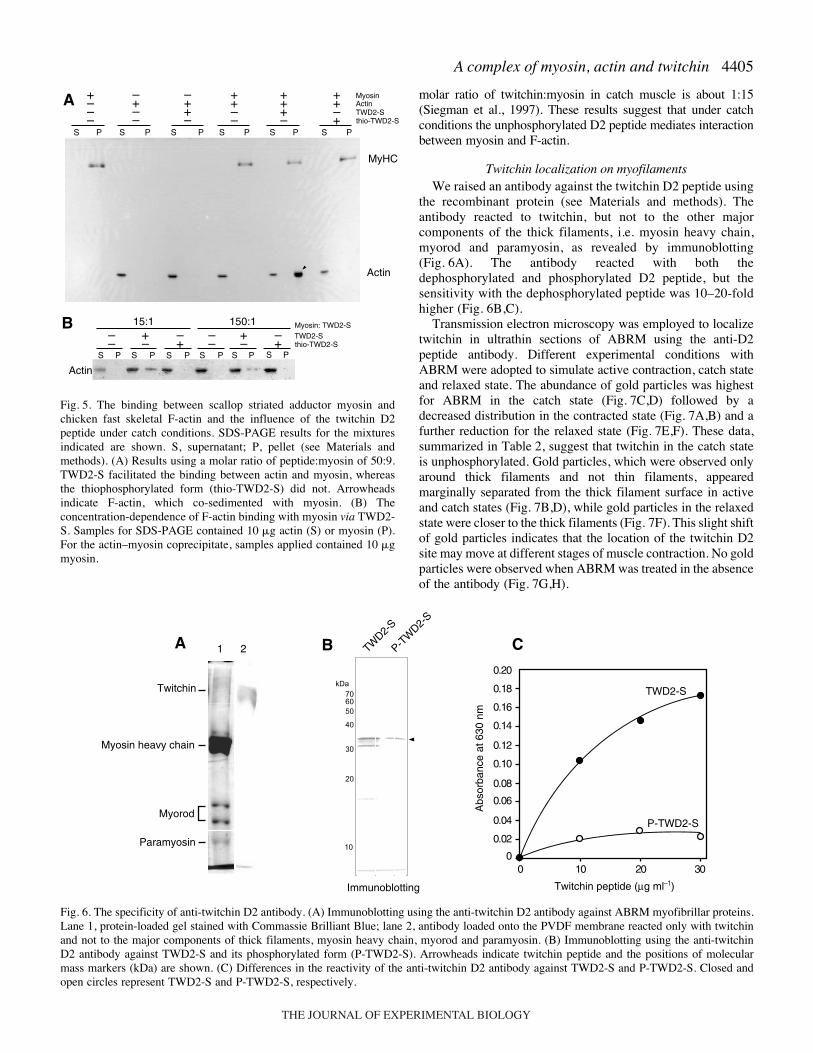
large amounts of myosin and F-actin, scallop striated adductorand chicken fast skeletal muscle were used as starting materials,respectively. The binding of myosin, actin and unphosphorylatedTWD2-S was assayed under conditions mimicking catch usinglow-speed sedimentation at low ionic strength where myosin isinsoluble and actin soluble (Fig.·5A). Initially, an excess ofunphosphorylated TWD2-S or thiophosphorylated TWD2-S wasused (molar ratio for twitchin construct:myosin of 50:9).
Thiophosphorylated TWD2-S had no effect on the distribution ofactin in the supernatant fraction (Fig.·5A). Cosedimentation ofeach of the three components was detected only whenunphosphorylated TWD2-S was used. Subsequently, it was foundthat lower ratios of unphosphorylated TWD2-S:myosin alsopromoted cosedimentation. Even at a ratio of 1:150(unphosphorylated TWD2-S:myosin) a trimeric complexformation was detected (Fig.·5B). It is proposed that the in vivo
D. Funabara and others
20
30
4050
70100
15
10
M 1 2 3 6 12 24
Actin
Trypsin
kDa
25
24
2322
2120
19
18
17
16
15
14
13
1211
109
7
6
54
3
2
1
8
00 5 10 15 20 25 30 35 40 45 50
1.0
0.8
0.6
0.4
0.2
0
1.2
1.4
1.6
35
30
25
20
15
10
5*A
bsor
banc
e at
215
nm
(––
––)
Abs
orba
nce
at 2
15 n
m (
––––
)
1 2 3 4 5 6
7 8 9 10 11 12 13 14
15 16 17 18 19 20 21 22
23 24 25
16-1
16-216-3
20
23
0
0.1
0.2
0.3
0.4
0.5
0 5 10 15 20 25 30 35 40 45 50
22
21
16-1 16-2 16-3
*
0.25
0.20
0.15
0.10
0.05
00 5
Peptide (�g ml–1)
10 15
Actin 1 MCDEDETTALVCDNGSGLVKAGFAGDDAPRAVFPSIVGRPRHQGV 45
A
B
C
D
E
Twitchin binding region
Abs
orba
nce
at 6
30 n
m
Time (min)
Time (min)
F
Ace
toni
trile
(%
) (
– –
– )
Ace
toni
trile
(%
) (
– –
– )
Fig.·4. Identification of a twitchin D2 peptide binding region onactin. (A) SDS-PAGE patterns of the digests of chicken fast skeletalactin by trypsin. Numbers above the gel represent the digestion time(h). M, molecular markers. (B) Isolation of the peptide that reactsto unphosphorylated TWD2-S from the digests of actin by reverse-phase high performance liquid chromatography with a TSKgelODS-80T column (4.6·mm� 15·cm). Numbers in the graphrepresent the fraction numbers collected in this experiment. Eachfraction was subjected to a solid-phase binding assay with TWD2-S. Only Fraction 16 (asterisk) reacted with TWD2-S. The insetshows the results of the colorimetric binding assay. Note the changeof color only for fraction 16. (C) Second reverse-phasechromatography for fraction 16. Fraction 16 was separated intothree peaks and only fraction 16-2 reacted to TWD2-S. The insetshows the results of the colorimetric binding assay; only fraction16-2 was positive. (D) The binding region of actin with the D2peptide. Asterisks represent two aspartic acid residues essential formyosin-driven movement of thick filaments on actin-containingthin filaments in the presence of ATP and Ca2+. The sequence ofthe isolated peptide that reacted to TWD2-S is shown in red. Thepeptide synthesized and used for competitive binding assay withTWD2-S in the present study is represented in green. (E)Competitive binding assay between chicken fast skeletal actin andits synthetic peptide AGFAGDDAP, measured by solid-phasebinding assays (see Materials and methods). TWD2-S wasdisplaced from actin by increasing concentrations of the syntheticpeptide. (F) A structural representation of actin. The TWD2-Sbinding region is shown in yellow.
THE JOURNAL OF EXPERIMENTAL BIOLOGY
4405A complex of myosin, actin and twitchin
molar ratio of twitchin:myosin in catch muscle is about 1:15(Siegman et al., 1997). These results suggest that under catchconditions the unphosphorylated D2 peptide mediates interactionbetween myosin and F-actin.
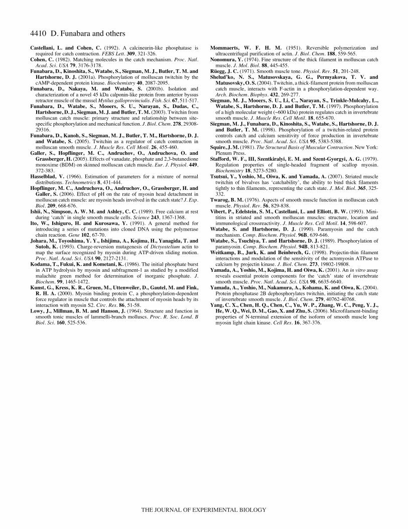
Twitchin localization on myofilamentsWe raised an antibody against the twitchin D2 peptide using
the recombinant protein (see Materials and methods). Theantibody reacted to twitchin, but not to the other majorcomponents of the thick filaments, i.e. myosin heavy chain,myorod and paramyosin, as revealed by immunoblotting(Fig.·6A). The antibody reacted with both thedephosphorylated and phosphorylated D2 peptide, but thesensitivity with the dephosphorylated peptide was 10–20-foldhigher (Fig.·6B,C).
Transmission electron microscopy was employed to localizetwitchin in ultrathin sections of ABRM using the anti-D2peptide antibody. Different experimental conditions withABRM were adopted to simulate active contraction, catch stateand relaxed state. The abundance of gold particles was highestfor ABRM in the catch state (Fig.·7C,D) followed by adecreased distribution in the contracted state (Fig.·7A,B) and afurther reduction for the relaxed state (Fig.·7E,F). These data,summarized in Table·2, suggest that twitchin in the catch stateis unphosphorylated. Gold particles, which were observed onlyaround thick filaments and not thin filaments, appearedmarginally separated from the thick filament surface in activeand catch states (Fig.·7B,D), while gold particles in the relaxedstate were closer to the thick filaments (Fig.·7F). This slight shiftof gold particles indicates that the location of the twitchin D2site may move at different stages of muscle contraction. No goldparticles were observed when ABRM was treated in the absenceof the antibody (Fig.·7G,H).
S P S PS P S P S P S P
+–––
–+––
–++–
++––
+++–
++–+
Actin
S P S P S P S P
––
+–
–+
––
+–
–+
S P S P
15:1 150:1
Actin
A
B Myosin: TWD2-STWD2-Sthio-TWD2-S
MyosinActinTWD2-Sthio-TWD2-S
MyHC
Fig.·5. The binding between scallop striated adductor myosin andchicken fast skeletal F-actin and the influence of the twitchin D2peptide under catch conditions. SDS-PAGE results for the mixturesindicated are shown. S, supernatant; P, pellet (see Materials andmethods). (A) Results using a molar ratio of peptide:myosin of 50:9.TWD2-S facilitated the binding between actin and myosin, whereasthe thiophosphorylated form (thio-TWD2-S) did not. Arrowheadsindicate F-actin, which co-sedimented with myosin. (B) Theconcentration-dependence of F-actin binding with myosin via TWD2-S. Samples for SDS-PAGE contained 10·�g actin (S) or myosin (P).For the actin–myosin coprecipitate, samples applied contained 10·�gmyosin.
1 2
0 10 20 300
0.02
0.04
0.06
0.08
0.10
0.12
0.14
0.16
0.18
0.20
TWD2-S
P-TWD2-S
TWD2-
S
P-TW
D2-S
A B C
Abs
orba
nce
at 6
30 n
m
Twitchin peptide (µg ml–1)Immunoblotting
Twitchin
Myosin heavy chain
Myorod
Paramyosin
Fig.·6. The specificity of anti-twitchin D2 antibody. (A) Immunoblotting using the anti-twitchin D2 antibody against ABRM myofibrillar proteins.Lane 1, protein-loaded gel stained with Commassie Brilliant Blue; lane 2, antibody loaded onto the PVDF membrane reacted only with twitchinand not to the major components of thick filaments, myosin heavy chain, myorod and paramyosin. (B) Immunoblotting using the anti-twitchinD2 antibody against TWD2-S and its phosphorylated form (P-TWD2-S). Arrowheads indicate twitchin peptide and the positions of molecularmass markers (kDa) are shown. (C) Differences in the reactivity of the anti-twitchin D2 antibody against TWD2-S and P-TWD2-S. Closed andopen circles represent TWD2-S and P-TWD2-S, respectively.
THE JOURNAL OF EXPERIMENTAL BIOLOGY

4406
To investigate the arrangement of twitchin on the ABRM thickfilaments (isolated from ABRM in the catch state) the distancesbetween closest neighboring two gold particles (recognizing thetwitchin D2 site) were measured. These were measured bymodifying a measurement position perpendicular to the thickfilament axis. A total of 223 measurements were made. Sincelarger distances are considered to be less reliable for statisticalanalysis, distances between two gold particles of not more than400·nm (N=155) were subjected to frequency distributionanalysis (Hasselblad, 1966) and separated into a mixture ofmultiple normal distributions (Fig.·8A). The distribution showeda periodicity of 36.25·nm and the estimated slope of theregression line (36.29) through the origin (Fig.·8B) was notsignificantly different from the observed period (F-test, P=0.91).This interval of 36.25·nm on the thick filaments is half of the72.5·nm periodicity of paramyosin filaments (Squire, 1981).
Furthermore, electron microscopy of isolated ABRM thickfilaments reacted with the anti-D2 peptide antibodydemonstrated that gold particles conjugated with the secondaryantibody were located periodically on the surface of the thickfilaments (Fig.·9A), confirming the above observations withultrathin sections. In fact the homogenate of ABRM prepared
D. Funabara and others
F
AAA BB
C
E
G H
D
Active
Catch
Relaxed
Control
Table·2. Colloidal gold distribution on thick filaments inactive, catch and relaxed fibers
Contractile condition
Active Catch Relaxed Control
Gold particles 3.9±0.9a 8.5±4.5b 3.7±0.9a 0(per 10·�m2)
Gold particles were measured on thick filaments per 10·�m2 inelectron micrographs. Different suffixes represent significantdifferences in the number of gold particles on thick filaments fromdifferent contractile conditions, as revealed by Student’s t-test(P<0.01). Values are means ± s.d. for 11–12 different areas from threeultrathin sections each from active, catch and relaxed state samplesexcept for the antibody-free control, the data of which were obtainedfrom five different areas of three sections.
Photographs of ultrathin sections are shown in Fig.·7.
0 1 2 3 4 5 6 7 8 9 10 11
Mea
n di
stan
ce (
nm)
Distance peak no.0
0 0
100
200
300
400
0.01
0.02
0.03
0.04
0.05
50 100 150 200 250 300 350 400
A B
Distance (nm)
Fre
quen
cy
Fig.·8. Histogram showing frequency distribution of distances between any two gold particles (corresponding to the location of the D2 antibody)on the same thick filament, as seen by electron microscopy, and the statistical significance analysis. (A) Frequency was set up for a total of 155measurements of not more than 400·nm and analyzed on the assumption of compound normal distributions with parameters indicated by themaximum likelihood method. (B) Estimated slope (36.29) of the regression line through the origin, compared to 36.25 (see text for details). Thetwo values were not statistically different (F-test, P=0.91).
Fig.·7. Electron microscopic observation on ultrathin sections of ABRMreacted with anti-twitchin D2 peptide antibody in active contraction,catch and relaxation stages. Longitudinal (A,C,E,G) and cross-sectional(B,D,F,H) views for ABRM in active contraction (A,B), ABRM in catch(C,D), ABRM in relaxation (E,F) and ABRM without the antibody(control) (G,H). Bars, 200·nm (A–F), 500·nm (G), 100·nm (H).
THE JOURNAL OF EXPERIMENTAL BIOLOGY

4407A complex of myosin, actin and twitchin
in the relaxed state contained both thick and thin filaments, butonly thick filaments were selected and subjected to analysis.Gold particles attached to the secondary antibody also wereassociated with granular structures having an average diameterof 20.2·nm, i.e. about the diameter of the myosin head(Fig.·9B), suggesting an interaction between the D2 peptideand the myosin head. Also noted were ultrathin structures ofover 50·nm in length and 3.8·nm diameter that extended alongthe axis of the thick filaments (Fig.·9C). These dimensionssuggest, tentatively, that the 50·nm structures may be part oftwitchin filaments, extending along the surface of the thickfilament. Resolution was not adequate to obtain more accurateestimates of the filament length. ABRM twitchin as observed
by rotary shadowing is an elongated molecule approximately225·nm in length with a spherical head (Fig.·9D), as reportedelsewhere (Vibert et al., 1993). The antibody against the kinasedomain (Funabara et al., 2001a) reacted to this head region(Fig.·9D). [For these experiments the antibody to the kinasedomain was used since its titer for twitchin was higher than theD2 peptide antibody (data not shown).] Models of the parallelarray of twitchin molecules (red) are superimposed on theBear-Selby net pattern (Bear and Selby, 1956) and shownrelative to the myosin head distribution (blue) (Fig.·9E). TheD2 site is located adjacent to the kinase domain at its C-terminal edge (Fig.·1A) and thus may be positioned close to themyosin head.
B
C
A D
E
Fig.·9. Electron microscopic observation on thick filaments labeled with anti-twitchin D2 peptide antibody. (A) Electron micrographs of ABRMthick filaments labeled with the anti-twitchin D2 peptide antibody and negatively stained. Antibodies conjugated with gold particles, indicatinglocalization of twitchin, are distributed on the surface of the filaments at intervals (upper panel) and at helical turns (lower panel). (B) Electronmicrograph of a thick filament treated with low angle rotary shadowing after negative staining. The secondary antibody-conjugated gold particlesare localized on globular structures. (C) Stereo views of negatively stained thick filaments. Ultrathin filaments, possibly representing twitchinmolecules, expand longitudinally on the thick filament as indicated by the white arrow. Arrowheads indicate location of the antibody-conjugatedgold particles. (D) Electron microscopic observation of twitchin molecules by rotary shadowing. Twitchin molecule (left) and after treatment withanti-twitchin kinase domain antibody (right). Twitchin (0.06·mg·ml–1) was reacted with the anti-twitchin kinase domain antibody (Funabara et al.,2001a) and mixed with 40% glycerol. This preparation, and a sample of twitchin without antibody, were sprayed onto mica and subjected to rotaryshadowing using platinum and carbon as described above. (E) Models of the parallel array of twitchin molecules (red) superimposed on the Bear-Selby net pattern (Bear and Selby, 1956) and relative to myosin head distribution (blue). Bars, 100·nm (A–C), 50·nm (D). The Bear-Selby netreflects the arrangement of paramyosin molecules in the thick filament. The paramyosin molecules assemble into fibers with an axial repeat of72.5·nm and staggering of these filaments generates the characteristic ‘checkerboard’ array of nodes. In negatively stained samples the nodes arethe gaps between molecules where stain is trapped (Squire, 1981; Cohen, 1982).
THE JOURNAL OF EXPERIMENTAL BIOLOGY

4408
DiscussionIn the present study we have shown that the dephosphorylated
D2 peptide (consisting of the D2 phosphorylation site, S4316,flanked by two Ig motifs) binds to both F-actin and myosin, butthe phosphorylated form does not bind. The binding site on actinwas located within the sequence L10–P29 and this region alsobinds to loop 2 of the myosin head. This interaction betweenmyosin and actin is essential to initiate cross-bridge cycling(Johara et al., 1993). Thus it is expected that binding of eithermyosin or twitchin to this N-terminal region of actin would becompetitive. However, this competition would occur only withunphosphorylated D2 peptide. The binding site for the D2peptide on myosin remains to be determined, but the electronmicroscopy data suggests an interaction with the myosin head.As emphasized, the D2 peptide interaction with myosin isphosphorylation-dependent, and it is not known if otherphosphorylation-independent links are formed between intacttwitchin and the contractile apparatus. The finding that twitchin
remains attached to thick filaments isolated undervarious conditions suggest that there are othertwitchin–thick filament interactions (with myosinand/or paramyosin). We propose that a criticalfeature of the catch mechanism is thephosphorylation-dependent interaction of thetwitchin D2 region with both F-actin and myosin.This complex does not involve cross-bridge–actininteractions. The locations of the actin-binding andthe myosin-binding sites on the D2 peptide are notknown. It is assumed that distinct sites are involved(possibly in the two flanking Ig motifs) and that bothare regulated by phosphorylation of D2. It is notnecessary that direct binding to the D2 site (S4316)is involved, but this could occur for one of the sites.In addition to twitchin there are several muscleproteins that contain Ig motifs [e.g. titin, myosinlight chain kinase (MLCK), telokin and the myosin-binding proteins (MyBPs)] and the ability of certainIg motifs to bind to myosin has been documented.For example, in cardiac MyBP-C the interactionwith myosin was mapped to the C-terminal Ig motif(Alyonycheva et al., 1997). Subsequently, it wasfound that the unphosphorylated MyBP-C motifbound to the S2 region of myosin and influencedcardiac mechanics. Interestingly, phosphorylation ofthe MyBP-C motif by PKA abolished these effectsby eliminating binding to myosin (Kunst et al.,2000). In cardiac muscle the stoichiometry ofMyBP-C to myosin heads is about 1:8 (Kunst et al.,2000). The binding of Ig motifs to actin is not welldocumented, although the N-terminal two Ig motifsof long MLCK have been implicated in actinbinding (Yang et al., 2006).
These results are consistent with those of otherinvestigators (see Introduction) and, for example,may explain the myofilament interconnectionsproposed by Andruchova et al. (Andruchova etal., 2005). The competition between theunphosphorylated D2 peptide and loop 2 ofmyosin for the binding site on actin also may be
pertinent to the results of Butler et al. regarding the low- andhigh-force states (Butler et al., 2006). A higher affinity ofmyosin for actin in the high-force state would effectivelydisplace the twitchin–actin interaction. This is consistent withthe observation (Butler et al., 2006) that activation of thecatch muscle by Ca2+ increases the proportion of myosin inthe high-force state and decreases interaction of twitchin withactin. Transition to the low-force state, by decreasing Ca2+
levels, would favor the twitchin–actin interaction. It shouldbe emphasized that a direct role for Ca2+ in binding of the D2peptide to either actin or myosin is not suggested and there isno evidence from our data to implicate a Ca2+ sensitive step,other than the regulation of actin-activated ATPase by Ca2+
binding to myosin. Dephosphorylation of twitchin bycalcineurin (Castellani and Cohen, 1992; Yamada et al., 2004)would be coincident with initiation of contraction since bothevents are Ca2+ dependent, and thus for most of the contractilephase twitchin is dephosphorylated and theoretically
D. Funabara and others
Twitchin Phosphorylated
Ace
tylc
holin
e
Ser
oton
in
RelaxedCatchRelaxed Active
Tension
[Ca2+]
[Ca2+
]
Phosphorylated
Active myosin
Inactive myosin
Actin
Paramyosin
Twitchin
A
B
Catch state Active state
Relaxed state
dephosphorylationTwitchin
Tens
ion
Dephosphorylated
Twitchin
phosphorylation
Fig.·10. A model of interactions of twitchin with myosin and actin for differentstages in the contractile cycle. For explanation, see text. Tropomyosin in thinfilaments is not shown.
THE JOURNAL OF EXPERIMENTAL BIOLOGY

4409A complex of myosin, actin and twitchin
available to compete with myosin for actin. In practice,twitchin does not inhibit the in vivo contraction of ABRMfibers in the presence of Ca2+ (Siegman et al., 1998). Acompetition between twitchin and myosin might explain thein vitro inhibition of Mg2+ ATPase activity of actomyosin byrelatively high concentrations of TWD2-S.
Results from the localization of twitchin on the thick filamentindicate that the twitchin molecules are distributed on thesurface of the filament at a periodicity of 36.25·nm, half of the72.5·nm periodicity of paramyosin filaments (Squire, 1981). Itis suggested that the D2 site at the C-terminal end of twitchinaligns with myosin heads, i.e. at half of the 72.5·nm axialperiodicity (Cohen, 1982). In the model proposed in Fig.·9E thetwitchin molecule could extend through about three of the72.5·nm repeats or nodes, if arranged parallel to the filamentaxis. The myosin rod extends axially through two nodes. Thusthere is a possibility of other interactions between twitchin andthe myosin rod. The molar ratio of twitchin to myosin is about1:15 in ABRM catch muscle (Siegman et al., 1997). Therefore,even if all of the twitchin molecules interact with myosin, thenumber of trimeric complexes is small compared with totalmyosin. It is not known if there are cooperative effects onneighboring myosin molecules induced by the binding oftwitchin to myosin. The ultrathin filaments observed above onthe surface of the thick filaments are thought to be twitchin andthese reflect the underlying Bear-Selby net (Bear and Selby,1956) (see Fig.·9C).
There are several aspects of the catch mechanism to beresolved. In an earlier report (Funabara et al., 2003) it wasstated that phosphorylation of both the D1 and D2 sites isrequired for relaxation from the catch state. The D1 site isunusual in that relatively high levels of phosphorylation(40–50%) are found while catch is maintained. The D2 site ismore sensitive to effects of phosphorylation; low levels ofphosphorylation are found during catch and high levels ofphosphorylation accompany the release from catch (Funabaraet al., 2003). The more marked phosphorylation dependenceof the D2 site prompted this present study, but the role of theD1 site is not understood and should be resolved by furtherstudies. Another area for future study is to identify thephosphorylation-dependent binding site on myosin for the D2peptide and also to determine if other interactions betweennative twitchin and myosin occur. If present, these couldanchor the twitchin molecule to the surface of the thickfilament. Based on the in vitro results of Yamada et al.(Yamada et al., 2001) it is assumed that paramyosin is not anessential component of the catch mechanism, but whether ithas any influence on catch under in situ conditions remainsto be determined. The phosphorylation-dependent binding ofthe D2 peptide to paramyosin is shown above. It is knownthat in fibers of molluscan smooth muscle the catch state issensitive to pH (Hopflinger et al., 2006) and is reduced onmoderate alkalinization to pH 7.2–7.7, i.e. at those pH valuesused for the binding experiments described above. It isimportant to determine if any of the interactions betweenisolated proteins show a marked pH dependence and this canbe tested experimentally. On the other hand, the sensitivity topH may be associated only with the intact contractile system.Recently, it was found that striated muscles from oyster and
scallop contain twitchin and this regulates interactionbetween thin and thick filaments at low [Ca2+] (Tsutsui et al.,2007), as it does in molluscan catch muscle. These resultssuggest a similar role for twitchin in striated and smoothmolluscan muscle and the molecular scheme outlined abovemay be applicable to both muscle types.
In summary, our results provide novel data on the molecularinteractions involved in catch. In Fig.·10A the different phases ofthe contractile cycle in catch muscle are shown, with associatedchanges in [Ca2+], tension and twitchin phosphorylation. Startingwith relaxed muscle (Fig.·10B), twitchin is phosphorylated (byPKA) and the D2 peptide does not interact with either actin ormyosin. It is assumed that in the relaxed state there are nointerfilament connections. Stimulation by acetylcholine increases[Ca2+], which promotes contraction (by binding to and activatingmyosin) and activates calcineurin. Twitchin is dephosphorylatedbut does not bind to actin (although theoretically this interactionis still possible) because of competition with the increasedpopulation of high-force states, i.e. the cross-bridge–actininteraction predominates. The onset of catch follows a reductionin [Ca2+] to close to resting levels and inactivation of myosin. Thelow-force state (the detached cross-bridge being part of the low-force state) allows the D2 twitchin region to bind to F-actin andmyosin and form a mechanical force-bearing complex. In thismodel there is no direct effect of Ca2+ on the binding of theunphosphorylated D2 peptide to either myosin or actin and ourhypothesis is that the obligatory role for a reduction in Ca2+ topromote the catch state reflects the inactivation of myosin and anincrease in low-force state. This complex is proposed to representat least part of the mechanical connection (catch bridge) betweenthe myofilaments. Serotonin release causes an increase in[cAMP] and activation of PKA. One of the PKA sites on twitchinis the D2 site and its phosphorylation eliminates binding of theD2 peptide to actin and myosin. The catch connection is lost andthe muscles enters the relaxed phase.
We thank Dr M. J. Siegman and Dr T. M. Butler for theiradvice and Dr Takashi Yamakawa for help in statisticalanalysis. This work was supported by a Grant-in-Aid forScientific Research from the Ministry of Education, Culture,Sports, Science and Technology of Japan (to D.F.) and Japan-US Cooperative Science Program from Japan Society for thePromotion of Science (to S.W.) and NIH grant HL-23615 (toD.J.H.).
ReferencesAlyonycheva, T. N., Mikawa, T., Reinach, F. C. and Fischman, D. A. (1997).
Isoform-specific interaction of the myosin-binding proteins (MyBPs) withskeletal and cardiac myosin is a property of the C-terminal immunoglobulindomain. J. Biol. Chem. 272, 20866-20872.
Andruchova, O., Hopflinger, M. C., Andruchov, O. and Galler, S. (2005).No effect of twitchin phosphorylation on the rate of myosin head detachmentin molluscan catch muscle: are myosin heads involved in the catch state? Eur.J. Physiol. 450, 326-334.
Bear, R. S. and Selby, C. C. (1956). The structure of paramyosin fibrilsaccording to X-ray diffraction. J. Biophys. Biochem. Cytol. 2, 55-69.
Butler, T. M., Narayan, S. R., Mooers, S. U., Hartshorne, D. J. andSiegman, M. J. (2001). The myosin cross-bridge cycle and its control bytwitchin phosphorylation in catch muscle. Biophys. J. 80, 414-426.
Butler, T. M., Mooers, S. U. and Siegman, M. J. (2006). Catch force linksand the low to high force transition of myosin. Biophys. J. 90, 3139-3202.
Castellani, L. and Cohen, C. (1987). Myosin rod phosphorylation and the catchstate of molluscan muscles. Science 235, 334-337.
THE JOURNAL OF EXPERIMENTAL BIOLOGY
4410
Castellani, L. and Cohen, C. (1992). A calcineurin-like phosphatase isrequired for catch contraction. FEBS Lett. 309, 321-326.
Cohen, C. (1982). Matching molecules in the catch mechanism. Proc. Natl.Acad. Sci. USA 79, 3176-3178.
Funabara, D., Kinoshita, S., Watabe, S., Siegman, M. J., Butler, T. M. andHartshorne, D. J. (2001a). Phosphorylation of molluscan twitchin by thecAMP-dependent protein kinase. Biochemistry 40, 2087-2095.
Funabara, D., Nakaya, M. and Watabe, S. (2001b). Isolation andcharacterization of a novel 45 kDa calponin-like protein from anterior byssusretractor muscle of the mussel Mytilus galloprovincialis. Fish. Sci. 67, 511-517.
Funabara, D., Watabe, S., Mooers, S. U., Narayan, S., Dudas, C.,Hartshorne, D. J., Siegman, M. J. and Butler, T. M. (2003). Twitchin frommolluscan catch muscle: primary structure and relationship between site-specific phosphorylation and mechanical function. J. Biol. Chem. 278, 29308-29316.
Funabara, D., Kanoh, S., Siegman, M. J., Butler, T. M., Hartshorne, D. J.and Watabe, S. (2005). Twitchin as a regulator of catch contraction inmolluscan smooth muscle. J. Muscle Res. Cell Motil. 26, 455-460.
Galler, S., Hopflinger, M. C., Andruchov, O., Andruchova, O. andGrassberger, H. (2005). Effects of vanadate, phosphate and 2,3-butanedionemonoxime (BDM) on skinned molluscan catch muscle. Eur. J. Physiol. 449,372-383.
Hasselblad, V. (1966). Estimation of parameters for a mixture of normaldistributions. Technometrics 8, 431-444.
Hopflinger, M. C., Andruchova, O., Andruchov, O., Grassberger, H. andGaller, S. (2006). Effect of pH on the rate of myosin head detachment inmolluscan catch muscle: are myosin heads involved in the catch state? J. Exp.Biol. 209, 668-676.
Ishii, N., Simpson, A. W. M. and Ashley, C. C. (1989). Free calcium at restduring ‘catch’ in single smooth muscle cells. Science 243, 1367-1368.
Ito, W., Ishiguro, H. and Kurosawa, Y. (1991). A general method forintroducing a series of mutations into cloned DNA using the polymerasechain reaction. Gene 102, 67-70.
Johara, M., Toyoshima, Y. Y., Ishijima, A., Kojima, H., Yanagida, T. andSutoh, K. (1993). Charge-reversion mutagenesis of Dictyostelium actin tomap the surface recognized by myosin during ATP-driven sliding motion.Proc. Natl. Acad. Sci. USA 90, 2127-2131.
Kodama, T., Fukui, K. and Kometani, K. (1986). The initial phosphate burstin ATP hydrolysis by myosin and subfragment-1 as studied by a modifiedmalachite green method for determination of inorganic phosphate. J.Biochem. 99, 1465-1472.
Kunst, G., Kress, K. R., Gruen, M., Uttenweiler, D., Gautel, M. and Fink,R. H. A. (2000). Myosin binding protein C, a phosphorylation-dependentforce regulator in muscle that controls the attachment of myosin heads by itsinteraction with myosin S2. Circ. Res. 86, 51-58.
Lowy, J., Millman, B. M. and Hanson, J. (1964). Structure and function insmooth tonic muscles of lammelli-branch molluscs. Proc. R. Soc. Lond. BBiol. Sci. 160, 525-536.
Mommaerts, W. F. H. M. (1951). Reversible polymerization andultracentrifugal purification of actin. J. Biol. Chem. 188, 559-565.
Nonomura, Y. (1974). Fine structure of the thick filament in molluscan catchmuscle. J. Mol. Biol. 88, 445-455.
Rüegg, J. C. (1971). Smooth muscle tone. Physiol. Rev. 51, 201-248.Shelud’ko, N. S., Matusovskaya, G. G., Permyakova, T. V. and
Matusovsky, O. S. (2004). Twitchin, a thick-filament protein from molluscancatch muscle, interacts with F-actin in a phosphorylation-dependent way.Arch. Biochem. Biophys. 432, 269-277.
Siegman, M. J., Mooers, S. U., Li, C., Narayan, S., Trinkle-Mulcahy, L.,Watabe, S., Hartshorne, D. J. and Butler, T. M. (1997). Phosphorylationof a high molecular weight (~600 kDa) protein regulates catch in invertebratesmooth muscle. J. Muscle Res. Cell Motil. 18, 655-670.
Siegman, M. J., Funabara, D., Kinoshita, S., Watabe, S., Hartshorne, D. J.and Butler, T. M. (1998). Phosphorylation of a twitchin-related proteincontrols catch and calcium sensitivity of force production in invertebratesmooth muscle. Proc. Natl. Acad. Sci. USA 95, 5383-5388.
Squire, J. M. (1981). The Structural Basis of Muscular Contraction. New York:Plenum Press.
Stafford, W. F., III, Szentkiralyi, E. M. and Szent-Gyorgyi, A. G. (1979).Regulation properties of single-headed fragment of scallop myosin.Biochemistry 18, 5273-5280.
Tsutsui, Y., Yoshio, M., Oiwa, K. and Yamada, A. (2007). Striated muscletwitchin of bivalves has ‘catchability’, the ability to bind thick filamentstightly to thin filaments, representing the catch state. J. Mol. Biol. 365, 325-332.
Twarog, B. M. (1976). Aspects of smooth muscle function in molluscan catchmuscle. Physiol. Rev. 56, 829-838.
Vibert, P., Edelstein, S. M., Castellani, L. and Elliott, B. W. (1993). Mini-titins in striated and smooth molluscan muscles: structure, location andimmunological crossreactivity. J. Muscle Res. Cell Motil. 14, 598-607.
Watabe, S. and Hartshorne, D. J. (1990). Paramyosin and the catchmechanism. Comp. Biochem. Physiol. 96B, 639-646.
Watabe, S., Tsuchiya, T. and Hartshorne, D. J. (1989). Phosphorylation ofparamyosin. Comp. Biochem. Physiol. 94B, 813-821.
Weitkamp, B., Jurk, K. and Beinbrech, G. (1998). Projectin-thin filamentinteractions and modulation of the sensitivity of the actomyosin ATPase tocalcium by projectin kinase. J. Biol. Chem. 273, 19802-19808.
Yamada, A., Yoshio, M., Kojima, H. and Oiwa, K. (2001). An in vitro assayreveals essential protein components for the ‘catch’ state of invertebratesmooth muscle. Proc. Natl. Acad. Sci. USA 98, 6635-6640.
Yamada, A., Yoshio, M., Nakamura, A., Kohama, K. and Oiwa, K. (2004).Protein phosphatase 2B dephosphorylates twitchin, initiating the catch stateof invertebrate smooth muscle. J. Biol. Chem. 279, 40762-40768.
Yang, C. X., Chen, H. Q., Chen, C., Yu, W. P., Zhang, W. C., Peng, Y. J.,He, W. Q., Wei, D. M., Gao, X. and Zhu, S. (2006). Microfilament-bindingproperties of N-terminal extension of the isoform of smooth muscle longmyosin light chain kinase. Cell Res. 16, 367-376.
D. Funabara and others
THE JOURNAL OF EXPERIMENTAL BIOLOGY