unusual presentation of the conn's syndrome: a case...
TRANSCRIPT

Case Report Medical Journal of Islamic Republic of Iran, Vol. 25, No. 3, Nov 2011, pp. 158-161
__________________________________________________________________________________________________ 1. (Corresponding author) MD. Specialist in Internal Medicine Department. AlAmiri Hospital, P.O. Box 4077, Safat-13041-Kuwait. Tel: +965 22469627. [email protected] 2. Consultant in Internal Medicine Department. AlAmiri Hospital, Departments of Medicine, Al- Amiri Hospital, Kuwait. [email protected]
Unusual presentation of the Conn's syndrome: a case report
Maryam Al-Rajhi1, Sahasranamaiyer Narayanan2
Department of Medicine, Al- Amiri Hospital, Kuwait.
Received: 20 Dec 2010 Revised: 10 Mar 2011 Accepted: 8 May 2011
__________________________________________________________________________________________ Abstract A 26 -year- old woman presented with rhabdomyolysis secondary to severe hypokalemia. Hypertension and metabolic alkalosis could lead to the suspicion of primary aldosteronism, which was confirmed by a decreased plasma rennin, elevated plasma aldosterone levels and high aldosterone/rennin ratio additionally. Additionally adrenal computed tomography showed an adrenal tumour. Blood pressure and hypokalemia returned to the nor-mal level after adrenalectomy was performed. This case report highlights the need to be alert to the possibility of primary aldosteronism incidence in a patient presenting with rhabdomyolysis and hypertension caused by severe hypokalemia.
Keywords: Conn's syndrome, hypokalemia, rhabdomyolysis.
__________________________________________________________________________________________
Introduction The primary aldosteronism (PA) resulting
from an adrenocortical adenoma (Conn's syndrome) is a common and a curable cause of secondary hypertension [5]. The combina-tion of hypertension, hypokalemia, and metabolic alkalosis is important for a diag-nosis of PA. In few cases, hypokalemia caused by PA can be severe enough to cause rhabdomyolysis. Here we report a case of rhabdomyolysis caused by severe hypo-kalemia, which in turn was resulted in the PA. The patient was cured after performing adrenalectomy.
Case report A 26-year-old-woman was admitted to the
hospital because of five days history of gen-eralized muscle weakness involving pre-dominantly the lower limbs. That was asso-ciated with myalgia, muscle cramp and paresthesia. She had history of episodic
muscle weakness of the lower limbs over the past few months. There was no history of infection or trauma. She also had not experi-enced diarrhea or vomiting, and any use of diuretics, herbal supplements, liquorice, laxatives or other medications. She did not suffer from renal disease or hypertension and had no symptoms suggestive of thyroid disease. She had regular menstrual cycles.
On physical examination, her weight was 58Kg and height 160cm (BMI = 22.65), blood pressure 170/115 mmHg, body tem-perature 36.8 °C, pulse rate 70 beats/min, and respiratory rate 18/min. There was weakness of both lower limbs (4/5) with normal sensations. The power and sensation of both upper limbs were normal and normal cardiovascular and respiratory rates. The ab-dominal examination did not reveal hepa-tosplenomegaly or bruits on auscultation with no features of Cushing's syndrome.
Laboratory investigations were as follow: potassium 1.5 mEq/L [normal range (NR):
Dow
nloa
ded
from
mjir
i.ium
s.ac
.ir a
t 3:3
6 IR
ST
on
Sun
day
Feb
ruar
y 3r
d 20
19
M. Alrajhi.
159
MJIRI, Vol. 25, No. 3, Nov 2011, pp. 158-161
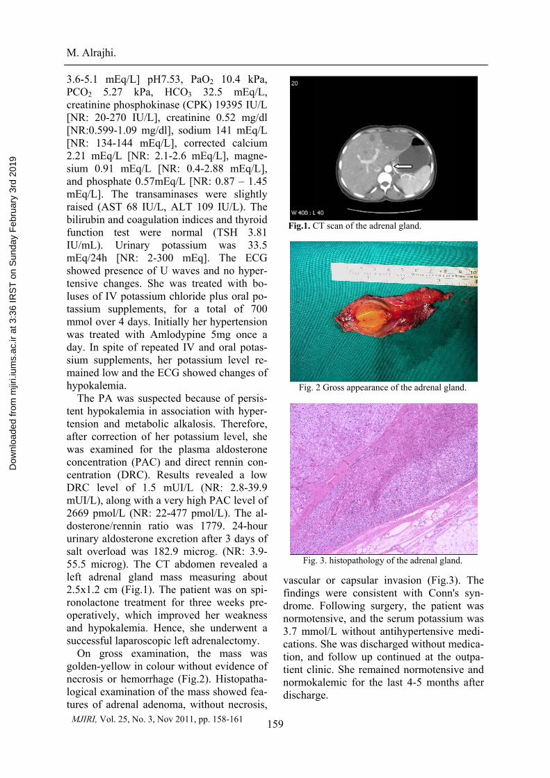
Fig.1. CT scan of the adrenal gland.
Fig. 2 Gross appearance of the adrenal gland.
Fig. 3. histopathology of the adrenal gland.
3.6-5.1 mEq/L] pH7.53, PaO2 10.4 kPa, PCO2 5.27 kPa, HCO3 32.5 mEq/L, creatinine phosphokinase (CPK) 19395 IU/L [NR: 20-270 IU/L], creatinine 0.52 mg/dl [NR:0.599-1.09 mg/dl], sodium 141 mEq/L [NR: 134-144 mEq/L], corrected calcium 2.21 mEq/L [NR: 2.1-2.6 mEq/L], magne-sium 0.91 mEq/L [NR: 0.4-2.88 mEq/L], and phosphate 0.57mEq/L [NR: 0.87 – 1.45 mEq/L]. The transaminases were slightly raised (AST 68 IU/L, ALT 109 IU/L). The bilirubin and coagulation indices and thyroid function test were normal (TSH 3.81 IU/mL). Urinary potassium was 33.5 mEq/24h [NR: 2-300 mEq]. The ECG showed presence of U waves and no hyper-tensive changes. She was treated with bo-luses of IV potassium chloride plus oral po-tassium supplements, for a total of 700 mmol over 4 days. Initially her hypertension was treated with Amlodypine 5mg once a day. In spite of repeated IV and oral potas-sium supplements, her potassium level re-mained low and the ECG showed changes of hypokalemia.
The PA was suspected because of persis-tent hypokalemia in association with hyper-tension and metabolic alkalosis. Therefore, after correction of her potassium level, she was examined for the plasma aldosterone concentration (PAC) and direct rennin con-centration (DRC). Results revealed a low DRC level of 1.5 mUI/L (NR: 2.8-39.9 mUI/L), along with a very high PAC level of 2669 pmol/L (NR: 22-477 pmol/L). The al-dosterone/rennin ratio was 1779. 24-hour urinary aldosterone excretion after 3 days of salt overload was 182.9 microg. (NR: 3.9-55.5 microg). The CT abdomen revealed a left adrenal gland mass measuring about 2.5x1.2 cm (Fig.1). The patient was on spi-ronolactone treatment for three weeks pre-operatively, which improved her weakness and hypokalemia. Hence, she underwent a successful laparoscopic left adrenalectomy.
On gross examination, the mass was golden-yellow in colour without evidence of necrosis or hemorrhage (Fig.2). Histopatha-logical examination of the mass showed fea-tures of adrenal adenoma, without necrosis,
vascular or capsular invasion (Fig.3). The findings were consistent with Conn's syn-drome. Following surgery, the patient was normotensive, and the serum potassium was 3.7 mmol/L without antihypertensive medi-cations. She was discharged without medica-tion, and follow up continued at the outpa-tient clinic. She remained normotensive and normokalemic for the last 4-5 months after discharge.
Dow
nloa
ded
from
mjir
i.ium
s.ac
.ir a
t 3:3
6 IR
ST
on
Sun
day
Feb
ruar
y 3r
d 20
19

Unusual presentation of Conn's Syndrome
160
MJIRI, Vol. 25, No. 3, Nov 2011, pp. 158-161
Discussion PA is a common cause of secondary hy-
pertension, and characterised by hyperten-sion, hypokalemia, suppressed plasma renin, and increased aldosterone excretion. The rhabdomyolysis as a presentation feature of primary aldosteronism is an extremely rare association. There are about sixteen related reported cases in the literature [1] and to the best of our knowledge this is the first re-ported case in Kuwait. The rhabdomyolysis is characterized by muscle necrosis and the release of its contents into the circulation, including myoglobin, potassium, phosphate, urate and creatinine kinase. There are many causes of rhabdomyolysis, including crush injury, excessive exercise, metabolic and endocrine disorders, infections, drugs, toxins like alcohol or statins, and excessive heat exposure [2]. Hypokalemia is a recognised cause of rhabdomyolysis. The symptoms of malaise, muscle weakness, fatigability, and myalgia occur when serum concentration of potassium is below 3 mEq/L. However, muscle enzyme elevations are usually seen when potassium concentrations fall below 2.5 mEq/L[2].The important biochemical findings in rhabdomyolysis are hyper-kalemia and a high anion gap acidosis as a consequence of the release of organic acids from necrotic muscles. In our case, hypo-kalemic metabolic alkalosis and hyperten-sion raised the suspicion of primary aldos-teronism.
The aldosterone-producing adenoma (APA) and bilateral idiopathic hyperaldos-teronism (IHA) are the most common sub-types of PA. The APA is a small nodule (< 2cm) that mostly occurs in the left adrenal gland and commonly found in females, and usually present with severe hypertension and more profound hypokalemia. It is also more common in younger patients (between the ages of 30-50), with higher plasma and uri-nary levels of aldosterone [3].
Biochemical abnormalities of the APA in-clude hypokalemia, metabolic alkalosis, and a relative hypernatremia. Although sponta-neous hypokalemia in a patient with hyper-
tension is a strong indicator of aldostero-nism, only a minority of patients (9-30%) have a potassium level that is in the low-normal range [4]. Therefore, hypokalemia is not the criterion used for diagnosis of the PA. It is recommended to test for the PA in the following groups: patients with hyper-tension and hypokalemia, treatment-resistant hypertension (i.e on 3 antihypertensive medications with poor control), severe hy-pertension ( 160mmHg systolic or 100 mmHg diastolic), hypertension with adrenal insidentaloma, and the onset of hypertension under the age of 20 [5]. Screening is done by measuring the PAC level and PRC. When the PAC is greater than 15ng/mL, the PRC is less than 1ng/mL/h – as seen in this case- and the ratio of the two (PAC/PRC) is greater than 20, with the sensitivity and specificity of approximately 75%. This test is valid as long as a patient is not taking al-dosterone antagonists, such as spironolac-tone, epleranone, or renin inhibitors [6].
The screening test is not a diagnostic tool, and PA must be confirmed by demonstrating inappropriate aldosterone secretion. Confir-matory testing can be done by any of the four procedures: oral sodium loading and measurement of urinary aldosterone, intra-venous sodium chloride loading and meas-urement of PAC, fludrocortisone suppres-sion, or captoprill challenge [6]. In our case we used the oral sodium loading test which showed a non-suppressed urinary aldoster-one excretion.
Once PA has been diagnosed biochemi-cally, it is important to determine the sub-type to help in directing the therapy. Unilat-eral adrenalectomy in patients with the APA or unilateral adrenal hyperplasia results in normalization of hypokalemia and hyperten-sion, while in bilateral IHA, unilateral or bi-lateral adrenalectomy rarely corrects the hy-pertension [7], and medical therapy is there-fore the treatment of choice.
Adrenal CT is not a correct choice in dis-tinguishing between the APA and IHA. It cannot reliably visualize microadenomas or distinguish incidentalomas from the APAs, which makes the adrenal venous sampling
Dow
nloa
ded
from
mjir
i.ium
s.ac
.ir a
t 3:3
6 IR
ST
on
Sun
day
Feb
ruar
y 3r
d 20
19

M. Alrajhi.
161
MJIRI, Vol. 25, No. 3, Nov 2011, pp. 158-161
(AVS) to be the most accurate mean to dif-ferentiate between unilateral from bilateral forms of PA [8]. The AVS is essential for appropriate therapy in many patients with PA who have a high probability of having an APA and want to pursue surgical manage-ment [6], but it is expensive and invasive. The procedure itself has a relatively low success rate because of the difficulty in can-nulating the right adrenal vein (which is smaller than the left and empties directly into the IVC rather than the renal vein) [8]. The most important factors that determine the successful characterization of both adre-nal veins in a patient with PA are the experi-ence, dedication and presence of repetition of the radiologist performing the procedure [9]. A more practical approach is the selec-tive use of AVS as recommended by Young [5] which is based on patient preferences, age, adrenal morphologic appearance on CT, clinical comorbid conditions, and clinical probability of finding an APA. For patients younger than 40, in whom a solitary ade-noma is >1cm with normal contralateral ad-renal gland, a unilateral adrenalectomy may be done without venous sampling and in the absence of comorbid conditions. Therefore, the AVS was bypassed in our case based on these criteria.
The mainstays of therapy for the APA in-clude surgical adrenalectomy, and alterna-tively the use of aldosterone antagonists, such as spironolactone. The efficacy of adrenalectomy has been confirmed in a study where the diagnosis of APA was es-tablished, and in which a fall of BP observed in all patients. Moreover, 82% of patients were either improved markedly or cured (32%), thereby allowing the withdrawal of all antihypertensive medications [10,11,12].
Conclusion Rhabdomyolysis is uncommon in patients
with the APA. We recommend considering the APA, when rhabdomyolysis occurs in a patient with severe hypokalemia and meta-
bolic alkalosis since the disease could be curable like in our case.
References
1. Martinez JJ et al. Rhabdomyolysis due to pri-mary hyperaldosteronism. A case report. Endocrinol Nutr.2009;56:431-4.
2. Allison RC, Bedsole DL. The other medical causes of rhabdomyolysis. Am J Med Sci. 2003; 326:79-88.
3. Stewart PM. Mineralocorticoid hypertension. Lancet 1999; 353:1341-7.
4. Mulatero P, Stowasser M, Loh KC, Fardella CE, Gordon RD, Mosso L, Gomezsanchez CE, Veglio F, Youg WF, Jr: Increased diagnosis of primary aldos-teronism, including surgically correctable forms, in centers from five continents. J Clin Endocrinol Metab 2004; 89:1045-50
5. Young WF. Primary aldosteronism: renaissance of a syndrome. Clin Endocrinol (Oxf). 2007; 66:607-18.
6. Funder JW,Carey RM, Fardella C, Gomez-Sanchez CE, Mantero F, Stowasser M, et al. Endo-crine Society. Case detection, diagnosis, and treat-ment of patients with primary aldosteronism:an endo-crine society clinical practice guideline. J Clin Endo-crinol Metab. 2008; 93:3266-81.
7. Myer,A., Brabant, G. & Behrend, M. Long-term follow-up after adrenalectomy for primary aldostero-nism. World Journal of Surgery 2005; 29: 155-159.
8. Kempers MJ, Lenders JW, van Outheusden L, van der Wilt GJ, Schultze Kool LJ, Hermus AR, et al. Systematic review. Diagnostic procedures to differen-tiate unilateral from bilateral adrenal abnormality in primary aldosteronism. Ann Intern Med. 2009; 151:329-37.
9. Young WF, Stanson AW. What are the keys to successful adrenal venous sampling (AVS) in patients with primary aldosteronism? Clin Endocrinol (Oxf). 2009;70:14-7.
10. Rossi GP, Bolognesi M, Rizzoni D, Seccia TM, Piva A, Porteri E, et al. Vascular remodelling and duration of hypertension predict outcome of adrena-lectomy in primary aldosteronism patients. Hyperten-sion 2008; 51:1366-1371.
11. Hari Kumar KV, Jha S, Jha R, Modi KD. Conn's Syndrome – Atypical Presentations. A report of two cases. Saudi J Kidney Dis Transpl 2009; 20(2):278-281.
12. Tien-Hua Chuang, Chin-Hsein Wang, Bo-Yuan Tseng, Yung-Hsiang Hsu, Jen-Pi Tsai, Bang-Gee Hsu, et al. Conn's Syndrome with an Unusual Presen-tation of Rhabdomyolysis Secondary to Severe Hy-pokalemia. A case report. Tzu Chi Med J 2008; 20(4):327-331.
Dow
nloa
ded
from
mjir
i.ium
s.ac
.ir a
t 3:3
6 IR
ST
on
Sun
day
Feb
ruar
y 3r
d 20
19