upcoming arrs annual meetings · charity hospital, new orleans, 1967. diagnosis was based on...
TRANSCRIPT

by Bruce L. McClennan, MD
A Word From the ChairSRS
Senior RadiologistsSection Presented and published by
Imaging: 1967 – 2017 and the Impact of Ultrasoundby Christopher R. B. Merritt, MD
The Convergence of Change and Opportunity
Some years ago, I gave a number of talks or lectures in grand rounds-like style and settings with the above-listed title.
Actually the first time was on or about 1995, the 100th anniversary of the discov-ery of the x-ray, and when I went to Yale as department chair. Certainly, change has been the traveling companion of our spe-cialty these past few decades.
Perhaps overstating the obvious, but with the maturation of our imaging tools – for ex-ample, CT, ultrasound, MRI, and IR – have changed the role of the radiologist in the di-agnostic process, and in medicine in gen-eral. Add the “alphabet soup” of our daily practice the additions of PACS, EHR, voice recognition, RIS, HIS, and teleradiology to the nocturnal practice for a radiologist and we have a mere snapshot of what today is like in the reading room. There are those prognosticators who are predicting what to-morrow will be like with a whole new lex-icon – artificial intelligence (AI), machine learning (ML), deep learning (DL), neural networks, to name a few.
It is hard not to read, in the summer heat and humidity in Chicago, about the future, specifically self-driving or driverless automo-biles. Does the future augur for radiologist-less hospitals? Of great interest to me, even some alarm, is the computer scientist Geof-frey Hinton, who in essence said we should STOP training radiologists right now. He used the analogy of Wile E. Coyote having run over the cliff but yet to actually look down.
Will AI, including DL and ML, change what radiologists do in the future? The easy answer is yes, for sure. It will change how radiology is practiced, but I doubt it will eliminate the radiologist, just like the self-driving car has yet to eliminate the human driver – in fact its performance is improved with a human on board.
The perspective of many of us, not retired or in part-time practice, allows us to appre-ciate the advancements and promises to and for our field, but in some ways we actually have the best perspective.
In 1967, a week-ly feature for medical school seniors was the "bullpen" in the Char-ity Hospital amphithe-ater [Figure 1]. Stu-dents were assigned a patient and given 30 minutes do a history and physical exam and then present their dif-
ferential diagnosis and recommendations to an attending. Diagnosis was almost exclu-sively based on the history and physical ex-amination. Laboratory studies were generally confined to basic electrolytes, a CBC, urinal-ysis, sputum stains, and a chest x-ray. This was good preparation for internship and res-idency on the Osler Medical service at Hop-kins. Interns were on call 24 hours a day for 6 days a week and usually spent 16 to 18 hours a day attending patients at the bedside.
On Osler, there were no computers and handwritten or typed paper records hung on a chart rack. The wards were not air-condi-tioned, and yellow curtains separated each of the 28 beds. There were no patient mon-
Fall 2017
Chair continues on page 2
Senior Radiologists Section Notes 1
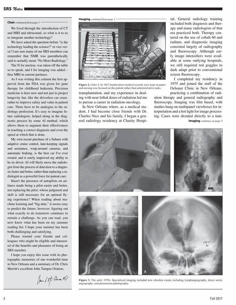
itors, IV pumps, or respirators, and interns performed basic lab work on their patients. Nursing care was excellent and house staff and nurses worked as a team caring for the patients [Figure 2]. Lack of technology was compensated for by close and direct inter-action with the patients and their families, and the practice of medicine was extremely satisfying and filled with empathy and com-passion. The patient, rather than the result
of an imaging test, was the object of all of our attention. Defensive medicine was not a concern.
In the late 1960s imaging was limited and played a relatively minor role in diagnosis and management. The most sophisticated im-aging technology was the nuclear medicine scan. In 1968, Radiology and the American Journal of Roentgenology reflected quite dif-ferent perspectives on the field of radiology.
In Radiology 22% of the papers dealt with new technology - nu-clear medicine and angiography. In contrast, the American Jour-nal of Roentgenology, dealt large-ly with articles on radiation thera-py and nuclear medicine.
Following my internal medi-cine residency at Hopkins, I had no interest in diagnostic radiolo-gy. I spent the next 3 years in the immunology branch of the Na-tional Cancer Institute in Bethes-da. Research centered on the new field of bone marrow transplan-tation and treatment of graft ver-sus host disease. Whole body ra-diation prepared candidates for
In the late 1960s imaging was limited and played a relatively minor role in
diagnosis and management
Christopher R. B. Merritt
Imaging continues on page 2
Figure 1. Charity Hospital, New Orleans, 1967. Diagnosis was based on history and physical exam. Imaging was limited to radiography and fluoroscopy and played a minor role.
2 Fall 2017
SRS Notes
Chair continued from page 1
We lived through the introduction of CT and MRI and ultrasound, so what is it to us to integrate another technology?
We have asked the question before "is the technology leading the science?" or vice ver-sa? I am sure many of our SRS members can remember that NMR was parenthetically said to actually mean "No More Radiology.”
The N for nuclear, was taken off the table so to speak, and I for imaging was added – thus MRI in current parlance.
As I was writing this column the first ap-proval from the FDA was given for gene therapy for childhood leukemia. Precision medicine is here now and not just to project longevity but, like the driverless car craze, rather to improve safety and value in patient care. There have to be analogies to the ra-diology profession. It’s easy to imagine fu-ture radiologists, helped along in the diag-nostic process by some AI method, which allows them to augment their effectiveness in reaching a correct diagnosis and even the speed at which that is done.
My own recent purchase of a Subaru with adaptive cruise control, lane-keeping signals and assistance, wrap-around cameras, and automatic braking, is the best car I've ever owned, and it surely improved my ability to be its driver. AI will likely move the radiolo-gist from the process of detection to a diagno-sis faster and better, rather than replacing a ra-diologist as a powerful force for patient care.
Hasn't the presence of autopilots on air-liners made being a pilot easier and better, not replacing the pilot, whose judgment and skill is still necessary for an optimal fly-ing experience? When reading about ma-chine learning and “big data,” it seems easy to predict the future; however, figuring out what exactly to do tomorrow continues to remain a challenge. As you can read, you now know what has been on my summer reading list. I hope your summer has been both challenging and satisfying.
Please remind your friends and col-leagues who might be eligible and interest-ed of the benefits and pleasures of being an SRS member.
I hope you enjoy this issue with its pho-tographic memories of our wonderful time in New Orleans and a summary of Dr. Chris Merritt's excellent John Tampas Oration.
transplantation, and my experience in deal-ing with near lethal doses of radiation led me to pursue a career in radiation oncology.
In New Orleans where, as a medical stu-dent, I had become close friends with Dr. Charles Nice and his family, I began a gen-eral radiology residency at Charity Hospi-
tal. General radiology training included both diagnosis and ther-apy and many radiologists of that era practiced both. Therapy cen-tered on the use of cobalt 60 and radium, and diagnostic imaging consisted largely of radiography and fluoroscopy. Although ear-ly image intensifiers were avail-able at some outlying hospitals, we still required red goggles to dark adapt prior to conventional screen fluoroscopy.
I completed my residency in 1975 and joined the staff of the Ochsner Clinic in New Orleans, practicing a combination of radi-
ation therapy and general radiography and fluoroscopy. Imaging was film based, with studies hung on multipanel viewboxes for in-terpretation and a hot light for image process-ing. Cases were dictated directly to a tran-
Figure 2. Osler 4. In 1967 handwritten medical records were kept on paper and nursing was focused on the patient rather than administrative tasks.
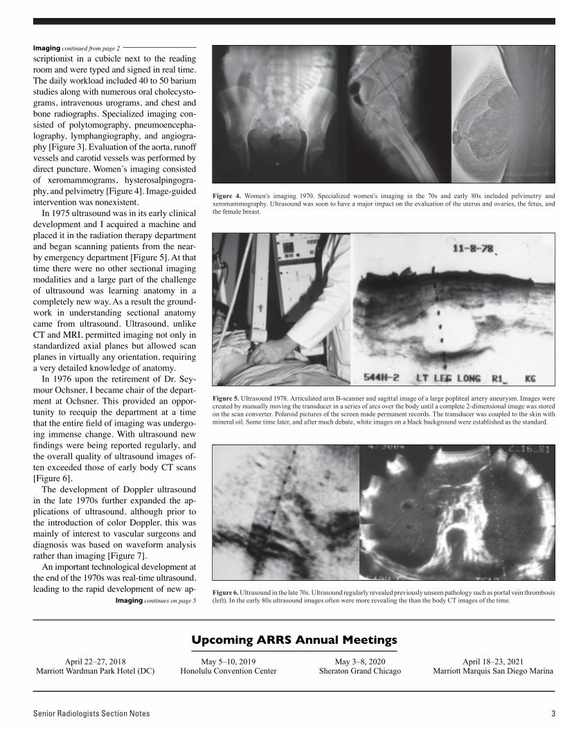
Figure 3. The early 1970s. Specialized imaging included now obsolete exams including lymphangiography, direct aortic angiography, and pneumoencephalography.
Imaging continues on page 3
Imaging continued from page 1
April 22–27, 2018Marriott Wardman Park Hotel (DC)
May 5–10, 2019Honolulu Convention Center
May 3–8, 2020Sheraton Grand Chicago
April 18–23, 2021Marriott Marquis San Diego Marina
Upcoming ARRS Annual Meetings
Imaging continued from page 2
scriptionist in a cubicle next to the reading room and were typed and signed in real time. The daily workload included 40 to 50 barium studies along with numerous oral cholecysto-grams, intravenous urograms, and chest and bone radiographs. Specialized imaging con-sisted of polytomography, pneumoencepha-lography, lymphangiography, and angiogra-phy [Figure 3]. Evaluation of the aorta, runoff vessels and carotid vessels was performed by direct puncture. Women’s imaging consisted of xeromammograms, hysterosalpingogra-phy, and pelvimetry [Figure 4]. Image-guided intervention was nonexistent.
In 1975 ultrasound was in its early clinical development and I acquired a machine and placed it in the radiation therapy department and began scanning patients from the near-by emergency department [Figure 5]. At that time there were no other sectional imaging modalities and a large part of the challenge of ultrasound was learning anatomy in a completely new way. As a result the ground-work in understanding sectional anatomy came from ultrasound. Ultrasound, unlike CT and MRI, permitted imaging not only in standardized axial planes but allowed scan planes in virtually any orientation, requiring a very detailed knowledge of anatomy.
In 1976 upon the retirement of Dr. Sey-mour Ochsner, I became chair of the depart-ment at Ochsner. This provided an oppor-tunity to reequip the department at a time that the entire field of imaging was undergo-ing immense change. With ultrasound new findings were being reported regularly, and the overall quality of ultrasound images of-ten exceeded those of early body CT scans [ Figure 6].
The development of Doppler ultrasound in the late 1970s further expanded the ap-plications of ultrasound, although prior to the introduction of color Doppler, this was mainly of interest to vascular surgeons and diagnosis was based on waveform analysis rather than imaging [Figure 7].
An important technological development at the end of the 1970s was real-time ultrasound, leading to the rapid development of new ap-
Figure 4. Women’s imaging 1970. Specialized women’s imaging in the 70s and early 80s included pelvimetry and xeromammography. Ultrasound was soon to have a major impact on the evaluation of the uterus and ovaries, the fetus, and the female breast.
Figure 5. Ultrasound 1978. Articulated arm B-scanner and sagittal image of a large popliteal artery aneurysm. Images were created by manually moving the transducer in a series of arcs over the body until a complete 2-dimensional image was stored on the scan converter. Polaroid pictures of the screen made permanent records. The transducer was coupled to the skin with mineral oil. Some time later, and after much debate, white images on a black background were established as the standard.
Figure 6. Ultrasound in the late 70s. Ultrasound regularly revealed previously unseen pathology such as portal vein thrombosis (left). In the early 80s ultrasound images often were more revealing the than the body CT images of the time.Imaging continues on page 5
Senior Radiologists Section Notes 3
SRS Notes
4 Fall 2017
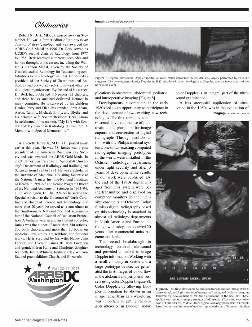
plications in obstetrical, abdominal, pediatric, and intraoperative imaging [ Figure 8].
Developments in computers in the early 1980s led to an opportunity to participate in the development of two exciting new tech-nologies. The first, unrelated to ul-trasound, involved the use of pho-tostimulable phosphors for image capture and conversion to digital radiographs. Through a collabora-tion with the Philips medical sys-tems one of two existing computed radiographic imaging prototypes in the world were installed in the Ochsner radiology department under tight security and after 3 years of development the results of our work were published. By the end of the 1980s digital im-ages from this system were be-ing transmitted and displayed on computer monitors in the inten-sive care units at Ochsner. Today filmless digital radiography based on this technology is standard in almost all radiology departments in North America and Europe, al-though wide adoption occurred 20 years after commercial units be-came available.
The second breakthrough in technology involved ultrasound and provided a method to image Doppler information. Working with a small company in Seattle and a large prototype device, we gener-ated the first images of blood flow in the abdomen and peripheral ves-sels using color Doppler [Figure 9]. Color Doppler, by allowing Dop-pler information be shown in an image rather than as a waveform, was important in getting radiolo-gists interested in Doppler. Today
color Doppler is an integral part of the ultra-sound examination.
A less successful application of ultra-sound in the 1980s was in the evaluation of
Figure 7. Doppler ultrasound. Doppler spectral analysis, when introduced in the 70s, was largely performed by vascular surgeons. The development of color Doppler in 1983 introduced many radiologists to Doppler, now an integral part of the ultrasound exam.
Imaging continued from page 3
Figure 8. Real-time ultrasound. Specialized transducers for intraoperative, transvaginal, and high-resolution breast, small parts, and pediatric imaging followed the development of real-time ultrasound in the late 70s. These applications remain a unique strength of ultrasound. (Top – intraoperative scan of brain abscess. Middle – transvaginal scan of gastroschisis in 14 week fetus. Lower – sagittal scan of newborn spine with cyst of filum terminiale.)
Imaging continues on page 6
ObituariesRobert N. Berk, MD, 87, passed away in Sep-
tember. He was a former editor of the American Journal of Roentgenology and was awarded the ARRS Gold Medal in 1996. Dr. Berk served as UCSD’s second chair of Radiology from 1977 to 1985. Berk received numerous accolades and honors throughout his career, including the Wal-ter B. Cannon Medal, given by the Society of Gastrointestinal Radiology for “outstanding con-tributions to GI Radiology” in 1984. He served as president of the Society of Gastrointestinal Ra-diology and played key roles in several other ra-diological organizations. By the end of his career, Dr. Berk had published 116 papers, 22 chapters, and three books, and had delivered lectures in many countries. He is survived by his children Daniel, Perry and Ellen; his grandchildren Adam, Aaron, Tammy, Michael, Emily, and Blythe; and his beloved wife Sondra Reidbord Berk, whom he celebrated in his memoir, “My Life with Son-dra and My Career in Radiology: 1955–1995, A Memoir with Special Memorabilia."
Senior Radiologists Section Notes 5
A. Everette James Jr., M.D., J.D., passed away earlier this year. He was 78. James was a past president of the American Roentgen Ray Soci-ety and was awarded the ARRS Gold Medal in 2003. James was the chair of Vanderbilt Univer-sity's Department of Radiology and Radiological Sciences from 1975 to 1991. He was a Scholar of the Institute of Medicine, a Visiting Scientist at the National Cancer Institute/National Institutes of Health in 1991–92 and Senior Program Officer of the National Academy of Sciences in 1993–94, all in Washington, DC. In 1994–95 he served the Special Advisor to the Governor of North Caro-lina and Board of Science and Technology. For more than 20 years he served as a consultant to the Smithsonian's National Zoo and as a mem-ber of the National Council of Radiation Protec-tion. A Vietnam veteran and an avid art collector, James was the author of more than 540 articles, 200 book chapters, and more than 20 books on medicine, law, ethics, art, folklore, and fictional works. He is survived by his wife, Nancy Jane Farmer; son Everette James III, wife Gretchen and grandchildren Katie and Charlotte; daughter Jeannette James Whitson, husband Clay Whitson Sr., and grandchildren Clay Jr. and Elizabeth.
SRS Notes
the breast. Early breast scanners produced quality images by scanning the breast, as the patient lay prone in a water tank. Un-fortunately, breast ultrasound was promot-ed aggressively by many manufacturers and by the mid 1980s was discredited as a use-ful addition to mammography. By the mid
1990s however, advances in breast ultra-sound demonstrated an important role in the evaluation of breast masses, making ultra-sound an indispensable part of breast imag-ing and leading to the BI-RADS breast im-aging and reporting system for ultrasound.
Ultrasound also has had a major impact in providing guidance for minimally invasive diagnostic procedures. Fine needle biopsy of lesions of the liver, kidney, retroperitone-um, as well as peripheral lymph nodes and
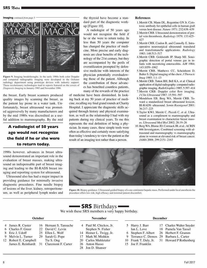
Figure 9. Imaging breakthroughs. In the early 1980s both color Doppler and computed radiographic imaging were developed in the Ochsner radiology department using prototype devices with industry support. These recolutionary technologies lead to reports featured on the covers of Diagnostis Imaging in January 1985 and November 1986.
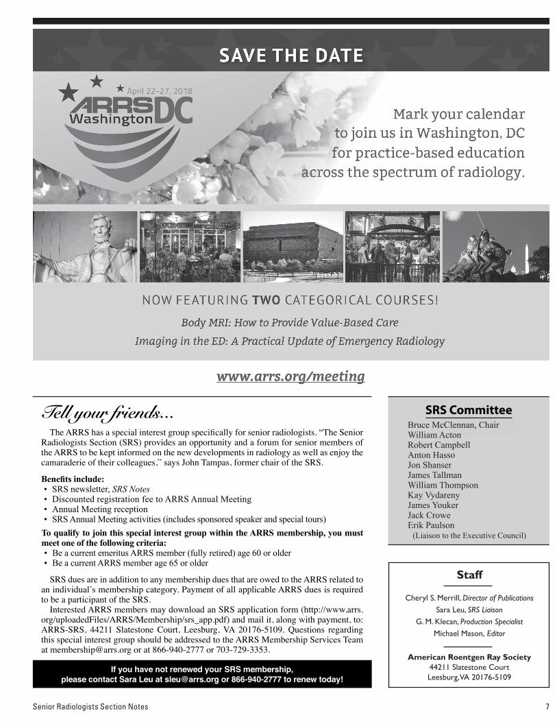
the thyroid have become a stan-dard part of the diagnostic work-up [Figure 10].
A radiologist of 50 years ago would not recognize the field if he or she were to return today. In less than 50 years the computer has changed the practice of medi-cine. More precise and early diag-nosis are clear benefits of the tech-nology of the 21st century, but they are accompanied by the perils of overutilization prompted by defen-sive medicine with interests of the physician potentially overshadow-ing those of the patient. Although the contribution of these advanc-es has benefited countless patients, many of the rewards of the practice
of medicine have been diminished. In look-ing back at my 50 years of practice of medi-cine, recalling my final grand rounds at Charity Hospital, I appreciate the diagnostic skills ac-quired through history and physical examina-tion, as well as the relationship I had with my patients during my clinical years. To me this represents the real definition of being a phy-sician. In many cases, these simple tools were often as effective and certainly more satisfying than today’s tendency to view the patient as the result of an imaging test rather than a person.
References 1. Merritt CB, Mann DL, Rogentine GN Jr. Cyto-
toxic antibody for epithelial cells in human graft versus host disease. Nature 1971; 232:638–639
2. Merritt CRB. Ultrasound demonstration of por-tal vein thrombosis. Radiology 1979; 133:425–427
3. Merritt CRB, Coulon R, and Connolly E. Intra-operative neurosurgical ultrasound: transdural and transfontanelle applications. Radiology 1983; 148:513–517
4. Merritt CRB, Goldsmith JP, Sharp MJ. Sono-graphic detection of portal venous gas in in-fants with necrotizing enterocolitis. AJR 1984; 143:1059–1062
5. Merritt CRB, Matthews CC, Scheinhorn D, Balter S. Digital imaging of the chest. J Thoracic Imag 1985; 1:1–13
6. Merritt CRB, Tutton RH, Bell KA, et al: Clinical application of digital radiography: computed radio-graphic imaging. RadioGraphics 1985; 5:397–414
7. Merritt CRB: Doppler color flow imaging. J Clin Ultrasound 1987; 15:591–597
8. Mendelson EB, Berg WA, Merritt CRB. To-ward a standardized breast ultrasound lexicon, BI-RADS: ultrasound. Semin Roentgenol 2001; 36:217–225
9. Taylor KWJ, Merritt C, Piccoli C, et al. Ultra-sound as a compliment to mammography and breast examination to characterize breast mass-es. Ultrasound Med Biol 2002; 28:19–26
10. Berg WA, Blume JD, Cormack JB, et al.; ACRIN 666 Investigators. Combined screening with ul-trasound and mammography vs mammography alone in women at elevated risk of breast cancer. JAMA 2008; 299:2151–2163
Imaging continued from page 5
Figure 10. Biopsy guidance. Ultrasound guided biopsy of a one centimeter hepatic mass. Performed under local anesthesia the procedure offers low risk, high efficacy and minimal patient discomfort.
6 Fall 2017
SRS BirthdaysWe wish these SRS members a very happy birthday.
4 Paul M. Chickos, Jr. Stephen N. Fisher14 Homer L. Twigg, Jr.17 Mark M. Mishkin20 Carlos Muhletaler26 Anton Hasso28 Jon D. Shanser
4 James R. Custer 6 Charles F. Greer 8 Eric J. Udoff 9 Melvin L. Turner12 Robert E. Campbell James E. Reinhardt
5 Harry J. Barr Ian L. Love 6 Stephen F. Albert 9 Terrance C. Demos10 Frank T. Daly, Jr.15 Joe F. Franklin
16 Hernani S. Tansuche22 David C. Levin25 Ellen L. Wolf29 Sarah G. Pope Tie S. Ong31 Claremont F. Carter
17 Charles Walter Snyder18 Pamela Van Tassel26 Herbert F. Gramm29 Barbara L. Carter31 Howard P Rothenberg
NovemberOctober December
A radiologist of 50 years ago would not recognize the field if he or she were
to return today.
Staff
Cheryl S. Merrill, Director of PublicationsSara Leu, SRS Liaison
G. M. Klecan, Production SpecialistMichael Mason, Editor
American Roentgen Ray Society44211 Slatestone CourtLeesburg, VA 20176-5109
To qualify to join this special interest group within the ARRS membership, you must meet one of the following criteria: • Be a current emeritus ARRS member ( fully r etired) age 60 or older • Be a current ARRS member age 65 or older
Tell your friends...
SRS dues are in addition to any membership dues that are owed to the ARRS related to an individual’s membership category. Payment of all applicable ARRS dues is required to be a participant of the SRS.
Interested ARRS members may download an SRS application form (http://www.arrs.org/ uploadedFiles/ARRS/Membership/srs_app.pdf) and mail it, along with payment, to: ARRS-SRS, 44211 Slatestone Court, Leesburg, VA 20176-5109. Questions regarding this special interest group should be addressed to the ARRS Membership Services Team at [email protected] or at 866-940-2777 or 703-729-3353.
The ARRS has a special interest group specifically for senior radiologists. “The Senior Radiologists S ection (SRS) provides an opportunity and a forum for senior members of the ARRS to be kept informed on the new developments in radiology as well as enjoy the camaraderie of their colleagues,” says John Tampas, f ormer chair of the SRS.
Benefits include: • SRS newsletter, SRS Notes • Discounted registration fee to ARRS A nnual Meeting • Annual Meeting reception • SRS Annual Meeting activities (includes s pon sored speaker and special tours)
If you have not renewed your SRS membership,please contact Sara Leu at [email protected] or 866-940-2777 to renew today!
SRS Committee Bruce McClennan, Chair William Acton Robert Campbell Anton Hasso Jon Shanser James Tallman William Thompson Kay Vydareny James Youker Jack Crowe Erik Paulson (Liaison to the Executive Council)
Senior Radiologists Section Notes 7
44211 Slatestone CourtLeesburg, VA 20176-5109USA
The Roentgen Fund® • 44211 Slatestone Court, Leesburg, VA 20176 • (703) 729-3353 • (866) 940-2777 • www.arrs.org/RoentgenFund/
For two decades, The Roentgen Fund® has been providing scholarshipsand awards for young radiologists. This past year, The Roentgen Fund®
was able to provide $384,600 to deserving radiologists.You can help us award even more… and get a special FREE gift.
Please help The Roentgen Fund® provide scholarships and awards to the next generation of radiologists with your tax-deductible gift of:
• Donors of $125 — will receive The Roentgen Fund® 12-pack Kooler with the famous “Hand of Mrs. Roentgen” image on it as their free gift for supporting the 2017 campaign.
• Donors of $250 — will receive a set of 4 CoasterStone® absorbing coasters with the famous “Hand of Mrs. Roentgen” image on them it as their free gift for supporting the 2017 campaign.
• Donors who contribute $500 or more will receive a unique double-layer inverted umbrella designed so you won’t get wet getting in or out of the car. The umbrella has the famous “Hand of Mrs. Roentgen” image on it.
Please visit www.arrs.org/RoentgenFund/ to make your tax-deductible gift today. Enter code S709 to receive your gift.
Roentgen AD_SRS Notes S708 Summer.indd 1 8/2/17 8:21 AM