upconverting nanoparticles: a versatile platform for wide
TRANSCRIPT
Upconverting nanoparticles: a versatile platform for wide-
field two-photon microscopy and multi-modal in vivo imaging
Journal: Chemical Society Reviews
Manuscript ID: CS-REV-05-2014-000173.R1
Article Type: Tutorial Review
Date Submitted by the Author: 04-Jul-2014
Complete List of Authors: Park, Yong Il; Seoul National University, School of Chemical and Biological
Engineering Lee, Kang Taek; Gwangju Institute of Science and Technology (GIST), Department of Chemistry Suh, Yung Doug; Korea Research Institute of Chemical Technology, Research Center for Convergence Nanotechnology Hyeon, Taeghwan; Seoul National University, School of Chemical and Biological Engineering
Chemical Society Reviews

1
Upconverting nanoparticles: a versatile platform for wide-field two-photon
microscopy and multi-modal in vivo imaging
Yong Il Park,abf Kang Taek Lee,
cf Yung Doug Suh*
de and Taeghwan Hyeon*
ab
a Center for Nanoparticle Research, Institute for Basic Science (IBS), Seoul 151-742, Korea
b School of Chemical and Biological Engineering, Seoul National University, Seoul 151-742,
Korea
E-mail: [email protected]
c Department of Chemistry, Gwangju Institute of Science and Technology (GIST), Gwangju
500-712, Korea
d Laboratory for Advanced Molecular Probing (LAMP), Research Center for Convergence
Nanotechnology, Korea Research Institute of Chemical Technology, Daejeon 305-600, Korea
E-mail: [email protected]
e School of Chemical Engineering, Sungkyunkwan University, Suwon 440-746, Korea
f These authors contributed equally to this work.
Page 1 of 41 Chemical Society Reviews

2
Yong Il Park received his B.S. (2005) and Ph.D. (2011) from
Chemical and Biological Engineering of Seoul National University
under the supervision of Prof. Taeghwan Hyeon. During his doctorate
period, he worked on the development of lanthanide-doped
upconverting nanoparticles and their biomedical application. He is
currently a postdoctoral researcher working with Prof. Hakho Lee and
Prof. Ralph Weissleder at Harvard Medical School/Massachusetts General Hospital.
Kang Taek Lee received his B.S. (1996), M.S. (1998), and Ph.D.
(2003) (supervisor: Prof. Seong Keun Kim) in the Department of
Chemistry of Seoul National University. He worked as a postdoctoral
researcher at Harvard University (supervisor: Prof. X. Sunney Xie)
and the University of Chicago (supervisor: Prof. Norbert F. Scherer)
until he became a senior researcher at Korea Research Institute of
Chemical Technology (KRICT) in 2007. In March 2013, he joined the faculty of the
Department of Chemistry at Gwangju Institute of Science and Technology (GIST). His new
research group is focusing on developing the optical imaging methods, particularly
employing multifunctional nanoparticle probes to directly monitor the dynamics in live cells.
Dr. Yung Doug Suh studied at Seoul National University for his
B.S. (1991), M.S. (1993), and Ph.D. (1999) under the guidance of
Prof. Seong Keun Kim in Chemistry Department, Dr. Dongho Kim
in Korea Research Inst. of Standards and Science (KRISS), and
Prof. Young Kuk in Physics researching gas phase molecular
reaction dynamics, laser spectroscopies, and surface physics with
Page 2 of 41Chemical Society Reviews
3
UHV-STM, respectively. After finishing the postdoctoral research in ETH Zurich with Prof.
Renato Zenobi (1999–2000), he worked at the Pacific Northwest National Laboratory
(PNNL), USA (2001–2002). He accepted his current position in 2003, to form his own
research group: Laboratory for Advanced Molecular Probing (LAMP) at the Korea Research
Institute of Chemical Technology (KRICT) in Daejeon, Korea.
Taeghwan Hyeon received his B. S. (1987) and M. S. (1989) from
Chemistry in Seoul National University. He obtained his Ph. D.
(1996) from Chemistry in University of Illinois at Urbana-
Champaign. Since he joined the faculty of the School of Chemical
and Biological Engineering of Seoul National University in
September 1997, he has focused on the synthesis and applications
of uniform-sized nanocrystals and nanoporous materials. He is a Director of Center for
Nanoparticle Research in the Institute for Basic Science (IBS). He is currently serving as an
Associate Editor of J. Am. Chem. Soc., and editorial advisory board members of Adv. Mater.,
Nanoscale, Nano Today, and Small.
Page 3 of 41 Chemical Society Reviews
4
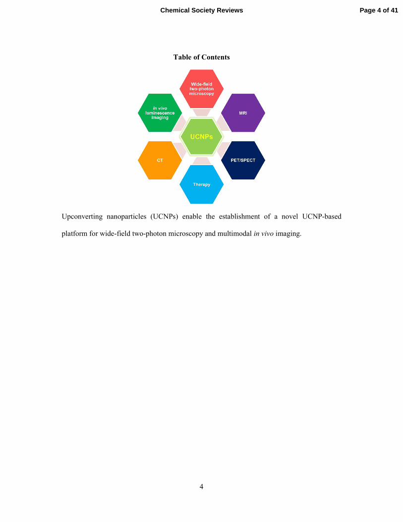
Table of Contents
Upconverting nanoparticles (UCNPs) enable the establishment of a novel UCNP-based
platform for wide-field two-photon microscopy and multimodal in vivo imaging.
Page 4 of 41Chemical Society Reviews
5
Abstract. Lanthanide-doped upconverting nanoparticles (UCNPs) have recently attracted
enormous attention in the field of biological imaging owing to their unique optical properties:
1) efficient upconversion photoluminescence, which is intense enough to be detected at the
single-particle level with a (nonscanning) wide-field microscope setup equipped with a
continuous wave (CW) near-infrared (NIR) laser (980 nm), and 2) resistance to photoblinking
and photobleaching. Moreover, the use of NIR excitation minimizes adverse photoinduced
effects such as cellular photodamage and the autofluorescence background. Finally, the
cytotoxicity of UCNPs is much lower than that of other nanoparticle systems. All these
advantages can be exploited simultaneously without any conflicts, which enable the
establishment of a novel UCNP-based platform for wide-field two-photon microscopy.
UCNPs are also useful for multimodal in vivo imaging because simple variations in the
composition of the lattice atoms and dopant ions integrated into the particles can be easily
implemented, yielding various distinct biomedical activities relevant to magnetic resonance
imaging (MRI), computed tomography (CT), and positron emission tomography (PET).
Those multiple functions embedded in a single type of UCNPs play a crucial role in precise
disease diagnosis. The application of UCNPs is extended to therapeutic fields such as
photodynamic and photothermal cancer therapies through advanced surface conjugation
schemes.
Page 5 of 41 Chemical Society Reviews
6
Key learning points
(1) Lanthanide-ion-doped upconverting nanoparticles (UCNPs) exhibit 1) anti-Stokes
photoluminescence in the visible spectral range upon absorption of near-infrared (NIR)
photons through an optical upconversion process, 2) extreme photostability (no photoblinking
and no photobleaching), and 3) low cytotoxicity.
(2) The use of NIR excitation completely suppresses cellular autofluorescence and
photoinduced damage to cells.
(3) UCNPs provide a novel versatile platform for wide-field two-photon imaging that
overcomes the drawbacks of conventional two-photon microscopy while maintaining its
advantages.
(4) The UCNP library with various lanthanide elements supports multiple imaging modalities,
including luminescence imaging, magnetic resonance imaging, positron emission tomography,
and computed tomography.
(5) UCNPs designed for medical purposes will provide enhanced diagnostic and therapeutic
efficacy.
Page 6 of 41Chemical Society Reviews

7
1. Introduction
Anti-Stokes emitting molecules and materials have attracted great attention recently as
next-generation luminescent materials in the fields of energy, biology, and medicine. Their
emission wavelength is shorter than that of the excitation light, where the relevant
photophysical processes include the upconversion process, second-harmonic generation, and
two-photon absorption. Among them, the upconversion process is the most efficient because
it involves stable intermediate states. Since their discovery in 1966, upconverting phosphors
have been synthesized by doping d and f elements into hosts.1,2 In particular, lanthanide-
doped upconverting materials have been studied most intensively because of their bright
upconversion luminescence at room temperature, and they have been used for various
applications such as lasers, infrared quantum counters, and displays.1,2 Most notably, there
have been tremendous efforts to synthesize nanosized “upconverting particles” and apply
them to biological imaging, early-stage diagnoses, and therapeutics in past years, in which
their unique optical properties, namely, the absorption of near-infrared (NIR) photons and the
emission of visible and ultraviolet (UV) photons are widely exploited.3,4 Technological
advances in nanomaterial synthesis and surface functionalization have prompted researchers
to develop upconverting nanoparticles (UCNPs) featuring a homogeneous chemical
composition, a narrow size distribution, and biological functions.5–7
The photophysical pathway associated with upconversion photoluminescence is relatively
well understood. The widely accepted energy level diagram of the representative UCNPs
NaYF4:Yb3+,Er
3+ and their photoluminescence spectra for the α type (cubic phase) and β type
(hexagonal phase) are shown in Figure 1.8 A Yb
3+ in the UCNPs absorbs a 980-nm photon to
the 2F5/2 state and transfers its excitation energy to the
4I11/2 state of a neighboring Er
3+. The
energies of those two states are very close, which allows the energy to be transferred
efficiently. Subsequently, an additional energy transfer occurs from another Yb3+ to the Er
3+,
Page 7 of 41 Chemical Society Reviews

8
resulting in further excitation to a higher level of Er3+ (4F7/2). After fast nonradiative
relaxation to one of the three states that are slightly lower in energy (2H11/2,
4S3/2, or
4F9/2), the
doubly exited Er3+ emits one photon (ca. 525, 550, or 660 nm, respectively) whose energy is
slightly less than that of two 980-nm photons. For the red emission around 660 nm, a possible
alternative photophysical mechanism has been suggested in which nonradiative relaxation
occurs from the intermediate excited state of Er3+ (
4I11/2) to a lower-energy state (
4I13/2)
immediately after the first energy transfer, instead of the above-mentioned possibility
occurring between 4F7/2 and
4F9/2 (Figure 1a).
9,10 The emission efficiency of UCNPs also
depends on the crystal structure of the host materials. Hexagonal phase UCNPs usually
exhibit about 10 times more intense green emission than cubic phase ones, which is
originated from the interaction of lanthanide ions in the two different lattice structures.2
Cubic phase sodium rare earth (RE) fluoride (NaREF4) has fluorite structure with one type of
high-symmetry cation sites that are randomly occupied by Na+ and RE
3+ ions (Figure 1d). On
the contrary, hexagonal phase NaREF4 has two types of low-symmetry cation sites that are
selectively occupied by Na+ and RE
3+ ions (Figure 1e).
11 Thus, the UCNPs with hexagonal
structure reduce unnecessary cross-relaxation among the doped-lanthanide ions, resulting in
more efficient upconversion luminescence than the cubic ones.
The upconversion photoluminescence of UCNPs is not only an exotic and interesting
photophysical phenomenon but also has a great deal of potential for biological imaging. The
upconversion process, which converts NIR photons to visible photons, improves cellular
imaging techniques by providing a variety of benefits resulting from the extreme
photostability of UCNPs and the use of a NIR (980-nm) laser as the excitation light source.
It is interesting to compare the upconversion process to two-photon absorption, which is
employed in conventional two-photon microscopy. They resemble each other in that the
emitters (Er3+ in UCNPs in the former and dye molecules in the latter) are excited twice by
Page 8 of 41Chemical Society Reviews
9
two quanta of energy. However, the first distinct feature of upconversion photoluminescence
is that the excitation occurs in the sensitizer Yb3+ cations rather than in the emitter Er
3+
cations themselves. This is because the absorption efficiency (extinction coefficient) of the
electronic transition of Yb3+ is much higher than that of Er
3+ at 980 nm.
12 The energy transfer
occurring after absorption is responsible for the two-photon excitation of the emitter, Er3+,
whereas, in normal two-photon microscopy, the emitter (dye molecule) itself is excited
directly. The second, and more important, difference is that the intermediate excited state of
Er3+ is a real state with a long lifetime on the millisecond time scale,
1 whereas the lifetime of
two-photon dye molecules is extremely short because the excitation involves a “virtual”
intermediate state. In the latter case, two-photon absorption becomes feasible only when the
photon flux is sufficiently high within a very short period of time. Therefore, ultrashort laser
pulses from femtosecond laser systems are commonly used to excite the dye molecules. On
the other hand, the long lifetime of the intermediate excited state of Er3+ prepared after the
first energy transfer from Yb3+ guarantees sufficient time for the second energy transfer to
occur. This fact indicates that only moderate excitation power is needed to excite UCNPs,
and it has been shown that a small, inexpensive continuous wave (CW) diode laser (980 nm)
is an adequate light source. As a result, it follows that the wide-field microscopy can easily be
realized for UCNP imaging. In typical two-photon microscopy, one has to focus the
femtosecond laser pulse tightly on the sample and acquire fluorescence signals by scanning
the sample or the laser beam over the field of view. In such cases, as in conventional confocal
microscopy, the data acquisition time is significantly long. However, in the wide-field
imaging mode, two-dimensional images are projected onto the detector in each round of data
acquisition; therefore, the imaging frame rate can routinely be set as high as 10–100 frames/s
if the luminescence is sufficiently strong, which is the case with UCNPs.13
Page 9 of 41 Chemical Society Reviews

10
UCNPs are also useful for in vivo imaging, which is related to biomedical applications
such as accurate diagnosis and efficient therapy in diagnosing and prescribing treatment for
certain diseases. In serious diseases such as cancer and neuronal disorders, treatment is
simpler and more effective if the condition is diagnosed early. For instance, the five-year
survival rate of women diagnosed with ovarian cancer at stage I is 93%, whereas that of
patients diagnosed at stage III is 30%.14 To achieve accurate diagnosis, a variety of medical
imaging techniques have been developed: magnetic resonance imaging (MRI), computed
tomography (CT), and positron emission tomography (PET), to name a few. These imaging
tools have diverse degrees of sensitivity, spatial resolution, and imaging depth. MRI provides
anatomical details based on soft-tissue contrast and functional information, but its sensitivity
is relatively low. PET provides high detection sensitivity with low spatial resolution. To
compensate for the limits of each imaging technique, a number of studies have attempted to
develop novel multimodal imaging probes. UCNPs were recently recognized as a suitable
candidate because various lanthanide ion dopants with distinct properties and functions can
be integrated into individual particles. In other words, UCNPs can be developed as useful
multimodal imaging probes for optical imaging, MRI, CT, and PET. Fortunately, it is
technically easy to dope multiple types of lanthanide ions together into a host matrix because
they have similar atomic sizes and chemical properties.
In this tutorial, we focus on the versatility of UCNPs as a platform for both wide-field two-
photon microscopy and multimodal in vivo imaging.
2. UCNP-based wide-field two-photon imaging
2.1 Nonscanning wide-field imaging of UCNPs
As mentioned in the introduction, one of the most important consequences of the two-
photon upconversion processes in UCNPs is that UCNPs can be imaged by wide-field
Page 10 of 41Chemical Society Reviews
11
microscopy, such as epi-fluorescence and total internal reflection fluorescence (TIRF)
microscopy, without scanning the sample or using laser excitation as in confocal microscopy
(Figure 2). Epi-fluorescence microscopy is characterized by the collimated illumination on
the sample and the collection of two dimensional fluorescence images by two-dimensional
detectors such as charge coupled device (CCD) cameras. In general, this technique is
appropriate for imaging a certain plane (focal plane of the objective lens) inside cells.
However, the illumination that passes through the entire sample generates a significant signal
not only from the focal plane but also from the out-of-focus region. The out-of-focus signal
acts as the background that smears the image and, therefore, lowers the image contrast. On
the other hand, confocal microscopy overcomes this problem by focusing the illuminating
light source and placing a pinhole in front of the photo-detector such as photomultiplier tube
(PMT) and avalanche photodiode (APD). In this scheme, the out-of-focus signals do not pass
the hole and are not detected by the detector. As a result, the fluorescence signal only from
the focal point reaches the detector without significant contamination. However, the rate of
image acquisition is limited (~10 frames/s) because they are constructed by sequentially
attaching the point signals obtained while scanning the sample or the focus of light. The
confocal microscopy might be the best choice to investigate the cellular structure that is fixed
or evolving very slowly. But it is not an appropriate tool for imaging fast dynamics in cells.
TIRF microscopy adopts the advantages of the above-mentioned two microscopic techniques
in that only the thin layers of evanescent electromagnetic field is illuminated, which removes
the background signal from the cell body, and at the same time, the detection of two
dimensional image is achieved with wide-field imaging scheme. But again, it cannot image
the deeper structure of cells. For UCNPs, it turns out that the epi-fluorescence microscopy
approach is especially useful. In particular, Park et al. showed that even photoluminescence
of a single UCNP (cubic phase, diameter of ca. 40 nm) could be imaged with wide-field epi-
Page 11 of 41 Chemical Society Reviews

12
fluorescence microscopy.5 Subsequently, Nam et al. reported the first real-time single-
particle tracking of a UCNP (hexagonal phase, diameter of ca. 30 nm) internalized in a living
cell at a frame rate of 20 Hz (50-ms exposure time per frame).13 In those studies, the wide-
field epi-fluorescence imaging setup consisted of an inverted microscope, a NIR (980-nm)
CW diode laser, an electron-multiplying charge-coupled device (EMCCD) camera, and the
optical components in the excitation and detection pathways. The laser was introduced to the
microscope and directed to the UCNP-containing sample after being reflected by a dichroic
beam splitter. This dichroic beam splitter was designed to reflect the excitation laser (980 nm)
toward the sample through the center of the objective and to transmit the photoluminescence
in a shorter wavelength range. Such short-pass dichroic separation is obviously required
because the UCNP photoluminescence lies in the visible spectral region with a huge anti-
Stokes shift from the NIR excitation (980 nm). As in typical epi-fluorescence microscopy, the
incident excitation laser beam is focused on the back focal point of the objective by a lens
placed in front of the entrance port of the microscope. This ensures that the beam is
collimated to dimensions of tens of micrometers (typically ca. 60 µm x 60 µm) when it
reaches the focal plane, where the samples are supposed to be. The photoluminescence from
the excited UCNPs is collected by the same objective and directed to and projected at the
detector chip of the EMCCD. In the middle of this detection pathway, the photoluminescence
signals pass the dichroic beam splitter, whereas strong stray light (980 nm) is either scattered
from the sample and the optics in the objective or reflected at the cover glass–sample
interface. The residual 980-nm photons are further filtered out by a short-pass or band-pass
filter with a transmission wavelength window below 700 nm. The image might be subject to
further magnification by placing a pair of lenses with different focal lengths at the correct
positions between the microscope output port and the camera. This additional magnification
is required to adjust the actual dimensions of the images, which determine the typical number
Page 12 of 41Chemical Society Reviews
13
of cells within the field of view in cellular imaging experiments. Sometimes it might be
necessary to separately acquire the images by monitoring two distinct channels, namely, the
green and red bands of the UCNPs, and this can be achieved by placing another set consisting
of a dichroic beam splitter and band-pass filters between the microscope output port and the
camera. Each of the separate images is finally projected on half of the EMCCD chip, as in
most single-molecule Förster resonance energy transfer (FRET) studies.15
Traditionally, confocal microscopy is preferred in cellular imaging when one needs to
construct three-dimensional images of cells because of its ability to eliminate the out-of-focus
background and the resultant image sectioning capability. With the NIR laser that excites
UCNPs, however, no autofluorescence is observed from the cell bodies, as will be elaborated
in the following section. Therefore, the out-of-focus background is very low, and the section
images over the z axis are readily resolved simply by adjusting the focal plane of the
objective. The “confocal-like” sectioning capability of wide-field imaging is one of the most
attractive aspects of UCNP-based imaging. More recently, UCNPs were successfully imaged
with TIRF microscopy.16 TIRF imaging will obviously provide more clear information on the
cell membrane dynamics adjacent to the cover glass, where the ca. 100-nm-thick evanescent
electromagnetic field is generated.
2.2 Photostability of UCNPs
The photostability of fluorescent molecules or luminescent nanomaterials can be discussed
according to two distinct characteristics: (1) how much the luminescence intensity fluctuates
over the data acquisition time and (2) how long the luminescence persists under continuous
irradiation.
Regarding the first point, photoblinking is of the most concern, especially in single-
molecule or live cell imaging. It is a type of on-off fluctuation of the photoluminescence
Page 13 of 41 Chemical Society Reviews

14
observed even under continuous excitation. Semiconductor nanocrystals (quantum dots, QDs)
are the most intensively studied systems with regard to photoblinking.17,18
Although a number
of mechanisms have been suggested, the exact mechanism is yet to be determined.
Photoblinking limits the use of QDs as an imaging probe, especially in single-particle
tracking studies, where continuous or uninterrupted photoluminescence is crucial. On the
other hand, UCNPs have been known to exhibit no photoblinking at the single-particle level,
as shown in Figure 3.5,19 The photoluminescence intensity is preserved constantly without
any dark period. This property of UCNPs, combined with the fact that they are bright enough
to be detected at the single-particle level, enables continuous imaging with millisecond or
microsecond temporal resolution.13 It is not clear yet whether photoblinking does not occur in
individual Er3+-Yb
3+ upconversion units. However, one may simply note that, even if it does,
the upconversion processes occurring in a particle among numerous randomly positioned Er3+
and Yb3+ ions would statistically produce constant photoluminescence.
19
The second point to consider regarding photostability is a phenomenon called
photobleaching. Most luminescent objects irreversibly lose their capability to emit photons
after a finite period of excitation owing to chemical degradation to nonfluorescent states,
occurring presumably in the excited state. For example, single organic dye molecules and
fluorescent proteins are bleached within seconds or minutes when they are excited by a laser
or any other light source with the typical power density for single-molecule imaging.
Photobleaching obviously limits the time window for observation or imaging because
dynamics with a duration longer than the typical lifetime of the probes cannot be fully
measured, so only short temporal fragments of information are obtained. Inorganic
nanoparticle systems generally exhibit much less photobleaching than organic dyes.15 In
particular, with single-particle imaging, it has been demonstrated that the luminescence of
UCNPs is extremely resistant to photobleaching even under hours of continuous
Page 14 of 41Chemical Society Reviews
15
excitation.5,19 This remarkable characteristic behavior indicates that the sensitizer-emitter
dopant cations do not undergo chemical degradation as a result of repeated excitation-
photoluminescence cycles.3 Moreover, the robust NaYF4 crystal systems are likely to protect
the dopant cations. This might be one of the greatest advantages of UCNPs for biological
imaging.7 As a result, long-term (6 h) continuous live cell imaging is possible using UCNPs,
providing rich information on the cellular dynamics (Figure 4).13
2.3 Suppression of cellular autofluorescence and photodamage by NIR light sources
The advantages of UCNPs as imaging probes derive not only from the optical and
chemical nature of the nanoparticles but also from the use of a NIR light source for exciting
the UCNPs. First, NIR photons are not well absorbed by most biomolecules. In particular, the
wavelength range of 750–900 nm is considered a spectrally transparent window with the
smallest absorption cross section (i.e., extinction coefficient, which is proportional to the
absorbance) of the biomolecules in cells.20 The wavelength of 980 nm does not fall within
this range, and it shows a slightly larger absorption cross section because the onset of the
vibrational transition of H2O molecules is around 980 nm, and its intensity increases at longer
wavelengths. However, the absorption cross section at 980 nm is only one order of magnitude
larger than that in the transparent window (i.e., 750–900 nm) and is still very much lower
than those in the UV, visible, and IR spectral ranges. Thus, absorption by water molecules at
980 nm may not be sufficiently critical to cause problems in most cases. In fact, a few studies
have investigated the effect of 980-nm excitation on cellular imaging.13,21
The advantages of
980-nm excitation found in those studies are (1) the absence of autofluorescence and (2) a
low level of cellular photodamage. Autofluorescence is the photoluminescence (fluorescence
in general) resulting from excitation of natural cellular molecules that contributes to the
background and competes with the signal from the probe we want to detect. Especially when
Page 15 of 41 Chemical Society Reviews

16
the signal intensity of the luminescent probe is not overwhelming, the background
autofluorescence causes significant problems in analyzing the probe signals.
Autofluorescence is usually generated in the visible spectral range, so we cannot intrinsically
avoid its adverse contributions to images. However, with 980-nm or, more generally, NIR
excitation, there is essentially no absorption by the biomolecules in the cell, as noted above;
therefore, there should be no detectable autofluorescence.5,13 As a result, overall the images
display a very high level of contrast, so even a single UCNP can readily be observed with a
very low background signal.13
Another advantage of using a NIR laser is that the photoinduced physical and chemical
degradation of biomolecules is minimized. Photodamage generally originates from
irreversible chemical reactions occurring in the excited states upon photon absorption. As
mentioned above, in the NIR spectral region, absorption by biomolecules and H2O is
minimized, which indicates that the NIR excitation scheme in UCNP imaging effectively
bypasses the first step in photodamage, namely, dumping excess energy to biological systems
through photon absorption.20 This noninvasive characteristic of NIR lasers has been discussed
intensively in the field of biological imaging, and a few research groups recently confirmed
that UCNPs do not induce noticeable photodamage even under harsh conditions.13,21
2.4 Versatile platform for wide-field two-photon microscopy
We have reviewed the benefits of using the novel imaging probe, UCNPs, especially from
the perspective of cellular imaging. They are bright (detectable at the single-particle level),
optically stable (nonblinking and nonbleaching), and biologically compatible (low
cytotoxicity). The NIR laser used for excitation of these probes is noninvasive to cells and
induces very little cellular autofluorescence. All these advantages can be integrated into and
embodied in single UCNP particles, each of which can be used as a probe for wide-field two-
Page 16 of 41Chemical Society Reviews
17
photon imaging. They can be used as labels for biomolecules such as nucleic acids and
proteins in cells or as drug carriers. In both cases, what is important is that this UCNP-based
imaging platform exhibits all the merits of conventional two-photon microscopy while
simultaneously overcoming its drawbacks. In other words, it can be used for long-term
continuous tracking of cellular dynamics, either slow or fast, using wide-field imaging
techniques such as TIRF and epi-fluorescence microscopy. With this platform at hand, all we
have to do is to endow UCNPs with the appropriate functions required for specific purposes
using surface functionalization, namely, conjugation of functional biomolecules. These are
necessary for the UCNPs to bind target molecules with high specificity and selectivity.
Interestingly, the transitions responsible for the multiple emission bands are spectrally well
separated, and each transition might be designed to perform different roles simultaneously.
For example, one can use the green bands of NaYF4/Yb3+,Er
3+ to monitor the location of the
UCNP-biomolecule complex and the red band to monitor the binding or dissociation of
biomolecules and UCNPs using FRET. Those studies will extend the scope of information on
the mechanism of biochemical reactions.
We should note at this point that there is still room for improvement in this promising
imaging platform. First, UCNPs need to be smaller because the typical UCNPs used in most
studies to date are tens of nanometers in diameter, which is quite bulky, especially when they
are used to label biomolecules a few nanometers in size. As for synthesis, miniaturizing
particle sizes to smaller than or comparable to those of biomolecules is nothing but
controlling the reaction conditions. However, such efforts will face difficulties because they
are intrinsically accompanied by a decrease in the upconversion efficiency. This is due
mostly to the reduction in the number of dopant cations in the UCNPs, which scales inversely
with volume, and partly to the decreased crystal field on Yb3+ and Er
3+ in smaller
nanoparticles. Fortunately, Cohen et al. recently reported methods of synthesizing small
Page 17 of 41 Chemical Society Reviews

18
UCNPs of <10 nm in diameter that were still bright enough to be detected at the single-
particle level.6 This is a very promising result, and efforts to further reduce the particle size
will make this system a more attractive and powerful platform for advanced bioimaging. To
preserve or enhance the upconversion photoluminescence of smaller UCNPs may require a
more profound understanding of the photophysics by which one can increase the absorption
cross section and quantum yield. The second barrier is the biochemical surface
functionalization. The coating material and conjugation of the biological moiety that allows
more specific and selective interaction with the target cellular molecules are obviously of
utmost importance in this field. Although a number of schemes for surface functionalization
have been reported, their practical applications are strongly system-dependent. The
development of a surface functionalization technology that is simultaneously universal and
specific is crucial for finalizing the establishment of a versatile UCNP-based platform for
wide-field two-photon imaging.
3. Multimodal in vivo imaging
3.1 Luminescence imaging
As described in the previous sections, the unique optical properties of UCNPs, such as the
lack of background autofluorescence and low photodamage, are advantageous for in vivo
luminescence imaging. The NIR excitation source offers an increased signal-to-background
ratio, resulting in high imaging contrast. Cheng et al. compared the in vivo imaging
sensitivity of UCNPs and QDs.22 Mice were subcutaneously injected with UCNPs
(NaYF4:Er3+,Yb
3+) and QDs (QD 545 and QD 625) at various nanoparticle concentrations
and then imaged. The results showed that the in vivo detection sensitivity of the UCNPs is
one order of magnitude higher than that of the QDs. In addition, NIR light also increases the
tissue penetration depth because of the low absorption of NIR by the biological environment,
Page 18 of 41Chemical Society Reviews
19
which is very important for noninvasive animal imaging. When 800-nm emitting Tm3+-doped
UCNPs were tested, the imaging depth in a tissue phantom was ~600 µm in two-photon laser
scanning microscopy and ~400 µm in two-photon wide-field microscopy, which is at least
twice the depth for visible-light emitting fluorophores.23
Over the last decade, there have been numerous reports on in vivo luminescence imaging
using UCNPs. Several papers on the biodistribution of UCNPs reported different distribution
patterns of UCNPs in the body depending on the size and surface coating.24 For example,
poly(acrylic acid)-coated UCNPs were imaged in vivo for more than 7 days to monitor the
long-term in vivo distribution of the nanoparticles; the results showed that the injected
UCNPs were excreted through the liver and spleen. The in vivo distribution of nanoparticles
during luminescence imaging was also confirmed using elemental analysis measurement
(inductively coupled plasma atomic emission spectroscopy), demonstrating the usefulness of
UCNPs for noninvasive whole-body imaging. Targeted tumor imaging was also successfully
demonstrated by conjugation of targeting peptides or antibodies.24 Cyclic-RGD-peptide-
conjugated UCNPs showed a high signal-to-noise ratio (~24) between the tumor and its
background. The high sensitivity of UCNPs can also be applied to in vivo stem cell
tracking.25 UCNP-labeled mesenchymal stem cells were subcutaneously transplanted into
mice, and images were acquired using the Maestro in vivo imaging system. About 10 UCNP-
labeled cells could be detected; this detection limit is lower by at least 2 orders of magnitude
than that of QDs or MRI contrast agents. Further, the emission intensity changes depending
on the number of cells. The high sensitivity and photostability of UCNPs can play an
important role in following the translocation of implanted stem cells. After UCNP-labeled
mesenchymal stem cells were injected intravenously, their temporal translocation from the
lung to the liver could be monitored for 24 h.
Page 19 of 41 Chemical Society Reviews
20
Multiplexing is a unique characteristic of the luminescence imaging modality and makes it
possible to measure different targets accurately and simultaneously. The emission wavelength
of QDs can be tuned by controlling the particle size. Although fine-tuning the particle size of
QDs is difficult, UCNPs are preferable to QDs for multiplexing because the emission
wavelength of UCNPs can be easily tuned by varying the dopant ions rather than the particle
size, as in QDs. The characteristic emission of UCNPs is determined by the ladder-like
atomic energy level of each lanthanide dopant ion; therefore, the preparation of UCNPs with
different sharp emission peaks is feasible simply by doping with the corresponding lanthanide
ion. There are several reports on the tuning of upconversion luminescence. First, by changing
the emitter dopant, the emission wavelength can be changed from the UV to the NIR. When
Er3+ or Ho
3+ is co-doped as an emitter, the UCNPs emit green and red light, whereas Tm
3+-
doped UCNPs emit blue and NIR (800-nm) light. In addition, the emitted color can be tuned
by combining different types of emitter ions, in which the resulting color is determined
through the relative ratio of green, red, and blue emission. Second, the emission wavelength
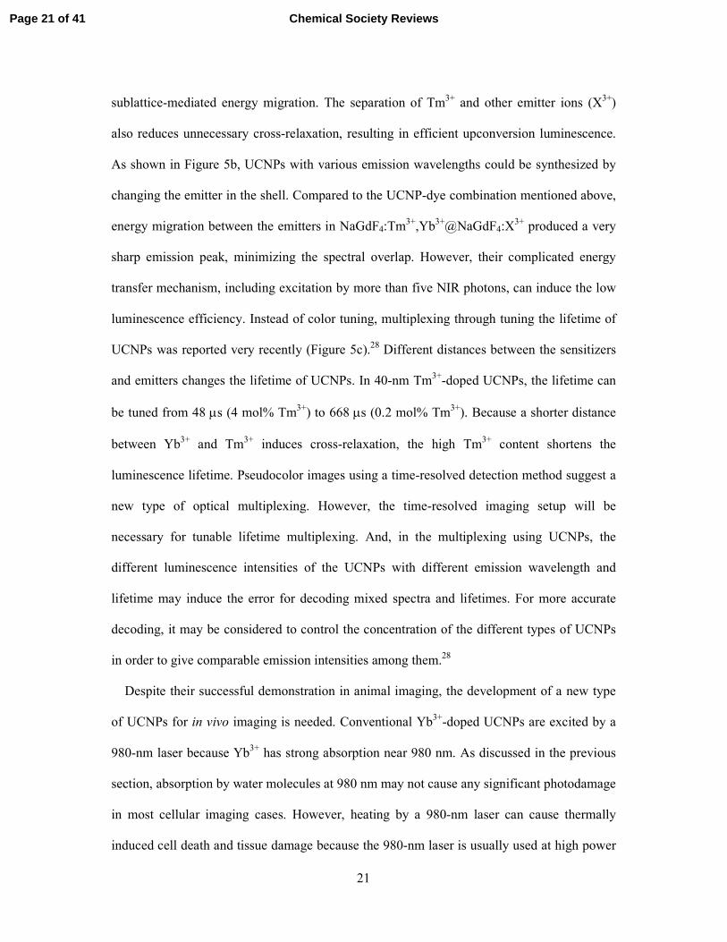
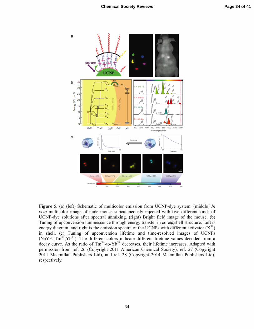
can be tuned using energy transfer between UCNPs and dyes (Figure 5a).26 If the absorption
band of the dyes matches the emission band of the UCNPs, the dye on the surface of the
UCNPs can be activated by 980-nm irradiation through energy transfer from the UCNPs to
the dyes. By incorporating different types of dyes into UCNPs, multicolor-emitting UCNPs
could be synthesized. In vivo multicolor imaging using a UCNP-dye combination was also
successfully demonstrated after spectral unmixing using the modified Maestro in vivo
imaging setup (Figure 5a).26 The Liu group recently suggested using energy migration among
the emitter ions for upconversion luminescence tuning (Figure 5b).27 The UCNPs have a
core–shell structure, in which the core is UCNPs (NaGdF4:Tm3+,Yb
3+) and the shell is
NaGdF4:X3+ (X
3+: emitter ions such as Dy
3+, Tb
3+, Eu
3+, and Sm
3+). The energy excited in
Tm3+ from Yb
3+ ions in the core is transferred to emitter ions (X
3+) in the shell through Gd
3+
Page 20 of 41Chemical Society Reviews
21
sublattice-mediated energy migration. The separation of Tm3+ and other emitter ions (X
3+)
also reduces unnecessary cross-relaxation, resulting in efficient upconversion luminescence.
As shown in Figure 5b, UCNPs with various emission wavelengths could be synthesized by
changing the emitter in the shell. Compared to the UCNP-dye combination mentioned above,
energy migration between the emitters in NaGdF4:Tm3+,Yb
3+@NaGdF4:X
3+ produced a very
sharp emission peak, minimizing the spectral overlap. However, their complicated energy
transfer mechanism, including excitation by more than five NIR photons, can induce the low
luminescence efficiency. Instead of color tuning, multiplexing through tuning the lifetime of
UCNPs was reported very recently (Figure 5c).28 Different distances between the sensitizers
and emitters changes the lifetime of UCNPs. In 40-nm Tm3+-doped UCNPs, the lifetime can
be tuned from 48 µs (4 mol% Tm3+) to 668 µs (0.2 mol% Tm
3+). Because a shorter distance
between Yb3+ and Tm
3+ induces cross-relaxation, the high Tm
3+ content shortens the
luminescence lifetime. Pseudocolor images using a time-resolved detection method suggest a
new type of optical multiplexing. However, the time-resolved imaging setup will be
necessary for tunable lifetime multiplexing. And, in the multiplexing using UCNPs, the
different luminescence intensities of the UCNPs with different emission wavelength and
lifetime may induce the error for decoding mixed spectra and lifetimes. For more accurate
decoding, it may be considered to control the concentration of the different types of UCNPs
in order to give comparable emission intensities among them.28
Despite their successful demonstration in animal imaging, the development of a new type
of UCNPs for in vivo imaging is needed. Conventional Yb3+-doped UCNPs are excited by a
980-nm laser because Yb3+ has strong absorption near 980 nm. As discussed in the previous
section, absorption by water molecules at 980 nm may not cause any significant photodamage
in most cellular imaging cases. However, heating by a 980-nm laser can cause thermally
induced cell death and tissue damage because the 980-nm laser is usually used at high power
Page 21 of 41 Chemical Society Reviews

22
for in vivo imaging. For this reason, a 915-nm laser has been used instead of a 980-nm
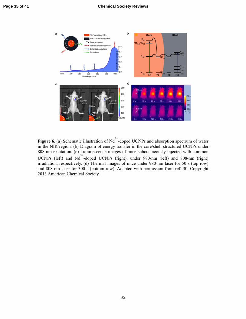
excitation source, with a sacrifice in emission efficiency.29 Nd
3+ was recently introduced to
UCNPs as a new sensitizer because it has a sharp absorption band around 800 nm.30 Because
the energy transfer from Nd3+ to Yb
3+ is highly efficient, Nd
3+ can be used to excite emitters
(Er3+, Tm
3+, and Ho
3+) through Yb
3+. The excitation wavelengths of Nd
3+ are shorter than that
of Yb3+ (980 nm), and the absorption by water molecules around 800 nm is much lower than
that at 980 nm (Figure 6a). The Yan group reported the synthesis of Nd3+-doped UCNPs
(NaGdF4:Er3+,Yb
3+@NaGdF4:Yb
3+,Nd
3+), which can be activated by an 808-nm laser (Figure
6b).30 Nd
3+ ions in the shell of the UCNPs absorb an 808-nm photon to the
4F5/2 state and then
relax to the 4F3/2 state by a nonradiative pathway. The excited energy could be transferred to
the 2F5/2 state of a neighboring Yb
3+, and then upconversion between Yb
3+ and Er
3+ occurs.
Because Nd3+ ions feature a larger absorption cross section (1.2 × 10
-19 cm
2 at 808 nm) in the
NIR region than Yb3+ (1.2 × 10
-20 cm
2 at 980 nm), and energy transfer from Nd
3+ to Yb
3+ is
highly efficient, the resulting UCNPs exhibit a high upconversion efficiency under 808-nm
excitation.30 This efficient emission from Nd
3+-doped UCNPs was also demonstrated in a
mouse imaging experiment (Figure 6c). Subcutaneously injected Nd3+-doped UCNPs showed
nearly the same photon flux as common UCNPs. More importantly, the use of an 808-nm
light source dramatically decreased the overheating effect (Figure 6d). At a power density of
130 mW/cm2, the temperature rise under 980-nm irradiation for 50 s was ~7 °C, whereas that
under 808-nm irradiation for 300 s was ~1 °C. The reduction in local overheating is beneficial
for minimizing tissue damage during continuous in vivo imaging. Changing the emitter ions
from Er3+ to Tm
3+ can produce 800-nm emitting Nd
3+-doped UCNPs under 808-nm
excitation, minimizing the laser toxicity and maximizing the in vivo imaging capability.
Page 22 of 41Chemical Society Reviews

23
3.2 Magnetic resonance imaging
As described above, doping is critical to control the crystal structures and optical properties
of UCNPs. Among lanthanide dopant ions, Gd3+ ions have been used to increase the emission
efficiency by inducing crystal phase transformations.11 Doped Gd
3+ ions in UCNPs can also
serve as the T1 MRI contrast-enhancing element. Because of the seven unpaired 4f electrons,
Gd complexes such as Gd-DTPA are typical MRI contrast agents for imaging blood vessels,
and Gd3+-based nanoparticles were recently investigated as potential T1 MRI contrast
agents.31 Consequently, Gd
3+-doped UCNPs are considered as inherently promising dual-
modal imaging probes for combining luminescence imaging and MRI.
To introduce MRI modality into UCNPs, several research groups have suggested a strategy
of Gd3+ co-doping into a UCNP host matrix. Kumar et al. reported that Gd
3+ co-doped
UCNPs (NaYF4:2%Er3+,10%Yb
3+,10%Gd
3+) have potential for optical and MR imaging
(relaxivity r1 = 0.14 mM-1s-1).32 The Li group also demonstrated the T1 MRI capability of
Gd3+ co-doped UCNPs (NaYF4:2%Er
3+,18%Yb
3+,60%Gd
3+) by imaging the liver and spleen
in mice (r1 = 0.405 mM-1s-1).33 However, the low relaxivities of Gd
3+ co-doped UCNPs
hamper their wide application in diagnostics. Gd3+ ions are distributed in the entire particle
volume, and the ratio of active Gd3+ ions exposed on the surface is very low. The relaxivity
(r1) could be increased from 0.14 mM-1s-1 to 0.405 mM
-1s-1 by changing the Gd
3+ doping
amount from 10% to 60%, showing that the low relaxivity of Gd3+ co-doped UCNPs is due to
the low Gd3+ dopant concentration. A very high r1 value (r1 = 28.39 mM
-1s-1) was recently
achieved by doping Gd3+ ions only on the particle surface, where they were synthesized using
cation exchange between Gd3+ ions and Y
3+ ions on the surface of UCNPs.
34
Instead of Gd3+ co-doping, a Gd
3+-based host matrix (NaGdF4) was also used for the MRI
modality.5 NaGdF4 UCNPs (NaGdF4:2%Er
3+,20%Yb
3+@NaGdF4) exhibited a higher r1 value
(r1 = 1.40 mM-1s-1) than Gd
3+ co-doped UCNPs. Compared to the Gd
3+ co-doped system, the
Page 23 of 41 Chemical Society Reviews

24
NaGdF4 host matrix has many more Gd3+ ions exposed to water molecules. However, their T1
contrast enhancement is not sufficient for clinical use because the r1 value is still lower than
that of Gd-DTPA (5−6 mM-1s-1). To achieve a higher r1 value, a Gd
3+-free UCNP core with a
NaGdF4 shell structure was suggested.35 Because Gd
3+ ions near the particle surface are
mainly responsible for shortening the T1 relaxation time of water molecules, control of the
NaGdF4 shell thickness made it possible to increase the r1 value to 6.18 mM-1s-1.36 The
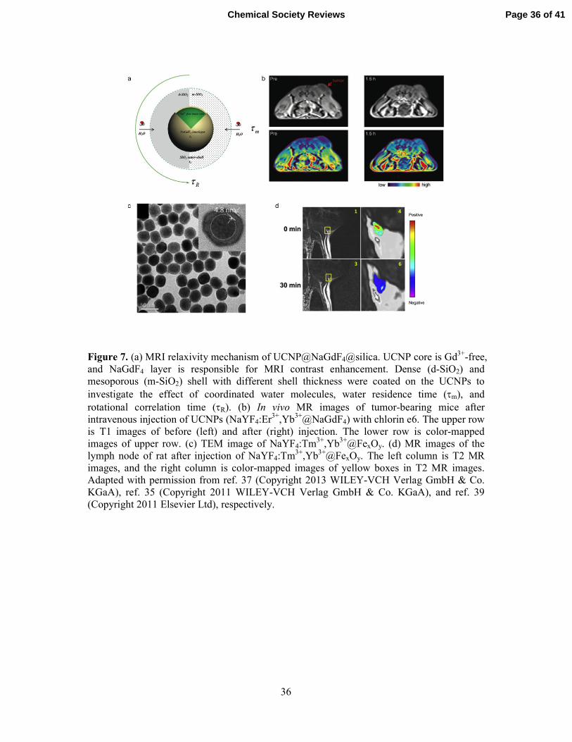
detailed relaxivity mechanism recently reported by the Shi group37 revealed that the
relaxation shortening depends on the surface modification method, such as silica coating, as
well as the NaGdF4 shell thickness (Figure 7a). For instance, the outer-sphere r1 relaxivity
mechanism can be applied to understand silica-coated UCNPs. The higher mobility of the
outer-sphere water molecules decreases the correlation diffusion time, resulting in a reduced
outer-sphere r1 relaxivity. A mesoporous silica shell with more permeable pores did not cause
any significant change in the r1 value, whereas the use of a dense silica shell with less water-
permeable micropores changed both the r1 and r2 values because water molecule diffusion is
restricted to the microporous channels in the dense silica. Ligand-free UCNPs, in which oleic
acid capping molecules were removed by protonation in acidic solution, can be explained by
both inner- and outer-sphere mechanisms. In ligand-free UCNPs, water molecules can bond
directly with surface Gd3+ ions, resulting in the highest r1 value (14.73 mM
-1s-1) among
NaGdF4-shell-coated UCNPs. On the basis of the understanding of the relaxivity mechanism,
the properties of UCNPs can be tuned for T1 or T2 MRI by optimizing the NaGdF4 shell
thickness and surface modification. NaGdF4-based UCNPs could be used to enhance the T1
contrast of tumors in the mouse model by the enhanced permeability and retention effect
(Figure 7b).35
Superparamagnetic UCNPs have also been developed as T2 MRI contrast agents. To
introduce superparamagnetism, magnetic iron oxide nanoparticles were usually assembled on
Page 24 of 41Chemical Society Reviews

25
the UCNPs, or the two components were encapsulated in polymer micelles. Cheng et al.
reported the preparation of multifunctional nanoprobes consisting of iron oxide nanoparticles
assembled on UCNPs.38 However, the overall size of the resulting probes is too large for use
by systemic administration. The Li group synthesized UCNP core–iron oxide shell
nanoparticles by growing a 5-nm FexOy shell on an as-synthesized NaYF4:Tm3+,Yb
3+ core
(Figure 7c).39 The saturation magnetization (Ms) of the resulting core–shell nanoparticles is
about 12 emu/g, and the r2 value is 189 mM-1s-1, which is higher than that of a conventional
T2 MRI contrast agent (r2 = 108 mM-1s-1). The T2 MRI images demonstrate successful
imaging of the lymph node (Figure 7d). Zhong et al. synthesized superparamagnetic UCNPs
by forming a NaYF4:Er3+,Yb
3+ shell on the seed Fe3O4 nanoparticles.
40 The Ms (1.1 emu/g) of
the resulting core–shell UCNPs was lower than that of the seed Fe3O4 nanoparticles (20.5
emu/g), which may be the result of the thick NaYF4 shell (~11 nm). The superparamagnetic
UCNPs prepared by the two methods exhibited significantly reduced emission intensity
because the excitation and emission light are absorbed by the iron oxide nanoparticles.
3.3 Computed tomography
CT is one of the most common imaging modalities in clinical use to diagnose disease and
monitor treatment because of its reasonable cost and deep tissue penetration capability.
Lanthanide elements can be used as CT contrast agents owing to their high X-ray attenuation.
Therefore, instead of the widely used NaYF4, various lanthanide host materials with high
atomic numbers have been designed for CT contrast enhancement. For instance, Yb3+-based
UCNPs (NaYbF4:Er3+) showed a higher contrast effect than a clinically used iodinated
contrast agent.41 Gd
3+-based UCNPs (NaGdF4:Er
3+,Yb
3+), which were mentioned in the MRI
section, can also be used for CT, suggesting that NaGdF4:Er3+,Yb
3+ UCNPs can be a trimodal
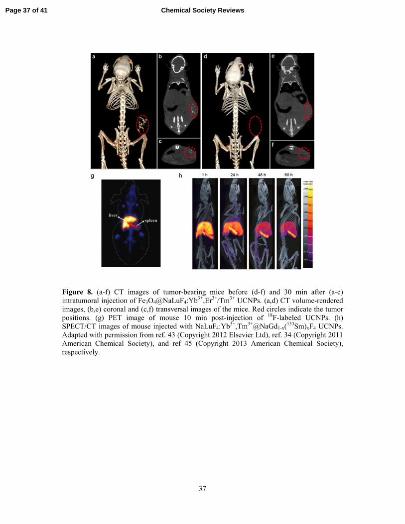
imaging probe for optical imaging, MRI, and CT.42 NaLuF4 UCNPs were recently developed
Page 25 of 41 Chemical Society Reviews
26
for more efficient CT imaging because Lu3+ ions have the highest atomic number among
lanthanide elements. These UCNPs exhibited almost five-fold higher contrast effects than the
commercial iodinated agent, and their long circulation time and low toxicity compared to
commercial agents make them a highly promising CT agent.43 In vivo CT imaging using
NaLuF4-based UCNPs could visualize tumors (Figure 8a–f).43 Xiao et al. recently
synthesized UCNPs decorated with tantalum oxide nanoparticles, which have been
intensively investigated as a CT imaging contrast agent owing to their high X-ray attenuation
and biocompatibility.44 In addition, transparent tantalum oxide nanoparticles do not quench
the upconversion luminescence, unlike gold nanomaterials, which have also been studied as
CT contrast agents.
3.4 Positron emission tomography and single-photon emission computed tomography
PET can have a synergistic effect when combined with other imaging modalities such as
MRI and CT because PET has extremely high sensitivity at the picomolar level. Common
multimodal imaging nanoprobes combined with PET have generally been prepared by
attaching radioactive isotopes to nanoparticles. The radioactive ions may be released from the
particle, which can lead to misinterpretation of the results. Liu et al. recently demonstrated
that 18F-labeled UCNPs can be prepared by cation-assisted ligand assembly.
34 Radioactive
18F- ions could be incorporated into a NaYF4 matrix via reactions with Y
3+ or Gd
3+, and the
liver and spleen could be visualized in 10 min after the 18F-labeled UCNPs were injected
(Figure 8g).
Single-photon emission computed tomography (SPECT) can also be added as an additional
modality in lanthanide-based UCNPs. As described in the previous sections, doping
lanthanide ions into the NaYF4 matrix is very easy. By adding 153Sm
3+ ions to UCNPs, the
SPECT modality can be simply added.45 Sun et al. synthesized a multimodal probe that has a
Page 26 of 41Chemical Society Reviews
27
NaLuF4:Yb3+,Tm
3+ core and NaGd1-x(
153Sm)xF4 shell and could be used for SPECT/CT
imaging (Figure 8h). They demonstrated that the probe could work as a quadruple-modal
imaging agent for NIR-to-NIR imaging (Yb3+, Tm
3+), CT (Lu
3+, Yb
3+), T1 MRI (Gd
3+), and
SPECT (153Sm
3+).
4. Multifunctionality for imaging and therapy
UCNPs that are stimulated by NIR light are considered a potential tumor therapy. NIR
light can induce local treatment, whereas the prognosis of the lesion can be estimated using
other imaging modalities such as MRI or CT. Visible light emission from UCNPs by NIR
excitation have been applied to photodynamic therapy (PDT). A typical PDT drug is
activated by visible light and then produces reactive oxygen species, which kill the target
cells. When UCNPs are used for PDT, NIR light can activate the PDT drug, which leads to
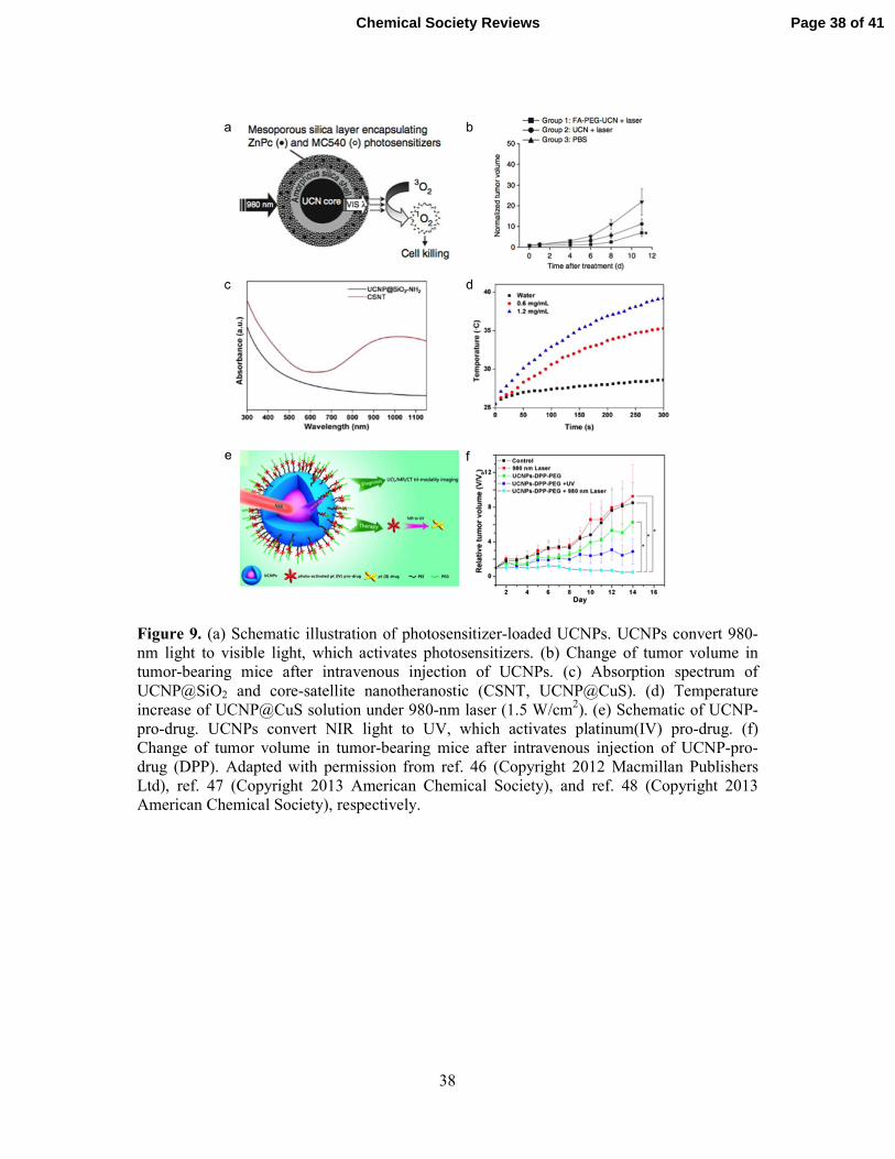
greater penetration depth and higher therapeutic efficacy. Park et al. showed that Ce6-
conjugated UCNPs could treat a tumor lesion through systemic administration.35 Later, the
Zhang group synthesized mesoporous-silica-coated UCNPs loaded with dual photosensitizers
(Figure 9a).46 The two types of photosensitizers are activated by green and red emission from
UCNPs (NaLuF4:Yb3+,Er
3+), and the UCNPs showed a much higher PDT efficiency than a
single-photosensitizer system, which was also demonstrated in a mouse tumor model (Figure
9b).
For photothermal therapy (PTT), NIR light can be used directly to induce target cell death
by increasing the temperature. Gold nanoshells and gold nanorods have been widely used for
PTT because they have a large absorption cross section in the NIR region and they efficiently
convert the absorbed radiation into heat, which causes target cell destruction. Cheng et al.
synthesized a UCNP@Fe3O4@gold structure for multifunctionality, and the nanocomposite
showed an advantage for magnetic targeted PTT. CuS-nanoparticle-decorated UCNPs
Page 27 of 41 Chemical Society Reviews

28
recently showed potential for PTT owing to their low cost and high photothermal conversion
efficiency (Figure 9c and d).47 The Shi group reported that PTT and radiation therapy were
combined for synergistic therapy because UCNPs enhance the radiation dose owing to the
heavy lanthanide elements. The synergistic therapeutic effect was also demonstrated in an in
vivo model.
NIR light can also control anticancer drug activities and their release for efficient therapy.
The Lin group recently reported platinum-prodrug-conjugated UCNPs for NIR-triggered drug
activation and release.48 For prodrug activation, UV-emitting UCNPs
(NaYF4:Yb3+,Tm
3+@NaGdF4:Yb
3+) were synthesized, and a platinum(IV) prodrug,
trans,trans,trans-[Pt(N3)2-(NH3)(py)(O2CCH2CH2COOH)2], was conjugated on the UCNPs
(Figure 9e). Optical imaging (NIR to NIR), MRI, and CT were used to determine the tumor
location, and tumor growth was successfully inhibited in the mouse tumor model (Figure 9f).
5. Conclusion
In this tutorial, the versatility of UCNPs as a platform for both wide-field two-photon
microscopy and multimodal in vivo imaging was described. Owing to their unique optical
properties, which include resistance to photoblinking and photobleaching, minimal
background autofluorescence, and low photoinduced toxicity, upconverting nanomaterials
have been attracting attention as promising bioimaging probes for both in vitro and in vivo
use. In addition, recent advances in synthetic methods for UCNPs have improved their
luminescence efficiency while decreasing their overall size to less than 10 nm. Therefore,
UCNPs fulfill the prerequisites for biological application, and they are actively used in the
field of imaging and therapy. The UCNP-based platform for wide-field two-photon
microscopy enables continuous real-time imaging of subcellular events at a higher frame rate
than that of conventional two-photon confocal microscopy. UCNPs could also be monitored
Page 28 of 41Chemical Society Reviews

29
in live cells for long-term tracking of a specific target. Their advantages are not limited to in
vitro imaging. Fine tuning of the material composition allows in vivo multimodal imaging
using UCNPs. Because the lanthanide elements have similar atomic sizes and chemical
properties, a UCNP library with different compositions can easily be synthesized, enabling a
combination of several imaging modalities. With current efforts to improve the properties of
UCNPs, detailed basic studies on the biodistribution and long-term toxicity will be required
for UCNPs to be widely used in the fields of biology and medicine.
Acknowledgements
T.H. acknowledges the financial support by the Research Center Program of the Institute for
Basic Science (IBS) in Korea. Y.D.S. was supported by KRICT (SI-1408, KK-0904-02,
OASIS Project), the Nano R&D Program (No. 2009-0082861) and the Public Welfare &
Safety Research Program (2011-0020957) through the NRF of Korea funded by the Ministry
of Science, ICT, and Future Planning (MSIP, Korea), and the Industrial Strategic Technology
Development Program (nos. 10033183 and 10037397) funded by the Ministry of Trade,
Industry, and Energy (MI, Korea). K.T.L. was supported by the Basic Science Research
Program of NRF (NRF-2013R1A1A1058451) and the Global University Projects of GIST
(K03954). We are also grateful to Dr. Jeong Hyun Kim, Dr. Hyung Min Kim, Dr. Sang Hwan
Nam, Dr. Yun Mi Bae, Byeongjun Yoo, and Dr. Ki-Seok Jeon for their productive
collaboration and discussion.
Page 29 of 41 Chemical Society Reviews

30
Figure 1. (a) Schematic diagram of upconversion processes in NaYF4:Yb3+,Er
3+. The solid
arrows indicate excitation and emission. The dotted, dashed, and wavy arrows represent
energy transfer, nonradiative transition, and multiphonon relaxation, respectively. (b-c)
Photoluminescence spectra of NaYF4:Yb3+,Er
3+ UCNPs with different crystal structures in
hexane under 980-nm excitation. (b) Cubic and (c) hexagonal phase NaYF4:Yb3+,Er
3+ UCNPs.
(d-e) Schematic illustration of cubic (d) and hexagonal (e) phase NaREF4 crystal structures.
Adapted with permission from ref. 8. (Copyright 2013 American Chemical Society), and ref.
11 (Copyright 2010 Macmillan Publishers Ltd), respectively.
Page 30 of 41Chemical Society Reviews

31
Figure 2. Fluorescence-based microscopy techniques. (a) Epi-fluorescence microscopy. (b)
Confocal microscopy. (c) TIRF (total internal reflection fluorescence) microscopy.
Page 31 of 41 Chemical Society Reviews

32
Figure 3. Single-particle measurements of UCNPs. (a) (top row) AFM images of the single
UCNPs (cubic phase NaGdF4:Er3+,Yb
3+@NaGdF4) and (bottom row) the result of section
analysis. (b) The luminescence time traces of the particles 1 and 2 acquired with 200 ms time
bins. Adapted with permission from ref. 5. Copyright 2009 WILEY-VCH Verlag GmbH &
Co. KGaA.
Page 32 of 41Chemical Society Reviews

33
Figure 4. (a) A trajectory of a UCNP (hexagonal phase NaYF4:Er3+,Yb
3) transported by
motor proteins such as dyneins. (b) Cumulative and relative displacement traces for the
trajectory shown in (a). (c) MSDs (mean square displacement = ⟨��⟩ where r is the distance
that the particle moves during a time interval, ∆t) for phases II–IV. These are fit to the
quadratic functions of ∆t (4D∆t + v2∆t
2), which is indicative of the two-dimensional active
transport of a vesicle-encapsulated UCNP by motor proteins. Adapted with permission from
ref. 13. Copyright 2011 WILEY-VCH Verlag GmbH & Co. KGaA.
Page 33 of 41 Chemical Society Reviews
34
Figure 5. (a) (left) Schematic of multicolor emission from UCNP-dye system. (middle) In
vivo multicolor image of nude mouse subcutaneously injected with five different kinds of
UCNP-dye solutions after spectral unmixing. (right) Bright field image of the mouse. (b)
Tuning of upconversion luminescence through energy transfer in core@shell structure. Left is
energy diagram, and right is the emission spectra of the UCNPs with different activator (X3+)
in shell. (c) Tuning of upconversion lifetime and time-resolved images of UCNPs
(NaYF4:Tm3+,Yb
3+). The different colors indicate different lifetime values decoded from a
decay curve. As the ratio of Tm3+-to-Yb
3+ decreases, their lifetime increases. Adapted with
permission from ref. 26 (Copyright 2011 American Chemical Society), ref. 27 (Copyright
2011 Macmillan Publishers Ltd), and ref. 28 (Copyright 2014 Macmillan Publishers Ltd),
respectively.
Page 34 of 41Chemical Society Reviews
35
Figure 6. (a) Schematic illustration of Nd3+-doped UCNPs and absorption spectrum of water
in the NIR region. (b) Diagram of energy transfer in the core/shell structured UCNPs under
808-nm excitation. (c) Luminescence images of mice subcutaneously injected with common
UCNPs (left) and Nd3+-doped UCNPs (right), under 980-nm (left) and 808-nm (right)
irradiation, respectively. (d) Thermal images of mice under 980-nm laser for 50 s (top row)
and 808-nm laser for 300 s (bottom row). Adapted with permission from ref. 30. Copyright
2013 American Chemical Society.
Page 35 of 41 Chemical Society Reviews
36
Figure 7. (a) MRI relaxivity mechanism of UCNP@NaGdF4@silica. UCNP core is Gd3+-free,
and NaGdF4 layer is responsible for MRI contrast enhancement. Dense (d-SiO2) and
mesoporous (m-SiO2) shell with different shell thickness were coated on the UCNPs to
investigate the effect of coordinated water molecules, water residence time (τm), and
rotational correlation time (τR). (b) In vivo MR images of tumor-bearing mice after
intravenous injection of UCNPs (NaYF4:Er3+,Yb
3+@NaGdF4) with chlorin e6. The upper row
is T1 images of before (left) and after (right) injection. The lower row is color-mapped
images of upper row. (c) TEM image of NaYF4:Tm3+,Yb
3+@FexOy. (d) MR images of the
lymph node of rat after injection of NaYF4:Tm3+,Yb
3+@FexOy. The left column is T2 MR
images, and the right column is color-mapped images of yellow boxes in T2 MR images.
Adapted with permission from ref. 37 (Copyright 2013 WILEY-VCH Verlag GmbH & Co.
KGaA), ref. 35 (Copyright 2011 WILEY-VCH Verlag GmbH & Co. KGaA), and ref. 39
(Copyright 2011 Elsevier Ltd), respectively.
Page 36 of 41Chemical Society Reviews
37
Figure 8. (a-f) CT images of tumor-bearing mice before (d-f) and 30 min after (a-c)
intratumoral injection of Fe3O4@NaLuF4:Yb3+,Er
3+/Tm
3+ UCNPs. (a,d) CT volume-rendered
images, (b,e) coronal and (c,f) transversal images of the mice. Red circles indicate the tumor
positions. (g) PET image of mouse 10 min post-injection of 18F-labeled UCNPs. (h)
SPECT/CT images of mouse injected with NaLuF4:Yb3+,Tm
3+@NaGd1-x(
153Sm)xF4 UCNPs.
Adapted with permission from ref. 43 (Copyright 2012 Elsevier Ltd), ref. 34 (Copyright 2011
American Chemical Society), and ref 45 (Copyright 2013 American Chemical Society),
respectively.
Page 37 of 41 Chemical Society Reviews
38
Figure 9. (a) Schematic illustration of photosensitizer-loaded UCNPs. UCNPs convert 980-
nm light to visible light, which activates photosensitizers. (b) Change of tumor volume in
tumor-bearing mice after intravenous injection of UCNPs. (c) Absorption spectrum of
UCNP@SiO2 and core-satellite nanotheranostic (CSNT, UCNP@CuS). (d) Temperature
increase of UCNP@CuS solution under 980-nm laser (1.5 W/cm2). (e) Schematic of UCNP-
pro-drug. UCNPs convert NIR light to UV, which activates platinum(IV) pro-drug. (f)
Change of tumor volume in tumor-bearing mice after intravenous injection of UCNP-pro-
drug (DPP). Adapted with permission from ref. 46 (Copyright 2012 Macmillan Publishers
Ltd), ref. 47 (Copyright 2013 American Chemical Society), and ref. 48 (Copyright 2013
American Chemical Society), respectively.
Page 38 of 41Chemical Society Reviews

39
References
1. F. Auzel, Chem. Rev., 2004, 104, 139-173.
2. M. Haase and H. Schäfer, Angew. Chem. Int. Ed., 2011, 50, 5808-5829.
3. F. Wang, D. Banerjee, Y. Liu, X. Chen and X. Liu, Analyst, 2010, 135, 1839-1854.
4. G. Chen and G. Han, Theranostics, 2013, 3, 289-291.
5. Y. I. Park, J. H. Kim, K. T. Lee, K.-S. Jeon, H. B. Na, J. H. Yu, H. M. Kim, N. Lee, S.
H. Choi, S.-I. Baik, H. Kim, S. P. Park, B.-J. Park, Y. W. Kim, S. H. Lee, S.-Y. Yoon,
I. C. Song, W. K. Moon, Y. D. Suh and T. Hyeon, Adv. Mater., 2009, 21, 4467-4471.
6. A. D. Ostrowski, E. M. Chan, D. J. Gargas, E. M. Katz, G. Han, P. J. Schuck, D. J.
Milliron and B. E. Cohen, ACS Nano, 2012, 6, 2686-2692.
7. S. Gai, C. Li, P. Yang and J. Lin, Chem. Rev., 2014, 114, 2343-2389.
8. Y. I. Park, S. H. Nam, J. H. Kim, Y. M. Bae, B. Yoo, H. M. Kim, K.-S. Jeon, H. S.
Park, J. S. Choi, K. T. Lee, Y. D. Suh and T. Hyeon, J. Phys. Chem. C, 2013, 117,
2239-2244.
9. J. Zhao, Y. Sun, X. Kong, L. Tian, Y. Wang, L. Tu, J. Zhao and H. Zhang, J. Phys.
Chem. B, 2008, 112, 15666-15672.
10. J.-C. Boyer, L. A. Cuccia and J. A. Capobianco, Nano Lett., 2007, 7, 847-852.
11. F. Wang, Y. Han, C. S. Lim, Y. Lu, J. Wang, J. Xu, H. Chen, C. Zhang, M. Hong and
X. Liu, Nature, 2010, 463, 1061-1065.
12. F. Wang and X. Liu, Chem. Soc. Rev., 2009, 38, 976-989.
13. S. H. Nam, Y. M. Bae, Y. I. Park, J. H. Kim, H. M. Kim, J. S. Choi, K. T. Lee, T.
Hyeon and Y. D. Suh, Angew. Chem. Int. Ed., 2011, 50, 6093-6097.
14. A. Jemal, R. Siegel, E. Ward, Y. Hao, J. Xu and M. J. Thun, CA Cancer J. Clin., 2009,
59, 225-249.
15. U. Resch-Genger, M. Grabolle, S. Cavaliere-Jaricot, R. Nitschke and T. Nann, Nat.
Methods, 2008, 5, 763-775.
16. Q. Zhan, S. He, J. Qian, H. Cheng and F. Cai, Theranostics, 2013, 3, 306-316.
17. M. Nirmal, B. O. Dabbousi, M. G. Bawendi, J. J. Macklin, J. K. Trautman, T. D.
Harris and L. E. Brus, Nature, 1996, 383, 802-804.
18. M. Pelton, G. Smith, N. F. Scherer and R. A. Marcus, Proc. Natl. Acad. Sci. USA,
2007, 104, 14249-14254.
19. S. Wu, G. Han, D. J. Milliron, S. Aloni, V. Altoe, D. V. Talapin, B. E. Cohen and P. J.
Schuck, Proc. Natl. Acad. Sci. USA, 2009, 106, 10917-10921.
Page 39 of 41 Chemical Society Reviews
40
20. R. Weissleder, Nat. Biotechnol., 2001, 19, 316-317.
21. Y. M. Bae, Y. I. Park, S. H. Nam, J. H. Kim, K. Lee, H. M. Kim, B. Yoo, J. S. Choi,
K. T. Lee, T. Hyeon and Y. D. Suh, Biomaterials, 2012, 33, 9080-9086.
22. L. Cheng, K. Yang, S. Zhang, M. Shao, S. Lee and Z. Liu, Nano Res., 2010, 3, 722-
732.
23. J. Pichaandi, J.-C. Boyer, K. R. Delaney and F. C. J. M. van Veggel, J. Phys. Chem. C,
2011, 115, 19054-19064.
24. J. Zhou, Z. Liu and F. Li, Chem. Soc. Rev., 2012, 41, 1323-1349.
25. C. Wang, L. Cheng, H. Xu and Z. Liu, Biomaterials, 2012, 33, 4872-4881.
26. L. Cheng, K. Yang, M. Shao, S.-T. Lee and Z. Liu, J. Phys. Chem. C, 2011, 115,
2686-2692.
27. F. Wang, R. Deng, J. Wang, Q. Wang, Y. Han, H. Zhu, X. Chen and X. Liu, Nature
Mater., 2011, 10, 968-973.
28. Y. Lu, J. Zhao, R. Zhang, Y. Liu, D. Liu, E. M. Goldys, X. Yang, P. Xi, A. Sunna, J.
Lu, Y. Shi, R. C. Leif, Y. Huo, J. Shen, J. A. Piper, J. P. Robinson and D. Jin, Nature
Photon., 2014, 8, 32-36.
29. Q. Zhan, J. Qian, H. Liang, G. Somesfalean, D. Wang, S. He, Z. Zhang and S.
Andersson-Engels, ACS Nano, 2011, 5, 3744-3757.
30. Y.-F. Wang, G.-Y. Liu, L.-D. Sun, J.-W. Xiao, J.-C. Zhou and C.-H. Yan, ACS Nano,
2013, 7, 7200-7206.
31. H. B. Na and T. Hyeon, J. Mater. Chem., 2009, 19, 6267-6273.
32. R. Kumar, M. Nyk, T. Y. Ohulchanskyy, C. A. Flask and P. N. Prasad, Adv. Funct.
Mater., 2009, 19, 853-859.
33. J. Zhou, M. Yu, Y. Sun, X. Zhang, X. Zhu, Z. Wu, D. Wu and F. Li, Biomaterials,
2011, 32, 1148-1156.
34. Q. Liu, Y. Sun, C. Li, J. Zhou, C. Li, T. Yang, X. Zhang, T. Yi, D. Wu and F. Li, ACS
Nano, 2011, 5, 3146-3157.
35. Y. I. Park, H. M. Kim, J. H. Kim, K. C. Moon, B. Yoo, K. T. Lee, N. Lee, Y. Choi, W.
Park, D. Ling, K. Na, W. K. Moon, S. H. Choi, H. S. Park, S.-Y. Yoon, Y. D. Suh, S.
H. Lee and T. Hyeon, Adv. Mater., 2012, 24, 5755-5761.
36. F. Chen, W. Bu, S. Zhang, X. Liu, J. Liu, H. Xing, Q. Xiao, L. Zhou, W. Peng, L.
Wang and J. Shi, Adv. Funct. Mater., 2011, 21, 4285-4294.
Page 40 of 41Chemical Society Reviews
41
37. F. Chen, W. Bu, S. Zhang, J. Liu, W. Fan, L. Zhou, W. Peng and J. Shi, Adv. Funct.
Mater., 2013, 23, 298-307.
38. L. Cheng, K. Yang, Y. Li, J. Chen, C. Wang, M. Shao, S.-T. Lee and Z. Liu, Angew.
Chem. Int. Ed., 2011, 50, 7385-7390.
39. A. Xia, Y. Gao, J. Zhou, C. Li, T. Yang, D. Wu, L. Wu and F. Li, Biomaterials, 2011,
32, 7200-7208.
40. C. Zhong, P. Yang, X. Li, C. Li, D. Wang, S. Gai and J. Lin, RSC Adv., 2012, 2,
3194-3197.
41. Y. Liu, K. Ai, J. Liu, Q. Yuan, Y. He and L. Lu, Angew. Chem. Int. Ed., 2012, 51,
1437-1442.
42. M. He, P. Huang, C. Zhang, H. Hu, C. Bao, G. Gao, R. He and D. Cui, Adv. Funct.
Mater., 2011, 21, 4470-4477.
43. X. Zhu, J. Zhou, M. Chen, M. Shi, W. Feng and F. Li, Biomaterials, 2012, 33, 4618-
4627.
44. Q. Xiao, W. Bu, Q. Ren, S. Zhang, H. Xing, F. Chen, M. Li, X. Zheng, Y. Hua, L.
Zhou, W. Peng, H. Qu, Z. Wang, K. Zhao and J. Shi, Biomaterials, 2012, 33, 7530-
7539.
45. Y. Sun, X. Zhu, J. Peng and F. Li, ACS Nano, 2013, 7, 11290-11300.
46. N. M. Idris, M. K. Gnanasammandhan, J. Zhang, P. C. Ho, R. Mahendran and Y.
Zhang, Nat. Med., 2012, 18, 1580-1585.
47. Q. Xiao, X. Zheng, W. Bu, W. Ge, S. Zhang, F. Chen, H. Xing, Q. Ren, W. Fan, K.
Zhao, Y. Hua and J. Shi, J. Am. Chem. Soc., 2013, 135, 13041-13048.
48. Y. Dai, H. Xiao, J. Liu, Q. Yuan, P. Ma, D. Yang, C. Li, Z. Cheng, Z. Hou, P. Yang
and J. Lin, J. Am. Chem. Soc., 2013, 135, 18920-18929.
Page 41 of 41 Chemical Society Reviews