upper limb injuries in children and adolescents€¦ · upper limb injuries in children and...
TRANSCRIPT

UPPER LIMB INJURIES IN CHILDREN AND
ADOLESCENTS
Paolo Simoni, M.D., Ph.D, M.B.A., ULB, Bruxelles, Belgium
Service de Radiologie Pédiatrique: HUDERF Bruxelles-Belgium

Outline
1. General introduction
Bone and periosteum features in children and their implications in imaging of upper limb traumas
Growth plate features and implications in imaging of upper limb traumas
Secondary ossification center (SOC) features and implications in imaging of upperlimb trauma
2. Systematic approach to upper limb trauma imaging in children and adolescents
Humerus
Elbow
Forearm
2

….Beyond the scope of this presentation…..
1. Wrist and Hand trauma imaging in children and adolescents:
some bio-mechanical and phisiologic differences
2. Systematic review of use of US (and MRI) to assess upper limb trauma
Subspecialty , US (and, in some instances, MRI) are a useful adjunct to radiographs
3

Children’s bones and joints are different
In kids large
plastic
region
before
rupture

Children’s periosteum is different
5
Periosteum is thick and strong in
children but is very flexible and it can
buldge or bow

Children’s periosteum is different
6
Periosteum is thick and
strong in children but is very
flexible and it can buldge or
bow. It is not as adherent to

7
Periosteum is not visible on radiographs: it
can fold without being injured

Bone + periosteum =Children’s fractures are different
8
but
also

“Torus or Buckling fracture” and “Greenstick fracture
9
“Greenstick fracture”, a transverse
fracture of the cortex which extends
into the middle portion of the bone
and becomes oriented along the
longitudinal axis of the bone without
disrupting but the opposite cortex.
“Torus or Buckling fracture”,
caused by a force acting on the
longitudinal axis. There is a
buckle and a break of the cortex
on the opposite side. The
fracture line can be visible or not

« Plastic bowing »
10
« Plastic bowing »:
if the traumatic force
exceeds the elastic
modulus of the bone but
it is not strong enough
to cause a complete
fracture. No cortical
break is visible.

Children bon is highly dynamic:
Enchondral ossification
TITRE CHAPITRE 1
11

Growth plate
12
GP
X*
P

Salter-Harris fractures
13
I-II: usually no
sequelae
III-IV-V: shortening, deformity,
surgery

Secondary ossification center (SOCSOC
14
?

Secondary ossification center (SOC)SOC
15SOCs are procteted by the GP-SOC all

Consider multiple SOC vs fracture
16
3 months 12 month3 months

Fractures in children: some tips and tricks
• Periosteum is thick and strong in children but is very flexible and it can buldge or bow
• Bone is more elastic/plastic in children .
• Always rule out Salter-Harris Fracture: look at the growth plate an around
• Consider that multiple SOC can appear in the epiphysis during bone growth before diagnose an epiphyseal fracture. SOC are protected by their own GP
17

Humerus
18

Promixal Humerus Fractures
19
Age: teens
Mechanism:
-Direct trauma (fall on the shoulder)
-Fall on outstreched hand
-Luxation are rare
Features:
-Fracture of the surgical neck more fragment before 3 yrs
-Salter II in adolescence
-Rule out pathological fracture (i.e. simple bonecyst)

Humeral diaphyseal fracture
20
Age:birth, teenagers
Mechanism:
Obstetrical trauma
Direct trauma
Torsion
Check for our paralysisof the radial nerve!!!

Elbow
21

CRITOE
22
-
!Open your book or…

Haemarthrosis
23
-
Hemarthrosis : criticial sign
Usually the capsule is adjacent to
the bone and the pad is adjacent
to the capsule.
If there is hemarthrosis :
The pads are displaced
Normal Haemarthrosi
s
*

24

25

26

27

28

Capitellum : a endless dilemma
29
-
Baumann’s Angle : coronal displacement
Humero-condyle angle : AP displacement

Elbow’s normal landmarks
30
-
Anterior humeral line:
- Extension of the anterior cortical
humerus
usually passes through the middle
third of the humeral condyle
Radius axis
- Must go through the capitellum
- If not, consider that it could be a
luxation of the radial head
(Monteggia’s Fracture)
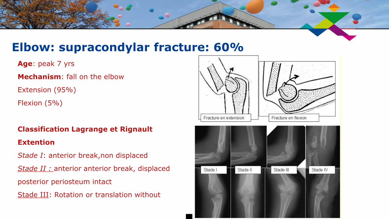
Elbow: supracondylar fracture: 60%
31
Age: peak 7 yrs
Mechanism: fall on the elbow
Extension (95%)
Flexion (5%)
Classification Lagrange et Rignault
Extention
Stade I: anterior break,non displaced
Stade II : anterior anterior break, displaced
posterior periosteum intact
Stade III: Rotation or translation without diastasis
Stade IV: Diastasis

Elbow: supracondylar fracture (60% elbow fractures)
32
Rule out a nerve paralysis in
displaced fractures.
AION is the most frequently
injured nerve

Elbow: medial epicondyle fracture (10%)
33
Age: peak 7-15 yrs
Mechanism: fall on the outstreched hand with elbow
in valgus
In 50% of patient postero-lateral luxation
Classification Watson-Jones
Grade I: fragment non displaced or <5mm
GradeII: fragment dispaced > 5mm
Grade III: fragment incarcerated into the joint
Grade IV: luxation associatedValg
us

Elbow: medial epicondyle fracture (10%)
34
Classification Watson-
Jones
Grade I: fragment non
displaced or <5mm
Grade II: fragment
dispaced > 5mm
Grade III: fragment
incarcerated into the joint
Grade IV: luxation
associated

Elbow: lateral epicondyle fracture (10%)
35
Age: 6-8 yrs
It is a Salter
IV
fracture!!!!!
Compression and
extension: radial
head vs lateral
condyle
Compression and flexion:
olecranus vs lateral
condyle Traction in extension,
varus and supination:
!Extensor muscles!

Elbow: lateral epicondyle fracture (10%)
36
1) de Lagrange et Rigault:
-grade I: non displaced fragment (<2 mm)
-grade II: laterally displaced fragment (>2
mm)
-grade III: major displacement and
rotation
:
2) de Milch
-type 1: Lateral to the throclear groove
Incomplete/complete
-type 2: Medial to the throclear groove
Incomplete/complete

Elbow: radial neck (10 % elbow fractures)
37
Age: peak 4-15 yrs
Mechanism:
High energy trauma fall on the outstreched hand with elbow in valgus
In 50% of patient postero-lateral luxation
Judet’s classification :
-grade 1: non diplaced-grade 2: laterally diplaced < 50%, angulation < 30°-grade 3: angulation >30°, <60°-grade 4 : angulation >60°
-

« Nursemaid’s elbow », « pulled elbow » » »
38
-

« Nursemaid’s elbow » or « pulled elbow »
39
-

«Nursemaid’s elbow » or « pulled elbow »: US axial view
40
-

Elbow: olecranon (5%)
41
-
Age: peak 9 yrs
Mechanism:
• Flexion (avulsion)
• Extention (intact peristeum)
• Direct trauma with comminuted fracture
Classification:
• Gaddy 1 :<3 mm intra-articular diplacement
• Gaddy 2 :>3 mm intra-articular diplacement
(surgery

Elbow: luxation (3%)
42
-
Age: 4-15 yrs
Mechanism:
Fall on the
outstreched
hand
Posterior
more frequent
Divergent:
Proximal
radio-ulnar
joint injured

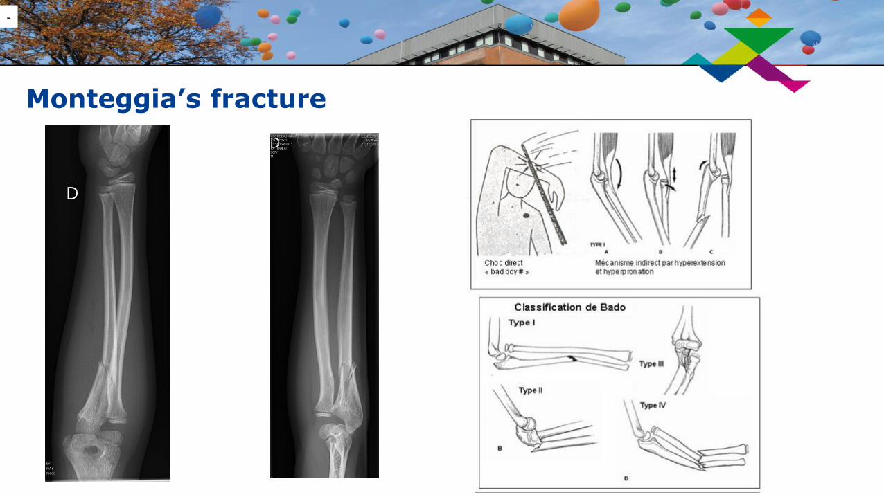
Monteggia’s fracture
43
-
Classification de Bado
-Type 1 (+++ 65%) extension: anterieur luxation of radial
head + # ulna diaphysis
-Type 2: flexion : posterior luxation + # 1/3 sup ou 1/3 moy
ulna
-Type 3: adduction: external luxation + # proximale ulna
metaphysis
-Type 4: type 1 + # middle 1/3 radial diaphysis (double
fracture)
Monteggia’s fracture
44
-

Forearm fracture
45
Mecanism:
-Fall on outstreched end
-Rarely, direct trauma
Biomechanics : rule of thumb
The pronator quadratus (distally)
and pronator teres (inserting on
the middle portion of the radius)
actively pronate the forearm,
while the biceps and supinator
(proximal insertions) provide
supination.

Galeazzi’s fracture (2.8%)
46
-
Age: peak 9-13 yrs
« Necessity fracture» in adults
but conservative treatment in
chidren
Mechanism:
Fall on outstreched hand in
hyperpronation
!Consider injury of DRUJ and
of TFCC!

Galeazzi’s fracture
47
-
Galeazzi’s fractures are classified according to the position of the distal radius:
type I: dorsal displacement of radius type II: volar
displacement of radius

Take home messages
48
Periosteum is thick and strong in childrenbut is very flexible and it can buldge or bow
Bone is more elastic/plastic in children .
Consider incomplete fracture in children
Always rule out Salter-Harris Fracture: look at the growth plate and around it
Consider that multiple SOC can appear in the epiphysis during bone growth beforediagnose an epiphyseal fracture.
Compare to the opposite side in case of doubt
Children’s MSK system is different in many ways.
….Think different when facing a upper limb trauma in kids…..

Ask the kid if you are right!
49

THANK YOU FOR YOUR ATTENTION
50