uptake, storage and excretion of uranium by mytilus …
TRANSCRIPT
HAL Id: jpa-00223793https://hal.archives-ouvertes.fr/jpa-00223793
Submitted on 1 Jan 1984
HAL is a multi-disciplinary open accessarchive for the deposit and dissemination of sci-entific research documents, whether they are pub-lished or not. The documents may come fromteaching and research institutions in France orabroad, or from public or private research centers.
L’archive ouverte pluridisciplinaire HAL, estdestinée au dépôt et à la diffusion de documentsscientifiques de niveau recherche, publiés ou non,émanant des établissements d’enseignement et derecherche français ou étrangers, des laboratoirespublics ou privés.
UPTAKE, STORAGE AND EXCRETION OFURANIUM BY MYTILUS EDULIS, A
STRUCTURAL, ULTRASTRUCTURAL ANDMICROANALYTICAL STUDY BY SECONDARY ION
EMISSION AND X RAY SPECTROMETRYC. Chassard-Bouchaud, F. Escaig
To cite this version:C. Chassard-Bouchaud, F. Escaig. UPTAKE, STORAGE AND EXCRETION OF URANIUMBY MYTILUS EDULIS, A STRUCTURAL, ULTRASTRUCTURAL AND MICROANALYTICALSTUDY BY SECONDARY ION EMISSION AND X RAY SPECTROMETRY. Journal de PhysiqueColloques, 1984, 45 (C2), pp.C2-545-C2-548. �10.1051/jphyscol:19842124�. �jpa-00223793�
JOURNAL DE PHYSIQUE
Colloque C2, suppigment au n02, Tome 45, fgvrier 1984 page C2-545
UPTAKE, STORAGE AND E X C R E T I O N OF URANIUM B Y M Y T I L U S E D U L I S ,
A STRUCTURAL, ULTRASTRUCTURAL AND M I C R O A N A L Y T I C A L STUDY B Y SECONDARY
I O N E M I S S I O N AND X RAY SPECTROMETRY
C. ~hassard-~ouchaud+* and F. ~ s c a i ~ *
'~boratoire de Zoologic, Biologie et Physiologie des Organismes Marins, Universits Pierre et Marie Curie, 4, place Jussieu, 75230 Paris Ceder 05, France *kiboratoire de Biophysique de la Facult6 de Mgdecine (EY. P. Gallel, 6, rue du Ge'n6raZ Sarrail, 94000 Cr6tei2, France
RESUME - 1-Dew me'thodes de microanalyse ont e'te' uti2ise'es:la spectrographic des rayons X ci Zf&chelon des microscopes optique et electronique et l'e'mission ionique secondaire Li l'e'chelon de la microscopic optique; elles ont permis de montrer que, des M.edulis re'colte'es sur les c6tes de la Manche,ainsi que des e'chantillons c o n t & ~ x p e ' r i m e n t a l e m e n t , accumulent 238U.
2-Les plus fortes concentrations d'uranim sont de'tecte'es dans les a- nimaux pre'leve's au niveau des zones de rejet des phosphogypses et des re'sidus de fabrication de Ti02 qui contiennent de l'uranim.
3-L'uranim est absorbe' par Zes voies branchiale et digestive.Le stockage est r&aZis& dans le manteau,la glande digestive,Zre'pithe'lim intesti- nal et La gonade 02 Les teneurs les plus e'leve'es sont souvent constate'es.L'ex- cre'tion se fait par le rein.
4-Les organites cibles sont les Zysosomes de La glande digestive et les sphe'rocristam du rein; dans ces deux sites e'lectifs d'accmuZation,Z'ura- niwn est toujours associe' Li du phosphore.Ainsi,l'uranim absorbe' sous forme soluble,est ensuite concentre' sous forme de phosphate insoluble dans les or- ganites de stockage.
5-Les hBmocytes macrophages jouent un r6le important dans la capture, le transport,le stockage et l'excretion du radionucleide.
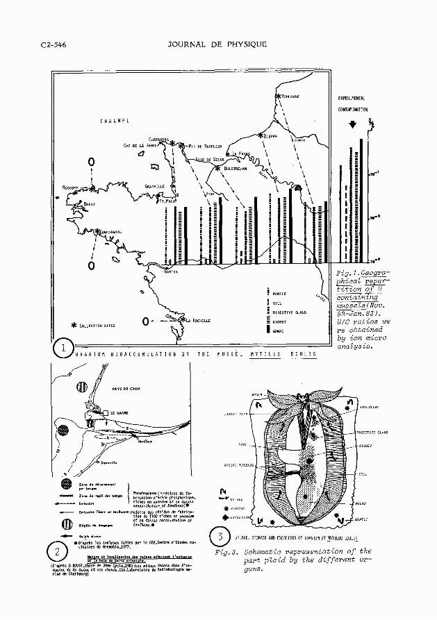
6-La Moule commune apparait come un systdme biologique capable d'accu muler et de concentrer,sous forme insoZuble,l'uranim pre'sent dans le milieu marin 6. lrBtat de traces.M.edulis est propose' comme organisme indicateur de po Zlution par 2 'uraniwn. ABSTRACT-1-Two microanalytical methods have been used: X ray emission at the Zight and electron microscope levels and secondary ion emission at the Zight microscope Zevel.They allowed to show that M.edulis collected on the Channel coasts or experimentally contaminated samples,bioaccumulate 238U.
2-The highest U Levels were detected in the samples collected in the areas where phosphogypsm and Ti02 industrial wastes,U containing,are released.
3-Uranim uptake happened via gill and digestive tractus.Storage or- gans were mantle, digestive gland,intestine epithelium and gonad where the hi- ghest values often occured.Excretion happened via kidney.
4-The target organelles were digestive gland Zysosomes and kidney sphe rocrystals: witthin both of these accmulation sites,uranim was always associa- ted with phosphorus.Thus,solubZe uranium which was absorbed,was then concentra- ted in the form of an unsoluble phosphate in the storage organelles.
5-Macrophage haemocytes plaid an important part in ingestion,transport, storage and excretion of the radionuclide.
6-The common marine Mussel,appears as a biological system accmuZating uranium which,present in the marine environment at trace leve2,is stored and concentrated under an unsoluble form by the MusseZ.M.edulis is proposed as an uraniwn pollution indicator organism.
The marine Mussel is of interest,as it is a sedentary filter feeding organism,which is likely to accumulate pollutants such as radionuclides which are released into the marine environment.The increasing use indeed of uranium by chemical and nuclear in-
Article published online by EDP Sciences and available at http://dx.doi.org/10.1051/jphyscol:19842124
C2-546 JOURNAL DE PHYSIQUE
dustries and its possible discharge into the aquatic environment give an ecotoxicolo gical interest to the radionuc1ide.Uranium is a heavy metal which consists of 3 iso- topes 238 U( 99,3 %),235 U( 0,7 %) and 234 U( 0,006 %),all a emitters,chemotoxic and radiotoxic. Recent microanalytlcal studies,performed on sea and freshwater Crustacea (1)(2),provided the first data on uranium metabolism in aquatic 0rganisms.A~ nothing was known about the uranium metabolism in Mollusca,our purpose was to investigate a- bout it,on a specles of economical interest:Mytilus edulis. 1-MATERIALS.Samples of mature M.edulis (L)(Mollusca Lamellibranchiata),were collec- ted from November 1982 to January 1983 in 6 stations of the French coasts of the Channel:Boulogne,Dieppe,0uistreham,Pointe de Barfleur,Cherbourg and Roscoff.The Baie de Seine is an area where phosphogypsum and Ti02 industrial wastes,U containing,are released.For comparison,other samples were collected from 2 stations of the French coasts of the Atlantic 0cean:Concarneau and La Rochelle. For experimental contamination,U free mussels were used.They were exposed to an ura- nium nitrate solution at a concentration of 10 ppm,during a period from 1 to 30 days. 2-SAMPLES PREPARATION NICROANALYTICAL METHODS. Cf the other paper of the same author ,in the same Congress Proceedings (3). - 3-RESULTS. Mussels collected "in situ" from the Channel and mussels experimentally contaminated were shown to concentrate Uranium. 3-1- Secondary ion emission microanalysis. 238 U + values normalized to 12 C+ are given in fig. 1 for samples of every collec- tion site; they point out the geographical repartition of uranium bioaccumulation in mussels collected from the Channel.This map shows that the highest values were de- tected in the eastern samples collected from Cherbourg to Boulogne.No uranium was de- tected neither in Roscoff samples nor in the Atantic Ocean ones. The experimental contamination results (fig. 1 )show that uranium was detected,in or- der of decreasing concentration,in gonad,digestive gland,kidney,mantle and gill-The- se results are in agreement with theq'in situ " samples results.Ion images obtained from several organs and tissues show the uranium distribution (fig.4,5,6 and 7 ) . 3-2- X.ray emission microanalysis. 3-2-1-Light microscope-1~vel:spectra obtained on the MS46 microprobe confirm the se- condary ion &nission microanalysis data. 3-2-2-E1gctr~nnm~crogcgpg level: electron dense microneedles were observed within the digestive gland lysosomes (fig. 8 )They were shown to contain high levels of uranium associated with phosphorus (fig. 9 ).F~loreover,intra and extracellular sphe- rocrystals (fig. 11 ) containing also U and P were observed in the kidney. Macrophage haemocytes,which occur within many organs and tissues of the musse1,exhi- bited many uranium containing inclusions (fig. 10 ).They play an important part in ingestion,transport,storage and excretion of the radionuclide. 4-DISCUSSION CONCLUSION. Uranium was detected in the samples collected in the areas where phosphogypsum and Ti02 industrial wastes, U containing, are released. These results were obtained from mussels collected from November 1982 to January 1983. Fig. 3 gives a schematic representation of the involved mechanisms by which M.=- lis is supposed to assume uranium uptake,storage and excretion.Detoxication proces- - ses happen via kidney from which the spherocrystals are released into the extracel- lular medium and via the macrophages. Uranium uptake,concentration and elimination mechanisms above described at the ultrastructural leve1,for the Musse1,may be com- pared to those previously described for Mammals (4) and for Crustacea (1)(2). As Mytilus edulis bioaccumulates uranium,present in the marine environment at trace leve1,it is then proposed to use it as an indicator of uranium pollution.
(1)CHASSARD-BOUCHAUD C.,C.R.Acad.Sc.Paris,294,s&rie 3,(1982),919. (2)CHASSARD-BOUCHAUD C.,Mar. Poll. Bu11.,~,4,(1983),133. (3)CHASSARD-BOUCHAUD C., HALLEGOT P. and MEIGNAN M. (4)GALLE P.,J. de Microscopie, g,1,(1974),17.
Sources of partial support for this work were CNRS (GRECO MANCHE I and INSERM iSC27) The technical assistance was provided by F.Kleinbauer and S. Halpern.Materia2 was, in part,coZlected by D.Calmet.
C2-548 JOURNAL DE PHYSIQUE
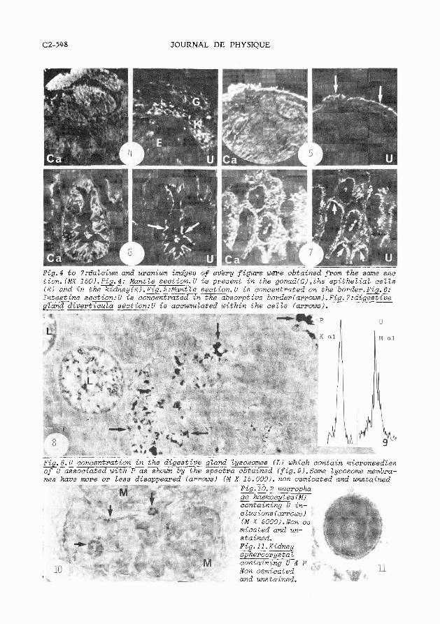
Fig.4 f o 7rEalciwn and uraniwn imciges of every f igurewere obtained from the same see tion.(MX 150/.%.4: Mantle secti0n.U i s present i n the gonad(Gl,the epithel ial , c e l l s ( E l and i n the k i d % e y m g = l e secti0n.U i s concentrated on the border.%.6: Zntestine section: U i s concentrated i n the absorptive border (arrows). F-&. l : d ige s t i z e gZand dive- section:U i s accumulated within the c e l l s (arrows).
%.g.c concentration i n the d iges t ive gZand lysosomes ( L I which contain microneedles of u associated with P as shown by the spectra obtained (fig.3I.Some lysosome meabra- nes have more or Less disappeared (arrows) ( M X 15.0001. non osmicated and unstained
~ i g . i S . 2 macropha ge haemoeytes(M) - containing U in- clusions (a~rows ) (M X 5000). Non os micated and un- $ stained. *. Fig. 11 . Kidney 7.'-
spherocrystat +:
containing U & P . . Non osmicated '
b and unstained.