uracil and apurinic/apyrimidinic site processing...
TRANSCRIPT

Uracil excision repair pathway activities are not intrinsic to nucleoside diphosphate
kinases from Mycobacterium tuberculosis and Escherichia coli.
Pradeep Kumar, Kurthkoti Krishna, Ramanujam Srinivasan, Parthasarathi Ajit Kumar and
Umesh Varshney*, Department of Microbiology and Cell Biology, Indian Institute of
Science, Bangalore, 560 012, India.
*Correspondence to:
U. Varshney
Department of Microbiology and Cell Biology
Indian Institute of Science, Bangalore, 560 012, India.
Phone: 91-80-2293 2686
Fax: 91-80-2360 2697
E-mail: [email protected]
Running title: Uracil processing activities are not intrinsic to NDKs
Key words: UDG, Ung, Ugi, NDK, AP-endonuclease.

Abstract
E. coli nucleoside diphosphate kinase (EcoNDK) is an important cellular enzyme
required to maintain balanced nucleotide pools in the cells. Recently, it was reported that
EcoNDK is also a multifunctional base excision repair enzyme, possessing a uracil-DNA
glycosylase (UDG), an AP-endonuclease and a deoxyribose-phosphodiesterase activities. We
investigated for the presence of similar activities in M. tuberculosis NDK (MtuNDK), which
shares 45.2 % identity, and 52.6 % similarity with EcoNDK. In contrast to the robust uracil
excision activity reported for EcoNDK, MtuNDK preparation exhibited very poor excision of
uracil from DNA. However, this activity was undetectable when MtuNDK was purified from
an ung- strain of E. coli, or when the assays were performed in the presence of extremely low
amounts of a highly specific proteinaceous inhibitor, Ugi which forms an extremely tight
complex with the host Ung (UDG), showing that MtuNDK preparation was contaminated
with UDG. Reinvestigation of uracil processing activities in EcoNDK purified by
conventional, and by Ni-NTA affinity techniques, showed that even EcoNDK lacked UDG
activity. All preparations of NDK were shown to be active by their autophosphorylation. Ugi
neither displayed a physical interaction with EcoNDK nor did it affect autophosphorylation
of NDKs. Further, neither of the NDK preparations processed the AP-DNA generated by
UDG treatment of the uracil containing DNA duplexes. However, partially purified
preparations of NDK did process such DNA substrates.

Introduction
DNA glycosylases excise damaged or unconventional bases from DNA and initiate
base excision repair (BER) pathway (Friedberg, 1978). In BER pathway, cleavage of the N-
glycosidic bond by a DNA glycosylase is followed by an AP-endonuclease, a deoxyribose-
phoshodiesterase (dRpase), a DNA polymerase and a DNA ligase functions (Dianov et. al.,
1994). The DNA glycosylases are highly specific for the unusual base(s) they excise from
DNA, and based on the associated activities, they have been grouped into monofunctional or
bifunctional enzymes (Lindahl, Krokan, Duncan, reviews Friedberg, 1995). The
monofunctional enzymes possess only the glycosylase activity whereas the bifunctional
enzymes, in addition, possess AP lyase and/or dRpase activities. The enzymes that remove
uracil from DNA are collectively referred to as uracil DNA glycosylases (UDGs). UDGs
have common evolutionary origin with a distinct α/β structural fold, and constitute a super
family of monofunctional DNA-glycosylases (arvind and koonin, 2000; Pearl 2000, sakumi
and sekiguchi, 1990). Based on the primary structure analysis, UDGs have been classified
into five families (Pearl, 2000). The family 1 comprises of UDGs with a high homology to E.
coli Ung (referred to hereafter as EcoUDG), which are specifically inhibited by a
proteinaceous inhibitor, Ugi (uracil DNA glycosylase inhibitor) encoded by Bacillus subtilis
phages PBS -1 and -2 (Mosbaugh; crystal structures). The binding of Ugi to EcoUDG is
essentially irreversible under physiological conditions, and stable to even 8 M urea at room
temperature. Such a tight interaction of Ugi with UDG is necessary for complete inhibition of
UDG activity of the complex. Mutations in EcoUDG or Ugi that weaken the stability of the
complexes, result in incomplete inhibition of EcoUDG (Mosbaugh, Narottam). Further,
despite a common evolutionary origin and a conserved three dimensional fold among the
members of UDG super family, only the members of the EcoUng family are known to be
inhibited by Ugi (Barret, family 4; Jiricny ).

Recently, nucleoside diphosphate kinase from E. coli (EcoNDK) was reported to be a
multifunctional protein. In addition to its NDK activity, it was shown to possess uracil
processing, AP- site processing and dRPase functions, suggesting it to be the first member of
a novel family of UDGs (Postel, 2003) which utilized both single- and double- stranded
uracil-DNA as substrates. The primary function of EcoNDK is to maintain cellular levels of
nucleoside triphosphates for the synthesis of DNA and RNA (Hama H, 1991). It catalyzes
transfer of γ-phosphate from nucleoside triphosphates to 5’ position of ribo- and deoxyribo-
nucleoside phosphates (Almaula N 1995). It also undergoes autophosphorylation at H117,
S119, and S121 (Inouye etal, J Bacteriol, 1995). A deletion of ndk gene in E. coli results in an
increased rate of spontaneous mutations and elevation of cellular levels of CTP, dCTP and
dGTP (Bernard MA 2000, Lu Q 1995 JMB).
The genome sequence of Mycobacterium tuberculosis (Cole, ST etal. 1998) revealed
that it is missing orthologs of many of the DNA repair proteins (Mizrahi V 1998), known to
play important functional roles in E. coli. Recently, we showed that UDG repair pathway is
crucial for survival of mycobacteria in the host macrophages especially under the conditions
of increased reactive nitrogen intermediate production (Venkatesh etal). M. tuberculosis
NDK (MtuNDK) shares 45.2 % identity, and 52.6 % similarity with EcoNDK (). The
potential implication of NDK in uracil excision repair in mycobacterium biology encouraged
us to carry out a comparative biochemical characterization of MtuNDK and EcoNDK (for
their possible role in uracil processing). Contrary to the findings of Postel et. al., but
consistent with a more recent independent study on EcoNDK (Bennett etal), by using NDKs
from a diverse group of bacteria, we show that neither MtuNDK nor EcoNDK possess
detectable uracil excision activities. In addition, we demonstrate that these NDKs do not
process the AP-site containing DNA duplexes generated by UDG, thereby ruling out a direct
role of the NDKs in uracil excision repair pathway.

Materials and Methods
Plasmids and biochemicals: The EcoNDK expressing plasmids pndkec[pJC20] and
pndkec[pJC20HisC] were kindly provided by Dr. M. Konrad (Max Planck Institute for
Biophysical Chemistry, Goettingen, Germany. Plasmid, pACT7 was generated by Dr. S.
Karnik in the laboratory of Dr. H. G. Khorana, M.I.T., Cambridge, USA. E. coli strain BL21
(DE3) was from Novagen, BW310 (ung-, Duncan 1985) was obtained from E. coli Genetic
Stock Center (New Haven, USA). EcoUDG and Ugi were earlier purified in the laboratory
(sudipt, priya). Exonuclease III (Roche, Germany); T4 polynucleotide kinase (NEB); [γ-
32P]ATP (6,000 Ci/mmol, Perkin Elmer); Ni-NTA agarose (QIAGEN); hydroxyapatite
(BioRad); Sephadex, Superdex-75, Superdex-200, DEAE-Sepharose, Source 15Q
(Amersham Biosciences); were purchased from the respective companies.
DNA substrate: Oligonucleotides with two modified phosphorothioate bonds (indicated by
‘~’) towards each end, 12U30 (5’- A~T~ATACCGCGGUCGGCCGATCAAGCTTA~T~T -
3’) and its complimentary strand, 12U30c (3’- T~A~TATGGCGCCGGCCGGCTAGTTC-
GAAT~A~A -5’) were obtained from Microsynth (Switzerland). Oligomer, 12U30 was end
labeled using [γ-32P]ATP and T4 polynucleotide kinase (Chaconas, 1980), purified by passing
through Sephadex G50 mini spin column, and annealed with 3 fold molar excess of its
complementary strand by heating at 90 °C for 5 min followed by slow cooling to 25 °C
(Dianov G, 1992). Formation of the duplex was verified by electrophoresis on a 15 %
polyacrylamide gel (Kumar NV, 1994).
Cloning and purification of M. tuberculosis NDK (MtuNDK). Using M. tuberculosis
genomic DNA as template, Rv2445c open reading frame was amplified by PCR with DNA
oligomers, 5'- GGGGGATCCGTGACCGAACGGACTCTGGTACTGATCAAG -3' and 5'-
CCGGAATTCGGCGCCGGGAAACCAGAGCG 3' containing BamHI and EcoRI sites,
respectively. The PCR product was cloned into pBluescript-KS between BamHI and EcoRI
sites, mobilized into pQE-30 (QIAGEN) as BamHI and HindIII fragment and verified by
DNA sequencing. The MtuNDK expression construct, pQE30Mtu-ndk, was introduced into
E. coli M15/pREP4 (ung+) or E. coli BW310 (ung-) by transformation. Transformants were
grown in 2YT medium (1 L) (Sambrook, molecular cloning) and harvested after induction
with IPTG (1 mM) for 3-4 h. The cells were lysed by sonication, and NDK was purified
using Ni-NTA agarose (Y Singh, 2003).
Purification of E. coli NDK-His6 (EcoNDK-His6): The construct pndkec[pJC20HisC] was
introduced into an E. coli BL21 (DE3, ung+), or into the BW310 (ung-) harboring pACT7, a
low copy compatible plasmid for expression of T7 RNA polymerase. Isolated colonies were
grown to a density of ~0.6 OD600 in 2 YT medium (250 ml) and induced with IPTG (0.5 mM)
for 4-5 h. The culture was lysed by sonication in 10 ml buffer (50 mM potassium phosphate,
pH 7.4) and the cell debris was removed by centrifugation (20, 000 X g, 30 min). The
supernatant was bound to 200 µl Ni-NTA-agarose affinity matrix for 30 min with slow
mixing, packed in a 2 ml mini column and washed with 10 ml buffer (50 mM potassium
phosphate, pH 7.4). The column was washed again with 10 ml of the same buffer containing
20 mM imidazole, the proteins were eluted with 0.5 M imidazole in the same buffer and
dialyzed against 50 mM potassium phosphate (pH 7.4).
Purification of E. coli NDK (EcoNDK): The plasmid pndkec [pJC20] was introduced into
E. coli BL21(DE3), and the transformants were grown in 2YT medium (1.8 L) to ~ 0.6
(OD600), induced with IPTG (0.5 mM) for 3-4 h before harvesting. The cells were lysed in
~30 ml of 10 mM potassium phosphate pH 7.2, 1 mM Na2EDTA, 2 mM β- mercaptoethanol
using french press (20K cell at 1000-1100 psi) and subjected to centrifugation at 14,000 X g
for 90 min. Proteins from the supernatant were subjected to precipitation by ammonium
sulfate to 60% saturation, recovered by centrifugation and dissolved in ~10 ml buffer (10 mM
potassium phosphate pH 7.2, 1 mM Na2EDTA, 2 mM β-mercaptoethanol and 10% glycerol)

and dialysed against the same buffer. Further purification of NDK by anion exchange column
chromatography (DEAE-sepharose/Source 15Q) and hydroxyapatite column chromatography
was carried out essentially as described by Postel and Ab,,(2003) to obtain preparation ‘A’.
Preparation ‘A’ was dialyzed against 20 mM Tris-HCl (pH 7.5) and 2 mM β-mercaptoethanol
and subjected to hydroxyapatite column chromatography according to Bennet el. Al. The
NDK eluted in the flow through to provide preparation ‘B’ of EcoNDK. Both preparations
were used for UDG assays.
Uracil DNA glycosylase activity assays: UDG assays were performed on single stranded 5’
end labeled 12U30 oligomer (5,000 cpm mixed with 0.5 pmol of unlabeled oligomer) in a 15
µl reaction containing 10 mM HEPES, pH 7.8, and varying amounts of proteins at 37 °C for
30 min, and stopped by addition of 5 µl of 0.2M NaOH and heating at 90 °C for 30 min
(Acharya, 2002, JMB). The reactions were mixed with 20 µl 80% formamide dye, separated
on 15% polyacrylamide gel containing 8 M urea (Sambrook) and visualized by electronic
autoradiography on BioImage analyzer (FLA 2000, Fuji, Japan). For Ugi inhibition assay, 10
µg of the NDK preparation was incubated with Ugi at room temperature for 15 min followed
by on ice for 15 min (Bennett, 1992; Roy S, 1998). In some assays, NDK preparations from
ung- strains were mixed with EcoUDG.
Autophosphorylation of NDK: NDK (1 µg) was incubated with or without 200 ng of Ugi at
room temperature for 15 min followed by on ice for 15 min and subjected to
autophophorylation in 15 µl reactions containing 20 mM HEPES, pH 7.8; 1 mM Na2EDTA;
100 mM NaCl and 0.5 µCi [γ-32P]ATP (Amlaula, 1995) for 10 min on ice. Reactions were
terminated by the addition of 5 µl of 5 X SDS sample buffer and heating at 90 °C for 5
minutes, and analyzed on SDS-PAGE (15%), stained with Coomassie brilliant blue, destained

extensively using 10 % acetic acid and 10% methanol, and subjected to electronic
autoradiography on BioImage analyzer (FLA 2000, Fuji, Japan).
AP-endonuclease assays: The oligomer, 12U30 was 5’ end labeled, annealed to the
complementary strand as described above, and treated with UDG in 100-200 µl reactions
consisting of 0.4 nM EcoUDG, 15 mM HEPES (pH 7.8) and 7.5 mM MgCl2 to generate the
AP-DNA. Aliquots (10 µl) from this reaction containing 0.5 pmol (5,000 cpm) of EcoUDG
treated duplex were mixed with 5 µl of NDK preparations (0.2 to 10 µg protein) or
exonuclease III (0.1 unit) and incubated at 37 °C for 20-30 min (as indicated in the figure
legends) and stopped with 15 µl of stop solution (90% formamide, 25 mM Na2EDTA, 20 mM
sodium borohydride). A marker for cleavage at AP-site was generated by heating the UDG
treated oligomer with 50 mM NaOH at 90 °C for 30 min. The products were separated by
electrophoresis on a 15% polyacrylamide gel containing 8 M urea and visualized by
BioImage analyzer (Fuji).

Results
Purification of NDK proteins: NDK proteins (MtuNDK-His, EcoNDK-His and EcoNDK)
were purified from ung+ (Fig. 1 A, lanes 1, 3 and 5) as well as ung- (Fig. 1 A, lanes 2, 4 and
6) strains of E. coli. As shown in Fig. 1 B, all preparations were readily autophosphorylated
suggesting that they were functionally active and that they retained native conformation
during purification. Importantly, treatment of none of the NDK preparations with Ugi
resulted in any apparent change in their autophosphorylation activity (Fig. 1 B, compare lanes
1, 3, 5, 7, 9 and 11 with lanes 2, 4, 6, 8, 10 and 12, respectively).
Uracil processing activity of NDK from M. tuberculosis: We used a 5’ 32P- labeled uracil
containing synthetic DNA oligomer (12U30) as substrate (S) to detect the uracil excision
activity. Cleavage of the glycosidic bond between uracil and the deoxyribose sugar by the
UDG generates an apyrimidinic (AP) site in the DNA, which is sensitive to alkaline
conditions and results in two fragments, one of which (32P-labeled) is detected as faster
migrating product band (P) upon gel electrophoresis (Fig. 2 A, lanes 1 and 2). Similar assays
using MtuNDK-His6 from ung+ E. coli revealed that 2 µg of the preparation produced
detectable UDG activity (lanes 3), and 5 to 10 µg protein resulted in significant uracil
processing activity (lanes 4 and 5). Interestingly, as low as 2 ng Ugi (~0.2 pmol) was
sufficient to completely abrogate this activity (lanes 6) from 10 µg of MtuNDK-His6
preparation (>500 pmol of NDK monomer). Hence, it appeared unlikely that physical
interaction of Ugi with NDK (with a molar stoichiometry of Ugi:NDK at ~1:2500) accounted
for the complete loss of uracil excision activity of MtuNDK preparation. On the other hand,
both biochemical and three-dimensional structural characterization of complexes of Ugi with
the family I UDGs from various organisms have shown that they interact in a molar
stoichiometry of 1:1 (crystal structures). Thus, it was important to analyze whether the uracil
processing activity of MtuNDK preparation was a result of the presence of contaminating

amounts of EcoUDG, known to be a highly efficient enzyme (Lindahl, 1977). Therefore, we
purified MtuNDK-His6 from an ung- strain of E. coli. Despite the fact that in an
autophosphorylation assay, this protein was as active as the one purified from the ung+ strain
(Fig. 1 B, lanes 3 and 4), and despite the fact that the assays were performed under the
conditions used by Postel and …(2003), it showed no detectable uracil processing activity
(Fig. 2 A, lanes 8 to 10). Further, when we supplemented this MtuNDK-His6 with low
amounts of EcoUDG (2 and 20 ng), it showed complete cleavage of the substrate (lanes 11
and 12) ruling out that lack of uracil processing activity of this preparation was due to the
presence of some inhibitors that co-purified with MtuNDK-His6 from the ung- strain. These
observations clearly suggest that the UDG activity is not intrinsic to MtuNDK-His6.
Uracil processing activity of NDK from E. coli: Observation that the uracil processing
activity of MtuNDK was a consequence of its purification from the ung+ host, and because
the primary sequence of MtuNDK is very similar to that of EcoNDK it necessitated re-
investigation of the uracil processing activity in EcoNDK. Hence, we purified EcoNDK-His6
from ung+ and ung- strains of E. coli (Fig. 1). As was the case with the MtuNDK-His6
preparation, the EcoNDK-His6 preparation from ung+ E. coli showed uracil processing
activity (Figs. 2 B, lanes 3 to 5). However, no such activity was observed in the preparation
obtained from the ung- strain (Fig. 2 B, lanes 8-10). It was also observed that uracil
processing activity of up to 10 µg of EcoNDK-His6 prepared from ung+ strain could be
completely inhibited by 2 ng of Ugi (Fig. 2 B, lane 6). And, supplementation of EcoNDK-
His6 preparation from ung- E. coli with 2 or 20 ng of EcoUDG resulted in complete
processing of the substrate (Fig. 2 B, lanes 11 and 12).
To gain further insights, we repeated our experiments on EcoNDK (native primary
sequence), which was purified by conventional methods essentially as in an earlier study ().
Not unexpectedly, the preparation showed UDG activity (Fig. 2 C, lane 3) which was
completely eliminated by very low levels of Ugi (lanes 4 and 5) in the reaction. Importantly,
when the EcoNDK preparation was purified further (Bannet), no UDG activity was
detectable in EcoNDK preparation (lanes 8-10) despite the fact that it retained efficient
autophosphorylation activity (Fig. 1 B, lanes 11 and 12). Taken together, these results clearly
show that neither EcoNDK nor MtuNDK possess intrinsic UDG activites.
Elution profiles of EcoNDK and Ugi on Superdex 200: To analyze if EcoNDK interacted
with Ugi, we chromatographed EcoNDK, Ugi, and the mixture of the two proteins on
Superdex-200 gel filtration column. As shown in Fig. 3, on this column, NDK elution peaked
in fraction 28 and Ugi elution peaked in fraction 34 (panels a and b, respectively). When the
two proteins were incubated together and chromatographed on Superdex-200 under the same
conditions, the NDK and Ugi elution still peaked in fractions 28 and 34, respectively (panel
c). Furthermore, analysis of the peak fractions on SDS-PAGE (Fig. 3 B) clearly shows that
the two proteins eluted independently as distinct peaks. This analysis provided no indication
of co-elution of the two proteins suggesting that the two proteins do not interact with one
another to any detectable level.
Assays for AP-endonuclease activity using Mtu- and Eco- NDKs. While the experiments
of the kind shown in Fig. 3, did not reveal any interaction between EcoNDK and EcoUDG
(data not shown), the observation of detectable UDG activities in the Ni-NTA affinity
column purified NDK preparations from ung+ strain of E. coli, could be argued to imply a
weak interaction between the two proteins. Hence, to check whether NDKs played any
functionally relevant role in the DNA repair pathway at a step downstream of the UDG, we
decided to check our NDK preparations for the presence of the reported AP-endonuclease
activity. The substrate for AP-endonuclease assays was generated by pretreating the uracil
containing duplex DNA (12U30 duplex) with EcoUDG. It may be noted that because of the
unstable nature of the AP sites, a background level of buffer mediated cleavage, producing

two product bands (Varshney and van de Sande, 1991), is seen in the UDG treated sample
(compare lanes 1 and 2). Therefore, the protein specific effects should be observed over and
above this background. As a control, when UDG processed oligomer was treated with NaOH,
all of the substrate, as expected, was cleaved to produce a single product band (lane 3),
suggesting that UDG treatment was complete and generated the desired AP-DNA substrate.
Treatment of the UDG processed substrate with exonuclease III (lane 4) served as yet another
control, which showed a faster migrating band when compared with NaOH treated sample
(lane 3). More importantly, treatment of the AP-DNA with 1 to 10 µg of MtuNDK-His6,
resulted in no apparent increase (over and above the background) in the intensity of any of
the bands corresponding to cleavage of AP-site cleavage (compare lanes 5-7 with 2 and 3).
Moreover, if the AP-endonuclease activity was present in the NDK preparation, band
intensity in lane 7 (10 µg protein) should have been more than that in lane 6 (5 µg protein),
which in turn should have been more than the intensity of the bands in lane 5 (1 µg protein).
Contrary to these expectations, the intensity across the lanes (lane 5 to 7) was uniform.
Similar to MtuNDK-His6, EcoNDK-His6 also showed no detectable AP-endonuclease activity
above the background (lanes 11-13). As a control, no product was detected when the
substrate was not pretreated with EcoUDG (lanes 8-10, and 14-16). Similar results were
obtained when assays were performed in the absence of Mg++ (data not shown).
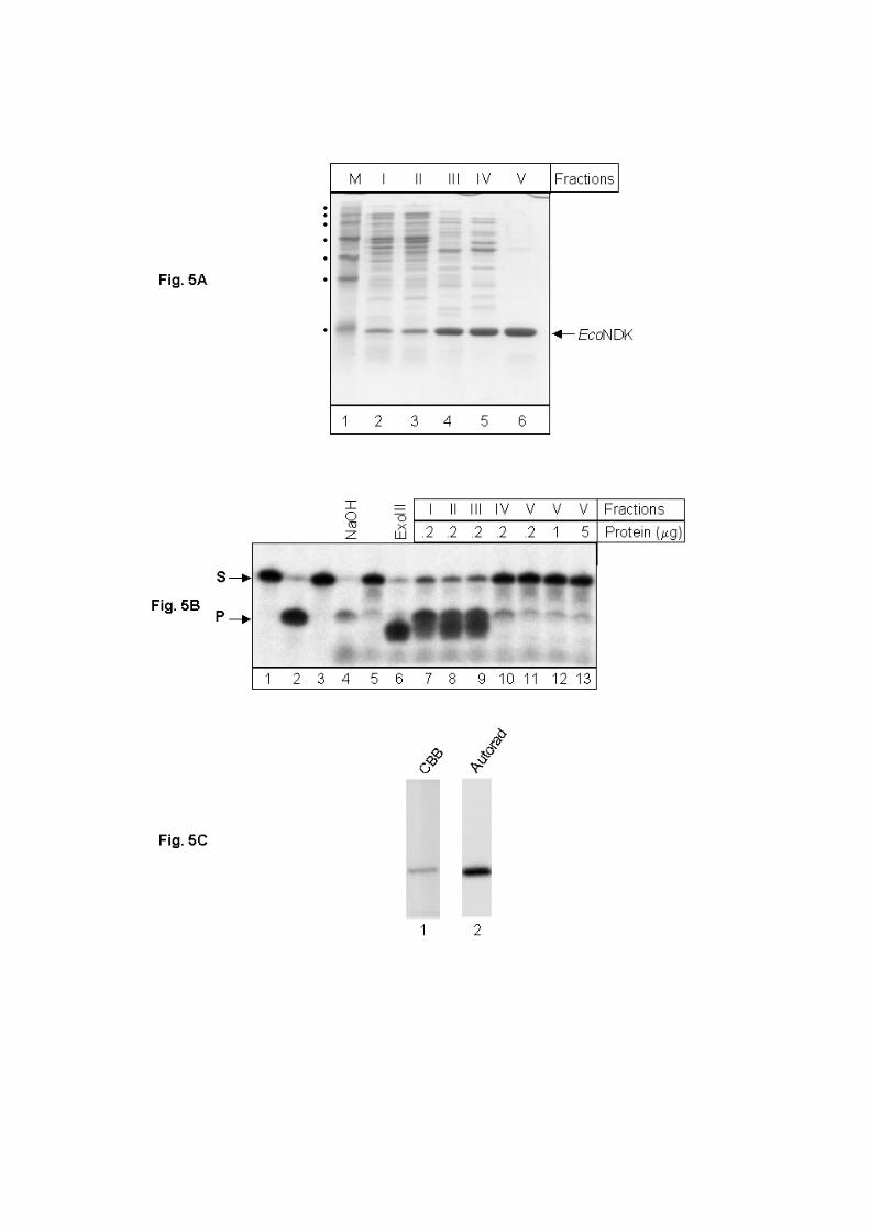
To further substantiate that NDKs do not possess AP-endonuclease activity, we
assayed the protein pools from various purification steps of one of our preparations of
EcoNDK (native primary sequence). Analysis of equal amounts of total proteins from various
pools on SDS-PAGE, as expected, showed enrichment of EcoNDK at each step of
purification (Fig. 5 A, lanes 2-6, fractions I to V). The AP-endonuclease assays performed
with 0.2 µg of total proteins from various fractions are shown in Fig. 5 A. The assays using
Fraction I-III showed efficient AP-DNA processing activities (compare product bands in lane

7 to 9, with the control products obtained by treatment of AP-DNA with NaOH, lane 4, and
exonuclease III, lane 6). However, at the purification step resulting in Fraction IV, the AP-
DNA processing activity was nearly eliminated (lane 10); and by Fraction V of EcoNDK
preparation, this activity was undetectable even when 5 µg preparation was used in the assay
(lanes 11-13). As a control, when checked for autophosphorylation activity, Fraction V
showed efficient autphosphorylation of EcoNDK (Fig. 5 C). Thus, the successive steps of
purification of EcoNDK resulted in gradual decrease in the AP-DNA processing activity. Had
this activity been intrinsic to NDK, we would expect an increase in its specific activity with
each step of purification.

Discussion.
Nucleoside diphosphate kinase is an important enzyme in nucleotide metabolism in
the cells. Recent studies showed that it has additional activities of uracil DNA glycosylase,
AP-endonuclease and dRpase, implicating its role in DNA repair pathways (Postel). To study
such DNA repair functions, we purified EcoNDK and MtuNDK, with and without hexa-
histidine tag from ung+ and ung- background of E. coli (Fig. 1 A). The autophosphorylation
assay with each one of the preparations indicated that the proteins retained their native
conformation (Fig 1 B). The EcoNDK as well as MtuNDK preparations from ung+ strains
showed detectable UDG activity. However, this activity was fully inhibited by extremely low
levels of Ugi protein (2 ng Ugi in 10 µg of NDK preparation). Analysis of EcoNDK and Ugi
on Superdex-200 column (Fig. 3) showed that the two proteins do not interact with one
another. Thus, the Ugi effect (especially when it is added to NDK in more than 2000 fold
molar shortage) could not be via its physical interaction with NDK, as argued (Postel). A
hypothetical possibility that presence of Ugi somehow altered the conformation of NDK, can
be ruled out by the fact that the autophosphorylation activity of NDK was not inhibited even
in the presence of 200 ng of Ugi in 1 µg NDK preparation (Fig. 1 B, panel ii). Also, the
observation that elution profile of NDK on Superdex-200 remained unchanged by the
presence of Ugi supports that it does not alter the overall folding of EcoNDK (Fig. 3 B).
On the other hand, as Ugi interaction with family I UDGs is highly specific, and it
forms a complex with them, which is essentially irreversible under the physiological
conditions (Mosbaugh, Narottam, JMB), a straightforward interpretation of these
observations is that the NDK preparations from ung+ strains retained contaminating amounts
of Ung. Addition of extremely low levels of Ugi results in complete sequestration of the
contaminating Ung into a highly stable complex, which no longer binds DNA (), and
therefore, results in total inhibition of UDG activity. More importantly, the NDK preparations
from ung- background fail to show any detectable UDG activity, which provides conclusive
evidence that UDG activity in the NDK preparations from ung+ strain was a consequence of
contaminating amounts of UDG. As the elution properties of NDK on DEAE-Sepharose and
hydroxyapatite, under the conditions used by Postel and ///() are common with those of E.
coli UDG (Ung) on such matrices (Lindahl, 1977), it is quite logical to argue that NDK
preparation (Postel//, 2003) might have remained contaminated with UDG. And, because
UDG has a very high turnover of ~800 per minute (Lindahl, 1977), it is not surprising to
detect its activity even when it is present in extremely low amounts. From a physiological
perspective, majority of the mutations in NDK deficient E. coli have been shown to be AT to
GC transition mutations (Miller JH, 2002, Lu Q 1995 JMB). On the contrary, the loss of
UDG activity results in GC to AT mutations (Miller, Bhagwat). Taken together, these
observations clearly show that the NDK from E. coli as well as M. tuberculosis do not
possess an intrinsic UDG activity. A recent independent study by Bennett et. al. (2004) on
EcoNDK also supports the conclusion that UDG activity is not intrinsic to NDK.
The other DNA repair activities reported to be present in EcoNDK led us to examine
the possibility of AP-endonuclease activity in NDK proteins. However, the data in Fig. 5, are
quite convincing to suggest that even such an activity is not intrinsic to NDK. On the other
hand, the data () that led to a proposal that EcoNDK formed a covalent complex with AP-site
containing DNA and catalyzed the lyase reaction apparently lack identification of the shifted
band (detected by autoradiography) to correspond to NDK.
Finally, while a recent independent report (Bennett et. al. 2004) also showed that
EcoNDK lacked UDG activity, it did not address the aspect of the reported AP-endonuclease
activity of EcoNDK or the interaction of Ugi and EcoNDK. In our studies, we used NDK
from two diverse groups of bacteria (E. coli and M. tuberculosis) to establish that UDG
activity is intrinsic to neither of these proteins. Furthermore, we show that the Eco-, and Mtu-
NDKs lack an intrinsic AP endonuclease activity, thereby ruling out a direct role of NDK in
base excision repair pathway. Additionally, our studies highlight the essentiality of the use of
ung- strains for testing of novel UDG activities.
Acknowledgements: We thank Dr. M. Konrad, Max Planck Institute, Goettingen, Germany
for providing us with the EcoNDK expressions constructs used in this study, and our
laboratory colleagues for their suggestions on this manuscript. This work was supported by
research grants from the Department of Biotechnology, and Indian Council of Medical
Research, New Delhi. P. K. and K. K. were supported by studentships from the Council of
Scientific and Industrial Research, New Delhi.
References:
1. Acharya, N., Roy, S. and Varshney, U. (2002) J Mol Biol, 321, 579-590.
2. Acharya, N., Kumar, P. and Varshney, U. (2003) Microbiology, 149, 1647-1658.
3. Almaula, N., Lu, Q., Delgado, J., Belkin, S. and Inouye, M. (1995) J Bacteriol, 177,
2524-2529.
4. Aravind, L. and Koonin, E.V. (2000) Genome Biol, 1, RESEARCH0007.
5. Bennett, S.E. and Mosbaugh, D.W. (1992) J Biol Chem, 267, 22512-22521.
6. Bennett, S.E., Chen, C.Y. and Mosbaugh, D.W. (2004) Proc Natl Acad Sci U S A.
7. Chaconas, G. and van de Sande, J.H. (1980) Methods Enzymol, 65, 75-85.
8. Chopra, P., Singh, A., Koul, A., Ramachandran, S., Drlica, K., Tyagi, A.K. and Singh,
Y. (2003) Eur J Biochem, 270, 625-634.
9. Cole, S.T., Brosch, R., Parkhill, J., Garnier, T., Churcher, C., Harris, D., Gordon,
S.V., Eiglmeier, K., Gas, S., Barry, C.E., 3rd et al. (1998) Nature, 393, 537-544.
10. Dianov, G., Price, A. and Lindahl, T. (1992) Mol Cell Biol, 12, 1605-1612.
11. Dianov, G. and Lindahl, T. (1994) Curr Biol, 4, 1069-1076.
12. Duncan, B.K. (1985) J Bacteriol, 164, 689-695.
13. Friedberg, E.C., Bonura, T., Cone, T., Simmons, R., & Anderson, C. (1978) DNA
Repair Mechanisms. Academic Press, Orlando, FL.
14. Friedberg, E.C., Walker, G.C. & Siede, W. (1995) DNA Repair and Mutagenesis.
ASM Press, Washington, DC.

15. Hama, H., Almaula, N., Lerner, C.G., Inouye, S. and Inouye, M. (1991) Gene, 105,
31-36.
16. Handa, P., Roy, S. and Varshney, U. (2001) J Biol Chem, 276, 17324-17331.
17. Handa, P., Acharya, N. and Varshney, U. (2002) Nucleic Acids Res, 30, 3086-3095.
18. Kumar, N.V. and Varshney, U. (1994) Nucleic Acids Res, 22, 3737-3741.
19. Kumar, N.V. and Varshney, U. (1997) Nucleic Acids Res, 25, 2336-2343.
20. Lindahl, T., Ljungquist, S., Siegert, W., Nyberg, B. and Sperens, B. (1977) J Biol
Chem, 252, 3286-3294.
21. Lu, Q., Zhang, X., Almaula, N., Mathews, C.K. and Inouye, M. (1995) J Mol Biol,
254, 337-341.
22. Lu, Q. and Inouye, M. (1996) Proc Natl Acad Sci U S A, 93, 5720-5725.
23. Lundquist, A.J., Beger, R.D., Bennett, S.E., Bolton, P.H. and Mosbaugh, D.W. (1997)
J Biol Chem, 272, 21408-21419.
24. Miller, J.H., Funchain, P., Clendenin, W., Huang, T., Nguyen, A., Wolff, E., Yeung,
A., Chiang, J.H., Garibyan, L., Slupska, M.M. et al. (2002) Genetics, 162, 5-13.
25. Mizrahi, V. and Andersen, S.J. (1998) Mol Microbiol, 29, 1331-1339.
26. Ouatas, T., Abdallah, B., Gasmi, L., Bourdais, J., Postel, E. and Mazabraud, A. (1997)
Gene, 194, 215-225.
27. Parikh, S.S., Putnam, C.D. and Tainer, J.A. (2000) Mutat Res, 460, 183-199.
28. Pearl, L.H. (2000) Mutat Res, 460, 165-181.
29. Postel, E.H. and Ferrone, C.A. (1994) J Biol Chem, 269, 8627-8630.
30. Postel, E.H. (1998) Int J Biochem Cell Biol, 30, 1291-1295.
31. Postel, E.H. (2003) J Bioenerg Biomembr, 35, 31-40.
32. Putnam, C.D., Shroyer, M.J., Lundquist, A.J., Mol, C.D., Arvai, A.S., Mosbaugh,
D.W. and Tainer, J.A. (1999) J Mol Biol, 287, 331-346.
33. Ravishankar, R., Bidya Sagar, M., Roy, S., Purnapatre, K., Handa, P., Varshney, U.
and Vijayan, M. (1998) Nucleic Acids Res, 26, 4880-4887.
34. Roy, S., Purnapatre, K., Handa, P., Boyanapalli, M. and Varshney, U. (1998) Protein
Expr Purif, 13, 155-162.
35. Saikrishnan, K., Bidya Sagar, M., Ravishankar, R., Roy, S., Purnapatre, K., Handa, P.,
Varshney, U. and Vijayan, M. (2002) Acta Crystallogr D Biol Crystallogr, 58, 1269-1276.
36. Sakumi, K. and Sekiguchi, M. (1990) Mutat Res, 236, 161-172.
37. Sambrook, J., Fritsch, E. F., & Maniatis, T. (1989) Molecular Cloning: A Laboratory
Manual. 2nd ed. Cold Spring Harbor Laboratory, Cold Spring Harbor, NY.
38. Venkatesh, J., Kumar, P., Krishna, P.S., Manjunath, R. and Varshney, U. (2003) J
Biol Chem, 278, 24350-24358.
39. Wang, Z. and Mosbaugh, D.W. (1989) J Biol Chem, 264, 1163-1171.
Figure legends
Figure 1: (A) Analysis of purified NDKs (~10 µg each) on a 15 % polyacrylamide-SDS gel.
Lanes: 1 & 2, MtuNDK-His6; 3 & 4, EcoNDK-His6; 5 & 6, EcoNDK (native primary
sequence) from ung+ strain of E. coli, preparations A (lane 5) and preparation B (lane 6). (B)
Autophosphorylation assay. Panel (i) shows Coomassie brilliant blue stained gel with ~1 µg
of each NDK proteins on a polyacrylamide-SDS gel. Panel (ii) shows electronic
autoradiograph obtained by BioImage analyser. Lanes: 1-4, MtuNDK-His6; 5-8, EcoNDK-
His6; 9 & 10, EcoNDK (preparation A); and 11 & 12, EcoNDK (preparation B). Genetic
background of E. coli (ung+ and ung-) from which the proteins were purified are as indicated
above the lanes. The symbols – and + indicate absence or presence of the treatment of NDK
preparations (1 µg) with Ugi (0.2 µg) prior to autophosphorylation.

Figure 2: Analysis of NDKs for uracil excision activity. (A) Lanes: 1, substrate control; 2,
product control; 3-5 assays with 2, 5 and 10 µg of MtuNDK-His6 (from ung+ strain),
respectively; 6-7, assays with 10 µg of MtuNDK-His6 (from ung+ strain) pretreated with 2
and 20 ng of Ugi, respectively; 8-10 assays with 2, 5 and 10 µg of MtuNDK-His6 (from ung-

strain), respectively; 11-12, assays with 10 µg of MtuNDK-His (ung- strain) supplemented
with 2 and 20 ng of EcoUDG, respectively. (B) Lanes: 1, substrate control; 2, product
control; 3-5 assays with 2, 5 and 10 µg of EcoNDK-His6 (from ung+ strain), respectively; 6-7,
assays with 10 µg of EcoNDK-His6 (from ung+ strain) pretreated with 2 and 20 ng of Ugi,
respectively; 8-10 assays with 2, 5 and 10 µg of EcoNDK-His6 (from ung- strain),
respectively; 11-12, assays with 10 µg of EcoNDK-His (ung- strain) supplemented with 2 and
20 ng of EcoUDG, respectively. (C) Lanes: 1 and 6, substrate controls; 2 and 7, product
controls; 3, assays with 10 µg of EcoNDK (preparation ‘A’), respectively; 4 and 5, assays
with 10 µg of EcoNDK (preparation ‘A’) pretreated with 2 and 20 ng of Ugi, respectively; 8-
10, assays with 1, 5 and 10 µg of EcoNDK (preparation ‘B’), respectively; 11-12, assays with
10 µg of EcoNDK (preparation ‘B’) supplemented with 2 and 20 ng of EcoUDG,
respectively.

Figure 3: (A) Analytical gel filtration using Superdex-200 (HR 10/30, Amersham
Biosciences). Elution profiles of approximately 100 µg of EcoNDK (panel a), 100 µg Ugi
(panel b), and a mixture of EcoNDK and Ugi, 100 µg each (panel c) using 10 mM potassium

phosphate, pH 7.2, 1 mM Na2EDTA, 2 mM β-mercaptoethanol and 150 mM KCl as elution
buffer. To analyze interaction between the two proteins, EcoNDK and Ugi were mixed and
incubated at room temperature for 30 min followed by on ice for 30 min. All the samples
were applied on to the column using a 100 µl sample loop. Fractions (0.5 ml) were collected,
and 100 µl aliquots from each were quantified in an ELISA plate set-up using Bradford’s
reagent (BioRad). The intensity of the resulting color was read at 620 nm, and plotted on the
Y-axis against the fraction numbers on the X-axis. (B) SDS-PAGE analysis of aliquots (20
µl) from the peak fractions corresponding to gel filtration chromatography shown in panel c
(fraction numbers are indicated above the lanes).
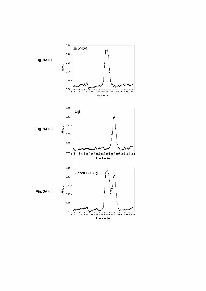

Figure 4: Analysis of AP-endonuclease activity. Assays were carried out as described in
Materials and Methods, for 30 min at 37 oC. Lanes: 1, substrate control without EcoUDG; 2,
substrate control treated with EcoUDG; 3, Alkali mediated cleavage of UDG treated sample
(positive control); 4, Exonuclease III treatment of UDG treated substrate; 5-7, assays with 1,
5 and 10 µg of MtuNDK-His6; 8-10, incubation of uracil containing DNA oligomer
(untreated with UDG) with 1, 5 and 10 µg of MtuNDK-His6; 11-13, assays with 1, 5 and 10
µg of EcoNDK; 14-16, incubation of uracil containing DNA oligomer (untreated with UDG)
with 1, 5 and 10 µg of with EcoNDK.

Figure 5: (A) Analysis of purification of EcoNDK. Total proteins (~10 µg) from successive
steps of purification were separated by SDS-PAGE (15%) and visualized by Coomassie
brilliant blue staining. Lanes: 1, standard markers (M-0671, Sigma Aldrich) corresponding to
~150, ~100, ~75, ~50, ~35, ~25 and ~ 15 kDa molecular sizes; 2, S100 extract (Fraction I);
3, dialyzed extract following ammonium sulfate fractionation (Fraction II); 4, enriched
fractions from Source 15Q column (Fraction III); 5, enriched fractions from hydroxyapatite
column (Fraction IV); 6, fraction IV was further purified on DEAE-sepharose and applied to
Superdex-75 column. Enriched fractions from Superdex-75 constituted Fraction V. (B)
Analysis of AP-endonuclease activity in NDK preparations. Assays were carried out as
described in Materials and Methods for 20 min at 37 oC. Lanes: 1, 5’ end labeled 12U30,
uracil containing DNA oligomer; 2, EcoUDG and NaOH treated 12U30 as product marker; 3,

12U30 untreated with EcoUDG; 4, 12U30 duplex treated with EcoUDG and NaOH; 5,
12U30 treated with EcoUDG alone; 6, 12U30 treated with EcoUDG and exonuclease III; 7-
11, EcoUDG treated12U30 was supplemented with ~200 ng proteins from various steps of
purification of NDK, respectively; lanes 12 & 13 reactions supplemented with 1 and 5 µg of
Fraction V, respectively. (C) Autophosphorylation activity of Fraction V. Fraction V (1 µg)
was autophosphorylated, electrophoresed of SDS-PAGE, stained with Coomassie brilliant
blue (lane 1, CBB) and subjected to electronic autoradiography on BioImage analyzer (lane
2, auto).