urinalysis testing today -- please fill the bottle · what is the typical normal volume of urine ?...
TRANSCRIPT

Eileen Whitehead 2010
East Lancashire HC NHS Trust
Urinalysis Testing Today -- Please Fill The Bottle
1

What is the typical normal volume of urine ?
1-2 litres / 24 hours per normal adult, however,
the amount per day varies considerably
2

The actual quantity is affected by factors such
as ?
Recent fluid intake
Recent food intake
Temperature
General Health
3

What are the physical characteristics of normal
urine ?
Volume is one main characteristics of urine.
Other physical characteristics that can apply to
urine include colour, turbidity (transparency),
smell and pH
4

Colour:
Typically yellow-amber but varies according to
recent diet and the concentration of the urine.
Drinking more water generally tends to reduce
the concentration of urine, and therefore cause it
to have a lighter colour. (The converse is also
true)
5
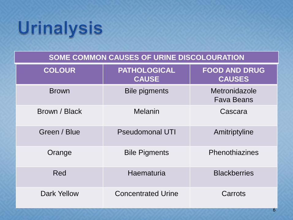
SOME COMMON CAUSES OF URINE DISCOLOURATION
COLOUR PATHOLOGICAL
CAUSE
FOOD AND DRUG
CAUSES
Brown Bile pigments Metronidazole
Fava Beans
Brown / Black Melanin Cascara
Green / Blue Pseudomonal UTI Amitriptyline
Orange Bile Pigments Phenothiazines
Red Haematuria Blackberries
Dark Yellow Concentrated Urine Carrots
6

Turbidity:
Cloudy urine may be due to
Contamination with vaginal mucus
Pyruria secondary to infection
Hyperuricosuria (high protein diet)
7

Smell:
Generally fresh urine has a mild smell but aged
urine has a stronger odour, similar to that of
ammonia. Diabetics may have a sweet or fruity
odour due to the presence of Ketones. Other
causes of abnormal odours are gastrointestinal
– bladder fistula (faecal smell) Vitamin B6 (Iron
smell) Asparagus (Sulphuric)
8

Specific Gravity:
Urine specific gravity shows the concentration of
urine and represents the hydration status of the
patient. Normal specific gravity varies from 1.001
to 1.035. A greater value shows relative
dehydration, diabetes mellitus. A lower value
may indicate diabetes insipidus, increased fluid
intake, or decreased concentrating ability of the
kidney
9

pH:
The pH of normal urine is generally in the range
4.6 - 8, a typical average being around 6.0.
Much of the variation is due to diet. For example,
high protein diets result in more acidic urine, but
vegetarian diets generally result in more alkaline
urine (both within the typical range 4.6 - 8).
10

What is contained in normal urine ?
Approx. 95% of the volume of normal urine is
due to water. The other 5% consists of solutes
(chemicals that are dissolved in the water)
11

Some of these solutes are the results of normal
biochemical activity within the cells of the body.
Other solutes may be due to chemicals that
originated outside of the body, such as
pharmaceutical drugs
1. Organic Molecules
2. Ions
12

Organic molecules are electrically neutral and
can be relatively large (compared with the
'simpler' ions - below).
These include:
13

Urea - Urea is an organic (i.e. carbon-based)
compound also known as carbamide. The
amount of urea in urine is related to quantity of
dietary protein
14

Creatinine - Creatinine is a normal (healthy)
constituent of blood. It is produced mainly as a
result of the breakdown of creatine phosphate in
muscle tissue. It is usually produced by the body
at a fairly constant rate
15

Uric acid - Uric acid is an organic (i.e. carbon-
based) Due to its insolubility, uric acid has a
tendency to crystallize, and is a common part of
kidney stones
16

Ions are atoms or groups of atoms that have
either, lost some outer electrons, hence have a
positive electric charge, or have gained some
outer electrons and hence have a negative
electric charge
17

Individual elements:
Sodium (Na+) and Potassium (K+) The amount
in urine varies with diet and the amount of
aldosterone (a steroid hormone in the body)
Chloride (Cl-) : The amount in urine also varies
with dietary intake
18

Magnesium (Mg2+) : Amount in urine varies with
diet and the amount of parathyroid hormone in
the body. (Parathyroid hormone increases the
reabsorption of magnesium by the body, which
therefore decreases the quantity of magnesium
in urine)
19

Calcium (Ca2+) : Amount in urine also varies
with diet and the amount of parathyroid hormone
in the body. (Parathyroid hormone increases the
reabsorption of calcium by the body, which
therefore decreases the quantity of calcium in
urine)
20

WHY TEST URINE ?
21

Urinalysis a useful procedure as an indicator of
health or disease, and as such, is a part of all
routine health screening
22

Reagent dip-sticks can be used to test for the following chemicals in a fresh urine sample:
Leucocytes NitriteUrobilinogenProteinpHBloodSpecific GravityKetones BilirubinGlucose
23

Leucocytes:
The presence of leucocytes in urine is an
important finding in inflammatory conditions of
the kidneys and urinary tract. In most cases,
where there is a bacterial UTI, leucocytes are
found in the urine
24

Nitrates
Nitrate eliminated via the urinary tract only arise
due to the conversion of nitrate into bacteria
inside the urinary tract. Therefore one of the
most important symptoms of a bacterial UTI is
the presence of nitrate in the urine
25

Urobilinogen
Urobilinogen is normally present in low
concentrations. It is formed in the intestine from
bilirubin, and a portion of it is absorbed back into
the bloodstream. When urobilinogen it could be
a sign of liver disease such as hepatitis and
cirrhosis. When urine urobilinogen is low or
absent, it can mean hepatic or biliary obstruction
26
Protein
Healthy adults normally excrete 80-150 mg
protein in urine daily. However, detectible
proteinuria may be the first sign of renovascular,
glomerular or tubulo-interstitial renal disease.
Alternatively, it may be caused by overflow of
abnormal proteins in diseases such as myeloma
27

Blood:
Haematuria the appearance of blood in the urine
most commonly presents in very small quantities
and is only detected by a simple dipstick test
Some of the causes of haematuria include
infection, tumours, trauma, inflammation, calculi
28

Other possible causes of haematuria
1. Kidney cysts2. Tumours or kidney stones3. Blockages or stones in the tube to
the bladder4. Cystitis (bladder infection), 5. Bladder stones6. Tumours in the bladder7. Disease of the prostate gland
29

Ketones:
Ketonuria is a condition where abnormally high
amounts of ketone bodies are present in the
urine. It is seen most commonly in uncontrolled
diabetes mellitus. However it may develop as a
result of fasting, dieting, starvation and eating
disorders
30

Bilirubin
Bile (mainly conjugated bilirubin) is converted to
urobilinogen by intestinal bacteria. Most of the
urobilinogen is excreted in faeces or reabsorbed
and transported back to the liver to be converted
back into bile. The remaining urobilinogen (about
1% of total) is excreted in the urine.
31

An elevated level of conjugated serum bilirubin
implies liver disease and can be an early feature
of hepatobiliary disease
32

Glucose
Nearly all glucose filtered by the glomeruli is
reabsorbed in the proximal tubules, only
undetectable amounts appear in urine in healthy
patients
33

There are two basic causes for glycosuria.
1. One is that the level of blood glucose is so high
that the renal tubules are unable to reabsorb all
that is presented
2. The other is a failure of the tubules to reabsorb
all glucose at a level where this should be
possible. The latter is called renal glycosuria
34

If glycosuria occurs because a normal renal
threshold has been exceeded, this is usually
indicative of impaired glucose tolerance or frank
diabetes
It can occur in the non-diabetic if a substantial
amount of food high in sugar is consumed and
transiently overwhelms the insulin response
causing hyperglycaemia.
35

HOW TO TEST URINE ?
36

Urinalysis
Ensure that you have the correct equipment -
urine dipsticks, disposable gloves, apron and
sterile receiver
Obtain informed consent for procedure
37

Explain to the patient how to take a mid-stream
urine sample:
Urine samples should be collected using 'clean-catch'
midstream sampling, which ensures that any bacteria
present in the urethra are washed away in the first
portion of urine (Higgins, 2000)
Catheter specimens of urine should be collected
using an aseptic technique in order to avoid
contamination (Higgins, 2000)
38

Check manufacturer's recommendations
Check product expiry date
Wash hands and don gloves and apron
Collect specimen
Remove reagent dipstick and immediately
replace cap
39

Dipstick urine
Immerse the dipstick into urine, then remove
(the duration that the dipstick remains in the
urine is governed by manufacturer's
recommendations)
Wait for appropriate length of time
40

Wipe the edge of the strip against the rim of the
vessel in order to remove any excess urine.
Hold dipstick at a slight angle (This prevents
pad-to-pad contamination)
41

Read the reagent pads against the reference
guide
Dispose of urine and dipstick as with
organisational policy
Remove gloves and apron.
Wash hands
Document results
42

Any Questions?
43

References
Dougherty, L., Lister, S. (2004) The Royal Marsden Hospital
Manual of Clinical Nursing Procedures. Oxford: Blackwell
Publishing.
Higgins, C. (2000) Microbiology testing. In: Understanding
Laboratory Investigations. Oxford: Blackwell Science
Urine Dipstick Analysis -http://www.patient.co.uk/DisplayConcepts.asp?WordId=URINALYSIS
44