ursolic acid exhibits protective property against dss
TRANSCRIPT

·实验研究·
1 IntroductionInflammatory bowel disease (IBD) consists primari-
ly of ulcerative colitis (UC) and Crohn's disease (CD).These disorders are chronic inflammatory conditionsof the intestines and are characterized by the presence
Ursolic Acid Exhibits Protective Property against DSS-inducedUlcerative Colitis in Mice through STAT3 Signaling Pathway
ZHUANG Qunchuan1, 2, 3, 4, SHEN Aling2, 3, LIU Liya2, 3, SANKARARAMAN Senthilkumar5,SFERRA Thomas Joseph5, CHEN Qi1, 4*, CHEN Youqin2, 3, 5*
1 Biomedical Research Center of South China, Fujian Normal University, Fuzhou, Fujian 350117, China;2 Academy of Integrative Medicine, Fujian University of Traditional Chinese Medicine, Fuzhou, Fujian 350122, China;3 Fujian Key Laboratory of Integrative Medicine in Geriatrics, Fujian University of Traditional Chinese Medicine, Fuzhou, Fujian 350122,China;
4 Fujian Key Laboratory of Innate Immune Biology, Fujian Normal University, Fuzhou, Fujian 350117, China;5 Department of Pediatrics, Case Western Reserve University School of Medicine, Rainbow Babies and Children's Hospital, Cleveland44106, OH, USA
*Correspondence: CHEN Qi, E-mail: [email protected]; CHEN Youqin, E-mail: [email protected]
Received 2020-11-12; accepted 2020-12-24Foundation: The National Natural Science Foundation of China (81803882)DOI:10.3724/SP.J.1329.2021.01005 开放科学(资源服务)标识码(OSID):
ABSTRACT Objective: Anti-inflammatory effects of ursolic acid (UA) on a dextran sulfate sodium (DSS)-in-duced experimental murine colitis models was investigated to elucidate its possible molecular mechanisms on in-testinal epithelial cells (IEC). Methods: For in vivo study, a total of 15 male BALB/c mice weighing (20-22) gwere randomly divided into three groups: normal control group, DSS model group and DSS+UA treatmentgroup; the mouse colitis model was induced by 3% dextran sodium sulfate (DSS) for 8 days; one of the DSS-in-duced groups was pretreated with UA. The body weight and colon length of mice in each group were measuredby physical balance and vernier caliper, respectively. The mice in each group were scored according to the clini-cal disease activity index (DAI) score method. HE staining was used to observe the histopathological changes ofcolon in each group. The changes of serum amyloid protease A (SAA) and IL-6 expression in colon tissue weremeasured by ELISA. Using IL-6-stimulated differentiated Caco-2 cells as an in vitro inflammatory model of hu-man intestinal epithelium, the effects of UA on the activation of IL-6/signal transducer and activator of transcrip-tion 3 (STAT3) signal pathway in IEC were examined by Western blot for STAT3 phosphorylation. Results: 1)Compared with the normal control group, the DAI score was increased, the length of the colon was shortened,and the histological damage was obvious in the DSS model group (P<0.05); administration of UA significantlyreduced the severity of DSS-induced murine colitis as assessed by DAI score, colon length, and histology damageof colon (P<0.05). 2) Compared with the normal control group, the SAA level and the IL-6 level of colon tissuein the DSS model group increased significantly. The DSS-induced increases of SAA and colonic IL-6 levels werereversed by UA treatment (P<0.05). 3) Compared with normal IEC, IL-6 stimulation significantly increased thephosphorylation level of STAT3; STAT3 phosphorylation in IEC-treated with IL-6 and UA was significantly in-hibited compared with only IL-6 stimulation (P<0.05). Conclusion: Our findings implicate that UA amelioratesDSS-induced colonic inflammation by blocking IL-6/STAT3 signaling pathway, and therefore indicate that UAmay have clinical potential as a novel targeted therapy for ulcerative colitis.KEY WORDS ulcerative colitis; ursolic acid; inflammation; STAT3 signaling pathway; dextran sulfate sodium
引用格式:庄群川,沈阿灵,刘丽雅,等. 熊果酸通过靶向 STAT3 信号通路对葡聚糖硫酸钠诱导的小鼠溃疡性结肠炎的保护作用[J]. 康复学报,2021,31(1):30-36.ZHUANG Q C,SHEN A L,LIU L Y,et al. Ursolic acid exhibits protective property against DSS-induced ulcerative colitis in mice through STAT3 signalingpathway [J]. Rehabilitation Medicine,2021,31(1):30-36.DOI:10.3724 / SP.J.1329.2021.01005
2021 年 第 31 卷 第 1 期 www.scicloudcenter.com /RM /
30

in the gut of extensive areas of ulceration, pronouncedinflammatory cell infiltrates, and epithelial cell necro-sis [1-3]. Though there has been rapid introduction ofnew therapies for IBD not all patients respond com-pletely and the therapies are associated with side ef-fects including immunosuppression. Thus, the devel-opment of new therapies with less toxicity is needed inthe care of patients with IBD.STAT3 is one of the signal transducer and activator
of transcription (STAT) protein family members. Theactivation of STAT3 plays a major role in the patho-physiology of IBD [4-7]. STAT3 can be activated by adiverse group of growth factors and cytokines, includ-ing interleukin-6 (IL-6) [8]. STAT3 activation is medi-ated by phosphorylation at tyrosine 705. The phospho-rylation of cytosolic STAT3 induces its homodimer-ization, nuclear translocation, and DNA binding re-sulting in the expression of genes involved in inflam-matory diseases such as IBD [9-12]. Previous studies haveshowed that IL-6 is elevated in serum of IBD patientsand predicts clinical relapse in CD [13-15] and anti-IL-6therapy might have a therapeutic role [16-19]. Therefore,IL-6/STAT3 pathway is an appealing target for thera-py in IBD.
Natural compounds have been used extensively fortreating various inflammatory disorders. These com-pounds are especially sought out by and are useful forthose who are unresponsive to or develop significantside effects with conventional treatments. Recentlystudies have suggested that a large group natural com-pound have the potential ability to limit the develop-ment and severity of certain cancers and inflammatorydiseases [20-22]. A natural pentacyclic triterpenoid, urso-lic acid (UA) has been reported to possess anti-inflam-matory and antitumor properties [23-25 ]. Studies haveshown that UA can block nuclear factor-kappaB(NF-κB)-mediated inflammatory signaling and sup-press the phosphorylation of STAT3 [26-28]. However, itis unknown whether UA might have a therapeutic rolein IBD. Thus, in the present study we explored the ef-fect of UA in a well-established murine model of ul-cerative colitis and elucidated its possible mechanismof action in an in vitro model of inflamed human in-testinal epithelium.
2 Materials and methods2.1 Reagents, kits and antibodiesCell culture media and reagents were obtained from
Invitrogen Thermofisher (Grand Island, NY, USA).Dextran sulfate sodium (DSS, molecular weight 40 000Da) was purchased from MP Biochemical (Solon, OH,USA). Ursolic acid was purchased from Sigma Chem-icals (St. Louis, MO, USA). Antibodies forWestern blotwere from Cell Signaling Technology (Danvers, MA,
USA). The immunoblot detection system (ECL Plus)was fromPierce(Rockford, IL,USA).Bicinchoninic acid(BCA) protein assay reagent was from Pierce (Rock-ford, IL, USA). Mouse-specific IL-6 ELISA kit wasfrom Biolegend Inc. (San Diego, CA, USA). Mouse-specific serum amyloid A (SAA) ELISA kit was fromImmunologyConsultants laboratory, Inc (Newberg, OR,USA). All the other chemicals, unless otherwise stat-ed, were obtained from Sigma Chemicals (St. Louis,MO, USA).2.2 Preparation of UA stock solutionUA was dissolved in DMSO (10 mM) and stored at
-20 ℃ until used in the in vitro experiments. UA wasfreshly dissolved in saline for the animal experiments.2.3 Cell culture
Human colon cancer Caco-2 cells were purchasedfrom the American Type Culture Collection (Cat no.HTB37Rockville, MA, USA). The cells were main-tained in Dulbecco's modified Eagle medium (DMEM)(Grand Island, NY, USA) containing 1,000 mg/L ofglucose, and supplemented with 20% (v/v) fetal bovineserum (FBS) (Thermofisher, Grand Island, NY, USA)and antibiotics (50 U/mL penicillin and 50 μg/mLstreptomycin), cultured at 37 ℃ in a humidified aircontaining 5% CO2. Caco-2 cells usually reached con-fluence 3 days after seeding and differentiated into en-terocyte-like cells 18-20 days post-confluence. Thefully differentiated cells were used in the experiments.The media was changed every 2-3 days. The mediawas removed and supplemented with 0.5% FBS medi-um before the day of experiment.2.4 Induction of colitis and treatment with UAMale BALB/c mice (with an initial body weight of
20-22 g) were purchased from Shanghai SLAC Labo-ratory Animal Co., Ltd. (Shanghai, China). The micewere housed under pathogen-free conditions with a 12 hlight/dark cycle and food and water were provided adlibitum throughout the experiments. The mice wereacclimatized for 1 week prior to the experiment. TheInstitutional Animal Care and Use Committee of Fu-jian University of Traditional Chinese Medicine (Fu-zhou, China) approved the animal experiments con-ducted in this study. Colitis was induced by adminis-tering 3% DSS in the drinking water ad libitum fromexperimental day 1 through day 8, as previously de-scribed [29]. Starting at the first day of induction of col-itis, the mice were randomly divided into three groups(n=5): normal control group in which mice receivedneither DSS stimulation nor UA treatment; DSS mod-el, or DSS+UA groups in which mice received DSSstimulation and intra-gastric administration of UA(0.075 mg/g) or the saline alone daily for 12 days.2.5 Evaluation of colitis
The progression of DSS-induced colitis was moni-
庄群川等:熊果酸通过靶向 STAT3 信号通路对葡聚糖硫酸钠诱导的小鼠溃疡性结肠炎的保护作用
31

tored daily in a blinded manner. This included mea-surement of body weight, evaluation of stool consis-tency, and presence of blood in the stools by a guaiacpaper test. Stool consistency was assessed using thefollowing 3-point scale: 0, normal; 2, loose stools; and4, watery diarrhea. The intensity of the guaiac papertest was scored by the following scale: 0, no bleeding;2, slight bleeding (+ occult test); and 4, gross bleeding(blood visible in stool or blood stains visible). Diseaseactivity index (DAI) was represented as the sum ofscores for weight loss, stool consistency and rectalbleeding, as previously described [21].2.6 Sample collection
At the end of the experiment, the mice were anes-thetized with Avertin. Blood was collected via rightheart ventricle puncture with lightly heparinized sy-ringes and kept on ice. Sera were separated by 5 mincentrifugation at 5, 000 ×g and stored at -80 ℃ prior tothe analysis. After sacrifice, the colons were excisedand length and weight were measured. One portion ofeach distal colon was dissected and fixed in 10% for-malin for histological examination. The remainder ofeach distal colon was snap-frozen in liquid nitrogenand stored at -80 ℃ for further analysis of the tissueIL-6 level.2.7 Assessment of histologyThe formalin fixed section of the distal colons were
processed and stained with hematoxylin and eosin(H&E). The severity of colitis was assessed in a blind-ed fashion by an experienced pathologist using a vali-dated histological grading score of the colon as de-scribed previously [22].2.8 Enzyme-linked immunosorbent (ELISA) as-say2.8.1 IL-6 level Proteins were extracted from thefrozen colon using T-PER Tissue Protein ExtractionReagent Kit according to the manufacturer's instruc-tions. Protein concentrations were determined by BCAprotein assaykit (Rockford, IL, USA) and equal amountsof protein were utilized in triplicate for each condition.The concentrations of IL-6 in the colons were mea-sured using a mouse IL-6 ELISA kit (Biolegend) ac-cording to the manufacturers' instructions. All sampleswere assayed in triplicate. Absorbance was read at450 nm using a microplate reader (model ELX800;BioTek Instruments, Inc., Winooski, VT, USA). Resultsare presented as mean (expressed in pg/mg total pro-tein) and standard deviations (SD).2.8.2 Serum amyloid A (SAA) level The concentra-tions of SAA in the sera were measured using a mouseSAA ELISA kit according to the manufacturer's in-structions. All samples were assayed in triplicate. Ab-sorbance was read at 450 nm using a microplate reader(model ELX800; BioTek Instruments, Inc., Winooski,
VT, USA). Results are presented as mean (expressedin pg/mL) and standard deviations (SD).2.9 Western blot analysis
Differentiated Caco-2 cells (20 days post-conflu-ence) in 24-well bicameral inserts were pre-incubatedwith or without UA (20 μM) for 1 hour prior to stimu-lation with IL-6 (10 μg/mL) by addition to the basolat-eral pole for 15 minutes. At the end of the experiment,cells were washed with ice cold phosphate-bufferedsaline (PBS) and lysed with mammalian cell lysisbuffer (Thermofisher, Grand Island, NY, USA) con-taining protease and phosphatase inhibitor cocktails(Millipore, Bedford, MA, USA). The lysis buffer con-taining the disrupted cells was centrifuged at 11,000 ×gat 4℃ for 10 min and the protein concentrations of thesupernatant quantified with the BCA assay kit. Equiv-alent amounts of protein were resolved on 4% to 12%Novex Bis-Tris gels (NuPAGE; Grand Island, NY, US-A). Proteins were then transferred into nitrocellulosemembranes in the iBlot Western blot system (Invitro-gen, Grand Island, NY, USA). The membranes wereblocked for 1 h at room temperature with super Block-ing Buffers (Pierce Biotechnology, Rockford, IL, USA)and incubated at 4 ℃ overnight with primary antibod-ies against pSTAT3 (rabbit, polyclonal, 1:1 000, CST,9145), STAT3 (rabbit, polyclonal, 1:1 000, CST, 12640)and β-actin (rabbit, polyclonal, 1:2 000, CST, 4967) inblocking buffer. Following the primary antibody incu-bation period the membranes were washed and, subse-quently, incubated with appropriate anti-rabbit IgG,HRP-linked secondary antibody (1:5 000, CST, 7074)for 1 h at room temperature. Membranes were analyzedusing enhanced chemiluminescence Plus reagents andscanned with the Storm Scanner (Amersham Pharma-cia Biotech Inc, NJ, USA).2.10 Statistical analysisData are presented as the means±standard deviations
(SD) for the indicated number of independently per-formed experiments and analyzed using Student'st-test or one-way ANOVA, followed by LSD's test orDunnett's test. Data were considered significant whenP<0.05. All the analyses were performed using theSPSS package for Windows (version 22.0; SPSS, Inc.,Chicago, IL, USA).
3 Results3.1 UA prevents clinical manifestations of DSS-induced ulcerative colitis in mice
To evaluate whether UA can protect mice againstDSS-induced ulcerative colitis, we first determined theclinical manifestations in experimental mice. Com-pared with the normal control group, DSS administra-tion induced weight loss, higher disease activity index(DAI), and shortening of the colon (Fig. 1). However,
康复学报 2021 年 第 31 卷 第 1 期
32

3.2 UA prevents the histological damage of colontissue in mice with DSS-induced ulcerative colitisTo determine whether the reduction in clinical man-
ifestations induced by UA is associated with the pre-vention of colonic inflammation, the anatomy of andhistopathological changes within the colon were as-sessed. The colon length shortening observed in theDSS treated mice was prevented in the group withDSS + UA (Fig. 1C). Histologic evaluation (Fig. 2),revealed the distal colons from normal control micehas normal colonic histology with an intact epitheli-
um, well-defined gland lengths with no observable in-flammation in the mucosa, whereas colon tissues ofDSS only showed severe colonic histological damages,including destruction of crypt structure with loss ofgoblet cells, disruption of the epithelial layer and sub-mucosal infiltration of inflammatory cells. Noticeably,UA treatment showed a significantly reduction in thecolonic inflammation. All mice receiving saline onlyor UA only had normal histology of the colon (datanot shown).
in the DSS + UA group there was a significant amelio-ration of the weight loss and DAI as compared toDSS-induced group (Fig. 1A and Fig. 1B). Important-ly, there was no significant difference in the body
weight changes and clinical manifestations betweennormal control group and those mice receiving salineor UA only (data not shown).
3.3 UA decreases the SAA level in mice with DSS-induced ulcerative colitis
To further evaluate the inflammatory process inthese mice, we measured the serum SAA. Consistentwith the result from H&E staining assay, DSS-induced
elevation of SAA level was reduced by UA treatment(Fig.3). The SAA level in mice of normal control,DSS model, and DSS+UA group was (292.1±61.7),(1 427.3±452.8), and (444.5±172.8) μg/mL, respec-tively (P<0.05).
庄群川等:熊果酸通过靶向 STAT3 信号通路对葡聚糖硫酸钠诱导的小鼠溃疡性结肠炎的保护作用
Note: Ulcerative colitis was induced in Balb/c mice by administering fresh tap water with 3% DSS from day 1 to day for 8 days.Starting at the first day of induction of colitis, the mice of DSS model and DSS + UA received DSS stimulation and were giv-en intra-gastric administration with saline or UA (0.075 mg/g), respectively, daily for 12 days. The progression of DSS-in-duced colitis was daily monitored in a blinded manner. A. The weight change of each mouse was measured. B. The DAI wasevaluated upon termination of the experiment in accordance with the scoring system. C. Colons were excised on day 12 andits length was measured. Data were presented as the means ± standard deviations (SD) (n=5 mice/group). 1) P<0.05 versuscontrols; 2) P<0.05 versus DSS+UA.
Figure 1 Effect of UA on the clinical manifestations in mice with DSS-induced colitis
1)
2)
2)
1)
2)
1)
Percento
finitia
lweight/%
Dise
aseactiv
ityindex
Colonic
length
/cm
A B C
Note: At the end of experiment, distal colonic samples were processed with H&E staining. Histopathologicalchanges were observed under the microscope. Images were captured at a magnification of ×100.
Figure 2 Effect of UA on histopathological damage of colon in mice with DSS-induced colitis (×100)
A B C
33
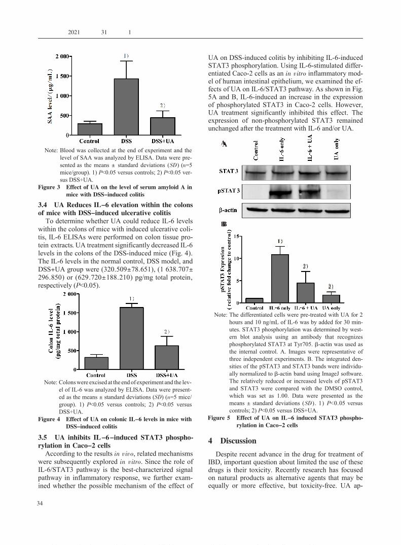
3.4 UA Reduces IL-6 elevation within the colonsof mice with DSS-induced ulcerative colitis
To determine whether UA could reduce IL-6 levelswithin the colons of mice with induced ulcerative coli-tis, IL-6 ELISAs were performed on colon tissue pro-tein extracts. UA treatment significantly decreased IL-6levels in the colons of the DSS-induced mice (Fig. 4).The IL-6 levels in the normal control, DSS model, andDSS+UA group were (320.509±78.651), (1 638.707±296.850) or (629.720±188.210) pg/mg total protein,respectively (P<0.05).
3.5 UA inhibits IL-6-induced STAT3 phospho-rylation in Caco-2 cellsAccording to the results in vivo, related mechanisms
were subsequently explored in vitro. Since the role ofIL-6/STAT3 pathway is the best-characterized signalpathway in inflammatory response, we further exam-ined whether the possible mechanism of the effect of
UA on DSS-induced colitis by inhibiting IL-6-inducedSTAT3 phosphorylation. Using IL-6-stimulated differ-entiated Caco-2 cells as an in vitro inflammatory mod-el of human intestinal epithelium, we examined the ef-fects of UA on IL-6/STAT3 pathway. As shown in Fig.5A and B, IL-6-induced an increase in the expressionof phosphorylated STAT3 in Caco-2 cells. However,UA treatment significantly inhibited this effect. Theexpression of non-phosphorylated STAT3 remainedunchanged after the treatment with IL-6 and/or UA.
4 DiscussionDespite recent advance in the drug for treatment of
IBD, important question about limited the use of thesedrugs is their toxicity. Recently research has focusedon natural products as alternative agents that may beequally or more effective, but toxicity-free. UA ap-
Note: Colonswere excised at the endof experiment and the lev-el of IL-6 was analyzed by ELISA. Data were present-ed as the means ± standard deviations (SD) (n=5 mice/group). 1) P<0.05 versus controls; 2) P<0.05 versusDSS+UA.
Figure 4 Effect of UA on colonic IL-6 levels in mice withDSS-induced colitis
1)
2)
Note: Blood was collected at the end of experiment and thelevel of SAA was analyzed by ELISA. Data were pre-sented as the means ± standard deviations (SD) (n=5mice/group). 1) P<0.05 versus controls; 2) P<0.05 ver-sus DSS+UA.
Figure 3 Effect of UA on the level of serum amyloid A inmice with DSS-induced colitis
SAAlevel/( μ
g/mL)
1)
2)
Note: The differentiated cells were pre-treated with UA for 2hours and 10 ng/mL of IL-6 was by added for 30 min-utes. STAT3 phosphorylation was determined by west-ern blot analysis using an antibody that recognizesphosphorylated STAT3 at Tyr705. β-actin was used asthe internal control. A. Images were representative ofthree independent experiments. B. The integrated den-sities of the pSTAT3 and STAT3 bands were individu-ally normalized to β-actin band using ImageJ software.The relatively reduced or increased levels of pSTAT3and STAT3 were compared with the DMSO control,which was set as 1.00. Data were presented as themeans ± standard deviations (SD). 1) P<0.05 versuscontrols; 2) P<0.05 versus DSS+UA.
Figure 5 Effect of UA on IL-6 induced STAT3 phospho-rylation in Caco-2 cells
1)
2)
2)
A
B
康复学报 2021 年 第 31 卷 第 1 期
34

pears to have pharmacological effects combined withlow toxicity [30-31]. Previous studies in vitro and in vivohave demonstrated that UA has biological effects, suchas anti-oxidative, anti-inflammation, and an-ticanceractivities [23, 25-28]. However, there is limited data avail-able evaluated whether UA has an effect on colonicinflammation. In the present study, we first examinedthe effects of UA on an experimental model of DSS-induced colitis that is a commonly used model for theinflammation component of IBD. Colitis was charac-terized by weight loss, increased disease activity in-dex, shortened colon length, microscopic inflamma-tion, and increases in serum SAA. In our study, for thefirst time, we demonstrated that UA has therapeuticefficacy in this model of colitis ameliorating each ofthese effects.
IL-6 is a cytokine secreted by lamina propria cellsin patients with IBD. In patients with IBD serum IL-6levels positively correlate with severity of intestinalhistopathology [14-16 ]. Previous studies in mice havedemonstrated that the colonic expression of IL-6 is in-creased in acute colitis [32-33]. In the present study, weobserved the increased level of IL-6 in the distal colonof mice with DSS-induce colitis, providing supportingevidence for the crucial role of IL-6 in colonic inflam-mation. Moreover, UA significantly inhibited the pro-duction of IL-6 in this model.
In general, IL-6 binds to soluble or membrane-bound IL-6 receptors (e.g., sIL-6Ra or IL-6Ra) result-ing in the activation of Janus kinase 2 (JAK2) and thedownstream effectors signal transducer and activatorof STAT3 and phosphatidyl inositol 39 kinase [34-35].Recent studies have demonstrated a beneficial effectof anti-IL-6 therapy in Crohn's disease as well as inanimal models of colitis [18-19]. These data strongly sug-gested that activation of the IL-6/STAT3 pathway hasa key role in the development of IBD and the preven-tion of activation of this pathway maybe useful in thetreatment of IBD. Previous reports have demonstratedthat UA down-regulates activation of various pro-in-flammatory pathways including NF-κB, STAT3, AKT,and IKKα/β phosphorylation [26-28]. Therefore, UA'santi-inflammatory effect on colitis may through the in-hibition of STAT3 phosphorylation. To confirm thishypothesis, we investigated STAT3 activation in IL-6stimulated Caco-2 cells. Our results indicate that UAstrongly suppresses STAT3 phosphorylation and thismight be the possible mechanism for its anti-inflam-matory action in UC.
In conclusion, our present study demonstrates thatUA attenuates intestinal inflammation in the experi-mental murine colitis model and may mediate thisthrough the inhibition of activation of the IL-6/STAT3pathway. These findings provide, for the first time, thenovel protective effect of UA against inflammation inthe colon. The present study suggests that further stud-ies should focus on the clinical development of UA forthe treatment of IBD.
References[1] PODOLSKY D K. Inflammatory bowel disease [J]. N Engl J Med,
2002, 347: 417-429.[2] MALOY K J, POWRIE F. Intestinal homeostasis and its break-
down in inflammatory bowel disease [J]. Nature, 2011, 474: 298-306.
[3] KASER A, ZEISSIG S, BLUMBERG R S. Inflammatory boweldisease [J]. Annu Rev Immunol, 2010, 28: 573-621.
[4] DABRITZ J, JUDD L M, CHALINOR H V, et al. Altered gp 130signaling ameliorates experimental colitis via myeloid cell-spe-cific STAT3 activation and myeloid-derived suppressor cells [J].Sci Rep, 2016, 6: 584.
[5] GRIVENNIKOV S, KARIN E, TERZIC J, et al. IL-6 and Stat3are required for survival of intestinal epithelial cells and devel-opment of colitis-associated cancer [J]. Cancer Cell, 2009, 15:103-113.
[6] HAN, THEISS A L. Stat3: friend or foe in colitis and colitis-associated cancer? [J]. Inflamm Bowel Dis, 2014, 20: 2405-2411.
[7] ANDJAR I, RECIO M C, GINER R M, et al. Inhibition of ul-cerative colitis in mice after oral administration of a polyphenol-enriched cocoa extract is mediated by the inhibition of STAT1and STAT3 phosphorylation in colon cells [J]. J Agric Food Chem,2011, 59: 6474-6483.
[8] HODGE D R, HURT E M, FARRAR W L. The role of IL-6 andSTAT3 in inflammation and cancer [J]. Eur J Cancer, 2005, 41:2502-2512.
[9] ZHANG J, WU J, PENG X, et al. Associations between STAT3rs744166 polymorphisms and susceptibility to ulcerative colitisand Crohn's disease: a meta-analysis [J]. PLoS One 2014, 9 (10):e109625.
[10] WELTE T, ZHANG S S, WANG T, et al. STAT3 deletion dur-ing hematopoiesis causes Crohn's disease-like pathogenesis andlethality: a critical role of STAT3 in innate immunity [J]. ProcNatl Acad Sci USA, 2003, 100: 1879-1884.
[11] BACKERT I, KORALOV S B, WIRTZ S, et al. STAT3 activationin Th17 and Th22 cells controls IL -22 -mediated epithelialhost defense during infectious colitis [J]. J Immunol, 2014, 193:3779-3791.
[12] WILLSON T A, JURICKOVA I, COLLINS M, et al. Deletion ofintestinal epithelial cell STAT3 promotes T-lymphocyte STAT3activation and chronic colitis following acute dextran sodiumsulfate injury in mice [J]. Inflamm Bowel Dis, 2013, 19: 512-525.
[13] HYAMS J S, FITZGERALD J E, TREEM W R, et al. Relation-ship of functional and antigenic interleukin 6 to disease activityin inflammatory bowel disease [J]. Gastroenterology, 1993, 104:1285-1292.
[14] VANKEMSEKE C, BELAICHE J, LOUIS E. Frequently relaps-ing Crohn's disease is characterized by persistent elevation ininterleukin-6 and soluble interleukin-2 receptor serum levelsduring remission [J]. Int J Colorectal Dis, 2000, 15: 206-210.
[15] NEURATH M F. Cytokines in inflammatory bowel disease [J].Nat Rev Immunol, 2014, 14: 329-342.
[16] DANESE S, VERMEIRE S, HELLSTERN P, et al. Randomisedtrial and open-label extension study of an anti -interleukin-6antibody in Crohn's disease (ANDANTE Ⅰand Ⅱ) [J]. Gut, 2019,68: 40-48.
[17] ITO H, TAKAZOE M, FUKUDA Y, et al. A pilot randomizedtrial of a human anti -interleukin -6 receptor monoclonal anti-body in active Crohn's disease [J]. Gastroenterology, 2014, 126:989-996.
[18] NEURATH M F. New targets for mucosal healing and therapy ininflammatory bowel diseases [J]. Mucosal Immunol, 2014, 7:6-19.
[19] NEURATH M F. Current and emerging therapeutic targets forIBD [J]. Nat Rev Gastroenterol Hepatol, 2017, 14: 269-278.
[20] SIVEEN K S, SIKKA S, SURANA R, et al. Targeting the STAT3signaling pathway in cancer: role of synthetic and natural in-
庄群川等:熊果酸通过靶向 STAT3 信号通路对葡聚糖硫酸钠诱导的小鼠溃疡性结肠炎的保护作用
35

熊果酸通过靶向 STAT3 信号通路对葡聚糖硫酸钠诱导的小鼠溃疡性结肠炎的保护作用庄群川 1,2,3,4,沈阿灵 2,3,刘丽雅 2,3,SANKARARAMAN Senthilkumar5,SFERRA Thomas Joseph5,陈 骐 1,4*,陈友琴 2,3,5*
1 福建师范大学南方生物医学研究中心,福建 福州 350117;2 福建中医药大学中西医结合研究院,福建 福州 350122;3 福建省中西医结合老年性疾病重点实验室,福建 福州 350122;4 福建省天然免疫生物学重点实验室,福建 福州 350117;5 凯斯西储大学医学院儿科系,美国,俄克拉荷马 克利夫兰,44106* 通信作者:陈骐,E-mail: [email protected];陈友琴,E-mail: [email protected]
摘要 目的:研究熊果酸(UA)对葡聚糖硫酸钠(DSS)诱导的实验性小鼠结肠炎模型的抗炎作用,旨在阐明其对肠上皮细胞(IEC)作用的可能分子机制。 方法:在体内研究中,将体质量为 20~22 g 的雄性 BALB/c 小鼠 15 只采用随机数字表法分为正常对照组、DSS 模型组、DSS+熊果酸治疗组。 通过 3% DSS 诱导 8 d,制备小鼠结肠炎模型;其中一组 DSS 诱导的小鼠结肠炎模型采用 UA 预处理。 各实验组的小鼠体质量和结肠长度分别采用物理天平和游标卡尺测定;参照临床疾病活动指数(DAI)评分法对小鼠进行评分。 采用 HE 染色观察各组结肠组织病理学变化;通过 ELISA 法测定各组小鼠血液中血清淀粉样蛋白酶 A 和结肠组织中 IL-6的水平。以 IL-6 刺激分化的 Caco-2 细胞制备体外人肠上皮细胞炎症模型,通过 Western blot 检测 UA 对肠上皮细胞 IL-6 / STAT3 信号通路中 STAT3 磷酸化的影响。 结果:① 与正常对照组比较,DSS 模型组的 DAI评分增加、结肠长度缩短和组织损伤明显,差异均具有统计学意义(P<0.05);与 DSS 模型组比较,UA 显著抑制了 DSS 诱导的 DAI 评分增加、结肠长度缩短和组织学损伤,差异均具有统计学意义(P<0.05)。 ② 与正常对照组比较,DSS 模型组的血清淀粉酶(SAA)水平和结肠组织中 IL-6 水平升高明显,差异均具有统计学意义(P<0.05);与 DSS 模型组比较,UA 可显著逆转 DSS 诱导的血清淀粉酶(SAA)水平和结肠 IL-6 水平的升高,差异均具有统计学意义(P<0.05)。③ 与正常肠上皮细胞比较,IL-6 刺激显著上调 STAT3 的磷酸化水平;而 UA 可显著抑制肠上皮细胞中 IL-6 诱导的 STAT3 磷酸化水平的增加,差异具有统计学意义(P<0.05)。结论:熊果酸可通过阻断 IL-6/STAT3 信号通路改善了 DSS 诱导的结肠炎,提示熊果酸在靶向治疗溃疡性结肠炎中可能具有临床应用潜力。关键词 溃疡性结肠炎;熊果酸;炎症;STAT3 信号通路;葡聚糖硫酸钠DOI:10.3724/SP.J.1329.2021.01005
hibitors [J]. Biochim Biophys Acta, 2014, 1845: 136-154.[21] SALEHI B, STOJANOVIC-RADIC Z, MATEJIC J, et al. The
therapeutic potential of curcumin: A review of clinical trials [J].Eur J Med Chem, 2019, 163: 527-545.
[22] TASNEEM S, LIU B, LI B, et al. Molecular pharmacology ofinflammation: Medicinal plants as anti-inflammatory agents [J].Pharmacol Res, 2019, 139: 126-140.
[23] YIN R, LI T, TIAN J X, et al. Ursolic acid, a potential anticancercompound for breast cancer therapy [J]. Crit Rev Food Sci Nutr,2018, 58: 568-574.
[24] HUSSAIN H, GREEN I R, ALI I, et al. Ursolic acid derivativesfor pharmaceutical use: a patent review (2012-2016) [J]. Ex-pert Opin Ther Pat, 2017, 27: 1061-1072.
[25] ZHAO J, ZHENG H, SUI Z, et al. Ursolic acid exhibits anti-inflammatory effects through blocking TLR4 -MyD88 pathwaymediated by autophagy [J]. Cytokine, 2019, 123: 154726.
[26] FONTANA G, BRUNO M, NOTARBARTOLO M, et al. Cyto-toxicity of oleanolic and ursolic acid derivatives toward hepato-cellular carcinoma and evaluation of NF-κB involvement [J].Bioorg Chem, 2019, 90: 103054.
[27] PENG J, REN X, LAN T, et al. Renoprotective effects of ursolicacid on ischemia/reperfusion-induced acute kidney injury throughoxidative stress, inflammation and the inhibition of STAT3 andNF-κB activities [J]. Mol Med Rep, 2016, 14: 3397-3402.
[28] MA J Q, DING J, XIAO Z H, et al. Ursolic acid amelioratescarbon tetrachloride -induced oxidative DNA damage and in-flammation in mouse kidney by inhibiting the STAT3 and NF-κB activities [J]. Int Immunopharmacol, 2014, 21: 389-395.
[29] LI L, SHEN A, CHU J, et al. Pien Tze Huang ameliorates DSSinduced colonic inflammation in a mouse colitis model throughinhibition of the IL-6/STAT3 pathway [J]. Mol Med Rep, 2018,18: 1113-1119.
[30] NOVOTNY L, VACHALKOV魣 A, BIGGS D. Ursolic acid: an anti-tumorigenic and chemopreventive activity [J]. Minireview. Neo-plasma, 2001, 48: 241-246.
[31] HUSSAIN H, GREEN I R, ALI I, et al. Ursolic acid derivativesfor pharmaceutical use: a patent review (2012-2016) [J]. Ex-pert Opin Ther Pat, 2017, 27: 1061-1072.
[32] LEE M J, LEE J K, CHOI J W, et al. Interleukin-6 inducesS100A9 expression in colonic epithelial cells through STAT3activation in experimental ulcerative colitis [J]. PLoS One, 2012,7: e38801.
[33] YAMAMOTO M, YOSHIZAKI K, KISHIMOTO T, et al. IL-6 isrequired for the development of Th1 cell-mediated murine coli-tis [J]. J Immunol, 2000, 164: 4878-4882.
[34] WANG Y, VAN BOXEL-DEZAIRE A H, CHEON H, et al.STAT3 activation in response to IL-6 is prolonged by the bind-ing of IL-6 receptor to EGF receptor [J]. Proc Natl Acad SciU S A, 2013, 110: 16975-16980.
[35] MATSUMOTO S, HARA T, MITSUYAMA K, et al. Essentialroles of IL -6 trans -signaling in colonic epithelial cells, in-duced by the IL-6/soluble-IL-6 receptor derived from laminapropria macrophages, on the development of colitis -associatedpremalignant cancer in a murine model [J]. J Immunol, 2010,184: 1543-1551.
康复学报 2021 年 第 31 卷 第 1 期
36