usaf rsvp lower quarter screening examination maj. robert · pdf filelower quarter screening...
TRANSCRIPT
USAF RSVP
LOWER QUARTER SCREENING EXAMINATION
Maj. Robert S. Wainner
Capt. Julie Whitman
TABLE OF CONTENTS
1. INTRODUCTION
2. HISTORY
3. ORDER OF EXAMINATION
4. ITEMS OF THE CLINICAL EXAMINATION & THEIR MEASUREMENT PROPERTIES BY CATEGORY
5. APPENDIX:
1. MODIFIED OSWESTRY DISABILTY INDEX
2. FABQ
1
INTRODUCTION
The lower quarter screening exam (LQSE) consists of both a historical and a physical examination of the lower quarter, to include the thoracic and lumbar spine, the sacroiliac joint region, and the lower extremities. For patients with low-back pain (LBP), the Modified Oswestry Disability Index (Appendix 1) may be helpful for staging a patient’s acuity status.7 The objectives of the LQSE are to: 1) identify medical problems which may preclude further physical therapy evaluation or treatment; 2) identify signs of peripheral or central nervous system compromise; 3) determine which areas or regions of the body will require a more detailed examination and which areas can be ruled out or deferred for evaluation at a later time. ,
By the completion of the historical examination, the therapist must establish an initial hypothesis concerning which area(s) of the body require examination. Because many patients will present with signs and symptoms that could originate from one of several different regions of the lower quarter, the LQSE is used to determine which region(s) require further examination. In practice, a therapist will often incorporate specific regional examination procedures based on key findings during the screening examination. The LQSE is a dynamic process and may be deviated from in part or whole based on results obtained during the examination. However, the therapist should always make note of those areas that have not been cleared during the LQSE and perform a screening examination of those areas as needed at follow-up appointments. The lower quarter screen is required for any airman with complaints suggestive of a neurological lesion. These symptoms may include paresthesias, referred or radiating pain in the lower extremities, difficulty with balance, etc. Finally, the LQSE should identify airmen with medical conditions not amenable to further physical therapy evaluation and treatment. Examples may include pain of a visceral origin, medical or surgical emergencies such as cauda equina syndrome, undiagnosed serious pathology such as neoplasms, nervous system disorders, or inconsistent examination findings possibly suggestive of significant psychological contributions to the patient’s pain.
Inclusion of Waddel’s non-organic signs and symptoms has been purposely omitted from the screening examination. The original purpose of these indexes were to screen for psychological distress (not malingering) in LBP patients by identifying selected abnormal pain behaviors.37,39 Although correlated with other measures of illness behavior25,40,24 in patients with chronic LBP, Waddel’s signs and symptoms have not been well studied in patients with acute LBP. Unfortunately, there is evidence that Waddel’s signs and symptoms are not sensitive for outcomes (return to work) associated with abnormal illness behavior in patients with acute LBP,14 may be over interpreted,39,14and, at worst, lead to labeling of patients.22 Waddell et. al. have developed a Fear-Avoidance Beliefs Questionnaire (FABQ) that has been found to be highly related to chronic disability in patients with LBP40,16 and is a significant predictor of disability and return to work status at 4-weeks.12 Preliminary evidence suggest that a work section score with a cut-off of 30 points is highly sensitive for predicting return to work in a sample of patients with acute, occupationally related LBP. In other words, a patients with a FABQ work score of ≤ 30pts. is not likely to develop work disability. Likewise, a patient with a FABQ work score of > 30pts does not mean work disability is highly likely, only that the patients warrants closer monitoring or work-up.12 The FABQ is provided in Appendix 2.

2
HISTORY
Although the prevalence of serious non-musculoskeletal origin pain is extremely low, the therapist should screen every patient for red flag signs or symptoms and refer to the appropriate medical provider as needed. The therapist should ask every patient about unexplained weight loss, severe pain which is not affected by position or movement, recent alterations in bowel or bladder function, symptoms that awaken the airman at night, and the presence of fevers, chills, night sweats, nausea, or vomiting. It is important to distinguish between the airman who awakens due to a change in sleeping position versus the individual who awakens for no apparent reason or because of anxiety related to the military deployment. If the patient does not awaken for positional reasons, he or she should be referred to the appropriate medical practitioner.
Specific information regarding historical screening for pain of non-musculoskeletal origin is listed below:
Cancer: Deyo and Diehl8 studied 1,171 individuals with low back pain in general practice (13 with diagnosed vertebral cancer). The diagnostic values of different historical findings for detecting underlying cancer are presented in the table below.
HX Findings: Prior hx of cancer
No relief with
bedrest
Unexplained weight loss
> 50 yrs of age
Duration of sxs > 1 month
Appears to be in severe pain
Sensitivity 0.31 0.31 0.15 0.77 0.50 0.23
Specificity 0.98 0.90 0.94 0.71 0.81 0.85
Positive Likelihood
Ratio 14.7 3.1 2.7 2.7 2.6 1.6
Negative Likelihood
Ratio 0.32 0.77 0.90 0.32 0.62 0.91
Table 1
Spinal Fracture, Tumor or Infection, Cauda Equina Syndrome: The Guidelines of the Agency for Health Care Policy and Research (AHCPR)3 for the treatment of adults with low back pain summarized several red flags that the clinician should recognize as potentially indicative of serious pathology, and possibly necessitating referral for further medical work-up.
Spinal Fracture: Minor trauma or strenuous lifting in older and / or potentially osteoporotic individuals; Major trauma such as a fall from a height or a vehicle accident
Tumor or Infection: Over 50 or under 20 years of age; Prior history of cancer; Recent fever, chills, or unexplained weight loss; risk factors for infection (to include recent bacterial infection, IV drug abuse, or immune suppression (from steroids, transplant, or HIV)

3
Cauda Equina Syndrome: Recent onset of bladder dysfunction (increased retention or frequency or overflow incontinence); saddle anesthesia; serious or progressive neurological deficit in one or both lower extremities.
All airmen should be questioned regarding the nature, onset, and progression of their symptoms, specific movements or positions that make the symptoms better or worse, and any 24-hour pattern of symptom behavior.
Finally, a good historical examination may help with early differentiation when seeking to identify the source of the airman’s pain. Pain or other symptoms that are aggravated by the following activities or positions may implicate the specific region.
Lumbar: bending forward, bending backward, prolonged sitting or standing.
SI Joint: transfers from sit to stand or stand to sit, crossing the legs.
Hip: crossing legs (either a flexion/abduction/external rotation or flexion/adduction/internal rotation position), sitting for long periods of time or sit to stand after prolonged sitting, lying on the involved side, deep squatting
Knee: motion of the knee (flexion or extension), up or down stairs, squatting
Ankle / Foot: up or down stairs, squatting, ankle / foot motion.
4
ORDER OF EXAMINATION & PROCEDURES
STANDING
Observation
Posture: Spine, Hips, Knees, & Feet
Bony landmarks: PSIS, ASIS, & Iliac Crests
Gait:
General: Gait pattern, squat
Specific: Heel & Toe Walking
Lumbar AROM:
Gross AROM, AROM with overpressure
Special Tests:
Stork Test, Quadrant
SITTING
Thoracic Rotation
PSIS Palpation
Hip ROM:
Gross (internal/external rotation), Overpressure
Neurologic: ( *)= predominate root
MSR’s: Patellar Tendon Reflex (L4), Achilles Reflex(S1)
Motor: hip flexion (L1-L2) knee extension (L2 - L4), ankle dorsiflexion (L4-L5), great toe extension (L5),ankle plantar flexion with eversion (S1-S2), & ankle plantar flexion (S1-S2).
Sensory: inguinal area (L1), anterior mid-thigh (L2), distal anterior thigh and medial knee (L3), medial lower leg and foot (L4) lateral lower leg and foot (L5), posterior calf (S1), medial calcaneus (S2)
SUPINE
Special Tests
Straight leg raise (SLR); ipsilateral & contralateral, Patrick Test (FABER),
Range-of-Motion

5
Hip flexion, “Sign of the Buttock”
Vascular
Peripheral pulses (posterior tibial, dorsalis pedis)
Neurologic
Babinski response
PRONE
Range-of-Motion
Hip rotation
Special Tests
Femoral nerve stretch, Spring test (T6 – sacrum)
6
ITEMS OF THE CLINICAL EXAMINATION & THEIR MEASUREMENT PROPERTIES BY CATEGORY
OBSERVATION
PALPATION
ACTIVE & PASSIVE MOVEMENTS
NEUROLOGIC
SPECIAL TESTS
OBSERVATION
Postural (General): The posture of the spine and the lower quarter is observed. During the LQSE, the main concern is with obvious asymmetry or deformity that would indicate the need for further examination.
The Spine
The position of the lumbar spine is noted in the sagittal and frontal planes. If a scoliosis or lateral shift are observed, a more focused examination of the lumbosacral spine may be necessary.
The reliability of observational judgments of the shape of the lumbar curve have shown modest agreement (kappa values 0.33-0.50),29,34,36. Higher levels of agreement have been demonstrated for judgements of a lateral shift (kappa values 0.89-1.0).41,31
The Hips
The gluteal muscle bulk is observed for signs of unilateral atrophy that may occur with long-standing lumbar spine or hip problems. Additionally, the relative heights of the gluteal folds are compared bilaterally. Asymmetry may indicate a leg length discrepancy.
The position of the hip joint in the transverse plane may be inferred from the foot placement and patellar position. Excessive toe-in position and/or internally rotated patellae may indicate anteversion of the hip, whereas excessive toe-out or externally rotated patellae may indicate retroversion of the hip joint.
The Knees
Observe the knees in a relaxed stance posture and look for a flexed, hyperextended, varus, or valgus position. Asymmetries in the position of the knee joints may be associated with leg length discrepancy, previous injury or asymmetrical degenerative changes and may warrant closer examination.
From the anterior view the position of the patellae are noted. “Squinting” or medially facing patellae may be associated with anteversion of the hip joint, medial torsion of the femur, or external torsion of the tibia.
7
The Feet
Concentrate on the height of the medial longitudinal arches and calcaneal inclination in the frontal plane. The high arched foot, often with a calcaneal varus, and the low arched foot, often with a vertical or valgus position of the calcaneus, are the two most frequently observed positions of the feet.
Identify obvious deviations from normal or asymmetry between sides of the body. For example, the foot on the side of a long lower extremity very often presents a lower arch appearance than the opposite foot. This may represent an attempt to compensate for a leg length difference because pronation of the subtalar joint can produce functional shortening of the limb. If unilateral pronation of the foot is present as a compensatory mechanism for another osseous or soft tissue deformity, the unilateral pronation may create a functional leg length imbalance.
Postural (Specific): Pelvic landmark assessment is used to identify leg length discrepancies or signs of sacroiliac region dysfunction that may require further evaluation. This assessment should include observation and palpation of the iliac crests, posterior superior iliac spines (PSIS), and the anterior superior iliac spines (ASIS). See the PALPATION section below for details.
Gait: The patient’s general gait pattern should be observed as part of the lower quarter screening process. Because judgments of antalgic gait have shown only modest agreement (kappa = 0.38)36, one should focus on major asymmetries or obvious antalgic patterns that might indicate the need for further evaluation of the involved region.
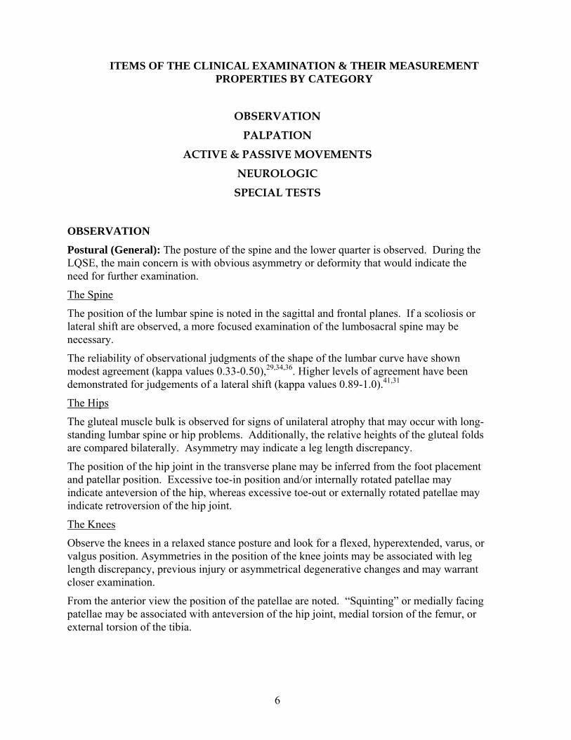
Heel and Toe Walking (Fig. 1 & 2)
The airman is next asked to walk on the heels and then on the toes. Heel walking and toe walking are performed to assess the strength of the L4/5 myotomes (ankle dorsiflexors) and S1/2 myotomes (plantarflexors).
Non-weightbearing strength tests of the plantarflexor or dorsiflexor muscles should be used to differentiate true weakness from inability to heel or toe walk due to pain. The reliability of judgments of disturbed ability to heel or toe walk has shown moderate to excellent reliability (kappa values: 0.57 heel walking, and 0.82 toe walking).36
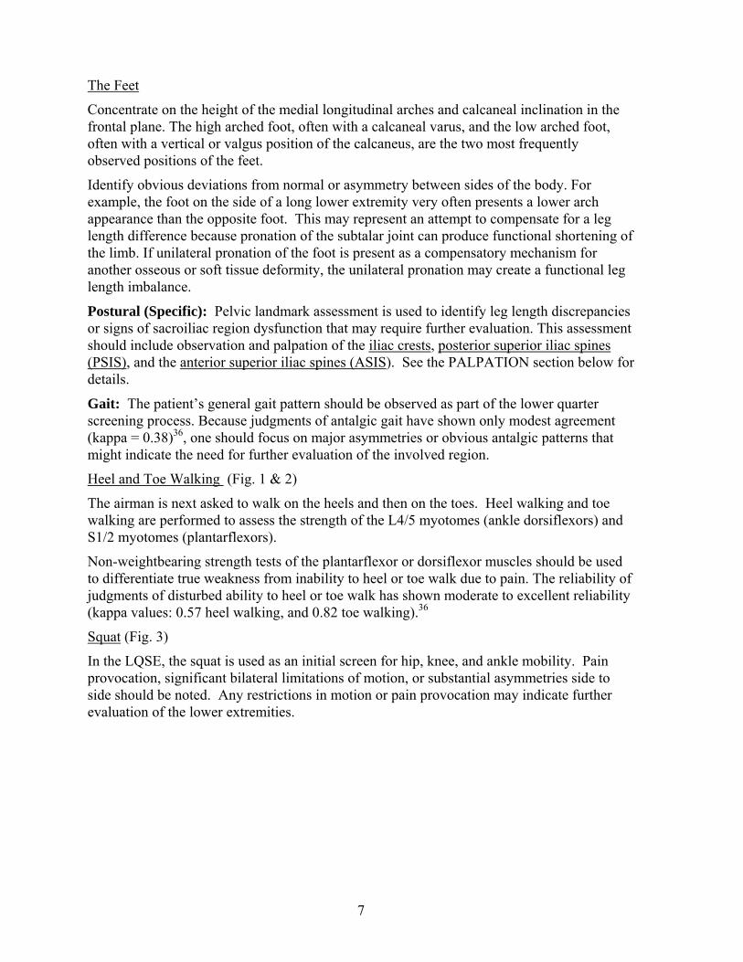
Squat (Fig. 3)
In the LQSE, the squat is used as an initial screen for hip, knee, and ankle mobility. Pain provocation, significant bilateral limitations of motion, or substantial asymmetries side to side should be noted. Any restrictions in motion or pain provocation may indicate further evaluation of the lower extremities.
8
Figure 1 Figure 2
Figure 3
9
PALPATION
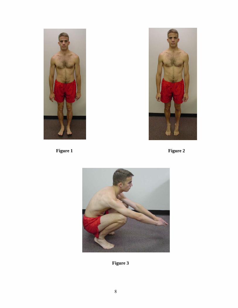
Iliac crests are evaluated from the posterior aspect of the airman using the physical therapist’s hands. The therapist should be at eye-level with the airman’s pelvic landmarks in order to judge the symmetry. The PSIS levels are determined by placing the edges of the thumbs directly beneath the inferior aspect of the bony landmark on each side and comparing the height (Figs. 4 & 5). ASIS levels are determined in a manner similar to that for the PSISs, but from the anterior aspect of the airman.
Figure 4 Figure 5
Asymmetry of the pelvic landmarks should be compared to determine one of the following possibilities: 1) normal: even pelvic landmarks in standing; 2) leg length discrepancy: symmetrically increased height of the ASISs, PSISs and iliac crests on one side; 3) innominate rotation: asymmetrical heights of ASISs, PSISs and iliac crests (for example, low PSIS on right, high iliac crest on right and high ASIS on right). Note that a leg length discrepancy and a rotated innominate may both occur simultaneously.
If a leg length discrepancy is identified and is deemed significant, correction with a heel lift is indicated. The presence of an innominate rotation will require further examination of the lumbosacral region.
Varied reliability values have been reported in the literature for assessment of the pelvic landmarks. Potter and Rothstein27 reported percent agreement on symmetry of the pelvic landmarks ranging between 35% - 38%. O’Haire and Gibbons found a 51% agreement on PSIS symmetry and a kappa value of only 0.04.26 Whitman et al reported a kappa value of 0.43 for standing iliac crest height and 0.67 for standing ASIS height for a more experienced rater pair, but only 0.03 and 0.20 respectively for a less experienced rater pair.42
10
Levangie19 studied the relationship between asymmetry of the ASIS and PSIS measured with calipers and the presence of low back pain and found weak relationships. Stronger associations were found between PSIS symmetry and back pain in males and in younger subjects.19 The diagnostic value of symmetry tests have not been compared with a reference standard more indicative of sacroiliac region dysfunction.
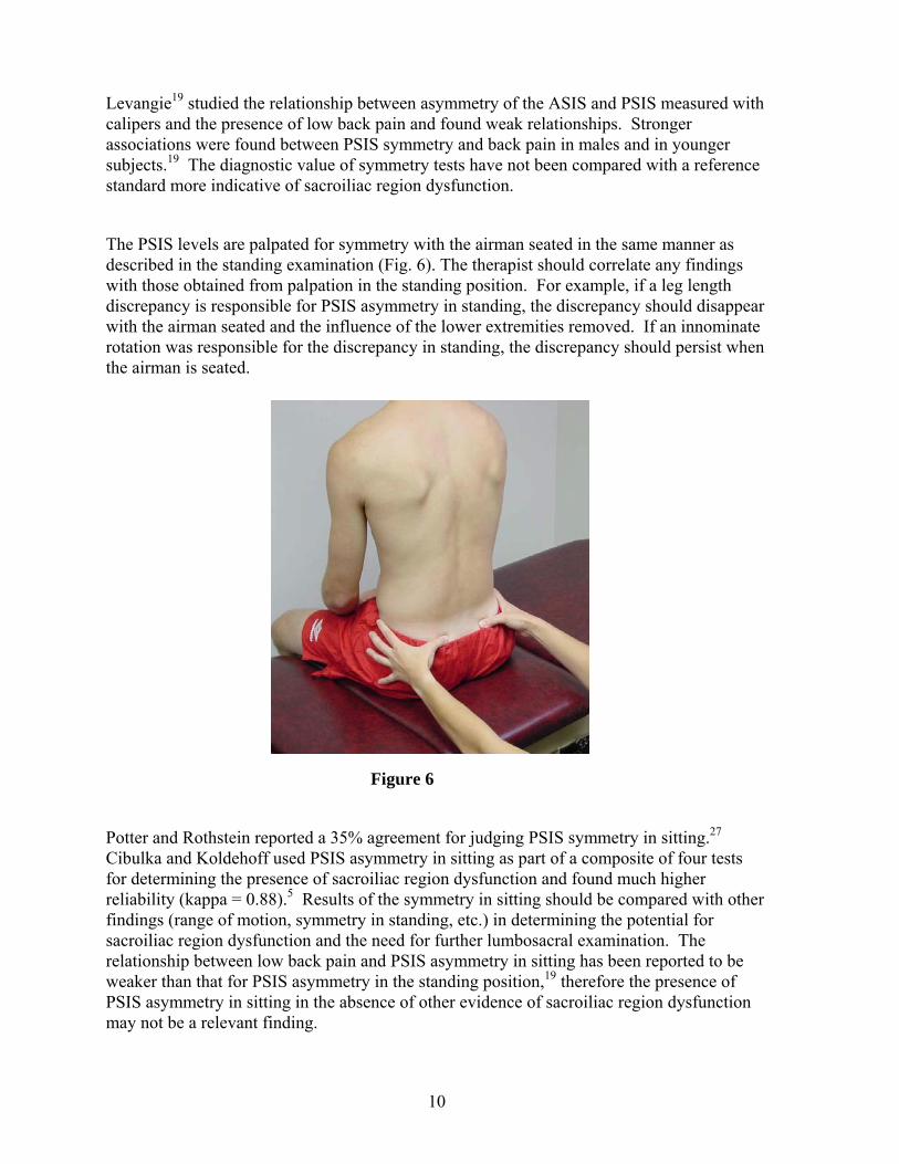
The PSIS levels are palpated for symmetry with the airman seated in the same manner as described in the standing examination (Fig. 6). The therapist should correlate any findings with those obtained from palpation in the standing position. For example, if a leg length discrepancy is responsible for PSIS asymmetry in standing, the discrepancy should disappear with the airman seated and the influence of the lower extremities removed. If an innominate rotation was responsible for the discrepancy in standing, the discrepancy should persist when the airman is seated.
Figure 6
Potter and Rothstein reported a 35% agreement for judging PSIS symmetry in sitting.27 Cibulka and Koldehoff used PSIS asymmetry in sitting as part of a composite of four tests for determining the presence of sacroiliac region dysfunction and found much higher reliability (kappa = 0.88).5 Results of the symmetry in sitting should be compared with other findings (range of motion, symmetry in standing, etc.) in determining the potential for sacroiliac region dysfunction and the need for further lumbosacral examination. The relationship between low back pain and PSIS asymmetry in sitting has been reported to be weaker than that for PSIS asymmetry in the standing position,19 therefore the presence of PSIS asymmetry in sitting in the absence of other evidence of sacroiliac region dysfunction may not be a relevant finding.
11
ACTIVE & PASSIVE MOVEMENTS
Range-of-Motion: Active range-of-motion (AROM) of the lumbar spine is performed in standing, active assistive range-of-motion of the thoracic spin is performed in sitting, and passive range-of-motion (PROM) of the hips is performed in sitting, supine, and prone. During the lower quarter screening, the clinician is most concerned about the effect of movement on the airman’s symptoms. If the airman’s symptoms are affected in any way by movements of the spine or hips, further evaluation of these areas is indicated.
Lumbar Spine
The first step is to establish the baseline status of all symptoms in standing, to include the location and intensity of the symptoms. The airman is then asked to perform flexion, extension, right and left side-bending, and a combined movement of extension with same side rotation and sidebending. If the active motion is does not change the patient’s symptoms in any way, the therapist should apply gentle, graded overpressure and again ask about any change in the airman’s symptoms.
Flexion is assessed by instructing the airman to keep the knees straight and bend forward as far as possible. The physical therapist should be aware that symptoms may occur at any point throughout the motion, including the return from the flexed position. If the symptom status does not change, the therapist should apply gentle overpressure to increase the flexion moment between the upper thoracic region and the pelvis. If any change in symptom status is noted, further examination of the thoraco-lumbar and lumbo-pelvic regions is indicated.
Next, the airman is asked to keep the knees straight, keep both feet on the ground, and to bend to either side as far as possible. The therapist should guide the patient as needed to avoid any sagittal plane motion. It may be helpful to ask the patient to slide one hand down the lateral thigh while performing the side-bending motion to help keep the motion in the frontal plane. Additionally, the final position of the patient’s fingertips allows for a quick comparison of between right and left side-bending motion. If the patient’s symptom status remains unchanged, the therapist should apply gentle overpressure into both left and right side-bending and note any change in symptoms.
Extension range of motion is assessed next. The airman is asked to keep the knees straight, place the hands along the buttocks, and bend backward as far as possible while sliding the hands down the back of his/her thighs. If there is no change in symptom status, the therapist should apply gentle overpressure into extension and note any change in symptoms
Finally, the quadrant position (combined motion of extension and ipsilateral side-bending and rotation) should be assessed. The airman should keep the knees straight while the therapist guides him / her into the quadrant position If the symptom status does not change, the therapist should apply gentle overpressure and note any change in symptoms.
The reliability of judgments of symptom provocation has been reported. Levels of agreement between examiners have generally been high, but variable. Kappa values (or chance corrected measures of reliability) have ranged between 0.23-0.98 for side-bending, 0.46-0.74 for extension, and 0.51-0.97 for flexion.34,35,41,25
Thoracic Spine

12
To evaluate potential thoracic spine and rib cage dysfunction, the therapist should guide the airman into rotation to the left and right, and apply overpressure at the end of the motion (Fig. 8). Any restriction in motion symptom reproduction is noted and may indicate further evaluation of the thoracolumbar region. While the validity of this test as screening tool for thoracic spine or rib cage dysfunction has not been studied, the reliability of judging pain provocation during rotation is similar to other movements of the spine (kappa = 0.58).25
Figure 7
Hips
Restriction of motion or pain provocation may indicate hip joint dysfunction and necessitates a more focused regional examination of the hip joint. A unilateral restriction of internal rotation is consistent with a capsular pattern and may indicate degenerative changes of the hip joint.
• Rotation-Passive range of motion for internal and external rotation of the hip can be assessed in many positions. In this LQSE, we will assess hip rotation in sitting and other hip motion with the airman supine or prone. Restrictions in range of motion and pain provocation should be noted during passive movement both with and without overpressure. To assess hip internal rotation, the airman should place his / her hands on the table on either side of the knees to maintain the hips in a neutral abduction/adduction position (Fig. 8) The clinician then internally rotates both hips simultaneously by moving the airman’s ankles outward. The relative range of motion of the left and right hip joints can be assessed visually by comparing the excursion of the airman’s lower legs. Overpressure can be applied into hip internal rotation. The alternate method of assessing hip internal rotation in supine is illustrated in Figure 9.

13
Figure 8 Figure 9
For external rotation, each hip is assessed one at a time. The non-tested hip should be is abducted to create space for the rotation of the opposite side. The clinician stabilizes the airman’s thigh by placing one hand just proximal to the knee, and then uses the other hand to externally rotate the limb. Overpressure should be applied at the limit of the available range of motion. The opposite limb is tested in the same manner.
• Flexion & Abduction- The range of motion of hip flexion and abduction are assessed with the airman supine. For the purposes of the LQSE, measurement of range of motion is not required. For each movement, the clinician is concerned about symptom reproduction, gross bilateral restriction in motion, or a restriction of motion of one hip as compared with the opposite hip.
Hip flexion is assessed by holding the airman’s knee flexed and the hip in neutral rotation while passively flexing the hip. The opposite leg is maintained in a neutral sagittal plane position. Gentle overpressure into hip flexion should be applied (Fig 10).
Figure 10
14
Abduction is assessed by passively moving the airman’s leg into abduction while maintaining knee extension and neutral hip rotation. Gentle overpressure may be applied at the end of the available range of motion.
Both flexion and abduction range of motion restrictions are components of the capsular pattern of the hip, along with internal rotation as was previously described. The restriction of all three of these motions may indicate the presence of hip joint arthrosis, and necessitates further examination. Fair to moderate correlation between limited hip range of motion and radiologic evidence of hip OA has been reported.32
An unusual but potentially important finding may emerge when the results of the straight leg raise test and passive range of motion assessments of hip joint flexion are compared. A finding of a restricted SLR with concurrent limited hip flexion (with the knee flexed) and a non-capsular pattern of restriction of range of motion of the hip joint constitutes what Cyriax terms the “Sign of the Buttock”6 This constellation of signs may indicate the presence of serious pathology in the pelvic or gluteal region and should prompt a referral for further medical examination of the region
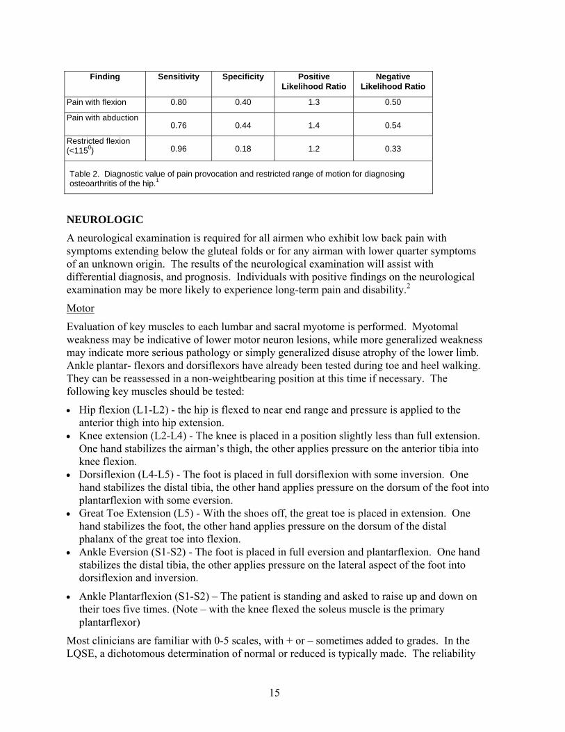
Restrictions of motion or pain provocation with individual range of motion tests or with overpressure into the movements may indicate the need for further evaluation of the hip joint. Once the other motions of the hip joint are assessed, an examination of the pattern of motion restrictions may also be diagnostically useful. For example, Cyriax describes the capsular pattern of the hip, in order from most to least limited, as limitations of internal rotation, flexion, and lastly abduction and external rotation.6 Cyriax further noted that internal rotation may be the only motion restricted in the early stages of hip joint arthrosis, and internal rotation is typically the most painful movement in cases of arthrosis. Therefore a finding of restricted and/or painful internal rotation may be an early sign of degenerative changes in the hip joint and necessitate further regional examination. Judgments of pain provocation with movement or visual detection of range of motion restrictions have not been evaluated for reliability. The validity of pain provocation with internal and external rotation, or restricted range-of-motion has been studied with a reference standard of osteoarthritis of the hip. The diagnostic accuracy coefficients of pain provocation and range-of-motion for osteoarthritis of the hip are listed in table 2.1 Pain with rotation was found to be more sensitive than specific, with low negative likelihood ratios indicating that the absence of pain with rotation movements reduces the likelihood of osteoarthritis of the hip. A positive finding (ie, pain with movement) may indicate hip joint dysfunction requiring further evaluation, but should not necessarily be interpreted to indicate the presence of osteoarthritis. A restriction of internal rotation range of motion of greater than fifteen degrees showed improved specificity, and therefore the presence of this finding does increase the likelihood of osteoarthritis of the hip joint. This result attests to the validity of the concept of a capsular pattern of the hip, and the importance of assessing symmetry of internal rotation range of motion. Similar to rotation range of motion, the findings of pain with the movements of flexion and abduction showed higher sensitivity than specificity values.1 Thus, the absence of pain with movement may be a somewhat useful finding for ruling out hip joint arthrosis. This same study also found high sensitivity for flexion range of motion of less than 1150, further reducing the negative likelihood ratio to 0.33
15
Finding Sensitivity Specificity Positive
Likelihood Ratio Negative
Likelihood Ratio
Pain with flexion 0.80 0.40 1.3 0.50
Pain with abduction
0.76
0.44
1.4
0.54
Restricted flexion (<1150)
0.96
0.18
1.2
0.33
Table 2. Diagnostic value of pain provocation and restricted range of motion for diagnosing osteoarthritis of the hip.1
NEUROLOGIC
A neurological examination is required for all airmen who exhibit low back pain with symptoms extending below the gluteal folds or for any airman with lower quarter symptoms of an unknown origin. The results of the neurological examination will assist with differential diagnosis, and prognosis. Individuals with positive findings on the neurological examination may be more likely to experience long-term pain and disability.2
Motor
Evaluation of key muscles to each lumbar and sacral myotome is performed. Myotomal weakness may be indicative of lower motor neuron lesions, while more generalized weakness may indicate more serious pathology or simply generalized disuse atrophy of the lower limb. Ankle plantar- flexors and dorsiflexors have already been tested during toe and heel walking. They can be reassessed in a non-weightbearing position at this time if necessary. The following key muscles should be tested:
• Hip flexion (L1-L2) - the hip is flexed to near end range and pressure is applied to the anterior thigh into hip extension.
• Knee extension (L2-L4) - The knee is placed in a position slightly less than full extension. One hand stabilizes the airman’s thigh, the other applies pressure on the anterior tibia into knee flexion.
• Dorsiflexion (L4-L5) - The foot is placed in full dorsiflexion with some inversion. One hand stabilizes the distal tibia, the other hand applies pressure on the dorsum of the foot into plantarflexion with some eversion.
• Great Toe Extension (L5) - With the shoes off, the great toe is placed in extension. One hand stabilizes the foot, the other hand applies pressure on the dorsum of the distal phalanx of the great toe into flexion.
• Ankle Eversion (S1-S2) - The foot is placed in full eversion and plantarflexion. One hand stabilizes the distal tibia, the other applies pressure on the lateral aspect of the foot into dorsiflexion and inversion.
• Ankle Plantarflexion (S1-S2) – The patient is standing and asked to raise up and down on their toes five times. (Note – with the knee flexed the soleus muscle is the primary plantarflexor)
Most clinicians are familiar with 0-5 scales, with + or – sometimes added to grades. In the LQSE, a dichotomous determination of normal or reduced is typically made. The reliability
16
of such judgments has generally been found to be high. Kappa values between 0.59 and 0.82 (1.0 by McCombe 89) have been reported, with the highest value for great toe extension, and lowest for ankle eversion.36,41
The validity of great toe extension strength deficit for diagnosing lumbar disc herniation and any strength deficit for diagnosing lumbar spinal stenosis has been evaluated. The results have generally found greater values for specificity than for sensitivity, and therefore the absence of strength deficits should not be over-interpreted.
Sensory
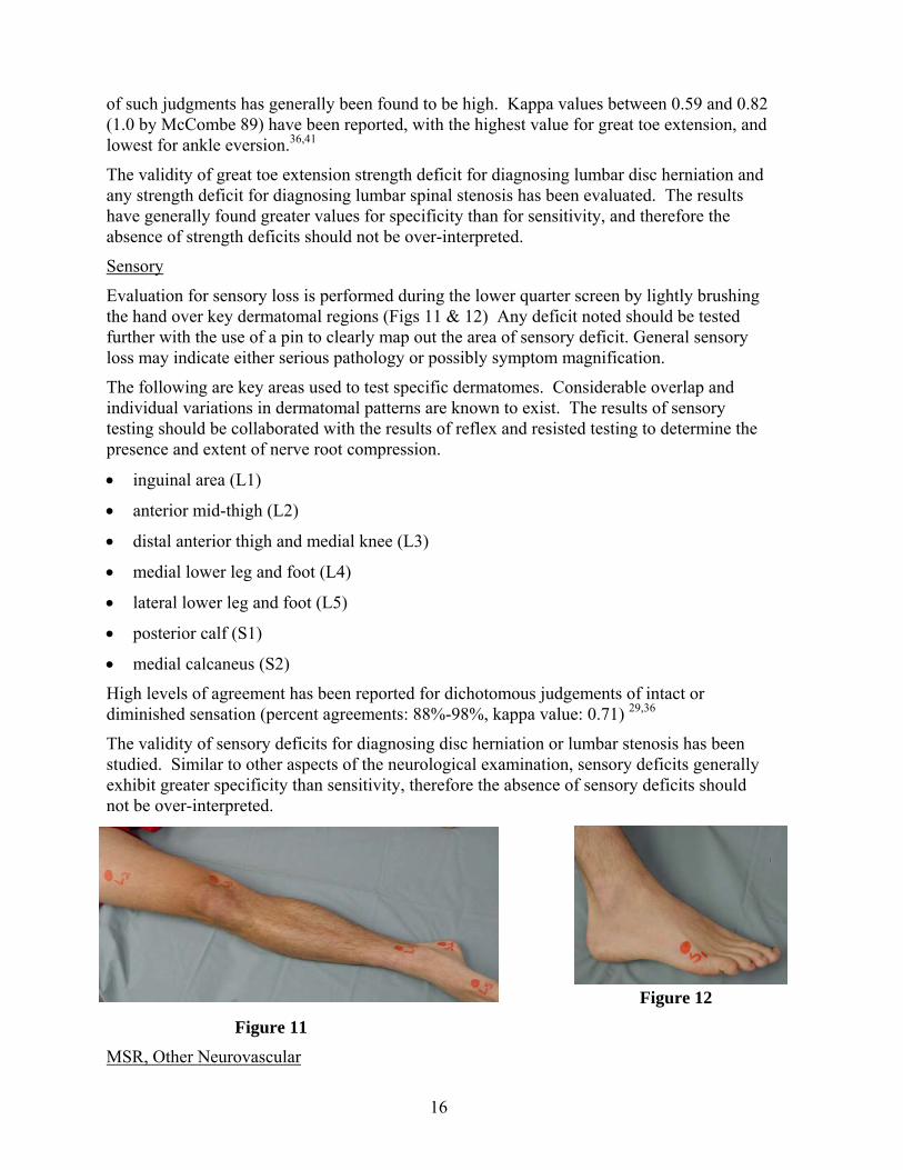
Evaluation for sensory loss is performed during the lower quarter screen by lightly brushing the hand over key dermatomal regions (Figs 11 & 12) Any deficit noted should be tested further with the use of a pin to clearly map out the area of sensory deficit. General sensory loss may indicate either serious pathology or possibly symptom magnification.
The following are key areas used to test specific dermatomes. Considerable overlap and individual variations in dermatomal patterns are known to exist. The results of sensory testing should be collaborated with the results of reflex and resisted testing to determine the presence and extent of nerve root compression.
• inguinal area (L1)
• anterior mid-thigh (L2)
• distal anterior thigh and medial knee (L3)
• medial lower leg and foot (L4)
• lateral lower leg and foot (L5)
• posterior calf (S1)
• medial calcaneus (S2)
High levels of agreement has been reported for dichotomous judgements of intact or diminished sensation (percent agreements: 88%-98%, kappa value: 0.71) 29,36
The validity of sensory deficits for diagnosing disc herniation or lumbar stenosis has been studied. Similar to other aspects of the neurological examination, sensory deficits generally exhibit greater specificity than sensitivity, therefore the absence of sensory deficits should not be over-interpreted.
Figure 12
Figure 11
MSR, Other Neurovascular

17
• For the LQSE, both the patellar tendon and the Achilles’ tendon reflexes are assessed.
The Achilles’ reflex is performed to test the integrity of the S1-S2 nerve roots. The airman is seated and relaxed with the ankle supported at approximately neutral dorsiflexion. The Achilles tendon is struck with the reflex hammer and reflexive plantarflexion is felt with the supporting hand.
The patellar tendon reflex is performed to test the integrity of the L2-L4 nerve roots. The airman is seated with the knee flexed to approximately 90 degrees. The patellar tendon is struck with the reflex hammer and reflexive knee extension is observed.
Reflex assessment is used to evaluate the reflex pathway. Diminished or absent reflexes may be indicative of nerve root impingement and subsequent lower motor neuron disturbance. Confirming evidence that accompanies a diminished reflex would be myotomal weakness or sensation loss or abnormality in a particular dermatome. Hyperactive reflexes in the patellar tendon or Achilles’ tendon reflexes can represent upper motor neuron disturbances (e.g., myelopathy). At the same time, hyperactive reflexes can be a normal variant. If encountered, the clinician should at least suspect a myelopathic process or an upper motor neuron pathology. Confirming evidence for upper motor neuron involvement that accompanies hyperactive reflexes would be clonus or the presence of a Babinski response.
A 5-point scale for reflex responses has been advocated as follows:
4+ : hyper-reflexive and indicative of upper motor neuron dysfunction or myelopathy
3+: reflex in upper half of normal
2+: reflex in lower half of normal
1+ : present but diminished either as compared to the other side or as compared to normal
0 : absent
The reliability of using a five-point scale for judging reflexes has been reported by one author to be good, with kappa values between 0.61-0.74.20. However another study found much lower reliability (kappa values –0.06-0.43).23
Several authors have reported the reliability of a two-point scale (normal or diminished) and have found high levels of agreement between examiners.36 Low prevalence of reflex disturbances has led most researchers to report only percent agreement values which have range between 86% and 100%.41,29,36 Vroomen et al36 also reported a kappa value for the Achilles’ tendon reflex of 0.52. The prevalence of a diminished Achilles’ reflex is this study was 13%.
The validity of reflex deficits for diagnosing lumbar disc herniation and lumbar spinal stenosis has been evaluated. The results show that the findings are generally more specific than sensitive, and therefore the absence of reflex deficits should not be over-interpreted. 28,33,30,9,18,17
• Babinski Responses- The Babinski responses are tested bilaterally to assist in ruling out upper motor neuron disease. The test is performed with the airman supine and relaxed. The examiner places a moderately sharp object (end of the handle of a reflex hammer) at the lateral plantar surface of the airman’s foot near the heel and, with moderate pressure, strokes the foot on the lateral surface to the ball of the foot medially. The normal response for an

18
adult is toe flexion. The abnormal response for an adult, hyperextension of the great toe with or without fanning of the other toes or withdrawal of the limb, is called the Babinski response. A positive Babinski response may require referral of the airman for further evaluation unless an explanation for the finding exists from the airman’s past medical history (e.g., prior stroke, brain injury, etc).
One report in the literature evaluated the reliability of the Babinski response using experienced physicians as examiners. The reliability was poor, with a kappa value of 0.17.15 The validity of the Babinski response for detecting upper motor neuron lesions has not been subjected to formal study, but it has been noted in the literature that the response may not be present with all lesions, and may only occur with dysfunction of a specific part of the pyramidal tract.13 The sensitivity of the Babinski sign may therefore be suspect. The results should be corroborated with other findings (e.g. hyperreflexia).
• Pulses- Assessment of peripheral pulses to evaluate the sufficiency of the vascular supply to the foot is not required with every performance of the LQSE. Peripheral pulse palpation should be included in the screening of airmen with claudication symptoms (leg pain and cramping with walking that is relieved by sitting), or any other airman for whom vascular insufficiency is a concern.
The pulse is assessed in the posterior tibial and dorsalis pedis arteries. The posterior tibial artery may be palpated just posterior and inferior to the medial malleolus. The dorsalis pedis artery can usually be located on the dorsum of the foot just lateral to the tendon of the anterior tibialis muscle. Palpation of the posterior tibial pulse is usually more difficult than the dorsalis pedis artery, and a pulse may be difficult to detect even in a healthy individual. Absence or diminished pulses without a known explanation are grounds for referral or consultation. Absence of peripheral pulses is commonly found in individuals with diabetes or peripheral vascular disease.
Moderate reliability between raters has been reported for the palpation of peripheral pulses with kappa values ranging between 0.33-0.84 reported for vascular surgeons, laboratory personnel, and medical students.38 The same study found that peripheral pulses were often palpable in patients with lower limb arterial disease, and therefore should not be considered diagnostically definitive for vascular disease.
SPECIAL TESTS
Straight leg raise (SLR) (Fig. 13 & 14)
The SLR test is described many different ways in the literature. In the LQSE, we will use this test as a dural tension test used to place tension on the sciatic nerve to aid in the diagnosis the presence of nerve root compression of the lower lumbar nerve roots (L4-S1) due to a lumbar intervertebral disc herniation.
The straight leg raise test is performed by standing at the airman’s side to be examined. The airman’s lower extremity is raised slowly to the maximum tolerable level of hip flexion. The knee must be maintained in full extension and the hip in a neutral hip rotation. The test must be performed passively. The examiner should stop the test when the limit of range of motion is reached, not when the airman first reports pain. The opposite leg is maintained in extension during the test. Figures 13 & 14 demonstrate the use of the bubble goniometer to measure the SLR.

19
Figure 13 Figure 14
The contra-lateral lower extremity is usually examined first, then the symptomatic extremity is evaluated in the same manner. For each test, the examiner must note any symptoms that are produced during the test and the degree of hip flexion at which the symptoms are produced.
A positive test requires that the airman’s familiar leg symptoms be reproduced between 300 and 700 or less of hip flexion. It is important that the clinician distinguish between the airman’s familiar lower extremity symptoms and symptoms due to hamstring tightness. Production of the later symptom would not constitute a positive test.
The reliability of range of motion measurements during straight leg raise testing is high, with ICC values between 0.87-0.96 when an inclinometer is used.41 Kappa values for a dichotomous judgment of the straight leg raise as either positive or negative have also been high (.66-.83).25,29,36
The validity of the straight leg raise for diagnosing lumbar disc herniation has been reported in several studies. The ipsilateral straight leg raise test has been found to be highly sensitive with low negative likelihood ratios. The contralateral straight leg raise has been highly specific, with a larger positive likelihood ratio, but lower sensitivity values. The absence of an ipsilateral straight leg raise is therefore useful for excluding a diagnosis of disc herniation, while a positive contralateral straight leg raise is an important confirmatory finding. The addition of dorsiflexion (also called Laségue’s sign) has not been found to improve diagnostic accuracy over the straight leg test.30
Patrick (FABER) Test (Fig. 15)
The Patrick test, or FABER test, has been proposed to serve as a screening test for both the hip joint and the sacroiliac region. The airman is supine. The heel of the lower extremity to be tested is placed over the knee of the opposite extremity. The airman is then instructed to relax so as to allow the knee of the leg being examined to fall laterally toward the examination table. Overpressure is applied to the medial surface of the knee.

20
Figure 15
The range of motion present and the nature and location of any symptoms produced should be noted for each lower extremity. A discrepancy in range of motion between the left and right sides may indicate arthrosis of the hip joint on the restricted side. The nature and location of any pain created during the FABER test is also assessed. Pain arising from the hip joint is generally reported in the antero-medial thigh (groin). The sacroiliac region is also stressed by this maneuver and may cause discomfort in the gluteal area. The results should be corroborated with other sacroiliac joint findings.
The reliability of the judgment of pain provocation with the FABER test has been found to be high (percent agreements 85%, kappa value = 0.62).10
A high degree of correlation between the degree of restriction during FABERs test (distance measured from the lateral knee to table surface) and degenerative changes on radiographs has been reported (r=.54).32 The reported sensitivity and specificity of the FABER test for detecting sacroiliac joint dysfunction (diagnostic joint block reference standard) are 0.7110 - .774, and .2210 – 1.04, respectively. The moderate sensitivity values do not support the use of FABER as a screening test and the diagnostic value of FABERS is questionable, given the range of specificity values reported. Findings should therefore be interpreted cautiously and confirmed by other examination findings related to sacroiliac region dysfunction.
Femoral Nerve Stretch (Fig. 16 & 17)
The femoral nerve stretch is a dural tension test used to place tension on the femoral nerve in order to diagnosis the presence of nerve root compression of the mid-lumbar nerve roots (L2-L4) due to a lumbar intervertebral disc herniation. The femoral nerve stretch test is performed by first passively extending the hip, and second passively flexing the knee. The airman is questioned about the production of anterior thigh symptoms with this procedure. If the airman’s familiar anterior thigh symptoms are reproduced or intensified, the test is considered positive. Because this test position will also stress the rectus femoris muscle, it is important to distinguish between symptoms due to tension on the femoral nerve, and stretching of the rectus femoris. It has been suggested that symptoms due to femoral nerve tension will be intensified by these maneuvers, while rectus femoris stretching will be unaffected.29
Studies of the reliability and validity of the femoral nerve stretch test have been hampered by the relatively low prevalence of mid-lumbar disc herniations. Strender et al29 in a study

21
involving 50 subjects found the prevalence of positive tests to be only 3-4%. Kappa values ranging from 0.37-0.62 have been reported.41,25
Figure 16 Figure 17
Spring Test
Spring testing is performed over the sacrum and over the spinous processes of the vertebrae from L5 through T6.
The sacral spring test is a provocation test. To perform this test, the therapist should stand at the airman’s side. He/she should first place the palm of the hand closest to the patient’s head on the patient’s sacrum. The “heel” of the therapist’s hand should lie over the base of the sacrum and the rest of the hand should drape over the rest of the sacrum. The therapist’s other hand should be placed on top of the other hand and the elbows should be fully extended or maintained in a slightly flexed, but rigid position. The therapist should use his/her body, through the upper extremities, to translate a sharp, deliberate thrust over the dorsal sacrum. Pain produced by this test indicates the possible presence of an SI joint dysfunction and a detailed examination of the sacroiliac joint should be performed. The airman is instructed to report any change in symptoms during the test.
Although vertebral spring tests are both provocation and mobility tests of the lumbar segments, pain provocation is the focus when performed as a component of the LQSE. The lumbar spine spring tests are performed with the examiner at the airman’s side. The hypothenar eminence, just distal to the pisiform, is placed over the spinous process of the vertebra. The other hand assists in maintaining the testing hand in an extended and radially deviated position. The elbows should be fully extended or maintained in a slightly flexed, but rigid position and the therapist should use his/her body, through the upper extremities, to perform the spring test. Springing is performed from L5 to the mid-thoracic region. The airman is instructed to report any change in symptoms during the test.
If springing elicits pain, further evaluation of the lumbo-sacral spine is indicated. A positive spring test can be an important finding in guiding treatment, particularly if other signs and symptoms are leading toward a segmental mobilization or manipulation.11 In addition, spring testing in the thoracic area that reproduces lumbar complaints will lead the examiner toward a regional examination of the thoracic spine.
22
Strender et al. found poor reliability for the spring test in the lumbar spine.29 A kappa value of .31 was reported when a positive spring test was defined as either increased pain or decreased mobility. Maher and Adams assessed reliability for pain and mobility separately using a ten point scale to assess each variable and reported their results as an ICC. Inter-tester reliability for judgments of pain were moderate (ICC=.67 - .72) while judgments for mobility were poor (ICC= .03 - .37).21 These findings indicate that reliability of judgments for pain provocation are better than for mobility testing when the spring test is used.

23
APPENDIX 1
24
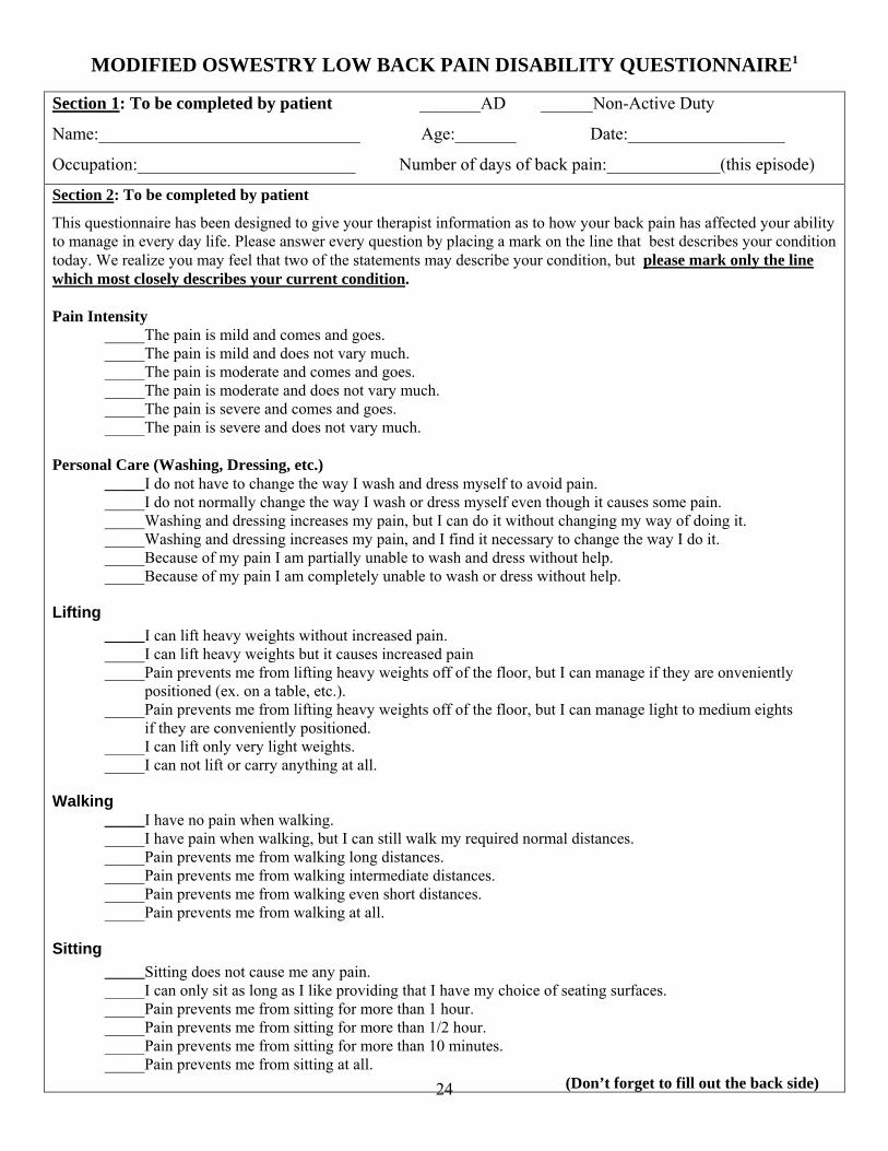
MODIFIED OSWESTRY LOW BACK PAIN DISABILITY QUESTIONNAIRE1
Section 1: To be completed by patient _______AD ______Non-Active Duty
Name:______________________________ Age:_______ Date:__________________
Occupation:_________________________ Number of days of back pain:_____________(this episode)
Section 2: To be completed by patient
This questionnaire has been designed to give your therapist information as to how your back pain has affected your ability to manage in every day life. Please answer every question by placing a mark on the line that best describes your condition today. We realize you may feel that two of the statements may describe your condition, but please mark only the line which most closely describes your current condition. Pain Intensity _____The pain is mild and comes and goes. _____The pain is mild and does not vary much. _____The pain is moderate and comes and goes. _____The pain is moderate and does not vary much. _____The pain is severe and comes and goes. _____The pain is severe and does not vary much. Personal Care (Washing, Dressing, etc.) _____I do not have to change the way I wash and dress myself to avoid pain. _____I do not normally change the way I wash or dress myself even though it causes some pain. _____Washing and dressing increases my pain, but I can do it without changing my way of doing it. _____Washing and dressing increases my pain, and I find it necessary to change the way I do it. _____Because of my pain I am partially unable to wash and dress without help. _____Because of my pain I am completely unable to wash or dress without help.
Lifting _____I can lift heavy weights without increased pain. _____I can lift heavy weights but it causes increased pain _____Pain prevents me from lifting heavy weights off of the floor, but I can manage if they are onveniently positioned (ex. on a table, etc.). _____Pain prevents me from lifting heavy weights off of the floor, but I can manage light to medium eights if they are conveniently positioned. _____I can lift only very light weights. _____I can not lift or carry anything at all.
Walking _____I have no pain when walking. _____I have pain when walking, but I can still walk my required normal distances. _____Pain prevents me from walking long distances. _____Pain prevents me from walking intermediate distances. _____Pain prevents me from walking even short distances. _____Pain prevents me from walking at all.
Sitting _____Sitting does not cause me any pain. _____I can only sit as long as I like providing that I have my choice of seating surfaces. _____Pain prevents me from sitting for more than 1 hour. _____Pain prevents me from sitting for more than 1/2 hour. _____Pain prevents me from sitting for more than 10 minutes. _____Pain prevents me from sitting at all. (Don’t forget to fill out the back side)
25
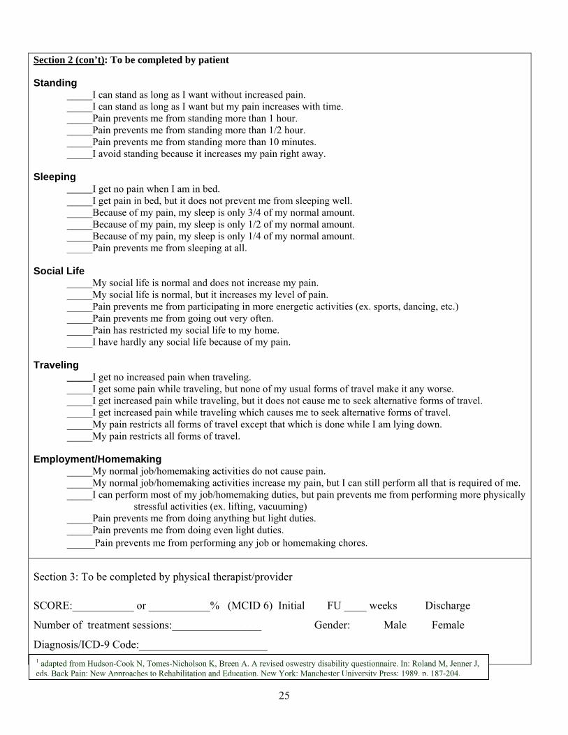
Section 2 (con’t): To be completed by patient
Standing _____I can stand as long as I want without increased pain. _____I can stand as long as I want but my pain increases with time. _____Pain prevents me from standing more than 1 hour. _____Pain prevents me from standing more than 1/2 hour. _____Pain prevents me from standing more than 10 minutes. _____I avoid standing because it increases my pain right away.
Sleeping _____I get no pain when I am in bed. _____I get pain in bed, but it does not prevent me from sleeping well. _____Because of my pain, my sleep is only 3/4 of my normal amount. _____Because of my pain, my sleep is only 1/2 of my normal amount. _____Because of my pain, my sleep is only 1/4 of my normal amount. _____Pain prevents me from sleeping at all.
Social Life _____My social life is normal and does not increase my pain. _____My social life is normal, but it increases my level of pain. _____Pain prevents me from participating in more energetic activities (ex. sports, dancing, etc.) _____Pain prevents me from going out very often. _____Pain has restricted my social life to my home. _____I have hardly any social life because of my pain.
Traveling _____I get no increased pain when traveling. _____I get some pain while traveling, but none of my usual forms of travel make it any worse. _____I get increased pain while traveling, but it does not cause me to seek alternative forms of travel. _____I get increased pain while traveling which causes me to seek alternative forms of travel. _____My pain restricts all forms of travel except that which is done while I am lying down. _____My pain restricts all forms of travel.
Employment/Homemaking _____My normal job/homemaking activities do not cause pain. _____My normal job/homemaking activities increase my pain, but I can still perform all that is required of me. _____I can perform most of my job/homemaking duties, but pain prevents me from performing more physically stressful activities (ex. lifting, vacuuming) _____Pain prevents me from doing anything but light duties. _____Pain prevents me from doing even light duties. _____Pain prevents me from performing any job or homemaking chores.
Section 3: To be completed by physical therapist/provider SCORE:___________ or ___________% (MCID 6) Initial FU ____ weeks Discharge
Number of treatment sessions:________________ Gender: Male Female
Diagnosis/ICD-9 Code:_______________________ 1 adapted from Hudson-Cook N, Tomes-Nicholson K, Breen A. A revised oswestry disability questionnaire. In: Roland M, Jenner J, eds. Back Pain: New Approaches to Rehabilitation and Education. New York: Manchester University Press; 1989. p. 187-204.

26
APPENDIX 2 (FABQ)

27
Name: ____________________________________________ Date: _____________________ Last four digits of SSN: ________________ Here are some of the things which other patients have told us about their pain. For each statement please circle any number from 0 to 6 to say how much physical activities such as bending, lifting, walking or driving affect or would affect your back pain. COMPLETELY UNSURE COMPLETELY DISAGREE AGREE 1. My pain was caused by physical activity 0 1 2 3 4 5 6 2. Physical activity makes my pain worse 0 1 2 3 4 5 6 3. Physical activity might harm my back 0 1 2 3 4 5 6 4. I should not do physical activities 0 1 2 3 4 5 6 which (might) make my pain worse 5. I cannot do physical activities which 0 1 2 3 4 5 6 (might) make my pain worse Score__/24 The following statements are about how your normal work affects or would affect your back pain. COMPLETELY UNSURE COMPLETELY DISAGREE AGREE 6. My pain was caused by my work or by 0 1 2 3 4 5 6 an accident at work 7. My work aggravated my pain 0 1 2 3 4 5 6 8. I have a claim for compensation for my pain 0 1 2 3 4 5 6 9. My work is too heavy for me 0 1 2 3 4 5 6 10. My work makes or would make my pain worse 0 1 2 3 4 5 6 11. My work might harm my back 0 1 2 3 4 5 6 12. I should not do my normal work with my 0 1 2 3 4 5 6 present pain 13. I cannot do my normal work with my 0 1 2 3 4 5 6 present pain 14. I cannot do my normal work until my pain 0 1 2 3 4 5 6 is treated 15. I do not think that I will be back to my 0 1 2 3 4 5 6 normal work within 3 months 16. I do not think that I will ever be able to go 0 1 2 3 4 5 6 back to that work SCORE: ____/42 Waddell: The Back Pain Revolution pp. 191-195. Waddell et al: A fear avoidance beliefs questionnaire (FABQ) and the role of fear avoidance beliefs in chronic low back pain and disability; Pain. 1993; 52: 157-68.
28
Reference List
,1. Altman R, Alarcon G, Appelrouth D, Bloch D, Borenstein D, Brown C, et al. The American College of Rheumatology criteria
for the classification and reporting of osteoarthritis of the hip. Arthritis Rheum 1991; 34:(5)505-514.
,2. Balagué F, Nordin M, Sheikhzadeh A, Echegoyen AC, Brisby H, Hoogewoud HM, et al. Recovery of severe sciatica. Spine 1999; 24:2516-2524.
,3. Bigos S, Bowyer O, Braen G, Brown K, Deyo R, Haldeman S, et al. Acute Low Back Problems in Adults. 1994; AHCPR Publication 95-0642 ed., Rockville, MD: Agency for Health Care Policy and Research, Public Health Service, US Department of Health and Human Services.
,4. Broadhurst NA, Bond MJ. Pain provocation tests for the assessment of sacroiliac joint dysfunction. J Spinal Disorders 1998; 11:(4)341-345.
,5. Cibulka MT, Delitto A, Koldehoff R. Changes in inominate tilt after manipulation of the sacroiliac joint in patients with low back pain: An experimental study. Phys.Ther. 1988; 68:1359-1363.
,6. Cyriax JH, Cyriax PJ. Cyriax's Illustrated Manual of Orthopaedic Medicine. Butterworth, 1978.
,7. Delitto A, Erhard RE, Bowling RW. A treatment-based classification approach to low back syndrome: identifying and staging patients for conservative management. Phys.Ther. 1995; 75:470-489.
,8. Deyo RA, Diehl AK. Cancer as a cause of back pain: frequency, clinical presentation, and diagnostic strategies. J Gen Intern Med 1988; 3:230-238.
,9. Deyo RA, Rainville J, Kent DL. What can the history and physical examination tell us about low back pain? JAMA 1992; 268:760-765.
,10. Dreyfuss P, Dreyer S, Griffin J, Hoffman J, Walsh N. Positive sacroiliac screening tests in asymptomatic adults. Spine 1994; 19:1138-1143.
,11. Flynn T, Fritz J, Whitman J, Wainner RS, Reindero D, Butler B, et al. Clinical Prediction Rule for Classifying Patients with Low Back Pain Likely to Respond to a Manipulation Technique. Spine (in review) 2002; Abstract.
,12. Fritz J. FABQ. Spine 2002;
,13. Fritz JM, Irrgang JJ. A Comparison of a Modified Oswestry Disability Questionnaire and the Quebec Back Pain Disability Scale. Phys Ther 2001; 81:776-788.
,14. Fritz JM, Wainner RS, Hicks GE. The use of nonorganic signs and symptoms as a screening tool for return-to-work in patients with acute low back pain. Spine 2000; 25:1925-1931.
,15. Frymoyer JW, Cats-Baril W. An overview of the incidence and cost of low back pain. Orthop Clin North Am 1991; 22:263-271.
,16. Karas R, McIntosh G, Hall H, Wilson L, Melles T. The relationship between nonorganic signs and centralization of symptoms in the prediction of return to work for patients with low back pain. Phys Ther 1997; 77:354-360.
,17. Katz JN, Dalgas M, Stucki G, Lipson SG. Degenerative lumbar spinal stenosis. Diagnostic value of the history and physical examination. Arthritis Rheum 1995; 38:1236-1241.
,18. Kerr RS, Cadoux-Hudson TA, Adams CB. The value of accurate clinical assessment in the surgical management of the lumbar disc protrusion. J Neurol Neurosurg Psychiatry 1988; 51:169-173.
,19. Levangie PK. The association between static pelvic asymmetry and low back pain. Spine 1999; 24:1234-1242.
29
,20. Litvan I, Mangone CA, Werden W, Beuri JA, Estol CJ, Rey RC, et al. Reliability of the NINDS myotactic reflex scale. Neurology 1996; 47:969-972.
,21. Maher C, Adams R. Reliability of pain and stiffness assessments in clinical manual lumbar spine examination. Phys.Ther. 1994; 74:801-811.
,22. Main CJ, Waddell G. A reappraisal of the interpretation of "nonorganic signs". Spine 1998; 23:67-71.
,23. Manschot S, van Passel L, Buskens E, Algra A, van Gijn J. Mayo and INIDS scale for assessment of tendon reflexes: between observer agreement and implications for communication. J Neurol Neurosurg Psychiatry 1998; 64:253-255.
,24. Maruta T, Goldman S, Chan CW, Ilstrup DM, Kunselman AR, Colligan RC. Waddell's nonorganic signs and Minnesota Multiphasic Personality Inventory profiles in patients with chronic low back pain. Spine 1997; 22:72-75.
,25. McCombe PF, Fairbank JC, Cockersole BC, Pynsent PB. 1989 Volvo Award in clinical sciences. Reproducibility of physical signs in low-back pain. Spine 1989; 14:908-918.
,26. O'Haire C, Gibbons P. Inter-examiner and intra-examiner agreemtn for assessing sacroiliac anatomical landmarks using palpation and observation: pilot study. Manual Therapy 2000; 5:13-20.
,27. Potter NA, Rothstein JM. Intertester reliability of selected clinical tests of the sacroiliac joint. Phys.Ther. 1985; 65:1671-1675.
,28. Spangfort EV. The lumbar disc herniation. Acta Orth Scand (Suppl) 1972; 142:1-95.
,29. Strender LE, Sjoblom A, Sundell K, Ludwig R, Taube A. Interexaminer reliability in physical examination of patients with low back pain. Spine 1997; 22:814-820.
,30. Supik LF, Broom MJ. Sciatic tension signs and lumbar disc herniation. Spine 1994; 19:1066-1069.
,31. Tenhula JA, Rose SJ, Delitto A. Association between direction of lateral lumbar shift, movement tests, and side of symptoms in patients with low back pain syndrome. Phys.Ther. 1990; 70:480-486.
,32. Theiler R, Stucki G, Shutz R, Hofer H, Seifert B, Tyndall A, et al. Parametric and non-parametric measures in the assessment of knee and hip osteoarthritis: interobserver reliability and correlation with radiology. Osteoarthritis and Cartilage 1996; 4:35-42.
,33. van den Hoogen HMM, Koes BW, van Eijk JTM, Bouter LM. On the accuracy of history, physical examination, and erthrocyte sedimentation rate in diagnosing low back pain in general practice. Spine 1995; 20:318-327.
,34. Van Dillen LR, Sahrmann SA, Norton BJ, Caldwell CA, Fleming DA, McDonnell MK, et al. Reliability of physical examination items used for classification of patients with low back pain. Phys Ther 1998; 78:979-988.
,35. Viikari-Juntura E, Takala EP, Riihimaki H, Malmivaara A, Martikainen R, Jäppinen P. Standardized physical examination protocol for low back disorders: feasibility of use and validity of symptoms and signs. J Clin Epidemiol 1998; 51:245-255.
,36. Vroomen PCAJ, de Krom M, Knottnerus JA. Consistency of history taking and physical examination in patients with suspected lumbar nerve root involvement. Spine 2000; 25:91-97.
,37. Waddell G, Birchner M, Finlayson D, Main CJ. Symptoms and signs: Physical disease or illness behaviour? BMJ 1984; 289:739-741.
,38. Waddell G, Burton AK. Occupational health guidelines for the management of low back pain at work - evidence review. 2000; London. Faculty of Occupational Medicine.
,39. Waddell G, McCulloch JA, Kummel E, Venner RM. Nonorganic signs in low-back pain. Spine 1980; 5:117-125.

30
,40. Waddell G, Newton M, Henderson I, Somerville D, Main CJ. A Fear-Avoidance Beliefs Questionnaire (FABQ) and the role of fear-avoidance beliefs in chronic low back pain and disability. Pain 1993; 52:157-168.
,41. Waddell G, Somerville D, Henderson I, Newton M. Objective clinical evaluation of physical impairment in chronic low back pain. Spine 1992; 17:617-628.
,42. Whitman J, Flynn T, Fritz J, Magel J, Rendeiro D, Wainner RS, Allison S. Does manual therapy experience influence reliability for selected pelvic girdle tests and measures? Journal of Manual and Manipulative Therapy 2001; 9: