use of probiotic bacteria to improve the growth of farmed new
TRANSCRIPT
Use of Probiotic Bacteria to Improve the Growth of
Farmed
New Zealand Abalone (Haliotis iris)
Jinan Abass Hadi
Thesis submitted in fulfillment of the Master of
Applied Science
Auckland University of Technology
Auckland, New Zealand
May, 2012

Dedicated to my wonderful late parents

II
Table of Contents
Table of Contents i
LIST OF FIGURES iv
LIST OF TABLES v
LIST OF ABBREVIATIONS vi
ATTESTATION OF AUTHORSHIP vii
ACKNOWLEDGMENT viii
CONFIDENTIAL MATERIAL ix
Abstract x
Chapter 1 Introduction 1
1.1 Introduction ..................................................................................................................... 2
1.2 Probiotics Concept ........................................................................................................... 4
1.3 Aims and objectives of the study .................................................................................... 7
1.4 Research Questions and Hypotheses .............................................................................. 8
Chapter 2 Literature Review 9
2.1 New Zealand Abalone Haliotis iris ................................................................................. 10
2.2 Abalone Nutrition .......................................................................................................... 11
2.3 Probiotic: Definition and Principle ................................................................................ 11
2.4 The Rationale for the Use of Probiotics in Aquaculture ................................................ 13
2.5 Kinds of Probiotics ......................................................................................................... 14
2.6 Probiotics Mechanisms and Mode of Actions ............................................................... 16
2.6.1 Competition for Adhesion Sites .......................................................................... 16
2.6.2 Competitive exclusion ......................................................................................... 17
2.6.3 Competition for Iron ........................................................................................... 18

ii
2.6.4 Supply of Nutrients and Digestive enzymes ....................................................... 18
2.6.5 Disease Resistance .............................................................................................. 19
2.6.6 Enhancement of Immune response .................................................................... 20
2.6.7 Improving Water Quality .................................................................................... 21
2.7 Probiotics Criteria and Selection of Candidate Probionts ............................................. 22
2.8 Probiotics in Abalone ..................................................................................................... 24
2.8.1 Effect of Microflora on Abalone Digestibility and Growth ................................. 24
2.8.2 Probiotics Use in Abalone Aquaculture .............................................................. 25
Chapter 3 Isolation, Screening and Identification of Potential Probiotic Bacteria for
Abalone (Paua) 28
3.1 Introduction ................................................................................................................... 29
3.2 Materials and Methods ................................................................................................. 30
3.2.1 Isolation of potential probiotic bacteria ............................................................. 30
3.2.2 Screening of Potential Probiotic Isolates ............................................................ 33
3.2.3 Phenotypic Characterization ............................................................................... 36
3.2.4. Identification of the Isolates .............................................................................. 39
3.2.5 Phylogenetic Analysis for the Isolates ................................................................ 39
3.3 Results ........................................................................................................................... 40
3.3.1 Isolation and Enumeration of Potential Probiotic Bacteria ................................ 40
3.3.2 Screening of Potential Probiotic Isolates ............................................................ 42
3.3.2 Phenotypic characterization ............................................................................... 50
3.3.3 Identification ....................................................................................................... 52
3.4 Discussion ...................................................................................................................... 56
3.5 Conclusion ..................................................................................................................... 56
Chapter 4 Enzyme Activity Assays and Quantitative Screening for Potential Probiotics 62
4.1 Introduction ................................................................................................................... 63
4.2 Material and methods ................................................................................................... 64
4.2.1 Bacterial cultures ................................................................................................ 64
iii
4.2.2 Preparation of crude extracellular enzyme ........................................................ 64
4.2.3 Enzyme Assays .................................................................................................... 64
4.4 Results and Discussion ................................................................................................... 68
4.4.1 Proteolytic Enzyme Assays .................................................................................. 69
4.4.2 Amylolytic Enzyme Assays .................................................................................. 72
4.4.3 Alginolytic Enzyme Assays .................................................................................. 74
4.5 Conclusion ..................................................................................................................... 80
Chapter 5 Feeding Trials Using isolates of Probiotics 81
5.1 Introduction ................................................................................................................... 82
5.2 Materials and Methods ................................................................................................. 83
5.2.1 Experimental Animals ......................................................................................... 83
5.2.2 Cultivation of Potential Probionts ...................................................................... 83
5.2.3 Abalone Commercial Feed .................................................................................. 84
5.2.4 Preparation of Probiotic- supplemented Feed ................................................... 84
5.2.5 Measurements of Weight and Shell Length ....................................................... 88
5.2.6 Rearing Tanks ...................................................................................................... 89
5.2.7 Water System of Feeding Trial ............................................................................ 90
5.2.8 Feeding Trial ........................................................................................................ 90
5.2.9 Statistical analyses .............................................................................................. 96
5.3 Results ........................................................................................................................... 97
5.3.1 Application of probiotic- supplemented feed..................................................... 97
5.3.2 Growth trial ......................................................................................................... 98
5.3.3 Mortality ........................................................................................................... 102
5.3.4 Compositions of Growth Trial Samples............................................................. 103
5.4 Discussion .................................................................................................................... 104
Chapter 6 Conclusions and Recommendations 108
6.1 Conclusions .................................................................................................................. 109
6.2 Significance of the Study ............................................................................................. 112
iv
6.2.1 Commercial benefit of the research ................................................................. 112
6.2.2 Scientific benefit of the research ...................................................................... 113
6.3 Future Studies and Recommendations ....................................................................... 113
REFRENCES 116
APPENDICES 126
LIST OF FIGURES
Figure 1-1 Statistics of commercial catch of abalone in New Zealand 3
Figure 3-1 Anterior view of the abalone Haliotis showing the main organs 30
Figure 3-2 Abalone digestive tract 31
Figure 3-3 Alginate agar plates with change in colour 44
Figure 3-4 Alginate agar plate with no change in colour and no clear zone 44
Figure 3-5 Experiment showing clear solution for alginate hydrolysis 46
Figure 3-6 Flow chart for Gram-negative bacteria isolates 52
Figure 3-7 flow chart for Gram- positive bacterial isolates 53
Figure 3-8 The scanning electron microscopy photograph of Ex. sp. 58
Figure 3-9 Exiguobacterium JHEb1 under 1000 X 59
Figure 3-10 Vibrio JH1 growth on TCBS Agar, and on Marine Agar 60
Figure 3-11 Enterococcus JHLDc under 100X 61
Figure 4-1 The main activities to assess the potential probiotic isolates 68
Figure 4-2 Protein hydrolysis of potential probiotic isolates 70
Figure 4-3 Enzyme activity of proteolytic isolates (IU /ml ± SE) 71
Figure 4-4 Starch hydrolysis of potential probiotic isolates 73
Figure 4-5 Enzyme activity (IU /ml ± SE) of amylolytic bacteria isolates 74
Figure 4-6 Alginate hydrolysis of potential probiotic isolates 76
v
Figure 4-7 Enzyme activity (IU /ml± SE) of alginolytic isolates 78
Figure 4-8 ß- haemolysis in Bacillus JHBa2 78
Figure 5-1 Preparation of 2-probiotic supplemented feed 86
Figure 5-2 Preparation of 3-probiotic supplemented feed 87
Figure 5-3 Abalone weighing and shell length measuring 88
Figure 5-4 Feeding trial tanks with mesh cover, and plastic covers 89
Figure 5-5 Protocol of feeding trial 91
Figure 5-6 Application of feed to juvenile abalone and collecting samples 92
Figure 5-7 Shell length increase and daily shell length increase 99
Figure 5-8 Improvement of shell coloring associated with probiotic diet 100
Figure 5-9 Weight gain and daily weight gain 101
Figure 5-10 Mortality rate percentages for growth trial 102
Figure 5-11 Abalone faeces compositions 103
LIST OF TABLES
Table 1-1 Top ten New Zealand seafood exports in the year 2010 4
Table 2-1 Authorized microorganism as probiotics in feeding stuffs 23
Table 3-1 Viable count of bacterial population in abalone, and rearing water 40
Table 3-2 Bacterial strains isolated from abalone (GIT), and rearing water 41
Table 3-3 Casein hydrolysis of bacterial isolates 42
Table 3-4 Starch- hydrolysing property of the isolates 43
Table 3-5 Alginate hydrolysis of the bacterial isolates 46
Table 3-6 Bile salt tolerance of isolates which grew on Marine and MRS agar 47
Table 3-7 Bile salt tolerance for isolates that grew on TCBS agar 47

vi
Table 3-8 Acid tolerance of isolates measured as viable count at 0, 3 hr 48
Table 3-9 Acid production in potential probiotic isolates 49
Table 3-10 Cell and colony morphology of the Gram-positive isolates 50
Table 3-11 Cell and colony morphology of the Gram-negative isolates 50
Table 3-12 Biochemical characteristics of bacterial isolates 51
Table3-13 API 20E results for Gram-negative isolates 54
Table 3-14 Identification of the isolates by 16S rRNA sequencing 55
Table 4-1 Enzyme activity of proteolytic isolates 69
Table 4-2 Enzyme activity of amylolytic isolates 72
Table 4-3 Enzyme activity of alginolytic isolates 75
Table 5-1 Bacterial populations as viable count in feed and water samples 97
Table 5-2 Growth data and mortality for three groups 99
Table 5-3 Compositions Percentage of fecal matter and commercial feed 104
LIST OF ABBREVIATIONS
TACC Total Allowable Commercial Catch
CFU Colony forming unit
GIT Gastrointestinal tract
LAB Lactic acid bacteria
MRS De Man–Rogosa–Sharpe Medium
TCBS Thiosulphate citrate bile sucrose medium
UV Ultraviolet
P.a. Per annum
FAO United Nations Food and Agriculture Organization
WHO World Health Organization

vii
ATTESTATION OF AUTHORSHIP
“I hereby declare that the work in this thesis has not previously been submitted for a
degree nor has it been submitted as part of requirement for a degree.
I also certify that the thesis has been written by me. Any help that I have received in my
research has been acknowledged.
Signed:
Date:

viii
ACKNOWLEDGMENT
Firstly, I would like to thank my primary supervisor Dr. Noemi Gutierrez-Maddox for her
continuous support throughout my study journey, her helpful suggestions in the
practical aspects of my research as well as her patience while editing this thesis.
Secondly, I would like to thank Associated Professor Andrea C. Alfaro – my secondary
supervisor. Her suggestion of weekly aquaculture meetings as well as editing some
chapters of my thesis has been an immeasurable contribution.
Thirdly, I would like to thank Dr. Rodney Roberts for his great help in providing my
research with experimental animals, sampling from rearing tanks in the Paua farm,
beneficial information guidelines that greatly aided the laboratory feeding trial and
everything that was sent to AUT that resulted in much saved time.
A special acknowledgement to all the members of the Biotechnology Aquaculture
group. Great thanks Professor John Brooks for his priceless advice in microbiology. I
thank Dr. Coleen Higgins for her helpful suggestions especially in the area of coding the
bacterial isolates. Her wonderful smile has been a tremendous encouragement for me. I
thank Dr. Mark Duxbury for his advice and time and support in enzymatic study. He
played an intrinsic role in my research. I thank Mr Dave Bryant for his help in API
identification system.
I thank Tim Young for his help in experiment design and statist. Annapoorna Maitrayee
Ganesan and Adam Rusk who have always been on hand to help me with their wonderful
suggestions throughout the length of my research. I thank Emma Beatson and Neil for
their help in aquaculture laboratory.
I am deeply thankful to Saeedeh Sadooghy-Saraby, Meie Zou, Christine, Minaxi Patel,
Yan Wang, Chris Whyburd, Percy Perera and Chris Books as allied staff in the AUT
applied science laboratory. They have always been there to help me with great
willingness even in the busiest of times.
I would like to finally thank my wonderful family in my home country in Iraq as well as
here in New Zealand. A special thanks to my amazing husband Mohammed for his
encouragement and support, this would not be possible without his contribution. He
even aided me with the statistical analysis of my results, which I could not have done
without him.
Before and after all I would like to thank Allah for listening to my prayers and I know He
is pleased with the people who seek knowledge.

ix
CONFIDENTIAL MATERIAL
This thesis contains confidential information which if publicly available may jeopardise the
future intellectual property rights of the author.

x
Abstract
Abalone are known to have a very slow growth rate that results in significant financial
constraints for its cultivation. Commercially farmed abalone which are given formulated
feed consisting of soy flour and seaweed still require 4 to 5 years to attain a market size
of (80-100 mm) for shell length.
To improve growth in farmed New Zealand abalone (paua), Haliotis iris, potential
probiotic isolates were isolated from healthy adult abalone obtained from OceaNZ Blue
(Bream Bay, New Zealand) and from farm tanks. The isolates were screened
qualitatively according to their ability to hydrolyze feed nutrients (such as proteins,
starch, and alginate), produce lactic acid, and resist bile salts. Phenotypic and 16s rRNA
techniques were used to identify the potential probiotic isolates. Biochemical analyses
to determine which isolate exerted the strongest proteolytic, amylolytic, and alginolytic
activities were carried out.
This study has developed a multi-strain conglomerate of 2- and 3- probiotic bacterial
strains that have been supplemented into a commercial abalone feed to determine if
probiotic microorganisms can increase the growth rate of farmed H. iris. The 2-probiotic
conglomerate consisted of Exiguobacterium JHEb1 and Vibrio JH1, the 3-probiotic
conglomerate consisted of Enterococcus JHLDc in addition to Exiguobacterium JHEb1
and Vibrio JH1.
The probiotic feeds were used in a laboratory feeding trial involving abalone juveniles
(sized 20-30 mm) to determine if probiotic microorganisms can increase the growth rate
of farmed H. iris. Two groups of abalone (in 3 replicates) were fed 2- probiotic
supplemented, and 3- probiotic supplemented feed were compared with the control
group (3 replicates) administered with un-supplemented feed. Proximate analysis of
abalone faeces were performed to determine differences in proteins, carbohydrate,
and lipid in all abalone groups and determine if these nutrients were more efficiently
metabolized in the presence of probiotic bacteria.
A significant growth improvement was obtained with the 3-probiotic supplemented
feed that produced a significant shell length increase of 20.9%, wet weight gain of
19.8% and reduced mortality (3.33%) (p<0.05). The 2-probiotic supplemented feed also
resulted in significant increase in shell length and survival (p<0.05) but not in weight
gain.
This study is the first to report of the application of Exiguobacterium JHEb1in abalone.
This species was incorporated in both the 2-probiotic and the 3-probiotic feeds. This
study is also the first to report that a combination of three probiotic species
supplemented into the commercial feed of farmed H. iris increased growth measured by
shell length and wet weight.

1
Chapter 1
Introduction
2
1.1 Introduction
The black-footed abalone Haliotis iris is the main commercial species of abalone
in New Zealand; it is called “pāua” in New Zealand Maori language (Dutton,
1986). At present, there are about 20 species of abalone harvested commercially
worldwide inclusive of the New Zealand species (Hahn, 1989 a). The main
abalone fishing countries are Japan, Australia, New Zealand, South Africa,
Mexico and the United States (Tung, 2010).
This kind of abalone is infrequently distributed all along the coasts of both main
islands of New Zealand, Chatham Islands, Stewart Island, and the Snares
Islands. Maori people have harvested H. iris for hundreds of years as far back as
1150 AD. Traditionally they harvested and traded abalone for their flesh and
their decorative shells. The abalone flesh (the muscular foot) is eaten as a
delicacy, while the polished shells are used for decorative purposes and to make
jewelry. Abalone meat is exported and sold locally as both fresh and canned
products (Sainsbury, 1982).
H. iris can live in both sheltered and exposed shores, and is considered as a
shallow-water species which is habitually most abundant at <5 m depth. However
its distribution may extend to 20 m in depth (Sainsbury, 1982; Schiel, 1991). It
inhabits rocky substrates by clinging firmly to the rocks, particularly flat boulder
bottoms, and like other abalone species prefers well oxygenated sites where
there is sea water with stable salinity (Hahn, 1989 b). They also grow much
larger in colder waters around the coast of Southland of New Zealand and
Stewart Island where can be found in much more abundant (Sainsbury, 1982).
Research in aquaculture has recently focused on abalone since the supply of
wild abalone seems to be dwindling due to overfishing, their slow growth rate and
their tendency to aggregate in one region (Heath & Moss, 2009). To increase the
supply of abalone, land-based aquaculture of abalone was started in New
Zealand in the 1980’s using developed techniques to gain sustainable yield.
There are now about 40 farms around the country cultivating paua for meat,
shell, and pearl production (Heath & Moss, 2009).
3
Abalone aquaculture is an economically important industry in New Zealand
where aquaculture has been identified by the government as the country’s
fastest growing primary industry. Aquaculture is believed to bring New Zealand
significant economic growth in an environmentally sustainable approach.
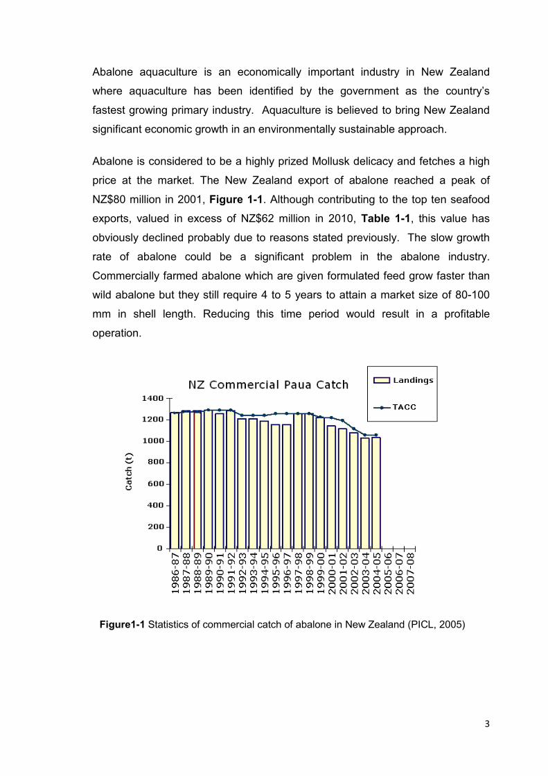
Abalone is considered to be a highly prized Mollusk delicacy and fetches a high
price at the market. The New Zealand export of abalone reached a peak of
NZ$80 million in 2001, Figure 1-1. Although contributing to the top ten seafood
exports, valued in excess of NZ$62 million in 2010, Table 1-1, this value has
obviously declined probably due to reasons stated previously. The slow growth
rate of abalone could be a significant problem in the abalone industry.
Commercially farmed abalone which are given formulated feed grow faster than
wild abalone but they still require 4 to 5 years to attain a market size of 80-100
mm in shell length. Reducing this time period would result in a profitable
operation.
Figure1-1 Statistics of commercial catch of abalone in New Zealand (PICL, 2005)

4
Table 1-1 Top ten New Zealand seafood exports in the year 2010 (SeaFIC, 2010)
To address the issue of the slow growth rate of abalone, studies have focused on
the development of nutrient-rich artificial diets. Further, the concept of probiotic
bacteria which aid digestion in humans and livestock has been adopted in
aquaculture and its application has been rapidly increasing. The use of probiotics
to improve growth in abalone presents an enormous potential.
1.2 Probiotics Concept
The use of probiotics in aquaculture, in general, has become more popular due
to increasing demand to use safe, environment-friendly additives to the feed to
improve nutrition and growth of aquaculture animals (Irianto & Austin, 2002;
Macey & Coyne, 2005). Gatesoupe (1999) defined ‘probiotics’ as “microbial cells
that are administered in such a way as to enter the gastrointestinal tract and to
be kept alive with the aim of improving health”. Studies have shown evidence of
remarkable improvement in health and survival of aquaculture animals in
intensive rearing system, which have been administered diet containing

5
probiotics. Manipulating the gut microflora with probiotics could lead to great
benefit to host health and improving their growth rate (Olafsen, 2001a).
The use of probiotics is now commonplace in ‘functional foods’ not only for
humans but also for animals as therapeutic and prophylactic feed supplements
(Kailasapathy & Chin, 2000; Stanton et al., 2005). The lactic acid bacteria (LAB)
are commonly used in probiotic dairy products and terrestrial animal feeds
(Ringø & Gatesoupe, 1998). LAB are characterized by their resistance to acidic
and bile environment in the intestinal tract. Sugars are fermented by LAB to
lactic acid and other organic acids thus lowering the pH in the intestinal tract
which could inhibit many bacteria including intestinal pathogens. LAB have been
shown to produce antimicrobials which again could inhibit pathogens (Ringø et
al., 2005).
Yeasts and the spore-forming Bacillus have also been used as probiotics, (Hong
et al., 2005; Kesarcodi-Watson et al., 2008). Several studies indicated in their
study on shrimp (Litopenaeus vannamei and Penaeus monodon) that the use of
Bacillus sp. in the diet, have generally increased the shrimp survivals and
immune parameter when challenged with pathogenic Vibrio harveyi and white
spot syndrome virus (Li et al., 2009; Rengpipat et al., 2000). Yeasts, such as
Saccharomyces, and Debaryomyces have been used widely in aquaculture, they
showed great immunostimulatory activity and high production of inhibitory
substances (Irianto & Austin, 2002; Van der Aa Kühle et al., 2005). Growth rate
improvement noticed in abalone Haliotis midae, when they fed supplemented
feed with two yeast and one bacterial strain (Macey & Coyne, 2005).
Several modes of action have been suggested to explain how probiotics could be
beneficial to the host’s health:
1. Enzymatic assistance to digestion and absorption of nutrients.
Many studies suggested that probiotic microorganisms can improve the
digestion of nutrients in GIT of the host by production of protein and
carbohydrates hydrolysis enzymes (Erasmus et al., 1997; Krishnaprakash et al.,
2009; Sahu et al., 2008).
6
2. Antagonism towards pathogens
Several studies have reported the effect of probiotics to be strongly inhibitory
against pathogens in aquaculture e.g. Vibrio anguillarum. Several strains of the
bacterium Roseobacter are inhibitory toward other pathogenic bacteria namely
Vibrio splendidus and Vibrio anguillarum, Roseobacter strain T5 can produce
inhibitory compound such as the sulfur containing tropodithietic acid (Balcázar et
al., 2007 b; Bruhn et al., 2006).
3. Competitive exclusion
Microbial interactions play an effective role between competing beneficial and
potentially pathogenic microorganisms. Probiotics can prevent bacterial diseases
in aquaculture through specific competition for pathogen receptor sites on the
mucus of the gut of the host (Balcázar et al., 2006; Verschuere et al., 2000).
4. Improvement of water quality
It has been reported that improved water quality in aquaculture associated with
the use of probiotics, such as Bacillus species. Probiotics can reduce the
concentration of nitrogen and phosphorus in the pond, consequently increased
the survival rate and yield of aquaculture animals, e.g. shrimp Penaeus monodon
(Balcázar et al., 2006; Dalmin et al., 2001).
5. Immunostimulatory function.
Probiotic microorganisms may benefit the host by stimulating the immune system
in both cellular and humoral immune defense. Stimulating the host immune
system could occur through the probionts production of specific substances
called immunostimulants (Balcázar et al., 2007 a; Rengpipat et al., 2000; Sahu et
al., 2008).
More details for each mode of action of probiotic microorganisms are in the next
Chapter (Literature Review).
A single probiotic species could exert either one or a combination of these
modes of action. The use of multi-strain or multi-species probiotics would serve
as an approach by which all the potential benefits from probiotic microorganism
7
could be obtained. Few studies used multi-strain probiotic in supplementation of
feed for aquaculture animals (Macey & Coyne, 2005)
Bairagi et al. (2004) reported the advantage of the addition of two Bacillus spp.
(B. subtilis and B. circulans) to the diet of rohu Labeo rohita. Their study showed
that application of the two fish intestinal Bacillus to the leaf meal (instead of fish
meal), improved the growth, conversion ratio, and protein uptake ratio. These
effects attributed to amylolytic and cellulolytic enzymes of the additive bacteria. A
study carried out by Balcázar et al. (2007a), showed the use of three LAB
(Lactococcus lactis ssp., Leuconostoc mesenteroides and Lactobacillus sakei)
enhanced the cellular and humoral immune functions in the host rainbow trout.
Macy and Coyne (2005) reported that using multi-strains feed supplement for
abalone (H. midae), improved the growth rate. The combination of three
probionts enhanced the immunity of abalone by increasing the survival rate after
challenging with the pathogen bacteria Vibrio anguillarum.
1.3 Aims and objectives of the study
New Zealand aquaculture operations such as OceaNZ Blue (Bream Bay, Bay of
Islands) are committed to naturally processed paua of the highest quality (PAUA,
2011).They prefer not to use any chemicals or unnatural substances in the
feeding, breeding and protection against diseases. Therefore, supplementation
of probiotic microorganism into abalone feed is an attractive strategy to enhance
abalone growth and improve profitability in the industry.
The aim of this study is to develop a probiotic supplement for Haliotis iris to
improve the growth rate.
The study is designed to:
1. Isolate potential probiotic strains from the digestive tract of healthy adult
abalone, and from abalone farm tanks.

8
2. Select potential probiotics according to their ability to hydrolyze feed
nutrients such as proteins, starch, and alginate.
3. Apply multi-strain probiotics into formulated feed to assess their effects on
growth of aquacultured abalone.
4. Analyze the fecal matter of 2 and 3-probiotic supplemented abalone and
compare the composition of protein, carbohydrate and lipid in feces with
those of unsupplemented feed abalone.
1.4 Research Questions and Hypotheses
The general purpose of this study is mainly to examine the probiotic-
supplemented feed and observe improvement in the growth of abalone (H. iris)
by testing the following hypotheses:
• The selected probiotic bacteria can enhance the digestion of nutrients
components of the commercial feed such as protein, carbohydrate, and
alginate.
• The use of probiotic-supplemented feed can increase the growth rate (body
weight, shell length) of Haliotis iris over the period of the feeding trial.

9
Chapter 2
Literature Review
10
2.1 New Zealand Abalone Haliotis iris
Paua are highly valuable resource in New Zealand, both as food and raw
material for jewelry and other handcraft products. Paua meat is considered a
healthy food with its high protein (71.99%) and low fat content (Imai, 1997).
Abalone are univalve mollusks. The New Zealand abalone are classified as
(Tung, 2010):
Phylum Mollusca
Class Gastropoda
Family Haliotidae
Haliotis
Haliotis iris (Paua)
“Abalone” is a name derived from Spanish name “Abulon”, which means “sea
ear” (Tung, 2010), used for a variety of species of single-shelled mollusks from
the Haliotidae family genera Haliotis.
In New Zealand, there are three documented species of abalone. The first
species the black-footed abalone H. iris (Martyn 1784), known as ‘paua’ are the
main commercial species (Dutton & Tong, 1981). The yellow-foot abalone, H.
australisGmelin,1790 commonly named the queen, are another species smaller
than paua but also of good commercial value (OΉalloran, 1986). The white-
footed abalone, H. virginea (Gmelin, 1790) commonly named the virgin, are the
smallest species with the least commercial values, thus not harvested
commercially (Dutton & Tong, 1986). The lifespan of all three species can reach
more than ten years (Dutton & Tong, 1981; Tung, 2010).

11
2.2 Abalone Nutrition
Abalone are herbivorous mollusks. Wild abalone naturally feed on macroalgae
(Tanaka & Sugimura, 2003). H. iris, similar to H. australis, are known to prefer
and grow faster on red algae (Hymenocladia) (Poore, 1972). The smaller
abalone juveniles (<5 mm) graze microscopic diatoms and ingest bacterial cells
The sedentary, larger juveniles feed mainly on drifting macroalgae (Dutton
&Tong, 1981; Garland et al., 1985).
Most farmed abalone are fed also with algae and depend on the availability of
these algae. Marine algae consist mainly of high carbohydrate: protein ratio, with
carbohydrate making up to 70% of the dry weight, in some cases (Pakulski &
Benner, 1992). The wild algae Ulva lactuca contains lower protein concentration
(3.7-19.9%) (Tung, 2010).
Abalone are known to grow slowly H. iris reaches its commercial size of 100 mm
in 5 years (Sales & Britz, 2001) The slow growth rate of abalone in the wild and
those of farmed abalone fed with only algae has been attributed to the low
protein: carbohydrate ratio in marine algae. A diet consisting mainly of algae can
hardly meet the 30-40% protein requirement of the New Zealand abalone (Tung,
2010). Schiel (1993) and Erasmus et al. (1997) similarly realized that farmed
abalone fed on the kelp Ecklonia radiata did not exhibit high growth rates. Kelp
and other macroalgae contain low protein at approximately 15% and an
unbalanced amino acid profile. These low nutrient levels could not supply the
proteins required for growth in some abalone species (Tung, 2010: Erasmus et
al, 1997).
2.3 Probiotic: Definition and Principle
The word’ probiotic' was derived from Greek words meaning 'for life' (Gismondo
et al., 1999). This is in contrast to 'antibiotic', which literally means 'against life'.
Probiotic as a term has evolved throughout the years.
The first definition for probiotics was by (Lilly, 1965), who described probiotic as
“Substances produced by microorganisms which promote the growth of other
12
microorganisms”. Later, a new definition included both substance and
microorganism as probiotic where (Parker, 1974) defined probiotics as
“Organisms and substances which contribute to intestinal microbial balance”.
Fuller (1989) reported that probiotic should be live microorganisms that can
improve microbial balance in the host gut, hence, defining probiotic as “A live
microbial feed supplement which beneficially affects the host animal by
improving its intestinal microbial balance”. After Fuller’s definition, additional
terms were commonly used to describe probiotics such as “friendly”, “healthy”,
and “beneficial” (Wang & Li, 2008).
In aquaculture, the term “probiont” is used synonymously with the term
“probiotic”. This is done likewise in this study.
Havenaar and Huis In’t Veld (1992) have indicated that one or more live
microorganisms can benefit the host through improving gastrointestinal
microflora, thus “A viable mono- or mixed-culture of microorganisms which when
applied to animals or humans, beneficially affects the host by improving the
properties of the indigenous microflora”. Schaafsma (1996) pointed for the first
time the importance of the amount and concentration of microorganisms, thus
probiotics are: “Living microorganisms, which upon ingestion in certain numbers,
exert health benefits beyond inherent basic nutrition”. Naidu and Bidlack (1999)
added more effects of probiotics to the host such as; improving physiology and
immunity which extended the definition to “A microbial dietary adjuvant that
beneficially affects the host physiology by modulating mucosal and systemic
immunity, as well as improving nutritional and microbial balance in the intestinal
tract”. Scherzenmeir and de Vrese (2001) have strongly recommended that
probiotic should be well-known microorganisms which when applied in adequate
amount can colonize the host microflora, thus: “A preparation of or a product
containing viable, defined microorganisms in sufficient numbers, which alter the
microflora (by implantation or colonization) in a compartment of the host and by
that exert beneficial health effects in this host”
Lately, the most acceptable definition approved by the United Nations Food and
Agriculture Organization (FAO), and the World Health Organization (WHO) has

13
been “Live microorganisms which when administered in adequate amounts
confer a health benefit on the host” (WHO/FAO, 2001).
The concept of probiotic was first applied to human foods but is now widely
accepted and utilized in animal feed in aquaculture where the immediate ambient
environment has even larger influence on the health status than in terrestrial
animals or humans (Verschuere et al., 2000), Within the context of aquaculture,
the term probiotic not only refers to live microorganisms in feed but also to those
which are added to the water (i.e. water additives) used to farm fish and shellfish
(Gatesoupe, 1999), and as a biocontrol agent. Thus the 2001 WHO definition of
WHO may be modified to include organisms and supplement to the host,
environment or feed (Verschuere et al., 2000).
2.4 The Rationale for the Use of Probiotics in Aquaculture
The application of probiotics in aquaculture, particularly in mollusk aquaculture,
has mostly been towards disease management with special reference to
molluscan culture (Kesarcode-Watson et al., 2008). Few studies demonstrated
the rationale of using probiotics from the perspective of the digestion process in
the host and the evaluation of safety of use (Wang & Li, 2008).
Krishnaprakash et al., (2009) recognized that in addition to reducing the effect of
pathogenic organisms in the gut of shrimp, the incorporation of probiotics in
shrimp diet could improve intestinal microbial balance, resulting in enhancement
of food absorption and increased activity of digestive enzymes.
Irianto & Austin (2002) have suggested that the beneficial effects attributed to the
use of probiotics in aquaculture could be due to competitive exclusion and
inhibition of pathogenic microorganisms, assistance in the metabolism of the host
and stimulation of the host immunity. Studies have shown that probiotic strains
could inhibit pathogenic bacteria both in vitro and in vivo through several different
mechanisms. These mechanisms include production of inhibitory compounds,
such as bacteriocins, siderophores, lysozymes, proteases, hydrogen peroxide,

14
formation of ammonia, diacetyl, and alteration of pH values by organic acids
(Verschuere et al., 2000).
The use of probiotics in aquaculture represents a potentially ideal alternative to
the use of antibiotics in the industry, thus preventing the development of
antibiotic resistance in both humans and microorganisms. Without the use of
antibiotics, improved disease resistance against a pathogenic strain of Vibrio
anguillarum was obtainedwhen atlantic cod fry were fed on dry feed containing
lactic acid bacteria (Carnobacterium divergens) (Gómez, 2007).
Bruhn (2006) tried to define the beneficial effect of the probiotics in aquaculture,
and how to determinate if the potential microorganism is a successful probiotic to
use. He suggested that live microorganisms may be administrated for different
purposes; disease prevention, water quality improvement, or as a feed, and all of
these can endorse directly or indirectly health and /or survival of the farmed
animals (Bruhn, 2006). Other suggestions of how to measure the effect of
probiotic could be via its ability to decrease frequency of disease and/or increase
survival from lethal diseases (Gram & Ringø, 2005).
2.5 Kinds of Probiotics
Most probiotics tested for use in aquaculture:
1) Gram-positive bacteria; such as Bacillus and lactic acid bacteria LAB ;
Carnobacterium, Lactobacillus, Lactococcus (Gatesoupe, 1999).
2) Gram-negative bacteria such as Aeromonas, Pseudoalteromonas,
Pseudomonas, Roseobacter and Vibrio (Gatesoupe, 1999; Verschuere et
al., 2000)
3) Yeasts; such as Saccharomyces, and Debaryomyces ( Irianto & Austin,
2002).
LAB are the most common probiotics used in the food industry, they are
nonmotile, nonsporulating, generally catalase-negative, Gram-positive bacteria
that produce lactic acid as a major or the only product of fermentation. LAB are
commonly found in the gastrointestinal tract of various endothermic animals, in

15
milk, dairy products, seafood products, and on some plant surfaces. Probiotic
LAB can colonize fish and crustacean intestinal tracts and improve the survival
and growth of host fish species (Balcázar et al., 2008; Iehata, 2009).
Enterococcus faecium as LAB also shown significant improve of the survival
rates of European eels (Anguilla Anguilla L.) when added to the feed compared
with control groups after challenged with pathogenic Edwardsiella tarda (Chang
& Liu, 2002).
Lactic acid bacteria (LAB) are used regularly as probiotics in aquaculture, due to
their production of benefical substances other than lactic acid, e.g. bacteriocins
and other chemicals that have inhibitory activity against pathogenic
microorganisms. The production of organic acids such as lactic acid primarily
benefits the host by lowering the pH of the GIT that naturally prevent pathogenic
bacteria from colonization (Kesarcodi-Watson et al., 2008). Recently, Sarkono et
al. (2010) has pointed that two of ten LAB strains isolated from the gut of abalone
Haliotis asinine; Lactobacillus OPA4 and AL1, showed the ability to suppress the
growth of pathogenic bacteria namely Escherichia coli, Bacillus cereus and
Staphylococcus aureus. The two strains showed high tolerance to bile salts and
acidity (Sarkono et al., 2010).
In the past, LAB have not been considered as part of the indigenous microflora
of some aquatic animals like fish, therefore, it was thought that including these
bacteria to the fish feed could be useless. However, LAB have been observed to
colonize the GIT of several wild species of marine fish as well as farmed Atlantic
salmon (Gildberg,1997). LAB were observed to benefit the host through
colonization in the gut and improving digestion, immunity, growth rate, and
survival rate of a wide range of host species (Iehata et al., 2009).
Other frequently probiotics to use in aquaculture are the benefical Bacillus
species. They are spore-forming bacteria that produce the antimicrobial peptide
bacteriocin. Bacillus spp. showed the ability for adhesion and provide
immunostimulation of the host, they have been used in aquaculture to improve
growth rate, survival, and water quality (Cherif et al., 2001; Cladera-Olivera et al.,
2004). The spores of Bacillus have good storage property (Hong et al, 2005).

16
These bacteria improved the health status of juvenile shrimp Penaeus monodon
either by reducing or preventing the effect of the pathogenic Vibrio spp. (Dalmin
et al., 2001).
Decamp and Moriarty (2006) showed that Bacillus species and yeasts have the
same effect as antimicrobial agents when applied during shrimp cultivation.
Bairagi et al.(2004) found that adding two fish intestinal B. subtilis and B.
circulans to the diet of Indian Carp fish rohu Labeo rohita improved feed
conversion ratio and protein efficiency ratio resulting in improved growth. The two
species produce extracellular cellulolytic and amylolytic enzymes(Bairagi et al.,
2004; Van der Aa Kühle et al., 2005).
Vibrio species have shown significant benefits to aquaculture animals. For
example Vibrio halioticoli increased the activity of the digestive enzymes in
abalone (Sawabe et al., 2003).Vibrio and Pseudomonas are common used
genera associated with aquatic environments. Both genera have been
recommended as potential probiotic bacteria for cultivating fish, mollusks, and
crustaceans (Sawabe & Sugimura, 1998). Balcázar et al. (2007 b) reported that
Vibrio alginolyticus UTM 102, Pseudomonas aestumarina SLV22 with
combination of Bacillus subtilis UTM 126, Roseobacter gallaeciensis SLV03 were
antagonistic against the shrimp-pathogenic bacterium, Vibrio parahaemolyticus
(Balcázar et al., 2007 b).
2.6 Probiotics Mechanisms and Mode of Actions
2.6.1 Competition for Adhesion Sites
Competing for adhesion sites on the mucus of GIT and other tissue surfaces is a
possible mechanism to prevent pathogen colonization. Bacterial adhesion
to tissue surface is a major stage for pathogenic infection, therefore, the first
course of action for probiotics would be competing with the pathogens for
adhesion sites (Verschuere et al., 2000).

17
Chabrillón et al. (2006) observed that the selected potential probiotic Vibrio strain
Pdp11 adhered in high numbers to the intestinal mucus of the fish Gilthead sea
bream. Although Vibrio Pdp11 showed no antagonistic activity against Vibrio
anguillarum in vitro, the mortality rate was significantly low for the fish when
challenging with V. anguillarum, These result suggested that the mode of the
potential probiotic in the study was the colonization of GIT mucus and
preventing the pathogenic Vibrio anguillarum from invasion (Bruhn, 2005;
Chabrillón et al., 2006). However, colonization of potential probionts in the GIT of
the animal host has always been questionable. Several studies could not detect
the probiotics adhering to the mucus in vitro and in vivo. For example, some
studies have been reporting the success of certain bacteria to adhere to
intestinal mucus in vitro, however, the same bacteria failed to adhere to the
animal intestinal mucus in vivo (Hansen & Olafsen,1999). Fuller (1992)
suggested that colonization of the GIT of animals by probiotics is probable only
after birth, and only high doses of addition of probiotics can cause its temporary
domination. Mature animals that have been introduced to probiotics, showed
quick decrease of probiotics populations in GIT within days after treatment has
stopped (Fuller, 1992).
2.6.2 Competitive exclusion
Antagonism between bacteria commonly occurs in nature such as in the GIT of
animals. This interaction keeps the ecological balance between beneficial and
harmful microorganisms. Manipulation of the microflora in the GIT can be
achieved by introduction of probiotics resulting in the reduction or elimination of
pathogenic bacteria (Balcázar et al., 2006). Rosenfeld & Zobell (1947) reported
the first study on marine microorganisms that can either compete with
opportunistic pathogens or produce antibiotic compounds. Recent studies have
again demonstrated the inhibitory role of beneficial microorganisms against
pathogens in aquaculture (Verschuere et al., 2000; Vine et al., 2006).

18
2.6.3 Competition for Iron
Most animal pathogens require iron for growth. However, iron is not only limited
in the tissues and body fluids of animals but also is present as insoluble Ferric
iron, Fe3+ (Verschure et al 2000). Some pathogens produce siderophores to
acquire their essential iron. Siderophores are low molecular- weight substances
that act as iron chelators. There are probiotic bacteria that can similarly produce
siderophores. In highly iron-stressed tissues and body fluids of aquatic animals,
the siderophore-producing probiotics could deprive pathogens of iron (Kesarcodi-
Watson et al, 2008; Verschure et al 2000) The first evidence of this mechanism
was presented by Gram et al. (1999). V. anguillarum was observed to be
inhibited by a culture supernatant of Pseudomonas fluorescens grown under
iron-limitation, while no inhibition occurred with P. fluorescens grown in high iron
concentration (Gram et al., 1999).
2.6.4 Supply of Nutrients and Digestive enzymes
Benefical microorganisms could aid the digestive processes in aquatic animals. It
has been reported that Bacteroides species. and Clostridium Species contributed
to fish nutrition, through supplying fatty acids and vitamins (Sakata, 1990).
Krishnaprakash (2009) suggested that using probiotics in the diet of shrimp can improve
intestinal microbial balance, causing increased activity of digestive enzymes and enhance
food absorption in the gut.
Bacteria isolated from the gut of the sea hare, sea urchins, the minke whale, and
abalone, have been observed to produce enzymes capable of hydrolyzing
complex polysaccharides present in the host's feed (Erasmus et al., 1997). In
agreement, Sahu et al. (2008)added that the use of beneficial microorganisms
may speed up the rate of breaking down food to free amino acids and glucose.
Mutual benefits between bacteria and hosts such as shrimp would occur in the
presence of simple products resulting in improved health of shrimp.

19
Altering enzyme production in the host gut via probiotic application can increase
the yield of animals. A study in China showed that the application of effective
microorganisms such as probiotics to commercial freshwater prawn (Penaeus
orientalis) cultures achieved an increased harvest at 103% (Qi et al., 2009).
2.6.5 Disease Resistance
An outbreak of cholera in 1991-1994 occurred in Ecuador involving multi-drug
resistance in Vibrio cholerae. The outbreak started with people working in
shrimp farms. The strain V. cholera strain 01 was sensitive to 12 antimicrobial
agents. In Ecuador, the same strain developed multiple resistances through
gene transfer from non-cholera Vibrio pathogens of shrimp (Weber et al., 1994).
With the reluctance in using antibiotics in aquaculture, there has been a
heightened need for alternative to antibiotics (Verschure et al, 2000; Vine et al
2006). Several studies have reviewed the use of probiotics in aquaculture as a
potential alternative to antibiotics in controlling pathogens (Fuller, 1992; Rinkinen
et al., 2003).
The mechanism by which probiotic microorganism improve disease resistance in
aquaculture animals is a subject of several studies. Disease resistance may be
attributed to improved health, growth performance, feed utilization, and stress
response when the host’s microflora is modulated by probiotics (Merrifield et al.,
2010 b).
The abundance of lactic acid bacteria (LAB) in rainbow trout during a
furunculosis outbreak provided a clear evidence of the contribution of LAB to the
elimination of the causal organism of furunculus, Vibrios that caused
furunculosis. Three LAB species, Lactococcus lactis, Lactobacillus plantarum,
and Lactobacillus fermentum, were observed in healthy fish during the outbreak.
These bacteria are non-pathogenic and have not been reported to cause any
infectious diseases in fish. Carnobacterium, Vibrio alginolyticus, the genera
Pseudomonas, Aeromonas and Flavobacterium have been used as biological
control agents (Balcázar et al., 2006; Balcázar et al., 2007 a).
20
That could indicate that these bacteria can increase resistance against
pathogens. LAB are known to produce bacteriocins and other antimicrobial that
are inhibitory to other bacteria (Kesarcodi-Watson et al., 2008). Probiotic bacteria
have been evaluated on their activity against fish aquaculture diseases, however,
commercial vaccines are available for the majority of pathogens in adult fish.
Hence, the use of probiotic bacteria is promising to prevent infectious diseases in
fish larvae, mollusks, and crustaceans. In which there are no available vaccines.
In general, probiotic bacteria may not have wide ranging application in
aquaculture but may be effective in a specific production of animals (Bruhn,
2006).
2.6.6 Enhancement of Immune response
Probiotic microorganisms can stimulate non- specific immune system of the host.
Administration of probiotic bacteria Clostridium butyricum to rainbow trout
increased the phagocytic activity of leucocytes in the blood, resulting in
enhanced resistance of the fish against vibriosis (Sakai et al., 1995).
The mechanism of probiotics to stimulate the host immune response is usually
through producing immunostimulants. These products enhance the defense
system against pathogens by induce the host to increase phagocytosis,
antibodies, chemiluminescent response and by producing superoxide anion
(Sakai,1998). Immunostimulants are varied and depend on the type of probiotics;
e.g. lipopolysaccharides (cell wall component) of Gram-negative bacteria have
been demonstrated to increase macrophage phagocytic activity in red sea bream
Pagrus major. Bacterin, from Vibrio anguillarum is the most successful vaccine,
spores from Clostridium butyricum, and glucan from yeast cell walls. All these
Immunostimulants have been used in aquaculture (Sakai, 1998).
Balcázar (2003) explained that application of mixed culture of Vibrio and Bacillus
sp. enhanced the resistance of juvenile white shrimp against pathogenic Vibrio
harveyi and the virus that causes white spot syndrome by stimulating the
immune system and increasing phagocytosis (Balca´zar, 2003). This increased
the survival and growth rate of white shrimp. Further the use of Bacillus. sp.

21
strain S11 activated both cellular (phagocytes) and humoral (antibodies) immune
defenses in tiger shrimp, P. mondon (Rengpipat et al., 2000; Sahu et al., 2008).
2.6.7 Improving Water Quality
Probiotics in aquaculture can be applied as live, dead or a component of
microbial cell via the feed or into the rearing water from which both the host and
the ambient environment benefit (Balcázar et al., 2006; Merrifield et al., 2010 a;
Olmos et al., 2011).
Commercial probiotics used in shrimp Penaeus vannamei rearing ponds were
found to reduce concentrations of dissolved nitrogen and phosphorus leading to
an increase in white shrimp yields (Tegner, 1985; Wang & Li, 2008; Wang et al.,
2005). Further, Sahu et al.(2008) stated that this role of the benefical bacteria is
very important in reducing slime and sludge formation, foul odours are eliminated
by degradation of organic materials. Beneficial bacteria also consume the
inorganic forms of nitrogen; such as ammonia, nitrate, and nitrite. These
activities improve water quality and reduce diseases caused by Vibrio sp.,
Aeromonas sp., and viruses as well as increasing zooplankton numbers which
benefits the animals feeding on live feed (Krishnaprakash et al., 2009; Sahu et
al., 2008).
Application of high density of Bacillus spp. during cultivation of juvenile shrimp
Penaeus monodon improved water quality in the farm by degradation of organic
matter to CO2 (Balcázar et al., 2006). Recent study showed that the newly
known Exiguobacterium spp. has been used as probiotic in shrimp culture and
contributed to improving the water quality of the shrimp earthen pond
(Sombatjinda et al., 2011). Most benefical bacteria, such as NItrosomonas can
convert ammonia to nitrite while others, such as. Nitrobacter, can further
mineralizing nitrite into nitrate (Krishnaprakash et al., 2009).
22
2.7 Probiotics Criteria and Selection of Candidate Probionts
It is fundamental to understand the mechanism of action of selected probionts.
The criteria of selection is based mainly on biosafety, method of administration
and the target part of the host body the probiont is expected to affect (Huis in’t
Veld et al., 1994).
Candidate probiotic species should have one or more of the properties below
that benefit the host. There is no single probiont that has all appropriate
properties but the more present would make the probiont a good candidate for
selection (Jöborn et al., 1997; Merrifield et al., 2010 a; Nikoskelainen et al., 2001;
Qi, 2009):
� Must not be pathogenic, not only to the host species, but also to other
aquatic animals and humans
� Must not possess plasmid-encoded antibiotic resistance genes.
� Must be able to adhere and grow well on intestinal mucus, and be able to
colonize in large numbers
� Should have tolerance against high acidity inside the host stomach, and
against bile salt in other parts of the gut like hepatopancreas.
� Should be able to produce extracellular digestive enzymes and/or vitamins.
� Should be native to the host, and host environment.
� Should be registered for use as safe feed additive (Table 2-1).
� Should remain viable during processing and normal storage.
� Should be able to multiply well with short doubling time in the host rearing
temperature.
� Should possess antagonistic properties against one or more key pathogens.

23
Table 2-1 Authorized microorganism as probiotics in feeding stuffs
under Council Directive 70/524/EEC (Balcázar et al., 2006)
Selecting the appropriate probiont has not been easy for most studies due to
factors such as uncertainty of how the probiont would react under a stressful
condition. Selection of Vibrio species as probiont can be controversial since the
genus has been associated with pathogenicity. V. alginolyticus has been
suggested as probiont in shrimp Litopenaeus vannamei larvae. but some strains
have been the causal organism of vibriosis in shrimps (Gomez-Gil & Roque,
2000).
Probiotics
Bacillus cereus var. toyoi
Bacillus licheniformis
Bacillus subtilis
Enterococcus faecium
Lactobacillus casei
Lactobacillus farciminis
Lactobacillus plantarum
Lactobacillus rhamnosus
Pediococcus acidilactici
Saccharomyces cerevisiae
Streptococcus infantarius
24
2.8 Probiotics in Abalone
2.8.1 Effect of Microflora on Abalone Digestibility and Growth
The endogenous microflora found in the GIT of the South African abalone
Haliotis midae is diverse consisting of Gram- positive bacteria, Gram- negative
bacteria, and yeasts (Erasmus, 1996; Goosen, 2007; Macey & Coyne, 2005).
Mollusks are considered exclusive accumulators of specific microorganisms that
lead to unique interaction between the animals and microorganisms
(Romanenkoa et al., 2008). It is important to study the microbial diversity of
microflora of marine animals to understand the role they play in their host, and to
determine the effects of modulating the microflora the host animal (Olafsen,
2001b).
Bacteria isolated from the gut of the sea hare Aplysia juliana, sea urchins, minke
whale, and abalone, have been shown to produce enzymes capable of
hydrolysing complex polysaccharides present in the host's feed. Seaweed
contains agar, carrageenan, laminarin, and alginate. Erasmus et al (1997)
suggested that bacteria in the digestive tract improved the digestive efficiency of
a host by supplying polysaccharolytic enzymes. Bacteria contribute to
degradation of seaweed that consists of agar, carrageenan, laminarin, and
alginate to simpler sugars easily assimilated by the host (Erasmus et al., 1997).
Coyne and Doeschate (2008) reported that 70–90% of enzyme activity in H.
midae was extracellular enzymes secreted by bacteria into the lumen of the gut.
It appears that enzyme production is an important role of microorganisms
present in abalone gut (ten Doeschate & Coyne, 2008).
Benefical bacteria comprise 40–65% of the microflora detected in three species
of the Japanese abalone, Haliotis discus, Haliotis diversicolor aquatilis, Haliotis
diversicolor diversicolor, and a South African abalone, Haliotis midae. Most of
these bacteria were Vibrio halioticoli. The abundance of Vibrio halioticoli in the
gut of Japanese and South African abalone presents a strong evidence that
Vibrio halioticoli provides vital contributions to abalone digestive process
(Sawabe et al., 2003).

25
Vibrio bacteria are commonly associated with abalone species; several studies
showed that they can have a helpful influence on the health of host abalone.
Vibrio midae was confirmed as a probiotic organism in abalone Haliotis midae
(Macey &Coyne, 2005; Macey & Coyne, 2006b).
2.8.2 Probiotics Use in Abalone Aquaculture
In contrast to other aquaculture animals, studies on application of probiotics in
abalone have been directed towards growth enhancement rather than disease
control. ten Doeschate & Coyne (2008) suggested three mechanisms by which
improved growth in abalone can be achieved with probiotic supplement. Firstly,
by increasing the concentration of simple nutrients which can easily be absorbed
in the gut. Secondly, increasing the pool of important enzymes like amylase and
protease in the abalone digestive gut. And thirdly, the host can use the bacterial
supplement as an additional source of nutrients (ten Doeschate & Coyne, 2008).
Probiotic bacterial to abalone artificial feed containing extracts of two algae (E.
maxima and G. gracilis) have shown to increase the feed nutritional value and
consequently improve the growth of abalone (Troell et al., 2006). Such
supplementation can work well with (LAB) which can increase digestive enzyme
activities and increase production of volatile short-chain fatty acids (VSCFA) that
may potentially enhance the abalone growth (Iehata et al., 2009).
To increase the growth rate of the South African abalone, Haliotis midae, a
probiotic-supplemented diet was administered (Macey & Coyne, 2005). The
probiotic microorganisms included a Gram-negative species, Vibrio midae (SY9)
and two yeast species namely Cryptococcus sp. (SS1) and Debaryomyces
hansenii (AY1). SY 9 and SS1 were described as proteolytic and amylolytic
while no specific characteristics were ascribed with AY1.
Increased levels of protease and amylase activity were observed in H. midae fed
with a diet supplemented with the three microorganisms. These enzyme activities
resulted in increased growth rate of small and large abalone by 8% and 34%,
26
respectively, after 8 months of feeding with a probiotic feed (Macey & Coyne,
2005).
H. midae showed significant improvement in growth rate when fed a kelp diet
supplemented with Pseudoalteromonas C4 compared with unsupplemented
group in both laboratory and farm feeding trials. In the study antibiotics were
used to clear the abalone GIT from its microflora before conducting feeding trial,
thus any changes of shell length or weight gain can be related to the treatment
with P Pseudoalteromonas C4. Result later were shown that animals fed C4-
supplemented diet after pre antibiotics treatment a significant improvement in
shell length and weight gain by 38% and 39% respectively over 163 days of
feedings trial, compared with the animals neither treated with C4 strain nor with
antibiotics. These results can be correlated with role of C4 strain in increasing
the alginolytic activity in the gut (ten Doeschate & Coyne, 2008).
The dominant species observed in the gut of Haliotis discus hannai were non-
motile fermenter like Vibrio halioticoli clade and Vibrio spp. These bacteria may
enhance the digestive efficiency, and increase , accordingly, improve the growth
rate of a host by supplying polysaccharolytic enzymes (El-Shanshoury et al.,
1994). In a study, the administration of Pediococcus sp. Ab1, a potential
probiotic, increased the production of volatile short chain fatty acids (VSCFAs).
The bacteria colonized the gut of H. gigantea for 12 days after termination of
feeding trial. In addition, abalone fed with commercial feed supplemented with P.
sp. Ab1 exhibited dominance of other probiotic bacteria namely Vibrio halioticoli
clade in the gut. Other Vibrio species were dominant in the gut of abalone that
received unsupplemented feed with P. sp. AB1. These results can be explained
by a suggestion of Merrifield et al. (2010 b) that the “presence of a probiotic
strain within the digestive tract may modulate the complex microbial communities
and promote a more functional population”.
Endogenous polysaccharases enzymes in abalone H. midae fed either kelp
(Ecklonia maxima) or red algae (Gracilaria verrucosa), were found to vary in
response to diet, which may be related to the bacterial that are capable of
hydrolysing different complex of polysaccharides in algae (Erasmus et al., 1997).
27
Probiotic bacterial isolates included in diets of H. midae containing E. maxima
and G. gracilis extracts have shown to improve nutritional value of artificial feed,
then improve growth of abalone (Macey & Coyne, 2005; Troell, 2006).
Goosen (2007) and Tuomola et al. (2001) reported that probiotics can produce
organic acid as growth-inhibiting metabolites, these acids and their salts can
promote growth of abalone in South Africa H. midae. They can play as
manipulators of microflora in the GIT of abalone (Goosen, 2007)
Multispecies probiotics have demonstrated that it is possible to provide
synergistic bacteria to abalone with complementary modes of action to develop
protection against pathogens or improve growth rate (Timmermana et al., 2004)
28
Chapter 3
Isolation, Screening and Identification of Potential Probiotic
Bacteria for Abalone (Paua)
29
3.1 Introduction
Increased growth rate in cultured abalone is typically achieved by using specially
formulated diets (Britz, 1994). However, probiotic bacteria were observed to
increase abalone growth rate by assisting the digestive system in H. midae via
increasing the pool of hydrolysis enzymes (Erasmus et al., 1997). Olafsen (2001)
pointed to the importance of the initial study of the microbial diversity in the gut of
marine animals to determine the effects of an alteration of the microflora on the
host growth and health.
The aim of this experiment is to isolate and screen naturally-occurring bacteria
from healthy cultured paua (H. iris), that could assist in the digestion of nutrients
present in AbMax 16 ,the commercial feed used in paua farming at OceaNZ Blue
(Bream bay, Bay of Islands). The isolates could be considered as potential
probiotic which could be administered with the feed in land-based paua
aquaculture.
The potential probiotic bacteria were initially screened based on the following
selection criteria:
1. Ability to hydrolyze protein (proteolytic)
2. Ability to break down starch (amylolytic)
3. Ability to hydrolyze alginate (alginolytic)
4. Ability to tolerate bile salts
5. Ability to tolerate high acidity
6. Ability to produce acid
Isolates were subjected to initial basic screening of potential probiotic bacteria.
Qualitative screening was performed using plate or tube assay to determine
hydrolysis of protein, carbohydrate, and alginate. Tolerance experiments for all
isolates against bile salts and acidity were conducted in different concentration
for further qualitative screening.
Samples were treated according to specific methods for environmental bacteria
although some methods were adopted from diagnostic laboratories due to lack of
methods suitable for marine bacteria.
30
3.2 Materials and Methods
3.2.1 Isolation of potential probiotic bacteria
3. 2.1.1 Dissection of Abalone
Isolation of potential probiotic bacteria from healthy adult abalone was initiated
by dissecting abalone samples. Twenty, adult abalone sized 75-85mm, were
dissected aseptically on two occasions using dissection board and flame-
sterilized scalpel. Initially the adductor muscle (foot) was removed to expose the
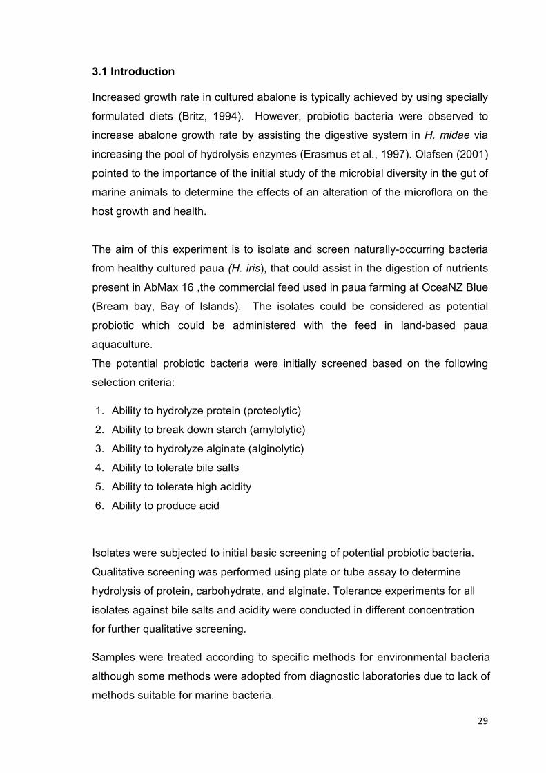
parts of the whole gut using Fig. 3-1 and 3-2 as guide.
Figure 3-1 Anterior view of the abalone Haliotis showing the main organs; (a) gills and
intestine, (b) kidney and heart, (c) heart and stomach, (d) stomach and crop, (e) foot
(Macey & Coyne, 2005)
31
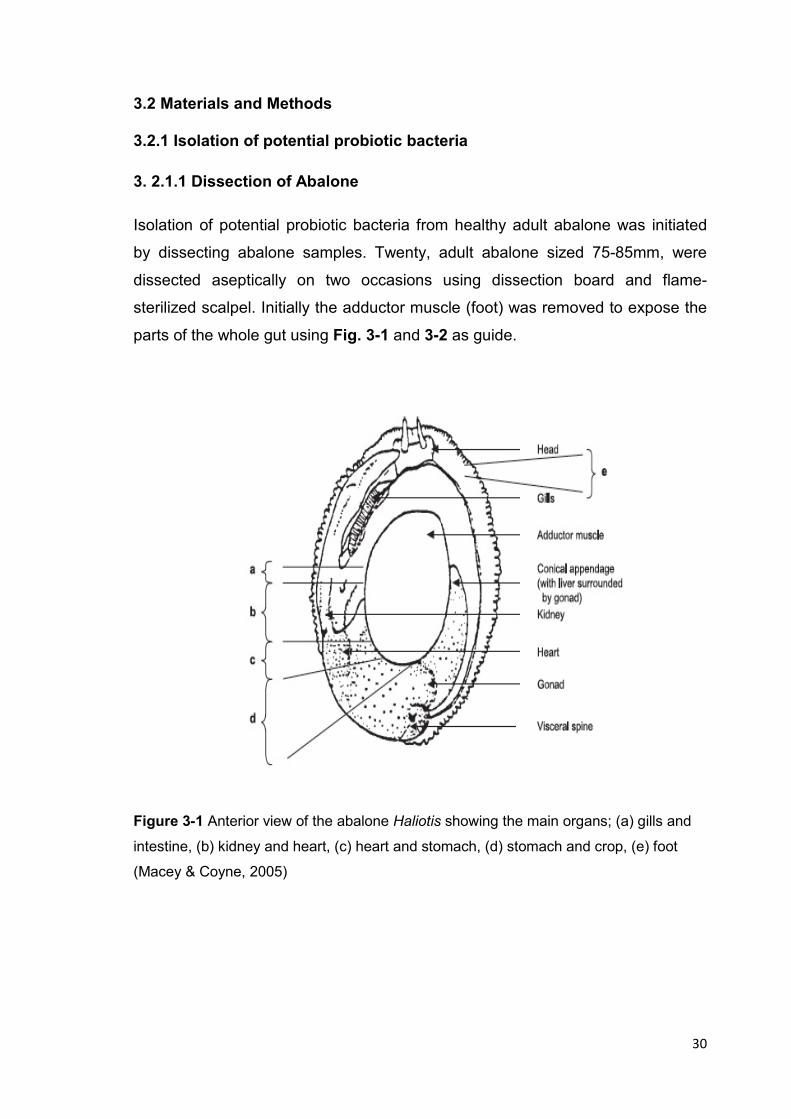
Figure 3-2 Abalone digestive tract (Erasmus et al., 1997)
Isolation of bacterial strains was following the method of Vijayabaskar &
Somasundaram (2008 ). Samples were taken each from the stomach, intestine,
digestive gland, hepatopancreas, rectum, and anus by swabbing using sterile
cotton swabs. Samples from juice in the stomach and hepatopancreas were
collected aseptically. The samples were then suspended in sterile Marine Broth.
Each sample suspension was serially diluted and spread plated in Marine Agar
and Plate Count Agar (DIFCO) with supplementation of 2% NaCl, to obtain the
viable count and to allow growth of most bacterial isolates present in each
abalone part. Aerobic and anaerobic using (GasPakTM EZ) plates were prepared
for incubation at two temperatures, 16oC and 22oC until colonies were obtained.
To obtain potential probiotics including lactic acid producing bacteria, samples
from Marine Broth suspension above were spread plated in MacConkey
(DIFCO), TCBS (DIFCO), and MRS (DIFCO) Agar with adjusting salinity to
2%NaCl. Two sets of plates were prepared for incubation at 16oC and 22oC.
Pure cultures of isolates were obtained by a series of five-phase streak plating
on appropriate medium.
32
3.2.1.2 Collection of Environmental Isolates
Potential probiotic bacteria were isolated from faecal samples and water from
OceaNZ abalone tanks. Isolates were also collected from the bottom of the
rearing tanks of adult and juvenile abalone by swabbing the surface with sterile
cotton swab which was then suspended in sterile Marine broth (DIFCO). The
samples were diluted and plated for isolation and viable count as described in
Section 3.2.1.3. Isolation using MacConkey, TCBS, and MRS Agar was done as
described in the previous section. A series of five-phase streak plating was used
to obtain pure cultures of desired isolates.
3.2.1.3 Viable Count of Bacteria in the Abalone Gut and Rearing Water
The purpose of conducting viable count experiment is to determine abundance
and species composition of culturable microorganisms in the host gut and
rearing water (Iehata et al., 2010). Viable cell count, usually reported as the
number of colony-forming units (CFU) of the organism of interest per unit area,
volume, or weight of sample (Macey & Coyne, 2006a).
Viable cell count was obtained by spread plating aliquots from a series of
dilutions into Marine agar (DIFCO), MacConkey agar (DIFCO), TCBS agar
(DIFCO), and De Man–Rogosa–Sharpe agar MRS (DIFCO).
Bacterial isolates were preserved for long term storage at -80oC in Marine broth
(Difco) supplemented with 15% Glycerol (Vishnivetskaya & Kathariou, 2009).

33
3.2.2 Screening of Potential Probiotic Isolates
3.2.2.1Screening for Protein Hydrolysis
Isolates were cultured on 0.2% casein agar plates, described by Gutierrez-
Maddox (2002) with modification, in order to determine protein hydrolysis. Casein
agar was prepared by mixing (1.0g) sodium caseinate (ACROS organics), (0.1g)
magnesium sulphate, (0.5g) glucose, (0.005g) ferrous sulphate, 0.10g di-
potassium phosphate (K2 HPo4 ), (7.5g) agar, and (500ml) deionized water. The
medium was autoclaved at 121oC for 15 min. After inoculation the cultures were
incubated at (22oC) until visible colonies were present. Hydrolysis of protein
(casein) is indicated by the presence of clear halo around the colonies. Isolates
were compared based on the diameter of clear halo surrounding a single colony.
In case of hydrolysis not easily observed against natural light, the plates were
flooded with 1% HCl acid, then pouring off the excess. The acid should
precipitate any unhydrolyzed casein.
3.2.2.2 Screening for Starch Hydrolysis
Isolates were screened for their ability to hydrolyze starch by spread-plating on
1% starch Marine Agar plates (Gutierrez-Maddox, 2002). Starch agar was
prepared by mixing starch dextrin 10g, nutrient agar, and NaCl 19.4% dissolved
in 1L of deionized water, then autoclaved.
Cultures were incubated at 22oC until colonies were visible. Starch-hydrolyzing
(amylolytic) colonies were distinguished from non-hydrolyzers with the
application of Gram iodine solution into the cultures. Amylolytic colonies
produced clear zones while non-amylolytic colonies did not remove the blue
colour of the starch-iodo complex.

34
3.2.2.3 Screening for Alginate Hydrolysis
3.2.2.3.1. Alginate Agar Plate Method
Screening for alginate hydrolysis in solid medium was carried out using a
modified method of (Nakamura, 1987). Isolates were spread plated on 2.0%
alginate agar medium. The medium was prepared by adding (20g) sodium
alginate (SIGMA), (5g) yeast extract (DIFCO), (10g) agar (DIFCO), (0.2g)
MgSO4, (1g) NH4H2PO4, (1g) K2HPO4, (19.4g) NaCl, to 1L of deionized water,
in addition of and 0.003%bromothymolblue as pH indicator. All ingredients were
dissolved in 1L of deionized water, then autoclaved.
The plates were incubated at 22o C until colonies were visible (2-5 days).
Colonies producing acids from alginate hydrolysis were surrounded by a yellow
zone. Confirmation of results was carried out by the addition of 70% ethanol for 1
hour and observation of precipitation of alginate (Hu et al., 2006).
3.2.2.3.2. Alginate Broth Method
Potential probiotic isolates were screened for alginate hydrolysis by cultivation in
(5 ml) of 0.1% sodium alginate broth consisting of 0.5% peptone, 0.1% yeast
extract, 3.0% NaCl and 0.1% sodium alginate (Kitamikado, 1990). The pH of the
medium was adjusted to 7.6 for cultivation of marine bacteria. The cultures were
incubated at 22oC for 1-5 days.
Alginate hydrolysis was determined every 24 hours using precipitation test with
acidic albumin (Kitamikado, 1990). The principle of the test is the development of
turbidity (precipitation) when alginic acid and bovine albumin are mixed in an
acidic condition. Acidic albumin was prepared by dissolving 1g of bovine
albumin fraction V (Thermo Scientific, USA) in 1L of deionized water before
adding 3.26g of sodium acetate and 4.56ml of glacial acetic acid. The pH of the
solution was adjusted to pH 3.72-3.78 using 2M HCl. After every 24 hours of
incubation, 0.5 ml of each bacterial culture was withdrawn and centrifuged at
3,000-4,000 rpm for 10 min. A 0.2 ml fraction of the supernatant was collected
and transferred into a small cuvette. Acidic albumin (0.2ml) was added just
35
before reading the transmission at 600nm in a spectrophotometer
(Ultrospec™2100 pro UV/Visible). The control for the experiment was prepared
by using sterile 0.1% sodium alginate broth (without bacteria). This was
centrifuged and tested with acidic albumin in the same way as the test solutions.
3.2.2.4 Screening for Bile Salts Tolerance
Evaluation of bile salts tolerance for the bacterial isolates was performed using a
modified method of (Hyronimus et al., 2000). The isolates were grown on Marine
Agar containing different concentrations of Oxgall bile salt (BDH). The Gram-
positive bacteria were evaluated in Marine Agar containing bile salts at 0.1%,
0.2%, 0.3%, 0.5%, 1%, and 1.5%. The same concentrations of Oxgall were
added to MRS agar medium for screening lactic acid bacteria. For Gram-
negative bacteria obtained from TCBS agar, higher concentrations of bile salt
were used; 1%, 1.2%, 1.4%, 1.6%, and 1.8%. The rationale behind this was that
TCBS agar medium already contained 0.8% Oxgall and the isolates were able to
grow on it, thus the screening for Oxgall (bile salt) tolerance used a range with
the lowest at 1%. The plates were streak plated with each isolate and incubated
at 22oC until growth was present.
3.2.2.5 Screening for Acid Tolerance
Bacterial isolates were assessed for their ability to tolerate high acidity following
the method of Iehata et al.(2009), and Lin et al.(2006) with modification to fit the
range of acidity inside the gut of paua which is pH 1.5 - 5.5. Tubes of Marine
broth (9 ml) at pH 1.5, pH 2.0, and pH 5.0 were prepared using 5M HCl. The
broths were inoculated with a colony and incubated for 3 hours at 22oC. Viability
at each pH value was determined by comparing the plate count obtained after 0
hour with that after 3 hour incubation.

36
3.2.2.6 Screening for Acid Production
For detection of acid production, the acid-base titration method was utilized
(ADMI, 1971). Marine broth with 5% glucose was prepared in 100 ml of capped
bottles and steriled by autoclaving for 15 min in 121oC. A colony of bacterial
isolate was inoculated into modified Marine broth in two replicates.
The cultures were centrifuged at room temperature for 15 min at 4,000 x g. The
supernatant (50 ml) was collected in a 250 ml flask and titrated with 0.1M NaOH
using phenolphthalein indicator until a light pink colour appeared. Lactic acid
concentration was calculated using the equation (ADMI, 1971):
Lactic acid % = (ml NaOH) x (M NaOH) x (mol. weight lactic acid) x 100
volume of sample x 100
3.2.3 Phenotypic Characterization
Initial phenotypic analysis was performed by assigning the isolates to major
phenotypic groups using standard Gram staining (Bergey et al., 1994).
Cultivation in selective media, such as MacConkey Agar ,TCBS Agar and
Pseudomonas Agar was done as complementary tests for Gram-negative
isolates (Hansen & Sorheim, 1991). The isolates were then subjected for further
morphological, cultural, physiological and biochemical analyses described later in
this Chapter.
3.2.3.1 Cell and Colony Morphology
The morphological cell characteristics of the isolates were determined by Gram-
staining using ethyl alcohol as decolourizing agent (Bergey et al., 1994). Spore
formation was determined by (5%) malachite green (BDH) staining (Bergey et al.,
1994). Motility was studied using (0.3% agar) motility medium was included; 10g
beef heart infusion, 10g tryptose, 19.4g sodium chloride, 53.4g gelatin, 3.0g
agar, and 1Lof deionized water. The medium was prepared by boiling for 1 min
to dissolve the ingredients then autoclaved (Gutierrez-Maddox, 2002; Kaiser,
37
2011). The characterisation of colonies of the isolates was carried out by
culturing on Marine Agar, TCBS agar, and MRS agar ( refer to Section 3. 2.1.1).
3.2.3.2 Cultural Characteristics
The temperature requirement of the isolates was determined by incubation of
isolates grown in Marine Agar at 16oC, and 22oC. Incubation time was 48-72
hours, except for alginate agar culture which required longer period (3-5 days).
Cultures were incubated in both aerobic and anaerobic conditions.
3.2.3.3 Biochemical Characteristics
The following tests were used for initial biochemical phenotyping of the isolates,
all the methods were conducted according to and Bergey’s Manual (Bergey&
Holt, 1994) and Systemic Microbiology Manual (Gutierrez-Maddox, 2002) :
� Catalase test using 3% H2O2 (BDH) was performed on colonies in Marine Agar
to detect production of catalase which breaks down H2O2 into H2O and
O2.Catalase producers would produce gas bubbles upon reaction with H2O2.
� Oxidase test for the presence of cytochrome oxidase was performed on 24
hour old colonies grown in Marine Agar. Tetra-methyl-p-phenylenedi-amine di-
hydrochloride (ACROS Organics) was used as oxidase reagent. Oxidase
positive isolates would produce a blue-purple color change within 30 sec.
� Blood haemolysis was tested on isolates streak plated on Colombia agar
supplemented with 5% Sheep Blood agar (Fort Richard Laboratories). Cultures
which are ß-haemolytic would remove the red colour of the medium
completely, while α-haemolytic isolates have incomplete heamolysis. Ŵ-
haemolytic isolates have no haemolysis.
� Gelatine hydrolysis test was performed by inoculating gelatine stabs (DIFCO)
Isolates capable of gelatine hydrolysis would liquefy and not allow gelatine agar
to set at refrigeration temperature.
38
� Oxidation/Fermentation (O/F) test was used to determine whether the isolates
oxidize or ferment glucose. (Hugh & Leifson O/F agar (DIFCO) was used to
determine glucose metabolism.
� Triple Sugar Iron test (TSI) from (DIFCO) was used to determine sugar
fermentation (glucose, sucrose and fructose) and hydrogen sulphide production
in the Gram-negative isolates. Sugar fermenter would produce yellow colour
while hydrogen sulphide producers would produce black colonies.
� DNase agar (Fort Richard Laboratories) was used for the detection of
deoxyribonuclease activity. DNA is a polynucleotide which binds to methyl
green and gives stable colour in alkaline pH. In the presence of DNase enzyme,
DNA will be degraded and methyl green releases and loses its colour in lower
pH. Thus DNase-positive bacteria show clear zones around colonies. .
� Baird-Parker agar containing egg yolk emulsion (Fort Richard Laboratories)
was used for detection of lipase production in Gram-positive isolates.
� XLD agar (Fort Richard Laboratories) was streak plated for degradation of
xylose, lactose, and sucrose to acid. Acid from fermentation lowers the pH and
changes phenol red color to yellow.
� Bile Eesculine Agar, BEA (Fort Richard Laboratories), a selective, differential
medium, was used to isolate and identify members of the genus Enterococcus.
All members of the Enterobacteriaceae family grow on this medium, yet certain
bacteria can hydrolyze esculine to 6, 7-dihydroxycoumarin, which reacts with
iron to produce a characteristic blackening of the medium.
� Tryptic Nitrate broth (Fort Richard Laboratories): Was used for detection of
nitrate reduction to nitrite. Nitrate-reducing isolates which produce nitrite
produce a red colour with sulphanilic acid and α- naphthylamine.
39
3.2.4. Identification of the Isolates
All bacterial isolates were identified according to Gram-positive and Gram-
negative bacteria flow charts that mentioned in Bergey’s Manual of Determinative
Bacteriology (9th Edition)(Bergey et al., 1994). In addition API20 E® strips and
API10 S® strips (from BioMerieux) were used for further identification of Gram-
negative isolates. The suspension of one colony was inoculated into mini cuvette
of the strip, and incubated for 24-48 hour. Reading andinterpreting the results
was through Bio Mérieux website [www.apiweb.biomerienx.com].
3.2.5 Phylogenetic Analysis for the Isolates
Bacterial isolates were sent to Landcare Research (Auckland 1072) for 16S
rRNA classification and to confirm the phenotypic identification carried out
earlier. The primers used to amplify the 16S rRNA gene were 27F (forward) and
1525R (reverse) primers for 16S rRNA (Lane, 1991).

40
3.3 Results
3.3.1 Isolation and Enumeration of Potential Probiotic Bacteria
The purpose of conducting viable count experiment is to determine abundance
and species composition of culturable microorganisms (Section 3.2.1.3) found in
the paua digestive tract and rearing tank (Table 3-2).Viable counts obtained after
incubation for 48 hr in different media are shown in Table 3-1; viable count from
Marine agar reflects the total number of culturable bacteria in the sample. The
intestine region with pH 7.0 showed the greatest abundance of microorganisms,
while the anus with pH 8.2 had the least number (1.1 x10⁸ and 3.0 x10³,
respectively). The highest number of presumptive lactic acid bacteria LAB at 3.8
x105, were found in the stomach. There was a low number of presumptive LAB
in the intestine. Bile salt resistant bacteria that were presumptive Gram-negative
and presumptive Vibrio bacteria were obtained in MacConkey Agar and TCBS
Agar, respectively.
A lower number of microorganisms were sampled from the rearing tank and
water compared to those from the animals.
Table 3-1 Viable count (CFU/ml) of bacterial population in abalone (GIT), and rearing
water
Sample
pH value of the gut region
Viable count CFU/ml (average)
Marine agar
MacConkey agar
MRS agar
TCBS agar
Stomach, digestive gland
5.5
2.5 x 10⁶
3.0 x 10³
3.8 x10⁵
3.0 x10⁵
Intestine
7.0
1.1 x 10
8
3.0 x 10
3
1.2 x10
3
3.0 x10
5
Hepatopancreas
8.0
5.0 x10
4
2.2 x 10
4
< 1
2.5 x10
4
Anus
8.2
3.0 x10
3
1.2 x 10
4
6.7x10
3
Tank swab
-
1.15 x 10
6
2.0 x10
3
<1
1.8 x10
3
Adult tank water
8.3
8.8 x10
3
<2.0 x10
3
<1
3.0 x 10
3
Juvenile tank water
8.2
3.0 x10
2
< 2.0 x10
3
<1
1.4 x10
3

41
Isolates
Culture Medium
Source of isolates
JHDC (LAB)
MRS
Stomach, digestive gland
JHEb1
Marine
Intestine, V tank
JHBa2
Marine
Intestine, Rectum
JH1
TCBS
Stomach, Water sample
JH2
TCBS
Intestine, Tub water
JH3
TCBS
Intestine
JH4
TCBS
Juveniles abalone Tank
JH5
TCBS
Intestine
JHC Marine
Stomach, Hepatopancreas, Intestine,
Adult abalone tank
Table 3-2 Bacterial strains isolated from abalone (GIT), and rearing water
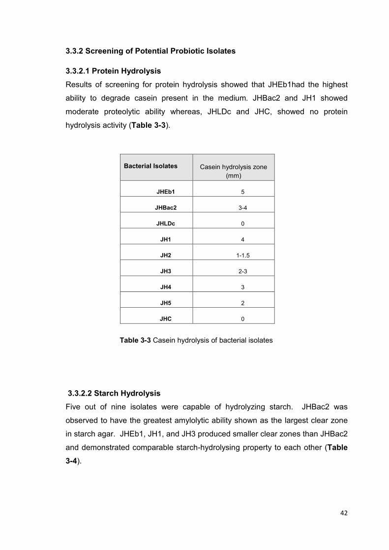
42
3.3.2 Screening of Potential Probiotic Isolates
3.3.2.1 Protein Hydrolysis
Results of screening for protein hydrolysis showed that JHEb1had the highest
ability to degrade casein present in the medium. JHBac2 and JH1 showed
moderate proteolytic ability whereas, JHLDc and JHC, showed no protein
hydrolysis activity (Table 3-3).
Bacterial Isolates
Casein hydrolysis zone
(mm)
JHEb1
5
JHBac2
3-4
JHLDc
0
JH1
4
JH2
1-1.5
JH3
2-3
JH4
3
JH5
2
JHC
0
Table 3-3 Casein hydrolysis of bacterial isolates
3.3.2.2 Starch Hydrolysis
Five out of nine isolates were capable of hydrolyzing starch. JHBac2 was
observed to have the greatest amylolytic ability shown as the largest clear zone
in starch agar. JHEb1, JH1, and JH3 produced smaller clear zones than JHBac2
and demonstrated comparable starch-hydrolysing property to each other (Table
3-4).
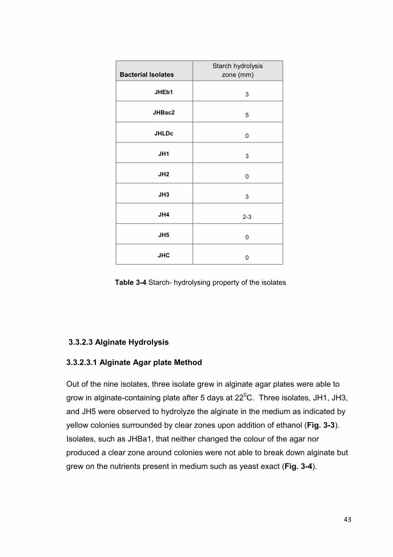
43
Bacterial Isolates
Starch hydrolysis
zone (mm)
JHEb1
3
JHBac2
5
JHLDc
0
JH1
3
JH2
0
JH3
3
JH4
2-3
JH5
0
JHC
0
Table 3-4 Starch- hydrolysing property of the isolates
3.3.2.3 Alginate Hydrolysis
3.3.2.3.1 Alginate Agar plate Method
Out of the nine isolates, three isolate grew in alginate agar plates were able to
grow in alginate-containing plate after 5 days at 220C. Three isolates, JH1, JH3,
and JH5 were observed to hydrolyze the alginate in the medium as indicated by
yellow colonies surrounded by clear zones upon addition of ethanol (Fig. 3-3).
Isolates, such as JHBa1, that neither changed the colour of the agar nor
produced a clear zone around colonies were not able to break down alginate but
grew on the nutrients present in medium such as yeast exact (Fig. 3-4).

44
Figure 3-3 Alginate agar plates with change in colour (positive) and clear zone of
hydrolysis (1- JHI, 2- JH3, and 3- JH5)
Figure 3-4 Alginate agar plate with no change in colour and no clear
zone around the colonies indicative of absence of alginate hydrolysis

45
3.3.2.3.2 Alginate broth tube method
Results obtained at day 2, and day 5 of a five day- incubation period are shown
in Table 3.5. The supernatant of the isolates were each added to acidic albumin-
alginate solution and then compared with the control. Isolates were evaluated on
their ability to clear the albumin-alginate precipitate (Fig. 3-5). Transmission was
measured by spectrophotometer at 650 nm. The control solution produced a
transmission value at 10.2%. JH1 was the only isolate which completely cleared
the precipitate and produced a transmission value of 100% after 2 days of
incubation (Table 3-5) . On this basis, JH1 exhibited the fastest ability to break
down alginate. Lower transmission values were obtained with JH3 and JH5 at
day 2, but these isolates later produced 100% transmission, hence, complete
hydrolysis after 5days. JH3 and JH5 were slower alginate degraders. The
results obtained with this method confirmed those with the alginate agar plate
method.
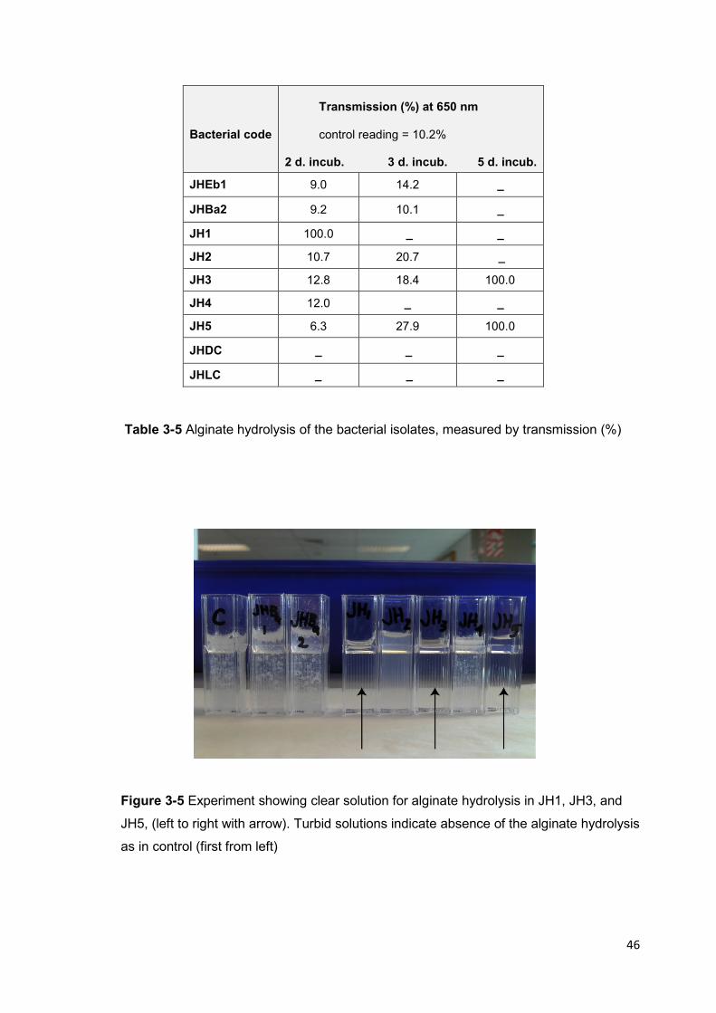
46
Bacterial code
Transmission (%) at 650 nm control reading = 10.2%
2 d. incub. 3 d. incub. 5 d. incub.
JHEb1 9.0 14.2 _
JHBa2 9.2 10.1 _
JH1 100.0 _ _
JH2 10.7 20.7 _
JH3 12.8 18.4 100.0
JH4 12.0 _ _
JH5 6.3 27.9 100.0
JHDC _ _ _
JHLC _ _ _
Table 3-5 Alginate hydrolysis of the bacterial isolates, measured by transmission (%)
Figure 3-5 Experiment showing clear solution for alginate hydrolysis in JH1, JH3, and
JH5, (left to right with arrow). Turbid solutions indicate absence of the alginate hydrolysis
as in control (first from left)
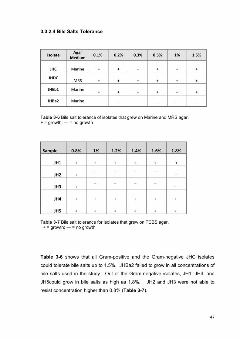
47
3.3.2.4 Bile Salts Tolerance
Isolate Agar
Medium 0.1% 0.2% 0.3% 0.5% 1% 1.5%
JHC
Marine
+
+
+
+
+
+
JHDC
MRS
+
+
+
+
+
+
JHEb1 Marine
+
+
+
+
+
+
JHBa2 Marine _ _ _ _ _ _
Table 3-6 Bile salt tolerance of isolates that grew on Marine and MRS agar. + = growth; ― = no growth
Sample
0.8%
1%
1.2%
1.4%
1.6%
1.8%
JH1
+
+
+
+
+
+
JH2
+
_ _ _ _ _
JH3
+
_ _ _ _ _
JH4
+
+
+
+
+
+
JH5
+
+
+
+
+
+
Table 3-7 Bile salt tolerance for isolates that grew on TCBS agar. + = growth; ― = no growth
Table 3-6 shows that all Gram-positive and the Gram-negative JHC isolates
could tolerate bile salts up to 1.5%. JHBa2 failed to grow in all concentrations of
bile salts used in the study. Out of the Gram-negative isolates, JH1, JH4, and
JH5could grow in bile salts as high as 1.8%. JH2 and JH3 were not able to
resist concentration higher than 0.8% (Table 3-7).

48
3.3.2.5 Acidity Tolerance
Viable counts expressed as CFU/ml of culture at 0 hour and after 3 hour of
incubation at pH 5, pH2, and pH1.5 are shown in Table 3-8. Viable counts of all
isolates were stable at pH 5.0 and pH 2.0 for 3 hours. JHEb1, JH1, JH2, and JH
3 showed a reduction in number in pH 2.0 by 1 log unit after incubation. JH 5
and JHC were the least tolerant in pH 2.0. JH2 and JH 4 were completely
inhibited at pH 1.5 with the acid challenge while all the other isolates remained
viable for 3 hours although with a slight reduction in number.
Isolate
Viable count in pH 5,
(CFU/ml)
0h. 3h.
Viable count in pH 2,
(CFU/ml)
0h. 3h.
Viable count in pH1.5
(CFU/ml)
0h. 3h.
JHEb1
5.0 x10⁸
5.5 x10⁸
2.3 x10⁵ 2.8x10
4
1.4 x10⁵
2.0 x10³
JHBac2
2.7 x10 4
2.0 x10 4
6.8x10³
1.1 x10³
8 x10³
18 x103
JHLDc
2.2 x10 7
2.0 x10 7
8.2 x10⁵
1.4 x10⁵
6.0 x10⁴
1.6 x10³
JH1
2.0 x10⁵ 2.9 x10
4
3.0 x10⁶ 2.0 x10
5
6.5 x10⁴
4.0 x10³
JH2
4.8 x10³
1.4 x10³
4.0 x10⁴
2 .0 x103
3.0 x10³
0
JH3
5.0 x10⁴
5.0 x10⁴
7.8 x10⁶
5.4 x10⁵
3.0 x10⁴
2.4 x10³
JH4
1.2 x10⁴ 10 x10
4
5.0 x10⁶
3.3 x10⁵
2.5 x10⁶
3.0 x10³
JH5
3.0 x10⁴
2.8 x10⁴
4.0 x10⁵
2 x10³
1.0 x10⁵ 0
JHC
5.0 x10 6
4.7 x10⁶
5.2 x10⁵
4.0 x10³
6.0 x10⁶
3.2 x10⁵
Table 3-8 Acid tolerance of isolates measured as viable count (average) at 0 hr and 3 hr

49
3.3.2.6 Acid production
Acid production through sugar fermentation, amongst the nine isolates was
varied. JHLDc produced the highest concentration of lactic acid (0.67%). JHC
and JH4 were the second highest producers with 0.46% and 0.42%, respectively.
JHEb1, JH1, and JH3 produced comparable acid concentrations while JH2 and
JH5 were the lowest producing isolates (Table 3-9).
Isolates Lactic Acid % (average)
JHEb1 0.18
JHBa2 0.36
JHLDc 0.67
JHC 0.46
JH1 0.18
JH2 0.09
JH3 0.22
JH4 0.42
JH5 0.11
Table 3-9 Acid production in potential probiotic isolates
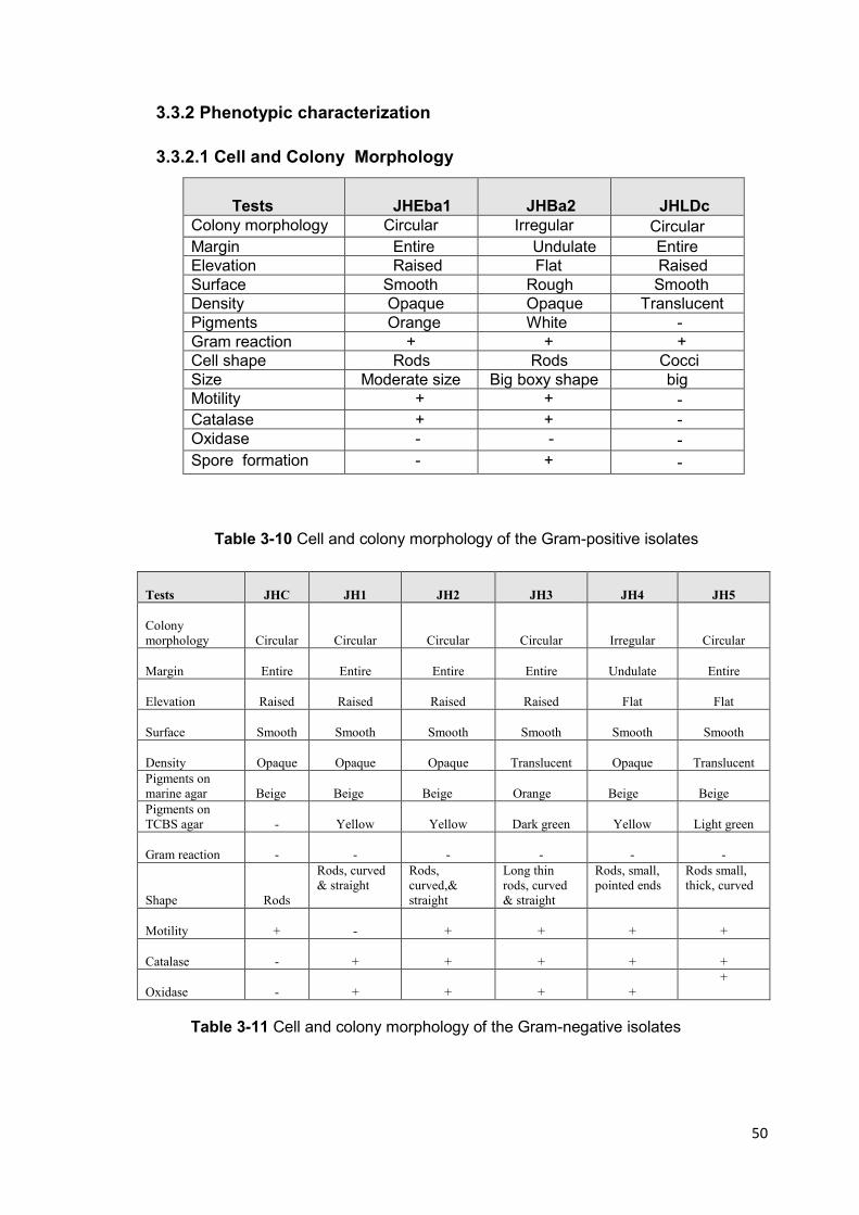
50
3.3.2 Phenotypic characterization
3.3.2.1 Cell and Colony Morphology
Table 3-10 Cell and colony morphology of the Gram-positive isolates
Table 3-11 Cell and colony morphology of the Gram-negative isolates
Tests
JHEba1
JHBa2
JHLDc
Colony morphology Circular Irregular Circular
Margin Entire Undulate Entire Elevation Raised Flat Raised Surface Smooth Rough Smooth Density Opaque Opaque Translucent Pigments Orange White - Gram reaction + + + Cell shape Rods Rods Cocci Size Moderate size Big boxy shape big Motility + + -
Catalase + + - Oxidase - - -
Spore formation - + -
Tests
JHC
JH1
JH2
JH3
JH4
JH5
Colony
morphology
Circular
Circular
Circular
Circular
Irregular
Circular
Margin
Entire
Entire
Entire
Entire
Undulate
Entire
Elevation
Raised
Raised
Raised
Raised
Flat
Flat
Surface
Smooth
Smooth
Smooth
Smooth
Smooth
Smooth
Density
Opaque
Opaque
Opaque
Translucent
Opaque
Translucent
Pigments on
marine agar
Beige
Beige
Beige
Orange
Beige
Beige
Pigments on
TCBS agar
-
Yellow
Yellow
Dark green
Yellow
Light green
Gram reaction
-
-
-
-
-
-
Shape
Rods
Rods, curved
& straight
Rods,
curved,&
straight
Long thin
rods, curved
& straight
Rods, small,
pointed ends
Rods small,
thick, curved
Motility
+
-
+
+
+
+
Catalase
-
+
+
+
+
+
Oxidase
-
+
+
+
+
+
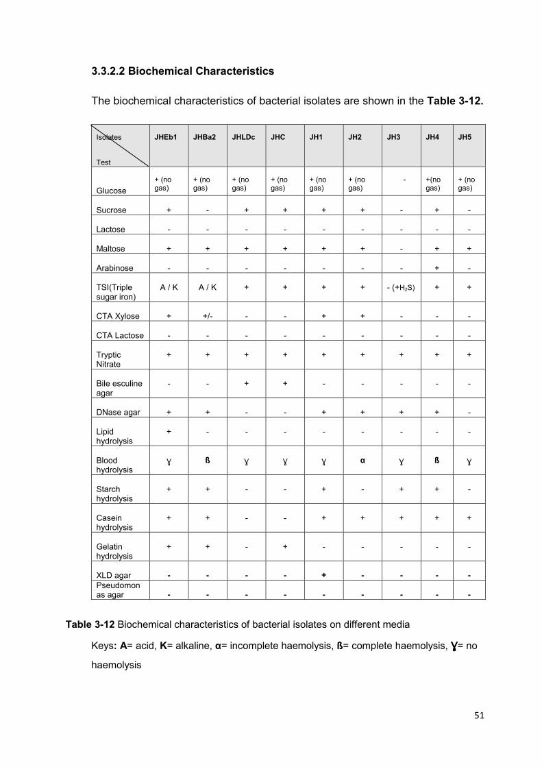
51
3.3.2.2 Biochemical Characteristics
The biochemical characteristics of bacterial isolates are shown in the Table 3-12.
Isolates Test
JHEb1
JHBa2
JHLDc
JHC
JH1
JH2
JH3
JH4
JH5
Glucose
+ (no gas)
+ (no gas)
+ (no gas)
+ (no gas)
+ (no gas)
+ (no gas)
-
+(no gas)
+ (no gas)
Sucrose
+
-
+
+
+
+
-
+
-
Lactose
-
-
-
-
-
-
-
-
-
Maltose
+
+
+
+
+
+
-
+
+
Arabinose
-
-
-
-
-
-
-
+
-
TSI(Triple sugar iron)
A / K
A / K
+
+
+
+
- (+H2S)
+
+
CTA Xylose
+
+/-
-
-
+
+
-
-
-
CTA Lactose
-
-
-
-
-
-
-
-
-
Tryptic Nitrate
+
+
+
+
+
+
+
+
+
Bile esculine agar
-
-
+
+
-
-
-
-
-
DNase agar
+
+
-
-
+
+
+
+
-
Lipid hydrolysis
+
-
-
-
-
-
-
-
-
Blood hydrolysis
Ŵ
ß
Ŵ
Ŵ
Ŵ
α
Ŵ
ß
Ŵ
Starch hydrolysis
+
+
-
-
+
-
+
+
-
Casein hydrolysis
+
+
-
-
+
+
+
+
+
Gelatin hydrolysis
+
+
-
+
-
-
-
-
-
XLD agar
-
-
-
-
+
-
-
-
-
Pseudomonas agar
-
-
-
-
-
-
-
-
-
Table 3-12 Biochemical characteristics of bacterial isolates on different media
Keys: A= acid, K= alkaline, α= incomplete haemolysis, ß= complete haemolysis, ŴŴŴŴ= no
haemolysis

52
3.3.3 Identification
3.3.3.1 Identification Using Dichotomous key
The flow charts of dichotomous key adopted from Bergey’s Manual of Systematic
Bacteriology (Brenner et al., 2005; DeVos et al., 2009) were used in identifying
the isolates.
Gram- Negative Rods (Brenner et al.)
Oxidase Test
+ _
Glucose fermentation
Aeromonas spp. + Acid - Acid Pseudomonas spp.
Vibrio spp.
Na+ required for growth Aeromonas spp.
Vibrio spp.
Figure 3-6 Flow chart for Gram-negative bacteria isolates
Aeromonas, Pseudomonas,
and Vibrio
Enterobacteriaceae

53
Gram- positive Bacteria (DeVos et al., 2009)
Facultative aerobes
― Catalase Bile esculine - Streptococcus spp.
Bile esculine + Enterococcus spp.
Figure 3-7 Flow chart for Gram- positive bacterial isolates
Bacilli Cocci
Round in clusters &
tetrads:
Staphylococcus
Micrococcus
Peptostreptococcus
Oval shape in
chains;
Streptococcus
Peptococcus
Enterococcus spp.
Spore-bearing
Bacillus spp.
Non Spore-bearing
Exiguobacterium spp.
α-Haemolysis.
Streptococcus spp.
ß-Haemolysis
Streptococcus spp.
Ŵ-haemolysis
Ŵ-Enterococcus spp.
Ŵ-Streptococcus spp.

54
3.3.3.2 API 20E Strip Results for Gram-negative Isolates
Isolate code
Isolation medium
Colony API code Bacteria ID Id. Assessment
JH1
TCBS
Big yellow
1027735
Aeromonas hydrophila
possibility; Vibrio fluvialis
JH2
TCBS
Small yellow
0004014
Aeromonas spp.
possibility; Vibrio fluvialis
JH3
TCBS
Dark green
0004772
Shewanella putrefaciens or Citrobacter freudii
Good identification
JH4
TCBS
Big yellow
2206066
Pseudomonas
99.9% with supplementary tests (2206066 73)
JH5
TCBS
Light green
00047734
Pasteurella multocida
Unacceptable profile
JHC
Marine
Beige
7306521
Serratia marcescens
Unacceptable profile
Table 3-13 API 20E results for Gram-negative isolates
The Gram-negative isolates JH1, JH2, JH4, and JH5 were easily identified as
Vibrio species using the dichotomous key and were distinguished from
Enterobacteriaceae due to the presence of oxidase. API 20E system failed to
confirm the identification of 4 out of 5 Gram-negative isolates in this study.
These results were similar to what was reported by Santos et al. (1993) when the
API system could not confirm the identification of bacterial isolates from fish and
shellfish. JH3 was identified as Shewanella with a positive confirmation using
API 20E kit.
The Gram-positive isolates were identified using the dichotomous key with
JHLDc belonging to genus Enterococcus. JHBa2 was Bacillus and JHEb1 was
Exiguobacterium. JHEb1 could be distinguished from lactic acid bacteria by its
positive catalase production and orange big colonies and from Bacillus by the
absence of spore formation.

55
3.3.3.3 Identification Based on 16S rRNA Gene Sequence
Results of the 16S rRNA sequencing revealed matching identification from
classical phenotypic characterization except for Exiguobacterium and Cobetia
(Table 3-14); Cobetia is not included in the API system.
Bacterial Isolates
code
Bacterium scientific Name
1. JHEb1
Exiguobacterium sp.
2. JHBa2
Bacillus sp.
3. JHLDc
Enterococcus sp.
4. JHC
Cobetia sp.
5. JH1
Vibrio sp.
6. JH2
Vibrio sp.
7. JH3
Shewanella sp.
8. JH4
Vibrio sp.
9. JH5
Vibrio sp.
Table 3-14 Identification of the isolates by 16S rRNA sequencing
56
3.4 Discussion
Viable counts obtained from MRS and TCBS media suggest that putative Vibrio
species and putative Enterococcus were dominant and probably colonizing in the
stomach, intestine and hepatopancreas of paua. The concentrations of isolates
in the water of rearing tanks and in faecal matter suggest that the potential
probiotic microorganisms could interact with the animals externally. These two
factors, namely, colonizing property and being local to the rearing environment
are one of the important considerations in isolating potential probionts (Merrifield
et al., 2010 a). Since the objective of this study is to increase the growth rate of
paua by introducing beneficial probiotic microorganisms, it was important to
screen the isolates for properties which could enhance digestion of nutrients
present in the artificial feed used in the farm. Hence, the isolates were screened
qualitatively in this experiment for protein hydrolysis, starch hydrolysis and
alginate hydrolysis. In addition, acid production, acid tolerance and bile tolerance
of the isolates were evaluated.
3.5 Conclusion
Nine isolates demonstrated predominance in the gastrointestinal tract of paua.
These isolates displayed the characteristics which were required for probiotics in
paua. The isolates exhibited the ability to hydrolyze protein (proteolytic)
hydrolyze alginate (alginolytic), tolerate bile salts and high acidity, and produce
lactic acid. Using phenotypic characterization and 16S rRNA classification, the
isolates were identified as Exiguobacterium JHEb1, Bacillus JHBa2,
Enterococcus JHLDc, Cobetia JHC, Vibrio JH1, Vibrio JH2, Vibrio JH4, Vibrio
JH5, and Shewanella JH3.
Using qualitative screening, the isolates that answered the criteria with the
highest value were Exiguobacterium JHEb1, Vibrio JH1, and Enterococcus
JHLDc. The properties which make these isolates potential probiotic are as
follow:
57
• Exiguobacterium JHEb1: starch hydrolysis, protein hydrolysis, resistance to high
bile salts and acid concentrations and production of high lactic acid
concentration.
• Vibrio JH1: starch hydrolysis, protein hydrolysis, fast alginate hydrolysis,
resistance to high bile salts and acid concentrations
• Enterococcus JHLDc: high lactic acid production, resistance to high bile salts
and acid concentrations.
All isolates that exhibited proteolytic protein, starch, and alginate hydrolysis
activities, including the three isolates above, were subjected to further
quantitative biochemical studies in the next chapter to evaluate enzyme activities
involved in digestion of nutrients in paua feed.
Profiles of Candidate Probionts for Feeding Trial
Exiguobacterium spp.
The genus first described by Collins in 1983 is afacultative anaerobic,
asporogenous (non-sporeforming), irregularly shaped cells which vary from rods
in the exponential phase to short, almost coccoid forms in the stationary phase
(Collins et al., 1983). Exiguobacterium colonies grown on nutrient agar are
circular, entire, raised, smooth, and orange in color. It has the ability to grow in a
wide range of pH (5.5-10.5), temperature (5-40oC) aerobically and anaerobically.
Moreover, this genus can grow in medium containing salt (NaCl) within the range
of 0-5% (Kasana & Yadav, 2007).
Cells are Gram-positive, non-acid fast, motile with peritrichous flagella (GEE et
al., 1980) catalase positive and oxidase negative. Acid is formed from glucose,
sucrose, galactose, and some other sugars. In anaerobic conditions with glucose
as substrate, lactate, acetate, and formate are main end products (Collins et al.,
1983). Classification of the genus is as follows:

58
Phylum Firmicutes
Class Bacilli
Order Bacillales
Family Bacillaceae
Genus Exiguobacterium (Collins et al., 1983)
Figure 3-8 The scanning electron microscopy
photograph of Exiguobacterium sp. (Tan et
al., 2009)
Exiguobacterium has been found in a variety of environments; thermophilic, a
slightly alkaline, and hot spring water with high carbonate concentrations, such
as samples collected from Angel Terrace, which is known as part of Mammoth
Terrace (Vishnivetskaya et al., 2011). Exiguobacterium spp. have been isolated
from Greenland glacial ice, the hot springs at Yellowstone National Park, and the
environment of food processing plants (Vishnivetskaya & Kathariou, 2009)
Some species of the genus Exiguobacterium have been found in aquaculture
system, and they were considered to have the potential to improve the growth
and survival rate of brine shrimps (Artemia sp.). Further, a production of
beneficial polypeptide antibiotics e.g. bacitracin, gramicidin S, polymyxin, and
tyrotricidin have been found in aquaculture water which included
Exiguobacterium spp. Those antimicrobial substances were reported to be active
against a wide range of bacteria (Sombatjinda et al., 2011).
In the present study Exiguobacterium JHEb1 (Fig 3-9) exhibited typical
characteristics of the genus Exiguobacterium.
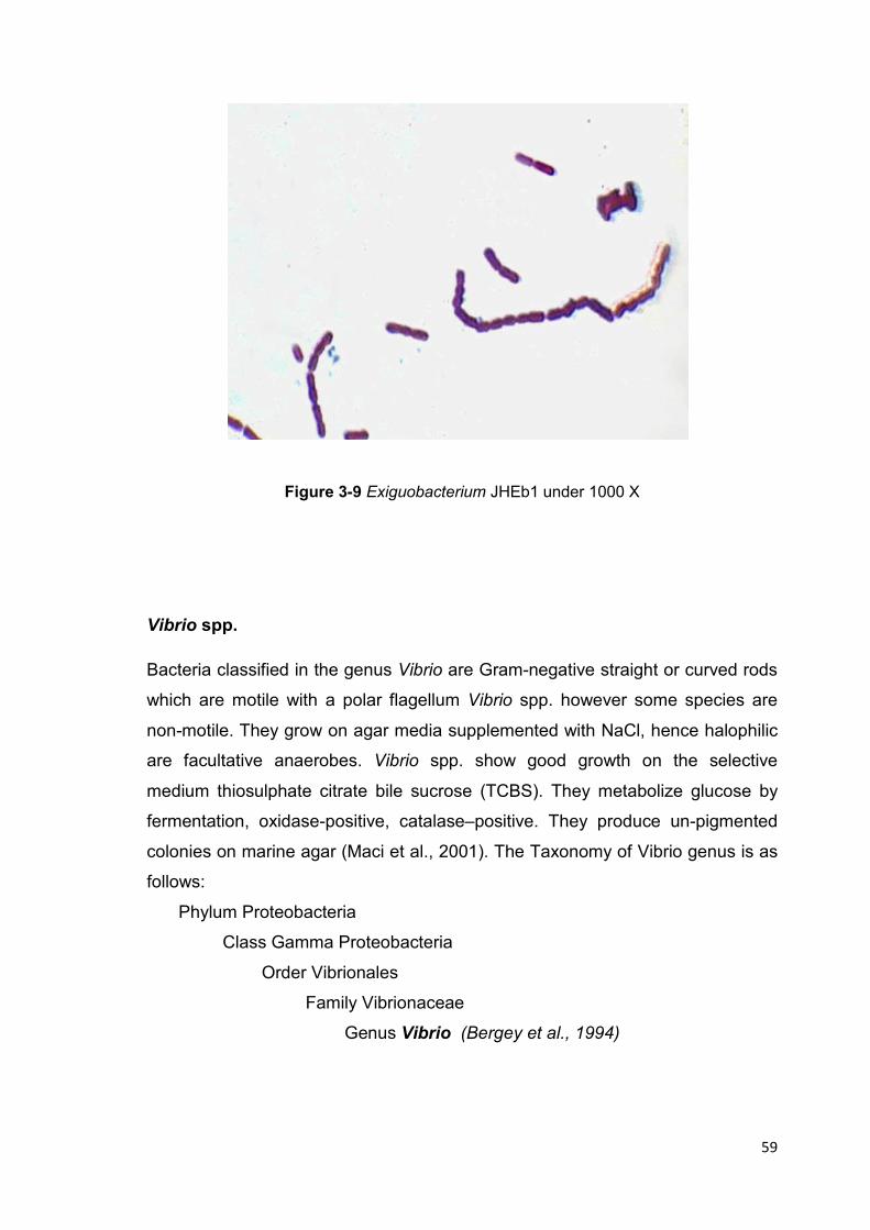
59
Figure 3-9 Exiguobacterium JHEb1 under 1000 X
Vibrio spp.
Bacteria classified in the genus Vibrio are Gram-negative straight or curved rods
which are motile with a polar flagellum Vibrio spp. however some species are
non-motile. They grow on agar media supplemented with NaCl, hence halophilic
are facultative anaerobes. Vibrio spp. show good growth on the selective
medium thiosulphate citrate bile sucrose (TCBS). They metabolize glucose by
fermentation, oxidase-positive, catalase–positive. They produce un-pigmented
colonies on marine agar (Maci et al., 2001). The Taxonomy of Vibrio genus is as
follows:
Phylum Proteobacteria
Class Gamma Proteobacteria
Order Vibrionales
Family Vibrionaceae
Genus Vibrio (Bergey et al., 1994)
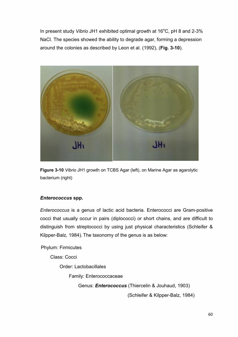
60
In present study Vibrio JH1 exhibited optimal growth at 16oC, pH 8 and 2-3%
NaCl. The species showed the ability to degrade agar, forming a depression
around the colonies as described by Leon et al. (1992), (Fig. 3-10).
Figure 3-10 Vibrio JH1 growth on TCBS Agar (left), on Marine Agar as agarolytic
bacterium (right)
Enterococcus spp.
Enterococcus is a genus of lactic acid bacteria. Enterococci are Gram-positive
cocci that usually occur in pairs (diplococci) or short chains, and are difficult to
distinguish from streptococci by using just physical characteristics (Schleifer &
Kilpper-Balz, 1984). The taxonomy of the genus is as below:
Phylum: Firmicutes
Class: Cocci
Order: Lactobacillales
Family: Enterococcaceae
Genus: Enterococcus (Thiercelin & Jouhaud, 1903)
(Schleifer & Kilpper-Balz, 1984)
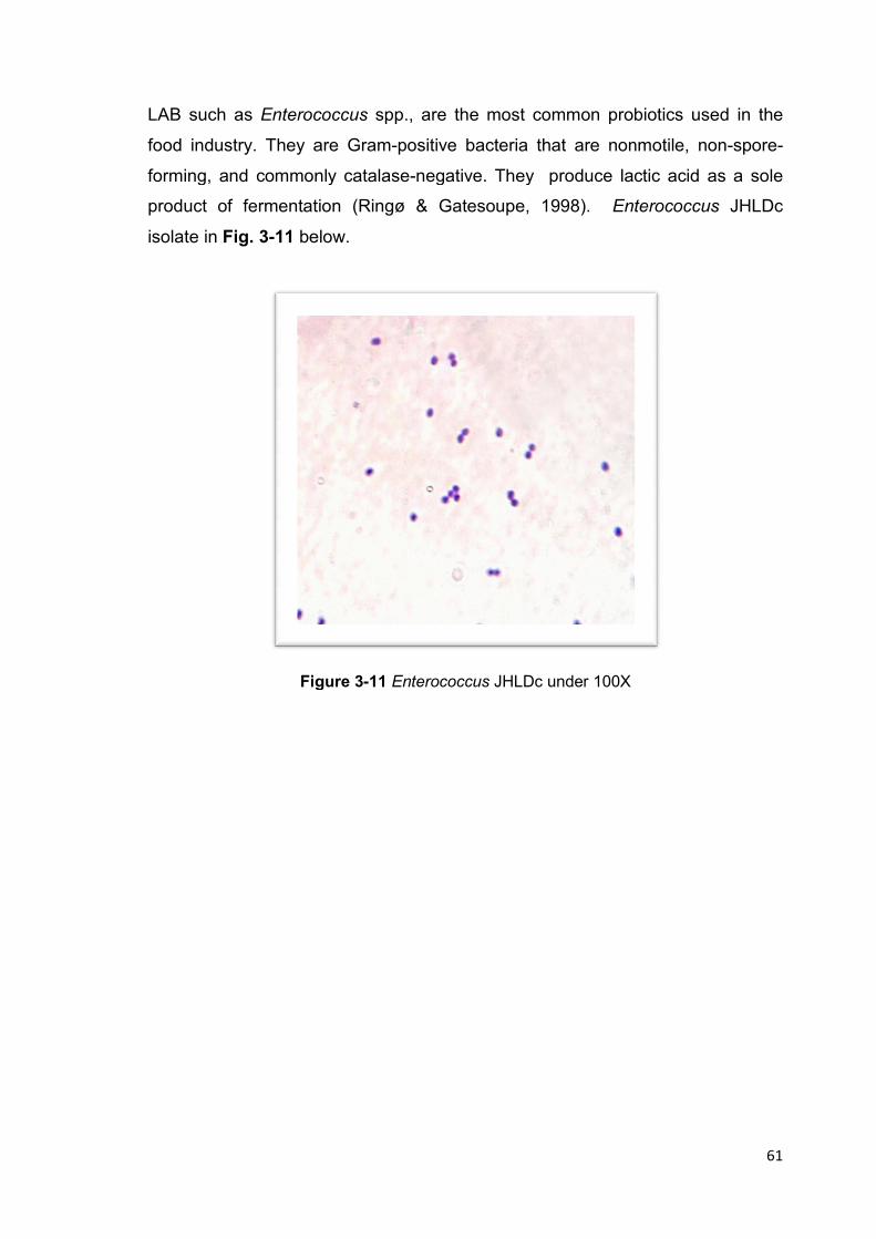
LAB such as Enterococcus
food industry. They are Gram
forming, and commonly catalase
product of fermentation
isolate in Fig. 3-11 below.
Figure
Enterococcus spp., are the most common probiotics used in the
food industry. They are Gram-positive bacteria that are nonmotile, non
forming, and commonly catalase-negative. They produce lactic acid as a sole
product of fermentation (Ringø & Gatesoupe, 1998). Enterococcus
below.
Figure 3-11 Enterococcus JHLDc under 100X
61
spp., are the most common probiotics used in the
positive bacteria that are nonmotile, non-spore-
negative. They produce lactic acid as a sole
Enterococcus JHLDc
62
Chapter 4
Enzyme Activity Assays and Quantitative Screening for Potential
Probiotics
63
4.1 Introduction
There has been an increasing number of evidence suggesting that the health
and survival of organisms grown in intensive farming systems could be improved
by modulating the intestinal microflora with probiotic microorganisms (Olafsen,
2001; Macey and Coyne, 2005). Enteric bacteria play an essential role in
abalone nutrition, by hydrolysing complex polysaccharide components of
macroalgae to simple polymers. The majority of enzyme activities in abalone gut
were microbial extracellular enzymes (Erasmus et al., 1997; ten Doeschate &
Coyne, 2008).
Tanaka et al. (2004) investigated the numbers of microorganisms in the gut of
abalone as a first step to understand their abilities and role in digestion. The gut
microflora of abalone Haliotis discus hannai varied with the growth stage, yet it
seemed to be stable in abalone older than 1 year (Tanaka & Sugimura, 2003).
Controlling microbial balance in the GIT of abalone by using probiotic
microorganisms can lead to high feeding efficiencies (Erasmus, 1996).
Increased levels of protease and amylase activity were observed in Haliotis
midae fed with a diet supplemented with the three probiotic microorganisms.
These enzyme activities resulted a significant improving in abalone growth rate
(Macey & Coyne, 2005).
A recent experiment using a single strain of the Gram-positive Bacillus sp. to
supplement the formulated feed of abalone Haliotis iris (paua) in OceaNZ Blue
(Bream Bay, Bay of Islands) did not show any significant increase in the growth
rate of juvenile paua [Bruno Cedat, (2009), personal communications].
The purpose of the experiments in this Chapter is to screen the potential
probiotic isolates obtained in Chapter 3 by determining their enzyme activities on
protein, starch, and alginate. The enzyme activities could provide a quantitative
basis for selection of the appropriate probiotic supplement to add to the
commercial feed in the feeding trial.
64
4.2 Material and methods
4.2.1 Bacterial cultures
The nine isolates obtained in Chapter 3 were streak plated in Marine Agar. The
cultures were incubated at 22oC for 48 hour, except cultures for alginate
hydrolysis which needed longer incubation.
4.2.2 Preparation of crude extracellular enzyme
One colony from the bacterial isolates was inoculated into 1% casein-marine
broth, 1% starch-marine broth, and 1% alginate-marine broth for protease,
amylase, and alginase assay, respectively. All cultures were incubated at 22oC
with shaking at 100rpm for 48 hour except for alginate cultures which required
longer incubation time (48-120 hour).
After incubation, the liquid broth cultures were transferred into sterile plastic
centrifuge tubes (40-50ml) with lids, and were centrifuged for 10 min at 10,000 x
g in a Sorval centrifuge at 40C to avoid denaturation of the enzymes. The cell-
free supernatants were collected with care using test tubes that have been put
on ice earlier, and were handled with gloves to optimize the conditions of
collecting the enzymes and avoid denaturation. All samples were filtered using a
0.22 µm millipore filter (Millex® syringe filter units, SIGMA-ALDRIC) to remove
any residual bacterial cells.
4.2.3 Enzyme Assays
4.2.3.1 Proteolytic Enzyme Assay
Azocasein assay was adopted to assess protein hydrolysis for bacterial isolates
from abalone gut. Azocasein assay was observed to be a highly sensitive assay for
detection of amino acids resulting from the hydrolysis of protein substrates
(Bisswanger, 2004) Azocasein assay was preferred over casein assay to minimize
the interfering elements experienced with casein. Azocasein is an artificial orange-
65
red, flaky shaped substance. It is a chemically modified protein, prepared by adding
sulphanamide groups to casein; hence, azocasein is a chromophore-labeled
protein. Azocasein solubilized in water with gentle heating appears clear, dark
orange in colour. The method is very resistant to interference and is consequently
appropriate for the detection of proteolytic activity in crude samples(Rowan &Buttle,
1994).
The azo group covalently bound to casein, which is liberated by proteolytic
cleavage, and their intense color is determined in the form of trichloroacetic acid-
soluble peptides after adding trichloroacetic acid (TCA). When azocasein is
subjected to proteolytic action, short peptides and amino acids are liberated from
the chain and stay in solution resulting in orange colour of the solution. The greater
the proteolytic activity the more intense the orange colour of the solution. Therefore
it is easy to be quantified in a spectrophotometer (Ultrospec™ 2100 pro UV/Visible)
at 340 nm.
In the present study, azocasein was purchased from (Sigma-Aldrich NZ Ltd /
Auckland 1030). It was prepared as a solution at 0.2% in 0.1M potassium
phosphate buffer (pH 8.0) with gentle warming to about 40-50oC. Reactions
were carried out in 1.9 ml micro-centrifuge tubes. A 0.5ml of azocasein was
mixed with 0.2 ml of protease sample (from culture supernatant) and incubated
at 25oC for 30 minutes. The reaction was stopped by adding 0.2ml of 10%
trichloroacetic acid. The mixture was centrifuged at room temperature for 5
minutes at 12,000 rpm. The supernatant was transferred to a quartz cuvette and
the absorbance was measured in a spectrophotometer at 340 nm, with two
replicates for each sample of cultural supernatants. A standard curve for Trypsin
amino acid was used to calibrate the amount of amino acids generated from
azocasein hydrolysis in µg/ml of product.
Proteolytic enzyme (protease) activity was determined as previously described
(Bisswanger, 2004; Iehata et al., 2009; Macey &Coyne, 2005) :
One unit of protease activity is defined as the amount of enzyme that gives an
increase in optical density at 340 nm of 0.1 in 30 min at 25oC.

Therefore, the absorbance reading for the fastest
minutes of reaction was divided by 0.1 and multiplied by 3 for 30 minutes to
calculate numbers of units of protease
enzyme activity as standard International Units/ml, values were multiplied by 5 to
account for the 0.2 ml volume of culture supernatant used in the assay.
4.2.1.2 Amylolytic Enzyme Assay
The amylolytic ability of
sugars, produced from starch hydrolysis, using the
colorimetric method (Wang, 198
aldehyde group and results in the oxidation of the aldehyde functional group in
the reducing sugars, such as glucose, to a carboxylic acid group.
Aldehyde group
DNS (3, 5-dinitrosalicylic acid) in the presence of Carboxyl group is reduced to 3
amino, 5-nitrosalicylic acid and can be detected by the intensity of the developed
color.
3, 5-dinitrosalicylic acid
Different color intensities
essential to calibrate for each sugar. In this study, reducing sugars were
calibrated with anhydrous glucose as glucose is the product of starch hydrolysis.
DNS reagent was prepared by adding 10g of
sodium sulfite, and 10g of sodium hydroxide pellets to one liter of deionized
water. Potassium sodium tartrate solution (Rochelle salt) was prepared at 40%
for colour stabilization. The starch substrate was prepared at 2%
starch powder (Acros Organic) was dissolved in a small amount of cold
deionized water and then boiled, after which phosphate buffer (pH 7.0) was
added to take the starch solution to the required final concentration of 1% w/v.
Therefore, the absorbance reading for the fastest rate of proteolysis
reaction was divided by 0.1 and multiplied by 3 for 30 minutes to
calculate numbers of units of protease enzyme for each isolate. To express the
enzyme activity as standard International Units/ml, values were multiplied by 5 to
account for the 0.2 ml volume of culture supernatant used in the assay.
4.2.1.2 Amylolytic Enzyme Assay
The amylolytic ability of the isolates was determined by assaying the reducing
sugars, produced from starch hydrolysis, using the Dinitrosalicylic acid (DNS)
(Wang, 1988). The DNS method detects the presence of an
aldehyde group and results in the oxidation of the aldehyde functional group in
the reducing sugars, such as glucose, to a carboxylic acid group.
Aldehyde group carboxyl group
salicylic acid) in the presence of Carboxyl group is reduced to 3
nitrosalicylic acid and can be detected by the intensity of the developed
dinitrosalicylic acid 3-amino, 5-nitrosalicylic acid
Different color intensities relate to different reducing sugars, therefore it is
essential to calibrate for each sugar. In this study, reducing sugars were
calibrated with anhydrous glucose as glucose is the product of starch hydrolysis.
DNS reagent was prepared by adding 10g of Dinitrosalicylic acid powder,
sodium sulfite, and 10g of sodium hydroxide pellets to one liter of deionized
Potassium sodium tartrate solution (Rochelle salt) was prepared at 40%
for colour stabilization. The starch substrate was prepared at 2%
starch powder (Acros Organic) was dissolved in a small amount of cold
deionized water and then boiled, after which phosphate buffer (pH 7.0) was
added to take the starch solution to the required final concentration of 1% w/v.
oxidation
reduction
66
of proteolysis in the first 10
reaction was divided by 0.1 and multiplied by 3 for 30 minutes to
enzyme for each isolate. To express the
enzyme activity as standard International Units/ml, values were multiplied by 5 to
account for the 0.2 ml volume of culture supernatant used in the assay.
the isolates was determined by assaying the reducing
Dinitrosalicylic acid (DNS)
The DNS method detects the presence of an
aldehyde group and results in the oxidation of the aldehyde functional group in
the reducing sugars, such as glucose, to a carboxylic acid group.
salicylic acid) in the presence of Carboxyl group is reduced to 3-
nitrosalicylic acid and can be detected by the intensity of the developed
nitrosalicylic acid
relate to different reducing sugars, therefore it is
essential to calibrate for each sugar. In this study, reducing sugars were
calibrated with anhydrous glucose as glucose is the product of starch hydrolysis.
trosalicylic acid powder, 0.5g of
sodium sulfite, and 10g of sodium hydroxide pellets to one liter of deionized
Potassium sodium tartrate solution (Rochelle salt) was prepared at 40%
for colour stabilization. The starch substrate was prepared at 2% w/v. Soluble
starch powder (Acros Organic) was dissolved in a small amount of cold
deionized water and then boiled, after which phosphate buffer (pH 7.0) was
added to take the starch solution to the required final concentration of 1% w/v.
67
The reaction mixture for the amylase assay was prepared by mixing the crude
enzyme with the starch solution at 1:1 ratio in screw-capped test tubes. The
tubes were incubated at 37 oC for 30 min. Two replicates were set up for each
isolate. A 3 ml sample was collected every 10 min to which 3 ml of DNS reagent
were added. The DNS-sample mixture was heated in a 90-100oC boiling water
bath for 10-15 min until red-brown colour has developed. One ml of Rochelle
salt solution was added, then the tubes were cooled using cold water. The
absorbance of the solution was determined in a spectrophotometer at 575 nm.
One international unit (IU) of amylase activity is defined as the amount of
enzyme that released 1 µmol product ( of reducing group) per min, as described
by Ghose (1987) and Bisswanger (2004):
1 IU = 1 µMole / min of librated hydrolysis product
4.2.1.3 Alginolytic Enzyme Assay
Determination of alginate hydrolysis in the bacterial isolates was conducted using
the same DNS colorimetric method above (Hu et al., 2006). Alginate is a linear
copolymer with homopolymeric blocks of (1-4)-linked β-D-mannuronate (M) and
its C-5 epimer α-L-guluronate (G) residues, respectively, covalenty linked
together in different sequences or blocks.
Alginate solutions contain a polysaccharide anion with a negative charge for
carboxylic acid group (d’Ayala et al., 2008). DNS (3, 5-dinitrosalicylic acid) in the
presence of carboxyl group can be reduced to 3-amino, 5-nitrosalicylic acid
which can be detected through measuring the developed color. Hence, the DNS
method for reducing sugar was employed to detect the alginate hydrolysis.
The three isolates observed to demonstrate alginate hydrolysis in Section
3.2.2.3.2 were subjected to alginate hydrolysis assay, namely, Vibrio JH1, the
fastest alginolytic isolate (1-2 day), Shewanella JH3, and Vibrio JH5 as two slow
alginolytic (3-5 day) isolates. Determination of the amount of reducing sugars in
each sample and the enzyme activity were calculated as in Section 4.2.1.2.

4.4 Results and Discussion
To assess the isolates for their activities, potential probiotics should meet one
more of the criteria shown in
Figure 4-1 The m
Screening the isolates for acid and bile salt tolerance
production was conducted in Chapter 3.
In the present Chapter the rest of screening was conducted through enzymatic
assays for protein, starch, and alginate hydrolysis.
acidtolerance
bile salt tolerance
4.4 Results and Discussion
To assess the isolates for their activities, potential probiotics should meet one
shown in Fig 4-1 below:
The main activities to assess the potential probiotic isolates
Screening the isolates for acid and bile salt tolerance, in addition
ion was conducted in Chapter 3.
In the present Chapter the rest of screening was conducted through enzymatic
assays for protein, starch, and alginate hydrolysis.
Selected Probiont
proteolytic activity
amylolytic activity
alginolytic activity
acid-production
acid-tolerance
bile salt tolerance
68
To assess the isolates for their activities, potential probiotics should meet one or
the potential probiotic isolates
in addition to acid
In the present Chapter the rest of screening was conducted through enzymatic

69
4.4.1 Proteolytic Enzyme Assays
Isolates
Amino acids
concentration (30 min)
µg/ml ± SE
Enzyme activity IU/ml ± SE
Exiguobacterium JHEb1 547 ± 10.8 128.3 ± 9
Bacillus JHBa2 182 ± 9 26.85 ± 1.6
Vibrio JH1 321.5 ± 6.5 62.4 ± 0.8
Vibrio JH2 34.7 ± 1.87 4.5 ± 0.4
Shewanella JH3 83 ± 14.6 3.75 ± 1.7
Vibrio JH4 630 ± 2.8 116 ± 8.5
Vibrio JH5 83.6 ± 0.37 10 ± 0.23
Table 4-1 Enzyme activity of proteolytic isolates
Results (Table 4-1 and Fig. 4-2) showed that Exiguobacterium JHEb1, Vibrio
JH4 and Vibrio JH1 had the highest proteolytic activity measured as IU/ml of
cultural supernatant; (128.3 ± 9), (116 ± 8.5), and (62.4 ± 0.8), respectively.
Vibrio JH5, Shewanella JH3 and Vibrio JH2 exhibited lower proteolytic enzyme
activity (10 ± 0.23), (3.75 ± 1.7), and (4.5 ± 0.4), respectively. The proteolytic
activity of Bacillus JHBa2 was mid-range (26.85 ± 1.6).
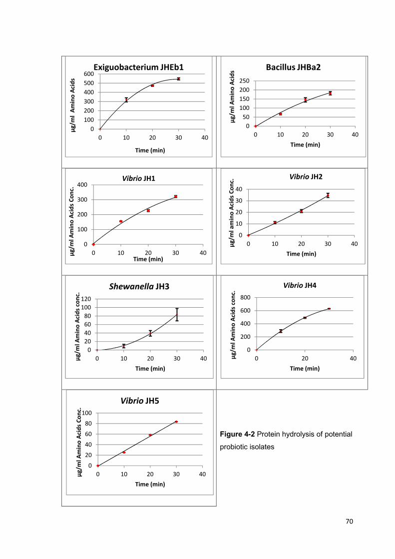
70
Figure 4-2 Protein hydrolysis of potential
probiotic isolates
0
100
200
300
400
500
600
0 10 20 30 40
µg
/ml
Am
ino
Aci
ds
Time (min)
Exiguobacterium JHEb1
0
50
100
150
200
250
0 10 20 30 40
µg
/ml
Am
ino
Aci
ds
Time (min)
Bacillus JHBa2
0
100
200
300
400
0 10 20 30 40µg
/ml
Am
ino
Aci
ds
Co
nc.
Time (min)
Vibrio JH1
0
10
20
30
40
0 10 20 30 40µg
/ml
am
ino
Aci
ds
Co
nc.
Time (min)
Vibrio JH2
0
20
40
60
80
100
120
0 10 20 30 40µg
/ml
Am
ino
Aci
ds
con
c.
Time (min)
Shewanella JH3
0
200
400
600
800
0 20 40µg
/ml
Am
ino
Aci
ds
con
c.
Time (min)
Vibrio JH4
0
20
40
60
80
100
0 10 20 30 40µg
/ml
Am
ino
Aci
ds
Co
nc.
Time (min)
Vibrio JH5
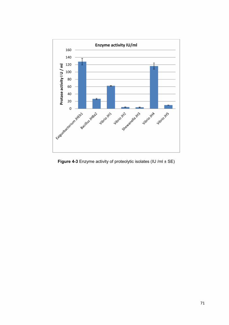
71
Figure 4-3 Enzyme activity of proteolytic isolates (IU /ml ± SE)
0
20
40
60
80
100
120
140
160
Pro
tase
act
ivit
y I
U /
ml
Enzyme activity IU/ml

72
4.4.2 Amylolytic Enzyme Assays
Isolates
Glucose concentration
(for first 15 min. of
hydrolysis) µ Mol± SE
Enzyme
activity
IU ±SE
enzyme activity
IU/ml ±SE
Exiguobacterium
JHEb1 7827 ± 55.7
521.8 ± 3.8
347.8 ± 2.5
Bacillus JHBa2 5984 ± 518
398.6 ± 34.6
265.7 ± 23
Vibrio JH1 4214 ± 418
281 ± 27.9
186.6 ± 18.7
Shewanella JH3 2839 ± 345
189 ± 23
126.0 ± 15.4
Vibrio JH4 3824 ± 72
255 ± 5
169.7 ± 3.3
Table 4-2 Enzyme activity of amylolytic isolates
The rates of starch hydrolysis are shown in Fig. 4-4. All the amylolytic isolates
exhibited high enzyme activity. Exiguobacterium JHEb1 was observed to have
the highest amylolytic activity of 347.8 ± 2.5, followed in rank by Bacillus JHBa2
(265.7 ± 23) and Vibrio JH1 (186.6 ± 18.7), Table 4-2, Shewanella JH3 had the
lowest activity of 126.0 ± 15.4

73
Figure 4-4 Starch hydrolysis of potential
probiotic isolates
0
2
4
6
8
10
0 10 20 30 40
mM
ole
Glu
cose
Co
nce
ntr
ati
on
Time min
Exiguobacterium JHEb1
0
2
4
6
8
10
12
14
16
18
0 10 20 30 40
mM
ole
Glu
cose
Co
nce
ntr
tio
n
Time min
Bacillus JHBa2
0
1
2
3
4
5
6
7
8
9
0 20 40
mM
ole
Glu
cose
co
nce
ntr
ati
on
Time min
Vibrio JH1
0
0.5
1
1.5
2
2.5
3
3.5
4
4.5
0 20 40
mM
ole
Glu
cose
Co
nce
ntr
ati
on
Time min
Vibrio JH3
0
1
2
3
4
5
0 20 40
mM
ole
Glu
cose
Co
nce
ntr
ati
on
Time min
Vibrio JH4
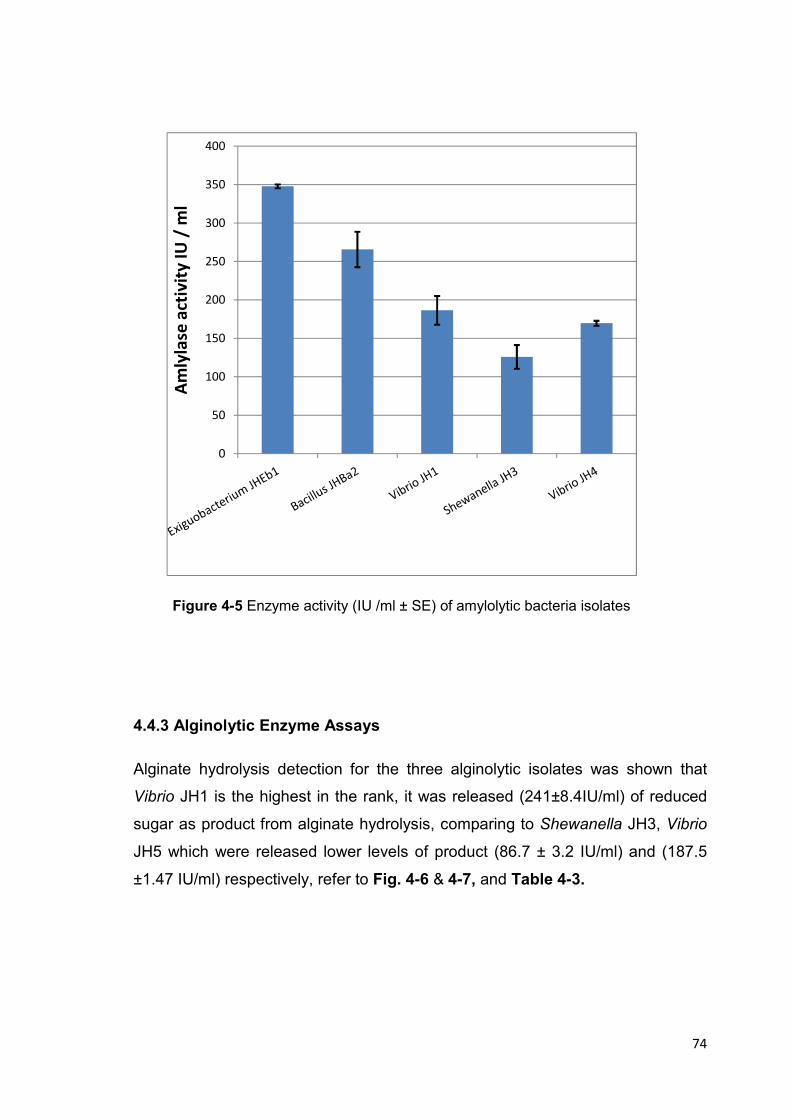
74
Figure 4-5 Enzyme activity (IU /ml ± SE) of amylolytic bacteria isolates
4.4.3 Alginolytic Enzyme Assays
Alginate hydrolysis detection for the three alginolytic isolates was shown that
Vibrio JH1 is the highest in the rank, it was released (241±8.4IU/ml) of reduced
sugar as product from alginate hydrolysis, comparing to Shewanella JH3, Vibrio
JH5 which were released lower levels of product (86.7 ± 3.2 IU/ml) and (187.5
±1.47 IU/ml) respectively, refer to Fig. 4-6 & 4-7, and Table 4-3.
0
50
100
150
200
250
300
350
400
Am
lyla
se a
ctiv
ity
IU
/ m
l

75
The rates of alginate hydrolysis in the three alginolytic isolates are shown in Fig.
4-6. The highest alginolytic activity was observed in Vibrio JH1 (241±8.4IU/ml),
Table 4.3, Fig. 4-7. Vibrio JH5 had only slightly lower activity than Vibrio JH 1 at
187 ± 1.47. Shewanella JH3 had the lowest alginolytic activity (86.7 ± 3.2).
Isolates
Reducing sugar
concentration (for first 15
min. of hydrolysis)
µ Mol± SE
Enzyme activity
IU ±SE
Specific Enzyme activity
IU/ml ±SE
Vibrio JH1
5422 ± 189
361.5 ± 12.6
241 ± 8.4
Shewanella
JH3
1950 ± 72
130 ± 4.8
86.7 ± 3.2
Vibrio JH5
4211 ± 33
280.7 ±2.2
187 ± 1.47
Table 4-3 Enzyme activity of alginolytic isolates

76
Figure 4-6 Alginate hydrolysis of potential
probiotic isolates
0
1
2
3
4
5
6
7
8
9
10
11
12
0 10 20 30 40
mM
ole
glu
cose
co
nce
ntr
ati
on
Time min
Vibrio JH1
0
0.5
1
1.5
2
2.5
3
3.5
4
4.5
0 10 20 30 40
mM
ole
Glu
cose
Co
nce
ntr
ati
on
Time min
Shewanella JH3
0
1
2
3
4
5
6
0 10 20 30 40
mM
ole
Glu
cose
co
nce
ntr
ati
on
Time min
Vibrio JH5

77
Figure 4-7 Enzyme activity (IU /ml± SE) of alginolytic isolates
The experiments in this Chapter were performed to provide a quantitative basis
for the selection of the most suitable probionts to be used in the feeding trial.
Enzyme activities from protein hydrolysis, starch hydrolysis and alginate
hydrolysis were calculated. Similar assessment of digestive enzyme activities
have been carried out in abalone and shrimps to evaluate the contribution of
bacteria introduced to the animals (Iehata et al., 2010; Macey &Coyne, 2005; ten
Doeschate & Coyne, 2008; Ziaei-Nejad et al., 2006)
The seven host-derived proteolytic isolates vary in their ability of protein
hydrolysis, the higher proteolytic activity measured as specific protease units was
observed by Exiguobacterium JHEb1 (128.3 ± 9 IU/ml), Vibrio JH4 (116 ± 8.5
IU/ml), and Vibrio JH1 (62.4 ± 0.8 IU/ml). They are more likely to be in the
shortlist of the candidates of potential proteolytic probiotics for feeding trial.
Unlike Bacillus JHBa2, Vibrio JH5, Shewanella JH3 and Vibrio JH2 that cannot
be included in the probiotic treatment of feeding trial for their low production of
proteolytic enzymes.
Out of the seven host-derived proteolytic isolates, Exiguobacterium JHEb1 Vibrio
JH4 and Vibrio JH1 would be short-listed as probiotic candidates to be used in
0
50
100
150
200
250
300
Vibrio JH1 Shewanella
JH3
Vibrio JH5
En
zym
e A
ctiv
ity
Fo
r a
lgin
ase
IU
/ml
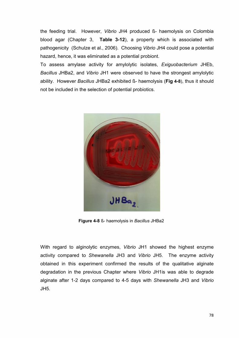
78
the feeding trial. However, Vibrio JH4 produced ß- haemolysis on Colombia
blood agar (Chapter 3, Table 3-12), a property which is associated with
pathogenicity (Schulze et al., 2006). Choosing Vibrio JH4 could pose a potential
hazard, hence, it was eliminated as a potential probiont.
To assess amylase activity for amylolytic isolates, Exiguobacterium JHEb,
Bacillus JHBa2, and Vibrio JH1 were observed to have the strongest amylolytic
ability. However Bacillus JHBa2 exhibited ß- haemolysis (Fig 4-8), thus it should
not be included in the selection of potential probiotics.
Figure 4-8 ß- haemolysis in Bacillus JHBa2
With regard to alginolytic enzymes, Vibrio JH1 showed the highest enzyme
activity compared to Shewanella JH3 and Vibrio JH5. The enzyme activity
obtained in this experiment confirmed the results of the qualitative alginate
degradation in the previous Chapter where Vibrio JH1is was able to degrade
alginate after 1-2 days compared to 4-5 days with Shewanella JH3 and Vibrio
JH5.

79
The proteolytic activity of Vibrio JH1 is similar to those of the probiotic isolates
used in H. midae (Macey & Coyne, 2005). Supplementation of the basal feed for
H. midae with proteolytic isolates resulted in a significant increase in protein
digestion and absorption within the intestine of abalone.
Vibrio JH1 also was the only isolate that exhibited fast rate of alginate hydrolysis,
and agarolysis (pits around colonies on Marine agar plate, Chapter 3). The
remaining two alginolytic isolates (Shewanella JH3, Vibrio JH5) could not be
included in the feeding trial because of their poor, slow ability to degrade
alginate. It is recommended to include isolate exhibiting fast rate of alginate
degradation in the supplementation of the feed for feeding trial to aid the
digestion of seaweed powder present in the commercial feed for abalone
(Section 5.2.3).
The basis of including Vibrio JH1 in the feed supplement for H. iris is similar to
that of Erasmus et al. (1997). It was suggested that the role of the enteric
bacteria of abalone in pre-degradation of alginate that supplied a more easily
digestible substrate for the host digestive enzymes. By decreasing the amount of
energy required for digestion, there would be more available energy that can be
utilized for growth. Similar results were obtained by ten Doeschate, V.E. Coyne
(2008) who reported that Pseudoalteromonas sp. strain C4 increased the
alginate lyase activity in the crop and stomach of H. midae abalone fed a kelp
diet supplemented with strain C4. The alginate lyase activity was significantly
greater compared to abalone fed a standard kelp diet. Erasmus et al. (1997)
proposed that bacteria resident in the GIT of H. midae assist in the digestion of
alginate, laminarin, agarose, carrageenan, and cellulose.
The screening experiment of bile salt tolerance; Exiguobacterium JHEb1, Vibrio
JH1, and Enterococcus JHLDc were the most tolerant isolates with no potential
pathogenicity (Table 3-6and 3-7, Chapter 3). Screening for acidity tolerance
showed that all isolates can tolerate acidity within the range 2-5 pH, and all
isolates were able to tolerate to 1.5 pH except Vibrio JH2, and Vibrio JH5 (Table
3-8).

80
4.5 Conclusion
The goal of quantitative screening was to follow up on those isolates of interest
to develop mixtures of probiotic bacteria for application to the feed of the abalone
(paua) in the feeding trial. The most promising isolates according to quantitative
enzyme screening are:
Exiguobacterium JHEb1; a proteolytic, amylolytic, lipolytic, bile salts tolerant, and
high acidity tolerant isolate.
Vibrio JH1; a proteolytic, amylolytic, fast alginolytic, agarolytic, high bile salts
tolerant, and high acidity tolerant isolate.
Enterococcus JHLDc; a high acid producing, high bile salts and acidity tolerant
isolate.
This study proposes to a 2-probiotic-supplemented feed that include
Exiguobacterium JHEb1 and Vibrio JH1.
A 3-probiotic-supplemented feed should include Exiguobacterium JHEb1, Vibrio
JH1, and Enterococcus JHLDc.
81
Chapter 5
Feeding Trials Using Isolates of Probiotics
82
5.1 Introduction
Growth rate improvement in abalone may be achieved by the use of probiotic
microorganisms. through these mechanisms: increasing the nutrients readily
absorbed in the gastrointestinal tract; increasing the amount of digestive
enzymes in the gut; or through use of the bacterial supplement as an additional
nutrient source (ten Doeschate & Coyne, 2008).
The objective of this study was to determine if growth of abalone could be
improved with application of the probiotics selected in the previous chapter.
The experiment was designed to incorporate probiotic bacteria with nutrient-
degrading properties so that they could improve the digestion process inside the
abalone gut through protein degradation and polysaccharide (starch and
alginate) degradation that could result in increased shell length and weight of
juvenile abalone.
The hypothesis of the study is the probiotic bacteria supplemented to the feed
help the abalone benefit from the feed more than the unsupplemented feed
resulting in lower concentration of protein, carbohydrate, and lipid in the faeces
of the host.
Two multi-strain probiotic combinations were formulated as a conglomerate of 2
or 3 potential probiotic strains. This study also aimed to compare the effect of the
two probiotic mixes on abalone growth.
The methods of administration of probiotic strains to aquaculture animals are
several (Geovanny et al., 2007) in which the appropriate application with
adequate concentration remains a critical issue. In this study, the application of
the selected probionts was through supplementing them into the commercial
feed. For effective and practical application of the probiotic mixes in the feeding
trial, the following points were considered:
• Maintenance of the desired high concentration of viable bacteria.
• Retention of probiotic feed at the bottom of the tank during the feeding
periods.

83
• Distinct difference between abalone faeces and probiotic feed for ease of
collection of faecal sample for biochemical analyses.
5.2 Materials and Methods
5.2.1 Experimental Animals
Juvenile abalone, 20-30mm in shell length, were obtained from OceaNZ Blue Ltd
(Paua farm), Ruakaka, northern New Zealand. The abalone were kept moist
during transport from the paua farm to the aquaculture laboratory in AUT
University by placing them in a cool box containing an ice bag. The ice bag was
wrapped with thick pad of tissue to protect abalone from freezing and at the
same time to maintain cold environment to allow survival during transport.
The abalone samples were tagged with water-proof tags with numbers to label
each individual. The numbers of each individual and their shell length and weight
measurement were record. The animals were placed into the tanks (20) abalone
for each, and were acclimatized at aquaculture laboratory for one week prior to
the experiment. During acclimatization time, they were fed commercial feed
(AbMax 16).
5.2.2 Cultivation of Potential Probionts
The selected isolates from screening experiments were: Exiguobacterium
JHEb1, Vibrio JH1, and Enterococcus JHLDc, (Section 4.5). The isolates were
propagated in enriched media. Exiguobacterium JHEb1 was propagated in
Marine broth supplemented with 1% yeast extract. The culture was incubated in
a rotary shaker (100 rpm) at 22oC for 48 hr. Vibrio JH1 was grown in Marine
broth supplemented with 0.5% glucose in a rotary shaker (100 rpm) at 22oC for
48 hr. Enterococcus JHLDc was propagated anaerobically in MRS broth
containing 2% NaCl for 48 hr at 22oC.

84
5.2.3 Abalone Commercial Feed
The commercial feed, AbMax 16, used in this study was manufactured by E. N.
Hutchinson Ltd (Auckland, New Zealand). AbMax 16 was obtained from OceaNZ
Blue Ltd and was maintained in AUT laboratory at 4oC until required. The feed
existed as dry, oval-shaped pellets (Fig. 5-6). The manufacturer ingredients and
contents were listed as:
Wheat Flour 35%, Fish Meal (Milled NZ) 20%, Defatted Soy Flour & Isolate
13%, Seaweed Powder, Tapioca Starch, Propylene Glycol, Propylene Glycol,
Sugar, Yeast – Saccharomyces, Wheat Gluten, Fish Oil (mixed), Phosphoric
Acid (85%), Carrageenan, Lecithin Powder, Dicalcium Phosphate, Potassium
Sorbate, Vitamin Mineral Mix (Abalone), Betaine, Stay-C (25%), and Vitamin E.
Some components percentages are not given due to commercial sensitivity.
Proximate analyses (i.e., protein, carbohydrates, and lipid) were conducted on
AbMax to determine its general composition.
5.2.4 Preparation of Probiotic- supplemented Feed
For effective delivery of the probionts and maintenance of adequate probiotic
concentration three methods of application to the feed have been tested before
the appropriate application was adopted. Firstly, preparation of feed cakes
containing bacteria with agar, secondly, feed cakes containing immobilized
bacteria with agar, and thirdly, application of bacterial broth added directly to the
feed pellets.
In a pilot feeding trial, which conducted to check the appropriate application of
feed and has not been mentioned in full details in this thesis, two options of feed
cakes have been tested; feed cakes containing bacteria with agar at different
concentrations (1.5 % w/v, 2% w/v, 2.5% w/v) and different amounts of feed
pellets. The feed cakes were not stable and disintegrated into small pieces. The
pieces of feed and broken agar were mixed with the slimy feces of abalone and
made it difficult to separate the fecal matter for biochemical analysis. The same
difficulty occurred with the second option of feed cakes which contained alginate
beads of immobilized probiotic bacteria. However, the immobilized bacteria were
beyond the reach of the abalone, for they had to graze onto the agar and the
85
feed pellets with immobilized beads this was not ideal for erratic feeders like
abalone, where they were either fed on agar which was poor in nutrients or
stopped eating. Application of broth cultures of the isolates added directly to the
feed pellets was chosen for the feeding trial.
Three to five colonies of Exiguobacterium JHba1 grown in Marine Agar were
inoculated into 50 ml of modified Marine Broth containing an additional 1% of
yeast extract. Vibrio JH1 (3-5 colonies) from Marine Agar was transferred to 50
ml of modified Marine Broth containing 1% yeast extract and 0.5% glucose.
Three to five colonies of Enterococcus JHLDc from MRS agar were transferred
to MRS broth containing 2% NaCl. All cultures were incubated at 22oC in a
rotary shaker at 100 rpm.
Viable count was determined using spread plate count at day 0 and day 2. This
enumeration was necessary to determine if a viable concentration between 106
CFU /g to 108 CFU/g is maintained in the feed (Gatesoupe, 1999; Macey &
Coyne, 2006b; ten Doeschate & Coyne, 2008).
The cultures were transferred into sterile (50 ml) centrifuge tubes with caps.
Centrifugation was conducted for 10 minutes and bacterial pellets (probiotic
biomass) were collected and re-suspended in (25ml) sterile marine broth. For 2-
probiotic supplemented feed, one bacterial pellet of Gram- positive isolate
(Exiguobacterium JHBa1), two bacterial pellets of Gram- negative isolate (Vibrio
JH1) were re-suspended in (25ml) of sterile marine broth, Fig. 5-1.

86
Figure 5-1 Preparation of 2-probiotic supplemented feed
For 3-probiotic supplemented feed, one bacterial pellet of Exiguobacterium
JHBa1, two bacterial pellets of (Vibrio JH1, and two bacterial pellets of lactic acid
bacteria (Enterococcus JHLDc) were included in 25ml of sterile marine broth.
The rationale for this was Exiguobacterium JHBa1 is a very competitive bacteria
and from previous experiments of viable counts of mixed culture of three isolates,
it showed more growth than the two other isolates (Vibrio JH1 & Enterococcus
JHLDc), Fig. 5-2.
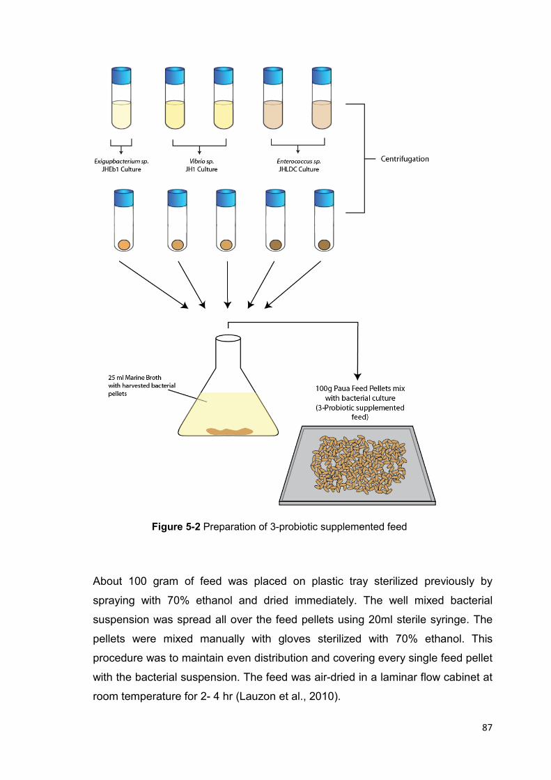
87
Figure 5-2 Preparation of 3-probiotic supplemented feed
About 100 gram of feed was placed on plastic tray sterilized previously by
spraying with 70% ethanol and dried immediately. The well mixed bacterial
suspension was spread all over the feed pellets using 20ml sterile syringe. The
pellets were mixed manually with gloves sterilized with 70% ethanol. This
procedure was to maintain even distribution and covering every single feed pellet
with the bacterial suspension. The feed was air-dried in a laminar flow cabinet at
room temperature for 2- 4 hr (Lauzon et al., 2010).

88
Storage for the prepared probiotics- supplemented feed was in 4oC in covered
glassware for no longer than one week to maintain probiotic viability
(Gatesoupe, 1999; Wang & Li, 2008)
5.2.5 Measurements of Weight and Shell Length
Individual abalone weights were taken using a top pan balance (METTLER
TOLEDO PB602-S, USA). First, individual abalone were blotted dry using tissue
paper, and then they were placed on the balance pan. Measurements were
taken to the nearest 0.01g at the start and end of the feeding trial to calculate
weight gain and daily weight gain:
Weight gain/day = (final weight - initial weight) / 60 days
Shell length measurements were taken as maximum diameter (longest axis) of
the shell, using digital vernier calipers (Mitutoyo, Japan made) to the nearest
0.01mm. Initial and final shell length measurements for each abalone, were
recorded to use for the calculation of daily shell growth as mentioned by (Olmos
et al., 2011):
Shell growth/day = (final shell length - initial shell length) / 60 days
Figure 5-3 Abalone weighing and shell length measuring
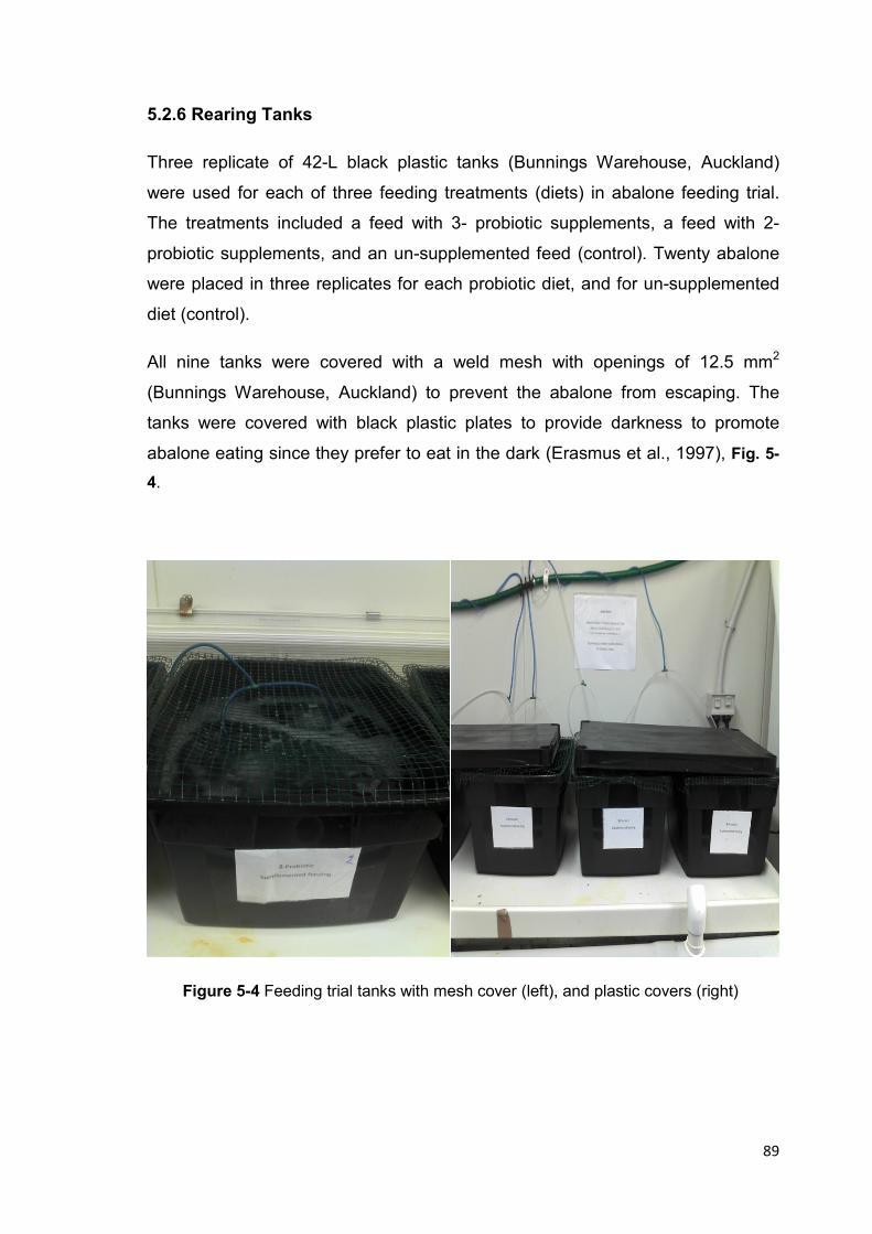
89
5.2.6 Rearing Tanks
Three replicate of 42-L black plastic tanks (Bunnings Warehouse, Auckland)
were used for each of three feeding treatments (diets) in abalone feeding trial.
The treatments included a feed with 3- probiotic supplements, a feed with 2-
probiotic supplements, and an un-supplemented feed (control). Twenty abalone
were placed in three replicates for each probiotic diet, and for un-supplemented
diet (control).
All nine tanks were covered with a weld mesh with openings of 12.5 mm2
(Bunnings Warehouse, Auckland) to prevent the abalone from escaping. The
tanks were covered with black plastic plates to provide darkness to promote
abalone eating since they prefer to eat in the dark (Erasmus et al., 1997), Fig. 5-
4.
Figure 5-4 Feeding trial tanks with mesh cover (left), and plastic covers (right)

90
5.2.7 Water System of Feeding Trial
The feeding trial was carried out at the AUT aquaculture laboratory. A static
water system was employed to maintain the animals with seawater collected
from Okahu Bay, Auckland. Immediately after collection, the seawater was
filtered via (0.2 µm) filtration system and sterilized with a UV system. The sterile
seawater was stored in a 2000 litre storage tank until needed within a week.
Abalone were kept at a constant temperature of 16-16.9oC in the aquaculture
laboratory.
5.2.8 Feeding Trial
The trial period was for 2 months that started in October and ended December
2011. A total of 180 juvenile abalone were starved for 2-3 days in order to
guarantee eating to full satisfaction in the start of the feeding trial. After
starvation, the abalone were fed probiotic-supplemented feed (3 -combination, 2
-combination supplemented feed) and commercial feed for control group, all in
three replications. The protocol of the feeding trail is outlined in Fig. 5-5.
Feeding abalone was conducted once a day, and according to the farm
instructions with regard to feed rations, which depend on temperature. At 16-
17°C, abalone of 25mm size usually feed at around 1.1% of body weight [Dr.
Rodney Roberts (personal communication)].

91
Figure 5-5 Protocol of feeding trial
Cleaning the tanks daily from uneaten feed pellet and faeces was done before
applying fresh feed. Samples of feces had been collected for the last ten days of
the trial. The relatively long period is referred to the nature of the feces which is
slimy with thin consistency. Biochemical analysis of protein, lipid, carbohydrates,
moisture, and ash required at least 2-3g of faeces to be collected, Fig. 5-6.
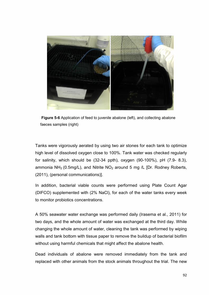
92
Figure 5-6 Application of feed to juvenile abalone (left), and collecting abalone
faeces samples (right)
Tanks were vigorously aerated by using two air stones for each tank to optimize
high level of dissolved oxygen close to 100%. Tank water was checked regularly
for salinity, which should be (32-34 ppth), oxygen (90-100%), pH (7.9- 8.3),
ammonia NH3 (0.5mg/L), and Nitrite NO2 around 5 mg /L [Dr. Rodney Roberts,
(2011), (personal communications)].
In addition, bacterial viable counts were performed using Plate Count Agar
(DIFCO) supplemented with (2% NaCl), for each of the water tanks every week
to monitor probiotics concentrations.
A 50% seawater water exchange was performed daily (Irasema et al., 2011) for
two days, and the whole amount of water was exchanged at the third day. While
changing the whole amount of water, cleaning the tank was performed by wiping
walls and tank bottom with tissue paper to remove the buildup of bacterial biofilm
without using harmful chemicals that might affect the abalone health.
Dead individuals of abalone were removed immediately from the tank and
replaced with other animals from the stock animals throughout the trial. The new
93
individuals were not included with the measurements of changes in growth. They
were kept in the tanks to maintain the same number of abalone in all tanks.
At the end of the trial, mortality percentage was calculated and comparison
between treatment groups was evaluated.
5.2.8.1 Biochemical Analysis
Samples of water from the bottom of each tank were placed in glass bakers, and
then the faecal materials were separated from un-eaten feed pellets by collecting
the upper layer of water. The collected samples were centrifuged and were
dehydrated in an oven (80-100oC) for 1 hr. The dried samples were grinded
using a mortar and pestle to obtain fine powder. Analyses of total protein, lipid,
and carbohydrate were conducted in AUT chemistry laboratory.
5.2.8.2 Protein analysis
Total protein for the abalone feed and faecal matter samples were estimated with
the Kjeldahl method for total nitrogen (AOAC, 2005). The Kjeldahl method
assumes that each gram of protein contains (0.16g) of nitrogen, which is
equivalent to the factor 6.25. Consequently, total protein can be obtained by
multiplying the nitrogen concentration by 6.25.
The method includes three main processes:
1) Digestion of the samples was conducted using sulphuric acid which converts
protein to ammonia. Approximately 0.5g of each sample was placed in a 300ml
Kjeldahl digestion tube. Then, 10ml of concentrated sulphuric acid (98%) was
added. Tubes were then inserted into a digestion oven (420oC). After about 45-
60 minutes, hydrogen peroxide was added. Decomposition of samples was
completed when the solutions became colorless compared to the initially dark
color. Then, 50ml of deionized water were added.
2) Distillation with 35% sodium hydroxide to liberate ammonia from the mixture was
performed, followed by distillation using a semi-automatic distillation unit, (UDK
139, VELP SCIENTIFICA). Digested materials were distilled with 25ml Boric acid
94
solution (4%). The final solutions were collected in 250ml Erlenmeyer flasks.
Then, ten drops of green methyl indicator were added.
Titration with 0.1mol hydrochloric acid was used to quantify the amount of
ammonia in the solution. Titration was stopped when the solution turned red. The
results were recorded for calculations. Blank tubes were treated in the same
manner as described above, but without the protein samples. The percent of total
protein was determinate using the equation below:
Total protein % = 6.25 x14.01 x (ml titrant – ml blank) x (0.1mol HCl) x 100
Sample weight (g) 1000
5.2.8.3 Lipid analysis
A modified Bligh and Dyer (1959) extraction method was used to determine total
lipid in feed and abalone faeces. Three replicates were prepared by blending the
dried faeces samples and feed pellet using a blender. Approximately 0.5g of
each powder sample was weighed using an analytical balance, capable of
weighing to 0.0001g. Then, the samples were put into 15 ml centrifuge tubes and
rehydrated with 2 ml deionized water. Three (3) ml of a mixture of two solvents
(methanol and chloroform) at 2:1 ratio were added into the tubes with the
samples and shaken to obtain a well homogenized mixture.
Additional chloroform (1 ml) was added to the tubes and mixed by shaking for
about 10 minutes and then centrifuge for 10 minutes at 2000 rpm. Two phases of
water and chloroform containing lipids were shown separated by the sample
protein disc. Due to the high density of the chloroform, it was observed as yellow
lower phase.
The lower chloroform layer was collected using a Pasteur pipette and transferred
into clean glass tubes, which have been pre weighed. The tubes with the
samples were reweighed after collecting chloroform mixture.
Tubes that contained the chloroform layer were exposed to the nitrogen gas flow
for a few minutes to evaporate the chloroform to dryness and leaving the lipid

95
layer on the tubes wall. The tubes were reweighed to determine the weight of the
lipid extract alone. The percent of total lipids was determinate using the equation
below:
Total lipid % = Lipid extract (g) x 100
Lipid in chloroform solvent (g)
5.2.8.4 Carbohydrate analysis
According to (Porter &Earl, 1990), total contents of carbohydrates in both feed
and faecal matter were estimated by difference. The sum of percent protein, lipid,
ash, and moisture was subtracted from 100 to calculate the total percent of
carbohydrates as in the equation:
Total estimated carbohydrates % = 100 - [%protein +% lipid +% moisture + %ash]
5.2.8.5 Moisture analysis
The moisture content of the samples was determined by taking the loss in weight
(%) after oven drying at (100oC) for 3 hours (De Knegt & Brink, 1998), according
to the equation described by (McClements, 2003) as follows:
Total moisture % = M (initial) – M (dried) X 100
M (initial)

96
5.2.8.6 Ash analysis
A modified (AOAC, 1970) method was used to determine the ash content in the
feed , and faecal matter samples. The samples were heated in a furnace at
520°C for approximately 2 hr. All samples with three replicates were weighed as:
Weight of porcelain crucible and about 0.5g of dried sample and Weight of
crucible with the sample after burning (ashing). Then, samples were cooled to
room temperature in a dessicator. Percentage of ash was determined using the
equation below:
Total ash % = M (ash) X 100
M (dried sample)
5.2.9 Statistical analyses
Collected data for biochemical analysis, growth changes (biomass and shell
length), and mortality rate were analyzed using an SPSS statistics program (IBM
SPSS version 20). One-way analyses of variances (ANOVA), and Tukey’s post
hoc tests at p-value = 0.05 were conducted to determine the differences between
the means of the three groups of study (control, 2-probiotics, and 3-probiotics).
Percentage of growth improvement was calculated as follows:
Improvement in shell length % = (P - C) / C X 100
Improvement in biomass % = (P - C) / C X 100 (P; probiotic diet, C; control diet)
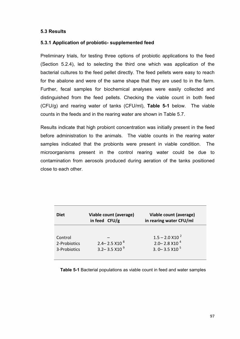
97
5.3 Results
5.3.1 Application of probiotic- supplemented feed
Preliminary trials, for testing three options of probiotic applications to the feed
(Section 5.2.4), led to selecting the third one which was application of the
bacterial cultures to the feed pellet directly. The feed pellets were easy to reach
for the abalone and were of the same shape that they are used to in the farm.
Further, fecal samples for biochemical analyses were easily collected and
distinguished from the feed pellets. Checking the viable count in both feed
(CFU/g) and rearing water of tanks (CFU/ml), Table 5-1 below. The viable
counts in the feeds and in the rearing water are shown in Table 5.7.
Results indicate that high probiont concentration was initially present in the feed
before administration to the animals. The viable counts in the rearing water
samples indicated that the probionts were present in viable condition. The
microorganisms present in the control rearing water could be due to
contamination from aerosols produced during aeration of the tanks positioned
close to each other.
Diet Viable count (average) Viable count (average)
in feed CFU/g in rearing water CFU/ml
Control – 1.5 – 2.0 X10 2
2-Probiotics 2.4– 2.5 X10 8 2.0– 2.8 X10
4
3-Probiotics 3.2– 3.5 X10 9 3. 0– 3.5 X10
5
Table 5-1 Bacterial populations as viable count in feed and water samples
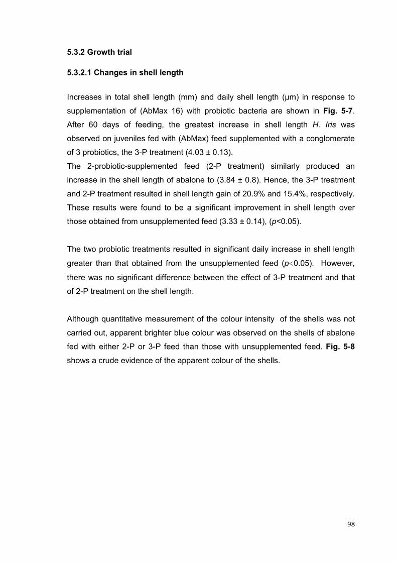
98
5.3.2 Growth trial
5.3.2.1 Changes in shell length
Increases in total shell length (mm) and daily shell length (µm) in response to
supplementation of (AbMax 16) with probiotic bacteria are shown in Fig. 5-7.
After 60 days of feeding, the greatest increase in shell length H. Iris was
observed on juveniles fed with (AbMax) feed supplemented with a conglomerate
of 3 probiotics, the 3-P treatment (4.03 ± 0.13).
The 2-probiotic-supplemented feed (2-P treatment) similarly produced an
increase in the shell length of abalone to (3.84 ± 0.8). Hence, the 3-P treatment
and 2-P treatment resulted in shell length gain of 20.9% and 15.4%, respectively.
These results were found to be a significant improvement in shell length over
those obtained from unsupplemented feed (3.33 ± 0.14), (p<0.05).
The two probiotic treatments resulted in significant daily increase in shell length
greater than that obtained from the unsupplemented feed (p<0.05). However,
there was no significant difference between the effect of 3-P treatment and that
of 2-P treatment on the shell length.
Although quantitative measurement of the colour intensity of the shells was not
carried out, apparent brighter blue colour was observed on the shells of abalone
fed with either 2-P or 3-P feed than those with unsupplemented feed. Fig. 5-8
shows a crude evidence of the apparent colour of the shells.

Figure 5-7 Shell length increase and daily shell length increase
(■) and (O) indicate a significant difference (ANOVA, Tukey post
Diet Shell length gain
(mm) (µm) (g
Control 3.33 ±0.14
2-Probiotics 3.84 ± 0.
3-Probiotics 4.03 ± 0.
Table 5-2 Growth data and mortality for three groups; means
Confidence Interval)
0
0.5
1
1.5
2
2.5
3
3.5
4
4.5
Sh
ell
le
ng
th i
ncr
ea
se (
mm
) /6
0 d
ay
s
Shell Gain
Shell length increase and daily shell length increase, different number
indicate a significant difference (ANOVA, Tukey post-hoc,
Shell length gain Shell length/day Weight gain Weight gain/day Mortality %
(mm) (µm) (g) (mg)
0.14 55.57 ±2.41 0.36 ± 0.25 5.98 ± 0.4
± 0.8 64.05± 1.29 0.39 ± 0.03 6.55 ± 0.06
4.03 ± 0.13 67.14 ± 2.16 0.43 ± 0.01 7.12 ± 0.15
Growth data and mortality for three groups; means ± SE differences
Confidence Interval)
■
■■
■■
O
OOOO
Shell Gain Sh. Gain/Day
99
ifferent numbers of
hoc, p< 0.05)
gain/day Mortality %
0.4 16.67± 3.33
06 3.33 ±0.33
15 3.33 ± 0.66
± SE differences (95%
0.0
5.0
10.0
15.0
20.0
25.0
30.0
35.0
40.0
45.0
50.0
55.0
60.0
65.0
70.0
75.0
she
ll len
gth
incre
ase
(µm
)/da
y
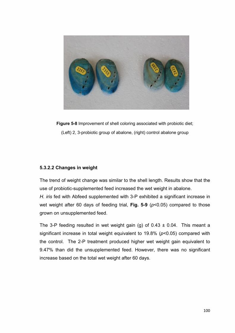
100
Figure 5-8 Improvement of shell coloring associated with probiotic diet;
(Left) 2, 3-probiotic group of abalone, (right) control abalone group
5.3.2.2 Changes in weight
The trend of weight change was similar to the shell length. Results show that the
use of probiotic-supplemented feed increased the wet weight in abalone.
H. iris fed with Abfeed supplemented with 3-P exhibited a significant increase in
wet weight after 60 days of feeding trial, Fig. 5-9 (p<0.05) compared to those
grown on unsupplemented feed.
The 3-P feeding resulted in wet weight gain (g) of 0.43 ± 0.04. This meant a
significant increase in total weight equivalent to 19.8% (p<0.05) compared with
the control. The 2-P treatment produced higher wet weight gain equivalent to
9.47% than did the unsupplemented feed. However, there was no significant
increase based on the total wet weight after 60 days.
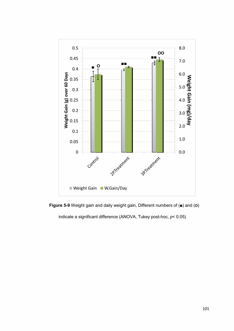
Figure 5-9 Weight gain and daily weight gain, Different number
indicate a significant difference (ANOVA, Tukey post
0
0.05
0.1
0.15
0.2
0.25
0.3
0.35
0.4
0.45
0.5
We
igh
t G
ain
(g
) o
ve
r 6
0 D
ay
s
Weight Gain
Weight gain and daily weight gain, Different number
a significant difference (ANOVA, Tukey post-hoc,
■■■
■■
O
OO
Weight Gain W.Gain/Day
101
Weight gain and daily weight gain, Different numbers of (■) and (O)
hoc, p< 0.05)
0.0
1.0
2.0
3.0
4.0
5.0
6.0
7.0
8.0W
eig
ht G
ain
(mg
)/da
y
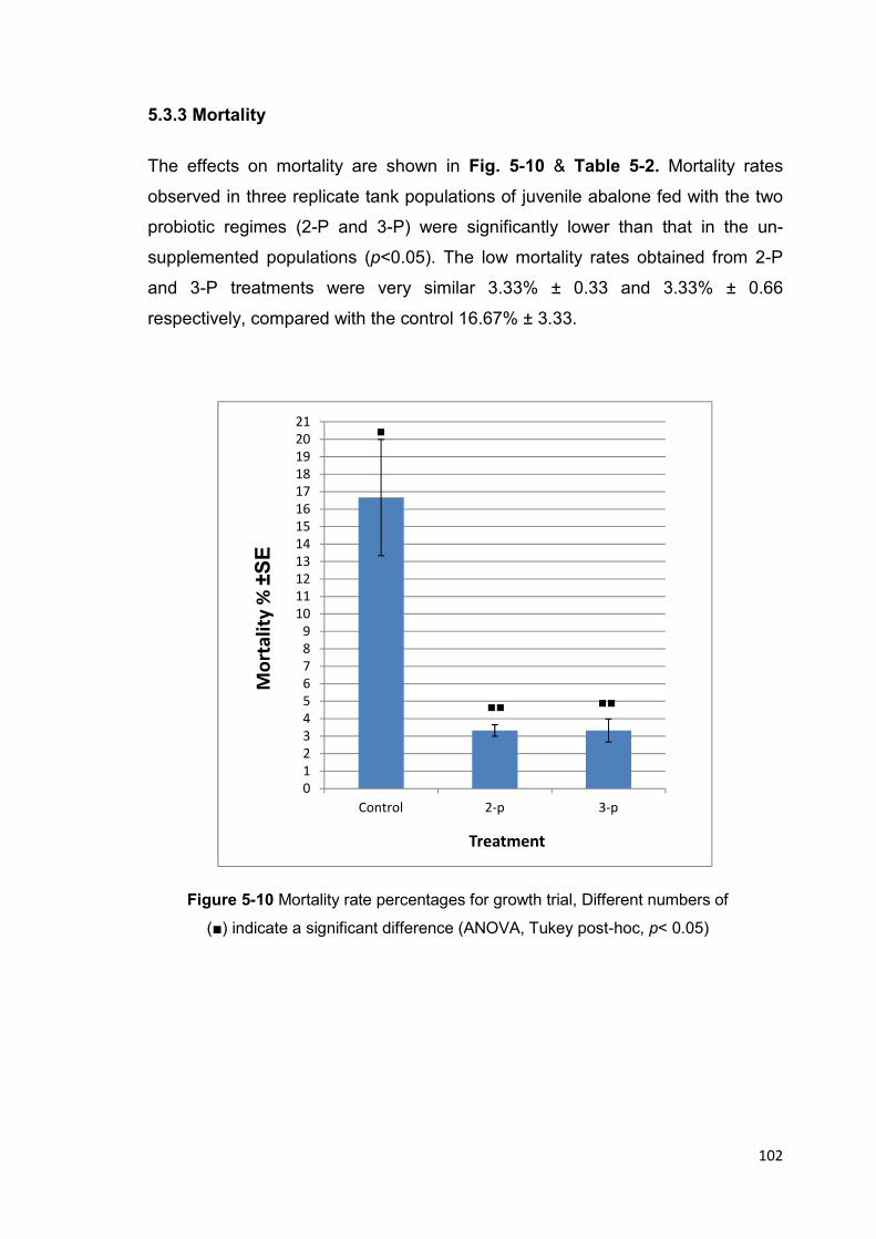
102
5.3.3 Mortality
The effects on mortality are shown in Fig. 5-10 & Table 5-2. Mortality rates
observed in three replicate tank populations of juvenile abalone fed with the two
probiotic regimes (2-P and 3-P) were significantly lower than that in the un-
supplemented populations (p<0.05). The low mortality rates obtained from 2-P
and 3-P treatments were very similar 3.33% ± 0.33 and 3.33% ± 0.66
respectively, compared with the control 16.67% ± 3.33.
Figure 5-10 Mortality rate percentages for growth trial, Different numbers of
(■) indicate a significant difference (ANOVA, Tukey post-hoc, p< 0.05)
■
■■ ■■
0
1
2
3
4
5
6
7
8
9
10
11
12
13
14
15
16
17
18
19
20
21
Control 2-p 3-p
Mo
rta
lity
% ±
SE
Treatment

103
5.3.4 Compositions of Growth Trial Samples
Faecal samples taken in the last 10 days of feeding trial showed that there was
an apparent decrease in crude protein, crude lipid, and crude carbohydrate from
the two probiotic groups (2-P and 3-P) compared to those from the control group
(Fig. 5-11; Table 5-3). Further, there was no statistical difference between the
faecal components in the 2-P and 3-P treatments. Interestingly, though, the
protein level in the faeces produced from both 2-P and 3-P (9.01 ± 0.25 and 7.96
± 0.26, respectively) were significantly lower than those from unsupplemented
feed 11.42 ± 0.83, (p>0.05).
Figure 5-11 Abalone faeces compositions: Crude carbohydrate, Crude protein, and Crude
lipid different numbers of (O), (x) or (■) indicate a significant difference (ANOVA, Tukey
post-hoc, p< 0.05)
x
xx
O
O O
■
■■
■■
0
1
2
3
4
5
6
7
8
9
10
11
12
13
0
5
10
15
20
25
30
35
40
45
50
55
60
Pro
tein
& L
ipid
%
Ca
rbo
hy
dra
te %
Carbohydrates Lipid Protein

104
Diet Protein% Lipid % Carbohydrate%
Control 11.42 ± 0.83 4.44 ± 0.55 50.52 ± 2.62
2-Probiotics 9.01 ± 0.25 3.32 ± 0.14 39.26 ± 3.21
3-Probiotics 7.96 ± 0.26 3.24 ± 0.1 39.81 ± 2.55
Feed 33.59 ± 0.89 4.47 ± 1.3 42.93 ± 4.46
Table 5-3 Compositions Percentage of faecal matter and commercial feed
5.4 Discussion
A 60-day growth experiment was conducted to identify the optimum probiotic
feed developed using a combination of potential probiotic bacteria for the
juveniles of H. iris (paua).
Results have shown that an increase in growth rate was possible with the
supplementation of feed with live, beneficial microorganisms. The highest growth
improvement in terms of weight and shell length (19.8% and 20.9%, respectively,
at p<0.05) was obtained from feeding with 3-P supplemented feed. The 3-P
conglomerate consisted of 3 putative probiotic bacteria Exiguobacterium JHEb1,
Vibrio JH1, and Enterococcus JHLDc. A lower but significant increase in shell
length was observed in juvenile abalone fed with 2-P supplemented feed which
contained Exiguobacterium JHEb1 and Vibrio JH1. There was no significant
weight gain obtained with the 2-P supplemented feed suggesting that the more
metabolically diverse the probiotic bacteria used, the better the growth
performance of abalone. A similar feeding trial in another abalone species, H.
midae was carried out for a longer period of 163 days (ten Doeschate & Coyne,
2008). In their study, a single probiont was used, Pseudoalteromonas strain C4
that was alginolytic and appropriate for the kelp cakes used. After 163 days,
strain C4 increased shell length in H. midae by 38% and that of weight by 39%.
Using 60 days as a basis of comparison, the C4 strain would have produced only
a 14% increase in shell length. Results of the present study for the same sized
105
abalone were higher than those obtained with C4 strain. The 3-P conglomerate
produced a 21% increase in shell length, while the 2-P conglomerate produced a
15.4% shell length increase.
Growth improvements obtained in this study are similarly higher than those
obtained by Macey & Coyne (2005) in similar sized H. midae fed with probiotic
feed also consisting of three but different types of microorganisms. H. midae fed
with a combination of bacteria (SY9) and 2 yeasts strains (SS1 and AY1)
exhibited a lower improvement in shell length and weight (7% and 8%,
respectively). Macey & Coyne (2005) probiotic combination consisted of
proteolytic Vibrio and amylolytic yeasts. It can speculated that the lower growth
performance in H. midae was due to a lower level of digestive enzymes
contributed by their selected probionts than those of the 3-P probionts in this
study.
The LAB probiont included in the 3-P treatment, Enterococcus JHLDc, would
have also assisted in the metabolism of carbohydrate by production of lactic
acid. El- Shanshoury et al. (1994) and Iehata et al. (2010) suggested that
addition of lactic acid bacteria Pediococcus spp. as probionts to commercial feed
of abalone can alter the gut microflora and increase volatile short chain fatty
acids (VSCFAs) and alginate hydrolysis enzymes. Joborn et al. (1997) reported
that (LAB) can colonize the gut and improve growth and survival rate in fish and
crustaceans. Enterococcus JHLDc was selected since it was the highest lactic
acid-producing isolate.
Vibrio spp. were used widely as probiotics, for their ability ferment sucrose
(Gomez-Gil et al., 2000). Iehata et al. (2010) reported that the dominant species
in the GIT of abalone Haliotis discus hannai were the non-motile fermenter Vibrio
halioticoli that could increase the digestive efficiency of abalone. The major role
of Vibrio spp. could be the provision of additional polysaccharolytic enzymes. In
this study, Vibrio JH1 was included in two probiotic diets (2 and 3-probiotics) for
its efficient alginate hydrolysis, good proteolytic activity, strong polysaccharolytic
activity on starch and a ‘bonus’ property of sucrose fermentation (Chapter 3).

106
Exiguobacterium JHEb1 was included in both probiotic diets for its high
proteolytic, amylolytic, and lipolytic properties. Exiguobacterium spp. were used
in aquaculture to enhance the growth and survival rate of fish and shrimps when
used in single or mixed strains treatment (Orozco-Medina et al., 2009).
This study is the first to report the application of this genus to enhance the
growth of abalone. This study has provided evidence that Exiguobacterium could
be a safe probiotic that could be used in abalone rearing environment.
Statistical analysis for faecal protein composition showed that both probiotic
feeds were significantly more digestible than the unsupplemented feed. These
results support the assumption that the use of probiotic-supplemented feed can
help the abalone derive more benefit from the feed more than from the
unsupplemented feed. The nutrient digestibility suggested by the faecal
composition values obtained in this study is markedly different than those
obtained by Macey & Coyne (2005). They reported that the difference between
the probiotic group and the control group was not statistically different despite the
% protein remaining in the faeces following digestion being higher (28.7%) in H.
midae fed the basal diet compared to those fed the probiotic diet (20.5%).
Macey & Coyne (2005) stated that the digestion and absorption of protein in the
GIT of animals fed the probiotic feed was significantly different (p<0.05)
compared to the control, yet that was not shown in the analysis of faecal protein.
The significant decrease in faecal protein in this study could be attributed to the
inclusion of highly proteolytic bacteria in both 2-P and 3-P probiotic
conglomerates, Exiguobacterium JHEb1 and Vibrio JH1. These isolates
demonstrated high extracellular proteolytic activity (128.25 IU/ml and 62.4 IU/ml,
respectively (Section 4.4.1).
The biochemical analysis for lipid showed no significant difference between the
effect of 2-P and 3-P diets. This may be explained by the lack of proteolytic
isolates in the probiotic feeds. Only Exiguobacterium JHEb1 had lipid-degrading
ability according to its phenotypic properties (Table 3-12). It should be noted
that lipid degradation was not included in the main criteria of probiotic selection.

107
Both probiotic diets included two highly amylolytic bacteria (Exiguobacterium
JHEb1 and Vibrio JH1). However the percentages of carbohydrate remaining in
faeces were not significantly different from the control group. The explanation for
this is not certain but this could be due to enzyme regulation such as catabolite
repression or substrate inhibition that occurs in each isolate.
The improvement in survival rate (lower mortality rate) observed with the
probiotic treatments may be explained by the effect of the bacteria in enhancing
the immune system of abalone. Probiotics have been shown to be capable of
improving immunity in abalone (Macey & Coyne, 2005; ten Doeschate & Coyne,
2008). Similar increase in survival was observed in brine shrimp Artemia
administered with Exiguobacterium during feeding (Sombatjinda et al., 2011).
Chang & Liu (2002) obtained similar survival rate increase with the use of the
LAB Enterococcus spp.
Interestingly, an improvement in iridescence of H. iris shell color has been
perceived (although, not quantified) in abalone fed probiotic supplemented feed
compared to control animals. This finding can be related to availability of the
components of feed in probiotic diet which are essential to build healthy colourful
shell. Similar to findings of Nash (1991) who indicated that shell colour of
abalone can be affected by the diet and its components
The increase in shell length and weight of H. iris fed probiotic feeds in this has
demonstrated the strong potential of 3 probiotic isolates in improving growth of
farmed H. iris through, most probably, enhancement of the pool of extracellular
enzymes such as proteases, amylases, and alginolytic enzymes.

108
Chapter 6
Conclusions and Recommendations

109
6.1 Conclusions
The use of probiotics in aquaculture is becoming increasingly popular for a
number of reasons. Prevention of infectious diseases and improvement of
nutrition are two main objectives of probiotic application. The work presented in
this thesis was concerned with improving the growth of New Zealand abalone
(paua) Haliotis iris, by using probiotic-supplemented feed.
One of the objectives of the study was to isolate bacterial strains from the
microflora in the gut of healthy adult abalone (paua) and to evaluate these strains
according to well documented criteria for probiotic microorganisms. The potential
probiotic bacteria were included in the feed as a multi- strain-supplementation for
juvenile paua.
The application of probiotics was aimed at increasing the growth rate of abalone
to reduce the cultivation time required to obtain their commercial size. Hence,
the isolation and screening of potential probiotic bacteria used criteria or
requirements geared towards improvement of the nutritional status of abalone.
These selection criteria involved a) proteolytic ability, b) amylolytic ability, c)
alginolytic ability, d) bile tolerance, e) high acid tolerance, and f) acid production.
These criteria were designed to obtain probiotic bacteria that could aid the
digestion of nutrients present in the artificial feed, AbMax 16, consisting of
different carbohydrate sources (e.g. wheat flour, soy flour, tapioca starch, sugar,
carrageenan, seaweed) and protein sources (e.g. fishmeal, wheat gluten, yeast).
Enzyme assays were conducted not only to detect presence of particular
enzymes but also to quantitatively assess which isolates exhibited the greatest
proteolytic activity, amylolytic activity, alginolytic activity.
Findings of Chapter three on isolation and identification were nine isolates that
were considered potential probiotic bacteria. These were classified and
identified as:
• Gram-positive isolates: Bacillus JHBa2, Exiguobacterium JHEb1, and
Enterococcus JHLDc.

110
• Gram-negative: isolates Vibrio JH1, Vibrio JH2, Vibrio JH4, Vibrio JH5,
Shewanella JH3, and Cobetia (or Halomonas) JHC.
After evaluation of bacteria by enzyme activity, screening in Chapter four
pointed to three most acceptable isolates to use in feeding trial. These were:
• Exiguobacterium JHEb1: proteolytic, amylolytic, lipid degrading, bile salts and
acid tolerant.
• Vibrio JH1: proteolytic, amylolytic, fast alginolytic, and high bile salts and acid
tolerant.
• Enterococcus JHLDc: high acid producer (LAB) with high bile salts and acid
tolerance.
Using these results of the intensive screening for probiotic bacteria, this study
designed two novel conglomerates of probiotic bacteria that were supplemented
to abalone feed. The two probiotic treatments consisted of:
� a conglomerate of 2 probionts, 2-P, (Exiguobacterium JHEb and Vibrio JH1)
� a conglomerate of 3 probionts, 3-P, (Exiguobacterium JHEb, Vibrio JH1, and
Enterococcus JHLDc)
This study has shown that the two novel probiotic treatments improved growth of
H. iris juveniles in pilot scale by increasing shell length gain, wet weight gain, and
reducing mortality rate. These results confirm growth improvement due to
probiotic treatment obtained with the South African abalone, H. midae. However,
improvement in H. midae growth resulted from administration of probiotics
consisting of a Vibrio sp. and two yeast species (Macey & Coyne, 2005).
Hence, the present study reports for the first time a significant improvement (p<
0.05) in the growth rate of H. iris by using feed supplemented with three bacterial
species. This is also the first time Exiguobacterium sp. was used as a probiont in
abalone.
111
Results suggest that by incorporating the probiotic bacteria with specific
enzymatic abilities, they add to the pool of digestive enzymes in abalone leading
to improvement in digestion and absorption of nutrients. This was demonstrated
significantly (p< 0.05) in the lower crude protein in the faecal matter present in
abalone fed with 2-P, and 3-P diets compared to those fed with unsupplemented
diet.
Low levels of crude carbohydrate in faecal matter were also observed in two of
the probiotic diets for abalone. However, the results were not significantly
different from the control (p>0.05). The possible explanation for insignificant
carbohydrate uptake might be related to catabolite repression of enzymes, which
is known to occur when more than one substrate is present for metabolism.
Since in the feed there were several carbohydrate sources (wheat flour, soy
flour, tapioca starch, sugar, carrageenan, seaweed), the phenomenon of
catabolite repression of bacterial enzymes could have occurred.
The bacterial enzymes and their optimal requirements for activity, such as pH
and temperature, were not covered in this study, and more studies are needed to
investigate whether or not these are fulfilled in the abalone digestive tract.
Low levels of crude lipid were observed in faecal matter of the two probiotics
diets of abalone compared with the control animals, yet the differences were not
significant ( p>0.05). A possible reason for this may relate to the use of the only
one host-derived isolate that can produce lipolytic enzyme (Exiguobacterium
JHEb), which was not sufficient to degrade the lipid composition in the
commercial feed.
Extrapolating the increase in growth rate of H. iris fed with feed supplemented
with probiotics (based on shell length increase over 2 months) juveniles (20mm)
could reach the commercial size (80mm) in 29.8 and 31.2 months, when fed on
3-probiotics and 2-probiotics diet respectively, compared with control abalone
which need 36.0 months. This would result in high productivity and lower cost of
production. These results were obtained from laboratory scale experiments;
hence, it is essential to conduct farm growth trials to make projections more
accurate.
112
Further, it would be useful to determine the effect of the probiotic-supplemented
feed on both post-larvae and larger abalone.
Mortality observed in three replicates tanks populations of juvenile abalone, fed
with the two probiotic regimes (2-P and 3-P) was significantly (p<0.05) lower than
that in the un-supplemented (control) populations of abalone. The low mortality
can be related to the immune-modulation as possible mechanisms of probiotic
microorganisms.
6.2 Significance of the Study
6.2.1 Commercial benefit of the research
Abalone is considered to be a highly valued mollusk delicacy and fetches a high
price at the market. Because of their high market value the prospect of
aquaculture of abalone continues to be extremely attractive. New Zealand mainly
exports paua to China, Japan, USA, Canada, and England. In order to continue
to stay competitive, New Zealand must continue to deliver good quality products.
Abalone alone is a $62 million /p.a. industry and it is therefore critical to
investigate methods to increase the growth of abalone which will result in greater
profits.
Abalone aquaculture continues to expand in New Zealand, and the demand for
high quality and effective feed for abalone in different stages of cultivation is
increasing. Therefore, the present study could help the farm to reach greater
potentials by using more effective feed containing probiotic microorganisms.
New Zealand aquaculture operations such as OceaNZ Blue are committed to
naturally processed paua of the highest quality (PAUA, 2011). Consequently it is
preferable not to use any chemicals or unnatural substances into the feeding,
breeding, and protection against diseases. Therefore utilizing probiotic
organisms as feeding additives to enhance growth and health is meeting the
overall requirements of aquaculture in the country.

113
6.2.2 Scientific benefit of the research
This study is based on the use of the addition of friendly bacteria
(nonpathogenic) like probiotic bacteria to the feed. This is so that the abalone
can reach its commercial size in less time by increasing the rate of its growth.
An increasing concern worldwide for the high use of antibiotics in aquaculture
has initiated research into less harmful alternative methods of disease control.
The use of probiotics can improve resistance against infectious diseases and
most importantly may be to avoid severe ecological damages that might caused
by use of antimicrobial treatment.
The study was testing the selected potential probiotics for their probiotic criteria,
as growth enhancer and approved that three of them to be considered as safe
probiotic bacteria to use in aquaculture animals in future. Furthermore the
research was employed the genera Exiguobacterium for the first time as a
probiotic in abalone aquaculture, for there are some studies were utilized this
strain in fish and shrimp farms, but not in abalone farms.
6.3 Future Studies and Recommendations
1) Identification of bacterial strains in this study was to the genus level, for
comparison with other studies it would be more reliable to be identified to the
species level, especially with the most used probiotics like Vibrio spp. and
Enterococcus spp. which already used in this work. For example Vibrio JH1in
present study was: non-motile Gram-negative, fermentative, and alginolytic
strain was showing similarity to Vibrio halioticoli, which found in abalone Haliotis
discus hannai (Sawabe & Sugimura, 1998); (Tanaka & Sugimura, 2003), and to
Vibrio alginolyticus (Gomez-Gil & Roque, 2000).
2) The use of two and three probiotic bacteria as combination added to the feed
would not clarify the role of each individual isolate. It is similar to a team work,
but if the feeding trial involved extra tanks for abalone that would be fed the
same isolates separately added to the feed, it would give clear confirmation
about their role in any changes in growth.

114
3) In the growth trial, a static water system was employed. However for better
results for growth improvement in abalone, a circulating water system is highly
recommended. In the abalone farm, circulation (or recirculation) system was
shown to give higher growth rates than in the flow-through system in all the
time. Usually, the farmed abalone growth rate should be 2mm per month;
studies based on the site showed 50% growth increase in recirculation system
compared to those in the flow-through system.
4) Tanks that were used in the growth trial were rectangular shaped with 90o
corners. From experience, abalone prefer to climb up and cluster in the corners
away from the bottom of the tank. Being away from the tank bottom where the
feed is placed made it hard for some abalone to reach the feed especially,
when they have to creep over each other. Utilizing round tanks with no corners
would increase the chances for abalone to be closer to the feed.
5) Future work could involve a histology study for the abalone gastro-intestinal
tract epithelia, to study the attachment of probiotic bacteria and the time that is
required for each isolate to colonize the epithelia of the gut. Colonization and
attachment of probiotic bacteria to the gut epithelium can be correlated with the
days of application of probiotic-supplemented feed. It is worth trying if the study
involves more than one probiotic to compare which bacteria can colonize the
gut in a shorter time and how long they can remain colonized after stopping the
probiotic feeding.
6) In this study, protease, amylase, and alginolytic enzymes were studied each as
one kind of enzyme during enzyme assay. Nevertheless many bacteria can
produce a number of extracellular proteases, and amylases. Therefore,
studying each type of enzyme that is produced by an isolate can help with
selection of ideal probionts. Isolates that produce more than one protease, for
example, are more likely to be selected in feeding trial.
7) Studying enzyme activity in present work was (in vitro) for future work, studying
of feed -degrading enzymes like protease inside the gut of abalone (in vivo) fed
probiotic diet, and correlation of the amount of protease with an increase of
digested and absorbed protein, could explain the role of such enzyme in growth
improvement of the abalone.

115
8) Evaluation of the ability of selected probiotic bacteria for their ability to
challenge pathogenic microorganisms can be great value to aquaculture
animals including abalone. A few paua farms experienced disease outbreaks in
the summers of 1999/2000 and 2000/2001, and high rate of mortalities of 82.5
and 90% of juvenile paua were reported in one commercial aquaculture
operation (Diggles & Oliver, 2005). The use of probiotic strains of bacteria like
Exiguobacterium spp. that have been approved to produce antibiotics; e.g.
bacitracin, gramicidin S, polymyxin, and tyrotricidin, could added to the
aquaculture water. Those antimicrobial substances were reported to be active
against wide range of bacteria (Sombatjinda et al., 2011).
9) Exiguobacterium JHEb1 isolate has shown remarkable ability to produce
hydrolytic enzymes for protein, carbohydrate, and lipid. Thus it would be worthy
of further investigation, for the possibility of commercial use as probiotic
additive to the feed of aquatic and terrestrial animals for growth and survival
rate enhancement.
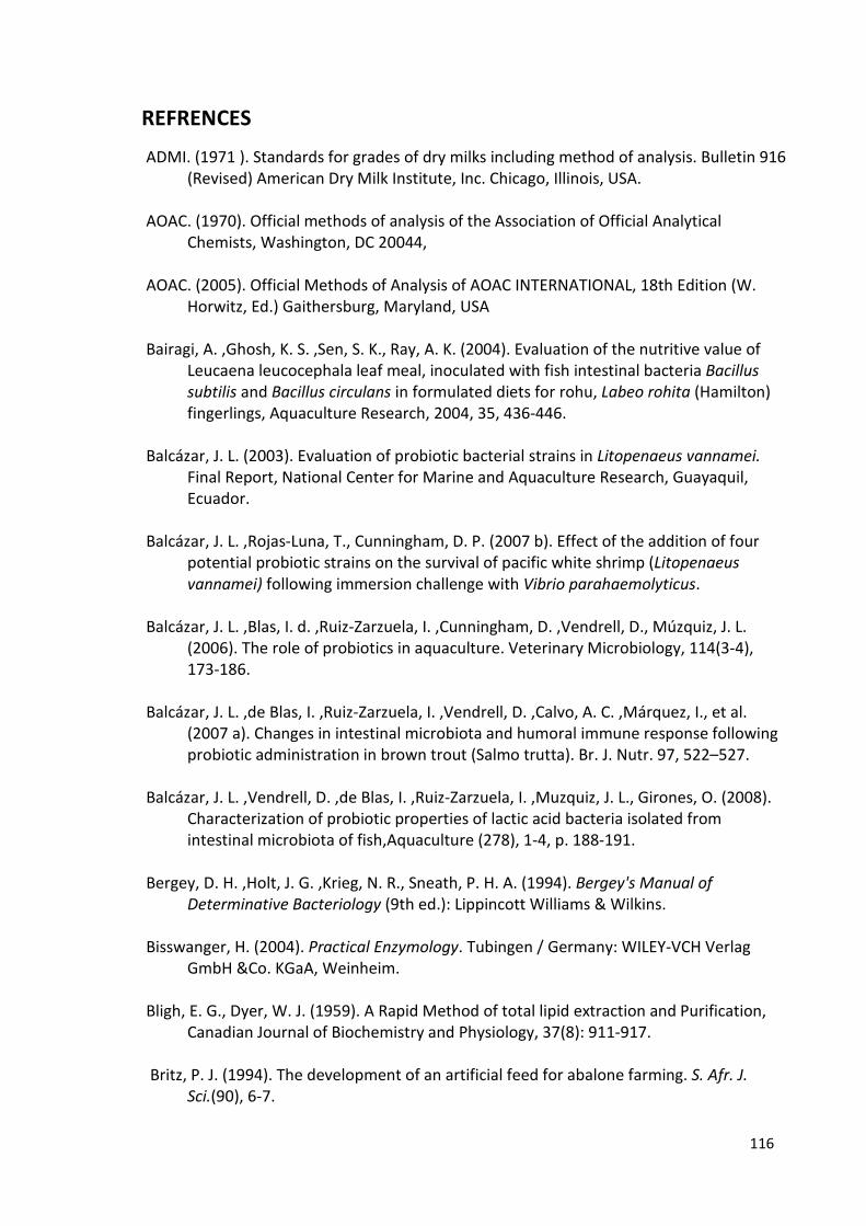
116
REFRENCES
ADMI. (1971 ). Standards for grades of dry milks including method of analysis. Bulletin 916
(Revised) American Dry Milk Institute, Inc. Chicago, Illinois, USA.
AOAC. (1970). Official methods of analysis of the Association of Official Analytical
Chemists, Washington, DC 20044,
AOAC. (2005). Official Methods of Analysis of AOAC INTERNATIONAL, 18th Edition (W.
Horwitz, Ed.) Gaithersburg, Maryland, USA
Bairagi, A. ,Ghosh, K. S. ,Sen, S. K., Ray, A. K. (2004). Evaluation of the nutritive value of
Leucaena leucocephala leaf meal, inoculated with fish intestinal bacteria Bacillus
subtilis and Bacillus circulans in formulated diets for rohu, Labeo rohita (Hamilton)
fingerlings, Aquaculture Research, 2004, 35, 436-446.
Balcázar, J. L. (2003). Evaluation of probiotic bacterial strains in Litopenaeus vannamei.
Final Report, National Center for Marine and Aquaculture Research, Guayaquil,
Ecuador.
Balcázar, J. L. ,Rojas-Luna, T., Cunningham, D. P. (2007 b). Effect of the addition of four
potential probiotic strains on the survival of pacific white shrimp (Litopenaeus
vannamei) following immersion challenge with Vibrio parahaemolyticus.
Balcázar, J. L. ,Blas, I. d. ,Ruiz-Zarzuela, I. ,Cunningham, D. ,Vendrell, D., Múzquiz, J. L.
(2006). The role of probiotics in aquaculture. Veterinary Microbiology, 114(3-4),
173-186.
Balcázar, J. L. ,de Blas, I. ,Ruiz-Zarzuela, I. ,Vendrell, D. ,Calvo, A. C. ,Márquez, I., et al.
(2007 a). Changes in intestinal microbiota and humoral immune response following
probiotic administration in brown trout (Salmo trutta). Br. J. Nutr. 97, 522–527.
Balcázar, J. L. ,Vendrell, D. ,de Blas, I. ,Ruiz-Zarzuela, I. ,Muzquiz, J. L., Girones, O. (2008).
Characterization of probiotic properties of lactic acid bacteria isolated from
intestinal microbiota of fish,Aquaculture (278), 1-4, p. 188-191.
Bergey, D. H. ,Holt, J. G. ,Krieg, N. R., Sneath, P. H. A. (1994). Bergey's Manual of
Determinative Bacteriology (9th ed.): Lippincott Williams & Wilkins.
Bisswanger, H. (2004). Practical Enzymology. Tubingen / Germany: WILEY-VCH Verlag
GmbH &Co. KGaA, Weinheim.
Bligh, E. G., Dyer, W. J. (1959). A Rapid Method of total lipid extraction and Purification,
Canadian Journal of Biochemistry and Physiology, 37(8): 911-917.
Britz, P. J. (1994). The development of an artificial feed for abalone farming. S. Afr. J.
Sci.(90), 6-7.
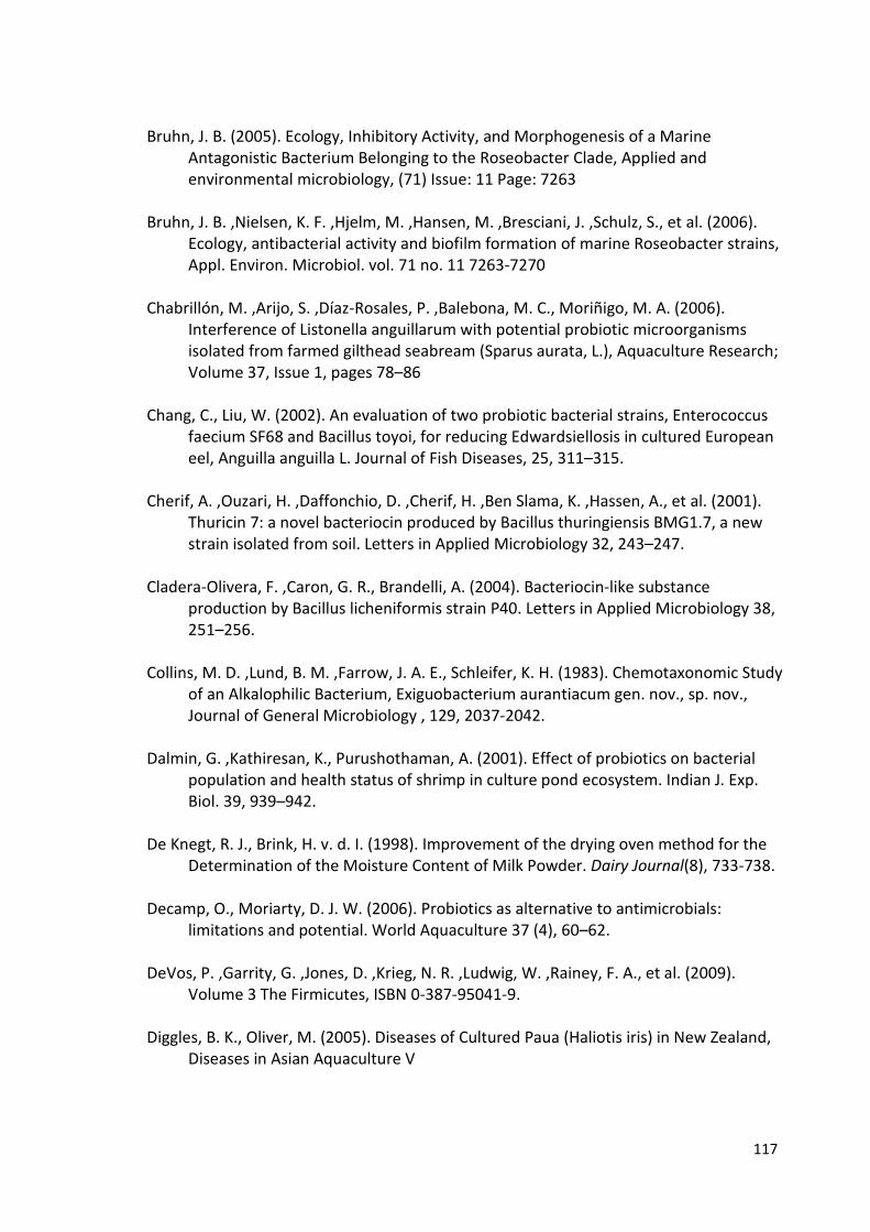
117
Bruhn, J. B. (2005). Ecology, Inhibitory Activity, and Morphogenesis of a Marine
Antagonistic Bacterium Belonging to the Roseobacter Clade, Applied and
environmental microbiology, (71) Issue: 11 Page: 7263
Bruhn, J. B. ,Nielsen, K. F. ,Hjelm, M. ,Hansen, M. ,Bresciani, J. ,Schulz, S., et al. (2006).
Ecology, antibacterial activity and biofilm formation of marine Roseobacter strains,
Appl. Environ. Microbiol. vol. 71 no. 11 7263-7270
Chabrillón, M. ,Arijo, S. ,Díaz-Rosales, P. ,Balebona, M. C., Moriñigo, M. A. (2006).
Interference of Listonella anguillarum with potential probiotic microorganisms
isolated from farmed gilthead seabream (Sparus aurata, L.), Aquaculture Research;
Volume 37, Issue 1, pages 78–86
Chang, C., Liu, W. (2002). An evaluation of two probiotic bacterial strains, Enterococcus
faecium SF68 and Bacillus toyoi, for reducing Edwardsiellosis in cultured European
eel, Anguilla anguilla L. Journal of Fish Diseases, 25, 311–315.
Cherif, A. ,Ouzari, H. ,Daffonchio, D. ,Cherif, H. ,Ben Slama, K. ,Hassen, A., et al. (2001).
Thuricin 7: a novel bacteriocin produced by Bacillus thuringiensis BMG1.7, a new
strain isolated from soil. Letters in Applied Microbiology 32, 243–247.
Cladera-Olivera, F. ,Caron, G. R., Brandelli, A. (2004). Bacteriocin-like substance
production by Bacillus licheniformis strain P40. Letters in Applied Microbiology 38,
251–256.
Collins, M. D. ,Lund, B. M. ,Farrow, J. A. E., Schleifer, K. H. (1983). Chemotaxonomic Study
of an Alkalophilic Bacterium, Exiguobacterium aurantiacum gen. nov., sp. nov.,
Journal of General Microbiology , 129, 2037-2042.
Dalmin, G. ,Kathiresan, K., Purushothaman, A. (2001). Effect of probiotics on bacterial
population and health status of shrimp in culture pond ecosystem. Indian J. Exp.
Biol. 39, 939–942.
De Knegt, R. J., Brink, H. v. d. I. (1998). Improvement of the drying oven method for the
Determination of the Moisture Content of Milk Powder. Dairy Journal(8), 733-738.
Decamp, O., Moriarty, D. J. W. (2006). Probiotics as alternative to antimicrobials:
limitations and potential. World Aquaculture 37 (4), 60–62.
DeVos, P. ,Garrity, G. ,Jones, D. ,Krieg, N. R. ,Ludwig, W. ,Rainey, F. A., et al. (2009).
Volume 3 The Firmicutes, ISBN 0-387-95041-9.
Diggles, B. K., Oliver, M. (2005). Diseases of Cultured Paua (Haliotis iris) in New Zealand,
Diseases in Asian Aquaculture V

118
Dutton, S., Tong, L. J. (1981). New Zealand Paua species. Ministry of Agriculture and
Fisheries. Catch 3, 15-16.
Dutton, S., Tong, L.J. (1986). Food preferences of Paua. Catch, 8(2), 15-16.
El-Shanshoury, A. R. ,Mona, M. H. ,Shoukr, F. A., E1-Bossery, A. M. (1994). The
enumeration and characterization of bacteria and fungi associated with marine
wood.boring isopods, and the ability of these microorganisms to digest cellulose
and wood. Mar. Biol. 119, 321–326.
Erasmus, J. H. (1996). The role of enteric bacteria in the abalone, Haliotis midae. M.Sc,
Thesis, University of Cape town, Cape town, pp. 1-83.
Erasmus, J. H. ,Cook, P. A., Coyne, V. E. (1997). The role of bacteria in the digestion of
seaweed by the abalone Haliotis midae. Aquaculture, 155(1-4), 377-386.
Fuller, R. (1989). Probiotics in man and animals. J. Appl. Bacteriol. 66:365-378. .
Fuller, R. (1992). History and development of probiotics. In: Fuller, R. (Ed.), Probiotics: The
Scientific Basis. Chapman and Hall, London, pp. 1–8.
Garland, C. D. ,Cooke, S. L. ,Grant, J. F., McMeekin, T. A. (1985). Ingestion of the bacteria
on and the cuticle of crustose (non-articulated) coralline algae by post-larval and
juvenile abalone (Haliotis rubra Leach) from Tasmanian waters. J. Exp. Mar. Biol.
Ecol. 91, 137-149.
Gatesoupe, F. J. (1999). The use of probiotics in aquaculture. Aquaculture, 180(1-2), 147-
165.
Gee, J. M. ,Lund, B. M. ,Metcalf, G., Peel, J. L. (1980). Properties of a New Group of
Alkalophilic Bacteria. General Microbiology( 117), 9-1 7.
Geovanny, G. R. ,Luis, B. J., Shen, M. (2007). Probiotics as Control Agents in Aquaculture,
Journal of Ocean University of China (Oceanic and Coastal Sea Research), Vol.6,
No.1, pp.76-79.
Gildberg, A. E. R., E. Mikkelsen, H. (1997). Probiotic effect of lactic acid bacteria in the
feed on growth and survival of fry of Atlantic cod (Gadus morhua),Hydrobiologia
(352); 279–285.
Gismondo, M. ,Drago, L., Lombardi, A. (1999). Review of probiotics available to modify
gastrointestinal flora. Int J Antimicrob Agents. 1999;12(4):287-92.
Gomez-Gil, B., Roque, A. (2000). The use and selection of probiotic bacteria for use in the
culture of larval aquatic organisms,Aquaculture (191); 259–270
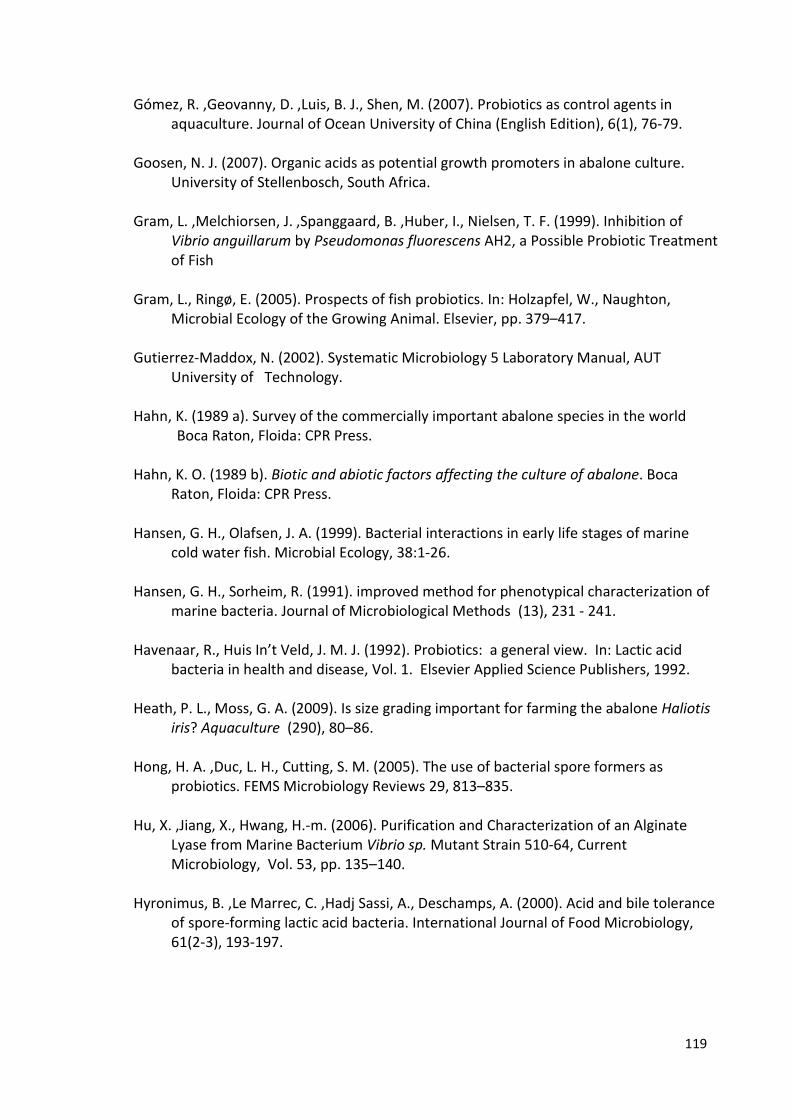
119
Gómez, R. ,Geovanny, D. ,Luis, B. J., Shen, M. (2007). Probiotics as control agents in
aquaculture. Journal of Ocean University of China (English Edition), 6(1), 76-79.
Goosen, N. J. (2007). Organic acids as potential growth promoters in abalone culture.
University of Stellenbosch, South Africa.
Gram, L. ,Melchiorsen, J. ,Spanggaard, B. ,Huber, I., Nielsen, T. F. (1999). Inhibition of
Vibrio anguillarum by Pseudomonas fluorescens AH2, a Possible Probiotic Treatment
of Fish
Gram, L., Ringø, E. (2005). Prospects of fish probiotics. In: Holzapfel, W., Naughton,
Microbial Ecology of the Growing Animal. Elsevier, pp. 379–417.
Gutierrez-Maddox, N. (2002). Systematic Microbiology 5 Laboratory Manual, AUT
University of Technology.
Hahn, K. (1989 a). Survey of the commercially important abalone species in the world
Boca Raton, Floida: CPR Press.
Hahn, K. O. (1989 b). Biotic and abiotic factors affecting the culture of abalone. Boca
Raton, Floida: CPR Press.
Hansen, G. H., Olafsen, J. A. (1999). Bacterial interactions in early life stages of marine
cold water fish. Microbial Ecology, 38:1-26.
Hansen, G. H., Sorheim, R. (1991). improved method for phenotypical characterization of
marine bacteria. Journal of Microbiological Methods (13), 231 - 241.
Havenaar, R., Huis In’t Veld, J. M. J. (1992). Probiotics: a general view. In: Lactic acid
bacteria in health and disease, Vol. 1. Elsevier Applied Science Publishers, 1992.
Heath, P. L., Moss, G. A. (2009). Is size grading important for farming the abalone Haliotis
iris? Aquaculture (290), 80–86.
Hong, H. A. ,Duc, L. H., Cutting, S. M. (2005). The use of bacterial spore formers as
probiotics. FEMS Microbiology Reviews 29, 813–835.
Hu, X. ,Jiang, X., Hwang, H.-m. (2006). Purification and Characterization of an Alginate
Lyase from Marine Bacterium Vibrio sp. Mutant Strain 510-64, Current
Microbiology, Vol. 53, pp. 135–140.
Hyronimus, B. ,Le Marrec, C. ,Hadj Sassi, A., Deschamps, A. (2000). Acid and bile tolerance
of spore-forming lactic acid bacteria. International Journal of Food Microbiology,
61(2-3), 193-197.
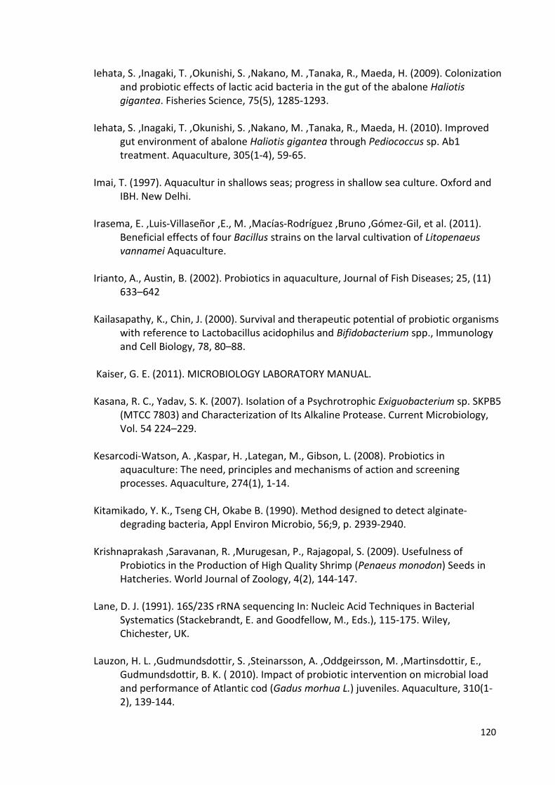
120
Iehata, S. ,Inagaki, T. ,Okunishi, S. ,Nakano, M. ,Tanaka, R., Maeda, H. (2009). Colonization
and probiotic effects of lactic acid bacteria in the gut of the abalone Haliotis
gigantea. Fisheries Science, 75(5), 1285-1293.
Iehata, S. ,Inagaki, T. ,Okunishi, S. ,Nakano, M. ,Tanaka, R., Maeda, H. (2010). Improved
gut environment of abalone Haliotis gigantea through Pediococcus sp. Ab1
treatment. Aquaculture, 305(1-4), 59-65.
Imai, T. (1997). Aquacultur in shallows seas; progress in shallow sea culture. Oxford and
IBH. New Delhi.
Irasema, E. ,Luis-Villaseñor ,E., M. ,Macías-Rodríguez ,Bruno ,Gómez-Gil, et al. (2011).
Beneficial effects of four Bacillus strains on the larval cultivation of Litopenaeus
vannamei Aquaculture.
Irianto, A., Austin, B. (2002). Probiotics in aquaculture, Journal of Fish Diseases; 25, (11)
633–642
Kailasapathy, K., Chin, J. (2000). Survival and therapeutic potential of probiotic organisms
with reference to Lactobacillus acidophilus and Bifidobacterium spp., Immunology
and Cell Biology, 78, 80–88.
Kaiser, G. E. (2011). MICROBIOLOGY LABORATORY MANUAL.
Kasana, R. C., Yadav, S. K. (2007). Isolation of a Psychrotrophic Exiguobacterium sp. SKPB5
(MTCC 7803) and Characterization of Its Alkaline Protease. Current Microbiology,
Vol. 54 224–229.
Kesarcodi-Watson, A. ,Kaspar, H. ,Lategan, M., Gibson, L. (2008). Probiotics in
aquaculture: The need, principles and mechanisms of action and screening
processes. Aquaculture, 274(1), 1-14.
Kitamikado, Y. K., Tseng CH, Okabe B. (1990). Method designed to detect alginate-
degrading bacteria, Appl Environ Microbio, 56;9, p. 2939-2940.
Krishnaprakash ,Saravanan, R. ,Murugesan, P., Rajagopal, S. (2009). Usefulness of
Probiotics in the Production of High Quality Shrimp (Penaeus monodon) Seeds in
Hatcheries. World Journal of Zoology, 4(2), 144-147.
Lane, D. J. (1991). 16S/23S rRNA sequencing In: Nucleic Acid Techniques in Bacterial
Systematics (Stackebrandt, E. and Goodfellow, M., Eds.), 115-175. Wiley,
Chichester, UK.
Lauzon, H. L. ,Gudmundsdottir, S. ,Steinarsson, A. ,Oddgeirsson, M. ,Martinsdottir, E.,
Gudmundsdottir, B. K. ( 2010). Impact of probiotic intervention on microbial load
and performance of Atlantic cod (Gadus morhua L.) juveniles. Aquaculture, 310(1-
2), 139-144.
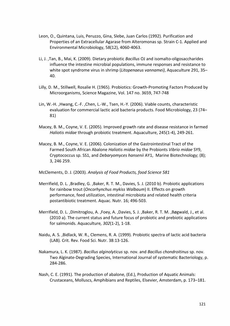
121
Leon, O., Quintana, Luis, Peruzzo, Gina, Slebe, Juan Carlos (1992). Purification and
Properties of an Extracellular Agarase from Alteromonas sp. Strain C-1. Applied and
Environmental Microbiology, 58(12), 4060-4063.
Li, J. ,Tan, B., Mai, K. (2009). Dietary probiotic Bacillus OJ and isomalto-oligosaccharides
influence the intestine microbial populations, immune responses and resistance to
white spot syndrome virus in shrimp (Litopenaeus vannamei), Aquaculture 291, 35–
40.
Lilly, D. M., Stillwell, Rosalie H. (1965). Probiotics: Growth-Promoting Factors Produced by
Microorganisms, Science Magazine, Vol. 147 no. 3659, 747-748
Lin, W.-H. ,Hwang, C.-F. ,Chen, L.-W., Tsen, H.-Y. (2006). Viable counts, characteristic
evaluation for commercial lactic acid bacteria products. Food Microbiology, 23 (74–
81)
Macey, B. M., Coyne, V. E. (2005). Improved growth rate and disease resistance in farmed
Haliotis midae through probiotic treatment. Aquaculture, 245(1-4), 249-261.
Macey, B. M., Coyne, V. E. (2006). Colonization of the Gastrointestinal Tract of the
Farmed South African Abalone Haliotis midae by the Probionts Vibrio midae SY9,
Cryptococcus sp. SS1, and Debaryomyces hansenii AY1, Marine Biotechnology; (8);
3, 246 259.
McClements, D. J. (2003). Analysis of Food Products, food Science 581
Merrifield, D. L. ,Bradley, G. ,Baker, R. T. M., Davies, S. J. (2010 b). Probiotic applications
for rainbow trout (Oncorhynchus mykiss Walbaum) II. Effects on growth
performance, feed utilization, intestinal microbiota and related health criteria
postantibiotic treatment. Aquac. Nutr. 16; 496-503.
Merrifield, D. L. ,Dimitroglou, A. ,Foey, A. ,Davies, S. J. ,Baker, R. T. M. ,Bøgwald, J., et al.
(2010 a). The current status and future focus of probiotic and prebiotic applications
for salmonids. Aquaculture, 302(1-2), 1-18.
Naidu, A. S. ,Bidlack, W. R., Clemens, R. A. (1999). Probiotic spectra of lactic acid bacteria
(LAB). Crit. Rev. Food Sci. Nutr. 38:13-126.
Nakamura, L. K. (1987). Bacillus alginolyticus sp. nov. and Bacillus chondroitinus sp. nov.
Two Alginate-Degrading Species, International Journal of systematic Bacteriology, p.
284-286.
Nash, C. E. (1991). The production of abalone, (Ed.), Production of Aquatic Animals:
Crustaceans, Molluscs, Amphibians and Reptiles, Elsevier, Amsterdam, p. 173–181.
122
Olafsen, J. A. (2001). Interactions between fish larvae and bacteria in marine aquaculture,
Aquaculture, 200; p.223–247.
Olmos, J. ,Ochoa, L. ,Paniagua-Michel, J., Contreras, R. (2011). Functional Feed
Assessment on Litopenaeus vannamei Using 100% Fish Meal Replacement by
Soybean Meal, High Levels of Complex Carbohydrates and Bacillus Probiotic Strains
Orozco-Medina, C. ,Maeda-Mart|nez, A. ,Cordova-Murueta, J. ,Lopez-Cortes, A.,
Navarrete-del-Toro, M. (2009). Activity, molecular mass and hydrolysis on baker’s
yeast protein of extracellular proteases from the putative probiotic bacteria
Microbacterium sp. strain 8L and Exiguobacterium mexicanum strain 8N
Aquaculture Research, 41, 45-52.
OΉalloran, S. (1986). Paua fishery management in New Zealand. Shellfisheries Newsletter
30, Supplement to Catch, 13(3), pp.1.
Pakulski, j., Benner, R. (1992). An improved method for the hydrolysis and MBTH analysis
of dissolved and particulate carbohydrates in seawater, Marine Chemistry (40), 3-4,
p.143-160.
Parker, R. B. (1974). Probiotics, the other half of the antibiotic story. Anim. Nutr. Health.
29:4-8. .
PAUA, O. B. (2011). What makes OceaNZ Blue Paua so Special? Retrieved from
http://www.oceanzblue.co.nz.
PICL. Paua Industry Council Ltd., Ministry of Fisheries (2005 ); The commercial paua
fishery. http://www.paua.org.nz
Poore, C. B. (1972). Ecology of the New Zealand abalones, Haliotis species (Molluscan:
Gastropoda). 2. Seasonal and diurnal movement. NZ. J. Mar.Freshw. Res. 6, 246-
258.
Porter, D., Earl, O. (1990). Nutrition labeling issues and directions for the 1990s: National
Academy Press, Washington, DC.
Qi, X.-H. Z., Nico Boonb and Peter Bossierc. (2009). Probiotics in aquaculture of China —
Current state, problems and prospect Aquaculture, 290 (1-2), 15-21.
Rengpipat, S. ,Rukpratanporn, S. ,Piyatiratitivorakul, S., Menasaveta, P. (2000). Immunity
enhancement in black tiger shrimp (Penaeus monodon) by a probiont bacterium
(Bacillus S11). Aquaculture 191, 271–288.
Ringø, E., Gatesoupe, F. (1998). Lactic acid bacteria in fish: a review. Aquaculture
160:177–203.

123
Ringø, E. ,Schillinger, U., Holzapfel, W. (2005). Antimicrobial activity of lactic acid bacteria
isolated from aquatic animals and the use of lactic acid bacteria in aquaculture. In
Biology of Growing Animals (Vol. 2, pp. 418-453)
Rosenfeld, W. D., Zobell, C. E. (1947). Antibiotic production by marine microorganisms. J.
Bacteriol. 54, 393–398.
Sahu, M. K. ,Swarnakumar, N. S. ,Sivakumar, K. ,Thangaradjou, T., Kannan, L. (2008).
Probiotics in aquaculture: importance and future perspectives, Indian J.
Microbiology, 48; 299-308.
Sainsbury, K. J.(1982). Population dynamics and fishery management of the paua, H. iris.1.
Population structure, growth, reproduction, and mortality. NZ. J. Mar.Freshw (Res.
16), 147-161.
Sakai, M. (1998). Current research status of fish immunostimulants. Aquaculture 172, 63–
92.
Sakata, T. (1990). Microflora in the digestive tract of fish and shellfish. In: Lesel, R. (Ed.),
Microbiology Poecilotherms. Elsevier, Amsterdam, 171–176.
Sales, J., Britz, P. J. (2001). Research on abalone (Haliotis midae L.) cultivation in South
Africa, Aquaculture Research (32), 11, 863-874.
Sarkono ,Faturrahman, Sofyan, Y. (2010). Isolation and identification of lactic acid bacteria
from abalone (Haliotis asinina) as a potential candidate of probiotic, Bioscience; Vol.
2, No. 1, 38-42 ISSN: 2087-3956.
Sawabe, T. ,Setoguchi, N. ,Inoue, S. ,Tanaka, R. ,Ootsubo, M. ,Yoshimizu, M., et al. (2003).
Acetic acid production of Vibrio halioticoli from alginate: a possible role for
establishment of abalone- V. halioticoli association. Aquaculture, 219(1-4), 671-679.
Sawabe, T., Sugimura, l. (1998). Vibrio halioticoli Spm nov., a non-motile alginolytic
marine bacterium isolated from the gut of the abalone Haliotis discus hannai,
International Journal of Systematic Bacteriology, 48, 573-580
Schaafsma, G. (1996). State of art concerning probiotic strains in milk products. 5:23-4.
Schiel, D. B., PA. (1991). Population structure, ageing, and fish mortality of the New
Zealand abalone Haliotis iris, Fishery Bulletin, 89;4, p.681-691.
Schiel, R. D. (1993). Experimental evaluation of commercial-scale enhancement of
abalone Haliotis iris populations in New Zealand. MAarine Ecology Progress Series,
97, 167-181.
Schleifer, K. H., Kilpper-Balz, R. (1984). Transfer of Streptococcus faecalis and
Streptococcus faecium to the Genus Enterococcus norn. rev. as Enterococcus
124
faecalis comb. nov.and Enterococcus faecium comb. International Journal of
Systematic Bacteriology, P 31-34.
Schrezenmeir, J., de Vrese, M. ( 2001). Probiotics, prebiotics, and synbiotics –
approaching a definition. Am .J. Clin. Nutr. 73 (suppl): 361S-4S.
Schulze, A. D. ,Alabi, A. O. ,Tattersall-Sheldrake, A. R., Miller, K. M. (2006). Bacterial
diversity in a marine hatchery: Balance between pathogenic and potentially
probiotic bacterial strains. Aquaculture 256; 50–73
SeaFIC. (2010). New Zealand Seafood Industry Council statistics for 2010 (Ministery of
fisheries); Quarterly Report / Fisheries and Aquaculture Production and Trade.
Sombatjinda, S. ,Boonapatcharoen, N. ,Wantawin, C. ,Withyachumnarnkul, B.,
Techkarnjanaruk, S. (2011). Dynamics of Microbial Communities in an Earthen
Shrimp Pond during the Shrimp Growing Period. Environment and Natural
Resources Research Vol. 1, No. 1, 171-180.
Stanton, C. ,Ross, R. P. ,Fitzgerald, G. F., Sinderen, D. V. (2005). Fermented functional
foods based on probiotics and their biogenic metabolites, Current Opinion in
Biotechnology, Volume 16, Issue 2, Pages 198–203
Tan, L. ,Qu, Y.-y. ,Zhou, J.-t. ,Li, A., Gou, M. ( 2009). Identification and Characteristics of A
Novel Salt-Tolerant Exiguobacterium sp. for Azo Dyes Decolorization. Applied
Biochemistry and Biotechnology. Volume 159, No. 3, 728-738
Tanaka, R. ,Ootsubo, M. ,Sawabe, T. ,Ezura, Y., Tajima, K. (2004). Biodiversity and in situ
abundance of gut microflora of abalone (Haliotis discus hannai) determined by
culture-independent techniques, Aquaculture 241, 453–463
Tanaka, R., Sugimura, I. (2003). Gut microflora of abalone Haliotis discus hannai in culture
changes coincident with a change in diet Fisheries Science, 69: 951–958.
Tegner, M. J., Butler, R.A., . (1985). The survival and mortality of seeded and native red
abalones, Haliotis rufescens, on the Palos Verdes Peninsula California USA.
ten Doeschate, K. I., Coyne, V. E. (2008). Improved Growth Rate in Farmed Haliotis midae
through Probiotic Treatment. Aquaculture, 284(1-4), 174-179.
Timmermana, H. M. ,Koning, C. J. M. ,Mulderc, L. ,Romboutsd, F. M., Beynen, A. C. (2004).
Monostrain, multistrain and multispecies probiotics A comparison of functionality
and efficacy; Volume 96, ( 3), P; 219–233
Troell, M. ,Robertson-Andersson, D. ,Anderson, R. J. ,Bolton, J. J. ,Maneveldt, G. ,Halling,
C., et al. (2006). Abalone farming in South Africa: An overview with perspectives on.
Aquaculture 257, 266-281.
125
Tung, C.-H. (2010). Dietary protein requirements of the New Zealand Black-footed
Abalone (Haliotis iris, Martyn 1784) (Doctoral). AUT Auckland
Tuomola, E. ,Crittenden, R. ,Playne, M. ,Isolauri, E., Salminen, S. (2001). Quality assurance
criteria for probiotic bacteria. American Journal of Clinical Nutrition, Vol. 73, No. 2,
393-398.
Van der Aa Kühle, A. ,Skovgaard, K., Jespersen, L. (2005). In vitro screening of probiotic
properties of Saccharomyces cerevisiae var. boulardii and foodborne
Saccharomyces cerevisiae strains. International Journal of Food Microbiology 101,
29–39.
Verschuere, L. ,Rombaut, G. ,Sorgeloos, P., Verstraete, W. (2000). Probiotic bacteria as
biological control agents in aquaculture. Microbiology and Molecular Biology
Reviews, 64(4), 655.
Vijayabaskar, P., Somasundaram, S. T. (2008 ). Isolation of bacteriocin producing lactic
acid bacteria from fish gut and probiotic activity against common fresh water fish
pathogen Aeromonas hydrophila. Biotechnology, 7(1), 124-128.
Vine, N. G. ,Leukes, W. D., Kaiser, H. (2006). Probiotics inmarine larviculture; FEM
Microbiology Reviews, Volume 30, Issue 3, pages 404–427
Vishnivetskaya, T. A., Kathariou, S. (2009). The Exiguobacterium genus: biodiversity and
biogeography, Extremophiles 13:541–555.
Vishnivetskaya, T. A. ,Lucas, S. ,Copeland, A. ,Lapidus, A., Rio, T. G. d. (2011). Complete
Genome Sequence of the Thermophilic Bacterium Exiguobacterium sp. AT1bv.
Journal of bacteriology, 2880–2881.
Wang, N.S. (1988). Biochemical engineering laboratory manual,experiment N0. 4A; Starch
hydrolysis by amylase.
Wang, Y.-B., jian-Rong Li, J. L. (2008). Probiotics in aquaculture: Challenges and outlook,
aquaculture(281), 1-4
Wang, Y. B. ,Xu, Z. R., Xia, M. S. (2005). The effectiveness of commercial probiotics in
Northern White Shrimp (Penaeus vannamei L.) ponds. Fish. Sci. 71, 1034–1039.
Weber, J. T. ,Mintz, E. D. ,Canizares, R. ,Semiglia, A. ,Gomez, I. , Sempertegui, R., et al.
(1994). Epidemia cholera in Ecuador: multidrug resistance and transmission by
water and seafood. Epidemiol. Infect. 112, 1–11.
WHO/FAO. (2001). Health and Nutritional Properties of Probiotics in Food including
Powder Milk with Live Lactic Acid Bacteria. Córdoba, Argentina.
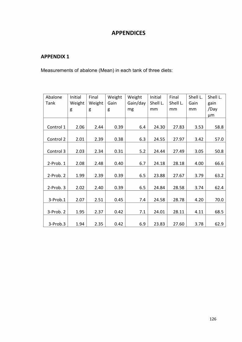
126
APPENDICES
APPENDIX 1
Measurements of abalone (Mean) in each tank of three diets:
Abalone
Tank
Initial
Weight
g
Final
Weight
g
Weight
Gain
g
Weight
Gain/day
mg
Initial
Shell L.
mm
Final
Shell L.
mm
Shell L.
Gain
mm
Shell L.
gain
/Day
µm
Control 1 2.06 2.44 0.39 6.4 24.30 27.83 3.53 58.8
Control 2 2.01 2.39 0.38 6.3 24.55 27.97 3.42 57.0
Control 3 2.03 2.34 0.31 5.2 24.44 27.49 3.05 50.8
2-Prob. 1 2.08 2.48 0.40 6.7 24.18 28.18 4.00 66.6
2-Prob. 2 1.99 2.39 0.39 6.5 23.88 27.67 3.79 63.2
2-Prob. 3 2.02 2.40 0.39 6.5 24.84 28.58 3.74 62.4
3-Prob.1 2.07 2.51
0.45 7.4 24.58 28.78 4.20 70.0
3-Prob. 2 1.95 2.37 0.42 7.1 24.01 28.11 4.11 68.5
3-Prob.3 1.94 2.35 0.42 6.9 23.83 27.60 3.78 62.9

127
APPENDIX 2
Statistical Analysis for Shell Length Changes
Descriptives
Shell length changes
Diet N Mean Std.
Deviatio
n
Std. Error 95% Confidence Interval for
Mean
Minimum Maximum
Lower Bound Upper Bound
1 (Control) 3 3.33 .249 .143 2.71 3.95 3.05 3.53
2 (2-P) 3 3.84 .134 .077 3.50 4.17 3.74 4.00
3 (3-P) 3 4.02 .222 .128 3.47 4.57 3.78 4.20
Total 9 3.73 .359 .119 3.45 4.00 3.05 4.20
ANOVA
Shell length changes
Sum of
Squares
df Mean Square F Sig.
Between
Groups
(Combined) .777 2 .388 8.991 .016
Linear Term Contrast .722 1 .722 16.723 .006
Deviation .054 1 .054 1.260 .305
Within Groups .259 6 .043
Total 1.036 8
Multiple Comparisons(Tukey HSD)
Dependent Variable: Shell length changes
Diet (Diet Mean Difference
(I-J)
Std. Error Sig. 95% Confidence Interval
Lower Bound Upper Bound
1 (Control) 2 (2-P) -.51 .169 .053 -1.0326 .0087
3 (3-P) -.69 .169 .015 -1.2146 -.1733
*. The mean difference is significant at the 0.05 level.
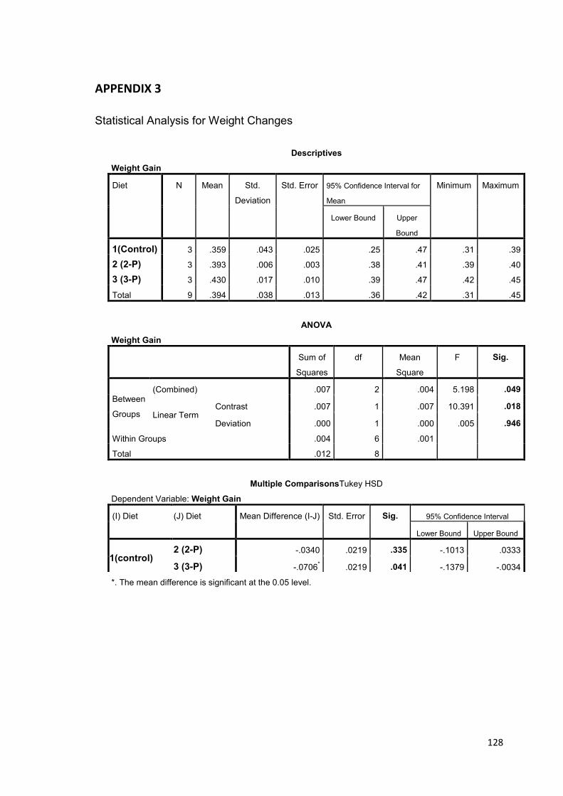
128
APPENDIX 3
Statistical Analysis for Weight Changes
Descriptives
Weight Gain
Diet N Mean Std.
Deviation
Std. Error 95% Confidence Interval for
Mean
Minimum Maximum
Lower Bound Upper
Bound
1(Control) 3 .359 .043 .025 .25 .47 .31 .39
2 (2-P) 3 .393 .006 .003 .38 .41 .39 .40
3 (3-P) 3 .430 .017 .010 .39 .47 .42 .45
Total 9 .394 .038 .013 .36 .42 .31 .45
ANOVA
Weight Gain
Sum of
Squares
df Mean
Square
F Sig.
Between
Groups
(Combined) .007 2 .004 5.198 .049
Linear Term Contrast .007 1 .007 10.391 .018
Deviation .000 1 .000 .005 .946
Within Groups .004 6 .001
Total .012 8
Multiple ComparisonsTukey HSD
Dependent Variable: Weight Gain
(I) Diet (J) Diet Mean Difference (I-J) Std. Error Sig. 95% Confidence Interval
Lower Bound Upper Bound
1(control) 2 (2-P) -.0340 .0219 .335 -.1013 .0333
3 (3-P) -.0706* .0219 .041 -.1379 -.0034
*. The mean difference is significant at the 0.05 level.
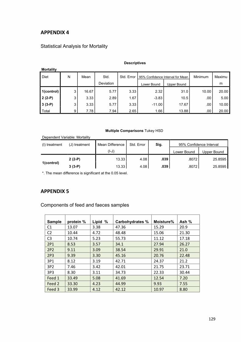
129
APPENDIX 4
Statistical Analysis for Mortality
Multiple Comparisons Tukey HSD
Dependent Variable: Mortality
(I) treatment (J) treatment Mean Difference
(I-J)
Std. Error Sig. 95% Confidence Interval
Lower Bound Upper Bound
1(control) 2 (2-P) 13.33 4.08 .039 .8072 25.8595
3 (3-P) 13.33 4.08 .039 .8072 25.8595
*. The mean difference is significant at the 0.05 level.
APPENDIX 5
Components of feed and faeces samples
Sample
protein %
Lipid %
Carbohydrates %
Moisture%
Ash %
C1 13.07 3.38 47.36 15.29 20.9
C2 10.44 4.72 48.48 15.06 21.30
C3 10.74 5.23 55.73 11.12 17.18
2P1 8.53 3.57 34.1 27.94 26.27
2P2 9.11 3.09 38.54 29.91 21.0
2P3 9.39 3.30 45.16 20.76 22.48
3P1 8.12 3.19 42.71 24.37 21.2
3P2 7.46 3.42 42.01 21.75 23.71
3P3 8.30 3.11 34.73 22.33 30.44
Feed 1 33.49 5.08 41.69 12.54 7.20
Feed 2 33.30 4.23 44.99 9.93 7.55
Feed 3 33.99 4.12 42.12 10.97 8.80
Descriptives
Mortality
Diet N Mean Std.
Deviation
Std. Error 95% Confidence Interval for Mean Minimum Maximu
m Lower Bound Upper Bound
1(control) 3 16.67 5.77 3.33 2.32 31.0 10.00 20.00
2 (2-P) 3 3.33 2.89 1.67 -3.83 10.5 .00 5.00
3 (3-P) 3 3.33 5.77 3.33 -11.00 17.67 .00 10.00
Total 9 7.78 7.94 2.65 1.66 13.88 .00 20.00