use of two-replisome plasmids to characterize how
TRANSCRIPT

Portland State University Portland State University
PDXScholar PDXScholar
Dissertations and Theses Dissertations and Theses
Spring 7-19-2019
Use of Two-replisome Plasmids to Characterize How Use of Two-replisome Plasmids to Characterize How
Chromosome Replication Completes Chromosome Replication Completes
Nicklas Alexander Hamilton Portland State University
Follow this and additional works at: https://pdxscholar.library.pdx.edu/open_access_etds
Part of the Biology Commons
Let us know how access to this document benefits you.
Recommended Citation Recommended Citation Hamilton, Nicklas Alexander, "Use of Two-replisome Plasmids to Characterize How Chromosome Replication Completes" (2019). Dissertations and Theses. Paper 5064. https://doi.org/10.15760/etd.6940
This Thesis is brought to you for free and open access. It has been accepted for inclusion in Dissertations and Theses by an authorized administrator of PDXScholar. Please contact us if we can make this document more accessible: [email protected].

Use of Two-replisome Plasmids to cCaracterize how Chromosome Replication
Completes
by
Nicklas Alexander Hamilton
A thesis submitted in partial fulfillment of the requirements for the degree of
Master of Science in
Biology
Thesis Committee:
Justin Courcelle, Chair Jeffrey Singer
Rahul Raghavan
Portland State University 2019

i
Abstract
All living organisms need to accurately replicate their genome to survive. Genomic
replication occurs in three phases; initiation, elongation, and completion. While initiation
and elongation have been extensively characterized, less is known about how replication
completes. In Escherichia coli completion occurs at sites where two replication forks
converge and is proposed to involve the transiently bypass of the forks, before the
overlapping sequences are resected and joined. The reaction requires RecBCD, and
involves several other gene products including RecG, ExoI, and SbcDC but can occur
independent of recombination or RecA. While several proteins are known to be involved,
how they promote this reaction and the intermediates that arise remain uncharacterized.
In the first part of this work, I describe the construction of plasmid “mini-
chromosomes” containing a bidirectional origin of replication that can be used to examine
the intermediates and factors required for the completion reaction. I verify that these
substrates can be used to study the completion reaction by demonstrating that these
plasmids require completion enzymes to propagate in cells. The completion enzymes are
required for plasmids containing two-replisomes, but not one replisome, indicating that the
substrate these enzymes act upon in vivo is specifically created when two replication forks
converge.
Completion events in E. coli are localized to one of the six termination (ter)
sequences within the 400-kb terminus region due to the autoregulated action of Tus, which
binds to ter and inhibits replication fork progression in an orientation-dependent manner.
In the second part of this work, I examine how the presence of ter sequences affect
completion on the 2-replisome plasmid. I show that addition of ter sequences modestly
decreases the stability of the two-replisome plasmid and that this correlates with higher

ii
levels of abnormal, amplified molecules. The results support the idea that ter sites are not
essential to completion of DNA replication; similar to what is seen on the chromosome.
Rec-B-C-D forms a helicase-nuclease complex that, in addition to completion, is
also required for double-strand break repair in E. coli. RecBCD activity is altered upon
encountering specific DNA sequences, termed chi, in a manner that promotes crossovers
during recombinational processes. In the third part of this work, I demonstrate that the
presence of chi in a bidirectional plasmid model promotes the appearance of over-
replicated linear molecules and that these products correlate with a reduced stability of the
plasmid. The effect appears specific to plasmids containing two replisomes, as chi on the
leading or lagging strand of plasmids containing one replisome had no-effect. The
observation implies chi promotes a reaction that may encourage further synthesis during
the completion reaction, and that at least on the mini-chromosomes substrates, this appears
to be a destabilizing force.

iii
Dedication
I would like to thank my adviser, Dr. Justin Courcelle, for never giving up on my
continuous progress and providing me the resources and much needed advice and expertise,
along with a ton of patience, that I required to better myself in my graduate tenure.
Secondly, I would like to thank my other committee members Dr. Rahul Raghavan and Dr.
Jeffrey Singer for their patience, and assistant with my various learning endeavors
including classes and facilitating seminars. In equal addition I would like to thank Dr.
Charmain Courcelle for the limitless hands-on training and continuous support with
countless “upping my game” pep talks which was necessary to overcome many of my self-
made obstacles; additionally, helping engineer some rather difficult plasmids. I would also
like to thank my lab mates, Dr. Brian Wendel (who first synthesized the two bidirectional
plasmids) and Jessica Cole, who have made the hardest challenge of my life achievable
and have installed a new and intense appreciation for scientific research and comradery.
Lastly, I want to thank my husband, Jeremy Hamilton, who has spent countless hours
encouraging and supporting me through the rough days, celebrating my triumphs, and
overall being a pillar of figurative and actual physical strength for me; I could not have
done this without him, everyone mentioned above, and of course the unconditional love
and support from family and friends not mentioned. Thank you all!

iv
TABLE OF CONTENTS
PAGE
Abstract i
Dedication iii
List of tables vii
List of Figures viii
Chapter I: Research purposes
Introduction 01
Figures 11
Chapter II: Materials and Methods
Bacterial strains and plasmids 14
Transformation efficiency assay 16
Plasmid stability assay 17
Total genomic DNA extraction 17
Southern analysis of plasmid replication intermediates 18
Copy number analysis 18
Strain Growth assay 18
Tables 20
Chapter III: Results
Part I: One-replisome to two-replisome plasmids during completion of replication
Plasmids containing two replisomes can be stably transformed, replicated, and grown in cells similar to the one-replisome plasmid.
22
Plasmids replicated by two replisomes are less stable than one-replisome plasmids and contain more aberrant, multimeric species.
24

v
Similar to the completion of replication on the chromosome, amplifications and genetic instability on the two-replisome plasmid are driven by an aberrant RecA-mediated recombination mechanism.
25
Transformation of plasmids with two replisomes, but not one replisome, depends on the enzymes required to complete replication on the chromosome.
26
Part II: The effect of Replication fork traps and the Completion reaction
Two-replisome plasmids with two addition ter sequences, terB and terC from the Escherichia coli chromosome, are replicated and maintained at similar frequencies as two-replisome plasmids without replication fork trap capabilities.
27
Plasmids replicated by two replisomes with the addition of ter sequences contain more RecA-driven aberrant, multimeric species.
29
Part III: The effect of chi sequences on completing replication
chi sequences located at sites where replication forks converge promote a RecA-mediated aberrant replication that correlates with instability
29
Addition of a terB and terC trap reduces the destabilizing effect of chi by limiting the amount of aberrant replication that can occur
31
Figures 33
Chapter IV: Discussion
One-replisome to two-replisome plasmids during completion of replication
52
Replication fork traps and the completion reaction 58

vi
Chi sequences and the completion of replication 59
Chapter V: References 61

vii
List of Tables
Page
2.1 E. coli strains used in this study 20
2.2 Plasmids used in this study 20

viii
List of Figures
Page
1.1A Schematic of the Escherichia coli circular chromosome containing a single bidirectional origin of replication and ter sequences which block replication progression in an orientation-specific manner.
11
1.1B Replication profiling reveals that recBC is required to maintain the region where replication forks converge on the chromosome, whereas recD, or sbcDC and xonA are required to resect over-replicated DNA that arises in this region. Genomic DNA from replicating cultures was purified, fragmented, and profiled using high-throughput sequencing.
11
1.1C Model of completion of replication. 12
3.1A Schematic of pBR322, containing a unidirectional origin of replication, and pCL01 which contains a λ bidirectional origin of replication.
33
3.1B Strains containing the two-replisome plasmid grow similar to those containing the one-replisome plasmid or no plasmid; E. coli strain included ampicillin cassette in this assay.
33
3.1C Plasmids containing two replisomes can be stably transformed at frequencies similar to those with one replisome.
34
3.1D The two-replisome plasmid is maintained at a copy number similar to the one replisome plasmid.
34
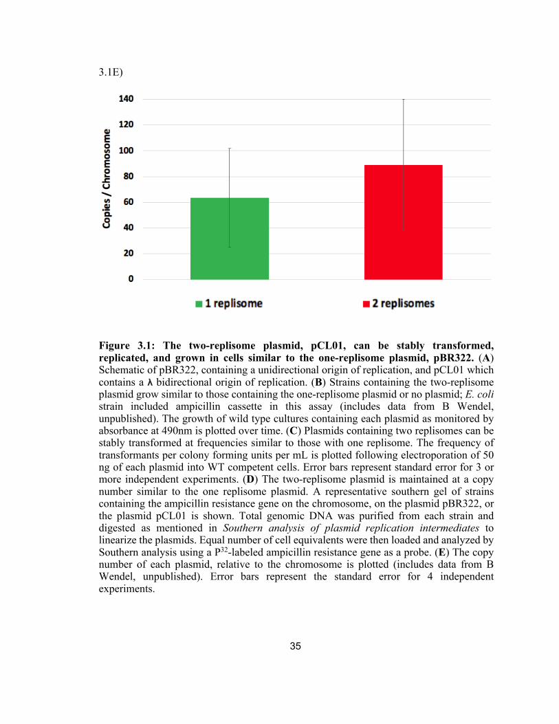
3.1E The copy number of each plasmid, relative to the chromosome is plotted.
35
3.2A In the absence of selection, the two-replisome plasmids are lost more rapidly than the one-replisome plasmid.
36
3.2B The fraction of cells retaining the one-replisome and two-replisome plasmid is plotted over time.
ix
36
3.2C The instability of the two-replisome plasmid, relative to the one-replisome plasmid, correlates with the presence of more abnormal, multimeric species.
37
3.2D The fraction of unit length, non-monomeric plasmid in cultures containing the one-replisome and two-replisome plasmid is plotted.
38
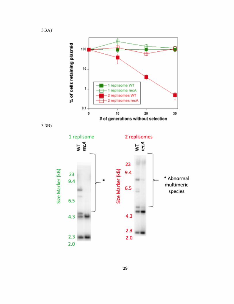
3.3A Inactivation of RecA restores the stability of the two-replisome plasmid to levels that approach that of the one-replisome plasmid.
39
3.3B The increased stability in the absence of the aberrant recombination pathway correlates with an increase in unit-length monomeric plasmids.
39
3.3C The fraction of non-monomeric plasmid in each culture is plotted for the one-replisome and two-replisome plasmid in the presence and absence of RecA.
40
3.4A Transformation of plasmids with two replisomes, but not one replisome, depends on the enzymes required to complete replication on the chromosome.
41
3.5A Diagram of plasmid constructs comparing 2-replisome plasmids to its counterpart with ter insertions (terB + terC).
42
3.5B The fraction of cells retaining the two-replisome plasmid in the absence of selection is plotted over time.
42
3.5C Increased amounts of RecA-dependent, non-monomeric plasmid species are observed in the presence of ter traps.
43
3.5D The fraction of non-monomeric plasmid species in each culture is plotted. 43
3.6A Diagram of plasmid constructs comparing 2-replisome plasmid, pCL01, no chi control, pCL03 where chi is mutated near the ori site, pCL05 where chi is mutated opposite of ori site.
44

x
3.6B Diagram of one-replisome plasmids containing a chi inserted into the leading, pCL07, or lagging strand, pCL08, pBR322 template.
45
3.6C When present in the terminus region, chi destabilized the two-replisome plasmid. 45
3.6D When present in the terminus region of the two-replisome plasmid, chi induces the formation of an aberrant linear plasmid multimeric species.
46
3.6E The amount of the linear plasmid multimeric species observed is plotted. 47
3.6F The stability of the one-replisome plasmid is not affected by the presence of chi. 47
3.6G No multimeric species are induced by chi on one-replisome plasmids. 48
3.6H The amount of the linear plasmid multimeric species observed is plotted. 48
3.7A Diagram of two-replisome plasmids containing either a chi site in the terminus region or ter traps or both.
50
3.7B There is less destabilization by chi observed in the presence of a ter trap. 50
3.7C The amount of the linear plasmid multimeric species observed is plotted. 51
1
Chapter I
Research purposes
Introduction
The DNA of a genome must be accurately replicated and passed on to daughter
cells each generation. In addition to the challenge of accomplishing this task, DNA is also
constantly bombarded by chemicals and radiation that compromise its integrity. These
include chemicals found in tobacco smoke that oxidize bases or induce single strand
breaks, UV-C radiation that induces pyrimidine adducts, deamination by hydrolysis in the
presence of acid or heat, and harmful chemical metabolites that can react with the DNA to
form adducts (Sachs et al., 1992; Friedberg 1995; Leanderson and Tagesson, 1992;
reviewed in Sinha et al., 2002; reviewed in Zeman et al., 2014; Gorden et al., 2018).
Therefore, cells contain numerous enzymatic systems that ensure the DNA is faithfully
copied and can be repaired when damaged. Some of these regulatory mechanisms include
proofreading during and after replication, nucleotide excision repair, mismatch repair
mechanisms, and global stress responses such as the SOS and SoxR/OxyR response during
DNA damage (Hopefield et al., 1976; Wang and Smith 1986; Gonzalez et al., 1998;
reviewed in Crowley and Courcelle, 2002; reviewed in Hanawalt et al., 2003; Heyer et al.,
2010; reviewed in O’Donnell et al., 2013; Lee et al., 2015). An additional process that
presents challenges to maintaining genomic stability is the completion of DNA replication
(Wendel et al., 2014). The mechanism by which completion occurs has only recently been
recognized and far less is known about this process than the associated steps of initiation
or elongation. However, these events must occur thousands of times per division in human
cells; implying that it must occur with high efficiency (Hopefield 1974; Errico and
Constanzo, 2012; reviewed in Costa et al., 2013; Wendel et al., 2014). Each singular

2
convergence must somehow recognize replicated regions, resolve torsional complexities
created by the supercoiling of two convergent replisomes, ensure that any overlapping or
redundant sequences are resected or degraded, and finally joining the nascent strands at the
precise point where all sequences have doubled (reviewed in Courcelle et al., 2015).
Our lab has recently identified several enzymes that are required for this reaction
to occur in Escherichia coli. These include RecBCD, RecG, ExoI, and SbcDC (Wendel et
al., 2014; Wendel et al., 2018). Of these enzymes RecBCD appears to play a critically
central role, as cells lacking RecBCD have severely reduced viability and growth rates, and
fail to maintain the region of the chromosome where replication completes (Wendel et al.,
2014). Interestingly, RecBCD also has a long-established role in double strand break repair
and homologous recombination (Klein and Kreuzer, 2002; reviewed in Smith 2012;
Wendel et al., 2014). The enzyme complex has a number of remarkable activities, including
two helicases with differing polarities, as well as both exonucleolytic, and endonucleolytic
activities. Despite decades of characterization, both in vivo and in vitro, many of the
molecular aspects and intermediates for which RecBCD catalyzes recombination remain
uncharacterized. Considering RecBCD’s newly identified and critical role in completion
of replication, it seems likely that its functional role in both processes will be similar if not
identical. Thus, characterization of the completion reaction presents a real opportunity to
learn more about the cellular role and function of this complex enzymatic machine.
In this work, I examine two DNA sequence motifs that are known to affect RecBCD
function in recombination or completion. Chi sequences are nonpalindromic G-rich
octamers that are heavily enriched and over-represented in the leading strand template
(Blattner et al., 1997). They alter RecBCD activities, and during recombination processes,
determine where cross-over events are joined between two parental molecules (Lam et al.,
3
1974; Henderson and Weil, 1975; Stahl and Stahl, 1977). They are hotspots for
spontaneous recombination events mediated by RecBCD (Kuempel et al., 1977; Horiuchi
et al., 1994). The other sequence motif that was examined is the ter sequence. These are
23-bp sequences that bind Tus protein and block replication forks in the terminus region
of the chromosome in a polar manner and have been identified throughout the chromosome
as can be seen in Figure 1.1A. To further characterize the completion event and the cellular
role of RecBCD, I engineered these sequences into “mini-chromosomes”, or plasmids, that
we adapted in our lab to examine completion events on defined substrates. While the
completion reaction is independent of recombination (Courcelle et al., 2015), the two
processes share many of the same enzymes, suggesting they are likely to function on
similar substrates or intermediates, even though the end products of these reactions are
quite distinct. While little is known about completion, recombination has been extensively
studied over the last decades, and I briefly review aspects of this process in E. coli below.
Basics of Homologous Recombination
Homologous recombination is a highly conserved process found across all
domains of life. It plays a critical role in the production of genetic diversity during
meiosis and sexual cycles in eukaryotes and prokaryotes, and the gene products are
important for maintaining genomic integrity and survival in the presence of DNA damage
(Boyce and Howard-Flanders, 1964; reviewed in Bianco 1998; Sung and Klein 2006;
reviewed in San et al., 2008; Amunugama and Fishel, 2012). Common to all homologous
recombination systems is a core recombinase capable of searching DNA for homologous
regions, performing DNA strand pairing and exchange (reviewed in Bianco et al., 1998;
Meselson and Weigle, 1961). Fascinatingly, this conserved archetype is found in nearly
all organisms; eukaryotes depend on Rad51 and Dmc1, archaea rely on RadA, viruses
4
like B=bacteriophage T4 need UvsX, and RecA is associated with the bacterium
Escherichia coli (Lee et al., 2015; reviewed in Bianco et al., 1998; Qi et al., 2015; Seitz
et al., 1998). A physical and biochemical comparison of a few of these recombinases
suggests highly conserved functionality; implying that studying one will give insight into
the others (Reviewed in Bianco 1998).
recA was originally identified as a mutation in a screen for recombination deficient
strains of E. coli K-12 as monitored by conjugation of a mutagenized F- strain with a Hfr
strain (Clark and Margulies, 1965). In control experiments, the authors demonstrated that
the donor DNA was taken up by the recipient cells, leading the authors to infer that the
defect in recA mutants was related to a failure in their ability to exchange or recombine
DNA strands (Clark and Margulies, 1965; Howard-Flanders and Theriot, 1966B).
Purified RecA has the ability to form filaments. The monomeric form of RecA has
two binding sites, one capable of binding to another RecA monomer and the other capable
of binding to either ssDNA or a ssDNA-dsDNA complex. These properties are thought to
allow RecA to form extended filaments that are able to survey and identify homologous
sequences and pair them together with single stranded regions (Chen et al., 2008; Savir et
al., 2010; De Vlaminck et al., 2012; Lesterlin et al., 2014). Once the homology is found
the DNA strand exchange occurs through a process in which the RecA filament displaces
single-stranded DNA binding protein (SSB) (Mackay et al., 1974) creating a three-stranded
D-loop intermediate (Cox and Lehman, 1982; Stasiak et al., 1984). DNA binding, exchange
and release are regulated through ATP hydrolysis via creating DNA duplexes where base
pairing is not only subject to Watson-Crick strategies but reliant on the intact strand, which
plays an important role in differentiating between homologous and non-homologous
sequences (Chen et al., 2008).
5
RecBCD creates 3’ ssDNA substrates for RecA in DNA repair
In E. coli three different homologous recombination ‘pathways’ have been
characterized, which act on different substrates or conditions to promote RecA-mediated
recombination: RecBCD, RecET, and RecFOR (Smith 1989; Kiem and Lark, 1990; Clark
1991; Shiraishi et al., 2006).
Early genetic screens identified mutations in each of these three pathways. recBC
mutations reduced conjugation or transductional recombination by more than three orders
of magnitude (reviewed in Anderson 1997A). In the absence of recBC the remaining 0.1%
of recombination was dependent on recFOR or recE genes (Birge and Low, 1974). The
recombination defects in recBC mutants could also be suppressed by mutations in sbcDC
or xonA (Allgood and Silhavy, 1991). Several early studies suggested that these mutations
activated the RecF or RecE pathways since the recombination remained dependent on these
proteins (Karu and Belk, 1982; Lloyd and Thomas, 1983; Clark et al., 1993). RecE and T
were subsequently found to be prophage genes that were absent in many of the strains used
(Clark et al., 1993; Handa and Kobayashi, 2005A; Shiraishi et al., 2006). Genes that were
placed into the RecF pathway were often suggested to be responsible for repairing single-
strand gaps or plasmid recombination (Kushner et al., 1971; Stahl et al., 1977; reviewed in
Smith 1989; Keim 1990). However, more recent studies have shown that the RecF pathway
genes are intimately associated with replication (Stahl et al., 1972) and much of the
recombination associated with these genes appears to occur through the initiation of
replication when single strand 3’ ends are paired with homologous duplex (Courcelle et
al., 1997). In the presence of DNA damage that blocks DNA polymerase, RecF pathway
genes are associated with processing and maintaining the arrested replication fork structure
in a manner that allows the blocking lesions to be repaired so that replication may resume
6
(Courcelle et al., 1997; Courcelle et al., 1999; Courcelle and Hanawalt, 1999; Courcelle et
al., 2001; Courcelle et al., 2003; Chow and Courcelle, 2004; Courcelle et al., 2006).
RecBCD, the enzyme pathway of interest, is essential to maintain the chromosome
during the completion reaction, forms an ATP-dependent helicase-nuclease heterotrimeric
complex that contains a slow 3’-5’ helicase and nuclease on the RecB subunit (Yu et al.,
1998; Amundsen et al., 1990; Taylor et al., 2003), a fast 5’-3’ helicase on the RecD subunit
(Amundsen et al., 1986; Taylor et al., 2003) and a sequence-dependent recognition site for
a unique octamer called Crossover hotspot instigator (chi) in the RecC subunit (Lam et al.,
1974, Amundsen et al., 1990; Taylor et al., 2016; Amundsen et al., 2016), and an
exonucleolytic and endonucleolytic activity in the RecB (Yu et al., 1998).
Biochemically, the enzyme complex was initially thought to be made up of two
subunits, RecB and RecC (Amundsen et al., 1986). RecD was subsequently identified as a
58-kDa polypeptide that dissociated at higher salt concentrations during purification
(Amundsen et al., 1986). Over the years, RecBCD’s helicase and nuclease activities have
been dissected through both genetic and biochemical characterization of point mutants.
It was observed that in the absence of the RecD subunit, no nuclease activity was
detected, in vitro or in vivo, leading early work to infer that RecD likely contained the
nuclease (Biek and Cohen, 1986). However, a point mutation in RecBD1080A was also seen
to attenuate nuclease activity (Anderson et al., 1999). Subsequent work demonstrated that
RecB contained the nuclease, which was activated by the presence of RecD (Anderson et
al., 1999). This was later confirmed from X-ray crystallographic imaging of the subunit
revealing components necessary for nuclease activity similar to other nucleases (Singleton
et al., 2004).
7
In vivo and in vitro, the nuclease activity of RecBC is also attenuated upon
encountering a chi site (Dabert et al., 1992). This led to some to suggest that RecD
dissociated from the complex at these sites (Stahl et al., 1990). However, in vitro
comparisons of RecBC(D-) purified enzyme do not entirely mimic those of the RecBCD
following chi, leading to the idea that the subunit may remain associated in an altered
conformation (Thaler et al., 1989; Anderson et al., 1997B). This view was additionally
supported in single molecule studies using fluorescent tagged RecBCD molecules
demonstrating that RecD remained associated as it approached and passed chi (Handa et
al., 2005B).
How the enzyme complex degrades DNA as it unwinds has also been debated.
Some suggest RecBCD degrades both strands of DNA up to encountering a chi site, at
which point only 3’ strand is degraded (Dixon and Kowalczykowski, 1993). A second
model purposes that the enzyme primarily unwinds the DNA, and cuts at chi (Singleton et
al., 2004). Support for both models can be observed in vitro and appears to be primarily
depend on the Mg+2 and ATP concentration used in the reaction (Taylor and Smith, 1995;
Fan and Li, 2009).
RecBCD change linked by Crossover Hotspot Instigator (chi)
The conformational changes and altered activities of RecBCD are all triggered by
the ssDNA recognition of a non-palindromic, chi sequence 5’-GCTGGTGG-3’ (Smith et
al., 1981; Stahl et al., 1990; Taylor and Smith, 1992; Dixon and Kowalcyzkowski, 1993;
Bianco et al., 1997; Kulkarni and Julin, 2004; Amundsen et al., 2007A; Handa et al., 2012;
Taylor et al., 2016). The RecC subunit recognizes chi, inducing a pause in the processive
unwinding (reviewed in Bianco et al., 1997; Dohoney and Gelles, 2001; Handa et al.,
2012). A conformational shift in RecD subunit (Anderson et al., 1997B; Handa et al.,

8
2005B; Yang et al., 2012) alters the enzyme’s activity effectively slowing down the
processivity by inactivating the fast 5’-3’ helicase RecD activity and shifting the leading
translocation motor to that of the slower helicase in the RecB subunit (Anderson et al.,
1997B, Spies et al., 2003; Handa et al., 2005B; Spies et al., 2007; Yang et al., 20012).
Additionally, this conformational change modifies the location of the nuclease activity in
RecB such that DNA is incised a few nucleotides before chi on the opposite strand (Spies
et al., 2005; Cheng et al., 1987; Dixon and Kowalczykowski, 1993; Anderson et al.,
1997B). RecB’s slow helicase is seemingly the only activity unaffected by the chi (Cho et
al., 2018). The net result of the single helicase action is the creation of a 3’ loop ssDNA,
which is thought to produce a substrate for RecA loading (Wong et al., 2006).
chi was first identified as a mutation arising on lambda (λ) phage genome that
increased the plaque size during infection (Lam et al., 1974). The molecular process by
which chi affected this is through the protection of the λ-phage DNA by inactivation of the
RecBCD nuclease, allowing λ to persist and initiate lytic rolling circle replication
(Chattoraj et al., 1979; Stahl 1979; Murphy 1991; Dabert et al., 1992; Köppen et al., 1995;
Kuzminov et al., 1994).
Although extensively characterized, aspects of chi’s functional role in the E. coli
life cycle remain unaddressed. The non-palindromic sequence means that chi affects
RecBCD in an orientation specific manner and is found several times more frequently on
the E. coli chromosome than would be expected to occur (Malone et al., 1978; Stahl and
Stahl 1977; Kobayashi et al., 1982; Blattner et al., 1997). Additionally, these sequences are
heavily over-represented specifically on the leading strand template of the E. coli genome
(Blattner et al., 1997). The reason for this orientation specific effect and strand bias is
difficult to explain based on current models of recombination.

9
RecBCD’s new found role in completion of replication
Although all previous work has focused on RecBCD’s role in double strand break
repair via homologous recombination, this multicomponent enzyme has recently also been
characterized to be essential to complete DNA replication by accurately resecting and
rejoining over-replicated replication forks (Wendel et al., 2014). This new role was initially
inferred from observations that the growth and viability of recBCD mutants was severely
impaired and plasmids were less stable in the mutants of recD (Wendel et al., 2014). These
phenotypes are not seen in recA mutants, meaning that some functions of RecBCD appear
to be independent of double strand break repair or RecA. As see in Figure 1.1B, using high-
throughput sequencing to compare the copy number of the sequences around the
chromosome of E. coli showed that recBC mutants were unable to maintain the region of
the chromosome where replication forks converge (Wendel et al., 2014). Importantly, no
defects are observed in recA mutants, arguing that the inability to maintain this region is
not associated with double strand breaks (Wendel et al., 2014). Other mutants, including
recD, xonA sbcDC, and recG, exhibit an over-replication of this region on the chromosome
(Wendel et al., 2014; Wendel et al., 2018). However, much is yet to be determined;
including the biochemical mechanisms by which RecBCD and these other enzymes
catalyze this reaction (Fig 1.1C).
Could chi be involved in completion as well?
Considering RecBCD’s new-found role in completion, and its known interactions
with chi, it seems reasonable that chi may affect the completion reaction (Stahl 1979;
Kobayashi et al., 1982; Wendel et al., 2014).
In order to examine this question, in this work I describe the construction of plasmid
“mini-chromosomes” that contain a bidirectional origin of replication that can be used to
10
examine the intermediates and factors required for the completion reaction. I initially verify
that these substrates can be used to study the completion reaction by demonstrating that
these plasmids require completion enzymes to propagate in cells. The completion enzymes
are required for plasmids containing two-replisomes, but not one replisome, indicating that
the substrate these enzymes act upon in vivo is specifically created when two replication
forks converge. I then utilize these plasmids to examine how chi and or ter sequences affect
the ability to complete replication in the presence and absence of the various genes required
to complete replication on the chromosome.

11
Figures
Figure 1.1A)
Figure 1.1B)

12
Figure 1.1C)
13
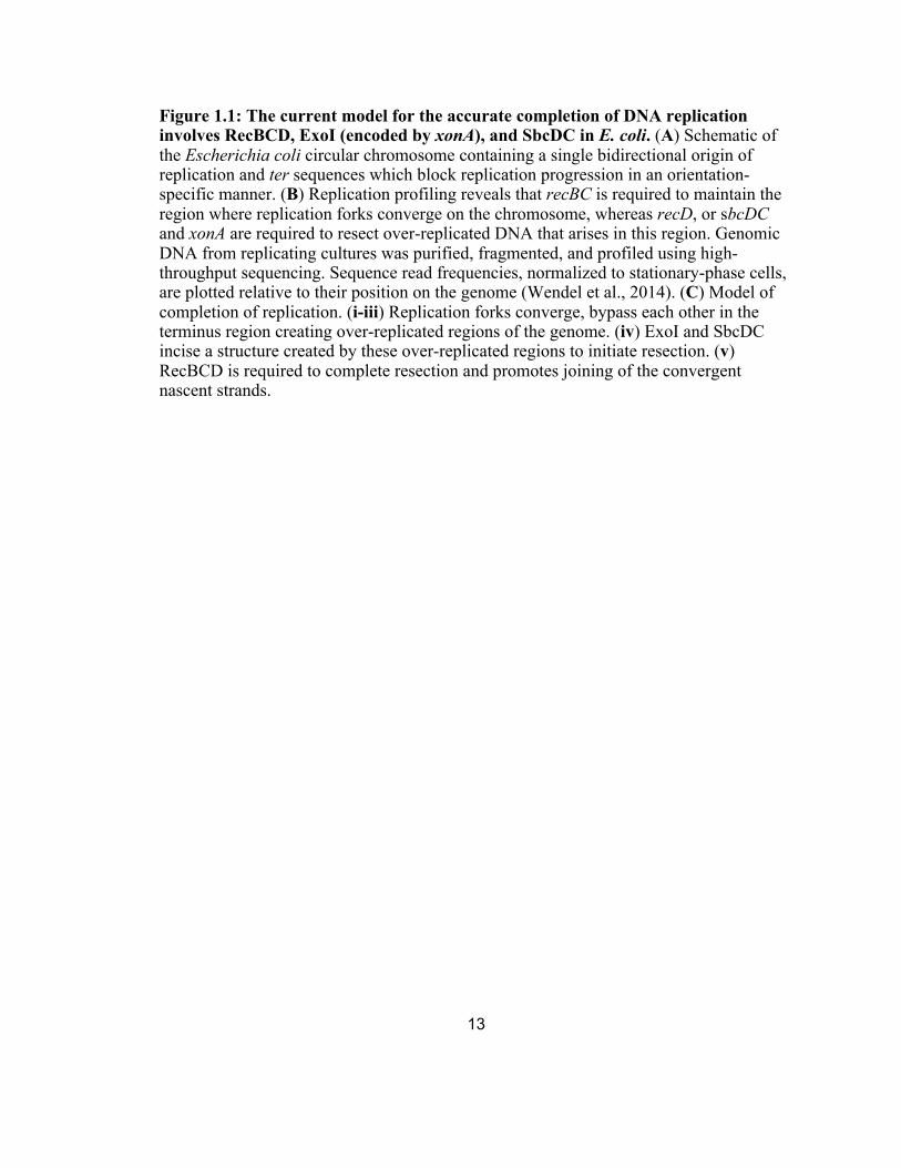
Figure 1.1: The current model for the accurate completion of DNA replication involves RecBCD, ExoI (encoded by xonA), and SbcDC in E. coli. (A) Schematic of the Escherichia coli circular chromosome containing a single bidirectional origin of replication and ter sequences which block replication progression in an orientation-specific manner. (B) Replication profiling reveals that recBC is required to maintain the region where replication forks converge on the chromosome, whereas recD, or sbcDC and xonA are required to resect over-replicated DNA that arises in this region. Genomic DNA from replicating cultures was purified, fragmented, and profiled using high-throughput sequencing. Sequence read frequencies, normalized to stationary-phase cells, are plotted relative to their position on the genome (Wendel et al., 2014). (C) Model of completion of replication. (i-iii) Replication forks converge, bypass each other in the terminus region creating over-replicated regions of the genome. (iv) ExoI and SbcDC incise a structure created by these over-replicated regions to initiate resection. (v) RecBCD is required to complete resection and promotes joining of the convergent nascent strands.

14
Chapter II
Methods and Materials
Bacterial strains and Plasmids
The strains used in this study were all derivatives of W3110; SR108 is a strain
incapable of synthesizing its own thymine; thyA36 deoC2 (Mellon and Hanawalt, 1989).
All other mutants are derived from this parent and described in Table 2.
Plasmid constructions were performed according to published protocols for in vivo
recombineering (Sawitzke et al., 2012), construction by amplification (Casini et al., 2014),
and Gibson assembly (Gibson et al., 2009). Plasmid and their features used in this study
are listed in Table 2.2. All plasmids containing unidirectional origins of replication were
derived from pBR322 (Ampicillin and Tetracycline resistance, pMB1 origin), which has
been described previously (Bolivar et al., 1977). pCL07 contains a chi sequence engineered
into leading strand of parent pBR322. pCL08 contains a chi sequence engineered into
lagging strand of parent pBR322. To accomplish this, primer pairs 5'-
catgcccggttactggaacggctggtggttgtgagggtaaacaactgg-3' + 5'-cgccgcatacactattctca-3' and 5'-
ccagttgtttaccctcacaaccaccagccgttccagtaaccgggcatg-3' + 5'-tgagaatagtgtatgcggcg-3' were
used for pCL07 and pCL08 respectively, with pBR322 as a template and amplified for 25
cycles using Pfu Turbo Polymerase (Agilent). PCR products were examined and purified
by agarose gel electrophoresis. The amplified fragments were combined with DpnI
digested pBR322, and the fragments were then joined and transformed using Gibson
assembly (New England Biolabs) to generate pCL07 and pCL08. Plasmids were sequenced
to verify sequence changes.
All plasmids containing bidirectional origins of replication are derived from
pCB104 (Potrykus et al., 2002) and contain an ampicillin-resistant cassette from pBR322.

15
pCL03 was constructed using primer pairs 5’-gtcggttcagggcagggtcgtgga-
cggtctgacagttaccaatgc-3’ and 5’-ggcggtttgcgtattgggcgcggtctgacagttaccaatgc-3’ to amplify
the ampR gene from pBR322. 0.2 μg gel purified PCR product was then combined with
0.5 μg BamHI-digested pCB104 and amplified for 25 cycles using Pfu Turbo Polymerase
(Agilent). PCR products were examined by agarose gel electrophoresis and products
running larger than 5kb were gel purified and transformed into recombineering strain
DY329 (Yu et al., 2000) to generate the ampicillin resistant plasmid, pCL03. pCL01 was
made by removing a chi sequence proximal to the origin using primer sets 5’-
attgctgataaatctgga-3’ + 5’-ctttggaatccagtccctcttcctcctgctgatctgcgacttatcaac-3’ and 5’-
tccagatttatcagcaat-3’ + 5’-gttgataagtcgcagatcagcaggaggaagagggactggattccaaag-3’ to
amplify overlapping fragments of the plasmid template using Pfu Turbo Polymerase
(Agilent). The fragments were then joined and transformed using Gibson assembly (New
England Biolabs) to generate pCL01. pCL05 was constructed by inserting a chi sequence
into the terminus region of plasmid pCL03, using plasmid pairs 5'-ctgcgctcggcccttccg-
gctgccaccagcattgctgataaatctgga-3' + 5'-tccagatttatcagcaatgctggtggcagcggaagggccgag-
-cgcag-3' and 5'-gttgataagtcgcagatcagcaggaggagaagagggactggattcc-aaag-3' + 5'-
ctttggaatccagtccctcttcctcctgctgatctgcgacttatcaac-3' to amplify overlapping fragments
which were joined and transformed using Gibson assembly (New England Biolabs) to
generate pCL05; mutation to inactive chi near λ bacteriophage origin of parental plasmid
pCL03 as well as mutation to active chi inside the ampicillin cassette. pCL02, pCL04, and
pCL06 are identical to pCL01, pCL03, and pCL05 but contain terB and terC sequences
inserted flanking the ampR in the terminus region. pCL04 was constructed using primer
pairs 5’-gtcggttcagggcagggtcgtggatccactttagttacaacatacttattcgcggaacccctatttgttt-3’and 5’-
ggcggtttgcgtattgggcgcatattagttacaacatcctatatggtctgacagttaccaatgc-3’to amplify the ampR

16
gene from pBR322. 0.2 μg gel purified PCR product was then combined with 0.5 μg
BamHI-digested pCB104 and amplified for 25 cycles using Pfu Turbo Polymerase
(Agilent). PCR products were examined by agarose gel electrophoresis and products
running larger than 5kb were gel purified and transformed into recombineering strain
DY329 (Yu et al., 2000) to generate the ampicillin resistant plasmid, pCL04. Primer pairs
5'-ctgcgctcggcccttccggctgccaccagcattgctgataaatctgga-3' +5'-tccagatttatcagcaatgct-
-ggtggcagcggaagggccgagcgcag-3' and 5'-gttgataagtcgcagatcagcaggaggagaagagggact-
-ggattccaaag-3' + 5'-ctttggaatccagtccctcttcctcctgctgatctgcgacttatcaac-3' were used to
amplify overlapping fragments of each plasmid. Fragments were joined with pCL04 as a
template and transformed using Gibson assembly (New England Biolabs) to generate
pCL02 and pCL06.
Transformation efficiency assay
Electro-competent cells were prepared by growing a 100-fold dilution of a fresh
overnight culture in 10 mL LB with thymine (LBthy) to an OD 600 of 0.4. Cells were then
pelleted, and serially washed with 30 mL water, 30 mL 10% glycerol, and then resuspended
in 200 μL of 10% glycerol and stored at -80°C. 40 μL of competent cells were mixed with
50 ng of plasmid and electroporated at 1.8 kV 25 μFD 200 Ohms and allowed to recover
at 37 °C for 30-60 minutes in 1 mL SOC media. The transformation reactions were then
diluted and aliquots were spread on LB thy plates with and without 50 ug/mL ampicillin to
determine the number of transformants and viable cells, respectively. Colonies were
counted following overnight incubation at 37 °C. The same preparations of competent cells
and plasmid preparations were used for comparisons between strains and plasmids. The
relative transformation efficiency of each strain was calculated as the ration of the

17
transformants per viable cells in the mutant cultures to the transformants per viable cells in
wild-type cultures.
Plasmid stability assay
Cells from overnight cultures of strains containing the plasmid grown in LBthy medium
with 50 μg/ml ampicillin were pelleted and used to inoculate 10ml cultures of LBthy
medium at 1:1000 dilution. Cultures were grown without ampicillin selection at 37 °C with
aeration overnight. The resulting cultures were then sampled to determine the ratio of cells
retaining the plasmid and used to reinoculated 10ml LBthy medium at 1:1000 dilution.
This was repeated for three iterations. To determine plasmid retention, 10-μl aliquots of
serial 10-fold dilutions were spotted on LBthy plates in the presence or absence of 50 μg/ml
ampicillin. Colonies were counted following overnight incubation at 37 °C to determine
the percent of plasmid-containing cells (Wendel et al., 2014).
Total genomic DNA extraction
750 μl of cultures was mixed with 750 μl of cold 2x NET (100 mM NaCl, 10 mM
Tris, pH 8.0, 10 mM EDTA). Cells were pelleted and frozen at -80 °C. Samples were
resuspended in 140 μl of lysozyme (1 mg/mL) and RNaseA (0.2 mg/mL) in TE (10 mM
Tris, pH 8.0, 1 mM EDTA) and lysed for 30 minutes at 37 °C. Then Sarkosyl (10 μl of
20% [wt/wt]) and Proteinase K (10 ul of 10 mg/mL) was added and incubation continued
for 60 minutes. Samples were then serially extracted with 4 volumes phenol/chloroform
(1/1) and 4 volumes chloroform followed by dialysis for 1 hour on 47 mm Whatman 0.05-
um pore disks (Whatman #VMWP04700) which were floated on a 250-mL beaker of TE
(1 mM Tris, pH 8.0, 1 mM EDTA).

18
Southern analysis of plasmid replication intermediates
Total genomic DNA was digested with SacII (New England BioLabs) for strains
containing pBR322 derived plasmids or NheI (New England BioLabs) for pCB104 derived
plasmids. In both cases, plasmids lack restriction sites for these enzymes. Samples were
then extracted with 1 volume of chloroform before equal cell equivalents were loaded on
to 0.5% and 1.0% TAE/TBE (220 mM Tris, 180 mM Borate, 5 mM EDTA, pH 8.3) agarose
gels and electrophoresed at 1 V/cm. Gels were transferred to Hybond N+ nylon membranes
(Amersham GE Healthcare) and probed with either the pBR322 or the pCL01 P32-labelled
plasmid. Radioactive labeling was carried out by nick translation kit (PerkinElmer) (Spivak
and Hanawalt, 1995). Radioactivity was visualized using a Storm 840 and its associated
ImageQuant Software (Molecular Dynamics).
Copy number analysis
Strains containing the plasmids or containing a chromosomal copy of the ampR
gene (HL946 or CL007) were grown and the genomic DNA purified as described above.
Plasmid DNA and chromosomal DNA was digested with EcoRV (New England Biolabs)
to linearize plasmid and chromosome species. DNA was then analyzed by standard
Southern analysis and quantified as described above.
Strain growth assay
Cultures containing 1 or 2-replisome plasmids, or neither, were grown in LB plus
selection over 800 minutes and continuously cataloged in 20-minute intervals for
absorbance in OD. The strain of E. coli used for this assay was also used to quantify the
copy number analysis which has the ampR gene (HL946 OR CL007). Growth curves

19
were graphed showing each cultures ability to replicate until plateauing into stationary
phase.

20
Tables
Table 2.1: E. coli strains used in this study
Courcelle Catalogue Genotype Source CL001 (SR108) WT Mellon & Hanawalt, 1989 CL002 recA Franklin, 1967 CL003 recBC Kushner, 1974 CL004 recD Thaler et al., 1989 CL039 xonA Kushner S et al., 1972 CL2344 sbcCD Gibson et al., 1992 CL2357 xonA sbcCD Jensen 1993 CL008 recG Chua., et al 1993 CL2542 recBC xonA sbcCD Wendel et al., 2018
Table 2.2: Plasmids used in this study Courcelle Catalogue
Construction Replisomes
Ter sequence trap
chi
pBR322 (Bolivar et al., 1977) +
pCL07 5’-catgcccggttactggaacggctggtg gtt- gtgagggtaaacaactgg-3’ +
+ + Leading strand
5’-cgccgcatacactattctca-3’ 5’-ccagttgtttaccctcacaaccaccag- ccgttccagtaaccgggcatg-3’ 5’-tgagaatagtgtatgcggcg-3’
pCL08 5’-ccagttgtttaccctcacaagctggtg gc- gttccagtaaccgggcatg-3’
+ + Lagging strand
5’-cgccgcatacactattctca-3’ 5’-catgcccggttactggaacgccaccagctt gtgagggtaaacaactgg-3’ 5’-tgagaatagtgtatgcggcg-3’
pCL01 5’-attgctgataaatctgga-3’ + ++ 5’-gttgataagtcgcagatcagca ggaggagaagagggactggattccaaag-3’ 5’-tccagattatcagcaat-3’ + 5’-ctttggaatccagtccctcttcctcctgctgatctgcgacttatcaac-3
pCL02 5’-attgctgataaatctgga-3’ + ++ ++ 5’-gttgataagtcgcagatcagcaggaggagaagag-ggactggattccaaag-3’

21
+: One-replisome plasmid
++: Two-replisome plasmid
+: Chi inserted
5’-tccagattatcagcaat-3’ + 5’-ctttggaatccagtccctcttcctcctgctga- tctgcgacttatcaac-3’
pCL03 5’-gtcggttcagggcagggtcgtggatcccgcggacccctatttgttt-3’ and 5’-ggcggtttgcgtattgggcg- -cggtctgacagttaccaatgc-3’
++ +proximalto origin
pCL04 5’gtcggttcagggcagggtcgtggatccactttagttaca- -acatacttattcgcggaacccctatttgttt-3’ and 5’ggcggtttgcgtattgggcgcatattagttacaacatc--ctatatggtctgacagttaccaatgc-3’
++ ++ +proximalto origin
pCL05 5’-ctgcgctcggcccttccg-gctgccaccagcatt- gctgataaatctgga-3’ +
terminus region +
5’-ctttggaatccagtccctcttcctcctgctga-tctgcgacttatcaac-3’ 5’-tccagatttatcagcaatgctggtggcagc-ggaagggccgagcgcag-3’ + 5’-gttgataagtcgcagatcagcaggaggagaag-agggactggattccaaag-3’
pCL06 5’-ctgcgctcggcccttccggctgccaccagcatt- gctgataaatctgga-3’ +
++ ++ terminus region +
5’-ctttggaatccagtccctcttcctcctgctgatctgcgacttatcaac-3’ 5’-tccagatttatcagcaatgctggtggcagcgg- aagggccgagcgcag-3’ + 5’-gttgataagtcgcagatcagcaggaggaga-agagggactggattccaaag-3’

22
Chapter III
Results
Part I - One-replisome versus two-replisome plasmids during completion of replication
Plasmids containing two-replisomes can be stably transformed, replicated, and grown in
cells similar to one-replisome plasmids.
Completion of replication on the chromosome involves an enzymatic process that
involves two convergent replisomes resolving with high fidelity (Wendel et al., 2014).
The convergent replisomes are thought to transiently bypass each other, before the excess
DNA is resected and joined at the point where all DNA has precisely doubled (Wendel et
al., 2014). Completion on the chromosome of E. coli can be challenging to study because
the event occurs once per cell cycle and its location can vary over a 400 kb stretch of the
genome (Campbell and Kleckner, 1990). In order to study the process and enzymes
involved in this reaction in more detail, it would be useful if it could be studied on
plasmids, which contain higher copy numbers and are only a few kilobases in size.
However, most plasmids contain unidirectional origins of replication and are replicated
by a single replisome, avoiding the event where two replisomes may converge (Reviewed
in del Solar et al., 1998). λ phage contains a bidirectional origin of replication that is
functionally homologous to that on the E. coli chromosome (Furth et al., 1977; Tabata et
al., 1983; Meijer et al., 1979), and has previously been shown to replicate as a mini-
chromosome when placed on a plasmid (Moore et al., 1977). Dr. Brian Wendel therefore
constructed a 5 kb plasmid containing the λ origin to determine if it could be used as a
model to study the completion reaction (Fig 3.1A), and initially characterized how well it
could transform cells, whether it affected cell growth, and the copy number at which it is
propagated in cells.

23
To examine how the plasmid with two replisomes affected cell growth, we
compared the growth of cell cultures containing the two-replisome plasmid, to that of
cultures containing the one-replisome plasmid pBR322 utilizing pMB1 (close relative of
ColE1) ori (Bolivar et al., 1977), or no plasmid at all. Cultures were grown over 800
minutes and continuously cataloged in 20-minute intervals for absorbance. As shown in
Figure 3.1B, the growth rate of each culture was similar; indicating that the two-
replisome plasmid does not impair growth of the host during replication.
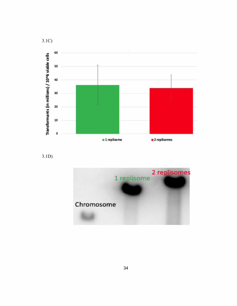
To determine how efficiently the two-replisome plasmid was transformed, 50 ng
of plasmid were transformed in 40 ul of wild-type competent cells via electroporation in
2 mm gap cuvettes at 2.5 kilovolts. After a 60-minute recovery period, dilutions of the
culture were then plated on LB with 50 ug/mL of ampicillin as well as on LB plates to
determine ratio of viable cells that were transformed (Figure 3.1C). Under transformation
similar conditions, both the one-replisome and two-replisome plasmid transformed with
similar efficiencies, ~200-400 transformants /106 viable cells, demonstrating the two-
replisome plasmid could enter and initially establish replication similar to other plasmids.
To determine the copy number at which the two-replisome plasmid was
maintained, we used Southern analysis in which we used a 32P-labeled ampicillin
resistance gene as a probe to compare the radioactive intensity of the signal on the
plasmid to the signal from a single copy ampicillin resistance gene integrated into the
chromosome. To this end, total genomic DNA was purified from cells containing the
plasmid pBR322, pCL01, or without plasmid but having an ampicillin resistance gene on
the chromosome. The genomic DNA was then digested with a restriction enzyme that
linearized each plasmid and then analyzed by Southern analysis following agarose-gel
electrophoresis. A representative gel is shown in Figure 3.1D. Overall, we found through

24
the radioactive intensity of the signals that the two-replisome plasmid was maintained
about 89 copies per chromosome, compared to about 63 copies for pBR322 (Fig. 3.1E).
Taken together, the two-replisome plasmid transform, propagate, and are maintained
similar to the one-replisome plasmid, pBR322.
Plasmids replicated by two replisomes are less stable than one replisome plasmids and
contain more aberrant, multimeric species.
To determine how stably the plasmid is maintained during replication, we
monitored the rate of plasmid loss over time in the absence of selection. To this end,
wild-type cultures containing either pBR322 or pCL01 were diluted 1/1000 and grown in
media without selection overnight, before the process was repeated the next day; similar
to the previously published plasmid stability protocol (Wendel et al., 2014). On each
passage, dilutions of a passaged sample, to promote proliferation of generations, sample
were plated to determine the ratio of cells that maintained the plasmid. Figure 3.2A
shows an example of these dilutions for cultures containing both the one-replisome and
two-replisome plasmid. We quantified and plotted these ratios over time and the results
are shown in Fig 3.2B. Whereas the one-replisome plasmid was stably maintained over
the ~30 generation assay, the proportion of cells that maintained the two-replisome
plasmid was reduced by ~two orders of magnitude over this same time period.
The results shown in Figure 3.1B-E argue that the instability of the two-replisome
plasmid, relative to the one-replisome plasmid is unlikely to be due to detrimental effects
on cultures growth rates or comparatively lower copy numbers. Further, neither pBR322
or pCL01 encode any partitioning mechanisms, which would control plasmid segregation

25
between daughter cells, that could account for the difference in stability (Nordstrom et
al., 1980; Austin et al., 1986).
Although the overall copy number between these plasmids were similar, we did
observe noticeable differences in the proportion of aberrant non-monomeric, amplified
substrates that appeared during the propagation of each plasmid. To examine the form in
which the plasmid DNA was maintained in each cell, total genomic DNA was purified
from cultures containing each plasmid, and the plasmid DNA was then analyzed by
Southern analysis following agarose gel electrophoresis using P32-labeled pBR322 and
pCL01 as probe. Representative gels from each are shown in Figure 3.2C and the results
are plotted in Figure 3.2D. Overall, the two-replisome plasmid contains elevated amounts
of abnormal, multimeric species relative to the one-replisome plasmid.
Similar to the completion of replication on the chromosome, amplifications and genetic
instability on two-replisome plasmid are driven by an aberrant RecA-mediated
recombination mechanism.
On the chromosome most of the genetic instability and amplifications that arise in
the region where replication completes is driven by an aberrant form of RecA-mediated
recombination (Wendel et al., 2014; Wendel et al., 2018). To determine if RecA plays a
similar role on the plasmid, we examined how its presence or absence affected the
stability of the plasmid when grown in the absence of selection, as before. As shown in
Figure 3.3A, inactivation of RecA increased the stability of the two-replisome plasmid.
The increase in stability brought the two-replisome plasmid to a level that was
comparable to the one-replisome plasmid once the ability for homologous recombination
was taken away.

26
We next examined how the form of the plasmid was maintained became affected
by the presence of RecA. To this end, total genomic DNA was purified from both wild-
type and recA mutant cultures containing plasmid and analyzed by Southern analysis as
previously described. The increased stability in the absence of RecA correlated with an
overall reduction in the amount of amplified, multimeric species (Figure 3.3B and C). A
similar reduction in multimeric plasmid species was also seen with the one-replisome
plasmid. The observations are consistent with the idea that RecA is driving the instability
on two-replisome plasmid and that the instability arises due to amplification or
multimeric species generated in its presence (Wendel et al., 2018).
Transformation of plasmids with two replisomes, but not one replisome, depends on the
enzymes required to complete replication on the chromosome.
On the chromosome, the completion of replication requires the RecBCD helicase-
nuclease (Wendel et al., 2014; reviewed in Courcelle et al., 2015). In its absence, the
genomic region where replication forks converge cannot be maintained, is extensively
degraded, and growth is severely compromised. Nucleases SbcCD and ExoI are also
required to initiate the completion reaction. In the absence of these gene products,
maintaining the region where replication forks converge becomes dependent on the
aberrant RecA-mediated reaction. In the absence of RecA, growth is severely
compromised in these mutants (Wendel et al., 2014; Wendel et al., 2018). In order to
determine if these genes are also involved in completing replication in the two-replisome
plasmid, we examined the ability of the two-replisome plasmid to transform mutant
strains deficient in completion enzymes. As a control, we also examined the ability of the
one-replisome plasmid to transform into these mutants. 50 ng of plasmid DNA was

27
transformed by electroporation into each mutant and the transformation efficiency for
each strain was determined, relative to wild-type. In the case of the one-replisome
plasmid, transformant samples were obtained for each of the mutants examined, and
successful transformation in each case occurred independently of recombination or RecA
(Figure 3.4A). However, in plasmid replication involving two replisomes, we observed
transformants in most of the mutants examined. Additionally, in recBC mutants the
transformation efficiency was reduced by greater than two orders of magnitude. In some
attempts, a few rare microcolonies could be observed on the selective plates following 3
days incubation (as opposed to the normal overnight incubation). However, in these
cases, we were unable to grow the transformants in liquid media beyond a single passage
(data not shown). Similarly, in mutants lacking the SbcDC ExoI nucleases,
transformation efficiency became dependent on the presence of recombination and RecA
(Figure 3.4 A). No other mutants examined depended on RecA for transformation of the
two-replisome plasmid. These genetic requirements for transformation of the two-
replisome plasmid are required to complete replication on the chromosome and suggest
that they are similarly required to complete replication on the two-replisome plasmid.
Further, the results would argue that the substrates acted upon by these enzymes, in vivo,
are specifically created when two replisomes converge, since one-replisome plasmids do
not exhibit any requirement for their presence.
Part II: The effect of Replication fork traps and the Completion reaction
Two-replisome plasmids with two additional ter sequences, terB and terC from
the Escherichia coli chromosome, are replicated and maintained at similar frequencies
as two-replisome plasmids without replication fork trap capabilities.

28
ter sequences, and their homologs, are found on several bacterial genomes,
including E. coli and Bacillus subtillis (Coskun-Ari et al., 1994) and function as
replication fork “traps” which bind to a protein called Tus, and halt replisomes
approaching from one side, in a polar manner (Kuempel et al., 1977; MacAllister et al.,
1990). Their presence ensures replisome convergence at the terminus area of the
chromosome. However, deletion of the tus reveals Tus-ter mutants have no observable
phenotype on growth or viability, arguing that replication fork traps are not essential
(Iismaa et al., 1987; Roecklein et al., 1991). Previous experiments have examined ter
elements in unidirectional plasmids. Perhaps not surprisingly, when ter is oriented in a
manner that blocks replisomes prior to the point of convergence this causes replication
difficulties and induces the SOS response (Hill and Marians., 1990; MacAllister et al.,
1990; Hasebe et al., 2018). In order to study how the Tus-ter traps affect the completion
reaction in E. coli, we engineered two ter sequences into the two-replisome plasmids in a
trapping orientation, similar to that found on the chromosome. The placement of terB and
terC flanking the terminus region, on the two-replisome plasmid is shown in Figure 3.5A.
As replication fork “trap” (Tus-ter) is dependent on the host derived protein Tus. The tus
gene is autoregulated and induced in the presence of unbound ter sequences (Natarajan et
al., 1991; Roecklein et al., 1991). Thus, the presence of additional ter sequences in the
newly constructed pCL02, would be expected to contain sufficient levels of Tus to ensure
that the polar arrest off the replication occurs at these sequences (Figure 3.1E).
To determine how the presence of ter traps affects the stability of plasmids
containing two replisomes, we monitored the rate of plasmid loss overtime in the absence
of selection with similar protocols as described in the previous section. As shown in
Figure 3.5B the two-replisome plasmid containing the ter trap approximately 10-fold less

29
stable than the non-ter containing plasmid during the ~30 generations of the experiment.
In recA mutants, which are deficient in homologous recombination, there is an increase in
plasmid stability similar to that seen in non-ter containing plasmids.
Plasmids replicated by two replisomes with the addition of ter sequences contain more
RecA-driven aberrant, multimeric species.
I next examined how the presence of ter sequences effect the form of replicating
plasmids that contain two replisomes. To this end, plasmids in replicating cultures were
purified and examined by Southern analysis as described above. As shown in Figure
3.5C, plasmids containing a ter trap contain a larger fraction of multimeric species,
however, these species migrate with a pattern that suggests they are intermediates that
form unit circles of dimers, trimers, and tetramers. By contrast, most of the multimeric
species in the plasmids lacking ter sequences migrate as a high linear multimers or
branched species. In recA mutants, fewer multimeric intermediates were observed
irrespective of the presence of ter sequences. In Figure 3.5D, I quantified the overall
levels of abnormal (non-monomeric) species in each strain. Overall, the results reflect the
RecA-catalyzed propagation of abnormal species in both pCL01 vs pCL02. Further, the
proportion of abnormal products correlates with the overall level of instability consistent
with what is observed on the chromosome; seen more specifically by the overamplified
products around the terminus region versus the amplified intermediates of the plasmid
substrates.
Part III: The effect of chi sequences on completing replication
chi sequences located at sites where replication forks converge promote a RecA-mediated
aberrant replication that correlates with instability
30
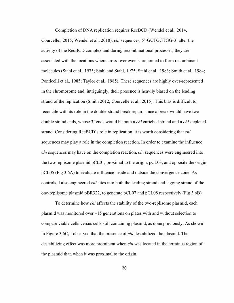
Completion of DNA replication requires RecBCD (Wendel et al., 2014,
Courcelle., 2015; Wendel et al., 2018). chi sequences, 5’-GCTGGTGG-3’ alter the
activity of the RecBCD complex and during recombinational processes; they are
associated with the locations where cross-over events are joined to form recombinant
molecules (Stahl et al., 1975; Stahl and Stahl, 1975; Stahl et al., 1983; Smith et al., 1984;
Ponticelli et al., 1985; Taylor et al., 1985). These sequences are highly over-represented
in the chromosome and, intriguingly, their presence is heavily biased on the leading
strand of the replication (Smith 2012; Courcelle et al., 2015). This bias is difficult to
reconcile with its role in the double-strand break repair, since a break would have two
double strand ends, whose 3’ ends would be both a chi enriched strand and a chi-depleted
strand. Considering RecBCD’s role in replication, it is worth considering that chi
sequences may play a role in the completion reaction. In order to examine the influence
chi sequences may have on the completion reaction, chi sequences were engineered into
the two-replisome plasmid pCL01, proximal to the origin, pCL03, and opposite the origin
pCL05 (Fig 3.6A) to evaluate influence inside and outside the convergence zone. As
controls, I also engineered chi sites into both the leading strand and lagging strand of the
one-replisome plasmid pBR322, to generate pCL07 and pCL08 respectively (Fig 3.6B).
To determine how chi affects the stability of the two-replisome plasmid, each
plasmid was monitored over ~15 generations on plates with and without selection to
compare viable cells versus cells still containing plasmid, as done previously. As shown
in Figure 3.6C, I observed that the presence of chi destabilized the plasmid. The
destabilizing effect was more prominent when chi was located in the terminus region of
the plasmid than when it was proximal to the origin.

31
To determine if the destabilizing effects of chi was dependent on the aberrant
RecA-mediated form of replication, we also examined the stability in recA mutants. I
observed that the absence of RecA improved the maintenance of all the plasmids,
although it did not eliminate all of the instability observed with one of the chi containing
plasmids. The results argue that chi destabilization is at least partially dependent on
RecA, but that chi has some effect even in the absence of RecA.
To examine whether the chi-destabilization of the plasmids altered its form during
the replication in vivo, we purified the DNA from replicating cultures and examined it by
Southern analysis. As shown in Figure 3.6D and E, in the presence of chi, an aberrant
high molecular weight intermediate was formed. The intermediate was most prominent
when chi was present in the terminus region of the chromosome, but could still be
observed when the chi site was located proximal to the origin of replication. The presence
and intensity of the specific aberrant intermediate correlated with the destabilization
effect chi has on the stable propagation of the plasmid.
The effect of chi appeared to be specific to the plasmid with two-replisomes as no
effect on plasmid stability was observed when chi sequences were present in either the
leading or lagging strand on the one-replisome plasmid (Fig 3.6F, Fig 3.6G and Fig
6.7H). The observation suggests that RecBCD processing during the completion reaction
is altered upon encountering a chi site in a manner that promotes further replication. At
least on the plasmid mini-chromosome substrate, this replication is destabilizing in its
effect.
Addition of a terB and terC trap reduces the destabilizing effect of chi by limiting the
amount of aberrant replication that can occur
32
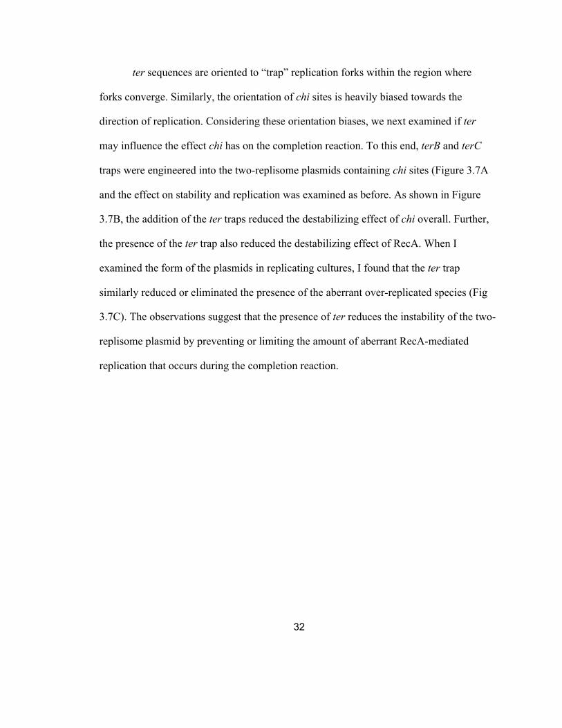
ter sequences are oriented to “trap” replication forks within the region where
forks converge. Similarly, the orientation of chi sites is heavily biased towards the
direction of replication. Considering these orientation biases, we next examined if ter
may influence the effect chi has on the completion reaction. To this end, terB and terC
traps were engineered into the two-replisome plasmids containing chi sites (Figure 3.7A
and the effect on stability and replication was examined as before. As shown in Figure
3.7B, the addition of the ter traps reduced the destabilizing effect of chi overall. Further,
the presence of the ter trap also reduced the destabilizing effect of RecA. When I
examined the form of the plasmids in replicating cultures, I found that the ter trap
similarly reduced or eliminated the presence of the aberrant over-replicated species (Fig
3.7C). The observations suggest that the presence of ter reduces the instability of the two-
replisome plasmid by preventing or limiting the amount of aberrant RecA-mediated
replication that occurs during the completion reaction.

33
Figures
3.1A)
3.1B)
34
3.1C)
3.1D)
35
3.1E)
Figure 3.1: The two-replisome plasmid, pCL01, can be stably transformed, replicated, and grown in cells similar to the one-replisome plasmid, pBR322. (A) Schematic of pBR322, containing a unidirectional origin of replication, and pCL01 which contains a λ bidirectional origin of replication. (B) Strains containing the two-replisome plasmid grow similar to those containing the one-replisome plasmid or no plasmid; E. coli strain included ampicillin cassette in this assay (includes data from B Wendel, unpublished). The growth of wild type cultures containing each plasmid as monitored by absorbance at 490nm is plotted over time. (C) Plasmids containing two replisomes can be stably transformed at frequencies similar to those with one replisome. The frequency of transformants per colony forming units per mL is plotted following electroporation of 50 ng of each plasmid into WT competent cells. Error bars represent standard error for 3 or more independent experiments. (D) The two-replisome plasmid is maintained at a copy number similar to the one replisome plasmid. A representative southern gel of strains containing the ampicillin resistance gene on the chromosome, on the plasmid pBR322, or the plasmid pCL01 is shown. Total genomic DNA was purified from each strain and digested as mentioned in Southern analysis of plasmid replication intermediates to linearize the plasmids. Equal number of cell equivalents were then loaded and analyzed by Southern analysis using a P32-labeled ampicillin resistance gene as a probe. (E) The copy number of each plasmid, relative to the chromosome is plotted (includes data from B Wendel, unpublished). Error bars represent the standard error for 4 independent experiments.

36
3.2A)
3.2B)

37
3.2C)

38
3.2D)
Figure 3.2: Plasmids replicated by two replisomes are less stable than one replisome plasmids and contain more aberrant species. (A) In the absence of selection, the two-replisome plasmids are lost more rapidly than the one-replisome plasmid. Cultures containing the one-replisome (pBR322) or two-replisome (pCL01) plasmid were grown for ~30 generations without selection. 10ul drops of 10-fold serial dilutions were plated with and without ampicillin selection to determine the fraction of cells that retain the plasmid. Arrows indicate the highest dilution that was observed to retain the plasmid (B) The fraction of cells retaining the one-replisome and two-replisome plasmid is plotted over time. Error bars represent the standard error of 4 or more independent experiments. (C) The instability of the two-replisome plasmid, relative to the one-replisome plasmid,correlates with the presence of more abnormal, multimeric species. Total genomic DNAfrom cells containing the one-replisome or two-replisome plasmid was purified andanalyzed by Southern analysis following agarose gel electrophoresis using P32-labeledpBR322 or pCL01 as a probe. (D) The fraction of unit length, non-monomeric plasmid incultures containing the one-replisome and two-replisome plasmid is plotted. Error barsrepresent the average of four or more independent experiments.
39
3.3A)
3.3B)

40
3.3C)
Figure 3.3: Similar to the chromosome, the amplifications and instability on two-replisome plasmids species are driven by the aberrant recombinational mechanism of completing DNA, RecA. (A) Inactivation of RecA restores the stability of the two-replisome plasmid to levels that approach that of the one-replisome plasmid. The fraction of cells retaining the one-replisome and two-replisome plasmid in the absence of selection is plotted over time. Error bars represent the standard error of at least 4 independent experiments. (B) The increased stability in the absence of the aberrant recombination pathway correlates with an increase in unit-length monomeric plasmids. Total genomic DNA from cells containing the one-replisome or two-replisome plasmid was purified and analyzed by Southern analysis following agarose gel electrophoresis using P32-labeled pBR322 or pCL01 as a probe. (C) The fraction of non-monomeric plasmid in each culture is plotted for the one-replisome and two-replisome plasmid in the presence and absence of RecA. Graphs represent the average of at least 4 independent experiments. Error bars represent the standard error.
WT
WT
recA
recA

41
3.4A)
Figure 3.4: Transformation of plasmids with two replisomes, but not one replisome, depends on the enzymes required to complete replication on the chromosome. (A) The transformation efficiency of the one-replisome and two-replisome plasmid, relative to wild type cells, is shown for the strains indicated. Error bars represent the standard error of at least two independent experiments (Includes data from B Wendel, unpublished).

42
PART II: No ter vs ters
3.5A)
3.5B)
terC terB

43
3.5C)
3.5D
WT
WT
recA
recA

44
Figure 3.5: ter traps engineered onto the two-replisome plasmid only modestly affect the overall stability of the plasmid but appear to increase the amount of abnormal non-monomeric circular plasmid species (A) Diagram of 2-replisome plasmids with and without ter traps (terB + terC). (B) The fraction of cells retaining the two-replisome plasmid in the absence of selection is plotted over time. Error bars represent the standard error of at least two independent experiments. (C) Increased amounts of RecA-dependent, non-monomeric plasmid species are observed in the presence of ter traps. Total genomic DNA from cells containing the indicated plasmid was purified and analyzed by Southern analysis following agarose gel electrophoresis using P32-labeled pCL01 as a probe. (D) The fraction of non-monomeric plasmid species in each culture is plotted. Graphs represent the average of at least 4 independent experiments. Error bars represent the standard error.
PART III: No chi versus chi
3.6A)
5’-GCTGGTGG-3’
5’-GCTGGTGG-3’

45
3.6B)
3.6C)
5’-GCTGGTGG-3’
5’-GCTGGTGG-3’

46
3.6D)

47
3.6E)
3.6F)
WT WT
WT
recA
recA
recA*

48
3.6G)
3.6H)
Lead Lead Lagg Lagg

49
Figure 3.6: Chi decreases the stability of two-replisome, but not one replisome plasmids, in a manner that correlates with the amount of aberrant linear multimeric plasmid that is observed. (A) Diagram of plasmid constructs comparing 2-replisome plasmid, pCL01, no chi control, pCL03 where chi is mutated near the ori site, pCL05 where chi is mutated opposite of ori site. (B) Diagram of one-replisome plasmids containing a chi inserted into the leading, pCL07, or lagging strand, pCL08, pBR322 template. (C) When present in the terminus region, chi destabilized the two-replisome plasmid. The fraction of cells retaining the two-replisome plasmid in the absence of selection is plotted over time. Error bars represent the Standard error of at least two independent experiments. (D) When present in the terminus region of the two-replisome plasmid, chi induces the formation of an aberrant linear plasmid multimeric species. Total genomic DNA from cells containing the indicated plasmid was purified and analyzed by Southern analysis following agarose gel electrophoresis using P32-labeled pCL01 as a probe. Arrow indicates the position of the linear plasmid multimers. (E) The amount of the linear plasmid multimeric species observed is plotted. Graphs represent the average of at least two independent experiments, except for pCL05 recA which represents a singular experiment. For all other strains, error bars represent the Standard error. (F) The stability of the one-replisome plasmid is not affected by the presence of chi. The fraction of cells retaining the two-replisome plasmid in the absence of selection is plotted over time. Error bars represent the Standard error of at least four independent experiments. (G) No multimeric species are induced by chi on one-replisome plasmids. Total genomic DNA from cells containing the indicated plasmid was purified and analyzed by Southern analysis following agarose gel electrophoresis using P32-labeled pBR322 as a probe. (H) The amount of the linear plasmid multimeric species observed is plotted. Graphs represent the average of at least 5 independent experiments. Error bars represent the standard error.

50
3.7A)
3.7B) terB terC

51
Figure 3.7C)
Figure 3.7: The presence of ter traps reduces the destabilization by chi, and prevents the formation of the RecA-mediated linear plasmid multimeric species (A) Diagram of two-replisome plasmids containing either a chi site in the terminus region or ter traps or both. (B) There is less destabilization by chi observed in the presence of a ter trap. The fraction of cells retaining the two-replisome plasmid in the absence of selection is plotted over time. Error bars represent the Standard error of at least two independent experiments (C) The amount of the linear plasmid multimeric species observed is plotted. Graphsrepresent the average of at least two independent experiments. Total genomic DNA fromcells containing the indicated plasmid was purified and analyzed by Southern analysisfollowing agarose gel electrophoresis using P32-labeled pCL01 as a probe. Except forpCL06 recA, which represents only a single experiment. For all other strains, error barsrepresent the Standard error.
2-replisome plasmids with ter, with chi, and with both
pCL02 pCL05 pCL06
% sp
ecie
s of i
nter
est w
ith c
hi /
tota
l spe
cies
WT
WT
WT recA
recA
recA
52
Chapter IV
Discussion
Part I: One-replisome versus two-replisome plasmids during completion of replication
The completion reaction involves two replisomes which converge, resect, resolve,
and join the nascent strands of DNA at the point where all sequences have doubled
(Wendel et al., 2014). These events occur thousands of times per cell cycle across human
chromosomes, meaning the event must occur with remarkable efficiency to maintain cell
viability (reviewed in Cvetic and Walter, 2005; Gao and Zhang, 2007; reviewed in
Méchali 2010). Prokaryotes, which contain a single bidirectional origin, offer a chance to
characterize this event and reaction in a less complex and better-defined system. The use
of plasmids can further simplify this analysis and have been successfully used in other
works to characterize the molecular mechanisms of replication initiation and elongation
(Reviewed in del Solar et al., 1998). However, commonly-used plasmids, such as
pBR322 that have origins of replication derived from ColE1, replicate with one-
replisome plasmids are unlikely to ever have a completion event where replication forks
converge similar to the chromosome (Abe 1980; Wendel et al., 2014). Thus, in this study,
I used a plasmid mini-chromosome that contained a bidirectional origin that could be
used to assess and examine the completion reaction. I show that maintaining plasmids
containing two replisomes depends on the enzymes needed to complete replication on the
chromosome. The completion of chromosomal replication requires RecBCD to join the
strands of convergent replication forks. In its absence, DNA ends persist, are extensively
degraded, and cells fail to maintain these regions of the chromosome (Dimude et al.,
2018A; Wendel et al., 204; Courcelle et al., 2015; Wendel et al., 2018). Similarly, I show
that transformation of two-replisome plasmids in recBC mutants is severely impaired and
53
the plasmids fail to propagate in cells under selection. On the chromosome, the ExoI
SbcDC structure-specific nucleases are required to initiate the faithful completion
reaction. In the absence of these proteins, normal completion cannot occur, excess over-
replicated regions persist, and the reaction is shunted through a RecA-mediated pathway
that is associated with amplifications and genetic instabilities (Indiani et al., 2013;
Dimude et al., 2018A; Wendel et al., 2014; Courcelle et al., 2015; Wendel et al., 2018).
Here, I show that transformation of two-replisome plasmids in xonA sbcCD mutants
similarly depends on the RecA-mediated pathway. Thus, both the normal RecBCD-
mediated reaction, and the aberrant recombinational process appear to operate on the
two-replisome plasmids. Additionally, I show that the requirement for these completion
enzymes is specific to plasmids with two replisomes, as transformation was not impaired
in the one-replisome plasmid pBR322. The observation implies that the substrate acted
upon by ExoI, SbcDC, and RecBCD during the completion reaction is specific to a
structure created when two replisomes converge.
Finally, I found that the two-replisome plasmid was less stable than the one-
replisome plasmid when propagated in the absence of selection, and showed that this
instability was driven by amplifications arising from a RecA-mediated recombination
reaction, similar to what is observed on the chromosome at sites where completion occurs
(Wendel et al., 2018). Inactivation of RecA increased the stability of the two-replisome
plasmid and reduced the proportion of abnormal amplification products that were
observed (Fig 3.3A). The high rate of RecA-driven instability on the two-replisome
plasmid indicates that the aberrant pathway occurs at relatively high frequency on the
plasmid, even when the normal, faithful pathway remains functional. I would speculate
that this is likely due to the high copy number of the bidirectional plasmid, and that the

54
frequency of aberrant completion events on the chromosome is likely to be much less.
Although pBR322 demonstrated relatively similar copy number I would not suggest this
to have the same effect as the accumulative assays suggest 1-replisome plasmids do not
engage or require the same enzymes to complete DNA replication. Under normal
conditions in E. coli, the completion enzymes are only required to catalyze a single
reaction on the chromosome. Whereas the bidirectional plasmid, which is maintained at
~80 copies/chromosome (Fig 3.1E), may exceed the reaction-capacity of the faithful
pathway, whose genes are not highly expressed (Eichler and Lehman, 1977; Taylor and
Smith, 1980), allowing the aberrant mechanism to operate more frequently than would
normally occur.
ExoI and SbcDC nuclease complexes are additive in their effect on the
completion reaction and inactivation of the normal completion reaction required deletion
of both genes (Wendel et al., 2014; Wendel et al., 2018; Dimude et al., 2018A). On the
chromosome, in the absence of both nucleases, the over-replicated regions persist, and
the ability to maintain growth and the chromosome region where forks converge becomes
entirely dependent on RecA; as the triple mutant xonA sbcDC recA shows a complete
lack of maintenance in the terminus region instead of over amplification (Wendel et al.,
2018). In this report, we show that transformation of two-replisome plasmids similarly
becomes dependent on RecA only in the absence of both nucleases (Fig. 3.4B).
Mechanistically, ExoI and SbcDC could functionally interact to cooperate and enhance
their ability to degrade the substrate(s) created by convergent replication forks. This
appears to occur in eukaryotes, where the homologous Mre11-Rad50 interacts with Sae2
and increase its exonuclease activity (Lengsfeld et al., 2007; Deng et al., 2015; Andres
and Williams, 2017). Consistent with this, a recent study from Dimude et al. found that
55
SbcDC alone prevented much of the degradation that occurs in recBC mutants (Dimude
et al., 2018B), an observation we have confirmed in our lab. In vitro, SbcCD has been
demonstrated to cleave a palindrome-like substrate similar to that predicted to arise when
replication forks bypass each other (Lim et al., 2015; Saathoff et al., 2018). Alternatively,
ExoI may act independently of SbcDC to suppress the aberrant recombinational pathway.
Early studies suggested a strong physical interaction between ExoI exonuclease and
RecA (Bedale et al., 1991; Bedale et al., 1993; Kowalczykowski et al., 1994).
Association of this 3’ exonuclease with RecA would be expected to strongly degrade
recombinogenic 3’ ends preventing RecA from initiating recombination at these sites.
In an alternative interpretation of RecBCD function, a recent study and review
speculated that chromosome cleavage may frequently occur during septation at cell
division, resulting in double strand breaks that require RecBCD for repair (Sinha et al.,
2018; Michel et al., 2018). The authors based this argument primarily on the observation
that the extensive degradation in recBC mutants’ centers upon the dif locus, where
chromosomes ultimately separate as cells divide. However, several observations argue
strongly against this possibility. If the defects in recBC mutants were due to the presence
of double strand breaks, then recA mutants, which is essential for repair of double strand
breaks, should be similarly, or more severely affected. Yet, in their initial study, the
authors failed to examine or address recA mutants (Sinha et al., 2018). However, recA
mutants grow at rates similar to wild type cultures, and unlike, recBC mutants, maintain
this region of the chromosome normally (Courcelle et al., 2015; Wendel et al., 2018).
Further, these authors and previous investigators all note that DNA breaks are not
detected on the chromosome of recA mutants (Sinha et al., 2018; Wendel et al., 2014;
Courcelle et al., 2015; Wendel et al., 2018; Pennington and Rosenberg, 2007). To explain

56
the absence of DNA breaks in recA mutants, a subsequent study from this group
proposed that the septation-induced breaks in recA genomes are subsequently degraded
and therefore undetectable (Sinha et al., 2018). However, based on the viability of recBC
mutants, ~90% of cells in culture would be experiencing these septation-induced breaks.
Synthesis and subsequent degradation of these genomes would be expected to slow
culture growth considerably and generate partial genomic degradation intermediates that
should be easily detectable, neither of which are observed in recA cultures (Sinha et al.,
2018; Wendel et al., 2014; Courcelle et al., 2015; Wendel et al., 2018). Further, such
models do not consider, and fail to address, how the inactivation of exonucleases SbcDC
and ExoI, which are not essential for double strand break repair, would restore recBCD
mutant growth defects, or why maintaining the terminus region of the chromosome in the
absence of these exonucleases would depend on RecA (Templin et al., 1972; Lloyd and
Buckman, 1985; Wendel et al., 2018). Finally, as shown here, plasmids containing two
replisomes require RecBCD to propagate but do not require RecA and are actually
stabilized in its absence. These plasmids lack dif sequences, and segregate prior to and
independent from cell division, and would therefore not be subject to septation-induced
breaks. These observations are all inconsistent with the idea that the requirement for
RecBCD is due to double strand breaks caused by cell division, and argue strongly in
favor of its role in joining DNA ends of convergent forks, which can occur independently
of recombination or RecA. It is also worth considering that although current models for
RecBCD in double-strand break repair propose that RecBCD acts prior to RecA, the early
in vivo studies led several independent labs to conclude that RecBCD acted after RecA, at
a later step in completing the recombination reaction (Wilkins 1969; Willets 1975; Hall
and Howard-Flanders, 1972; Birge and Low, 1974). Current models placing RecBCD as
57
an initiator are heavily derived from biochemical studies in which linear double-stranded
substrates were used to characterize enzyme binding, helicase, and exonuclease activities
(reviewed in (Yeeles and Dillingham, 2010; Dillingham and Kowalczykowski, 2008;
Smith 2012)). The initial concept that RecBC acts late in recombination arose from the
observation that although recA mutants receiving an F’ factor were unable to transfer
chromosomal genes to another cell, recBC mutants could do so at frequencies that
approached those of wild-type cells. However, over time (~1 generation), this ability
rapidly declined (Hall and Howard-Flanders, 1972; Wilkins 1969; Willetts 1975). The
authors inferred that recombination proceeds beyond the point at which the incoming
DNA is joined to the chromosome in recBC mutants, but that recA mutants are blocked
prior to this event. In recombinational crosses between Hfr and F- strains carrying non-
complementing mutations in lacZ, Birge and Low found that although recA mutants were
entirely blocked, recBC mutants initially produced beta-galactosidase within two-fold of
those seen in wild-type cells, indicating that recombination reactions progressed beyond
the point where transcribable, mutation-free copies of LacZ+ were produced (Birge and
Low, 1974). However, although these recombination intermediates were detectable, the
completion of these recombination events was impaired in the absence of RecBC and
viable LacZ+ recombinant progeny were reduced 100-1000-fold. The authors inferred
that “early steps in recombination can proceed efficiently in RecBC- and RecC-strains,
but that late steps, such as the degradation of excess DNA ‘tails’, might be defective.”
Using combinations of single and double mutations, Willetts confirmed these previous
studies and suggested that recombination proceeded past the first joining reaction of the
two DNA molecules, but that RecBCD was required for a second joining needed “to
generate a circular unit” that could be inherited (Willetts 1975). These interpretations are

58
strikingly consistent with RecBCD’s apparent role in completing replication on the
chromosome, and may suggest that RecBCD function during recombinational events is
similar to its role in completion of replication (Wendel et al., 2014; Courcelle et al.,
2015).
Replication fork traps and the completion reaction
Although not essential for viability, Tus-ter DNA-protein complexes are
important to localize the region where converging replication forks meet on the
chromosome (Roecklein et al., 1991; Reviewed in Rothstein et al., 2000). By adding terB
and terC sequences to the mini-chromosomes, I observed that their presence altered the
form of the replicating plasmid, effectively increasing the proportion of the plasmids that
migrated as circular forms. This had a modest but detrimental effect on the overall
stability of the plasmid, but could be argued to promote a more ‘normal’ circular DNA
product that can be passed on to the next generation, similar to the circular chromosome
of the bacterial host. The modest reduction in stability may be due to the unnaturally
small plasmid substrate in which the completion must occur. On the chromosome, this
reaction takes place within a 400-kb region. On the plasmid we ask the reaction to occur
within 5-kb, which may present some formational impediments for the reaction and
provide a rationale for why large circular products are observed. In future work, it would
be of interest to create a large plasmid substrate, to determine if this improves the
efficiency of the completion reaction. If plasmid stability increased with increasing
plasmid size, it would suggest that the completion reaction requires larger substrates to
occur. Given that RecBCD degrades and unwinds DNA at rates approaching 1000 bp/sec
(Roman et al., 1992) and that the distance between chi sites on the chromosome average

59
several kilobases (Burland et al., 1993), it seems reasonable to suspect that large
substrates may be required.
Nevertheless, the increased level of circle DNA products in the presence of ter
sequences implies that the ter sequences are limiting the amount of aberrant runaway
replication that occurs, and may provide more time for the completion to occur. I
observed that the presence of the ter sequences seemed particularly critical or beneficial
to the reaction, when chi sequences were present, which appear to promote the runaway
replication.
Chi sequences and the completion of replication
Chi sequences have been previously shown to determine where RecBCD
promoted crossover events that occur during recombination processes; thus, named
crossover hotspot instigator by Franklin Stahl (Lam et al., 1974; Henderson and Weil,
1975; Stahl et al., 1977; Stahl 1979). I examined how the presence of chi effected
RecBCD’s ability to catalyze the completion reaction on the bidirectional plasmid. I
observed that chi sequences increase the aberrant replication on the plasmid, and that this
correlates with a reduced stability of the plasmid. This appears to suggest that upon
encountering a chi site, RecBCD promotes the recruitment of replication. On the plasmid
substrates, this appears detrimental. This could be due to the small size or the absence of
other critical sequences on the plasmid. On the chromosome, it seems reasonable to
consider that this is likely to be a beneficial step. It may be that the chi sites induce the
RecBCD complex to cease degrading the DNA, perhaps as a ‘check’ to see if all of the
over-replicated regions have been degraded. If it has degraded beyond the over-replicated
region, then further synthesis would be needed to complete the replication process.
During recombination, chi sequences induce RecBCD to promote crossover events at

60
those locations (Stahl et al., 1977; Stahl 1979). We speculatively suggest that this may
similarly reflect joining events during the completion of replication or during the repair
of double strand breaks. In both cases, it would be critical that the joining occur at the
point where excess tails have been degraded, and we postulate that chi is acting to
promote this event. This type of activity may also explain why the location of chi
sequences is heavily biased to the leading strand template of the chromosome (Burland et
al., 1993), as the degradation of excess sequences are always orientated away from the
origin of replication, allowing more ‘checks’ to occur during resection in this direction.

61
Ch V
References
1. Abe, M. 1980. Replication of ColE1 Plasmid Deoxyribonucleic Acid in aThermosensitive dnaA Mutant of Escherichia coli. Journal of Bacteriology. 141: 1024-1030.
2. Al-Hadid Q, Ona Katherine, Courcelle C, Courcelle J. 2008. RecA433 Cells areDefective in recF-Mediated Processing of Disrupted Replication Forks but RetainrecBCD-Mediated Functions. Mutation Research. 645: 19-26.
3. Allgood N, Silhavy T. 1991. Escherichia coli xonA (sbcB) Mutants EnhanceIllegitimate Recombination. Genetics. 127: 671-680.
4. Amundsen S, Taylor A, Chaudhury A, Smith G. 1986. recD: The Gene for an EssentialThird Subunit of Exonuclease V. Proceedings in the National Academy of Sciences ofthe United States of America. 83: 5558-5562.
5. Amundsen S, Neiman A, Thibodeaux S, Smith G. 1990. Genetic Dissection of theBiochemical Activities of RecBCD Enzyme. Genetics. 126: 25-40.
6. Amundsen S, Smith G. 2007A. Chi Hotspot Activity in Escherichia coli WithoutRecBCD Exonuclease Activity: Implications for the Mechanism of Recombination.Genetics. 175: 41-54.
7. Amundsen S, Taylor A, Reddy M, Smith G. 2007B. Intersubunit Signaling in RecBCDEnzyme, a Complex Protein Machine Regulated by Chi Hot Spots. Genes &Development. 21: 3296-3307.
8. Amundsen S, Sharp J, Smith G. 2016. RecBCD Enzyme “Chi Recognition” MutantsRecognize Chi Recombination Hotspots in the Right DNA Context. Genetics. 204:139-152.
9. Amunugama R, Fishel R. 2012. Homologous Recombination in Eukaryotes. Progressin Molecular Biology and Translational Science. 110: 155-206.
10. Anderson D, Kowalcyzkowski S. 1997A. The Recombination Hot Spot 𝝌 is aRegulatory Element that Switches the Polarity of DNA Degradation by the RecBCDEnzyme. Genes & Development. 11: 571-581.
11. Anderson D, Churchill J, Kowalczykowski S. 1997B. Chi-Activated RecBCD EnzymePossesses 5’-3’ Nucleolytic Activity, but RecBC Enzyme Does Not: EvidenceSuggesting that the Alteration Induced by Chi is Not Simply Ejection of the RecDSubunit. Genes to Cells. 2: 117-128.
12. Anderson D, Churchill J, Kowalcyzkowski S. 1999. A Single Mutation, RecBD108AEliminates RecA Protein Loading but Not Chi Recognition by RecBCD Enzyme*. TheJournal of Biological Chemistry. 274: 27139-27144.
13. Andres S, Williams R. 2017. CtIP/Ctp1/Sae2, Molecular Form Fit for Function. DNARepair. 56: 109-117.
14. Austin S, Friedman S, Ludtke D. 1986. Partition Functions of Unit-Copy Plasmids canStabilize the Maintenance of Plasmid pBR322 at Low Copy Number. Journal ofBacteriology. 168: 1010-1013.
15. Bedale W, Inman R, Cox M. 1991. RecA Protein-Facilitated DNA Strand Breaks. Amechanism for Bypassing DNA Structural Barriers during Strand Exchange. Journalof Biological Chemistry. 266: 6499-6510.

62
16. Bedale W, Inman R, Cox M. 1993. A Reverse DNA Strand Exchange Mediated byrecA Protein and Exonuclease I. The Generation of Apparent DNA Strand Breaks byRecA Protein is Explained. Journal of Biological Chemistry. 268: 15004-15016.
17. Bianco P, Tracy R, Kowalczykowski S. 1998. DNA Strand Exchange Proteins: aBiochemical and Physical Comparison. Frontiers in Bioscience. 3:570-603.
18. Biek D, Cohen S. 1986. Identification and Characterization of recD, a Gene AffectingPlasmid Maintenance and Recombination in Escherichia coli. Journal of Bacteriology.167: 594-603.
19. Birge E, Low K. 1974. Detection of Transcibable Recombination Products FollowingConjugation in rec+, RecCB- and RecC-strains of Escherichia coli K12. Journal ofMolecular Biology. 83: 447-457.
20. Blattner F, Plunkett G, Block C, Perna N, Burland V, Riley M, Colladovides J, GlasnerJ, Rode c, Mayhew G, Gregor J, Davis N, Kirkpatrick H, Goeden M, Rose D, Mau B,Shao Y. The Complete Genome Sequence of Escherichia coli K-12. Science. 277:1453-1462.
21. Bolivar F, Rodriquez R, Greene P, Betlach M, Heyneker H, Boyer H, Crosa J,Falkow S. 1977. Construction and Characterization of New Cloning Vehicles. II. AMultipurpose Cloning System. Gene. 2: 95-113.
22. Boyce R, Howard-Flanders P. 1964. Release of Ultraviolet Light-induced ThymineDimers from DNA in E. coli K-12. Proceedings in the National Academy of Sciencesof the United States of America. 51:293-300.
23. Burland V, Plunkett G, Daniels D, Blattner F. 1993. DNA Sequence and Analysis of136 Kilobases of the Escherichi coli Genome: Organizational Symmetry around theOrigin of Replication. Genomics. 16: 551-561.
24. Campbell J, Kleckner N. 1990. E. coli oriC and the dnaA Gene Promoter areSequestered from dam methyltransferase Following the Passage of the ChromosomalReplication Fork. Cell. 62: 967-979.
25. Casini A, MacDonald J, De Jonghe J, Christodoulou G, Freemont P, Baldwin G,Elllis T. 2014. One-pot DNA Construction for Synthetic Biology: the ModularOverlap-Directed Assembly with Linkers (MODAL) strategy. Nucleic AcidsResearch. 42: e7.
26. Chattoraj D, Crasemann J, Dower N, Faulds D, Faulds P, Malone R, Stahl F, Stahl M.1979. Chi. Cold Spring Harbor Symposia On Quantitative Biology. 43: 1063-1066.
27. Chen Z, Yang H, Pavletich N. 2008. Mechanism of Homologous Recombination fromthe RecA-ssDNA/dsDNA Structures. Nature. 453: 489-494.
28. Cheng K, Smith G. 1987. Cutting of Chi-like Sequences by the RecBCD Enzyme ofEscherichia coli. Journal of Molecular Biology. 194: 747-750.
29. Cho C, Chung C, Li Hung. 2018. How Chi Sequences Modifies RecBCD Single-Stranded DNA Translocase Activity. ChemphysChem. 19: 243-247.
30. Chow K, Courcelle J. 2004. RecO Acts with RecF and RecR to Protect and MaintainReplication Forks Blocked by UV-Induced DNA Damage in Escherichia coli. Journalof Biological Chemistry. 279: 3492-3496.
31. Clark A, Margulies A. 1965. Isolation and Characterization of Recombination-Deficient Mutants of Escherichia Coli K12*. Proceedings in the National Academy ofSciences of the United States of America. 53: 451-459.
32. Clark A. 1991. Rec Genes and Homologous Recombination Proteins in Escherichiacoli. Biochimie. 73: 523-532.

63
33. Clark A. 1993, Sharma V, Brenowitz S, Chu C, Sandler S, Satin L, Templin A, BergerI, Cohen A. 1993. Genetic and Molecular Analyses of the C-Terminal Region of therecE Gene from the Rac Prophage of Escherichia coli K-12 Reveal the recT Gene.Journal of Bacteriology. 175: 7673-7682.
34. Coskun-Ari F, Skokotas A, Moe G, Hill T. 1994. Biophysical Characteristics of Tus,the Replication Arrest Protein of Escherichia coli. Journal of Biological Chemistry.269: 4027-4034.
35. Costa A, Hood I, Berger J. 2013. Mechanisms for Initiating Cellular DNA Replication.Annual Review Biochemistry. 82: 25-54.
36. Courcelle C, Chow K, Casey A, Courcelle J. 2006. Nascent DNA Processing by RecJFavors Lesion Repair Over Translesion Synthesis at Arrested Replication Forks inEscherichia coli. Proceedings of the National Academy of Science United States ofAmerica. 103: 9154-9159.
37. Courcelle J, Carswell-Crumpton C, Hanawalt P. 1997. recF and recR Are Required forthe Resumption of Replication at DNA Replication Forks in Escherichia coli.Proceedings of the National Academy of Science United States of America. 94: 3714-3719.
38. Courcelle J, Crowley D, Hanawalt P. 1999A. Recovery of DNA Replication in UV-Irradiated Escherichia coli Requires both Excision Repair and recF Protein Function.Journal of Bacteriology. 181: 916-922.
39. Courcelle J, Hanawalt P. 1999B. RecQ and RecJ Process Blocked Replication ForksPrior to the Resumption of Replication in UV-Irradiated Escherichia coli. Molecularand General Genetics. 262: 543-551.
40. Courcelle J, Khodursky A, Peter B, Brown P, Hanawalt P. 2001. Comparative GeneExpression Profiles Following UV Exposure in Wild Type and SOS DeficientEscherichia coli. Genetics. 158: 41-64.
41. Courcelle J, Donaldson J, Chow K, Courcelle C. 2003. DNA Damage-InducedReplication Fork Regression and Processing in Escherichia coli. Science. 299: 1064-1067.
42. Courcelle J, Wendel B, Livingstone D, Courcelle C. 2015. RecBCD is Required toComplete Chromosomal Replication: Implications for Double-Strand BreakFrequencies and Repair Mechanisms. DNA Repair. 32: 86-95.
43. Cox M, Lehman I. 1982. RecA Protein-Promoted DNA Strand Exchange. The Journalof Biological Chemistry. 257: 8523-8532.
44. Crowley D, Courcelle J. 2002. Answering the Call: Coping with DNA Damage at theMost Inopportune Time. Journal of Biomedicine and Biotechnology. 2: 66-74.
45. Cvetic C, Walter J. 2005. Eukaryotic Origins of DNA Replication: Could you PleaseBe More Specific? Seminars in Cell and Developmental Biology. 16: 343-353.
46. Dabert P, Ehrlich S, Gruss A. 1992. 𝝌 Sequence Protects Against RecBCD Degradationof DNA in vivo. Proceedings in the National Academy of Sciences of the United Statesof America. 89: 12073-12077.
47. de Vlaminck Iwijn, Loenhout M, Zweifel L, Blanken j, Hooning K, Hage S,Kerssemakers J. 2012. Mechanism of Homology Recognition in DNA Recombinationfrom Dual-Molecule Experiments. Molecular Cell. 46: 616-624.
48. del Solar G, Giraldo R, Ruiz-Echevarría M, Espinosa M, Díaz-Orejas R. 1998.Replication and Control of Circular Bacterial Plasmids. Microbiology and MolecularBiology Review. 62: 434-464.

64
49. Deng S, Yin Y, Petes T, Symington L. 2015. Mre11-Sae2 and RPA Collaborate toPrevent Palindromic Gene Amplification. Molecular Cell. 60: 500-508.
50. Dillingham M, Kowalczykowski S. 2008. RecBCD Enzyme and the Repair of Double-Stranded DNA Breaks. Microbiology and Molecular Biology Review. 72: 642-671.
51. Dimude J, Stein M, Andrzejewska E, Khalifa M, Gajdosova A, Retkute R, SkovgaardO, Rudolph C. 2018A. Origins Left, Right, and Centre: Increasing the Number ofInitiation Sites in the Escherichia coli Chromosome. Genes. 9: 2018/08/01.
52. Dimude J, Midgley-Smith S, Rudolph C. 2018B. Replication-Transcription ConflictsTrigger Extensive DNA Degradation in Escherichia coli Cells Lacking RecBCD. DNARepair. 70: 37-48.
53. Dixon D, Kowalcyzkowski S. 1993. The Recombination Hotspot 𝝌 Is a RegulatorySequence That Acts by Attenuating the Nuclease Activity of the E. coli RecBCDEnzyme. Cell. 73: 87-96.
54. Dohoney K, Gelles J. 2001. 𝝌-Sequence recognition and DNA Translocation by singleRecBCD Helicase/Nuclease molecules. Nature. 409: 370-374.
55. Eichler D, Lehman I. 1977. On the Role of ATP in Phosphodiester Bond HydrolysisCatalyzed by the RecBC Deoxyribonuclease of Escherichia coli. Journal of BiologicalChemistry. 252: 499-503.
56. Errico A, Constanzo V. 2012. Mechanisms of Replication Fork Protection: a Safeguardfor Genome Stability. Critical Review of Biochemical Molecular Biology. 47: 222-235.
57. Fan H, Li H. 2009. Studying RecBCD Helicase Translocation Along 𝝌-DNA UsingTethered Particle Motion with a Stretching Force. Biophysical Journal. 96: 1875-1883.
58. Franklin N. 1967. Extraordinary Recombinational Events in ESCHERICHIA COLI.Their Independence of the rec+ Function. Genetics. 1967: 699-707.
59. Friedberg E. 1995. Out of the Shadows and Into the Light: the Emergence of DNARepair. Trends in Biochemical Science. 20: 381.
60. Furth M, Blattner F, McLeester C, Dove W. 1977. Genetic Structure of theReplication Origin of Bacteriophage Lambda. Science. 198: 1046-1051
61. Gao F, Zhang C. 2007. DoriC: a Database of oriC Regions in Bacterial Genomes.Bioinformatics. 14: 1866-1867.
62. Gibson F, Leach D, Lloyd R. 1992. Identification of sbcD Mutations as Co-Suppressors of recBC That Allow Propagation of DNA Palindromes in Escherichiacoli K-12. Journal of Bacteriology. 174: 1222-1228.
63. Gibson D, Young L, Chuang R, Venter J, Hutchison C, Smith H. 2009. EnzymaticAssembly of DNA Molecules up to Several Hundred Kilobases. Nature Methods. 6:343-345.
64. Gonzalez M, Frank E, McDonald J, Levine A, Woodgate R. 1998. Structural Insightsinto the Regulation of SOS Mutagenesis. The Journal of the Polish Biochemical Societyand of the Committee of Biochemistry and Biophysics Polish Academy of Sciences. 45:163-172.
65. Gorden E, Sturk-Andreaggi K, Marshall C. 2018. Repair of DNA Damage Caused byCytosine Deamination in Mitochondrial DNA of Forensic Case Samples. ForensicScience International: Genetics. 34: 257-264.
66. Hall J, Howard-Flanders P. 1972. Recombinant F’ Factors from Escherichia coli K-12Strains Carrying RecB or RecC. Journal of Bacteriology. 110: 578-584.
67. Hanawalt P, Ford J, Lloyd D. 2003. Functional Characterization of Global GenomicDNA Repair and its Implications for Cancer. Mutation Research. 544: 107-114.

65
68. Handa N, Kobayashi I. 2005A. Type III Restriction Is Alleviated by Bacteriophage(RecE) Homologous Recombination Function but Enhanced by Bacterial (RecBCD)Function. Journal of Bacteriology. 187: 7362-7373.
69. Handa N, Bianco P, Baskin R, Kowalcyzkowski S. 2005B. Direct Visualization ofRecBCD Movement Reveals Cotranslocation of the RecD Motor after 𝝌 Recognition.Molecular Cell. 17: 745-750.
70. Handa N, Yang L, Dillingham M, Kobayashi I, Wigley D, Kowalcyzkowski S. 2012.Molecular Determinants Responsible for Recognition of the Single-Stranded DNARegulatory Sequence, 𝝌, by RecBCD Enzyme. Proceedings in the National Academyof Sciences of the United States of America. 109: 8901-8906.
71. Hasebe T, Narita K, Hidaka S, Su’etsugu M. 2018. Efficient Arrangement of theReplication Fork Trap for in vitro Propagation of Monomeric Circular DNA in theChromosome-Replication Cycle Reaction. Life. 8: 2018/09/28.
72. Henderson D, Weil J. 1975. Recombination-Deficient Deletions in Bacteriophage λAnd Their Interaction with Chi Mutations. Genetics. 79:143-174.
73. Heyer WD, Ehmsen KT, Liu J. 2010. Regulation of Homologous Recombination inEukaryotes. Annual Review of Genetics. 44: 113-139.
74. Hill T, Marians K. 1990. Escherichia coli Tus Protein Acts to Arrest the Progressionof DNA Replication Forks in vitro. Proceedings in the National Academy of Sciencesof the United States of America. 87: 2481-2485.
75. Hopefield J. 1974. Kinetic Proofreading: a new Mechanism for Reducing Errors inBiosynthetic Processes Requiring High Specificity. Proceedings in the NationalAcademy of Sciences of the United States of America. 71: 4135-139.
76. Hopefield J, Yamane T, Yue V, Coutts S. 1976. Direct Experimental Evidence forKinetic Proofreading in Amino Acalystion of tRNAIle. Proceedings in the NationalAcademy of Sciences of the United States of America. 73: 1164-1168.
77. Horiuchi T, Nishitani H, Kobayashi T. 1995. A New Type of E. coli RecombinationalHotspot Which Requires for Activity Both DNA Replication Termination Events andthe Chi Sequence. Advanced Biophysics. 31: 133-147.
78. Howard-Flanders P, Boyce R. 1966A. DNA Repair and Genetic Recombination:Studies on Mutants of Escherichia coli Defective in These Processes. RadiationResearch Supplement. 6: 156-184.
79. Howard-Flanders P, Theriot L. 1966B. Mutants of Escherichia Coli K-12 Defective inDNA Repair and in Genetic Recombination. Genetics. 53: 1137-1150.
80. Iismaa T, Wake R. 1987. The Normal Replication Terminus of the Bacillus subtilisChromosome, terC, is Dispensable for Vegetative Growth and Sporulation. Journal ofMolecular Biology. 195: 299-310.
81. Indiani C, Patel Meghna, Goodman M, O’Donnell M. 2013. RecA Acts as a Switch toRegulate Polymerase Occupancy in a Moving Replication Fork. Proceedings of theNational Academy of Sciences of the United States of America. 110: 5410-5415.
82. Jensen K. 1993. The Escherichia coli K-12 “Wild Types” W3110 and MG1655 havean rph Frameshift Mutation that Leads to Pyrimidine Starvation Due to Low pyrEExpression Levels. Journal of Bacteriology. 175: 3401-3407.
83. Karu A, Belk E. 1982. Induction of E. coli RecA protein via RecBC and AlternativePathways: Quantitation by Enzyme-Linked Immunosorbent Assay (ELISA).Molecular Genomics and Genetics. 185: 275-282.

66
84. Kiem P, Lark KG. 1990. The RecE Recombination Pathway Mediates RecombinationBetween Partially Homologous DNA Sequences: Structural Analysis ofRecombination Products. Journal of Structural Biology. 104: 97-106.
85. Klein H, Kreuzer K. 2002. Replication, Recombination, and Repair: Going for theGold. Molecular Cell. 9: 471-480.
86. Kobayashi I, Murialdo H, Crasemann J, Stahl M, Stahl F. 1982. Orientation ofCohesive End Site cos Determines the Active Orientation of 𝝌 Sequence in StimulatingrecA・recBC-mediated Recombination in Phage λ Lytic Infections. Proceedings in theNational Academy of Sciences of the United States of America. 79: 5981-5985.
87. Köppen A, Krobitsch S, Thoms B, Wackernagel W. 1995. Interactions with therecombination hot spot 𝝌 in vivo converts the RecBCD enzyme of Escherichia coli into a 𝝌-independent recombinase by inactivation of the RecD subunit. Proceedings inthe National Academy of Sciences of the United States of America. 92: 6249-6253.
88. Kowalczykowski S, Dixon D, Eggleston A, Lauder S, Rehrauer W. 1994. Biochemistryof Homologous Recombination in Escherichia coli. Microbiology Review. 58: 401-465.
89. Kuempel P, Duerr S, Seeley N. 1977. Terminus Region of the Chromosome inEscherichia coli Inhibits Replication Forks. Proceedings in the National Academy ofScience of the United States of America. 74: 3927-3931.
90. Kulkarni A, Julin D. 2004. Specific Inhibition of the E. coli RecBCD Enzyme by ChiSequences in Single-Stranded Oligodeoxyribonucleotides. Nucleic Acids Research. 32:3672-3682.
91. Kushner S, Nagaishi H, Templin A, Clark A. 1971. Genetic Recombination inEscherichia coli: The Role of Exonuclease I. Proceedings in the National Academy ofSciences of the United States of America. 68: 824-827.
92. Kushner S, Nagaishi H, Clark A. 1972. Indirect Suppression of recB and recCMutations by Exonuclease I Deficiency. Proceedings in the National Academy ofSciences of the United States of America. 69: 1366-1370.
93. Kushner S. 1974. Differential Thermolability of Exonuclease and EndonucleaseActivities of the recBC Nuclease Isolated from the Thermosensitive recB and recCMutants. Journal of Bacteriology. 120: 1219-1222.
94. Kuzminov A, Schabtach E, Stahl F. 1994. 𝝌 Sites in Combination with RecA ProteinIncrease the Survival of Linear DNA in Escherichia coli By Inactivating ExoV Activityof RecBCD Nuclease. The EMBO Journal. 13: 2764-2776.
95. Lam S, Stahl M, McMilin K, Stahl F. 1974. Rec-mediated Recombinational Hot SpotActivity In Bacteriophage Lambda. Genetics. 77: 425-433.
96. Leanderson P, Tagesson C. 1992. Cigarette Smoke-Induced DNA Damage in CulturedHuman Lung Cells: Role of Hydroxyl Radicals and Endonuclease Activation.Chemical Biology Interactions 81: 197-208.
97. Lee J, Terakawa T, Qi Z, Steinfeld J, Redding S, Kwon Y, Gaines W, Zhao W, SungP, Greene E. 2015. Base triplet stepping by the Rad51/RecA family of recombinases.Science. 349: 977-981.
98. Lengsfeld B, Rattray A, Bhaskara V, Ghirlando R, Paull T. 2007. Sae2 is anEndonuclease that Processes Hairpin DNA Cooperatively with the Mre11/Rad50/Xrs2Complex. Molecular Cell. 28: 638-651.
99. Lesterlin C, Ball G, Schermelleh L, Sherratt D. 2014. RecA bundles mediate homologypairing between distant sisters during DNA break repair. Nature. 506: 249-253.

67
100. Lloyd R, Thomas A. 1983. On the Nature of the RecBC and RecF Pathways ofConjugal Recombination in Escherichia coli. Molecular Genomics and Genetics. 190:156-161.
101. Lloyd R, Buckman C. 1985. Identification and Genetic Analysis of sbcC Mutationsin Commonly Used recBC sbcB Strains of Escherichia coli K-12. Journal ofBacteriology. 164: 836-844.
102. MacAllister T, Khatri G, Bastia D. 1990. Sequence-Specific and PolarizedReplication Termination in vitro: Complementation of Extracts of tus- Escherichia coliby Purified Ter Protein and Analysis of Termination Intermediates. Proceedings of theNational Academy of Science of the United States of America. 87: 2828-2832.
103. Lim C, Lai P, Leach D, Maki H, Furukohri A. 2015. A Novel Mode of NucleaseAction is Revealed by the Bacterial Mre11/Rad50 Complex. Nucleic Acids Research.43: 9804-9816.
104. Mackay V, Linn S. 1974. The Mechanism of Degradation of DuplexDeoxyribonucleic Acid by the recBC Enzyme of Escherichia coli K-12*. Journal ofBiological Chemistry. 249: 4286-4294.
105. Malone R, Chattoraj D, Faulds D, Stahl M, Stahl F. 1978. Hotspots for GeneralizedRecombination in the Escherichia coli Chromosome. Journal of Molecular Biology.121: 473-491.
106. Mellon I, Hanawalt P. 1989. Induction of the Escherichia coli Lactose OperonSelectively Increases Repair of its Transcribed DNA Strand. Nature. 342: 95-98.
107. Meijer M, Beck E, Hansen F, Bergmans H, Messer W, Meyenburg K, Schaller H.1979. Nucleotide Sequence of the Origin of Replication of the Escherichia coli K-12Chromosome. Proceedings of the National Academy of Science. 76: 580-584.
108. Méchali M. 2010. Eukaryotic DNA Replication Origins: Many Choices forAppropriate Answers. Nature Reviews of Molecular Cell Biology. 11: 728-738.
109. Meselson M, Weigle J. 1961. Chromosome Breakage Accompanying GeneticRecombination in Bacteriophage*. Genetics. 47: 857-868.
110. Michel B, Sinha A, Leach D. 2018. Replication Fork Breakage and Restart inEscherichia coli. Microbiology and Molecular Biology Review. 82: 2018/06/15.
111. Moore D, Denniston-Thompson K, Furth M, Williams B, Blattner F. 1977.Construction of Chimeric Phages and Plasmids Containing the Origin of Replicationof Bacteriophage Lambda. Science. 198: 1041-1046.
112. Murphy K. 1991. λ Gam Protein Inhibits the Helicase and 𝝌-StimulatedRecombination Activities of Escherichia coli RecBCD Enzyme. Journal ofBacteriology. 173: 5808-5821.
113. Natarajan S, Kelley W, Bastia D. 1991. Replication Terminator Protein ofEscherichia coli is a Transcriptional Repressor of its Own Synthesis. Proceedings ofthe National Academy of Science of the United States of America. 88: 3867-3871.
114. Nordstrom K, Molin S, Aagaad-Hansen H. 1980. Partitioning of Plasmid R1 inEscherichia coli. I. Kinetics of Loss of Plasmid Derivatives Deleted of the par Region.Plasmid. 4: 215-217.
115. O’Donnell M, Langston L, Stillman B. 2013. Principles and concepts of DNAreplication in bacteria, archaea, and eukarya. Cold Spring Harbor Perspective inBiology. 5: 1-13.
116. Pennington J, Rosenberg S. 2007. Spontaneous DNA Breakage in Single LivingEscherichia coli cells. Nature Genetics. 39: 797-802.
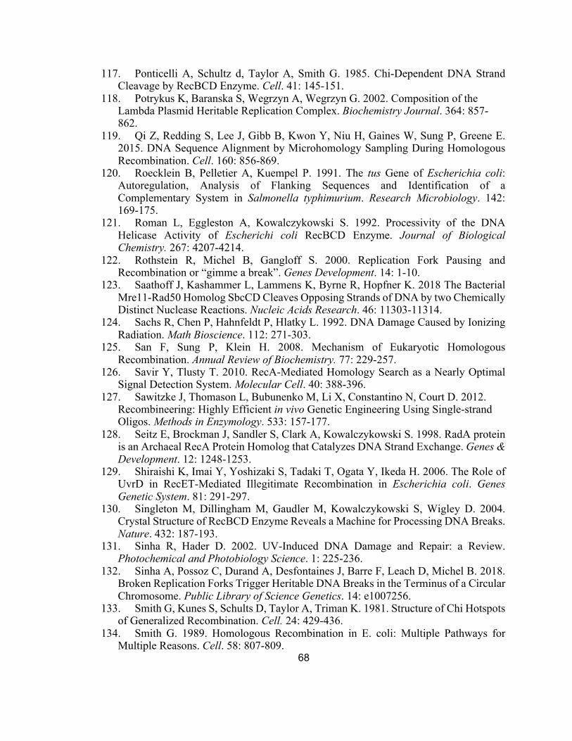
68
117. Ponticelli A, Schultz d, Taylor A, Smith G. 1985. Chi-Dependent DNA StrandCleavage by RecBCD Enzyme. Cell. 41: 145-151.
118. Potrykus K, Baranska S, Wegrzyn A, Wegrzyn G. 2002. Composition of theLambda Plasmid Heritable Replication Complex. Biochemistry Journal. 364: 857-862.
119. Qi Z, Redding S, Lee J, Gibb B, Kwon Y, Niu H, Gaines W, Sung P, Greene E.2015. DNA Sequence Alignment by Microhomology Sampling During HomologousRecombination. Cell. 160: 856-869.
120. Roecklein B, Pelletier A, Kuempel P. 1991. The tus Gene of Escherichia coli:Autoregulation, Analysis of Flanking Sequences and Identification of aComplementary System in Salmonella typhimurium. Research Microbiology. 142:169-175.
121. Roman L, Eggleston A, Kowalczykowski S. 1992. Processivity of the DNAHelicase Activity of Escherichi coli RecBCD Enzyme. Journal of BiologicalChemistry. 267: 4207-4214.
122. Rothstein R, Michel B, Gangloff S. 2000. Replication Fork Pausing andRecombination or “gimme a break”. Genes Development. 14: 1-10.
123. Saathoff J, Kashammer L, Lammens K, Byrne R, Hopfner K. 2018 The BacterialMre11-Rad50 Homolog SbcCD Cleaves Opposing Strands of DNA by two ChemicallyDistinct Nuclease Reactions. Nucleic Acids Research. 46: 11303-11314.
124. Sachs R, Chen P, Hahnfeldt P, Hlatky L. 1992. DNA Damage Caused by IonizingRadiation. Math Bioscience. 112: 271-303.
125. San F, Sung P, Klein H. 2008. Mechanism of Eukaryotic HomologousRecombination. Annual Review of Biochemistry. 77: 229-257.
126. Savir Y, Tlusty T. 2010. RecA-Mediated Homology Search as a Nearly OptimalSignal Detection System. Molecular Cell. 40: 388-396.
127. Sawitzke J, Thomason L, Bubunenko M, Li X, Constantino N, Court D. 2012.Recombineering: Highly Efficient in vivo Genetic Engineering Using Single-strandOligos. Methods in Enzymology. 533: 157-177.
128. Seitz E, Brockman J, Sandler S, Clark A, Kowalczykowski S. 1998. RadA proteinis an Archaeal RecA Protein Homolog that Catalyzes DNA Strand Exchange. Genes &Development. 12: 1248-1253.
129. Shiraishi K, Imai Y, Yoshizaki S, Tadaki T, Ogata Y, Ikeda H. 2006. The Role ofUvrD in RecET-Mediated Illegitimate Recombination in Escherichia coli. GenesGenetic System. 81: 291-297.
130. Singleton M, Dillingham M, Gaudler M, Kowalczykowski S, Wigley D. 2004.Crystal Structure of RecBCD Enzyme Reveals a Machine for Processing DNA Breaks.Nature. 432: 187-193.
131. Sinha R, Hader D. 2002. UV-Induced DNA Damage and Repair: a Review.Photochemical and Photobiology Science. 1: 225-236.
132. Sinha A, Possoz C, Durand A, Desfontaines J, Barre F, Leach D, Michel B. 2018.Broken Replication Forks Trigger Heritable DNA Breaks in the Terminus of a CircularChromosome. Public Library of Science Genetics. 14: e1007256.
133. Smith G, Kunes S, Schults D, Taylor A, Triman K. 1981. Structure of Chi Hotspotsof Generalized Recombination. Cell. 24: 429-436.
134. Smith G. 1989. Homologous Recombination in E. coli: Multiple Pathways forMultiple Reasons. Cell. 58: 807-809.

69
135. Smith G. 2012. How RecBCD Enzyme and Chi Promote DNA Break Repair andRecombination: a Molecular Biologist’s View. Microbiology and Molecular BiologyReview. 76: 217-228.
136. Spies M, Bianco P, Dillingham M, Handa N, Baskin R, Kowalczykowski S. 2003.A Molecular Throttle: The Recombination Hotspot 𝝌 Controls DNA Translocation bythe RecBCD Helicase. Cell. 114: 647-654.
137. Spies M, Dillingham M, Kowalczykowski S. 2005. Translocation by the RecBMotor Is an Absolute Requirement for 𝝌-Recognition and RecA Protein Loading byRecBCD Enzyme. Journal of Biological Chemistry. 280: 37078-37087.
138. Spies M, Amitani I, Baskin R, Kowalcyzkowski S. 2007. RecBCD EnzymeSwitches Lead Motor Subunits in Response to 𝝌 Recognition. Cell. 131: 694-705.
139. Spivak G, Hanawalt P. 1995. Determination of Damage and Repair in SpecificDNA Sequences. Methods: A Companion to Methods in Enzymology. 7: 147-161.
140. Stahl F, McMilin K, Stahl M, Nozu Y. 1972. Proceedings of the National Academyof Science United States of America. 69: 3598-3601.
141. Stahl F, Crasemann J, Stahl M. 1975. Rec-mediated Recombinational Hot SpotActivity in Bacteriophage Lambda. III. Chi Mutations are Site-Mutations StimulatingRec-Mediated Recombination. Journal of Molecular Biology. 94: 203-212.
142. Stahl F, Stahl M. 1975. Rec-mediated Recombinational Hot Spot Activity inBacteriophage Lambda. IV. Effect of Heterology on Chi-Stimulated Crossing over.Molecular Genetics and Genomics. 140: 29-37.
143. Stahl F, Stahl M. 1977. Recombination Pathway Specificity of Chi. Genetics. 86:715-725.
144. Stahl F. 1979. Special Sites in Generalized Recombination. Annual Review ofGenetics. 13:7-24.
145. Stahl M, Kobayashi I, Stahl F, Huntington S. 1983. Activation of Chi, aRecombinator, by the Action of an Endonuclease at a Distant Site. Proceedings of theNational Academy of Science United States of America. 80: 2310-2313.
146. Stahl F, Thomason L, Siddiqi I, Stahl M. 1990. Further Tests of a RecombinationModel in which 𝝌 Removes the RecD Subunit from the RecBCD Enzyme ofEscherichia coli. Genetics. 126: 519-533.
147. Stasiak A, Stasiak AZ, Koller T. 1984. Visualization of RecA-DNA ComplexesInvolved in Consecutive Stages of an In Vitro Strand Exchange Reaction. Cold SpringHarbor Symposium Quantitative Biology. 49: 561-570.
148. Sung P, Klein H. 2006. Mechanism of Homologous Recombination: Mediators andHelicases Take on Regulatory Functions. Nature Review Molecular Cell Biology. 7:739-750.
149. Tabata S, Oka A, Sugimoto K, Takanami M, Yasuda S, Hirota Y. 1983. The 245Base-pair oriC Sequence of the E. coli Chromosome directs Bidirectional Replicationat an Adjacent Region. Nucleic Acids Research. 11: 2617-2626.
150. Taylor A, Smith G. 1980. Unwinding and Rewinding of DNA by the RecBCDEnzyme. Cell. 22: 447-457.
151. Taylor A, Schultz D, Ponticelli A, Smith G. 1985. RecBC Enzyme Nicking at ChiSites During DNA Unwinding: Location and Orientation-Dependence of the Cutting.Cell. 41:153-163.
152. Taylor A, Smith G. 1992. RecBCD Enzyme is Altered Upon Cutting DNA at a ChiRecombination Hotspot. Proceedings in the National Academy of Sciences of theUnited States of America. 89: 5226-5230.

70
153. Taylor A, Smith G. 1995. Strand Specificity of Nicking of DNA at Chi Sites byRecBCD Enzyme. Journal of Biological Chemistry. 270: 24459-24467.
154. Taylor A, Smith G. 2003. RecBCD Enzyme is a DNA Helicase with Fast and SlowMotors of Opposite Polarity. Nature. 423: 889-893.
155. Taylor A, Amundsen S, Guttman M, Lee K, Luo Jie, Ranish J, Smith G. 2014.Control of RecBCD Enzyme Activity by DNA Binding- and Chi Hotspot-DependentConformational Changes. Journal of Biological Chemistry. 426: 3479-3499.
156. Taylor A, Amundsen S, Smith G. 2016. Unexpected DNA Context-DependenceIdentifies a New Determinant of Chi Recombination Hotspots. Nucleic Acids Research.44: 8216-8226.
157. Templin A, Kushner S, Clark A. 1972. Genetic Analysis of Mutations IndirectlySuppressing recB and recC Mutations. Genetics. 72: 105-115.
158. Thaler D, Sampson E, Siddiqi I, Rosenberg S, Thomason L, Stahl F, Stahl M. 1989.Recombination of Bacteriophage λ in recD Mutants of Escherichia coli. Genome. 31:53-67.
159. Wang T, Smith K. 1986. Inviability of dam recA and dam recB Cells of Escherichiacoli Is Correlated with Their Inability to Repair DNA Double-Strand Breaks Producedby Mismatch Repair. Journal of Bacteriology. 165: 1023-1025.
160. Wendel B, Courcelle C, Courcelle J. 2014. Completion of DNA replication inEscherichia coli. Proceedings in the National Academy of Science of the United Statesof America. 111: 16454-16459.
161. Wendel B, Cole J, Courcelle C, Courcelle J. 2018. SbcC-SbcD and ExoI processconvergent forks to complete chromosome replication. Proceedings in the NationalAcademy of Science of the United States of America. 115: 349-354.
162. Wilkins B. 1969. Chromosome Transfer from F-lac+ Strains of Escherichia coli K-12 Mutant at recA, recB, or recC. Journal of Bacteriology. 98: 599-604.
163. Willetts N. 1975. Recombination and the Escherichia coli K-12 Sex Factor F.Journal of Bacteriology. 121: 36-43.
164. Wong C, Rice R, Baker N, Ju T, Lohman T. 2006. Probing 3’-ssDNA LoopFormation in E. coli RecBCD/RecBC-DNA Complexes Using Non-Natural DNA: AModel for “Chi” Recognition Complexes. Journal of Biological Chemistry. 362: 26-43.
165. Yang L, Handa N, Liu B, Dillingham M, Wigley D, Kowalczykowski S. 2012.Alteration of 𝝌 Recognition by RecBCD Reveals a Regulated Molecular Latch andSuggests a Channel-Bypass Mechanism for Biological Control. Proceedings in theNational Academy of Sciences of the United States of America. 109: 8907-8912.
166. Yeeles J, Dillingham M. 2010. The Processing of Double-Stranded DNA Breaksfor Recombinational Repair by Helicase-Nuclease Complexes. DNA Repair. 9: 276-285.
167. Yu M, Souaya J, Julin D. 1998. Identification of the Nuclease Active Site in theMultifunctional RecBCD Enzyme by Creation of a Chimeric Enzyme. Journal ofMolecular Biology. 283: 797-808.
168. Yu D, Ellis H, Lee E, Jenkins N, Copeland N, Court D. 2000. An EfficientRecombination System for Chromosome Engineering in Escherichia coli. Proceedingsof the National Academy of Sciences of the United States of America
169. Zeman M, Climprich K. 2014. Causes and Consequences of Replication Stress.Nature Cell Biology. 16: 2-9.