usoo5994.132a united states patent (19) 11 patent … · united states patent (19) chamberlain et...
TRANSCRIPT

United States Patent (19) Chamberlain et al.
USOO5994.132A
11 Patent Number: 5,994,132 (45) Date of Patent: Nov.30, 1999
54 ADENOVIRUS VECTORS
75 Inventors: Jeffrey S. Chamberlain, Ann Arbor, Mich.; Rajendra Kumar-Singh, Los Angeles, Calif.
73 Assignee: University of Michigan, Ann Arbor, Mich.
21 Appl. No.: 08/735,609
22 Filed: Oct. 23, 1996 51 Int. Cl. ............................... C12N 5/08; C12N 5/10;
C12N 15/86 52 U.S. Cl. ......................................... 435/369; 435/320.1 58 Field of Search .............................. 435/235.1, 320.1,
435/325, 366, 369
56) References Cited
PUBLICATIONS
Friedmann (1997) Overcoming the obstaclesto gene therapy. Scientific American, Jun. 1997, 96-101. Tinsley et al. (1993) "Dystrophin and related proteins,” Curr. Opin. Genet. Dev. 3:484-490. Tinsley et al. (1992) “Primary styructure of dystrophin-re lated protein,” Nature 360:591-593. Tinsley and Davies (1993) “Utrophin: A Potential Replace ment for Dystrophin'?" Neuromusc. Disord. 3:537-539. Maniatis et al. (1987) “Regulation of Inducible and Tis Sue-Specific Gene Expression,” Science 236:1237-1245. Voss et al. (1986) “The role of enhancers in the regulation of cell-type-specific transcriptional control,” Trends Biochem. Sci. 11:287-289.
Marmur and Lane (1960) “Strand Separation and Specific Recombination in Deoxyribonucleic Acids: Biological Stud ies.” Proc. Natl. Acad. Sci. USA 46:453-461. Doty et al. (1960) “Strand Separation and Specific Recom bination in Deoxyribonucleic Acids: Physical Chemical Studies,” Proc. Natl. Acad. Sci. USA 46:461–476. Graham et al. (1977) “Characteristics of a Human Cell Line Transformed by DNA from Human Adenovirus Type 5, J. Gen. Virol. 36:59-72.
Imperiale et al. (1984) “Common Control of the Heat Shock Gene and Early Adenovirus Genes: Evidence for a Cellular E1A-like Activity,” Mol. Cell Biol. 4:867–874. Nevins (1981) “Mechanism of Activation of Early Viral Transcription by the Adenovirus E1A Gene Product,” Cell 26:213-220.
Gaynor and Berk (1983) “Cis-Acting Induction of Aden ovirus Transcription,” Cell 33:683–693. Yang and Wilson (1995) “Clearance of Adenovirus-Infected Hepatocytes by MHC Class I-Restricted CD4" CTLS. In Vivo, J. Immunol. 155:2564-2570. Yang et al. (1994) “Cellular immunity to viral antigens limits E1-deleted adenoviruses for gene therapy,” Proc. Natl. Acad. Sci. USA 91:4407-4411.
Vincent et al. (1993) “Long-term correction of mouse dystrophic degeneration by adenovirus-mediated transfer of a minidystrophin gene,” Nature Genetics 5:130-134.
Yang et al. (1995) “Upregulation of class I major histocom patibility complex antigens by interferon Y is necessary for T-cell-mediated elimination of recombinant adenovirus-in fected hepatocytes in vivo,” Proc. Natl. Acad. Sci. USA 92.7257 /261. Mitani et al. (1995) “Rescue, propagation, and partial puri fication of a helper virus-dependent adenovirus vector,” Proc. Natl. Acad. Sci. USA 92:3854-3858. Thomas and Mathews (1980) “DNA Replication and the Early to Late Transition in Adenovirus Infection,” Cell 22:523-533. Yeh et al. (1996) “Efficient Dual Transcomplementation of Adenovirus E1 and E4 Regions from a 293-Derived Cell Line Expressing a Minimal E4 Functional Unit,” J. Virol. 70:559-565. Wang et al. (1995) “A packaging cell line for propagation of recombinant adenovirus vectors containing two lethal gene-region deletions,” Gene Therapy 2:775-783. Gorziglia et al. (1996) “Elimination of both E1 and E2a from Adenovirus Vectors Further Improves Propsects for In Vivo Human Gene Therapy,” J. Virol. 70:41.73-4178. Caravokyri and Leppard (1995) “Constitutive Episomal Expression of Polypeptide IX (plX) in a 293-Based Cell Line Complements the Deficiency of pX Mutant Adenovi rus Type 5,” J. Virol. 69:6627–6633. Krougliak and Graham (1995) “Development of Cell Lines Capable of Complementing E1, E4, and Protein IX Defec tive Adenovirus Type 5 Mutants,” Hum. Gene Ther. 6:1575-1586.
Schaack et al. (1995) “Adenovirus Type 5 Precursor Termi nal Protein-Expressing 293 and HeLa Cell Lines,” J. Virol. 69:4O79-4085.
Freimuth and Ginsburg (1986) “Codon insertion mutants of the adenovirus terminal protein,” Proc. Natl. Acad. Sci. USA 83:781 6-782O.
Miller and Williams (1987) “Cellular Transformation by Adenovirus Type 5 Is Influenced by the Viral DNA Poly merase,” J. Virol. 61:3630-3634. Graham and Prevec (1991) “Manipulation of Adenovirus Vectors,” in Methods in Molecular Biology, vol. 7: Gene Transfer and Expression Protocols, pp. 109-128, Murray (ed.), Humana Press, Clifton, NJ.
(List continued on next page.) Primary Examiner John L. LeGuyader ASSistant Examiner Robert Schwartzman Attorney, Agent, or Firm Medlen & Carroll, LLP 57 ABSTRACT
The present invention provides improved adenovirus vectors and packaging cell lines. One type of improved adenoviral vector comprises deletions within the E2b region of the adenoviral genome. These E.2b-deleted virus are used in conjunction with novel cell lines that constitutively express E.2b gene products. The present invention further provides adenoviral vectors deleted for all viral coding regions. These “gutted' vectors permit the transfer of large genes to cells as demonstrated herein by the transfer of the dystrophin gene to the muscle of mice. The E2b-deleted vectors and the gutted vectors provide improved adenoviral vectors useful for a wide variety of gene therapy applications.
11 Claims, 19 Drawing Sheets

5,994,132 Page 2
OTHER PUBLICATIONS
Zhao and Padmanabhan (1988)“Nuclear Transport of Aden ovirus DNA Polymerase is Facilitated by Interaction with Preterminal Protein, Cell 55:1005-1015. Jones and Shenk (1979) “Isolation of Adenovirus Type 5 Host Range Deletion Mutants Defective for Transformation of Rat Embryo Cells,” Cell 17:683–689. Sambrook et al. (1989) “Standard Protocol for Calcium Phosphate-mediated Transfection of Adherent Cells,” in Molecular Cloning: A Laboratory Manual, Cold Spring Harbor Press, Plainview, NY, pp. 16.33–16.36. Klessig et al. (1984) “Introduction, Stable Integration, and Controlled Expression of a Chimeric Adenovirus Gene Whose Product is Toxic to the Recipient Human Cell,” Mol. Cell. Biol. 4:1354-1362. Roberts et al. (1986) “A Consensus Sequence for the Aden ovirus-2 Genome,” in Adenovirus DNA, The Viral Genome and Its Expression, Doerfler (ed.), Martinus Nijhoff Pub lishing, Boston, MA, pp. 1–51. Akusjárvi et al. (1986) “Structure and Function of the Adenovirus-2 Genome,” in Adenovirus DNA, The Viral Genome and Its Expression, Deorfler (ed.), Martinus Nijhoff Publishing, Boston, MA, pp. 53-95. Chen et al. (1994) “Biochemical Characterization of a Temperature-Sensitive Adenovirus DNA Polymerase,” Virolgy 205:364-370. Wilkie et al. (1973) “Characterization of Temperature-Sen sitive Mutants of Adenovirus Type 5: Nucl. Acid Synthesis,” Virology 51:499–503. Chomczynski and Sacchi (1987) “Single-Step Method of RNA Isolation by Acid Guanidinium Thiocyanate-Phe nol-Chloroform Extraction’ Anal. Biochem. 162:156-159. Ghosh-Choudhury (1986) “Human adenovirus cloning vec tors based on infectious bacterial plasmids,' Gene 50:161-171.
Schaak and Shenk (1989) “Adenovirus Terminal Protein Mediates Efficient and Timely Activation of Viral Transcrip tion,” Curr. Top. Microbiol. Immunol. 144:185-190. Hauser and Chamberlain (1996) “Progress towards gene therapy for Duchenne muscular dystrophy, J. Endo. 149:373-378. Miyake et al. (1996) “Efficient generation of recombinant adenoviruses using adenovirus DNA-terminal protein com pleX and a cosmid bearing the full-length virus genome,” Proc. Natl. Acad. Sci. USA 93:1320–1324. MacGregor and Caskey (1989) “Construction of plasmids that express E. coli B-galactosidase in mammalian cells,” Nucleic Acids Res. 17:2365. Phelps et al. (1995) “Expression of full-length and truncated dystrophin mini-genes in transgenic mdX mice,” Hum. Mol. Genet. 4:1251-1258. Jaynes et al. (1986) “Transcriptional Regulation of the Muscle Creatine Kinase Gene and Regulated Expression in Transfected Mouse Myoblasts,” Mol. Cell. Biol. 6:2855-2864. Muller et al. (1994) “Catheter-Mediated Pulmonary Vascu lar Gene Transfer and Expression,” Circ. Res. 75:1039-1049. MacGregor et al. (1991) Use of E. coli lacZ (B-Galactosi dase) as a Reporter Gene, in Methods in Molecular Biology, vol. 7: Gene Transfer and Expression Protocols, pp. 217-225, Murray (ed.), Human Press Inc., Clifton, NJ. Daniell (1976) “Genome Structure of Incompelte Particles of Adenovirus,” J. Virol. 19:685–708.
Rosenwirth et al. (1974) “Incomplete Particles of Adenovi rus, II. Kinetics of Formation and Polypeptide Composition of Adenovirus Type 2,” Virology 60:431-437. Burlingham et al. (1974) “Incomplete Particles of Adenovi rus, I. Characteristics of the DNA ASSociated with Incom plete Adenovirions of Types 2 and 12, Virology 60:419-430.
Lochmüller et al. (1994) “Emergence of Early Region 1-Containing Replication-Competent Adenovirus in Stocks of Replication-Defective Adenovirus Recombinants (AE1+ AE3) During Multiple Passages in 293 Cells.” Hum. Gene Ther. 5:1485-1491.
Anet and Strayer (1969) “Density Gradient Relaxation: A Method for Preparative Bouyant Density Separations of DNA," Biochem. Biophys. Res. Commun. 34:328-334. Linkhart et al. (1981) “Myogenic Differentiation in Perma nent Clonal Mouse Myoblast Cell Lines: Regulation by Macromolecular Growth Factors in the Culture Medium’ Dev. Biol. 86:19-29.
Clegg et al. (1987) “Growth Factor Control of Skeletal Muscle Differentiation: Commitment to Terminal Differen tiation Occurs in G Phase and Is Repressed by Fibroblast Growth Factor, J. Cell. Biol. 105:949–956. Shield et al. (1996) "E-Box Sites and a Proximal Regulatory Region of the Muscle Creatine Kinase Gene Differentially Regulate Expression in Diverse Skeletal Muscles and Car diac Muscle of Transgenic Mice,” Mol. Cell. Biol. 16:5058-5068.
Sauer and Henderson (1988) “Site-specific DNA recombi nation in mammalian cells by the Cre recombinase of bacteriophage P1, Proc. Natl. Acad. Sci. USA 85:5166-517O.
Huang and Gorman (1990) “Intervening sequences increase efficiency of RNA3' processing and accumulation of cytop lamic RNA, Nuc. Acids Res. 18:937–947. Gu et al. (1993) “Independent Control of Immunoglobulin Switch Recombination at Individual Switch Regions Evi denced through Cre-loxP-Mediated Gene Targeting,” Cell 73:1155-1164.
O'Gorman et al. (1991) “Recombinase-Mediated Gene Activation and Site-Specific Integration in Mammalian Cells,” Science 251:1351-1355. Bett et al. (1993) “Packaging Capacity and Stability of Human Adenovirus Type 5 Vectors,” J. Virol. 67:5911-5921.
Amalfitano et al. (1996) "Improved adenovirus packaging cell lines to Support the growth of replication-defective gene-delivery vectors.” Proc. Natl. Acad. Sci. USA 93:3352-3356.
Imler et al. (1995) “Trans-Complementation of E1-Deleted Adenovirus: A New Vector to Reduce the Possibility of Codissemination of Wild-Type and Recombinant Adenovi ruses.” Hum. Gene Ther. 6:711–721. Hearing et al. (1987) “Identification of a Repeated Sequence Element Required for Efficient Encapsidation of the Aden ovirus Type 5 Chromosome,” J. Virol. 61:2555-2558. Grable and Hearing (1990) “Adenovirus Type 5 Packaging Domain Is Composed of a Repeated Element That Is Func tionally Redundant,” J. Virol. 64:2047–2056. Higuchi (1990) In: PCR Protocols: A Guide to Methods and Applications, Innis et al. (eds.) Academic Press, San Diego, CA, pp. 177-183.

5,994,132 Page 3
Reich and Zarybnicky (1979) “Rapid Evaluation of the Initial and Equilibrium Parameters of CsC1 and RbC1 in Equilibrium Ultracentrifugation,” Anal. Biochem. 94:193-201 Tripathy et al. (1996) “Immune responses to transgene-en coded proteins limit the Stability of gene expression after injection of replication-defective adenovirus vectors,” Nature Med. 2:545-550. Rafael et al. (1996) “Forced Expression of Dystrophin Deletion Constructs Reveals Structure-Function Correla tions,” J. Cell Biol. 134:93-102. Stedman et al. (1991) “The mdx mouse diaphragm repro duces the degenerative changes of Duchenne muscular dyS trophy,” Nature 352:536–539. Shrager et al. (1992) “A PCR-based assay for the wild-type dystrophin gene transferred into the mdx mouse, Muscle Nerve 15:1133-1137. Huard et al. (1995) “The route of administration is a major determinant of the transduction efficiency of rat tissues by adenoviral recombinants,” Gene Therapy 2:107-115. Rafael et al. (1994) “Prevention of dystrophic pathology in mdx mice by a truncated dystrophin isoform,” Hum. Mol. Genet. 3:1725-1733. Cox et al. (1993) “Overexpression of dystrophin in trans genic mdX mice eliminates dystrophic Symptoms without toxicity,” Nature 364:725-729. Corrado et al. (1996) “Transgenic mdx Mice Expressing Dystrophin with a Deletion in the Actin-binding Domain Display a Mild Becker Phenotype,” J. Cell Biol. :873-884. Zhou et al. (1996) “Development of a Complementing Cell Line and a System for Construction of Adenovirus Vectors with E1 and E2a Deleted,” J. Virol. 70:7030-7038. Armentano et al. (1995) “Characterization of an Adenovirus Gene Transfer Factor Containing an E4 Deletion,” Hum. Gene Therapy 6:1343-1353. Bett et al. (1994) “An efficient and flexible system for construction of adenovirus vectors with insertions or dele tions in early regions 1 and 3.” Proc. Natl. Acad. Sci. 91:88O2-8806. Brough et al. (1992) “Construction, Characterization, and Utilization of Cell Lines Which Inducibly Express the Adenovirus DNA-Binding Protein,” Virol. 190:624–634. Chamberlain et al. (Sep. 25–29, 1996) “Development of Large Insert Carrying Adenovirus Vectors for Gene Transfer to Muscle Tissue,” CSHL Gene Therapy Meeting, Poster. Clemens et al. (1995) “Recombinant Truncated Dystrophin Minigenes: Construction, Expression, and Adenoviral Delivery,” Hum. Gene Therapy 6:1477-1485.
Dai et al. (1995) “Cellular and humoral immune responses to adenoviral vectors containing factor IX gene: Tolerization of factor IX and vector antigens allows for long-term expression,” Proc. Natl. Acad. Sci. 92: 1401-1405. Engelhardt et al. (1994) “Ablation of E2A in recombinant adenoviruses improves transgene persistence and decreases inflammatory response in mouse liver,” Proc. Natl. Acad. Sci. 91:6196-6200. Fisher et al. (1996) “Recombinant Adenovirus Deleted of All Viral Genes for Gene Therapy of Cystic Fibrosis,” Virol. 217:11-22.
Graham et al. (1989) “Infectious circular DNA of human adenovirus type 5: regeneration of viral DNA termini from molecules lacking terminal sequences,” EMBO J. 8:2O77-2085. Hardy et al. (Sep. 25-29, 1996) “A Gutless Adenovirus Vector For Gene Therapy,” CSHL Gene Therapy Meeting, Abstract, p. 156. Kochanek et al. (1996) “A new adenoviral vector: Replace ment of all viral coding sequences with 28 kb of DNA independently expressing both full-length dystrophin and |B-galactosidase,” Proc. Natl. Acad. Sci. 93:5731-5736. Kumar-Singh and Chamberlain (1996) “Encapsidated aden Ovirus minichromosomes allow delivery and expression of a 14 kb dystrophin cDNA to muscle cells,” Hum. Molec. Genet. 5:913-921. Microbix Catalogue, “Gene Transfer with Adenovirus Vec tors.”. Microbix Biosystems Product Information Sheet (1994) “Plasmids for Adenovirus Vector Construction, pp. 1-8. Parks et al. (Sep. 25–29, 1996) “A New Helper-Dependent Adenovirus Vector System: Removal of The Helper Virus By Cre-Mediated Excision Of The Viral Packaging Signal.” CSHL Gene Therapy Meeting, Abstract, p. 157. Parks et al. (1996) “A helper-dependent adenovirus vector system: Removal of helper virus by Cre-mediated excision of the viral packaging signal.” Proc. Natl. Acad. Sci. USA 93:13565-1357O.
Pronk and van der Vliet (1993) “The adenovirus terminal protein influences binding of replication proteins and changes the origin Structure,” Nucleic Acids Res. 21:2293-2300.
Wang and Finer (1996) “Second-generation adenovirus vectors,” Nature Med. 2:714-716. Yang et al. (1994) “Inactivation of E2a in recombinant adenoviruses improves the prospect for gene therapy in cystic fibrosis,” Nature Genet. 7:362-369.

5,994,132 Sheet 1 of 19 Nov.30, 1999 U.S. Patent
NNNNNNINNN

U.S. Patent Nov.30, 1999 Sheet 2 of 19 5,994,132
k: ; : *Si-p3:
G.

U.S. Patent Nov.30, 1999 Sheet 3 of 19 5,994,132
3°
C. 33 3-8 - - - - -3 C-3 Sic
~8-8:

U.S. Patent Nov.30, 1999 Sheet 4 of 19 5,994,132
U.S. Patent Nov.30, 1999 Sheet 5 of 19 5,994,132
B-6 C-7

U.S. Patent Nov.30, 1999 Sheet 6 of 19 5,994,132
93 3- C-7
28S we
U.S. Patent Nov.30, 1999 Sheet 7 of 19 5,994,132

5,994,132 U.S. Patent
FIG. 8

U.S. Patent Nov.30, 1999 Sheet 9 of 19 5,994,132

5,994,132 Sheet 10 0f 19 Nov.30, 1999 U.S. Patent

U.S. Patent Nov.30, 1999 Sheet 11 of 19 5,994,132
293 CELLS <> INPUT Ad5Bdys D INPUT hpAP 0 OUTPUT Ad5 Bdys
PARTICLES
or go one victor SERAL PASSAGE
FIG. 11

U.S. Patent Nov.30, 1999 Sheet 12 0f 19 5,994,132
F.C. 2A F.C. 2B

5,994,132 Sheet 13 0f 19 Nov.30, 1999 U.S. Patent

U.S. Patent Nov.30, 1999 Sheet 14 of 19 5,994,132
-- B-GALACTOSIDASE -(). ALKALINE PHOSPHATASE
NO. TRANSDUCING PARTICLES
2e + 6
1e + 6
RATIO 30 (LACZAP)
20 15 10
FRACTION
FIG. 14

U.S. Patent Nov.30, 1999 Sheet 15 0f 19 5,994,132
8x -kkara:''x''xwww.rry-rrrrrl--------- ----------------Moore..." ii: Y3ES +A833dys - six r
Y38. ASS ::itix --~-
--~- Ex MYouSES ; : fy 3S fix A353dys
YESSS ;38x +ix358ys YO3S
ifix co-rr; . . . . . . . . .............. mix Y8 SSS MES $A3:33yS. RSi-32 - . . C57
F. A FG. 53
U.S. Patent Nov.30, 1999 Sheet 16 0f 19 5,994,132
. . . C. 88 C. C

5,994,132 Sheet 17 0f 19 Nov.30, 1999 U.S. Patent
-- -- SNO|10ETNI ALIA||10V BSWOISO1OWTW0-9 BAILWTBH
Ø
N
0099 -

5,994,132 U.S. Patent

5,994,132 1
ADENOVIRUS VECTORS
FIELD OF THE INVENTION
The invention relates to improved adenovirus vectors, and more Specifically, adenovirus vectors useful for gene therapy.
BACKGROUND
Adenoviruses (Ad) are double-stranded DNA viruses. The genome of adenoviruses (~36 kb) is complex and contains over 50 open reading frames (ORFs). These ORFs are overlapping and genes encoding one protein are often embedded within genes coding for other Ad proteins. Expression of Ad genes is divided into an early and a late phase. Early genes are those transcribed prior to replication of the genome while late genes are transcribed after repli cation. The early genes comprise E1a, E1b, E2a, E.2b, E3 and E4. The E1a gene products are involved in transcrip tional regulation; the E1b gene products are involved in the shut-off of host cell functions and mRNA transport. E2a encodes the a DNA-binding protein (DBP); E2b encodes the viral DNA polymerase and preterminal protein (pTP). The E3 gene products are not essential for viral growth in cell culture. The E4 region encodes regulatory protein involved in transcriptional and post-transcriptional regulation of Viral gene expression; a Subset of the E4 proteins are essential for Viral growth. The products of the late genes (e.g., L1-5) are predominantly components of the virion as well as proteins involved in the assembly of virions. The VA genes produce VARNAS which block the host cell from shutting down viral protein Synthesis.
Adenoviruses or Ad vectors have been exploited for the delivery of foreign genes to cells for a number of reasons including the fact that Ad vectors have been shown to be highly effective for the transfer of genes into a wide variety of tissues in vivo and the fact that Ad infects both dividing and non-dividing cells, a number of tissues which are targets for gene therapy comprise largely non-dividing cells.
The current generation of Advectors suffer from a number of limitations which preclude their widespread clinical use including: 1) immune detection and elimination of cells infected with Ad vectors, 2) a limited carrying capacity (about 8.5 kb) for the insertion of foreign genes and regu latory elements, and 3) low-level expression of Ad genes in cells infected with recombinant Ad vectors (generally, the expression of Ad proteins is toxic to cells).
The latter problem was thought to be solved by using vectors containing deletions in the E1 region of the Ad genome (E1 gene products are required for viral gene expression and replication). However, even with Such vectors, low-level expression of Ad genes is observed. It is now thought that most mammalian cells contain E1-like factors which can Substitute for the missing Ad E1 proteins and permit expression of Ad genes remaining on the E1 deleted vectors.
What is needed is an approach that overcomes the prob lem of low level expression of Ad genes. Such an approach needs to ensure that adenovirus vectors are Safe and non immunogenic.
SUMMARY OF THE INVENTION
The present invention contemplates two approaches to improving adenovirus vectors. The first approach generally contemplates a recombinant plasmid, together with a helper adenovirus, in a packaging cell line. The helper adenovirus
15
25
35
40
45
50
55
60
65
2 is rendered safe by utilization of loxP sequences. In the Second approach, “damaged’ adenoviruses are employed. While the “damaged” adenovirus is capable of self propagation in a packaging cell line, it is not capable of expressing certain genes (e.g., the DNA polymerase gene and/or the adenovirus preterminal protein gene).
In one embodiment of the first approach, the present invention contemplates a recombinant plasmid, comprising in operable combination: a) a plasmid backbone, comprising an origin of replication, an antibiotic resistance gene and a eukaryotic promoter element; b) the left and right inverted terminal repeats (ITRs) of adenovirus, said ITRs each hav ing a 5' and a 3' end and arranged in a tail to tail orientation on Said plasmid backbone; c) the adenovirus packaging Sequence, Said packaging Sequence having a 5' and a 3' end and linked to one of said ITRs; and d) a first gene of interest operably linked to Said promoter element. While it is not intended that the present invention be
limited by the precise Size of the plasmid, it is generally desirable that the recombinant plasmid have a total size of between 27 and 40 kilobase pairs. It is preferred that the total size of the DNA packaged into an EAM derived from these recombinant plasmids is about the length of the wild-type adenovirus genome (-36 kb). It is well known in the art that DNA representing about 105% of the wild-type length may be packaged into a viral particle; thus the EAM derived from recombinant plasmid may contain DNA whose length exceeds by ~105% the size of the wild-type genome. The Size of the recombinant plasmid may be adjusted using reporter genes and genes of interest having various sizes (including the use of different sizes of introns within these genes) as well as through the use of irrelevant or non-coding DNA fragment which act as "stuffer” fragments (e.g., por tions of bacteriophage genomes).
In one embodiment of the recombinant plasmid, said 5' end of Said packaging Sequence is linked to Said 3' end of said left ITR. In this embodiment, said first gene of interest is linked to Said 3' end of Said packaging Sequence. It is not intended that the present invention be limited by the nature of the gene of interest; a variety of genes (including both cDNA and genomic forms) are contemplated; any gene having therapeutic value may be inserted into the recombi nant plasmids of the present invention. For example, the transfer of the adenosine deaminase (ADA) gene is useful for the treatment of ADA- patients; the transfer of the CFTR gene is useful for the treatment of cystitic fibrosis. A wide variety of diseases are known to be due to a defect in a single gene. The plasmids, vectors and EAMS of the present invention are useful for the transfer of a non-mutated form of a gene which is mutated in a patient thereby resulting in disease. The present invention is illustrated using recombi nant plasmids capable of generating encapsidated adenovi rus minichromosomes (EAMs) containing the dystrophin cDNA gene (the cDNA form of this gene is preferred due to the large size of this gene); the dystrophin gene is non functional in muscular dystrophy (MD) patients. However, the present invention is not limited toward the use of the dystrophin gene for treatment of MD; the use of the utrophin (also called the dystrophin related protein) gene is also contemplated for gene therapy for the treatment of MD Tinsley et al. (1993) Curr. Opin. Genet. Dev. 3:484 and (1992) Nature 360:591); the utrophin gene protein has been reported to be capable of functionally Substituting for the dystrophin gene Tinsley and Davies (1993) Neuromusc. Disord. 3:539). As the utrophin gene product is expressed in the muscle of muscular dystrophy patients, no immune response would be directed against the utrophin gene prod
5,994,132 3
uct expressed in cells of a host (including a human) con taining the recombinant plasmids, Ad Vectors or EAMs of the present invention. While the present invention is illus trated using plasmids containing the dystrophin gene, the plasmids, Ad vectors and EAMs of the present invention have broad application for the transfer of any gene whose gene product is missing or altered in activity in cells.
Embodiments are contemplated wherein the recombinant plasmid further comprises a Second gene of interest. In one embodiment, Said Second gene of interest is linked to Said 3' end of Said right ITR. In one embodiment, Said Second gene of interest is a reporter gene. A variety of reporter genes are contemplated, including but not limited to E. coli B-galactosidase gene, the human placental alkaline phos phatase gene, the green fluorescent protein gene and the chloramphenicol acetyltransferase gene. AS mentioned above, the first approach also involves the
use of a helper adenovirus in combination with the recom binant plasmid. In one embodiment, the present invention contemplates a helper adenovirus comprising i) first and a Second loxP sequences, and ii) the adenovirus packaging Sequence, Said packaging Sequence having a 5' and a 3' end. It is preferred that said first loxpsequence is linked to the 5' end of Said packaging Sequence and Said Second loxP Sequence is linked to Said 3' end of Said packaging Sequence. In one embodiment, the helper virus comprises at least one adenovirus gene coding region. The present invention contemplates a mammalian cell line
containing the above-described recombinant plasmid and the above-described helper virus. It is preferred that said cell line is a 293-derived cell line. Specifically, in one embodiment, the present invention contemplates a mamma lian cell line, comprising: a) a recombinant plasmid, comprising, in operable combination: i) a plasmid backbone, comprising an origin of replication, an antibiotic resistance gene and a eukaryotic promoter element, ii) the left and right inverted terminal repeats (ITRs) of adenovirus, said ITRs each having a 5' and a 3' end and arranged in a tail to tail orientation on said plasmid backbone, iii) the adenovirus packaging Sequence, Said packaging Sequence having a 5' and a 3' end and linked to one of said ITRs, and iv) a first gene of interest operably linked to Said promoter element; and b) a helper adenovirus comprising i) first and a second loxPsequences, and ii) the adenovirus packaging sequence, Said packaging Sequence having a 5' and a 3' end. AS noted previously, Said helper can further comprise at least one adenovirus gene coding region.
Overall, the first approach allows for a method of pro ducing an adenovirus minichromosome. In one embodiment, this method comprises: A) providing a mam malian cell line containing: a) a recombinant plasmid, comprising, in operable combination, i) a plasmid backbone, comprising an origin of replication, an antibiotic resistance gene and a eukaryotic promoter element, ii) the left and right inverted terminal repeats (ITRS) of adenovirus, said ITRs each having a 5' and a 3' end and arranged in a tail to tail orientation on said plasmid backbone, iii) the adenovirus packaging Sequence, Said packaging Sequence having a 5' and a 3' end and linked to one of the ITRs, and iv) a first gene of interest operably linked to said promoter element; and b) a helper adenovirus comprising i) first and a second loxP Sequences, ii) at least one adenovirus gene coding region, and iii) the adenovirus packaging Sequence, Said packaging Sequence having a 5' and a 3' end; and B) growing said cell line under conditions Such that Said adenovirus gene coding region is expressed and Said recombinant plasmid directs the production of at least one adenoviral minichromosome. It is desired that Said adenovirus minichromosome is encapsi dated.
15
25
35
40
45
50
55
60
65
4 In one embodiment, the present invention contemplates
recovering Said encapsidated adenovirus minichromosome and, in turn, purifying Said recovered encapsidated adenovi rus minichromosome. Thereafter, Said purified encapsidated adenovirus minichromosome can be administered to a host (e.g., a mammal). Human therapy is thereby contemplated.
It is not intended that the present invention be limited by the nature of the administration of Said minichromosomes. All types of administration are contemplated, including direct injection (intra-muscular, intravenous, Subcutaneous, etc.), inhalation, etc. AS noted above, the present invention contemplates a
Second approaches to improving adenovirus vectors. In the Second approach, “damaged” adenoviruses are employed. In one embodiment, the present invention contemplates a recombinant adenovirus comprising the adenovirus E2b region having a deletion, Said adenovirus capable of Self propagation in a packaging cell line and Said E.2b region comprising the DNA polymerase gene and the adenovirus preterminal protein gene. In this embodiment, Said deletion can be within the adenovirus DNA polymerase gene. Alternatively, Said deletion is within the adenovirus preter minal protein gene. Finally, the present invention also con templates embodiments wherein said deletion is within the adenovirus DNA polymerase and preterminal protein genes. The present invention further provides cell lines capable
of Supporting the propagation of Ad virus containing dele tions within the E2b region. In one embodiment the inven tion provides a mammalian cell line Stably and constitutively expressing the adenovirus E1 gene products and the aden ovirus DNA polymerase. In one embodiment, these cell lines comprise a recombinant adenovirus comprising a deletion within the E2b region, this E.2b-deleted recombinant aden Ovirus being capable of Self-propagation in the cell line. The present invention is not limited by the nature of the deletion within the E2b region. In one embodiment, the deletion is within the adenoviral DNA polymerase gene.
The present invention provides cells lines Stably express ing E1 proteins and the adenoviral DNA polymerase, wherein the genome of the cell line contains a nucleotide Sequence encoding adenovirus DNA polymerase operably linked to a heterologous promoter. In a particularly preferred embodiment, the cell line is Selected from the group con sisting of the B-6, B-9, C-1, C-4, C-7, C-13, and C-14 cell lines.
The present invention further provides cell lines which further constitutively express the adenovirus preterminal protein (pTP) gene product (in addition to E1 proteins and DNA polymerase). In one embodiment, these pTP expressing cell lines comprise a recombinant adenovirus comprising a deletion within the E.2b region, the recombi nant adenovirus being capable of Self-propagation in the pTP-expressing cell line. In a preferred embodiment, the deletion within the E2b region comprises a deletion within the adenoviral preterminal protein gene. In another preferred embodiment, the deletion within the E.2b region comprises a deletion within the adenoviral (Ad) DNA polymerase and preterminal protein genes.
In a preferred embodiment, the cell lines coexpressing pTP and Ad DNA polymerase, contain within their genome, a nucleotide Sequence encoding adenovirus preterminal pro tein operably linked to a heterologous promoter. In the invention is not limited by the nature of the heterologous promoter chosen. The art knows well how to select a suitable heterologous promoter to achieve expression in the desired host cell (e.g., 293 cells or derivative thereof). In a particu

5,994,132 S
larly preferred embodiment, the pTP- and Ad polymerase expressing cell line is Selected from the group consisting of the C-1, C.4, C-7, C-13, and C-14 cell lines.
The present invention provides a method of producing infectious recombinant adenovirus particles containing an adenoviral genome containing a deletion within the E2b region, comprising: a) providing: i) a mammalian cell line Stably and constitutively expressing the adenovirus E1 gene products and the adenovirus DNA polymerase; ii) a recom binant adenovirus comprising a deletion within the E2b region, the recombinant adenovirus being capable of Self propagation in Said cell line; b) introducing the recombinant adenovirus into the cell line under conditions Such that the recombinant adenovirus is propagated to form infectious recombinant adenovirus particles; and c) recovering the infectious recombinant adenovirus particles. In a preferred embodiment, the method further comprises d) purifying the recovered infectious recombinant adenovirus particles. In yet another preferred embodiment, the method further com prises e) administering the purified recombinant adenovirus particles to a host which is preferably a mammal and most preferably a human.
In another preferred embodiment the mammalian cell line employed in the above method further constitutively expresses the adenovirus preterminal protein.
The present invention further provides a recombinant plasmid capable of replicating in a bacterial host comprising adenoviral E2b Sequences, the E.2b Sequences containing a deletion within the polymerase gene, the deletion resulting in reduced polymerase activity. The present invention is not limited by the Specific deletion employed to reduce poly merase activity. In a preferred embodiment, the deletion comprises a deletion of nucleotides 8772 to 9385 in SEQ ID NO:4. In one preferred embodiment, the recombinant plas mid has the designation papol. In another preferred embodiment, the recombinant plasmid has the designation pBHG11Apol.
The present invention also provides a recombinant plas mid capable of replicating in a bacterial host comprising adenoviral E2b Sequences, the E.2b Sequences containing a deletion within the preterminal protein gene, the deletion resulting in the inability to express functional preterminal protein without disruption of the VA RNA genes. The present invention is not limited by the Specific deletion employed to render the pTP inactive; any deletion within the pTP coding region which does not disrupt the ability to express the Ad VA RNA genes may be employed. In a preferred embodiment, the deletion comprises a deletion of nucleotides 10,705 to 11,134 in SEQ ID NO:4. In one preferred embodiment, the recombinant plasmid has the designation papTP. In another preferred embodiment, the recombinant plasmid has the designation pBHG 11 ApTP.
In a preferred embodiment, the recombinant plasmid containing a deletion with the pTP region further comprises a deletion within the polymerase gene, this deletion resulting in reduced (preferably absent) polymerase activity. The present invention is not limited by the Specific deletion employed to inactivate the polymerase and pTP genes. In a preferred embodiment, the deletion comprises a deletion of nucleotides 8,773 to 9586 and 11,067 to 12,513 in SEQ ID NO:4. In one preferred embodiment, the recombinant plas mid has the designation paXBApolApTPVARNA+t13. In another preferred embodiment, the recombinant plasmid has the designation pBH11 ApolApTPVARNA+t13.
BRIEF DESCRIPTION OF THE FIGURES
FIG. 1A is a Schematic representation of the Ad poly merase expression plasmid pRSV-pol indicating that the
15
25
35
40
45
50
55
60
65
6 Ad2 DNA polymerase Sequences are under the transcrip tional control of the RSV-LTR/promoter element and are flanked on the 3' end by the SV-40 small t intron and SV40 polyadenylation addition site.
FIG. 1B is a Schematic representation of the expression plasmid pRSV-pTP indicating that the Ad2 preterminal protein Sequences are under the transcriptional control of the RSV-LTR/promoter element and are flanked on the 3' end by the SV40 Small-t intron and SV40 polyadenylation signals.
FIG. 2 is an ethidium bromide-stained gel depicting the presence of Ad poll DNA sequences in genomic DNA from LP-293 cells and several hygromycin-resistant cell lines. The 750 bp PCR products are indicated by the arrow.
FIG. 3 is an autoradiograph depicting the results of a viral replication-complementation assay analyzing the functional activity of the Ad polymerase protein expressed by LP-293 cells and several hygromycin resistant cell lines. The 8,010 bp HindIII fragments analyzed by densitometry are indi cated by an arrow.
FIG. 4 is an autoradiograph indicating that cell lines B-6 and C-7 contained a Smaller and a larger Species of Ad polymerase mRNA while LP-293 derived RNA had no detectable hybridization signal. The location of the two Species of Ad polymerase mRNA are indicated relative to the 28S and 18S ribosomal RNAS, and the aberrant tran script expressed by the B-9 cell line is indicated by an arrow.
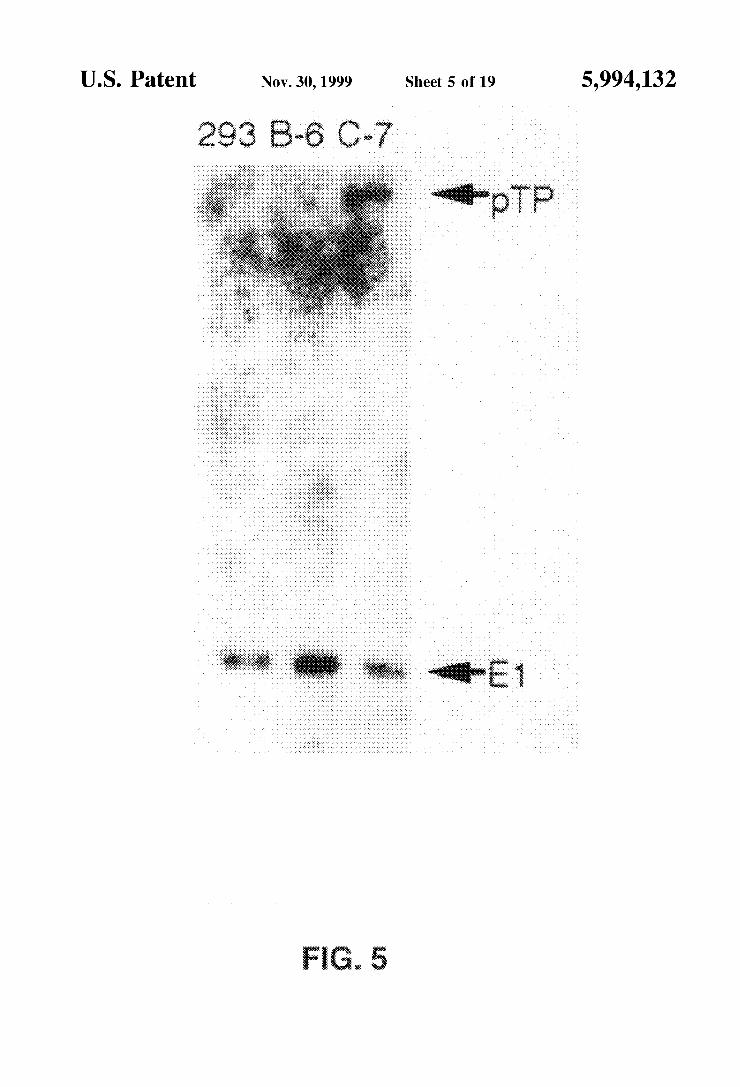
FIG. 5 is an autoradiograph showing which of the cell lines that received the preterminal protein expression plas mid indicated the presence of pRSV-pTP sequences (arrow labelled “pTP) and E1 sequences (arrow labelled “E1').
FIG. 6 is an autoradiograph indicating in which of the cell lines transcription of preterminal protein is occurring (arrow labelled “-3kb”).
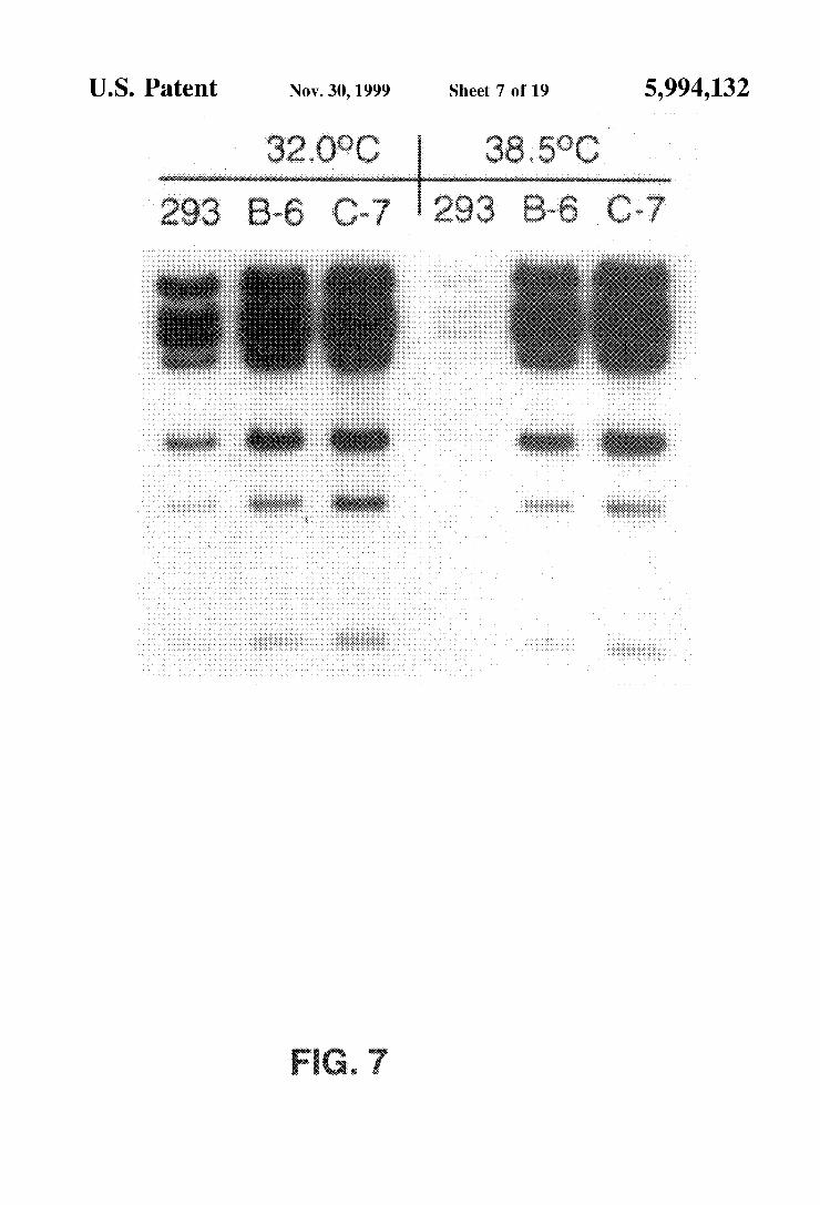
FIG. 7 is an autoradiograph indicating that the expression of the Ad-polymerase could overcome the replication defect of H5sub100 at non-permissive temperatures.
FIG. 8 graphically depicts plaque titre for LP-293, B-6 and C-7 cell lines infected with witAd5, H5ts36, or H5Sub100, and the results demonstrate that the C-7 cell line can be used as a packaging cell line to allow the high level growth of E1, preterminal, and polymerase deleted Ad VectOrS.
FIG. 9 is an autoradiograph showing that the recombinant pol virus is viable on pol-expressing 293 cells but not on 293 cells which demonstrates that recombinant Ad viruses containing the 612 bp deletion found within papol lack the ability to express Ad polymerase.
FIG. 10 is a schematic representation of the structure of pAd5Bdys wherein the two inverted adenovirus origins of replication are represented by a left and right inverted terminal repeat (LITR/RITR). P1 and P2 represent location of probes used for Southern blot analysis.
FIG. 11 graphically illustrates the total number of trans ducing adenovirus particles produced (output) per Serial passage on 293 cells, total input virus of either the helper (hpAP) or Ad5 Bdys, and the total number of cells used in each infection.
FIGS. 12A-B show Southern blot analyses of viral DNA from lysates 3, 6, 9 and 12, digested with the restriction enzymes BSSHII, Niru I and EcoRV. For the analyses, frag ments from the C terminus of mus musculus dystrophin cDNA (A) or the N terminus of E Coli B-galactosidase (B) were labeled with dCTp and used as probes.
FIGS. 13 A-B show the physical separation of Ad5pdys from hpAP virions at the third (A) and final (B) stages of CsCl purification.

5,994,132 7
FIG. 14 graphically depicts the level of contamination of Ad5Bdys EAMs by hpAP virions obtained from the final Stage of CsCl purification as measured by B-galactosidase and alkaline phosphatase expression. The ratio of the two types of virions-Ad5 Bdys EAMs (Lacz) or hpAP (AP) in each fraction is indicated in the lower graph.
FIGS. 15A-B are western blot (immunoblot) analyses of protein extracts from mdx myoblasts and myotubes demon Strating the expression off-galactosidase (A) and dystro phin (B) in cells infected with Ad5(Bdys EAMs.
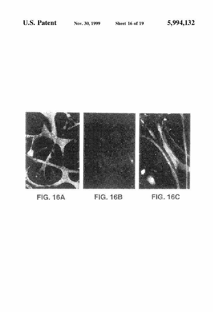
FIGS. 16A-C depict immunofluorescence of dystrophin expression in wild type MM14 myotubes (A), uninfected mdx (B) and infected mdx myotubes (C).
FIG. 17 is a schematic representation of the MCK/lacZ constructs tested to determine what portion of the 3.3 kb DNA fragment containing the enhancer/promoter of the MCK gene is capable of directing high levels of expression of linked genes in muscle cells.
FIG. 18 is a schematic representation of a GFP/B-gal reporter construct Suitable for assaying the expression of Cre recombinase in mammalian cells.
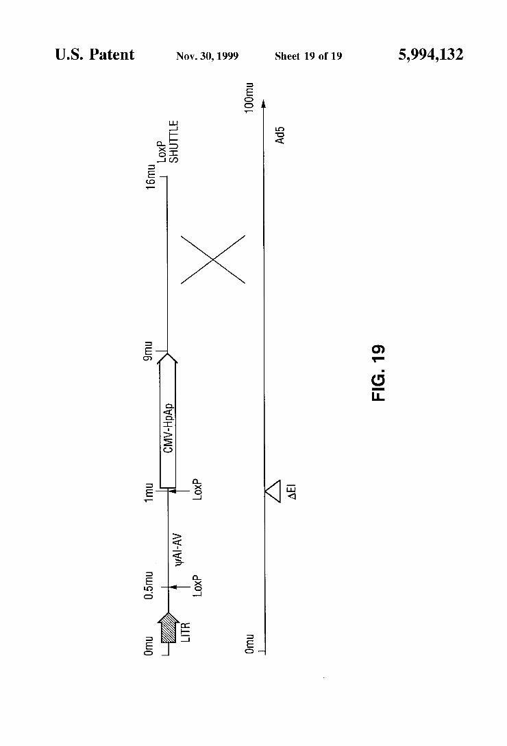
FIG. 19 is a schematic representation of the recombina tion event between the loxP shuttle vector and the Ad5d17001 genome.
DEFINITIONS
To facilitate understanding of the invention, a number of terms are defined below.
The term “gene” refers to a DNA sequence that comprises control and coding Sequences necessary for the production of a polypeptide or precursor thereof. The polypeptide can be encoded by a full length coding Sequence or by any portion of the coding Sequence So long as the desired enzymatic activity is retained. The term “gene' encom passes both cDNA and genomic forms of a given gene.
The term “wild-type” refers to a gene or gene product which has the characteristics of that gene or gene product when isolated from a naturally occurring Source. A wild-type gene is that which is most frequently observed in a popu lation and is thus arbitrarily designated the “normal” or “wild-type” form of the gene. In contrast, the term “modi fied’ or “mutant” refers to a gene or gene product which displayS modifications in Sequence and or functional prop erties (i.e., altered characteristics) when compared to the wild-type gene or gene product. It is noted that naturally occurring mutants can be isolated; these are identified by the fact that they have altered characteristics when compared to the wild-type gene or gene product.
The term "oligonucleotide' as used herein is defined as a molecule comprised of two or more deoxyribonucleotides or ribonucleotides, usually more than three (3), and typically more than ten (10) and up to one hundred (100) or more (although preferably between twenty and thirty). The exact Size will depend on many factors, which in turn depends on the ultimate function or use of the oligonucleotide. The oligonucleotide may be generated in any manner, including chemical Synthesis, DNA replication, reverse transcription, or a combination thereof.
AS used herein, the term “regulatory element” refers to a genetic element which controls Some aspect of the expres Sion of nucleic acid Sequences. For example, a promoter is a regulatory element which facilitates the initiation of tran Scription of an operably linked coding region. Other regu latory elements are splicing Signals, polyadenylation signals, termination signals, etc. (defined infra).
15
25
35
40
45
50
55
60
65
8 Transcriptional control Signals in eucaryotes comprise
“promoter” and “enhancer' elements. Promoters and enhancers consist of Short arrays of DNA sequences that interact specifically with cellular proteins involved in tran scription Maniatis, T. et al., Science 236:1237 (1987). Promoter and enhancer elements have been isolated from a variety of eukaryotic Sources including genes in yeast, insect and mammalian cells and viruses (analogous control elements, i.e., promoters, are also found in procaryotes). The Selection of a particular promoter and enhancer depends on what cell type is to be used to express the protein of interest. Some eukaryotic promoters and enhancers have a broad host range while others are functional in a limited Subset of cell types for review see Voss, S.D. et al., Trends Biochem. Sci., 11:287 (1986) and Maniatis, T. et al., supra (1987)). The term “recombinant DNA vector” as used herein refers
to DNA sequences containing a desired coding Sequence and appropriate DNA sequences necessary for the expression of the operably linked coding Sequence in a particular host organism (e.g. mammal). DNA sequences necessary for expression in procaryotes include a promoter, optionally an operator Sequence, a ribosome binding site and possibly other Sequences. Eukaryotic cells are known to utilize promoters, polyadenlyation signals and enhancers. The terms “in operable combination”, “in operable order'
and “operably linked” as used herein refer to the linkage of nucleic acid Sequences in Such a manner that a nucleic acid molecule capable of directing the transcription of a given gene and/or the Synthesis of a desired protein molecule is produced. The term also refers to the linkage of amino acid Sequences in Such a manner So that a functional protein is produced. The term "genetic cassette' as used herein refers to a
fragment or Segment of DNA containing a particular group ing of genetic elements. The cassette can be removed and inserted into a vector or plasmid as a single unit. A plasmid backbone refers to a piece of DNA containing at least plasmid origin of replication and a Selectable marker gene (e.g., an antibiotic resistance gene) which allows for Selec tion of bacterial hosts containing the plasmid; the plasmid backbone may also include a polylinker region to facilitate the insertion of genetic elements within the plasmid. When a particular plasmid is modified to contain non-plasmid elements (e.g., insertion of Ad Sequences and/or a eukaryotic gene of interest linked to a promoter element), the plasmid Sequences are referred to as the plasmid backbone.
Because mononucleotides are reacted to make oligonucle otides in a manner Such that the 5' phosphate of one mononucleotide pentose ring is attached to the 3' oxygen of its neighbor in one direction via a phosphodiester linkage, an end of an oligonucleotide is referred to as the “5' end” if its 5' phosphate is not linked to the 3' oxygen of a mononucle otide pentose ring and as the '3' end” if its 3' oxygen is not linked to a 5" phosphate of a Subsequent mononucleotide pentose ring. AS used herein, a nucleic acid Sequence, even if internal to a larger oligonucleotide, also may be said to have 5' and 3' ends. When two different, non-overlapping oligonucleotides
anneal to different regions of the same linear complementary nucleic acid Sequence, and the 3' end of one oligonucleotide points towards the 5' end of the other, the former may be called the "upstream” oligonucleotide and the latter the "downstream” oligonucleotide. The term “primer' refers to an oligonucleotide which is
capable of acting as a point of initiation of Synthesis when placed under conditions in which primer extension is initi

5,994,132
ated. An oligonucleotide “primer' may occur naturally, as in a purified restriction digest or may be produced Syntheti cally. A primer is Selected to be “Substantially complementary
to a Strand of Specific Sequence of the template. A primer must be sufficiently complementary to hybridize with a template Strand for primer elongation to occur. A primer Sequence need not reflect the exact Sequence of the template. For example, a non-complementary nucleotide fragment may be attached to the 5' end of the primer, with the remainder of the primer Sequence being Substantially complementary to the Strand. Non-complementary bases or longer Sequences can be interspersed into the primer, pro Vided that the primer Sequence has Sufficient complementa rity with the Sequence of the template to hybridize and thereby form a template primer complex for Synthesis of the extension product of the primer. “Hybridization” methods involve the annealing of a complementary Sequence to the target nucleic acid (the Sequence to be detected). The ability of two polymers of nucleic acid containing complementary Sequences to find each other and anneal through base pairing interaction is a well-recognized phenomenon. The initial observations of the “hybridization' process by Marmur and Lane, Proc. Natl. Acad. Sci. USA 46:453 (1960) and Doty et al., Proc. Natl. Acad. Sci. USA 46:461 (1960) have been followed by the refinement of this proceSS into an essential tool of modern biology.
The complement of a nucleic acid Sequence as used herein refers to an oligonucleotide which, when aligned with the nucleic acid Sequence Such that the 5' end of one Sequence is paired with the 3' end of the other, is in “antiparallel asSociation.” Certain bases not commonly found in natural nucleic acids may be included in the nucleic acids of the present invention and include, for example, inosine and 7-deazaguanine. Complementarity need not be perfect; Stable duplexes may contain mismatched base pairs or unmatched bases. Those skilled in the art of nucleic acid technology can determine duplex Stability empirically con sidering a number of variables including, for example, the length of the oligonucleotide, base composition and Sequence of the oligonucleotide, ionic Strength and inci dence of mismatched base pairs.
Stability of a nucleic acid duplex is measured by the melting temperature, or “T.” The T of a particular nucleic acid duplex under Specified conditions is the temperature at which on average half of the base pairs have disassociated. The equation for calculating the T of nucleic acids is well known in the art.
The term “probe' as used herein refers to a labeled oligonucleotide which forms a duplex Structure with a Sequence in another nucleic acid, due to complementarity of at least one Sequence in the probe with a Sequence in the other nucleic acid.
The term “label” as used herein refers to any atom or molecule which can be used to provide a detectable (preferably quantifiable) Signal, and which can be attached to a nucleic acid or protein. Labels may provide Signals detectable by fluorescence, radioactivity, colorimetry, gravimetry, X-ray diffraction or absorption, magnetism, enzymatic activity, and the like.
The terms “nucleic acid Substrate” and “nucleic acid template” are used herein interchangeably and refer to a nucleic acid molecule which may comprise Single- or double-stranded DNA or RNA.
“Oligonucleotide primerS matching or complementary to a gene Sequence” refers to oligonucleotide primers capable
15
25
35
40
45
50
55
60
65
10 of facilitating the template-dependent Synthesis of Single or double-Stranded nucleic acids. Oligonucleotide primers matching or complementary to a gene Sequence may be used in PCRs, RT-PCRs and the like. A “consensus gene Sequence” refers to a gene Sequence
which is derived by comparison of two or more gene Sequences and which describes the nucleotides most often present in a given Segment of the genes, the consensus Sequence is the canonical Sequence. The term “polymorphic locus” is a locus present in a
population which shows variation between members of the population (i.e., the most common allele has a frequency of less than 0.95). In contrast, a “monomorphic locus” is a genetic locus at little or no variations Seen between members of the population (generally taken to be a locus at which the most common allele exceeds a frequency of 0.95 in the gene pool of the population). The term “microorganism' as used herein means an
organism too small to be observed with the unaided eye and includes, but is not limited to bacteria, Viruses, protozoans, fungi, and ciliates. The term “microbial gene Sequences' refers to gene
Sequences derived from a microorganism. The term “bacteria” refers to any bacterial species includ
ing eubacterial and archaebacterial Species. The term “virus' refers to obligate, ultramicroscopic,
intracellular parasites incapable of autonomous replication (i.e., replication requires the use of the host cells machinery). Adenoviruses, as noted above, are double stranded DNA viruses. The left and right inverted terminal repeats (ITRs) are short elements located at the 5' and 3' termini of the linear Ad genome, respectively and are required for replication of the viral DNA. The left ITR is located between 1-130 bp in the Ad genome (also referred to as 0–0.5 mu). The right ITR is located from -3.7500 bp to the end of the genome (also referred to as 99.5-100 mu). The two ITRs are inverted repeats of each other. For clarity, the left ITR or 5' end is used to define the 5' and 3' ends of the ITRS. The 5' end of the left ITR is located at the extreme 5' end of the linear adenoviral genome; picturing the left ITR (LITR) as an arrow extending from the 5' end of the genome, the tail of the 5' ITR is located at mu 0 and the head of the left ITR is located at -0.5 mu (further the tail of the left ITR is referred to as the 5' end of the left ITR and the head of the left ITR is referred to as the 3' end of the left ITR). The tail of the right or 3' ITR is located at mu 100 and the head of the right ITR is located at ~mu 99.5; the head of the right ITR is referred to as the 5' end of the right ITR and the tail of the right ITR is referred to as the 3' end of the right ITR (RITR). In the linear Ad genome, the ITRs face each other with the head of each ITR pointing inward toward the bulk of the genome. When arranged in a “tail to tail orientation” the tails of each ITR (which comprise the 5' end of the LITR and the 3' end of the RITR) are located in proximity to one another while the heads of each ITR are separated and face outward (see for example, the arrangement of the ITRS in the EAM shown in FIG. 10 herein). The “adenovirus packaging Sequence” refers to the Sequence which comprises five (AI-AV) packaging signals and is required for encapsidation of the mature linear genome; the packaging Signals are located from ~194 to 358 bp in the Ad genome (about 0.5–1.0 mu). The phrase “at least one adenovirus gene coding region'
refers to a nucleotide Sequence containing more than one adenovirus gene coding gene. A "helper adenovirus' or “helper virus' refers to an adenovirus which is replication

5,994,132 11
competent in a particular host cell (the host may provide Ad gene products Such as E1 proteins), this replication competent virus is used to Supply in trans functions (e.g., proteins) which are lacking in a second replication incompetent virus, the first replication-competent virus is Said to "help' the Second replication-incompetent virus thereby permitting the propagation of the Second viral genome in the cell containing the helper and Second viruses.
The term “containing a deletion within the E2b region” refers to a deletion of at least one basepair (preferably more than one bp and preferably at least 100 and most preferably more than 300 bp) within the E2b region of the adenovirus genome. An E2b deletion is a deletion that prevents expres Sion of at least one E.2b gene product and encompasses deletions within exons encoding portions of E2b-specific proteins as well as deletions within promoter and leader Sequences. An “adenovirus minichromosome” refers to a linear mol
ecule of DNA containing the Ad ITRs on each end which is generated from a plasmid containing the ITRS and one or more gene of interest. The term “encapsidated adenovirus minichromosome” or “EAM” refers to an adenovirus min ichromosome which has been packaged or encapsidated into a viral particle; plasmids containing the Ad ITRS and the packaging Signal are shown herein to produce EAMs. When used herein, "recovering encapsidated adenovirus min ichromosomes refers to the collection of EAMs from a cell containing an EAM plasmid and a helper virus; this cell will direct the encapsidation of the minichromosome to produce EAMs. The EAMs may be recovered from these cells by lysis of the cell (e.g., freeze-thawing) and pelleting of the cell debris to a cell extract as described in Example 1 (Ex. 1 describes the recovery of Ad virus from a cell, but the same technique is used to recover EAMs from a cell). “Purifying” Such minichromosomes refers to the isolation of the recov ered EAMs in a more concentrated form (relative to the cell lysate) on a density gradient as described in Example 7: purification of recovered EAMS permits the physical Sepa ration of the EAM from any helper virus (if present).
The term “transfection” as used herein refers to the introduction of foreign DNA into eukaryotic cells. Trans fection may be accomplished by a variety of means known to the art including calcium phosphate-DNA co-precipitation, DEAE-dextran-mediated transfection, polybrene-mediated transfection, electroporation, microinjection, liposome fusion, lipofection, protoplast fusion, retroviral infection, and biolistics.
The term “stable transfection” or “stably transfected” refers to the introduction and integration of foreign DNA into the genome of the transfected cell. The term “stable transfectant” refers to a cell which has Stably integrated foreign DNA into the genomic DNA. AS used herein, the term “gene of interest” refers to a gene
inserted into a vector or plasmid whose expression is desired in a host cell. Genes of interest include genes having therapeutic value as well as reporter genes. A variety of Such genes are contemplated, including genes of interest encod ing a protein which provides a therapeutic function (Such as the dystrophin gene, which is capable of correcting the defect seen in the muscle of MD patients), the utrophin gene, the CFTR gene (capable of correcting the defect seen in cystitic fibrosis patients), etc.
The term “reporter gene' indicates a gene Sequence that encodes a reporter molecule (including an enzyme). A “reporter molecule' is detectable in any detection System, including, but not limited to enzyme (e.g., ELISA, as well as
15
25
35
40
50
55
60
65
12 enzyme-based histochemical assays), fluorescent, radioactive, and luminescent Systems. In one embodiment, the present invention contemplates the E. coli f-galactosidase gene (available from Pharmacia Biotech, Pistacataway, N.J.), green fluorescent protein (GFP) (commercially available from Clontech, Palo Alto, Calif.), the human placental alkaline phosphatase gene, the chloram phenicol acetyltransferase (CAT) gene; other reporter genes are known to the art and may be employed. AS used herein, the terms “nucleic acid molecule
encoding,” “DNA sequence encoding,” and “DNA encod ing” refer to the order or Sequence of deoxyribonucleotides along a Strand of deoxyribonucleic acid. The order of these deoxyribonucleotides determines the order of amino acids along the polypeptide (protein) chain. The DNA sequence thus codes for the amino acid Sequence.
DESCRIPTION OF THE INVENTION
The present invention provides improved adenovirus Vec tors for the delivery of recombinant genes to cells in Vitro and in Vivo. AS noted above, the present invention contem plates two approaches to improving adenovirus vectors. The first approach generally contemplates a recombinant plas mid containing the minimal region of the Ad genome required for replication and packaging (i.e., the left and right ITR and the packaging or I sequence) along with one or more genes of interest; this recombinant plasmid is pack aged into an encapsidated adenovirus minichromosome (EAM) when grown in parallel with an E1-deleted helper virus in a cell line expressing the E1 proteins (e.g., 293 cells). The recombinant adenoviral minichromosome is pref erentially packaged. To prevent the packaging of the helper virus, a helper virus containing loXP Sequences flanking the I sequence is employed in conjunction with 293 cells expressing Cre recombinase; Cre-loXP mediated recombi nation removes the packaging Sequence from the helper genome thereby preventing packaging of the helper during the production of EAMS. In the Second approach, “dam aged' or “deleted’ adenoviruses containing deletions within the E2b region are employed. While the “damaged” aden Ovirus is capable of Self-propagation in a packaging cell line expressing the appropriate E2b protein(s), the E2b-deleted recombinant adenovirus are incapable of replicating and expressing late Viral gene products outside of the packaging cell line.
In one embodiment, the Self-propagating recombinant adenoviruses contain deletions in the E.2b region of the adenovirus genome. In another embodiment, “gutted” Viruses are contemplated; these viruses lack all viral coding regions. In addition, packaging cell lines co-expressing E1 and E.2b gene products are provided which allow the pro duction of infectious recombinant virus containing deletions in the E1 and E2b regions without the use of helper virus. The Description of the Invention is divided into the
following Sections: I. Self-Propagating Adenovirus Vectors, II. Packaging Cell Lines, and III. Encapsidated Adenoviral Minichromosomes.
I. Self-Propagating Adenovirus Vectors Self-propagating adenovirus (Ad) vectors have been
extensively utilized to deliver foreign genes to a great variety of cell types in Vitro and in Vivo. “Self-propagating viruses” are those which can be produced by transfection of a single piece of DNA (the recombinant viral genome) into a Single packaging cell line to produce infectious virus, Self-propagating viruses do not require the use of helper Virus for propagation.
5,994,132 13
Existing Advectors have been shown to be problematic in Vivo. This is due in part because current or first generation Advectors are deleted for only the early region 1 (E1) genes. These vectors are crippled in their ability to replicate nor mally without the trans-complementation of E1 functions provided by human 293 cells, a packaging cell line ATCC CRL 1573; Graham et al. (1977) J. Gen. Virol. 36:59). Unfortunately, with the use of high titres of E1 deleted vectors, and the fact that there are E1-like factors present in many cell types, E1 deleted vectors can overcome the block to replication and express other viral gene products Imperiale et al. (1984) Mol. Cell Biol. 4:867; Nevins (1981) Cell 26:213; and Gaynor and Berk (1983) Cell 33:683). The expression of Viral proteins in the infected target cells elicits a Swift host immune response, that is largely T-cell mediated Yang and Wilson (1995) J. Immunol. 155:2564 and Yang et al. (1994) Proc. Natl. Acad. Sci. USA91:4407). The trans duced cells are Subsequently eliminated, along with the transferred foreign gene. In immuno-incompetent animals, Ad delivered genes can be expressed for periods of up to one year Yang et al. (1994), Supra; Vincent et al. (1993) Nature Genetics 5:130; and Yang et al. (1995) Proc. Natl. Acad. Sci. USA 92:7257).
Another shortcoming of first generation Ad VectorS is that a single recombination event between the genome of an Ad vector and the integrated E1 Sequences present in 293 cells can generate replication competent Ad (RCA), which can readily contaminate viral StockS.
In order to further cripple viral protein expression, and also to decrease the frequency of generating RCA, the present invention provides Ad Vectors containing deletions in the E2b region. Propagation of these E.2b-deleted Ad vectors requires cell lines which express the deleted E.2b gene products. The present invention provides Such pack aging cell lines and for the first time demonstrates that the E.2b gene products, DNA polymerase and preterminal protein, can be constitutively expressed in 293 cells along with the E1 gene products. With every gene that can be constitutively expressed in 293 cells comes the opportunity to generate new versions of Ad VectorS deleted for the respective genes. This has immediate benefits, increased carrying capacity, Since the combined coding Sequences of the polymerase and preterminal proteins that can be theo retically deleted approaches 4.6 kb and a decreased inci dence of RCA generation, Since two or more independent recombination events would be required to generate RCA. Therefore, the novel E1, Ad polymerase and preterminal protein expressing cell lines of the present invention enable the propagation of Ad Vectors with a carrying capacity approaching 13 kb, without the need for a contaminating helper virus Mitani et al. (1995) Proc. Natl. Acad. Sci. USA 92:3854). In addition, when genes critical to the viral life cycle are deleted (e.g., the E2b genes), a further crippling of Ad to replicate and express other viral gene proteins occurs. This decreases immune recognition of Virally infected cells, and allows for extended durations of foreign gene expres Sion. The most important attribute of E1, polymerase, and preterminal protein deleted vectors, however, is their inabil ity to express the respective proteins, as well as a predicted lack of expression of most of the Viral Structural proteins. For example, the major late promoter (MLP) of Ad is responsible for transcription of the late Structural proteins LI through L5 Doerfler, In Adenovirus DNA, The Viral Genome and Its Expression (Martinus Nijhoff Publishing Boston, 1986)). Though the MLP is minimally active prior to Ad genome replication, the rest of the late genes get transcribed and translated from the MLP only after viral
1O
15
25
35
40
45
50
55
60
65
14 genome replication has occurred Thomas and Mathews (1980) Cell 22:523). This cis-dependent activation of late gene transcription is a feature of DNA viruses in general, such as in the growth of polyoma and SV-40. The poly merase and preterminal proteins are absolutely required for Ad replication (unlike the E4 or protein IX proteins) and thus their deletion is extremely detrimental to Ad vector late gene expression.
II. Packaging Cell Lines Constitutively Expressing E2B Gene Products
The present invention addresses the limitations of current or first generation Ad vectors by isolating novel 293 cell lines coexpressing critical viral gene functions. The present invention describes the isolation and characterization of 293 cell lines capable of constitutively expressing the Ad poly merase protein. In addition, the present invention describes the isolation of 293 cells which not only express the E1 and polymerase proteins, but also the Ad-preterminal protein. The isolation of cell lines coexpressing the E1, Ad poly merase and preterminal proteins demonstrates that three genes critical to the life cycle of Ad can be constitutively coexpressed, without toxicity.
In order to delete critical genes from Self-propagating Ad vectors, the proteins encoded by the targeted genes have to first be coexpressed in 293 cells along with the E1 proteins. Therefore, only those proteins which are non-toxic when coexpressed constitutively (or toxic proteins inducibly expressed) can be utilized. Coexpression in 293 cells of the E1 and E4 genes has been demonstrated (utilizing inducible, not constitutive, promoters) Yeh et al. (1996) J. Virol. 70:559; Wang et al. (1995) Gene Therapy 2:775; and Gorziglia et al. (1996) J. Virol. 70:4173). The E1 and protein IX genes (a virion structural protein) have been coexpressed Caravokyri and Leppard (1995) J. Virol. 69:6627), and coexpression of the E1, E4, and protein IX genes has also been described Krougliak and Graham (1995) Hum. Gene Ther. 6:1575). The present invention provides for the first time, cell lines
coexpressing E1 and E2b gene products. The E2b region encodes the Viral replication proteins which are absolutely required for Ad genome replication Doerfler, Supra and Pronket al. (1992) Chromosoma 102:S39-S45). The present invention provides 293 cells which constitutively express the 140 kD Ad-polymerase. While other researchers have reported the isolation of 293 cells which express the Ad-preterminal protein utilizing an inducible promoter Schaack et al. (1995) J. Virol. 69:4079), the present inven tion is the first to demonstrate the high-level, constitutive coexpression of the E1, polymerase, and preterminal pro teins in 293 cells, without toxicity. These novel cell lines permit the propagation of novel Ad Vectors deleted for the E1, polymerase, and preterminal proteins.
III. Encapsidated Adenoviral Minichromosomes The present invention also provides encapsidated aden
ovirus minichromosome (EAM) consisting of an infectious encapsidated linear genome containing Ad origins of replication, packaging signal elements, a reporter gene (e.g., a f-galactosidase reporter gene cassette) and a gene of interest (e.g., a full length (14 kb) dystrophin cDNA regu lated by a muscle specific enhancer/promoter). EAMs are generated by cotransfecting 293 cells with Supercoiled plas mid DNA (e.g., p.A.d5 Bdys) containing an embedded inverted origin of replication (and the remaining above elements) together with linear DNA from E1-deleted virions expressing human placental alkaline phosphatase (hpAP) (a helper virus). All proteins necessary for the generation of

5,994,132 15
EAMs are provided in trans from the hp AP virions and the two can be separated from each other on equilibrium CsCl gradients. These EAMS are useful for gene transfer to a variety of cell types both in vitro and in vivo.
Adenovirus-mediated gene transfer to muscle is a prom ising technology for gene therapy of Duchenne muscular dystrophy (DMD). However, currently available recombi nant adenovirus vectors have Several limitations, including a limited cloning capacity of ~8.5 kb, and the induction of a host immune response that leads to transient gene expres Sion of 3 to 4 weeks in immunocompetent animals. Gene therapy for DMD could benefit from the development of adenoviral vectors with an increased cloning capacity to accommodate a full length (1L4 kb) dystrophin cDNA. This increased capacity should also accommodate gene regula tory elements to achieve expression of transduced genes in a tissue-specific manner. Additional vector modifications that eliminate adenoviral genes, expression of which is asSociated with development of a host immune response, might greatly increase long term expression of Virally deliv ered genes in Vivo. The constructed encapsidated adenovirus minichromosomes of the present invention are capable of delivering up to 35 kb of non-viral exogenous DNA. These minichromosomes are derived from bacterial plasmids con taining two fused inverted adenovirus origins of replication embedded in a circular genome, the adenovirus packaging Signals, a Bgalactosidase reporter gene and a full length dystrophin cDNA regulated by a muscle Specific enhancer/ promoter. The encapsidated minichromosomes are propa gated in vitro by trans-complementation with a replication defective (E1+E3 deleted) helper virus. These minichromo Somes can be propagated to high titer (>10/ml) and purified on CsCl gradients due to their buoyancy difference relative to helper virus. These vectors are able to transduce myo genic cell cultures and express dystrophin in myotubes. These results demonstrate that encapsidated adenovirus minichromosomes are useful for gene transfer to muscle and other tissues.
The present invention further provides methods for modi fying the above-described EAM system to enable the gen eration of high titer stocks of EAMs with minimal helper virus contamination. Preferably the EAM stocks contain helper virus representing less than 1%, preferably less than 0.1% and most preferably less than 0.01% (including 0.0%) of the final viral isolate.
The amount of helper virus present in the EAM prepara tions is reduced in two ways. The first is by selectively controlling the relative packaging efficiency of the helper virus versus the EAM virus. The Cre-loxP excision method is employed to remove the packaging Signals from the helper Virus thereby preventing the packaging of the helper virus used to provide in trans viral proteins for the encapsidation of the recombinant adenovirus minichromosomes. The Sec ond approach to reducing or eliminating helper virus in EAM stocks is the use of improved physical methods for Separating EAM from helper virus.
EXPERIMENTAL
The following examples Serve to illustrate certain pre ferred embodiments and aspects of the present invention and are not to be construed as limiting the Scope thereof.
In the experimental disclosure which follows, the follow ing abbreviations apply: M (molar); mM (millimolar); uM (micromolar); mol (moles); mmol (millimoles); umol (micromoles); nmol (nanomoles); mu or m.u. (map unit); g (gravity), gm (grams); mg (milligrams), lug (micrograms);
15
25
35
40
45
50
55
60
65
16 pg (picograms); L (liters); ml (milliliters); ul (microliters); cm (centimeters); mm (millimeters); um (micrometers); nm (nanometers); hr (hour); min (minute); msec (millisecond); C. (degrees Centigrade); AMP (a de no sine
5'-monophosphate); cDNA (copy or complimentary DNA); DTT (dithiotheritol); ddH.0 (double distilled water); dNTP (deoxyribonucleotide triphosphate); rNTP (ribonucleotide triphosphate); ddNTP (dideoxyribonucleotide triphosphate); bp (base pair); kb (kilo base pair); TLC (thin layer chromatography); thNA (transfer RNA); nt (nucleotide); VRC (vanadyl ribonucleoside complex); RNase (ribonuclease); DNase (deoxyribonuclease); poly A (polyriboadenylic acid); PBS (phosphate buffered saline); OD (optical density); HEPES (N-2-Hydroxyethyl piperazine-N-2-ethanesulfonic acid); HBS (HEPES buff ered saline); SDS (sodium dodecyl sulfate); Tris-HCl (tris Hydroxymethylaminomethane-hydrochloride); rpm (revolutions per minute); ligation buffer (50 mM Tris-HCl, 10 mM MgCl, 10 mM dithiothreitol, 25 lug/ml bovine serum albumin, and 26 uM NAD+, and pH 7.8); EGTA (ethylene glycol-bis(B-aminoethyl ether) N, N, N', N-tetraacetic acid); EDTA (ethylenediaminetetracetic acid); ELISA (enzyme linked immunosorbant assay); LB (Luria Bertani broth: 10 g tryptone, 5g yeast extract, and 10 g NaCl per liter, pH adjusted to 7.5 with IN NaOH); Superbroth (12 g tryptone, 24 g yeast extract, 5 g glycerol, 3.8 g. KHPO and 12.5g, KHPO, per liter); DMEM (Dulbecco's modi fied Eagle's medium); ABI (Applied Biosystems Inc., Foster City, Calif.); Amersham (Amersham Corporation, Arlington Heights, Ill.); ATCC (American Type Culture Collection, Rockville, Md.); Beckman (Beckman Instruments Inc., Ful lerton Calif); BM (Boehringer Mannheim Biochemicals, Indianapolis, Ind.); Bio-101 (Bio-101, Vista, Calif.); BioRad (BioRad, Richmond, Calif.); Brinkmann (Brinkmann Instru ments Inc. Wesbury, N.Y.); BRL, Gibco BRL and Life Technologies (Bethesda Research Laboratories, Life Tech nologies Inc., Gaithersburg, Md.); CRI (Collaborative Research Inc. Bedford, Mass.); Eastman Kodak (Eastman Kodak Co., Rochester, N.Y.); Eppendorf (Eppendorf, Eppendorf North America, Inc., Madison, Wis.); Falcon (Becton Dickenson Labware, Lincoln Park, N.J.); IBI (International Biotechnologies, Inc., New Haven, Conn.); ICN (ICN Biomedicals, Inc., Costa Mesa, Calif.); Invitrogen (Invitrogen, San Diego, Calif.); New Brunswick (New Brun swick Scientific Co. Inc., Edison, N.J.).; NEB (New England BioLabs Inc., Beverly, Mass.); NEN (Du Pont NEN Products, Boston, Mass.); Pharmacia (Pharmacia LKB Gaithersburg, Md.); Promega (Promega Corporation, Madison, Wis.); Stratagene (Stratagene Cloning Systems, La Jolla, Calif.); UVP (UVP, Inc., San Gabreil, Calif); USB (United States Biochemical Corp., Cleveland, Ohio); and Whatman (Whatman Lab. Products Inc, Clifton, N.J.).
Unless otherwise indicated, all restriction enzymes and DNA modifying enzymes were obtained from New England Biolabs (NEB) and used according to the manufacturers directions.
EXAMPLE 1.
Generation Of Packaging Cell Lines That Coexpress The Adenovirus E1 And DNA
Polymerase Proteins In this example, packaging cell lines coexpressing Ad E1
and polymerase proteins were described. These cell lines were shown to support the replication and growth of H5ts36, an Ad with a temperature-Sensitive mutation of the Ad polymerase protein. These polymerase-expressing packag

5,994,132 17
ing cell lines can be used to prepare Ad VectorS deleted for the E1 and polymerase functions.
a) Tissue Culture And Virus Growth LP-293 cells (Microbix Biosystems, Toronto) were grown
and Serially passaged as Suggested by the Supplier. Plaque assays were performed in 60 mm dishes contain
ing cell monolayers at ~90% confluency. The appropriate virus dilution in a 2% DMEM solution was dripped onto the cells, and the plates incubated at the appropriate temperature for one hour. The Virus containing media was aspirated, the monolayer was overlaid with 10 mls of a pre-warmed EMEM agar overlay solution (0.8% Noble agar, 4% fetal calfserum, and antibiotics) and allowed to solidify. After the appropriate incubation time (usually 7 days for incubations at 38.5° C. and 10-12 days for incubations at 32° C), five mls of the agar-containing Solution containing 1.3% neutral red was overlaid onto the infected dishes and plaques were counted the next day. An aliquot of the virus H5ts36 Freimuth and Ginsberg (1986) Proc. Natl. Acad. Sci. USA 83:7816) was utilized to produce high titre stocks after infection of 293 cells at 32° C. H5ts36 is an Ad5-derived Virus defective for viral replication at the nonpermissive temperature Miller and Williams (1987) J. Virol. 61:3630).
The infected cells were harvested after the onset of extensive cytopathic effect, pelleted by centrifugation and resuspended in 10 mM Tris-Cl, pH 8.0. The lysate was freeze-thawed three times and centrifuged to remove the cell debris. The cleared lysate was applied to CsCl step gradi ents (heavy CsCl at density of 1.45 g/ml, the light CsCl at density of 1.20 g/ml), ultracentrifuged, and purified using standard techniques Graham and Prevec (1991) In Methods in Molecular Biology, Vol 7. Gene Transfer and Expression Protocols, Murray (ed.), Humana Press, Clifton, N.J., pp. 109-128). The concentration of plaque forming units (pfu) of this stock was determined at 32 C. as described above. Virion DNA was extracted from the high titre stock by pronase digestion, phenol-chloroform extraction, and etha nol precipitation. The leakiness of this Stock was found to be <1 in 2000 pful at the non-permissive temperature, consistent with previous reports Miller and Williams (1987), Supra).
b) Plasmids The expression plasmid pRSV-pol Zhao and Padmanab
han (1988) Cell 55:1005 contains sequences encoding the Ad2 polymerase mRNA (including the start codon from the exon at map unit 39) under the transcriptional control of the Rous Sarcoma Virus LTR/promoter element; the Ad2 DNA polymerase sequences are flanked on the 3' end by the SV40 Small t intron and SV-40 polyadenylation addition site (see FIG. 1A). FIG. 1A provides a schematic representation of the Ad polymerase expression plasmid pRSV-pol.pRSV-pol includes the initiator methionine and amino-terminal pep tides encoded by the exon at m.u. 39 of the Ad genome. The location of the PCR primers p602a and p2158c, the two Scal 1 kb probes utilized for Northern analyses, the polymerase terminator codon, and the polyadenylation site of the IVa2 gene (at 11.2 m.u. of the Ad genome) are indicated.
The expression plasmid pRSV-pTPZhao and Padmanab han (1988), Supra contains sequences encoding the Ad2 preterminal protein (including the amino terminal peptides encoded by the exon at map unit 39 of the Ad genome) under the transcriptional control of the Rous Sarcoma Virus LTR/ promoter element; the pTP Sequences are flanked on the 3' end by the SV40 Small-t intron and SV40 polyadenylation signals (see FIG. 1B for a schematic of pRSV-PTP). FIG. 1B also shows the location of the EcoRV subfragment utilized as a probe in the genomic DNA and cellular RNA
15
25
35
40
45
50
55
60
65
18 evaluations, as well as the initiator methionine codon present from map unit 39 in the Ad5 genome. The locations of the H5in 190 and H5Sub100 insertions are shown relative to the preterminal protein open-reading frame. The follow ing abbreviations are used in FIG. 1: ORF, open reading frame, Small-t, Small tumor antigen and m.u., map unit. pCEP4 is a plasmid containing a hygromycin expression
cassette (Invitrogen). pFG140 (Microbix Biosystems Inc. Toronto, Ontario) is a
plasmid containing sequences derived from Ad5d1309 which contain a deletion/Substitution in the E3 region Jones and Shek (1979) Cell 17:683). pFG140 is infectious in single transfections of 293 cells and is used as a control for transfection efficiency.
c) Transfection Of 293 Cells LP-293 cells were cotransfected with BamHI linearized
pRSV-pol and BamHI linearized pCEP4 at a molar ratio of 10:1 using a standard CaPO precipitation method Sambrook et al. (1989) Molecular Cloning: A Laboratory Manual, Cold Spring Harbor Press, Plainview, N.Y., pp. 16.33-16.36. In addition, 293 cells were cotransfected with or with BamHI linearized pRSV-pol, BamHI linearized pRSV-pTP and pCEP using a molar ratio of 10:1 (non Selectable:Selectable plasmids).
Forty-eight hours after transfection, the cells were pas Saged into media containing hygromycin at 100 lug/mL. Individual hygromycin resistant colonies were isolated and expanded.
d) Isolation Of Ad Polymerase Expressing 293 Cell Lines Twenty hygromycin resistant cell lines were expanded
and Screened for the ability to express the Ad polymerase protein. Initially, the individual cell lines were assayed for the ability to Support growth of the viral polymerase mutant H5ts36 using the plaque assay described in Section a) above. It was speculated that constitutive expression of the Wild type Ad polymerase protein in a clonal population of 293 cells should allow the growth of H5ts36 at 38.5° C. However, it was unclear if constitutive expression of the Ad polymerase would be toxic when coexpressed with the E1 proteins present in 293 cells. Similar toxicity problems have been observed with the Ad ssDBP, and the pTP Klessig et al. (1984) Mol. Cell. Biol. 4:1354 and Schaacket al. (1995) J. Virol. 69:4079). Of the twenty hygromycin resistant cell lines isolated,
seven were able to support plaque formation with H5ts36 at the non-permissive temperature, unlike the parental LP-293 cells; these cell lines were named B-6, B-9, C-1, C-4, C-7, C-13 and C-14 (see Table 1) (the B-6 and B-9 cell lines received only the pRSV-pol and CEP4 plasmids; C-1, C-4, C-7, C-13 and C-14 cell lines received the pRSV-pol, pRSV-pTP and CEP4 plasmids). For the results shown in Table 1, dishes (60 mm) of near confluent cells of each cell line were infected with the same dilution of H5ts36 at the temperature indicated, overlaid with agar media, and Stained for plaques as outlined in Section a. Passage number refers to the number of Serial passages after initial transfection with the plasmid pRSV-pol.
The cell line B-6 produced plaques one day earlier than the other Ad polymerase-expressing cell lines, which may reflect increased polymerase expression (see below). Cell line B-9 demonstrated an increased doubling time whereas each of the other cell lines displayed no growth disadvan tages relative to the parental LP-293 cells. As shown in Table 1, even after multiple passages (in Some instances up to four months of Serial passaging) the cells were still capable of H5ts36 plaque formation at the non-permissive temperature,

5,994,132 19
indicating that the RSV-LTR/promoter remained active for extended periods of time. However, the cell lines B-9 and C-13 displayed a decreased ability to plaque the virus at 32 C. as well as at 38.5 C., Suggesting that a global viral complementation defect had occurred in these cell lines after extended passaging. The remaining cell lines Screened at later passages demonstrated no Such defect, even after 20 passages (e.g., cell line B-6, Table 1).
TABLE 1.
Plaquing Ability Of H5ts36 At The Non-Permissive Temperature Utilizing 293-Ad Polymerase Expressing Cell Lines
Number Of Plaques At:
Cell Line Passage Number 32.O. C. 38.5° C.
293 >5OO O B-6 9 >5OO >500
2O >5OO >500 B-9 5 >5OO >500
14 90 18 C-1 5 >5OO >500
13 >5OO >500 C-4 5 >5OO >500
14 >5OO >500 C-7 5 >5OO >500
14 >5OO >500 C-13 5 >5OO >500
27 12O 4 C-14 5 >5OO >500
27 >5OO 370
e) Genomic Analysis Of Ad Polymerase-Expressing Cell Lines Genomic DNA from LP-293 cells and each of the seven
cell lines able to complement H5ts36 at 38.5° C. were analyzed by PCR for the presence of pRSV-pol derived sequences. Genomic DNA from LP-293 cells and the hygro mycin resistant cells lies were harvested using Standard protocols (Sambrook et al, supra) and 200 ng of DNA from each cell line was analyzed by PCR in a Solution containing 2 ng/mL of primers p602a and p2158c, 10 mM Tris-HCl, pH 8.3, 50 mM KCl, 1.5 mM MgCl, and 0.001% gelatin. The forward primer, p602a 5'-TTCATTTTATGTTTCAGGTTC AGGG-3' (SEQ ID NO:2) is located in the SV-40 polyade nylation sequence. The reverse primer p2158c 5'- TTACCGCCACACTCGCAGGG-3' (SEQ ID NO:3) is Ad-Sequence Specific with the 5' nucleotide located at posi tion 3394 of the Ad 5 genome numbering according to Doerfler (1986) Adenovirus DNA, The Viral Genome and Its Expression, Nijhoff, Boston, Mass., pp. 1-95). PCR was performed with a PerkinElmer 9600 Thermocy
cler utilizing the following cycling parameters: initial dena turation at 94 C. for 3 min, 3 cycles of denaturation at 94 C. for 30 sec, annealing at 50 C. for 30 sec, and extension at 72 C. for 60 sec, followed by another 27 cycles with an increased annealing temperature at 56 C., with a final extension at 72 C. for 10 minutes. PCR products were Separated on a 1.0% agarose gel and Visualized with ethidium bromide staining (FIG. 2). A 1 kb ladder (Gibco BRL) was used as a size marker, and the plasmid pRSV-pol was used as a positive control. The -750 bp PCR products are indicated by an arrow in FIG. 2. As shown in FIG. 2, all cell lines capable of H5ts36
plaque formation at 38.5 C. contained the Ad poll DNA sequences, whereas the LP-293 cells did not yield any amplification product with these primers. This result dem onstrates that each of the Selected cell lines Stably co-integrated not only the hygromycin resistance plasmid pCEP4, but also pRSV-pol.
15
25
35
40
45
50
55
60
65
20 f) Complementation Of The Replication Defect of H5ts36
By Ad Polymerase Expressing Cell Lines
The C to T transition at position 7623 of the H5ts36 genome alters the DNA binding affinity of the Ad poly merase protein, rendering it defective for viral replication at non-permissive temperatures Chen et al. (1994) Virology 205:364; Miller and Williams (1987) J. Virol. 61:3630; and Wilkie et al. (1973) Virology 51:499). To analyze the functional activity of the Ad polymerase protein expressed by each of the packaging cell lines, a viral replication complementation assay was performed. LP-293 cells or the hygromycin resistant cell lines were seeded onto 60 mM dishes at densities of 2.5–3.0x10'per dish, infected with H5ts36 at a multiplicity of infection (MOI) of 10, and incubated for 24 hours at 38.5 C., or 48 hours at 32° C. Total DNA was harvested from each plate, then 2 tug of each sample were digested with HindIII, separated on a 1.0% agarose gel, transferred to a nylon membrane, and hybrid ized with 'P-labeled H5ts36 virion DNA. Densitometric analysis of the 8,010 bp HindIII fragment in each lane was performed on a phophoroimager (Molecular Dynamics) utilizing a gel image processing System (IP Lab Version 1.5, Sunnyvale, Calif.). The resulting autoradiograph is shown in FIG. 3. In FIG.
3, the lane marked “Std.” (standard) contains 1 tug of HindIII-digested H5ts36 virion DNA. The 8,010 bp HindIII fragments analyzed by densitometry are indicated by an
OW.
As shown in FIG. 3, H5ts36 had a diminished ability to replicate in LP-293 cells at the non-permissive temperature. In contrast, all Seven of the previously Selected cell lines were able to support replication of H5ts36 virion DNA at 38.5 C. to levels approaching those occurring in LP-293 cells at 32° C.
A densitometric analysis of the amount of H5ts36 viral DNA replicated in each of the cell lines at permissive and nonpermissive temperatures is presented in Table 2 below. For this assay, the relative amounts of the 8,010 bp HindIII fragment were compared. The relative levels of H5ts36 virion DNA replication determined by densitometric analy sis of the 8,010 bp HindIII fragment isolated from each of the cell line DNA samples. The surface area of the 8,010 bp fragment in 293 cells incubated at 38.5 C. was designated as 1, and includes some replicated H5ts36 virion DNA. The numbers in each column represent the ratio between the densities of the 8,010 bp fragment isolated in the indicated cell line and the density of the same band present in LP-293 cells at 38.5 C.
As shown in Table 2, the levels of replication at the permissive temperature were all within four-fold of each other, regardless of which cell line was analyzed, but at the non-permissive temperature LP-293 cells reveal the H5ts36 replication defect. The viral bands that were present in the LP-293 DNA sample at 38.C represented input virion DNA as well as low level replication of H5ts36 DNA, which is generated due to the leakiness of the tS mutation at the high MOI utilized in this experiment. The Ad polymerase expressing cell lines were all found to be capable of aug menting
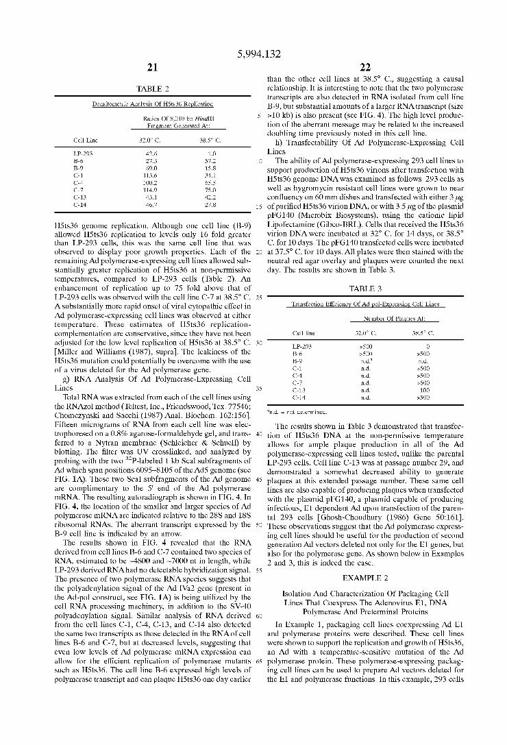
5,994,132 21
TABLE 2
Densitometric Analysis Of HSts36 Replication
Ratios Of 8,010 bp HindIII Fragment Generated At:
Cell Line 32.O. C. 38.5° C.
LP-293 42.6 1.O B-6 27.3 57.2 B-9 69.0 15.8 C-1 113.6 34.1 C-4 100.2 65.5 C-7 114.9 75.0 C-13 43.1 42.2 C-14 46.7 27.8
H5ts36 genome replication. Although one cell line (B-9) allowed H5ts36 replication to levels only 16 fold greater than LP-293 cells, this was the same cell line that was observed to display poor growth properties. Each of the remaining Ad polymerase-expressing cell lines allowed Sub Stantially greater replication of H5ts36 at non-permissive temperatures, compared to LP-293 cells (Table 2). An enhancement of replication up to 75 fold above that of LP-293 cells was observed with the cell line C-7 at 38.5° C. A Substantially more rapid onset of Viral cytopathic effect in Ad polymerase-expressing cell lines was observed at either temperature. These estimates of H5ts36 replication complementation are conservative, Since they have not been adjusted for the low level replication of H5ts36 at 38.5° C. Miller and Williams (1987), Supra). The leakiness of the H5ts36 mutation could potentially be overcome with the use of a virus deleted for the Ad polymerase gene.
g) RNA Analysis Of Ad Polymerase-Expressing Cell Lines
Total RNA was extracted from each of the cell lines using the RNAZol method (Teltest, Inc., Friendswood,Tex. 77546; Chomczynski and Sacchi (1987) Anal. Biochem. 162:156). Fifteen micrograms of RNA from each cell line was elec trophoresed on a 0.8% agarose-formaldehyde gel, and trans ferred to a Nytran membrane (Schleicher & Schuell) by blotting. The filter was UV crosslinked, and analyzed by probing with the two P-labeled 1 kb Scal subfragments of Ad which span positions 6095-8105 of the Ad5 genome (see FIG. 1A). These two Scal subfragments of the Ad genome are complimentary to the 5' end of the Ad polymerase mRNA. The resulting autoradiograph is shown in FIG. 4. In FIG. 4, the location of the smaller and larger species of Ad polymerase mRNA are indicated relative to the 28S and 18S ribosomal RNAS. The aberrant transcript expressed by the B-9 cell line is indicated by an arrow. The results shown in FIG. 4 revealed that the RNA
derived from cell lines B-6 and C-7 contained two species of RNA, estimated to be ~4800 and ~7000 nt in length, while LP-293 derived RNA had no detectable hybridization signal. The presence of two polymerase RNA species Suggests that the polyadenylation signal of the Ad IVa2 gene (present in the Ad-pol construct, see FIG. 1A) is being utilized by the cell RNA processing machinery, in addition to the SV40 polyadenylation signal. Similar analysis of RNA derived from the cell lines C-1, C-4, C-13, and C-14 also detected the same two transcripts as those detected in the RNA of cell lines B-6 and C-7, but at decreased levels, Suggesting that even low levels of Ad polymerase mRNA expression can allow for the efficient replication of polymerase mutants such as H5ts36. The cell line B-6 expressed high levels of polymerase transcript and can plaque H5ts36 one day earlier
15
25
35
40
45
50
55
60
65
22 than the other cell lines at 38.5 C., Suggesting a causal relationship. It is interesting to note that the two polymerase transcripts are also detected in RNA isolated from cell line B-9, but substantial amounts of a larger RNA transcript (size >10 kb) is also present (see FIG. 4). The high level produc tion of the aberrant message may be related to the increased doubling time previously noted in this cell line.
h) Transfectability Of Ad Polymerase-Expressing Cell Lines The ability of Ad polymerase-expressing 293 cell lines to
support production of H5ts36 virions after transfection with H5ts36 genome DNA was examined as follows. 293 cells as well as hygromycin resistant cell lines were grown to near confluency on 60 mm dishes and transfected with either 3 lig of purified H5ts36 virion DNA, or with 3.5 ug of the plasmid pFG140 (Microbix Biosystems), using the cationic lipid Lipofectamine (Gibco-BRL). Cells that received the H5ts36 virion DNA were incubated at 32° C. for 14 days, or 38.5° C. for 10 days. The pFG140 transfected cells were incubated at 37.5 C. for 10 days. All plates were then stained with the neutral red agar overlay and plaques were counted the next day. The results are shown in Table 3.
TABLE 3
Transfection Efficiency Of Ad pol-Expressing Cell Lines
Number Of Plaques At:
Cell line 32.O. C. 38.5° C.
LP-293 >500 O B-6 >500 >500 B-9 n.d. n.d. C-1 n.d. >500 C-4 n.d. >500 C-7 n.d. >500 C-13 n.d. 1OO C-14 n.d. >500
n.d. = not determined.
The results shown in Table 3 demonstrated that transfec tion of H5ts36 DNA at the non-permissive temperature allows for ample plaque production in all of the Ad polymerase-expressing cell lines tested, unlike the parental LP-293 cells. Cell line C-13 was at passage number 29, and demonstrated a Somewhat decreased ability to generate plaques at this extended passage number. These same cell lines are also capable of producing plaques when transfected with the plasmid pFG140, a plasmid capable of producing infectious, E1 dependent Ad upon transfection of the paren tal 293 cells Ghosh-Choudhury (1986) Gene 50:161). These observations Suggest that the Ad polymerase express ing cell lines should be useful for the production of Second generation Ad Vectors deleted not only for the E1 genes, but also for the polymerase gene. AS shown below in Examples 2 and 3, this is indeed the case.
EXAMPLE 2
Isolation And Characterization Of Packaging Cell Lines That Coexpress The Adenovirus E1, DNA
Polymerase And Preterminal Proteins In Example 1, packaging cell lines coexpressing Ad E1
and polymerase proteins were described. These cell lines were shown to support the replication and growth of H5ts36, an Ad with a temperature-Sensitive mutation of the Ad polymerase protein. These polymerase-expressing packag ing cell lines can be used to prepare Ad VectorS deleted for the E1 and polymerase functions. In this example, 293 cells

5,994,132 23
cotransfected with both Ad polymerase and preterminal protein expression plasmids are characterized. Cell lines co-expressing the Ad E1, polymerase and preterminal pro teins can be used to prepare Ad VectorS deleted for the E1, polymerase and preterminal protein (pTP) functions.
a) Tissue Culture And Virus Propagation The use of LP-293 cells (Microbix Biosystems Inc.,
Toronto), Ad-polymerase expressing cell lines, and plaquing efficiency assays of Ad viruses was conducted as described in Example 1. All cells were maintained in 10% fetal bovine serum supplemented DMEM media (GIBCO) in the pres ence of antibiotics. The virus H5sub 100 Freimuth and Ginsberg (1986) Proc. Natl. Acad. Sci. USA 83:7816) has a temperature Sensitive (ts) mutation caused by a three base pair insertion within the amino terminus of the preterminal protein, in addition to a deletion of the E1 Sequences (see FIG. 1B). H5sub100 was propagated and titred at 32.0° C. in LP-293 cells; the leakiness of this stock was less than 1 per 1000 plaque-forming units (pfu) at the nonpermissive temperature of 38.5 C. A lower titer cell lysate containing the virus H5in 190 (which contains a 12 base-pair insertion within the carboxy-terminus of the preterminal protein as well as a deletion of the E1 region, see FIG. 1B) was provided by Dr. P. Freimuth Freimuth and Ginsberg (1986), Supra). The polymerase and preterminal protein expressing cell lines were always maintained in media Supplemented with hygromycin (Sigma) at 100 ug/mL.
b) Isolation Of Ad Polymerase And Preterminal Protein Expressing 293 Cells The C-1, C-4, C-7, C-13, and C-14 cell lines (Ex. 1),
which had been cotransfected with pRSV-pol, pRSV-pTP and CEP4, were screened for presence of pTP sequences and for the ability to support the growth of H5ts36 (ts for the Ad-polymerase), H5in 190, and H5Sub100 using plaque assays as described in Example 1.
i) Analysis Of Genomic DNA And Cellular RNA Cell lines that had received the preterminal protein
expression plasmid were Screen for the presence of pRSV pTP sequences and E1 sequences. Total DNA was isolated from the LP-293, B-6 (transfected with pPSV-pol only) or C-7 (cotransfected with pRSV-pol and pRSV-pTP) cell lines, two micrograms (ug) of each DNA was codigested with the restriction enzymes Xbal and BamHI, electro phoretically Separated in a 0.6% agarose gel, and transferred onto a nylon membrane. The membrane was UV crosslinked, probed with both a 1.8 kb BlnI-Xbal fragment (spans the E1 coding region) isolated from the plasmid pFG 140, and a 1.8kb EcoRV subfragment of Ad serotype 5 (spans the preterminal protein coding sequences; see FIG. 11B), both of which were random-primer radiolabeled with p to a specific activity greater than 3.0x10 cpm/ug. The
membrane was Subsequently exposed to X-ray film with enhancement by a fluorescent Screen. The resulting autora diograph is shown in FIG. 5.
In FIG. 5, the preterminal Specific Sequences migrated as an ~11.0 kb DNA fragment while the E1 containing band migrated as a 2.3 kb DNA fragment. No hybridization of either probe to DNA isolated from either the LP-293 or B-6 cell lines was observed AS shown in FIG. 5, only the C-7 cell line genomic DNA had preterminal coding Sequences, unlike the parental LP-293 cells, or the Ad-polymerase expressing B-6 cells. In addition, all cell lines had E1 Specific Sequences present at nearly equivalent amounts, demonstrating that the Selection design has not caused the loss of the E1 sequences originally present in the LP-293 cells. The results presented in Example 1 demonstrated that
15
25
35
40
45
50
55
60
65
24 both the B-6 and C-7 cell lines contain polymerase specific Sequences within their genomes, unlike the parental LP-293 cells. To confirm that transcription of preterminal protein was
occurring, total RNA was isolated from each of the cell lines, transferred to nylon membranes, and probed to detect pre terminal protein-specific mRNA transcripts as follows. Total cellular RNA was isolated from the respective cell lines and 15 lug of total RNA from each cell line was transferred to nylon membranes. The membranes were probed with the 1.8 kb EcoRV radiolabeled subfragment of AdS (see FIG. 1B) complementary to the preterminal protein coding region. The resulting autoradiograph is shown in FIG. 6. As shown in FIG. 6, a single mRNA of the expected size
(-3 kb in length) is detected only in RNA derived from the C-7 cell line. No hybridization was detected in lanes con taining RNA derived from the LP-293 or B-6 cell lines. In Example 1, it was demonstrated that the C-7 cell line also expresses high levels of the Ad polymerase mRNA. Thus, the C-7 cell line constitutively expresses both the Ad poly merase and preterminal protein mRNAS along with E1 transcripts.
ii) Plaquing Efficiency Of pTP Mutants. On pTR Expressing Cell Lines The C-7 cell line was screened for the ability to
transcomplement the growth of preterminal mutant viruses. The virus H5in 190 (contains a 12 base pair insertion located within the carboxy-terminus of the preterminal protein) has been shown to have a Severe growth and replication defect, producing less than 10 plaque-forming units per cell Freimuth and Ginsberg (1986) Proc. Natl. Acad. Sci. USA 83:7816). The results are summarized in Table 4 below. For the results shown in Table 4, LP-293, B-6, or C-7 cells were seeded at a density of 2.0–2.5x10 cells per plate. The cells were infected with limiting dilutions of lysates derived from the preterminal protein-mutant viruses H5 in 190 or H5Sub100, incubated at 38.5 C., and plaques counted after six days. As shown in Table 4, only the C-7 cell line could allow efficient plaque formation of H5in 190 at 38.5° C. (the H5in 190 lysate used to infect the cells was of a low titer, relative to the high titer H5Sub100 stock), while both the B-6 and C-7 cell lines had nearly equivalent plaquing efficiencies when H5sub 100 was utilized as the infecting virus.
TABLE 4
Plaquing Efficiency Of Preterminal Protein-Mutant Viruses
Mutation Plaque Titers (pfu/ml
Virus Location LP-293 B-6 C-7
HS190 carboxy-terminus <1 x 10? &1 x 10° 1.4 x 10 HSSub100 amino-terminus <1 x 10' 9.0 x 10' 4.5 x 10
As shown in Table 4, when equivalent dilutions of H5in 190 were utilized, the plaquing efficiency of the C-7 cell line was at least 100-fold greater than that of the B-6 or LP-293 cells. This result demonstrated that the C-7 cell line produces a functional preterminal protein, capable of trans complementing the defect of the H5in 190 derived pretermi nal protein. The cell lines were next screened for the ability to
trans-complement with the temperature-Sensitive virus, H5Sub100, at nonpermissive temperatures. H5Sub100 has a codon insertion mutation within the amino-terminus of the preterminal protein, as well as an E1 deletion. The mutation is responsible both for a temperature Sensitive growth

5,994,132 25
defect, as well as a replication defect Freimuth and Gins berg (1986), Supra and Schaack et al. (1995) J. Virol. 69:4079). The plaquing efficiency of the cell line C-7 was found to be at least 1000 fold greater than that of the LP-293 cells (at non-permissive temperatures) (see Table 4). Interestingly, the cell line B-6 was also capable of producing large numbers of H5sub 10 derived plaques at 38.5 °C., even though it does not express any preterminal protein. This result Suggested that the high level expression of the poly merase protein was allowing plaque formation of H5sub 100 in the B-6 cell line. To examine this possibility, the nature of H5sub 100 growth in the various cell lines was examined.
c) Complementation Of The Replication And Growth Defects Of H5Sub 100
The cell lines B-6 and C-7 were shown to overcome the replication defect of H5ts36 (Ex. 1). Since the preterminal and polymerase proteins are known to physically interact with each other Zhao and Padmanabhan (1988) Cell 55:1005), we investigated whether the expression of the Ad-polymerase could overcome the replication defect of H5sub 100 at non-permissive temperatures using the follow ing replication-complementation assay.
LP-293, B-6, or C-7 cells were seeded onto 60 mM dishes at a density of 2x10 cells per dish and infected the next day with H5sub 100 at a multiplicity of infection (MOI) of 0.25, and incubated at 38.5° C. for 16 hours or 32.0° C. for 40 hours. The cells from each infected plate were then har vested and total DNA extracted as described in Example 1. Four micrograms of each DNA sample was digested with HindIII, electrophoresed through a 0.7% agarose gel, trans ferred to a nylon membrane, and probed with P-labeled H5ts36 virion DNA. The resulting autoradiograph is shown in FIG. 7. As seen in FIG. 7, the H5sub100 replication defect when grown in LP-293 cells at 38.5 C. is seen; this defect is not present when the virus is grown at the same tempera ture in either B-6 or C-7 cells.
The results depicted in FIG. 7 demonstrates that both cell lines B-6 and C-7 could trans-complement the replication defect of H5Sub100. This result demonstrated that the expression of the Ad polymerase in B-6 cells was able to overcome the preterminal protein-mediated replication defect of H5sub100. While not limiting the present invention to any particular mechanism, the ability of Ad polymerase to overcome the preterminal protein-mediated replication defect of H5sub 100 may be due to a direct physical inter action of the polymerase with the amino-terminus of the H5sub 100-derived preterminal protein.
In Support of this hypothesis, it has also been demon strated that the H5Sub100 replication defect can be over come when LP-293 cells were infected with a 100-fold greater amount of H5Sub100. However, complementation of the H5sub 100 replication defect is not sufficient to overcome the growth defect of E5sub 100, since temperature shift-up experiments have demonstrated that the H5sub 100 growth defect is not dependent upon viral replication Schaack et al. (1995), Supra). Therefore, the overexpression of the Ad-polymerase must have allowed a very low level but detectable production of infectious H5sub100 particles in the B-6 cell line. The reduced growth of H5sub 100 is therefore not due to a replication defect, but rather Some other critical activity that the preterminal protein has a role in, Such as augmention of Viral transcription by association with the nuclear matrix Schaack and Shenk (1989) Curr. Top. Microbiol. Immunol. 144:185 and Hauser and Cham berlain (1996) J. Endo. 149:373). This was confirmed by assessing the ability of the C-7 cell line to overcome the
15
25
35
40
45
50
55
60
65
26 growth defect of H5sub 100 utilizing one-step growth assays performed as follows. Each of the cell lines (LP-293, B-6 and C-7) were seeded
onto 60 mm dishes at 2.0x10 cells/dish. The cell lines were infected at an MOI of 4 with each of the appropriate viruses (wt Ad5, H5ts36, or H5sub100), and incubated at 38.5° C. for 40 hours. The total amount of infectious virions pro duced in each 60 mm dish was released from the cell lysates by three cycles of freeze-thawing, and the titer was then determined by limiting dilution and plaque assay on B-6 cells at 38.5 C. The results are summarized in FIG. 8. The results shown in FIG. 8, demonstrated that even
though the B-6 cell line allowed normal replication and plaque formation of H5Sub100 at 38.5° C. (in fact, 13-6 cells were utilized to determine the plaque titres depicted in FIG. 8) they could not allow high level growth of H5sub100 and only produced titres of H5sub100 equivalent to that produced by the LP-293 cells. The C-7 cell line produced 100 fold more virus than the LP-293 or 1B-6 cells, see FIG. 8. Encouragingly, the titre of H5sub 100 produced by the C-7 cells approached titres produced by LP-293 cells infected with wild-type virus, Ads. When the H5sub100 virions produced from infection of the C-7 cells were used to infect LP-293 cells at 38.5 C., all virus produced retained the ts mutation (i.e., at least a 1000 fold drop in pfu was detected when LP-293 cells were respectively infected at 38.5 C. vs. 32.0° C.). This finding effectively rules out the theoretical possibility that the H5sub 100 input virus genomes recom bined with the preterminal protein Sequences present in the C-7 cells.
In addition, the C-7 cell line allowed the high level growth of H5ts36, demonstrating that adequate amounts of the Ad-polymerase protein were also being expressed. The C-7 cell line was capable of trans-complementing the growth of both H5ts36 and H5Sub100 after 4 months of Serial passaging, demonstrating that the coexpression of the E1, preterminal, and polymerase proteins was not toxic.
These results demonstrate that the constitutive expression of both the polymerase and preterminal proteins is not detrimental to normal virus production, which might have occurred if one or both of the proteins had to be expressed only during a narrow time period during the Ad life cycle. In Summary, these results demonstrated that the C-7 cell line can be used as a packaging cell line to allow the high level growth of E1, preterminal, and polymerase deleted Ad VectOrS.
EXAMPLE 3
Production Of Adenovirus Vectors Deleted For E1 And Polymerase Functions
In order to produce an Ad vectors deleted for E1 and polymerase functions, a Small, frame-shifting deletion was introduced into the Ad-pol gene contained within an E1-deleted Ad genome. The plasmid pBHG11 (Microbix) was used as the Source of an E1-deleted Ad genome. pBHG 11 contains a deletion of Ad5 sequences from bp 188 to bp 1339 (0.5-3.7 m.u.); this deletion removes the pack aging Signals as well as E1 Sequences. pBHG 11 also con tains a large deletion within the E3 region (bp 27865 to bp 30995; 77.5–86.2 m.u.). The nucleotide sequence of pBHG11 is listed in SEQ ID NO:4 for cross-corrleation between the pBHG11 sequence and the Ad5 genome (SEQ ID NO:1), it is noted that nucleotide 8,773 in pBHG 11 is equivalent to nucleotide 7,269 in Ad5). pBHG 11 was chosen to provide the Ad backbone because
this plasmid contains a large deletion within the E3 region
5,994,132 27
(77.5 to 86.2 m.u.) and therefore vectors derived from this plasmid permit the insertion of large pieces of foreign DNA. A large cloning capacity is important when the pol Vectors is to be used to transfer a large gene Such as the dystrophin gene (cDNA=13.6 kb). However, the majority of genes are not this large and therefore other Adbackbones containing Smaller deletions within the E3 region (e.g., pBHG10 which contains a deletion between 78.3 to 85.8 m.u.; Microbix) may be employed for the construction of pol Vectors using the Strategy outlined below.
a) Construction Of A Plasmid Containing A Portion Of The Adenovirus Genome Containing The Polymers Gene A fragment of the Ad genome containing the pol gene
located on pBHG 11 was subcloned to create p3SA-XB. Due to the large size of the Ad genome, this intermediate plasmid was constructed to facilitate the introduction of a deletion within the pol gene the pol deletion. pBSA-XB was con Structed as follows. The polylinker region of pBlueScript (Stratagene) was modified to include additional restriction enzyme recognition sites (the sequence of the modified polylinker is provided in SEQ ID NO:5; the remainder of pBlueScript was not altered); the resulting plasmid was termed pBSX. pBHG 11 was digested with Xbal and BamHI and the 20.223 kb fragment containing the pol and pTP coding regions (E2b region) was inserted into pBSX digested with Xbal and BamHI to generate pBSA-XB.
b) Construction Of pBHG11Apol A deletion was introduced into the poll coding region
contained within pBSA-XB in such a manner that other key Viral elements were not disturbed (e.g., the major late promoter, the tripartite leader Sequences, the pTP gene and other leader Sequences critical for normal virus viability). The deletion of the poll sequences was carried out as follows. pBSA-XB was digested with BspEI and the ends were filled in using T4 DNA polymerase. The BspEI-digested, T4 polymerase filled DNA was then digested with BamHI and the 8809 bp BamHI/BspEI(filled) fragment was isolated as follows. The treated DNA was run on a 0.6% agarose gel (TAE buffer) and the 8809 bp fragment was excised from the gel and purified using a QIAEX Gel Extraction Kit accord ing to the manufacturers instructions (OIAGEN, Chatsworth, Calif.). A second aliquot of p3SA-XB DNA was digested with
BspHI and the ends were filled in with T4DNA polymerase. The BspHI-digested, T4 polymerase filled DNA was then digested with BamHI and the 13,679 bp BamHI/BspHI (filled) fragment was isolated as described above. The purified 8809 bp BamHI/BspEI(filled) fragment and
the purified 13,679 bp BamHI/BspHI(filled) fragment were ligated to generate papol.p.Apol contains a 612 bp deletion within the pol gene (bp 8772 to 9385; numbering relative to that of pBHG11) and lacks the 11.4 kb BamHI fragment containing the right arm of the Ad genome found within pBHG11.
To provide the right arm of the Ad genome, papol was digested with BamHI followed by treatment with calf intes tinal alkaline phosphatase. pBHG 11 was digested with BamHI and the 11.4 kb fragment was isolated and purified using a QIAEX Gel Extraction Kit as described above. The purified 11.4 kb BamHI fragment was ligated to the BamHI/ phosphatased papol to generate pBHG 11 Apol. Proper con struction of pBHG 11 Apol was confirmed restriction diges tion (HindIII).
c) Rescue And Propagation Of Ad5Apol Virus The Ad genome contained within pBHG 11 Apol lacks the
packaging Signals. Therefore, in order to recover virus
15
25
35
40
45
50
55
60
65
28 containing the 612 bp deletion within the pol gene from pBHG 11 Apol, this plasmid must be cotransfected into pack aging cells along with DNA that provides a Source of the Ad packaging Signals. The Ad packaging Signals may be pro vided by wild-type or mutant Ad viral DNA or alternatively may be provided using a shuttle vector which contains the left-end Ad5 Sequences including the packaging Signals. Such as paE1splA, p.AE1splB (Microbix) or padBglII (pAdBglII is a Standard Shuttle vector which contains 0-1 m.u. and 9-16 m.u. of the adenovirus genome). To rescue virus, p3HG 11 Apol was co-transfected with
Ad5d 17001 viral DNA. Ad5d 17001 contains a deletion in the E3 region; the E3 deletion contained within Ad5d17001 is Smaller than the deletion contained within the Ad genome contained within pBHG 11. It is not necessary that Ad5d17001 be used to recover virus; other adenoviruses, including wild-type adenoviruses, may be used to rescue of virus from pBHG 11 Apol.
It has been reported that the generation of recombinant Ads is more efficient if Ad DNA-terminal protein complex (TPC) is employed in conjunction with a plasmid containing the desired deletion Miyake et al. (1996) Proc. Natl. Acad. Sci. USA 93:1320). Accordingly, Ad5d17001-TPC were prepared as described Miyake et al. (1996), Supra). Briefly, purified Ad5d17001 virions (purified through an isopycnic CsCl gradient centered at 1.34 g/ml) were lysed by the addition of an equal volume of 8M guanidine hydrochloride. The released Ad5d17001 DNA-TPC was then purified through a buoyant density gradient of 2.8M CsCl/4M guani dine hydrochloride by centrifugation for 16 hr at 55,000 rpm in a VTió5 rotor (Beckman). Gradient fractions containing Ad5d17001 DNA-TPC were identified using an ethidium bromide spot test and then pooled, dialyzed extensively against TE buffer. BSA was then added to a final concen tration of 0.5 mg/ml and aliquots were stored at -80 C. The Ad5d17001 DNA-TPC was then digested with Scal and then gel-filtered through a SephadeX G-50 Spin column. One hundred nanograms of the digested Ad5d17001
DNA-TPC was mixed with 5 lug of pBHG 11 Apol and used to transfect pol-expressing 293 cells (i.e., C-7). Approxi mately 10 days post-transfection, plaques were picked. The recombinant viruses were plaque purified and propagated using Standard techniques (Graham and Prevac, Supra). Viral DNA was isolated, digested with restriction enzymes and Subjected to Southern blotting analysis to determine the organization of the recovered viruses. Two forms of Virus containing the 612 bp pol deletion were recovered and termed Ad5ApolAE3I and Ad5ApolAE3II. One form of recombinant poli virus recovered, Ad5ApolAE3I, underwent a double recombination event with the Ad5d17001 sequences and contains the E3 deletion contained within Ad5d17001 at the right end of the genome. The Second form of recombi nant poli virus, Ad5ApolAE3II, retained pBHG 11 sequences at the right end of the genome (i.e., contained the E3 deletion found within pBHG11). These results demonstrate the pro duction of a recombinant Ad Vector containing a deletion within the poll gene.
d) Characterization Of The E1+, pol- Viruses To demonstrate that the deletion contained within these
two pol Viruses renders the virus incapable of producing functional polymerase, Ad5ApolAE3I was used to infect 293 cells and pol-expressing 293 cells (B-6 and C-7 cell lines) and a viral replication-complementation assay was per formed as described in Example 1e. Briefly, 293, B-6 and C-7 cells were seeded onto 60 mm dishes at a density of 2x 10⁶ cells/dish and infected with H5Sub 100 or

5,994,132 29
Ad5ApolAE3I at an MOI of 0.25. The infected cells were then incubated at 37° C. or 38.5° C. for 16 hours, or at 32.0 C. for 40 hours. Cells from each infected plate were then harvested and total DNA was extracted. Four micrograms of each DNA sample was digested with HindIII, electrophore Sised through an agarose gel, transferred to a nylon mem brane and probed with 'P-labeled adenoviral DNA. The resulting autograph is shown in FIG. 9. In FIG.9, each panel shows, from left to right, DNA extracted from 293, B-6 and C-7 cells, respectively infected with either H5sub 100 or Ad5ApolAE3I (labeled Ad5APOL in FIG. 9). As shown in FIG. 9, the recombinant poli virus was found
to be viable on pol-expressing 293 cells but not on 293 cells. These results demonstrates that recombinant Ad viruses containing the 612 bp deletion found within papol lack the ability to express Ad polymerase. These results also dem onstrate that B-6 and C-7 cells efficiently complement the Apol found within Ad5ApolAE3I and Ad5ApolAE3II. In addition, these results show that replication of the ts pTP mutant H5sub 100 can be complemented by high level expression of the Ad polymerase with or without co-expression of pTP.
Because the expression of early genes is required for the expression of the late gene products, the ability of recom binant viruses containing the Apol deletion to direct the expression of late gene products was examined. 293 and C-7 cells were infected with Ad5ApolAE3I and 24 hours after infection cell extracts were prepared. The cell extracts were serially diluted and examined for the expression of the fiber protein (a late gene product) by immunoblot analysis. The immunoblot was performed as described in Example 3C with the exception that the primary antibody used was an anti-fiber antibody (FIBER-KNOB obtained from Robert Gerard, University of Texas Southwestern Medical School). The results of this immunoblotting analysis revealed that no fiber protein was detected from 293 cells infected with Ad5ApolAE3I (at any dilutuion of the cell extract). In contrast, even a 1:1000 dilution of cell extract prepared from C-7 cells infected with Ad5ApolAE3I produced a visible band on the immunoblot. Therefore, the pol deletion con tained within Ad5ApolAE3I resulted in a greater than 1000 fold decrease in fiber production.
The above results demonstrate that polymerase gene Sequences can be deleted from the Virus and that the result ing deleted virus will only grow on cells producing Ad-polymerase in trans. Using the pol-expressing cell lines described herein (e.g., B-6 and C-7), large quantities of the pol Viruses can be prepared. A dramatic shut-down in growth and late gene expression is Seen when cells which do not express Ad polymerase are infected with the pol Viruses.
e) Generation Of E1, Pol Ad Vectors Ad5di7001 used above to recuse virus containing the
polymerase deletion is an E1-containing virus. The presence of E1 Sequences on the recombinant poll viruses is unde sirable when the recombinant virus is to be used to transfer genes into the tissueS of animals, the E1 region encodes the transforming genes and Such viruses replicate extremely well in Vivo leading to an immune response directed against cells infected with the E1-containing virus. E1 viruses containing the above-described polymerase
deletion are generated as followS. p3HG 11 Apol is cotrans fected into pol-expressing 293 cells (e.g., B-6 or C-7) along with a shuttle vector containing the left-end Ad5 Sequences including the packaging Signals. Suitable Shuttle vectors include paE1splA (Microbix), p.AE1splB (Microbix) or
15
25
35
40
45
50
55
60
65
30 pAdBglII. The gene of interest is inserted into the polylinker region of the Shuttle Vector and this plasmid is then cotrans fected into B-6 or C-7 cells along with pBHGG 11 Apol to generate a recombinant E1, pol Ad Vector containing the gene of interest.
EXAMPLE 4
Production Of Adenovirus Vectors Deleted For E-1 And Preterminal Protein Functions
In order to produce an Ad vectors deleted for E1 and preterminal protein functions, a Small deletion was intro duced into the Ad preterminal protein (pTP) gene contained within an E1-deleted Ad genome. The plasmid pBHG 11 (Microbix) was used as the source of an E1-deleted Ad genome to maximize the cloning capacity of the resulting pTP vector. However, other Ad backbones containing Smaller deletions within the E3 region (e.g., pBHG1O which contains a deletion between 78.3 to 85.8 m.u.; Microbix) may be employed for the construction of pTP vectors using the Strategy outlined below.
a) Construction Of papTP A deletion was introduced into the pTP coding region
contained within pBSA-XB (Ex. 3) in such a manner that other key viral elements were not disturbed (e.g., the tripar tite leader Sequences, the i-leader Sequences, the VA-RNAI and II genes, the 55 kD gene and the pol gene). The deletion of the pTP sequences was carried out as follows. pBSA-XB was digested with Xbal and EcoRV and the 7.875 kb fragment was isolated as described (EX. 3). Another aliquot of pBSA-XB was digested with Mun and the ends were filled in using T4DNA polymerase. The MunI-digested, T4 polymerase filled DNA was then digested with Xbal and the 14.894 kb Xbal/MunI(filled) fragment was isolated as described (Ex. 3). The 7.875 kb MunI fragment and the 14.894 kb Xhal/MunI(filled) fragment were ligated together to generate papTP.
b) Construction Of pBHG11ApTP pApol contains a 429 bp deletion within the pTP gene (bp
10,705 to 11,134; numbering relative to that of pBHG11) and lacks the 11.4 kb BamHI fragment containing the right arm of the Ad genome found within pBHG 11. To provide the right arm of the Ad genome, papTP was
digested with BamHI followed by treatment with shrimp alkaline phosphatase (SAP; U.S. Biochemicals, Cleveland, Ohio). pBHG11 was digested with BamHI and the 11.4 kb fragment was isolated and purified using a QIAEX Gel Extraction Kit as described (EX. 3). The purified 11.4 kb BamHI fragment was ligated to the BamHI/phosphatased pApTP to generate pBHG 11 ApTP. Proper construction of pBHG 11 ApTP was confirmed restriction digestion.
c) Rescue And Propagation Of Ad Vectors Containing The pTP Delection
The Ad genome contained within pBHG 11 ApTP lacks the Ad packaging Signals. Therefore, in order to recover virus containing the 612 bp deletion within the pol gene from pBHG 11 Apol, this plasmid must be cotransfected into pack aging cells along with DNA that provides a Source of the Ad packaging Signals. The Ad packaging Signals may be pro vided by wild-type or mutant Ad viral DNA or alternatively may be provided using a shuttle vector which contains the left-end Ad5 Sequences including the packaging Signals. Such as paE1splA, p.AE1splB (Microbix) or padBgIII.
Recombinant Ad vectors containing the pTP deletion which contain a deletion within the E3 region are generated

5,994,132 31
by cotransfection of pBHG 11 ApTP (the gene of interest is inserted into the unique PacI site of pBHG11ApTP) with a E3-deleted Ad virus such as Ad5d1700 into pTP-expressing 293 cells (e.g., C-7); viral DNA-TPC are utilized as described above in Example 3.
Recombinant vectors containing the pTP deletion which also contain deletions within the E1 and E3 regions are generated by cotransfection of pBHG 11 ApTP into pTP expressing 293 cells (e.g., C-7) along with a shuttle vector containing the left-end Ad5 Sequences including the pack aging Signals. Suitable Shuttle vectors include pa. 1SplA (Microbix), p.AE1splB (Microbix) or padBglII. The gene of interest is inserted into the polylinker region of the shuttle vector and this plasmid is then cotransfected into B-6 or C-7 cells along with pBHG 11 ApTP to generate a recombinant E1, pTP Ad vector containing the gene of interest.
EXAMPLE 5
Production Of Adenovirus Vectors Deleted For E1, Polymerase And Preterminal Protein Functions
In order to produce an Ad Vectors deleted for E1, poly merase and preterminal protein functions, a deletion encom passing pol and pTP gene Sequences was introduced into the Ad Sequences contained within an E1-deleted Ad genome. The plasmid pBHG 11 AE4 was used as the source of an E1-deleted Ad genome to maximize the cloning capacity of the resulting pol, pTP vector. p3H11AE4 is a modified form of BHG11 which contains a deletion of all E4 genes except for the E4 ORF 6; the E4 region was deleted to create more room for the insertion of a gene of interest and to further disable the virus. However, other E1 Adbackbones, such as pBHG11 and pBHG1 (Microbix, pBHG 11 contains a Smaller deletion within the E3 region as compared to pBHG11), may be employed for the construction of pol, pTP vectors using the strategy outlined below. Due to the complexity of the cloning StepS required to
introduce a 2.3 kb deletion that removes portions of both the pTP and poll genes, this deletion was generated using Several StepS as detailed below.
a) Construction Of paXBApolApTPVARNA+t 13 In order to create a plasmid containing Ad Sequences that
have a deletion within the pol and pTP genes, pAXBApolApTPVARNA+t13 was constructed as follows. pBSA-XB was digested with BspEI and the 18 kb frag
ment was isolated and recircularized to create pAXBApolApTP; this plasmid contains a deletion of the Sequences contained between the BSpEI Sites located at 8,773 and 12,513 (numbering relative to pBHG11). A fragment encoding the VA-RNA3 Sequence and the
third leader of the tri-partite leader Sequence was prepared using the PCR as follows. The PCR was carried out in a solution containing H5ts36 virion DNA (any Ad DNA, including wild-type Ad, may be used), 2 ng/mL of primers 4005E and 4006E, 10 mM TrishC1, pH 8.3, 50 mM KC1, 1.5 mM MgCl, 0.001% gelatin and Pfu polymerase. The for ward primer, 4005E, 5'- TGCCGCAGCACCGGATGCATC-3' (SEQ ID NO:6) con tains Sequences complementary to residues 12,551 to 12,571 of pBHG 11 (SEQ ID NO:4). The reverse primer, 4006E, 5'-GCGTCCGGAGGCTGCCATG CGGCAGGG-3' (SEQ ID NO:7) is complementary to residues 11,091 to 11,108 of pBHG 11 (SEQ ID NO:4) as well as a BspEI site (underlined). The predicted sequence of the ~1.6 kb PCR product is listed in SEQ ID NO:8. PCR was performed with a PerkinElmer 9600 Thermocy
cler utilizing the following cycling parameters: initial dena
15
25
35
40
45
50
55
60
65
32 turation at 94 C. for 3 min, 3 cycles of denaturation at 94 C. for 30 sec, annealing at 50 C. for 30 sec, and extension at 72 C. for 60 sec, followed by another 27 cycles with an increased annealing temperature at 56 C., with a final extension at 72 C. for 10 minutes. The -1.6 kb PCR product was purified using a QIAEX Gel Extraction Kit as described (Ex. 3). The purified PCR fragment was then digested with BspEI.
pAXBApol.ApTP was digested with BspEI followed by treatment with SAP. The BspEI-digested PCR fragment and the BspEI-SAP-treated paXBApolApTP were ligated together to create paXBApolApTPVARNA+t13.
b) Construction Of pBHF11ApolApTPVARNA+t13 pAXBApolApTPVARNA+t13 contains a 2.3 kb deletion
within the pol and pTP genes (bp 8,773 to 11,091; number ing relative to that of pBHG11) and lacks the 11.4 kb BamHI fragment containing the right arm of the Ad genome. To provide the right arm of the Ad genome,
pAXBApolApTPVARNA+t 13 was digested with BamHI followed by treatment with SAP. pBHG 11AE4 (pBHG11 or pBHG10 may be used in place of pBHG11AE4) was digested with BamHI and the 11.4 kb fragment was isolated and purified using a QIAEX Gel Extraction Kit as described (EX.3). The purified 11.4 kb BamHI fragment was ligated to the BamHI/phosphatased pAXBApol.ApTPVARNA+t 13 to generate pBHG 11 Apol.ApTPVARNA+t 13. Proper construc tion of pBHG11ApolApTPVARNA+t13 was confirmed restriction digestion.
c) Rescue And Propagation Of Ad Vectors Containing The pol, pTP Double Delection The Ad ge no me contained within
pBHG 11 ApolApTPVARNA+t 13 lacks the Ad packaging Signals. Therefore, in order to recover virus containing the 2.3 kb deletion within the pol and pTP genes from pBHG 11 ApolApTPVARNA+t13, this plasmid must be cotransfected into packaging cells along with DNA that provides a Source of the Ad packaging Signals. The Ad packaging Signals may be provided by wild-type or mutant Ad viral DNA or alternatively may be provided using a Shuttle vector which contains the left-end Ad5 Sequences including the packaging Signals Such as p a 1 SplA, pAE1splB (Microbix) or padBglII.
Recombinant V Ad vectors containing the pol, pTP double deletion which also contain a deletion within the E3 region
C gene rated by cotransfection of pBHG11ApolApTPVARNA+t13 (the gene of interest is inserted into the unique Pac I site of pHBG 11 Apol.ApTPVARNA+t13) with a E3-deleted Ad virus such as Ad5d17001 into pol- and pTP-expressing 293 cells (e.g., C-7); viral DNA-TPC are utilized as described (Ex. 3).
Recombinant vectors containing the pol, pTP double deletion which also contain deletions within the E1 and E3 regions are generated by cotransfection of pBHG 11 ApolApTPVARNA+t 13 into pol- and pTP expressing 293 cells (e.g., C-7) along with a shuttle vector containing the left-end Ad5 Sequences including the pack aging Signals. Suitable Shuttle vectors include pa. 1SplA (Microbix), p.AE1splB (Microbix) or padBglII. The gene of interest is inserted into the polylinker region of the Shuttle vector and this plasmid is then cotransfected into B-6 or C-7 cells along with pBHG 11 Apol.ApTPVARNA+t 13 to generate a recombinant E1, polpTP Ad Vector containing the gene of interest.
EXAMPLE 6
Encapsidated Adenovirus Minichromosomes Containing A Full Length Dystrophin cDNA
In this Example, the construction of an encapsidated adenovirus minichromosome (EAM) consisting of an infec

5,994,132 33
tious encapsidated linear genome containing Ad origins of replication, packaging Signal elements, a f-galactosidase reporter gene cassette and a full length (14 kb) dystrophin cDNA regulated by a muscle Specific enhancer/promoter is described. EAMs are generated by cotransfecting 293 cells with Supercoiled plasmid DNA (pAd5 BdyS) containing an embedded inverted origin of replication (and the remaining above elements) together with linear DNA from E1-deleted Virions expressing human placental alkaline phosphatase (hpAP). All proteins necessary for the generation of EAMs are provided in trans from the hp AP virions and the two can be separated from each other on equilibrium CsCl gradients. These EAMs are useful for gene transfer to a variety of cell types both in vitro and in vivo.
a) Generation And Propagation Of Encapsidated Aden ovirus Minichromosomes
To establish a vector system capable of delivering full length dystrophin cDNA clones, the minimal region of Ad5 needed for replication and packaging was combined with a conventional plasmid carrying both dystrophin and a B-galactosidase reporter gene. In the first vector constructed, these elements were arranged such that the viral ITRs flanked (ITRS facing outward) the reporter gene and the dystrophin gene i.e., the vector contained from 5' to 3' the right or 3' ITR (mu 100 to 99), the dystrophin gene and the reporter gene and the left or 5' ITR and packaging Sequnce (mu 1 to 0)). Upon introduction of this vector along with helper virus into 293 cells, no encapsidated adenovirus minichromosomes were recovered.
The second and successful vector, pAd5 Bdys (FIG. 10) contains 2.1 kb of adenovirus DNA, together with a 14 kb murine dystrophin cDNA under the control of the mouse muscle creatine kinase enhancer/promoter, as well as a B-galactosidase gene regulated by the human cytomegalovi rus enhancer/promoter.
FIG. 10 shows the structure of pAd5 Bdys (27.8 kb). The two inverted adenovirus origins of replication are repre sented by a left and right inverted terminal repeat (LITR/ RITR). Replication from these ITRs generates a linear genome whose termini correspond to the 0 and 100 map unit (mu) locations. Orientation of the origin with respect to wild type adenovirus Serotype 5 Sequences in mu is indicated above the figure (1 mu=360 bp). Encapsidation of the mature linear genome is enabled by five (AI-AV) packaging signals (I). The E. coli B-galactosidase and mus musculus dystro phin cDNAs are regulated by cytomegalovirus (CMV) and muscle creatine kinase (MCK) enhancer/promoter elements, respectively. Both expression cassettes contain the SV40 polyadenylation (pA) signal. Since the E1A enhancer/ promoter overlaps with the packaging Signals, pad5pdyS was engineered Such that RNA polymerase transcribing from the E1A enhancer/promoter will encounter the SV40 late polyadenylation signal. Pertinent restriction Sites used in constructing pad5Bdys are indicated below the figure. An adenovirus fragment corresponding to mu 6.97 to 7.77 was introduced into pAd5BdyS during the cloning procedure (described below). P1 and P2 represent location of probes used for Southern blot analysis. Restriction sites destroyed during the cloning of the Ad5 origin of replication and packaging Signal are indicated in parentheses. pAd5Bdys was constructed as follows.pbSX (EX.3a) was
used as the backbone for construction of pad5Bdys. The inverted fused Ad5 origin of replication and five encapsi dation signals were excised as a Pst/Xbal fragment from pAd5ori, a plasmid containing the 6 kb HindIII fragment from pFG140 (Microbix Biosystems). This strategy also
15
25
35
40
45
50
55
60
65
34 introduced a 290 bp fragment from Ad5 corresponding to map units 6.97 to 7.77 adjacent to the right inverted repeat (see FIG. 10). The E. coli B-galactosidase gene regulated by the human CMV immediate early (IE) promoter/enhancer expression cassette was derived as an EcoRI/HindIII frag ment from pCMVB MacGregor and Caskey (1989) Nucleic Acids Res. 17:2365); the CMV IE enhancer/promoter is available from a number of Suppliers as is the E. coli f-galactosidase gene. The murine dystrophin expression cassette was derived as a BSSHII fragment from pCVAA, and contains a 3.3 kb MCK promoter/enhancer element Phelps et al. (1995) Hum. Mol. Genet. 4:1251 and Jaynes et al. (1986) Mol. Cell. Biol. 6:2855). The sequence of the -3.3 kb MCK promoter/enhancer element is provided in SEQ ID NO:9 (in SEQ ID NO:9, the last nucleotide of SEQ ID NO:9 corresponds to nucleotide +7 of the MCK gene). The enhancer element is contained within a 206 bp fragment located -1256 and -1050 upstream of the transcription start site in the MCK gene.
It was hoped that the pad5Bdys plasmid would be pack ageable into an encapsidated minichromosome when grown in parallel with an E1-deleted virus due to the inclusion of both inverted terminal repeats (ITR) and the major Ad packaging Signals in the plasmid. The ITRS and packaging signals were derived from pFG140 (Microbix), a plasmid that generates E1-defective Ad particles upon transfection of human 293 cells.
hp AP is an E1 deleted Ad5 containing the human placen tal alkaline phosphatase gene Muller et al. (1994) Circ. Res. 75:1039). This virus was chosen to provide the helper functions so that it would be possible to monitor the titer of the helper virus throughout Serial passages by quantitative alkaline phosphatase assays. 293 cells were cotransfected with pad5pdys and hp AP
DNA as follows. Low passage 293 cells (Microbix BioSystems) were grown and passaged as Suggested by the supplier. Five pad5(Bdys and hp AP DNA (5 and 0.5 lug, respectively) were dissolved in 70 ul of 20 mM HEPES buffer (pH 7.4) and incubated with 30 ul of DOTAP (BMB) for 15 min. at room temperature. This mixture was resus pended in 2 mls of DMEM Supplemented with 2% fetal calf serum (FCS) and added dropwise to a 60 mm plate of 293 cells at 80% confluency. Four hours post-transfection the media was replaced by DMEM with 10% FCS. Cytopathic effect was observed 6-12 days post-transfection.
Cotransfection of 293 cells with Supercoiled pad5Bdys and linear hp AP DNA produced Ad5 Bdys EAMs (the encapsidlated version of pads BdyS) approximately 6 to 12 days post transfection, as evidenced by the appearance of a cytopathic effect (CPE). Initially, the amount of Ad5 Bdys EAMs produced was significantly lower than that of hp AP Virions So the viral Suspension was used to re-infect fresh cultures, from which Virus was isolated and used for Serial infection of additional cultures (FIG. 11). Infection and Serial passaging of 293 cells was carried out as follows.
Lysate from one 60 mm plate of transfected 293 cells was prepared by Vigorously washing the cells from the plate and centrifuging at 1 K rpm in a clinical centrifuge. Cells were resuspended in DMEM and 2% FCS, freeze-thawed in a dry ice-ethanol bath, cell debris removed by centrifugation, and approximately 75% of the crude lysate was used to infect 293 cells in DMEM supplemented with 2% FCS for 1 hour and then supplemented with 10% FCS thereafter. Infection was allowed to proceed for 18–20 hrs before harvesting the virus. The total number of cells infected in each serial passage is indicated in FIG. 11.

5,994,132 35
In FIG. 11, the total number of transducing adenovirus particles produced (output) per Serial passage on 293 cells, total input virus of either the helper (hpAP) or Ad5 Bdys, and the total number of cells used in each infection is presented. The total number of input/output transducing particles were determined by infection of 293 cells plated in 6-well micro titer plates. Twenty four hours post-infection the cells were assayed for alkaline phosphatase or B-galactosidase activity (described below) to determine the number of cells trans duced with either Ad5pdys or hpAP. The number of trans ducing particles were estimated by extrapolation of the mean calculated from 31 randomly chosen 2.5 mm sectors of a 961 mm plate. The intra-sector differences in total output of each type of virus are presented as the Standard deviations, O, in FIG. 11. For each serial passage, 75% of the total output virus from the previous passage was used for infec tion.
Alkaline phosphatase or B-galactosidase activity was determined as follows. For detection of alkaline phosphatase, infected 293 cells on Petri dishes were rinsed twice with phosphate buffered saline (PBS) and fixed for 10 minutes in 0.5% glutaraldehyde in PBS. Cells were again rinsed twice with PBS for ten minutes followed by inacti Vation of endogenous alkaline phosphatase activity at 65 C. for 1 hr. in PBS prior to the addition of the chromogenic substrate BCIP (5-bromo-4-chloro-3-indolyl phosphate) at 0.15 mg/ml and nitro blue tetrazolium at 0.3 mg/ml). Cells were incubated at 37 C. in darkness for 3-24 hrs. For B-galactosidase assays, the cells were fixed and washed as above, then assayed using standard methods MacGregor et al. (1991) In Murray (ed.), Methods in Molecular Biology, Vol. 7: Gene Transfer and Expression Protocols, Human Press Inc., Clifton, N.J., pp. 217-225). As shown in FIG. 11, the rate of increase in titer of
Ad5Bdys EAMs between transfection and serial passage 6 was approximately 100 times greater than that for hp AP virions. This result indicated that Ad5Bdys has a replication advantage over the helper virus, probably due to the shorter genome length (a difference of approximately 8 kb) and hence an increased rate of packaging.
Interestingly, after Serial passage 6 there was a rapid decrease in the total titer of hpAP virions whereas the titer of Ad5Bdys EAMs continued to rise. While not limiting the present invention to any particular mechanism, at least two possible mechanisms could be responsible for this observa tion. Firstly, a buildup of defective hp AP virions due to infections at high multiplicities may slowly out-compete their full length counterparts, a phenomenon that has been previously observed upon Serial propagation of adenovirus Daniell (1976) J. Virol. 19:685; Rosenwirth et al. (1974) Virology 60:431; and Burlingham et al. (1974) Virology 60:419). Secondly, the emergence of replication competent Virions due to recombination events between E1 Sequences in cellular DNA and the hp AP genome could lead to a buildup of Virus particles defective in expressing alkaline phosphatase Lochmüller et al. (1994) Hum. Gene Ther. 5:1485).
Southern analysis of DNA prepared from serial lysates 3, 6, 9 and 12 indicated that full length dystrophin Sequences were present in each of these lysates (FIG. 12A). In addition, the correct Size restriction fragments were detected using both dystrophin and B-galactosidase probes against lysate DNA digested with several enzymes (FIGS. 12A-B).
FIG. 12 shows a Southern blot analysis of viral DNA from lysates 3, 6, 9 and 12, digested with the restriction enzymes BSSHII, NruI and EcoRV, indicating the presence of a full
15
25
35
40
45
50
55
60
65
36 length dystrophin cDNA in all lysates. Fragments from the C terminus of mus musculus dystrophin cDNA(A) or the N terminus of E coli B-galactosidase (B) were labeled with dCTP and used as probes Sambrook et al., supra). The position of these probes and the predicted fragments for each digest is indicated in FIG. 10. Note that one end of each fragment (except the 17.8 kb BSSHII dystrophin fragment) detected is derived from the end of the linearized Ad5Bdys genome (see FIG. 10). Low levels of shorter products, presumably derived from defective virions, become detect able only at high Serial passage number. At the later passages (9 and 12) there appeared to be an
emergence of truncated Ad5BdyS Sequences, Suggesting that deletions and/or rearrangements may be occurring at later passages. Hence, most experiments were performed with Ad5Bdys EAMs derived from the earlier passages. The possibility of the emergence of replication competent (E1 containing) viruses was also examined by infection of HeLa cells by purified and crude Serial lysates, none of which produced any detectable CPE.
b) Purification Of Encapsidated Adenovirus Minichromo Somes On CsCl Gradients The ability to separate Ad5Bdys EAMs from hp AP virions
based on their buoyancy difference (due presumably to their different genome lengths) on CsCl gradients was examined. Repeated fractionation of the viral lysate allows small differences in buoyancy to be resolved. CsCl purification of encapsidlated adenovirus minichromosomes was performed as follows. Approximately 25% of the lysate prepared from various passages during Serial infections was used to purify Virions. Freeze-thawed lysate was centrifuged to remove the cell debris. The cleared lysate was extracted twice with 1,1,2 tricholorotrifluoroethane (Sigma) and applied to CsCl step and Self forming gradients.
Purification of Virus was initially achieved by passing it twice through CsCl step gradients with densities of p=1.45 and p=1.20 in a SW28 rotor (Beckman). After isolation of the major band in the lower gradient, the virus was passed through a self forming gradient (initial p=1.334) at 37,000 rpm for 24 hrs followed by a relaxation of the gradient by reducing the speed to 10,000 rpm for 10 hrs. in a SW41 rotor (Beckman) at 12 C. Anet and Strayer (1969) Biochem. Biophys. Res. Commun. 34:328). The upper band from the gradient (composed mainly of Ad5BdyS Virions) was iso lated using an 18 gauge needle, reloaded on a fourth CsCl gradient (p=1.334) and purified at 37,000 rpm for 24 hrs followed by 10,000 rpm for 10 hrs at 12° C. The Ad5Bdys-containing CsCl band was removed in 100
all fractions from the top of the centrifugation tube and CsCl was removed by chromatography on Sephadex G-50 (Pharmacia). Aliquots from each fraction were used to infect 293 cells followed by B-galactosidase and alkaline phos phatase assays to quantitate the level of contamination by hpAP virions in the final viral isolate.
Results of the physical Separation between Ad5pdyS EAMs and hp AP virions are shown in FIG. 13. FIG. 13 shows the physical separation of Ad5Bdys from hp AP viri ons at the third (A) and final (B) stages of CsCl purification (initial p=1.334) in a SW41 tube. Aliquots of Ad5 Bdys EAMs from the final stage were drawn through the top of the centrifugation tube and assayed for B-galactosidase and alkaline phosphatase expression. Results of these assays are presented in FIG. 14.
In FIG. 14, shows the level of contamination of Ad5Bdys EAMs by hpAP virions obtained from passage 6. Following four cycles of CsCl purification, aliquots were removed

5,994,132 37
from the top of the centrifugation tube and used for infection of 293 cells, which were fixed 24 hrs later and assayed for f-galactosidase or alkaline phosphatase activity (described below). The number of transducing particles are an under estimation of the actual totals as in Some cases a positive cell may have been infected by more than one transducing particle. The ratio of the two types of virions -AdSBdys EAMs (LacZ) or hpAP (AP) in each fraction is indicated in the lower graph.
The maximum ratio of transducing Ad5Bdys EAMs to hp AP virions reproducibly achieved in this study was 24.8-a contamination with helper virus corresponding to approximately 4% of the final viral isolate.
c) Dystrophin and f-galactosidase Expression By Encapsidated Adenovirus Minichromosomes In Muscle Cells
To determine if the Ad5Bdys EAMs were able to express B-galactosidase and dystrophin in muscle cells, mouse mdX myogenic cultures were infected with CsCl purified EAMs.
i) Propagation And Infection Of Muscle Cells MM14 and mdx myogenic cell lines were kindly provided
by S. Hauschka (University of Washington) and were cul tured as previously described Linkhart et al. (1981) Dev. Biol. 86:19 and Clegg et al. (1987) J. Cell Biol. 105:949). Briefly, myoblasts were grown on plastic tissue culture plates coated with a 0.1% gelatin in Ham's F-10 medium containing 15% (v/v) horse serum, 0.8 mM CaCl2, 200 ng/ml recombinant human basic fibroblast growh factor (b-FGF) and 60 tug/ml genitimicin (proliferation medium). Cultures were induced to differentiate by Switching to growth in the presence of growth medium lacking b-FGF and containing 10% horse serum (differentiation medium). Myoblasts or differentiated myotubes (three days post Switching) were infected at a multiplicity of infection of 2.2 Ad5Bdys EAMs per cell. Fractions containing minimal contamination with hp AP virions (3, 4 and 5 of passage 6) were used for Western and immunofluorescence analysis. Infection was allowed to proceed for 3 days for both the myoblasts and myotubes before harvesting cells.
ii) Total Protein Extraction And Immunoblot Analysis For protein extraction, muscle cells were briefly
trypsinized, transferred to a microcentrifuge tube, centri fuged at 14 K for 3 min at room temp and resuspended two times in PBS. After an additional centrifugation, the cell pellet was resuspended in 80 ul of RIPA buffer (50 mM Tris-C1, pH 7.5; 150 mM NaCi; 1% Nonidet P-40; 0.5% sodium deoxycholate, 0.1% SDS) (Sambrook et al., Supra). The Sample was briefly sheared using a 22 gauge needle to reduce Viscosity and total protein concentration assayed using the bicinconinic acid protein assay reagent (Pierce, Rockford, Ill.). Expression of full length dystrophin or B-galactosidase in infected mdx and MM14 myoblasts or myotubes was analyzed by electrophoresis of 40 ug of total protein extract on a 6% SDS-PAGE gel (in 25 mM Tris, 192 mM glycine, 10 mM f-mercaptoethanol, 0.1% SDS). After transferring to Gelman Biotrace NT membrane (in 25 mM Tris, 192 mM glycine, 10 mM P-mercaptoethanol, 0.05% SDS, 20% methanol), the membrane was blocked with 5% non-fat milk and 1% goat-serum in Tris-buffered saline Tween (TBS-T) for 12 hrs at 4 C. Immunostaining was done according to the protocol for the ECL Western blotting detection reagents (Amersham Life Sciences, Buckingham, UK). The primary antibodies used were Dys-2 (Vector Laboratories) and anti-p-galactosidase (BMB, Indianapolis, Ind.) with a horseradish peroxidase-conjugated anti-mouse Secondary antibody.
5
15
25
35
40
45
50
55
60
65
38 Western blot analysis of EAM-infected mdx myoblasts
and myotubes (three days post-fusion) indicated that EAMs were able to infect both of these cell types (FIG. 15). In FIG. 15, immunoblots of protein extracts from mdx myoblasts and myotubes demonstrating the expression of f-galactosidase (A) and dystrophin (B) in cells infected with Ad5Bdys EAMs. Total protein was extracted 3 days post infection in all cases. Myotubes were infected at three days following a Switch to differentiation media. In FIG. 15A, lane 1 contains total protein extract from 293 cells infected with a virus expressing f-galactosidase as a control (RSV Lacz); lanes 2-5 contain total protein extracts from unin fected mdx myoblasts, mdx myoblasts infected with Ad5pdys EAMs, mdx myotubes and mdx myotubes derived from mdx myoblasts infected with Ad5Bdys EAMs, respec tively. In FIG. 15B, lanes 1 and 7 contain total protein from mouse muscle (“C57”) while lane 2 contains protein from wild type MM14 myotubes, as controls. Lanes 3-5 contain total protein extracts from uninfected mdX myoblasts, mdx myoblasts infected with Ad5Bdys EAMs, mdx myotubes and mdx myotubes derived from mdx myoblasts infected with Ad5Bdys EAMs, respectively. AS shown in FIG. 15, expression of B-galactosidase was
detected in both the infected mdx myoblasts and myotubes indicating that the CMV promoter was active at both these early Stages of differentiation in muscle cells. However, only infected mdX myotubes produced protein detected by dyS-2, an antibody recognizing the 17 C-terminal amino acids of dystrophin (FIG. 15). No dystrophin expression was detected in infected myoblasts by Western analysis, indicat ing that the muscle creatine kinase promoter functions minimally, if at all, within the Ad5Bdys EAM prior to terminal differentiation of these cells.
Dystrophin expression in mdx cells infected with EAMs was confirmed by immunofluorescence Studies using N-terminal dystrophin antibodies. In agreement with the western analysis, dystrophin expression from the MCK promoter was detected only in differentiated mdX myotubes infected by Ad5Bdys EAMs (FIG. 16). FIGS. 16A-C show immunofluorescence of dystrophin in wild type MM14 myotubes (A), uninfected mdx (B) and infected mdx myo tubes (C), respectively. The results shown in FIG. 16 dem onstrate the transfer and expression of recombinant dystro phin to differentiated mdx cells by Ad5Bdys EAMs.
Immunofluorescence of myogenic cells was performed as follows. Approximately 1.5x10, MM14 or mdx myoblasts were plated on Poly-L-lysine (Sigma) coated glass slides (7x3 cm) which had been previously etched with a 0.05% chromium potassium Sulfate and 0.1% gelatin Solution. For myotube analysis, the cultures were Switched to differentia tion media Clegget al. (1987), Supra 48 hours after plating, immediately infected and then allowed to fuse for 3 days, whereas myoblasts were continuously propagated in prolif eration media Clegg et al. (1987), Supra). Cells were washed three times with PBS at room temperature and fixed in 3.7% formaldehyde. For immunostaining, cells were incubated in 0.5% Triton X-100, blocked with 1% normal goat Serum and incubated with an affinity purified antibody against the N-terminus of murine dystrophin for 2 hrs. followed by extensive washing in PBS and 0.1% Tween-20 with gentle shaking. Cells were incubated with a 1:200 dilution of biotin conjugated anti-rabbit antibody (Pierce) for one hour and washed as above. Cells were further incubated with a 1:300 dilution of streptavidin-fluorescein isothiocyanate conjugate (Vectorlabs, Burlingam, Calif.) for one hour and washed as above, followed by extensive washing in PBS.

5,994,132 39
The above results show that embedded inverted Ad ori gins of replication coupled to an encapsidation signal can convert circular DNA molecules to linear forms in the presence of helper virus and that these genomes can be efficiently encapsidated and propagated to high titers. Such Viruses can be purified on a CsCl gradient and maintain their ability to transduce cells in Vitro, and their increased cloning capacity allows the inclusion of large genes and tissue Specific gene regulatory elements. The above results also show that the dystrophin gene was expressed in cells trans duced by Such viruses and that the protein product was correctly localized to the cell membrane. The above method for preparing EAMS theoretically enables virtually any gene of interest to be inserted into an infectious minichromosome by conventional cloning in plasmid vectors, followed by cotransfection with helper viral DNA in 293 cells. This approach is useful for a variety of gene transfer Studies in Vitro. The observation that vectors completely lacking viral genes can be used to transfer a full-length dystrophin cDNA into myogenic cells indicates that this method may be used for the treatment of DMD using gene therapy.
d) A Modified MCK Enhancer Increases Expression Of Linked Genes. In Muscle The DNA fragment containing enhancer/promoter of the
MCK gene utilized in the Ad5Bdys EAM plasmid is quite large (-3.3. kb). In order to provide a smaller DNA fragment capable of directing high levels of expression of linked genes in muscle cells, portions of the 3.3 kb MCK enhancer/ promoter were deleted and/or modified and inserted in front of a reporter gene (lacZ). The enhancer element of the MCK gene was modified to produce the 2RS5 enhancer; the sequence of the 2RS5 enhancer is provided in SEQ ID NO:10. The first 6 residues of SEQ ID NO:10 represent a KpnI site added for ease of manipulation of the modified MCK enhancer element. Residue number 7 of SEQ ID NO:10 corresponds to residue number 2164 of the wild-type MCK enhancer sequence listed in SEQ ID NO:9 (position 2164 of SEQ ID NO:9 corresponds to position -1256 of the MCK gene). Residue number 174 of SEQ ID NO:10 cor responds to residue number 2266 of the wild-type MCK enhancer sequence listed in SEQ ID NO:9.
These MCK/lacZ constructs were used to transfect cells in culture (i.e., myogenic cultures) or were injected as naked DNA into the muscle of mice and 3-galactosidase activity was measured. FIG. 17 provides a schematic of the MCK/ lac7, constructs tested. The first construct shown in FIG. 17 contains the 3.3. kb wild- type MCK enhancer/promoter fragment linked to the E. coli lac7 gene. The wild-type enhancer element (-1256 to -1056) is depicted by the box containing “E”; the core promoter element (-358 to -80) is indicated by the light cross-hatching and the minimal pro moter element (-80 to +7) is indicated by the dark cross hatching. The core promoter element is required in addition to the minimal promoter element (which is required for basal expression) in order to achieve increased muscle-specific expression in tissue culture Shield et al. (1996) Mol. Cell. Biol. 16:5058). The modified enhancer element, the 2RS5 enhancer, is indicated by the box labeled “E*.” The box labeled “minx” contains a synthetic intron derived from adenovirus and is used to increase expression of the con Structs (any intron may be utilized for this purpose). The box labeled “CMV" depicts the CMV IE enhancer/promoter which was used a positive control.
In FIG. 17, B-galactosidase activity is expressed relative to the activity of the wild-type MCK enhancer/promoter construct shown at the top of the figure. As shown in FIG. 17, a construct containing the 2RS5 enhancer (SEQ ID
15
25
35
40
45
50
55
60
65
40 NO:10) linked to either the minimal MCK promoter (a ~261 bp element) or the core and minimal MCK promoter ele ments (a ~539 bp element) directs higher levels of expres Sion of the reporter gene in muscle cells as compared to the ~3.3 kb fragment containing the wild-type enhancer ele ment. These modified enhancer/promoter elements are con siderably smaller than the -3.3 kb fragment used in the Ad5Bdys EAM plasmid and are useful for directing the expression of foreign genes in muscle cells. These Smaller elements are particularly useful for driving the expression of genes in the context of Self-propagating adenoviral vectors which have more Severe constraints on the amount of foreign DNA which can be inserted in comparison to the use of “gutted” adenoviruses such as the EAMs described above.
EXAMPLE 7
Generation of High Titer Stocks of Encapsidated Adenovirus Minichromosomes Containing Minimal
Helper Virus Contamination The results presented in Example 6 demonstrated that
encapsidated adenovirus minichromosomes (EAMS) can be prepared that lack all viral genes and which can express full-length dystrophin cDNAS in a muscle Specific manner. The propagation of these EAMS requires the presence of helper adenoviruses that contaminate the final EAM prepa ration with conventional adenoviruses (about 4% of the total preparation). In this example the EAM System is modified to enable the generation of high titer stocks of EAMs with minimal helper virus contamination. Preferably the EAM Stocks contain helper virus representing less than 1%, pref erably less than 0.1% and most preferably less than 0.01% of the final viral isolate. Purified EAMs are then injected in Vivo in muscles of dystrophin minus mdX mice to determine whether these vectors lead to immune rejection and whether they can alleviate dystrophic Symptoms in mdx muscles. The amount of helper virus present in the EAM prepara
tions is reduced in two ways. The first is by selectively controlling the relative packaging efficiency of the helper virus versus the EAM virus. The second is to improve physical methods for Separating EAM from helper virus. These approaches enable the generation of dystrophin expressing EAMS that are contaminated with minimal levels of helper virus.
a) Development and Characterization of Adenovirus Packaging Cell Lines Expressing The Cre Recombinase From Bacteriophage P1
Cell lines expressing a range of Cre levels are used to optimize the amount of helper virus packaging that occurs during growth of the EAM vectors. The Cre-loxP system Sauer and Henderson (1988) Proc. Natl. Acad. Sci. USA 85:5166 is employed to selectively disable helper virus packaging during growth of EAMS. The bacterial Cre recombinase catalyzes efficient recombination between a 34 bp target Sequence called loxP. To delete a desired Sequence, loXP Sites are placed at each end of the Sequence to be deleted in the same orientation; in the presence of Cre recombinase the intervening DNA segment is efficiently excised.
Cell lines expressing a range of Cre recombinase levels are generated. The expression of too little Cre protein may result in high levels of helper virus being generated, which leads to unacceptably high levels of helper virus contami nating the final EAM preparation. If very high levels of Cre expression are present in a cell line, excision of the pack

5,994,132 41
aging signal from the helper virus would be 100% efficient (i.e., it would completely prevent helper virus packaging). AS shown in Example 6, Serial passage of EAM preparations containing low levels of helper virus increased the titer of the EAM. Therefore, it is desirable, at least in the initial passages of a Serial passage that Some helper virus capable of being packaged is present. A low level of packagable helper virus may be provided by using a cell line expressing levels of Cre recombinase which are not high enough to achieve excision of the packaging Signals from 100% of the helper virus, these cell lines would be used early in the Serial passaging of the EAM Stock and a cell line expressing high enough levels of Cre recombinanse to completely prevent helper virus packaging would be used for the final passage.
Alternatively, EAMS may be prepared using a packaging cell line that Supports high efficiency Cre recombinase mediated excision of the packaging Signals from the helper virus by transfection of the Cre-expressing cell line with the EAM plasmid followed by infection of these cells with loxP-containing helper virus using an MOI of 21.0. Human 293 cell lines that express a variety of levels of
Cre recombinase are generated as follows. An expression vector containing the Cre coding region, p0G231, was cotransfected into 293 cells along with pcDNA3 (Invitrogen); pcDNA3 contains the neo gene under the control of the SV40 promoter. pCG231 uses the human CMV enhancer/promoter derived from CDM8 (Invitrogen) to express a modified Cre gene pCG231 was obtained from S. O’Gorman). The modified Cre gene con tained within p0G231 has had a nuclear localization Signal inserted into the coding region to increase the efficiency of recombination.
pOG231 was constructed as follows. A BglI site was introduced into the 5' Xbal site of the synthethic intron of pMLSISCAT (Haung and Gorman (1990) Nucleic Acids Res. 18:937 by linker tailing. A BglII site in the synthethic intron of pMLSISCAT was destroyed and a BamHI linker was inserted into the Pst site at the 3' end of the synthethic intron in pMLSISCAT. BglII and SmaI sites and a nuclear localization signal were introduced into the 5' end of pMC Cre. Gu et al. (1993) Cell 73:1155) using a PCR fragment that extended from the novel BglII and SmaI sites to the BamHI site in the Cre coding region. This PCR fragment was ligated to a BamHI/Sal fragment containing a portion of the Cre coding region derived from plC-Cre (Gu et al., Supra) and the intron plus Cre coding sequence was inserted into a modified form of pCG44 O'Gorman et al. (1991) Science 251:1351 to generate pCG231. The predicted sequence of pCG231 from the BglII site to the BamHI site located in the middle of the Cre coding Sequence is listed in SEO ID NO:11. One 60 mm dish of 293 cells (Microbix) were transfected
with 10 ug of Pvull-linearized pCG231 and 1 lug of Not linearized pcDNA3 using a Standard calcium phosphate precipitation protocol. Two days after the addition of DNA, the transfected cells were split into three 100 mm dishes and 1000 tug/ml of active G418 was added to the medium. The cells were fed periodically with G418-containing medium and three weeks later, 24 G418-resistant clones were iso lated.
The isolated clones were expanded for testing. Aliquots were frozen in liquid nitrogen at the earliest possible pas Sage. The neomycin resistant cell lines were examined for the expression of Cre recombinase using the following transfection assay. The neomycin resistant cells were trans fected with PGK-1-GFP-lacz (obtained from Sally Camper,
15
25
35
40
45
50
55
60
65
42 Univ. of Michigan, Ann Arbor, Mich.), which contains a green fluorescent protein (GFP) expression cassette that can be excised by Cre recombinase to allow expression of B-galactosidase; transfection was accomplised using Stan dard calcium phosphate precipitation. FIG. 18 provides a schematic of a GFP/B-gal reporter construct suitable for assaying the expression of Cre recombinase in mammalian cells; GFP Sequences and f-gal (i.e., lacz) sequences are avaialbale from commmerical Sources (e.g., Clonetech, Palo Alto, Calif. and Pharmacia Biotech, Piscataway, N.J., respectively).
Control experiments verified that 293 cells transfected with PGK-1-GFP-lacz expressed significant amounts of B-galactosidase only if these cells also expressed Cre recom binanse. B-galactosidase assays were performed as described in Example 6. Neomycin resistant cells expressing Cre recombinase were grouped as high, medium or low expressors based upon the amount of B-galactosidase activ ity produced (estimated by direct counting of B-galactosidase-positive cells per high-power field and by observing the level of staining and the rapidity with which the blue Stain was apparent) when these cell lines were transfected with the GFP/B-gal reporter construct. Thirteen positive (i.e., Cre-expressing) lines, D608H12, #7, #22, #18, #17, #4, #8, #2, #2/2, #13, #5, #15 and #21 were retained for further use.
The results of this transfection analysis revealed that cultures of 293 cells expressing medium to high levels of Cre recombinase could be generated without apparent tox icity.
b) Generation of Helper Adenovirus Strains that Contain loXP Sites Flanking the Adenovirus Packaging Signals
Studies of EAM production demonstrated that the EAM vector has a packaging advantage over the helper adenovirus (Ex. 6). While not limiting the present invention to a particular mechanism, it is hypothesized that this packaging and replication advantage can be greatly increased by using helper viruses that approach the packaging Size limits of Ad5 Bett et al. (1993) J. Virol. 67:5911), by using viruses with mutations in E4 and/or E2 genes Yeh et al. (1996) J. Virol. 70:559; Gorziglia et al. (1996) J. Virol. 70:4173; and Amalfitano et al. (1996) Proc. Natl. Acad. Sci. USA 93:3352), by inclusion of mutations or alterations in the packaging Signals of the helper virus Imler et al. (1995) Hum. Gene Ther. 6:711 and by combining these strategies. The Cre-loxP excision method is used to disable the
packaging Signals from the helper virus genomes. The Ad5 packaging domain extends from nucleotide 194 to 358 and is composed of five distinct elements that are functionally redundant Hearing et al. (1987) J. Virol. 61:2555). Theoretically, any molecule containing the Ad5 origin of replication and packaging elements should replicate and be packaged into mature virions in the presence of non defective helper virus. Disabling the packaging Signals should allow replication and gene expression to proceed, but will prevent packaging of Viral DNA into infectious par ticles. This in turn should allow the ratio of EAM to helper Virus to be increased greatly. To disable the packaging Signals within the helper virus
used to encapsidate the EAMs, the loXP Sequences are incorporated into a helper virus that has a genome approach ing the maximal packaging size for Ad-105 map units, Bett et al. (1993), supra which further decreases the efficiency of helper virus packaging. Final virus Size can be adjusted by the choice of introns inserted into the reporter gene, by the choice of reporter genes, or by including a variety of DNA

5,994,132 43
fragments of various sizes to act as "stuffer fragments. A convenient reporter gene is the alkaline phosphatase gene (see Ex. 6). The optimized Cre-loxP system is also incor porated into a helper Viral backbone containing disruptions of any or all of the E1-E4 genes. Use of such deleted genomes requires viral growth on appropriate complement ing cell lines, Such as 293 cells expressing E2, and/or E4 gene products. The loXP Sequences are incorporated into the helper virus by placing the loXP Sequences on either Side of the packaging Signals on a shuttle vector. This modified shuttle vector is then used to recombine with the Ad DNA derived from a virus containing disruptions of any or all of the E1-E4 genes to produce the desired helper virus con taining the packaging Signals flanked by loXP Sequences. pAdBglII, an adenovirus shuttle plasmid containing Ad Sequences from 0–1 and 9-16 map units (mu) was used as the Starting material. Synthetic oligonucleotides were used to create a polylinker which was inserted at 1 mu within pAdB glII as follows. The BglII LoxP oligo 5'- GAA GATCTATAACTTC GTATAATGTATG CTA TACGAAGTTATTACCGAAGAAATGGCTCGAGATCT TC-3' (SEQ ID NO:12) and its reverese complement 5'- GAAGATCTCGAGCCATTTCTTCGGTAATAACTTCGT
ATAGCATACATTATACGAAGTTATAGATCTTC-3' (SEQ ID NO:13) were synthesized. The AflIII LoxP oligo 5'- C CA CAT G TATAACTTCG TATAG CATACA
TTATACGAAGTTATACATGTGG-3' (SEQ ID NO:14) and its C. W. C. S. C. comple ment 5'- C CA CAT G TATAACTTCG TATAATGTAT GCTATACGAAGTTATACATG TGG-3' (SEQ ID NO:15) were synthesized. The double stranded form of each loxP oligonuceotide was digested with the appropriate restriction enzyme (e.g., BglII or AflIII) and inserted into padBglII which had been digested with BglII and AflIII. This resulted in the insertion of LoxP sequences into the shuttle vector flanking the packaging Signals that are located between 0.5 and 1 mu of Ad5 (one mu equals 360 bp in the Ad5 genome; the sequence of the Ad5 genome is lsited in SEQ ID NO:1). The 3' loXP sequence was inserted into the BglII site within pAdBglII. The 5' loxPsequence was inserted into the AflIII site located at base 143 (-0.8 mu) (numbering relative to Ad5). DNA sequencing was used to verify the final Structure of
the modified shuttle plasmid between 0-1 mu. If Cre recombinase-mediated excision of the packaging Signals is found to be too efficient (as judged by the production of too little helper virus), alternate sites for the insertion of the loxP Sequences are used that would result in deletion of 2, 3 or 4 packaging sequences rather than all 5 Grable and Hearing (1990).J. Virol. 64:2047). The insertion of loxPsequences at Sites along the Ad genome contained within the Shuttle vector which would result in the deletion of 2, 3 or 4 packaging Sequences are easily made using the technique of recombinant PCR Higuchi (1990) In: PCR Protocols. A Guide to Methods and Applications, Innis et al. (eds.) Academic Press, San Diego, Calif., pp. 177-183). The optimal amount of Cre recombinase-mediated excision of the packaging Signals is that amount which permits the production of enough packaged helper virus to permit the Slow spread of Virus on the first plate of cells co-transfected with helper virus DNA (containing the loxPsequences) and the EAM vector. This permits serial passage of the EAM preparation onto a Subsequent lawn of cells to increase the titer of the EAM preparation. Alternatively, if Cre recombinase-mediated excision of the packaging Signals is essentially 100% efficient in all Cre-expressing cells lines (i.e., regardless of the level of Cre-expression, that is even
15
25
35
40
45
50
55
60
65
44 a cell line expressing a low level of Cre as judged by the GFP/B-gal assay described above), the packaged EAMs may be used along with helper virus (used at a MOI of ~1.0) to infect the Second or Subsequent lawn of cells to permit Serial passaging to increase the titer of the EAM preparation.
Following introduction of the loxP sequences into the Shuttle vector, the human placental alkaline phosphatase (HpAp) cDNA under control of the RSV promoter was inserted into the polylinker to provide a reporter gene for the helper virus. This is the same reporter used previously during EAM generation (EX. 6). The HpAp sequences were inserted as follows. The loxP-containing shuttle vector was linearized with XhoI and the hp Ap cassette was ligated into the XhoI site (a XhoI site was inserted into padBglII during the insertion of the loXP Sequences as a XhoI site was located on the 3' end of the loxP sequences inserted into the BglII Site of padBglII). The HpAp cassette was constructed as follows. pRSVhAPT40 (obtained from Gary Nabel, Univ. of Michigan, Ann Arbor, Mich.) was digested with EcoRI to generate an EcoRI fragment containing the HpAP cl)NA and the SV40 intron and polyadenylation sequences. pRc/RSV (Invitrogen) was digested with HindIII, then partially digested with EcoRI. A 5,208 bp fragment was then size Selected on an agarose gel, treated with calf alkaline phosphatase, and ligated to the EcoRI fragment derived from pRSVhAPT40 to generate pRc/RSVAP. p.Rc/RSVAP was then digested with SalI and XhoI to liberate the RSV promoter linked to the HpAP clDNA cassette (including the SV40 intron and polyadenylation sequences). This Sall XhoI fragment was inserted into the loXP-containing Shuttle vector which had been digested with Sal and XhoI to generate pADLOXP-RSVAP.
Helper virus containing loxP sites flanking the Ad pack aging Signals is generated by co-transfection of the LOXP shuttle plasmid (pADLOXP-RSVAP) and Clal-digested Ad5d17001 DNA into 293 cells Graham and Prevec (1991) Manipulation of Adenovirus Vectors, In Methods in Molecu lar Biology, Vol. 7. Gene Transfer and Expression Protocols, Murray (ed.), Humana Press Inc., Clifton, N.J., pp. 109-128). FIG. 19 provides a schematic showing the recom bination event between the loxP shuttle vector and the Ad5d17001 genome. Co-transfection is carried out as described in Example 3c.
Alternatively, the reporter gene may be inserted into the E3 region of pBHG10 or pBHG11 (using the unique PacI Site) rather than into the polylinker located in the E1 region of the shuttle vector. The reporter gene-containing pBHG10 or 11 is then used in place of Ad5d17001 for cotransfection of 293 cells along with the loxP-containing padBglII.
Following cotransfection, recombinant plaques are picked, plaque purified, and tested for incorporation of both hpAp and loxP sequences by PCR and Southern analysis (Ex. 3). Viruses which contain loxP sites flanking the packaging signals and the marker gene (hpAp) are retained, propagated and purified. The isolated helper virus containing loXP Sites flanking
the packaging Signals and the marker gene is then used to infect both 293 cells and the 293 cell lines that express Cre (Section a, above). Cre recombinase-expressing cell lines that produce optimal levels of Cre recombinase when infected with the loxP-containing helper virus are then used for the generation of EAMs as described below.
c) Generation of Encapsidated Adenovirus Minichromo Somes that Express Dystrophin The growth of EAMs is optimized using the following
methods. In the first method, plasmid DNA from the EAM

5,994,132 45
vector (Ex. 6) are co-transfected into 293 cells with purified viral DNA from the helper virus, and viruses are harvested 10-14 days later after appearance of a viral cytopathic effect (CPE) as described in Example 6. This approach is simple, yet potentially increases helper virus levels by allowing viral Spread throughout the culture dishes.
In the second method, the co-transfected 293 cells are overlaid with agar and Single plaques are picked and tested for EAM activity by the ability to express B-galactosidase activity following infection of Hela cells (Ex. 6). This Second approach is more time consuming, but will result in leSS contamination by helper.
The ability of these two methods to generate EAMs are directly compared. The preferred method is that which produces the highest titer of EAM with the lowest contami nation of helper virus. The efficiency of EAM generation may also be increased by using helper virus DNA that retains the terminal protein (TP) (i.e., the helper virus is used as a viral DNA-TPC as described in Ex. 3) The use of viral DNA-TPC has been shown to increase the efficiency of viral production following transfection of 293 cells by an order of magnitude Miyake et al. (1996), Supra). The initial transfection will utilize 293 cells that do not
express Cre recombinase, So that efficient spread of the helper can lead to large Scale production of EAM. The method producing the highest ratio of EAM to helper virus is then employed to optimize conditions for the Serial propagation of the EAM on 293 cells expressing Cre recom binase. This optimization is conducted using cell lines expressing different levels of Cre recombinase. The follow ing variables are tested: 1) the ratio of input viral titer to cell number, 2) the number of Serial passages to use for EAM generation, 3) the use of cell lines producing different levels of Cre to achieve the optimal ratio between high EAM titer and low helpertiter, 4) continuous growth on Cre-producing 293 cells verSuS alternating between Cre-producing cells and the parental 293 cells, or to alternate between high and low Cre-producing cells and 5) CsCl purification of EAMs prior to re-infection of 293 cells increases the ratio of the final EAM/helper titers. The protocols that result in the highest yield of EAM with minimal helper virus are used to generate large Volumes of crude Viral lysates for purification by density gradient centrifugation (EX. 6). EAMs were purified from helper virus using standard
CsCl density gradient centrifugation protocols in Example 6 and resulted in preparations containing ~4% helper virus. In order to improve the physical Separation of EAMS and helper virus, a variety of different centrifugation conditions are possible, including changing the gradient shape, type of rotor and tube used, combinations of Step and continuous gradients, and the number of gradients used. Materials with better resolving powers than CsCl can also be employed. These include rubidium chloride and potassium bromide Reich and Zarybnicky (1979) Annal. Biochem. 94.193). d) Use of EAMs Encoding Dystrophin for Long Term
Expression of Dystrophin in Muscle To demonstrate the ability of the purified dystrophin
expressing EAMs (prepared as described above) to deliver and express dystrophin in the muscle of an animal, the following eXperiment is conducted. First, purified dystrophin-EAMs are delivered to the muscle by direct intramuscular injection into newborn, 1 month, and adult (3 month) mouse quadriceps muscle. The dystrophin express ing EAM Ad5BDys are injected into mdx mice and into transgenic mdX mice that express B-galactosidase in the pituitary gland Tripathy et al. (1996) Nature Med. 2:545).
15
25
35
40
45
50
55
60
65
46 These latter mice are used to avoid potential immune rejection of cells expressing B-galactosidase from the Ad5 BDys vector. An alternate EAM lacking the B-galactosidase reporter gene may also be employed; however, the presence of the B-Gal reporter simplifies EAM growth and purification (vectors lacking f-Gal have their purity estimated by PCR assays rather than by B-Galassays).
Following intramuscular injection of EAM, animals are sacrificed at intervals between 1 week and 6 months to measure dystrophin expression by western blot analysis and by immunofluorescence (Phelps et al. (1995) Hum. Mol. Genet. 4:1251 and Rafael et al. (1996) J. Cell Biol. 134:93) and muscle extracts will also be assayed for B-Gal activity MacGregor et al. (1991), Supra). These results are com pared with previous results obtained using current genera tion viral vectors (i.e., containing deletions in E1 and E3 only) to demonstrate that EAMs improve the prospects for long term gene expression in muscle.
e) Use of Dystrophin-EAMs to Prevent, Halt, or Reverse the Dystrophic Symptoms that Develop in the Muscles of mdx Mice
To demonstrate that beneficial effects on dystrophic muscle are achieved by delivery of dystrophin expressing EAMS to mdX mice, the following experiments are con ducted. Central nuclei counts are performed on SoleuS muscle at intervals following injection of EAM into newborn, 1 month, and 3 month (adult) mice (Phelps et al., 1995). Central nuclei arise in mouse muscle only after a myofiber has undergone dystrophic necrosis followed by regeneration, and is a quantitative measure of the degree of dystrophy that has occurred in a muscle group (Phelps et al., 1995). More informative assays are also contemplated. These
assays require administration of EAMS to mouse diaphragm muscles.
The diaphragm is Severely affected in mdx mice (Stedman et al., 1991; Cox et al., 1993), and displays dramatic decreases in both force and power generation. Administra tion of virus to the diaphragm will allow the strength of the muscles to be measured at intervals following dystrophin delivery. The force and power generating assays developed by the Faulkner lab are used to measure the effect of dystrophin transgenes (Shrager et al., 1992; Rafael et al., 1994; Lynch et al., 1996). To detemine whether dystrophin delivery to dystrophic
muscle reverses dystrophy or Stabilizes muscle, varying amounts of the dystrophin EAM are delivered to the to diaphragm at different Stages of the dystrophic process and then strength is measured at intervals following EAM administration. First, various titers of EAM are tested to determine the minimal amount of Virus needed to transduce the majority of muscle fibers in the diaphragm. It has been shown that conventional adenovirus vectors can transduce the majority of diaphragm fibers when 10 pfu are admin istrated by direct injection into the intraperitoneal cavity Huard et al. (1995) Gene Therapy 2:107). In addition, it has been shown that transduction of a simple majority of fibers in a muscle group is Sufficient to prevent Virtually all the dystrophic symptoms in mice Rafael et al. (1994) Hum. Mol. Genet. 3:1725 and Phelps et al. (1995) Hum. Mol. Genet. 4:1251). Virus is administered to mdx animals at three different ages (neonatal, 1 month, and 3 months). Animals are Sacrificed for physiological analysis of dia phragm muscle at two different times post infection (1 month and 3 months). Error control is achieved by perform ing these experiments in Sextuplicate. Control animals con

5,994,132 47
sist of mock injected wild-type (C57B1/10) and dystrophic (mdx) mice. Three month old mdx mice display a 40% reduction in force and power generation compared with wild-type mice, while 6 month animals display a greater than 50% reduction (Cox et al. (1993) Nature 364:725; Rafael et al. (1994), Supra; Phelps et al (1995), Supra; and Corrado et al. (1996) J. Cell. Biol. 134:873).
EXAMPLE 8
Improved Shuttle Vectors for the Production of Helper Virus Containing LOXP Sites
In the previous example, a shuttle vector containing adenoviral Sequences extending from 9 mu to 16 mu of the Ad5 genome was modified to contain loXP Sequences Sur rounding the packaging Signals (the LOXP shuttle vector). This modified shuttle vector was then recombined with an Ad Virus to produce a helper virus containing the loXP Sequences. This helper virus was then used to infect cells expressing Cre recombinase along with DNA comprising a minichromosome containing the dystrophin gene, a reporter gene and the packaging Signals and ITRS of Ad in order to preferentially package the minichromosomes. Using this approach the helper virus, which has had the packaging Signals removed by Cre-loxP recombination, contains the majority of the Ad genome (only a portion of the E3 region is deleted). Thus, if low levels of helper virus are packaged and appear in the EAM preparation, the EAM preparation has the potential of passing on helper virus capable of directing the expression of Ad proteins in cells which are exposed to the EAM preparation. The expression of Ad proteins may lead to an immune response directed against the infected cells.
Another approach to reducing the possibility that the EAM preparation contains helper virus capable of provoking an immune response is to use helper viruses containing deletions and/or mutations within the pol and pTP genes. Helper virus containing a deletion in the pol and/or pTP genes is cotransfected with the EAM construct into 293 derived cell lines expressing poll or pol and pTP to produce EAMs. Any helper virus present in the purified EAM preparation will be replication defective due to the deletion in the pol and/or pTP genes. AS shown in Example 3, Viruses containing a deletion in the pol gene are incapable of directing the expression of Viral late genes, therefore, helper Viruses containing a deletion in the poll gene or the pol and pTP genes should not be capable of provoking an immune response (i.e., a CTL response) against late viral proteins Synthesized de novo. Shuttle vectors containing deletions within the Ad pol and/or pTP genes are constructed as described below.
a) Construction of a Shuttle Vector Containing the Apol Deletion
pAdBglII was modified to contain Sequences correspond ing to 9 to 40 mu of the Ad5 genome as follows. p.AdBglII was digested with BglII and a linker/adapter containing an AScI Site was added to create padBglIIASc. pAdBglIIASc was then digested with Bst 1071.I. Ad5d17001 viral DNA was digested with AscI and the ends were filled in using T4DNA polymerase. The AscI-digested, T4 DNA polymerase filled Ad5d17001 viral DNA was then digested with Bst 1071 I and the -9.9 kb AscI-Bst1107I (filled) fragment containing the pol and pTP genes was isolated (as described in Ex. 3) and ligated to the Bst1107I-digested padBglIIASc to generate pAdASc. pAdASc is a shuttle vector which contains the genes encoding the DNA polymerase and preterminal pro
15
25
35
40
45
50
55
60
65
48 tein (the inserted AscI-Bst1107I fragment corresponds to nucleotides 5767 to 15, 671 of the Ad5 genome). A shuttle vector, padASCApol, which contains the 612 bp
deletion in the pol gene (described in Ex. 3) was constructed as follows. Ad5ApolAE3I viral DNA (Ex. 3) was digested with AscI and the ends were filled in using T4 DNA polymerase. The AscI-digested, T4 DNA polymerase filled Ad5ApolAE3I viral DNA was then digested with Bst1107I and the -9.3 kb AscI-Bst1107I(filled) fragment containing the deleted pol gene and the pTP gene was isolated (as described in Ex. 3) and ligated to Bst1107I-digested pad BglIIASc to generate padASCApol.
b) Construction of a Shuttle Vector Containing the ApolApTP Deletion A shuttle vector, padAscApolApTP, which contains a 2.3
kb deletion within the pol and pTP genes (described in Ex. 5) was constructed as follows. pAXBApolApTPVARNA+ t13 (Ex. 5b) was digested with AscI and the ends were filled in using T4 DNA polymerase. The AscI-digested, T4 DNA polymerase filled pAXBApol.ApTPVARNA+t13 DNA was then digested with Bst1107I and the -7.6 kb AscI-Bst1107I (filled) fragment containing the deleted pol gene and the pTP gene was isolated (as described in Ex. 3) and ligated to Bst 1107 I-digested p AdB gl II Asc to generate pAdAscApol.ApTP.
In order to reduce the packaging of the above helper viruses, the poll or pol, pTP helper viruses can be modi fied to incorporate loXP Sequences on either Side of the packaging Signals as outlined in Example 7. The loXP containing poll or pol, pTP shuttle vectors (pTP- shuttle vectors may also be employed) are cotransfected into 293 cells expressing Cre recombinase and pol or Cre recombinase, pol and pTP, respectively along with an appro priate E1 viral DNA-TPC (the E1 viral DNA may also contain deletions elsewhere in the genome Such as in the E4 genes or in the E2a gene as packaging cell lines expressing the E4 ORF 6 or 6/7 and lines expressing E2a genes are avaialble) to generate helper virus containing loxP Sites flanking the packaging Signals as well as a deletion in the pol gene or the pol and pTP genes. The resulting helper virus(es) is used to cotransfect 293 cells expressing pol or pol and pTP along with the desired EAM construct. The resulting EAM preparation should contain little if any helper virus and any contaminating helper virus present would be replication defective and incapable of expressing viral late gene prod ucts. Helper viruses containing loXP Sequences and deletions in all essential early genes may be employed in conjunction with Cre recombinanse-expressing cell lines expressing in trans the E1, E4 ORF 6, E2a, and E2b (e.g., Ad polymerase and pTP) proteins (the E3 proteins are dispensible for growth in culture). Cell lines coexpressing E1, polymerase and pTP are provided herein. Cell lines expressing E1 and E4 proteins have been recently described Krougliak and Graham (1995) Hum. Gene Ther. 6:1575 and Wang et al. (1995) Gene Ther. 2:775 and cell lines expressing E1 and E2a proteins have been recently described Zhou et al. (1996) J. Virol. 70.7030). Therefore, a cell line co-expressing E1, E2a, E.2b, and E4 is consructed by intro duction of expression plasmids containing the E2a and E4 coding regions into the E1-, Ad polymerase- and pTP expressing cell lines of the present invention. These pack aging cell lines are used in conjunction with helper viruses containing deletions in the E1, E2a, E2b and E4 regions.
All publications and patents mentioned in the above Specification are herein incorporated by reference. Various modifications and variations of the described method and

5,994,132 49 SO
system of the invention will be apparent to those skilled in unduly limited to Such specific embodiments. Indeed, vari the art without departing from the Scope and Spirit of the ouS modifications of the described modes for carrying out invention. Although the invention has been described in the invention which are obvious to those skilled in molecular connection with Specific preferred embodiments, it should biology or related fields are intended to be within the scope be understood that the invention as claimed should not be of the following claims.
SEQUENCE LISTING
(1) GENERAL INFORMATION:
(iii) NUMBER OF SEQUENCES: 15
(2) INFORMATION FOR SEQ ID NO : 1 :
(i) SEQUENCE CHARACTERISTICS: (A)
(C) (D)
LENGTH: 35935 base pairs (B) TYPE : nucleic acid
STRANDEDNESS TOPOLOGY: linear
double
(ii) MOLECULE TYPE : other nucleic acid (A) DESCRIPTION: /desc = "DNA"
(xi). SEQUENCE DESCRIPTION: SEQ ID NO : 1 :
CATCATCAAT
TTGTGACGTG
GATGTTGCAA
TAAATTTGGG
AGTGAAATCT
GACTTTGACC
CGGGTCAAAG
TGAGTTCCT.C
TCCGACACCG
AATGGCCGCC
TCCTAGCCAT
CGAAGATCCC
GCAGGAAGGG
CCTTTCCCGG
CCTTGTACCG
CGAGGATGAA
CAGGTCTTGT
CTATATGAGG
TAGAGTGGTG
GAATTTTGTA
CCAGAACCGG
CGCCCGACAT
CCTTCTAACA
GCCGTGAGAG
CCTGGGCAAC
AATATACCTT
GTGTGGCGGA
GTGTACACAG
CGTAACCGAG
GAATAATTTT
GTTTACGTGG
TTGGCGTTTT
AAGAGGCCAC
GGACTGAAAA
AGTCTTTTGG
TTTGAACCAC
AACGAGGAGG
ATTGACTTAC
CAGCCCGAGC
GAGGTGATCG
GAGGGTGAGG
CATTATCACC
ACCTGTGGCA
GGTTTGGTGT
TTGTGATTTT
AGCCTGCAAG
CACCTGTGTC
CACCTCCTGA
CTTTGGACTT
ATTTTGGATT
TGGGAACGGG
ACACATGTAA
GAAGTGACAA
TAAGATTTGG
GTGTTACTCA
AGACTCGCCC
ATTATTATAG
TCTTGAGTGC
TGAGACATAT
ACCAGCTGAT
CTACCCTTCA
CGGTTTCGCA
TCACTTTTCC
AGCCGGAGCA
ATCTTACCTG
AGTTTGTGTT
GGAGGAATAC
TGTTTGTCTA
GGTAATTTTT
TTTAAAAGGT
ACCTACCCGC
TAGAGAATGC
GATACACCCG
TCGCCAGGCT
GAGCTGTAAA
GAAGCCAATA
GCGGGTGACG
GCGACGGATG
TTTTCGCGCG
CCATTTTCGC
TAGCGCGTAA
AGGTGTTTTT
TCAGCTGACG
CAGCGAGTAG
TATCTGCCAC
CGAAGAGGTA
CGAACTGTAT
GATTTTTCCC
GCCGGCGCCC
GAGAGCCTTG
CCACGAGGCT
AGATTATGTG
GGGGGACCCA
CAGTAAGTGA
TTTTTAATTT
CCTGTGTCTG
CGTCCTAAAA
AATAGTAGTA
GTGGTCCCGC
GTGGAATGTA
CGCCCCAGGC
TGATAATGAG. GGGGTGGAGT 60
TAGTAGTGTG GCGGAAGTGT 120
TGGCAAAAGT, GACGTTTTTG 18O
GTTTTAGGCG GATGTTGTAG 240
GGGAAAACTG AATAAGAGGA 3OO
TATTTGTCTA. GGGCCGCGGG 360
CTCAGGTGTT TTCCGCGTTC 420
TGTAGTGTAT TITATACCCGG 480
AGTTTTCTCC. TCCGAGCCGC 540
GGAGGTGTTA TTACCGAAGA 600
CTGGCTGATA ATCTTCCACC 660
GATTTAGACG TGACGGCCCC 720
GACTCTGTAA TGTTGGCGGT 78O
GGTTCTCCGG AGCCGCCTCA 840
GGTCCGGTTT CTATGCCAAA 9 OO
GGCTTTCCAC CCAGTGACGA 96.O
GAGCACCCCG GGCACGGTTG O20
GATATTATGT, GTTCGCTTTG O8O
AAATTATGGG CAGTGGGTGA 1 4 0
TTACAGTTTT GTGGTTTAAA 200
AACCTGAGCC TGAGCCCGAG 260
TGGCGCCTGC TATCCTGAGA 320
CGGATAGCTG TGACTCCGGT 38O
TGTGCCCCAT TAAACCAGTT 4 40
TCGAGGACTT GCTTAACGAG 5 OO
CATAAGGTGT AAACCTGTGA 560


5,994,132 79
-continued
TTACAACAGG AAAAACAACC CTTATAAGCA. TAAGACGGAC TACGGCCATG CCGGCGTGAC
CGTAAAAAAA CTGGTCACCG TGATTAAAAA GCACCACCGA CAGCTCCTCG GTCATGTCCG
GAGTCATAAT GTAAGACTCG GTAAACACAT CAGGTTGAT CATCGGTCAG TGCTAAAAAG
CGACCGAAAT AGCCCGGGGG AATACATACC CGCAGGCGTA GAGACAACAT TACAGCCCCC
ATAGGAGGTA TAACAAAATT. AATAGGAGAG AAAAACACAT. AAACACCTGA. AAAACCCTCC
TGCCTAGGCA AAATAGCACC CTCCCGCTCC AGAACAACAT ACAGCGCTTC. ACAGCGGCAG
CCTAACAGTC. AGCCTTACCA GTAAAAAAGA AAACCTATTA. AAAAAACACC ACTCGACACG
GCACCAGCTC AATCAGTCAC AGTGTAAAAA AGGGCCAAGT, GCAGAGCGAG TATATATAGG
ACTAAAAAAT GACGTAACGG TTAAAGTCCA CAAAAAACAC CCAGAAAACC, GCACGCGAAC
CTACGCCCAG AAACGAAAGC CAAAAAACCC. ACAACTTCCT CAAATCGTCA CTTCCGTTTT
CCCACGTTAC GTAACTTCCC ATTTTAAGAA AACTACAATT CCCAACACAT ACAAGTTACT
CCGCCCTAAA ACCTACGTCA, CCCGCCCCGT TCCCACGCCC. CGCGCCACGT CACAAACTCC
ACCCCCTCAT TATCATATTG GCTTCAATCC AAAATAAGGT ATATTATTGA TGATG
(2) INFORMATION FOR SEQ ID NO: 2:
(i) SEQUENCE CHARACTERISTICS: (A) LENGTH: 25 base pairs (B) TYPE: nucleic acid (C) STRANDEDNESS: single (D) TOPOLOGY: linear
(ii) MOLECULE TYPE: other nucleic acid (A) DESCRIPTION: / desc = "DNA"
(xi). SEQUENCE DESCRIPTION: SEQ ID NO:2:
TTCATTTTAT GTTTCAGGTT CAGGG
(2) INFORMATION FOR SEQ ID NO:3:
(i) SEQUENCE CHARACTERISTICS: (A) LENGTH: 20 base pairs (B) TYPE: nucleic acid (C) STRANDEDNESS: single (D) TOPOLOGY: linear
(ii) MOLECULE TYPE: other nucleic acid (A) DESCRIPTION: / desc = "DNA"
(xi). SEQUENCE DESCRIPTION: SEQ ID NO:3:
TTACCGCCAC ACTCGCAGGG
(2) INFORMATION FOR SEQ ID NO: 4:
(i) SEQUENCE CHARACTERISTICS: (A) LENGTH: 34303 base pairs (B) TYPE: nucleic acid (C) STRANDEDNESS: double (D) TOPOLOGY: linear
(ii) MOLECULE TYPE: other nucleic acid (A) DESCRIPTION: / desc = "DNA"
(xi). SEQUENCE DESCRIPTION: SEQ ID NO: 4:
TTATTTTGGA TTGAAGCCAA TATGATAATG AGGGGGTGGA GTTTGTGACG TGGCGCGGGG
CGTGGGAACG. GGGCGGGTGA CGTAGTAGTG TGGCGGAAGT, GTGATGTTGC AAGTGTGGCG
GAACACATGT AAGCGACGGA TGTGGCAAAA. GTGACGTTTT TGGTGTGCGC CGGATCCACA
GGACGGGTGT. GGTCGCCATG ATCGCGTAGT, CGATAGTGGC. TCCAAGTAGC GAAGCGAGCA
35220
3528O
3534. O
35 400
3546 O
35.520
3558O
35 640
357 OO
3576 O.
3582O
3588 O
35935
25
20
60
120
18O
240
80


AATAGCACCC
GCCTTACCAG
ATCAGTCACA
ACGTAACGGT
AACGAAAGCC
TAACTTCCCA
CCTACGTCAC
ATCATATTGG
TCCCGCTCCA
TAAAAAAGAA
GTGTAAAAAA
TAAAGTCCAC
AAAAAACCCA
TTTTAAGAAA
CCGCCCCGTT
CTTCAATCCA
109
GAACAACATA
AACCTATTAA
GGGCCAAGTG
AAAAAACACC
CAACTTCCTC
ACTACAATTC
CCCACGCCCC
AAATAAGGTA
(2) INFORMATION FOR SEQ ID NO: 5:
CAGCGCTTCA
AAAAACACCA.
CAGAGCGAGT
CAGAAAACCG
AAATCGTCAC
CCAACACATA
GCGCCACGTC
TATTATTGAT
(i) SEQUENCE CHARACTERISTICS: (A)
(C) (D)
LENGTH: 380 base pairs (B) TYPE: nucleic acid
STRANDEDNESS TOPOLOGY: linear
double
(ii) MOLECULE TYPE: other nucleic acid (A) DESCRIPTION: / desc = "DNA"
(xi). SEQUENCE DESCRIPTION: SEQ ID NO:5:
CCAGGCTTTA
ATTTCACACA
AAGGGAACAA
GCGTGGCCTA
CCACCGCGGC
GCGCGCTCAC
CTTAATCGCC
CACTTTATGC
GGAAACAGCT
AAGCTGGGTA
GGCGGCCGAA
GCCTTAATTA
TGGCCGTCGT
TTGCAGCACA
TTCCGGCTCG
ATGACCATGA
CCGGGCCCCC
TTCCTGCAGC
ATACGTAAGC
TTTACAACGT
(2) INFORMATION FOR SEQ ID NO: 6:
TATGTTGTGT
TTACGCCAAG
CCTCGAGGTC
CCGGGGGATC
TCCAATTCGC
CGTGACTGGG
(i) SEQUENCE CHARACTERISTICS: (A)
(C) (D)
LENGTH: 21 base pairs (B) TYPE: nucleic acid
STRANDEDNESS: single TOPOLOGY: linear
(ii) MOLECULE TYPE: other nucleic acid (A) DESCRIPTION: / desc = "DNA"
(xi). SEQUENCE DESCRIPTION: SEQ ID NO: 6:
TGCCGCAGCA CCGGATGCAT C.
(2) INFORMATION FOR SEQ ID NO: 7 :
(i) SEQUENCE CHARACTERISTICS: (A)
(C) (D)
LENGTH: 27 base pairs (B) TYPE: nucleic acid
STRANDEDNESS: single TOPOLOGY: linear
(ii) MOLECULE TYPE: other nucleic acid (A) DESCRIPTION: / desc = "DNA"
(xi). SEQUENCE DESCRIPTION: SEQ ID NO : 7 :
GCGTCCGGAG GCTGCCATGC GGCAGGG
(2) INFORMATION FOR SEQ ID NO: 8:
(i) SEQUENCE CHARACTERISTICS:
5,994, 132
-continued
CAGCGGCAGC
CTCGACACGG
ATATATAGGA
CACGCGAACC
TTCCGTTTTC
CAAGTTACTC
ACAAACTCCA
GAT
GGAATTGTGA
CGCGCAATTA.
GACGGTATCG
CACTAGTTCT
CCTATAGTGA
AAAACCCTGG
CTAACAGTCA,
CACCAGCTCA
CTAAAAAATG
TACGCCCAGA
CCACGTTACG
CGCCCTAAAA
CCCCCTCATT
GCGGATAACA
ACCCTCACTA
ATAAGCTTAC
AGAGCGGCCG
GTCGTATTAC
CGTTACCCAA
339 OO
3396 O
34 O20
34 080
34140
34,200
34,260
34.303
60
120
18O
240
360
21
27
110


CCAGGTGCTG
TCTGAACCTC
GGATTGCCAT
AAAAACAATT
GATTGTTACA
AGAGTAAGGC
CAGGCAATGA
ATCAAGGCTG
TGCCTGGGAC
GCCAGCTAGA
CATACAAGGC
GGGCAACGAG
GGCTAGTCAC
TCAC
(2) INFORMATION FOR SEQ ID NO: 10 :
TAGGCTAGAG
CAACTGCAAA.
GAGTGCAAAG
AAATAACTAA
TGTCTGAGGC
CCTATTTCAA
AGCCTGGTGA
TGGGGGACTG
TCCCAAAGTA
CTCAGCACTT
CATGGGGCTG
CTGAAAGCTC
ACCCTGTAGG
115
TCACGGCTCC
AAATGACACA
CCAGACTGGG
AGTCAGGCAA
CAGCCTGGAC
AAACACAAAC
GCATTAGCAA
AGGGCAGGCT
TTACTGTTCC
AGTTTAGGAA
GGCAAGCTGC
ATCTGCTCTC
CTCCTCTATA
CAAGAACAGT
TACCTTGACC
TGGCATAGTT
GTAATCCTAC
TACATAGGGT
AAAATGGTTC
TGAAGGCAAT
GTAACAGGCT
ATGTTCCCGG
CCAGTGAGCA
ACGCCTGGGT
AGGGGCCCCT
TAACCCAGGG
(i) SEQUENCE CHARACTERISTICS: 174 base pairs
(B) TYPE: nucleic acid (A) LENGTH:
(C) STRANDEDNESS: (D) TOPOLOGY: linear
double
(ii) MOLECULE TYPE: other nucleic acid (A) DESCRIPTION: /desc
(xi). SEQUENCE DESCRIPTION: SEQ ID NO:10:
5,994,132
-continued
GCTTGCCTGG
CTTGGAAGGC
AGACCCTGTC
TCGGGAGACT
TTCAGGCTAG
TCCCAGCTGC
GAAGGAGGGT
TGGGGGCCAG
CGAAGGGCCA
AGTCAGCCCT
CCGGGGTGGG
CCCTGGGGAC
GCACAGGGGC
CATGCATGGT
TGAGGCAGGG
TCAAAAAACC
GAGGCAGAGG
CCCTGTCTAC
TAATGCTCAC
GCTGGCTACA
GGCTTATACG
GCTGTCCCCC
TGGGGCAGCC
CACGGTGCCC
AGCCCCTCCT
TGCCCCCGGG
GGTACCACTA CGGGTCTAGG CTGCCCATGT AAGGAGGCAA. GGCCTGGGGA CACCCGAGAT
GCCTGGTTAT AATTAACCCC AACACCTGCT, GCCCCCCCCC CCCCAACACC TGCTGCCTGA
GCCTGAGCGG TTACCCCACC CCGGTGCCTG GGTCTTAGGC. TCTGTACACC ATGG
(2) INFORMATION FOR SEQ ID NO: 11:
(i) SEQUENCE CHARACTERISTICS: (A) LENGTH: 699 base pairs (B) TYPE: nucleic acid (C) STRANDEDNESS: (D) TOPOLOGY: linear
double
(ii) MOLECULE TYPE: other nucleic acid (A) DESCRIPTION: / desc = "DNA"
(xi). SEQUENCE DESCRIPTION: SEQ ID NO : 11:
AGATCTTCCT AGAAATTCGC TGTCTGCGAG GGCCGGCTGT TGGGGTGAGT
AAAAGCGGGC
ATTCACCTGG
AATCTTTTTG
GAGTGACAAT
GCGGATCTCC
ACCAAAATTT
TGGACATGTT
TTTGCCGGTC
ATGACTTCTG
CCCGCGGTGA
TTGTCAAGCT
GACATCCACT
CGGGACCATG
GCCTGCATTA
CAGGGATCGC
GTGGGCGGCA
CGCTAAGATT
TGCCTTTGAG
TGAGGTGTGG
TTGCCTTTCT
CCCAAGAAGA
CCGGTCGATG
CAGGCGTTTT
TGGTGCAAGT
GTCAGTTTCC
GGTGGCCGCG
CAGGCTTGAG
CTCCACAGGT
AGAGGAAGGT
CAACGAGTGA
CTGAGCATAC
TGAATAACCG
AAAAACGAGG
TCCATCTGGT
ATCGATCTGG
GTCCACTCCC
GTCCAATTTA
TGAGGTTCGC
CTGGAAAATG
GAAATGGTTT
ACTCCCTCTC
AGGATTTGAT
CAGAAAAGAC
CCATACACTT
AGGTCCAACC
CTGACCGTAC
AAGAACCTGA
CTTCTGTCCG
CCCGCAGAAC
264 O
27 OO
276 O.
282O
2880
2.940
3060
312 O
318O
324 O
3360
3364
60
120
174
60
120
18O
240
360
420
480
540
116
5,994,132 117 118
-continued
CTGAAGATGT TCGCGATTAT. CTTCTATATC TTCAGGCGCG CGGTCTGGCA GTAAAAACTA 600
TCCAGCAACA TTTGGGCCAG CTAAACATGC TTCATCGTCG GTCCGGGCTG CCACGACCAA 660
GTGACAGCAA TGCTGTTTCA CTGGTTATGC GGCGGATCC 699
(2) INFORMATION FOR SEQ ID NO: 12:
(i) SEQUENCE CHARACTERISTICS: (A) LENGTH: 68 base pairs (B) TYPE: nucleic acid (C) STRANDEDNESS: single (D) TOPOLOGY: linear
(ii) MOLECULE TYPE: other nucleic acid (A) DESCRIPTION: / desc = "DNA"
(xi). SEQUENCE DESCRIPTION: SEQ ID NO:12:
GAAGATCTAT. AACTTCGTAT. AATGTATGCT ATACGAAGTT ATTACCGAAG AAATGGCTCG 60
AGATCTTC 68
(2) INFORMATION FOR SEQ ID NO: 13:
(i) SEQUENCE CHARACTERISTICS: (A) LENGTH: 68 base pairs (B) TYPE: nucleic acid (C) STRANDEDNESS: single (D) TOPOLOGY: linear
(ii) MOLECULE TYPE: other nucleic acid (A) DESCRIPTION: / desc = "DNA"
(xi). SEQUENCE DESCRIPTION: SEQ ID NO : 13:
GAAGATCTCG AGCCATTTCT TCGGTAATAA CTTCGTATAG CATACATTAT ACGAAGTTAT 60
AGATCTTC 68
(2) INFORMATION FOR SEQ ID NO: 14 :
(i) SEQUENCE CHARACTERISTICS: (A) LENGTH: 50 base pairs (B) TYPE: nucleic acid (C) STRANDEDNESS: single (D) TOPOLOGY: linear
(ii) MOLECULE TYPE: other nucleic acid (A) DESCRIPTION: / desc = "DNA"
(xi). SEQUENCE DESCRIPTION: SEQ ID NO:14:
CCACATGTAT. AACTTCGTAT AGCATACATT ATACGAAGTT ATACATGTGG 5 O
(2) INFORMATION FOR SEQ ID NO:15:
(i) SEQUENCE CHARACTERISTICS: (A) LENGTH: 50 base pairs (B) TYPE: nucleic acid (C) STRANDEDNESS: single (D) TOPOLOGY: linear
(ii) MOLECULE TYPE: other nucleic acid (A) DESCRIPTION: / desc = "DNA"
(xi). SEQUENCE DESCRIPTION: SEQ ID NO:15:
CCACATGTAT. AACTTCGTAT AATGTATGCT ATACGAAGTT ATACATGTGG 5 O
We claim: a) a plasmid backbone, comprising an origin of 1. A recombinant plasmid, comprising in operable com- replication, an antibiotic resistance gene and a eukary
bination: otic promoter element;

5,994,132 119
b) the left and right inverted terminal repeats (ITRs) of adenovirus, said ITRs each having a 5' and a 3' end and arranged in a tail to tail orientation on Said plasmid backbone;
c) the adenovirus packaging Sequence, Said packaging Sequence having a 5' and a 3' end and linked to one of said ITRs, and
d) a gene of interest operably linked to said promoter element.
2. The recombinant plasmid of claim 1 wherein the total size of said plasmid is between 27 and 40 kilobase pairs.
3. The recombinant plasmid of claim 1, wherein said 5' end of Said packaging Sequence is linked to Said 3' end of said left ITR.
4. A mammalian cell line containing the recombinant plasmid of claim 1.
5. The recombinant plasmid of claim 3, wherein said first gene of interest is linked to Said 3' end of Said packaging Sequence.
15
120 6. The cell line of claim 4, wherein said cell line is a
293-derived cell line. 7. The recombinant plasmid of claim 5, wherein said first
gene of interest is the dystrophin cDNA gene. 8. The recombinant plasmid of claim 5, further compris
ing a Second gene of interest. 9. The recombinant plasmid of claim 8, wherein said
Second gene of interest is linked to Said 3' end of Said right ITR.
10. The recombinant plasmid of claim 9, wherein said Second gene of interest is a reporter gene.
11. The recombinant plasmid of claim 10, wherein said reporter gene is Selected from the group consisting of the E. coli B-galactosidase gene, the human placental alkaline phosphatase gene, the green fluorescent protein gene and the chloramphenicol acetyltransferase gene.
k k k k k